Note: Descriptions are shown in the official language in which they were submitted.
CA 03089693 2020-07-27
WO 2019/145764
PCT/IB2018/057585
1
COHERENCE GATED PHOTOACOUSTIC REMOTE. SENSING (CG-PARS)
CROSS-REFERENCE TO RELATED APPLICATION(S)
[0001] This patent application claims benefit of priority under 35 U.S.C.
119 to U.S.
Provisional Patent Application No. 62/622..816, filed January 26, .2018, the
entirety of which
is incorporated herein by reference.
FIELD
[0002] This relates to the field of optical imaging and, in particular, to
a laser-based
method and system for non-contact imaging of biological tissue in vivo, ex
vivo, or in vitro.
BACKGROUND
[0003] The entireties of the U.S. Patents and Patent Publications set forth
herein are
expressly incorporated by reference.
[000.4] Photoacousfic imaging is an emerging hybrid imaging technology
providing
optical contrast with high spatial resolution. Nanosecond or picosecond laser
pulses fired into
tissue lama theimo-elastic-induced acoustic waves, which are detected and
reconstructed to
forn high-resolution images.
[0005] Photoaconstic imaging has been developed into multiple embodiments,
primarily
including photoacoustic tomography (PAT), photoacoustic microscopy (PAM) which
is
sometimes referred to as acoustic-resolution photoacoustic microscopy (AR-PW),
and
optical-resolution photoacoustic microscopy (OR-PAM). In PAT, signals are
collected from
multiple transducer locations and reconstructed to form a tomographic image in
a way similar
to ultrasound (US) or X-ray computed tomography (CT). One of the differences
between
PAT and the other two modalities is that an assumption must be made about the
sample in
order to facilitate reconstmction; .typically, this involves assuming the
acoustic propagation
velocity within the sample. In PAM, typically, a single element focused high-
frequency
ultrasound transducer is used to collect photoacoustic signals providing
acoustic focusing.
This transducer, along with the excitation beam may be scanned laterally about
the sample to
perform volumetric imaging. Both PAT and PAM are typically implemented using
an
unfocused excitation beam. Both modalities provide acoustic-limited resolution
and have
penetration depth limited by surface optical exposure limits and acoustic
attenuation. OR-
PAM. typically, utilizes both optical and acoustic focusing providing further
improved
resolution (--3um) at further reduced penetration depths (---Imin) now limited
by fundamental
light transport, that is, the distance which optical focus can be reasonably
maintained. In all
CA 03089693 2020-07-27
WO 2019/145764
PCT/IB2018/057585
three embodiments, the acoustic signal is typically collected through an
acoustically coupled
transducer or other acoustic- or acousto-optic resonator. In all cases the
photoacoustic signal
can be recorded for various positions to form a. 2D or 3D photoacoustic image
representing
the optical absorption in the sample at the excitation wavelength. The
amplitude of the
various recorded peaks implies the local optical absorption, and the relative
time delay infers
the depth from the time required for acoustic propagation..
[0006] Photoacoustic microscopy has shown sigrificant potential for imaging
vascular
structures from macro-vessels to micro-vessels. It has also shown great
promise for
functional and molecular imaging, including imaging of nanoparticie contrast
agents and
imaging of gene expression. Multi-wavelength photoacoustic imaging has been
used for
spectral umnixing, such as mapping of blood oxygen saturation, by using blown
ox3,-- and
deoxy-hemoglobin molar extinction spectra. Since conventional photoacoustic
imaging
requires acoustic coupling to the sample the technique is inappropriate for
many clinical
applications such as wound healing, burn diagnostics, surgery, and many
endoscopic
procedures. Here, physical contact, coupling, or immersion is undesirable or
impractical.
Some non-contact photoacoustic detection strategies have been reported.
[0007] However., until recently no technique has demonstrated practical non-
contact in
vivo microscopy in reflection mode with confocal resolution and optical
absorption as the
contrast mechanism. Most previous approaches detected surface oscillations
with
interferometric methods which have suffered from poor sensitivity and have
been ineffective
for high quality in vivo imaging. One example of a low-coherence
interferometry method for
sensing photoacoustic signals was proposed in (Gluten et al., US Patent
Publication No.
2014/0185055) to be combined with an optical coherence tomography (OCT)
system,
resulting in 30mm lateral resolution. Another system is described in (Rousseau
et al., US
Patent Publication No. 2012/0200845) entitled "Biological Tissue Inspection
Method and
System", which describes a nonconta.ct photoacoustic imaging system for in
vivo or ex vivo,
non-contact imaging of biological tissue without the need for a coupling
agent. Other systems
use a fiber based interferometer with optical amplification to detect
photoacoustic signals and
form photoacoustic images of phantoms with acoustic. (not optical) resolution.
However,
these systems suffer from a. poor signal-to-noise ratio. Furthermore, in vivo
imaging was not
demonstrated, and optical-resolution excitation was not demonstrated.
[0008] A recently reported photoacoustic technology blown as photoacoustic
remote
sensing (PARS) microscopy (Haji Rem et al., US Patent Publication No.
2016/0113507, and
Hai Reza et al., US Patent Publication No. 2017/0215738) has been able to
solve many of
CA 03089693 2020-07-27
WO 2019/145764
PCT/IB2018/057585
these sensitivity issues through its detection mechanism. PARS utilizes the
elasto-optic effect
in which the large photoac.oustic initial pressures generate nontrivial
modulations in the local
refractive index of a material. By co-focusing a continuous wave inteirogation
beam with the
excitation spot, the back-reflected time-varying intensity of .the
interrogation beam encodes
infonnation regarding this elasto-optic modulation, which in turn implies the
magnitude of
the generated photoacoustic initial pressure, which is directly related to the
local optical
absorption in .the sample at the excitation spot. PARS has thus far
demonstrated improved
sensitivity and resolution characteristics over conventional contact-based OR-
PAM, with
lateral resolutions on-par with confocal microscopy (-600nm). However, in some
examples,
depth sensitivity can be improved. Since PARS may be solely sensitive to the
large initial
photoacoustic pressures near the excitation spot, time-domain information is
not indicative of
depth. This may require three dimensional optical scanning when recording 3D
volumes.
Since PARS has been implemented, in some examples, using a low-coherence
superluminescent diode (SID) as the detection source, some advantages may be
gained by
implementing a low-coherence interferometer.
[0009] Optical coherence tomography (OCT) provides a means of capturing
depth-
resolved optical scattering information from a sample. This is generally
accomplished by the
use of low-coherence interferometry. Two common embodiments of the technique
involve a
fime-domain approach, known as time-domain optical coherence tomography (TD-
OCT), and
a frequency-domain approach, blown as frequency-domain optical coherence
tomography
(FD-OCT) or spectral-domain optical coherence tomography (SD-OCT). 'ID-OCT
generally
is implemented with a single broadband continuous-wave interrogation source
which is split
into a sample- and reference-path, where the total path length of the
reference-path is scanned
such that low-coherence interferomety is performed at various depths along the
sample-path.
This modality still may necessitate a 3D voxel-ba.sed scan for capturing for
volumes. SD-
OCT generally is implemented with either a broadband source, or a modulated
frequency
source, where imaging is commonly performed with a fixed reference-path length
and depth
information is acquired through Fourier transform of the collected spectral
data. Here,
volumetric sc:anning oily may necessitate lateral scanning as full depth-
resolved information
is collected with a. single, acquisition event. There has been a great body of
work within the
OCT field to provide quantitative optical absorption measurement. This is the
particular
interest within the opthalmic imaging community, which requires oxygen
saturation
measurement about the fundus of the eye. While there have been several notable
works on
this topic, the cuirent approach is still incapable of direct optical
absorption. rireasurement
CA 03089693 2020-07-27
WO 2019/145764
PCT/IB2018/057585
4
(unlike photoacoustic modalities). Rather, optical absorption must be inferred
through the use
of a visible probe source which can greatly limit the penetration depth into
the sample. The
resulting OCT image is fit to optical extinction curves providing optical
absorption. It would
be beneficial to the biomedical imaging community to offer an improved optical
absorption
modality.
[00101 There have been several notable attempts to provide a multi-modality
implementation of non-PARS-based non-contact photoacoustics and OCT. These
include but
are not limited to (Wang, US Patent Publication No. 2014/0185055, Johnson et
al., US
Patent Publication No, 2014/0275942, and Ode, US Patent No. 9335253). However,
all of
these works do not provide the same method of operation presented here in that
they simply
provide separately a non-contact PAT and OCT system. The proposed approach is
not to be
confused with previous OCT-based photoacoustic detection methods which aimed
to detect
propagated acoustic waves manifesting themselves as subtle oscillations at the
sample outer
surface. Instead the proposed approach locally detects optical-absorption-
induced initial
pressures directly at their sub-surface origins. Additionally, the
photoacoustic component of
each is specifically analogous to a PAT system in that lateral tomographic
reconstruction is
required and acoustic-resolution is provided
[00111 Given these complementary properties between PARS and OCT, there
would be a
clear benefit towards augmenting PARS with various coherence-gated detection
mechanisms.
However, for reasons which will be discussed in further sections, a great deal
of technical
challenges arise with these implementation which are addressed in this
disclosure.
SUMMARY
[00121 According to an aspect, there is provided a coherence-gated
photoacoustic remote
sensing system (CG-PARS) for imaging a subsurface structure in the sample
known as
coherence-enhanced photoacoustic remote sensing (CEPARS) microscopy, which
provides
significant axial-resolution characteristics over conventional PARS. This may
be
accomplished through the addition of a low-coherence interferometer between
the sample-
path and a newly included reference-path, wherein by virtue of low-coherence
interferometry,
signals which are associated with path lengths significantly longer or shorter
than the
reference-path length (when compared with the coherence-length of the
broadband
interrogation source) are rejected. This may comprise an excitation beam
configured to
generate ultrasonic signals in the sample-path at an excitation location; an
interrogation beam
incident on the sample at the excitation location, a portion of the
interrogation beam returning
CA 03089693 2020-07-27
WO 2019/145764
PCT/IB2018/057585
from the sample that is indicative of the generated ultrasonic signals; a
single reference-path,
or multiple reference-paths which may provide various phase offsets, or an
optical qthadrature
detector; an optical combiner to compare the back-reflected sample beam with
the single
reference, or multiple combiners to compare the back-reflected sample beam
with the
multiple reference-paths; single, or multiple detectors for collecting the
combined beams; and
a processing unit for interpreting collected results.
[00111] According to another aspect, there is provided an endoscopic CEPARS
which may
provide significant axial-resolution characteristics over conventional
endoscopic PARS. This
may comprise of a fiber optic cable having an input end and a detection end;
an excitation
beam coupled to the input into the input end of the optical fiber configured
to generate
ultrasonic sipiaLs in the sample-path at an excitation location; an
interrogation beam coupled
into the input end of the optical fiber incident on the sample at the
excitation location, a
portion of the interrogation beam returning from the sample back along the
optical fiber that
is indicative of the generated ultrasonic signals; a single reference-path, or
multiple
reference-paths, which may provide various phase offsets; an optical combiner
to compare
the back-reflected sample beam with the single reference, or multiple
combiners to compare
the back-reflected sample beam with the multiple reference-paths; single, or
multiple
detectors for collecting the combined beams; and a processing unit for
interpreting collected
results.
[0014} According to another aspect, there is provided a CG-PARS system for
imaging a
subsurface structure in the sample blown as spectral-domain coherence-gate
photoacoustic
remote sensing (SDCG-PARS) microscopy which provides the ability to image full
depth-
resolved optical-absorption within a sample within a single rapid pulse-train
drastically
improving imaging speeds over conventional PARS, and A)rementioned CEPARS.
This is
accomplished through the addition of a low-coherence interferometer between
the sample-
path and a reference-path, a detector capable of detecting the spectral
content of the
combined reference- and sample-paths, and the addition of a rapid (<100ns)
interrogation
mechanism such as a pulsed interrogation source, a rapidly modulated
continuous-wave
(CW) source, photodiode array, rapid shudder, etc. This allows for acquisition
of the depth-
resolved scattering profile both before, and directly after the sample has
undergone
photoacousfic excitation. The difference between these two scattering profiles
being
indicative of the optical absorption. This may comprise an excitation beam
configured to
generate ultrasonic signals in the sample-path at an excitation location; an
interrogation beam
incident on the sample at the excitation location, a portion of the
interrogation beam returning
CA 03089693 2020-07-27
WO 2019/145764
PCT/IB2018/057585
6
from the sample, where the spectrum is indicative of the generated ultrasonic
signals; a
reference-path which may provide various phase offsets; and optical combiner
to compare the
back-reflected sample beam with the reference beam.; a. spectrum detector,
which by its own
virtue, or virtue of other components is capable of short interrogation times
(<10Ons); and a
processing unit for interpreting collected results.
[0015] According to another aspect, there is provided an endoscopic SDCG-
PARS which
provides full depth-resolved acquisitions. This comprises a .fiber optic cable
having an input
end and a detection end; an excitation beam coupled to the input into the
input end of die
optical fiber configured to generate ultrasonic signals in the sample-path at
an excitation
location; an interrogation beam coupled into the input end of the optical
fiber incident on the
sample at the excitation location, a portion of the interrogation beam
returning from the
sample back along the optical fiber where the spectrum is indicative of the
generated.
Ultrasonic signals; a reference-path which may provide various phase offsets;
and optical
combiner to compare the back-reflected sample beam with the reference beam; a
spectrum
detector, which by its own .virtue, or virtue of other components is capable
of short
interrogation times (<10Ons); and a processing unit for interpreting collected
results.
[0016] For other embodiments of CEPARS and SDCG-PARS, the excitation source
may
comprise of a single or multiple sources which are pulsed, or CW and
modulated. Excitation
sources may be narrow-band and may cover a. wide range of wavelengths or
broadband.
individually providing wider spectra. This variety of excitation spectral
content provides a
means of implementing absorption-contrast spectral unmixing of the various
target species in
a sample. The interrogation source may likewise comprise of a single or
multiple sources
which are pulsed, or CW and modulated. Interrogation sources may be narrow-
hand and may
cover a wide range of wavelengths or broadband individually providing wider
spectra. This
variety of interrogation spectral content provides a means of controlling the
extinction
(thereby the penetration) of the interrogation beam and a means of controlling
the effective
coherence-length which dictates the axial-resolution of the device. The
optical beam
combiner may comprise of an optical coupler such as a beam-splitting cube for
bulk optical
implementation or a fiber coupler for .fiber-based implementation, or some
variety of
interferometer such as a bulk- or fiber-based Michelson interferometer, common
path
interferometer (using specially designed interferometer objective lenses),
Fizeau
interferometer, Ramsey interferometer, Fabry- Perot interferometer or
Mach¨Zehnder
interferometer. Scanning of the interrogation location may be performed
through optical
scanning, such a galvo-mnror. MFMS mirror, resonant scanner, polygon scanner,
etc., or
CA 03089693 2020-07-27
WO 2019/145764
PCT/IB2018/057585
through mechanical scanning of either the optics or the sample using single-
or multiple-axes
linear, or rotational stages. Extraction of relevant, signal data may be
performed in a solely
programmatic implementation, to a relevant circuit-based processor, or through
some
combination of the two.
[0017] The CEPARS may be implemented using a. single reference-path where
phase
variation is contained within a polarization state (such as circular
polarization), or may
require that multiple acquisitions be performed, or may be implemented using
multiple
reference-paths which inherently provide phase variation through the use of
different path
lengths. Detection of the various combined beams may be performed by some
manner of
optical intensity detector such as a photodiode, balanced photodiodeõ
avalanche photodiodeõ
etc., CCDõ EMCCD, iCCD, CMOS, etc., or an array of aforementioned detectors.
[0018] The SDCG-PARS interrogation may be implemented using either a pulsed
source
or a CW source which is modulated when using some form of sample-and-hold
detector array
such as a CCD, EMCCD, iCCD etc., or may be implemented using a C.'.137 source
when using
some form of rapid optical switching such as a. shutter or optical switch, or
when using a
higher bandwidth detector array such as a photodiode, balanced photodiode,
avalanche
photodiode, etc..
[0019] The CEPAR.S is distinct from time-domain optical coherence
tomography (TD-
OCT) in that it: (1) may include the use of a pulsed excitation laser, and (2)
may be sensitive
to optical absorption contrast. CERARS may necessitate the use of at least two
optical beams
such that one acts to excite the sample and the other acts to detect
perturbations in the sample.
The CEPARS system may be distinct from PARS in that it may include: (1) one or
more
reference paths, (2) a means of separating the in-phase (sample with no delay
reference) and
quadrature (sample with delayed reference) beams, and (3) a means of detecting
at least two
of these beams.
[0020] The SDCG-PARS may be distinct from spectral-domain optical coherence
tomography (SD-OCT) and PARS in that may include: (1) the use of a pulsed
excitation laser
(2) the use of a pulsed interrogation laser, or a rapidly modulated continuous-
wave laser, or a
continuous-wave laser along with the use of a gated camera exposure .to detect
signals on a
sufficiently short timescale such that acoustic propagation is negligible, (3)
a system to
subtract the depth-resolved scatterer distributions before and immediately
after the excitation
pulse, and that it may require (4) at least two distinct interrogation events
per acquisition
location such that the difference between acquisitions infers depth-resolved
optical absorption
distribution. SDCG-PARS may necessitate the use of at least two optical beams
such that one
CA 03089693 2020-07-27
WO 2019/145764
PCT/IB2018/057585
acts to excite the sample and the other acts to detect perturbations in the
sample.
[0021] Other aspects will be apparent from the description and claims
below. In other
aspects, the aspects described herein may be combined together in any
reasonable
combination as will be recognized by those skilled in the art.
[0022] A coherence gated photoacoustic remote sensing system for imaging a
subsurface
structure in a sample with optical resolution may include an excitation beam
source
configured to generate an excitation beam that induces ultrasonic signals in
.the sample at an
excitation location, all interrogation beam source configured to generate an
interrogation
beam incident on the sample at an interrogation location, a portion of the
interrogation beam
returning from the sample that is indicative of the generated ultrasonic
signals, the
interrogation beam being a low-coherent beam; an optical system that focuses
the excitation
beam onto the sample at an excitation location, and focuses the interrogation
beam onto the
sample at an interrogation location, at least the interrogation location being
below the surface
of and within the sample; and a low coherence interferometer that isolates a
returning portion
of the interrogation beam that corresponds to an interrogation event of the
sample.
[0023] The system may include a. reference beam source configured to generate
a reference
beam that travels along a reference path, and wherein the low coherence
interferometer
isolates the returning portion using the reference beam.. The reference beam
source is
configured to generate one or more additional reference beams that are phase
shifted relative
to the reference beam, and wherein the low coherence interferometer isolates
the returning
portion using the reference beam and the one or more additional reference
beams. One or
more additional reference beams are phased shifted by at least one of a
different path length,
one or more wave plates, and one or more circulators. The one or more
additional reference
beams are detected either in parallel or serially with the reference beam. The
excitation beam
and the interrogation beam are pulsed or intensity-modulated. The excitation
location and the
interrogation location are each below the surface of and within the sample. At
least one of the
excitation location and the interrogation location are within 1 ram of the
surface of the
sample.. At least one of .the excitation location and the interrogation
location are greater than I
p.m below the surface of the sample. The excitation location and the
interrogation location are
focal points that are at least partially overlapping. The system includes a
processor that
calculates an image of the sample based on the returning portion of the
interrogation beam.
The interrogation beam has pulses that are sufficiently short that acoustic
propagation is
negligible. For each detection location, the system applies an excitation beam
with more than
one .frequencyõ bandwidth, phase shift, or combination thereof. The optical
system.
CA 03089693 2020-07-27
WO 2019/145764
PCT/IB2018/057585
9
interrogates each interrogation location the sample in a non-excited state and
after an
excitation beam excites the sample. The excitation beam source is configured
to generate one
or more excitation beams that excites the sample with a plurality of
frequencies, a plurality of
bandwidths or combinations thereof.
[0024] A method of using the system may include functional imaging during
brain surgery;
assessing internal bleeding and cauterization verification; imaging perfusion
sufficiency of
organs and organ transplants; imaging angiogenesis around islet transplants;
imaging of skin-
sr: = c, imaain of tissue scaffolds and biomaterials to evaluate
vascularization andlor
immune rejection; imaging to aid microsurgery; or procedures for guidance to
avoid cutting
critical blood vessels and nerves. A method of using the system of claim may
be combined
with fluorescenc microscopy, two-photon and confocal fluorescence microscopy,
Coherent-
Anti-Raman-Stokes microscopy, Raman microscopy, or Optical coherence
tomography.
[0025] The method may include performing microcirculation imaging or
performing blood
oxygenation parameter imaging with the system,
[00.26] An endoscope may include the system.
[0027] A surgical microscope may include the system.
[0028] A method of remote sensing a sample may comprise the steps of:
providing a
coherence gated photoacoustic remote sensing system comprising an excitation
beam and an
interrogation beam, the interrogation beam being a low-coherent beam; causing
the excitation
beam to induce ultrasonic. signals in the sample at an excitation location;
causing the
interrogation beam to interrogate the sample at an interrogation location,
wherein a portion of
the interrogation beam returns from the sample that is indicative of the
generated ultrasonic
signals, the interrogatiim location being below the surface of and within the
sample; using a
low coherence interferometer to isolate a returning portion of the
interrogation beam to
achieve an interrogation event of the sample.
[0029] The method further comprises providing a reference beam that travels
along a
reference path, and wherein the low coherence interferometer isolates the
returning portion
using the reference beam. The method further comprises the step of providing
one or more
additional reference beams that are phase shifted relative to the reference
beam, and wherein
the low coherence interferometer isolates the returning portion using the
reference beam and
the one or more additional reference beams. The one or more additional
reference beams are
phased shifted by at least one of a different path length, one or more wave
plates; and one or
more circulators. The one or more additional reference beams are detected
either in parallel
or serially with the reference beam_ The excitation beam and the interrogation
beam are
CA 03089693 2020-07-27
WO 2019/145764
PCT/IB2018/057585
pulsed or intensity-modulated. The excitation location and the interrogation
location are each
below the surface of and within the sample. At least one of the excitation
location and the
interrogation location are within I mm of the surface of the sample. At least
one of the
excitation location and the interrogation location are greater than 1 um below
the .surface of
the sample. The excitation location and the interrogation location are focal
points that are at
least partially overlapping. The method further comprises the step of
calculating an image of
.the sample based on the returning portion of the interrogation beam. The
interrogation beam
has pulses that are sufficiently short that acoustic propagation is
negligible. For each
detection location, the excitation beam is operated to provide with more than
one frequency,
bandwidth, phase shift, or combination thereof: The method further comrpises
the step of
interrogating each interrogation location in a non-excited state and after the
excitation beam
excites the sample. The excitation beam comprises one or more excitation beams
that excites
the sample with a plurality of frequencies, a plurality of bandwidths or
combinations thereof
BRIEF DESCRIPTION OF THE DRAWINGS
[0030] These and other features will become more apparent from the
following
description in which reference is made to the appended drawings, the drawings
are for the
purpose of illustration only and are not intended to be in any way limiting,
wherein:
[0031] In this patent document, the word "comprising" is used in its non-
limiting sense to
mean that items following the word are included, but items not specifically
mentioned are not
excluded A reference to an element by the indefinite article "a" does not
requires that there
be one and only one of the elements.
[0032] The scope of the following claims should not be limited by the
preferred
embodiments set forth in the examples above and in the drawings, but should be
given the
broadest interpretation consistent with the description as a whole.
[0033] Fig. 1 depicts a schematic overview of the excitation pathway.
[0034] Fig. 2 depicts a schematic overview of the interrogation pathway.
[0035] Fig. 3 depicts a schematic view implementation of optical sources.
[0036] Fig. 4 depicts a schematic. view of yet another implementation of
optical sources.
[0037] Fig. 5 depicts a schematic view of an implementation of a beam
combiner.
[0038] Fig. 6 depicts a. schematic view of yet another .implementation of a
beam
CA 03089693 2020-07-27
WO 2019/145764
PCT/IB2018/057585
I
combiner.
[0039] Fig. 7 is a graphical illustration of the PARS mechanism.
[0040] Fig. 8 is a graphical illustration of CEPARS signals.
[0041] Fig. 9 depicts an imaging process flow diagram for CEPARS.
[0042] Fig. 10 is a schematic view of an example system layout for a CEPARS
(Parallel).
[0043] Fig. 11 is a schematic view of another example system layout for a
CEPARS
(Parallel).
[0044] Fig. 12 is a schematic view of yet another example system layout for
a CEPARS
(Serial).
[0045] Fig. 13 is a schematic view of an endoscopic example system layout
for a
CEPARS.
[0046] Fig. 14 is a paphical illustration of an outline of the SDCG-PARS
detection
mechanism, primarily highlighting the relative time with which key process is
are carried out.
[0047] Fig. 15 is a graphical illustration and enlargement of an example of
a SDCG-
PARS spectrum both before and after photoacoustic excitation.
[0048] Fig. 16 is an imaging process flow diagram for SDCG-PARS
[0049] Fig. 17 is a schematic view of an example system layout for a =SDCG-
PARS.
[0050] Fig. 18 is a schematic view of an example endoscopic system layout
for a SDCG-
PARS
[0051] Fig. 19 is a schematic view of a system layout for a CEPARS with a
quadrature
interferometer.
DETAILED DESCRIPTION
[0052] Fig. 1 shows a high-level overview of the excitation path. This
primarily consists
of an optical excitation source (1), an optical scanning system (2), and
focusing optics (3)
such as an objective lens which focuses the light onto the sample (4). The
purpose of the
excitation path is to direct the excitation source onto the sample to produce
photoacoustic
excitation within the sample.
[0053] Fig. 2 Shows a high-level overview of the interrogation path. In
general this
CA 03089693 2020-07-27
WO 2019/145764
PCT/IB2018/057585
12
consists of an optical interrogation source (5), an optical combiner (6), an
optical reference
path (7), an optical detector (8), and is directed onto the same optical
scanning system (2),
focusing optics (3), and sample (4) as in Fig. 1. The primary purpose, of the
interrogation path
is to direct a portion of the interrogation source onto the sample, another
portion being
directed to the reference path to provide a desired reference path length,
then to combine the
beam from the sample path and the reference path as to perform low-coherence
interferometry at the beam combiner. These combined optical signals are then
processed
appropriately at the detector to extract desired information.
[0054] Fig. 3 shows one possible implementation of the (1) excitation
source, or the (5)
interrogation source which consists of one or more pulsed or modulated optical
radiation
sources (101) of one or more optical wavelength (1,2,, ,,N) which are fiber
coupled (102)
together at their respective outputs. The optical fibers may be of any type
such as multimode,
single mode, polarization maintaining, nonlinear, etc.
[0055] Fig. 4 shows another possible implementation of the (1) excitation
source, or the
(5) interrogation source which consists of one or more pulsed or modulated
optical radiation
sources (101) of one or more optical wavelengths (1,2,...,N) which are coupled
together
through free-space optics (103) such as beam combiners or dichroic mirrors.
[0056] Fig. 5 shows one possible implementation of the (6) beam combiner
which
consists of a (11) fiber-based device such as a fiber-based interferometer or
fiber-based
coupler.
[0057] Fig. 6 shows another possible implementation of the (6) beam
combiner which
consists of a (10) free-space optical beam combiner in a Michelson
interferometer layout.
Note that other free-space interferometer layouts may be used such as common
path
interferometer (using specially designed interferometer objective lenses),
Fizeau
interferometer, Ramsey interferometer., Fabry-Perot interferometer and
Mach¨Zehnder
interferometer,
[0058] Fig. 7 highlights aspects of the PARS mechanism. Upon absorption of
a
sufficiently short excitation pulse (such that thermal and stress confinement
conditions are
met, typically shorter than 100ns) rapid heating will occur proportional to
the local optical
absorption at the excitation wavelength. This heating will in turn produce
significant
pressures known as photoacoustic initial pres-sures .through -thermo-elastic
expansion
following po
iluil4lta where ritn is a conversion efficiency factorõ is a material property
CA 03089693 2020-07-27
WO 2019/145764
PCT/IB2018/057585
13
blown as the Grtineissen parameter, 4) is the fbcal fluence of the excitation
beam and p is
the optical absorption of the medium at the given excitation wavelength. These
pressures can
be substantial, easily surpassing I OOMPa for excitation pulses within the
ANSI optical
exposure limits These nontrivial pressures can produce modulations 6n in the
local
refractive-index no through the elasto-optic effect following n' = no + n= no
(engp0)/(2pv,2), where n* is the new refractive-index profile, e is the clasto-
optic
coefficient p is the mass density, and v, Is the acoustic propagation
velocity. In certain
PARS imaging embodiments, these refractive-index modulations can be measured
using a
continuous-wave interrogation beam which is co-focused to the excitation
location. This is
detected as a total intensity measured on a photodiode such .that all phase
information from
the interrogation spectrum is rejected. This can be simply represented as a
change in
reflectivity AR from the interrogation location which will be the difference
between the
2
Ii, -fl
perturbed reflectivity = +8111 , and the unperturbed reflectivity R ¨
ni+An+ri.2 +11,1
between the two media ri, and n, such that for small perturbations 6n (which
are themselves
proportional to the optical absorption La) we get the approximate relationship
AR cc 6n(n1
(Haji Reza et al., Light Science & Applications volume 6, page 16278 (2017),
the entirety
of which is incorporated by reference herein). One interpretation of this
result is that the
intensity reflectivity from an excited interface relates directly to the
inherent scattering
contrast (ni ¨ n2) and to the optical absorption.
[0059] In CEPARS, it may be desirable to exclude signals which have
originated far from
the focus. Previously, with conventional PARS embodiments, the axial
characteristics were
solely provided by the optical section defined by the focusing optics.
However, it was found
experimentally that axial performance can easily be far worse than this value.
To improve
this, CEPARS may add low-coherence interferometry such that signals which have
originated
from a path length significantly longer or shorter (defined by the coherence
length of the
interrogation source) than the reference path length will be excluded. In
other words, signals
which have originated from a path length that is more than a threshold amount
different than
the reference path length may be excluded. However, this will lead to
ambiguity within the
received signal as the two paths may still provide a signal which has -
undergone some amount
of deconstmetive interference. To combat this. CEPARS captures several (at
least two) low-
coherence interferometry signals which involve different reference path
lengths. One
example would be to compare half of the sample signal with one .reference
path, and the other
CA 03089693 2020-07-27
WO 2019/145764
PCT/IB2018/057585
14
half of the sample signal with a reference path where the phase has been
offset by 7: /2 For
complete characterization of the received signal, at least four components
with appropriate
phase offset such as of 0, 7/2, IT, and 37/2 are required following from
quadrature
interterometrv. This would allow for extraction of both an in-phase and
quadrature signal
simultaneously by rejecting undesired self-interference effects and reference-
path signals
such that phase-derived ambiguity can be eliminated.
[0060] Fig. 8
(CEPARS Signals) shows an example of the above described signals. Ti we
assume a single optical scatterer is placed at some location in the sample
path (E, (t, v)), and.
the mean reference path length is scanned about that same distance then the
following two
signals are acquired: for interference between reference path 1 (Mt, v)) no
added delay) the
corresponding measured intensity signal would be processed to provide Ii(t) f
+
E,I2dv ¨ lc likewise, for interference between reference path 2 (Ei.2(t, v)) (
/2 added
delay) the corresponding measured intensity signal would be processed to
provide 0)
iEs + Er212dv ¨I where lc is a calibration intensity, v is the optical
frequency and
Es, Er, Er2 are considered to have wide spectral content. Note that this is
approximate as it
assumes small self-interference effects within the sample. These signals then
.undergo high-
pass filtering to remove the remaining mean signal offset, they are rectified,
then finally their
squares are summed producing the final time-domain signal Sig (t). These steps
are
highlighted in Fig. 9. To capture Sig(t) without approximation or calibration,
a Rill-
quadrature detection may be implemented providing a measure fbr example of
4, 1.õ/,,./,, /3,72 which corresponds to sample-reference path delays of 0,
Tr/2, r. and 317/2
respectively. From here the complete low-coherence optical quadrature can be
determined as
Sig(t) = (to ¨ 4)2 + (170 ¨ This
process allows for acquisition of the low-
coherence information within a rapid time scale. This is in .contrast to other
low-coherence
methods such as time-domain optical-coherence tomography (TD-OCT) which
commonly
may necessitate axial .scanning across the scatterer to properly characterize
the sample. Such
a TD-OCT approach would be ineffective for capturing the PARS mechanism
largely due .to
issues involving acquisition time and repeatability concerns.
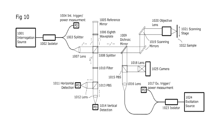
[0061] Fig. 10
highlights one possible implementation of CEPARS. A polarized
interrogation source (1001) is fed into a beam-splitter (1008) which directs a
portion of the
beam towards the sample path and another portion towards the reference mirror
(1005). The
sample path of the interrogation is then combined .with the excitation path
using an
CA 03089693 2020-07-27
WO 2019/145764
PCT/IB2018/057585
appropriate dichroic mirror (1009). The two beams are then directed onto the
sample (1022)
using a set of scanning mirrors (1019) and an objective lens (1020)õ Here,
scanning can also
be performed using a mechanical scanning stage (1021) to overcome field of
view limitations
of the objective. The reference path passes through an eighth wave plate
(1006) twice
providing a circular polarized state where the total path length is controlled
by position of the
reference mirror. This circular polarized state will inherently provide the
two desired
reference phases. The linear polarized sample path returning from the sample
is then
combined with the circular reference path at the beam-splitter. Excess
excitation light which
has been transmitted through the dichroic mirror is further rejected by the
use of a narrow
filter (1010). Finally the two polarization states are split using a polarized
beam-splitter
(1013) and individual detection is then performed. Since this device will
inherently be
sensitive to polarization-dependent scattering in the sample, it may also be
necessary to first
characterize the given interrogation location with the reference path blocked
such that the
relative received values can be appropriately adjusted.
[0062] Fig. 11 highlights another possible implementation of CHARS. This
implementation features primarily fiber-based optics and takes advantage of a
randomly
polarized interrogation source to avoid polarization-dependent sensitivity at
the sample. The
interrogations source (1101) is split (1110) between the reference and sample
pathways as
before. Here, the reference path is further split (1114) to provide the
desired added phase
offsets. Polarization-independent circulators (1113. 1115, 1116) then redirect
the reference
paths (R1, R2) towards respective beam couplers (1106, 1107) where they are
combined with
the sample path components (Si, S2).
[0063] Fig. 12 highlights another possible implementation of CEPARS. This
implementation features a serial acquisition as opposed to those represented
and Figs. 10 and
11, which utilize a parallel capture. Serial CEPARS only may necessitate a
single low-
coherence interferometer, but may require multiple acquisitions. Moreover,
subsequent
acquisitions must be performed with a varied reference path-length. For
example, a dual
acquisition might take one acquisition with a 7/2 phase offset relative to a
first acquisition
provided by a piezo-mounted mirror (1205). In this manner, the in-phase and
quadrature data
can still be captured.
[0064] Fig.13 highlights yet another possible implementation of CEPARS.
This
implementation features a serial acquisition as that presented in Fig. 12.
However, rather than
focusing directly onto a sample with free -space optics, here of the
excitation and sample-path
CA 03089693 2020-07-27
WO 2019/145764
PCT/IB2018/057585
16
of the interrogation beams are coupled into a .fiber which is fed through an
endoscopic probe.
At the distal end, optical focusing is provided by a GRIN lens (1327), and
optical scanning is
provided by a set of MEMS mirrors (1319). This represents a compact
implementation
capable of accessing challenging locales.
[00651 Fig. 19 highlights yet another possible implementation of CEPARS.
This
implementation utilizes a full optical-quadrature detection path. Unlike other
and simpler
described architectures, this embodiment may not require additional
calibration, may not
require assumption of small self-interference terms, and may not require
multiple acquisition
events providing more complete characterization of the tissue. The detection
pathways
include an interrogation source (1901), which is polarized (1905) and split
(1903). The
sample path transmits through a polarization-sensitive splitter (1923), is
circularly polarized
(by a quarter waveplate 1925), combined with the excitation pathway (at
dichroic minor
1926) and directed to the sample. The back-reflected portion is converted back
to a linear
polarization state (at quarter waveplate 1925), has remaining excitation
removed by a filter
(1924) and the light is again passed through a linear polarizer (1922) to
ensure a clean
polarization state. The reference path consists of a similar non-reciprocal
pathways using a
quarter-wave plate (1910) and PBS (1911). A dispersion cell (1909) can be
added .to
compensate for sample-path dispersion. The length of this path can be
controlled by changing
the reference mirror (1908) position thr appropriate depth selection within
the sample. This
light is circularly polarized (by quarter waveplate 1912) contributing a. -
rr/2 phase shift along
one axis and recombined with the sample pathway in a non-polarizing splitter
(1917). These
two paths which are composed to multiple polarization states are further
separated in two
PBSs (1916, 1921) yielding the desired combinations of sample-path and
reference-path
phase delays for full-quadrat-lire detection across four sensors (1913, 1915,
1918, 1920). The
sensors 1913,. 1915, 1918, and 1920 may be optical sensors, such as, e.g., a
single
photodiode, array of photodiodes, CCD etc. Then, the collected data will be
processes to
extract the PARS modulation quadratnre information.
[0066] In some embodiments of SDCG-PARS, one .goal is to provide a full
depth-
resolved optical absorption profile of a sample without necessitating axial
optical scanning.
Conceptually, this is similar to how SD-OCT is operated. However the
techniques are highly
distinct from each other. First, it is assumed that the optical section can be
considered a
collection of ideal reflectors at some spatial distribution (along the z
direction) such .that is
can be represented as r,(z). By interrogating the sample with a range of
optical frequencies,
CA 03089693 2020-07-27
WO 2019/145764
PCT/IB2018/057585
17
commonly implemented as either a swept source or a stationary broadband
spectrum source,
a respective reflection spectrum can be collected. This involves combining the
back-reflected
light .from the sample with a reference such that the interference .fringes
now encode the
locations of the optical scatterers within the optical section. Recovery of
the spatial reflection
distribution then simply involves performing a frequency transform on the
collected.
spectrum. Since the PARS mechanism involves modulation of the optical
scattering
properties within a sample where these modulations correspond to locations of
optical
absorption, by comparing the distribution of scatterers both before and
directly after
photoacousfic excitation by an execution pulse, the difference at a. given
location will now
correspond to the PARS-modulated regions which are optically absorbing.
However.,
although high-bandwidth detectors are ideal for such a task they may prove
highly
impractical for implementation, and as such there is a requirement for a means
of providing
these two distinct interrogations. One proposed method is the use of a short
(<10Ons) pulsed,.
or modulated interrogation laser which can effectively force a short
interrogation time on a
lower bandwidth detector such as a. CCD array by reducing the amount of time
back-reflected
light from the sample will be incident on the array. This method allows for
proper control
over the relative timing of the excitation and interrogation pulses and the
duration of the
interrogation time.
[0067] Fig. 14 shows an example of the relative timing between the
reflectivity properties
of a. given wavelength in the sample, and the excitation and interrogation
pulses. The second
interrogation pulse which corresponds to be excited sample, must be timed such
that it takes
full advantage of the perturbed sample. This exact. timing will vat
significantly given all the
available parameters such as the sample under consideration, the time
evolution
characteristics of the excitation, and the time revolution Characteristics of
the interrogation. In
general, the rising edge of .the interrogation will be less .than ins from the
rising edge of the
excitation. The duration of the interrogation pulse will also be less than
his.
[0068] Fig. 15 shows an example of two collected spectra. One of the
spectra is
associated with an unperturbed interrogation event, the other is associated
with an excited
interrogation event. The small differences An between the spectra are
associated with the
PARS-modulated regions.
[0069] Fig. 16 shows a flow chart of the collection and processing involved
with SDCG-
PARS. The two collected spectra are first deconvolved with the original
spectral content
SM. Here, other processing steps may be taken to reduce the effects of noise
and other non-
CA 03089693 2020-07-27
WO 2019/145764
PCT/IB2018/057585
18
desirable effects. The spectra are then transfoimed back into a physical
distribution
representing the relative strength of optical scattering at a given depth
rs(z). The envelope of
each scattering distribution is taken, then the two envelopes are subtracted
from each other to
form a SDCG-PARS A-line. One of the two original envelopes can also be used to
produce a
conventional SD-OCT A-line,
[0070] Fig. 17 shows an example system of a fiber-based SDCG-PARS. A pulsed
interrogation source (1701) is split (by a splitter 1703) such that a portion
is collected at a
detector (1704) to characterize pulse-to-pulse consistency. The other portion
is split into a
sample path and a reference path. The reference path is directed on a
reference minor (1711)
such that the total path length will be appropriately similar to the total
sample path length
facilitating low-coherence interferometry. The sample path is combined with
the pulsed
excitation source through a multiplexer (1713). The two beams are then scanned
along the
surface of the sample with a set of galvanometer mirrors (1725) and an
appropriate objective
lens (1726). The back-reflected light from the reference path and the sample
path are then
combined in a. fiber coupler (1706) such that they interfere with each other.
This resulting
light is then fed into a CCD-based spectrometer (1705) for detecting of the
spectra.
[0071] Fig. 18 shows another example of a SDCG-PARS, here with an
.endoscopic
implementation. The primary difference between this and the previous
embodiment is that
after the multiplexer (1813), the combined beams are fed into an endoscopic
casing (1812).
Positioning of the .final focus is now controlled by the use of a GRIN lens
(1815) at the distal
end of the fiber which is focusing through a MEMS mirror (1816) which provides
lateral
scanning of the interrogation spot on the sample (1817).
[0072] It will be apparent that other examples may be designed with
different
components to achieve similar results. Other alternatives may include various
coherence
length sources, use of balanced photodetectors, interrogation-beam
modulation., incorporation
of optical amplifiers in the return signal path, etc.
[0073] During in vivo imaging experiments, no agent or ultrasound coupling
medium are
required. However the target can be prepared with water or any liquid such as
oil before non-
contact imaging session. No special holder or immobilization is required to
hold the target
during imaging sessions.
[0074] Other advantages that are inherent to the structure will be apparent
to those skilled
in the art. The embodiments described herein are illustrative and not intended
to limit the
CA 03089693 2020-07-27
WO 2019/145764
PCT/IB2018/057585
19
scope of the claims, which are to be interpreted in light of the specification
as a whole.
[0075] The excitation beam may be any pulsed or modulated source of
electromagnetic
radiation including lasers or other optical sources. In one example, a
nanosecond-pulsed laser
was used. The excitation beam may be set to any wavelength suitable for taking
advantage of
optical (or other electromagnetic) absorption of the sample. The source may be
monochromatic or polychromatic.
[0076] The interrogation beam may be any pulsed, or modulated source of
electromagnetic radiation including lasers or other optical sources. Any
wavelength can be
used for interrogation purpose depending on the application.
[0077] CG-PARS may use any interferometry designs such as common path
interferometer (using specially designed interferometer objective lenses),
Michelson
interferometer, Fizeau interferometer, Ramsey interferometer, Fabrv-Perot
interferometer,
Mach¨Zelmder interferometer, and optical-quadrature detection. The basic
principle is that
phase (and maybe amplitude) oscillations in the probing receiver beam can be
detected using
interferometry and detected at AC, RF or ultrasonic frequencies using various
detectors.
[0078] In one example, both excitation and receiver beams may be combined
and
scanned. In this way, photoacoustic excitations may be sensed in the same area
as they are
generated and where they are the largest. Other arrangements may also be used,
including
keeping the receiver beam fixed while scanning the excitation beam or vice
vena.
Galvanometers, MEMS mirrors, polygon scanners, and stepper/DC motors may be
used as a
means of scanning the excitation hem'', probe/receiver beam or both.
[0079] The excitation beam and sensing/receiver beam can be combined using
dichroic
minors, prisms, beam splitters, polarizing beam splitters etc. They can also
be focused using
ditierent optical paths.
[0080] The reflected light may be collected by photodiodes, avalanche
photodiodes,
phototubes, photomultipliers, CMOS cameras, CCD cameras (including EM-CCD,
intensified-CCDs, back-thinned and cooled CCDs), etc. The detected signal may
be amplified
by an RF amplifier, lock-in amplifier, trans-impedance amplifier, or other
amplifier
configuration. Also different methods may be used in order to filter the
excitation beam from
the receiver beam before detection. CG-PARS may use optical amplifiers to
amplify detected
light.
[0081] A table top, handheld, endoscopic, surgical microscope, or
ophthalmic CG-PARS
CA 03089693 2020-07-27
WO 2019/145764
PCT/IB2018/057585
system may be constructed based on principles known in the art. CG-PARS may be
used for
A-. B- or C- scan images for in vivo, ex vivo or phantom studies.
[0082] CC-PARS may be optimized in order to take advantage of a multi-focus
design
for improving the depth-of-focus of 2D and 3D OR-CG-PARS imaging. The
chromatic
aberration in the collimating and objective lens pair may be harnessed to
refocus light from a
fiber into the object so that each wavelength is focused at a slightly
different depth location.
Using these wavelengths simultaneously may be used to improve the depth of
field and signal
to noise ratio (SNR) of CG-PARS images. During CG-PARS imaging, depth scanning
by
wavelength tuning may be performed.
[0083] The CG-PARS system may be combined with other imaging modalities
such as
fluorescence microscopy, two-photon and confocal fluorescence microscopy,
Coherent-Anti-
Raman-Stokes microscopy, Raman microscopy, Optical coherence tomography, other
photoacoustic and ultrasound systems, etc. This systems could be combined by
designing, an
optical combiner to integrate the optical paths of each systems. Also a
processor to
synchronise the results if necessary and analyse the results either separately
or in
combination. These integrated modalities can bring complementary imaging
contrast. This
could permit imaging of the microcirculation, blood oxygenation parameter
imaging, and
imaging of other molecularly-specific targets simultaneously, a potentially
important task that
is difficult to implement with only fluorescence based microscopy methods. A
multi-
wavelength visible laser source may also be implemented to generate
photoaconstic signals
for functional or structural 'imaging.
[0084] Polarization analyzers may be used to decompose detected light into
respective
polarization states. The light detected in each polarization state may provide
information
about ultrasound-tissue interaction.
[0085] APPLICATIONS
[0086] It will be understood that the system described herein may be used
in various
ways, such as those purposes described above, and also may be used in other
ways to take
advantage of the aspects described above. A non-exhaustive list of
applications is discussed
below.
[0087] The system may be used for imaging akg,iogenesis for different pre-
clinical tumor
models.
[0088] The system may also be used for clinical imaging of micro- and macro-
circulation
CA 03089693 2020-07-27
WO 2019/145764
PCT/IB2018/057585
21
and pigmented cells, which may find use for applications such as in (1) the
eye, potentially
augmenting or replacing fluorescein angiography; (2) imaging dermatological
lesions
including melanoma, basal cell carcinoma, hemangioma, psoriasis, eczema,
dermatitis,
imaging Molts surgery, imaging to verify tumor margin resections; (3)
peripheral vascular
disease; (4) diabetic. and pressure ulcers; (5) bum imaging; (6) plastic
surgery and
microsingely; (7) imaging of circulating tumor cells, especially mehmoma.
cells; (8) imaging
lymph node angiogenesis; (9) imaging response to photodynamic therapies
including those
with vascular ablative mechanisms; (10) imaging response to chemotherapeutics
including
anti-angiogenic drugs; (11) imaging response to radiotherapy.
[0089] The system may be useful in estimating oxygen saturation using multi-
wavelength
photoacoustic excitation and CO-PARS detection and applications including: (1)
estimatim,
venous oxygen saturation where pulse oximetry cannot be used including
estimating
cerebrovenous oxygen saturation and central venous oxygen saturation. This
could
potentially replace catheterization procedures which can be risky, especially
in small children
and infants.
[0090] Oxygen flux and oxygen consumption may also be estimated by using CO-
PARS
imaging to estimate oxygen saturation, and an auxiliary method to estimate
blood flow in
vessels flowing into and out of a region of tissue.
[0091] The system may also have some gastroenterological applications, such
as imaging
vascular beds and depth of invasion in Barrett's esophagus and colorectal
cancers. Depth of
invasion is key to prognosis and metabolic potential. Gastmentemlogical
applications may be
combined or piggy-backed off of a clinical endoscope and the miniaturized CO-
PARS system
may be designed either as a standalone endoscope or fit within the accessory
channel of a
clinical endoscope.
[0092] The system may have some surgical applications, such as functional
imaging
during brain surgery, use for assessment of internal bleeding and
cauterization verification,
imaging perfusion sufficiency of organs and organ transplants, imaging
angiogenesis around
islet transplants, imaging of skin-grafts, imaging of tissue scaffolds and
biomaterials to
evaluate vascularization and immune rejection, imaging to aid microsurgery,
guidance to
avoid cutting critical blood vessels and nerves.
[0093] Other examples of applications may include CO-PARS imaging of
contrast agents
in clinical or pre-clinical applications; identification of sentinel lymph
nodes; non- or
CA 03089693 2020-07-27
WO 2019/145764
PCT/IB2018/057585
minimally-invasive identification of tumors in lymph nodes; imaging of
genetically-encoded
reporters such as tyrosinaseõ c.hromoproteins, fluorescent proteins for pre-
clinical or clinical
molecular imaging applications; imaging actively or passively targeted
optically absorbing
nanoparticles for molecular imaging; and imaging of blood clots and
potentially staging the
age of the clots.
[00941 In some embodiments, any suitable technology, such as, e.g.. OCTõ
can be used
for surface topology (for constant- or variable-depth focusing kw
photoaeoustic remote
sensing technologies) before imaging with CG-PARS.
[0095] In at least some embodiments, systems of the present disclosure may
include
variable-focal-length lenses (including voice-coil-driven, MEMS-based,
piezoelectric-based,
and tunable acoustic gradient lenses). Furthermore, systems of the present.
disclosure may
include double-clad fiber couplers for both OCT and PARS microscopy (including
CG-
PARS) to deliver excitation light (and/or interrogation light) from a single-
mode fiber to the
sample, but collect interrogation light using the multi-mode cladding of the
double-clad fiber.
Systems of the present disclosure also may be used with angiography or
Doppler.
[0096] Embodiments of the present disclosure may include one or more of the
following
advantages:
1. The proposed CG-PARS provides depth-dependent contrast which is directly
proportional to optical absorption of the excitation laser. For example, CW CE-
PARS
extracts modulated components of signals using high-pass or bandpass filters.
Pulsed
detection systems associated with Pulsed CE-PARS or SD-CG-PARS uses
differences
in detected signals with and without excitation pulses.
.2. The coherence length of the source is preferably shorter than the depth-of-
focus of the
interrogation beam into the sample, and more preferably significantly shorter.
In this
way, improved depth resolution can be achieved by use of coherence-gating.
3. The proposed SD-CG-PARS system incorporates a spectrometer and is able to
detect
enveloped A-scans with and without excitation pulses (or with different pulse
energies). The system uses a processor for extracting differences in the
enveloped A-
scans with and without excitation pulse (or with different pulse energies).
4. In the proposed CE-PARS system, there may be two or more interferometers,
or a
method for sequentially interrogating with two or more successive reference
path
phase shifts and a processor for combining serial or parallel acquisitions to
extract
CA 03089693 2020-07-27
WO 2019/145764
PCT/IB2018/057585
23
temporal modulations of the envelope signal.
5. The proposed CG-PARS methods uses OCT signals to detect refractive index
changes
associated with initial pressures and uses at least two acquisitions (either
in serial or
parallel with multiple detectors). In SD-CO-PARS, an A-scan OCT envelope
acquisition is obtained with and without and excitation pulse, where each A-
scan is
acquired with a spectrometer. hi CE-PARS we in-phase and quadrature components
of the signal are acquired.
6. As noted, the SD-CG PARS method uses a spectrometer. Additionally, SD-CG-
PARS
may be used to detect enveloped OCT A-scans with and without excitation pulses
(or
with different pulse energies). The phase in the detected signal may be
removed to
form an envelope. For SD-CG-PARS, a processor may be used to extract
differences
in the enveloped A-scans with and without excitation pulses (or with different
pulse
energies).
[0097] Certain examples of remote sensing systems may be described as
follows:
1. A Spectral-Domain Coherence-Gated PARS Tomography (SD-CG-PARS
Tomography) system having:
a. A pulsed excitation electromagnetic radiation source
b. A low-coherence interrogation light source, the coherence length being
the principal determinant of the depth resolution. Typically, the
interrogation
wavelengths and the excitation wavelengths are spectrally distinct, but in an
optional
embodiment, the excitation and interrogation sources could be one and the
same.
c. A combiner to combine the pulsed excitation beam and .the
interrogation beam to enable co-scanning of both beams
d. Focusing lens(es) for focusing the respective or combined beams and
for collecting interrogation light from the sample.
e. An interferometer, having a splitter to split the interrogation beam
into
a reference path and a signal path, the reference path having an adjustable
path-length,
and the signal path returning collected signals back to interfere with
reference path
light.
f. A light analysis module consisting of a spectrometer (with various
CA 03089693 2020-07-27
WO 2019/145764
PCT/IB2018/057585
24
types of dispersive elements: gratings, prisms, etc) and detector arrays (CCD,
CMOS,
photodiode array).
g. A temporal gating system to ensure that optical signals recorded after
the excitatioi . pulses are read out within a short (<::: tens of nanoseconds
or <ins) time-
scale before acoustic waves propagate far from their ofigin. Specifically, the
acoustic
distance-of-propagation over the interrogation readout time should not be
significantly greater than the desired axial or lateral spatial resohition.
This temporal
gating could be accomplished using (1) a (fs-us-scale pulsed interrogation
source and
pulse-sequencer and acquisition electronics carefully timed to read out
signals within
nanoseconds after the excitation source. (.2) an optical or electronic shutter
with
nanosecond-scale response times to enable the capture of ONLY the
interrogation
light within the desired temporal window (3) fast photodiode array to
electronically
capture time domain signals from each element and capturing only the first I
time
samples.
h. A pulse sequencer and acquisition system for forming at least two
OCT A-scan lines per scan position: one with an excitation pulse and one
without an
excitation pulse OR one with a different excitation pulse energy than another.
i. Optional reference photodiode measurement subsystem to account for
pulse-to-pulse variations of the excitation source and for variations in the
interrogation source.
J- Optional programmable controller and actuator to adjust the
reference
pathiength between scans or to adjust the desired depth-sectioning.
k. Optional filter to reject excitation laser wavelengths from
being
detected by the spectrometer detectors.
I. A processor for processing the OCT RF A-scan lines (with- and
without excitation pulses or with differing pulse energies and optionally with
and.
without reference pathlength shifts) to form CG-PARS A-Scans with contrast
proportional to optical absorption at each depth position. One such processor
embodiment includes fuming the envelope of each OCT A-scan and subtracting the
envelopes of A-scans with and without excitation laser pulses. This strategy
has the
advantage of eliminating unwanted phase-noise sensitivity but will still
capture
refractive index changes associated with photoacoustic initial pressures.
CA 03089693 2020-07-27
WO 2019/145764
PCT/IB2018/057585
ra A processing system to render and display OCT and CG-PARS
images
2. A coherence-enhanced PARS (CE-PARS) microscopy system having:
a. A pulsed excitation light source
b. A low-coherence interrogation laser, the coherence length being the
principal determinant of the depth resolution. Typically, the interrogation
wavelengths
and the excitation wavelengths are spectrally distinct, but in an optional
embodiment,
the excitation and interrogation sources could be one and the same..
c. A combiner to combine the pulsed excitation beam and .the
interrogation beam to enable co-scanning of both beams
d. Focusing lens(es) for focusing the respective or combined beams and
for collecting interrogation light from the sample.
e. An interferometer, having a splifter to split the inteno.g,ation beam
into
a reference path and a signal path, the reference path having an adjustable
path-length,
and the signal path returning collected signals back to interfere with
reference path
light
f. Light detection module(s) including associated optional amplifiers and
filters, for example, consisting of photodiode(s) or balanced photodiode(s).
Filters
may be included to reject DC scattered light and collect only the modulated
component in the case of CW interrogation beams. See description of module for
pulsed interrogation light below.
g. A method for acquiring effective inphase- and quadrature complex
envelope skuials from the interfering light using one of two methods (1)
serially, by
performing a point-scan, lateral-scan, depth-scan or C-scan thei . adjusting
the
reference pathlength by 7/2 phase then scanning again, (2) in parallel by
using an
additional interferometer with a reference path differing by 7/2 from the
reference
path of the other interferometer. This parallel interferometer may be
implemented.
with separate optical paths or as a common-path configuration. Note that this
quadrature-sampling scheme offers the flexibility of C-scanning or en-face
scanning
at a particular depth gating (or depth range) without requiring acquisition of
complete
depth-scans (A-scans) to create an effective PARS image within a precise depth-
CA 03089693 2020-07-27
WO 2019/145764
PCT/IB2018/057585
'")6
section. If an A-scan is acquired, there mast be an excitation pulse for every
depth
sample in the A-scan line, which could lead to unwanted persistent laser
exposure
compared to the scanned beam case.
Ii. A processor for estimating the envelope or specifically, the
magnitude
of the complex envelope signal for cases with and without an excitation pulse
(or with
excitation pulses of different strengths).
i. A processor for processing the OCT RE envelope signals (with- and
without excitation pulses or with differing pulse energies) to form CE-PARS
signals
with contrast proportional to optical absorption at each scan position. One
such
processor embodiment includes forming the envelope of each OCT signal and
subtracting the envelopes with and without excitation laser pulses. This
strategy has
the advantage of eliminating unwanted phase-noise sensitivity but will still
capture
refractive index changes associated with photoacoustic initial pressures.
j. A temporal gating system to ensure that optical signals recorded after
the excitation pulses are read out within a short (.< tens of nanoseconds)
time-scale
before acoustic waves propagate far from their origin. Specifically, the
acoustic
distance-of-propagation over the interrogation readout time should not be
significantly greater than the desired axial or lateral spatial resolution.
This temporal
gating could be accomplished using (1) a nanosecond-scale pulsed interrogation
source and pulse-sequencer and acquisition electronics carefully timed to read
out
signals within nanoseconds afler the excitation source. (2) an optical or
electronic
shutter with nanosecond-scale response .times to enable the capture of ONLY
the
interrogation light within the desired temporal window (3) acquiring the
photodiode
signal as a function of time then sampling only the first few tens to hundreds
of us OR
(4) using an analog or digital peak detector to extract the peak (envelope)
signal or
peak-to-peak (envelope) signal for each pulse.
k. A processing system to render and display OCT and CG-PARS
images.
3. A pulsed interrogation detection subsystem which involves capturing an
interrogation pulsed signal from the sample (with or without reference beam
interference)
both with or without an excitation pulse (or with differing pulse energies)
and subtraction of
CA 03089693 2020-07-27
WO 2019/145764
PCT/IB2018/057585
the respective signals or estimating then relative difference normalized to
the OCT signal
without an excitation pulse present. This can be done by recording amplified
photodiode
signals with an analog-to-digital converter and doing the subtraction (and
optionally division)
operations digitally. It can also be done with analog electronics
4. A functional imaging system involving sequential pulses using (I) different
excitation wavelengths or (2) different pulse widths (e.g.. ps pulses and us
pulses). In the case
of both (I) and (2) the PARS initial pressure signals are proportional to
optical absorption
and detected optically using interrogation beams using the CG-PARS systems
described
above, or using previously described interferometric or non-interferometric
PARS systems.
[0098] Examples of methods of for remote sensing may be described as
follows:
a. (SDCG-PARS) A method for interrogating the optical properties of a sample
which
comprises
A method of generating photoacoustic signals within a sample;
A low-coherence interferometer used to detect photoacoustic signals;
A method for directing light towards a sample at a given location;
A method of collecting light from a sample at a given location;
An optical spectrum detector;
A processor for collecting multiple optical spectra; and
A processor for extracting differences between multiple optical spectra.
I. The method of statement a., wherein the method of generating
photoacoustic signals within a sample comprises a nanowband or
broadband electromagnetic source which is one of a pulsed source, or a
continuous-wave source which is intensity modulated.
i. The method of L. wherein the portion of the excitation
source is
detected by photodiode to account for pulse-to-pulse variations.
II. The method of statement a., wherein the low-coherence interferometer
comprises a broadband electron awietic source which is one of a
pulsed source, or a continuous-wave source which is intensity
modulated, a method a splitting this beam into a reference path and a
sample path, and a method of combining the beams returning from the
CA 03089693 2020-07-27
WO 2019/145764
PCT/IB2018/057585
28
reference path and the sample path.
i. The method of statement IL wherein the optical spectrum
detector comprises one or more dispersive elements (gratings,
prisms, etc) and one or more detector arrays (CCD, CMOS,
photodiode, etc.).
ii. The method of statement IL. wherein the portion of the
interrogation source is detected by photodiode to account for
pulse-to-pulse variations.
III. The method of statement a, wherein the low-coherence interferometer
comprises a broadband continuous-wave source electromapietic
source, a method of splitting this beam into a reference path and a
sample path, and a method of combining the beams returning from the
reference path and the sample path.
i. The method of statement III, wherein the optical spectrum
detector comprises one or more dispersive elements (gratings,
prisms, etc) and one or more high-bandwidth detector arrays
(photodiode, avalanche photodiode, etc.).
ii. The method of statement HT, wherein the portion of the
interrogation source is detected by photodiode to account for
power and variations.
IV. The method of statement a., wherein the method for directing towards
and from a sample comprises of an optical seamier (one or more of
Galvanometer mirrors, resonant mirrors, MEMS mirrors, polygon
seamier, etc.), focusing optic subsystem (objective lens, reflective
objective, parabolic mirror, GRIN lens, and a system of optical filters
to reject excitation wavelengths along the detection path..
V. The method of statement a., wherein the method for directing towards
and from a sample comprises of an light guide (optical fiber, double
clad fiber, optical fiber bundle, etc,), an optical scanner (one or more of
Galvanometer mirrors, resonant mirrors, MEMS mirrors, polygon
seamier, etc.), and focusing optic subsystem (objective lens, reflective
objective, parabolic minor, GRIN lens), and a system of optical filters
to reject excitation wavelengths along the detection path.
VI. The method of statement a, wherein the processors for collecting
CA 03089693 2020-07-27
WO 2019/145764
PCT/IB2018/057585
29
multiple optical spectra and for extracting differences between multiple
optical spectra are implemented as electronic devices.
b. (Parallel CEPARS) A method for interrogating the optical properties of a
sample
which comprises:
A method of generating photoacoustic sigials within a sample;
Two or more optical low-coherence interferometers used to detect
photoacoustic signals;
A method for directing light towards a sample at a given location;
A method of collecting light from a sample at a given location;
A processor to combine data channels from the interferometers; and
A processor to extract temporal modulations of the envelope signal.
I. The method of statement b, wherein the method of generating
photoac.oustic signals within a sample comprises a narrowband or
broadband electromagnetic source which is one of a pulsed source, or a
continuous-wave source which is intensity modulated.
i. The method of statement I, wherein the portion of the
excitation source is detected by photodiode to account for
pulse-to-pulse variations.
H. The method of statement b, wherein the method for directing tol,varlis
and from a sample comprises' of an optical scanner (one or more of
Galvanometer mirrors, resonant mirrors, MEMS mirrors, polygon
seamier, etc.), focusing optic subsystem (objective lens, reflective
objective, parabolic. mirror. GRIN lens, and a system of optical filters
to reject excitation wavelengths along the detection path.
I. The method of statement b, wherein the method for directing towards
and from a sample comprises of an light guide (optical fiber, double
clad fiber, optical fiber bundle, etc.), an optical scanner (one or more
of Galvanometer mirrors:, resonant mirrors, MEMS mirrors, polygon
seamier, etc.), and focusing optic subsystem (objective lens, reflective
objective, parabolic mirror, GRIN lens), and a system of optical filters
to reject excitation wavelengths along the detection path.
CA 03089693 2020-07-27
WO 2019/145764
PCT/IB2018/057585
c. (serial QSCG-PARS) A method for interrogating the optical properties of a
sample
which comprises:
A method of generating photoacoustic .sipials within a sample;
A low-coherence interferometer used to detect photoacoustic signals where
reference phase must be adjusted between sequential acquisitions;
A method for directing light towards a. sample at a given location;
A method of collecting light from a sample at a given location:.
A method of acquisition which necessitates multiple acquisitions;
A processor to combine serial data channels from the interferometer; and
A processor to extract temporal modulations of the envelope signal.
I. The method of statement c, wherein the method of generating
photoacoustic signals within a sample, comprises a narrowband or
broadband electromagnetic source which is one of a pulsed source, or a
-continuous-wave source which is intensity modulated.
ii. The method of statement I, wherein the portion of the
excitation source is detected by photodiode to account for
pulse-to-pulse variations.
H. The method of statement
c, wherein the method for directing towards
and from a sample comprises of an optical scanner (one or more of
Galvanometer mirrors, resonant minors, ME-MS mirrors, polygon
scanner, etc.), focusing optic subsystem (objective lens, reflective
objective, parabolic minor, GRIN lens, and a system of optical filters
to reject excitation wavelengths along the detection path.
HI. The method of statement
c, wherein the method for directing towards
and from a sample comprises of .an light guide (optical fiber, double
clad fiber, optical fiber bundle, etc.), an optical .scanner (one or more
of Galvanometer mirrors., resonant mirrors, MEMS mirrors, polygon
scanner, etc.), and focusing optic subsystem (objective lens, reflective
objective, parabolic mirror. GRUNT lens), and a system of optical filters
to reject excitation wavelengths along the detection path.
d. (Quadrat:me CEPARS) A method for intenogating the optical properties of a
sample
which comprises:
A method of generating photoacoustic signals within a sample;
CA 03089693 2020-07-27
WO 2019/145764
PCT/IB2018/057585
31
An optical quadrature detector;
A method for directing light towards a sample at a given location;
A method of collecting light from a sample at a given location;
A processor to combine data. channels from the quadrature .detector; and
A processor to extract temporal modulations of the envelope signal.
1. The method of statement d., wherein the method of
generating
photoacoustic signals within a sample comprises a. narrowband or
broadband electromagnetic source which is one of a pulsed source,
or a. continuous-wave source which is intensity modulated.
i. The method of statement 1, wherein the portion of the
excitation source is detected by pho.todiode to account for
pulse-to-pulse variations.
The method of statement d., wherein the method for directing
towards and from a sample comprises of an optical scanner (one or
more of Galvanometer mirrors, resonant mirrors, MEMS mirrors,
polygon scanner, etc.), focusing optic subsystem (objective lens,
reflective objective, parabolic mirror, GRIN lens, and a system of
optical filters to reject excitation wavelengths along the detection
path.
III. The method of statement d, wherein the method for directing
towards and .from a sample comprises of an light guide (optical
fiber, double clad fiber, optical fiber bundle, etc.), an optical
scanner (one or more of Galvanometer mirrors, resonant minors,
MEMS mirrors, polygon scanner, etc.), and focusing optic
subsystem (objective lens, reflective .objective, parabolic minor,
GRIN lens.), and a system of optical filters to reject excitation
wavelengths along the detection path.