Note: Descriptions are shown in the official language in which they were submitted.
CA 03089708 2020-07-27
WO 2019/156985
PCT/US2019/016702
PATIENT-SPECIFIC BONE FRACTURE PROSTHESES
AND METHODS OF MAKING THE SAME
INCORPORATION BY REFERENCE TO ANY PRIORITY APPLICATIONS
[0001] Any and all applications for which a foreign or domestic
priority claim
is identified in the Application Data Sheet as filed with the present
application are hereby
incorporated by reference under 37 C.F.R. 1.57.
BACKGROUND OF THE INVENTION
Field of the Invention
[0002] The invention relates to methods for manufacturing patient-
specific
prostheses for a fractured long bone of a patient and to patient-specific
prostheses for a
fractured long bone of a particular patient.
Description of the Related Art
[0003] US 2004/0230311 Al discloses a shoulder prosthesis comprising
a
stem to be inserted into the canal of the diaphyseal fragment of the humerus
of a patient,
an intermediary part reduced to a medial pillar and a head which is a
generally spherical
hollow cap. This type of shoulder prosthesis allows reattaching tuberosity
fragments and
humeral head fragments of the original humerus to the prosthesis.
SUMMARY OF THE INVENTION
[0004] For reducing the risk of osteonecrosis of the reattached bone
fragments
and diaphyseal fragment, it is preferable that these fragments are
mechanically loaded in
the patient's body. The known shoulder prosthesis do not always ensure that
every
fragment is appropriately mechanically loaded, since the shape and the size of
the
fragments differ from one patient to another and do not always correspond to
the bone
fragments.
[0005] The inventions aim to solve the deficiencies of the
abovementioned
prior art.
[0006] An aim of one or more of the inventions is to provide a new
method for
manufacturing a prosthesis for a fractured long bone of a patient, reducing
the likelihood
of osteonecrosis of the reattached bone fragments after implantation into the
patient's
body.
-1-
CA 03089708 2020-07-27
WO 2019/156985
PCT/US2019/016702
[0007] In another embodiment, data representative of a fractured
long bone of
a patient is obtained (step A). The data is used for designing (step B) and
manufacturing
(step C) a patient-specific prosthesis. The prosthesis includes a stem part
with patient-
specific features that allow, specifically to the patient, distributing
mechanical loads onto
one or more bone fragments, when the prosthesis is inserted into the patient's
body. In
other words, the prosthesis is manufactured with a shape tailored to the bone
fragment(s).
Thus, a patient-specific mechanical stress is applied to the bone fragment(s)
when the
prosthesis is introduced into the patient's body and during healing of the
fracture. The
mechanical stress applied to the bone fragment corresponds to conditions that
may be
planned in advance, prior to the step C of manufacturing the prosthesis. In
particular,
before the step of manufacturing, the surgeon may choose or plan, specifically
to a
particular patient, how the bone fragment will be submitted to mechanical
stress when the
prosthesis is implemented in the patient' s body, and the prosthesis is
manufactured in a
way that this mechanical stress is actually achieved. The risk of post-surgery
osteonecrosis is highly reduced, since the bone fragment is mechanically
loaded exactly
according to the needs of the patient's body.
[0008] The stem part may have a shape to be nested into the
medullary cavity
of the diaphyseal fragment of the patient to whom the prosthesis is intended,
although the
shape, the bone density and/or other parameters of the diaphyseal fragment may
differ
from one patient to another. Thanks to its patient-specific nesting shape, the
stem part
applies an appropriate mechanical stress onto one or more contact zones of the
medullary
cavity. The mechanical stress to be applied can be chosen in advance, for
example by the
surgeon, as the patient's data is provided at step A, prior to designing the
prosthesis (step
B). Thus, the risk of osteonecrosis is reduced after the prosthesis is
implemented into the
patient.
[0009] An aim of the embodiments described herein is to provide new
methods for manufacturing a prosthesis for a fractured long bone of a patient,
implying
fewer chances of osteonecrosis of the reattached bone fragments after
implantation into
the patient's body.
[0010] A method for manufacturing a prosthesis for a fractured long
bone of a
patient, the method comprising the steps of:
-2-
CA 03089708 2020-07-27
WO 2019/156985
PCT/US2019/016702
[0011] A) providing data representative of the fractured long bone
of the
patient, the fractured long bone comprising epiphyseal fragments, each
epiphyseal
fragment preferably being:
= [0012] either a tuberosity fragment, secured to a
muscle of the
patient, the muscle being attached to the tuberosity fragment by means
of a tendon of the muscle, or
= [0013] an articular fragment, being part of a
joint of the patient, for
articulating the long bone with an auxiliary bone of the patient;
[0014] B) based on the data provided at step A), designing the
prosthesis
specifically to the patient, the prosthesis comprising a stem part configured
for securing
the epiphyseal fragments to the stem part, step B) comprising:
= [0015] a sub-step of choosing, specifically to the
patient, respective
securing positions of the epiphyseal fragments relative to each other,
representative of how the epiphyseal fragments are planned to be
positioned relative to each other when secured to the stem part, the
securing positions being chosen so that a respective chosen mechanical
stress is applied onto each epiphyseal fragment by at least one of the
other epiphyseal fragments, when the epiphyseal fragments are secured
at the respective chosen securing positions of the stem part, and
= [0016] a sub-step of designing the stem part so
that the stem part is
configured for securing the epiphyseal fragments at the respective
chosen securing positions; and
[0017] C) manufacturing the prosthesis including the stem part
designed at
step B).
[0018] In the invention, the data representative of the fractured
long bone of
the patient (step A) is used for designing (step B) and manufacturing (step C)
a patient-
specific prosthesis including a stem part with patient-specific features that
allow,
specifically to the patient, distributing mechanical loads onto the bone
fragments, when
the prosthesis is inserted into the patient's body. In other words, the
prosthesis is
manufactured with the most appropriate shape, tailored to the bone fragments.
Thus, a
patient-specific mechanical stress is applied to the bone fragments when the
prosthesis is
introduced into the patient's body and during healing of the fracture. The
mechanical
stress effectively applied to the bone fragments corresponds to conditions
that may be
-3-
CA 03089708 2020-07-27
WO 2019/156985
PCT/US2019/016702
planned in advance, prior to the step C of manufacturing the prosthesis. In
particular,
before the step of manufacturing, the surgeon may choose or plan, specifically
to a
particular patient, how the bone fragments will be submitted to mechanical
stress when
the prosthesis is implemented in the patient's body, and the prosthesis is
manufactured in
a way that this mechanical stress is actually achieved. The risk of post-
surgery
osteonecrosis is highly reduced, since the bone fragments are mechanically
loaded
according to the needs of the specific patient.
[0019] The stem part is built so that the epiphyseal fragments may
be
reattached at predetermined respective securing positions by the surgeon.
Preferably, the
stem part is configured with a patient-specific shape and features, such that
the epiphyseal
fragments can only be reattached at these respective securing positions. When
positioned
in this way, a patient-specific mechanical stress is applied by the bone
fragments to each
other. In this method, the stress applied on the bone fragments is chosen
prior to designing
the prosthesis, due to the data representative of the patient. This method
ensures that the
manufactured prosthesis, intended for a respective patient, will apply the
intended
mechanical stress to the fragments, even if the fragments differ from one
patient to the
other, in terms of shape, bone density or other parameters. This provides a
reduction in
the risk of osteonecrosis when the prosthesis is implemented in the specific
patient.
[0020] Further optional and advantageous features of the invention
are defined
below:
= [0021] Step B) comprises the further sub-step of
designing the
stem part so that the stem part comprises a visible mark indicating the
respective securing positions of the epiphyseal fragments on the stem
part.
= [0022] Step B) comprises the further sub-step of
designing the
stem part based on the data provided at step A), so that the stem part
comprises, specifically to the patient, coverable epiphyseal portions,
being configured for being covered respectively by the epiphyseal
fragments when the epiphyseal fragments are secured to the stem part
at the respective chosen positions; wherein the visible mark is designed
so as to visually delimit the coverable epiphyseal portions from each
other.
-4-
CA 03089708 2020-07-27
WO 2019/156985
PCT/US2019/016702
= [0023] The stem part comprises, for at least one
of the epiphyseal
fragments, a respective plug, for securing the concerned epiphyseal
fragment to the stem part, said at least one plug applying mechanical
stress onto the epiphyseal fragment secured thereto.
[0024] Another object of the invention is defined as follows:
[0025] A patient-specific prosthesis for a fractured long bone of a
particular
patient, the fractured long bone comprising epiphyseal fragments, the
prosthesis
comprising a stem part that is designed based on data representative of the
fractured long
bone of this particular patient, the stem part being configured so that each
epiphyseal
fragment may be secured to the stem part at a chosen securing position
relative to the
other epiphyseal fragments, the securing positions being chosen based on said
data so
that, when the epiphyseal fragments are secured at the respective securing
positions, a
respective chosen mechanical stress is applied onto each epiphyseal fragment
by at least
one of the other epiphyseal fragments.
[0026] Further optional and advantageous features of the invention
are defined
below:
= [0027] The stem part comprises, specifically to
the patient:
o [0028] a visible mark indicating the respective securing positions
of the epiphyseal fragments on the stem part; and
o [0029] coverable epiphyseal portions, being configured for being
covered respectively by the epiphyseal fragments when the
epiphyseal fragments are secured to the stem part at the respective
securing positions; wherein the visible mark is designed so as to
visually delimit the coverable epiphyseal portions from each other.
= [0030] The stem part comprises, for at least one
of the epiphyseal
fragments, a respective plug, for securing the concerned epiphyseal
fragment to the stem part, said at least one plug applying mechanical
stress onto the epiphyseal fragment secured thereto.
[0031] An aim of the invention is to provide a new method for
manufacturing
a prosthesis for a fractured long bone of a patient, implying fewer chances of
osteonecrosis of the reattached bone fragments after implantation into the
patient's body.
[0032] The invention is defined as follows:
-5-
CA 03089708 2020-07-27
WO 2019/156985
PCT/US2019/016702
[0033] A method for manufacturing a prosthesis for a fractured long
bone of a
patient, the method comprising the steps of:
[0034] A) providing data representative of the fractured long bone
of the
patient, the fractured long bone comprising:
= [0035] at least one viable tuberosity fragment,
secured to a muscle
of the patient, the muscle being attached to said at least one viable
tuberosity fragment by means of a tendon of the muscle and
= [0036] a damaged articular fragment, initially
being part of a
damaged joint of the patient, for articulating the long bone with an
auxiliary bone of the patient;
[0037] B) based on the data provided at step A), designing the
prosthesis
specifically to the patient, the prosthesis comprising:
= [0038] a stem part, configured for securing said
at least one viable
tuberosity fragment to the stem part, and
= [0039] a head part, being configured to be secured
to the stem part
in replacement for the damaged articular fragment,
[0040] step B) comprising:
= [0041] a sub-step of choosing, specifically to the
patient, respective
securing positions of said at least one viable tuberosity fragment and of
the head part relative to each other, representative of how said at least
one viable tuberosity fragment and head part are planned to be
positioned relative to each other when secured to the stem part, the
securing positions being chosen so that a respective chosen mechanical
stress is applied to each of said at least one viable tuberosity fragment
by the head part, when said at least one viable tuberosity fragment and
the head part are secured at the respective chosen securing positions of
the stem part, and
= [0042] a sub-step of designing the stem part so
that said at least
one viable tuberosity fragment and the head part may be secured to the
stem part at the respective chosen securing positions; and
[0043] C) manufacturing the prosthesis including the stem part
designed at
step B) and providing or manufacturing the head part of the prosthesis.
-6-
CA 03089708 2020-07-27
WO 2019/156985
PCT/US2019/016702
[0044] In the embodiments, the data representative of the fractured
long bone
of the patient (step A) is used for designing (step B) and manufacturing (step
C) a patient-
specific prosthesis including a stem part and a head part with patient-
specific features that
allow, specifically to the patient, distributing mechanical loads onto the
bone fragments,
when the prosthesis is inserted into the patient's body. In other words, the
prosthesis is
manufactured with the most appropriate shape, tailored to the bone fragments.
Thus, a
patient-specific mechanical stress is applied to the bone fragments when the
prosthesis is
introduced into the patient's body and during healing of the fracture. The
mechanical
stress effectively applied to the bone fragments corresponds to conditions
that may be
planned in advance, prior to the step C of manufacturing the prosthesis. In
particular,
before the step of manufacturing, the surgeon may choose or plan, specifically
to a
particular patient, how the bone fragments will be submitted to mechanical
stress when
the prosthesis is implemented in the patient's body, and the prosthesis is
manufactured in
a way that this mechanical stress is actually achieved in the specific
patient. The risk of
post-surgery osteonecrosis is highly reduced, since the bone fragments are
mechanically
loaded according to the needs of the specific patient.
[0045] In the embodiments, a head part of standard design, or at
least partially
of patient-specific design, is used for replacing a damaged fragment and for
applying a
chosen mechanical stress onto a viable tuberosity fragments. Also, the stem
part,
including securing positions, is designed so that the viable fragments and the
head part
may be positioned, or must be positioned, so that the chosen mechanical stress
is actually
applied when the prosthesis is implanted within the patient. The head part and
the stem
part, including the securing positions, are designed specifically to one
particular patient,
based on the data provided at step A), so that mechanical stress tailored to
the patient is
applied when the prosthesis manufactured at step C) is actually positioned
within the
patient. This method ensures that every manufactured prosthesis, intended for
a respective
patient, will apply the intended mechanical stress to the fragments, even if
the fragments
differ from one patient to the other, in terms of shape, bone density or other
parameters.
This allows reducing the risk of osteonecrosis when the prosthesis is
implemented in the
patient.
[0046] Further optional and advantageous features of the invention
are defined
below:
[0047] The head part comprises:
-7-
CA 03089708 2020-07-27
WO 2019/156985
PCT/US2019/016702
= [0048] a standard cap, comprising an articular
surface of concave
or convex shape for forming a prosthetic joint for replacement of the
damaged joint of the patient, and comprising a securing surface
opposed to the articular surface, the standard cap being secured to the
stem part by means of the securing surface; and
= [0049] a patient-specific insert, designed during
step B) patient
specifically, and manufactured during step C), configured to:
o [0050] be interposed between the standard cap and the stem part,
and
o [0051] apply the respective mechanical stress onto each of said at
least one viable tuberosity fragment.
[0052] Another object of the invention is defined as follows:
[0053] A patient-specific prosthesis for a fractured long bone of a
particular
patient, the fractured long bone comprising:
= [0054] at least one viable tuberosity fragment,
secured to a muscle
of the patient by means of a tendon of the muscle,
= [0055] a damaged articular fragment, initially
being part of a
damaged joint of the patient, for articulating the long bone with an
auxiliary bone of the patient;
[0056] wherein the prosthesis comprises:
= [0057] a stem part, configured for securing said
at least one viable
tuberosity fragment to the stem part, and
= [0058] a head part, being configured to be secured
to the stem part
in replacement for the damaged articular fragment,
[0059] wherein the stem part is designed based on data
representative of the
fractured long bone of this particular patient, the stem part comprising
securing positions,
chosen specifically to this patient based on said data, so that the viable
tuberosity
fragment and the head part may each be secured to the stem part at a
respective securing
position, the securing positions being chosen so that, when said at least one
viable
tuberosity fragment and the head part are secured at the securing positions, a
respective
chosen mechanical stress is applied onto each viable tuberosity fragment by
the head part.
-8-
CA 03089708 2020-07-27
WO 2019/156985
PCT/US2019/016702
[0060] Further optional and advantageous features of the invention
are defined
below:
[0061] The head part comprises:
= [0062] a standard cap, comprising an articular surface of concave
or convex shape for forming a prosthetic joint for replacement of the
damaged joint of the patient, and comprising a securing surface
opposed to the articular surface, the standard cap being secured to the
stem part by means of the securing surface;
= [0063] a patient-specific insert, designed patient specifically
and
configured to:
o [0064] be interposed between the standard cap and the stem part,
and
o [0065] apply the respective mechanical stress onto each of said at
least one viable tuberosity fragment.
[0066] Further advantages and advantageous features of the invention
are
disclosed in the following description, provided in reference to the appended
drawings,
solely for exemplary non-limitative purpose.
BRIEF DESCRIPTION OF THE DRAWINGS
[0067] These and other features, aspects and advantages are
described below
with reference to the drawings, which are intended for illustrative purposes
and should in
no way be interpreted as limiting the scope of the embodiments. Furthermore,
various
features of different disclosed embodiments can be combined to form additional
embodiments, which are part of this disclosure. In the drawings, like
reference characters
denote corresponding features consistently throughout similar embodiments. The
following is a brief description of each of the drawings.
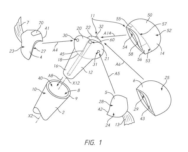
[0068] FIG. 1 is an exploded view
illustrating aspects of several
embodiments;
[0069] FIG. 2 is a schematic view of a human shoulder joint where
the
proximal humerus has suffered a fracture, separating into at least four bone
segments;
[0070] FIG. 3 illustrates a system for making components of patient
specific
prostheses and related tools;
[0071] FIG. 4 is a flow chart illustrating a method of making a stem
implant
and optionally a fragment jig as disclosed herein;
-9-
CA 03089708 2020-07-27
WO 2019/156985
PCT/US2019/016702
[0072] FIG. 5 is a flow chart illustrating a method performing
surgery with a
stem made according to the methods disclosed herein.
DETAILED DESCRIPTION OF THE PREFERRED EMBODIMENT
[0073] FIG. 1 shows a fractured long bone of a patient. In the
illustration, the
fractured long bone is a humerus, broken in four fragments at a proximal
(shoulder) end.
However, the invention also applies to other long bones of the body, such as
the hip end
of a femoral bone.
[0074] The embodiments preferably are applied to a fractured long
bone of a
human patient. However, it may be applied to a fractured long bone of an
animal patient.
[0075] The depicted fractured long bone comprises a diaphyseal
fragment 2,
or shaft fragment. The diaphyseal fragment 2 includes essentially the shaft
part of the
original bone.
[0076] The fragment 2 defines a diaphyseal axis X2, which is
extending along
the fragment 2. This fragment 2 is of generally tubular shape around axis X2.
The
fragment 2 comprises a medullary cavity 8, extending along axis X2, in
particular
coaxially. The cavity 8 is opened at a proximal end 10 of the fragment 2,
where the bone
is fractured. In the present case of a humeral diaphyseal fragment 2, the
proximal end 10
is defined along axis X2 towards the shoulder joint of the patient, opposite
to a distal end
of the fragment 2 directed towards the elbow joint of the patient (not shown).
[0077] At the proximal end 10, the fragment 2 forms a fracture line
9,
delineating the open cavity 8. The fracture line 9 surrounds the axis X2.
[0078] The depicted fractured long bone further comprises three
epiphyseal
fragments 4, 5 and 6. At least one of these fragments 4, 5 and 6 are to be
reattached at the
end 10 of the fragment 2 for reconstructing the original bone in some
embodiments.
[0079] Each fragment 4, 5 and 6 respectively has an internal surface
27, 28
and 29, as well as an external surface 23, 24 and 25. The surface 27 of the
fragment 4 is
opposed to the surface 23. The surface 27 is delimited from the surface 23 by
a fracture
line 41 of the fragment 4, surrounding surface 23 and surface 27. The surface
28 of the
fragment 5 is opposed to the surface 24. The surface 28 is delimited from the
surface 24
by a fracture line 42 of the fragment 5, surrounding surface 23 and surface
27. The surface
29 of the fragment 6 is opposed to the surface 25. The surface 29 is delimited
from the
surface 25 by a fracture line 43 of the fragment 6, surrounding surface 25 and
surface 29.
-10-
CA 03089708 2020-07-27
WO 2019/156985
PCT/US2019/016702
[0080] Depending on the fracture type and on the long bone
considered, more
or less than three epiphyseal fragments may be formed at the end of the
patient's long
bone. Only one epiphyseal fragment may be formed. However, the present
invention
preferably applies to cases where more than one epiphyseal fragments are
formed.
[0081] Some of the epiphyseal fragments, like fragments 4 and 5 of
the
present example, may be designated as "tuberosity fragments". These particular
fragments
are each secured to a muscle of the patient by means of a tendon of the
concerned muscle.
In other words, the implantation sites of the concerned muscles are located on
these
tuberosity fragments.
[0082] A muscle 7 is coupled to the external surface 23 of the
fragment 4. In
the present case, the fragment 4 includes the greater tuberosity of the
original bone,
originally fixed with a partially-illustrated supraspinatus muscle 7 of the
patient.
[0083] A muscle 13 is coupled to the external surface 24 of the
fragment 5. In
the present case, the fragment 5 includes the lesser tuberosity of the
original bone,
originally fixed with a partially-illustrated subscapularis muscle 13 of the
patient.
[0084] Some other epiphyseal fragments, like fragment 6 of the
present
example, may be designated as "articular fragment". Each articular fragment is
initially
part of a joint of the patient, for articulating the long bone with an
auxiliary bone of the
patient.
[0085] In the present case, the articular fragment 6 is a humeral
head of the
humerus, which is originally part of the shoulder joint, for articulating the
humerus with a
scapula 50 of the patient (see FIG. 2). In this case, the scapula forms the
"auxiliary bone"
of the shoulder joint. More precisely, the external surface 25 of the humeral
head 6 has a
generally spherical and convex shape and is initially articulated with a
corresponding
concave surface of a glenoid part 52 of the scapula 50.
[0086] Should some fragments have cracks or have geometrical defects
may
be spatially reconstructed with bone graft, cement or any other suitable
replacement
material. In the present example, fragment 4 comprises a portion 70 of
replacement
material for filling a notch of the fragment 4. In this case, the portion 70
forms a part of
the edge of the fragment 4, considered as a part of the fracture line 41, for
the sake of
simplicity.
[0087] As visible in Fig. 1, a prosthesis 11 comprises a stem part
12 and
advantageously a head part 14. In the case the fractured long bone is a
humerus, the
-11-
CA 03089708 2020-07-27
WO 2019/156985
PCT/US2019/016702
prosthesis 11 constitutes a shoulder prosthesis, or at least a humeral
component of a
shoulder prosthesis. This prosthesis 11, at least the stem part 12, is patient-
specific. In
other words, the prosthesis 11 is designed depending on the geometry of the
fractured
long bone of one specific patient, the prosthesis 11 being intended to be
implanted into
this particular patient.
[0088] In one embodiment, the stem part 12 comprises:
= [0089] a coverable diaphyseal portion 16, forming
a distal end of
the stem part 12 along axis X12; and
= [0090] coverable epiphyseal portions 20, 21 and
22, located at a
proximal end of the stem part 12 along axis X12.
[0091] In some embodiments, the portions 16, 20, 21 and 22 form a
single
integral piece. In some other embodiments, one or more portions may be a
separate piece
assembled with the others.
[0092] The stem part 12 is configured to be inserted into the
medullary cavity
8 of the diaphyseal fragment 2, through the open proximal end 10, as depicted
with the
arrow A8, along or parallel to axis X2. The stem part 12 preferably defines
the axis X12,
that is along, parallel to or coaxial with axis X2 when the stem part 12 is
secured to the
fragment 2. This insertion of the stem part 12 preferably ensures securing the
stem part 12
to the fragment 2. In some embodiments, supplementary means may be used for
reinforcing the securing, such as cement, fasteners, or other means.
[0093] The coverable diaphyseal portion 16 is configured for
securing the
stem part 12 to the fragment 2, preferably by fitting or anchoring of the
portion 16 into the
medullary cavity 8. At least, the portion 16 has a shape that corresponds to
the shape of
the medullary cavity 8, specifically to this patient. The portion 16 is
configured so that its
exterior surface is covered, e.g., entirely covered, by the diaphyseal
fragment 2 when the
stem part 12 is secured to the diaphyseal fragment 2, whereas the rest of the
stem part 12,
located at a proximal end of the stem part 12, is left uncovered by the
diaphyseal fragment
2. In this case, "the rest of the stem part 12" includes the coverable
epiphyseal portions
20, 22 and 24.
[0094] When inserted into the medullary cavity 8, the stem part 12
applies
mechanical stress to the fragment 2, to an extent than may be chosen in
advance prior or
during designing of the prosthesis 11, thanks to a method explained below. In
the present
example, the stem part 12 is configured so that the predetermined mechanical
stress is
-12-
CA 03089708 2020-07-27
WO 2019/156985
PCT/US2019/016702
applied to a contact zone 40 of the fragment 2, also chosen in advance. This
zone 40 is
formed at the surface thereof, inside the cavity 8, as depicted in FIG. 1.
This zone 40 is
entirely in contact with the portion 16 of the stem part 12. The zone 40 may
be a
continuous contact zone.
[0095] The zone 40 preferably extends all around axis X2, drawing a
ring-like
shape. In other words, the zone extends continuously along a circumference of
the
medullary cavity around axis X2.
[0096] In other embodiment, the mechanical stress may be applied to
several
distinct zones of the cavity 8. The distinct zones may be spaced apart and not
continuous
with each other. The distinct zones can be disposed about a circumference of
the cavity 8
at one location of the axis X2. The distinct zones can be two or more
continuous
peripheries of the cavity 8 spaced apart from each other along the axis X2.
The distinct
zones can be non-continuous about the circumference and spaced apart along the
axis X2.
[0097] In any case, the amount of mechanical stress to be applied is
chosen in
advance, the contact zone is chosen in advance, and the prosthesis is
manufactured so that
said chosen mechanical stress is actually applied onto said chosen contact
zone 40 when
the prosthesis 11 is secured to the fragment 2. Thus, the prosthesis is made
patient-
specifically with regard to contact in the cavity 8. An appropriate mechanical
stress may
be applied onto the diaphyseal fragment 2 so as to avoid osteonecrosis, or any
other
consequence relative to a lack of osseous mechanical loading.
[0098] In the present invention, by "mechanical stress" applied to a
bone
fragment, it is meant a mechanical stress sufficient to avoid osseous
necrosis. The
mechanical stress applied to the bone fragments is also chosen not to exceed
the
mechanical resistance of the bone fragments. In other words, the mechanical
stress is
chosen not to break or fracture the bone fragments. The appropriate stress to
be applied
may be calculated depending on the geometry of the considered bone fragments
and on
the osseous density of these fragments. For example, the mechanical stress may
be a
pressure exerted onto the considered bone fragment, traction, flexion or the
like.
[0099] Preferably, the mechanical stress is distributed evenly on
the contact
zone 40, or on the contact zones if several contact zones are foreseen. In
other words, a
same amount of stress is applied to any part of the contact zone 40 or zones.
Preferably,
the mechanical stress is applied in radial outward directions around axis X12
by the
coverable portion 16 onto the zone 40.
-13-
CA 03089708 2020-07-27
WO 2019/156985
PCT/US2019/016702
[0100] In order to be able to apply the appropriate amount of stress
onto the
contact zone and to choose what zone of the medullar cavity should serve as
the contact
zone, a specific method of manufacturing is performed for obtaining the
patient-specific
prosthesis 11.
[0101] FIG. 3 illustrates a method according to certain embodiments.
For
each prosthesis to be obtained and each patient to be treated, the method
comprises a
preliminary step 100 of providing data representative of the fractured long
bone of this
patient, including essentially information relative to geometry of the
diaphyseal fragment
2. The information may also be relative to the osseous density of the fragment
2.
[0102] Preferably, this step of providing data is at least partially
achieved by
scanning the relevant part of the patient including the fractured long bone,
for example
with a method of CT-scan ("computerized tomography").
[0103] Successively to step 100, based on the data gathered at step
100, a step
104 of the method is performed. The step 104 can involve designing a
prosthesis, e.g., the
stem part 12, to be specific to the patient. The step 104 includes, in one
embodiment,
sub-steps of choosing:
= [0104] which zone or zones of the medullary cavity
8 need to be
stressed, and
= [0105] the magnitude of the stress to be applied
onto said zone.
[0106] Once the zone and the magnitude of the stress are chosen, the
stem part
12 of the prosthesis is further designed accordingly, during a subsequent sub-
step of step
104. The designed stem part 12 is designed so as to be in contact with the
chosen zone,
becoming the aforementioned contact zone or zones 40, so as to apply the
chosen stress or
pressure, when effectively mounted to the epiphyseal fragment.
[0107] Thus, the step 104 allows designing the prosthesis 11,
including in
particular the stem part 12, specifically to the patient intended to receive
this prosthesis
11.
[0108] During a step 108 of the method, successive to the step B,
the
prosthesis 11 is manufactured, including the stem part 12 as it was designed
during the
second step. Thus, the patient-specific prosthesis 11 is obtained, for a
specific patient.
[0109] The step 108 of manufacturing preferably includes additive
manufacturing of the entire stem part 12. In some embodiment, only portions of
the stem
part 12 that are required to be patient-specific, such as the exterior surface
in contact with
-14-
CA 03089708 2020-07-27
WO 2019/156985
PCT/US2019/016702
the contact zone 40, are manufactured by additive manufacturing, these patient
specific
portions being combined with standard portions for forming the stem part 12.
[0110] For the present invention, any part obtained by additive
manufacturing
may be metallic. In some embodiments, appropriate plastic material may be
used.
[0111] Preferably, the stem part 12 comprises a visible mark 18 as
visible in
FIG. 1. The visible mark 18 is formed at the exterior surface of the stem part
12. The
mark 18 may be a shallow carving, an embossed marking, a colored marking, or
the
combination thereof, on the surface of the stem part 12. In any case, the mark
18 is
configured to be visible to the eye of the surgeon during surgery. The mark 18
preferably
forms a line, continuous or dashed. Instead, the mark 18 may form a dot, or
several dots.
The mark 18 can be formed as part of the step 108.
[0112] The mark 18 visually delimits the coverable diaphyseal
portion 16
from the rest of the stem part 12. The prosthesis 11, in particular the
coverable portion 16
and the mark 18, is designed specifically to the patient so that, when the
stem part 12 is
inserted into the cavity 8 properly, in particular at the right position
relative to the
fragment 2 along the diaphyseal axis X2, the mark 18 and the fracture line 9
are
superposed. Thus, during surgery, the surgeon is informed by the mark 18
whether the
stem part 12 is properly positioned into the cavity 8, at least concerning the
position of the
stem part 12 relative to the fragment 2 along the axis X2. Also, the shape of
the mark 18
may indicate to the surgeon whether the stem part 12 is properly positioned
relative to
fragment 2, around axis X2. For example, the mark 18 may indicate with a dot,
or have a
portion reproducing the shape of, a differential pattern around axis X2, such
as a dent or a
notch, of the fracture line 9. The surgeon has to ensure that the dot or the
portion is
aligned with the dent, notch or other visually distinct portion of the
fracture line 9
identifying a unique rotational position about the axis X2.
[0113] Preferably, a correct position of the stem part 12 is
obtained when the
mark 18 and the fracture line 9 are aligned and/or superposed along and/or
about the axis
X2. In case of misalignment, the surgeon may adjust the position of the stem
part 12
relative to the fragment 2 during surgery. Thus, the mark 18 promotes adequate
positioning of the stem part 12, and makes surgery easier. In this adequate
positioning, the
chosen stress is sure to be applied to the chosen contact zone 40 by the stem
part 12 onto
the fragment 2.
-15-
CA 03089708 2020-07-27
WO 2019/156985
PCT/US2019/016702
[0114] For obtaining the mark 18 disclosed above, the data of the
patient
provided during the preliminary step 100 of the method of FIG. 3 is used.
During the step
104 of designing the prosthesis 11, the stem part 12 is designed including the
coverable
diaphyseal portion 16 and the mark 18, so that they may achieve the above-
disclosed
functions. In particular, the stem part 12 is designed so that the mark 18
delimits the
coverable diaphyseal portion 16 and/or indicates where the fracture line 9 is
foreseen to be
located when the stem part 12 is inserted into the medullary cavity 8, and the
bone of the
fragment 2 covers the portion 16. When so received and positioned in the
medullary
cavity 8, the stem part 12 applies the chosen stress to the zone 40 in the
cavity 8.
[0115] In an embodiment of the method of FIG. 3, the portion 16 of
the stem
part 12 is configured to be radially outwardly expansible around axis X12. The
expansion
may be activated by the surgeon in a method of using the stem part 12. For
example, the
stem part 12 is designed and manufactured including an expansion screw 60,
coaxial with
axis X12. The screw may be actuated from outside of the portion 16 by the
surgeon: for
example the screw head is accessible from the opposite end of the stem part
12. The stem
part 12 may be introduced into the cavity 8 to the desired position, and then,
the surgeon
may actuate the screw 60 for expanding the stem part radially outwardly, so
that the stem
part applies the chosen mechanical stress onto the zone 40. Any other suitable
stem part
expansion actuator than the screw 60 may be provided instead. The method of
FIG. 3 can
include within step 104 designing the actuatable device, e.g., the screw 60
and
corresponding threads and structure of the stem part 12 such that a selected
number of
turns or advancement of the screw provides the designated stress onto the zone
or zones
40.
[0116] The stem part 12 is configured for receiving the epiphyseal
fragments
4, 5 and 6. In other words, these fragments 4, 5 and 6 may be secured to the
stem part 12.
[0117] Specifically to the patient, the stem part 12 is designed so
as to ensure
a patient-specific positioning of the fragments 4, 5 and 6 relative to the
stem part 12 and
to each other. The positioning of the fragment 2 relative to the stem part 12
may also be
planned patient-specifically, as explained above, so that eventually, the
fragments 2, 4, 5
and 6 are positioned relative to each other in a chosen manner when secured to
the stem
part 12. As concerns the fragments 4, 5 and 6, the stem part 12 is designed so
that the
fragments are positioned at chosen securing positions, defined in advance,
specifically to
-16-
CA 03089708 2020-07-27
WO 2019/156985
PCT/US2019/016702
the patient. In particular, this positioning is chosen specifically to the
shape of the
fragments 4, 5 and 6, and preferably also to the shape of the fragment 2.
[0118] In the method of manufacturing the prosthesis 11, step 100
may
include providing data relative to the fragments 4, 5 and 6 of the long bone
of the patient.
This may be performed alternatively or additionally to providing data relative
to the
fragment 2. The data provided may be relative to the shape of the fragments 4,
5 and 6.
The data may also be relative to the osseous density of the fragments 4, 5 and
6. The data
for fragments 4, 5 and 6 may be obtained by CT scanning, as explained above
for
fragment 2.
[0119] Step 104 may include a sub-step of choosing, specifically to
the
patient, respective securing positions of the fragments 4, 5 and 6. The chosen
securing
positions illustrate how the fragments 4, 5 and 6 will be positioned relative
to each other
when secured to the stem part 12. The chosen securing positions may depend
from the
actual size and shapes of the fragments 4, 5 and 6 of the considered patient.
[0120] This sub-step of choosing the securing positions may be
performed
alternatively (as shown in HG. 3) or additionally to the sub-step of choosing
the contact
zone 40 for fragment 2. If the sub-step of choosing the securing positions is
in addition to
the sub-step of choosing the contact zone 40, either the contact zone(s) may
be chosen
first, the securing positions may be chosen first or these aspects of the stem
may be
configured iteratively or simultaneously.
[0121] In one example, once the securing positions are chosen, the
sub-step of
designing the stem part 12 can be performed, so that the stem part 12 is
configured for
securing the epiphyseal fragments at the respective chosen securing positions.
In other
words, the designed stem part enables or even imposes that, when the fragments
4, 5 and
6 are secured thereto, they are in the planned positions.
[0122] Then, during step 108, the prosthesis 11, including in
particular the
stem part 12 with the features designed in step 104 above, is manufactured.
[0123] The fragments 4, 5 and 6 are configured to be positioned onto
the stem
part 12 as illustrated in HG. 1 with the arrows A4, AS and A6, respectively.
[0124] When the fragments 4, 5 and 6 are secured to the stem part
12, each
coverable epiphyseal portion 20, 21 and 22 is configured for being essentially
covered,
preferably completely covered, by one of the epiphyseal fragments 4, 5 and 6,
respectively. Preferably, for receiving the fragments 4, 5 or 6, each
respective coverable
-17-
CA 03089708 2020-07-27
WO 2019/156985
PCT/US2019/016702
portion 20, 21 and 22 has an external surface, shaped in correspondence to an
internal
surface 27, 28 or 29 of the respective concerned fragment 4, 5 or 6. When
received
properly, namely according to the chosen securing position, each fragment 4, 5
and 6
preferably entirely covers the exterior surface of the concerned portion 20,
21 and 22. For
this purpose, the portions 20, 21 and 22 of the stem part 12 are designed
patient
specifically during step 104.
[0125] Preferably, the stem part 12 is designed so that the
reattached
fragments 4, 5 and 6 are positioned in their original position relative to
each other and to
the diaphyseal fragment 2, as at the time when the bone was not yet fractured.
[0126] In an alternative embodiment, the stem part 12 may be
configured so
that one or more of the fragment 2, 4, 5 and 6 is planned to be in a different
position than
its original position relative to the other fragments.
[0127] Preferably, the stem part 12 is designed so that the
fragments 2, 4, 5
and 6 bear against each other by means of their respective fracture lines 9,
41, 42 or 43
when they are secured to the stem part 12. Thus, the chosen securing positions
are
preferably positions where the fragments 4, 5 and 6 bear against each other,
and
optionally at least one of said fragments 4, 5 and 6 bear against fragment 2.
At least two
of the fragments 2, 4, 5 and 6 bear against each other in this manner. For
example, when
secured to the stem part 12, fragment 4 is in abutting contact with fragment
5, the fracture
line 41 being in abutting contact with the fracture line 42. When secured to
the stem part
12, and when fragment 4 is in abutting contact with fragment 5, the fracture
line 41 also
can be being in abutting contact with the fracture line 9. When secured to the
stem part
12, and when fragment 4 is in abutting contact with fragment 5, and the
fracture line 41 is
in abutting contact with the fracture line 9, the fracture line 42 also can be
being in
abutting contact with the fracture line 9.
[0128] In an embodiment, the stem part 12 is designed with a chosen
securing
position for the fragments 4, 5 and 6, enabling that the fragments 4, 5 and 6,
and
optionally fragment 2, apply mechanical stress to each other when the
fragments 2, 4, 5
and 6 are actually positioned at these chosen securing positions.
[0129] For example, the stem part 12 is designed so that positioning
the
fragment 4 at its securing position on the stem part 12 will result in the
fragment 4 being
compressed between fragments 2, 5 and 6, if the fragments 2, 5 and 6 are also
positioned
at their respective securing positions. In this case, the fragment 2, 5 and 6
apply
-18-
CA 03089708 2020-07-27
WO 2019/156985
PCT/US2019/016702
mechanical stress to the fragment 4 by means of their respective fracture
lines 9, 42 and
43, onto the fracture line 41 of the fragment 4. In this case, resulting
mechanical stress is
also applied onto the fragments 2, 5, and 6. This mechanical stress is
achieved by
planning an adequate positioning of the fragments 2, 4, 5 and 6 relative to
each other.
Specifically, the stem part 12 may be designed so that the fragments 2, 4, 5
and 6 must be
tightly fitted against each other by the surgeon when secured to the stem part
12 at their
respective chosen securing positions. The amount of mechanical stress to be
applied to
each fragment 2, 4, 5 and 6 by one or more other of these fragments may also
be chosen
by appropriate designing of the stem part 12 and choice of positioning of the
fragments.
[0130] Thus, choosing the securing positions during step 104 is made
so that
the abovementioned mechanical stress, with a chosen magnitude, is applied on
the
fragments 4, 5 and 6 when actually positioned this way. Thus, the obtained
prosthesis 11
avoids osteonecrosis by submitting the reattached fragment to an appropriate
mechanical
stress.
[0131] For ensuring that the planned positioning of the fragments 4,
5 and 6 is
achieved, the exterior surface of the stem part 12, in particular for the
portions 20, 21 and
22, preferably has a shape corresponding to the shape of fragments 4, 5 and 6,
in
particular corresponding to the surfaces 27, 28 and 29 of said fragments. The
surgeon is
informed that one epiphyseal fragment is correctly positioned, according to
the chosen
securing position, if the epiphyseal fragment fits onto the stem part 12. In
case of
incorrect positioning of the epiphyseal fragment, said fragment does not fit
with the stem
part 12.
[0132] Additionally or alternatively, for ensuring that the chosen
securing
position of the fragments 4, 5 and 6 is achieved, in the example illustrated
in FIG. 1, the
stem part 12 may comprise three plugs 30, 31 and 32, each constituting a
distinct securing
element of one of the fragments 4, 5 and 6, respectively. Each plug 30, 31 and
32
protrudes from the exterior surface of one of the portions 20, 21 or 22,
respectively. Each
plug is preferably frustoconical, pyramidal, or shaped as a dome. Conversely,
each
fragment 4, 5 and 6 may have a blind bore, provided on its internal surface
27, 28 and 29
respectively. These blind bores are preferably drilled or carved in the
fragments 4, 5 and 6
by the surgeon with a specific tool. The shape of each bore corresponds to the
shape of the
plugs 30, 31 and 32, so that each plug may be inserted into the bore of one
fragment 4, 5
or 6 when the concerned fragment 4, 5 or 6 is positioned on the stem part 12.
Thus, when
-19-
CA 03089708 2020-07-27
WO 2019/156985
PCT/US2019/016702
the plug is inserted into the appropriate bore of a given fragment 4, 5 or 6,
the surgeon is
sure that this fragment is correctly positioned onto the stem part 12.
[0133] Preferably, the methods of manufacturing includes a step 112
of
designing and/or manufacturing one or more patient-specific jigs, depending on
the
chosen securing position and on the data representative of the patient. Each
jig, for
example embodied as a drill guide, may be used by the surgeon for drilling the
blind bore
on one of the epiphyseal fragments. Each jig is adapted to the shape of the
bone fragments
to be reworked by the surgeon. Thus, the surgeon is sure to drill the blind
bores at the
appropriate position on the fragments 4, 5 and 6, thus enabling a positioning
of the
fragments 4, 5 and 6 at the chosen securing positions.
[0134] In a preferable embodiment, the plugs are configured so as to
apply
mechanical stress onto the fragments 4, 5 and 6. For example one of the
epiphyseal
fragments may be compressed between the plug, on which it is mounted, and
another
epiphyseal fragment. In this example, the plug applies radially directed
mechanical stress
to the blind bore of the epiphyseal fragment and said fragment receives
mechanical stress
onto the fracture line in an opposed direction due to reaction force at the
fracture line.
Thus, the risk of osseous necrosis is reduced by the applications of these
stresses.
[0135] Furthermore, the plugs may be designed so as to apply
mechanical
stress to the blind hole of the fragment by tight fitting into the blind hole.
[0136] More than one plug may be provided for each epiphyseal
portion, and a
plurality of plugs may be provided for positioning each epiphyseal fragment.
[0137] In some embodiments, the plugs are formed integral with the
concerned epiphyseal portion. Alternatively, one or more of the securing
element may be
a separate part assembled with the concerned epiphyseal portion.
[0138] In some embodiments, instead of the aforementioned plugs, any
other
type of positioning element may be provided.
[0139] Additionally or alternatively, the stem part 12 comprises a
visible mark
45 indicating the respective chosen securing positions of the epiphyseal
fragments 4, 5
and 6 on the stem part. The visible mark 45 is formed at the exterior surface
of the stem
part 12. The mark 45 may be a shallow carving, an embossed marking, a colored
marking,
or the combination thereof, on the surface of the stem part 12. In any case,
the mark 45 is
configured to be visible to the eye of the surgeon during surgery. The mark 45
preferably
-20-
CA 03089708 2020-07-27
WO 2019/156985
PCT/US2019/016702
forms one or more lines, continuous or dashed. Instead, the mark 45 may form
one or
more dots.
[0140] In a preferred embodiment, as illustrated on FIG. 1, the mark
45
visually delimits the coverable epiphyseal portions 20, 21 and 22 from each
other and
optionally from the rest of the stem part 12. The coverable portions 20, 21
and 22 and the
mark 45 are designed specifically to the patient so that, when the fragments
4, 5 and 6 of
this patient are positioned at the planned position on the stem part 12, the
fracture lines
41, 42 and 43 are superposed with the mark 45. In this aspect, the chosen
securing
positions are indicated by the mark 45. During surgery, the surgeon is
informed by the
mark 45 whether the fragments 4, 5 and 6 are properly positioned on the stem
part 12.
[0141] Thus, the mark 45 promotes adequate positioning of the
fragments 4, 5
and 6, and makes surgery easier.
[0142] For obtaining the mark 45 disclosed above, the data of the
patient
provided during the step 100 of the method is used. During the step 104, the
stem part 12
is designed including the coverable portions 20, 21, 22 and the mark 45, so
that they may
achieve the above-disclosed functions. In particular, the stem part 12 is
designed so that
the mark 45 delimits the coverable epiphyseal portions 20, 21 and 22 from each
other
and/or indicates where the fracture lines 41, 42 and 43 are foreseen to be
located when the
fragments 4, 5 and 6 are positioned according to the chosen securing
positions.
[0143] For securing the fragments 4, 5 and 6 to the stem part 12,
further
conventional means suitable to the situation may be used, such as fasteners,
cement and/or
sutures S (see FIG. 2).
[0144] The surgeon may decide that the initial articular fragment 6
is
reattached to the stem part 12, as disclosed above. In this situation, the
head part 14 is
optional. For this situation, the prosthesis 11 may be provided without such
head part 14.
Or, the head part 14 can be included but the surgeon may determine
intraoperatively that
the head part 14 is not needed.
[0145] In some specific cases, the surgeon may decide that some of
the
epiphyseal fragments are viable and may be reattached to the stem part 12 and
some other
fragments are damaged and may not be reattached. In this case, the damaged
epiphyseal
fragments may require to be replaced with prosthetic means.
[0146] In the case the articular fragment 6 is damaged, while the
fragments 4
and 5 are viable, the fragment 6 may be replaced by the prosthetic head part
14 shown in
-21-
CA 03089708 2020-07-27
WO 2019/156985
PCT/US2019/016702
FIG. 1. Thus, the head part 14 is configured to be secured to the prosthetic
stem part 12 so
as to replace the damaged articular fragment 6 when the prosthesis 11 is
introduced in the
patient's body. The head part 14 is preferably secured at portion 22 of the
stem part 12.
The securing may be achieved with fasteners or any other suitable securing
means. In the
case the head part 14 is provided, the plug 32 is optional. Appropriate
securing means
may be provided on the portion 22 additionally or alternatively to plug 32.
[0147] In this case, the method comprises, during step 100,
providing data
relative to the viable fragments 4 and 5 and to the damaged fragment 6. Then,
during step
104, the prosthesis 11 is designed specifically to the patient, including the
stem part 12
and the head part 14 being patient-specific.
[0148] In the case a head part 14 is used instead of reattaching the
fragment 6,
the method is similar than the previously explained method. Step 104 includes
a sub-step
of choosing, specifically to the patient, respective securing positions of the
viable
fragments 4 and 5 and of the head part 14 relative to each other, and
advantageously,
relative to fragment 2. In other words, in this sub-step, the position of the
head part 14 is
chosen, instead of the position of the reattached fragment 6 for the case
explained above.
The chosen "securing positions" are representative of how the viable fragments
4 and 5
and the head part 14 are planned to be effectively positioned relative to each
other when
secured to the stem part 12 by the surgeon. The fragments 4 and 5 and the head
part 14 are
configured to be positioned onto the stem part 12 as illustrated in FIG. 1
with the arrows
A4, AS and A14, respectively. The fragments 4 and 5 are positioned as
disclosed above.
The fragment 6 can be replaced by the head part 14. Thus, the head part 14 can
be
positioned in a similar way than the fragment 6 of the previous case. When
secured to the
stem part 12, the head part 14 preferably substantially or completely covers
the portion
22.
[0149] In this case, once the securing positions are chosen, the sub-
step of
designing the stem part 12 is performed. The head part 14 may also be designed
patient-
specifically. In this designing sub-step, the stem part 12, and optionally at
least a part of
the head part 14, is designed so that the stem part 12 allows securing the
fragments 4 and
and the head part 14 at the chosen securing positions. In other words, the
designed stem
part 12 enables or even imposes that, when the fragments 4 and 5 the head part
14 are
secured thereto, they are in the planned positions.
-22-
CA 03089708 2020-07-27
WO 2019/156985
PCT/US2019/016702
[0150] Then, during step 108, the prosthesis 11, including in
particular the
stem part 12 with the features designed in step 104 above, is manufactured. If
all or part
of the head part 14 is designed patient-specifically during step 104, the
patient specific
features of the head part 14, or all the head part 14 is also manufactured
during step 108.
[0151] The head part 14 may comprise a standard cap 50. "Standard"
means
that the cap 50 is not patient-specific, although a cap of appropriate size
and shape may be
chosen among a definite collection of standard caps. Thus, the standard cap 50
is provided
or manufactured separately from the patient-specific features manufactured at
step 108.
[0152] The standard cap 50 comprises an articular surface 52 of
convex shape,
preferably spherical, for forming a prosthetic joint of the patient, combined
with the
glenoid cavity of the scapula of the patient, or any other auxiliary bone
considered, in
replacement for the surface 25 of fragment 6. In other words, the cap 50 may
be shaped as
a dome as depicted in FIG. 1. By "prosthetic joint", it is meant that at least
a part of the
joint is prosthetic. In this case, the standard cap 50 is a prosthetic part of
the joint.
[0153] Alternatively to a convex shape, depending on the situation,
a concave
shape may be used.
[0154] The standard cap 50 also comprises a trunnion 55, shown in
dashed
lines in FIG. 1, protruding in an opposed to the surface 52.
[0155] The standard cap 50 also comprises a securing surface 54,
opposed to
the surface 52 and formed at the end of the trunnion 55. The standard cap 50
is configured
to be secured to the stem part 12 by means of the securing surface 54.
[0156] The head part 14 also comprises a patient-specific insert 56.
This insert
56 is patient specifically designed during step 104 according to the data
provided at step
100, and manufactured during step 108 according to this design. The insert 56
is a
separate piece from the cap 50 and is assembled with it before or during
surgery.
[0157] The insert 56 is interposed between the cap 50 and the stem
part 12.
More precisely, the insert 56 has a surface 57 bearing against a border
surface 53 of the
cap 50. The border surface 53 surrounds the trunnion 55, and is opposed to the
surface 52.
The insert 56 also has an opposed surface 58, that is configured to bear
against the
fracture lines 41 and 42 of the fragments 4 and 5. The insert 56 is preferably
shaped as a
ring or a washer, as depicted in FIG. 1, so that the cap 50 may be secured to
the stem part
12 through the insert 56. In this case, the trunnion 55 passes through a
central hole of the
-23-
CA 03089708 2020-07-27
WO 2019/156985
PCT/US2019/016702
insert 56. The insert 56 is preferably fitted, for example conically fitted,
onto the trunnion
55.
[0158] Alternatively, the cap of the head part 14 is made patient-
specifically,
designed during step 104 and manufactured during step 108. In this case, the
cap and the
insert may form a single piece instead of two distinct assembled pieces.
[0159] During step 104, the respective securing positions of the
fragments 4
and 5 and of the head part 14 are chosen so that a respective chosen
mechanical stress is
applied to each fragment 4 and 5 by the head part 14. In a preferable
embodiment, the
fragments 4 and 5 are compressed between the head part 14 and the fragment 2.
Alternatively, the head part 14 may apply mechanical stress on the fragments 4
and 5
without relying on the fragment 2.
[0160] More specifically, step 108 may include patient-specifically
designing
the insert 56 so that the insert 56 applies the respective stress onto the
fragments 4 and 5
with a chosen magnitude. In particular, the shape of the surface 58 may be
designed in
correspondence with the shape of the fracture lines 41 and 42, so that the
surface 58 may
distribute the applied stress along the fracture lines 41 and 42.
[0161] Thus, when the prosthesis 11 is manufactured and implanted in
the
patient, a mechanical stress defined in advance patient specifically is
applied to the
reattached fragments 4 and 5, and optionally to the fragment 2, by the head
part 14, and
optionally by the stem part 12.
[0162] The patient specific parts of the head part 14 may be
obtained, during
step 108, by additive manufacturing. Preferably, the insert 56 is manufactured
by additive
manufacturing. If the cap 50 is patient-specific, the insert 56 and the cap 50
may both be
obtained by additive manufacturing.
[0163] Instead of having a patient specific surface 58, the head
part 14 may be
chosen standard. Thus, only the relative securing position of the fragments 4
and 5 and of
the head part 14 may allow defining the mechanical stress to be applied.
[0164] FIG. 3 shows a system schematically illustrates a planning
system 120
that can be used in the method of FIG. 2 and other corresponding methods for
forming the
stem part 12 and/or fragment jigs as discussed in step 112. The stem part 12
and fragment
jigs (if provided) are formed as patient specific parts and jigs by
referencing pre-operative
imaging such as MRI, CT imaging or the like. The planning system 120 receives
inputs
122 and produces outputs 124 including the stem part 12. The inputs 122
include pre-
-24-
CA 03089708 2020-07-27
WO 2019/156985
PCT/US2019/016702
operative shoulder images 126 and patient input information 128. The pre-
operative
shoulder images 126 can include MRI, CT imaging and other information that
allows the
planning system 120 to render or characterize at least the fragments 2, 4, 5,
and 6. For
example, the pre-operative shoulder images 126 can include the location,
shape, and
orientation of any of the fracture lines 9, 41, 42, 43, as well as other
features such as
rotational orientation landmarks of the proximal end 10 of the fragment 2. The
pre-
operative shoulder images 126 can include information about the location,
shape and
orientation of landmarks of any of the fragments 2, 4, 5, and/or 6 or other
parts of the
bones of the joint being treated. In the case of the shoulder, the images 126
can include
location, orientation or other characterizing information about portions of
the humerus
such as the surface contour of the canal 8, the greater tubercle, the lesser
tubercle, the
surgical neck, the bicipital groove or other landmarks of the proximal
humerus.
[0165] The patient input information 128 can include the patients
name, the
shoulder being treated, other past and future therapies and other information
relevant to
the procedure.
[0166] The planning system 120 can include a number of modules that
can
process the inputs 122. The planning system 120 can include a module 130 for
selecting
among reverse and anatomic implant configuration. The planning system 120 can
include
a module 132 configured to define stem shape as rotationally symmetric or
asymmetric.
A stem shape module 132 can also define the taper line of the stem part 2
along the
longitudinal axis thereof The planning system 120 can have a force level
module 134
that can determine how much force is appropriate to achieve a lessening of
osseous
necrosis while not resulting in fracture of fragment coupled with the stem
part 12. The
system 120 also can include an orientation module 136 that defines the
rotational position
of the stem part 12 in the canal 8, e.g., by defining a visual relationship
between the stem
part and a landmark of the fragment 2. Each of these and other features can be
determined
and prescribed by the planning system 120 and can be incorporated into the
outputs 124
of the planning system 120.
[0167] The outputs 124 can include the stem part 12, a fragment jig
(not
shown), an implant identification 140, and patient procedural information 142.
The stem
part 12 that can be output by the planning system 120 can be a plan for
forming the stem
part 12 or can be an actual stem part if the planning system 120 is configured
with or
coupled with a manufacturing facility or manufacturing process 144. In some
-25-
CA 03089708 2020-07-27
WO 2019/156985
PCT/US2019/016702
applications, the process 144 that is used to form the stem part 12 includes
additive
manufacturing such as three dimensional printing. Examples of three
dimensional
printing include direct metal laser sintering (DMLS), fused deposition
modeling (FDM),
fused filament fabrication (FFF), and electron beam melting (EBM). Any one or
a
combination of these or other additive manufacturing processes can be used in
the
manufacturing process 144. In these processes a three dimensional object is
formed by
sequentially forming individual layers of the object on top of previously
formed
individual layers. These processes can closely control the gross dimensions of
the object
and also can form complex features and shapes such as contours. As discussed
further
below, these processes can be used to form complementary surface that can mate
with
specific anatomy of a specific patient, e.g., concave surfaces that can nest
on top of
corresponding convex surfaces. 0
[0168] With reference to FIG. 3, the planning system 120 can thus
include a
processor for receiving the inputs 122. The inputs 122 include the patient
input
information 128 and the pre-operative shoulder images 126. The processor and
software
process this information with other selections regarding the implant and the
patient's
anatomy that can be made by a user regarding the nature of the shoulder
procedure. The
software can produce data that can be input to the manufacturing process 144
to control
the operation thereof. The data can be configured for directing a three
dimensional printer
or other additive manufacturing process to form the stem part 12. In other
approaches the
manufacturing process 144 can include multiple steps such as a first step of
forming a
mold with an additive manufacturing process and thereafter forming the stem
part 12 in
the mold. These approaches enable the pre-operative images 126 to be utilized
to
configure patient specific surfaces of the stem part 12 to be
complementary/negative
surfaces such that the guide is seated according to the optimized fit as
determined by the
surgeon or other user.
[0169] The planning system 120 can also provide implant
identification 140
that corresponds to the stem part 12. For example, the planning system 120 can
provide a
type of implant that the stem part 12 is suitable to mate with. For example,
the implant
identification 142 identifies an anatomic articular head, such as the head
part 14 with a
convex articular surface to be coupled with the stem part 12. The implant
identification
140 can identify a reverse implant (not shown) to be coupled with the stem
part 12
following repair of the fracture. The outputs 124 can include the patient
output
-26-
CA 03089708 2020-07-27
WO 2019/156985
PCT/US2019/016702
information 142 which can include not only the name of the patient but also
the shoulder
to be treated and other specific information about the stem part 12 and/or
fragment jigs
(not shown) if provided such as which rotational landmarks may be used to
position the
stem part 12.
[0170] After the prosthesis 11 is manufactured at step 108 for the
specific
patient to be treated, the surgeon may proceed as follows for installing the
prosthesis 11 in
the patient's body as discussed further below.
[0171] FIG. 4 shows a method of implanting the stem part 12. In a
step 150
the surgeon firstly secures the stem part 12 onto the fragment 2, by
introduction of the
coverable portion 16 into the cavity 8. The surgeon optionally checks that the
mark 18 is
superposed with the fracture line 9 and may adjust the position of the stem
part if
necessary. In the adequate position, the stem part 12 applies the chosen
mechanical stress
to the zone 40 of the cavity 8.
[0172] Secondly, in a step 154 the surgeon secures the fragments 4
and 5 onto
the stem part 12. For this purpose, the surgeon may have prepared the
fragments 4 and 5
in advance, including drilling the blind holes. The blind holes may be drilled
with help of
a tool and/or a jig made patient-specifically during step 112, designed at
step 108 based
on the data of step 100. In this case, the fragments 4 and 5 are positioned
onto the stem
part 12 with the plugs 30 and 31 inserted into the blind holes. In a step 158,
the surgeon
checks whether the positioning of the fragments 4 and 5 is correct by checking
if the
fragments 4 and 5 match with the mark 45. If necessary, in a step 162 the
position of the
fragments 4 and 5 is adjusted by the surgeon with help of the mark 45.
[0173] Thirdly, the fragment 6 is secured to the stem part 12. This
can involve
repeating any of steps 150, 154, 158, and 162. For this purpose, the surgeon
may have
prepared the fragment 6 in advance, including drilling the blind hole. The
blind hole may
be drilled with help of a tool and/or a jig made patient-specifically during
step 112,
designed at step 104 based on the data of step 100. The surgeon checks whether
the
positioning of the fragment 6 is correct by checking if the fragment 6 matches
with the
mark 45. If necessary, the position of the fragment 6 is adjusted by the
surgeon with help
of the mark 45. Securing the fragment 6 applies mechanical stress onto the
fragments 4
and 5. The method of FIG. 4 can be completed at step 166 when the fragments,
4, 5, and
6 are all properly placed.
-27-
CA 03089708 2020-07-27
WO 2019/156985
PCT/US2019/016702
[0174] It can be provided that the fragments 4 and 5 are interposed
between
the fracture line 9 of fragment 2 and the fragment 6. In this case, mechanical
stress is also
applied to fragment 2 by fragments 4 and 5. In another embodiments, the
fragments 4 and
are supported by the plugs 30 and 31, onto which the mechanical stress is
transmitted,
without transmitting the mechanical stress to the fragment 2.
[0175] Fasteners, sutures S (as seen in FIG. 2), adhesive means or
any suitable
means may be used by the surgeon for maintaining the fragments 2, 4, 5 and 6
onto the
stem part 12.
[0176] If the fragment 6 is to be replaced, said fragment 6 is not
secured to the
stem part 12. Instead, the head part 14 is secured to the stem part 12.
Securing the head
part 14 applies mechanical stress onto the fragments 4 and 5, and optionally
to the
fragment 2, in a similar manner.
[0177] Fasteners, sutures S (as seen in FIG. 2), adhesive means or
any suitable
means may be used by the surgeon for maintaining the fragments 2, 4 and 5 and
the head
part onto the stem part 12.
[0178] The external surface of at least one of the coverable
portions 16, 20, 21
and 22 is preferably rough, or even comprises barbs, for helping securing the
bone
fragments 2, 4, 5 and 6 secured thereto.
EXAMPLE EMBODIMENTS
[0179] The following example embodiments identify some possible
permutations of combinations of features disclosed herein, although other
permutations of
combinations of features are also possible.
[0180] Embodiment 1.- A method for manufacturing a prosthesis (11)
for a
fractured long bone of a patient, the method comprising the steps of:
[0181] A) providing data representative of the fractured long bone
of the
patient, the fractured long bone comprising a diaphyseal fragment (2)
comprising a
medullary cavity (8);
[0182] B) based on the data provided at step A), designing the
prosthesis (11)
specifically to the patient, the prosthesis (11) comprising a stem part (12)
configured to be
inserted into the medullary cavity (8) for securing the stem part (12) to the
diaphyseal
fragment (2), step B) comprising:
a sub-step of choosing, specifically to the patient, at least one contact
zone (40) of the medullary cavity (8) onto which a respective chosen
mechanical stress is
-28-
CA 03089708 2020-07-27
WO 2019/156985
PCT/US2019/016702
planned to be applied by the stem part (12), when the stem part (12) is
inserted into the
medullary cavity (8) and
a sub-step of designing the stem part (12) so that the stem part (12)
may be inserted into the medullary cavity (8) and thus apply the chosen
mechanical stress
to said at least one contact zone (40); and
[0183] C) manufacturing the prosthesis (11) including the stem part
(12)
designed at step B).
[0184] Embodiment 2.- The method according to Embodiment 1, wherein
said
at least one contact zone (40) of the medullary cavity (8) continuously covers
a
circumference of the medullary cavity (8).
[0185] Embodiment 3.- The method according to any one of Embodiment
1-
Embodiment 2, wherein during sub-step B1), the chosen mechanical stress is
chosen to be
evenly distributed onto each of said at least one contact zone (40) of the
medullary cavity
(8).
[0186] Embodiment 4.- The method according to any one of Embodiment
1-
Embodiment 3, wherein step B) comprises the further sub-step of, based on the
data
provided at step A), designing the stem part (12) so that the stem part (12)
comprises,
specifically to the patient:
a coverable diaphyseal portion (16), configured to be covered by the
diaphyseal fragment (2) when the stem part (12) is secured to the diaphyseal
fragment (2),
whereas the rest of the stem part (12) is left uncovered by the diaphyseal
fragment (2);
and
a first visible mark (18), visually delimiting the coverable diaphyseal
portion (16) from the rest of the stem part (12).
[0187] Embodiment 5.- The method according to any one of Embodiment
1-
Embodiment 4, wherein:
step A) comprises providing data representative of epiphyseal
fragments (4, 5, 6) of the fractured long bone; and
step B) comprises, based on the data provided at step A), designing the
prosthesis (11) so that the stem part (12) is configured for securing at least
one of the
epiphyseal fragments (4, 5, 6) to the stem part (12).
[0188] Embodiment 6.- The method according to Embodiment 5, wherein
step
B) comprises:
-29-
CA 03089708 2020-07-27
WO 2019/156985
PCT/US2019/016702
a sub-step of choosing, specifically to the patient, respective securing
positions of the epiphyseal fragments (4, 5, 6) relative to each other,
representative of how
the epiphyseal fragments (4, 5, 6) are planned to be positioned relative to
each other when
secured to the stem part (12), the securing positions being chosen so that a
respective
chosen mechanical stress is applied onto each epiphyseal fragment (4, 5) by at
least one of
the other epiphyseal fragments (6), when the epiphyseal fragments (4, 5, 6)
are secured at
the respective chosen securing positions of the stem part (12), and
a sub-step of designing the stem part (12) so that the stem part (12) is
configured for securing the epiphyseal fragments (4, 5, 6) at the respective
chosen
securing positions.
[0189] Embodiment 7.- The method according to Embodiment 6, wherein
step
B) comprises the further sub-step of designing the stem part (12) so that the
stem part (12)
comprises a second visible mark (45) indicating the respective securing
positions of the
epiphyseal fragments (4, 5, 6) on the stem part (12).
[0190] Embodiment 8.- The method according to Embodiment 7, wherein
step
B) comprises the further sub-step of designing the stem part (12) based on the
data
provided at step A), so that the stem part (12) comprises, specifically to the
patient,
coverable epiphyseal portions (20, 21, 22), being configured for being covered
respectively by the epiphyseal fragments (4, 5, 6) when the epiphyseal
fragments (4, 5, 6)
are secured to the stem part (12) at the respective securing positions;
wherein the second
visible mark (45) is designed so as to visually delimit the coverable
epiphyseal portions
(20, 21, 22) from each other.
[0191] Embodiment 9.- The method according to any one of Embodiments
6
to 8, wherein the stem part (12) comprises, for at least one of the epiphyseal
fragments (4,
5, 6), a respective plug (30, 31, 32), for securing the concerned epiphyseal
fragment (4, 5,
6) to the stem part (12), said at least one plug (30, 31, 32) applying
mechanical stress onto
the epiphyseal fragment (4, 5, 6) secured thereto.
[0192] Embodiment 10.- The method according to any one of
Embodiments 1
to 5, wherein:
[0193] the epiphyseal fragments (4, 5, 6) comprise:
at least one viable tuberosity fragment (4, 5), secured to a muscle (7,
13) of the patient, the muscle (7, 13) being attached to said at least one
viable tuberosity
fragment (4, 5) by means of a tendon of the muscle (7, 13) and
-30-
CA 03089708 2020-07-27
WO 2019/156985
PCT/US2019/016702
a damaged articular fragment (6), initially being part of an damaged
joint of the patient, for articulating the long bone with an auxiliary bone of
the patient;
[0194] step B) comprises:
based on the data provided at step A), designing the prosthesis (11) so
that:
[0195] o the stem part (12) is configured for securing said at least
one viable
tuberosity fragment (4, 5) to the stem part (12), and
[0196] o the prosthesis (11) further comprises a head part (14),
being
configured to be secured to the stem part (12) in replacement for the damaged
articular
fragment (6) of the fractured long bone of the patient,
a sub-step of choosing, specifically to the patient, respective securing
positions of said at least one viable tuberosity fragment (4, 5) and of the
head part (14)
relative to each other, representative of how said at least one viable
tuberosity fragment
(4, 5) and head part (14) are planned to be positioned relative to each other
when secured
to the stem part (12), the securing positions being chosen so that a
respective chosen
mechanical stress is applied to each of said at least one viable tuberosity
fragment (4, 5)
by the head part (14), when said at least one viable tuberosity fragment (4,
5) and the head
part (14) are secured at the respective chosen securing positions of the stem
part (12), and
a sub-step of designing the stem part (12) so that said at least one
viable tuberosity fragment (4, 5) and the head part (14) may be secured to the
stem part
(12) at the respective chosen securing positions;
[0197] step C) comprises providing or manufacturing the head part
(14) of the
prosthesis (11).
[0198] Embodiment 11.- The method according to Embodiment 10,
wherein
the head part (14) comprises:
a standard cap (50), comprising an articular surface (52) of concave or
convex shape for forming a prosthetic joint for replacement of the damaged
joint of the
patient, and comprising a securing surface (54) opposed to the articular
surface (52), the
standard cap (50) being secured to the stem part (12) by means of the securing
surface
(54);
a patient-specific insert (56), designed during step B) patient
specifically, and manufactured during step C), configured to:
-31-
CA 03089708 2020-07-27
WO 2019/156985
PCT/US2019/016702
[0199] o be interposed between the standard cap (50) and the stem
part (12),
and
[0200] o apply the respective mechanical stress onto each of said at
least one
viable tuberosity fragment (4, 5).
[0201] Embodiment 12.- The method according to any one of Embodiment
1-
Embodiment 11, wherein step A) includes using CT scans of the patient and step
C)
includes additive manufacturing of at least a part of the prosthesis (11).
[0202] Embodiment 13.- The method according to any one of the
preceding
Embodiment 1-Embodiment 12, wherein the fractured long bone is a fractured
humerus,
the prosthesis (11) being a shoulder prosthesis (11).
[0203] Embodiment 14.- A patient-specific prosthesis (11) for a
fractured long
bone of a particular patient, the prosthesis (11) comprising a stem part (12)
that is
designed based on data representative of the fractured long bone of this
particular patient,
so as to:
be inserted into the medullary cavity (8) of a diaphyseal fragment (2) of
the fractured long bone of the patient for securing the stem part (12) to the
diaphyseal
fragment (2), and
apply a chosen mechanical stress to at least one contact zone (40) of
the medullary cavity (8), chosen based on said data.
[0204] Embodiment 15.- The patient-specific prosthesis (11)
according to
Embodiment 14, wherein the stem part (12) comprises, specifically to the
patient:
a coverable diaphyseal portion (16), including at least a part of the stem
part (12) and being configured to be covered by the diaphyseal fragment (2)
when the
stem part (12) is inserted into the medullary cavity (8), whereas the rest of
the stem part
(12) is left uncovered by the diaphyseal fragment (2); and
a first visible mark (18), visually delimiting the coverable diaphyseal
portion (16) from the rest of the stem part (12).
Terminology
[0205] Certain methods are described as sliding a first component
relative to a
second component. However, it should be understood that relative motion
between first
and second components can also include sliding the second component relative
to the first
component. For example, "sliding the intermediate portion in a transverse
direction
relative to a longitudinal axis of the metaphyseal portion" can also include
"sliding the
-32-
CA 03089708 2020-07-27
WO 2019/156985
PCT/US2019/016702
metaphyseal portion in a transverse direction relative to a longitudinal axis
of the
intermediate portion."
[0206] "Implant" is a broad term and is to be given its ordinary and
customary
meaning to a person of ordinary skill in the art and includes, without
limitation, temporary
implants (e.g., for trialing) or permanent implants (also referred to herein
as definitive
implants) for any anatomy, including, but not limited to, shoulder
replacements, knew
replacements, femoral replacements, and hip replacements.
[0207] As used herein, the term "metaphyseal portion" refers to
either a
reverse metaphyseal portion or an anatomic metaphyseal portion unless
otherwise
specified.
[0208] As used herein, the relative terms "proximal" and "distal"
shall be
defined from the perspective of the humeral component. Thus, proximal refers
to the
direction of the metaphyseal portion and distal refers to the direction of the
stem portion.
[0209] For expository purposes, the term "transverse" as used herein
is
defined as a direction generally perpendicular to the longitudinal axis of the
assembly,
unless otherwise specified.
[0210] Conditional language, such as "can," "could," "might," or
"may,"
unless specifically stated otherwise, or otherwise understood within the
context as used, is
generally intended to convey that certain embodiments include, while other
embodiments
do not include, certain features, elements, and/or steps. Thus, such
conditional language
is not generally intended to imply that features, elements, and/or steps are
in any way
required for one or more embodiments.
[0211] The terms "comprising," "including," "having," and the like
are
synonymous and are used inclusively, in an open-ended fashion, and do not
exclude
additional elements, features, acts, operations, and so forth. Also, the term
"or" is used in
its inclusive sense (and not in its exclusive sense) so that when used, for
example, to
connect a list of elements, the term "or" means one, some, or all of the
elements in the
list.
[0212] The terms "approximately," "about," "generally," and
"substantially"
as used herein represent an amount close to the stated amount that still
performs a desired
function or achieves a desired result. For example, the terms "approximately,"
"about,"
"generally," and "substantially" may refer to an amount that is within less
than 10% of the
stated amount, as the context may dictate.
-33-
CA 03089708 2020-07-27
WO 2019/156985
PCT/US2019/016702
[0213] The ranges disclosed herein also encompass any and all
overlap, sub-
ranges, and combinations thereof. Language such as "up to," "at least,"
"greater than,"
"less than," "between" and the like includes the number recited. Numbers
preceded by a
term such as "about" or "approximately" include the recited numbers. For
example,
"about four" includes "four"
[0214] Any methods disclosed herein need not be performed in the
order
recited. The methods disclosed herein include certain actions taken by a
practitioner;
however, they can also include any third-party instruction of those actions,
either
expressly or by implication. For example, actions such as "distally moving a
locking
element" include "instructing distal movement of the locking element."
[0215] Although certain embodiments and examples have been described
herein, it will be understood by those skilled in the art that many aspects of
the humeral
assemblies shown and described in the present disclosure may be differently
combined
and/or modified to form still further embodiments or acceptable examples. All
such
modifications and variations are intended to be included herein within the
scope of this
disclosure. A wide variety of designs and approaches are possible. No feature,
structure,
or step disclosed herein is essential or indispensable.
[0216] Some embodiments have been described in connection with the
accompanying drawings. However, it should be understood that the figures are
not drawn
to scale. Distances, angles, etc. are merely illustrative and do not
necessarily bear an
exact relationship to actual dimensions and layout of the devices illustrated.
Components
can be added, removed, and/or rearranged. Further, the disclosure herein of
any particular
feature, aspect, method, property, characteristic, quality, attribute,
element, or the like in
connection with various embodiments can be used in all other embodiments set
forth
herein. Additionally, it will be recognized that any methods described herein
may be
practiced using any device suitable for performing the recited steps.
[0217] For purposes of this disclosure, certain aspects, advantages,
and novel
features are described herein. It is to be understood that not necessarily all
such
advantages may be achieved in accordance with any particular embodiment. Thus,
for
example, those skilled in the art will recognize that the disclosure may be
embodied or
carried out in a manner that achieves one advantage or a group of advantages
as taught
herein without necessarily achieving other advantages as may be taught or
suggested
herein.
-34-
CA 03089708 2020-07-27
WO 2019/156985
PCT/US2019/016702
[0218] Moreover, while illustrative embodiments have been described
herein,
the scope of any and all embodiments having equivalent elements,
modifications,
omissions, combinations (e.g., of aspects across various embodiments),
adaptations
and/or alterations as would be appreciated by those in the art based on the
present
disclosure. The limitations in the claims are to be interpreted broadly based
on the
language employed in the claims and not limited to the examples described in
the present
specification or during the prosecution of the application, which examples are
to be
construed as non-exclusive. Further, the actions of the disclosed processes
and methods
may be modified in any manner, including by reordering actions and/or
inserting
additional actions and/or deleting actions. It is intended, therefore, that
the specification
and examples be considered as illustrative only, with a true scope and spirit
being
indicated by the claims and their full scope of equivalents.
-35-