Note: Descriptions are shown in the official language in which they were submitted.
CA 03089709 2020-07-27
WO 2019/148204
PCT/US2019/015723
THERAPIES AND METHODS TO TREAT TLR2-MEDIATED
DISEASES AND DISORDERS
CROSS REFERENCE TO RELATED APPLICATIONS
[0001] This application claims priority under 35 U.S.C. 119
from Provisional Application Serial No. 62/623,276, filed January
29, 2018, the disclosures of which are incorporated herein by
reference.
GOVERNMENT LICENSE RIGHTS
[0002] This invention was made with Government support under
Grant No. HL088093 awarded by the National Institutes of Health.
The Government has certain rights in the invention.
TECHNICAL FIELD
[0003] The disclosure provides for methods and treatments of
TLR2-mediated diseases and disorders comprising administering an
antibody, antibody fragment, or polypeptide that binds to and
inhibits the biological activity of an oxidized phospholipids.
INCORPORATION BY REFERENCE OF SEQUENCE LISTING
[0004] Accompanying this filing is a Sequence Listing entitled
"Sequence-Listing 5T25.txt", created on January 29, 2019 and having
34,203 bytes of data, machine formatted on IBM-PC, MS-Windows
operating system. The sequence listing is hereby incorporated
herein by reference in its entirety for all purposes.
BACKGROUND
[0005] Phospholipids containing polyunsaturated fatty acids are
highly prone to modification by reactive oxygen species. Such
phospholipids tend to undergo lipid peroxidation to form oxidized
phospholipids (OxPLs) which induce cytotoxicity and apoptosis and
which play a significant role in inflammation. OxPLs have been
shows to play a role in interleukin transcription, phenotype
switching of smooth muscle cells and apoptotic mechanisms of the
modified phospholipids. Thus, peroxidation greatly alters the
physiochemical properties of membrane lipid bilayers and
consequently induces signaling depending upon the formation or
reorganization of membrane domains or molecular binding. Distinct
OxPLs species may interact with specific binding sites and
1
ak 03089709 2020-07-27
WO 2019/148204
PCT/US2019/015723
receptors leading to the activation of individual signaling
pathways.
[0006] Human coronary atherosclerosis is a chronic inflammatory
disease that occurs due to lipid abnormalities. Pro-inflammatory
oxidized low-density lipoprotein (0xLDL) has been suggested to be a
link between lipid accumulation and inflammation in vessel walls.
Moreoer, increased levels of phospholipids' oxidation products have
been detected in different organs and pathological states,
including atherosclerotic vessels, inflamed lung, non-alcoholic
liver disease, plasma of patients with coronary artery disease,
as well as in apoptotic cells, virus-infected cells and cells
stimulated with inflammatory agonists. Studies have been done on
two HDL-associated enzymes: serum paraoxonase (PON1) and PAF-
acetylhydrolase (PAF-AH); both of which are responsible for
hydrolysis of plasma oxidized phospholipids thereby providing
evidence for their role in atherosclerosis. Another important
marker of oxidative stress is the association of OxPLs with the
apolipoprotein B-100 particle (OxPLs/apoB) of LDL. Increased
levels of OxPLs/apoB are implicated in coronary artery disease,
progression of carotid and femoral atherosclerosis and the
prediction of cardiovascular events.
SUMMARY
[0007] The disclosure provides for methods and treatments of
TLR2-mediated diseases and disorders comprising administering an
antibody, antibody fragment, or polypeptide that binds to and
inhibits the biological activity of an oxidized phospholipid. As
shown in the studies presented herein, neutralization of OxPL by
the in vivo endogenous expression of the E06 antibody (using the
E06-scFv transgenic mouse) greatly inhibits atherosclerosis
formation caused by TLR2 agonism. Injections of the TLR2 agonist
PAM3CSK4 into cholesterol-fed Ldlr-/- mice leads to dramatic
enhancement of atherosclerosis. A similar set of injections into
the E06-scFv transgenic mice (on the Ldlr-/- background) resulted in
a significant inhibition of lesion formation. In other studies,
presented herein, it was shown that neutralization of OxPL can
protect against disease progression in a TLR2-driven mouse model of
Kawasaki Disease. Administration of the pathogen Lactobaccilus
2
ak 03089709 2020-07-27
WO 2019/148204
PCT/US2019/015723
casei has been shown to cause Kawasaki-like disease in mice, with
resultant enhanced atherosclerosis, coronary artery arteritis and
abdominal aneurysms. This is TLR2 dependent, as administering L.
Casei to TLR-2 deficient mice has no disease causing effect. IL-1
expression has also been strongly associated with Kawasaki Disease.
OxPL are also potent inducers of IL-1 release and inflammation.
Injection of L. Casei into the E06 transgenic mice (in the Ldlr-/-
background) under an identical protocol resulted in dramatic
reductions not only in atherosclerosis, but of great relevance, in
coronary arteritis as compared to injections into Ldlr-/- mice. The
E06 antibody does not directly bind L. Casei and therefore, it
presumably neutralizes the inflammatory effects of OxPL caused by
the inflammatory effects associated with TLR2 mediated agonism.
[0008] The development of coronary arteritis and subsequently
coronary aneurysms are a fatal complication in children afflicted
with Kawasaki disease, estimated to occur in up to 25% of children
despite current therapy, which is mainly treatment with intravenous
immune globulin (IVIg) derived from pooled and purified human
plasma and aspirin, which is a generalized, non-specific anti-
inflammatory therapy. Due to the ability of any anti-OxPL
antibodies to decrease inflammatory processes, including the
decrease in IL-1B production, as well as its ability to confer
protection in the TLR2-mediated mouse model of Kawasaki's disease,
injections of humanized or human equivalent anti-OxPL antibody
modified to enhance its biological effectiveness might then confer
protection against TLR2-associated diseases including Kawasaki
disease. Since such anti-OxPL antibodies are present in human B
cell repertoire, a targeted, recombinant therapy could confer
clinical benefit to such disease without the side effects of
immunosuppression or plasma-derived therapies.
[0009] The experimental data thus demonstrate that
atherosclerosis and inflammatory arteritis caused by TLR2-mediated
agonism in vivo in mice can be prevented by neutralization of OxPL.
TLR2 agonism has been implicated in numerous bacterial diseases of
course, but also in a variety of so-called autoimmune mediated
diseases such as lupus, rheumatoid arthritis, and others. In
summary, the data demonstrates that neutralization of OxPL by the
3
CA 03089709 2020-07-27
WO 2019/148204
PCT/US2019/015723
use of antibodies, antibody fragments or other binding domains
targeting the PC of OxPL can ameliorate or prevent many diseases
that are accentuated, or caused to worsen in progression, by
activation of TLR 2 mediated signaling pathways.
In a certain embodiment, the disclosure provides a method of
treating a subject with a toll-like receptor 2 (TLR2)-mediated
disease or disorder comprising, administering a therapeutically
effective amount of an antibody, antibody fragment, or polypeptide
that binds specifically to an oxidative phospholipid (OxPL),
wherein the antibody, antibody fragment or polypeptide inhibits a
biological activity of the OxPL. In a further embodiment, the
method further comprises administering to the subject an additional
therapeutic agent that is useful for treating a TLR2-mediated
disease or disorder. Examples of TLR2-mediated diseases or
disorders, includes but are not limited to, Kawasaki disease, type
2 diabetes, rheumatoid arthritis, dermatologic disease, multiple
sclerosis, systemic lupus erythematosus, ulcerative colitis,
Graves' Disease, Sjogren's syndrome, autoimmune thyroid diseases,
or vasculitis. In a particular embodiment, the TLR2-mediated
disease or disorder is Kawasaki disease. In a further embodiment,
the method further comprises administering to the subject
Intravenous immunoglobulin (IVIG) and/or salicylates. In yet a
further embodiment, the subject is a human subject that is less
than five years old. In another embodiment, the biological
activity of the OxPL comprises activation of CD36-TLR2 apoptosis
pathway. In yet another embodiment, the antibody, antibody
fragment, or polypeptide is a single-chain variable fragment
(ScFv). In a certain embodiment, the antibody or antibody fragment
recognizes and binds to a phosphocholine headgroup of an oxidized
phospholipid, wherein the antibody or antibody fragment comprises a
variable heavy chain (VO domain and/or a variable light chain (Vd
domain, and wherein (a) the VH domain comprises an amino acid
sequence that includes one, two or three complementarity
determining regions (CDRs) selected from the group consisting of:
SEQ ID NO:6 and sequence that are at least 95%, 96%, 97%, 98%, 99%
or 99.9% identical to SEQ ID NO:6; SEQ ID NO:7 and sequence that
are at least 95%, 96%, 97%, 98%, 99% or 99.9% identical to SEQ ID
4
ak 03089709 2020-07-27
WO 2019/148204
PCT/US2019/015723
NO:7; and SEQ ID NO:8 and sequence that are at least 95%, 96%, 97%,
98%, 99% or 99.9% identical to SEQ ID NO:8; and (b) the VL domain
comprises an amino acid sequence that includes one, two or three
complementarity determining regions (CDRs) selected from the group
consisting of: SEQ ID NO:9 or 12 and sequence that are at least
95%, 96%, 97%, 98%, 99% or 99.9% identical to SEQ ID NO:9 or 12;
SEQ ID NO:10 and sequence that are at least 95%, 96%, 97%, 98%, 99%
or 99.9% identical to SEQ ID NO:10; and SEQ ID NO:11 and sequence
that are at least 95%, 96%, 97%, 98%, 99% or 99.9% identical to SEQ
ID NO:11. In a further embodiment, the antibody, antibody fragment
or polypeptide is administered intravascularly. In yet a further
embodiment, the VH domain comprises an amino acid sequence that
includes CDRs comprising SEQ ID NO:6, 7 and 8, and/or the VL domain
comprises an amino acid sequence that includes CDRs comprising SEQ
ID NO:9, 10 and 11, or SEQ ID NO:10, 11 and 12. In another
embodiment, the antibody or antibody fragment is selected from the
group consisting of: (a) an antibody or scFv with heavy and light
chain domains comprising the complementarity determining regions of
SEQ ID NO:6, 7, 8, 9, 10 and 11; and (b) an antibody or scFv with
heavy and light chain domains comprising the complementarity
determining regions of SEQ ID NO:6, 7, 8, 10, 11 and 12. In
another embodiment, the heavy and light chain domains are linked to
an Fc or FC2 region. In yet another embodiment, the antibody
fragment comprises a single chain variable fragment ("scFv") that
recognizes a phosphocholine headgroup of an oxidized phospholipid.
In a particular embodiment, the scFv is soluble under physiological
conditions. Other OxPL binding agents that inhibit the biological
activity of OxPL can be used (see, e.g., International Publ. No.
WO/2013/020995, which is incorporated herein by reference for all
purposes).
DESCRIPTION OF DRAWINGS
[0010] Figure 1A-B provides (A) a diagram of the process that
can be used to produce a single-chain variable fragment ("scFv").
As indicated, site directed mutagenesis can be employed to mutate
the variable domain of the heavy chain ("Vii") of a double chain
immunoglobulin antibody to increase the solubility of scFv (left).
Linker, leader, and effector regions of the scFv are also indicated
ak 03089709 2020-07-27
WO 2019/148204
PCT/US2019/015723
(right). (B) Provides a generalized map demonstrating the layout
of the genetic components that encode the scFv E06 antibody
fragment (top); and a generalized vector map indicating the
encoding sequence for the E06-scFv antibody fragment in relation to
other vector elements that was used to generate transgenic mice
(bottom).
[0011] Figure 2 shows the nucleotide (SEQ ID NO:1) and amino
acid (SEQ ID NO:2) sequence and annotations of the scFv.
[0012] Figure 3 diagrams an in vivo mice model for studying the
functions of scFv of the disclosure in pro-inflammatory and high-
fat cholesterol diet fed mice when the mice are exposed to an
exogenous agonist of Toll-like receptor (TLR2), Pam3CSK4 (PAM3).
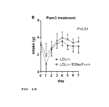
[0013] Figure 4A-C shows the effects on the intake and body
mass of mice using the in vivo model described in FIG. 3. (A) No
significant difference was observed in the intake of food by Ldlr-/-
mice vs. Ldlr-/- E06scFv-P/' mice when treated with vehicle. (B) When
the mice were treated with Pam3, Ldlr-/- mice had less intake of
food in comparison to Ldlr-/- E06scFv-P/' mice. (C) At 12 weeks, the
body mass of Ldlr-/- E06scFv-P/' mice was significantly higher than
Ldlr-/- mice. No significant differences in body mass were seen
between the Ldlr-/- E06scFv-P/' mice and Ldlr-/- mice when treated with
vehicle at week 12.
[0014] Figure 5A-B demonstrates that there were no significant
differences in (A) cholesterol or (B) triglycerides in blood plasma
of E06scTg Ld1r (LDLrK0 (knock-out)) mice vs. Ldlr-/- mice.
[0015] Figure 6A-B demonstrates that there were no significant
differences in (A) lipoprotein cholesterol profile, or (B)
lipoprotein triglycerides profile in blood plasma of Ld1r-/-
E06scFv-P/' mice vs. Ldlr-/- mice, e.g., lipoprotein levels were
similar in both mice.
[0016] Figure 7A-E indicates that there was less measurable
atherosclerosis in Ldlr-/- E06scFv-P/' mice vs. Ldlr-/- mice. (A) The
extent of total aortic atherosclerosis was greater in Ldlr-/- mice
vs. Ldlr-/- E06scFv-P/' mice. (B) There was especially a greater
extent of atherosclerosis in the abdominal aorta (below the
diaphragm) in the Ldlr-/- mice vs. Ldlr-/- E06scFv-P/' mice. (C)
Figures (A) and (B) above represent two-dimensional analysis by
6
CA 03089709 2020-07-27
WO 2019/148204
PCT/US2019/015723
planimimetry of the extent of atherosclerosis. The actual weight
of dissected and cleaned aortas are a better integration of total
atherosclerosis as it contains a dimension of thickness. The
weight of aortas from Ldlr-/- mice were significantly higher than
the aortas from Ldlr-/- E06scFv-P/' mice. (D) When controlled for
body mass, the aorta per body mass from Ldlr-/- mice was
significantly higher than the aorta per body mass from Ld1r-/-
E06scFv-P/' mice. (E) The extent of atherosclerosis at the aortic
root was not different between the two groups.
[0017] Figure 8A-H provides the results of quantitative PCR
(qPCR) looking at inflammatory gene expression in adipose tissue of
E06scFv-P/' mice vs. Ldlr-/- mice. In particular, the
expression of (A) IL1b, (B) IL6, (C) TNFa, (E) MCP1, (F) MIP1a, (G)
miplp, and (H) IL10 were generally lower for Ldlr-/- E06scFv-P/' mice
vs. Ldlr-/- mice, whereas IL12 was slightly higher (D).
[0018] Figure 9A-G provides the results of enzyme-linked
immunosorbent assays (ELISAs) looking at measured cytokine levels
in adipose tissue extracts of Ldlr-/- E06scFv-P/' mice vs. Ldlr-/-
mice. In particular, the measured cytokine levels of (A) IL1b, (B)
IL6, (C) TNFa, (D) MCP1, (E) MIP1a, (F) miplp, and (G) IL10
mirrored the gene expression results presented in FIG. 8.
[0019] Figure 10A-H shows that bone marrow derived cells from
Ldlr-/- E06scFv-P/' when differentiated to macrophage M1 or M2 cells
and stimulated with PAM3 showed less expression of (A) ILlp, (B)
IL6, (C) IL12, (D) TNFa, (E) MCP1, (F) MIP1a, (G) miplp and (H)
RANTES in comparison to differentiated macrophages from Ldlr-/-
mice. Data shown are comparison of M1 cells derived from Ldlr-/-
E06scFv-P/' or Ldlr-/- mice. Similar data were found from M2 cells,
e.g., less expression from M2 cells of Ld/r-/-E06scFv+/+ compared to
Ldlr-/-, except the absolute levels of cytokine expression was less.
[0020] Figure 11 the robust gene expression of the E06-scFv
mRNA in different adipose tissue derived from the Ldlr-/- E06scFv-P/'
mice. This is due to macrophage infiltration. Macrophages express
the apoE promoter, and thus express the E06-scFv transgene.
[0021] Figure 12 provides fluorescence microscope images
indicating that treatment of macrophages (RAW264.7) with PAM3
induced the production of OxPL. Because macrophages from Ld1r-/-
7
CA 03089709 2020-07-27
WO 2019/148204
PCT/US2019/015723
E06scFv-P/' mice were less responsive to TLR2 stimulation, and
because macrophages express and secrete E06-scFv into the culture,
it was postulated that TLR2 stimulated macrophages generated OxPL,
and that this in turn activated inflammatory gene expression in an
autocrine manner. To test this hypothesis macrophages were
stimulated with PAM3: Left panel, treatment with vehicle; right
panel, treatment with the TLR2 agonist, PAM3. RAW264.7 cultures
were incubated with Pam3 (1 pg/mL) or control vehicle for 18 h, and
surface stained for OxPL with E06 IgM antibody and Goat anti-ms
IgM-FITC conjugate. This demonstrates that TLR2 stimulated
macrophages generate OxPL.
[0022] Figure 13A-F indicates that there was less measurable
atherosclerosis in Ldlr-/- E06scFv-P/' RAG1-/- mice vs. Ldlr-/- RAG1-/-
mice. As RAG1 KO mice do not have B or T cells, all
atherosclerosis events related to immunological cells are directly
attributable to the action of macrophages. Notably, the absolute
level of atherosclerosis of the Ldlr-/- RAG1-/- mice was reduced by
half in comparison to Ldlr-/- mice. (A) Abdominal aortas isolated
from Ldlr-/- RAG1-/- mice (left) and from Ldlr-/- E06scFv-P/' RAG1-/-
mice (right). The abdominal aortas from Ldlr-/- E06scFv-P/' RAG1-/-
mice demonstratively had less atherosclerotic lesions than Ldlr-/-
RAG1-/- mice. (B) The aortic sinus lesion size was significantly
smaller for Ldlr-/- E06scFv-P/' RAG1-/- mice v. Ldlr-/- RAG1-/- mice.
(C) Ldlr-/- RAG1-/- mice had a higher percentage of total body
lesions vs. Ldlr-/- E06scFv-P/' RAG1-/- mice. (D) Ldlr-/- RAG1-/- mice
had a higher percentage of abdominal lesions vs. Ldlr-/- E06scFv-P/'
RAG1-/- mice. (E) The weight of aortas from Ldlr-/- RAG1-/- mice were
significantly higher than the aortas from Ldlr-/- E06scFv-P/' RAG1-/-
mice. (F) When controlled for body mass, the aorta per body mass
from Ldlr-/- RAG1-/- mice was significantly higher than the aorta per
body mass from Ldlr-/- E06scFv-P/' RAG1-/- mice. Because RAG1 KO mice
lack both B and T cells, the major immunological cell type
promoting atherosclerosis in these mice the macrophage. Thus, these
data indicate that a major mechanism by which macrophages
contribute to atherosclerosis in this TLR2 induced model is due to
responses of OxPL.
8
ak 03089709 2020-07-27
WO 2019/148204
PCT/US2019/015723
[0023] Figure 14 presents images of children with Kawasaki
Disease, and further, coronary and abdominal artery aneurysms
associated with the Kawasaki Disease.
[0024] Figure 15 presents a mouse model to study the impact of
LCWE, and HFC diet on atherosclerosis in Ldlr-/- mice, Ldlr-/- E06Tg
mice, Ldlr-/- IK17Tg mice, TLR2-/- Ldlr-/- mice.
[0025] Figure 16 presents en face analysis of aortic lesions in
mice fed HFC diet for 12-wk and TLR2 activated by LCWE. E06scFv-Tg
reduced en face aortic lesions significantly.
[0026] Figure 17 provides analysis of the abdominal aortic
lesion area in various Ldlr-/- mice injected with LCWE and fed with
HFC diet for 12-wks. Data are expressed as the percentage of
atherosclerosis measured in the abdominal aorta by Sudan IV
staining. E06Tg and IK17Tg were found to exert a statistically
significant protective effect. TLR2 was also found to
statistically decrease in comparison to the Ldlr-/- mice as well.
[0027] Figure 18 presents cross-sections of aortic roots from
Ldlr-/- mice and from E06-Tg Ldlr-/- mice treated with LCWE. E06scFv-
Tg reduced the aortic root lesions, necrotic core size and most
importantly in the context of Kawasaki disease manifestations,
coronary arteritis. The arrows point to the coronary arteries in
the cross section. Extensive arteritis (large cell mass) was
present in the Ldlr-/- mice but missing in the E06-Tg Ldlr-/- mice.
[0028] Figure 19 presents further cross-sections of aortic
roots from Ldlr-/- mice and from E06-Tg Ldlr-/- mice treated with
LCWE. The arrows point to the coronary arteries in the cross
section. Extensive arteritis (large cell mass) was present in the
Ldlr-/- mice but missing in the E06-Tg Ldlr-/- mice.
[0029] Figure 20 presents cross-sections of aortic roots from
Ldlr-/- TLR2-/- mice and from IK17-Tg'/' Ldlr-/- mice treated with
LCWE. The arrows point to the coronary arteries in the cross
section. Both E06scFv and IK17scFv decreased coronary arteritis.
E06 (Anti-OxPL) but not IK17 (anti-MDA) reduced the aortic root
lesions.
[0030] Figure 21 presents further cross-sections of aortic
roots from Ldlr-/- TLR2-/- mice and from IK17-Tg'/' Ldlr-/- mice
treated with LCWE. The arrows point to the coronary arteries in
9
ak 03089709 2020-07-27
WO 2019/148204
PCT/US2019/015723
the cross section. The lack of arteritis in coronary arteries in
the Ldlr-/- TLR2-/- mice indicates the importance of TLR2 activation
in the mouse model of Kawasaki Disease.
[0031] Figure 22 depicts plasma inflammatory cytokine levels of
LDLr-/- control mice and E06-Tg LDLr-/- mice treated with LCWE and
fed HFC diets for 12 wks. The plasma cytokines were measured by
multiplex cytokine assays simultaneously using Bio-Plex Pro mouse
cytokine assay kit (Bio-Rad Laboratories, USA). Significant
reduction (p < 0.04) in serum TNFa, RANTES, MCP-1, CXCL1, IL-6, and
IL-12 protein levels was observed in E06scFv-Tg LDLr-/- mice
compared to control mice.
DETAILED DESCRIPTION
[0032] As used herein and in the appended claims, the singular
forms "a," "an," and "the" include plural referents unless the
context clearly dictates otherwise. Thus, for example, reference
to a "single-chain variable fragment" or "scFv" includes a
plurality of single-chain variable fragments and reference to
"oxidized phospholipid" includes reference to one or more oxidized
phospholipids and equivalents thereof known to those skilled in the
art, and so forth.
[0033] Unless defined otherwise, all technical and scientific
terms used herein have the same meaning as commonly understood to
one of ordinary skill in the art to which this disclosure belongs.
Although any methods and reagents similar or equivalent to those
described herein can be used in the practice of the disclosed
methods and compositions, the exemplary methods and materials are
now described.
[0034] All publications mentioned herein are incorporated
herein by reference in full for the purpose of describing and
disclosing the methodologies, which are described in the
publications, which might be used in connection with the
description herein. Moreover, with respect to any term that is
presented in one or more publications that is similar to, or
identical with, a term that has been expressly defined in this
disclosure, the definition of the term as expressly provided in
this disclosure will control in all respects.
CA 03089709 2020-07-27
WO 2019/148204
PCT/US2019/015723
[0035] Also, the use of "and" means "and/or" unless stated
otherwise. Similarly, "comprise," "comprises," "comprising"
"include," "includes," and "including" are interchangeable and not
intended to be limiting.
[0036] It is to be further understood that where descriptions
of various embodiments use the term "comprising," those skilled in
the art would understand that in some specific instances, an
embodiment can be alternatively described using language
"consisting essentially of" or "consisting of."
[0037] The terms "antibody" and "immunoglobulin" are used
interchangeably in the broadest sense and include monoclonal
antibodies (e.g., full length or intact monoclonal antibodies),
polyclonal antibodies, multivalent antibodies, multispecific
antibodies (e.g., bispecific antibodies so long as they exhibit the
desired biological activity) and may also include certain antibody
fragments. An antibody can be human, humanized and/or affinity
matured.
[0038] Depending on the amino acid sequence of the constant
domain of their heavy chains, immunoglobulins can be assigned to
different classes. There are five major classes of immunoglobulins:
IgA, IgD, IgE, IgG, and IgM, and several of these can be further
divided into subclasses (isotypes), e.g., IgGl, IgG2, IgG3, IgG4,
IgAl, and IgA2. The heavy-chain constant domains that correspond to
the different classes of immunoglobulins are called a, 5, s, y, and
p, respectively. The subunit structures and three-dimensional
configurations of different classes of immunoglobulins are well
known.
[0039] "Antibody fragments" comprise only a portion of an
intact antibody, wherein the portion typically retains at least
one, more commonly most or all, of the functions normally
associated with that portion when present in an intact antibody.
Examples of antibody fragments include Fab, Fab', F(ab')2, and Fv
fragments; diabodies; linear antibodies; single-chain antibody
molecules; and multispecific antibodies formed from antibody
fragments. In one embodiment, an antibody fragment comprises an
antigen binding site of the intact antibody and thus retains the
ability to bind antigen. In another embodiment, an antibody
11
CA 03089709 2020-07-27
WO 2019/148204
PCT/US2019/015723
fragment, for example one that comprises the Fc region, retains at
least one of the biological functions normally associated with the
Fc region when present in an intact antibody, such as FcRn binding,
antibody half-life modulation, ADCC function and complement
binding. In one embodiment, an antibody fragment is a monovalent
antibody that has an in vivo half-life substantially similar to an
intact antibody. For example, such an antibody fragment may
comprise on antigen binding arm linked to an Fc sequence capable of
conferring in vivo stability to the fragment. It should be
recognized however, a long half-life of the antibody is not
necessary for certain indication (e.g., acute ishemic/reperfusion
treatments).
[0040] An "antigen" is a predetermined antigen to which an
antibody can selectively bind. The target antigen may be
polypeptide, carbohydrate, nucleic acid, lipid, hapten or other
naturally occurring or synthetic compound. In one embodiment, of
the disclosure an antigen is an OxPL.
[0041] The term "anti-OxPL antibody" or "an antibody that binds
to OxPL" refers to an antibody that is capable of binding OxPL with
sufficient affinity such that the antibody is useful as a
diagnostic and/or therapeutic agent in targeting OxPL. In some
embodiments of the disclosure an anti-OxPL antibody has the same or
a similar binding specificity and Kd as the E06 antibody or the QX5
antibody. In yet another embodiment, the anti-OxPL antibody binds
to the PC headgroup of OxPLs.
[0042] A "blocking" antibody or an "antagonist" antibody is one
which inhibits or reduces biological activity of the antigen it
binds. Certain blocking antibodies or antagonist antibodies
substantially or completely inhibit the biological activity of the
antigen.
[0043] "Binding affinity" generally refers to the strength of
the sum total of non-covalent interactions between a single binding
site of a molecule (e.g., an antibody) and its binding partner
(e.g., an antigen). Unless indicated otherwise, as used herein,
"binding affinity" refers to intrinsic binding affinity which
reflects a 1:1 interaction between members of a binding pair (e.g.,
antibody and antigen). The affinity of a molecule X for its partner
12
CA 03089709 2020-07-27
WO 2019/148204
PCT/US2019/015723
Y can generally be represented by the dissociation constant (K,I).
Affinity can be measured by common methods known in the art,
including those described herein. Low-affinity antibodies
generally bind antigen slowly and tend to dissociate readily,
whereas high-affinity antibodies generally bind antigen faster and
tend to remain bound longer. A variety of methods of measuring
binding affinity are known in the art, any of which can be used for
purposes of the present invention.
[0044] A "biological sample" encompasses a variety of sample
types obtained from an individual and can be used in a diagnostic
or monitoring assay. The definition encompasses blood and other
liquid samples of biological origin, solid tissue samples such as a
biopsy specimen or tissue cultures or cells derived therefrom, and
the progeny thereof. The definition also includes samples that
have been manipulated in any way after their procurement, such as
by treatment with reagents, solubilization, or enrichment for
certain components, such as proteins or polynucleotides, or
embedding in a semi-solid or solid matrix for sectioning purposes.
The term "biological sample" encompasses a clinical sample, and
also includes cells in culture, cell supernatants, cell lysates,
serum, plasma, biological fluid, and tissue samples. The source of
the biological sample may be solid tissue as from a fresh, frozen
and/or preserved organ or tissue sample or biopsy or aspirate;
blood or any blood constituents; bodily fluids such as cerebral
spinal fluid, amniotic fluid, peritoneal fluid, or interstitial
fluid; cells from any time in gestation or development of the
subject. In some embodiments, the biological sample is obtained
from a primary or metastatic tumor. The biological sample may
contain compounds which are not naturally intermixed with the
tissue in nature such as preservatives, anticoagulants, buffers,
fixatives, nutrients, antibiotics, or the like.
[0045] The term "diabodies" refers to small antibody fragments
with two antigen-binding sites, which fragments comprise a heavy-
chain variable domain (VO connected to a light-chain variable
domain (Vd in the same polypeptide chain (VH-VL) . By using a linker
that is too short to allow pairing between the two domains on the
same chain, the domains are forced to pair with the complementary
13
CA 03089709 2020-07-27
WO 2019/148204
PCT/US2019/015723
domains of another chain and create two antigen-binding sites.
Diabodies are described more fully in, for example, EP 404,097; WO
93/11161; and Hollinger et al., Proc. Natl. Acad. Sci. USA,
90:6444-6448 (1993). Triabodies and tetrabodies are also described
in Hudson et al., Nat. Med. 9:129-134 (2003).
[0046] A "disorder" or "disease" is any condition that would
benefit from treatment with a substance/molecule or method of the
disclosure. This includes TLR2 mediated disease or disorders, such
as Kawasaki disease.
[0047] An "effective amount" refers to an amount effective, at
dosages and for periods of time necessary, to achieve the desired
therapeutic or prophylactic result.
[0048] The term "Fc region" as used herein refers to the C-
terminal region of an immunoglobulin heavy chain, including native
sequence Fc regions and variant Fc regions.
[0049] A "functional Fc region" possesses an effector function
of a native sequence Fc region. Such effector functions generally
require the Fc region to be combined with a binding to domain
(e.g., an antibody variable domain) and can be assessed using
various assays as disclosed, for example, in definitions herein.
[0050] A "native sequence Fc region" comprises an amino acid
sequence that is identical to the amino acid sequence of an Fc
region found in nature. Native sequence human Fc regions include a
native sequence human IgG1 Fc region (non-A and A allotypes);
native sequence human IgG2 Fc region; native sequence human IgG3 Fc
region; and native sequence human IgG4 Fc region as well as
naturally occurring variants thereof.
[0051] "Fc receptor" or "FcR" describes a receptor that binds
to the Fc region of an antibody. In some embodiments, an FcR is a
native human FcR. In other embodiments, an FcR is one which binds
an IgG antibody (a gamma receptor) and includes receptors of the
FcyRI, FcyRII, and FcyRIII subclasses, including allelic variants
and alternatively spliced forms of those receptors. FcyRII
receptors include FcyRIIA (an "activating receptor") and FcyRIIB
(an "inhibiting receptor"), which have similar amino acid sequences
that differ primarily in the cytoplasmic domains thereof.
Activating receptor FcyRIIA contains an immunoreceptor tyrosine-
14
CA 03089709 2020-07-27
WO 2019/148204
PCT/US2019/015723
based activation motif (ITAM) in its cytoplasmic domain. Inhibiting
receptor FcyRIIB contains an immunoreceptor tyrosine-based
inhibition motif (ITIM) in its cytoplasmic domain. (see, e.g.,
Daeron, Annu. Rev. Immunol. 15:203-234 (1997)). FcRs are reviewed,
for example, in Ravetch and Kinet, Annu. Rev. Immunol 9:457-92
(1991); Capel et al., Immunomethods 4:25-34 (1994); and de Haas et
al., J. Lab. din. Med. 126:330-41 (1995). Other FcRs, including
those to be identified in the future, are encompassed by the term
"FcR" herein.
[0052] Fc receptor also include the neonatal receptor, FcRn,
which is responsible for the transfer of maternal IgGs to the fetus
(Guyer et al., J. Immunol. 117:587 (1976) and Kim et al., J.
Immunol. 24:249 (1994)) and regulation of homeostasis of
immunoglobulins. Methods of measuring binding to FcRn are known
(see, e.g., Ghetie and Ward., Immunol. Today 18(12):592-598 (1997);
Ghetie et al., Nature Biotechnology, 15(7):637-640 (1997); Hinton
et al., J. Biol. Chem. 279(8):6213-6216 (2004); WO 2004/92219
(Hinton et al.).
[0053] Binding to human FcRn in vivo and serum half-life of
human FcRn high affinity binding polypeptides can be assayed, e.g.,
in transgenic mice or transfected human cell lines expressing human
FcRn, or in primates to which the polypeptides with a variant Fc
region are administered. WO 2000/42072 (Presta) describes antibody
variants with improved or diminished binding to FcRs. See also,
e.g., Shields et al., J. Biol. Chem. 9(2):6591-6604 (2001).
[0054] "Fv" is the minimum antibody fragment which contains a
complete antigen-recognition and -binding site. In a two-chain Fv
species, this region consists of a dimer of one heavy- and one
light-chain variable domain in tight, non-covalent association. In
a single-chain Fv species, one heavy- and one light-chain variable
domain can be covalently linked by a flexible peptide linker such
that the light and heavy chains can associate in a "dimeric"
structure analogous to that in a two-chain Fv species. It is in
this configuration that the three HVRs of each variable domain
interact to define an antigen-binding site on the surface of the
VH-VL dimer. Collectively, the six HVRs confer antigen-binding
specificity to the antibody. However, even a single variable
CA 03089709 2020-07-27
WO 2019/148204
PCT/US2019/015723
domain (or half of an Fv comprising only three HVRs specific for an
antigen) has the ability to recognize and bind antigen, although at
a lower affinity than the entire binding site.
[0055] The Fab fragment also contains the constant domain of
the light chain and the first constant domain (CH1) of the heavy
chain. Fab' fragments differ from Fab fragments by the addition of
a few residues at the carboxy terminus of the heavy chain CH1
domain including one or more cysteines from the antibody hinge
region. Fab'-SH is the designation herein for Fab' in which the
cysteine residue(s) of the constant domains have a free thiol
group. F(ab')2 antibody fragments originally were produced as pairs
of Fab' fragments which have hinge cysteines between them. Other
chemical couplings of antibody fragments are also known.
[0056] Papain digestion of antibodies produces two identical
antigen-binding fragments, called "Fab" fragments, each with a
single antigen-binding site, and a residual "Fc" fragment, whose
name reflects its ability to crystallize readily. Pepsin treatment
yields an F(ab')2 fragment that has two antigen-combining sites and
is still capable of cross-linking antigen.
[0057] "Framework" or "FR" residues are those variable domain
residues other than the hypervariable region residues as herein
defined.
[0058] "Humanized" forms of non-human (e.g., murine) antibodies
are chimeric antibodies that contain minimal sequence derived from
non-human immunoglobulin. In one embodiment, a humanized antibody
is a human immunoglobulin (recipient antibody) in which residues
from a HVR of the recipient are replaced by residues from a HVR of
a non-human species (donor antibody) such as mouse, rat, rabbit, or
nonhuman primate having the desired specificity, affinity, and/or
capacity. In some instances, framework residues of the human
immunoglobulin are replaced by corresponding non-human residues.
Furthermore, humanized antibodies may comprise residues that are
not found in the recipient antibody or in the donor antibody.
These modifications may be made to further refine antibody
performance. In general, a humanized antibody will comprise
substantially all of at least one, and typically two, variable
domains, in which all or substantially all of the hypervariable
16
CA 03089709 2020-07-27
WO 2019/148204
PCT/US2019/015723
loops correspond to those of a non-human immunoglobulin, and all or
substantially all of the FRs are those of a human immunoglobulin
sequence. The humanized antibody optionally will also comprise at
least a portion of an immunoglobulin constant region (Fc),
typically that of a human immunoglobulin. For further details,
see, e.g., Jones et al., Nature 321:522-525 (1986); Riechmann et
al., Nature 332:323-329 (1988); and Presta, Curr. Op. Struct. Biol.
2:593-596 (1992). See also, e.g., Vaswani and Hamilton, Ann.
Allergy, Asthma & Immunol. 1:105-115 (1998); Harris, Biochem. Soc.
Transactions 23:1035-1038 (1995); Hurle and Gross, Curr. Op.
Biotech, 5:428-433 (1994); and U.S. Pat. Nos. 6,982,321 and
7,087,409.
[0059] A "human antibody" is one which possesses an amino acid
sequence which corresponds to that of an antibody produced by a
human and/or has been made using any of the techniques for making
human antibodies as disclosed herein. This definition of a human
antibody specifically excludes a humanized antibody comprising non-
human antigen-binding residues. Human antibodies can be produced
using various techniques known in the art, including phage-display
libraries. Hoogenboom and Winter, J. Mol. Biol., 227:381 (1991);
Marks et al., J. Mol. Biol., 222:581 (1991). Also available for the
preparation of human monoclonal antibodies are methods described in
Cole et al., Monoclonal Antibodies and Cancer Therapy, Alan R.
Liss, p. 77 (1985); Boerner et al., J. Immunol., 147(1):86-95
(1991). See also van Dijk and van de Winkel, Curr. Opin.
Pharmacol., 5: 368-74 (2001). Human antibodies can be prepared by
administering the antigen to a transgenic animal that has been
modified to produce such antibodies in response to antigenic
challenge, but whose endogenous loci have been disabled, e.g.,
immunized xenomice (see, e.g., U.S. Pat. Nos. 6,075,181 and
6,150,584). See also, for example, Li et al., Proc. Natl. Acad.
Sci. USA, 103:3557-3562 (2006) regarding human antibodies generated
via a human B-cell hybridoma technology. It should be important to
note that a "human antibody" does not include naturally occurring
antibodies produced by a human, but rather refer to antibodies that
do not contain any epitope or antigenic fragment a human subject
would not recognize as "foreign".
17
CA 03089709 2020-07-27
WO 2019/148204
PCT/US2019/015723
[0060] "Human effector cells" are leukocytes which express one
or more FcRs and perform effector functions. In certain
embodiments, the cells express at least FcyRIII and perform ADCC
effector function(s). Examples of human leukocytes which mediate
ADCC include peripheral blood mononuclear cells (PBMC), natural
killer (NK) cells, monocytes, cytotoxic T cells, and neutrophils.
The effector cells may be isolated from a native source, e.g., from
blood.
[0061] The term "hypervariable region," "HVR," or "HV," when
used herein refers to the regions of an antibody variable domain
which are hypervariable in sequence and/or form structurally
defined loops. Generally, antibodies comprise six HVRs; three in
the VH domain (H1, H2, H3), and three in the VL domain (L1, L2, L3).
In native antibodies, H3 and L3 display the most diversity of the
six HVRs, and H3 in particular is believed to play a unique role in
conferring fine specificity to antibodies. See, e.g., Xu et al.,
Immunity 13:37-45 (2000); Johnson and Wu, in Methods in Molecular
Biology 248:1-25 (Lo, ed., Human Press, Totowa, N.J., 2003).
Indeed, naturally occurring camelid antibodies consisting of a
heavy chain only are functional and stable in the absence of light
chain. See, e.g., Hamers-Casterman et al., Nature 363:446-448
(1993); Sheriff et al., Nature Struct. Biol. 3:733-736 (1996).
[0062] An "individual," "subject," or "patient" is a
vertebrate. In certain embodiments, the vertebrate is a mammal.
Mammals include, but are not limited to, farm animals (such as
cows), sport animals, pets (such as cats, dogs, and horses),
primates, mice and rats. In certain embodiments, a mammal is a
human.
[0063] An "isolated" antibody or antibody fragment is one which
has been identified and separated and/or recovered from a component
of its natural environment. Contaminant components of its natural
environment are materials which would interfere with diagnostic or
therapeutic uses for the antibody, and may include enzymes,
hormones, and other proteinaceous or nonproteinaceous solutes. In
some embodiments, the antibody will be purified (1) to greater than
95% by weight of antibody as determined by the Lowry method, and
typically more than 99% by weight, (2) to a degree sufficient to
18
CA 03089709 2020-07-27
WO 2019/148204
PCT/US2019/015723
obtain at least 15 residues of N-terminal or internal amino acid
sequence by use of a spinning cup sequenator, or (3) to homogeneity
by SDS-PAGE under reducing or nonreducing conditions using
Coomassie blue or silver stain. An isolated antibody includes the
antibody in situ within recombinant cells since at least one
component of the antibody's natural environment will not be
present. Ordinarily, however, an isolated antibody will be prepared
by at least one purification step.
[0064] An "isolated" nucleic acid molecule is a nucleic acid
molecule that is identified and separated from at least one
contaminant nucleic acid molecule with which it is ordinarily
associated in the natural source of the antibody nucleic acid. An
isolated nucleic acid molecule is other than in the form or setting
in which it is found in nature. Isolated nucleic acid molecules
therefore are distinguished from the nucleic acid molecule as it
exists in natural cells. However, an isolated nucleic acid
molecule includes a nucleic acid molecule contained in cells that
ordinarily express the antibody where, for example, the nucleic
acid molecule is in a chromosomal location different from that of
natural cells.
[0065] The word "label" when used herein refers to a compound
or composition which is conjugated or fused directly or indirectly
to a reagent such as a nucleic acid probe or an antibody and
facilitates detection of the reagent to which it is conjugated or
fused. The label may itself be detectable (e.g., radioisotope
labels or fluorescent labels) or, in the case of an enzymatic
label, may catalyze chemical alteration of a substrate compound or
composition which is detectable.
[0066] The "light chains" of antibodies (immunoglobulins) from
any vertebrate species can be assigned to one of two clearly
distinct types, called kappa (K) and lambda (A), based on the amino
acid sequences of their constant domains.
[0067] The term "monoclonal antibody" as used herein refers to
an antibody obtained from a population of substantially homogeneous
antibodies, i.e., the individual antibodies comprising the
population are identical except for possible mutations, e.g.,
naturally occurring mutations, that may be present in minor
19
CA 03089709 2020-07-27
WO 2019/148204
PCT/US2019/015723
amounts. Thus, the modifier term "monoclonal" indicates the
character of the antibody as not being a mixture of discrete
antibodies. In certain embodiments, such a monoclonal antibody
typically includes an antibody comprising a polypeptide sequence
that binds a target, wherein the target-binding polypeptide
sequence was obtained by a process that includes the selection of a
single target binding polypeptide sequence from a plurality of
polypeptide sequences. For example, the selection process can be
the selection of a unique clone from a plurality of clones, such as
a pool of hybridoma clones, phage clones, or recombinant DNA
clones. It should be understood that a selected target binding
sequence can be further altered, for example, to improve affinity
for the target, to humanize the target binding sequence, to improve
its production in cell culture, to reduce its immunogenicity in
vivo, to create a multispecific antibody, etc., and that an
antibody comprising the altered target binding sequence is also a
monoclonal antibody for purposes of this disclosure. In contrast
to polyclonal antibody preparations, which typically include
different antibodies directed against different determinants
(epitopes), each monoclonal antibody of a monoclonal antibody
preparation is directed against a single determinant on an antigen.
In addition to their specificity, monoclonal antibody preparations
are advantageous in that they are typically uncontaminated by other
immunoglobulins.
[0068] The modifier term "monoclonal" indicates the character
of the antibody as being obtained from a substantially homogeneous
population of antibodies, and is not to be construed as requiring
production of the antibody by any particular method. For example,
the monoclonal antibodies to be used in accordance with the
disclosure may be made by a variety of techniques, including, for
example, the hybridoma method (e.g., Kohler and Milstein, Nature,
256:495-97 (1975); Hongo et al., Hybridoma, 14 (3): 253-260 (1995),
Harlow et al., Antibodies: A Laboratory Manual, (Cold Spring Harbor
Laboratory Press, 2nd ed. 1988); Hammerling et al., in: Monoclonal
Antibodies and T-Cell Hybridomas 563-681 (Elsevier, N.Y., 1981)),
recombinant DNA methods (see, e.g., U.S. Pat. No. 4,816,567),
phage-display technologies (see, e.g., Clackson et al., Nature,
CA 03089709 2020-07-27
WO 2019/148204
PCT/US2019/015723
352: 624-628 (1991); Marks et al., J. Mol. Biol. 222: 581-597
(1992); Sidhu et al., J. Mol. Biol. 338(2): 299-310 (2004); Lee et
al., J. Mol. Biol. 340(5): 1073-1093 (2004); Fellouse, Proc. Natl.
Acad. Sci. USA 101(34): 12467-12472 (2004); and Lee et al., J.
Immunol. Methods 284(1-2): 119-132 (2004), and technologies for
producing human or human-like antibodies in animals that have parts
or all of the human immunoglobulin loci or genes encoding human
immunoglobulin sequences (see, e.g., WO 1998/24893; WO 1996/34096;
WO 1996/33735; WO 1991/10741; Jakobovits et al., Proc. Natl. Acad.
Sci. USA 90: 2551 (1993); Jakobovits et al., Nature 362: 255-258
(1993); Bruggemann et al., Year in Immunol. 7:33 (1993); U.S. Pat.
Nos. 5,545,807; 5,545,806; 5,569,825; 5,625,126; 5,633,425; and
5,661,016; Marks et al., Bio/Technology 10: 779-783 (1992); Lonberg
et al., Nature 368: 856-859 (1994); Morrison, Nature 368: 812-813
(1994); Fishwild et al., Nature Biotechnol. 14: 845-851 (1996);
Neuberger, Nature Biotechnol. 14: 826 (1996); and Lonberg and
Huszar, Intern. Rev. Immunol. 13: 65-93 (1995).
[0069] The monoclonal antibodies herein specifically include
"chimeric" antibodies in which a portion of the heavy and/or light
chain is identical with or homologous to corresponding sequences in
antibodies derived from a particular species or belonging to a
particular antibody class or subclass, while the remainder of the
chain(s) is identical with or homologous to corresponding sequences
in antibodies derived from another species or belonging to another
antibody class or subclass, as well as fragments of such
antibodies, so long as they exhibit the desired biological activity
(see, e.g., U.S. Pat. No. 4,816,567; and Morrison et al., Proc.
Natl. Acad. Sci. USA 81:6851-6855 (1984)). Chimeric antibodies
include antibodies wherein the antigen-binding region of the
antibody is derived from an antibody produced by, e.g., immunizing
macaque monkeys with the antigen of interest.
[0070] A "polynucleotide," or "nucleic acid," as used herein,
refer to polymers of nucleotides of any length, and include DNA and
RNA. The nucleotides can be deoxyribonucleotides, ribonucleotides,
modified nucleotides or bases, and/or their analogs that can be
incorporated into a polymer by DNA or RNA polymerase, or by a
synthetic reaction. A polynucleotide may comprise modified
21
CA 03089709 2020-07-27
WO 2019/148204
PCT/US2019/015723
nucleotides, such as methylated nucleotides and their analogs. If
present, modification to the nucleotide structure may be imparted
before or after assembly of the polymer. The sequence of
nucleotides may be interrupted by non-nucleotide components. A
polynucleotide may be further modified after synthesis, such as by
conjugation with a label. Other types of modifications include, for
example, "caps", substitution of one or more of the naturally
occurring nucleotides with an analog, internucleotide modifications
such as, for example, those with uncharged linkages (e.g., methyl
phosphonates, phosphotriesters, phosphoamidates, carbamates, etc.)
and with charged linkages (e.g., phosphorothioates,
phosphorodithioates, etc.), those containing pendant moieties, such
as, for example, proteins (e.g., nucleases, toxins, antibodies,
signal peptides, poly-L-lysine, etc.), those with intercalators
(e.g., acridine, psoralen, etc.), those containing chelators (e.g.,
metals, radioactive metals, boron, oxidative metals, etc.), those
containing alkylators, those with modified linkages (e.g., alpha
anomeric nucleic acids, etc.), as well as unmodified forms of the
polynucleotide(s). Further, any of the hydroxyl groups ordinarily
present in the sugars may be replaced, for example, by phosphonate
groups, phosphate groups, protected by standard protecting groups,
or activated to prepare additional linkages to additional
nucleotides, or may be conjugated to solid or semi-solid supports.
The 5' and 3' terminal OH can be phosphorylated or substituted with
amines or organic capping group moieties of from 1 to 20 carbon
atoms. Other hydroxyls may also be derivatized to standard
protecting groups. Polynucleotides can also contain analogous forms
of ribose or deoxyribose sugars that are generally known in the
art, including, for example, 2T-0-methyl-, 2T-0-allyl, 2T-fluoro-
or 2'-azido-ribose, carbocyclic sugar analogs, alpha-anomeric
sugars, epimeric sugars such as arabinose, xyloses or lyxoses,
pyranose sugars, furanose sugars, sedoheptuloses, acyclic analogs
and basic nucleoside analogs such as methyl riboside. One or more
phosphodiester linkages may be replaced by alternative linking
groups. These alternative linking groups include, but are not
limited to, embodiments wherein phosphate is replaced by P(0)S
("thioate"), P(S)S ("dithioate"), "(0)NR2 ("amidate"), P(0)R,
22
ak 03089709 2020-07-27
WO 2019/148204
PCT/US2019/015723
P(0)OR', CO or CH2 ("formacetal"), in which each R or R' is
independently H or substituted or unsubstituted alkyl (1-20 C)
optionally containing an ether (--0--) linkage, aryl, alkenyl,
cycloalkyl, cycloalkenyl or araldyl. Not all linkages in a
polynucleotide need be identical. The preceding description applies
to all polynucleotides referred to herein, including RNA and DNA.
[0071] "Single-chain Fv" or "scFv" antibody fragments comprise
the VH and VL domains of antibody, wherein these domains are present
in a single polypeptide chain. Generally, the scFv polypeptide
further comprises a polypeptide linker between the VH and VL domains
which enables the scFv to form the desired structure for antigen
binding. FIG. 1 shows an antibody and scFv structure. For a review
of scFv see Pluckthun, in The Pharmacology of Monoclonal
Antibodies, vol. 113, Rosenburg and Moore eds., Springer-Verlag,
New York, pp. 269-315 (1994).
[0072] The term "substantially similar" or "substantially the
same," as used herein, denotes a sufficiently high degree of
similarity between two numeric values (for example, one associated
with an antibody of the disclosure and the other associated with a
reference/comparator antibody), such that one of skill in the art
would consider the difference between the two values to be of
little or no biological and/or statistical significance within the
context of the biological characteristic measured by the values
(e.g., Kd values). The difference between said two values is, for
example, less than about 50%, less than about 40%, less than about
30%, less than about 20%, and/or less than about 10% as a function
of the reference/comparator value.
[0073] The phrase "substantially reduced," or "substantially
different," as used herein, denotes a sufficiently high degree of
difference between two numeric values (generally one associated
with a molecule and the other associated with a
reference/comparator molecule) such that one of skill in the art
would consider the difference between the two values to be of
statistical significance within the context of the biological
characteristic measured by said values (e.g., Kd values). The
difference between said two values is, for example, greater than
about 10%, greater than about 20%, greater than about 30%, greater
23
CA 03089709 2020-07-27
WO 2019/148204
PCT/US2019/015723
than about 40%, and/or greater than about 50% as a function of the
value for the reference/comparator molecule.
[0074] "TLR2 related disease and disorders" includes, but are
not limited to autoimmune diseases including rheumatoid arthritis,
systemic lupus erythematosus, systemic sclerosis, Sjogren's
syndrome, psoriasis, multiple sclerosis, and autoimmune diabetes.
TLR-related conditions (e.g., directly and/or indirectly associated
with TLRs such as TLR2, etc.) can include any one or more of:
diabetes, obesity, sepsis, inflammatory disease (e.g., Crohn's
disease), immune disorders, metabolic disease (e.g., conditions
associated with metabolic syndrome), endocrine disease,
atherosclerosis, asthma, cardiovascular disease, immune-related
conditions, and/or any other suitable conditions. For example, the
TLR2-mediated disease or disorder can be selected from the group
consisting of Kawasaki disease, type 2 diabetes, rheumatoid
arthritis, dermatologic disease, multiple sclerosis, systemic lupus
erythematosus, ulcerative colitis, Graves' Disease, SjOgren's
syndrome, autoimmune thyroid diseases, vasculitis and any
combination thereof.
[0075] As used herein, "treatment" refers to clinical
intervention in an attempt to alter the natural course of the
individual or cell being treated, and can be performed either for
prophylaxis or during the course of clinical pathology. Desirable
effects of treatment include preventing occurrence or recurrence of
disease, alleviation of symptoms, diminishment of any direct or
indirect pathological consequences of the disease, decreasing the
rate of disease progression, amelioration or palliation of the
disease state, and remission or improved prognosis. In some
embodiments, an antibody (humanized or non-humanized), antibody
fragment, or polypeptide of the disclosure or a humanized antibody
of the disclosure are used to delay development of a disease or
disorder.
[0076] The term "variable" refers to the fact that certain
portions of the variable domains differ extensively in sequence
among antibodies and are used in the binding and specificity of
each particular antibody for its particular antigen. However, the
variability is not evenly distributed throughout the variable
24
CA 03089709 2020-07-27
WO 2019/148204
PCT/US2019/015723
domains of antibodies. It is concentrated in three segments called
complementarity-determining regions or hypervariable regions (CDRs
or HVRs, used interchangeably herein) both in the light-chain and
the heavy-chain variable domains. The more highly conserved
portions of variable domains are called the framework (FR). The
variable domains of native heavy and light chains each comprise
four FR regions, largely adopting a (3-sheet configuration,
connected by three HVRs, which form loops connecting, and in some
cases forming part of, the (3-sheet structure. The HVRs in each
chain are held together in close proximity by the FR regions and,
with the HVRs from the other chain, contribute to the formation of
the antigen-binding site of antibodies (see Kabat et al., Sequences
of Proteins of Immunological Interest, Fifth Edition, National
Institute of Health, Bethesda, Md. (1991)). The constant domains
are not involved directly in binding an antibody to an antigen, but
exhibit various effector functions, such as participation of the
antibody in antibody-dependent cellular toxicity.
[0077] The term "vector," as used herein, is intended to refer
to a nucleic acid molecule capable of transporting another nucleic
acid to which it has been linked. One type of vector is a
"plasmid", which refers to a circular double stranded DNA loop into
which additional DNA segments may be ligated. Another type of
vector is a phage vector. Another type of vector is a viral vector,
wherein additional DNA segments may be ligated into the viral
genome. Certain vectors are capable of autonomous replication in a
host cell into which they are introduced (e.g., bacterial vectors
having a bacterial origin of replication and episomal mammalian
vectors). Other vectors (e.g., non-episomal mammalian vectors) can
be integrated into the genome of a host cell upon introduction into
the host cell and replicate along with the host genome. Moreover,
certain vectors are capable of directing the expression of genes to
which they are operatively linked. Such vectors are referred to
herein as "expression vectors". In general, expression vectors of
utility in recombinant DNA techniques are often in the form of
plasmids.
[0078] A "variant Fc region" comprises an amino acid sequence
which differs from that of a native sequence Fc region by virtue of
CA 03089709 2020-07-27
WO 2019/148204
PCT/US2019/015723
at least one amino acid modification, typically one or more amino
acid substitution(s). Typically, the variant Fc region has at least
one amino acid substitution compared to a native sequence Fc region
or to the Fc region of a parent polypeptide, e.g. from about one to
about ten amino acid substitutions, and typically from about one to
about five amino acid substitutions in a native sequence Fc region
or in the Fc region of the parent polypeptide. The variant Fc
region of a disclosure possesses at least about 80% homology with a
native sequence Fc region and/or with an Fc region of a parent
polypeptide, at least about 90% homology therewith, and typically
at least about 95% homology therewith.
[0079] "Oxidized phospholipids" (OxPL) refer to phospholipids
with a phosphocholine (PC) headgroup. OxPL are highly pro-
inflammatory and proatherogenic. Phosphorylcholine, a polar head
group on certain phospholipids, has been extensively implicated in
cardiovascular disease. Reactive oxygen species generated during
coronary inflammation causes the oxidation of low density
lipoprotein (LDL) to generate oxidized LDL (oxLDL). In fact,
cardiovascular diseases (CVD) such as atherosclerosis, unstable
angina, or acute coronary syndrome have been shown to be associated
with elevated plasma levels of oxLDL. LDL is a circulating
lipoprotein particle that contains lipids with a PC polar head
group and proteins, an apoB100 protein.
[0080] During oxidation of LDL PC containing neo-epitopes that
are not present on unmodified LDL are generated. Newly exposed PC
on oxLDL is recognized by scavenger receptors on macrophages, such
as CD36, and the resulting macrophage-engulfed oxLDL proceeds
towards the formation of proinflammatory foam cells in the vessel
wall. Oxidized LDL is also recognized by receptors on endothelial
cell surfaces and has been reported to stimulate a range of
responses including endothelial dysfunction, apoptosis, and the
unfolded protein response. PC neo-epitopes are also exposed on LDL
following modification with phospholipase A2 or amine reactive
disease metabolites, such as aldehydes generated from the oxidation
of glycated proteins. These alternately modified LDL particles are
also pro-inflammatory factors in CVD. Antibodies towards
phosphorylcholine (PC) have been shown to bind oxidized, or
26
CA 03089709 2020-07-27
WO 2019/148204
PCT/US2019/015723
otherwise modified, LDL and block the pro-inflammatory activity of
oxLDL in in vivo models or in vitro studies.
[0081] Glycerophospholipids represent a common class of lipids
important for integrity of cellular membranes. Oxidation of
esterified unsaturated fatty acids dramatically changes biological
activities of phospholipids. Apart from impairment of their
structural function, oxidation makes oxidized phospholipids (OxPLs)
markers of "modified-self" type that are recognized by soluble and
cell-associated receptors of innate immunity, including scavenger
receptors, natural (germ line-encoded) antibodies, and C-reactive
protein, thus directing removal of senescent and apoptotic cells or
oxidized lipoproteins. In addition, OxPLs acquire novel biological
activities not characteristic of their unoxidized precursors,
including the ability to regulate innate and adaptive immune
responses. Effects of OxPLs described in vitro and in vivo suggest
their potential relevance in different pathologies, including
atherosclerosis, acute inflammation, lung injury, and many other
conditions.
[0082] Glycerophospholipids comprise an abundant class of
lipids consisting of a glycerol backbone, phosphate-containing
polar head group and two fatty acid residues. PL-bound
polyunsaturated fatty acids (PUFAs) represent the major target for
nonenzymatic or enzymatic oxidation that is not linked to the
generation of metabolic energy. Oxidative fragmentation of a PL
molecule generates several biologically active products, including
small chemically reactive fragments of PUFAs, such as unesterified
oxidized fatty acids (e.g., hydroperoxides and isoprostanes) and
lyso-phospholipids. These products demonstrate multiple biological
activities. Available evidence suggests that nonenzymatic
oxidation of PL-PUFAs proceeds according to the same basic
mechanisms as oxidation of free (unesterified) PUFAs. This
assumption is supported by identification of similar classes of
molecular species generated by oxidation of free and PL-bound PUFAs
that are described herein. In contrast to the nonenzymatic
oxidation, oxidation of PL-PUFAs by enzymes significantly differs
from oxidation of unesterified PUFAs. While free PUFAs can be
oxidized by multiple enzymes belonging to different protein
27
CA 03089709 2020-07-27
WO 2019/148204
PCT/US2019/015723
families and introducing various oxidized groups, only one group of
lipoxygenases (12/15 lipoxygenases) accepts PL-PUFAs as substrates
producing PL-hydroperoxides. Further oxidation and rearrangements
continue without participation of enzymes, and therefore oxidation
initiated by enzymatic and nonenzymatic mechanisms produces many
similar advanced PL oxidation products.
[0083] Toll-like receptor 2 also known as TLR2 is a protein
that in humans is encoded by the TLR2 gene. TLR2 has also been
designated as CD282 (cluster of differentiation 282). TLR2 plays a
role in the immune system. TLR2 is a membrane protein receptor,
which is expressed on the surface of certain cells and recognizes
foreign substances and passes on appropriate signals to the cells
of the immune system. TLR2 plays a fundamental role in pathogen
recognition and activation of innate immunity. Toll like receptors
(TLRs) are highly conserved from Drosophila to humans and share
structural and functional similarities. They recognize pathogen-
associated molecular patterns (PAMPs) that are expressed on
infectious agents, and mediate the production of cytokines
necessary for the development of effective immunity. The various
TLRs exhibit different patterns of expression. This gene is
expressed most abundantly in peripheral blood leukocytes, and
mediates host response to Gram-positive bacteria and yeast via
stimulation of NF-KB. TLR2 detects a large range of microbial
components, such as gram-positive-derived lipoteichoic acid,
bacterial lipoproteins, and zymosan. Of the 11 characterized TLRs,
TLR2 is unique by virtue of its ability to heterodimerize with TLR1
or TLR6, resulting in a relatively broad ligand specificity.
[0084] CD36 by being a coreceptor for TLR2, has suggested that
there is proinflammatory pathway existing between endogenously
derived lipids and activation of innate immunity. Studies have
further found enhanced endothelial TLR2 expression and activation
occurring at areas of disturbed blood flow, such as the areas of
lesion predilection within the aortic tree and heart. Thus, TLR2
expression may promote atherosclerosis in cells that are not of BM
origin, such as endothelial cells, and thus may contribute to the
proinflammatory phenotype of activated endothelial cells.
28
ak 03089709 2020-07-27
WO 2019/148204
PCT/US2019/015723
[0085] In atherosclerosis-susceptible low-density lipoprotein
receptor-deficient (Ldlr-/-) mice, complete deficiency of TLR2 led
to a reduction in atherosclerosis. Loss of TLR2 expression from
BM-derived cells had no effect on disease progression, however.
The data suggests that an unknown endogenous TLR2 agonist
influenced lesion progression by activating TLR2 in cells that were
not of BM cell origin. As shown herein, intraperitoneal
administration of a synthetic TLR2/TLR1 agonist, Pam3CSK4, disease
burden was dramatically increased in Ldlr-/- mice. A complete
deficiency of TLR2 in Ldlr-/- mice, as well as a deficiency of TLR2
only in BM-derived cells in Ldlr-/- mice, attenuated Pam3CSK4-
mediated atherosclerosis, suggesting a role for BM-derived cell
expression of TLR2 in transducing the effects of an exogenous TLR2
agonist.
[0086] OxPL can activate cell signaling via TLR2 mediated
pathways, resulting in proinflammatory cell signaling. In
addition, OxPL mediated activation of TLR2 can lead to apoptosis
and cell death when done in association with signaling pathways
that promote ER stress. OxPL induces IL-8 signaling from
endothelial cells and induces IL-113 and TNFa signaling in
macrophages via a TLR2-dependent signaling pathway. As further
demonstrated herein, activation of macrophages via the synthetic
TLR2 agonist, PAM3CSK4, directly stimulates macrophages to generate
OxPL. It has also been reported that activation of TLR4 via
agonists, such as LPS, will also lead macrophages to generate OxPLs
(Popat et al., JCI, 2017). In aggregate, the data presented herein
demonstrate that OxPL can both directly activate macrophages via
TLR2 (or TLR4) to induce proinflammatory signaling and/or
apoptosis, and that conversely, activation of macrophages via
either TLR2 or TLR4 signaling will in turn cause macrophages to
make OxPL. In the latter situation, when macrophages are
stimulated by TLR2/4 agonists, the locally generated OxPL has the
potentially to amplify and enhance the inflammatory pathway by
auto-paracrine effects. Thus, the studies presented herein suggest
that OxPL can both directly stimulate TLR2 pathways, as well as act
in a paracrine fashion to amplify proinflammatory TLR2/4 agonist
signaling. These insights explain why neutralizing OxPL with an
29
CA 03089709 2020-07-27
WO 2019/148204
PCT/US2019/015723
antibody to OxPL in vivo, in a variety of inflammatory settings,
confers such profound anti-inflammatory effects that are manifested
in reduced disease development.
[0087] The data suggest that the antibodies, or fragments
thereof, that bind OxPL, including E06, or others designed to bind
the phosphocholine (PC) headgroups of PC-containing oxidized
phospholipids (OxPL), could be useful in ameliorating the
deleterious effects of TLR2 agonism present in wide variety of
diseases. These, include atherosclerosis, autoimmune disorders and
specifically in Kawasaki Disease, a disease of children of unknown
origin in which TLR2 mediated agonism is believe to promote
coronary arteritis that leads to coronary aneurysms, severe
coronary calcification, disordered coronary blood flow, acute
thrombosis and major morbidity and death. The disease can also
affect young adults when asymptomatic coronary aneurysms transition
to acute thrombosis causing acute myocardial infarction. Kawasaki
disease can also be associated with myocarditis, heart failure and
need for heart transplantation.
[0088] Innate natural antibodies (NAbs) provide the first line
of host defense against common oxidation-specific epitopes (OSE) on
endogenous neo-epitopes (0xLDL and apoptotic cells) and exogenous
epitopes of pathogens, and maintain host homeostasis. OSEs are
ubiquitous, formed in many inflammatory tissues, including
atherosclerotic lesions, and are a major target of IgM NAbs. The
prototypic IgM NAb E06, binds to the phosphocholine (PC) headgroup
in oxidized phospholipids (OxPL), and blocks uptake of OxLDL by
macrophages. A murine IgM natural antibody to OxPL that binds to
the phosphorylcholine ("PC") headgroup of OxPL but not to native,
non-oxidized phospholipids ("PL") has been cloned and
characterized. However, antibodies like IgM Nab E06 have limited
solubility and cannot be readily synthesized.
[0089] The parent E06 antibody is a murine IgM antibody that
was cloned and characterized and which is the subject of U.S.
Patent No. 6,225,070, which is incorporated herein by reference.
U.S. Patent Publication No. 20150376268A1 describes a fully
functional single chain antibody and humanized antibodies that bind
to OxPL. It describes the numerous unique molecular changes to the
CA 03089709 2020-07-27
WO 2019/148204
PCT/US2019/015723
DNA sequence of the parent antibody framework regions, heavy and
light chains, and a linker sequences that was determined by
repeated rounds of experimentation, which resulted in the
development of a fully functional E06-scFv. When this sequence was
inserted into the appropriate vector, the resultant scFc is
expressed in a soluble form, and possesses all the immunological
binding properties of the parent toward its identified target
antigens, including the ability to bind to a unique anti-idiotypic
antibody, AB1-2, whose epitopes consists of both the heavy and
light chains of the parent antibody. The disclosure of that
application also provides for single chain variable antibody
fragments ("scFv"), VH, VL and complementarity determining regions
that selectively bind to oxidized phospholipids. The scFvs of the
disclosure are soluble and can be readily synthesized. The
disclosure of U.S. Pat. Publ. No. 20150376268A1 is incorporated
herein by reference for all purposes.
[0090] In the studies presented herein, neutralization of OxPL
by the in vivo endogenous expression of the E06 antibody (using the
E06-scFv transgenic mouse) greatly inhibited atherosclerosis
formation caused by TLR2 agonism. In particular, injections of the
TLR2 agonist PAM3CSK4 into cholesterol-fed Ldlr-/- mice lead to
dramatic enhancement of atherosclerosis. A similar set of
injections into the E06-scFv transgenic mice (on Ldlr-/- background)
resulted in a significant inhibition of lesion formation.
[0091] In other studies presented herein, neutralization of
OxPL can protect against a mouse model of Kawasaki Disease.
Administration of the pathogen Lactobaccilus casei has been shown
to cause Kawasaki-like disease in mice, with resultant enhanced
atherosclerosis, coronary artery arteritis and abdominal aneurysms.
This is TLR2 dependent, as administering L. Casei to TLR2 deficient
mice had no disease-causing effect. Importantly, IL-1 has been
shown to be involved, and as noted, OxPL are also a potent inducer
of IL-1 release. Injection of L. Casei into the E06 transgenic
mice (in the Ldlr-/- background) under an identical protocol
resulted in dramatic reductions not only in atherosclerosis, but of
great relevance, in coronary arteritis as compared to injections
into Ldlr-/- mice. The E06 antibody does not directly bind L. Casei
31
CA 03089709 2020-07-27
WO 2019/148204
PCT/US2019/015723
and therefore neutralizes the OxPL caused by the inflammatory
effects associated with TLR2 mediated agonism. The development of
coronary arteritis and subsequently coronary aneurysms has been a
major and feared complication in children developing Kawasaki
disease, and has been estimated to occur in up to 25% of children
despite current therapy. Typically, Kawasaki disease is treated
with intravenous immune globulin (IVIG) derived from pooled and
purified human plasma, and aspirin (which is a generalized but non-
specific anti-inflammatory therapy). Injections of high tittered
humanized or human equivalent anti-OxPL antibody modified to
enhance its biological effectiveness might then confer protection
without any anticipated side effects, as such anti-OxPL antibodies
are present in human B cell repertoire.
[0092] The experimental data thus demonstrates that
atherosclerosis and inflammatory arteritis caused by TLR2 mediated
agonism in vivo in mice can be prevented by neutralization of OxPL.
TLR2 agonism has been implicated in numerous bacterial diseases of
course, but also in a variety of so-called autoimmune mediated
diseases such as lupus, rheumatoid arthritis, and others. The data
demonstrates that neutralization of OxPL by the use of antibodies
targeting the PC of OxPL can ameliorate or prevent many diseases
that are accentuated or are influenced by activation of TLR2
mediated signaling pathways.
[0093] The disclosure provides use of antibodies, antibody
fragments and humanized antibodies that bind to OxPL and which in
some instances have the same or similar binding specificity as the
E06 antibody. Antibody fragments may be generated by traditional
means, such as enzymatic digestion, or by recombinant techniques.
In certain circumstances there are advantages of using antibody
fragments, rather than whole antibodies. The smaller size of the
fragments allows for rapid clearance, and may lead to improved
access to tissue. In an acute setting, the half-life of antibody
fragments is not critical. For a review of certain antibody
fragments, see Hudson et al. (2003) Nat. Med. 9:129-134.
[0094] The disclosure, although providing specific antibody
sequences and antibody sequence fragments having biological
activity, further disclose that these sequences can be used to
32
ak 03089709 2020-07-27
WO 2019/148204
PCT/US2019/015723
generate improved variants. Accordingly, in some instances an
antibody or antibody fragment may have a percent identity to the
sequences of the disclosure.
[0095] In some embodiments, amino acid sequence modification(s)
of the antibodies described herein are contemplated. For example,
it may be desirable to improve the binding affinity and/or other
biological properties of the antibody. Amino acid sequence
variants of the antibody may be prepared by introducing appropriate
changes into the nucleotide sequence encoding the antibody, or by
peptide synthesis. Such modifications include, for example,
deletions from, and/or insertions into and/or substitutions of,
residues within the amino acid sequences of the antibody. Any
combination of deletion, insertion, and substitution can be made to
arrive at the final construct, provided that the final construct
possesses the desired characteristics. The amino acid alterations
may be introduced in the subject antibody amino acid sequence at
the time that sequence is made.
[0096] The disclosure provides an antibody or antibody fragment
capable of binding to OxPL or phosphorylcholine and/or a
phosphorylcholine conjugate, wherein the antibody or antibody
fragment comprises a variable heavy chain (VH) domain and/or a
variable light chain (Vd domain, and wherein
(a) the VH domain comprises an amino acid sequence that
includes one, two or three complementarity determining regions
(CDRs) selected from the group consisting of:
SEQ ID NO:6 and sequence that are at least 95%, 96%, 97%,
98%, 99% or 99.9% identical to SEQ ID NO:6;
SEQ ID NO:7 and sequence that are at least 95%, 96%, 97%,
98%, 99% or 99.9% identical to SEQ ID NO:7; and
SEQ ID NO:8 and sequence that are at least 95%, 96%, 97%,
98%, 99% or 99.9% identical to SEQ ID NO:8;
(b) the VL domain comprises an amino acid sequence that
includes one, two or three complementarity determining regions
(CDRs) selected from the group consisting of:
SEQ ID NO:9 or 12 and sequence that are at least 95%, 96%,
97%, 98%, 99% or 99.9% identical to SEQ ID NO:9 or 12;
33
ak 03089709 2020-07-27
WO 2019/148204
PCT/US2019/015723
SEQ ID NO:10 and sequence that are at least 95%, 96%, 97%,
98%, 99% or 99.9% identical to SEQ ID NO:10; and
SEQ ID NO:11 and sequence that are at least 95%, 96%, 97%,
98%, 99% or 99.9% identical to SEQ ID NO:11.
[0097] In one embodiment, the antibody or antibody fragment
comprises a VH domain that comprises an amino acid sequence that
includes CDRs comprising SEQ ID NO:6, 7 and 8, and/or the VL domain
comprises an amino acid sequence that includes CDRs comprising SEQ
ID NO:9, 10 and 11, or SEQ ID NO:10, 11 and 12.
[0098] In one embodiment, the disclosure provides an antibody
or an scFv selected from the group consisting of: (a) an antibody
or scFv with heavy and light chain domains comprising the
complementarity determining regions of SEQ ID NO:6, 7, 8, 9, 10 and
11; and (b) an antibody or scFv with heavy and light chain domains
comprising the complementarity determining regions of SEQ ID NO:6,
7, 8, 10, 11 and 12. In one embodiment either of (a) or (b) are
linked to an Fc region.
[0099] In one embodiment, the disclosure provides an antibody
comprising a light-chain variable region as set forth in SEQ ID
NO:2 from amino acid 1 to about 146. In another embodiment, the
disclosure provides an antibody with a humanized light chain
variable region comprising the sequence of SEQ ID NO:4 from amino
acid 1 to about 135. In another embodiment, the disclosure
provides an antibody that comprises a heavy chain variable region
comprising a sequence of SEQ ID NO:2 from about 162 to about 269.
In another embodiment, the disclosure provides an antibody that
comprises a humanized heavy chain variable region comprising a
sequence of SEQ ID NO:4 from about 152 to about 258.
[00100] In another embodiment, the disclosure provides a
chimeric antibody comprising, for example, a murine VH and/or VL
and a human Fc region. For example, SEQ ID NO:14 provides the
sequence of a chimeric antibody of the disclosure. In SEQ ID NO:14
amino acids 1-33 comprise and Ig kappa chain leader sequence for
antibody secretion; amino acid 34-146 comprise an E06 light-chain
variable region; amino acids 147-161 provide a flexible linker
sequence; amino acids 162-284 provide an E06 heavy-chain variable
region with a triple mutation of P201A, S224A and A225D relative to
34
CA 03089709 2020-07-27
WO 2019/148204
PCT/US2019/015723
the wild-type urine E06 antibody; amino acids 285-517 comprise an
Fc region, in SEQ ID NO:14 the Fc region is a human IgG1-Fc with a
modification of C290S and H294Y to increase ADCC activity. SEQ ID
NO:14 also provide a further linker and His tag sequence, which one
of skill in the art are optional (e.g., SEQ ID NO:14 from amino
acid 518 to 528). The disclosure also contemplates and provides a
coding sequence for SEQ ID NO:14 comprising SEQ ID NO:13. One of
skill in the art can readily identify the nucleic acid sequence
corresponding to the various domains identified above. The
disclosure also includes a chimeric antibody sequence that is at
least 9096, 95%, 96%, 97%, 98%, 99% or 99.9% identical to SEQ ID
NO:14 from amino acid 1 to 284 linked to an Fc region from an
different immunoglobulin (e.g., IgA, IgD, IgE, IgG, and IgM, or any
of the subclasses (isotypes), e.g., IgGl, IgG2, IgG3, IgG4, IgAl, and
IgA2).
[00101] In one embodiment, the disclosure provides an scFv
comprising a linker between the light change variable region and
the heavy-chain variable region. The linker can be any number of
commonly used peptide linkers. In one embodiment, the linker
comprises a repeating unit of GGGS (SEQ ID NO:5). The repeat of
GGGS (SEQ ID NO:5) may be 2, 3, 4 or more times.
[00102] In another embodiment, the disclosure comprises a scFv
comprising a light chain variable region of SEQ ID NO:2 from amino
acid 1 to 146 linked by a peptide linker to a heavy chain variable
region of SEQ ID NO:2 from amino acid 162 to about 269. In a
specific embodiment, the scFv comprises a sequence of SEQ ID NO:2
form amino acid 1 to 269. In another embodiment, the disclosure
provides for an scFv which has a polypeptide sequence of SEQ ID
NO:2 from amino acid 1 to about 269 or 1 to about 303. In a
further embodiment, the disclosure provides for an scFv that has a
polypeptide sequence that has at least 75%, at least 80%, at least
85%, at least 90%, at least 95%, at least 98%, or at least 9990-
sequence identity to SEQ ID NO:2 from amino acid 1 to about 303 and
which selectively binds to an oxidized phospholipid.
[00103] In yet further embodiments, fusion constructs comprising
a first domain comprising SEQ ID NO:2 from amino acid 1 to about
269 or 1 to about 303 or a variant thereof is operably linked to a
CA 03089709 2020-07-27
WO 2019/148204
PCT/US2019/015723
second domain comprising (i) a detectable label or (ii) a
polypeptide of interest. One of skill in the art will recognize
that such fusion constructs can be generated using chemical or
molecular biology techniques that link a coding sequence comprising
a sequence of SEQ ID NO:1 or variant thereof with a coding sequence
of, for example, a polypeptide of interest. The coding sequences
and domains may be separated by a linker or directly linked.
[00104] In yet another embodiment, the disclosure comprises a
scFv comprising a light chain variable region of SEQ ID NO:4 from
amino acid 1 to 135 linked by a peptide linker to a heavy chain
variable region of SEQ ID NO:4 from amino acid 152 to about 258.
In a specific embodiment, the scFv comprises a sequence of SEQ ID
NO:4 form amino acid 1 to 258. In another embodiment, the
disclosure provides for an scFv which has a polypeptide sequence of
SEQ ID NO:4 from amino acid 1 to about 258 or 1 to about 263. In a
further embodiment, the disclosure provides for an scFv that has a
polypeptide sequence that has at least 75%, at least 80%, at least
85%, at least 90%, at least 95%, at least 98%, or at least 99%
sequence identity to SEQ ID NO:4 from amino acid 1 to about 258 and
which selectively binds to an oxidized phospholipid.
[00105] In yet further embodiments, fusion constructs comprising
a first domain comprising SEQ ID NO:4 from amino acid 1 to about
258 or 1 to about 264 or a variant thereof is operably linked to a
second domain comprising (i) a detectable label or (ii) a
polypeptide of interest. One of skill in the art will recognize
that such fusion constructs can be generated using chemical or
molecular biology techniques that link a coding sequence comprising
a sequence of SEQ ID NO:3 or variant thereof with a coding sequence
of, for example, a polypeptide of interest. The coding sequences
and domains may be separated by a linker or directly linked.
[00106] Nucleic acid molecules encoding the amino acid sequences
of the antibodies, antibody fragments and variants of the antibody
are prepared by a variety of methods known in the art. For
preparing variants such methods include, but are not limited to,
isolation from a natural source (in the case of naturally occurring
amino acid sequence variants) or preparation by oligonucleotide-
mediated (or site-directed) mutagenesis, PCR mutagenesis, and
36
CA 03089709 2020-07-27
WO 2019/148204
PCT/US2019/015723
cassette mutagenesis of an earlier prepared variant or a non-
variant version of the antibody.
[00107] In a particular embodiment, the disclosure provides for
a murine scFv which is encoded by a polynucleotide sequence of SEQ
ID NO:1. In a further embodiment, the disclosure provides for a
murine scFv which is encoded by a polynucleotide sequence that has
at least 75%, at least 80%, at least 85%, at least 90%, at least
95%, at least 98%, or at least 99% sequence identity to SEQ ID NO:1
and which produces a polypeptide that selectively binds to oxidized
phospholipids.
[00108] In another embodiment, the disclosure provides an scFv
comprising a linker between the light change variable region and
the heavy-chain variable region. The linker can be any number of
commonly used peptide linkers. In one embodiment, the linker
comprises a repeating unit of GGGS (SEQ ID NO:5). The repeat of
GGGS (SEQ ID NO:5) may be 2, 3, 4 or more times.
[00109] The disclosure also encompasses humanized antibodies.
Various methods for humanizing non-human antibodies are known in
the art. For example, a humanized antibody can have one or more
amino acid residues introduced into it from a source which is non-
human. These non-human amino acid residues are often referred to as
"import" residues, which are typically taken from an "import"
variable domain. Humanization can be essentially performed
following the method of Winter and co-workers (Jones et al. (1986)
Nature 321:522-525; Riechmann et al. (1988) Nature 332:323-327;
Verhoeyen et al. (1988) Science 239:1534-1536), by substituting
hypervariable region sequences for the corresponding sequences of a
human antibody. Accordingly, such "humanized" antibodies are
chimeric antibodies (U.S. Pat. No. 4,816,567) wherein substantially
less than an intact human variable domain has been substituted by
the corresponding sequence from a non-human species. In practice,
humanized antibodies are typically human antibodies in which some
hypervariable region residues and possibly some FR residues are
substituted by residues from analogous sites in rodent antibodies.
[00110] The choice of human variable domains, both light and
heavy, to be used in making the humanized antibodies can be
important to reduce antigenicity. According to the so-called "best-
37
CA 03089709 2020-07-27
WO 2019/148204
PCT/US2019/015723
fit" method, the sequence of the variable domain of a rodent
antibody is screened against the entire library of known human
variable-domain sequences. The human sequence which is closest to
that of the rodent is then accepted as the human framework for the
humanized antibody. See, e.g., Sims et al. (1993) J. Immunol.
151:2296; Chothia et al. (1987) J. Mol. Biol. 196:901. Another
method uses a particular framework derived from the consensus
sequence of all human antibodies of a particular subgroup of light
or heavy chains. The same framework may be used for several
different humanized antibodies. See, e.g., Carter et al. (1992)
Proc. Natl. Acad. Sci. USA, 89:4285; Presta et al. (1993) J.
Immunol., 151:2623.
[00111] It is further generally desirable that antibodies be
humanized with retention of high affinity for the antigen and other
favorable biological properties. To achieve this goal, according to
one method, humanized antibodies are prepared by a process of
analysis of the parental sequences and various conceptual humanized
products using three-dimensional models of the parental and
humanized sequences. Three-dimensional immunoglobulin models are
commonly available and are familiar to those skilled in the art.
Computer programs are available which illustrate and display
probable three-dimensional conformational structures of selected
candidate immunoglobulin sequences. Inspection of these displays
permits analysis of the likely role of the residues in the
functioning of the candidate immunoglobulin sequence, i.e., the
analysis of residues that influence the ability of the candidate
immunoglobulin to bind its antigen. In this way, FR residues can be
selected and combined from the recipient and import sequences so
that the desired antibody characteristic, such as increased
affinity for the target antigen(s), is achieved. In general, the
hypervariable region residues are directly and most substantially
involved in influencing antigen binding.
[00112] In a particular embodiment, the disclosure provides for
a humanized scFv which is encoded by a polynucleotide sequence of
SEQ ID NO:3. In a further embodiment, the disclosure provides for a
humanized scFv which is encoded by a polynucleotide sequence that
has at least 75%, at least 80%, at least 859.-, at least 90%, at
38
CA 03089709 2020-07-27
WO 2019/148204
PCT/US2019/015723
least 95%, at least 98%, or at least 99% sequence identity to SEQ
ID NO:3 and which produces a polypeptide that selectively binds to
oxidized phospholipids.
[00113] The disclosure further provides for a scFv disclosed
herein that further comprises a fragment crystallizable region
("Fc") of an antibody. In a particular embodiment, the Fc region
is from a human or humanized antibody. The Fc region is the tail
region of an antibody that interacts with cell surface receptors
called Fc receptors and some proteins of the complement system.
This property allows antibodies to activate the immune system. In
IgG, IgA and IgD antibody isotypes, the Fc region is composed of
two identical protein fragments, derived from the second and third
constant domains of the antibody's two heavy chains; IgM and IgE Fc
regions contain three heavy chain constant domains (CH domains 2-4)
in each polypeptide chain. The Fc regions of IgGs bear a highly
conserved N-glycosylation site. Glycosylation of the Fc fragment is
essential for Fc receptor-mediated activity. The N-glycans
attached to this site are predominantly core-fucosylated
diantennary structures of the complex type. In addition, small
amounts of these N-glycans also bear bisecting GlcNAc and a-2,6
linked sialic acid residues. The other part of an antibody, called
the Fab region, contains variable sections that define the specific
target that the antibody can bind. The scFv of the disclosure are
comprised of elements from the Fab region. By contrast, the Fc
region of all antibodies in a class are the same for each species;
they are constant rather than variable. The Fc region is,
therefore, sometimes termed the "fragment constant region".
Accordingly, the polynucleotide and polypeptide sequences which
encode the Fc regions for countless species have already been
determined and would be known by one of skill in the art. In a
particular, embodiment, the disclosure provides for an scFv
polynucleotide sequence disclosed herein that further comprises a
polynucleotide sequence which encodes an Fc region from IgG
antibody (e.g., from a human IgG antibody). In a further
embodiment, the disclosure provides for an scFv polypeptide
sequence disclosed herein that further comprises a polypeptide
sequence of an Fc region from an IgG antibody.
39
CA 03089709 2020-07-27
WO 2019/148204
PCT/US2019/015723
[00114] In a particular, embodiment, the disclosure provides for
a scFv polynucleotide sequence disclosed herein that further
comprises a polynucleotide sequence which encodes an Fc region from
IgG antibody (e.g., from a human IgG antibody). In a further
embodiment, the disclosure provides for an scFv polypeptide
sequence disclosed herein that further comprises a polypeptide
sequence of an Fc region from an IgG antibody. In one embodiment
the coding sequence for the Fc region comprises a sequence as set
forth in SEQ ID NO:3 from about nucleotide 790 to about nucleotide
1518.
[00115] In a further embodiment, the disclosure provides for a
vector which comprises a polynucleotide sequence encoding a scFv as
set forth above with reference to SEQ ID NO:1 and 3, or sequences
having sequence identity of at least 99%, at least 95%, at least
90%, at least 85%, at least 80%, at least 75% or at least 70%
identity to SEQ ID NO:1 or SEQ ID NO:3.
[00116] The disclosure also provides a humanized antibody that
has the binding specificity of an E06 antibody. The humanized
antibody comprises (i) a sequence as set forth in SEQ ID NO:4 from
amino acid 1 to about amino acid 506 or (ii) a sequence that is at
least 90%, 91%, 92%, 93%, 94%, 95%, 96%, 97%, 98%, 99%, 99.5% or
99.8% identical to SEQ ID NO:4 from amino acid 1 to about 506.
[00117] The disclosure also provides a polynucleotide that
encodes a humanized antibody of the disclosure. The polynucleotide
comprises a sequence selected from the group consisting of (i) a
polynucleotide that encodes SEQ ID NO:4, (ii) a polynucleotide that
hybridizes under stringent conditions to a polynucleotide
consisting of SEQ ID NO:3 and encodes a humanized antibody that
binds to OxPL with a specificity substantially similar to the E06
antibody, (iii) a polynucleotide that is at least 90%, 91%, 92%,
939.-, 94%, 95%, 96%, 97%, 98%, 99%, 99.5% or 99.8% identical to SEQ
ID NO:3 and which encodes an antibody that binds to OxPL with a
specificity substantially similar to the E06 antibody; (iv) a
polynucleotide as set forth in SEQ ID NO:3; (v) a polynucleotide of
any of (i) to (iv) wherein the polynucleotide comprises RNA.
[00118] Polynucleotide sequences encoding polypeptide components
of the antibody or antibody fragments of the disclosure can be
CA 03089709 2020-07-27
WO 2019/148204
PCT/US2019/015723
obtained using standard recombinant techniques. Desired
polynucleotide sequences may be isolated and sequenced from
antibody producing cells such as hybridoma cells. Alternatively,
polynucleotides can be synthesized using nucleotide synthesizer or
PCR techniques. Once obtained, sequences encoding the polypeptides
are inserted into a recombinant vector capable of replicating and
expressing heterologous polynucleotides in prokaryotic hosts. Many
vectors that are available and known in the art can be used for the
purpose of the present invention. Selection of an appropriate
vector will depend mainly on the size of the nucleic acids to be
inserted into the vector and the particular host cell to be
transformed with the vector. Each vector contains various
components, depending on its function (amplification or expression
of heterologous polynucleotide, or both) and its compatibility with
the particular host cell in which it resides. The vector
components generally include, but are not limited to: an origin of
replication, a selection marker gene, a promoter, a ribosome
binding site (RBS), a signal sequence, the heterologous nucleic
acid insert and a transcription termination sequence.
[00119] In general, plasmid vectors containing replicon and
control sequences which are derived from species compatible with
the host cell are used in connection with these hosts. The vector
ordinarily carries a replication site, as well as marking sequences
which are capable of providing phenotypic selection in transformed
cells. For example, E. Coli is typically transformed using pBR322,
a plasmid derived from an E. Coli species. pBR322 contains genes
encoding ampicillin (Amp) and tetracycline (Tet) resistance and
thus provides easy means for identifying transformed cells.
pBR322, its derivatives, or other microbial plasmids or
bacteriophage may also contain, or be modified to contain,
promoters which can be used by the microbial organism for
expression of endogenous proteins. Examples of pBR322 derivatives
used for expression of particular antibodies are described in
detail in Carter et al., U.S. Pat. No. 5,648,237.
[00120] In addition, phage vectors containing replicon and
control sequences that are compatible with the host microorganism
can be used as transforming vectors in connection with these hosts.
41
CA 03089709 2020-07-27
WO 2019/148204
PCT/US2019/015723
For example, bacteriophage vectors may be utilized in making a
recombinant vector which can be used to transform susceptible host
cells such as E. Coli LE392.
[00121] The expression vector of the disclosure may comprise two
or more promoter-cistron pairs, encoding each of the polypeptide
components. A promoter is an untranslated regulatory sequence
located upstream (5') to a cistron that modulates its expression.
Prokaryotic promoters typically fall into two classes, inducible
and constitutive. Inducible promoter is a promoter that initiates
increased levels of transcription of the cistron under its control
in response to changes in the culture condition, e.g. the presence
or absence of a nutrient or a change in temperature.
[00122] A large number of promoters recognized by a variety of
potential host cells are well known. The selected promoter can be
operably linked to cistron DNA encoding the light or heavy chain by
removing the promoter from the source DNA via restriction enzyme
digestion and inserting the isolated promoter sequence into the
vector of the invention. Both the native promoter sequence and many
heterologous promoters may be used to direct amplification and/or
expression of the target genes. In some embodiments, heterologous
promoters are utilized, as they generally permit greater
transcription and higher yields of expressed target gene as
compared to the native target polypeptide promoter.
[00123] Promoters suitable for use with prokaryotic hosts
include the PhoA promoter, the (3-galactamase and lactose promoter
systems, a tryptophan (trp) promoter system and hybrid promoters
such as the tac or the trc promoter. However, other promoters that
are functional in bacteria (such as other known bacterial or phage
promoters) are suitable as well. Their nucleotide sequences have
been published, thereby enabling a skilled worker operably to
ligate them to cistrons encoding the target light and heavy chains
(Siebenlist et al., (1980) Cell 20: 269) using linkers or adaptors
to supply any required restriction sites.
[00124] In another embodiment, the production of the
immunoglobulins according to the disclosure can occur in the
cytoplasm of the host cell, and therefore does not require the
presence of secretion signal sequences within each cistron. In
42
CA 03089709 2020-07-27
WO 2019/148204
PCT/US2019/015723
that regard, immunoglobulin light and heavy chains are expressed,
folded and assembled to form functional immunoglobulins within the
cytoplasm. Certain host strains (e.g., the E. Coli trxB-strains)
provide cytoplasm conditions that are favorable for disulfide bond
formation, thereby permitting proper folding and assembly of
expressed protein subunits. Proba and Pluckthun Gene, 159:203
(1995).
[00125] Prokaryotic host cells suitable for expressing
antibodies of the invention include Archaebacteria and Eubacteria,
such as Gram-negative or Gram-positive organisms. Examples of
useful bacteria include Escherichia (e.g., E. Coli), Bacilli (e.g.,
B. subtilis), Enterobacteria, Pseudomonas species (e.g., P.
aeruginosa), Salmonella typhimurium, Serratia marcescans,
Klebsiella, Proteus, Shigella, Rhizobia, Vitreoscilla, or
Paracoccus. In one embodiment, gram-negative cells are used. In
one embodiment, E. Coli cells are used as hosts for the disclosure.
Examples of E. Coli strains include strain W3110 (Bachmann,
Cellular and Molecular Biology, vol. 2 (Washington, D.C.: American
Society for Microbiology, 1987), pp. 1190-1219; ATCC Deposit No.
27,325) and derivatives thereof, including strain 33D3 (U.S. Pat.
No. 5,639,635). Other strains and derivatives thereof, such as E.
Coli 294 (ATCC 31,446), E. Coli B, E. ColiX 1776 (ATCC 31,537) and
E. Coli RV308 are also suitable. These examples are illustrative
rather than limiting. Methods for constructing derivatives of any
of the above-mentioned bacteria having defined genotypes are known
in the art and described in, for example, Bass et al., Proteins,
8:309-314 (1990). It is generally necessary to select the
appropriate bacteria taking into consideration replicability of the
replicon in the cells of a bacterium. For example, E. Coli,
Serratia, or Salmonella species can be suitably used as the host
when well-known plasmids such as pBR322, pBR325, pACYC177, or
pKN410 are used to supply the replicon. Typically, the host cell
should secrete minimal amounts of proteolytic enzymes, and
additional protease inhibitors may desirably be incorporated in the
cell culture.
[00126] Host cells are transformed with the above-described
expression vectors and cultured in conventional nutrient media
43
CA 03089709 2020-07-27
WO 2019/148204
PCT/US2019/015723
modified as appropriate for inducing promoters, selecting
transformants, or amplifying the genes encoding the desired
sequences.
[00127] Transformation means introducing DNA into the
prokaryotic host so that the DNA is replicable, either as an
extrachromosomal element or by chromosomal integrant. Depending on
the host cell used, transformation is done using standard
techniques appropriate to such cells. The calcium treatment
employing calcium chloride is generally used for bacterial cells
that contain substantial cell-wall barriers. Another method for
transformation employs polyethylene glycol/DMSO. Yet another
technique used is electroporation.
[00128] Prokaryotic cells used to produce the polypeptides of
the disclosure are grown in media known in the art and suitable for
culture of the selected host cells. Examples of suitable media
include luria broth (LB) plus necessary nutrient supplements. In
some embodiments, the media also contains a selection agent, chosen
based on the construction of the expression vector, to selectively
permit growth of prokaryotic cells containing the expression
vector. For example, ampicillin is added to media for growth of
cells expressing ampicillin resistant gene.
[00129] Any necessary supplements besides carbon, nitrogen, and
inorganic phosphate sources may also be included at appropriate
concentrations introduced alone or as a mixture with another
supplement or medium such as a complex nitrogen source. Optionally
the culture medium may contain one or more reducing agents selected
from the group consisting of glutathione, cysteine, cystamine,
thioglycollate, dithioerythritol and dithiothreitol. The
prokaryotic host cells are cultured at suitable temperatures.
[00130] In one embodiment, the expressed polypeptides are
secreted into and recovered from the periplasm of the host cells.
Protein recovery typically involves disrupting the microorganism,
generally by such means as osmotic shock, sonication or lysis.
Once cells are disrupted, cell debris or whole cells may be removed
by centrifugation or filtration. The proteins may be further
purified, for example, by affinity resin chromatography.
Alternatively, proteins can be transported into the culture media
44
CA 03089709 2020-07-27
WO 2019/148204
PCT/US2019/015723
and isolated therein. Cells may be removed from the culture and
the culture supernatant being filtered and concentrated for further
purification of the proteins produced. The expressed polypeptides
can be further isolated and identified using commonly known methods
such as polyacrylamide gel electrophoresis (PAGE) and Western blot
assay. Large scale or small-scale fermentation can be used and can
be optimized using skills well known in the art.
[00131] Standard protein purification methods known in the art
can be employed. The following procedures are exemplary of
suitable purification procedures: fractionation on immunoaffinity
or ion-exchange columns, ethanol precipitation, reverse phase HPLC,
chromatography on silica or on a cation-exchange resin such as
DEAE, chromatofocusing, SDS-PAGE, ammonium sulfate precipitation,
and gel filtration.
[00132] The disclosure further provides for an expression vector
which encodes an antibody, antibody fragment or polypeptide
disclosed herein that is transferred into a suitable host organism.
The suitable host organism is a microorganism, yeast or a mammalian
cell system. Typically, the mammalian cell system is monocyte-
derived (e.g., macrophages, monocytes, and neutrophils),
lymphocyte-derived (e.g., myeloma, hybridoma, and a normal
immortalized B cell), parenchymal (e.g., hepatocytes) and non-
parenchymal cells (e.g., stellate cells).
[00133] The disclosure also provides for pharmaceutical
compositions or formulations which comprise a therapeutically
effective amount of an antibody, antibody fragment or polypeptide
of the disclosure. The pharmaceutical compositions or
formulations may further comprise carriers, excipients, diluents,
solubilizers, stabilizers, buffers, tonicity modifiers, bulking
agents, viscosity enhancers/reducers, surfactants, chelating
agents, and adjuvants.
[00134] A "therapeutically effective amount" of a
substance/molecule of the disclosure, agonist or antagonist may
vary according to factors such as the disease state, age, sex, and
weight of the individual, and the ability of the
substance/molecule, agonist or antagonist to elicit a desired
response in the individual. A therapeutically effective amount is
CA 03089709 2020-07-27
WO 2019/148204
PCT/US2019/015723
also one in which any toxic or detrimental effects of the
substance/molecule, agonist or antagonist are outweighed by the
therapeutically beneficial effects.
[00135] A "prophylactically effective amount" refers to an
amount effective, at dosages and for periods of time necessary, to
achieve the desired prophylactic result. Typically, but not
necessarily, since a prophylactic dose is used in subjects prior to
or at an earlier stage of disease, the prophylactically effective
amount will be less than the therapeutically effective amount.
[00136] Therapeutic formulations comprising an antibody or
fragment thereof of the disclosure are prepared for storage by
mixing the antibody or fragment having the desired degree of purity
with optional physiologically acceptable carriers, excipients or
stabilizers (Remington: The Science and Practice of Pharmacy 20th
edition (2000)), in the form of aqueous solutions, lyophilized or
other dried formulations. Acceptable carriers, excipients, or
stabilizers are nontoxic to recipients at the dosages and
concentrations employed, and include buffers such as phosphate,
citrate, histidine and other organic acids; antioxidants including
ascorbic acid and methionine; preservatives (such as
octadecyldimethylbenzyl ammonium chloride; hexamethonium chloride;
benzalkonium chloride, benzethonium chloride; phenol, butyl or
benzyl alcohol; alkyl parabens such as methyl or propyl paraben;
catechol; resorcinol; cyclohexanol; 3-pentanol; and m-cresol); low
molecular weight (less than about 10 residues) polypeptides;
proteins, such as serum albumin, gelatin, or immunoglobulins;
hydrophilic polymers such as polyvinylpyrrolidone; amino acids such
as glycine, glutamine, asparagine, histidine, arginine, or lysine;
monosaccharides, disaccharides, and other carbohydrates including
glucose, mannose, or dextrins; chelating agents such as EDTA;
sugars such as sucrose, mannitol, trehalose or sorbitol; salt-
forming counter-ions such as sodium; metal complexes (e.g., Zn-
protein complexes); and/or non-ionic surfactants or polyethylene
glycol (PEG).
[00137] The pharmaceutical compositions disclosed herein may be
administered parenterally by injection, infusion, or implantation,
for local or systemic administration. Parenteral administration,
46
CA 03089709 2020-07-27
WO 2019/148204
PCT/US2019/015723
as used herein, include intravenous, intraarterial,
intraperitoneal, intrathecal, intraventricular, intraurethral,
intrasternal, intracranial, intramuscular, intrasynovial, and
subcutaneous administration.
[00138] The pharmaceutical compositions disclosed herein may be
formulated in any dosage forms that are suitable for parenteral
administration, including solutions, suspensions, emulsions, and
solid forms suitable for solutions or suspensions in liquid prior
to injection. Such dosage forms can be prepared according to
conventional methods known to those skilled in the art of
pharmaceutical science (see, Remington: The Science and Practice of
Pharmacy, supra).
[00139] The pharmaceutical compositions intended for parenteral
administration may include one or more pharmaceutically acceptable
carriers and excipients, including, but not limited to, aqueous
vehicles, water-miscible vehicles, non-aqueous vehicles,
antimicrobial agents or preservatives against the growth of
microorganisms, stabilizers, solubility enhancers, isotonic agents,
buffering agents, antioxidants, local anesthetics, suspending and
dispersing agents, wetting or emulsifying agents, complexing
agents, sequestering or chelating agents, cryoprotectants,
lyoprotectants, thickening agents, pH adjusting agents, and inert
gases.
[00140] Suitable aqueous vehicles include, but are not limited
to, water, saline, physiological saline or phosphate buffered
saline (PBS), sodium chloride injection, Ringers injection,
isotonic dextrose injection, sterile water injection, dextrose and
lactated Ringers injection. Non-aqueous vehicles include, but are
not limited to, fixed oils of vegetable origin, castor oil, corn
oil, cottonseed oil, olive oil, peanut oil, peppermint oil,
safflower oil, sesame oil, soybean oil, hydrogenated vegetable
oils, hydrogenated soybean oil, and medium-chain triglycerides of
coconut oil, and palm seed oil. Water-miscible vehicles include,
but are not limited to, ethanol, 1,3-butanediol, liquid
polyethylene glycol (e.g., polyethylene glycol 300 and polyethylene
glycol 400), propylene glycol, glycerin, N-methyl-2-pyrrolidone,
dimethylacetamide, and dimethylsulfoxide.
47
CA 03089709 2020-07-27
WO 2019/148204
PCT/US2019/015723
[00141] In one embodiment, the pharmaceutical compositions
disclosed herein are formulated as ready-to-use sterile solutions.
In another embodiment, the pharmaceutical compositions are
disclosed herein are formulated as sterile dry soluble products,
including powders and hypodermic tablets, which, if so necessary,
may be reconstituted with a vehicle prior to use. In yet another
embodiment, the pharmaceutical compositions are disclosed as ready-
to-use sterile suspensions. In yet another embodiment, the
pharmaceutical compositions are disclosed as sterile dry insoluble
products to be reconstituted with a vehicle prior to use. In still
another embodiment, the pharmaceutical compositions are disclosed
as ready-to-use sterile emulsions.
[00142] The pharmaceutical compositions may be formulated as a
suspension, solid, semi-solid, or thixotropic liquid, for
administration as an implanted depot. In one embodiment, the
pharmaceutical compositions disclosed herein are dispersed in a
solid inner matrix, which is surrounded by an outer polymeric
membrane that is insoluble in body fluids but allows the active
ingredient in the pharmaceutical compositions diffuse through.
[00143] Suitable inner matrixes include polymethylmethacrylate,
polybutylmethacrylate, plasticized or unplasticized
polyvinylchloride, plasticized nylon, plasticized
polyethyleneterephthalate, natural rubber, polyisoprene,
polyisobutylene, polybutadiene, polyethylene, ethylene-vinylacetate
copolymers, silicone rubbers, polydimethylsiloxanes, silicone
carbonate copolymers, hydrophilic polymers, such as hydrogels of
esters of acrylic and methacrylic acid, collagen, cross-linked
polyvinylalcohol, and cross-linked partially hydrolyzed polyvinyl
acetate.
[00144] Suitable outer polymeric membranes include polyethylene,
polypropylene, ethylene/propylene copolymers, ethylene/ethyl
acrylate copolymers, ethylene/vinylacetate copolymers, silicone
rubbers, polydimethyl siloxanes, neoprene rubber, chlorinated
polyethylene, polyvinylchloride, vinylchloride copolymers with
vinyl acetate, vinylidene chloride, ethylene and propylene, ionomer
polyethylene terephthalate, butyl rubber epichlorohydrin rubbers,
48
CA 03089709 2020-07-27
WO 2019/148204
PCT/US2019/015723
ethylene/vinyl alcohol copolymer, ethylene/vinyl acetate/vinyl
alcohol terpolymer, and ethylene/vinyloxyethanol copolymer.
[00145] The formulation herein may also contain more than one
active compound as necessary for the particular indication being
treated, preferably those with complementary activities that do not
adversely affect each other. Such molecules are suitably present in
combination in amounts that are effective for the purpose intended.
[00146] The active ingredients may also be entrapped in
microcapsule prepared, for example, by coacervation techniques or
by interfacial polymerization, for example, hydroxymethylcellulose
or gelatin-microcapsule and poly-(methylmethacylate) microcapsule,
respectively, in colloidal drug delivery systems (for example,
liposomes, albumin microspheres, microemulsions, nano-particles and
nanocapsules) or in macroemulsions. Such techniques are disclosed
in Remington: The Science and Practice of Pharmacy 20th edition
(2000).
[00147] The formulations to be used for in vivo administration
must be sterile. This is readily accomplished by filtration through
sterile filtration membranes.
[00148] Sustained-release preparations may be prepared. Suitable
examples of sustained-release preparations include semipermeable
matrices of solid hydrophobic polymers containing the
immunoglobulin of the invention, which matrices are in the form of
shaped articles, e.g., films, or microcapsule. Examples of
sustained-release matrices include polyesters, hydrogels (for
example, poly(2-hydroxyethyl-methacrylate), or poly(vinylalcohol)),
polylactides (U.S. Pat. No. 3,773,919), copolymers of L-glutamic
acid and yethyl-L-glutamate, non-degradable ethylene-vinyl acetate,
degradable lactic acid-glycolic acid copolymers such as the LUPRON
DEPOT (injectable microspheres composed of lactic acid-glycolic
acid copolymer and leuprolide acetate), and poly-D-(-)-3-
hydroxybutyric acid. While polymers such as ethylene-vinyl acetate
and lactic acid-glycolic acid enable release of molecules for over
100 days, certain hydrogels release proteins for shorter time
periods. When encapsulated immunoglobulins remain in the body for a
long time, they may denature or aggregate as a result of exposure
to moisture at 37 C, resulting in a loss of biological activity
49
CA 03089709 2020-07-27
WO 2019/148204
PCT/US2019/015723
and possible changes in immunogenicity. Rational strategies can be
devised for stabilization depending on the mechanism involved. For
example, if the aggregation mechanism is discovered to be
intermolecular S--S bond formation through thio-disulfide
interchange, stabilization may be achieved by modifying sulfhydryl
residues, lyophilizing from acidic solutions, controlling moisture
content, using appropriate additives, and developing specific
polymer matrix compositions.
[00149] The antibodies, antibody fragments and polypeptides
disclosed herein bind to OxPLs and block their pro-inflammatory
effects. It is anticipated that the in vivo use of an antibody,
antibody fragment or polypeptide of the disclsoure to blockade OxPL
biological effects in many different situations. For example, it
has been shown that OxPLs are generated by macrophages and other
cells via a TLR2-mediated mechanism. These released OxPLs could
bring about adverse vasoactive effects throughout the patient's
body. Acute and/or chronic injection/ infusion of an antibody,
antibody fragment or polypeptide of the disclosure therefore could
block these adverse effects and/or alternatively block or attenuate
similar inflammatory events, resulting from TLR2 activation.
Similarly, an antibody, antibody fragment, or polypeptide of the
disclosure could also be infused to a subject so as to block
proinflammatory effects mediated by OxPLs generated from a variety
of pathological conditions, such as viral or bacterial infections,
or autoimmune disorders. Accordingly, an antibody, antibody
fragment of polypeptide of the disclosure would be effective as
anti-inflammatory agents in other systemic inflammatory settings
mediated by TLR2 activation, such as in rheumatoid arthritis.
Accordingly, an antibody, antibody fragment of polypeptide of the
disclosure can be used in many clinical applications or settings
where anti-inflammatories and/or anti-atherosclerotic agents need
to be administered temporally and/or chronically.
[00150] The disclosure provides methods of treatment using an
antibody, antibody fragment, and polypeptides of the disclosure to
treat a subject with a TLR2-mediated disease or disorder. In a
particular embodiment, the disclosure provides for treating a TLR2-
mediated disease or disorder with a therapeutically effective
CA 03089709 2020-07-27
WO 2019/148204
PCT/US2019/015723
amount of an antibody, antibody fragment, or polypeptide of the
disclosure. Examples of TLR2-mediated diseases or disorders
include, but are not limited to, Kawasaki disease (Kang et al.,
Korean J Pediatr 60(7):208-215 (2017)), type 2 diabetes (Sepehri et
al., Cell Mol Biol Lett 21:2 (2016)), rheumatoid arthritis (McGarry
et al., Arthritis Res Ther 17:153 (2015)), dermatologic disease
(Kang et al., Journal of American Academy of Dermatology,
54(6):951-983 (2006)), multiple sclerosis (Hossain et al.,
Oncotarget 6(34):35131-35132 (2015)), systemic lupus erythematosus
(Liu et al., European Journal of Immunology, 45(9):2683-2693
(2015)), ulcerative colitis (Folova et al., Journal of
Histochemistry & Cytochemistry 56(3):267-274 (2008), Graves Disease
(Peng et al., Front Immunol, 7:578 (2016)), SjOgren's syndrome
(Sisto et al., Clin Exp Med 17(3):341-350 (2017), autoimmune
thyroid diseases (Peng et al., Front Immunol 7:547 (2016), and
vasculitis (Summers et al., Arthritis Rheum 63(4):1124-35 (2011)).
In a particular embodiment, the disclosure provides for treating a
subject with Kawasaki disease with a therapeutically effective
amount of an antibody, antibody fragment, or a polypeptide of the
disclosure
[00151] For use in the therapeutic applications described
herein, kits and articles of manufacture are also described herein.
Such kits can comprise a carrier, package, or container that is
compartmentalized to receive one or more containers such as vials,
tubes, and the like, each of the container(s) comprising one of the
separate elements to be used in a method described herein. Suitable
containers include, for example, bottles, vials, syringes, and test
tubes. The containers can be formed from a variety of materials
such as glass or plastic.
[00152] For example, the container(s) can comprise one or more
antibodies, antibody fragments, or polypeptides described herein,
optionally in a composition or in combination with another agent as
disclosed herein. The container(s) optionally have a sterile access
port (for example the container can be an intravenous solution bag
or a vial having a stopper pierceable by a hypodermic injection
needle). Such kits optionally comprise an identifying description
51
CA 03089709 2020-07-27
WO 2019/148204
PCT/US2019/015723
or label or instructions relating to its use in the methods
described herein.
[00153] A kit will typically comprise one or more additional
containers, each with one or more of various materials (such as
reagents, optionally in concentrated form, and/or devices)
desirable from a commercial and user standpoint for use of a
compound described herein. Non-limiting examples of such materials
include, but are not limited to, buffers, diluents, filters,
needles, syringes; carrier, package, container, vial and/or tube
labels listing contents and/or instructions for use, and package
inserts with instructions for use. A set of instructions will also
typically be included.
[00154] A label can be on or associated with the container. A
label can be on a container when letters, numbers or other
characters forming the label are attached, molded or etched into
the container itself, a label can be associated with a container
when it is present within a receptacle or carrier that also holds
the container, e.g., as a package insert. A label can be used to
indicate that the contents are to be used for a specific
therapeutic application. The label can also indicate directions for
use of the contents, such as in the methods described herein. These
other therapeutic agents may be used, for example, in the amounts
indicated in the Physicians' Desk Reference (PDR) or as otherwise
determined by one of ordinary skill in the art.
[00155] The following examples are intended to illustrate but
not limit the disclosure. While they are typical of those that
might be used, other procedures known to those skilled in the art
may alternatively be used.
EXAMPLES
[00156] Materials. Synthetic standards 1,2-dinonanoyl-sn-
glycero-3-phosphocholine (DNPC), 1-palmitoyl- 2-(5-oxovaleroy1)-sn-
glycero-3-phosphocholine (POVPC), 1-palmitoy1-2-glutaroyl-sn-
glycero-3- phosphocholine (PGPC), 1-Palmitoy1-2-azelaoyl-sn-
glycero-3-phosphocholine (PAzPC), 1- palmitoy1-2-(9-oxo)nonanoyl-
sn-glycero-3-phosphocholine (PONPC), and the IgM murine natural
antibody E06, which is LPS free, were obtained from Avanti Polar
Lipids (Alabaster, AL). 1-(palmitoy1)-2-(5-keto-6-octene-dioy1)-3-
52
CA 03089709 2020-07-27
WO 2019/148204
PCT/US2019/015723
phosphocholine (KOdiAPC) and, 1-palmitoy1-2- (4-keto-dodec-3-ene-
dioy1)-sn-glycero-3-phosphocholine (KDdiAPC) were purchased from
Cayman Chemicals (Ann Arbor, MI). All solvents were HPLC grade.
[00157] Generation and characterization of E06-scPv transgenic
mice. The generation of transgenic C57BL/6 mice expressing the
T15/E06 idiotype as a single chain variable antibody fragment -
termed E06-scFv-Tg. In brief, the cDNAs encoding E06 variable
regions of the heavy and light chains were linked with a 15-amino
acid peptide by overlapping PCR, and cloned into an expression
vector pSecTag2A (Invitrogen) containing a murine Ig kappa-chain
leader sequence for secretion and c-myc and polyHis as epitope
tags. HEK293 cells were transfected and the binding properties of
E06-scFv secreted into culture supernatant were shown to mimic
those of the intact E06. The same construct was then cloned into
the liver-specific expression vector pLiv7 and used to generate
transgenic (Tg) mice in the C57BL/6 background expressing the E06-
scFv transgene driven by the apoE promoter. Offspring were screened
both for plasma E06-scFv titer and integration of the transgene by
PCR amplification of the tail DNA. The transgenic E06-scFv founder
lines were bred with each other to generate "homozygous" transgenic
mice, and in turn, these were crossed into Ldl-/- mice on the
C57BL/6 background. All animals were genotyped for E06-scFv and
Ldlr-/-, respectively and plasma assayed to confirm expression of
the E06-scFv by immunoassay. The E06-scFv mRNA was strongly
expressed in liver, peritoneal macrophages and spleen, and to a
lesser extent in heart. Plasma levels of the E06-scFv averaged 20-
30 pg/ml in these studies.
[00158] OxPL mass spectrometry. PC-containing phospholipids were
extracted from NNCM. Cell media was removed, and cells were washed
with PBS. Each well was scraped into 1 mL of methanol/acetic acid
(3% v/v) solution containing 0.01% BHT and transferred to a 10 mL
glass conical tube and capped under N2 (g). Ten nanograms of DNPC
was added as internal standard into each sample for quantitation
purposes. Two milliliters of hexane containing BHT was added to the
tube, capped under N2 (g), vortexed for five seconds, and then
centrifuged for 5 min at 3500 rpm at 4 C. The upper hexane layer
was then siphoned off using a glass Pasteur pipette and discarded.
53
CA 03089709 2020-07-27
WO 2019/148204
PCT/US2019/015723
The hexane/BHT wash was repeated three times, capping under N2 (g),
vortexing for five seconds, and centrifuging after each wash. After
the final hexane/BHT wash, 2 mL of chloroform containing BHT and
750 pL of PBS were added to the tube then vortexed and centrifuged
as described above. The lower organic layer was removed using a
glass Pasteur pipette and transferred to a clean 15 mL glass
conical tube where the solution was aspirated off using a nitrogen
evaporator, and then reconstituted into 300 pL of
chloroform/methanol (2:1 v/v) for storage at -80 C.
[00159] The separation of OxPLs was carried out using reverse-
phase (RP) chromatography. Extracted hearts were reconstituted in
RP eluent consisting of 60:40 acetonitrile:water, 10 mM ammonium
formate and 0.1% formic acid immediately prior to injection. Thirty
microliters of the sample were injected onto an Ascentis Express
C18 HPLC column (15 cm x 2.1 mm, 2.7 pm; Supelco Analytical,
Bellefonte, Pennsylvania, USA) with separation by a Prominence UFLC
system from Shimadzu Corporation (Canby, Oregon, USA). Elution was
performed using a linear gradient of solvent A (acetonitrile/water,
60:40 v/v) and solvent B (isopropanol/acetonitrile, 90:10, v/v)
with both solvents containing 10 mM ammonium formate and 0.1%
formic acid. The mobile phase composition that was used is as
follows: initial solvent B at 32% until 4.00 min; switched to 45%
B; 5.00 min 52% B; 8.00 min 58% B; 11.00 min 66% B; 14.00 min 70%
B; 18.00 min 75% B; 21.00 min 97% B; 25.00 min 97% B; 25.10 min 32%
B. A flow rate of 260 pl/min was used for analysis, and the sample
tray and column oven were held at 4 and 45 C, respectively.
[00160] Detection of OxPL was carried out by mass spectrometry
in positive polarity mode. MRM scans were performed on 6
transitions using a product ion of 184.3 m/z, corresponding to the
cleaved phosphocholine moiety. Six commercially available standards
of PONPC, POVPC, PGPC, PAzPC, KOdiAPC, and KDdiAPC were injected
and accurate peak assignments were based upon retention times and
mass transitions. The mass spectrometry settings were as follows:
curtain gas, 26 psi; collision gas, medium; ion spray voltage, 5500
V; temperature, 500.0 C; ion source gas 1, 40.0 psi; ion source gas
2, 30.0 psi; declustering potential, 125 V, entrance potential, 10
V; collision energy, 53 V; collision cell exit potential, 9 V; and
54
CA 03089709 2020-07-27
WO 2019/148204
PCT/US2019/015723
dwell time, 50 msec. External mass calibration was performed at
regular intervals. For quantitation, multiple reaction monitoring
(MRM) calibration curves were made for each of the 6 commercially
available OxPL standards and peaks were normalized based on their
relative responses. Ten nanograms of internal standard was added to
all samples during extraction. A 4000 QTRAPED triple quadrupole mass
spectrometer system with a Turbo V electrospray ion source from AB
Sciex (Framingham, Massachusetts, USA) was coupled to the liquid
chromatography system.
[00161] Development of Abdominal Aorta Aneurysms and Dilatation
in LCWE Induced KD Vasculitis Mouse Model. Kawasaki disease causes
persistent coronary arteritis (CA) in young children and is
recognized as the leading cause of acquired heart disease in
children in the developed world today. In the animal model,
Lactobacillus casei cell wall extract (LCWE) induced CA in mice
accurately mimics the pathogenesis of KD in humans. Group B L. casei
(ATCC 11578) were grown in Lactobacillus MRS broth, harvested by
centrifugation during the exponential growth phase, and washed with
PBS at pH 7.40. After harvested, the cells were treated overnight
with 4% SDS, and then sequentially incubated with 250 ug/ml RNase,
DNaseI, and trypsin. The final pellet was then sonicated (5g packed
wet weight in 15 ml PBS) for 2 h at a pulse setting of 9s pulse/5s
pause at 20kHz frequency (Vibra Cell, Sonics & Materials Inc.,
Newtown, CT). Following 1 h centrifugation at 20,000 xg, the
supernatant concentration was determined on the basis of its
rhamnose content by using a phenol-sulfuric acid colorimetric assay
(Dubois et al. 1956). The endotoxin concentration of this
preparation was <1.5 pg/pg, as determined by the Limulus amoebocyte
lysate assay (Associates of Cape Cod Inc., East Falmouth, MA).
[00162] Four-week old C57/BL6 mice were injected with 250 ug of
LCWE in PBS or with saline alone. Mice were sacrificed at 4-time
points of 7, 14, 21 and 28 day. The abdomiminal and coronary
arteries were identified in serial sections (7 pm), fixed with
formalin, and stained with hematoxylin and eosin. For the
immunohistochemical analysis, sections were pre-treated with 0.3%
hydrogen peroxide in PBS for 30 min. Inflammatory marker antibodies
or isotype control antibodies were applied in 0.5% bovine serum
CA 03089709 2020-07-27
WO 2019/148204
PCT/US2019/015723
albumin in PBS at 1:100 for 1 hr. Slides were then washed and
biotinylated horseradish peroxidase conjugated secondary antibody
(Vector Lab, Burlingame, CA) was applied at 1:500 for 30 min, washed
and stained with streptavidin conjugated horseradish peroxidase at
1:1,000 for 30 min. Immunohistochemical staining was detected using
the SK-4100 DAB kit, as per manufacturer's instructions (Vector
Lab). The data showed that LCWE-induced AAA formation and intense
inflammatory histology in E06-Tg mice were significantly reduced
compared to the control mice (Fig. 17 - Fig. 21).
[00163] Serum inflammatory cytokine assays. The Bio-Plex Pro
Mouse Cytokine 23-Plex Immunoassay kit (Bio-Rad Laboratories, Inc.)
was used to detect different cytokines simultaneously in the plasma
of Ldlr-/- or Ld/r-/-/E06scFv-Tg mice treated with LCWE and placed on
HFC diet for 12 weeks. Measurements and data analysis of all assays
were performed based on the protocol of Bio-Plex system in
combination with the Bio-Plex Manager software. Results are shown
in Fig. 10. Compared to Ldlr-/- mice (n=7-10), Ld/r-/-E06-scFv-Tg
mice (n=7-10) showed that the plasma levels of certain pro-
inflammatory cytokines/chemokines (TNF-alpha, CCL2, CCL5, CXCL1, IL6
and IL12) were significantly decreased by multiplex Bio-Plex (Bio-
Rad) assays, indicating a generalized decrease in systemic
inflammation in E06scFv-TG Ldlr-/- mice.
[00164] A number of embodiments have been described herein.
Nevertheless, it will be understood that various modifications may
be made without departing from the spirit and scope of this
disclosure. Accordingly, other embodiments are within the scope of
the following claims.
56