Note: Descriptions are shown in the official language in which they were submitted.
CA 03091373 2020-08-14
WO 2019/161320
PCT/US2019/018377
CANCER TREATMENT USING COMBINATION OF NEUTROPHIL
MODULATOR WITH MODULATOR OF IMMUNE CHECKPOINT
CROSS-REFERENCE TO RELATED APPLICATIONS
[0001] This application claims priority to U.S. provisional patent
application nos.
62/631,771, filed February 17, 2018, and 62/757,729, filed November 08, 2018,
the
disclosure of which is incorporated herein by reference.
FIELD OF THE INVENTION
[0002] The present invention generally relates to cancer treatment.
In particular, the
present invention relates to methods for treating a cancer using combination
of a neutrophil
modulator with a modulator of immune checkpoint.
BACKGROUND
[0003] Cancer immunotherapy that modulates a patient's own immune
system to fight
the tumor highlights the significance of the mechanisms that cancer cells
evolve to shun
immune surveillance, e.g., by promoting immune tolerance to tumor antigens
expressed by
cancer¨associated genetic alteration. Several immune checkpoint inhibitors,
represented by
monoclonal antibodies against PD-1, PD-Li or CTLA4, have yielded remarkable
and durable
responses for some patients with an increasingly broad array of cancer types.
However,
current immunotherapies as single agents, such as PD-1 or PD-Li blockade, only
exhibit
limited response in cancer patients (see, e.g., Padmanee Sharma and James P.
Allison,
"Immune Checkpoint Targeting in Cancer Therapy: Toward Combination Strategies
with
Curative Potential" Cell (2015) 161: 205-214).
[0004] To extend the application of cancer immunotherapies,
combination therapies
that modulate the activity of immune checkpoint pathways have been explored.
For example,
combination of c-Met inhibitors with antibodies of PD-1 has been tested (see,
e.g., WO
2017/106810; Glodde et al., Immunity (2017) 47:789-802). However, the
responsiveness to
the combination treatment of c-Met inhibitors and anti-PD-1 antibodies are
context dependent
(Glodde et al., Immunity (2017) 47:789-802). Therefore, there is a continuing
need to
develop new methods to increase the responsiveness of combinational
immunotherapies for
treating cancer.
SUMMARY
[0005] In one aspect, the present disclosure provides a method of
treating a subject
having a cancer. In one embodiment, the method comprises: measuring a base
level of a
1
CA 03091373 2020-08-14
WO 2019/161320
PCT/US2019/018377
biomarker selected from a group consisting of hepatocyte growth factor,
absolute neutrophil
count, c-Met+ neutrophils and neutrophil to lymphocyte ratio (NLR) in a sample
from the
subject; determining that the base level of said biomarker is equal or more
than a threshold
value; and administering to the subject a combination of a therapeutically
effective amount of
a neutrophil modulator and a modulator of an immune checkpoint.
[0006] In another embodiment, the method comprises: measuring a first
level of a
biomarker selected from a group consisting of hepatocyte growth factor,
absolute neutrophil
count, c-Met+ neutrophils and NLR in the subject; administering to the subject
a modulator
of an immune checkpoint for a time period; measuring a second level of the
biomarker in the
subject; determining that a difference between the second level of the
biomarker and the first
level of biomarker is equal or more than a critical value; and administering
to the subject a
combination of a therapeutically effective amount of a neutrophil modulator
and a modulator
of an immune checkpoint.
[0007] Yet in another embodiment, the method of the present
disclosure
administering to the subject a combination of a therapeutically effective
amount of a c-Met
inhibitor and an anti-PD-1 antibody or an anti-PD-Li antibody.
BRIEF DESCRIPTION OF DRAWING
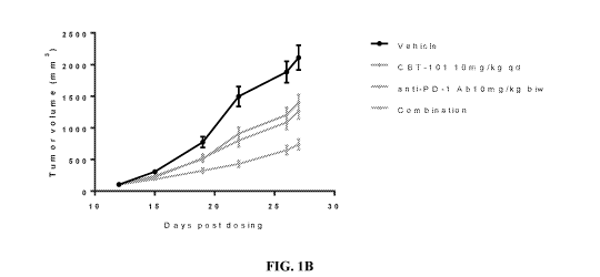
[0008] FIGS. 1A-1C illustrate the synergistic effect of a combination
of c-Met
inhibitor and an anti-PD-1 antibody in MC-38 syngeneic colon cancer model.
FIG. 1A
illustrates the design of the experiments. FIG. 1B illustrates that the
combination of c-Met
inhibitor (APL-101) and anti-PD-1 antibody synergistically inhibited the tumor
growth. FIG.
1C illustrates that the treatment of c-Met inhibitor and anti-PD-1 antibody,
alone or in
combination, did not affect the body weight of the mice being treated.
[0009] FIGS. 2A-2C illustrate the synergistic effect of a combination
of c-Met
inhibitor and an anti-PD-1 antibody in H-22 syngeneic hepatocellular carcinoma
model. FIG.
2A illustrates the design of the experiments. FIG. 2B illustrates that the
combination of c-
Met inhibitor (APL-101) and anti-PD-1 antibody synergistically inhibited the
tumor growth.
FIG. 2C illustrates that the treatment of c-Met inhibitor and anti-PD-1
antibody, alone or in
combination, did not affect the body weight of the mice being treated.
[0010] FIGS. 3A-3C illustrate the synergistic effect of a combination of c-
Met
inhibitor and an anti-PD-1 antibody in RENCA syngeneic renal cell carcinoma
model. FIG.
3A illustrates the design of the experiments. FIG. 3B illustrates that the
combination of c-
Met inhibitor (APL-101) and anti-PD-1 antibody synergistically inhibited the
tumor growth.
2
CA 03091373 2020-08-14
WO 2019/161320
PCT/US2019/018377
FIG. 3C illustrates that the treatment of c-Met inhibitor and anti-PD-1
antibody, alone or in
combination, did not affect the body weight of the mice being treated.
[0011] FIGS. 4A-4C illustrate that a combination of c-Met inhibitor
and an anti-PD-1
antibody deceased the neutrophil percentage in tumor microenvironment. FIG. 4A
illustrates
that a treatment of anti-PD-1 antibody increased c-Met positive neutrophils in
an IHC
analysis. FIG. 4B illustrates that a combination of a c-Met inhibitor and an
anti-PD-1
antibody decreased neutrophil percentage in tumor microenvironment. FIG. 4C
illustrates
that a treatment of anti-PD-1 antibody increased c-Met positive neutrophils in
peripheral
circulation, and a combination of a c-Met inhibitor and an anti-PD-1 antibody
decreased the
neutrophil percentage in peripheral circulation.
[0012] FIG. 5 is a schematic of a Phase 1 study of combination
immunotherapy anti-
PD1 with c-Met inhibitor.
[0013] FIG. 6 is a schematic of a Phase 2 study of combination
immunotherapy anti-
PD1 with c-Met inhibitor.
DETAILED DESCRIPTION OF THE INVENTION
[0014] Before the present disclosure is described in greater detail,
it is to be
understood that this disclosure is not limited to particular embodiments
described, and as
such may, of course, vary. It is also to be understood that the terminology
used herein is for
the purpose of describing particular embodiments only, and is not intended to
be limiting,
since the scope of the present disclosure will be limited only by the appended
claims.
[0015] Unless defined otherwise, all technical and scientific terms
used herein have
the same meaning as commonly understood by one of ordinary skill in the art to
which this
disclosure belongs. Although any methods and materials similar or equivalent
to those
described herein can also be used in the practice or testing of the present
disclosure, the
preferred methods and materials are now described.
[0016] All publications and patents cited in this specification are
herein incorporated
by reference as if each individual publication or patent were specifically and
individually
indicated to be incorporated by reference and are incorporated herein by
reference to disclose
and describe the methods and/or materials in connection with which the
publications are cited.
The citation of any publication is for its disclosure prior to the filing date
and should not be
construed as an admission that the present disclosure is not entitled to
antedate such
publication by virtue of prior disclosure. Further, the dates of publication
provided could be
different from the actual publication dates that may need to be independently
confirmed.
3
CA 03091373 2020-08-14
WO 2019/161320
PCT/US2019/018377
[0017] As will be apparent to those of skill in the art upon reading
this disclosure,
each of the individual embodiments described and illustrated herein has
discrete components
and features which may be readily separated from or combined with the features
of any of the
other several embodiments without departing from the scope or spirit of the
present
disclosure. Any recited method can be carried out in the order of events
recited or in any
other order that is logically possible.
[0018] Definitions
[0019] The following definitions are provided to assist the reader.
Unless otherwise
defined, all terms of art, notations and other scientific or medical terms or
terminology used
.. herein are intended to have the meanings commonly understood by those of
skill in the
chemical and medical arts. In some cases, terms with commonly understood
meanings are
defined herein for clarity and/or for ready reference, and the inclusion of
such definitions
herein should not necessarily be construed to represent a substantial
difference over the
definition of the term as generally understood in the art.
[0020] As used herein, the singular forms "a", "an" and "the" include
plural
references unless the context clearly dictates otherwise.
[0021] As used herein, the term "administering" means providing a
pharmaceutical
agent or composition to a subject, and includes, but is not limited to,
administering by a
medical professional and self-administering.
[0022] As used herein, an "antibody" encompasses naturally occurring
immunoglobulins as well as non-naturally occurring immunoglobulins, including,
for
example, single chain antibodies, chimeric antibodies (e.g., humanized murine
antibodies),
and heteroconjugate antibodies (e.g., bispecific antibodies). Fragments of
antibodies include
those that bind antigen, (e.g., Fab', F(ab')2, Fab, Fv, and rIgG). See also,
e.g., Pierce Catalog
and Handbook, 1994-1995 (Pierce Chemical Co., Rockford, Ill.); Kuby, J.,
Immunology, 3rd
Ed., W.H. Freeman & Co., New York (1998). The term antibody also includes
bivalent or
bispecific molecules, diabodies, triabodies, and tetrabodies. The term
"antibody" further
includes both polyclonal and monoclonal antibodies.
[0023] As used herein, an "anti-angiogenesis agent" means a substance
that reduces
or inhibits the growth of new blood vessels, such as, e.g., an inhibitor of
vascular endothelial
growth factor (VEGF) and an inhibitor of endothelial cell migration. Anti-
angiogenesis
agents include without limitation 2-methoxyestradiol, angiostatin,
bevacizumab, cartilage-
derived angiogenesis inhibitory factor, endostatin, IFN-a, IL-12,
itraconazole, linomide,
platelet factor-4, prolactin, 5U5416, suramin, tasquinimod, tecogalan,
tetrathiomolybdate,
4
CA 03091373 2020-08-14
WO 2019/161320
PCT/US2019/018377
thalidomide, thrombospondin, thrombospondin, TNP-470, ziv-aflibercept,
pharmaceutically
acceptable salts thereof, prodrugs, and combinations thereof.
[0024] As used herein, the term "cancer" refers to any diseases
involving an abnormal
cell growth and includes all stages and all forms of the disease that affects
any tissue, organ
or cell in the body. The term includes all known cancers and neoplastic
conditions, whether
characterized as malignant, benign, soft tissue, or solid, and cancers of all
stages and grades
including pre- and post-metastatic cancers. In general, cancers can be
categorized according
to the tissue or organ from which the cancer is located or originated and
morphology of
cancerous tissues and cells. As used herein, cancer types include, acute
lymphoblastic
leukemia (ALL), acute myeloid leukemia, adrenocortical carcinoma, anal cancer,
astrocytoma,
childhood cerebellar or cerebral, basal-cell carcinoma, bile duct cancer,
bladder cancer, bone
tumor, brain cancer, breast cancer, Burkitt's lymphoma, cerebellar
astrocytoma, cerebral
astrocytoma/malignant glioma, cervical cancer, chronic lymphocytic leukemia,
chronic
myelogenous leukemia, colon cancer, emphysema, endometrial cancer, ependymoma,
esophageal cancer, Ewing family of tumors, Ewing's sarcoma, gastric (stomach)
cancer,
glioma, head and neck cancer, heart cancer, Hodgkin lymphoma, islet cell
carcinoma
(endocrine pancreas), Kaposi sarcoma, kidney cancer (renal cell cancer),
laryngeal cancer,
leukaemia, liver cancer, lung cancer, medulloblastoma, melanoma,
neuroblastoma, non-
Hodgkin lymphoma, ovarian cancer, pancreatic cancer, pharyngeal cancer,
prostate cancer,
rectal cancer, renal cell carcinoma (kidney cancer), retinoblastomaõ skin
cancer, stomach
cancer, supratentorial primitive neuroectodermal tumors, testicular cancer,
throat cancer,
thyroid cancer, vaginal cancer, visual pathway and hypothalamic glioma.
[0025] Cytotoxic agents according to the present invention include
DNA damaging
agents, antimetabolites, anti-microtubule agents, antibiotic agents, etc. DNA
damaging agents
include alkylating agents, platinum-based agents, intercalating agents, and
inhibitors of DNA
replication. Non-limiting examples of DNA alkylating agents include
cyclophosphamide,
mechlorethamine, uramustine, melphalan, chlorambucil, ifosfamide, carmustine,
lomustine,
streptozocin, busulfan, temozolomide, pharmaceutically acceptable salts
thereof, prodrugs,
and combinations thereof. Non-limiting examples of platinum-based agents
include cisplatin,
carboplatin, oxaliplatin, nedaplatin, satraplatin, triplatin tetranitrate,
pharmaceutically
acceptable salts thereof, prodrugs, and combinations thereof Non-limiting
examples of
intercalating agents include doxorubicin, daunorubicin, idarubicin,
mitoxantrone,
pharmaceutically acceptable salts thereof, prodrugs, and combinations thereof
Non-limiting
examples of inhibitors of DNA replication include irinotecan, topotecan,
amsacrine,
5
CA 03091373 2020-08-14
WO 2019/161320
PCT/US2019/018377
etoposide, etoposide phosphate, teniposide, pharmaceutically acceptable salts
thereof,
prodrugs, and combinations thereof Antimetabolites include folate antagonists
such as
methotrexate and premetrexed, purine antagonists such as 6-mercaptopurine,
dacarbazine,
and fludarabine, and pyrimidine antagonists such as 5-fluorouracil,
arabinosylcytosine,
capecitabine, gemcitabine, decitabine, pharmaceutically acceptable salts
thereof, prodrugs,
and combinations thereof. Anti-microtubule agents include without limitation
vinca alkaloids,
paclitaxel (Taxo1g), docetaxel (Taxotereg), and ixabepilone (Ixemprag).
Antibiotic agents
include without limitation actinomycin, anthracyclines, valrubicin,
epirubicin, bleomycin,
plicamycin, mitomycin, pharmaceutically acceptable salts thereof, prodrugs,
and
combinations thereof
[0026] As used herein, the term "effective amount" or
"therapeutically effective
amount" means the amount of agent that is sufficient to prevent, treat, reduce
and/or
ameliorate the symptoms and/or underlying causes of any disorder or disease,
or the amount
of an agent sufficient to produce a desired effect on a cell. In one
embodiment, a
"therapeutically effective amount" is an amount sufficient to reduce or
eliminate a symptom
of a disease. In another embodiment, a therapeutically effective amount is an
amount
sufficient to overcome the disease itself.
[0027] In the present invention, the term "immunomodulator" means a
substance that
alters the immune response by augmenting or reducing the ability of the immune
system to
produce antibodies or sensitize cells that recognize and react with the
antigen that initiated
their production. Immunomodulators may be recombinant, synthetic, or natural
preparations
and include cytokines, corticosteroids, cytotoxic agents, thymosin, and
immunoglobulins.
Some immunomodulators are naturally present in the body, and certain of these
are available
in pharmacologic preparations. In certain embodiments, immunomodulators are
modulators
of an immune checkpoint. Examples of immunomodulators include, but are not
limited to,
granulocyte colony-stimulating factor (G-C SF), interferons, imiquimod and
cellular
membrane fractions from bacteria, IL-2, IL-7, IL-12, CCL3, CCL26, CXCL7, and
synthetic
cytosine phosphate-guanosine (CpG).
[0028] The phrase "pharmaceutically acceptable" refers to those
compounds,
materials, compositions, and/or dosage forms which are, within the scope of
sound medical
judgment, suitable for use in contact with the tissues of human beings and
animals without
excessive toxicity, irritation, allergic response, or other problem or
complication,
commensurate with a reasonable benefit/risk ratio.
6
CA 03091373 2020-08-14
WO 2019/161320
PCT/US2019/018377
[0029] The phrase "pharmaceutically-acceptable carrier" as used
herein means a
pharmaceutically-acceptable material, composition or vehicle, such as a liquid
or solid filler,
diluent, excipient, or solvent encapsulating material, involved in carrying or
transporting the
subject compound from one organ, or portion of the body, to another organ, or
portion of the
body. Each carrier must be "acceptable" in the sense of being compatible with
the other
ingredients of the formulation and not injurious to the patient. Some examples
of materials
which can serve as pharmaceutically-acceptable carriers include: sugars, such
as lactose,
glucose and sucrose; starches, such as corn starch and potato starch;
cellulose, and its
derivatives, such as sodium carboxymethyl cellulose, ethyl cellulose and
cellulose acetate;
powdered tragacanth; malt; gelatin; talc; excipients, such as cocoa butter and
suppository
waxes; oils, such as peanut oil, cottonseed oil, safflower oil, sesame oil,
olive oil, corn oil and
soybean oil; glycols, such as propylene glycol; polyols, such as glycerin,
sorbitol, mannitol
and polyethylene glycol; esters, such as ethyl oleate and ethyl laurate; agar;
buffering agents,
such as magnesium hydroxide and aluminum hydroxide; alginic acid; pyrogen-free
water;
isotonic saline; Ringer's solution; ethyl alcohol; pH buffered solutions;
polyesters,
polycarbonates and/or polyanhydrides; and other non-toxic compatible
substances employed
in pharmaceutical formulations.
[0030] "Pharmaceutically-acceptable salts" refers to the relatively
non-toxic,
inorganic and organic acid addition salts of compounds.
[0031] As used herein, the term "photoactive therapeutic agent" means
compounds
and compositions that become active upon exposure to light. Certain examples
of
photoactive therapeutic agents are disclosed, e.g., in U.S. Patent Application
Publication
Serial No. 2011/015223.
[0032] As used herein, the term "radiosensitizing agent" means a
compound that
makes tumor cells more sensitive to radiation therapy. Examples of
radiosensitizing agents
include misonidazole, metronidazole, tirapazamine, and trans sodium
crocetinate.
[0033] The terms "responsive," "clinical response," "positive
clinical response," and
the like, as used in the context of a patient's response to a cancer therapy,
are used
interchangeably and refer to a favorable patient response to a treatment as
opposed to
unfavorable responses, i.e. adverse events. In a patient, beneficial response
can be expressed
in terms of a number of clinical parameters, including loss of detectable
tumor (complete
response, CR), decrease in tumor size and/or cancer cell number (partial
response, PR), tumor
growth arrest (stable disease, SD), enhancement of anti-tumor immune response,
possibly
resulting in regression or rejection of the tumor; relief, to some extent, of
one or more
7
CA 03091373 2020-08-14
WO 2019/161320
PCT/US2019/018377
symptoms associated with the tumor; increase in the length of survival
following treatment;
and/or decreased mortality at a given point of time following treatment.
Continued increase
in tumor size and/or cancer cell number and/or tumor metastasis is indicative
of lack of
beneficial response to treatment. In a population the clinical benefit of a
drug, i.e., its
efficacy can be evaluated on the basis of one or more endpoints. For example,
analysis of
overall response rate (ORR) classifies as responders those patients who
experience CR or PR
after treatment with drug. Analysis of disease control (DC) classifies as
responders those
patients who experience CR, PR or SD after treatment with drug. A positive
clinical
response can be assessed using any endpoint indicating a benefit to the
patient, including,
without limitation, (1) inhibition, to some extent, of tumor growth, including
slowing down
and complete growth arrest; (2) reduction in the number of tumor cells; (3)
reduction in
tumor size; (4) inhibition (i.e., reduction, slowing down or complete
stopping) of tumor cell
infiltration into adjacent peripheral organs and/or tissues; (5) inhibition of
metastasis; (6)
enhancement of anti-tumor immune response, possibly resulting in regression or
rejection of
the tumor; (7) relief, to some extent, of one or more symptoms associated with
the tumor; (8)
increase in the length of survival following treatment; and/or (9) decreased
mortality at a
given point of time following treatment. Positive clinical response may also
be expressed in
terms of various measures of clinical outcome. Positive clinical outcome can
also be
considered in the context of an individual's outcome relative to an outcome of
a population of
patients having a comparable clinical diagnosis, and can be assessed using
various endpoints
such as an increase in the duration of recurrence-free interval (RFI), an
increase in the time of
survival as compared to overall survival (OS) in a population, an increase in
the time of
disease-free survival (DFS), an increase in the duration of distant recurrence-
free interval
(DRFI), and the like. Additional endpoints include a likelihood of any event
(AE)-free
survival, a likelihood of metastatic relapse (MR)-free survival (MRFS), a
likelihood of
disease-free survival (DFS), a likelihood of relapse-free survival (RFS), a
likelihood of first
progression (FP), and a likelihood of distant metastasis-free survival (DMFS).
An increase in
the likelihood of positive clinical response corresponds to a decrease in the
likelihood of
cancer recurrence or relapse.
[0034] As used herein, the term "subject" refers to a human or any non-
human animal
(e.g., mouse, rat, rabbit, dog, cat, cattle, swine, sheep, horse or primate).
A human includes
pre and post-natal forms. In many embodiments, a subject is a human being. A
subject can
be a patient, which refers to a human presenting to a medical provider for
diagnosis or
treatment of a disease. The term "subject" is used herein interchangeably with
"individual"
8
CA 03091373 2020-08-14
WO 2019/161320
PCT/US2019/018377
or "patient." A subject can be afflicted with or is susceptible to a disease
or disorder but may
or may not display symptoms of the disease or disorder.
[0035] As used herein, "synergistic" means more than additive.
Synergistic effects
may be measured by various assays known in the art.
[0036] As used herein, the term "toxin" means an antigenic poison or venom
of plant
or animal origin. An example is diphtheria toxin or portions thereof.
[0037] The term "treatment," "treat," or "treating" refers to a
method of reducing the
effects of a cancer (e.g., breast cancer, lung cancer, ovarian cancer or the
like) or symptom of
cancer. Thus, in the disclosed method, treatment can refer to a 10%, 20%, 30%,
40%, 50%,
60%, 70%, 80%, 90%, or 100% reduction in the severity of a cancer or symptom
of the
cancer. For example, a method of treating a disease is considered to be a
treatment if there is
a 10% reduction in one or more symptoms of the disease in a subject as
compared to a control.
Thus, the reduction can be a 10%, 20%, 30%, 40%, 50%, 60%, 70%, 80%, 90%, 100%
or
any percent reduction between 10 and 100% as compared to native or control
levels. It is
understood that treatment does not necessarily refer to a cure or complete
ablation of the
disease, condition, or symptoms of the disease or condition.
[0038] Neutrophil Related Bi om arker
[0039] The present disclosure in one aspect provides a method of
treating cancer
patients with a combinational immunotherapy based on a neutrophil related
biomarker that
can predict the responsiveness of the combinational immunotherapy. In one
embodiment, the
method comprises: measuring a base level of the neutrophil related biomarker
in a sample
from the subject; determining that the base level of said biomarker is equal
or more than a
threshold value; and administering to the subject a combinational
immunotherapy.
[0040] Neutrophils, also known as neutrocytes or polymorphonuclear
myeloid-
derived suppressor cells (PMN-MDSCs), are a type of phagocyte normally found
in the
bloodstream. In most mammals, neutrophils are the most abundant type of
granulocytes and
the most abundant type of white blood cell. Neutrophils form an essential part
of the innate
immune system and play various functions in different contexts. During an
acute
inflammation, particularly as a result of bacterial infection and some
cancers, neutrophils are
one of the first-responders of inflammatory cells to migrate to the site of
inflammation.
[0041] Methods of detecting and measuring the number of neutrophils
are known in
the art. For example, hematoxylin and eosin (H&E) staining has long been used
to
differentiate neutrophils from basophilic and eosinophilic white blood cells.
Neutrophils can
9
CA 03091373 2020-08-14
WO 2019/161320
PCT/US2019/018377
also be identified by the expression of certain markers, e.g., CD11 c, CD13,
CD15, CD16,
CD33 and CD68.
[0042] Myeloid derived suppressor cells (MDSCs) are a heterogeneous
group of
immature myeloid cells which suppress the immune system. Collectively a MDSC
population is comprised of monocyte-like MDSCs and polymorphonuclear MDSC (PMN-
MDSCs or neutrophils). The number of MDSCs is increased with the presence of
tumors. It
has been shown that PMN-MDSCs represent the majority of MDSCs in cancers and
protect
the cancers from the immune system.
[0043] As used herein, the term "neutrophil related biomarkers" refer
to biomarkers
that are indicative of the presence, abundance or activation of neutrophils in
any sample or
tissue of the subject. In certain embodiments, the neutrophil related
biomarker is selected
from a group consisting of hepatocyte growth factor, absolute neutrophil
count, c-Met+
neutrophils and neutrophil to lymphocyte ratio (NLR).
[0044] In certain embodiments, the neutrophil related biomarker is
NLR and the
threshold value is about 3, 3.5, 4, 4.5 or 5.
[0045] In another embodiment, the method comprises: measuring a first
level of the
biomarker in the subject; administering to the subject an immunotherapy for a
time period;
measuring a second level of the biomarker in the subject; determining that a
difference
between the second level of the biomarker and the first level of biomarker is
equal or more
than a critical value; and administering to the subject a combinational
immunotherapy.
[0046] In certain embodiments, wherein the neutrophil related
biomarker is NLR and
the critical value is about 2, 2.5, 3, 3.5 or 4.
[0047] In certain embodiments, the subject being treated is a mammal.
In certain
embodiments, the mammal is selected from the group consisting of humans,
primates, farm
animals and domestic animals. In certain embodiments, the mammal is a human.
[0048] In certain embodiment, the cancer being treated is selected
from the groups
consisting of a lung cancer, a melanoma, a renal caner, a liver cancer, a
myeloma, a prostate
cancer, a breast cancer, a colorectal cancer, a pancreatic cancer, a thyroid
cancer, a
hematological cancer, a leukemia and a non-Hodgkin's lymphoma.
[0049] Combinatorial Usage of c-Met Inhibitor and Modulators of Immune
Checkpoint
[0050] In another aspect, the present disclosure provides a method of
treating cancer
using a combination immunotherapy. In certain embodiments, when it is
determined that the
subject is likely responsive to a combinational immunotherapy, e.g., by
monitoring the
CA 03091373 2020-08-14
WO 2019/161320
PCT/US2019/018377
neutrophil related biomarker as discussed above, the combinational
immunotherapy is
administered to the subject. In certain embodiment, the combinational
immunotherapy is a
combination use of a c-Met inhibitor and a modulator of an immune checkpoint.
In some
embodiments, the modulator of an immune checkpoint is an anti-PD-1 antibody or
an anti-
PD-Li antibody.
[0051] c-MET is a proto-oncogene that encodes a protein known as
hepatocyte
growth factor receptor (HGFR). c-Met protein is composed of the a chain and 0
chain
generated by cleaving a precursor of c-Met (pro c-Met) and forms a dimer by a
disulfide
linkage, c-Met is a receptor penetrating a cell membrane and the entire a
chain and a part of
the 0 chain are present extracellularly (see, e.g., Mark, et al., The Journal
of Biological
Chemistry, 1992, Vol. 267, No. 36, pp. 26166-26171; Journal of Clinical and
Experimental
Medicine (IGAKU NO AYUMI), 2008, Vol. 224, No. 1, pp. 51-55). See also GenBank
Accession No: NP_000236.2 for human c-Met and its a chain and 0 chain. It has
been
shown that abnormal MET activation in cancer correlates with poor prognosis,
where
aberrantly active c-Met triggers tumor growth, formation of new blood vessels
that supply the
tumor with nutrients, and cancer spread or other organs.
[0052] A "c-Met inhibitor," as used herein, refers an agent that can
suppress the
expression or activity of c-Met protein. In certain embodiments, c-Met
inhibitor is selected
from the group consisting of crizotinib, cabozantinib, APL-101, PLB1001,
bozitinib,
SU11274, PHA665752, K252a, PF-2341066, A1V17, JNJ-38877605, PF-04217903,
MK2461,
GSK1363089 (XL880, foretinib), AMG458, tivantinib (ARQ197), INCB28060 (INC280,
capmatinib), E7050, BMS-777607, savolitinib (volitinib), HQP-8361, merestinib,
ARGX-111,
onartuzumab, rilotumumab, emibetuzumab, and XL184.
[0053] In some embodiments, the c-Met inhibitor comprises a compound
of the
following formula
X R3
RI R2
Ar A
E x1
wherein:
and R2 are independently hydrogen or halogen;
X and Xl are independently hydrogen or halogen;
A and G are independently CH or N, or CH=G is replaced with a sulfur atom;
11
CA 03091373 2020-08-14
WO 2019/161320
PCT/US2019/018377
E is N;
J is CH, S or NH;
M is N or C;
Ar is aryl or heteroaryl, optionally substituted with 1-3 substituents
independent
selected from: Ci.6alkyl, Ci.6alkoxyl, halo Ci.6alkyl, halo Ci.6alkoxy,
C3_7cycloalkyl,
halogen, cyano, amino, -CONR4R5, -NHCOR6, -SO2NR7R8, C1-6alkoxyl-, C 1-6 alkyl-
,
amino-C 1.6 alkyl-, heterocyclyl and heterocyclyl-C 1.6 alkyl-, or two
connected
substituents together with the atoms to which they are attached form a 4-6
membered
lactam fused with the aryl or heteroaryl;
3 i R s hydrogen, Ci.6alkyl, Ci.6alkoxy, haloCi.6alkyl, halogen, amino, or -
CONH- C1.
6a1ky1- heterocyclyl;
R4 and R5 are independently hydrogen, C 1.6 alkyl, C 3_7 cycloalkyl,
heterocyclyl-C1.
6a1ky1, or R4 and R5 together with the N to which they are attaches form a
heterocyclyl;
R6 is Ci.6alkyl or C3.7cycloalkyl; and
R7 and R8 are independently hydrogen or Ci.6alkyl;
[0054] In some embodiments, the c-Met inhibitor is selected from the
group
consisting of:
12
CA 03091373 2020-08-14
WO 2019/161320
PCT/US2019/018377
,e-
ti6..,..õ.= .41- ft-r-rt
r"-4,--
=== lir%
.**,...,=444
..
cA.N...,,õ3õ4., .. =
IT Pi ,&i
Tbs:,N
`=,,k..,,,- 1,4:,
P
,
ixts,
A.c.õ- jtteLl
13
CA 03091373 2020-08-14
WO 2019/161320
PCT/US2019/018377
A)õ
,
0, 11
iN
,
toe ''' 471
,
= I
,
...
r-4,---K
F.
P \"'"A. -14'
..-^N.k..
.,
r,rg(
),..c,..õ
/
^
Fe 4,
PteleN11.44
/
NC ,0
is),,,,i4pc, T Ncel.
: r4s
,
\
14
CA 03091373 2020-08-14
WO 2019/161320
PCT/US2019/018377
Mat*
..fi4
ci...i_.
- ill:p
,..
Afr #
t. rcli
F'........ t
=
..,
= N
P k,N IL
rc
4.*:k
Ne
;...,.:'
(1)?..11 e
,
ry...k
.kr M'ttl'lit
c tt,k
4"eNr6 s'% gt
/
L 1 ki
41 ,
= 4
CA 03091373 2020-08-14
WO 2019/161320
PCT/US2019/018377
= F et?'
tc,
."
& ? *
,,,,' ,
rLi.
' rli
P rk,..., !St
P' +-i,k,,,,,00r
W
4k.--.-L414
,
F rt
F r \-1,0t1
,
Q V
rc.1
,<õ,=='o,i=
1\YYN , le
--4-',
_.
..
...; ,
F I
'-
16
CA 03091373 2020-08-14
WO 2019/161320
PCT/US2019/018377
.,ro'
, Ic F r-Svasi
t 1 '''`' 4'''VOJ
#4'''t..,.. ,4µfri i
;144,1"1.4'?4
N"
....
=-r c N14, P fs- ti
*-4,--
i. , , vu-n,s
"kk.-A14
e
rif
Jr1"
ri f )04
0 r..,....
v
4 F µc . -*=4st
.;Nill F-4....-=
l'i It OC, t4.-4,,N or
1....cõ..4:
17
CA 03091373 2020-08-14
WO 2019/161320
PCT/US2019/018377
NIN-
tet.fro
rts$µ=15
ft_
1:rw'p
#"(
r 4-4
-r-
e
f
F r
[0055] In certain embodiments, c-Met inhibitor is APL-101 (previously
named CBT-
101, see US20150218171, which is incorporated in its entirety by reference),
which has the
following formula:
.11
t,
F -=;N1
N
14 A
'N F
[0056] In certain embodiments, c-Met inhibitor can be formulated with
a
pharmaceutically acceptable carrier. The carrier, when present, can be blended
with c-Met
inhibitor in any suitable amounts, such as an amount of from 5% to 95% by
weight of carrier,
18
CA 03091373 2020-08-14
WO 2019/161320
PCT/US2019/018377
based on the total volume or weight of c-Met inhibitor and the carrier. In
some embodiments,
the amount of carrier can be in a range having a lower limit of any of 5%,
10%, 12%, 15%,
20%, 25%, 28%, 30%, 40%, 50%, 60%, 70% or 75%, and an upper limit, higher than
the
lower limit, of any of 20%, 22%, 25%, 28%, 30%, 40%, 50%, 60%, 70%, 75%, 80%,
85%,
90%, and 95%. The amount of carrier in a specific embodiment may be determined
based on
considerations of the specific dose form, relative amounts of c-Met inhibitor,
the total weight
of the composition including the carrier, the physical and chemical properties
of the carrier,
and other factors, as known to those of ordinary skill in the formulation art.
[0057] As used herein, the term "immune checkpoint" or "cancer immune
checkpoint"
refers to a molecule in the immune system that either turns up a signal (i.e.,
co-stimulatory
molecules) or turns down a signal (i.e., inhibitory molecule) of an immune
response. In
certain embodiments, the immune checkpoint is selected from the group
consisting of PD-1,
PD-L1, PD-L2, LAG-3, TIM-1, CTLA-4, VISTA, B7-H2, B7-H3, B7-H4, B7-H6, 284,
ICOS,
HVEM, CD160, gp49B, PIR-B, KIR family receptors, TIM-1, TIM-4, BTLA, SIRPalpha
(CD47), CD48, 284 (CD244), B7.1, B7.2, ILT-2, ILT-4, TIGIT and A2aR.
[0058] In certain embodiments, the modulator of immune checkpoint is
a monoclonal
antibody against the immune checkpoint. In certain embodiments, the immune
checkpoint is
PD-1 or PD-Li. In certain embodiments, the anti-PD-1 antibody is selected from
those
disclosed in PCT application publication No. W02016/014688, which is
incorporated in its
entirety by reference. In certain embodiments, the anti-PD-1 antibody is APL-
501
(previously named as CBT-501, see W02016/014688), GB226 or genolimzumab. In
certain
embodiments, the anti-PD-Li antibody is selected from those disclosed in PCT
application
publication No. W02016/022630, which is incorporated in its entirety by
reference. In
certain embodiments, the anti-PD-Li antibody is APL-502 (previously named as
CBT-502,
see W02016/022630) or TQB2450.
[0059] According to the present disclosure, the c-Met inhibitor and
the modulator of
immune checkpoint (or another anti-cancer therapeutic agent) may be co-
administered to the
subject, either simultaneously or at different times, as deemed most
appropriate by a
physician. If the c-Met inhibitor and the immune checkpoint modulator are
administered at
different times, for example, by serial administration, the immune checkpoint
modulator may
be administered to the subject before the c-Met inhibitor. Alternatively, the
c-Met inhibitor
may be administered to the subject before immune checkpoint modulator.
[0060] The c-Met inhibitor or the modulator of immune checkpoint or
other anti-
cancer therapeutic agents may be administered in any desired and effective
manner: for oral
19
CA 03091373 2020-08-14
WO 2019/161320
PCT/US2019/018377
ingestion, or as an ointment or drop for local administration to the eyes, or
for parenteral or
other administration in any appropriate manner such as intraperitoneal,
subcutaneous, topical,
intradermal, inhalation, intrapulmonary, rectal, vaginal, sublingual,
intramuscular,
intravenous, intraarterial, intrathecal, or intralymphatic. Further, the c-Met
inhibitor or the
modulator of immune checkpoint or other anti-cancer therapeutic agents may be
administered
in conjunction with other treatments. The c-Met inhibitor or the modulator of
immune
checkpoint or other anti-cancer therapeutic agents may be encapsulated or
otherwise
protected against gastric or other secretions, if desired.
[0061] A suitable, non-limiting example of a dosage of the c-Met
inhibitor or the
modulator of immune checkpoint or other anti-cancer therapeutic agents
disclosed herein is
from about 1 mg/kg to about 2400 mg/kg per day, such as from about 1 mg/kg to
about 1200
mg/kg per day, 75 mg/kg per day to about 300 mg/kg per day, including from
about 1 mg/kg
to about 100 mg/kg per day. Other representative dosages of such agents
include about 1
mg/kg, 5 mg/kg, 10 mg/kg, 15 mg/kg, 20 mg/kg, 25 mg/kg, 30 mg/kg, 35 mg/kg, 40
mg/kg,
45 mg/kg, 50 mg/kg, 60 mg/kg, 70 mg/kg, 75 mg/kg, 80 mg/kg, 90 mg/kg, 100
mg/kg, 125
mg/kg, 150 mg/kg, 175 mg/kg, 200 mg/kg, 250 mg/kg, 300 mg/kg, 400 mg/kg, 500
mg/kg,
600 mg/kg, 700 mg/kg, 800 mg/kg, 900 mg/kg, 1000 mg/kg, 1100 mg/kg, 1200
mg/kg, 1300
mg/kg, 1400 mg/kg, 1500 mg/kg, 1600 mg/kg, 1700 mg/kg, 1800 mg/kg, 1900 mg/kg,
2000
mg/kg, 2100 mg/kg, 2200 mg/kg, and 2300 mg/kg per day. In some embodiments,
the
dosage of the c-Met inhibitor in human is about 400 mg/day given every 12
hours. In some
embodiments, the dosage of the c-Met inhibitor in human ranges 300-500 mg/day,
100-600
mg/day or 25-1000 mg/day. The effective dose of c-Met inhibitor or the
modulator of
immune checkpoint or other anti-cancer therapeutic agents disclosed herein may
be
administered as two, three, four, five, six or more sub-doses, administered
separately at
appropriate intervals throughout the day.
[0062] Other Combinational Therapies
[0063] In one embodiment, the method further comprises administering
at least one
additional therapeutic agent selected from the group consisting of a cytotoxic
agent, a toxin, a
radionuclide, an immunomodulator, a photoactive therapeutic agent, a
radiosensitizing agent,
a hormone, an anti-angiogenesis agent, and combinations thereof. In certain
embodiments,
the administration of the c-Met inhibitor, the modulator of immune checkpoint
and the
additional therapeutic agent provides a synergistic effect.
CA 03091373 2020-08-14
WO 2019/161320
PCT/US2019/018377
[0064] The following examples are provided to better illustrate the
claimed invention
and are not to be interpreted as limiting the scope of the invention. All
specific compositions,
materials, and methods described below, in whole or in part, fall within the
scope of the
present invention. These specific compositions, materials, and methods are not
intended to
limit the invention, but merely to illustrate specific embodiments falling
within the scope of
the invention. One skilled in the art may develop equivalent compositions,
materials, and
methods without the exercise of inventive capacity and without departing from
the scope of
the invention. It will be understood that many variations can be made in the
procedures
herein described while still remaining within the bounds of the present
invention. It is the
intention of the inventors that such variations are included within the scope
of the invention.
Example 1
[0065] This example illustrates the synergic effect of combination
treatment using a
c-Met inhibitor (APL-101) and an anti-PD-1 antibody in MC-38 syngeneic colon
cancer
model.
[0066] Experimental Design
[0067] The inventors undertook a combination study of APL-101 and an
anti-PD-1
antibody to evaluate the safety and efficacy of the combination. In the MC-38
colon cancer
model in syngeneic mice, four groups, five animals per group received either
vehicle (water
at 20 mg/kg orally, once a day), APL-101 (10 mg/kg orally, once a day), anti-
PD-1 (10 mg/kg
intraperitoneal injection, twice a week), or APL-101 plus anti-PD-1. In the
vehicle group as
well as the APL-101 group, animals were dosed daily on Days 1 ¨ 15 whereas in
the single
agent anti-PD-1 group, doses were administered on Days 1, 4, 8, 11, and 15. In
the
combination arm of APL-101 and anti-PD-1, APL-101 was administered on Days 5-
15 (4-
day delay) while the anti-PD-1 was dosed on Days 1, 4, 8, 11, and 15.
[0068] Materials and Methods
[0069] Animals: female C57BL/6 mice, age 6-8 weeks and of body weight
18-20 g,
were provided by Shanghai Lingchang Bio-Technology Co. Ltd.
[0070] APL-101 were provided by CBT pharmaceuticals (now Apollomics,
Inc.).
Anti-PD 1 antibodies were supplied by BioXcell.
[0071] Cell culture: The MC38 tumor cells were thawed and maintained in
vitro as a
monolayer culture in DMEM medium supplemented with 10% heat inactivated fetal
bovine
serum at 37 C in an atmosphere of 5% CO2 in air. The tumor cells were
routinely
subcultured twice weekly by trypsin-EDTA treatment. The cells growing in an
exponential
growth phase were harvested and counted for tumor inoculation.
21
CA 03091373 2020-08-14
WO 2019/161320
PCT/US2019/018377
[0072] Tumor inoculation: Each mouse was inoculated subcutaneously at
the right
lower flank with MC38 tumor cells (1x10⁶) in 0.1 ml of PBS. The treatments
started when the
mean tumor size reached approximately 80-120 mm3. The date of tumor cell
inoculation is
denoted as day 0.
[0073] Group assignment: Before grouping and treatment, all animals were
weighed
and the tumor volumes were measured using a caliper. Since the tumor volume
can affect the
effectiveness of any given treatment, tumor volume was used as numeric
parameter to
randomize selected animals into specified groups. The grouping was performed
by using
StudyDirectorTM software (Studylog Systems, Inc. CA, USA).
[0074] Observation and data collection: After tumor cells inoculation, the
animals
were checked daily for morbidity and mortality. During routine monitoring, the
animals were
checked for any effects of tumor growth and treatments on normal behavior such
as mobility,
visual estimation of food and water consumption, body weight gain/loss (body
weights were
measured twice per week after randomization), eye/hair matting and any other
abnormal
effect. Death and observed clinical signs were recorded in the comment of
datasheet for each
animal in detail. Tumor volumes were measured twice weekly after randomization
in two
dimensions using a caliper, and the volume was expressed in mm3 using the
formula: V = 0.5
a x b2 where a and b are the length and width of the tumor, respectively.
(Tumor weight was
measured at the end of study). The entire procedures of dosing as well as
tumor and body
weight measurement were conducted in a Laminar Flow Cabinet.
[0075] Statistics: the mean and standard error of the mean (SEM) were
provided for
the tumor volumes of each group at every time point. Statistical analysis of
difference in
tumor volume between the two comparing groups was conducted on the data
obtained at the
best therapeutic time point (usually after the final dose) using One-way ANOVA
Test. All
data were analyzed in SPSS (Statistical Product and Service Solutions) version
18.0 (IBM,
Armonk, NY, U.S.). P-values were rounded to three decimal places, with the
exception when
raw P-values were less than 0.001, then they were stated as P<0.001. All tests
were two-sided.
P<0.05 was considered to be statistically significant.
[0076] Results
[0077] As shown in FIGS. 1A-1C and Table 1, mean percent tumor growth
inhibition of the combination anti-PD-1 10 mg/kg IP BIW x 2 weeks plus APL-101
10 mg/kg,
QD x 2 weeks demonstrated a 65.1% tumor growth inhibition, versus 39.9% and
33.6% for
anti-PD-1 IP 10 mg/kg BIW x 3 weeks and APL-101 PO 10 mg/kg, QD x 3 weeks,
respectively. The combination regimen was well tolerated by the animals. Tumor
tissue
22
CA 03091373 2020-08-14
WO 2019/161320
PCT/US2019/018377
collected for c-Met positivity and PD-Li neutrophils is evaluated along with
neutrophil to
lymphocyte ratio.
[0078] Table 1. Mean percent tumor growth in MC 38 syngeneic model.
Vehicle
APL-101 10mg/kg qd 35.61
Anti-PD-1 Ab 10mg/kg biw 42.06
Combination 23.97
Example 2
[0079] This example illustrates the synergic effect of combination
treatment using a
c-Met inhibitor (APL-101) and an anti-PD-1 antibody in H22 syngeneic liver
cancer model.
[0080] Experimental Design
[0081] The inventors undertook a combination study of APL-101 and an
anti-PD-1
antibody to evaluate the safety and efficacy of the combination. In the H22
liver cancer
model in syngeneic mice, four groups, ten animals per group received either
vehicle (PVP
K30 at 20 mg/kg orally, once a day for three weeks), APL-101 (10 mg/kg orally,
once a day
for three weeks), anti-PD-1 (10 mg/kg intraperitoneal injection, twice a week
for three
weeks), or APL-101 plus anti-PD-1.
[0082] Materials and Methods
[0083] Animals: female C57BL/6 mice, age 6-8 weeks and of body weight
18-20 g,
were provided by Shanghai Lingchang Bio-Technology Co. Ltd.
[0084] APL-101 were provided by CBT pharmaceuticals (now Apollomics,
Inc.).
Anti-PD 1 antibodies were supplied by BioXcell. PVP K30 were supplied by Fluka
Analytical.
[0085] Cell culture: The H22 tumor cell line were maintained in vitro
in RPMI-1640
medium supplemented with 10% fetal bovine serum at 37 C in an atmosphere of 5%
CO2 in
air. The tumor cells were routinely subcultured twice weekly by trypsin-EDTA
treatment.
The cells growing in an exponential growth phase were harvested and counted
for tumor
inoculation.
[0086] Tumor inoculation: Each mouse was inoculated subcutaneously at
the right
front flank with H22 tumor cells (2 x 10⁶) in 0.1 ml of PBS for tumor
development. The
treatments were started when the mean tumor size reaches approximately 80-120
mm3. The
date of tumor cell inoculation was denoted as day 0.
23
CA 03091373 2020-08-14
WO 2019/161320
PCT/US2019/018377
[0087] Randomization: The randomization started when the mean tumor
size reached
approximately 80-120 mm3. 40 mice were enrolled in the study. All animals were
randomly
allocated to 4 study groups. Randomization was performed based on randomized
block
design.
[0088] Observation and data collection: After tumor cells inoculation, the
animals
were checked daily for morbidity and mortality. During routine monitoring, the
animals were
checked for any effects of tumor growth and treatments on behavior such as
mobility, food
and water consumption, body weight gain/loss (body weights were measured twice
weekly
after randomization), eye/hair matting and any other abnormalities. Mortality
and observed
clinical signs were recorded for individual animals in detail. Tumor volumes
were measured
twice weekly in two dimensions using a caliper, and the volume was expressed
in mm3 using
the formula: "V = (L x W x W)/2, where V is tumor volume, L is tumor length
(the longest
tumor dimension) and W is tumor width (the longest tumor dimension
perpendicular to L).
(Tumor weight were measured at the end of study). Dosing as well as tumor and
body weight
measurements were conducted in a Laminar Flow Cabinet.
[0089] Statistics analysis: For comparison among three or more
groups, a one-way
ANOVA was performed followed by multiple comparison procedures. For survival
analysis,
Kaplan-Meier survival curves was generated and Log Rank test was performed.
All data was
analyzed using SPSS 18Ø P <0.05 was considered statistically significant.
[0090] Results
[0091] As shown in FIGS. 2A-2C and Table 2, mean percent tumor growth
of the
combination anti-PD-1 10 mg/kg IP BIW x 3 weeks plus APL-101 10 mg/kg, QD x 3
weeks
demonstrated a 40.38% tumor growth, versus 108.73% for APL-101 10 mg/kg, QD x
3
weeks and 65.85% for anti-PD-1 IP 10 mg/kg BIW x 3 weeks, respectively. The
combination regimen was well tolerated by the animals.
[0092] Table 2. Mean percent tumor growth in H22 syngeneic liver
cancer model.
Vehicle
APL-101 10mg/kg qd 108.73
Anti-PD-1 Ab 10mg/kg biw 65.85
Combination 40.38
Example 3
24
CA 03091373 2020-08-14
WO 2019/161320
PCT/US2019/018377
[0093] This example illustrates the synergic effect of combination
treatment using a
c-Met inhibitor (APL-101) and an anti-PD-1 antibody in a syngeneic Renca
kidney cancer
model.
[0094] Experimental Design
[0095] The inventors undertook a combination study of APL-101 and an anti-
PD-1
antibody to evaluate the safety and efficacy of the combination. In the Renca
kidney cancer
model in syngeneic mice, four groups, ten animals per group received either
vehicle (PVP
K30 at 20 mg/kg orally, once a day for three weeks), APL-101 (20 mg/kg orally,
once a day
for three weeks), anti-PD-1 (10 mg/kg intraperitoneal injection, twice a week
for three
weeks), or APL-101(20 mg/kg orally, once a day for three weeks) plus anti-PD-1
(10 mg/kg
intraperitoneal injection, twice a week for three weeks).
[0096] Materials and Methods
[0097] Animals: female C57BL/6 mice, age 6-8 weeks and of body weight
18-20 g,
were provided by Shanghai Lingchang Bio-Technology Co. Ltd.
[0098] APL-101 were provided by CBT pharmaceuticals (Apollomics, Inc.).
Anti-
PD 1 antibodies were supplied by BioXcell. PVP K30 were supplied by Fluka
Analytical.
[0099] Cell culture: The Renca tumor cell line was maintained in
vitro in DMEM
medium supplemented with 10% fetal bovine serum at 37 C in an atmosphere of 5%
CO2 in
air. The tumor cells were routinely subcultured twice weekly. The cells
growing in an
exponential growth phase were harvested and counted for tumor inoculation.
[00100] Tumor inoculation: Each mouse was inoculated subcutaneously at
the right
front flank with RENCA tumor cells (1 x 10⁶) in 0.1 ml of PBS for tumor
development. The
treatments were started when the mean tumor size reaches approximately 80-120
mm3. The
date of tumor cell inoculation wass denoted as day 0.
[00101] Randomization: The randomization started when the mean tumor size
reached
approximately 80-120 mm3. 40 mice were enrolled in the study. All animals were
randomly
allocated to 4 study groups. Randomization was performed based on randomized
block
design.
[00102] Observation and data collection: After tumor cells
inoculation, the animals
were checked daily for morbidity and mortality. During routine monitoring, the
animals were
checked for any effects of tumor growth and treatments on behavior such as
mobility, food
and water consumption, body weight gain/loss (body weights were measured twice
weekly
after randomization), eye/hair matting and any other abnormalities. Mortality
and observed
clinical signs were recorded for individual animals in detail. Tumor volumes
were measured
CA 03091373 2020-08-14
WO 2019/161320
PCT/US2019/018377
twice weekly in two dimensions using a caliper, and the volume was expressed
in mm3 using
the formula: "V = (L x W x W)/2, where V is tumor volume, L is tumor length
(the longest
tumor dimension) and W is tumor width (the longest tumor dimension
perpendicular to L).
(Tumor weight were measured at the end of study). Dosing as well as tumor and
body weight
measurements were conducted in a Laminar Flow Cabinet.
[00103] Statistics analysis: For comparison among three or more
groups, a one-way
ANOVA was performed followed by multiple comparison procedures. For survival
analysis,
Kaplan-Meier survival curves was generated and Log Rank test was performed.
All data was
analyzed using SPSS 18Ø P <0.05 was considered statistically significant.
[00104] Results
[00105] As shown in FIGS. 3A-3C and Table 3, mean percent tumor growth
of the
combination anti-PD-1 10 mg/kg IP BIW x 3 weeks plus APL-101 10 mg/kg, QD x 3
weeks
demonstrated a 47% tumor growth, versus 77% for APL-101 10 mg/kg, QD x 3 weeks
and 71%
for anti-PD-1 IP 10 mg/kg BIW x 3 weeks, respectively. The combination regimen
was well
tolerated by the animals.
[00106] Table 2. Mean percent tumor growth in syngeneic Renca kidney
cancer model.
Vehicle
APL-101 10mg/kg qd 77
Anti-PD-1 Ab 10mg/kg biw 71
Combination 47
Example 4
[00107] This example illustrates that a combination of c-Met inhibitor
(APL-101) and
an anti-PD-1 antibody deceased the neutrophil percentage in tumor
microenvironment.
[00108] Experimental Design
[00109] Tumor tissues was collected from the MC38 colon adenocarcinoma
syngeneic
model (described in Example 1) at the end of the study and fixed in formalin.
Double IHC
analysis of c-Met and neutrophils was used to quantify the expression of Met+
neutrophils.
[00110] Sample preparation: fresh specimens were collected and placed
in 10% NBF
(neutral-buffered formalin; fixative volume/tissue, 10-20 folds), fixed at
room temperature
for 24 hours. Fixed tissue was trimmed at the thickness of 3-5 mm. The trimmed
tissues
26
CA 03091373 2020-08-14
WO 2019/161320
PCT/US2019/018377
were moved into an embedding box. The box was snapped into deionized water for
30
minutes, with water changed twice every 30 minutes. If the dehydration
procedure could not
be carried out on time, the tissues were transferred into the 70% ethanol, and
placed in the 4 C
refrigerator. The tissues can be kept in 70% ethanol for about 3-5 days in the
refrigerator.
After dehydration, FFPE preparation and FFPE slide preparation of the fixed
tissues were
transferred to the LEICA ASP300S Vacuum Tissue Processor for dehydration.
[00111] FFPE slides preparation: The dehydrated tissues were be
embedded in paraffin
on Paraffin Embedding Station. The FFPE blocks were sectioned with a manual
rotary
microtome, 4 p.m thickness/section.
[00112] The FFPE slides were used for IHC with the following antibodies:
anti-
neutrophil (LY6G/C) (abcam Cat # ab2557); anti-c-Met (abcam Cat # ab51067);
goat anti-Rb
IgG (Leica Cat # D59800); anti-Rat IgG (vector Cat # MP-7444-15).
[00113] Image scan: All stained sections were scanned with NanoZoomer-
HT 2.0
Image system for 40x magnification (Hamamatsu photonics) with 3 fluorescence
channels:
Red, Green, Blue. High resolution picture for whole section were generated and
further
quantification analysis.
[00114] Score for IHC staining: The first step was to take an overall
look the staining
pattern and to exclude the necrosis and big stroma areas. Five representative
fields were
chosen from each sample to do quantification analysis. Five fields in each
staining were
selected and imaged at 20X magnification. All the images were analyzed with
Image J
software. c-Met and Ly6G/C co-localized cells and total cells were counted.
Double IF
scores were presented as the ratio of the average of the c-Met and Ly6G/C co-
localized cell
counts against the total cell numbers in the five fields.
[00115] Results
[00116] As shown in FIGS 4A-4B, anti-PD1 antibody increased c-Met positive
neutrophils, and anti-PD1 plus c-Met inhibitor decreased the neutrophil
percentage in tumor
microenvironment. As shown in FIG 4C, a treatment of anti-PD-1 antibody
increased c-Met
positive neutrophils in peripheral circulation, and a combination of a c-Met
inhibitor and an
anti-PD-1 antibody decreased the neutrophil percentage in peripheral
circulation.
Example 5
[00117] This example illustrates the evaluation of in vivo efficacy of
c-Met inhibitor
and anti-PD-1 antibodies in NSCLC, RCC, HCC and Gastric cancer patients.
27
CA 03091373 2020-08-14
WO 2019/161320
PCT/US2019/018377
[00118] A combination trial is designed to find the subset of patients
that are unlikely
to benefit from PD-1 single agent therapy (e.g., HCC and RCC) due to
infiltration of c-Met+
neutrophils in tumor, and co-administration of a c-Met inhibitor with PD-1 is
expected to
restore the full PD-1 effect in this population. Combination treatment with a
c-Met inhibitor
with a PD-1 inhibitor could form a bridge between T cells and tumor cells,
allowing the T
cells to target the tumor cells directly. With these distinct mechanisms of
action, APL-101
(c-Met inhibitor) and APL-501 (anti-PD-1 antibody) combination treatment acts
synergistically in enhancing the host anti-tumor response.
[00119] In Cycle 1, Day 1, starting in the evening, APL-101 is
administered
concomitantly with the PD-1 inhibitors administered continuously (Day 1 ¨ Day
28)
throughout the 28-day cycle. This allows to test if a blood biomarker can
predict the
population studied ¨ neutrophil or HGF ¨ either at baseline or change upon PD-
1 single agent
treatment. Neutrophil to lymphocyte ratio, platelet to lymphocyte ratio, HGF
and other
markers have been postulated as predictive biomarkers for PD-1 non-response in
HCC,
mRCC, and other tumors (e.g., NSCLC).
[00120] As illustrated in FIG. 5, in the Phase 1 portion, eligible HCC
and RCC
subjects receive APL-501 intravenously (IV) or nivolumab IV on Day 1 and Day
15 on a 28-
day cycle and APL-101 orally every 12 hours for 28 consecutive days of each 28-
day cycle.
The dose of APL-501 at 3 mg/kg administered intravenously on Day 1 and Day 15
of a 28-
day cycle is based on an ongoing Phase 1 clinical trial in Australia with
relapsed and
refractory select solid tumor subjects. Nivolumab 240 mg or 3 mg/kg every 2
weeks
administration (Day 1 and Day 15) is based on the approved label for the US or
Australia/New Zealand, respectively. The PD-1 inhibitor doses is fixed. The
APL-101 dose
is escalated or de-escalated pending toxicities. APL-101 starting dose is
based on (150 mg
every 12 hours; 300 mg total daily dose) is based on clinical data from
ongoing clinical trials
in China with APL-101 (NCT02896231 and NCT02978261). In each instance, the
Safety
Review Committee has deemed the 3 mg/kg and 300 mg dose as safe for APL-501
and APL-
101, respectively. The trial is designed to find a safe dose combination
(R2PD) of APL-
501+APL-101 primarily and nivolumab+APL-101 secondarily.
[00121] If two or more DLTs occur among 6 subjects in a cohort, then
enrollment into
that cohort is stopped and the previous dose level is considered the tentative
MTD. All 6
additional subjects in the tentative MTD group must complete one cycle of
combination PD-1
plus APL-101 administration. Subjects who drop out before they complete the
first cycle of
treatment for reasons other than toxicity are replaced. Dose escalation to
Dose Level 2 is
28
CA 03091373 2020-08-14
WO 2019/161320
PCT/US2019/018377
only allowed after review and approval of the SRC of all Cycle 1 safety data.
The SRC
evaluates the overall tolerability of combination therapy (e.g., sustained
Grade 2 adverse
events, dose reductions, and dose interruptions and any occurrences of delayed
toxicities)
prior to recommending the RP2D for further evaluation. Once the RP2D has been
determined, intra-patient dose escalation is permitted for subjects enrolled
at lower doses that
continue to receive clinical benefit from PD-1 plus APL-101 and may be
escalated to the
RP2D. PK sampling and evaluation occurs in Phase 1 for all cohorts levels
evaluated.
[00122] Phase 2 confirms safety, tolerability and efficacy of the RP2D
as determined
in Phase 1 in subjects with locally advanced and metastatic HCC and RCC. As
illustrated in
FIG. 6, based on Simon Minimax design, the recommended APL-101 Phase 2 dosed
is
further evaluated in twenty-three and twenty-two HCC and RCC subjects
respectively. If the
ORR demonstrates 4 responses of the 23 subjects enrolled in Stage 1 of the HCC
arm, an
additional 19 subjects are enrolled in Stage 2. Similarly, if the ORR
demonstrates 5
responses of the 23 subjects enrolled in Stage 1 of the RCC arm, an additional
19 subjects are
enrolled in Stage 2. No PK sampling and evaluation occurs in Phase 2.
[00123] For each potential subject, there is a 28-day screening and
eligibility
assessment period before enrollment; the first dose of study treatment is
administered on Day
1 of Cycle 1 (C1 D1) (Safety and Intent-to-Treat population). Subjects
continue to receive
their assigned treatment throughout the study until the occurrence of
confirmed disease
progression [progressive disease (PD)] by irRECIST, and secondarily by mRECIST
for HCC
subjects, death, unacceptable treatment-related toxicity, or until the study
is closed by the
Sponsor. During the treatment period, study visits occur on Day 1, Day 2, Day
8, Day 15,
and Day 16 during Cycle 1 and Day 1 and Day 15 of every subsequent cycle.
Subjects who
experience a response [Complete Response (CR), Partial Response (PR)] > 2
cycles, PD-1
plus APL-101 combination is continued for at least 2 additional cycles beyond
response.
Subjects receive a minimal of 2 cycles of PD-1 and APL-101 for adequate
evaluation of
response (Evaluable population). Discontinuation of PD-1 and APL-101 occurs
upon
determination of progressive disease (PD) as determined by irRECIST,
secondarily by
mRECIST (HCC subjects only), intolerable toxicity or when the risk/benefit
ratio is no longer
beneficial for the subjects as determined by the Principal Investigator, or
upon subject
withdrawal of consent. Upon permanent discontinuation of study treatment,
there is a
Treatment Termination visit and a 30-Day Safety Follow-up visit. Subjects who
drop out
29
CA 03091373 2020-08-14
WO 2019/161320
PCT/US2019/018377
before they complete the first cycle of combination treatment for reasons
other than toxicity
are replaced.
[00124] Tolerability and safety of study treatment are evaluated
throughout the study
by collection of clinical and laboratory data, including information on
adverse events (AEs),
serious adverse events (SAEs), DLTs, concomitant medications, vital signs,
electrocardiograms (ECGs), and Eastern Cooperative Oncology Group (ECOG)
performance
status. Antitumor response is assessed according to standard RECIST v1.1 and
secondarily
with irRECIST using computed tomography (CT) or magnetic resonance imaging
(MRI)
scans. Serum or plasma samples are collected for PK and PD analysis at
specified time
.. points.
[00125] Phase 1 and 2 assess the association of absolute neutrophil
count (ANC) and
neutrophil to lymphocyte ratio (NLR) at baseline and change in ANC and NLR
ratio with
combination treatment, to hepatocyte growth factor (HGF) and myeloid derived
suppresser
cells (MDSCs), and its correlation with pharmacokinetics.
[00126] The results indicate that the expression of HGF, the number of
neutrophil and
NLR correlate with the efficacy of the combination treatment.