Note: Descriptions are shown in the official language in which they were submitted.
CA 03091543 2020-08-18
WO 2019/158955 PCT/GB2019/050454
COMPOSITIONS AND METHODS FOR ORGAN-PROTECTIVE EXPRESSION AND
MODULATION OF CODING RIBONUCLEIC ACIDS
FIELD
The present invention relates to messenger ribonucleic acid (mRNA) delivery
technologies, and methods of using these mRNA delivery technologies in a
variety of
therapeutic, diagnostic and prophylactic indications. Such delivery systems
may be used as
stand-alone interventions, or in combination with other therapeutic
components.
BACKGROUND
Gene therapy is the process of introducing coding polynucleotides into the
cells of a
patient in order to treat disease. For example, a mutated and/or functionless
gene can be
replaced in target cells by an intact copy. Gene therapy often relies on viral
vectors to
introduce coding polynucleotides into target cells, but other techniques exist
to deliver
polynucleotides to cells without the use of viruses. The advantages of viruses
include
relatively high possible transfection rates, as well as the ability to target
the virus to particular
cell types by control of the binding proteins by which viruses enter a target
cell. In contrast,
non-viral methods of introducing coding polynucleotides into cells can have
problems with
low transfection rates, as well as having limited options for targeting
expression to particular
organs and cell types. However, the nature of viral intervention carries risks
of toxicity and
inflammation, but also has limited control over the duration and degree of the
expression of
the introduced factor.
Tumour therapies based upon biological approaches have advantages over
traditional chemotherapeutics because they can employ numerous diverse
mechanisms to
target and destroy cancers more precisely ¨ e.g. via direct cell lysis,
cytotoxic immune
effector mechanisms and vascular collapse amongst others. As a result, there
has been a
significant increase in the number of clinical studies into the potential of
such approaches.
However due to the diverse range of therapeutic activities, pre-clinical and
clinical study is
complex, as multiple parameters may affect their therapeutic potential and,
hence, defining
reasons for treatment failure or methodologies that might enhance the
therapeutic activity
can be difficult. Maintaining on-target activities, tumour specificity and
reducing side effects
is also a major challenge for such experimental and powerful therapies.
In non-clinical contexts, too, the ability to induce expression of a
particular gene
product such as a polypeptide in a particular target tissue or organ is
frequently desired. In
1
CA 03091543 2020-08-18
WO 2019/158955 PCT/GB2019/050454
many situations, a target tissue or organ, will comprise more than one type of
cell, and in
such cases it is also frequently desired to express the gene product to
different degrees in
the different cell types ¨ that is, to provide differential expression in the
different cell types.
While methods exist to introduce polynucleotides in vitro and in vivo, they
have the same
limitations as discussed above.
WO-2017/132552-A1 describes recombinant oncolytic virus with an engineered
genome that includes micro-RNA binding sites.
US-2013/156849-A1 relates to methods for expressing a polypeptide of interest
in a
mammalian cell or tissue, the method comprising, contacting said mammalian
cell or tissue
with a formulation comprising a modified mRNA encoding the polypeptide of
interest. WO-
2016/011306-A2 describes design, preparation, manufacture and/or formulation
of nucleic
acids comprising at least one terminal modification that may comprise a micro-
RNA binding
site. The aforementioned prior art do not address the problems of ensuring
effective
protection of single or multiple organ types in the body of a subject who is
treated with a co-
administered therapeutic agent or factor.
There is therefore a need to further develop methods and compositions for
delivery
of polynucleotide sequences, such as mRNA, to specific organs and/or tissues,
and methods
to modulate the expression of the delivered polynucleotide sequences in
specific cells.
SUMMARY
In a first aspect, there is provided an isolated mRNA sequence for expression
of one or more
polypeptides within one or more target organs. The sequence comprises at least
one coding
sequence which codes for the at least one polypeptide; at least a first
untranslated region
(UTR) sequence; and a plurality of micro-RNA (miRNA) binding site sequences.
Each of the
miRNA binding site sequences is located within, immediately 5' to or
immediately 3' to, the
first UTR sequence. The miRNA binding site sequences allow for differential
expression of
the coding sequence in at least a first and a second cell type within the
target organ or
organs.
The mRNA sequence may comprise greater than two, suitably greater than three,
typically
greater than four binding site sequences. The plurality of miRNA binding site
sequences
may comprise at least two substantially similar sequences, and/or the
plurality of miRNA
binding site sequences may comprise at least two substantially different
sequences.
2
CA 03091543 2020-08-18
WO 2019/158955 PCT/GB2019/050454
In some embodiments, the plurality of miRNA binding site sequences are
substantially
complementary to miRNA sequences selected from at least one or more of the
group
consisting of: miRNA-122; miRNA-125a; miRNA-125b; miRNA-199, miRNA-124a; Let-
7;
miRNA-148a; miRNA-148b; miRNA-375; miRNA-143; miRNA-145; miRNA192; miRNA194;
miRNA-204; miRNA215; miRNA-30b, and miRNA-30c. At least one of the plurality
of miRNA
binding site sequences may comprises one or more of SEQ ID NOS: 1 to 7,
suitably SEQ ID
NO: 1.
In some embodiments, the binding site sequences comprise each of SEQ ID NOs:
1, 2, 3, 4
and 5; or comprise each of SEQ ID NOs: 1, 2, 5, 6 and 7.
In some embodiments, the first and second cell types are different selections
from the group
consisting of non-neoplastic cells, a transformed cell phenotype; a pre-
cancerous phenotype;
and a neoplastic phenotype. The target organ or organs may be selected from
the group
consisting of: liver; brain; lung; breast; pancreas; colon and kidney.
In some embodiments, at least one of the one or more polypeptides comprises a
therapeutic
enhancement factor. The therapeutic enhancement factor may be selected from
the group
consisting of:
(i) cytokines (or their ligands) involved in immune response and inflammation
selected from one or more of: TNF a, TNFI3, IFNa, IFNI3, IFNgamma, IL1, IL2,
IL3, IL4, IL5,
IL6, IL7, IL8, IL9, IL10, IL11, IL12, CCL 2, CCL3, CCL4, CCL5 CXCL 9, and
CXCL10;
(ii) dendritic cell activators selected from one or more of: GM-CSF, TLR7 and
TLR9;
(iii) molecules targeting the following cellular receptors and their ligands
selected
from one or more of: CD40, CD4OL, CD160, 264, Tim-3, GP-2, B7H3 and B7H4;
(iv) TGF p, inhibitors;
(v) T-cell membrane protein 3 inhibitors;
(vi) inhibitors of programmed death 1 (PD1), programmed death-ligand 1 (PDL1),
programmed death-ligand 2 (PDL2), cytotoxic T-lymphocyte antigen 4 (CTLA4),
and
lymphocyte-activation gene 3 (LAG3); and
(vii) NF-KB inhibitors.
The mRNA comprises may comprise than one open reading frame (ORF).
In some embodiments, the mRNA comprises a sequence selected from one of the
group
consisting of: SEQ ID NOs: 18 to 29.
3
CA 03091543 2020-08-18
WO 2019/158955 PCT/GB2019/050454
In another aspect, there is provided a pharmaceutical composition comprising
the isolated
mRNA sequence as described herein, and a delivery particle, the sequence being
comprised
within the delivery particle, and a pharmaceutically acceptable carrier. The
delivery particle
may be selected from at least one of the group consisting of: an aminoalcohol
lipidoid
particle; a liposome; an exosome; a cell-derived vesicle; and a polymeric
particle.
In some embodiments, the delivery particle is targeted towards one or more of
the target
organ or organs. In such cases, the delivery particle may comprise a targeting
agent
selected from: proteins, peptides, carbohydrates, glycoproteins, lipids, small
molecules and
nucleic acids; wherein the targeting agents associate preferentially with
cells in the target
organ or organs.
In a still further aspect, there is provided a polynucleotide expression
vector construct
encoding the mRNA sequence as described herein.
In yet another aspect, there is provided a viral vector comprising the the
mRNA sequence or
the polynucleotide expression vector construct as described herein.
Another aspect provides a method for the treatment of cancer, the method
comprising
administering to a subject in need thereof a composition comprising the
isolated mRNA
sequence, composition, vector construct, or viral vector as described herein.
The method may further comprise administering a therapy or therapeutic agent
to the
subject. The therapy or therapeutic agent may be selected from chemotherapy,
radiotherapy,
a biological agent, an oncolytic virus, a small molecule drug, a CAR-T or
adoptive cell
therapy, and combinations thereof. In embodiments of the method, the subject
is a human,
or is a non-human animal.
In certain embodiments, the cancer is selected from at least one of the group
consisting of:
liver, brain, lung, breast, pancreas, colorectal and kidney cancer, suitably
liver cancer. Liver
cancer may include primary liver cancer, or secondary liver cancer. Primary
liver cancer may
be selected from the group consisting of: a hepatocarcinoma; a hepatoblastoma;
a
cholangiocarcinoma; and a angiosarcoma. Secondary liver cancer may be a
metastatic liver
cancer from a known or unknown primary solid tumor.
The method may further comprise administering an oncolytic virus to the
subject. In such
embodiments, the isolated mRNA sequence may code for a therapeutic agent which
4
CA 03091543 2020-08-18
WO 2019/158955 PCT/GB2019/050454
increases the efficacy of the oncolytic virus. The oncolytic virus may have
been attenuated
by mutation of one or more virulence genes, and in such embodiments the mRNA
sequence
may code for the one or more virulence genes, or an equivalent or homologue
thereof. In
some embodiments, the oncolytic virus is selected from any one of the Groups I
- VII of the
Baltimore classification of viruses. The oncolytic virus may be selected from
the group
comprising one or more of: Vesicular Stomatitis Virus, Maraba virus, Polio
virus, Reovirus,
Measles virus, Newcastle disease virus, Coxsackievirus A21, Parvovirus, Herpes
Simplex
Virus Type 1, Vaccinia Virus, and Adenovirus. Typically, the oncolytic virus
is a Herpes
Simplex Virus.
The method as described may in some embodiments further comprise administering
a CAR-
T or adaptive cell therapy to the subject. In such embodiments, the isolated
mRNA
sequence, composition, vector construct, or viral vector may encode one or
more
immunomodulatory molecules selected from the group consisting of:
(i) cytokines (or their ligands) involved in immune response and inflammation
selected from one or more of: TNF a, TNFI3, IFNa, IFNI3, IFNgamma, IL1, IL2,
IL3, IL4, IL5,
IL6, IL7, IL8, IL9, IL10, IL11, IL12, CCL 2, CCL3, CCL4, CCL5 CXCL 9, and
CXCL10;
(ii) dendritic cell activators selected from one or more of: GM-CSF, TLR7 and
TLR9;
(iii) molecules targeting the following cellular receptors and their ligands
selected
from one or more of: CD40, CD4OL, CD160, 264, Tim-3, GP-2, B7H3 and B7H4;
(iv) TGF p, inhibitors;
(v) T-cell membrane protein 3 inhibitors;
(vi) inhibitors of programmed death 1 (PD1), programmed death-ligand 1 (PDL1),
programmed death-ligand 2 (PDL2), cytotoxic T-lymphocyte antigen 4 (CTLA4),
and
lymphocyte-activation gene 3 (LAG3); and
(vii) NF-KB inhibitors.
The method may in other embodiments further comprise administering a cell
checkpoint
inhibitor to the subject. Again, in such embodiments, the isolated mRNA
sequence,
composition, vector construct, or viral vector may encode one or more
immunomodulatory
molecules selected from the group consisting of:
(i) cytokines (or their ligands) involved in immune response and inflammation
selected from one or more of: TNF a, TNFI3, IFNa, IFNI3, IFNgamma, IL1, IL2,
IL3, IL4, IL5,
IL6, IL7, IL8, IL9, IL10, IL11, IL12, CCL 2, CCL3, CCL4, CCL5 CXCL 9, and
CXCL10;
(ii) dendritic cell activators selected from one or more of: GM-CSF, TLR7 and
TLR9;
(iii) molecules targeting the following cellular receptors and their ligands
selected
from one or more of: CD40, CD4OL, CD160, 264, Tim-3, GP-2, B7H3 and B7H4;
5
CA 03091543 2020-08-18
WO 2019/158955 PCT/GB2019/050454
(iv) TGF p, inhibitors;
(v) T-cell membrane protein 3 inhibitors;
(vi) inhibitors of programmed death 1 (PD1), programmed death-ligand 1 (PDL1),
programmed death-ligand 2 (PDL2), cytotoxic T-lymphocyte antigen 4 (CTLA4),
and
lymphocyte-activation gene 3 (LAG3); and
(vii) NF-k13 inhibitors.
In a further aspect, a composition comprising the isolated mRNA sequence,
vector construct,
or virus as described herein, or a composition described herein, is provided
for use in
medicine.
In another aspect, a composition comprising the isolated mRNA sequence, vector
construct,
or virus as described herein, or a composition described herein, is provided
for use in the
treatment of cancer, suitably wherein the cancer is selected from the group
consisting of:
liver, brain, lung, breast, pancreas, colorectal, and kidney cancer.
DRAWINGS
The invention is further illustrated by reference to the accompanying drawings
in which:
Figure 1 shows a schematic of a method of administration of a lipidoid
encapsulated mRNA
composition according to one embodiment of the invention.
Figure 2 shows an example of a cloning method to produce DNA synthesis
vectors, which
vectors were used to produce the mRNA constructs according to embodiments of
the
invention.
Figure 3 shows three variants of mRNA constructs used in embodiments of the
invention,
and illustrated in Figure 4, and possible options for the insertion point of a
pair of miRNA
binding sequences (here sequences that bind to miR-122) within or adjacent to
a UTR
sequence located 3' to the coding sequence.
Figure 4 shows examples of DNA plasmids, template plasmids, as well as
synthesis vectors
for producing the mRNA constructs depicted in Figure 3.
Figures 5, 6 and 7 show examples of methods which may be used to produce a
synthesis
vector for producing the mRNA construct variants as depicted in Figure 3.
6
CA 03091543 2020-08-18
WO 2019/158955 PCT/GB2019/050454
Figure 8 shows the chemical formulae of examples of constituent compounds that
can be
used in the preparation of delivery particles according to an embodiment of
the invention.
Figure 9A shows a method of preparation of a nanoformulation of delivery
particles
comprising mRNA according to an embodiment of the invention.
Figure 9B shows the structure of a cross section of a delivery particle
comprising mRNA
according to an embodiment of the invention, and further comprising the
encapsulating
constituent compounds depicted in Figure 8.
Figure 10A fluorescent microscopy images indicating the results of an
experiment where
cells from healthy human hepatocyte culture (Human Plateable Hepatocytes,
HMCPP5),
human hepatocarcinoma (Hep3B) and human hepatoblastoma (HepG2) cells were
transfected in vitro with compositions according to embodiments of the
invention. Two
delivery particles were administered: one containing a mRNA encoding the
fluorescent
protein mcherry (mRNA-mCh-DMPc-rx) and one one containing an mRNA encoding the
fluorescent protein mcherry but where differential expression is controlled by
miRNA-122
content in the the target cells (mRNA-mCh-122-DMPc-rx).
Figure 10B shows a quantification of fluorescence intensity after 48 hours of
cells transfected
according to the experiment of Figure 10A. Results are shown as means SD.
Statistical
significance was determined using the t test. Asterisks indicate statistically
significant
difference between mRNA-mCherry, mRNA-mCherry-122 expression in transfected
cells
(****p < 0.0001, ***p < 0.001).
Figure 11 shows a graph of results from an experiment in which human
hepatocytes
(HMCPP5) were transfected either multiple times (MPT) or singly (ST) with the
delivery
particles used in Figure 10A. Expression of mCherry is determined by the level
of
fluorescence intensity measured at 24, 28, 72, 96 and 144 hours after
transfection. Results
are shown as means SD. Statistical significance was determined using the t
test. Asterisks
indicate statistically significant difference between mRNA-mCherry, mRNA-
mCherry-122
expression in transfected cells (*p < 0.01, **p < 0.05).
Figure 12A shows fluorescent microscopy images indicating the results of an
experiment
where healthy mice hepatocytes (AML12 cell line) were transfected in vitro
with the delivery
7
CA 03091543 2020-08-18
WO 2019/158955 PCT/GB2019/050454
particles used in Figure 10A with relative expression levels of mCherry shown
at 24 hours
post transfection..
Figure 12B shows a graph providing quantification of fluorescence intensity as
`)/0 pixels
counted for the results of Figure 12A as well as a further post-transfection
time point of 72
hours.
Figure 13 shows the results of a Western blot in two experiments (denoted Run
1 and Run 2)
where human hepatocytes (HMCPP5), human hepatoblastoma (HepG2) and human
hepatocarcinoma (Hep3B) cells were transfected with a composition according to
an
embodiment of the invention which comprised an mRNA encoding an exemplary
human
polypetide of 25 kDa molecular mass under miRNA differential expression
control.
Figure 14 shows the effect of the Herpes Simplex Virus variant R7041 on the
viability of
human cells from a model of hepatocarcinoma (Hep3B) and hepatoblastoma
(HepG2). The
effects of viral application on relative cell viability are shown.
Figure 15 shows a timetable for an in vitro experiment where human cells from
a model of
hepatocarcinoma were treated with a composition and method according to an
embodiment
of the invention and then tested via MTS colorimetric assay.
Figures 16A and 16B show the results of in vitro experiments where human cells
from a
model of hepatoblastoma (Figure 16 A) and hepatocarcinoma (Figure 16 B) were
treated
with virus alone or in combination with a composition according to an
embodiment of the
invention following the timetable of Figure 15. The composition is a delivery
particle
comprising mRNA coding for U53 (U53 mRNA DMPc-rx). The effects of the
treatments on
cell viability are shown.
Figures 17A and 17B show the results of an in vivo experiments using a mouse
model of
human hepatocarcinoma. Figure 17A shows the tumor growth (Hep3B cells are
labelled with
luciferase). Figure 17B shows fluorescent microscopy images of the healthy
mouse liver
using mRNA coding for mCherry ¨ no fluorence was detected when using the
mCherry-
DMPc-rx-miRNA122 compositon.
Figure 18 shows an immunohistochemistry micrograph result of an in vivo
experiment using
the same mouse model as shown in Figure 17A. A delivery particle comprising
mRNA
coding for U53 (U53 mRNA DMPc-rx miRNA-122) is administered via the tail vein
and
8
CA 03091543 2020-08-18
WO 2019/158955 PCT/GB2019/050454
provides differential expression between non-diseased hepatocytes and tumoural
liver tissue
as evidenced by darker staining for US3 protein in the tumoural tissue. The
boundary
between the tumour tissue and the non-diseased tissue is shown with a dashed
line.
Figure 19A shows examples of miRNA binding site sequences which can be used in
mRNA
constructs according to the present invention.
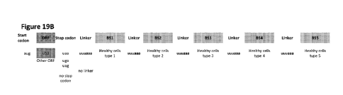
Figure 19B shows general schematics for examples of mRNA constructs according
to some
embodiments of the invention, where up to five binding sites are included.
Figures 19C and 19D show examples of combinations of binding site sequences
which can
be used in mRNA constructs according to the invention.
Figure 19E shows some specific combinations of coding sequences, including
coding
sequences for multiple polypeptides, and binding sites according to some
embodiments.
DETAILED DESCRIPTION
Unless otherwise indicated, the practice of the present invention employs
conventional
techniques of chemistry, molecular biology, microbiology, recombinant DNA
technology, and
chemical methods, which are within the capabilities of a person of ordinary
skill in the art.
Such techniques are also explained in the literature, for example, M.R. Green,
J. Sambrook,
2012, Molecular Cloning: A Laboratory Manual, Fourth Edition, Books 1-3, Cold
Spring
Harbor Laboratory Press, Cold Spring Harbor, NY; Ausubel, F. M. et al. (1995
and periodic
supplements; Current Protocols in Molecular Biology, ch. 9, 13, and 16, John
Wiley & Sons,
New York, N. Y.); B. Roe, J. Crabtree, and A. Kahn, 1996, DNA Isolation and
Sequencing:
Essential Techniques, John Wiley & Sons; J. M. Polak and James OD. McGee,
1990, In Situ
Hybridisation: Principles and Practice, Oxford University Press; M. J. Gait
(Editor), 1984,
Oligonucleotide Synthesis: A Practical Approach, IRL Press; and D. M. J.
Lilley and J. E.
Dahlberg, 1992, Methods of Enzymology: DNA Structure Part A: Synthesis and
Physical
Analysis of DNA Methods in Enzymology, Academic Press. Each of these general
texts is
herein incorporated by reference.
Prior to setting forth the invention, a number of definitions are provided
that will assist
in the understanding of the invention. All references cited herein are
incorporated by
reference in their entirety. Unless otherwise defined, all technical and
scientific terms used
9
CA 03091543 2020-08-18
WO 2019/158955 PCT/GB2019/050454
herein have the same meaning as commonly understood by one of ordinary skill
in the art to
which this invention belongs.
As used herein, the term 'comprising' means any of the recited elements are
necessarily included and other elements may optionally be included as well.
'Consisting
essentially of' means any recited elements are necessarily included, elements
that would
materially affect the basic and novel characteristics of the listed elements
are excluded, and
other elements may optionally be included. 'Consisting of' means that all
elements other than
those listed are excluded. Embodiments defined by each of these terms are
within the scope
of this invention.
The term 'isolated', when applied to a polynucleotide sequence, denotes that
the
sequence has been removed from its natural organism of origin and is, thus,
free of
extraneous or unwanted coding or regulatory sequences. The isolated sequence
is suitable
for use in recombinant DNA processes and within genetically engineered protein
synthesis
systems. Such isolated sequences include cDNAs, mRNAs and genomic clones. The
isolated sequences may be limited to a protein encoding sequence only, or can
also include
5' and 3' regulatory sequences such as promoters and transcriptional
terminators. Prior to
further setting forth the invention, a number of definitions are provided that
will assist in the
understanding of the invention.
A `polynucleotide' is a single or double stranded covalently-linked sequence
of
nucleotides in which the 3 and 5' ends on each nucleotide are joined by
phosphodiester
bonds. The polynucleotide may be made up of deoxyribonucleotide bases or
ribonucleotide
bases. Polynucleotides include DNA and RNA, and may be manufactured
synthetically in
vitro or isolated from natural sources. Sizes of polynucleotides are typically
expressed as the
number of base pairs (bp) for double stranded polynucleotides, or in the case
of single
stranded polynucleotides as the number of nucleotides (nt). One thousand bp or
nt equal a
kilobase (kb). Polynucleotides of less than around 40 nucleotides in length
are typically
called `oligonucleotides'. The term 'nucleic acid sequence' as used herein, is
a single or
double stranded covalently-linked sequence of nucleotides in which the 3' and
5' ends on
each nucleotide are joined by phosphodiester bonds. The polynucleotide may be
made up of
deoxyribonucleotide bases or ribonucleotide bases. Nucleic acid sequences may
include
DNA and RNA, and may be manufactured synthetically in vitro or isolated from
natural
sources. Sizes of nucleic acid sequences, also referred to herein as
`polynucleotides' are
typically expressed as the number of base pairs (bp) for double stranded
polynucleotides, or
in the case of single stranded polynucleotides as the number of nucleotides
(nt). One
CA 03091543 2020-08-18
WO 2019/158955 PCT/GB2019/050454
thousand bp or nt equal a kilobase (kb). Polynucleotides of less than around
40 nucleotides
in length are typically called `oligonucleotides' and may comprise primers for
use in
manipulation of DNA such as via polymerase chain reaction (PCR).
The term 'nucleic acid' as used herein, is a single or double stranded
covalently-linked
sequence of nucleotides in which the 3 and 5' ends on each nucleotide are
joined by
phosphodiester bonds. The polynucleotide may be made up of deoxyribonucleotide
bases or
ribonucleotide bases. Nucleic acids may include DNA and RNA, and may be
manufactured
synthetically in vitro or isolated from natural sources. Nucleic acids may
further include
modified DNA or RNA, for example DNA or RNA that has been methylated, or RNA
that has
been subject to post-translational modification, for example 5'-capping with 7-
methylguanosine, 3'-processing such as cleavage and polyadenylation, and
splicing. Nucleic
acids may also include synthetic nucleic acids (XNA), such as hexitol nucleic
acid (HNA),
cyclohexene nucleic acid (CeNA), threose nucleic acid (TNA), glycerol nucleic
acid (GNA),
locked nucleic acid (LNA) and peptide nucleic acid (PNA). Sizes of nucleic
acids, also
referred to herein as `polynucleotides' are typically expressed as the number
of base pairs
(bp) for double stranded polynucleotides, or in the case of single stranded
polynucleotides as
the number of nucleotides (nt). One thousand bp or nt equal a kilobase (kb).
Polynucleotides
of less than around 100 nucleotides in length are typically called
`oligonucleotides' and may
comprise primers for use in manipulation of DNA such as via polymerase chain
reaction
(PCR). In specific embodiments of the present invention the nucleic acid
sequence
comprises messenger RNA (mRNA).
According to the present invention, homology to the nucleic acid sequences
described
herein is not limited simply to 100% sequence identity. Many nucleic acid
sequences can
demonstrate biochemical or functional equivalence to each other despite having
apparently
low sequence identity. In the present invention homologous nucleic acid
sequences are
considered to be those that will hybridise to each other under conditions of
low stringency
(Sambrook J. et al, supra). In this regard, the term "substantially similar",
relating to two
sequences, means that the sequences have at least 70%, 80%, 90%, 95% or 100%
similarity. Likewise, the term "substantially complementary", relating to two
sequences,
means that the sequences are completely complementary, or that at least 70%,
80%, 90%,
95% or 99% of the bases are complementary. That is, mismatches can occur
between the
bases of the sequences which are intended to hybridise, which can occur
between at least
1%, 5%, 10%, 20% or up to 30% of the bases.
The term 'operatively linked', when applied to nucleic acid sequences, for
example in
an expression construct, indicates that the sequences are arranged so that
they function
11
CA 03091543 2020-08-18
WO 2019/158955 PCT/GB2019/050454
cooperatively in order to achieve their intended purposes. By way of example,
in a DNA
vector a promoter sequence allows for initiation of transcription that
proceeds through a
linked coding sequence as far as a termination sequence. In the case of RNA
sequences,
one or more untranslated regions (UTRs) may be arranged in relation to a
linked protein
.. coding sequence referred to as an open reading frame (ORF). A given mRNA
may comprise
more than one ORFs, a so-called polycistronic RNA. A UTR may be located 5' or
3' in
relation to an operatively linked coding sequence ORF. UTRs may comprise
sequences
typically found in mRNA sequences found in nature, such as Kozak consensus
sequences,
initiation codons, cis-acting regulatory elements, poly-A tails, internal
ribosome entry sites
.. (IRES), structures regulating mRNA longevity, sequences directing the
localisation of the
mRNA, and so on. A mRNA may comprise multiple UTRs that are the same or
different.
The term 'expressing a polypeptide' in the context of the present invention
refers to
production of a polypeptide for which the polynucleotide sequences described
herein code.
.. Typically, this involves translation of the supplied mRNA sequence by the
ribosomal
machinery of the cell to which the sequence is delivered.
The term 'delivery particle' as used herein refers to particles which can
comprise
therapeutic components by encapsulation, holding within a matrix, the
formation of complex
.. or by other means, and deliver a therapeutic component such as a coding
nucleic acid
sequence into a target cell. Delivery particles may on the micro- scale, but
in specific
embodiments may typically be on the nanoscale ¨ i.e. nanoparticles.
Nanoparticles are
typically sized at least 50 nm (nanometres), suitably at least approximately
100 nm and
typically at most 150nm, 200 nm, although optionally up to 300 nm in diameter.
In one
.. embodiment of the invention the nanoparticles have a mean diameter of
approximately at
least 60 nm. An advantage of these sizes is that this means that the particles
are below the
threshold for reticuloendothelial system (mononuclear phagocyte system)
clearance, i.e. the
particle is small enough not to be destroyed by phagocytic cells as part of
the body's defence
mechanism. This facilitates the use of intravenous delivery routes for the
compositions of the
.. invention.
Alternative possibilities for the composition of the nanoparticles include
polylactic acid
(PLA), poly(lactic-co-glycolic acid) (PLGA), a lipid- or phospholipid-based
particles such as
liposomes; particles based on proteins and/or glycoproteins such as collagen,
albumin,
.. gelatin, elastin, gliadin, keratin, legumin, zein, soy proteins, milk
proteins such as casein, and
others (Lohcharoenkal et al. BioMed Research International; Volume 2014
(2014)); and
12
CA 03091543 2020-08-18
WO 2019/158955 PCT/GB2019/050454
particles based on metals or metallic compounds such as gold, silver,
aluminium, copper
oxides and so on.
In particular, polymers comprising polyethyleneimine (PEI) have been
investigated for
the delivery of nucleic acids. Nanoparticle vectors composed of poly(11-amino
esters)
(PBAEs) have also been shown to be suitable for nucleic acid delivery,
especially in
coformulation with polyethylene glycol (PEG) (Kaczmarek JC et al Angew Chem
Int Ed Engl.
2016; 55(44): 13808-13812). Particles of such coformulations have been used to
deliver
mRNA to the lung.
Also considered are particles based on polysaccharides and their derivatives,
such as
cellulose, chitin, and chitosan. Chitosan is a cationic linear polysaccharide
obtained by
partial deacetylation of chitin, with nanoparticles comprising this substance
possessing
promising properties for drug delivery such as biocompatibility, low toxicity
and small size
(Felt et al., Drug Development and Industrial Pharmacy, Volume 24, 1998 -
Issue 11). It is
envisioned that combinations between the above constituents may be used.
US2010/0331234, US2011/0293703 and US2015/0203439 - which are incorporated
herein by reference - describe the production of aminoalcohol lipidoids by
reacting an amine
with an epoxide-terminated compound. Complexes, micelles, liposomes and
particles,
including nanoparticles, may be prepared with these lipidoids and their
chemical structure
makes them particularly suited to the delivery of a 'cargo' ¨ e.g. nucleic
acids such as
coding mRNAs - to target cell types within the body of a human or animal
subject. Delivery
platforms comprising aminoalcohol lipidoid compounds are particularly suitable
for use in the
delivery of net negatively charged cargo molecules given the tertiary amines
available for
protonation thus forming a cationic moiety. For example, aminoalcohol lipidoid
compounds
may be used in the preparation of particulate compositions to deliver DNA,
RNA, or other
polynucleotide cargoes to a subject or to a target cell or tissue. Suitable
particles may be in
the form of microparticles, nanoparticles, liposomes, exosomes, or micelles.
The aminoalcohol lipidoid based delivery particles possess tertiary amines
that are
available to interact with a polynucleotide cargo, such as a coding mRNA.
Polynucleotides,
or derivatives thereof, are contacted with the aminoalcohol lipidoid compounds
under
conditions suitable to form polynucleotide/lipidoid complexes. The lipidoid is
preferably at
least partially protonated so as to form a complex with the negatively charged
polynucleotide. In this way, the polynucleotide/lipidoid complexes form
particles that are
useful in the delivery of cargo polynucleotides to cells and tissues. In
certain embodiments,
13
CA 03091543 2020-08-18
WO 2019/158955 PCT/GB2019/050454
multiple aminoalcohol lipidoid molecules may be associated with a
polynucleotide molecule.
The complex may include at least 1, at least 5, at least 10, at least 20, at
least 50, or suitably
at least 100 aminoalcohol lipidoid molecules. The complex may include at most
10,000, at
most 5000, at most 2000, at most 1000, at most 500, or typically at most 100
aminoalcohol
lipidoid molecules.
Those of ordinary skill in the art will appreciate that a population of
particles follow
principles of particle size distribution. Widely used, art-recognized methods
of describing
particle size distributions include, for example, average diameters and D
values, such as the
D50 value, which is commonly used to represent the mean diameter of the range
of the
particle sizes of a given sample. In certain embodiments, the diameter of the
nanoparticles
particles ranges from 10-500 nm, more suitably the diameter of the particles
ranges from 10-
1200 nm, and particularly from 50-150 nm. In some embodiments, the
nanoparticles have
average diameters of at least about 10 nm, suitably at least about 30 nm. In
some
embodiments, nanoparticles have average diameters of less than about 150 nm in
average
diameter and greater than 50 nm in average.
The particles may be further associated with a targeting agent at facilitates
binding of
the delivery particle to a target cell type. The term 'targeted' as used
herein in relation to
refers to an object, or composition such as comprising a delivery particle,
which is intended
to associate with and facilitate transfection of cells within a particular
organ, tissue or cell
type within the body. In a particular embodiment, a delivery particle ¨ such
as a delivry
nanoparticle - may be targeted to deliver its cargo only to a certain organ,
tissue or cell type.
Targeting may be geographical, for example by the delivery of the targeted
object directly to
a particular tissue, or may be mediated chemically, through targeting agents
or binding
moieties which preferentially associate with target cells or tissues.
A variety of targeting agents that direct pharmaceutical compositions to
particular cells
are known in the art (see, for example, Cotten et al. Methods Enzym. 217:618,
1993;
Wagner et al. Advanced Drug Delivery Reviews, Volume 14, Issue 1, April¨May
1994, 113-
135; Fiume et al. Advanced Drug Delivery Reviews, Volume 14, Issue 1,
April¨May 1994,
51-65). The targeting agents may be included throughout the particle or may be
localised
only on the surface. The targeting agent may be a protein, peptide,
carbohydrate,
glycoprotein, lipid, small molecule, nucleic acids, etc. The targeting agent
may be used to
target specific cells or tissues or may be used to promote endocytosis or
phagocytosis of the
particle. Examples of targeting agents include, but are not limited to,
antibodies, fragments of
antibodies, low-density lipoproteins (LDLs), transferrin, asialoglycoproteins,
gp120 envelope
14
CA 03091543 2020-08-18
WO 2019/158955 PCT/GB2019/050454
protein of the human immunodeficiency virus (HIV), carbohydrates, receptor
ligands, sialic
acid, aptamers etc. If the targeting agent is distributed throughout the
particle, the targeting
agent may be included in the mixture or composite that is used to form the
particles. If the
targeting agent is only located on the surface, the targeting agent may be
associated with
the formed particles using standard chemical techniques e.g. by covalent
binding,
hydrophobic, hydrogen bonding, van der Waals, biotin-avidin linkage, or other
interactions.
The particulate compositions of certain embodiments of the invention may
suitably
deliver the encapsulated mRNA cargo over a period of time that may be
controlled by the
particular choice or formulation of the encapsulating biodegradable non-toxic
polymer or
biocompatible material. For example, the particulate compositions may release
the
encapsulated mRNA cargo over at least 30 minutes, at least 1 hour, at least 2
hours, at least
6 hours, at least 12 hours, or at least 1 day. The particulate compositions
may release the
encapsulated mRNA cargo over at most 2 days, at most 3 days, or at most 7
days.
The term 'diseased' as used herein, as in 'diseased cells' and/or 'diseased
tissue'
indicates tissues and organs (or parts thereof) and cells which exhibit an
aberrant, non-
healthy or disease pathology. For instance, diseased cells may be infected
with a virus,
bacterium, prion or eukaryotic parasite; may comprise deleterious mutations;
and/or may be
cancerous, precancerous, tumoural or neoplastic. Diseased cells may comprise
an altered
intra-cellular miRNA environment when compared to otherwise normal or so-
called healthy
cells. In certain instances disease cells may be pathologically normal but
comprise an
altered intra-cellular miRNA environment that represents a precursor state to
disease.
Diseased tissues may comprise healthy tissues that have been infiltrated by
diseased cells
from another organ or organ system. By way of example, many inflammatory
diseases
comprise pathologies where otherwise healthy organs are subjected to
infiltration with
immune cells such as T cells and neutrophils. By way of a further example,
organs and
tissues subjected to stenotic or cirrhotic lesions may comprise both healthy
and diseased
cells in close proximity.
The term 'cancer' as used herein refers to neoplasms in tissue, including
malignant
tumours which may be primary cancer starting in a particular tissue, or
secondary cancer
having spread by metastasis from elsewhere. The terms cancer, neoplasm and
malignant
tumours are used interchangeably herein. Cancer may denote a tissue or a cell
located
within a neoplasm or with properties associated with a neoplasm. Neoplasms
typically
possess characteristics that differentiate them from normal tissue and normal
cells. Among
such characteristics are included, but not limited to: a degree of anaplasia,
changes in
CA 03091543 2020-08-18
WO 2019/158955 PCT/GB2019/050454
morphology, irregularity of shape, reduced cell adhesiveness, the ability to
metastasize, and
increased cell proliferation. Terms pertaining to and often synonymous with
'cancer' include
sarcoma, carcinoma, malignant tumour, epithelioma, leukaemia, lymphoma,
transformation,
neoplasm and the like. As used herein, the term 'cancer' includes
premalignant, and/or
precancerous tumours, as well as malignant cancers.
The term 'healthy' as used herein, as in 'healthy cells' and/or 'healthy
tissue' indicates
tissues and organs (or parts thereof) and cells which are not themselves
diseased and
approximate to a typically normal functioning phenotype. It can be appreciated
that in the
context of the invention the term 'healthy' is relative, as, for example, non-
neoplastic cells in
a tissue affected by tumours may well not be entirely healthy in an absolute
sense. Therefore
'non-healthy cells' is used mean cells which are not themselves neoplastic,
cancerous or
pre-cancerous but which may be cirrhotic, inflamed, or infected, or otherwise
diseased for
example. Similarly, 'healthy or non-healthy tissue' is used to mean tissue, or
parts thereof,
without tumours, neoplastic, cancerous or pre-cancerous cells; or other
diseases as
mentioned above; regardless of overall health. For instance, in the context of
an organ
comprising cancerous and fibrotic tissue, cells comprised within the fibrotic
tissue may be
thought of as relatively 'healthy' compared to the cancerous tissue.
In an alternative embodiment, the health status of a cell, cell type, tissue
and/or organ
is determined by the quantification of miRNA expression. In certain disease
types, such as
cancer, the expression of particular miRNA species is affected, and can be up-
or down-
regulated compared to unaffected cells. This difference in the miRNA
transcriptome can be
used to identify relative states of health, and/or to track the progression of
healthy cells, cell
types, tissues and/or organs towards a disease state. The disease state may
include the
various stages of transformation into a neoplastic cell. In embodiments of the
present
invention the differential variations in the miRNA transcriptome of cell types
comprised within
a given organ or organ system is leveraged in order to control protein
expression in the
different cell types.
As used herein, the term 'organ' is synonymous with an 'organ system' and
refers to a
combination of tissues and/or cell types that may be compartmentalised within
the body of a
subject to provide a biological function, such as a physiological, anatomical,
homeostatic or
endocrine function. Suitably, organs or organ systems may mean a vascularized
internal
organ, such as a liver or pancreas. Typically organs comprise at least two
tissue types,
and/or a plurality of cell types that exhibit a phenotype characteristic of
the organ.
16
CA 03091543 2020-08-18
WO 2019/158955 PCT/GB2019/050454
The term 'therapeutic virus' as used herein refers to a virus which is capable
of
infecting and killing cancer cells, sometimes by direct viral lysis
(oncolysis), but also
including indirect killing by the stimulation of host anti-tumoural responses.
Oncolytic viruses
are frequently characterised by having increased activity in diseased cells,
including cancer
cells, compared with healthy cells.
Examples of oncolytic viruses include those provided in Table 1, and subtypes
thereof.
Table 1
Oncolytic virus Type
Vesicular Somatitis Virus Enveloped RNA
Vaccinia Virus Enveloped DNA
Maraba virus Enveloped rhabdovirus
Polio virus Non enveloped RNA
Reovirus Non enveloped RNA
Measles virus Enveloped RNA
Newcastle disease virus Enveloped RNA
Coxsackievirus A21 Non enveloped RNA
Parvovirus Non enveloped DNA
Herpes Simplex Virus Type 1 Enveloped DNA
Adenovirus Non enveloped DNA
In embodiments of the invention viruses may be selected from any one of the
Groups I
¨ VII of the Baltimore classification of viruses (Baltimore D (1971).
"Expression of animal
virus genomes". Bacteriol Rev. 35 (3): 235-41). In specific embodiments of the
invention
suitable viruses may be selected from Baltimore Group I, which are
characterised as having
double stranded DNA viral genomes; Group IV, which have single stranded
positive RNA
genomes; and Group V, which have single stranded negative RNA genomes.
The term 'virulence gene' or 'virulence factor' as used herein refers to a
gene or gene
product which aids in the replication of a therapeutic virus such as an
oncolytic virus within or
lysis of the cells which it infects. The term 'replication factor' is used as
a synonymous term
herein. Virulence factors may typically be viral genes encoded by the viral
genome.
Virulence factors may be involved in functions such as intracellular immune
system
suppression and evasion, viral genome replication, the spread or transmission
of virions, the
production or assembly of structural coat proteins, the activation of viruses
in a latent state,
the prevention of viral latency, and the takeover of host cell processes.
Several virulence
factors have cellular or other equivalents which can compensate for the
function of these
genes if lacking in the virus genome. Some viruses can be modified with
exogenous
virulence genes which increase their ability to replicate, lyse cells, and
spread.
17
CA 03091543 2020-08-18
WO 2019/158955 PCT/GB2019/050454
In specific embodiments of the present invention the mRNA sequences enhance or
sustain the oncolytic potency of a co-administered virus in a tumor located
within an organ
through differential expression of one or more proteins or polypeptides that
enhance virion
replication preferentially in the tumor. In this way the mRNA may code for one
or more
factors that enhance the biological activity of the oncolytic virus by:
increased replication
and/or increased direct oncolytic effect and/or increased viral progeny and/or
an adaptive
antitumor immune response In further embodiments of the invention the
compositions may
encode a gene product that controls the interaction between host immune cells
and oncolytic
virus within a tumour. In yet a further embodiment, the compositions of the
invention can be
used to produce gene products that modulate differential patterns of oncolytic
virus activity
as well as expression of immune co-stimulatory molecules that are administered
via the
virion, exogenously or via a delivery particle of the invention.
The term `polypeptide' as used herein is a polymer of amino acid residues
joined by
peptide bonds, whether produced naturally or in vitro by synthetic means.
Polypeptides of
less than around 12 amino acid residues in length are typically referred to as
"peptides" and
those between about 12 and about 30 amino acid residues in length may be
referred to as
"oligopeptides". The term "polypeptide" as used herein denotes the product of
a naturally
occurring polypeptide, precursor form or proprotein. Polypeptides can also
undergo
maturation or post-translational modification processes that may include, but
are not limited
to: glycosylation, proteolytic cleavage, lipidization, signal peptide
cleavage, propeptide
cleavage, phosphorylation, and such like. The term "protein" is used herein to
refer to a
macromolecule comprising one or more polypeptide chains.
The term 'gene product' as used herein refers to the product of the at least
one coding
sequence or ORF comprised within an mRNA construct of the invention as
described herein.
The gene product(s) may comprise a polypeptide or a protein. A polycistronic
mRNA
construct may be used which results in the production of multiple gene
products. It will be
appreciated that multiple ORFs may lead to the production in situ of a variety
of products that
may cooperate functionally, or may form complexes and/or multimeric proteins
with diverse
biological and potentially therapeutic effects.
Delivery of mRNA directly to cells allows direct and controllable translation
of the
desired gene products such as polypeptides and/or proteins in the cells.
Provision of mRNA
specifically allows not only for the use of cell expression modulation
mechanisms such as
miRNA mediated control (as detailed in specific embodiments below), but also
represents a
finite and exhaustible supply of the product, rather than the potentially
permanent change to
18
CA 03091543 2020-08-18
WO 2019/158955 PCT/GB2019/050454
the transcriptome of a target cell which an episomal or genomically inserted
DNA vector
might provide.
In embodiments of the present invention an mRNA sequence is provided that
comprises a sequence that codes for at least one polypeptide in operative
combination with
one or more untranslated regions (UTRs) that may confer tissue specificity,
and stability to
the nucleic acid sequence as a whole. By 'tissue specificity' it is meant that
translation of the
protein product encoded by the mRNA is modulated according to the presence of
the UTR.
Modulation may include permitting, reducing or even blocking detectable
translation of the
mRNA into a protein product. The UTRs may be linked directly to the mRNA in
cis ¨ i.e. on
the same polynucleotide strand. In an alternative embodiment, a first sequence
that codes
for a gene product is provided and a further second sequence, that hybridises
to a portion of
the first sequence, is provided that comprises one or more UTRs that confer
tissue specificity
to the nucleic acid sequence as a whole. In this latter embodiment the UTR is
operatively
linked to the sequence that encodes the gene product in trans.
According to specific embodiments of the invention, an mRNA is provided that
comprises such associated nucleic acid sequences operatively linked thereto as
are
necessary to prevent or reduce expression of a gene product in non-diseased
liver tissue,
e.g. in healthy hepatocytes. As such, an mRNA construct, or transcript, is
provided that
comprises a 5' cap and UTRs necessary for ribosomal recruitment and tissue
and/or organ
specific expression (typically, but not exclusively positioned 3' to the ORF),
as well as start
and stop codons that respectively define one or more ORFs. When the construct
is
introduced into a non-diseased liver, lung, pancreas, breast, brain, kidney
and/or colon-GI
tract, expression of the gene product is prevented or reduced. In contrast,
neoplastic or
otherwised diseased cells comprised within the aforementioned organs typically
do not
conform to normal non-diseased cell expression patterns, posessing a quite
different miRNA
transcriptome. The gene product(s) comprised within the mRNA is translated
specifically in
these aberrant cells but not in neighboring healthy cells. Delivery of the
mRNA construct to
the organs mentioned above may be achieved via a particulate delivery platform
as
described herein, or in any suitable way known in the art. Cell type specific
expression can
be mediated via microRNA modulation mechanisms such as those described in more
detail
below.
A 'therapeutic component' or 'therapeutic agent' as defined herein refers to a
molecule, substance, cell or organism that when administered to an individual
human or
other animal as part of a therapeutic intervention, contributes towards a
therapeutic effect
19
CA 03091543 2020-08-18
WO 2019/158955 PCT/GB2019/050454
upon that individual human or other animal. The therapeutic effect may be
caused by the
therapeutic component itself, or by another component of the therapeutic
intervention. The
therapeutic component may be a coding nucleic acid component, in particular an
mRNA.
The coding nucleic acid component(s) may code for therapeutic enhancement
factors, as
defined below. A therapeutic component may also comprise a drug, optionally a
chemotherapeutic drug such as a small molecule or monoclonal antibody (or
fragment
thereof). In some embodiments, a therapeutic component may comprise a cell,
such as a
recombinantly modified immune effector cell ¨ e.g. a CAR-T cell. In other
embodiments of
the invention, the therapeutic agent comprises a therapeutic virus, such as an
oncolytic virus
or a viral vector.
The term 'therapeutic effect' refers to a local or systemic effect in an
animal subject,
typically a human, caused by a pharmacologically or therapeutically active
agent that
comprises a substance, molecule, composition, cell or organism that has been
administered
to the subject, and the term 'therapeutic intervention' refers to the
administration of such a
substance, molecule, composition, cell or organism. The term thus means any
agent
intended for use in the diagnosis, cure, mitigation, treatment or prevention
of disease or in
the enhancement of desirable physical or mental development and conditions in
an animal or
human subject. The phrase 'therapeutically- effective amount' means that
amount of such an
agent that produces a desired local or systemic effect at a reasonable
benefit/risk ratio
applicable to any treatment. In certain embodiments, a therapeutically
effective amount of an
agent will depend on its therapeutic index, solubility, and the like. For
example, certain
therapeutic agents of the present invention may be administered in a
sufficient amount to
produce a reasonable benefit/risk ratio applicable to such treatment. In the
specific context of
treatment of cancer, a 'therapeutic effect' can be manifested by various
means, including but
not limited to, a decrease in solid tumour volume, a decrease in the number of
cancer cells,
a decrease in the number of metastases observed, an increase in life
expectancy, decrease
in cancer cell proliferation, decrease in cancer cell survival, a decrease in
the expression of
tumour cell markers, and/or amelioration of various physiological symptoms
associated with
the cancerous condition.
In one embodiment, the subject to whom therapy is administered is a mammal
(e.g.,
mouse, rat, primate, non-human mammal, domestic animal or livestock, such as a
dog, cat,
rabbit, cow, horse, sheep, goat and the like), and is suitably a human. In a
further
embodiment, the subject is an animal model of cancer. For example, the animal
model can
be an orthotopic xenograft animal model of a human-derived cancer, suitably
liver, lung,
pancreas, breast, brain, kidney and/or colon-GI tract cancer.
CA 03091543 2020-08-18
WO 2019/158955 PCT/GB2019/050454
In a specific embodiment of the methods of the present invention, the subject
has not
yet undergone a therapeutic treatment, such as therapeutic viral therapy,
chemotherapy,
radiation therapy, targeted therapy, and/or anti-immune checkpoint therapy. In
still another
embodiment, the subject has undergone a therapeutic treatment, such as the
aforementioned therapies.
In further embodiments, the subject has had surgery to remove cancerous or
precancerous tissue. In other embodiments, the cancerous tissue has not been
removed, for
example, the cancerous tissue may be located in an inoperable region of the
body, such as
in a tissue or organ that if subjected to surgical intervention may compromise
the life of the
subject, or in a region where a surgical procedure would cause considerable
risk of
permanent harm or even lethality.
In some embodiments, the provided mRNA may code for a 'therapeutic enhancement
factor'. According to the present invention therapeutic enhancement factors
are gene
products or polypeptides that may enhance or facilitate the ability of
another, co-
administered therapeutic agent, to exert a therapeutic effect upon a given
cell, suitably the
target cell. When introduced into or in the vicinity of the target cell,
expression of the
therapeutic enhancement factor may cooperate with a co-administered
therapeutic agent
thereby enabling or enhancing the therapeutic activity of the agent. In some
embodiments,
the therapeutic enhancement factor may enhance the ability of a co-
administered oncolytic
virus to lyse cancer cells. In other embodiments of the invention, the
therapeutic
enhancement factor may effect an alteration of a tumour microenvironment so as
to assist or
recruit the subject's own immune response. In this latter embodiment, the
alteration of the
tumour microenvironment may assist co-administration of an oncolytic virus or
a CAR-T or
other adoptive cell based therapy. In some embodiments, the therapeutic
enhancement
factor may enable the conversion of a prodrug into an active form.
Multiple therapeutic enhancement factors may be combined in compositions
according
to specific embodiments of the present invention. In such embodiments, the
coding
sequences for each therapeutic enhancement factor may be present in separate
mRNA
molecules. In some embodiments, sequences for more than one therapeutic
enhancement
factor may be present on the same mRNA molecule. In such cases the
polycistronic mRNA
molecule further comprises sequences as necessary for the expression of all
coded
sequences, such as internal ribosome entry sites (IRES).
21
CA 03091543 2020-08-18
WO 2019/158955 PCT/GB2019/050454
In embodiments where multiple different mRNA molecules are comprised in one or
more delivery system, it is contemplated that each delivery system ¨ e.g.
particle, liposome,
viral vector system - may comprise one or more than one type of mRNA molecule
as the
`payload'; that is, not every delivery payload in a particular embodiment will
necessarily
comprise all of the mRNA molecules provided in said embodiment. In this way,
it is also
considered possible to direct different delivery systems and their associated
sequences to
different target cells, with the targeting agents described herein.
The mRNA constructs of certain embodiments of the invention may be synthesised
from a polynucleotide expression construct, which may be for example a DNA
plasmid. This
expression construct may comprise any promoter sequence necessary for the
initiation of
transcription and a corresponding termination sequence, such that
transcription of the mRNA
construct can occur. Such polynucleotide expression constructs are
contemplated to
comprise embodiments of the invention in their own right.
The gene product encoded by the mRNA is typically a peptide, polypeptide or
protein.
Where a particular protein consists of more than one subunit, the mRNA may
code for one or
more than one subunit within one or more ORFs. In alternative embodiments, a
first mRNA
may code for a first subunit, whilst a second co-administered mRNA may code
for a second
subunit that, when translated in situ, leads to assembly of a multi-subunit
protein gene
product.
The gene product encoded by the mRNA may be of any type suitable for producing
a
therapeutic effect. In the context of treating cancer, the gene product
encoded by the mRNA
may suitably include genes which when expressed by a cancer cell cause or aid
in the
destruction of the cancer cell.
Tumour suppressor genes such as p53 may be provided by the constructs of the
invention. p53 plays a role in cell processes including apoptosis and genomic
stability. It is
.. involved in the activation of the DNA repair process in response to genomic
damage, and
can arrest cell growth and reproduction.
Genes which promote cell death by apoptosis ¨ so-called suicide genes ¨ which
when
expressed cause the cell to activate the process of apoptosis, may also be
provided by the
compositions and constructs of the invention. Cancer cells often possess
mutated and/or
functionless versions of these apoptosis-related genes, and so cannot undergo
apoptosis in
response to external signals. Suicide gene therapy may also refer to the
introduction of
22
CA 03091543 2020-08-18
WO 2019/158955 PCT/GB2019/050454
genes which allow the conversion of a non-toxic compound or prodrug into a
lethal drug
(Duarte et al. Cancer Letters, 2012). According to embodiments of the
invention, such gene
products can be introduced selectively into diseased cells, such as neoplastic
cells, marking
them for destruction by induced apoptosis or delivery of an otherwise non-
toxic compound or
prodrug.
In specific embodiments of the invention, the mRNA may encode inhibitors of
the
programmed cell death pathway, such as inhibitors of PD-1 receptor (CD279) or
its ligands
PD-L1 (B7-H1; CD274) and PD-L2 (B7-DC; CD273). Hence, the mRNA may encode a
protein or polypeptide that binds to or otherwise interferes with the function
of the PD-1/PDL-
1 or PD-1/PDL-2 axis within diseased or neoplastic cells within a target
organ. Suitable
proteins or polypeptides may include antibodies, which may be monoclonal or
polyclonal, or
antigen binding fragments thereof, or other antigen binding microproteins,
that bind to PD-1
receptor, PDL-1, PDL-2, or complexes of ligand and receptor. This effect may
also be
observed by use of protein or polypeptide inhibitors of the cytotoxic T
lymphocyte antigen 4
(CTLA4) pathway, another so-called immune checkpoint. Inhibition of either or
both
pathways is known to result in a change in the immune response within the
tumour
microenvironment that may positively benefit the health of the patient. In
addition, by
modulating the immune response in a subject the compositions of the present
invention may
show particular utility in combinatorial therapies with other anti-cancer
therapeutic
approaches, such as radiotherapy or chemotherapy. FDA approved anti-PD1
pathway
inhibitors include pembrolizumab and nivolumab. Known anti-PDL-1 inhibitors
include
MPDL-3280A, BMS-936559 and atezolizumab. Anti-CTLA4 therapeutic inhibitors
include
ipilimumab and tremelimumab. The compositions of the invention may be used to
deliver
such inhibitors of the programmed cell death pathway- or functional mimetics
thereof -
selectively to diseased cells within a target organ in a subject by leveraging
the differential
miRNA environment in those cells
Chimeric antigen receptor T-cells (CAR-T cells) are immune cells, typically T-
lymphocytes, which have been modified to express receptors which target cancer
cells.
Adoptive immunotherapy, which involves the transfer of autologous antigen-
specific T
cells generated ex vivo, is a promising strategy to treat viral infections and
cancer. The T
cells used for adoptive immunotherapy can be generated either by expansion of
antigen-
specific T cells or redirection of T cells through genetic engineering (see
e.g., Park, T. S., S.
A. Rosenberg, et al. (2011). "Treating cancer with genetically engineered T
cells." Trends
Biotechnol 29(11): 550-7).
23
CA 03091543 2020-08-18
WO 2019/158955 PCT/GB2019/050454
Novel specificities in T cells, also known as immune effector cells, have been
successfully generated through the genetic transfer of transgenic T cell
receptors or chimeric
antigen receptors (CARs) (see e.g., Jena, B., G. Dotti, et al. (2010).
"Redirecting T-cell
specificity by introducing a tumor-specific chimeric antigen receptor." Blood
116(7): 1035-
44). CARs are synthetic receptors consisting of at least three parts: an
extracellular antigen
recognition domain (also known as the ectodomain), a transmembrane domain, and
an
intracellular T-cell activation domain (also known as the endodomain). In some
embodiments, the engineered T cells comprise a specific class of T cells, such
as, for
example, gamma delta T cells, a subtype of T cells that selectively target
tumoral cells
without affecting healthy ones. CARs have successfully allowed T cells to be
redirected
against antigens expressed at the surface of tumor cells from various
malignancies including
lymphomas and solid tumors (Jena, Dotti et al. supra). In some embodiments,
the
engineered T cells comprise at least a population of autologous T cells in
which the CAR-T
cells are engineered to eliminate expression of the endogenous a 6 T-cell
receptor (TCR) to
prevent a graft-versus-host response without compromising CAR-dependent
effector
functions. In some embodiments, the engineered T cells comprise at least a
population of
allogeneic T cells. In some embodiments, the engineered T cells comprise at
least a
population of autologous T cells and a population of allogeneic T cells.
Generally, the extracellular antigen recognition domain is a targeting moiety
that is
associated with one or more signaling domains in a single fusion molecule from
an antibody,
receptor, or ligand domain that binds a specific target, typically a tumor-
associated target. In
some embodiments, the extracellular antigen recognition domain is or is
derived from a
single-chain Fragment variant (scFv) of an antigen-binding domain of a single-
chain antibody
(scFv), comprising the light and heavy variable fragments of a monoclonal
antibody joined by
a flexible linker.. In some embodiments, In some embodiments, extracellular
antigen
recognition domain is linked to the transmembrane domain by a linker, such as,
for example,
a flexible linker such as the IgG1 hinge linker. In some embodiments, the
transmembrane is
or is derived from a CD28 transmembrane domain. In some embodiments, the
endodomain
includes a co-stimulatory domain designed to enhance the immune response, for
example,
by enhancing survival and increasing proliferation of CAR modified T cells,
and an internal T-
cell activation domain designed to activate the T cell when it binds to the
desired target. In
some embodiments, the costimulatory domain is or is derived from a CD28
costimulatory
domain, an OX-40 (CD134) costimulatory domain, an ICOS costimulatory domain, a
4-1BB
(CD137) costimulatory domain, or any combination thereof. In some embodiments,
the
intracellular T-cell activation domain comprises the CD3 zeta (CD3) domain or
a biologically
24
CA 03091543 2020-08-18
WO 2019/158955 PCT/GB2019/050454
active portion thereof. In some embodiments, T cell activation results in
immune cell
activation in which inflammatory cytokines are released by the T cells to
promote an
inflammation and/or immune response. In some embodiments, T cell activation
results in
cytotoxic activity in which cytotoxins are released by the T cells to promote
cancer cell
apoptosis. In some embodiments, T cell activation results in proliferation in
which
interleukins are released by the T cells to promote cell development and
division. In some
embodiments, T cell activation results in a combination of at least two of
immune cell
activation, cytotoxic activity, and/or proliferation.
In some embodiments, the extracellular antigen recognition domain specifically
binds
to CD19. CD19 is an attractive target for immunotherapy because the vast
majority of B-
acute lymphoblastic leukemia (B-ALL) uniformly express CD19, whereas
expression is
absent on non-hematopoietic cells, as well as myeloid, erythroid, and T cells,
and bone
marrow stem cells. Clinical trials targeting CD19 on B-cell malignancies are
underway with
encouraging anti-tumor responses. Many of the current CAR-T therapies being
evaluated in
clinical trials use T cells genetically modified to express a chimeric antigen
receptor (CAR)
with specificity derived from the scFv region of a CD19-specific mouse
monoclonal antibody
FMC63 (see e.g., Nicholson, Lenton et al. (1997);. "Construction and
characterisation of a
functional CD19 specific single chain Fv fragment for immunotherapy of B
lineage leukaemia
and lymphoma." Mol Immunol. 1997 Nov-Dec;34(16-17):1157-65; Cooper, Topp et
al.
(2003). "T-cell clones can be rendered specific for CD19: toward the selective
augmentation
of the graft-versus-B-lineage leukemia effect." Blood. 2003 Feb 15;101(4):1637-
44; Cooper,
Jena et al. (2012) (International application: W02013/126712).
In some embodiments, extracellular antigen recognition domain specifically
binds to
CD22. CD22 is a transmembrane phosphoglycoprotein that belongs to the Siglec
family of
lectins and specifically binds sialic acid with its N-terminus seven
extracellular
immunoglobulin domains. It mainly acts as an inhibitory receptor for B cell
activation and
signaling and regulates the interaction of B cells with T cells and antigen
presenting cells
(APCs). Similar to CD19, CD22 is a B cell lineage-restricted marker, expressed
explicitly by
B lymphoid cells from the pre-B to mature B cell stage. However, it is lost
during
differentiation to plasma cells. CD22 is universally expressed in most B cell
malignancies,
including acute lymphoblastic leukemia (ALL), chronic lymphocytic leukemia
(CLL), and
various subtypes of non-Hodgkin lymphoma (NHL) such as diffuse large B cell
lymphoma.
Targeting CD22 as an attractive therapeutic target for B cell malignancies has
been
confirmed by positive results in clinical trials of anti-CD22 monoclonal
antibodies (e.g.,
epratuzumab) and immunotoxins (e.g., BL22, HA22). CD22 has been shown to be
CA 03091543 2020-08-18
WO 2019/158955 PCT/GB2019/050454
expressed on ALL cells that lost CD19 expression after treatment with anti-
CD19 CAR-T
cells, making anti-CD22 CAR-T cells suitable for combination and/or follow-on
therapy of
anti-CD19 CAR-T cells.
However, although numerous clinical studies have demonstrated the potential of
adoptive transfer of CAR T cells for cancer therapy, they have also raised the
risks
associated with the cytokine-release syndrome (CRS) and the "on-target off-
tumour" effect.
The mRNA delivery compositions provided in some embodiments herein are useful
to
improve the safety and efficacy of CAR-T-cells. For example, the mRNA
nanoparticle
delivery systems of embodiments described herein may be used to recruit
specific immune
cells or modified subsets of immune cells such as CAR-T cells to the tumour
microenvironment. Additionally, the mRNA nanoparticle delivery systems may be
used to
inhibit expression of endogenous T cell receptors (TCRs) to avoid graft-versus-
host disease
and/or to selectively delete immune checkpoint genes in these cells to
strengthen their anti-
cancer activity in the suppressive tumour milieu. (See e.g., Moffett, Coon, et
al. (2017) "Hit-
and-run programming of therapeutic cytoreagents using mRNA nanocarriers."
Nature
Communications. 8:389.)
In some embodiments, the coding mRNA is used to attract CAR-T cells to a
particular
site in a subject. In some embodiments, the coding mRNA is used to overcome
insufficient
migration of an immune cell to the tumour microenvironment. In response to
specific
chemokines, different immune cell subsets migrate into the tumour
microenvironment and
regulate tumour immune responses in a spatiotemporal manner. In some
embodiments, the
coding mRNA is used to enhance CAR-T cell activation. In addition, chemokines
can directly
target non-immune cells, including tumour cells and vascular endothelial
cells, in the tumour
microenvironment, and they have been shown to regulate tumour cell
proliferation, cancer
stem-like cell properties, cancer invasiveness and metastasis. In some
embodiments, the
immune cell is a T cell, a natural killer (NK) cell, a B cell, an antigen-
presenting cell (APC)
such as a macrophage or dendritic cell, or any combination thereof.
In some embodiments, the coding mRNA can be used to overcome insufficient
migration of CAR-T cells to the tumour microenvironment, and prevent off-
target CAR-T
activity. In some embodiments, the mRNA is delivered to the tumour
microenvironment, and
the coding mRNA encodes a gene product that attracts or otherwise recruits CAR-
T cells to
the tumour microenvironment. In some embodiments, the coding mRNA expresses a
chemokine. By way of non-limiting example, one or more coding mRNAs can encode
one or
26
CA 03091543 2020-08-18
WO 2019/158955 PCT/GB2019/050454
more chemokines that attract T-cells such as CCL2, CCL3, CCL4, CCL5, CCL20,
CCL22,
CCL28, CXCL8, CXCL9, CXCL10, CXCL11, CXCL12, XCL1, and any combination
thereof.
In situations where the reverse effect is desired, such as in autoimmune
disease, the coding
mRNA can express blockers, antagonists and/or inhibitors of the above-
mentioned factors.
In some embodiments, the the coding mRNA is transiently expressed in the
tumour
microenvironment. In some embodiments, the coding mRNA encodes a cytokine or
other
gene product involved in regulating the survival, proliferation, and/or
differentiation of
immune cells in the tumour response, such as, for example, activated T cells
and NK cells.
By way of non-limiting example, the coding mRNA can encode for a cytokine such
as IL-1,
IL-2, IL-4, IL-6, IL-8, IL-10, IL-12, IL-17, IL-33, IL-35, TGF-beta, and any
combination thereof.
Again, in situations where the reverse effect is desired, such as in
autoimmune disease, the
coding mRNA can express blockers, antagonists and/or inhibitors of the above-
mentioned
factors.
In some embodiments, of the invention, the coding mRNA is delivered in
conjunction
with CAR-T or other adoptive cell therapy to provide transient expression of
the coding
mRNA.
In some embodiments, a mRNA delivery system as described herein delivers an
mRNA that codes for a gene-editing agent to a target cell population. In some
embodiments,
the mRNA codes for a sequence-specific nuclease that targets a gene locus and
disrupts
expression of one or more endogenous gene produces in the target cell
population. In some
embodiments, the mRNA codes for a sequence-specific nuclease that targets a T
cell
receptor (TCR)-related gene locus, thereby disrupting expression of one or
more domains in
the TCR.
In some embodiments, the described mRNA delivery systems may be used to
deliver
an mRNA that codes for one or more agents that program engineered T cells
toward a
desired phenotype. In some embodiments, the mRNA may be used to induce markers
and
transcriptional patterns that are characteristic of a desired T cell
phenotype. In some
embodiments, the mRNA may be used to promote development of CD26L+ central
memory
T cells (Tcm), which have been shown to improve CAR-T treatment. (See e.g.,
Moffett, Coon
supra).
MicroRNAs (miRNAs) are a class of noncoding RNAs each containing around 20 to
25
nucleotides some of which are believed to be involved in post-transcriptional
regulation of
27
CA 03091543 2020-08-18
WO 2019/158955 PCT/GB2019/050454
gene expression by binding to complementary sequences in the 3' untranslated
regions (3'
UTR) of target mRNAs, leading to their silencing. These sequences are also
referred to
herein as miRNA binding sites, or miRNA binding site sequences. Certain miRNAs
are highly
tissue-specific in their expression; for example, miR-122 and its variants are
abundant in the
liver and infrequently expressed in other tissues (Lagos-Quintana (2002),
Current Biology,
Vol.12, Apr).
The miRNA system therefore provides a robust platform by which nucleic acids
introduced into cells can be silenced in selected cell types in a target
tissue, and expressed
in others. By including a binding site for a particular given miRNA sequence
into an mRNA
construct to be introduced into target cells, particularly in or immediately
5' or 3' to a UTR,
expression of certain introduced genes can be reduced or substantially
eliminated in some
cell types, while remaining in others (Brown and Naldini, Nature Reviews
Genetics volume
10, pages 578-585 (2009)). The use of the term 'immediately' is understood to
be
synonymous with terms such as 'highly proximate to' or 'very close to'. When
referring to 5'
or 3' positioning relative to a UTR sequence it encompasses variants in which
typically up to
around twenty, suitably not more than fifty, intervening nucleotide bases may
be placed
between the miRNA binding sequence and the adjacent UTR. It is contemplated
that one, or
a plurality, of such miRNA binding site sequences can be included in the mRNA
construct.
Where a plurality of miRNA binding site sequences are present, this plurality
may include for
example greater than two, greater than three, typically greater than four
miRNA binding site
sequences. These miRNA binding site sequences may be arranged sequentially, in
tandem
or at predetermined locations within, 3' to, or 5' to a specified UTR within
the mRNA
constructs. Multiple binding site sequences may be separated with a linker
sequence, which
may vary, or may comprise a particular sequence, for example, "uuuaaa". In
some
embodiments, no linker sequence may be present between binding site sequences.
Other
parts of the mRNA sequence can incorporate linker sequences, such as between
the stop
codon of the ORF and the UTR or binding sites.
miRNA-122, despite its abundance in healthy non-diseased liver tissue, is
reduced in
the majority of liver cancers as well as in diseased cells (Braconi et al.
2011, Semin Oncol;
38(6): 752-763, Brown and Naldini Nature 2009;10 578). By the above-mentioned
method, it
has been found that when the target tissue is the liver, translation of the
introduced mRNA
sequences can be facilitated in cancerous liver cells and reduced or
substantially eliminated
in transfected healthy cells, by including miRNA-122 binding sites (for
example, SEQ ID NO:
1) in or adjacent to their 3' UTRs.
28
CA 03091543 2020-08-18
WO 2019/158955 PCT/GB2019/050454
In a similar way, differential translation of such mRNA is also possible
between cancer
cells and healthy cells in other organs, by using other miRNA binding site
sequences.
Suitable candidates include (but are not limited to) binding sites for miRNA-
125, miRNA-
124a, miRNA-Let7, miRNA-375, miRNA-143, miRNA-145, miRNA-192, miRNA-194, miRNA-
204, miRNA-215 and miRNA-30b,c. Table 2 demonstrates further (non-limiting)
examples of
miRNA sequences where differential expression has been demonstrated.
miRNA-125 is downregulated in several solid tumors, such as hepatocellular
carcinoma (Coppola et al. Oncotarget 2017;8); breast (Mattie et al. Mol Cancer
2006;5), lung
(VVang ewt al. FEBS J 2009), ovarian (Lee et al. Oncotarget 2016;7), gastric
(Xu et al. Mol
Med Rep 2014;10), colon (Tong et al. Biomed Pharmacother 2015;75), and
cervical cancers
(Fan et al Oncotarget 2015;6); neuroblastoma, medulloblastoma (Ferretti et al.
Int J Cancer
2009;124), glioblastoma (Cortez et al. Genes Chromosomes Cancer 2010;49), and
retinoblastoma (Zhang et al; Cell signal 2016;28).
Several miRNA species are also differentially expressed in glioblastoma
multiforme
cells (Zhangh et al. J Miol Med 2009;87 / Shi et al. Brain Res 2008;1236)
compared to non
diseased brain cells (e.g. neurons), with miRNA-124a one of the most
dysregulated (Karsy et
al. Gene Cancer 2012;3; Riddick et al. Nat Rev Neurol 2011;7; Gaur et al.
Cancer Res
2007;67 / Silber et al. BMC Med 2008;6).
In lung cancer, a recent meta-analysis confirmed the downregulation of Let-7
(as
well as miRNA-148a and miRNA-148b) in non-small-cell lung cancer (Lamichhane
et al.
Disease Markers 2018).
Similarly, miRNA-375 expression has be found to be downregulated in pancreatic
cancer cells, compared to healthy pancreatic cells (Shiduo et al. Biomedical
Reports
2013;1).
In another embodiment, using IL-12 secreting cells can enhance the activity of
CAR-
T cells. Without wishing to be bound by theory, it is believed that IL-12
induced IFNy
accumulation in tumors promotes the penetration of CAR-T or other host immune
cells (e.g.
NK cells) into the tumours, thereby enhancing the therapeutic effects
(Chinnasamy D. et al.
Clin Cancer Res 2012:18/ Chmielewski M. et al. Cancer Res 2011;71 / Kerkar SP.
Et al. J
Clin Invest 2011;121 / Jackson HJ. Et al. Nat Rev Clin Oncol 2016;13). Hence,
according to
an embodiment of the present invention the mRNA may be used to promote
exogenous
29
CA 03091543 2020-08-18
WO 2019/158955 PCT/GB2019/050454
expression of IL-12 in the tumour microenvironment, by coding for IL-12 or a
functional
equivalent or derivative thereof.
In the context of disease-specific expression of introduced polynucleotides,
binding
sequences for any miRNA sequence which is disrupted in a particular disease ¨
that is,
upregulated or downregulated in diseased cells (such as tumour cells) in
comparison to non-
diseased cells ¨ is considered suitable for use in the invention. Table 2
discusses further,
non-limiting examples of tumour-associated miRNA binding sequences of this
kind which
may be used in embodiments of the present invention. It will be appreciated,
however, that
the present invention is not limited only to instances where a given miRNA or
class of
miRNAs is downregulated in a first cell type versus a second cell type within
a given organ or
organ system. On the contrary, it is merely required that there exists a
differential
expression pattern of a regulatory miRNA between first and second cell types
comprised
within the organ or organ system. The differential expression of the miRNA can
be exploited
using the compositions and methods described herein to enable corresponding
differential
translation of protein products in those cells.
Examples of cancers where evidence has been found for similar differential
miRNA
expression between healthy and cancer cells include breast (Nygaard et al, BMC
Med
Genomics, 2009 Jun 9;2:35), ovarian (Wyman et al, PloS One, 2009 ;4(4):e5311),
prostate
(Watahiki et al, PloS One, 2011; 6(9):e24950), and cervical cancers (Lui et
al. Cancer
Research, 2007 Jul 1;67(13):6031-43). WO 2017/132552 Al describes a wide range
of
miRNAs with differing expression levels in various cancer cells.
Table 2
Tissue/cancer type Implicated miRNA Expression profile Reference
Liver miRNA-122 Reduced in cancer Braconi, 2011,
cells Brown, 2009
Liver miRNA-125, Reduced in hepato- Coppola N.
miRNA-199 carcinoma Oncotarget, 2017.
Vol 8
Murakami Y.
Oncogene 2006;25
Brain miRNA-124a Reduced in Mazzacurati L.
glioblastoma Molecular therapy
multiforme 23, 2015
Lung, breast Let-7 Reduced in cancer Edge RE et al. Mob
CA 03091543 2020-08-18
WO 2019/158955 PCT/GB2019/050454
miRNA-148a/b cells Ther 2008;16:1437
Yu F. Cell 2007;
131(6):1109-23
Takamizawa J.
Cancer Res
2004;64
Pancreas miRNA-375 Reduced in cancer Song S, Zhou J et
cells al. Biomed Reports:
393-398, 2013
Colon miRNA-143, Reduced in cancer Michael MZ. Mol
miRNA-145 cells Cancer Res
2003;334
Kidney miRNA-192, -194, - Kidney-specific Sempere LF.
204, -215, -30b,c expression Genome Biol
2004;5
Sun Y. Nucleic
acids res 2004;32
In the pancreas, miRNA-375 expression has been indicated to be high in normal
pancreas cells but significantly lower in diseased and/or cancerous tissues
(Song, Zhou et
al. 2013). This expression has been shown to relate to the stage of cancer,
with expression
further reduced with more advanced cancer. It is thought that miRNA-375 is
involved with the
regulation of glucose-induced biological responses in pancreatic 13-cells, by
targeting 3-
phosphoinositide¨ dependent protein kinase-1 (PDK1) mRNA and so affecting the
PI 3-
kinase/PKB cascade (El Ouaamari et al. Diabetes 57:2708-2717, 2008). An anti-
proliferative
effect of miRNA-375 is implicated by this putative mode of action, which may
explain its
downregulation in cancer cells.
It is known that variants and polymorphisms of miRNA sequences can be found,
and
that miRNA families exist with similar properties. In the present invention,
it is envisioned that
all suitable variants and family members of particular miRNA sequences and
associated
binding sites can be used where appropriate. On the other hand, apparently
closely related
miRNA sequences can have different expression profiles (Sun et al, World J
Gastroenterol.
2017 Nov 28), so in some situations it will be necessary to determine whether
a specific
substitution is appropriate, by reference to the literature. For example, Let-
7 is part of a wider
family with a number of related variants, which can be denoted as Let-7a to
Let-7k, and so
on.
31
CA 03091543 2020-08-18
WO 2019/158955 PCT/GB2019/050454
Treating patients with immunotherapies like oncolytic viruses or CAR-T
therapies may
have safety issues due to the possibility of off-target effects. Even the
expression of certain
polypeptides by the provision of coding mRNA sequences can have negative
effects on
certain organs. Protecting healthy tissues, for example liver, brain, breast,
lung, pancreas,
colon/GI-tract and kidneys is thus paramount for successful clinical
applications. Hence, the
present compositions may represent an enabling technology platform for
enhancing and
facilitating the successful adoption of hitherto 'experimental' cellular or
viral therapies.
The presence of a plurality of miRNA binding site sequences in the mRNA
construct
enables improved efficacy of the differential expression of the supplied
polypeptide or
polypeptides. Without being bound by theory, it is thought that with an
increased number of
sites for binding, the likelihood of miRNA binding and consequent degradation
is increased.
Multiple miRNA binding sites can comprise multiple copies of substantially the
same binding
site sequence, thereby introducing redundancy. Alternatively or additionally,
the multiple
binding site sequences can comprise substantially different sequences, thereby
allowing the
mRNA construct to be targeted by more than one species of miRNA. In this way,
differential
expression of a supplied mRNA construct can be achieved for more than one cell
type,
and/or in more than one organ, as is evident from the discussion of organs and
their
associated miRNA sequences above. Both approaches are considered to be
possible within
the same sequence or multiple sequences. An intermediate approach is also
envisioned,
wherein multiple binding sequences are included which are intended to be
targets for the
same miRNA sequence, but have differences in order to bind different variants
of the same
miRNA sequence.
Some advantages associated with the use of multiple binding sites include an
increase in the efficiency of differential expression of polypeptides supplied
by the mRNA
sequences of the present invention, within a single organ. Use of different
binding site
sequences, or sequences which are applicable to more than one tissue or organ
type can
enable differential expression to be achieved in different cell types in more
than one organ or
tissue. This may be desirable when systemic administration of compositions
according to the
invention is used, and it is necessary to avoid off-target effects in more
than one organ.
Even with localised or targeted administration, it is possible that supplied
mRNA
constructs may encounter or accumulate in organs, tissues, and/or cells for
which they were
not intended. In particular, liver and kidney tissue may accumulate
administered
compositions, due to the physiological function of these organs. In these
cases, to avoid off-
32
CA 03091543 2020-08-18
WO 2019/158955 PCT/GB2019/050454
target effects, it may be advantageous for the supplied constructs to comprise
miRNA
binding site sequences which would enable reduced expression in these tissues.
Conversely, it may be desirable for expression to be encouraged in some
organs, tissues
and/or cell types but not others, which can be achieved by the selection of
miRNA binding
site sequences accordingly.
In some embodiments, more than one different mRNA sequence may be provided in
a
single composition. These different sequences can encode different
polypeptides, and/or
different miRNA binding sequences. In this way, a single composition can allow
for multiple
different polypeptides to be expressed. By using different combinations of
miRNA binding
sequences in the separate mRNA sequences, different cell types or target
organs can
express, or be protected from the expression of certain polypeptides,
according to the
desired objective. For instance, if healthy cells in liver and brain must be
protected from the
expression of a polypeptide 'A', but it is desired to express a polypeptide
'13' in healthy brain,
but not liver, a first mRNA sequence could comprise the sequence of 'A', with
binding sites
for miRNA-122, miRNA-125a and miRNA-124a, while a second mRNA sequence could
comprise the sequence of '13', with binding sites for miRNA-122 and miRNA-
125a.
It can be appreciated that the person of skill in the art will be able to
devise
combinations of binding sites, polypeptide sequences and multiple mRNA
sequences in
order to achieve any combination of expression in a given set of organ and
cell types. Some
examples are shown in Figures 19A, 19B, 19C, 19D and 19E and in SEQ ID NOs: 1
to 29.
Figure 19A shows the sequences of possible miRNA binding sites for miRNA-122,
miRNA-
125a, miRNA-124a, Let-7, miRNA-375, miRNA-192, and miRNA-143 (relating to SEQ
ID
NOs: 1 to 7, respectively). The relevant organs and tissue types relating to
these sequences
are discussed above and in Table 2. Figure 19B shows schematic views of mRNA
constructs
according to some embodiments of the invention. An ORF is preceded by a start
codon and
terminated with a stop codon, and a subsequent series of up to five binding
site sequences
follow. As shown in this figure, the binding site sequences may be separated
by linkers, or
no linker. The ORF can code for example for U53, or may be any ORF.
Variability in the stop
codon is envisioned in any embodiment, and there may in all embodiments be no
stop codon
between the ORF and the binding site sequences.
Particular combinations of miRNA binding site sequences are shown in Figures
19C and
19D, together with the organs which have improved protection as a result of
these
selections. These can be seen to be exemplified by SEQ ID NOs: 8 to 16. It
will be
appreciated that protection is relative, rather than absolute, and that levels
of microRNA
33
CA 03091543 2020-08-18
WO 2019/158955 PCT/GB2019/050454
expression will vary within an organ between different tissues, and between
organs.
Variations will also exist within a population, based upon ethnicity, age,
sex, health status,
and pathology, as well as genetic variation and polymorphisms between
individuals.
In one embodiment, the mRNA sequence can comprise SEQ ID NO: 11. This
sequence comprises binding sequences for the micro RNA sequences miRNA-122,
miRNA-
125, miRNA-124a, Let-7 and miRNA-375, These sequences provide protection
against
unwanted expression of the provided polypeptide in healthy tissue of organs
such as (but not
restricted to) liver, brain, lungs, pancreas, and breast. Similarly, a
sequence comprising
binding sequences for the miRNA sequences miRNA-122, miRNA-124a, Let-7, miRNA-
375,
miRNA-192 will provide improved protection to liver, brain, lungs, pancreas,
breast and
kidneys. For the treatment of gastro-intestinal tumours such as cob-rectal
cancer, protecting
healthy tissues in liver, lung, breast, pancreas, kidney and colon will be
important for medical
use.
Figure 19E indicates particular combinations of polypeptide coding sequences
and binding
sites. Namely, the U53 coding sequence is indicated in combination with any of
the binding
site sequence combinations shown in Figures 19B and 19C (for example, with SEQ
ID NOs:
8 to 16), as exemplified by SEQ ID NOs: 18 to 26, and the RR1 coding sequence
is indicated
in combination with the binding site sequences of, for example, SEQ ID NOs: 11
and 16, as
exemplified by SEQ ID NOs: 27 and 28. The IL-12 coding sequence is indicated
in
combination with the binding site sequences encompassed in, for example, SEQ
ID NO: 11,
as exemplified by SEQ ID NO: 29. An ORF comprising coding sequences for both
U53 and
RR1, both U53 and anti-PDL1, both U53 and anti-PDL1, and both RR1 and IL12 are
indicated in combination with the binding site sequences of, for example, SEQ
ID NO: 11.
However, all combinations of coding sequences and multiple binding site
sequences are
considered herein. In particular, the coding sequences for U53, RR1, anti-PDL1
and IL12
can be combined with any binding site sequences.
In some embodiments, it may be desirable for the binding site sequences to
have
mismatches with the miRNA sequences which target them. For example, mismatches
can
occur in relation to up to 5%, up to 10%, up to 20%, or up to 30% of the bases
of the binding
site sequences, compared to the whole target miRNA sequences. Without wishing
to be
bound by theory, it is thought that excessive uptake of miRNA sequences within
cells may in
some cell types lead to dysregulation of the endogenous miRNA system. Such
dysregulation
of miRNA profiles is associated with different liver diseases (Szabo G et al.
Gastroenterol
Hepatol 2013;10 / Schueller F. et al. Int J Mob Sci 2018;19). Reduced
complementarity
34
CA 03091543 2020-08-18
WO 2019/158955 PCT/GB2019/050454
between the provided binding site sequences and miRNA sequences can alleviate
this
potential side effect.
The UTR of the mRNA sequences supplied by the present invention can be
selected
to have similarity, for example greater than 90% similarity, to part or all of
a UTR sequence
expressed in one of the cell types within the target organ. Particular cell
types can have
genes which are up- or down-regulated in expression, and the UTR sequence can
mediate
this regulation, for instance through encouraging the stability or degradation
of the relevant
mRNA sequences.
As an example, UTRs associated with genes which are known to be upregulated in
cancer cells may have one or more features, such as miRNA binding site
sequences, which
encourage their stability and translation in these cancer cells. By
incorporating similar
sequences into supplied mRNA sequences, stability and translation can be
improved in
cancerous cells but not non-cancerous or healthy cells.
It is also considered that the cancer to be treated by the invention may be a
secondary
cancer in the target tissue, that is, a metastasis from a cancer elsewhere
than the target
tissue. For example, a liver metastasis might originate from a cancer of the
oesophageal,
stomach, colon, rectum, breast, kidney, skin, pancreas or lung, and may be
adenocarcinoma
or another type of cancer. In these cases alternative miRNA sequences may need
to be
selected in order to provide differential expression in healthy, non-cancerous
and/or
cancerous cells. Indeed, there may be an increased choice of candidate miRNA
sequences
in such cases, due to the different tissue origin of metastasised cells.
In certain situations, it is possible that more than one candidate for an
miRNA
sequence which exhibits differential expression in different cell types in a
target tissue may
exist. In such cases, it may be advantageous that a plurality of miRNA binding
site
sequences are included in the mRNA construct, and that these sequences may be
substantially different sequences. However, it is also envisaged that each of
the plurality of
miRNA binding site sequences may be substantially the same sequence.
COMBINATION THERAPIES
Oncolytic Viruses
As mentioned above, oncolytic viral therapy is the process of using viruses to
infect
and kill cancer cells, sometimes by direct viral lysis, but also including
indirect killing by the
stimulation of host anti-tumoural responses. While oncolytic viruses are
frequently
CA 03091543 2020-08-18
WO 2019/158955 PCT/GB2019/050454
characterised by having increased activity in cancer cells compared with
healthy cells, off-
target effects caused by damage to healthy cells have been documented (Russell
et al.
Nature Biotechnology, 2012).
In order to increase safety and decrease off-target effects, oncolytic viruses
may be
modified or selected to reduce their virulence, for example by the deletion of
virulence
factors or genes involved in functions such as intracellular immune system
suppression and
evasion, viral genome replication, and the takeover of host cell processes.
The historical
production of safe forms of live viruses for use in vaccination is another
source of attenuated
viruses. In other cases, particular mutations or even additional genes have
been seen to
enhance oncolytic activity in particular oncolytic viruses. Non-exhaustive
examples of the
virulence genes commonly added, mutated or deleted in oncolytic viruses may be
found in
Table 3.
Table 3
Oncolvtic virus Mutation Reference
Vesicular G protein (Q242R mutation) Brun et al 2010, Mol Ther.;
Stomatitis Virus, M protein (L123W mutation) 18(8): 1440-1449.
marabavirus
Vaccinia virus Ribonucleotide reductase (RR1, Buller et al. Nature
(London)
RR2) inactivation 1985;317. Puhlamm M et al.
Thymidine kinase (TK) inactivation Cancer Gene Ther 2000;7
A56R inactivation Slabaugh MB et al. J Virol
1984;52
Measles virus NIS gene ¨ Human thyroidal Aref et al 2016, Viruses,
8,294
iodide symporter
Newcastle disease Fusion protein (F) cleavage site Vigil et al 2007 Cancer
Res; 67:
virus (17).
Parvovirus NS protein NS1 Marchini et al 2015 Virology
Journal 12:6
Herpes Simplex Viral ribonucleotide reductase Liu et al (2003) Gene
Virus Type 1 (ICP6) inactivation; Therapy volume 10, 292-303;
(HSV-1) serine/threonine-protein kinase Goldsmith et al 1998 J
Exp
(U53) inactivation; Med. 187(3): 341-348;
ICP34.5 and ICP47 inactivation
(Neurovirulence and immune
36
CA 03091543 2020-08-18
WO 2019/158955 PCT/GB2019/050454
system evasion);
UL43 inactivation (Cell fusion)
inactivation;
UL49.5 inactivation (T-cell
evasion) inactivation;
UL55 and UL56
Adenovirus E1B-55, E3, E1a promoter, E3 Baker et al 2018, Cancers,
10,
gp19kD, E1A 924bp), E1A, 201
deletion in E3 and E4, E3 quaitotal
deletion, chimeric ad3/Ad11p E2B
region, E3-6.7K + gp19K E1A
The attenuation or modification of oncolytic viruses in this way can play a
role in the
selectivity of oncolytic viruses to cancer cells: since the process of
carcinogenesis often
involves the inactivation of genes that play protective roles against both
cancer (such as by
regulating cell division or apoptosis), and viral infection, oncolytic viruses
which are
attenuated as described can retain their virulence in cancer cells, due to the
absence of the
usual antiviral genes in these cells. Therefore in healthy cells the
attenuated virus cannot
defend against the normal antiviral responses, and is eliminated, whereas in
cancer cells this
response is absent, and the virus can lyse the cells. However, this approach
is rarely
completely effective, as firstly partial inactivation of antiviral responses
in cancer cells is
more common than a complete lack of antiviral activity (Haralambieva et al,
Mol. Ther.,
2007), meaning that virulence can still be reduced in these cells, and
secondly infection of
healthy cells can still occur.
Similarly, as viruses typically utilise the cellular machinery of the host
cell in order to
replicate their genomes, but this machinery is typically downregulated in
healthy, quiescent,
non-replicating cells which are not replicating their own genomes, many
viruses possess
genes which reactivate or compensate for the host machinery. For example,
ribonucleotide
reductase enzymes are necessary for the production of deoxyribonucleotides
from
ribonucleotides; these enzymes are typically downregulated in quiescent host
cells, and
several viruses possess genes for their own enzyme of this type, in order to
have a source of
demryribonucleotides. Since replicating cancer cells may have these enzymes
reactivated,
37
CA 03091543 2020-08-18
WO 2019/158955 PCT/GB2019/050454
an attenuated oncolytic virus with its own ribonucleotide reductase enzyme
gene deleted can
still replicate in cancer cells. However, for reasons similar to the above,
this approach may
not be completely effective, either in protecting healthy cells from
infection, or in restoring
virulence in cancer cells. For example, not all cells in a tumour are
replicating at any given
time, and as such sufficient deoxyribonucleotides may not be available for
viral replication in
the majority of cancer cells.
Following the above, when a composition or method according to the present
invention is used in conjunction with oncolytic viral therapy, the therapeutic
enhancement
factor provided by the constructs of the invention may be a factor which
increases the
efficacy of the oncolytic virus in cancer cells, for example enhancing
replication of the virus,
or the ability of the virus to lyse the cells in which it resides. In
particular, where the oncolytic
virus has been modified to attenuate its function, for example by the deletion
of one or more
genes for virulence factors, the therapeutic factor may replace the deleted
gene with mRNA
for a gene product which is a copy of the viral gene product, or a gene
product with
substantial homology to the deleted gene, or which otherwise compensates for
the deletion
of the gene. In such embodiments, by the differential expression in healthy
and cancerous
cells which is made possible by the invention, the replacement gene product
can be
expressed only in cancer cells, enhancing viral activity and lysis in these
cells, rather than in
healthy cells, where expression of the provided mRNA is inhibited by the
presence of the
miRNA binding sites.
By similar means, mRNA coding for factors which increase the resistance of
cells to
oncolytic viruses can be expressed preferentially in healthy cells, again
promoting viral
activity in cancerous cells compared to healthy cells.
A benefit of this approach is that, unlike previous therapies using oncolytic
viruses, it
does not rely on which cellular antiviral genes and processes may be
inactivated due to
carcinogenesis, nor on cell replication processes which may be activated in
some cancer
cells but not others. As a result, a greater scope of which virulence genes
can be deleted
from oncolytic viruses is allowed. Thus, oncolytic viruses can be modified to
completely lack
replicative ability in healthy cells, and, in cancer cells where the function
of the deleted
virulence genes are replaced by means of the invention, the virus can be
restored to full
potency. As a result, side effects can be reduced, and efficacy increased.
Similarly, since the
differential expression of the provided mRNA relies on miRNA expression
differences
between cancer and healthy cells, virulence can be restored in all transfected
cancer cells,
and not only those that, for example, are undergoing replication at time of
administration.
38
CA 03091543 2020-08-18
WO 2019/158955 PCT/GB2019/050454
In a particular embodiment, the oncolytic virus is HSV-1, part of the
herpesvirus family.
Attenuated versions of HSV may be engineered or selected to be deficient in
ICP6, which
encodes a viral ribonucleotide reductase (Aghi et al, Oncogene. 2008) and/or
in US3, which
encodes a serine/threonine-protein kinase, and plays several roles in the
virus' lifecycle,
including blocking host cell apoptosis (Kasuya et al, Cancer Gene Therapy,
2007).
In another embodiment, the oncolytic virus is part of the poxvirus family, in
particular, it may
be Vaccinia virus. Attenuated versions of Vaccinia may be engineered or
selected to be
deficient in one or more subunit of ribonucleotide reductase (RR1 and RR2)
and/or in
thymidine kinase (TK) (Buller et al. Nature (London) 1985;317. Puhlamm M et
al. Cancer
Gene Ther 2000;7 Slabaugh MB et al. J Virol 1984;52).
Examples of sequences comprising specific coding sequences for particular
polypeptides associated with oncolytic viruses, as well as a plurality of
miRNA binding site
sequences, are shown for example in SEQ ID NOs: 18 to 28, as well as in
Figures 19B to
19D.
Cytokines
It is contemplated that the compositions and methods as described herein may
act to
induce an immune response against disease. In particular, immune responses may
be
induced against cancer cells. The process of carcinogenesis frequently
involves ways in
which the cancer cells attempt to evade the immune system, involving changes
to the
antigens produced and displayed by these cells,
In some embodiments, the mRNA provided by the invention comprises at least one
polynucleotide encoding a protein that is a bispecific T-cell engager (BiTE),
an anti-
immunosuppressive protein, or an immunogenic antigen. The term "anti-
immunosuppressive
protein" as used herein is a protein that inhibits an immunosuppressive
pathway.
The invention encompasses compositions supplying mRNA coding for an anti-
immunosuppressive protein that is an anti-regulatory T-cell (Treg) protein or
an anti-myeloid-
derived suppressor cell (MDSC) protein. In some embodiments, the anti-
immunosuppressive
protein is a VHH-derived blocker or a VHH-derived BiTE.
The term "immunogenic antigen" as used herein refers to a protein that
increases an
inflammatory or immunogenic immune response. In particular embodiments, the
anti-
39
CA 03091543 2020-08-18
WO 2019/158955 PCT/GB2019/050454
immunosuppressive and immunogenic antigens induce an anti-tumour immune
response.
Examples of such proteins include antibody or antigen binding fragments
thereof that bind to
and inhibit immune checkpoint receptors (e.g. CTLA4, LAG3, PD1, PDL1, and
others),
proinflammatory cytokines (e.g., IFNy, IFNa, IPN6 , TNFa, IL-12, IL-2, IL-6,
IL-8, GM-CSF,
and others), or proteins that binding to and activate an activating receptor
(e.g., FcyRI,
Fcylla, Fcyllla, costimulatory receptors, and others). In particular
embodiments, the protein is
selected from EpCAM, IFN6, anti-CTLA-4, anti-PD1, anti-PDL1, A2A, anti-FGF2,
anti-
FGFR/FGFR2b, anti-SEMA4D, CCL5, CD137, CD200, CD38, CD44, CSF-1R, CXCL10,
CXCL13, endothelin B Receptor, IL-12, IL-15, IL-2, IL-21, IL-35, ISRE7, LFA-1,
NG2 (also
known as SPEG4), SMADs, STING, TGF6, and VCAM1.
The invention encompasses compositions supplying mRNA coding for functional
macromolecules to targeted cell populations used in cell-based therapies. In
some
embodiments, the targeted cell population is a genetically engineered T cell
population. In
some embodiments, the targeted cell population is a population of chimeric
antigen receptor
T cells (CAR-T cells).
The coding mRNA may be used to attract a population of immune cells or a
combination of immune cell populations to a particular site in a subject. In
some
embodiments, the coding mRNA and the delivery particles are used to attract
immune cells
to the tumour microenvironment. In some embodiments, the coding mRNA and the
delivery
particles are used to overcome insufficient migration of an immune cell to the
tumour
microenvironment. In some embodiments, the immune cell is a T cell, a natural
killer (NK)
cell, a B cell, an antigen-presenting cell (APC) such as a macrophage or
dendritic cell, or any
combination thereof. In some embodiments, the coding mRNA and the delivery
particles are
used to attract CAR-T cells to the tumour microenvironment.
The coding mRNA may be used to overcome insufficient migration of CAR T cells
to
the tumour microenvironment. In some embodiments, the delivery particles
specifically target
the tumour microenvironment, and the coding mRNA encodes a gene product that
attracts or
otherwise recruits CAR-T cells to the tumour microenvironment. In some
embodiments, the
coding mRNA expresses a chemokine. By way of non-limiting example, the coding
mRNA
can encode a chemokine that attracts T-cells such as CCL2, CCL3, CCL4, CCL5,
CCL20,
CCL22, CCL28, CXCL8, CXCL9, CXCL10, CXCL11, CXCL12, XCL1, and any combination
thereof. In situations where the reverse effect is desired, such as in
autoimmune disease, the
coding mRNA can express blockers, antagonists and/or inhibitors of the above-
mentioned
factors.
CA 03091543 2020-08-18
WO 2019/158955 PCT/GB2019/050454
The coding mRNA may be delivered to and transiently expressed within the
tumour
microenvironment. In some embodiments, the coding mRNA encodes a cytokine or
other
gene product involved in regulating the survival, proliferation, and/or
differentiation of
immune cells in the tumour response, such as, for example, activated T cells
and NK cells.
By way of non-limiting example, the coding mRNA can encode for a cytokine such
as IL-1,
IL-2, IL-4, IL-6, IL-8, IL-10, IL-12, IL-17, IL-33, IL-35, TGF-beta, and any
combination thereof.
Again, in situations where the reverse effect is desired, such as in
autoimmune disease, the
coding mRNA can express blockers, antagonists and/or inhibitors of the above-
mentioned
.. factors.
The compositions supplying mRNA may be designed to target particular cell
subtypes
and, upon binding to them, stimulate receptor-mediated endocytosis, thereby
introducing the
synthetic mRNA they carry to the cells, which can now express the synthetic
mRNA.
Because nuclear transport and transcription of the transgene are not required,
this process is
fast and efficient.
In some embodiments, the mRNA delivery system delivers an mRNA that codes for
a
gene-editing agent to a target cell population. In some embodiments, the mRNA
codes for a
sequence-specific nuclease that targets a gene locus and disrupts expression
of one or
more endogenous gene produces in the target cell population. In some
embodiments, the
mRNA codes for a sequence-specific nuclease that targets a T cell receptor
(TCR)-related
gene locus, thereby disrupting expression of one or more domains in the TCR.
In some embodiments, the mRNA delivery systems may be used to deliver an mRNA
that codes for one or more agents that program engineered T cells toward a
desired
phenotype. In some embodiments, the mRNA nanoparticle delivery compositions
may be
used to induce markers and transcriptional patterns that are characteristic of
a desired T cell
phenotype. In some embodiments, the mRNA nanoparticle delivery compositions
may be
used to promote development of CD26L+ central memory T cells (Tcm), which have
been
shown to improve CAR-T treatment. (See e.g., Moffett, Coon supra). In some
embodiments,
compositions supply mRNA encoding one or more transcription factors to control
cell
differentiation in a target cell population. In some embodiments, the
transcription factor is
Foxo1, which controls development effector-to-memory transition in CD8 T-
cells.
In some embodiments, the mRNA delivery compositions include a surface-anchored
targeting domain that is specific for a T cell marker, such as, for example, a
surface antigen
41
CA 03091543 2020-08-18
WO 2019/158955 PCT/GB2019/050454
found on T cells. In some embodiments, the surface-anchored targeting domain
is specific
for an antigen that selectively binds the nanoparticle to T-cells and
initiates receptor-induced
endocytosis to internalize the mRNA nanoparticle delivery compositions. In
some
embodiments, the surface-anchored targeting domain selectively binds CD3, CD8,
or a
combination thereof. In some embodiments, surface-anchored targeting domain is
or is
derived from an antibody that selectively binds CD3, CD8, or a combination
thereof.
By means of the invention, differential expression of the above-mentioned gene
products can be achieved in different cell types, for example, in healthy
cells, non-diseased,
diseased and cancer cells. By this method, an immune response can be triggered
targeted
towards diseased cells while sparing the non-diseased or healthy cells.
The introduction of coding nucleotide sequences into a target cell often
requires the
use of a delivery agent to transfer the desired substance from the
extracellular space to the
intracellular environment. Frequently, such delivery agents are in the form of
delivery
particles, which may undergo phagocytosis and/or fuse with a target cell.
Delivery particles
may contain the desired substance by encapsulation or by comprising the
substance within a
matrix or structure.
The delivery particles may be targeted to the cells of the target tissue. This
targeting
may be mediated by a targeting agent on the surface of the delivery particles,
which may be
a protein, peptide, carbohydrate, glycoprotein, lipid, small molecule, nucleic
acid, etc. The
targeting agent may be used to target specific cells or tissues or may be used
to promote
endocytosis or phagocytosis of the particle. Examples of targeting agents
include, but are
not limited to, antibodies, fragments of antibodies, low-density lipoproteins
(LDLs),
transferrin, asialycoproteins, gp120 envelope protein of the human
immunodeficiency vims
(HIV), carbohydrates, receptor ligands, sialic acid, aptamers etc.
The delivery particles may comprise aminoalcohol lipidoids. These compounds
may
be used in the formation of particles including nanoparticles, liposomes and
micelles, which
are particularly suitable for the delivery of nucleic acids. An illustrative
example for the
production of nanoformulations comprising particles according to some
embodiments of the
invention may be found in the Examples.
When administered to a subject, a therapeutic component is suitably
administered as
part of a composition that comprises a pharmaceutically acceptable vehicle.
Acceptable
pharmaceutical vehicles can be liquids, such as water and oils, including
those of petroleum,
42
CA 03091543 2020-08-18
WO 2019/158955 PCT/GB2019/050454
animal, vegetable or synthetic origin, such as peanut oil, soybean oil,
mineral oil, sesame oil
and the like. The pharmaceutical vehicles can be saline, gum acacia, gelatin,
starch paste,
talc, keratin, colloidal silica, urea, and the like. In addition, auxiliary,
stabilising, thickening,
lubricating and colouring agents may be used. When administered to a subject,
the
pharmaceutically acceptable vehicles are preferably sterile. Water is a
suitable vehicle when
the compound of the invention is administered intravenously. Saline solutions
and aqueous
dextrose and glycerol solutions can also be employed as liquid vehicles,
particularly for
injectable solutions. Suitable pharmaceutical vehicles also include excipients
such as starch,
glucose, lactose, sucrose, gelatin, malt, rice, flour, chalk, silica gel,
sodium stearate, glycerol
monostearate, talc, sodium chloride, dried skimmed milk, glycerol, propylene,
glycol, water,
ethanol and the like. Pharmaceutical compositions, if desired, can also
contain minor
amounts of wetting or emulsifying agents, or buffering agents.
The medicaments and pharmaceutical compositions of the invention can take the
form
of liquids, solutions, suspensions, gels, modified-release formulations (such
as slow or
sustained-release), emulsions, capsules (for example, capsules containing
liquids or gels),
liposomes, microparticles, nanoparticles or any other suitable formulations
known in the art.
Other examples of suitable pharmaceutical vehicles are described in
Remington's
Pharmaceutical Sciences, Alfonso R. Gennaro ed., Mack Publishing Co. Easton,
Pa., 19th
ed., 1995, see for example pages 1447-1676.
For any compound or composition described herein, the therapeutically
effective
amount can be initially determined from in vitro cell culture assays. Target
concentrations will
be those concentrations of active component(s) that are capable of achieving
the methods
described herein, as measured using the methods described herein or known in
the art.
As is well known in the art, therapeutically effective amounts for use in
human
subjects can also be determined from animal models. For example, a dose for
humans can
be formulated to achieve a concentration that has been found to be effective
in animals. The
dosage in humans can be adjusted by monitoring compounds effectiveness and
adjusting
the dosage upwards or downwards, as described above. Adjusting the dose to
achieve
maximal efficacy in humans based on the methods described above and other
methods is
well within the capabilities of the ordinarily skilled artisan.
It is contemplated that embodiments of the invention may include compositions
formulated for use in medicine. As such, the composition of the invention may
be suspended
in a biocompatible solution to form a composition that can be targeted to a
location on a cell,
43
CA 03091543 2020-08-18
WO 2019/158955 PCT/GB2019/050454
within a tissue or within the body of a patient or animal (i.e. the
composition can be used in
vitro, ex vivo or in vivo). Suitably, the biocompatible solution may be
phosphate buffered
saline or any other pharmaceutically acceptable carrier solution. One or more
additional
pharmaceutically acceptable carriers (such as diluents, adjuvants, excipients
or vehicles)
may be combined with the composition of the invention in a pharmaceutical
composition.
Suitable pharmaceutical carriers are described in 'Remington's Pharmaceutical
Sciences' by
E. W. Martin. Pharmaceutical formulations and compositions of the invention
are formulated
to conform to regulatory standards and can be administered orally,
intravenously, topically,
intratumorally, or subcutaneously, or via other standard routes.
Administration can be
systemic or local or intranasal or intrathecal.
Further intended are embodiments wherein the composition of some embodiments
of
the invention is administered separately to or in combination with alternative
antitumoral or
otherwise anti-cancer therapeutic components. These components can include
oncolytic
viruses, small molecule drugs, chemotherapeutics, radiotherapeutics or
biologicals. The
components may be administered concurrently with the composition of the
invention, and
may be comprised within delivery particles, or may be administered separately,
before or
after administration of the composition of the invention, by any means
suitable.
It is also contemplated that the composition of some embodiments of the
invention
may be used in in vitro and/or ex vivo methods, for example in a laboratory
setting. An
example of an in vivo method is wherein a composition including a delivery
system
comprising an mRNA sequence as described herein is administered to target in
vitro cells,
and the miRNA binding site sequences comprised in the mRNA sequence allow for
differential expression of the coding sequence of the mRNA in different cell
types within the
target in vitro cells. Similarly, a method is contemplated wherein a
composition comprising a
delivery system and an mRNA sequence as described herein is administered to a
target ex
vivo sample taken from an animal, and the miRNA binding site sequences
comprised in the
mRNA sequence allow for differential expression of the coding sequence of the
mRNA in
different cell types within the target sample.
The device of the invention is exemplified by, but in no way limited to, the
following
Examples.
EXAMPLES
General Protocols
44
CA 03091543 2020-08-18
WO 2019/158955 PCT/GB2019/050454
Cell lines
Human liver hepatocarcinoma (HCC) HepG2 (ATCC HB-8065-) and
Hep3B (ATCC HB-8064-) cell were purchased from ATCC. Cells were cultured in
Eagle's
Minimum Essential Medium (EMEM) (Cellgro, USA), 10 `)/0 FBS (HyClone, USA),
streptomycin (100 pg/mL) and penicillin (100 U/mL-1) (Cellgro) as monolayers
at 37 C in an
atmosphere of 5% CO2. HepG2 cells were grown on collagen (Gibco, USA) coated
plates at
a collagen concentration of 5 pg/cm2.
HMCPP5 (pooled plateable human hepatocytes; a mixture of plateable primary
hepatocytes produced by combining cells from 5 individual donors) were
purchased from
ThermoFisher Scientific, USA. Cells were plated in Williams E Medium (WEM),
supplemented with 5% FBS, 1 pM Dexamethasone, and Cocktail A
(Penicillin/Streptomycin,
Human Recombinant Insulin, GlutaMax, and HEPES, pH 7.4). 24 hours after
plating, the
WEM/Cocktail A medium was changed to maintenance/incubation medium WEM
supplemented with 0.1 pM Dexamethasone and Cocktail B
(Penicillin/Streptomycin, ITS
(Human Recombinant Insulin, Human transferrin, selenous acid, BSA, linoleic
acid),
GlutaMax, and HEPES, pH 7.4) as monolayers at 37 C in an atmosphere of 5% CO2.
During
all experiments, cells were cultured in WEM/Cocktail B medium with the
exception of during
transfection with nanoformulated mRNA. WEM/Cocktail B medium was exchanged for
fresh
every 3 days. HMCPP5 cell growth on collagen (Gibco) coated plates at protein
concentration 5 pg/cm2.
Am112 (mouse healthy hepatocytes) were purchased from ATCC, USA. Cells were
seeded into a 12-well plate at a density of 1x10⁵/well.
Vector Constructs
Constructing of pMRNA-CTx-mRNA Template
Plasmid pMRNA-CTx-mRNA template forming matrices for in vitro synthesis of all
mRNA used in the experiments were constructed according to commercially
available
mRNAExpressTM mRNA Synthesis Kit (SBI, USA). All plasmids were propagated in
E.coli
(Invitrogen, USA) and purified using Qiagen Mini or Maxi Kit (Qiagen, USA).
Restriction
maps of all plasmids were generated using pDRAW32 software (www.acaclone.com).
Cloning method 1 ¨ restriction endonuclease
As shown in Figure 2, the sequence of one or more genes, flanked with Kozak
sequence for optimal translation directly before the ATG codon at the 5'-end
or a stop codon
(TAA, TAG, TGA) at the 3'-end of gene, were synthesised by company GeneArt
(without
CA 03091543 2020-08-18
WO 2019/158955 PCT/GB2019/050454
codon optimisation) and delivered as plasmid DNA (referred to as DNA plasmids
or vectors),
shown in Figure 2 as pMA-T-CTx-Gene. Proprietary 5' and 3' UTR regions
flanking the
coding sequence were included in all synthesised sequences (not shown in the
appended
sequences). The 5' UTR is synthetic, and contains the Kozak sequence, and the
3' UTR is
.. based on a mouse alpha globin UTR and also comprises a poly A tail of 120
bases. To
generate the synthesis vector shown in Figure 2 as pMRNA-CTx-mRNA, a
nucleotide
fragment containing the gene or genes was cut out from the DNA plasmid using
restriction
endonucleases, here EcoRI and Nhel, and subcloned into EcoRI/Nhel restriction
sites in the
pMRNA template plasmid, this template plasmid comprising T7 promoter
recognized by T7
RNA polymerase, 5'- and 3'-UTRs, and a polyA sequence.
Cloning method 2 ¨ cold fusion
The sequence of one or more genes, flanked as in Cloning Method 1 with a Kozak
sequence and a stop codon (TAA, TAG, TGA), was synthesised by company GeneArt
(without codon optimisation) and delivered as a plasmid DNA pMAT-CTx-Gene,
with the
backbone of this plasmid the same as described above. To construct the pMRNA-
CTx-
mRNA template vector, the Cold Fusion cloning kit (SBI, USA) was deployed.
Briefly, the
gene sequence from the DNA plasmid was amplified by PCR with specific primers,
the
primers adding an extension of 14 bases of homology to each end of the gene
sequence.
These 14 bases were designed to be homologous to the ends of the linearised
vector
produced by digestion of the template plasmid with a restriction endonuclease
cutting in the
multicloning site located between the 5' and 3' UTRs. To produce the synthesis
vector, the
predicted PCR product was purified by PCR purification kit (Qiagen, USA) and
incorporated
to the pMRNA template plasmid following a Cold Fusion reaction (homology
recombination)
according to the manufacturer's protocol.
Construction of a template containing miRNA binding site sequences
For the production of mRNA sequences comprising miRNA binding site sequences,
for which examples using miR-122 are shown in Figure 3, exemplary methods of
creating
.. three variants are discussed below. In variant 1, two copies of the miRNA
binding site
sequence are included between the stop codon and the +1 position of the 3'
UTR. In variant
2, two copies of the miRNA binding site sequence are included at the beginning
or 5' end of
the 3' UTR, and in variant 3, two copies of the miRNA binding site sequence
are included at
the end 0r3' end of the 3' UTR.
Figure 4 shows examples of synthesis vectors comprising these three variants,
using
as an example Protein B, a gene of approximately 1400 base pairs.
46
CA 03091543 2020-08-18
WO 2019/158955 PCT/GB2019/050454
Variant 1
As shown in Figure 5, the sequence of one or more genes, flanked as in the
above
methods with a Kozak sequence and a stop codon (TAA, TAG, TGA), and
additionally
comprising two copies of a miRNA binding site sequence following the stop
codon, was
synthesised by company GeneArt (without codon optimisation) and delivered as a
plasmid
(here illustrated again with Protein A) DNA pMAT-CTx-Gene, with the backbone
of this
plasmid the same as described above. This sequence was then cloned into the
template
plasmid to create a synthesis vector by either of the methods described above.
Variants 2 and 3
The sequence of one or more genes, flanked as in the above methods with a
Kozak
sequence and a stop codon (TAA, TAG, TGA), and additionally comprising a 3'
UTR,
including two copies of a miRNA binding site sequence either at the
beginning/5 end of this
region (variant 2, as shown in Figure 6) or at the end/3'end of this region
(variant 3, as
shown in Figure 7), was synthesised by company GeneArt (without codon
optimisation) and
delivered as a plasmid DNA pMAT-CTx-Gene, with the backbone of this plasmid
the same
as described above. This sequence was then cloned into a template plasmid to
create a
synthesis vector by either of the methods described above, modified in that
restriction
enzymes were chosen (here EcoRI and Notl) to remove the 3' UTR from the
template vector,
such that the 3' UTR from the supplied DNA sequence would be present in the
final
synthesis vector, as this contained the miRNA binding site sequences.
In vitro transcription (IVT) of mRNA with in vitro mRNA synthesis
To perform IVT of mRNA with or without miRNA-modified 3' UTRs the commercially
available mRNAExpressTM mRNA Synthesis Kit was used. DNA templates for IVT
vectors
were constructed as described in the protocols as set forth above. The
procedure of in vitro
mRNA synthesis was performed according to the manufacturer's protocol.
Briefly, a polyA
tail was added to the DNA sequence using a PCR reaction with specific 5' and
3' primers
(provided with the kit). During in vitro transcription, the synthesised mRNA
on DNA template
was capped with anti-reverse cap analog (ARCA)-modified nucleotides (5-
Methylcytidine-5'-
Triphosphate). Cap analog, pseudouridine-5'-triphosphate and poly-A tail were
incorporated
in the in vitro transcribed mRNAs to enhance stability and to reduce the
immune response of
host cells.
Synthesis of DMPc-rx and formulation of mRNA
47
CA 03091543 2020-08-18
WO 2019/158955 PCT/GB2019/050454
The delivery and modulation platform of Combined Therapeutics (DMIpc-r)
formulation is a multi-component nanoparticle of ionizable lipid-like material
C12-200,
phospholipid DOPE, cholesterol and lipid-anchored polyethylene glycol C14-PEG-
DSPE2000 mixture. This particular composition of DMPc-rx and specific weight
ratio (10:1) of
C12-200:mRNA and molar [%] composition of lipid-like material, phospholipid,
cholesterol
and PEG (Table 4) was optimized and has revealed high efficiency of the
formulation in vivo
(Kauffman K.J., Nano Letter. 2015, 15, 7300-7306). The chemical structures of
these
exemplary components are shown in Figure 8.
To synthesize DMPCTx an ethanolic solution (Mix A) of C12-200 (VVuXi, China),
as
shown in Figure 9A, phospholipid DOPE (1,2-dioleyl-snglycero-3-
phosphoethanolamine)
(Avanti Polar Lipids, Alabaster, AL, USA), cholesterol (Sigma, USA) and C14-
PEG-
DSPE2000 (Avanti Polar Lipids, Alabaster, AL, USA) and an aqueous buffered
solution of
mRNA (Mix B) in (10 mM citrate, pH 4.5) was prepared. Both ethanolic Mix A and
aqueous
Mix B at ratio 3:1 were mixed/combined using syringe pumps and microfluidic
chip device
(Chen D, at al J. Am. Chem. Soc. 2012, 134 (16), 6948-6951). Alcoholic
solution of
nanoformulated mRNA from microfluidic chip was collected to the 1.5 mL tubes.
Table 4
Formulation
Compound Weight Ratio
Molar Composition [%]
C12-200:mRNA 10:1 n/a
C12:200 35
DOPE 16
n/a
Cholesterol 46.5
C14-PEG-DSPE200 2.5
To remove alcohol after formulation the DMPc-rx-mRNA mixture was transferred
to
the Slide-A-Lyzea Dialysis Cassette G2 and dialyzed in PBS in room temperature
on the
magnetic stirrer for 4 hours. Subsequently, formulated mRNA using syringe with
18 gauge,
1-inch beveled needles was transferred to new 1.5 mL tubes and ready for
characterization.
To calculate efficacy of mRNA encapsulation RiboGreen RNA assay (Invitrogen)
was used according manufacture protocol. Polydispersity (PDI) and size of
lipid
nanoparticles was measured using dynamic light scattering (ZetaPALS,
Brookhaven,
Instruments). The surface charge of DMPc-rx (Zeta potential) was measured
using the same
instrument. Solutions of mRNA sequences with and without two copies of an miR-
122
48
CA 03091543 2020-08-18
WO 2019/158955 PCT/GB2019/050454
sequence connected by a linker (SEQ ID NO: 30) and inserted after the stop
codon of the
coding mRNA sequence were prepared from a 1.05 and 1 mg/m1 stock,
respectively.
Examples of parameters after encapsulation of mRNA sequences comprising the
mCherry
(mCh) sequence, the sequence of protein A, a human protein of approximately 25
kDa, are
shown in Table 5, including the size, encapsulation efficiency and
polydispersity of the
delivery and modulation platform of Combined Therapeutics (DMPc-rx). An
illustrative
diagram of a delivery particle according to DMPc-rx may be seen in Figure 9B.
Table 5
Conc Encapsulation Size
mRNA Formulation Polydispersity
(ug/mL) efficacy (%) (nm)
Protein A - 202 78 93 0.12
022
U53 - 052 012-200 172 78 93 0.12
mCherry - 120 76 96 0.12
062
Differential expression of delivered mRNA constructs in vitro
To investigate the potential of the present invention to successfully
transfect target
cells with construct mRNA and subsequently drive differential expression in
different cell
types, the DMPc-rx mRNA platform, modified with miRNA-122 binding sites, was
used in a
model of liver hepatocarcinoma.
Transfection of cell lines
Fluorescence imaging and quantification
Single transfections of the human liver hepatocarcinoma cell lines HepG2 and
Hep3B were performed as follows: one day prior to transfection, HepG2 and
Hep3B cells
were seeded separately into a 12-well plate at a density of 2.7x10⁵/well, and
2x10⁵/well
(EMEM/10`)/oFCS), respectively. The next day, cells were transfected either
with a vehicle
control of PBS alone, with 0.5 pg/well of mRNA-mCherry-DMPc-rx, or with 0.5
pg/well of
mRNA-mCherry-122-DMPc-rx (the sequence comprising SEQ ID NO: 31). The
transfection
was carried out by direct addition of mRNA-DMPc-rx to the cultured medium in
the well, with
gentle mixture of the cultured cells as needed.
Single transfections of HMCPP5 (pooled plateable human hepatocytes), were
performed as follows: one day prior to transfection HMCPP5 cells were seeded
into a 12-
49
CA 03091543 2020-08-18
WO 2019/158955 PCT/GB2019/050454
well plate at a density of 2.5x10⁵/well (VVEM/Cocktail B). The next day, cells
were transfected
either with a vehicle control of DMPc-rx (PBS), with 0.5 pg/well of mRNA-
mCherry-DMPc-rx, or
with 0.5 pg/well of mRNA- mCherry-122-DMPcTx. The transfection was carried out
by the
direct addition of mRNA-DMPc-rx to the cultured medium in the well, with
gentle mixture of
the cultured cells as needed. During transfection of HMCPP5 the WEM/Cocktail B
medium
was supplemented with 5% FBS. Transfection was carried out in the manner
described for
liver cancer cells, above. 24 hours after transfection, the medium was again
changed to
WEM/Cocktail B.
To evaluate constitutive activity and expression of miRNA-122 in healthy human
hepatocytes, multiple transfections of HMCPP5 cells were performed as follows:
the
HMCPP5 cells were seeded and cultured as above, and were transfected with mRNA-
mCherry- DMPc-rx, or mRNA-mCherry-122-DMPc-rxõ three times (MPT) in total,
with an
interval of 48 hrs between each transfection. Transfection was carried out in
the same
manner described for single transfections of HMCPP5, as described above.
Single transfections of mouse healthy hepatocytes (Am112, ATCC, USA) were
performed as follows: one day prior to transfection Am112 cells were seeded
into a 12-well
plate at a density of 1x10⁵/well. The next day, cells were transfected either
with a vehicle
control of DMPc-rx (PBS), with 0.5 pg/well of mRNA-mCherry-DMPc-rx, or with
0.5 pg/well of
mRNA- mCherry-122-DMPcTx. The transfection was carried out by the direct
addition of
mRNA-DMPc-rx to the cultured medium in the well, with gentle mixture of the
cultured cells as
needed.
Following transfection, mCherry expression in the above cell lines was
detected
using a fluorescence imaging system (application from EVOS FL Imaging
Systems).
Pictures showing mCherry fluorescence were taken 16, 24, 48, 72, 96 and 144
hours after
transfection.
Quantification of the mCherry fluorescence signal was performed using ImageJ
software (NIH, USA) from 3 randomized fields on culture plates (mRNA-mCherry,
mRNA-
mCherry-122). Figures 10B, 11 and 12B show the results of such
quantifications. The pixel
count of mCherry transfected wells were set at 100% (mCherry fluorescence).
Statistical
significance was determined using the Student t-test. Results are shown as
means SD.
Significant difference was defined with p value <0.05. Asterisks indicate a
statistically
significant difference between mCherry fluorescence in cells transfected with
mRNA-
CA 03091543 2020-08-18
W02019/158955 PCT/GB2019/050454
mCherry, compared to cells transfected with mRNA-mCherry-122 (****, p <
0.0001, ***p <
0.001, **p < 0.01, *p < 0.05).
Example 1: Tumor-specific gene expression by miRNA-122 regulation
miRNA-122 is an abundant, liver-specific miRNA, the expression of which is
significantly decreased in human primary hepatocarcinoma (HCC) and HCC derived
cell
lines such as Hep3B and HepG2. The objective of this study was to demonstrate
that
modification of the 3'-untranslated region (UTR) of an mRNA sequence by the
insertion of
miRNA-122 targeted sequences (for example, SEQ ID NO: 30, as illustrated in
variant 1, top
of Figure 3) may result in translational repression and/or deadenylation
followed by
decapping of exogenous mRNA in normal hepatocytes, but not in tested HCC cell
lines.
To examine endogenous miRNA-122 activity in healthy hepatocytes, HMCPP5 cells
(pooled plateable human hepatocytes, which are a mixture of plateable primary
hepatocytes
produced by combining cells from 5 individual donors) were transfected with
mRNA-mCherry
or mRNA-mCherry-122 prepared according to the above general protocols, using
mCherry
(red fluorescent protein) as the introduced gene of interest and followed
mCherry (red
fluorescent protein) expression over time. As illustrated in Figure 10A,
mCherry (mCh)
expression was analyzed by fluorescence microscopy 48 hours post-transfection.
During the
entirety of the post-transfection time, mCherry expression was observed in
HMCPP5 cells
transfected with mRNA-mCherry (that is, without the 3'UTR modification to
introduce an miR-
122 sequence), indicating successful transfection and translation. In
contrast, in healthy
hepatocytes, which are known to be miRNA-122 positive, the expression of mRNA-
mCherry-
122 was downregulated to practically undetectable levels comparable to those
seen in
control untransfected cells, even 3 days after transfection. This indicated
that the presence
of an miRNA-122 targeted sequence in the mRNA-mCherry-122 inserted in 3' UTR
(Variant
1) prevents translation of the mRNA, most likely due to translational
repression in recipient
cells.
Quantification of the fluorescence signal exhibited by these cells confirmed
the
above. As shown in Figure 10B, fluorescence intensity was drastically reduced
in healthy
cells transfected with mRNA-mCherry-122, compared with those transfected with
mRNA-
mCherry.
The result obtained in the experiment above showed that native expression of
miRNA-122 and colocalisation with an miRNA-122 targeted sequence (Variant 1)
could
efficiently regulate protein expression in healthy hepatocytes thereby
significantly increasing
51
CA 03091543 2020-08-18
WO 2019/158955 PCT/GB2019/050454
tumor-specific gene expression. In the following experiment, the constitutive
expression and
activity of miRNA-122 in HMCPP5 cells was evaluated. The HMCPP5 cells were
transfected
with mRNA-mCherry or mRNA-mCherry-122 three times in total, with an interval
of 48 hr
each time. Six days after the first transfection (that is, 48 hours after the
last transfection)
expression of mCherry was determined by fluorescent microscopy. As before,
while cells
transfected with the mRNA-mCherry construct exhibited clear red fluorescence,
those
transfected with the mRNA-mCherry-122 construct did not. In Figure 11,
comparisons
between cells transfected with mRNA-mCherry-122, and those transfected with
mRNA-
mCherry are shown over a five-day period after final transfection, both for
singly (ST) and
multiply-transfected cells (MPT). Multiply-transfected cells can be seen to
exhibit the same
drastic reduction in fluorescence intensity when transfected with mRNA-mCherry-
122 as
singly-transfected cells, with the effect lasting longer following multiple
transfections. As
would be expected, this indicates that the differential expression effect
driven by the miRNA
control mechanism is robust to repeated transfection events, and that the
amount of miRNA-
122 available within the cells to drive this mechanism is not exhausted in
these timeframes.
To examine the effect of endogenous miRNA-122 activity using the human liver
hepatocarcinoma Hep3B and hepatoblastoma HepG2 cell lines, an experiment
similar to the
above was carried out. Cells were transfected with the mRNA sequence mRNA-
mCherry,
mRNA-mCherry-122 (Variant 1), or underwent a control transfection. As
previously, after 48
hours, fluorescence microscopy was used to determine the expression of mCherry
in the
transfected Hep3B and HepG2 cells, as shown in Figure 10A. In Hep3B cells
(Figure 10A,
middle column), mCherry fluorescence was clearly seen both in the mRNA-mCherry
and the
mRNA-mCherry-122 transfected lines, indicating that the miRNA-122 mediated
translation
repression is not active in these cells. In HepG2 cells, mCherry fluorescence
was clearly
seen in the mRNA-mCherry transfected line, but while some fluorescence was
evident in the
mRNA-mCherry-122 transfected cells, it appeared to be only partially reduced
and
significantly greater than that seen in normal hepatocytes. Further evidence
of this is shown
in Figure 10B, where quantification of mCherry fluorescence in mRNA-mCherry-
122
transfected lines indicates that no reduction of fluorescence is shown in
Hep3B cells, but a
reduction of around 50% is seen in the HepG2 cells.
The partial downregulation seen in HepG2 cells further implicates miRNA-122
mediated effects on translation, as cells from this line have been shown to
retain residual
miRNA-122 activity (Demonstration of the Presence of the "Deleted" MIR122 Gene
in HepG2
Cells, PLoS One. 2015; 10(3)).
52
CA 03091543 2020-08-18
WO 2019/158955 PCT/GB2019/050454
miRNA-122 is strongly conserved between vertebrate species, and, as in the
human,
a reduced level of miRNA-122 is associated with hepatocellular carcinoma in
mouse (Kutay
et al, 2006).The endogenous effect of miRNA-122 activity was therefore
examined, using the
mouse healthy hepatocyte cell line Am112.
Healthy mice hepatocytes were also transfected with the mRNA-Cherry sequences
previously described, i.e. mRNA-mCherry or mRNA-mCherry-122, encapsulated in
DMPc-rx.
A similar impact of the insertion of miRNA-122 binding site sequence on the
mCherry
fluorescence was observed at 24 and 72 hours after transfection.
As shown in Figure 12A, fluorescence was observed after transfection with mRNA-
mCherry. A marked reduction in fluorescence was shown after transfection with
mRNA-
mCherry-122, although some signal could still be seen.
Quantification of mCherry fluorescence 24 and 72 hr after transfection was
performed from 3 randomised fields on culture plates from each treatment group
(Figure
12B) showed that when transfected with mRNA-mCherry-122, more than 70%
translation
repression was observed.
As a preliminary conclusion, the above Example shows that the double targeting
features of the nanoparticle delivery system, and the inclusion of miRNA-122
target
sequence in the mRNA construct are sufficient to obtain quite significant
differential
expression of a protein product in hepatocarcinoma and hepatoblastoma cells
compared to
healthy hepatocytes. The observation of differential expression was evident in
both human
and mouse cell lines.
Example 2: Protein expression level after tumor-specific gene expression
In another experiment, Western blotting was employed to determine protein
expression level ultimately exhibited after transfection as follows.
Transfection of cell lines and immunoblot ¨ protein A
To evaluate tumor specific expression level of an exemplary 25 kDa human
protein
(denoted 'protein A') both liver cancer cells (HepG2 and Hep3B) and healthy
hepatocytes
(HMCPP5) were seeded into 12-well plates and transfected with 0.5 pg/well of
nanoformulated mRNA expressing human protein A, 25 kDa (mRNA-A-DMPc-rx) or
mRNA
expressing human protein A (a human protein of approximately 25 kDa)
comprising two
53
CA 03091543 2020-08-18
WO 2019/158955 PCT/GB2019/050454
miRNA122 binding sequences in the 3' UTR (SEQ ID NO: 30), Variant 1 (mRNA-A-
miRNA122-DMPcT), as described above in Example 1 for mCherry transfection. 24
hours
after transfection, immunoblot was performed following total protein
extraction.
For the immunoblot, culture media was removed, cells were washed with cold PBS
(Cellgro) and cell pellets lysed in RIPA (radioimmunoprecipitation assay)
buffer (Boston
Bioproducts) with a cocktail of protease inhibitors (Sigma). Protein
concentration was
determined by colorimetric Bradford assay. A total of 10 mg of protein was
separated by
NovexTM 4-12% mini gels (ThermoFisher Scientific) and transferred onto PVDF
(polyvinylidene difluoride) membranes by electroblotting (iBlot 2 Gel
Transfer Device,
Invitrogen). After blocking with 5% nonfat dry milk in TBS-Tween 20 (Boston
Bioproducts),
membranes were incubated at 4 C overnight with anti-protein A antibodies
(1:2000, Abcam),
or 8-actin (Cell Signaling), followed by incubation with appropriate HRP
(horseradish
peroxidase)-conjugated goat anti-rabbit secondary antibodies (1:10000; Abcam)
for 1 hour at
room temperature. Protein¨antibody complexes were visualized and imagined
using
Clarity TM Western ECL Substrate (Bio Rad) and LI-COR system (LI-COR),
respectively.
The results of the above can be seen in Figure 13, with the following
transfection
constructs encapsulated in DMPc-rxshown:
Lanes 1, 4 and 7 of Figure 13: vehicle (mock treated, PBS only),
Lanes 2, 5 and 8 of Figure 13: mRNA-A (mRNA comprising the sequence for
protein A), and
Lanes 3,6 and 9 of Figure 13: mRNA-A-122 constructs (comprising the sequence
for protein A and miRNA122, inserted in the variant 1 position, as illustrated
in Figure 3).
Transfection was carried out in healthy hepatocytes (HMCPP5) in lanes 1-3,
hepatocarcinoma model Hep3B in lanes 7 to 9 and cells from the hepatoblastoma
model
HepG2 in lanes 4 to 6 as described above, using 0.5pg mRNA-DMPc-rx per well.
Protein was
extracted from each cell line 24 hours after transfection. 10 pg of protein
was loaded into
each lane, and data was taken from two independent experiments. Protein A was
detected in
all tested cell lines when transfected with mRNA-A, indicating that successful
transfection
was achieved. While transfected with mRNA-A-122, translation repression was
observed
only in healthy hepatocytes, but not in Hep3B and HepG2 cells (lanes 3, 6 and
9), indicating
that the miRNA-122 does not fulfil its function in tested liver cancer cells.
However, for
HepG2 cells transfected with mRNA-A-122, expression of protein A was slightly
downregulated compared to cells transfected with mRNA-A, similar to the
pattern previously
seen for mCherry expression, in Example 1. This can be seen clearly in the
enhanced
54
CA 03091543 2020-08-18
WO 2019/158955 PCT/GB2019/050454
exposure photographs (bottom) which show incomplete downregulation in the
HepG2 cells.
The partial downregulation seen in HepG2 cells further implicates miRNA-122
mediated
effects on translation, as cells from this line have been shown to retain
residual miRNA-122
activity (Demonstration of the Presence of the "Deleted" MIR122 Gene in HepG2
Cells,
PLoS One. 2015; 10(3)).
In summary, modification of 3'UTR mRNA by inserting liver-specific miRNA-122
target sequence can significantly confine mRNA translation to hepatocarcinoma
Hep3B and
hepatoblastoma HepG2, but not in normal human hepatocytes.
Example 3: Oncolytic viral combination therapy in vitro
It is described herein that the differential expression of provided mRNA
constructs
allowed by the method of the invention, and shown in the above Examples, can
be used in
combination with oncolytic viral therapy. In particular, where oncolytic
viruses have been
modified to remove virulence genes, attenuating their replicative ability in
healthy cells, the
invention can be used to restore the function of those genes, or equivalents
thereof, in
diseased cells such as cancer cells. To investigate this possibility, the
combination of the
oncolytic virus HSV-1 (R7041), deficient in US3 (see Leopardi et al, 1997,
PNAS 94; 7891-
7896), and the DMPc-rx platform, providing an mRNA construct coding for US3,
and modified
with miRNA-122 binding sites, was used in a model of liver hepatocarcinoma
(SEQ ID NO:
32).
General Protocols:
Cell culture
Human liver hepatocarcinoma (HCC) HepG2 and Hep3B cells were cultured in
Eagle's Minimum Essential Medium (EMEM, Cellgro, USA), 10 `)/0 FBS,
streptomycin (100
pg/mL) and penicillin (100 U/mL-1) as monolayers, at 37 C and in an atmosphere
of 5%
CO2. HepG2 cells were grown on collagen coated plates at a collagen
concentration of 5
pg/cm2.
Virus preparation
Frozen R7041 virus was thawed in a water bath at 37 C, and sonicated for 30
sec
using a bath sonicator (Q500 sonicator, Qsonica, USA), then transferred to
ice, ready for
use.
Toxicity of R7041 alone against human HCC
CA 03091543 2020-08-18
WO 2019/158955 PCT/GB2019/050454
The US3 mutant R7041 virus is thought to be virtually apathogenic to healthy
cells
(Leopardi et al. 1997) and has even shown good safety in immunodeficient,
athymic mice
(Liu et al. 2007, Clin Cancer Res 2007;13(19)). To establish a baseline of the
efficacy of the
R7041 virus against liver hepatocarcinoma cells, the model cell lines were
treated with
oncolytic virus alone. Cells from the Hep3B and HepG2 lines were seeded, in
triplicate, into
96-well plates, at 15,000 and 17,000 per well, respectively.
24 hours later, cells were infected with 3-fold serial dilutions of virus,
from MOI 0.37
to 0.0001694.96 hours post-infection, the viability of tested cell lines was
measured by MTS
assay according to vendor instructions (CellTiter 96 AQueous One Solution
Cell
Proliferation Assay, Promega, USA). Absorbance was measured at 490nm with a 96-
well
plate reader (BioTek, Cytation 3, USA). Dose-response curves and 50% effective
dose
values (ED50) were obtained using GraphPad Prism, 7.03.
As shown in Figure 14, both Hep3B and HepG2 cell lines exhibited similar
susceptibility to
R7041, with ED50=0.01 and 0.02 MOI, respectively. However, Hep3B cell lines
were seen to
be slightly more susceptible to R7041, than were HepG2 cells.
CTx
The combinatorial effect of R7041 and of mRNA-DMP on human HCC viability
Prior to evaluation of the combinatorial effect of R7041 virus and mRNA-U53-
DMPc-rx on human hepatocarcinoma cellsõ we verified that transfection of Hep3B
and
HepG2 cells with mRNA-US3-DMPc-rx at 0.04 pg/mL mRNA-U53 has no significant
effect on
cell viability, as measured by the MTS assay.
Hep3B and HepG2 cells were seeded in triplicate into 96-well plates at 15,000
and
17,000 per well, respectively. 24 hours later, cells were infected with 3-fold
serial dilutions of
virus, starting from MOI 0.37 up to 0.0001694. Both tested cell lines were
transfected twice
with a fixed dose of 0.04 pg/mL mRNA-US3-DMPc-rx, 24 and 48 hours after
infection with
R7041, according to the experiment timeline as shown in Figure 15. Three days
post-
transfection, viability of tested cell lines was measured by MTS assay as
described above.
For both tested human HCCs, the combination of two different compounds: an
oncolytic
R7041 and a nontoxic dose of mRNA-US3-DMPc-rx (0.04 pg/mL) significantly
enhanced
.. tumor destruction at lower viral titres, as shown in Figure 16. In this
figure, the effect on
viability is shown for mRNA-US3-DMPc-rx alone at 0.04 pg/mL (y-axis cross),
for R7041 alone
at various dilutions (grey triangles/diamonds) and for the combination (black
circles).
56
CA 03091543 2020-08-18
WO 2019/158955 PCT/GB2019/050454
The above Examples indicate that the combination of an attenuated oncolytic
virus
with deleted virulence genes, and a supply of differentially expressed
replacements for the
deleted genes can significantly increase the efficacy of oncolytic viral
therapy in vitro. In
particular, greater effects were seen at lower viral titres when in
combination with the
composition of the invention.
Example 4: Expression of delivered fluorescent protein mCherry mRNA constructs
in
vivo
In order to determine the applicability of the invention for in vivo
approaches, a
mouse model of orthotopic human hepatocellular carcinoma was used.
Differential
expression of driven by miRNA-122 binding sites has been shown above (see
Example 1) to
be applicable in healthy mouse Am112 liver cells in vitro.
Orthotopic human hepatocellular carcinoma (HCC) model
Animals
Female (CB17/Ics-PrkdcSCID/IcrIcoCrI) Fox Chase SCID mice at 6-8 weeks old,
were purchased from Charles River, UK. All in vivo procedures were approved by
the
Subcommittee on Research Animal Care, at CrownBio in UK.
Cells
To generate an orthotopic HCC model, a bioluminescent variant of the human
Hep3B cell line expressing firefly luciferase (Hep3B-cLuX) was used. Cells
were cultured in
EMEM medium (Sigma, UK) supplemented with 10% heat inactivated FBS, 2 mM L-
Glutamine, 1% NEAA; Cells were treated weekly with 2 pg/mL Puromycin (Sigma).
lntrahepatic injection and tumor growth monitoring
Under anesthesia, human Hep3B-cLuX cells (2 x10⁶) suspended in 20 pL of 1:1
PBS:Matrigeirm were injected in the upper left lobe of the liver using a 29G
needle. The
injection site was covered using an absorbable gelatin sponge (AGS), the liver
was placed
back into the abdominal cavity without disturbing the AGS, and the skin was
stitched closed.
Tumor growth was checked twice weekly by bioluminescent imaging (BLI).
Briefly, the mice were anesthetised, and 150 mg/kg D-Luciferin was injected
subcutaneously 15 minutes prior to imaging. BLI image was captured and
processed using
Living Image 4.3.1 software (Caliper LS, US). Mice were weighed three times
weekly, or
once weekly prior to dosing. On the indicated days, the mice were sacrificed,
and the livers
57
CA 03091543 2020-08-18
WO 2019/158955 PCT/GB2019/050454
were fixed with 2 or 4% paraformaldehyde solution (PFA), before freezing in
OCT (Optimal
cutting temperature compound - embedding medium) for further histopathological
analysis.
Formulation of mRNA and evaluation of tumor targeting efficiency
mRNA sequences comprising the mCherry sequence, and the sequence of mCherry
comprising miRNA-122 (SEQ ID NO: 31) were formulated as described above in the
'Synthesis of DMPcTx and formulation of mRNA' paragraph, and Table 4.. To
evaluate
selective tumor targeting and the sparing of non-diseased liver cells,
formulated mRNA was
injected into the tail vein of mice bearing orthotopic liver cancer. Briefly,
2 x10⁶ of human
Hep3B-cLuX cells suspended in 20 pL of 1:1 PBS:MatrigelTm were injected in the
upper left
lobe of the liver as described above. Tumor growth was then monitored by BLI
imaging, also
as above. Eight days later, when the tumor was established (BLI 6 x
106), 20 pg of
formulated mRNA-mCherry-DMPcTx, mRNA-mCherry-122-DMPcTx or mRNA-A-122-DMPcTx
per mouse was injected through the tail vein, leading to the delivery
particles being taken
into the liver by returning blood flow. Twenty-four hours later last BLI was
performed, mice
were euthanised, and the livers were excised and imaged by BLI ex vivo at the
localised liver
lesions.
Histology
Briefly, following ex vivo imaging, left liver lobes with tumor were removed,
fixed with
2% PFA, immersed in 30% sucrose solution (in PBS; pH7.4) at 4 C, embedded in
OCT and
frozen in isopentane pre-cooled with dry ice bath, and then stored at -80 C. 5
pm frozen
sections (Leica CM300, USA) were subjected to nuclear counterstain with DAPI
(VECTASHIELD, Vector Laboratories, USA) or H&E (hematoxylin end eosin)
staining. Tumor
targeting was assessed by determination of mCherry vs mCherry-122 expression
level in
tumor and healthy liver using fluorescence microscopy and/or software .
Tumourous and
healthy tissue was determined by H&E staining.
An example of tumor growth monitored by BLI imaging on mock and mRNA-
mCherry-DMPcTx, and mRNA-mCherry-122-DMPcTx treated mice. The animals were
injected
subcutaneously with D-luciferin, and imaged 15 min later. Signal was present
in the
midsection of the animals only as shown in Figure 17 (A) (upper panel),
animals shown are
prior to treatment with compositions. All animals with similar intensities in
the midsection
were dissected, and the livers were imaged ex vivo, Figure 17 (A) (lower
panel). The left
lobe of liver with tumour was sectioned and counterstained with DAPI.
Fluorescence
microscopy was used to determine the expression of mCherry in healthy liver
cells, and in
liver tumour cells, 24 hours after injection of formulated mRNA, as shown in
Figure 17 (B).
58
CA 03091543 2020-08-18
WO 2019/158955 PCT/GB2019/050454
mCherry fluorescence was detected in healthy hepatocytes when mice were
treated with
mRNA-mCherry-DMPc-rx (Figure 17B, middle panel). When treated with mRNA-
mCherry-
122-DMPc-rx (Variant 1), translation repression was observed (Figure 17B, left
panel) Figure
17B shows healthy liver cells from (left to right), mock treated, mRNA-mCherry-
DMPc-rx, and
.. mRNA-mCherry-122-DMPc-rx mice.
In conclusion, the compositions of the invention can be administered in vivo
and can
successfully transfect targeted liver cells. When modified with miRNA binding
sites,
differential expression can be achieved in non-diseased and tumoural cells.
Example 5: Differential expression of delivered US3 mRNA construct in vivo
between
non-diseased and diseased tissue in the liver
The in vivo mouse model described in Example 4 was applied to administration
of a
delivery particle composition comprising a US3 mRNA DMPc-rx miRNA-122
construct.
Differential expression of US3 in the livers of mice containing Hep3B human
cancer was
analysed using immunohistochemistry with an anti US3 polyclonal antibody. The
results are
shown in Figure 18, where it can be seen that there is a visible difference in
US3 protein
levels between the tumour (darker staining) and non-diseased cells (lighter
staining). The
differential expression tracks the boundary of the tumour, as independently
verified by a
pathologist. It can be concluded, therefore, that the compositions of the
invention can
successfully drive differential expression of a potential therapeutic
enhancement factor in
vivo in a mammalian subject.
Immunohistochemistry
Fresh frozen sections were cut at 5pm (microns) and air-dried for
approximately one
hour prior to fixation with 4% paraformaldehyde at room temperature (RD for 15
minutes.
Sections were washed in running tap water and transferred to PBS-0.1%Tween.
Sections
.. were incubated with 2.5% normal horse serum (ready to use, ImmPRESS HRP
anti-rabbit
IgG peroxidase polymer detection kit, Vector MP-7401) for 20 minutes. The
slides were
drained and incubated with primary antibody to U53 diluted 1:400 (Acris
AP552665U-N).
The antibodies were diluted with PBS-0.1%Tween and negative controls were
included
where the primary antibody was omitted and slides incubated with the antibody
diluent PBS-
0.1%Tween for one hour at RT. Slides were washed with PBS-0.1%Tween and
endogenous
peroxidase blocked with 0.3% hydrogen peroxide diluted with elga water for 10
minutes.
Slides were washed with PBS-0.1%Tween and incubated with ImmPress anti-rabbit
IgG
59
CA 03091543 2020-08-18
WO 2019/158955 PCT/GB2019/050454
reagent (ready to use, ImmPRESS HRP anti-rabbit IgG peroxidase polymer
detection kit,
Vector MP-7401) for 30 minutes at RT. Slides were washed with PBS-0.1%Tween
and
incubated for 5 minutes with chromogen ImmPACT DAB (lmmPACT DAB Peroxidase
(HRP)
Substrate, Vector SK-4105) then washed with elga water and counterstained as
appropriate
with Mayer's haematoxylin. A further wash in elga water briefly was carried
out and blue in
running tap water for 5 minutes. The slides were dehydated, cleared and
mounted (95%
IMS, 99`)/01MS x2 and xylene x2) then covered with a coverslip.
Although particular embodiments of the invention have been disclosed herein in
detail, this has been done by way of example and for the purposes of
illustration only. The
aforementioned embodiments are not intended to be limiting with respect to the
scope of the
appended claims, which follow. It is contemplated by the inventors that
various substitutions,
alterations, and modifications may be made to the invention without departing
from the spirit
and scope of the invention as defined by the claims. Any non-human nucleic
acid and/or
polypeptide sequences that have been included in constructs and vectors
according to
embodiments of the invention have been obtained from sources within the UK,
USA and
European Union. To the inventor's knowledge, no genetic resources that would
be subject to
access and benefit sharing agreements, or associated traditional knowledge has
been
utilised in the creation of the present invention.
60