Note: Descriptions are shown in the official language in which they were submitted.
CA 03091708 2020-08-19
WO 2019/204902 1
PCT/CA2019/050388
ANTI-VIRAL FUSION PROTEIN OF RICIN A CHAIN PROTEIN (RTA) AND
POKEWEED ANTIVIRAL PROTEIN (PAP)
CROSS-REFERENCE TO RELATED APPLICATIONS
[001] The present application claims benefit of U.S. Provisional
Application No.
62/661,836 filed April 24, 2018, the content of which is hereby incorporated
by
reference in its entirety.
TECHNICAL FIELD
[002] It is provided an anti-viral fusion protein of ricin A chain protein
(RTA) and
the Pokeweed antiviral proteins (PAPs).
BACKGROUND
[003] Pokeweed antiviral proteins (PAPs) are expressed in several organs of
the
plant pokeweed (Phytolacca Americana) and are potent type I Ribosome
Inactivating
Proteins (RIPs). Their sizes vary from 29-kDa to 30-kDa and are able to
inhibit
translation by catalytically removing specific adenine residues from the large
rRNA of
the 60S subunit of eukaryotic ribosomes. Furthermore, PAPs can depurinate
specific
guanine residues, in addition to adenine, from the rRNA of prokaryotic
ribosomes.
PAPs possess antiviral activity on a wide range of plant and human viruses
through
various mechanisms. Transgenic plants expressing different forms of PAPs were
found
to be resistant to various viral and fungal infections. The anti-viral
activity of PAPs
against human viruses has been described against Japanese encephalitis virus
(Ishag
et al., 2013, Virus Res., 171: 89-96), human immunodeficiency virus-1 (HIV-1)
(Rajamohan et al., 1999, Biochem Biophys Res Commun., 260: 453-458), human T-
cell leukemia virus-1 (HTLV-1) (Mansouri et al., 2009, J Biol Chem., 284:
31453-
31462), herpes simplex virus (HSV) (Aron and Irvin, 1980, Antimicrob Agents
Chemother., 17: 1032-1033), influenza (Tomlinson et al., 1974, J. Gen. Virol.,
22: 225-
232), hepatitis B virus (HBV) (He et al., 2008, World J Gastroenterol., 14:
1592-1597),
and poliovirus (Ussery et al., 1977, Ann N Y Acad Sci., 284: 431-440).
[004] Ricin is expressed in the seeds of the castor oil plant (Ricinus
communis)
and is one of the most potent type II RIPs. It is highly toxic to mammalian
cells as its A
chain can efficiently be delivered into the cytosol of cells through the
mechanism of its
B chain. The B chain serves as a galactose/N-acetylgalactosamine binding
domain
(lectin) and is linked to the A chain via disulfide bonds. Ricin can induce
50% apoptosis
in mammalian cells at concentrations below 1 ng/mL while showing no to low
activity on
CA 03091708 2020-08-19
WO 2019/204902
PCT/CA2019/050388
2
plant and E. coil ribosomes. The ricin A chain (RTA) on its own has less than
0.01% of
the toxicity of the native protein in a cell culture test system. It was
furthermore shown
that RTA alone had no activity on non-infected and tobacco mosaic virus (TMV)-
infected tobacco protoplasts alike. RTA lacks the ability to enter the cell
without the
action of the B chain. RTA depurinates a universally conserved adenine residue
within
the sarcin/ricin loop (SRL) of the 28S rRNA to inhibit protein synthesis.
Though there
are currently no commercially available therapeutic applications, RTA is
extensively
studied in the development of immunotoxins.
[005] The therapeutic potential of PAPs and RTA has been explored for over
thirty
years, though dosage dependant side effects have limited clinical
applications. These
proteins have shown very low cytotoxicity to non-infected cells; however, PAPs
administration in mouse models has resulted in hepatic, renal and
gastrointestinal tract
damage with a median lethal dose (L050) as low as 1.6mg/Kg (Benigni et al.,
1995, Int
J Immunopharmacol., 17: 829-839). Interestingly, RTA shows no toxicity even at
high
doses with similar half-life times. Nevertheless, all RIPs show
immunosuppressive
effects to various degrees. Many studies have described the various dose-
limiting side
effects of these proteins when used as immunotoxins (i.e. vascular leak
syndrome,
hemolytic uremic syndrome and pluritis, among others) (Schindler et al., 2011,
British
Journal of Haematology, 154: 471-476; Meany et al., 2015, Journal of
immunotherapy,
38: 299-305).
[006] The engineering of novel therapeutic fusion proteins with higher
specificity,
selectivity, and potency with fewer side effects is a leading strategy in drug
development that is more often than not limited by current understanding of
protein
structure and function. Another limiting factor is the availability of
efficient protein
structure prediction and simulation software.
[007] There is still a need to be provided with new molecules acting
against
infectious diseases and that will be cheaper to produce than available
therapeutics.
SUMMARY
[008] It is provided an anti-viral fusion protein comprising the structure:
X-Y-Z
wherein X is a full length Ricin A chain (RTA) or a variant thereof, Y is
absent or a linker
and Z is a full length Pokeweed antiviral protein (PAP) or a variant thereof.
CA 03091708 2020-08-19
WO 2019/204902
PCT/CA2019/050388
3
[009] In an embodiment, Z is the Pokeweed Antiviral Protein from
Leaves (PAP1).
[0010] In another embodiment, PAP1 comprises amino acids 296-556 of SEQ
ID
NO: 2.
[0011] In an embodiment, the linker is chemical linker or a polylinker.
[0012] In a further embodiment, the linker is a flexible linker.
[0013] In another embodiment, the flexible linker comprises amino acids
275-295
of SEQ ID NO: 2.
[0014] In an additional embodiment, X is a mutant of RTA (RTAM).
[0015] In an embodiment, RTAM comprises amino acids 8-274 of SEQ ID NO:
2.
[0016] In an embodiment, the fusion protein described herein comprises
the amino
acid sequence of SEQ ID NO: 1.
[0017] In an embodiment, the fusion protein described herein comprises
the amino
acid sequence of SEQ ID NO: 2.
[0018] In an embodiment, the fusion protein described herein is for
treating a viral
infection.
[0019] In an embodiment, the viral infection is from the Hepatitis B
virus (HBV),
Hepatitis C virus (HCV), Kaposi Sarcoma-Associated Herpesvirus (KSHV), Merkel
Cell
Polyomavirus (MCV). Human T-Cell Lymphotropic Virus Type 1 (HTLV-1), Epstein-
Barr
Virus (EBV), human immunodeficiency virus-1 (HIV-1), Zika virus, Japanese
encephalitis virus, Herpes Simplex, Poliovirus, Influenza virus or
papillomavirus.
[0020] In another embodiment, the viral infection causes liver cancer,
Kaposi
sarcoma, skin cancer, Merkel cell carcinoma, leukemia, lymphoma, Burkitt's
lymphoma,
Nasopharyngeal carcinoma, Hodgkin's lymphoma, non-Hodgkin's lymphoma, T-cell
lymphomas, Post-transplant lymphoproliferative disorder, or Leiomyosarcoma.
[0021] In a further embodiment, the viral infection is from HBV.
[0022] In another embodiment, the viral infection is from Zika virus.
CA 03091708 2020-08-19
WO 2019/204902
PCT/CA2019/050388
4
[0023] In an embodiment, the fusion protein described herein is active
against
plant, animal or human pathogens.
[0024] It is also provided a fusion protein comprising the amino acid
sequence of
SEQ ID NO: 2.
[0025] It is further provided a composition comprising the fusion
protein as
described herein and a carrier.
[0026] It is further provided a method of treating a viral infection
in a patient
comprising administering to the patient the fusion protein described herein.
[0027] It is additionally provided the use of the fusion protein
described herein for
treating a viral infection in a patient.
BRIEF DESCRIPTION OF THE DRAWINGS
[0028] Reference will now be made to the accompanying drawings.
[0029] Fig. 1 illustrates the medium optimization and protein
purification showing in
(A) medium optimization for Ricin-PAPS1 (RP1) expression, wherein three
different
growth media including M9 (M9), Luria Bertani (LB) and terrific broth (TB)
were tested
for Ricin-PAPS1 expression at 30 C, soluble lysate (Sol) and inclusion body
(IB) from
each sample were analyzed by SOS PAGE and visualized by Coomassie blue
staining;
and in (B) validation of purified Ricin-PAPS1 protein, wherein recombinant
Ricin-PAPS1
was produced in 1L of culture that was induced with the optimized condition
(LB
medium with 1mM IPTG at 30 C for 4hr5) and purified from inclusion bodies
through
gel filtration before refolding, concentration and dialysis, the resulting
protein of approx.
60.5k0a was >90% purity determined by SOS-PAGE.
[0030] Fig. 2 illustrates a test of purified RTA-PAPS1 in the TnT
transcription/translation assay, wherein five different concentration points
(0.01M,
0.02nM, 0.03nM, 0.08nM, 0.25nM) were examined, values are calculated as
percent
Luciferase protein synthesis compared to control, and results represent the
mean for
two individual experiments and the curve is the logarithmic regression (Std
Error = Std
Deviation / (SQRT(n)), with n=2).
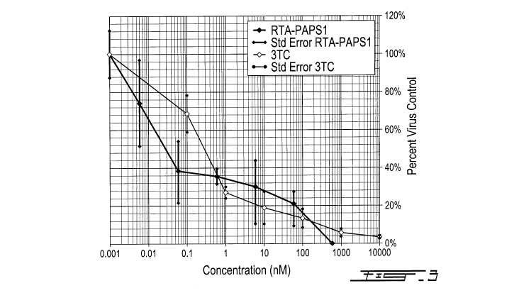
[0031] Fig. 3 is an anti-HBV evaluation of RTA-PAPS1, wherein
recombinant RTA-
PAPS1 was tested for its anti-HBV activity using 6 concentrations using a
serial dilution
by a factor of 10 in growth media (600nM, 60nM, 6nM, 0.6nM, 0.06nM, 0.006nM
for
CA 03091708 2020-08-19
WO 2019/204902
PCT/CA2019/050388
RTA-PAPS1 and 10000nM, 1000nM, 100nM, 10nM, 1nM, 0.1nM for 3TC), and values
are calculated as percent of virus DNA control [(amount of virus DNA in
treated
sample/amount of virus DNA in untreated sample) x100], results representing
the mean
for three individual experiments (Std Error = Std Deviation / (SQRT(n)), with
n=3).
[0032] Fig. 4 illustrates the predicted 3D Protein Structure, showing
in (A) protein
structure as determined by Phyre2 with the arrows showing the flexible linker
at
position 275-294 and the CASP2 recognition site at position 280-284; and in
(B) the
ligand binding sites of RTAM moiety (up) and of PAP1 moiety (down) as
determined by
I-Tasser (using the Phyre2 model as one of the templates).
[0033] Fig. 5 illustrates the production and purification of native
RTAM-PAP1,
showing in (A) loosely bound proteins were washed with the lysis buffer
containing
50mM imidazole (150) on a Ni-sepharose column and RTAM-PAP1 (RPAP1) proteins
were then eluted with the elution buffer containing 300mM lmidazole (1300); in
(B) the
Western Blot using ricin a chain antibody RA999 confirmed the presence of RTAM-
PAPS1 at approx. 61.5kDa, wherien the bands between 21kDa and 32kDa are
assumed to be degraded or/and premature RTAM-PAP1 proteins; in (C) (Lys) from
1L
culture; in (D) co-purified host cell proteins were further separated by a
hydroxylapatite
column, wherein most RTAM-PAP1 proteins were retained in the flow through (FT)
fraction, while most host cell proteins were bound to the hydroxylapatite
column (P200
elution); in (E) RTAM-PAP1 was peaked at fraction 15 and 16, the purest
fraction (F15)
was estimated at >95% homogeneity; and in (F) the inhibition assay.
[0034] Fig. 6 illustrates comparative inhibition activity of RTAM-PAP1
and RTA-
PAPS1 in the TnT transcription/translation assay; wherein five different
concentration
points (0.01M, 0.02nM, 0.03nM, 0.08nM, 0.25nM for RTA-PAPS1 and 0.02nM,
0.03nM, 0.06nM, 0.16nM, 0.40nM for RTAM-PAP1) were examined, values are
calculated as percent Luciferase protein synthesis compared to control, and
results
representinmg the mean for two individual experiments and the curves are the
logarithmic regression for RTA-PAPS1 and power regression for RTAM-PAP1 ((Std
Error = Std Deviation / (SQRT(n)), with n=2).
DETAILED DESCRIPTION
[0035] It is provided an anti-viral fusion protein of ricin A chain
protein (RTA) and
the Pokeweed antiviral proteins (PAPs).
CA 03091708 2020-08-19
WO 2019/204902
PCT/CA2019/050388
6
[0036] Ricin A chain (RTA) and Pokeweed antiviral proteins (PAPs) are
plant-
derived N-glycosidase ribosomal-inactivating proteins (RIPs) isolated from
Ricinus
communis and Phytolacca Americana respectively. It is provided herein the
amenability
and sub-toxic antiviral value of a novel fusion protein between RTA and PAPs
(RTA-
PAPs). RTA-Pokeweed antiviral protein isoform 1 from seeds (RTA-PAPS1;
previously
described in W02017/175060, the content of which is incorporated herein in its
entirety)
was produced in an E. coil in vivo expression system, purified from inclusion
bodies
using gel filtration chromatography and protein synthesis inhibitory activity
assayed by
comparison to the production of a control protein Luciferase. The antiviral
activity of the
RTA-PAPS1 against Hepatitis B virus (HBV) in HepAD38 cells was then determined
using a dose response assay by quantifying supernatant HBV DNA compared to
control virus infected HepAD38 cells. The cytotoxicity in HepAD38 cells was
determined by measuring cell viability using a tetrazolium dye uptake assay.
The fusion
protein was further optimized using in silico tools, produced in an E. coil in
vivo
expression system, purified by a three-step process from soluble lysate and
confirmed
in a protein synthesis inhibition activity assay.
[0037] Fusion and hybrid proteins of RTA and PAPs have also been
developed in
pursuit of selectively targeting infected cells and selectively recognizing
viral
components, though with limited success (Rothan et al., 2014, Antiviral Res.,
108, 173-
180; Chaddock et al., 1996, Eur J Biochem., 235: 159-166).
[0038] Based on the data gathered on these two proteins over the last
thirty years
and the newly available in silico tools, it is described herein the creation
of a novel
fusion protein between RTA and PAPs that is more effective than either of the
proteins
alone at sub-toxic dosages against specific infectious diseases and that is
cheaper to
produce than available therapeutics.
[0039] It is provided herein an effective and scalable production
system in
Escherichia coli and of purification methods that enabled accurate
determination of
RTA-PAPs protein synthesis inhibition in vitro. The in vitro reduced
cytotoxicity and
significant anti-HBV activity of RTA-Pokeweed antiviral protein isoform 1 from
seeds
(RTA-PAPS1) is described by detecting HBV DNA in the supernatant of HepAD38
cells.
The reengineering of RTA-PAPS1 into RTA mutant-Pokeweed antiviral protein
isoform
1 from leaves (RTAM-PAP1) to improve its production in Escherichia coil and to
enhance its gain of function is also described using the most up-to-date
protein
structure and function prediction software available online.
CA 03091708 2020-08-19
WO 2019/204902
PCT/CA2019/050388
7
[0040] As used herein, the term "RIP" refers to ribosome inactivating
proteins. As
used herein, the terms "PAP" or "pokeweed antiviral protein" refer to a
polypeptide with
substantial or complete sequence homology to pokeweed antiviral protein or a
polynucleotide encoding such a polypeptide, which may or may not include a
signal
peptide as evident by the context in which the term is used (for example,
GenBank
Entry Accession No. KT630652). When no variant is specified, PAP may refer to
the
unmodified polypeptide or polynucleotide or to a variant of PAP. As used
herein, the
terms "RTA" or "ricin A-chain" refer to a polypeptide or a polynucleotide
encoding a
polypeptide with substantial or complete sequence homology to ricin A-chain
GenBank
Entry Accession No. X52908.
[0041] It is demonstrated that RTA-PAPS1 could effectively be recovered
and
purified from inclusion bodies. The refolded protein was bioactive with a 50%
protein
synthesis inhibitory concentration (1050) of 0.06nM (3.63ng/m1). RTA-PAPS1 has
a
synergetic activity against HBV with a half-maximal response concentration
value (ECK)
of 0.03nM (1.82ng/m1) and a therapeutic index of >21818 with noticeable steric
hindrance. The optimized protein ricin A chain mutant-Pokeweed antiviral
protein
isoform 1 from leaves (RTAM-PAP1) can be recovered and purified from soluble
lysates with gain of function on protein synthesis inhibition activity, with
an 1050 of
0.03nM (1.82ng/m1), and with minimal, if any, steric hindrance.
[0042] RTA-PAPS1 is a monomeric polypeptide of 541 amino acids with an
apparent molecular mass of 60.5k0a, with the following amino acid sequence:
MIF PKQY P I INFTTAGATVQS YTNF IRAVRGRLT T GADVRHE I PVL PNRVGL PINQRFILVEL S
NHAE L SVTLAL DVTNAYVVGYRAGNSAYF FH PDNQEDAEAITHL FT DVQNRYT FAFGGNYDRL E
QLAGNLRENIELGNGPLEEAISALYYYSTGGTQL PTLARS El IC IQMI S EAARFQYIEGEMRT R
IRYNRRSAPDPSVITLENSWGRL STAIQESNQGAFAS P IQLQRRNGSKF SVYDVS IL I P I IALM
VYRCAP P PS SQFSLL IRPVVPNFNINT IT FDAGNAT INKYAT FME S LRNEAKDP S LKC YGI PML
PNTNST IKYLLVKL QGAS LKT IT LML RRNNL YVMGYS D PYDNKCRYHI FND IKGT EYS DVENT L
C PS SNPRVAKP INYNGL Y PT LEKKAGVT S RNQVQL GIQ IL SSDIGKISGQGS FT EKIEAKFL LV
AIQMVSEAARFKYIENQVKTNENRDFS PNDKVLDLEENWGKISTAIHNSKNGAL PKPL EL KNAD
GTKWIVLRVDE IKPDVGLLNYVNGTCQAT (SEQ ID NO: 1).
[0043] RTA-PAPs are amenable to effective production and purification
in native
form, possess significant gain of function on protein synthesis inhibition and
anti-HBV
activities in vitro with a high therapeutic index and, thus, is a potent
antiviral agent
CA 03091708 2020-08-19
WO 2019/204902
PCT/CA2019/050388
8
against chronic HBV infection to be used as a standalone or in combination
with
existent therapies.
[0044] The production of fusion Ricin A Chain-Pokeweed Antiviral
Protein from
Seeds lsoform 1 (RTA-PAPS1) in E. coil was found to be significantly better at
30 C
than at 37 C. In order to optimize the amount of protein produced from 1L at
30 C,
three media were tested: M9 (M9), Luria Bertani (LB) and terrific broth (TB).
Soluble
lysate (Sol) and inclusion body (IB) from each sample were analyzed by SOS
PAGE
and visualized by Coomassie blue staining (Fig. 1A). As can be seen, almost
all of the
overexpressed RTA-PAPS1 proteins were in the form of inclusion bodies, which
were
almost completely insoluble in either 6M Urea or 6M Guanidine. A total of 28
proprietary buffers were tested and only the denaturing buffer 8b (proprietary
formulation of AscentGene) was able to dissolve more than 50% of the Ricin-
PAPS1
present in the inclusion bodies. Once the soluble proteins were recovered and
purified
through the gel filtration column 5uperdex200 (single step) in their denatured
form,
they were allowed to refold for over 20hrs in a refolding buffer before being
concentrated. The resulting protein was found to be at a concentration of
0.22mg/m1 at
>90% purity (Fig. 1B).
[0045] The inhibitory activity of RTA-PAPS1 was determined using 5
different
concentrations of purified RTA-PAPS1 in duplicate with the Rabbit Reticulate
Lysate
TnT system using Luciferase as control. A Luciferase assay was used to
determine
Luciferase expression levels using a luminometer. The resulting plot is shown
in Fig. 2
while taking the standard deviation into account. As can be observed, the
difference
between the duplicate results is very minimal. The standard deviation varied
from
0.10% to 5% leading to very small standard errors. It can further be observed
that RTA-
PAPS1 has an IC50 at 0.06nM, slower than RTA IC50 at 0.03nM but comparable to
PAPS IC50 at 0.07. The IC100 however is attained faster than any of them at
0.24nM for
RTA-PAPS1, twice as fast as RTA IC100 at 0.60nM. These results show that RTA-
PAPS1 is bioactive with a synergetic activity between the RTA and PAPS1
moieties
being noticeable.
[0046] Recombinant RTA-PAPS1 was evaluated for anti-HBV activity and
cytotoxicity in the HBV chronically infected cell line A038 using a six
concentrations
dose response assay in triplicate. The lamivudine (3TC) control compound was
evaluated in parallel. The antiviral efficacy based on quantified DNA copies
in the
supernatant of both compounds are shown in Fig. 3 in a plot form. RTA-PAPS1
yielded
CA 03091708 2020-08-19
WO 2019/204902
PCT/CA2019/050388
9
a half-maximal response concentration value (ECK) of 0.03nM while 3TC yielded
an
E050 of 0.3nM, which is a ten-fold difference. RTA-PAPS1 was not cytotoxic to
HepAD38 cells at concentrations up to 600nM. These results led to a
therapeutic index
for RTA-PAPS1 of >21818, which is a huge improvement over values given in the
literature (ECK of 330nM and a therapeutic index of 9.3 for PAPS1 alone under
comparable conditions on HepG2 2.2.15 cells) (He et al., 2008, World J
Gastroenterol,
14: 1592-1597). These results clearly show the significant anti-HBV activity
of RTA-
PAPS1.
[0047] RTA-PAPS1 was found to be very effective against Hepatitis B
Virus and
also effective on HIV1, Zika and Hepatitis C Virus as shown. In anti-viral
cytoprotection
assay, as provided in Tables 1-4, RTA-PAPS1 showed high Therapeutic Index (TI)
for
HBV, which is preferable for a drug to have a favorable safety and efficacy
profile, and
high efficacy for HIV1, Zika and HCV..
Table 1
Anti-HIV1 cytoprotection assay
Compound CEM-SS/HIVRF
EC50 (PM) TC50 (PM) TI
RTA-PAPS1 0.19 >0.6 >3.16
AZT 0.0008 >1 >1250
Table 2
Anti-Zika cytoprotection assay
Compound HUH7-ZikaPRVABC59
EC50 (PM) TC50 (PM) TI
RTA-PAPS1 0.05 0.06 1.2
Sofosbuvir 2.09 >10 >4.78
CA 03091708 2020-08-19
WO 2019/204902
PCT/CA2019/050388
Table 3
Anti-HCV cytoprotection assay
Compound HCV Replicon
EC50 (PM) TC50 (PM) TI
RTA-PAPS1 0.012 0.04 3.42
Sofosbuvir 0.05 >1 >18.5
Table 4
Anti-HBV cytoprotection assay
Compound HBV AD38
EC50 (PM) TC50 (PM) TI
RTA-PAPS1 0.00003 >0.6 >21818
3TC 0.0003 >10 >35714
[0048] The design of the recombinant protein RTA-PAPS1 was completely
revisited
in order to further enhance the effect of the chimeric protein on HBV, reduce
general
toxicity and increase solubility to improve expression. The resulting design
Ricin A
Chain Mutant-Pokeweed Antiviral Protein from Leaves (RTAM-PAP1) was run
through
I-Tasser and Phyre2 and the resulting 3D models validated by Verify 30. The
model
generated by Phyre2 passed Verify 30 while the one generated by I-Tasser
failed. The
one generated by Phyre2 was thus chosen as one of the templates to run I-
Tasser
again. The newly generated structure by I-Tasser scored higher on Verify 30
than the
one generated by Phyre2 and was thus chosen as the model for the other
software.
The proper disulfide bond formations were confirmed by the DiANNA 1.1
webserver (at
positions 328-553 and 379-400). The new model had a normalized QMEAN4 score of
>0.6 and the introduction of the rigid CASP2 recognition site into the
flexible linker at
position 280-285 insured safe distance between the two proteins to safeguard
the
function of both moieties and minimize steric hindrance as can be seen in Fig.
4. The
grand average of hydropathicity was reduced from -0.236 for RTA-PAPS1 to -
0.265 for
RTAM-PAP1 as was determined by ProtParam, which represents an improvement of
12% in hydrophilicity.
CA 03091708 2020-08-19
WO 2019/204902
PCT/CA2019/050388
11
[0049] The anti-viral fusion protein RTAM-PAPS1 described herein
comprises the
following sequence:
[0050] MHHHHHH I F PKQY P I INFT TAGATVQS YTNF IRAVRGRL TT GADVRHE I
PVL PNRV
GL PINQRFILVELSNHAELSVTLALDVTNAYVVGYRAGNSAYFFHPDNQEDAEAITHL FT DVQN
RYT FAFGGNYDRLEQLAGNL RENIEL GNGPL EEAI SAL YYYS T GGT QL PTLARS F I IAIQMI S
E
AARFQYIEGEMRTRIRYNRRSAPDPSVITLENSWGRL STAIQESNQGAFAS P IQL QRRNGSKF S
VYDVS IL I P I IALMVYRAAP P P S SQFGGGGSDVADIGGGGSGGGGSVNT I IYNVGS T T IS
KYAT
FLNDLRNEAKDPSLKCYGIPML PNTNTNPKYVLVELQGSNKKT IT LML RRNNLYVMGYSD PFE T
NKCRYHI FNDI S GT ERQDVE TT L C PNANS RVS KNINFDSRYPT LE S KAGVKS RS QVQL
GIQIL D
SNIGKISGVMS FTEKTEAEFLLVAIQMVS EAARFKYIENQVKTNFNRAFNPNPKVLNL QE TWGK
I S TAIHDAKNGVL PKPLELVDASGAKWIVLRVDE IKPDVALLNYVGGSCQTT (SEQ ID NO:
2),
wherein amino Acids: 1 Vector Starting Residue;
amino Acids: 2-7 6-H is Tag;
amino Acids: 8-274 Ricin A Chain (RTA);
amino Acids: 275-295 Flexible Linker + Casp2 Site; and
amino Acids: 296-556 Pokeweed Protein (PAP1).
[0051] Accordingly, it is provided a fusion protein comprising the
structure X-Y-Z,
wherein X is the full length RTA or a variant thereof, Y is absent or a linker
and Z is the
full length PAP or a variant thereof. In an embodiment, X is RTA mutant
(RTAM). In
another embodiment, Z is the Pokeweed Antiviral Protein from Leaves (PAP1) as
described herein.
[0052] The linker encompassed herein can be a chemical linker and/or a
polylinker.
Preferably, the linker is a flexible linker, i.e. composed of flexible
residues like glycine
and serine so that the adjacent protein domains are free to move relative to
one
another . A "chemical linker as used herein is defined as a flexible linker,
within some
embodiments, the linker is a heterobifunctional linker, in some embodiments,
the linker
comprises a maleimido group. In various embodiments, the linker is selected
from the
group consisting of: GMBS; EMCS; SMPH; SPDP; and LC-SPDP.
CA 03091708 2020-08-19
WO 2019/204902
PCT/CA2019/050388
12
[0053] The term "polylinker" or "linker peptide" as used herein is
defined as a short
segment of DNA added between the DNA encoding the fused proteins, to produce a
short peptide or polypeptide to make it more likely that the proteins fold
independently
and behave as expected. This "polylinker" or "linker peptide" can also have
cleavage
sites for proteases or chemical agents that enable the liberation of the two
separate
proteins.
[0054] The production of RTAM-PAP1 was first tested under the same
conditions
as previously determined for RTA-PAPS1 and resulted in good production of
native
proteins. Soluble RTAM-PAP1 was recovered from the lysate, purified by Ni-
sepharose
column and analyzed by SDS-PAGE and Western Blot (Figs. 5A and B). The
production from 1L culture under the same conditions gave equally good results
(Fig.
50). The purified proteins were then submitted to a second purification step
using
hydroxylapatite column, which showed good separation of RTAM-PAP1 from co-
purified host proteins (Fig. 5D). The degraded (and/or premature) products
were further
separated by gel filtration on an FPLC column of Superose 12 (Fig. 5E) and the
purest
fraction (F15) reached >95% homogeneity at a concentration of 0.1mg/m1 (Fig.
5F) and
was used for the protein synthesis inhibition assay.
[0055] The inhibitory activity of RTAM-PAP1 was determined using 5
different
concentrations, in duplicate, of purified RTAM-PAP1 on the Rabbit Reticulate
Lysate
TnT system using Luciferase as the control as previously described. The
resulting
comparative plot of the activity on protein synthesis of both fusion proteins
is shown in
Fig. 6 while taking into account the standard deviations that ranged from 0.1%
to 1%.
As can be observed, the plot showed minimal difference between duplicates. It
also
shows that RTAM-PAP1 has an 1050 at 0.03nM, the same as RTA 1050 at 0.03nM,
which
is twice as fast as RTA-PAPS1 1050 at 0.06nM and about ten times faster than
PAP1
1050 at 0.29nM (Poyet et al., 1997, FEBS Lett., 406: 97-100). The 10100
however is
attained faster than any of them at 0.09nM for RTAM-PAP1, which is a bit less
than
three times faster than RTA-PAPS1 10100 at 0.24nM. These results show that
RTAM-
PAP1 is bioactive, both moieties' complementary catalytic activities
functional, with
minimal steric hindrance if any, and with a significant gain of function.
[0056] The chimeric protein RTA-PAPS1 was expressed only in inclusion
bodies
with very little solubility, except under heavy denaturing conditions. The
refolding
process was successful as more than one conformation was observed. This was
probably due to the two free Cysteine residues in RTA and to the nature of the
semi-
CA 03091708 2020-08-19
WO 2019/204902
PCT/CA2019/050388
1 3
flexible linker, which allowed the close proximity of Cys at position 260 to
the Cys
residues at position 364 and 385 (confirmed by DiANNA 1.1 webserver and I-
Tasser).
The addition of TCEP was necessary and a difference in bioactivity (>2 fold)
was
observed between samples. RTA-PAPS1 with the addition of TCEP was very
bioactive
and with a noticeable synergetic activity between RTA and PAPS1, which was
probably
limited by steric hindrance once again due to the nature of the semi-flexible
quality of
the linker. This was confirmed during the anti-HBV assays. The significant
anti-HBV
activity of RTA-PAPS1 was apparent and due to the ability of both moieties to
depurinate rRNA but also polynucleotide, single-stranded DNA, double stranded
DNA
and mRNA. HBV is a double stranded DNA reverse transcriptase virus.
[0057] The fusion protein RTAM-PAP1 expression went very well as native
protein
production with high solubility was obtained (barely any in inclusions
bodies). A three
step purification protocol was in order to obtain soluble proteins with >90%
homogeneity. Nonetheless, 0.1mg of protein at >95% purity and 0.22mg of
protein at
>90% purity were obtained from 1L of culture. This yield is probably explained
by the
increased toxicity of PAP1 to E. coil compared to that of PAPS1 (>10 fold).
The
bioactivity of RTAM-PAP1 was increased, much more than expected with very
little to
no sign of steric hindrance. The introduction of the two point mutations in an
embodiment in the RTA moiety and of the flexible linker further made a
difference in
solubility and activity. Also, perhaps, fine-tuning the formulation buffer to
better
preserve protein integrity allowed for optimum activity. The synergetic effect
of both
moieties was very apparent and due to the fact that RTA and PAP1 do not dock
onto
the ribosome at the same site and, thus, led to a reduction of partially
depurinated and
still functional ribosomes.
[0058] The chimeric proteins combining RTA and PAPs are potent novel
broad
range anti-viral proteins with gain of function in protein synthesis
inhibition activity and
anti-HBV activity in vitro with minimal cytotoxicity. As encompassed herein,
the anti-
viral proteins described herein have a broader anti-viral activity against
plant, animal
and human pathogens, including as trait in transgenic plants expressing it, as
a stand-
alone administration (therapeutics). In an embodiment, the broad range anti-
viral
proteins described herein are effective, for example and not limited to,
against Group IV
viruses (ssRNA viruses), Group V viruses (ssRNA viruses) and/or Group VI
viruses (or
ssRNA-RT viruses). The introduction of two point mutations in RTA and of a
flexible
linker further greatly improved solubility and activity. RTAM-PAP1 can be
overexpressed, recovered and purified from soluble lysate. It is expected that
the anti-
CA 03091708 2020-08-19
WO 2019/204902
PCT/CA2019/050388
14
viral properties of RTAM-PAP1 will be even greater than that of either RTA-
PAPS1 or
PAPs with even lesser general toxicity. It is further encompassed that the
fusion protein
encompassed herein will be effective against cancer and particularly cancer
caused by
viruses such as the papillomavirus. For example, HBV and HCV infection can
cause
liver cancer; the Kaposi Sarcoma-Associated Herpesvirus (KSHV) causing Kaposi
sarcoma; Merkel Cell Polyomavirus (MCV) causing skin cancer or Merkel cell
carcinoma;
Human T-Cell Lymphotropic Virus Type 1 (HTLV-1) causing leukemia and lymphoma;
Epstein-Barr Virus (EBV), causing Burkitt's lymphoma, Nasopharyngeal carcinoma
(cancer of the upper throat), Hodgkin's and non-Hodgkin's lymphoma, T-cell
lymphomas,
Post-transplant lymphoproliferative disorder, or Leiomyosarcoma. In another
embodiment, it is encompass that the fusion protein encompassed herein will be
effective against a viral infection caused by the Japanese encephalitis virus,
Herpes
Simplex, Influenza virus, and/or Poliovirus.
EXAMPLE I
E. coil in vivo expression system and Rabbit Reticulate Lysate protein
synthesis
inhibition
[0059] The two cDNA sequences coding for RTA-PAPS1 (541 amino acids)
and for
RTAM-PAP1 (556 amino acids including the N terminal 6-His tag) were optimized
for E.
coil expression and chemically synthesized by AscentGene.
[0060] The cDNA coding for RTA-PAPS1 and RTAM-PAP1 sequences described
above were generated by PCR using the primers RP1-A48
(5'TTTAACTTTAAGAAGGAGATATACATATGATCTTCCCGAAACAGTACC; SEQ ID
NO: 3) or RPAP1-A48 (5'TTTAACTTTAAGAAGGAGATATACATATGCACCA
CCATCACCACCATA; SEQ ID NO: 4) and RPAP1-B50 (5'CAGCCGGATC
TCAGTGGTGGTGCTCGAGTTAGGTAGTCTGGCAAGAACCG; SEQ ID NO: 5). Each
PCR fragment was then subcloned into the E. coil pET30a expression vector
(Novagene) between the Ndel and Xhol restriction endonuclease sites to
generate the
pET30a-RP1 and pET30a-6H-RPAP1 vectors respectively. The inserts were
validated
by DNA sequencing.
[0061] The above described vectors were transformed into E. coil
BL21(DE3) cells
(NEB) and expression of the proteins were examined from individual clones and
analyzed by either Western blot using a monoclonal antibody specific to ricin
A chain
(ThermoFisher, RA999) or SOS gel stained with Comassie blue (ThermoFisher).
CA 03091708 2020-08-19
WO 2019/204902
PCT/CA2019/050388
1 5
Optimal conditions were determined and protein production induced in the
presence of
1mM IPTG from 1L culture for each protein. The bacteria were then harvested by
centrifugation, followed by lysing the cell pellets with 50m1 of lysis buffer
(50mM Tris-C1,
150mM NaCI, 0.2% Triton X100 and 0.5mM EDTA). After sonication (3x2min), the
soluble lysates were recovered by centrifugation at 35K rpm for 40min. The
insoluble
pellets were further extracted with 40m1 of 6M Urea and the inclusion bodies
(IB) were
recovered by centrifugation at 16K rpm for 20min. Clarified IB were then
dissolved with
20m1 of buffer 8b (proprietary formulation of AscentGene). The soluble
proteins were
then recovered by centrifugation (please contact the authors for more
details).
[0062] Ricin-PAPS1 proteins were purified by gel filtration column
(Superdex 200
from GE Healthcare) under denaturing condition (6M Urea). Peak fractions were
pooled and powder Guanidine was added to a concentration of 5M for complete
denaturing. Denatured Ricin-PAPS1 was then added dropwise to the refolding
buffer
(50mM Tris-C1, pH8.1, 0.4M L-Arginine, 0.5mM oxidized glutathione and 5mM
reduced
glutathione) for refolding. The solution was stirred at room temperature for
10min
before allowing the refolding reaction to be further carried out at 4 C for
>20hrs.
Clarified and refolded Ricin-PAPS1 proteins were then concentrated before
going
through the endotoxin removal process and the ammonium sulfate precipitation
step.
The resulting mixture was dialyzed in the formulation buffer containing 20mM
HEPES-
Na, pH7.9, 20% glycerol, 100mM NaCI, 2.5mM tris(2-carboxyethyl)phosphine
(TCEP)
and 1mM EDTA.
[0063] The purification of the native RTAM-PAP1 from soluble lysate was
achieved
by affinity versus His-tag on Ni-sepharose column (GE Healthcare). After
extensive
washes with the lysis buffer, loosely bound proteins were eluted with the
lysis buffer
containing 40mM lmidazole (140). RTAM-PAP1 proteins were eluted with the
elution
buffer (20mM Tris-C1, pH7.9, 100mM NaCI, 1mM EDTA and 300mM lmidazole). A
second purification step using Hydroxylapatite column (GE Healthcare) was used
to
further separate RTAM-PAP1 from co-purified host proteins. A third
purification step, gel
filtration on a fast protein liquid chromatography (FPLC) column of Superose
12 (GE
Healthcare), was necessary to completely get rid of degraded and/or premature
protein
products. The resulting mixture was dialyzed in the formulation buffer
containing 20mM
HEPES-Na, pH7.9, 200mM NaCI, 0.2mM CaCl2 and 0.5mM EDTA.
[0064] The inhibitory activities of RTA-PAPS1 and RTAM-PAP1 were tested
by
using the Rabbit Reticulate Lysate TnT Quick Coupled
Transcription/Translation
CA 03091708 2020-08-19
WO 2019/204902
PCT/CA2019/050388
16
System and the Luciferase Assay System (Promega). Briefly, each
transcription/translation reaction was performed according to the instructions
for use
(IFU) in the presence of a T7 Luciferase reporter DNA, and the Luciferase
expression
level was determined with a Wallac Microplate Reader.
Transcription/translation runs
were done twice with and without addition of five different concentrations of
RTA-
PAPS1 and RTAM-PAP1 in order to determine the inhibitory effect of the
proteins. RTA-
PAPS1 and RTAM-PAP1 concentrations were adjusted by taking sample purity into
consideration.
EXAMPLE II
Anti-HBV Assay
[0065] The anti-HBV assay was performed as previously described (Min et
al.,
2017, Journal of Medicinal Chemistry, 60: 6220-6238) with the modification of
using
HepAD38 cells by ImQuest BioSciences. ImQuest BioSciences developed a multi-
marker screening assay utilizing the HepAD38 cells to detect proteins, RNA,
and DNA
intermediates characteristic of HBV replication. The HepAD38 cells are derived
from
HepG2 stably transfected with a single cDNA copy of hepatitis B virus
pregenomic
RNA, in which HBV replication is regulated by tetracycline. Briefly, HepAD38
cells were
plated in 96-well flat bottom plates at 1.5 x 10⁴ cells/well in Dulbecco's
modified Eagle's
medium supplemented with 2% FBS, 380 pg/mL G418, 2.0 mM L-glutamine, 100
units/mL penicillin, 100 pg/mL streptomycin, and 0.1 mM nonessential amino
acids
(ThermoFisher). After 24h, six tenfold serial dilutions of RTA-PAPS1 prepared
in the
same medium were added in triplicate. Lamivudine (3TC from Sigma Aldrich) was
used
as the positive control, while media alone was added to cells as a negative
control
(virus control, VC). Three days later, the culture medium was replaced with
fresh
medium containing the appropriately diluted RTA-PAPS1. Six days following the
initial
administration of RTA-PAPS1, the cell culture supernatant was collected,
diluted in
qPCR dilution buffer, and then used in a real-time quantitative qPCR assay
using a Bio-
Rad CFX384 Touch Real-Time PCR Detection System. The HBV DNA copy number in
each sample was interpolated from the standard curve by the supporting
software. A
tetrazolium dye uptake assay (ThermoFisher) was then employed to measure cell
viability, which was used to calculate cytotoxic concentration (TOO.
CA 03091708 2020-08-19
WO 2019/204902 17
PCT/CA2019/050388
EXAMPLE III
Protein Design Optimization
[0066] The molecular profile of the protein was determined using the
Protparam
tool of ExPASy, and the solubility of these proteins was determined using
Predict
Protein. The presence of disulfide bonds was determined using the DiANNA 1.1
webserver. Functional effects of point mutations were determined using SNAP2
of
Predict Protein.
[0067] The structure of the protein was predicted by fold recognition
methodology
using the I-TASSER and Phyre2 prediction servers. The determined protein
structures
were then validated by Verify 30. The quality of the structure was determined
using the
QMEAN6 program of the SWISS-MODEL workspace.
[0068] Three major changes were made to RTA-PAPS1 in order to increase
its
solubility, its efficacy against infected cells and to further reduce its
toxicity.
[0069] Firstly, two point mutations, as predicted by SNAP2 of Predict
Protein to
have the least effect on function, were introduced into the RTA moiety to
replace the
Cysteine (Cys) residues with Alanine residues in order to completely avoid
unwanted
disulfide bond formation at position 171 and 259 (C171A and 0259A) to create
RTA
mutant (RTAM).
[0070] Secondly, the natural semi-flexible linker previously used was
replaced with
a newly designed soluble flexible G rich linker with a rigid CASP2 recognition
site
(GGGGSDVADI(GGGGS)2) to allow better autonomous function of each moiety with
minimal steric hindrance and to further enhance the chimeric protein's ability
to induce
cell apoptosis.
[0071] Thirdly, a different variant than PAPS1 was used, PAP1,
retrieved from
National Centre for Biotechnology Information database (NCBI) with access
number
P10297.2 (SEQ ID NO: 6) in order to further enhance activity against HBV and
further
reduce toxicity of the chimeric protein.
[0072] Lastly, a 6-His tag was added at the N terminal of the protein
RTAM-PAP1 in
order to minimize effect on structure and function and to increase native
protein
recovery from E. coil production.
CA 03091708 2020-08-19
WO 2019/204902
PCT/CA2019/050388
1 8
[0073] While the present disclosure has been described in connection
with specific
embodiments thereof, it will be understood that it is capable of further
modifications and
this application is intended to cover any variations, uses, or adaptations,
including such
departures from the present disclosure as come within known or customary
practice
within the art and as may be applied to the essential features hereinbefore
set forth,
and as follows in the scope of the appended claims.