Note: Descriptions are shown in the official language in which they were submitted.
CA 03091956 2020-08-20
WO 2019/173468 PCT/US2019/020938
SYSTEMS, DEVICES AND METHODS FOR NEUROSTIMULATION
CROSS-REFERENCE TO RELATED APPLICATION
[0001] The present application claims priority to U.S. Provisional Application
No.
62/639,738, filed on March 7, 2018 in the United States Patent and Trademark
Office, and
entitled "SYSTEMS, DEVICES AND METHODS FOR NEUROSTIMULATION," the entire
contents of which are hereby incorporated by reference for all purposes.
TECHNICAL FIELD
[0002] The present disclosure relates to neurostimulation, and in particular,
to
neurostimulation systems, devices, and methods.
BACKGROUND
[0003] Neurostimulation is the therapeutic and/or diagnostic activation of one
or more
parts of the nervous system. The nervous system may be electrically stimulated
through invasive
means, such as implantable electrodes, or though less invasive means, such as
electrodes
attached to the skin. Non-electrical forms of neurostimulation may employ
electromagnetic
waves, light, sound, or temperature to stimulate the nervous system.
Neurostimulation has been
used for the purpose of medical treatment and/or diagnosis of various
disorders.
[0004] Vestibular stimulation is a form of neurostimulation that stimulates
the vestibular
branch of the vestibulocochlear nerve, the eighth cranial nerve. As used
herein, "vestibular
nerve" shall refer to the vestibular branch of the eighth cranial nerve. The
vestibular nerve may
be stimulated electrically, termed Galvanic Vestibular Stimulation ("GVS"), or
may be
stimulated using temperature, termed Caloric Vestibular Stimulation, or both.
SUMMARY
[0005] The present disclosure and aspects thereof provide systems, devices,
and methods
for neurostimulation that include vestibular stimulation. For example, one
general aspect
provides devices for administering thermal stimulation to an ear canal of a
subject. Such devices
may include: an earpiece configured to be at least partially insertable into
the ear canal of the
subject. The devices may also include a thermoelectric device thermally
coupled to the earpiece
and configured to heat and/or cool the earpiece to thereby heat and/or cool
the ear canal of the
subject. The devices may also include a controller associated with the
thennoelectric device, and
the controller may be configured to administer a selected treatment plan
including administering
1
CA 03091956 2020-08-20
WO 2019/173468 PCT/US2019/020938
a caloric vestibular stimulation (CVS) stimulus to the ear canal of the
subject in a condition-
treatment effective amount during a first treatment interval, with the
treatment plan effective to
produce a durable improvement in at least one symptom of the condition for a
time of at least 1
week following cessation of the administering. Other embodiments of this
aspect include
corresponding methods, systems (including computer systems), apparatuses, and
computer
programs recorded on one or more computer storage devices that control,
facilitate, and/or
supplement operation of such devices.
[0006] Another general aspect of the present disclosure provides one or more
methods of
treating a subject afflicted with a condition (e.g., a neurological disorder).
Such methods may
include: (a) administering, using a controlled vestibular stimulation device,
vestibular stimulation
to at least one ear of the subject in a condition-treatment effective amount
during a first treatment
interval. The vestibular stimulation may be effective to produce a durable
improvement in at
least one symptom of the condition for a time of at least 1 week following
cessation of the
administering. Various embodiments of this aspect include systems (including
computer
systems), apparatuses, and computer programs recorded on one or more computer
storage
devices, each configured to perform and/or cause performance of the operations
of the methods.
[0007] Another general aspect of the present disclosure provides one or more
methods.
Such methods may include: (a) administering a first vestibular stimulation
stimulus to a subject
afflicted with a neurological disorder and determining a time to entrainment
of at least one
physiological oscillatory pattern to the stimulus in the subject. The methods
may also include (b)
ceasing administering of the vestibular stimulation and determining a time to
relaxation of the at
least one physiological oscillatory pattern from the entrainment subsequent to
the ceasing,
Various embodiments of this aspect include systems (including computer
systems), apparatuses,
and computer programs recorded on one or more computer storage devices, each
configured to
perform and/or cause performance of the operations of the methods.
[0008] Another general aspect of the present disclosure provides one or more
methods
for selecting a treatment for a subject afflicted with a neurological
disorder, Such methods may
include: (a) sequentially administering a plurality of different vestibular
stimulation stimuli to the
subject; (b) determining a time to entrainment and/or a time to relaxation of
at least one
physiological oscillatory pattern to each of the stimuli in the subject; (c)
selecting, from among
the plurality of different vestibular stimulation stimuli, a vestibular
stimulation stimulus for
administering to the subject based on the detected time to entrainment and/or
time to relaxation
of each vestibular stimulation stimulus of the plurality of different
vestibular stimulation stimuli;
2
CA 03091956 2020-08-20
WO 2019/173468 PCT/US2019/020938
and (d) administering the selected vestibular stimulation stimulus to the
subject at least once.
Various embodiments of this aspect include systems (including computer
systems), apparatuses,
and computer programs recorded on one or more computer storage devices, each
configured to
perform and/or cause performance of the operations of the methods.
[0009] Another general aspect of the present disclosure provides methods. Such
methods
may include: (a') detecting a physiological oscillatory pattern in a subject
during and/or after
treatment(s) including administration of a vestibular stimulation stimulus;
(b') optionally
resetting the oscillatory pattern by administering an exogenous stimulus
(e.g., transcranial
magnetic stimulation) to the subject; and (c') repeating steps (a') through
(b') for a plurality of
different vestibular stimulation treatments to generate a database of
vestibular stimulation
treatment(s) correlated with different oscillatory patterns in a brain of the
subject. The methods
may also include (d') assigning efficacy scores to each different vestibular
stimulation treatment
in the database based on a durability of improvement of neurovascular coupling
in the subject;
(e') selecting from the database a vestibular stimulation treatment that
provides a durable
improvement in neurovascular coupling to the subject; and (f) administering
the selected
vestibular stimulation treatment to the subject in a subsequent treatment or
treatment session.
Various embodiments of this aspect include systems (including computer
systems), apparatuses,
and computer programs recorded on one or more computer storage devices, each
configured to
perform and/or cause performance of the operations of the methods.
[0010] Another general aspect of the present disclosure provides methods. Such
methods
may include: (a) administering a first vestibular stimulation stimulus to a
subject afflicted with a
neurological disorder to entrain at least one physiological oscillatory
pattern to the stimulus in
the subject, where the first vestibular stimulation stimulus includes a first
waveform
combination; (b) ceasing administering of the vestibular stimulation; (c)
detecting, using a
monitored proxy of the at least one physiological oscillatory pattern, a
natural resonance of the at
least one physiological oscillatory pattern; (d) modifying at least one
characteristic of the first
waveform combination of the first vestibular stimulation stimulus to target
the natural resonance
of the at least one physiological oscillatory pattern, resulting in a second
waveform combination
including the modified at least one characteristic; and (e) administering a
second vestibular
stimulation stimulus including the second waveform combination to the subject.
Various
embodiments of this aspect include systems (including computer systems),
apparatuses, and
computer programs recorded on one or more computer storage devices, each
configured to
perform and/or cause performance of the operations of the methods.
3
CA 03091956 2020-08-20
WO 2019/173468 PCT/US2019/020938
[0011] This summary provides only some examples of the aspects provided by the
present disclosure. While illustrative of the inventive concepts provided in
the present disclosure,
this summary is not to be construed as limiting thereof. Although a few
exemplary embodiments
of the inventive concepts have been described herein, numerous additional and
alternative
embodiments are provided herein in the detailed description, and furthermore
those skilled in the
art will readily appreciate that many modifications to the exemplary
embodiments are possible
without materially departing from the novel teachings and advantages of the
inventive concepts
provided in the present disclosure.
BRIEF DESCRIPTION OF THE DRAWINGS
[0012] The accompanying drawings, which are incorporated in and constitute a
part of
the specification, illustrate embodiments of the inventive concepts and,
together with the
description, serve to explain principles of the inventive concepts.
[0013] FIG. 1 is a schematic block diagram illustrating stimulation devices,
methods,
and systems according to some embodiments of the present inventive concepts;
[0014] FIG. 2 is a front view illustrating a stimulation device having in-ear
electrodes
according to some embodiments of the present inventive concepts;
[0015] FIG. 3 is a front and side view illustrating a user wearing a
stimulation device
according to some embodiments of the present inventive concepts;
[0016] FIG. 4 is a schematic block diagram illustrating a stimulation device
according to
some embodiments of the present inventive concepts;
[0017] FIGS. 5A and 5B are schematic block diagrams illustrating stimulation
devices
according to some embodiments of the present inventive concepts;
[0018] FIG. 6A is a front perspective view illustrating an earpiece of the
stimulation
device of FIG. 5;
[0019] FIG. 6B is a cross-sectional view schematically illustrating the
earpiece of FIG.
6A;
[0020] FIG. 7 is a side view illustrating various alternative shapes and sizes
of earpieces
of stimulation devices according to some embodiments of the present inventive
concepts;
[0021] FIG. 8 is a schematic diagram illustrating a path of a stimulation
signal for an
externally applied stimulation signal according to some embodiments of the
present inventive
concepts;
[0022] FIG. 9 is a cross-sectional view schematically illustrating an ear and
surrounding
4
CA 03091956 2020-08-20
WO 2019/173468 PCT/US2019/020938
portions of a human body;
[0023] FIG. 10 is a cross-sectional view schematically illustrating relative
placements of
electrodes with respect to a computerized tomography scan of a human head;
[0024] FIG. 11 is a graph illustrating a relationship between an impedance of
skin and a
frequency of a stimulation waveform according to some embodiments of the
present inventive
concepts;
[0025] FIG. 12 is a graph illustrating modulated stimulation waveform
according to
some embodiments of the present inventive concepts;
[0026] FIG. 13 is a graph illustrating a modulated separation in time between
adjacent
ones of a plurality of packets of electrical pulses according to some
embodiments of the present
inventive concepts;
[0027] FIG. 14 is a graph illustrating a modulated separation in time between
adjacent
ones of a plurality of packets of electrical pulses and a corresponding
modulated stimulation
waveform according to some embodiments of the present inventive concepts;
[0028] FIGS. 15A, 15C, and 15E are graphs illustrating modulated target
stimulus
frequencies according to some embodiments of the present inventive concepts;
[0029] FIGS. 15B, 15D, and 15F are graphs illustrating modulated separations
in time
between adjacent ones of a plurality of packets of electrical pulses according
to the modulated
target stimulus frequencies of FIGS. 15A, 15C, and 15E, respectively.
[0030] FIGS. 16A-D are graphs illustrating a method for modulating an
electrical signal
according to some embodiments of the present inventive concepts.
[0031] FIG. 17 is a schematic block diagram illustrating portions of a
controller
according to some embodiments of the present inventive concepts.
[0032] FIG. 18 is a cross-sectional view schematically illustrating an effect
of CVS on
vestibular nerves according to some embodiments of the inventive concepts.
[0033] FIG. 19 is a cross-sectional view schematically illustrating an effect
of GVS on
vestibular nerves according to some embodiments of the inventive concepts.
[0034] FIG. 20 is a cross-sectional view schematically illustrating an effect
of vestibular
neurostimulation on a brain according to some embodiments of the inventive
concepts.
[0035] FIG. 21 is a flowchart of operations in methods of administering
vestibular
stimulation, according to some embodiments of the present disclosure.
[0036] FIG. 22 is a flowchart of operations in methods of administering
vestibular
stimulation, according to some embodiments of the present disclosure.
CA 03091956 2020-08-20
WO 2019/173468 PCT/US2019/020938
[0037] FIG. 23 illustrates aspects of small-world coupling in a single ring
oscillator
model.
[0038] FIG. 24 illustrates phase amplitude cross-frequency-coupling (CFC)
between beta
and gamma bands.
[0039] FIG. 25 is a plot illustrating biomarker change scale over time,
including during
entrainment and relaxation.
[0040] FIG. 26 is a plot illustrating changes in pulsatility index over time,
including
during entrainment and relaxation.
[0041] FIG. 27 illustrates a difference between the habituation to repeated
sensory
stimulus for a migraneur as compared to a control subject.
[0042] FIG. 28 is a flowchart of operations in methods of administering
vestibular
stimulation and modifying characteristics of the administered vestibular
stimulation, according to
some embodiments of the present disclosure.
[0043] FIGS. 29A and 29B illustrate example waveform combinations, according
to
some embodiments of the present disclosure.
[0044] FIGS. 30A and 30B are plots demonstrating peripheral capillary oxygen
saturation and heart rate, respectively, before, during, and after
administration of vestibular
stimulation, in accordance with some embodiments of the present disclosure.
DETAILED DESCRIPTION
[0045] As used herein, the term "vestibular system" has the meaning ascribed
to it in the
medical arts and includes but is not limited to those portions of the inner
ear known as the
vestibular apparatus and the vestibulocochlear nerve. The vestibular system,
therefore, further
includes, but is not limited to, those parts of the brain that process signals
from the
vestibulocochlear nerve.
[0046] "Treatment," "treat," and "treating" as used herein refer to reversing,
alleviating,
reducing the severity of, delaying the onset of, inhibiting the progress of,
or preventing a disease
or disorder as described herein, or at least one symptom of a disease or
disorder as described
herein (e.g., treating one or more of tremors, bradykinesia, rigidity or
postural instability
associated with Parkinson's disease; treating one or more of intrusive
symptoms (e.g.,
dissociative states, flashbacks, intrusive emotions, intrusive memories,
nightmares, and night
terrors), avoidant symptoms (e.g., avoiding emotions, avoiding relationships,
avoiding
responsibility for others, avoiding situations reminiscent of the traumatic
event), hyperarousal
6
CA 03091956 2020-08-20
WO 2019/173468 PCT/US2019/020938
symptoms (e.g., exaggerated startle reaction, explosive outbursts, extreme
vigilance, irritability,
panic symptoms, sleep disturbance) associated with post-traumatic stress
disorder). In some
embodiments, treatment may be administered after one or more symptoms have
developed. In
other embodiments, treatment may be administered in the absence of symptoms.
For example,
treatment may be administered to a susceptible individual prior to the onset
of symptoms (e.g., in
light of a history of symptoms and/or in light of genetic or other
susceptibility factors).
Treatment may also be continued after symptoms have resolved¨for example, to
prevent or
delay their recurrence. Treatment may comprise providing neuroprotection,
enhancing cognition
and/or increasing cognitive reserve. Treatment may be as an adjuvant treatment
as further
described herein.
[0047] "Adjuvant treatment" as used herein refers to a treatment session in
which the
delivery of one or more galvanic and/or caloric waveforms to the vestibular
system and/or the
nervous system of a patient modifies the effect(s) of one or more active
agents and/or therapies.
For example, the delivery of one or more thermal waveforms to the vestibular
system and/or the
nervous system of a patient may enhance the effectiveness of a pharmaceutical
agent (by
restoring the therapeutic efficacy of a drug to which the patient had
previously become
habituated, for example). Likewise, the delivery of one or more galvanic
and/or caloric
waveforms to the vestibular system and/or the nervous system of a patient may
enhance the
effectiveness of counseling or psychotherapy. In some embodiments, delivery of
one or more
galvanic and/or caloric waveforms to the vestibular system and/or the nervous
system of a
patient may reduce or eliminate the need for one or more active agents and/or
therapies.
Adjuvant treatments may be effectuated by delivering one or more galvanic
and/or caloric
waveforms to the vestibular system and/or the nervous system of a patient
prior to, currently with
and/or after administration of one or more active agents and/or therapies.
[0048] "Chronic treatment," "Chronically treating," or the like as used herein
refers to a
therapeutic treatment carried out at least 2 to 3 times a week (or in some
embodiments at least
daily) over an extended period of time (typically at least one to two weeks,
and in some
embodiments at least one to two months), for as long as required to achieve
and/or maintain
therapeutic efficacy for the particular condition or disorder for which the
treatment is carried out.
[0049] "Waveform" or "waveform stimulus" as used herein refers to the galvanic
and/or
caloric stimulus delivered to a subject through a suitable apparatus to carry
out the methods
described herein. "Waveform" is not to be confused with "frequency," the
latter term concerning
the rate of delivery of a particular waveform. The term "waveform" is used
herein to refer to one
7
CA 03091956 2020-08-20
WO 2019/173468 PCT/US2019/020938
complete cycle thereof, unless additional cycles (of the same, or different,
waveform) are
indicated. As discussed further below, time-varying waveforms may be preferred
over constant
applications in carrying out the present inventive concepts.
[0050] "Actively controlled waveform" or "actively controlled time-varying
waveform"
as used herein refers to a waveform stimulus in which the intensity of the
stimulus is repeatedly
adjusted, or substantially continuously adjusted or driven, throughout the
treatment session,
typically by control circuitry or a controller in response to active feedback
from a suitably
situated sensor, so that drift of the stimulus from that which is intended for
delivery which would
otherwise occur due to patient contact is minimized.
[0051] "Packets of electrical pulses" as used herein refers to a series of at
least two
electrical pulses, wherein the pulses are separated apart from each other in
time by a first time
period and the last pulse of one packet is separated apart from the first
pulse of the next packet
by a second time period, the second time period being greater than the first
time period.
Although the electrical pulses are illustrated herein as a square wave, some
embodiments of the
inventive concept may include sinusoidal, sawtooth, or other suitable
waveforms.
[0052] "Modulation," "modulated signal," or "modulated waveform" as used
herein
refers to varying one or more parameters of a signal or waveform over time.
For example, in a
modulated waveform comprising a plurality of packets of electrical pulses, one
or more
parameters may vary from one packet to another.
[0053] Subjects may be treated in accordance with the present disclosure for
any reason.
In some embodiments, disorders for which treatment may be carried out include,
include, but are
not limited to, migraine headaches (acute and chronic), depression, anxiety
(e.g. as experienced
in post-traumatic stress disorder ("PTSD") or other anxiety disorders),
spatial neglect,
Parkinson's disease, seizures (e.g., epileptic seizures), diabetes (e.g., type
II diabetes), etc.
[0054] Headaches that may be treated by the methods and apparatuses of the
present
disclosure include, but are not limited to, primary headaches (e.g., migraine
headaches, tension-
type headaches, trigeminal autonomic cephalagias and other primary headaches,
such as cough
headaches and exertional headaches) and secondary headaches. See, e.g.,
International
Headache Society Classification ICHD-II.
[0055] Migraine headaches that may be treated by the methods and systems of
the
present disclosure may be acute/chronic and unilateral/bilateral. The migraine
headache may be
of any type, including, but not limited to, migraine with aura, migraine
without aura, hemiplegic
migraine, opthalmoplegic migraine, retinal migraine, basilar artery migraine,
abdominal
8
CA 03091956 2020-08-20
WO 2019/173468 PCT/US2019/020938
migraine, vestibular migraine and probable migraine. As used herein, the term
"vestibular
migraine" refers to migraine with associated vestibular symptoms, including,
but not limited to,
head motion intolerance, unsteadiness, dizziness and vertigo. Vestibular
migraine includes, but
is not limited to, those conditions sometimes referred to as vertigo with
migraine, migraine-
associated dizziness, migraine-related vestibulopathy, migrainous vertigo and
migraine-related
vertigo. See, e.g., Teggi, Roberto et al. "Migrainous vertigo: results of
caloric testing and
stabilometric findings" Headache vol. 49,3: 435-44. (2009).
[0056] Tension-type headaches that may be treated by methods and systems of
the
present disclosure, include, but are not limited to, infrequent episodic
tension-type headaches,
frequent episodic tension-type headaches, chronic tension-type headache and
probable tension-
type headache.
[0057] Trigeminal autonomic cephalagias that may be treated by methods and
systems of
the present disclosure, include, but are not limited to, cluster headaches,
paroxysmal
hemicranias, short-lasting unilateral neuralgiform headache attacks with
conjunctival injection
and tearing and probable trigeminal autonomic cephalagias. Cluster headache,
sometimes
referred to as "suicide headache," is considered different from migraine
headache. Cluster
headache is a neurological disease that involves, as its most prominent
feature, an immense
degree of pain. "Cluster" refers to the tendency of these headaches to occur
periodically, with
active periods interrupted by spontaneous remissions. The cause of the disease
is currently
unknown. Cluster headaches affect approximately 0.1% of the population, and
men are more
commonly affected than women (in contrast to migraine headache, where women
are more
commonly affected than men).
[0058] Other primary headaches that may be treated by methods and systems of
the
present disclosure, include, but are not limited to, primary cough headache,
primary exertional
headache, primary headache associated with sexual activity, hypnic headache,
primary
thunderclap headache, hemicranias continua and new daily-persistent headache.
[0059] Additional disorders and conditions that can be treated by methods and
systems of
the present disclosure include, but are not limited to, neuropathic pain
(e.g., migraine headaches),
tinnitus, brain injury (acute brain injury, excitotoxic brain injury,
traumatic brain injury, etc.),
spinal cord injury, body image or integrity disorders (e.g., spatial neglect),
visual intrusive
imagery, neuropsychiatric disorders (e.g. depression), bipolar disorder,
neurodegenerative
disorders (e.g. Parkinson's disease), asthma, dementia, insomnia, stroke,
cellular ischemia,
metabolic disorders, (e.g., diabetes), post-traumatic stress disorder
("PTSD"), addictive
9
CA 03091956 2020-08-20
WO 2019/173468 PCT/US2019/020938
disorders, sensory disorders, motor disorders, and cognitive disorders.
[0060] Sensory disorders that may be treated by methods and systems of the
present
disclosure include, but are not limited to, vertigo, dizziness, seasickness,
travel sickness
cybersicicness, sensory processing disorder, hyperacusis, fibromyalgia,
neuropathic pain
(including, but not limited to, complex regional pain syndrome, phantom limb
pain, thalamic
pain syndrome, craniofacial pain, cranial neuropathy, autonomic neuropathy,
and peripheral
neuropathy (including, but not limited to, entrapment-, heredity-, acute
inflammatory-, diabetes-,
alcoholism-, industrial toxin-, Leprosy-, Epstein Barr Virus-, liver disease-,
ischemia-, and drug-
induced neuropathy)), numbness, hemianesthesia, and nerve/root plexus
disorders (including, but
not limited to, traumatic radiculopathies, neoplastic radiculopathies,
vaculitis, and radiation
plexopathy).
[0061] Motor disorders that may be treated by methods and systems of the
present
disclosure include, but are not limited to, upper motor neuron disorders such
as spastic
paraplegia, lower motor neuron disorders such as spinal muscular atrophy and
bulbar palsy,
combined upper and lower motor neuron syndromes such as familial amyotrophic
lateral
sclerosis and primary lateral sclerosis, and movement disorders (including,
but not limited to,
Parkinson's disease, tremor, dystonia, Tourette Syndrome, myoclonus, chorea,
nystagmus,
spasticity, agraphia, dysgraphia, alien limb syndrome, and drug-induced
movement disorders).
[0062] Cognitive disorders that may be treated by methods and systems of the
present
disclosure include, but are not limited to, schizophrenia, addiction, anxiety
disorders, depression,
bipolar disorder, dementia, insomnia, narcolepsy, autism, Alzheimer's disease,
anomia, aphasia,
dysphasia, parosmia, spatial neglect, attention deficit hyperactivity
disorder, obsessive
compulsive disorder, eating disorders, body image disorders, body integrity
disorders, post-
traumatic stress disorder, intrusive imagery disorders, and mutism.
[0063] Metabolic disorders that may be treated by the methods and systems
present
disclosure include diabetes (particularly type II diabetes), hypertension,
obesity, etc.
[0064] Addiction, addictive disorders, or addictive behavior may be treated by
methods
and systems of the present disclosure. Such disorders include, but are not
limited to, alcohol
addiction, tobacco or nicotine addiction (e.g., using methods and systems in
accordance with the
present disclosure as a smoking cessation aid), drug addictions (e.g.,
opiates, oxycontin,
amphetamines, etc.), food addictions (compulsive eating disorders), etc.
[0065] In some embodiments, the subject has two or more of the above
conditions, and
both conditions are treated concurrently with methods and systems of the
present disclosure. For
CA 03091956 2020-08-20
WO 2019/173468 PCT/US2019/020938
example, a subject with both depression and anxiety (e.g., PTSD) can be
treated for both,
concurrently, with methods and systems of the present disclosure.
[0066] The methods and systems according to embodiments of the present
inventive
concepts utilize galvanic and/or caloric stimulation to induce physiological
and/or psychological
responses in a subject for medically diagnostic and/or therapeutic purposes.
Subjects to be
treated and/or stimulated with methods, devices and systems of the present
disclosure include
both human subjects and animal subjects. In particular, embodiments of the
present disclosure
may be used to diagnose and/or treat mammalian subjects such as cats, dogs,
monkeys, etc. for
medical research or veterinary purposes.
[0067] As noted above, some embodiments according to the present inventive
concepts
utilize galvanic and/or caloric stimulation to administer stimulation in the
ear canal of the
subject. The ear canal serves as a useful conduit to the subject's vestibular
system and to the
vestibulocochlear nerve. Without wishing to be bound by any particular theory,
it is believed
that galvanic and/or caloric stimulation of the vestibular system is
translated into electrical
stimulation within the central nervous system ("CNS") and propagated
throughout the brain,
including but not limited to the brain stem, resulting in certain
physiological changes that may be
useful in treating various disease states (increased blood flow, generation of
neurotransmitters,
etc). See, e.g., Zhang, et al. Chinese Medical J. 121:12:1120 (2008)
(demonstrating increased
ascorbic acid concentration in response to cold water CVS).
[0068] Some embodiments according to the present inventive concepts utilize
the
galvanic and/or caloric stimulation to entrain brain waves at a target
frequency and/or within a
target portion of the brain. Brainwave entrainment is any practice that aims
to cause brainwave
frequencies to fall into step with a periodic stimulus having a frequency
corresponding to an
intended brain-state or having a different frequency that induces entrainment
by cross frequency
coupling. Without wishing to be bound by any particular theory, it is believed
that when the
brain is presented with a rhythmic stimulus, the rhythm is reproduced in the
brain in the form of
electrical impulses. If the rhythm resembles the natural internal rhythms of
the brain,
brainwaves, the brain may respond by synchronizing its own electric cycles to
the same rhythm.
Examples of entrainment descriptors include: phase amplitude coupling, cross
frequency
coupling, and amplitude-amplitude coupling. The entrained brain waves may
continue at the
entrained frequency for some time after the stimulus is removed.
[0069] Without wishing to be bound by any particular theory, it is currently
believed that
various brain waves may be entrained by stimulation. For example, different
subcortical
11
CA 03091956 2020-08-20
WO 2019/173468 PCT/US2019/020938
structures may be associated with different frequencies of brain wave
modulations. See, e.g., K
Omata, T Hanakawa, M Morimoto, M Honda, "Spontaneous Slow Fluctuation of EEG
Alpha
Rhythm Reflects Activity in Deep-Brain Structures: A Simultaneous EEG-fMRI
Study." PLoS
ONE, vol 8, issue 6, e66869 (June 2013). Therefore, according to some
embodiments of the
present inventive concepts, stimulation frequencies and/or modulation
frequencies may be
selected corresponding to a region of the brain for which activation is
desired. For example, the
selected frequencies may correspond to the frequencies naturally associated
with a region of the
brain. Brain waves may be measured using electroencephalography (EEG). The
realization that
time-varying signals could be picked up on the scalp preceded any detailed
understanding of
what was being recorded. An EEG signal results from the collective action of a
region of
neurons that fire synchronously. That a voltage can be detected at all at the
scalp is a result of
the finite length over which voltage differences develop in the cortex (and
EEG can only pick up
signals from the cortex). Intraoperatively, there is a method called ECoG
(electrocorticography)
wherein an electrode array is placed directly on the surface of the cortex.
This allows for finer
scale measurements, but may be limited to patients undergoing brain surgery.
ECoG generally
confirms the findings of EEG in terms of larger-area synchronous firing.
Historically, EEG
signals were divided into non-overlapping frequency bands such that
researchers had a common
reference point for brain activity. This approach provided a gross map of
important brain
rhythms. For instance, the alpha band (8-13 Hz) may change a lot (increases
power) when the
eyes are closed and one focuses on internal thinking versus sensory
perception. The gamma
band (30-100+ Hz) may be associated with global "binding" and may be a marker
of unitary
thought processes. Brain waves in several bands may be entrained, for example,
by listening to
music. See, e.g., Doelling, K.B., & Poeppel, D., "Cortical entrainment to
music and its
modulation by expertise." Proceedings of the National Academy of Sciences, vol
112, no. 45,
E6233-E6242 (November 10, 2015).
[0070] Modulation of brain waves may be used for therapeutic effects. For
example,
non-invasive brain stimulation (NIBS) may improve behavioral performance in
patients that
have had a stroke or are suffering from neuropsychiatric disorders, such as
Parkinson's disease
(PD) or schizophrenia (SCZ). See, e.g., Krawinkel LK, Engel AK, & Hummel FC,
"Modulating
pathological oscillations by rhythmic non-invasive brain stimulation ¨ a
therapeutic concept?,"
first published online at http://biorxiv.org/content/early/2015/01/29/014548
(January 29, 2015),
also published in Front. Syst Neurosci. (March 17, 2015). Some disorders, such
as PD, may be
associated with significant alterations in connectivity between brain regions.
See, e.g., Tropinic
12
CA 03091956 2020-08-20
WO 2019/173468 PCT/US2019/020938
G, Chiangb J, Wangb ZJ, Tya E, & McKeown MJ, "Altered directional connectivity
in
Parkinson's disease during performance of a visually guided task," Neuroknage,
vol. 56, issue 4,
2144-2156 (June 15, 2011). PD patients have been found to have significantly
lower
interhemispheric EEG coherence in various frequencies than healthy control
subjects, which may
impair an ability of the PD patients with respect to cognitive and emotional
functioning. See,
e.g., Yuvaraj R, Murugappan M, Ibrahim NM, Sundaraj K, Omar MI, Mohamad K,
Palaniappan
R, & Satiyan M, "Inter-hemispheric EEG coherence analysis in Parkinson's
disease: Assessing
brain activity during emotion processing," J Neural Transm, 122:237-252
(2015). Some of the
effects of PD may be improved by the therapeutic use of neurostimulation. See,
e.g., Kim DJ,
Yogendrakumar V, Chiang J, Ty E, Wang ZJ, & McKeown MJ, "Noisy Galvanic
Vestibular
Stimulation Modulates the Amplitude of EEG Synchrony Patterns," PLoS ONE, vol.
8, issue 7,
e69055 (July 2013). Therapeutic neurostimulation may decouple inter-frequency
activity to
reduce or reverse abnormalities found in patients with neuropsychiatric
disorders, such as PD.
See, e.g., de Hemptirme C, Swann NC, Ostrem JL, Ryapolova-Webb ES, San Luciano
M,
Galifianakis NB, & Starr PA, "Therapeutic deep brain stimulation reduces
cortical phase-
amplitude coupling in Parkinson's disease," Nature Neuroscience, vol. 8, 779-
786 (2015).
[0071] Aberrant EEG activity has been documented in patients with some
neuropsychiatric disorders, such as PD. Non-invasive neuromodulation may be
used to alter
EEG. This can take the form of disrupting the dysfunctional rhythm or trying
to entrain and thus
guide the aberrant rhythm to a "proper" state. Success in achieving
neuromodulation may be
assessed by, for example, re-measuring EEG activity to see if the abnormal
power levels and/or
abnormal cross-frequency coupling has been addressed. Therefore, according to
some
embodiments, a therapeutic method may include identifying an EEG abnormality
and prescribing
an associated therapeutic rhythm. The method may include choosing a frequency
range/ranges
for neurostimulation, such as with GVS, that may couple to the abnormal
oscillations. The
chosen frequency range/ranges may not be exactly the same as frequencies of
the EEG
abnormality, because cross-frequency coupling can occur.
The method may include
administering the "corrective" GVS stimulation repeatedly over time. For
example, the
administration may continue until the desired change may be measured. The
desired change may
be measured, for example, using EEG or may be measured using other methods. In
some
embodiments, the effects may be measured by measuring a heart rate variability
(HRV).
[0072] Some embodiments according to the present disclosure utilize a
combination of
galvanic and caloric stimulation. In such embodiments, the galvanic vestibular
stimulation may
13
CA 03091956 2020-08-20
WO 2019/173468 PCT/US2019/020938
enhance a delivery of the caloric vestibular stimulation.
[0073] As noted above, some embodiments according to the present disclosure
utilize
galvanic stimulation to administer stimulation in the ear canal of the
subject. A modulated
electrical signal may be transmitted through the skin lining the ear canal,
which may stimulate
the vestibular system of the subject. The skin may provide an electrical
resistance in the
electrical path between the electrode and the vestibular system. The
electrical resistance of the
skin may be generally inversely proportional to the frequency of the
electrical signal. Thus, in
order to stimulate the vestibular system at lower frequencies, a waveform of
larger amplitude
may be required than a waveform at higher frequencies. The larger amplitude
may not be
desirable, as the subject may experience discomfort, pain, and/or physical
damage based on the
large voltage. However, the higher frequencies may not induce the desired
diagnostic and/or
therapeutic effects of galvanic vestibular stimulation. For example, some
diagnostic and/or
therapeutic uses of galvanic vestibular stimulation desire stimulation at
lower frequencies. See,
e.g., G. C. Albert, C. M. Cook, F. S. Prato, A. W. Thomas, "Deep brain
stimulation, vagal nerve
stimulation and transcranial stimulation: An overview of stimulation
parameters and
neurotransmitter release." Neurosci Biobehav Rev 33, 1042-1060 (2009);
published online
EpubJul (10.1016/j.neubiorev.2009.04.006) (reviewing parameters of stimulation
techniques that
explore or treat neurological disorders). In some embodiments of the present
disclosure, a
modulation scheme may be provided that generates an electrical signal with a
higher frequency
to produce the lower impedance and that stimulates the vestibular system at a
lower frequency.
[0074] For example, the modulation scheme may provide a repeating series of
spaced-
apart packets of electronic pulses. Each packet may comprise a plurality of
electronic pulses.
The electronic pulses within the packets may be closely separated in time
(e.g., closely spaced in
time) to provide the higher frequency and, thus, to produce the lower
impedance that permits
transmission through the skin. One or more parameters may be modulated
according to a lower
frequency. For example, one or more of the quantity of the plurality of pulses
within ones of the
plurality of packets of pulses, the width in time of the plurality of
electrical pulses within ones of
the plurality of packets of pulses, the amplitude of the plurality of pulses
within ones of the
plurality of packets of pulses, the separation in time between adjacent ones
of the plurality of
pulses within ones of the plurality of packets of pulses, and the separation
in time between
adjacent ones of the plurality of packets of pulses may be modulated. The
vestibular system may
be stimulated based on the lower frequency. For example, the lower frequency
modulation may
entrain brainwaves based on the low frequency of the modulation. Thus, the
modulation scheme
14
CA 03091956 2020-08-20
WO 2019/173468 PCT/US2019/020938
may produce the lower impedance based on the higher frequency of the pulses
within a packet
and stimulate the vestibular system based on the lower frequency of the
modulation.
[0075] In other embodiments, the modulation scheme may provide an electrical
signal.
The electrical signal may include a carrier function that includes an
amplitude and a carrier
frequency. For example, the carrier function may be a sine wave. However, in
other
embodiments the function may be another function such as a square wave,
sawtooth wave, or
another function. The frequency of the carrier function may be sufficiently
high to produce the
lower impedance that permits transmission through the skin. One or more
parameters of the
carrier function may be modulated according to modulation waveform. For
example, one or
more of the amplitude and frequency of the carrier function may be modulated
to produce a
modulated electrical signal. A frequency of the modulation waveform may be
lower than the
frequency of the carrier function. The vestibular system may be stimulated
based on the lower
frequency. For example, the lower frequency modulation may entrain brainwaves
based on the
low frequency of the modulation. Thus, the modulation scheme may produce the
lower
impedance based on the higher frequency of the pulses within a packet and
stimulate the
vestibular system based on the lower frequency of the modulation.
[0076] Some embodiments according to the present disclosure utilize sound-
based
stimulation and/or electronic stimulation based on sounds. Sounds may affect
brain activity. For
example, sounds containing significant quantities of non-stationary high-
frequency components
(HFCs) above the human audible range (approximately 20 kHz) may activate the
midbrain
and/or diencephalon, and may evoke various physiological, psychological and
behavioral
responses. See, e.g., Fukushima A, Yagi R, Kawai N, Honda M, Nishina E, &
Oohashi T,
"Frequencies of Inaudible High-Frequency Sounds Differentially Affect Brain
Activity: Positive
and Negative Hypersonic Effects," PLoS ONE, vol. 9, issue 4, e95464 (April
2014). Sounds
have been shown to activate vestibular responses at least up to 2000 Hz. See,
e.g., Welgampola
MS, Rosengren SM, Halmagyi GM, & Colebatch JG, "Vestibular activation by bone
conducted
sound," J Neurol Neurosurg Psychiatry, 74:771-778 (2003).
[0077] Without wishing to be bound by any particular theory, it is believed
that the
vestibular response to sound may be a leftover trait from early evolution when
the vestibular
system was the organ of sound detection when animals lived in the water. The
cochlea
developed after animals lived on land and enabled better hearing in the air
environment. Since
the basic hair cell configuration is similar in the cochlea and vestibular
organs, the basic ability
to respond to a range of frequencies may be very similar, if not identical.
Since hearing can
CA 03091956 2020-08-20
WO 2019/173468 PCT/US2019/020938
occur up to approximately 20 KHz in humans, the vestibular system may also
respond likewise.
Above approximately 1 KhZ, an A.C. component of cochlear response may be
dominated by a
D.C. response. See, e.g., A.R. Palmer and I.J. Russell, "Phase-locking in the
cochlear nerve of
the guinea-pig and its relation to the receptor potential of inner hair-
cells," Hearing Research,
vol. 24, 1-15 at FIG. 9 (1986). Therefore, even at around 2000 Hz where a
vestibular response
has been shown, the nerve may not be able to follow the stimulus sound wave
and instead a
direct current, DC, offset may occur.
[0078] System
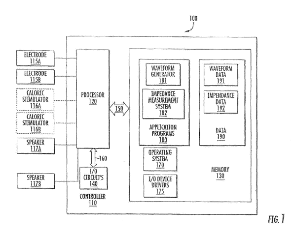
[0079] FIG. 1 is a schematic block diagram illustrating a stimulation device
according to
some embodiments of the present inventive concepts. Referring to FIG. 1, a
stimulation device
100 may include a controller 110 coupled to electrodes 115A, 115B and/or
caloric stimulators
116A, 116B. Although the device is illustrated has having both electrodes
115A, 115B for
providing galvanic vestibular stimulation and caloric stimulators 116A, 116B
for providing
caloric vestibular stimulation, it should be understood that in some
embodiments, only caloric
stimulators or only galvanic vestibular stimulation may be used. In some
embodiments, the
controller 110 may optionally be also coupled to speakers 117A, 117B. The
controller 110 may
include a processor 120, I/O circuits 140, and/or memory 130. The memory may
include an
operating system 170, I/O device drivers 175, application programs 180 (which
may be referred
to herein as applications), and/or data 190. The application programs 180 may
include a
waveform generator 181 and/or a measurement system 182. The data 190 may
include
waveform data 191 and/or measurement data 192. Although illustrated as
software, one or more
functions of the application programs 180 may be implemented in hardware or in
any
combination of hardware and/or software. Additionally, it should be understood
that one or
more of the functions of the stimulation device 100 may be provided by one or
more separate
devices. For example, one or more portions of the data 190 may be stored
remote from the
stimulation device 100, and the stimulation device 100 may communicate with
the remote
storage, for example via I/O circuits 140.
[0080] According to some embodiments of the present inventive concepts, the
stimulation device 100 may stimulate a nervous system by providing first and
second waveforms
to a first electrode 115A and a second electrode 115B. In some embodiments,
the first and
second waveforms may be modulated electric signals. In some embodiments, the
first and
second waveforms may be a modulated voltage level between the electrodes 115A,
115B. In
some embodiments, the first and second waveforms may be a modulated electrical
current
16
CA 03091956 2020-08-20
WO 2019/173468 PCT/US2019/020938
between the electrodes 115A, 115B. For example, the first and second waveforms
may be
asymmetric with respect to each other to provide the modulated voltage level
and/or modulated
electrical current between the electrodes 115A, 115B. Other embodiments may
include one or
more neutral connections to the subject. For example, in some embodiments, the
first waveform
may be a modulated voltage level between the first electrode 115A and at least
one of the neutral
connections and the second waveform may be a modulated voltage level between
the second
electrode 115B and at least one of the neutral connections. In some
embodiments, the first
waveform may be a modulated electrical current between the first electrode
115A and at least
one of the neutral connections and the second waveform may be a modulated
electrical current
between the second electrode 115B and at least one of the neutral connections.
Thus the
electrodes 115A, 115B may be used together to provide one stimulus or may be
used
independently to provide more than one stimulus.
[0081] The controller 110 may generate the first and second waveforms. The
controller
110 may include the memory 130, the processor 120 and the I/O circuits 140 and
may be
operatively and communicatively coupled to the electrodes 115A, 115B. The
processor 120 may
communicate with the memory 130 via an address/data bus 150 and with the I/O
circuits 140 via
an address/data bus 160. As will be appreciated by one of skill in the art,
the processor 120 may
be any commercially available or custom microprocessor. The memory 130 may be
representative of the overall hierarchy of memory devices containing software
and data used to
implement the functionality of the stimulation device 100. Memory 130 may
include, but is not
limited to, the following types of devices: cache, ROM, PROM, EPROM, EEPROM,
flash
memory, SRAM and DRAM. Memory 130 may include non-volatile memory.
[0082] As shown in FIG. 1, the memory 130 may comprise several categories of
software and data. For example, the memory may include one or more of: the
operating system
170, applications 180, data 190, and input/output (I/O) device drivers 175.
[0083] The applications 180 may include one or more programs configured to
implement
one or more of the various operations and features according to embodiments of
the present
inventive concepts. For example, the applications 180 may include the waveform
generator 181
configured to communicate a waveform control signal to one or both of the
electrodes 115A,
115B. The applications 180 may also include the measurement system 182 for
measuring an
impedance or other electrical characteristic (e.g., capacitance) between the
electrodes 115A,
115B. In some embodiments, the memory 130 may include additional applications,
such as a
networking module for connecting to a network. In some embodiments, the
waveform generator
17
CA 03091956 2020-08-20
WO 2019/173468 PCT/US2019/020938
181 may be configured to activate at least one electrode (i.e., to control the
magnitude, duration,
waveform and other attributes of stimulation delivered by the at least one
electrode). In some
such embodiments, the waveform generator 181 may be configured to activate at
least one
electrode based upon a prescription from a prescription database, which may
include one or more
sets of instructions for delivering one or more time-varying waveforms to the
vestibular system
of a subject.
[0084] The data 190 may comprise static and/or dynamic data used by the
operating
system 170, applications 180, I/O device drivers 175 and/or other software
components. The
data 190 may include the waveform data 191 including one or more treatment
protocols or
prescriptions. In some embodiments, the data 190 may further include
measurement data 192
including impedance measurements between the electrodes 115A, 115B and/or
estimates of
electrical contact based on electrical impedance measurements.
Electrical impedance
measurements may include resistive and capacitive components of the interface
between the
electrodes 115A, 115B and the ear canal. In some embodiments, the measurement
data 192 may
include measurements of electrical signals that are produced by the vestibular
system. For
example, the measurement data 192 may include electrovestibulography signals,
or EVestG
signals.
[0085] I/O device drivers 175 may include software routines accessed through
the
operating system 170 by the applications 180 to communicate with devices such
as I/O circuits
140, memory 130 components and/or the electrodes 115A, 115B.
[0086] In some embodiments, the waveform generator 181 may be configured to
pass an
electrical current through at least one of the electrodes 115A, 115B to
stimulate the nervous
system and/or the vestibular system of a subject. In particular embodiments,
the waveform
generator 181 may be configured to pass the electrical current through the at
least one electrode
115A, 115B based upon a prescription comprising a set of instructions for
delivering one or more
time-varying waveforms to the vestibular system of a subject. In some
embodiments, the
electrical current may be produced in response to an electrical voltage
differential provided
between the two electrodes 115A, 115B. However, in some embodiments, the
waveform
generator 181 may be configured to pass two independent electrical currents
through the two
electrodes 115A, 115B, respectively. The two independent electrical currents
may be produced
in response to electrical voltage differentials provided between each of the
two electrodes 115A,
115B and one or more additional points of electrical contact with the body of
the subject.
[0087] In some embodiments, the stimulation device 100 may be communicatively
18
CA 03091956 2020-08-20
WO 2019/173468 PCT/US2019/020938
connected to at least one electrode 115A, 115B via a conductive line. In some
embodiments, the
stimulation device 100 may be operatively connected to a plurality of
electrodes, and the
stimulation device 100 may be operatively connected to each electrode via a
separate conductive
line.
[0088] In some embodiments, the controller 110 may be operatively connected to
at least
one of the electrodes 115A, 115B via a wireless connection, such as a
Bluetooth connection. In
some embodiments, the stimulation device 100 may be configured to activate the
at least one of
the electrodes 115A, 115B to deliver one or more actively controlled, time-
varying waveforms to
the vestibular system and/or the nervous system of a patient. For example, one
or more of the
electrodes 115A, 115B may be electrically connected to a wireless receiver and
a power source
independent of the controller 110. The wireless receiver may receive the
wireless signal
corresponding to a modulated waveform and may activate the one or more of the
electrodes
115A, 115B.
[0089] In some embodiments, the stimulation device 100 may include one or more
caloric stimulators, 116A, 116B. The stimulation device 100 may stimulate a
nervous system by
providing third and fourth waveforms to the caloric stimulators, 116A, 116B.
The caloric
stimulation from the caloric stimulators may be combined with the galvanic
stimulation from the
electrodes 115A, 115B.
[0090] In some embodiments, the stimulation device 100 may include one or more
speakers, 117A, 117B. The stimulation device 100 may provide one or more audio
waveforms
to the speakers, 117A, 117B. In some embodiments, the stimulation device 100
may include an
input connector to receive one or more external audio waveforms that may be
provided to the
speakers 117A, 117B.
[0091] FIG. 2 is a front view illustrating a stimulation device according to
some
embodiments of the present inventive concepts. Referring to FIG. 2, a
stimulation device 200
may be an in-ear stimulation apparatus. The stimulation device 200 may be
similar to the
stimulation device 100 illustrated in FIG. 1 except for the differences as
noted. The stimulation
device 200 may include a support or headband 230, earphones 220, a controller
210 and/or
cables 240. In some embodiments, the stimulation device may not include the
cables 240 and the
controller 210 may connect to the earphones 220 wirelessly. The earphones 220
may include
respective electrodes 215A, 215B that are configured to be positioned in the
ear of a patient or
subject. The electrodes 215A, 215B may be configured to make electrical
contact with an inner
surface of the ear of the patient or subject such that, when activated, the
electrode 215A, 215B
19
CA 03091956 2020-08-20
WO 2019/173468 PCT/US2019/020938
may stimulate the vestibular system of the patient or subject.
[0092] The electrodes 215A, 215B may be configured as respective earpieces
250A,
250B or may be configured as parts of the respective earpieces 250A, 250B. For
example, in
some embodiments, an earpiece may be formed primarily of a conductive metal
and the entire
earpiece 250A, 250B may be an electrode 215A, 215B. In other embodiments, a
part of or all of
an exterior surface of an earpiece 250A, 250B may be coated with an
electrically conductive
metal to form the electrode 215A, 215B. In some embodiments, a part of or all
of an exterior
surface of an earpiece 250A, 250B may be coated with an thin layer of an
electrically insulating
material that covers the electrode 215A, 215B and electrically insulates the
electrode 215A,
215B from the ear of the patient or subject at DC. However, the thin layer of
the electrically
insulating material may allow higher frequency waveforms to pass through the
thin layer of the
electrically insulating material from the electrode 215A, 215B to the ear of
the patient or subject.
For example, in some embodiments, the thin layer of the electrically
insulating material may be
an anodized finish on an electrically conductive metal. However, in other
embodiments, the
electrically insulating material may be a thin layer of rubber, plastic, or
another insulating
material.
[0093] In some embodiments, the electrode 215A, 215B may be in electrical
contact with
the ear canal without directly physically contacting the ear canal. An
electrical conduit may be
positioned and configured to provide or improve electrical contact between the
ear canal and the
electrode 215A, 215B. The electrical conduit may be configured to conform to
the ear canal,
such as a flexible or conformable, electrically conductive material that is
configured to increase
contact and/or conductivity between the electrode 215A, 215B and the ear
canal. The
electrically conductive material may be a liquid or solid material or a
combination of liquid and
solid materials. Moreover, the electrically conductive material may be affixed
to the electrode
215A, 215B. For example, in some embodiments, the electrode 215A, 215B may be
covered by
a porous material that is permeated with an electrically conductive liquid. In
some embodiments,
the electrode 215A, 215B may be covered with a layer of cotton to avoid direct
physical contact
with the ear canal. The layer of cotton may be soaked with an electrically
conductive liquid, for
example a saline solution, to provide the electrical connection between the
electrode 215A, 215B
and the ear canal. In some embodiments, the electrically conductive liquid may
be positioned in
the ear canal. The ear canal may be sealed, for example, with an earplug or
other sealing
material to contain the electrically conductive liquid inside the ear canal.
In some embodiments
the electrode 215A, 215B and/or an electrical attachment thereto may pass
through or around the
CA 03091956 2020-08-20
WO 2019/173468 PCT/US2019/020938
earplug or other sealing material.
[0094] Although the electrodes 215A, 215B are illustrated in FIG. 2 as being
integrated
with the earpieces 250A, 250B, In some embodiments, the electrodes 215A, 216B
may not be
configured to fit within an ear cavity. For example, the electrodes 215A, 216B
may be
configured to contact a portion of the skin next to the ear and over a mastoid
part of a temporal
bone.
[0095] It should be understood that other configurations for supporting the
headphones
and/or earpieces 250A, 250B may be used, including support bands that are
positioned under the
chin or over the ear, for example, as may be used with audio earphones. For
example, FIG. 3 is
a front and side view illustrating a user wearing a stimulation device
according to some
embodiments of the present inventive concepts. Referring to FIG. 3, a
stimulation device 200'
may be similar to the stimulation devices 100, 200 illustrated in FIGS. 1-2
except for the
differences as noted. The stimulation device 200' may include straps 260
and/or headbands 270.
In some embodiments, the headbands 270 may provide increased stability of the
earphones 220
to provide potentially improved contact of the earpieces 250A, 250B (not
shown). In some
embodiments, one or more of the straps 260 and/or headbands 270 may provide an
additional
point of electrical contact to the user, for example a neutral connection to
the user.
[0096] Although embodiments according to the present inventive concepts are
illustrated
with respect to two ear stimulators in which an electric current is passed
from electrode to
another through the subject's tissue (e.g., the head), it should be understood
that, in some
embodiments, the stimulation device 200' may only include one electrode 215.
In such
embodiments, the stimulation device 200' may provide an electrical stimulus as
a voltage
between the electrode 215A and an additional point of electrical contact. For
example, the
additional point of electrical contact may be located on a strap 260 and/or
headband 270. In
some embodiments, two electrodes 215A, 215B in the ears or on the mastoids may
be used with
one or more additional points of electrical contact to pass separate
electrical currents from each
of the electrodes 215A, 215B to the one or more additional points of
electrical contact.
[0097] FIG. 4 is a schematic block diagram illustrating a stimulation device
according to
some embodiments of the present inventive concepts. Referring to FIG. 4, a
stimulation device
may be similar to the stimulation devices 100, 200 illustrated in FIGS. 1-2
except for the
differences as noted. The controller 210 may include a waveform generator 281
and a
measurement system 282 that may be similar to the waveform generator 181 and
an
measurement system 182 of FIG. 1, except for differences as noted. The
waveform generator
21
CA 03091956 2020-08-20
WO 2019/173468 PCT/US2019/020938
281 may be configured to communicate first and second waveforms to the
electrodes 215A,
215B. It should be understood that the first and second waveforms may be the
same, or in some
embodiments, the first and second waveforms may be different such that the
output delivered
from the electrodes 215A, 215B may be independently controlled and may be
different from one
another.
[0098] As illustrated in FIG. 4, in some embodiments, the measurement system
282 may
deliver an electrical current to one or more of the electrodes 215A, 215B. In
this configuration,
the impedance and/or capacitance value between the electrodes 215A, 215B may
be used to
monitor the electrical contact between the electrodes 215A, 215B. In some
embodiments,
impedance and/or capacitance values may be detected for a range of subjects to
determine a
range of impedance and/or capacitance values in which it may be assumed that
the electrodes
215A, 215B are in sufficient electrical contact with the subject's ear canal.
When a headset is
being fitted to a new patient, the impedance and/or capacitance between the
electrodes 215A,
215B may be detected, and if the impedance value is within the acceptable
range, it may be
assumed that there is good electrical contact between the electrodes 215A,
215B and the subject's
ear canal.
[0099] In some embodiments, when the headset is being fitted to a new patient,
the
impedance and/or capacitance value between electrodes 215A, 215B may be
detected and used
as a patient specific baseline to determine if the patient is later using the
headset and a proper
configuration. For example, the patient may use a headset according to
embodiments of the
present inventive concepts in a setting that may or may not be supervised by a
medical
professional. In either environment, the measurement system 282 may record an
impedance
and/or capacitance value at a time that is close in time or overlapping with
the time in which the
treatment waveforms are delivered to the electrodes 215A, 215B. The medical
health
professional or the measurement system 282 may analyze the impedance value to
determine
whether the electrodes 215A, 215B were properly fitting during treatment. In
some
embodiments, the measurement system 282 may be configured to provide feedback
to the user
when impedance values detected that are inconsistent with properly fitting
electrodes 215A,
215B in good electrical contact with the ear canal. In this configuration, the
measurement
system 282 may provide a degree of electrical contact between the electrodes
215A, 215B and
the ear canal in real-time or in data recorded and analyzed at a later time.
Accordingly, patient
compliance with treatment protocols may be monitored based on the detected
impedance during
or close in time to treatment.
22
CA 03091956 2020-08-20
WO 2019/173468 PCT/US2019/020938
[0100] In some embodiments, the impedance may be calculated based separately
for each
of the electrodes 215A, 215B. For example, in some embodiments, an impedance
may be
measured between ones of the electrodes 215A, 215B and an additional point of
connection
located on a cable 240 and/or the headband 270, as illustrated in FIG. 3.
[0101] In particular embodiments, the measurement system 282 may also provide
feedback to the waveform generator 281, for example, so that the waveform
generator 281 may
increase or decrease an amplitude of the waveform control signal responsive to
the degree of
electrical contact determined by the measurement system 282 based on the
impedance and/or
capacitance value. For example, if the measurement system 282 determines based
on the
impedance value that there is a poor fit and poor electrical contact with the
ear canal, then the
waveform generator 281 may increase an amplitude of the output to the
electrodes 215A, 215B
to compensate for the poor electrical contact. In some embodiments, the
measurement system
282 may determine patient compliance, e.g., whether the patient was actually
using the device
during administration of the waveforms.
[0102] Although embodiments of the present inventive concepts are illustrated
with
respect to two electrodes 215A, 215B, it should be understood that in some
embodiments, a
single electrode 215A may be used, and an electrical contact may be affixed to
another location
on the user's head instead of the second earpiece 250B to thereby provide an
electrical circuit for
determining impedance values and estimating thermal contact as described
herein.
[0103] In some embodiments, the measurement system 282 may measure one or more
impedance value based on the current and voltage levels of the first and
second waveforms. In
some embodiments, the measurement system 282 may include hardware to measure
the current
and/or voltage levels of the first and second waveforms. For example, the
measurement system
282 may calculate an impedance by dividing a voltage level by a current level.
In such
embodiments, the measurement system 282 may calculate an impedance value while
the
waveform generator 281 generates the first and second waveforms.
[0104] In some embodiments, the measurement system 282 may measure one or more
electrical signals that are produced by the vestibular system. For example,
the measurement
system 282 may measure electrovestibulography, or EVestG, signals. EVestG
signals may be
useful to determine an efficacy of a treatment. For example, EVestG signals
may be useful in
determining a presence and/or degree of one or more disorders. Accordingly, an
efficacy of a
treatment may be monitored based on feedback provided by the measured EVestG
signals during
or close in time to treatment. In some embodiments, a treatment may be revised
and/or
23
CA 03091956 2020-08-20
WO 2019/173468 PCT/US2019/020938
discontinued based on measured EVestG signals.
[0105] FIG. 5A is a schematic block diagram illustrating a stimulation device
according
to some embodiments of the present inventive concepts. Referring to FIG. 5A, a
stimulation
device 500 may be similar to the stimulation device 100 illustrated in FIGS. 1-
4 except for the
differences as noted. For example, the stimulation device may include a
controller 510A and
electrodes 515A, 515B that may be similar to the controller 210 and electrodes
215A, 215B of
FIGS. 1-4, except for differences as noted. The stimulation device may include
earphones
including earpieces 550A, 550B including the electrodes 515A, 515B. The
earphones may
further include thermal electric devices, "TEDs," attached to the earpieces
550A, 550B. The
controller 510A may include a galvanic waveform generator 581A that may be
similar to the
waveform generator 281 of FIGS. 1-4. The controller 510A may also include a
caloric
waveform generator 581B. The caloric waveform generator 518B may be configured
to activate
the TEDs attached to the earpieces 550A, 550B. In this configuration, caloric
vestibular
stimulation may be administered to a subject via the subject's ear canal.
Administration of
caloric vestibular stimulation using earpieces is discussed in U.S. Patent
Application Serial No.
12/970,312, filed December 16, 2010, U.S. Patent Application Serial No.
12/970,347, filed
December 16, 2010, U.S. Patent Application Serial No. 13/525,817, filed June
18, 2012, and
U.S. Patent Application Serial No. 13/994,266, filed May 15, 2014, the
disclosures of which are
hereby incorporated by reference in their entirety.
[0106] In some embodiments, the galvanic waveform generator 581A may deliver
first
and second waveforms to the electrodes 515A, 515B and the caloric waveform
generator may
deliver third and fourth waveforms to the TEDs attached to the electrodes
515A, 515B,
respectively. In some embodiments, the galvanic waveform generator 581A may
deliver first
and second waveforms and the caloric waveform generator may deliver third and
fourth
waveforms simultaneously. In such embodiments, the stimulation device may
deliver galvanic
vestibular stimulation and caloric vestibular stimulation. In some
embodiments, the galvanic
vestibular stimulation may enhance a delivery of the caloric vestibular
stimulation.
[0107] FIG. 5B is a schematic block diagram illustrating a stimulation device
according
to some embodiments of the present inventive concepts. Referring to FIG. 5B, a
stimulation
device may be similar to the stimulation device 100 illustrated in FIGS. 1-4
except for the
differences as noted. For example, the stimulation device may include a
controller 510B and
electrodes 515A, 515B that may be similar to the controller 210 and electrodes
215A, 215B of
FIGS. 1-4, except for differences as noted. The stimulation device 500 may
include earphones
24
CA 03091956 2020-08-20
WO 2019/173468 PCT/US2019/020938
including earpieces 550A, 550B including the electrodes 515A, 515B. The
earphones may
further include speakers attached to the earpieces 550A, 550B. In some
embodiments, the
speakers may be included in the earpieces, 550A, 515B. In other embodiments,
the earpieces
550A, 550B may include a tube or other channel of air that conducts sound from
externally
attached speakers to the inner ear. In yet other embodiments, the stimulation
device 500 may
include bone conduction speakers and the earpieces 550A, 550B may conduct
vibrations from
the bone conduction speakers to bones that are adjacent to the ear canals.
[0108] In some embodiments, the galvanic waveform generator 581A may deliver
first
and second waveforms to the electrodes 515A, 515B and the audio waveform
generator may
deliver audio waveforms to the speakers attached to the electrodes 515A, 515B,
respectively. In
some embodiments, the galvanic waveform generator 581A may deliver first and
second
waveforms and the audio waveform generator may deliver audio waveforms
simultaneously. In
such embodiments, the stimulation device 500 may deliver galvanic vestibular
stimulation and
audio stimulation. As used herein, an audio waveform is a waveform that
includes frequency
components that are within a hearing range of the subject. For example, an
audio waveform may
include frequency components within a range of about 20 to 20,000 Hz. In some
embodiments,
the audio waveforms may be time-varying and/or may include one or more
patterns. For
example, the audio- waveforms may include music and/or voice. In some
embodiments, the
waveforms of the galvanic vestibular stimulation may be modulated based on the
audio
waveforms.
[0109] For example, in some embodiments, the first and/or second waveforms of
the
galvanic vestibular stimulation may include a carrier function having a
frequency that may be
sufficiently high to produce the lower impedance that permits transmission
through the skin.
The audio waveforms may include one or more frequencies that are lower than
the frequency of
the carrier function. One or more parameters of the carrier function may be
modulated according
to the one or more lower frequencies of the audio waveforms. For example, one
or more of the
amplitude and frequency of the carrier function may be modulated to produce
the first and/or
second waveforms of the galvanic vestibular stimulation. In other embodiments,
the first and/or
second waveforms of the galvanic vestibular stimulation may be directly
proportional to the
audio waveforms.
[0110] FIG. 6A is a front perspective view illustrating an earpiece of the
stimulation
device of FIG. 5A. FIG. 6B is a cross-sectional view schematically
illustrating the earpiece of
FIG. 6A. Referring to FIGS. 6A-6B and FIG. 5A, an earpiece 550 may include an
electrode
CA 03091956 2020-08-20
WO 2019/173468 PCT/US2019/020938
515. As noted above, the electrode 515 may form all, part of, or a coating on
the surface of the
earpiece 550. A thermoelectric device 530 may be coupled between the earpiece
550 and a
heatsink 540.
[0111] The electrode 515 may receive a first or second electrical waveform
from the
galvanic waveform generator 581A of the controller 510A. The electrode 515 may
be
electrically conductive. For example, the electrode 515 may be formed of an
electrically
conductive metal. The electrode 515 may be formed to fit in an ear canal and
provide an
electrical interface to the ear canal. Thus, the galvanic waveform generator
581A may provide a
galvanic stimulus to stimulate the nervous system and/or vestibular system of
the subject based
on the first or second waveform delivered through the electrical connection
between the
electrode 515 and the ear canal.
[0112] In some embodiments, the electrode 515 may be in electrical contact
with the ear
canal without directly physically contacting the ear canal. An electrical
conduit may be
positioned and configured to provide or improve electrical contact between the
ear canal and the
electrode 515. The electrical conduit may be configured to conform to the ear
canal, such as a
flexible or conformable, electrically conductive material that is configured
to increase contact
and/or conductivity between the electrode 515 and the ear canal. The
electrically conductive
material may be a liquid or solid material or a combination of liquid and
solid materials.
Moreover, the electrically conductive material may be affixed to the electrode
515. For example,
in some embodiments, the electrode 515 may be covered by a porous material
that is permeated
with an electrically conductive liquid. In some embodiments, the electrode 515
may be covered
with a layer of cotton to avoid direct physical contact with the ear canal.
The layer of cotton may
be soaked with an electrically conductive liquid, for example a saline
solution, to provide the
electrical connection between the electrode 515 and the ear canal. In some
embodiments, the
electrically conductive liquid may be positioned in the ear canal. The ear
canal may be sealed,
for example, with an earplug or other sealing material to contain the
electrically conductive
liquid inside the ear canal. In some embodiments the electrode 515 and/or an
electrical
attachment thereto may pass through or around the earplug or other sealing
material.
[0113] The thermoelectric device 530 may receive a third or fourth thermal
waveform
from the caloric waveform generator 581B. The thermoelectric device 530 may
provide a
temperature differential between the earpiece 550 and the heatsink 540 based
on the third or
fourth waveform. The earpiece 550 and/or the electrode 515 of the earpiece 550
may provide a
thermal interface between the thermoelectric device 530 and the ear canal.
Thus, the caloric
26
CA 03091956 2020-08-20
WO 2019/173468 PCT/US2019/020938
waveform generator 581B may provide a caloric stimulus to stimulate the
nervous system and/or
vestibular system of the subject based on the third or fourth waveform
delivered through the
thermal interface between the electrode 515 and the ear canal.
[0114] FIG. 7 is a side view illustrating various alternative shapes and sizes
of earpieces
of stimulation devices according to some embodiments of the present inventive
concepts.
Referring to FIG. 7, an earpiece 750 may be similar to the earpieces
illustrated in FIGS. 2-6
except for the differences as noted. A shape and/or size of the earpiece 750
may be selected to
optimize the electrical and/or thermal connection. The shape and/or size of
the earpiece 750 may
be selected for optimal comfort of the subject. In some embodiments, the
earpiece 750 may be
user replaceable, however embodiments of the present inventive concepts are
not limited thereto.
For example, in some embodiments, the earpiece 750 may be permanently attached
to a TED
and/or earphone. In some embodiments, a size of the earpiece 750 may be
selected according to
a size of the ear canal of the subject. For example, the earpiece 750 may be
small, medium,
large, or extra large. In some embodiments, a shape of the earpiece 750 may be
selected based
upon a shape of the ear canal of the subject. For example, the earpiece 750
may be angled and/or
tortuous (twisted or curved) with respect to a base of the earpiece 750.
However, the present
disclosure is not limited to the illustrated shapes and sizes.
[0115] FIG. 8 is a schematic diagram illustrating a path of a stimulation
signal according
to some embodiments of the present inventive concepts. Referring to FIG. 8, a
path of a
stimulation signal according to some embodiments of the present inventive
concepts may include
the controller 210, an electrode 215, skin, and the vestibular system. The
controller 210 may be
the controller 210 as described above with reference to FIGS. 2-4. The
electrode 215 may be
one or more of the electrodes 215A, 215B, as described above with reference to
FIGS. 2-4. The
electrode 215 may be in physical and electrical contact with the skin of a
subject. For example,
the electrode 215 may be inserted into an ear canal of the subject and may be
in physical and
electrical contact with a portion of the skin lining the ear canal of the
subject.
[0116] FIG. 9 is a cross-sectional view schematically illustrating an ear and
surrounding
portions of a human body. Referring to FIG. 9, a vestibular nerve 910 of the
vestibular system
may be in proximity to an ear canal 920. FIG. 10 is a cross-sectional view
schematically
illustrating relative placements of electrodes with respect to a computerized
tomography scan of
a human head. Referring to FIG. 10, an electrode 1010 contacting a portion of
the skin lining
the ear canal may be in closer proximity to a vestibular nerve 1030
(approximate location shown)
than an electrode 1020 contacting the skin next to the ear and over a mastoid
part of a temporal
27
CA 03091956 2020-08-20
WO 2019/173468 PCT/US2019/020938
bone. Referring to FIGS. 8-10, an electrode 215 inserted into an ear canal of
the subject may be
in close proximity to a vestibular nerve.
[0117] Referring again to FIGS. 8 and 2-4, the waveform generator 281 of the
controller
210 may electrically stimulate the vestibular system based on a waveform. The
waveform may
be an electrical signal. The electrical signal may be modulated. The waveform
generator 281
may provide the modulated electrical signal to the electrode 215. In some
embodiments, the
waveform generator 281 may be electrically connected the electrode 215,
although the
embodiments of the present inventive concepts are not limited thereto. For
example, in some
embodiments, the waveform generator 281 may be wirelessly in communication
with an earpiece
250A, 250B that may generate and provide the electrical signal to the
electrode 215.
[0118] The electrode 215 may provide the electrical signal to the vestibular
system. For
example, the electrode 215 may provide the electrical signal to the vestibular
system via an
electrical connection through the skin. The skin may provide an electrical
resistance in the
electrical path between the electrode and the vestibular system. Thus, the
waveform generator
281 may control an amplitude of the waveform such that an amplitude of the
electrical signal is
sufficient to traverse the skin and stimulate the vestibular system. In some
embodiments, the
waveform may be modulated based on a frequency.
[0119] FIG. 11 is a graph illustrating a relationship between an impedance of
skin and a
frequency of a stimulation waveform according to some embodiments of the
present inventive
concepts. Referring to FIGS. 8 and 11, an impedance of the skin may decrease
as a frequency of
the waveform increases. See, e.g., J. Rosell, J. Colominas, P. Riu, R. Pallas-
Areny, J. G.
Webster, Skin impedance from 1 Hz to 1 MHz, IEEE Trans Biomed Eng 35, 649-651
(1988);
published online EpubAug (10.1109/10.4599).
[0120] For example, at a frequency of zero Hertz (0 Hz), in other words a
direct current
of a fixed amplitude, the skin may provide a large impedance in the electrical
path between the
electrode and the vestibular system. Thus, in order to stimulate the
vestibular system at a
frequency of zero Hertz (0 Hz), the waveform generator 281 may provide a
waveform of large
amplitude and, accordingly, the electrode may provide an electrical signal
with a large voltage.
This may not be desired as the subject may experience discomfort, pain, and/or
physical damage
based on the large voltage.
[0121] At higher frequencies, the skin may provide a lower impedance in the
electrical
path between the electrode and the vestibular system. Thus, in order to
stimulate the vestibular
system at higher frequencies, the waveform generator 281 may provide a
waveform of smaller
28
CA 03091956 2020-08-20
WO 2019/173468 PCT/US2019/020938
amplitude and, accordingly, the electrode may provide an electrical signal
with a smaller voltage.
At the lower voltage, the subject may not experience the discomfort, pain,
and/or physical
damage. However, the higher frequency may not induce the desired diagnostic
and/or
therapeutic effects of galvanic vestibular stimulation. For example, some
diagnostic and/or
therapeutic uses of galvanic vestibular stimulation desire stimulation at a
lower frequency. In
some embodiments of the present inventive concepts, a modulation scheme is
provided that
generates an electrical signal with a higher frequency to produce the lower
impedance and that
stimulates the vestibular system at a lower frequency.
[0122] FIG. 17 is a schematic block diagram illustrating portions of a
controller
according to some embodiments of the present inventive concepts. Referring to
FIG. 17, a
controller 1710 may be similar to the one or more of the controllers 210,
510A, 510B of FIGS.
4-5B except for the differences as noted. The controller 1710 may include a
waveform generator
1720 that may generate the modulated electrical signal based on a time-varying
modulation
waveform. The waveform generator may receive the modulation waveform from a
first function
generator 1730. The first function generator 1730 may define the modulation
waveform and
provide the modulation waveform as a modulated voltage to the waveform
generator 1720. The
waveform generator 1720 may include a second function generator 1740. The
second function
generator 1740 may receive a carrier function and the modulation waveform. The
second
function generator 1740 may modulate the carrier function based on the
modulation waveform.
For example, the second function generator 1740 may perform frequency
modulation or
amplitude modulation to generate a voltage-based modulated electrical signal.
In some
embodiments, the voltage-based modulated electrical signal may be received by
a current supply
1750 that produces a clamped current output that may be provided to the first
and second
electrodes as the modulated electrical signal.
[0123] Packet-Based Modulation
[0124] FIG. 12 is a graph illustrating modulated stimulation waveform
according to
some embodiments of the present inventive concepts. Referring to FIG. 12, a
waveform may
include a plurality of spaced-apart packets of pulses. The pulses may
correspond to electrical
pulses produced based on the waveform.
[0125] Ones of the plurality of packets may include a quantity, N, of pulses
and a
separation in time, S, between adjacent ones of the plurality of packets of
pulses. For example,
as illustrated in FIG. 12, the packets may each include a quantity, N, of 3
pulses, although the
present inventive concepts is not limited thereto. For example, the quantity,
N, of pulses may be
29
CA 03091956 2020-08-20
WO 2019/173468 PCT/US2019/020938
more or less than three but, in some embodiments, may be at least 2. The
separation in time, S,
between adjacent ones of the plurality of packets may be defined as a quantity
of time between
an end of a last pulse of one packet and a beginning of the first pulse of the
next adjacent packet.
[0126] Ones of the pulses may include a width in time, W, an amplitude, A, and
a
separation in time, X, between adjacent ones of the pulses within a packet.
The width in time,
W, of a pulse may be defined as a quantity of time between a rising edge and a
falling edge of a
single pulse, although the present inventive concepts is not limited thereto.
The amplitude, A, of
the waveform may correspond to the amplitude of the voltage of the electrical
signal provided by
the electrode 215 of FIG. 8. The separation in time, X, between adjacent ones
of the pulses
within a packet may be defined as a quantity of time between an end of one
pulse within a packet
and a beginning of the next pulse within the same packet.
[0127] An impedance provided by the skin of FIG. 8 in response to an
electrical signal
corresponding to the stimulation waveform may be based on the width in time,
W, of the pulses
and the separation in time, X, between adjacent ones of the pulses within a
packet. For example,
the width, W, and separation, X, may define a time period of a pulse. A
frequency of the pulses
may be the inverse of the time period. The impedance may be inversely
proportional to the
frequency of the pulses, as illustrated in FIG. 11. Thus, the width, W, and
separation, X, may be
selected to be smaller to provide a higher frequency and, thus, a lower
impedance.
[0128] At least one of the quantity, N, of the plurality of pulses within ones
of the
plurality of packets of pulses, the width in time, W, of the plurality of
electrical pulses within
ones of the plurality of packets of pulses, the amplitude, A, of the plurality
of pulses within ones
of the plurality of packets of pulses, the separation in time, X, between
adjacent ones of the
plurality of pulses within ones of the plurality of packets of pulses, and the
separation in time, S,
between adjacent ones of the plurality of packets of pulses may be modulated
to modulate the
stimulation waveform. The at least one modulated parameter may be modulated
based on a
target stimulus frequency. Referring to FIGS. 8 and 10, the vestibular system
may be stimulated
based on the target stimulus frequency. Thus, the target stimulus frequency
may be selected to
be low based on the desired diagnostic and/or therapeutic uses of the galvanic
vestibular
stimulation.
[0129] In some embodiments, the separation in time, S, between adjacent ones
of the
plurality of packets of pulses may be modulated to modulate the stimulation
waveform. In other
words, the separation in time, S, may not be constant and may be varied based
on the target
stimulus frequency. For example, the separation in time, Sibetween the first
packet and the
CA 03091956 2020-08-20
WO 2019/173468 PCT/US2019/020938
second packet illustrated in FIG. 12 may be different from the separation in
time, S2 between the
second packet and the third packet illustrated in FIG. 12.
[0130] FIG. 13 is a graph illustrating a modulated separation in time between
adjacent
ones of a plurality of packets of electrical pulses according to some
embodiments of the present
inventive concepts. Referring to FIGS. 12 and 13, the separation in time, S,
between adjacent
ones of the plurality of packets of pulses may be varied in a sinusoidal
modulation. The
separation in time, S, may vary between a minimum separation value and a
maximum separation
value. A period of the sinusoidal modulation may define the stimulation
frequency. For
example, a duration in time between minimum values or between maximum values
may define
the period. The stimulation frequency may be defined as the inverse of the
period. Thus, the
separation in time, S, may be varied in a sinusoidal modulation to stimulate
the vestibular system
based on the target stimulation frequency.
[0131] Target neurons of the vestibular system may require a minimum amount of
time
after stimulation to recover. The target neurons may be stimulated by each
pulse. Because the
separation in time, X, between pulses within a packet may be selected to be
small to provide
decreased impedance, the target neurons may not recover between pulses within
a packet. Thus,
the target neurons may be constantly stimulated within a duration of a packet
of pulses.
However, the minimum value of the separation in time, S, between packets may
be selected to be
sufficiently large to allow target neurons to recover before being activated
by the next packet of
pulses. Thus, by modulating the separation in time, S, the stimulation of the
target neurons may
be modulated based on the target stimulus frequency. See, e.g., M. W. Bagnall,
L. E. McElvain,
M. Faulstich, S. du Lac, Frequency-independent synaptic transmission supports
a linear
vestibular behavior. Neuron 60, 343-352 (2008); published online EpubOct 23 (S
0 896-
6273 (08)00845 -3 [pi] 10.1016/j .neuron.2008.10.002) (discussing recovery of
vestibular afferent
synapse after stimulus trains).
[0132] The galvanic vestibular stimulation may have downstream effects in
other
portions of the brain of the subject based on the target stimulus frequency.
In some
embodiments, a frequency of the modulated signal may be selected to induce
brain rhythms in a
target portion of the brain. In some embodiments, the galvanic vestibular
stimulation may
entrain endogenous brain rhythms in a target portion of the brain based on the
modulated signal.
[0133] In some embodiments, the separation in time, S, may be varied according
to the
formula S(t) = Smin Sc*sin((ot), wherein S(t) is the separation in time, S,
between adjacent ones
of the plurality of packets of electrical pulses, Smin and Sc are time
constants, and co is
31
CA 03091956 2020-08-20
WO 2019/173468 PCT/US2019/020938
proportional to the target stimulus frequency. However, embodiments of the
present inventive
concepts are not limited thereto. For example, in some embodiments, the
separation in time, S,
may be varied according to other formulas, such as S(t) = Smin Sc*cos(cot).
Without wishing to
be bound by any particular theory, it is believed that an amplitude of the
separation in time, S,
may be inversely proportional to an amplitude of an induced stimulus. For
example, with a
reduced separation in time, S, a vestibular system will receive more packets
of electrical pulses
within a given time. Conversely, with an increased separation in time, S, the
vestibular system
will receive fewer packets of electrical pulses within the given time. By
modulating the
separation in time, S, an amplitude of the induced stimulus may therefore be
modulated. Thus,
by modulating the separation in time, S, according to a target frequency, the
induced stimulus
may therefore be modulated according to the target frequency. Accordingly,
brainwaves may be
entrained according to the stimulus frequency.
[0134] FIG. 14 is a graph illustrating a modulated separation in time between
adjacent
ones of a plurality of packets of electrical pulses and a corresponding
modulated stimulation
waveform according to some embodiments of the present inventive concepts.
Referring to FfG.
14, a separation of time, S, between adjacent ones of a plurality of packets
of pulses is illustrated
as varied in a sinusoidal modulation. An amplitude of a waveform is
illustrated corresponding to
the modulation of S. For example, a longer separation in time is illustrated
between adjacent
packets when S is higher and a shorter separation in time is illustrated when
S is lower. In the
illustrated example, each of the packets includes three pulses of equal
amplitude, width, and
separation, however embodiments are not limited thereto.
[0135] FIGS. 15A, 15C, and 15E are graphs illustrating modulated target
stimulus
frequencies according to some embodiments of the present inventive concepts.
FIGS. 15B, 15D,
and 15F are graphs illustrating modulated separations in time between adjacent
ones of a
plurality of packets of electrical pulses according to the modulated target
stimulus frequencies of
FIGS. 15A, 15C, and 15E, respectively. Referring to FIGS. 12-13 and 15A-15F,
in some
embodiments, the formula may include more than one target stimulus frequency.
For example,
in some embodiments, the formula may include a range of frequencies. In some
embodiments,
the modulating may include modulating the target stimulus frequency between a
lower target
frequency and a higher target frequency.
[0136] Referring to FIGS. 15A-15B, in some embodiments, the modulating may
include
repeatedly decreasing the target stimulus frequency in a pattern between the
higher target
frequency and the lower target frequency. A period of the sinusoidal
modulation of the
32
CA 03091956 2020-08-20
WO 2019/173468 PCT/US2019/020938
separation in time, S, may increase over time as the target frequency
decreases. The patterns
illustrated in FIGS. 15A-15B may be consecutively repeated for a duration of
the galvanic
vestibular stimulation.
[0137] Referring to FIGS. 15C-15D, in some embodiments, the modulating may
include
repeatedly increasing the target stimulus frequency in a pattern between the
lower target
frequency and the higher target frequency. A period of the sinusoidal
modulation of the
separation in time, S, may decrease over time as the target frequency
increases. The patterns
illustrated in FIGS. 15C-15D may be consecutively repeated for a duration of
the galvanic
vestibular stimulation.
[0138] Referring to FIGS. 15E-15F, in some embodiments, the modulating may
include
repeatedly cycling the target stimulus frequency in a pattern of increasing
from the lower target
frequency to the higher target frequency and then decreasing back to the lower
target frequency.
A period of the sinusoidal modulation of the separation in time, S, may
decrease over time as the
target frequency increases and may increase as the target frequency decreases.
The patterns
illustrated in FIGS. 15E-15F may be consecutively repeated for a duration of
the galvanic
vestibular stimulation.
[0139] Carrier-Based Modulation
[0140] FIGS. 16A-D are graphs illustrating a method for modulating an
electrical signal
according to some embodiments of the present inventive concepts. For example,
FIG. 16A is a
graph illustrating a carrier waveform function according to some embodiments
of the present
inventive concepts, FIG. 16B is a graph illustrating a modulation waveform
according to some
embodiments of the present inventive concepts, FIG. 16C is a graph
illustrating an amplitude
modulated electrical signal according to some embodiments of the present
inventive concepts,
and FIG. 16D is a graph illustrating a frequency modulated electrical signal
according to some
embodiments of the present inventive concepts. Referring to FIG. 16A, a
carrier waveform
function may be a continuous cyclical function. For example, in some
embodiments, the carrier
waveform function may be a sine wave. In some embodiments, the carrier
waveform function
may be a square wave, a sawtooth wave, or another waveform function. The
carrier waveform
function may include an amplitude and a carrier frequency. The carrier
waveform function may
include a sequence of pulses that may correspond to electrical pulses produced
based on the
function.
[0141] Ones of the pulses may include a width in time, W and an amplitude, A.
The
width in time, W, of a pulse may be defined as a quantity of time between
corresponding phases
33
CA 03091956 2020-08-20
WO 2019/173468 PCT/US2019/020938
of adjacent pulses. The amplitude, A, of the waveform may correspond to the
amplitude of the
voltage and/or current of the electrical signal provided by the electrode 215
of FIG. 8.
[0142] An impedance provided by the skin as shown in FIG. 8 in response to an
electrical signal corresponding to the carrier waveform function may be based
on the width in
time, W, of the pulses. For example, the width, W, may define a time period of
a pulse. A
carrier frequency of the carrier waveform function may be the inverse of the
time period. The
impedance may be inversely proportional to the frequency of the pulses, as
illustrated in FIG.
11. Thus, the width, W, may be selected to be smaller to provide a higher
frequency and, thus, a
lower impedance. For example, in some embodiments, the carrier frequency may
be greater than
or equal to about 3 kHz. In some embodiments, the carrier frequency may be
about 10 kHz.
[0143] Referring to FIGS. 16A-16D, at least one of the amplitude, A, and the
carrier
frequency may be modulated to modulate a stimulation waveform. The at least
one modulated
parameter may be modulated based on a time-varying modulation waveform. For
example,
referring to FIGS. 16A-16C, the amplitude of the carrier waveform function may
be modulated
based on the modulation waveform to produce an amplitude modulated electrical
signal.
Referring to FIGS. 16A-16B and FIG. 16D, the frequency of the carrier waveform
function may
be modulated based on the modulation waveform to produce a frequency modulated
electrical
signal. In some embodiments, the modulation waveform may be a sinusoidal
function. In such
embodiments, the amplitude and/or frequency of the carrier waveform function
may be varied in
a sinusoidal modulation. However, in other embodiments, the modulation
waveform may not be
sinusoidal and may be another waveform. Referring to FIGS. 8 and 10, the
vestibular system
may be stimulated based on the modulation frequency. Thus, the modulation
frequency may be
selected to be low based on the desired diagnostic and/or therapeutic uses of
the galvanic
vestibular stimulation. In some embodiments, the modulation frequency may be
less than about
1 kHz. For example, in some embodiments, the modulation frequency may be
between about
0.005 Hz and about 200 Hz. However, in some embodiments, a modulation
frequency that is
greater than 1 kHz may be selected based on another desired diagnostic and/or
therapeutic use of
the galvanic vestibular stimulation.
[0144] The galvanic vestibular stimulation may have downstream effects in
other
portions of the brain of the subject based on the modulation frequency. In
some embodiments, a
frequency of the modulated signal may be selected to induce brain rhythms in a
target portion of
the brain. In some embodiments, the galvanic vestibular stimulation may
entrain endogenous
brain rhythms in a target portion of the brain based on the modulated signal.
34
CA 03091956 2020-08-20
WO 2019/173468 PCT/US2019/020938
[0145] In some embodiments, the modulation waveform may include more than one
modulation frequency. For example, in some embodiments, the modulation
waveform may
include a range of frequencies. In some embodiments, the modulating may
include modulating
the modulation waveform between a lower target frequency and a higher target
frequency. In
some embodiments, the modulating may include repeatedly decreasing the
modulation frequency
in a pattern between the higher target frequency and the lower target
frequency. In some
embodiments, the modulating may include repeatedly increasing the modulation
frequency in a
pattern between the lower target frequency and the higher target frequency. In
some
embodiments, the modulating may include repeatedly cycling the modulation
frequency in a
pattern of increasing from the lower target frequency to the higher target
frequency and then
decreasing back to the lower target frequency.
[0146] Applications
[0147] Embodiments according to the present inventive concepts will now be
described
with respect to the following non-limiting examples
[0148] ALTERATION OF CROSS-FREQUENCY COUPLING
[0149] The oscillatory activity in multiple frequency bands may be observed in
different
levels of organization from micro-scale to meso-scale and macro-scale. Studies
have been
shown that some brain functions are achieved with simultaneous oscillations in
different
frequency bands. The relation and interaction between oscillations in
different bands can be
informative in understanding brain function. This interaction between several
oscillations is also
known as cross-frequency coupling (CFC).
[0150] Two forms of recognized CFC in brain rhythms are: phase amplitude
coupling
(PAC), and phase-phase coupling (PPC). In phase amplitude coupling, the phase
of the lower
frequency oscillation may drive the power of the coupled higher frequency
oscillation, which
may result in synchronization of amplitude envelope of faster rhythms with the
phase of slower
rhythms. Phase-phase coupling is amplitude independent phase locking between
high and low
frequency oscillation.
[0151] It is believed that phase-amplitude coupling may be a mechanism for
communication within and between distinct regions of the brain by coordinating
the timing of
neuronal activity in brain networks. That brain rhythms modulate the
excitability of neuronal
ensembles through fluctuations in membrane potentials, biasing the probability
of neuronal
spiking at a specific phase of the slower rhythm. PAC is thought to
dynamically link
functionally related cortical areas that are essential for task performance.
CA 03091956 2020-08-20
WO 2019/173468 PCT/US2019/020938
[0152] Parkinson's disease (PD) has been shown to be associated with
exaggerated
coupling between the phase of beta oscillations and the amplitude of broadband
activity in the
primary motor cortex, likely constraining cortical neuronal activity in an
inflexible pattern whose
consequence is bradykinesia and rigidity. See, e.g., C. de Hemptinne, N. C.
Swann, J. L. Ostrem,
E. S. Ryapolova-Webb, M. San Luciano, N. B. Galifianakis, P. A. Starr,
Therapeutic deep brain
stimulation reduces cortical phase-amplitude coupling in Parkinson's disease,
Nat Neurosci 18,
779-786 (2015); published online EpubApr 13 (10.1038/nn.3997). Parkinson's
disease may be
associated with a range of symptoms that originate, or are centered, in
different brain regions. It
is believed that aberrant cross-frequency coupling between two different EEG
bands may be
present when a patient experiences tremor. It is believed that CVS and/or GVS
may be used to
alter the cross-frequency coupling so as to renormalize function and re-
establish proper balance.
[0153] Stimulation of a region in the brain stem called the PPN has been shown
to
improve, for example, the normalization of gait in PD patients. See, e.g., H.
Morita, C. J. Hass,
E. Moro, A. Sudhyadhom, R. Kumar, M. S. Okun, Pedunculopontine Nucleus
Stimulation:
Where are We Now and What Needs to be Done to Move the Field Forward? Front
Neurol 5,
243 (2014); published online (10.3389/fneur.2014.00243). With respect to the
present inventive
concepts, without wishing to be bound by theory, it is believed that
vestibular stimulation may
modulate activity of the PPN. For example, CVS may be used to stimulate the
PPN. In some
embodiments, GVS may be used to break up the aberrant cross frequency coupling
associated
with tremor simultaneous with the use of CVS to stimulate the PPN. Thus, the
two modalities
may modulate different brain regions to improve the efficacy of the treatment.
[0154] More generally, GVS may be used to sensitize or give preference to a
subset of
neural pathways that respond to a specific excitation frequency (for example,
within the EEG
bands), making them more responsive to CVS neuromodulation. These selected
pathways would
then be subject to differential modulation in the background of other, non-
selected pathways. An
illustrative example would be the use of GVS in the theta band frequency
range, associated with
hippocampal activity, to sensitize pathways associated with memory encoding.
GVS at a sub-
threshold intensity may be used, or the intensity may be above the activation
threshold of the
afferent vestibular nerves. The CVS waveform would be chosen so as to overlap
and enhance
the neuromodulatory effects of the targeted GVS modulation.
[0155] CONTROLLING IGF-1 ACCRETION
[0156] Insulin-like growth factor 1 (IGF-1) is a hormone that is similar in
molecular
structure to insulin that is believed to play an important role in childhood
growth and to have
36
CA 03091956 2020-08-20
WO 2019/173468 PCT/US2019/020938
anabolic effects in adults. The protein is encoded in humans by the IGF1 gene.
It is believed
that IGF-1 may also provide mitochondrial protection
[0157] Insulin-like growth factor number one (IGF-1) is a hormone (MW: 7649
daltons)
similar in molecular structure to insulin. It plays an important role in
childhood growth and
continues to have anabolic effects in adults. Its production is encoded by the
IGF1 gene and it is
produced primarily in the liver as an endocrine hormone, though production in
the central
nervous system has also been observed. In circulation, IGF-1 is bound to one
of six proteins, the
most common being IGFBP-3. These chaperone proteins increase the half life of
IGF-1 in
circulation from around 15 minutes (unbound) to 15 hours (bound). IGF-1
production is
associated with growth hormone (GH) and blood tests for GH use IGF-1 as a
surrogate, since the
concentration of the latter tends not to vary as much over a daily cycle as
that of GH does. IGF-
1 is one of the most potent natural activators of the AKT signaling pathway, a
stimulator of cell
growth and proliferation, and a potent inhibitor of programmed cell death. It
protects and
strengthens cells at one of their most vulnerable moments, when they are in
the process of and
immediately after dividing. It is believed that IGF-1 may provide
mitochondrial protection from
mitochondrial stress.
[0158] Electrical stimulation of the fastigial nucleus (FN) for a sufficient
time and at the
right frequency has been shown to lead to neuroprotection via reduction of
apoptosis in
mitochondria in the ischemic area. See, e.g., P. Zhou, L. Qian, T. Zhou, C.
Iadecola,
Mitochondria are involved in the neurogenic neuroprotection conferred by
stimulation of
cerebellar fastigial nucleus, J Neurochem 95, 221-229 (2005). It is believed
that IGF-1 may be a
factor in such neuroprotection. Electrical stimulation has been shown to
result in the recruitment
of IGF-1 from systemic circulation, through the blood-brain-barrier (BBB), and
to very localized
usage by or around the point of the stimulated nerves. Effectively, the nerve
activity signaled for
and recruited IGF-1. See, e.g., T. Nishijima, J. Piriz, S. Duflot, A. M.
Fernandez, G. Gaitan, U.
Gomez-Pinedo, J. M. Verdugo, F. Leroy, H. Soya, A. Nunez, I. Torres-Aleman,
Neuronal
activity drives localized blood-brain-barrier transport of serum insulin-like
growth factor-I into
the CNS, Neuron 67, 834-846 (2010). It is believed that vestibular stimulation
may similarly
accomplish the recruitment of and/or enhance the efficiency of IGF-1, either
directly affecting
nerves active during vestibular stimulation or possibly nearby nerves receive
IGF-1 in a
bystander effect. For migraine headache in particular, increased rCBF may be a
key factor
enabling increased IGF-1 uptake through the BBB. Mechanisms for altering blood
flow mesh
nicely with existing observations/beliefs around the etiology of migraines.
That IGF-1 also
37
CA 03091956 2020-08-20
WO 2019/173468 PCT/US2019/020938
facilitates synaptic plasticity could mean it has a role in mitigating central
pain associated with
chronic migraines.
[0159] With respect to the present inventive concepts, without wishing to be
bound by
theory, it is believed that vestibular stimulation, or in particular CVS
and/or GVS, may be used
to increase IF-1 transport across the blood-brain-barrier, and therefore
increase mitochondrial
protection, in frequency-dependent targeted regions of the central nervous
system. In some
embodiments, CVS and GVS may be combined. For example, CVS may be used to
provide
stimulation of frequency-dependent regions of the brain and GVS may be used to
provide
neuroprotection to the stimulated regions and/or surrounding regions.
[0160] Although embodiments have been described with reference to galvanic
vestibular
stimulation through an ear canal, the present inventive concepts are not
limited thereto. For
example, in some embodiments, the vestibular system may be stimulated through
at least one
electrode in contact with a portion of the skin behind the ear in proximity to
a mastoid part of a
temporal bone. In some embodiments, delivering the electrical signal may
include transdermal
electrical stimulation of other portions of a nervous system of the subject.
In some
embodiments, the described modulation scheme may be used with implantable
electrodes or
other devices that do not stimulate through the skin.
[0161] AUDIO WAVEFORMS
[0162] In some embodiments, neurostimulation may be performed based on an
audio
waveform. For example, a time-varying modulation waveform used for modulating
an electrical
signal that is delivered to a patient may be an audio waveform. As used
herein, an audio
waveform is a waveform that includes frequency components that are within a
hearing range of
the subject. For example, an audio waveform may include frequency components
within a range
of about 20 to 20,000 Hz. In some embodiments, the audio waveform may be time-
varying
and/or may include one or more patterns. For example, the audio waveforms may
include music
and/or voice. In some embodiments the audio waveforms may include sounds of an
environment, such as the sounds of rain, birds, moving water, cars, etc. In
some embodiments,
the audio waveforms may be based on audio recordings. In some embodiments, the
waveforms
of the galvanic vestibular stimulation may be modulated based on the audio
waveforms.
[0163] As used herein, modulation of an electrical signal based on an audio
waveform
means that the frequency components of the audio waveform within a hearing
range of the
subject are encoded into the electrical signal. For example, notes or sounds
that are in the audio
waveform may be encoded into the electrical signal. In some embodiments, the
electrical signal
38
CA 03091956 2020-08-20
WO 2019/173468 PCT/US2019/020938
may include a carrier waveform at a frequency that is at least twice a highest
frequency of the
frequency components of the audio waveform encoded into the electrical signal.
The audio
waveform may be encoded into the carrier waveform via, for example, frequency
or amplitude
modulation. In some embodiments, an electrical signal may not be referred to
as being
modulated based on the audio waveform if the electrical signal is modulated
based on beats or
other features of the audio waveform in the absence of the frequency
components of the audio
waveform that are within the hearing range of the subject.
[0164] COMBINATION OF CVS AND GVS TO ENHANCE IGF-1 DELIVERY
[0165] As discussed in more detail below, in some embodiments, a combination
of CVS
and GVS may be used to activate a neuroprotective function. For example,
without wishing to
be bound by any particular theory, apoptosis in mitochondria may be reduced
through
neurostimulation by increasing IGF-1 uptake through a blood-brain-barrier by
inducing
oscillations in cerebral blood flow.
[0166] FIG. 18 is a cross-sectional view schematically illustrating an effect
of CVS on
vestibular nerves according to some embodiments of the inventive concepts.
Referring to FIG.
18, administration of CVS may include raising and/or lowering temperatures of
the earpieces
550A, 550B. For example, in some embodiments, as illustrated in FIG. 18, the
temperature of
earpiece 550A may be controlled to a higher temperature while the earpiece
550B may be
controlled to a lower temperature. Regular neurons have an equilibrium firing
rate of about 100
Hz. Lowering the temperature of the regular neurons may lower the firing rate
of the neurons
and increasing the temperature of the neurons may increase the firing rate of
the neurons.
Accordingly, the temperatures of the earpieces 550A, 550B may be controlled to
alter the firing
rate of neurons in the respective ears corresponding to a CVS waveform. The
period of a CVS
waveform may be limited by the thermal conduction time of the temporal bone.
However, the
actual firing pattern created by time-varying CVS in the vestibular nuclei may
be complex. A
temperature ramp may create an increasing or decreasing frequency signal,
often termed a
"chirp." Thus, even though the frequency of a CVS thermal waveform is
significantly less than
1 Hz, the induced firing rate may extend for many 10's of Hz above and/or
below the
equilibrium firing rate. Furthermore, each ear may be stimulated
independently, leading to a
highly complex frequency modulation space in the brainstem.
[0167] FIG. 19 is a cross-sectional view schematically illustrating an effect
of GVS on
vestibular nerves according to some embodiments of the inventive concepts.
Referring to FIG.
19, administration of GVS may include providing modulated voltage and/or
current to the
39
CA 03091956 2020-08-20
WO 2019/173468 PCT/US2019/020938
electrodes 515A, 515B. For example, in some embodiments, as illustrated in
FIG. 19, a negative
voltage/current may be applied to the electrode 515A and a positive
voltage/current may be
applied to the electrode 515B. The negative voltage/current may increase the
firing rate of the
neurons and the positive voltage/current may lower the firing rate of the
neurons. Accordingly,
the modulated voltage and/or current applied to the electrodes 515A, 515B may
be controlled to
alter the firing rate of neurons in the respective ears corresponding to a
modulated GVS
waveform.
[0168] FIG. 20 is a cross-sectional view schematically illustrating an effect
of vestibular
neurostimulation on a brain according to some embodiments of the inventive
concepts. FIG. 20
illustrates a map of some brain regions (particularly relevant to migraine
headache) that may be
innervated by the vestibular system. Via thalamic relays, the vestibular
system may exert control
on other sensory cortices. For example, information en route to the cerebral
cortex may first pass
through the thalamus. However, a long-held view that the thalamus serves as a
simple high
fidelity relay station for sensory information to the cortex has over recent
years been dispelled.
There are multiple projections from the vestibular nuclei to thalamic nuclei,
including the
ventrobasal nuclei, and the geniculate bodies, regions typically associated
with other modalities.
Further, some thalamic neurons have been shown to respond to stimuli presented
from across
sensory modalities. For example, neurons in the anterodorsal and laterodorsal
nuclei of the
thalamus may respond to visual, vestibular, proprioceptive and somatosensory
stimuli and
integrate this information to compute heading within the environment.
Therefore, the thalamus
may serve crucial integrative functions, at least in regard to vestibular
processing, beyond that
imparted by a "simple" relay. Accordingly, a vestibular neuromodulation
waveform may be
relayed to target portions of the brain and may entrain brain waves at a
target frequency.
[0169] In mammals there are generally two types of vestibular hair cells, Type
I hair cells
and Type II hair cells. Each of these classes of hair cells has a distinctive
pattern of afferent
ending onto the hair cell. One important factor that varies between the
different afferents is the
regularity of the afferent's resting discharge rate. Afferents that are
innervated only by type I
hair cells have action potentials that generally fire at very irregular rates
when firing
spontaneously. Afferents that are innervated only by type II hair cells
generally have very
regular static discharge rates.
[0170] GVS may preferentially affect irregularly firing hair cells within the
vestibular
receptor organs. Further, GVS may affect hair cells in all of the semicircular
canals and otoliths
(not a subset). Cathodal (DC) GVS may increase afferent firing rate whereas
anodal GVS may
CA 03091956 2020-08-20
WO 2019/173468 PCT/US2019/020938
decrease the firing rate. This is analogous to an increase in firing rate
associated with warm CVS
(that is, above body temperature) and a decrease with cold CVS. Irregularly
firing hair cells
comprise roughly 25% of the afferent output of the vestibular organs.
[0171] CVS may be capable of activating both regularly and irregularly firing
hair cells.
Regular hair cells fire at approximately100 Hz in equilibrium and CVS may
alter firing rate
around this value. A rapid temperature change may engage irregularly firing
hair cells as well.
Whereas the horizontal semicircular canal is generally described as the
principal target of CVS,
the other canals and otoliths may also respond.
[0172] Therefore, CVS may affect regularly and irregularly firing hair cells
in all of the
vestibular sensing organs, whereas GVS primarily affects the irregularly
firing hair cells in all of
the vestibular sensing organs. Accordingly, CVS and GVS activation patterns
may not be
identical.
[0173] Irregularly firing hair cells may have evolved with amniotes in order
to facilitate
vestibular tracking of higher frequency movements. For example, land animals
developed necks
and this new degree of freedom may have necessitated a faster-responding hair
cell type
(irregular). So-called head direction cells have been identified in the
hippocampal complex and
this type of sensory cell may rely on irregularly firing vestibular hair cells
to properly provide
feedback on the position of the body in space. Across different species of
mammals, there is a
substantial constancy of the linear dimensions of the respective vestibular
organs. Across seven
orders of magnitude in size, the physical dimensions of the semicircular
canals may vary by less
than one order of magnitude. This level of evolutionary conservancy in
vestibular organs
attributes may be evidence of a high significance of a change in basic
vestibular function, such as
the emergence of irregularly firing hair cells.
[0174] Borrowing from a development in optics and visual processing, spatial
frequency
analysis, it may be possible to characterize an image by separating high and
low spatial
frequencies. High spatial frequencies are necessary to encode edges and sharp
changes in
contrast. Low spatial frequencies capture general shape and general contrast.
The visual system
in mammals may segregate raw input from the retina, with some cortical regions
responding to
edges, contrast changes, etc. In other words, the visual cortex may perform
operations analogous
to a spatial frequency decomposition on raw sensory flow from the retina. The
processing of
visual images has been studied in terms of areas in the visual cortex that
respond to rapid or less
rapid changes in image contrast over a given length scale, so-called high and
low spatial
frequencies respectively. The brain may unit the different spatial frequency
regimes into a
41
CA 03091956 2020-08-20
WO 2019/173468 PCT/US2019/020938
coherent whole, but may also utilize the two domains individually for some
processing tasks.
[0175] This result may be generalized to other sensory modalities, like the
vestibular
system. The vestibular system has fast-responding (irregular) and more slowly
responding
(regular) hair cells that may characterize movement. The brain's overall
perception of
movement may be informed by the integration of both movement categories. The
output of the
irregularly firing hair cells may contribute to excitation at high frequencies
and, therefore, GVS
stimulation may primarily contribute to excitation at high frequencies. Slowly
changing
temperatures delivered by CVS may primarily affect regularly firing hair cells
and may provide
higher information density in the lower spatial frequencies. Therefore, it may
be possible to
utilize CVS and GVS in combination to affect different ends of the frequency
spectrum of
vestibular sensory flow. Regular and irregular vestibular outputs may
innervate the same brain
regions (that is, the two components don't have divergent pathways), but the
information content
may be different and may enable different outcomes in terms of cognitive or
behavioral states.
As a specific example, the irregularly firing hair cells may have evolved
specifically because
new behavioral abilities of land animals were not being well-encoded by
regular hair cells alone.
[0176] As discussed above, electrical stimulation of the fastigial nucleus
(FN) for a
sufficient time and at the right frequency has been shown to lead to
neuroprotection via reduction
of apoptosis in mitochondria in the ischemic area. More specifically,
electrical stimulation of the
FN may reduce the release of cytochrome-c from mitochondria. Cytochrome-c
release is part of
the apoptotic chain. The neuroprotective effect may be frequency dependent and
a minimum
duration of stimulation may be needed to provide neuroprotection.
[0177] IGF-1 may inhibit cytochrome-c release from mitochondria. Passage of
IGF-1
through the blood-brain-barrier may occur in response to specific signaling in
the brain and may
be facilitated by enhancement of neurovascular coupling by increase blood
flow. This effect
may also be frequency dependent.
[0178] Stimulation of the FN may lead to changes in cerebral blood flow (CBF).
This
may be important in facilitating the signaling in the brain to activate
passage of IGF-1 though the
blood-brain-barrier. Time-varying CVS may induce oscillations in CBF.
Therefore, vestibular
stimulation may be used to activate the FN, which may induce oscillations in
CBF, which may
activate passage of IGF-1 through the blood-brain-barrier, which may protect
mitochondria
against apoptotic death, promote synaptogenesis, and/or improve neurovascular
coupling, thus
providing neuroprotective effects. For example, IGF-1 may inhibit cytochrome-c
release from
mitochondria, which may reduce apoptosis in mitochondria. This may be an
innate response that
42
CA 03091956 2020-08-20
WO 2019/173468 PCT/US2019/020938
protects neurons in the brain. Accordingly, time-varying CVS and/or GVS may be
used to
activate this innate protective mode.
[0179] The impact of vestibular stimulation on IGF-1 movement may be dependent
on a
time-varying aspect of the CVS and/or GVS. For example, time-varying CVS may
leads to
oscillations in CBF. In some embodiments, stimulation waveforms may be
selected that
facilitate the production of CBF oscillations. For example, the CBF
oscillations may be
measured using, for example, transcranial Doppler sonography. Accordingly, a
plurality of
stimulation waveforms may be sequentially tried while measuring for CBF
oscillations. One or
more waveforms that produce desired CBF oscillations may be selected. This may
be done with
a patient at a start of a therapy to optimize the waveform choice that
provides the most effective
amount of CBF oscillations.
[0180] In some embodiments, time-varying CVS may be used to excite CBF
oscillations
while using a narrow frequency GVS to select a subset of brain regions to
activate, thus
facilitating the movement of IGF-1 across the BBB. The time-varying CVS and
the narrow-
frequency GVS may both work to increase IGF-1 uptake through the blood-brain-
barrier.
[0181] In some embodiments, a positron-emitting radionuclide may be used as a
Positron
Emission Tomography (PET) label on IGF-1. The PET label may be introduced
systemically
while measuring uptake in the brain via PET imaging. Accordingly, it may be
seen where IGF-1
uptake increases based on the type of vestibular neurostimulation applied. In
some
embodiments, the positron-emitting radionuclide may be a PET label on glucose
or oxygen to
detect blood flow. For example, some embodiments may use fluorine-18
labeled
fluorodeoxyglucose or oxygen-15. In some embodiments, transcranial Doppler
sonography to
measure the induction of cerebral blood flow oscillations. In some
embodiments, CBF
oscillations may be measured via transcranial Doppler sonography while
sequentially trying a
plurality of stimulation waveforms to select one or more waveforms that
produce desired CBF
oscillations, as described above, at a start of a therapy to optimize the
waveform choice that
provides the most effective amount of CBF oscillations. In some embodiments,
the transcranial
Doppler sonography may be used to compare before and during vestibular
neuromodulation to
identify differences in PET uptake to hone in on regions where additional IGF-
1 has entered the
central nervous system due to the vestibular neuromodulation.
[0182] As an example, cerebral blood flow oscillations may include B waves. B
waves
are spontaneous oscillations in cerebral blood flow velocity (CBFv) that may
have a frequency of
about 0.5 to about 3 cycles per minute, thus a period of about 20 seconds to
about 2 minutes.
43
CA 03091956 2020-08-20
WO 2019/173468 PCT/US2019/020938
There is experimental evidence that B waves may be due to fluctuations in
vessel diameters
triggered by monoaminergic and serotonergic centers in the midbrain and pons.
B waves may be
part of an autoregulatory response and their average period may to correspond
to a complete
cycle time for blood moving from the heart to the brain and back. Some studies
have shown a
correlation between abnormal B wave activity and migraine headaches, that
period leg
movements (also called restless leg syndrome) may be part of a common
endogenous rhythm
matching the B wave period, and that B waves may be more prominent in NREM
sleep. B
waves may be a significant predictor of survival after traumatic brain injury.
[0183] The B wave period may fall in a range found in functional connectivity
studies of
the sensory cortices. Functional connectivity in the auditory, visual, and
sensorimotor cortices
may be significantly characterized by frequencies slower than those in the
cardiac and
respiratory cycles. In functionally connected regions, these low
frequencies may be
characterized by a high degree of temporal coherence. This functional
connectivity may have
the same pacing nexus that gives rise to B waves, which may indicate
neurovascular coupling.
Thus, the entrainment of B waves may also entrain the above describe sensory
functional
connectivity.
[0184] For example, time-varying CVS may induce significant oscillations in
the Gosling
Pulsatility Index (PI), which is a measure of cerebrovascular resistance
defined as [(peak systolic
velocity - end diastolic velocity)/ mean cerebral blood flow velocity], a
primary measure of
[0185] cerebrovascular dynamics. A time-varying CVS treatment may induce PI
spectral
peaks at intervals that may fall within the periodicity range of B waves and
may not match
periods of the warm and cold waveforms. Studies have provided evidence for a
monoaminergic
B-wave pacing center in the pons, an area that receives direct innervation
from the vestibular
nuclei in the brainstem, which may be how the time-varying CVS treatment may
induce
oscillations.
[0186] In other words, the time-varying CVS may entrain the pontine structures
responsible for B-wave pacing, as evidenced by a significant increase of
spectral power at
spectral frequencies within the range of B-waves in a post-CVS period.
[0187] CVS AND GVS CO-NEUROMODULATION
[0188] As used herein, the terms "vestibular neuromodulation" and "vestibular
neurostimulation" may each refer to the stimulation of the vestibular nerve,
which may include
CVS and/or GVS.
[0189] Methods of treating a patient using neuromodulation may include a
combination
44
CA 03091956 2020-08-20
WO 2019/173468 PCT/US2019/020938
of CVS and GVS that are applied together. For example, in some embodiments,
the CVS and
GVS may be applied simultaneously. The CVS and GVS may each include a time-
varying
stimulation waveform. As used herein, stimulations may be considered to be
applied
simultaneously when applied as part of a single treatment. For example, in
some embodiments,
the CVS and GVS may be applied simultaneously when applied within an hour of
each other as
measured from the end of the first to the beginning of the second. In some
embodiments, the
CVS and GVS may be applied simultaneously when applied within an thirty
minutes of each
other, or fifteen minutes, or five minutes. In some embodiments, the CVS and
GVS may be
applied simultaneously when there is an overlap in time between the
application of the CVS and
the application of the GVS.
[0190] In some embodiments, the CVS and GVS may excite different frequencies
in the
brain. For example, the CVS may be used to excite frequencies less than 1 Hz.
The GVS may
be used to excite frequencies greater than 1 Hz. In some embodiments, the GVS
may be used to
excite frequencies between about 0.005 Hz and about 200 Hz and may be
different than a
frequency of the CVS. In some embodiments, frequencies of the GVS may be at
least a multiple
of a maximum frequency of the CVS. For example, the frequencies of the GVS may
be at least
times a maximum frequency of the CVS.
[0191] In some embodiments, there may be a defined phase difference between
the
stimulation waveform of the CVS and the stimulation waveform of the GVS. For
example, the
stimulation waveform of the CVS and the stimulation waveform of the GVS may be
controlled
to maintain a 180 phase difference. Accordingly, the GVS may suppress an
irregular hair cell
contribution of the net applied vestibular neuromodulation. Therefore, a net
applied vestibular
neuromodulation may be controlled to affect mainly regular firing hair cells,
mainly irregular
firing hair cells, or a mixture of regular and irregular firing hair cells.
[0192] In some embodiments, a small relative frequency difference between the
stimulation waveform of the CVS and the stimulation waveform of the GVS may
result in a net
effect at a beat frequency. The beat frequency may be equal to the difference
between the
stimulation waveform of the CVS and the stimulation waveform of the GVS. For
example, a
target frequency may be selected for a desired effect in a treatment. The
stimulation frequency
of the CVS and the stimulation frequency of the GVS may each be controlled to
have a
difference equal to the desired target frequency. One or more of the
stimulation waveform of the
CVS and the stimulation waveform of the GVS may be modulated to vary the
target frequency.
[0193] DURABLE GAINS
CA 03091956 2020-08-20
WO 2019/173468 PCT/US2019/020938
[0194] Conditions such as neurological disorders, may be treated by
administering
stimulation, for example vestibular stimulation, for example CVS, to at least
one ear of a subject
in a condition-treatment effective amount during a treatment interval. The
stimulation may be
effective to produce a durable improvement in at least one symptom of the
condition. For
example, the durable improvement may remain for a time of at least 1 or 2
weeks, or 1, 2, or 3
months, or more, following cessation of the stimulation. Examples of such
conditions may
include, but may not be limited to, neurodegenerative diseases, such as
Parkinson's disease, and
other neurological conditions, such as headache, for example migraine
headache. In some
examples, the symptom may be a non-motor symptom of the condition.
[0195] Examples of non-motor symptoms of a condition as described herein
(including
but not limited to Parkinson's disease) may include, but are not limited to:
cardiovascular
symptoms (for example, light-headedness, dizziness, weakness on standing from
a sitting or
lying position; fall because of fainting or blacking out), sleepiness and
fatigue (for example, doze
off or fall asleep unintentionally during daytime activities; fatigue
(tiredness) or lack of energy
(not slowness) limiting daytime activities; difficulties falling or staying
asleep; urge to move the
legs or restlessness in legs that improves with movement when sitting or lying
down inactive);
mood and cognitive symptoms (for example, loss of interest in surroundings;
loss of interest in
doing things or lack of motivation to start new activities; nervous, worried
or frightened for no
apparent reason; sad or depressed; flat moods without normal "highs" and
"lows"; difficulty
experiencing pleasure from their usual activities); perceptual problems and
hallucinations (for
example, seeing things that are not there; having beliefs that are known to be
untrue; double
vision); attention and memory symptoms (for example, problems sustaining
concentration during
activities; forget things told a short time ago or events that happened in the
last few days; forget
to do things); gastrointestinal tract symptoms (dribble saliva during the day;
difficulty
swallowing; suffer from constipation, etc.), urinary tract symptoms (
incontinence/urgency;
frequent urination; nocturia (get up at night to urinate), etc.); sexual
dysfunction (for example,
altered interest in sex; problems having sex, etc.); and other miscellaneous
symptoms (for
example, pain not explained by other known conditions; change in ability to
taste or small;
change in weight not related to dieting; excessive sweating).
[0196] In some embodiments, the stimulation may be administered to both ears
of the
subject. In some embodiments, the stimulation to each ear may be different. In
some
embodiments, the stimulation may be-administered as a time-varying waveform.
The stimulation
may be carried out in a plurality of sessions. For example, the sessions may
be administered 2,
46
CA 03091956 2020-08-20
WO 2019/173468 PCT/US2019/020938
3, or 4 times per week over a time of from 1 week to 1, 2 or 3 months, or
more. For example, the
sessions may include stimulation 1-2 times per day, 7 days per week, over a
time from 1 week to
1, 2 or 3 months, or more.
[0197] In some embodiments, some or all of the sessions may be separated in
time by
rest intervals. For example, the treatment may include ceasing the
administering of the
stimulation for a rest interval having a duration of at least 1 or 2 weeks, or
at least 1, 2, or 3
months, and then cyclically repeating the treatment sessions and the rest
intervals at least once,
or for a plurality of cycles. For example, the cycles may be repeated over a
time of at least six
months, or at least one or two years, or more.
[0198] In some embodiments, the at least one symptom of the condition may be
measured during the rest interval. For example, the at least one symptom may
be measured after
ceasing the stimulation for a time of at least 1 or 2 weeks, or at least 1, 2,
or 3 months. The
stimulation may be modified in response to the measured at least one symptom.
For example, a
waveform may be selected based on the measurement. In some embodiments, an
amplitude,
duration, or other characteristic of the waveform may be altered. In some
embodiments, a
frequency of the waveform may be altered. In some embodiments, a type of
waveform may be
altered. In some embodiments, other characteristics of the stimulation may be
altered.
[0199] Some example flowcharts of the treatments described herein are
illustrated in
FIGS. 21 and 22. For example, in FIG. 21, a method 2100 may include
sequentially
administering at least one CVS stimulus to a subject. For each administered
CVS stimulus, a
time to entrainment (TB) and/or a time to relaxation (TR) of at least one
physiological oscillatory
pattern may be determined, such as cross-functional coupling. If the time to
entrainment and the
time to relaxation of the at least one physiological oscillatory pattern are
within target ranges
(e.g., exceed a threshold) for one of the at least one CVS stimulus, then that
at least one CVS
stimulus may be an optimized CVS stimulus, which may be administered to the
subject (e.g.,
once, or on multiple occasions during a first or an initial or subsequent
treatment interval) as
treatment. Periodic measurements of the at least one physiological oscillatory
pattern may be
performed to gauge when durable gains have been achieved. Upon achieving
durable gains,
administering of the optimized CVS stimulus may be paused, for example during
a rest interval.
Therapy may be repeated or restarted if continued measurements of the at least
one physiological
oscillatory pattern diverge from target values.
[0200] As another example, in FIG. 22, a method 2200 may include sequentially
administering at least one CVS stimulus to a subject. For each administered
CVS stimulus,
47
CA 03091956 2020-08-20
WO 2019/173468 PCT/US2019/020938
dynamic changes in sensory habituation resulting from the administered CVS
stimulus may be
measured. If the measured dynamic changes in sensory habituation are within a
target range
(e.g., exceed a threshold) for one of the at least one CVS stimulus, then that
at least one CVS
stimulus may be an optimized CVS stimulus, which may be administered to the
subject (e.g.,
once, or on multiple occasions during a first or an initial or subsequent
treatment interval) as
treatment. Periodic measurements of sensory habituation may be performed to
gauge when
durable gains have been achieved. Upon achieving durable gains, administering
of the optimized
CVS stimulus may be paused, for example during a rest interval. Therapy may be
repeated or
restarted if continued measurements of the sensory habituation or parameters
thereof diverge
from target values.
[0201] MODELING NEUROLOGICAL DISEASE AS DYSFUNCTION OF
INTERCONNECTED BRAIN OSCILLATORS
[0202] Brain dynamics may include collective oscillatory states.
Accordingly,
neurological disease may be modeled as dysfunctional brain oscillators. This
model may be used
as a platform for vestibular sensory neuromodulation as a treatment for, for
example, episodic
migraine (EM) headache and other neurological disorders.
Other forms of clinical
neuromodulation may work by applying (with implanted or externally placed
electrodes) an
empirically derived stimulation waveform. The applied stimulus for these other
forms may
affect nerves in proximity to the electrodes and may not reflect the
endogenous activity patterns
of those nerves. This can lead to unintended side effects, since empirical
optimization for one
outcome, say the reduction of tremor, may worsen other functions. It may be
challenging to
match an exogenously generated stimulus to a target as there may not be a
clear preferred
resonance or peak frequency. Neuromodulation of a sensory network may address
this matching
challenge. Using neuromodulation of a sensory network, the brain target may be
accessed by
endogenous neural pathways and the modulation signal applied to the sensory
organ may be
transformed in a way that it is matched to the native dynamics of the target
region. See Black et
al., "Sensory Neuromodulation" (publication forthcoming), incorporated by
reference herein in
its entirety.
[0203] In the modeling of biological oscillators, in order to make the
mathematics
tractable, simplifying assumptions, which may or may not be physically
realistic, may be made.
Common assumptions may include a narrow range of independent oscillator
frequencies and
extensive, but weak, coupling between component oscillators. Even with these
simplifications,
some oscillator models may coincide with current models of brain function. For
example, a
48
CA 03091956 2020-08-20
WO 2019/173468 PCT/US2019/020938
"small-world" coupling in a simple ring oscillator model, as seen in FIG. 23.
may enable
synchronization without the need for all-to-all coupling of the individual
nodes. This result is
congruent with real cortical networks that have predominantly local modularity
with sparse long-
range connections. Specifically, the observation of excessive phase amplitude
cross-frequency-
coupling (CFC) in the motor cortex between beta and gamma bands in PD, as seen
between
bands 2400 and 2450 in FIG. 24, may be viewed as a breakdown in optimal small-
world
architecture. Aberrant CFC may be used as a viable biomarker of disease.
[0204] Time-varying vestibular neuromodulation (VNM) may stimulate entrainment
of a
pontine pacing center, which may engender oscillations in cerebral blood flow
velocity.
Oscillations may sharpen to a natural resonance when VNM is stopped, which may
indicate
entrainment. Functional brain oscillators may not remain isolated (uncoupled)
from other
oscillators (this is the essence of the small-world network concept) and
sensory neuromodulation
may present a powerful method for exciting complete networks, innervated by
the vestibular
system. Accordingly, measuring the onset and strength of entrainment of
targeted brain
oscillators may be used for the titration of VNM. For example, VNM may be used
to reduce the
pernicious effects of beta-gamma CFC in PD, moving the pathways closer to a
pre-disease
(reduced CFC) status. Without wishing to be bound by theory, VNM may be used
to activate an
innate neuroprotective system. An innate neuroprotective system may be
responsive to
abnormalities in brain oscillations which may trigger a systemic response.
That is, the response
pathway may work to maintain normal oscillatory dynamics. For example, an IGF-
1 response
may provide a mechanism to protect mitochondria against apoptotic death,
improve
neurovascular coupling and aid synaptogenesis, all crucial to maintaining
baseline oscillator
function. Viewing neurological disease in terms of dysfunctional oscillators
enforces a systemic
viewpoint since all brain oscillators are ultimately connected to each other.
Neuromodulation via
a sensory organ may be categorically different from some other clinical
approaches in that the
modulation signal can be carried via endogenous neural pathways. The
vestibular system may
enable broad access to distributed brain regions.
[0205] The idea that neurological disease may be viewed as a change in
oscillatory brain
dynamics may build upon models proposed previously. In a "communication
through
coherence" model, neuronal communication is mechanistically subserved by
neuronal coherence.
See Fries, "A mechanism for cognitive dynamics: neuronal communication through
neuronal
coherence," Trends Cogn Sci, vol 9, pg 474-80 (2005). Neuronal damage may
change the
baseline nature of neuronal oscillators. For example, tremor in a Parkinson's
disease patient may
49
CA 03091956 2020-08-20
WO 2019/173468 PCT/US2019/020938
correlate with excessive phase-amplitude coupling between beta rhythms and
broadband gamma.
This coupling may dissipate during stimulation with a DBS implant. The DBS
signal may
disrupt an aberrant mode of oscillation, one that should not have naturally
existed. Oscillatory
activities in neural systems may play a role in orchestrating brain functions
and dysfunctions, in
particular those of neurological disorders. On the other hand, NIBS [non-
invasive brain
stimulation] techniques may be used to interact with these brain oscillations
in a controlled way.
Therefore, modulating brain oscillations may be an effective strategy for
clinical NIBS
interventions.
[0206] Hypothetically, viewing neurological disease through the lens of the
collective
dynamics of oscillators may explain why VNM may provide efficacy across a wide
range of
disease symptoms while manifesting a very low side effect profile. Brain
oscillators develop
during ontogeny and it may be possible to view many brain functions as being
enabled by
interactions of these oscillators. Neuronal damage, then, will obviously alter
the oscillators that
rely on those neurons. This may result in aberrant cross-frequency coupling, a
reduced ability of
the oscillator to resonate and therefore transfer information, etc. Time-
varying VNM may
entrain pathways innervated by the vestibular system and may thereby provide a
forcing function
that can rehabilitate oscillatory networks, returning them to baseline
function. If a network is
performing properly, this "stress test" may not alter it. VNM may modify,
neuroplastically,
brain oscillators and drive them towards developmentally established,
normative function.
[0207] OPTIMIZING TREATMENTS BY IDENTIFYING OSCILLATORY
PATTERNS
[0208] In some embodiments, an optimization protocol for selecting
characteristics of the
stimulation to be administered to the subject may be performed prior to a
first treatment interval,
or during a rest interval. An optimization protocol for selecting a treatment
for the subject may
include administering a stimulus, such as GVS or CVS, to the subject while
determining a time
to entrainment (TE) of at least one physiological oscillatory pattern to the
stimulus in the subject,
and then ceasing the stimulus and then determining a time to relaxation (TR)
of the oscillatory
pattern from the entrainment. The oscillatory pattern may optionally be reset
by administering
an exogenous stimulus to the subject. The optimization protocol may include
repeatedly
administering stimuli and measuring TE and/or TR for a plurality of different
stimuli. The
optimization protocol may include selecting a stimulus for administering to
the subject during
treatment based on the detected TE and/or TR, wherein a longer TE and/or a
shorter TR (as
compared to predetermined standard value for TE or TR, or as compared to those
values for other
CA 03091956 2020-08-20
WO 2019/173468 PCT/US2019/020938
CVS and/or GVS stimuli administered to that subject) indicates greater
efficacy of the stimulus
for the at least one symptom. The selected stimulus may be administered to the
subject, for
example, once, or on multiple occasions during a first or an initial or
subsequent treatment
interval.
[0209] In some embodiments, an optimization protocol may include repeating
cycles of
detecting a physiological oscillatory pattern in the subject during and/or
after the stimulation
treatment(s) and, optionally, resetting the oscillatory pattern by
administering an exogenous
stimulus to the subject, for a plurality of different stimulation treatments
to generate a database
of CVS and/or GVS treatment(s) correlated with different oscillatory patterns
in the brain of the
subject. Efficacy scores may be assigned to each different stimulation
treatment in the database
based on the durability of improvement of the at least one symptom. A
treatment may be
selected from the database that provides a durable improvement in the symptom
to the subject.
The selected treatment may be administered to the subject in a subsequent
treatment or treatment
session.
[0210] In some embodiments, at least one physiological oscillatory pattern may
be
measured in the subject during the stimulation. The rest interval may be
initiated when the
oscillatory pattern is detected. For example, a rest interval may be initiated
when a sufficient
degree of entrainment in the oscillatory pattern, as compared to a
predetermined target value, is
measured. Examples of oscillatory patterns may include, but are not limited
to, cross-frequency
coupling (CFC) and cerebrovascular blood flow velocity (CBF,) oscillations.
CFC may be
detected, for example, by electroencephalography (EEG). CBF, oscillations may
be detected, for
example, by transcranial Doppler sonography. In some embodiments, the
exogenous stimulus
may include transcranial magnetic stimulation (TMS), such as repetitive TMS
(rTMS), which
may be used as a means to "reset" acute changes created by VNM. In that way,
different VNM
treatment parameters can be evaluated in one session, using rTMS to perturb
the target cortical
oscillators, preparing them for serial VNM applications.
[0211] A calibration phase may include cycles of delivering a stimulation to
the subject,
measuring an onset and/or a recovery time resulting from the stimulation, and
determining if the
onset and/or recovery time are within a target range. For example, the onset
time may be TE and
the recovery time may be TR of changes in cross-frequency coupling. After the
calibration
phase, an optimized stimulation that is determined to produce the onset and/or
recovery time
within the target range may be delivered to the subject in a treatment phase.
During the
treatment phase, periodic measurements may be performed to gauge when durable
gains have
51
CA 03091956 2020-08-20
WO 2019/173468 PCT/US2019/020938
been achieved. These measurements may include, for example, the onset and/or
recovery times
for changes in CFC and/or the overall magnitude of the CFC for comparison with
a normative
range. In some embodiments, these measurements may include, for example, a
measure of
dynamic changes in sensory habituation to sensory stimuli. The therapy may be
paused or
completed when the target gains are achieved. Optionally, the therapy may be
repeated or
resumed if the measured parameters start to diverge from the target values.
[0212] In some embodiments, cross-frequency coupling EEG (CFC) may be used as
a
specific biomarker to gauge progress towards the neuroplastic changes that can
underpin durable
gains. FIG, 25 illustrates an example of measuring of onset and recovery as
entrainment and
relaxation of cross frequency coupling in plot 2500.
[0213] The center portions of the sine waves illustrate a slower wave, such as
a beta brain
wave being affected by a faster wave, such as a gamma brain wave. The gamma
wave may be
amplitude modulated at the frequency of the beta. The center portion 2520
illustrates a
phenomenon of phase-amplitude coupling that may be absent from first portion
2510 (from a
time To to entrainment at time TE) and from last portion 2530 (after
relaxation time TR). This
phase-amplitude coupling may be induced by VNM stimulation. The two times show
how long
it took the VNM stimulus to create phase-amplitude CFC and how long that
coupling lasted
when VNM was stopped. These times may be used to adjust the VNM stimulus
parameters to
achieve "target" values. For example, it may be desired for TE to be short and
TR to be long.
That is, fast coupling to the target oscillator system and robust duration of
the oscillations when
the VNM stimulus is stopped may be desirable. For example, if TE were to be
very long (on the
time scale of or longer than a duration of the VNM therapy), then the VNM
treatment may not be
effective at inducing CFC. It may be desirable for TR to be long to maximize
the duration of the
gains. For example, it may be desired to permanently create CFC. It may
therefore be desirable
to progress to the point where the baseline signal looks like the center part
of the diagram, which
may indicate that VNM has created a neuroplastic change that enabled baseline
CFC. In some
embodiments, such as in the case of beta and gamma waves for symptoms of PD,
it may be
desired to reduce CFC, and the opposite of the above may apply.
[0214] Thus the characteristic times may be measured, acutely and dynamically,
and
used to guide changes to the VNM stimulus parameters. The aim may be to move
the system to
a durable change. Then, as therapy progresses, the measurements may be
repeated to gauge how
rapidly a desired end goal is approached, for example a persistent change in
the baseline CFC
characteristics. In some embodiments, VNM stimulus parameters may be adjusted
on a per
52
CA 03091956 2020-08-20
WO 2019/173468 PCT/US2019/020938
patient basis to hit those target times. Accordingly, CFC may be used to
titrate a VNM therapy.
[0215] In some embodiments, cerebral blood flow velocity (CBFv) may be used as
a
specific biomarker to gauge progress towards the neuroplastic changes that can
underpin durable
gains. FIG. 26 illustrates a recording of CBFv changes in plot 2600, showing a
portion 2610
prior to administering of the vestibular stimulation. The plot 2600 also shows
the entrainment
time during portion 2620, where a pulsatility index may be between
approximately 0.68 and 0.78
during entrainment, and having a comparatively larger range of between
approximately 0.6 and
0.99 once entrainment has been achieved, and the relaxation time during
portion 2630, where the
pulsatility index may be between approximately 0.6 and 0.99 during relaxation,
and having a
comparatively smaller range of between approximately 0.68 and 0.83 after TR.
[0216] Other biomarkers that may be used to gauge progress towards the
neuroplastic
changes that can underpin durable gains may include heart-rate-variability
(HRV), eye tracking,
functional imaging, and/or measurement of sensory habituation. HRV may be
measured in the
dynamic way presented herein, measuring these characteristic times, and thus
provide a metric
for titrating VNM. Eye tracking may be observed with video or electrical
oculography. In
addition to nystagmus, eye movements including micro-saccadic bursts may be
induced during
VNM. The onset and relaxation times for these target eye movements may be used
to titrate
VNM treatments. With functional imaging, one specific measure may include
coupling between
B waves (CBFv oscillations) and sensory functional connectivity oscillations.
The two may be
part of a common pacing system. The onset and relaxation of oscillations in
functional
connectivity, induced by VNM, may provide valuable information about the
degree to which
sensory processing is balanced in the two hemispheres.
[0217] With respect to measurement of sensory habituation, for example in
migraineurs,
there may be a reduction in ability to habituate to sensory stimuli. For
example, FIG. 27
illustrates a difference between the habituation to repeated sensory stimulus
for a migraneur as
compared to a control subject.
[0218] Measurements that assess sensory habituation may include evoked
response
potentials in the motor, visual, auditory and somatosensory cortices.
Measurements of
habituation may be facilitated by repetitive sensory stimuli or indirectly by
using repetitive
transcranial magnetic stimulation (rTMS) to fatigue a target cortical region.
Measurements,
during the application of VNM, can be made to see the onset and relaxation
times associated
with changes in acute habituation to sensory stimuli. Migraineurs may show
less of a decrement
to TMS fatigue than controls. A VNM therapy may be titrated to most rapidly
converge on
53
CA 03091956 2020-08-20
WO 2019/173468 PCT/US2019/020938
normal levels of habituation by creating neuroplastic modification of the
relevant sensory
response.
[0219] Other potential biomarkers that can be measured dynamically in the way
described above may include, as examples, pulse rate variability (PRY)
measured using a pulse
oximeter, electrogastography (EGG), functional Transcranial Doppler sonography
(fTCD),
photoplethysmography (PPG), pupillometry, breathing rate, heart rate, and/or
biochemical
measurements with kinetics that are comparable to the typical VNM treatment
time (-15-30
minutes). Examples of such biochemicals may include histamine, vasopressin,
catecholomines,
serotonin, insulin, serum glucose, CGRP, orexin, IGF-1, growth hormone, BDNF,
alpha
amylase, and/or cytochrome c. Generally, an applicable biomarker may be
measured acutely in
time, in association with the application of VNM, so that onset and relaxation
time
measurements can be acquired in order to assess the level of entrainment of
the underlying brain
oscillators for the applied VNM treatment parameters. An overall goal of
titration may be to
choose the VNM parameters that reduce the onset time of entrainment and
lengthen the
relaxation time after VNM stops.
[0220] In some embodiments, the stimulation may include concurrently
administering
CVS and GVS to at least one ear of the subject. The GVS may be effective to
enhance an
efficacy of the CVS. In some embodiments, the GVS may be administered to both
ears of the
subject and, optionally, the GVS to each ear may be different. The GVS may be
delivered
between electrodes in each of the ears and a counter electrode that may be
attached to the
forehead, chest, etc., thus enabling two sides to operate independently.
Optionally, one ear may
be grounded while the other ear goes to a higher or lower voltage. The GVS may
include
modulating a waveform to vary a target stimulus frequency. The GVS may have an
average
waveform frequency at least 10 or 20 times greater than the CVS average
waveform frequency.
For example, the GVS may have a high frequency, for example about 5-10 KHz,
that may
traverse the skin with a lower impedance without rubbing the outer layer of
skin off and/or
applying an electrically conductive gel. The higher frequency "carrier" signal
may be modulated
at a desired GVS frequency, which may be very low, for example 0.01 Hz up to
100 Hz or more,
corresponding to the target stimulus frequency. Thus, in some embodiments the
frequency of
GVS may be matched to a frequency of the CVS and in some embodiments the
frequency of the
GVS may be higher than the frequency of the CVS. For example the frequency of
the GVS may
be selected to match a range of EEG bands.
[0221] Embodiments of the inventive concepts may provide chronic treatment in
order to
54
CA 03091956 2020-08-20
WO 2019/173468 PCT/US2019/020938
prevent return of the one or more symptoms. In other words, treatment may be
halted for some
period of time without the return of the one or more symptoms, thus improving
patient
experience and compliance.
[0222] Repeated caloric vestibular stimulation (CVS), a non-invasive form of
neuro-
modulation, has been shown to induce a lasting and clinically-relevant
reduction in Parkinson's
disease (PD) symptoms. See D. Wilkinson et al., A durable gain in motor and
non-motor
symptoms of Parkinson's Disease following repeated caloric vestibular
stimulation: A single-
case study. Neurorehabilitation 38(2) (Feb. 2016). Wilkinson describes a
patient diagnosed with
PD 7 years prior to study enrolment, who self-administered CVS at home 2x 20
minutes per day
for three months using a solid-state portable device. The patient in Wilkinson
showed behavioral
improvements that exceeded the minimal detectable change on the EQ5D, Unified
Parkinson's
Disease Rating Scale, Schwab and England scale, 2 minute walk, Timed up and
go, Non-motor
symptom assessment scale for PD, Montreal cognitive assessment, Hospital
depression scale and
Epworth sleepiness scale. The level of change exceeded the threshold for a
minimal clinically
important difference on all scales that were known to have a published
threshold. By contrast,
Wilkinson described little improvement seen during the sham (i.e. placebo)
phase.
[0223] The device used by the patient in Wilkinson for vestibular stimulation
included a
headset fashioned like music headphones with aluminum earpieces that contained
a solid-state
heater/cooler element which warmed and cooled the external ear canals via
controlled, time-
varying thermal waveforms. One earpiece delivered a cold sawtooth waveform
(ear canal
temperature to 17 C every 2mins) and the other delivered a warm sawtooth (ear
canal
temperature to 42 C every 1 minute). To ensure balanced hemispheric
activation over the
course of the study (warm currents primarily activate ipsilateral cortex while
cold currents
primarily activate contralateral cortex) we switched the waveform assigned to
each ear every 2
days. Each stimulation session lasted 20mins during which time the patient lay
passively supine
with his head resting on a wedge shaped pillow angled at 30 . Two sessions,
spaced at least 4hrs
apart, were administered by the patient (with the help of his wife) twice per
day, 5 days per week
for 3 months. Sham stimulation was delivered in the first month, followed by 2
months of active
stimulation.
[0224] FIG. 28 illustrates operations of methods of improving or enhancing
entrainment
and may improve a rate at which induction of beneficial effects using time-
varying CVS (tvCVS)
may be achieved. The methods and operations of FIG. 28 may be used in
conjunction with other
described methods and systems herein.
CA 03091956 2020-08-20
WO 2019/173468 PCT/US2019/020938
[0225] In FIG. 28, in operation 2801, a generic waveform combination may be
applied to
a subject to entrain an oscillator in the brain of the subject. The generic
waveform combination
may include a first waveform (e.g., a "cold" CVS waveform) and a second
waveform (e.g., a
"warm" CVS waveform), which may have different frequencies. In operation 2802,
application
of the generic waveform may be ceased or halted after the generic waveform has
been applied
for a duration of time (e.g., a predetermined period of time). In operation
2803, data values of a
monitored brain oscillator biomarker proxy (e.g., heart rate) may be examined,
and based on the
data values of the monitored proxy, a natural resonance or frequency of the
entrained brain
oscillator may be determined or identified. In operation 2804, based on the
determined natural
resonance of the entrained brain oscillator (determined from the monitored
oscillator proxy
values), one or more characteristics of at least one waveform may be modified
to target the
natural resonance of the brain oscillator entrained in operation 2801. For
example, a frequency of
either the first or second waveform may be modified, the temperature range of
the CVS
waveform may be modified, a number and time duration of each administering
session may be
modified, or so on. Targeting the natural resonance of the entrained brain
oscillator may include
modifying characteristics of the first or second waveform to increase a
strength, amplitude, or
power of the brain oscillator by a desired amount. The modification of the
characteristics of the
at least one waveform may result in a modified waveform combination. In
operation 2805, the
modified waveform combination may be applied to the subject.
[0226] In some embodiments, the method of improving or enhancing entrainment
described with respect to FIG. 28 may be repeated over a time period. For
example, a heart-rate-
variability of a subject may improve over time, and an entrainment frequency
could shift over
time. It may therefore be desirable to re-acquire a drifting entrainment
frequency over a course of
longitudinal therapy.
[0227] To further illustrate the method of FIG. 28, FIGS. 29A and 29B
illustrate
example waveform combinations 2900 and 2950, each including a cold waveform
2910/2960
and a warm waveform 2920/2970. In some embodiments, the cold waveform
2910/2960 may be
administered to a first external ear canal of a subject via a first
heating/cooling element while the
warm waveform 2920/2970 is applied to a second external ear canal of the
subject via a second
heating/cooling element. In FIG. 29A, the waveform combination 2900 may
include a cold
waveform 2910 that has a period twice as long as the warm waveform 2920. The
cold waveform
2910 and the waan waveform 2920 may each begin at a start time (e.g., a time
TO) at a first
temperature. For example, the temperature may be approximately 37 C. Over a
first time period
56
CA 03091956 2020-08-20
WO 2019/173468 PCT/US2019/020938
from TO to Ti, the warm waveform 2920 may increase in temperature while the
cold waveform
2910 may decrease in temperature. During a second time period from Ti to T2,
the warm
waveform 2920 and the cold waveform 2910 may both decrease in temperature.
During a third
time period from T2 to T3, the warm waveform 2920 and the cold waveform 2910
may both
increase in temperature. During a fourth time period from T3 to T4, the warm
waveform 2920
may decrease in temperature, and the cold waveform 2910 may increase in
temperature. At
approximately the time T4, both the warm waveform 2920 and the cold waveform
2910 may be
at the first temperature. Application of the waveform combination 2900 shown
in FIG. 29A may
result in entrainment of one or more brain oscillators. Without being bound by
any particular
theory, it is suggested that each time period of the cold waveform 2910 and/or
the warm
waveform 2920 leads to a change in a collective firing rate of the vestibular
nerves, with a
periodicity of the changes acting as a driver to entrain a brain oscillator.
Additionally, other
physiological indicators, such as heart rate and heart rate variability, may
also be entrained or
exhibit oscillatory behavior as a result of the administering of the waveform
combination 2900.
See Black et al., 2016, "Non-Invasive Neuromodulation Using Time-Varying
Caloric Vestibular
Stimulation," IEEE J Transl Eng Health Med, vol 4, 2000310.
[0228] With reference to FIG. 28, the waveform combination 2900 of FIG. 29A
may be
a default or first waveform combination applied in, e.g., operation 2801.
After application of the
waveform combination is ceased in operation 2802, one or more of the proxies
of brain
oscillatory behavior (e.g., heart rate) may be examined and a natural
resonance of the entrained
brain oscillator may be determined. For example, it may be determined that the
natural resonance
of the entrained brain oscillator is proximate to each half-wave of the warm
waveform 2920
(e.g., having a cyclical frequency that is based on the time periods of FIG.
29A). To further
target this natural resonance in operation 2804, characteristics of the cold
waveform 2910 or the
warm waveform 2920 may be modified. For example, with reference to FIG. 29B,
the waveform
combination 2950 may be selected or generated. Comparison of FIG. 29A and FIG.
29B shows
that the cold waveform 2960 has an equal period to the warm waveform 2970, and
has a higher
minimum temperature than cold waveform 2910. The waveform combination 2950 of
FIG. 29B
may be selected or generated based on a recognition that the natural resonance
of the entrained
brain oscillator may be driven by the warm waveform 2920/2970, in that that
the natural
resonance of the entrained brain oscillator is proximate to each half-wave of
the warm waveform
2920. Selection of a different cold waveform 2960 may be predicated on
improving the strength
of the entrainment. The waveform combination 2950 may be applied in operation
2805.
57
CA 03091956 2020-08-20
WO 2019/173468 PCT/US2019/020938
[0229] NEURO-VASCULAR COUPLING
[0230] As discussed above, the central nervous system may be segregated from
the
systemic blood supply by the blood-brain-barrier. (BBB). The BBB may be
comprised of
endothelial cells in capillary walls, the "feet" of astrocytes, and pericytes.
One way to negotiate
the BBB may be a signaling system called neurovascular coupling (NVC). NVC may
reflect the
dynamic relationship between active neurons and the cerebral blood supply that
enables that
activity. Historically, a neuro-centric bias has existed, with the vasculature
component of NVC
being minimized or ignored. The present disclosure recognizes that the two
halves of the term
"neurovascular" are equally relevant when considering both normal brain
function and
neurological disease.
[0231] With recognition of the discussion above with respect to oscillator-
centric models
of brain function, the coupling between cerebrovascular dynamics and the
activity of neuronal
networks may be bilateral, with neither capable of existing without the other.
Diminished
coupling may occur through damage to neurons, or through damage to the
capillary network
suppling a region of brain tissue, but in both cases the effects of the damage
will be felt by all
elements of the neurovascular unit, not just neurons. See, e.g., Cauli, B. and
E. Hamel,
"Revisiting the role of neurons in neurovascular coupling." Front Neuro, 2010.
2: p. 9.
[0232] Without wishing to be bound to any one particular theory, it is thought
that NVC
may be an efficient control mechanism, in that it supplies additional blood to
brain parenchyma
that requires increased blood flow. Cerebral autoregulation may have both
global and regional
control features. For example, globally, autoregulation implies the
maintenance of consistent,
continuous blood flow to the brain, irrespective of the other demands of the
body. Heart rate,
blood pressure, and cerebrovascular compliance may all work to maintain steady
cerebral blood
flow (CBF). As an example of regional control features, active neuronal tissue
may be able to
initiate enhanced blood flow so as to accommodate enhanced metabolic demand.
As an
evolutionary adaptation, NVC may be particularly relevant in humans, given the
significant
metabolic demands placed on the body by the brain. This complex system is not
without the
potential to fail, however, and consequential neurological dysfunction may
result.
[0233] NVC may act across a range of time scales. BOLD imaging has yielded
snap
shots of neurovascular contrast on the ¨1 second scale. NVC effects on longer
time scales, called
ultralow frequency or infra-slow, < 0.10 Hz, have been studied separately both
to better
understand slow cortical modulations of functional connectivity and slow
oscillations in CBF
velocity.
58
CA 03091956 2020-08-20
WO 2019/173468 PCT/US2019/020938
[0234] The present disclosure considers whether there is more than a
coincidental
overlap between B waves (discussed above, or more generally slow hemodynamic
cycles) and
infra-slow functional connectivity fluctuations. Without wishing to be bound
by any one
particular theory, it is thought that these phenomena may be coupled, as the
oscillators
supporting them may support entrainment. It may be that there is a more
fundamental
relationship between infra-slow fluctuations in autoregulation and those seen
in functional
connectivity.
[0235] It is thought that NVC dysfunction may play a role in neurological,
physiological
and/or psychiatric diseases and conditions, such as those discussed herein,
including cognitive
impairment, dementia, neuroinflammation, PD, Alzheimer's disease, migraine and
other
cephalalgias, symptoms resultant from traumatic brain injuries (such as
headache, dizziness,
fatigue, irritability, and/or insomnia), epilepsy, depression, and cognitive
decline resultant from,
for example, aging.
[0236] Some literature has reviewed the effects of exercise on the
vasculature, including
brain-related vasculature. For example, vascular aging has been evaluated with
a focus on
arterial elasticity and the vascular endothelium, and a conclusion that
aerobic exercise modulates
structural proteins, reduces oxidative stress and inflammation, and improves
nitric oxide (NO)
availability. Santos-Parker, J.R., et al., "Aerobic exercise and other healthy
lifestyle factors that
influence vascular aging." Adv Physiol Educ, 2014. 38(4): p. 296-307. It is
noted that NO has a
potential vasodilatory effect on blood vessels, and also may have a role as a
neurotransmitter. It
has also been demonstrated that exercise may create an increase in total and
regional CBF,
though such increases seem to abate during heavier, sustained exercise. Some
aspects of the
interplay between aging and exercise may include that exercise may promote
neuronal plasticity
and cerebrovascular plasticity, while aging is antagonistic to both neuronal
plasticity and
cerebrovascular plasticity. Nishijima, T., I. Torres-Aleman, and H. Soya,
"Exercise and
cerebrovascular plasticity." Prog Brain Res, 2016. 225: p. 243-68. Nishijima
et al. provide a
specific example of how the increased access to IGF-1 by the NVU may lead to
improved
cerebrovascular patency, with one expectation being that exercise may
strengthen NVC and
thereby improve baseline brain function in a broad fashion.
[0237] The present disclosure considers the action of time-varying CVS (tvCVS)
on
cerebrovascular dynamics. In an NVC context, tvCVS may induce neuronal
activity so as to
engage vascular coupling. tvCVS may entrain CBFv oscillations, and this action
suggests a
mechanism for improving the health and functioning of the neurovascular unit
(NVU). See Black
59
CA 03091956 2020-08-20
WO 2019/173468 PCT/US2019/020938
et al. "Does Time-Varying CVS Improve Neurovascular Coupling," (publication
forthcoming),
which is incorporated by reference herein in its entirety.
[0238] Without wishing to be bound to any one particular theory, it is thought
that the
modulatory effects of vestibular stimulation on CBFv, if applied
longitudinally, may lead to
plastic cerebrovascular change of the sort that has been attributed to
exercise. Exercise produces
CBFv changes in a dynamic fashion. Black et al., 2016, "Non-Invasive
Neuromodulation Using
Time-Varying Caloric Vestibular Stimulation," IEEE J Transl Eng Health Med,
vol 4, 2000310.
The application of tvCVS, and the ability to induce entrainment of CBFv, that
results in the
continuation of CBFv oscillations after the cessation of applied
neuromoduation is a mechanism
of action that may also lead to plastic cerebrovascular change. As described
herein, monitoring
CBFv may also provide a capability to titrate tvCVS therapy for a given
individual.
[0239] To restate a fundamental linkage again, cerebrovascular plasticity may
promote
neuronal plasticity and neuronal plasticity may depend on adaptation of the
vascular supply.
tvCVS may act to both enhance cerebrovascular plasticity and neuronal
plasticity through the
vestibular sensory network, for example by delivering sensory neuromodulation
as discussed
above.
[0240] In some embodiments, one or more biomarkers may be used to gauge
progress
towards changes in cerebral blood flow velocity (CBFv) caused by vestibular
stimulation. For
example, heart-rate-variability (HRV) may be used as a specific biomarker to
gauge progress.
HRV may be measured in the dynamic way presented herein, measuring these
characteristic
times, and thus provide a metric for titrating vestibular stimulation. In some
embodiments, HRV
may serve as a proxy measurement. Other biomarkers may be used as proxies to
gauge progress
towards changes in CBFv, including those discussed elsewhere herein.
[0241] As another example, blood oxygen level, measured as peripheral
capillary oxygen
saturation (Sp02), and/or variability in blood oxygen level/Sp02, may be used
as a biomarker
proxy. Normal values for Sp02 may fall within a range of 95%-100% with minimal
variation.
However, as seen in FIG. 30A, and in chart 3000 thereof, Sp02 may exhibit
oscillations or
variations during administration of vestibular stimulation, which may be used
to gauge progress
towards changes in CBFv and/or brain oscillators. CVS was administered to a
subject during a
period 3020, and as seen, the subject's peripheral capillary oxygen saturation
demonstrates a
greater variability than before CVS was administered (period 3010).
Interestingly, during a post-
administration period (period 3030), the Sp02 of the individual achieved a
higher value (99%) at
sub-period 3031 and remained relatively constant for a number of minutes over
sub-period 3032.
CA 03091956 2020-08-20
WO 2019/173468 PCT/US2019/020938
Chart 3050 of FIG. 30B shows a heart rate of the subject during the same
periods of before
(3010), during (3020), and after (3030) administration of the CVS.
[0242] The inventive concepts provided by the present disclosure have been be
described
above with reference to the accompanying drawings and examples, in which
examples of
embodiments of the inventive concepts are shown. The inventive concepts
provided herein may
be embodied in many different forms than those explicitly disclosed herein,
and the present
disclosure should not be construed as limited to the embodiments set forth
herein. Rather, the
examples of embodiments disclosed herein are provided so that this disclosure
will be thorough
and complete, and will fully convey the scope of the inventive concepts to
those skilled in the
art.
[0243] Like numbers refer to like elements throughout. In the figures, the
thickness of
certain lines, layers, components, elements or features may be exaggerated for
clarity.
[0244] The terminology used herein is for the purpose of describing particular
embodiments only and is not intended to be limiting of the inventive concepts.
As used herein,
the singular forms "a," "an" and "the" are intended to include the plural
forms as well, unless the
context clearly indicates otherwise. It will be further understood that the
terms "comprises"
and/or "comprising," when used in this specification, specify the presence of
stated features,
steps, operations, elements, and/or components, but do not preclude the
presence or addition of
one or more other features, steps, operations, elements, components, and/or
groups thereof. As
used herein, the term "and/or" includes any and all combinations of one or
more of the associated
listed items. As used herein, phrases such as "between X and Y" and "between
about X and Y"
should be interpreted to include X and Y. As used herein, phrases such as
"between about X and
Y" mean "between about X and about Y." As used herein, phrases such as "from
about X to Y"
mean "from about X to about Y."
[0245] Unless otherwise defined, all terms (including technical and scientific
terms) used
herein have the same meaning as commonly understood by one of ordinary skill
in the art to
which this disclosure belongs. It will be further understood that terms, such
as those defined in
commonly used dictionaries, should be interpreted as having a meaning that is
consistent with
their meaning in the context of the specification and relevant art and should
not be interpreted in
an idealized or overly formal sense unless expressly so defined herein. Well-
known functions or
constructions may not be described in detail for brevity and/or clarity.
[0246] It will be understood that when an element is referred to as being
"on," "attached"
to, "connected" to, "coupled" with, "contacting," etc., another element, it
can be directly on,
61
CA 03091956 2020-08-20
WO 2019/173468 PCT/US2019/020938
attached to, connected to, coupled with or contacting the other element or
intervening elements
may also be present. In contrast, when an element is referred to as being, for
example, "directly
on," "directly attached" to, "directly connected" to, "directly coupled" with
or "directly
contacting" another element, there are no intervening elements present. It
will also be
appreciated by those of skill in the art that references to a structure or
feature that is disposed
"adjacent" another feature may have portions that overlap or underlie the
adjacent feature.
[0247] Spatially relative terms, such as "under," "below," "lower," "over,"
"upper" and
the like, may be used herein for ease of description to describe one element
or feature's
relationship to another element(s) or feature(s) as illustrated in the
figures. It will be understood
that the spatially relative terms are intended to encompass different
orientations of the device in
use or operation in addition to the orientation depicted in the figures. For
example, if the device
in the figures is inverted, elements described as "under" or "beneath" other
elements or features
would then be oriented "over" the other elements or features. Thus, the
exemplary term "under"
can encompass both an orientation of "over" and "under." The device may be
otherwise oriented
(rotated 90 degrees or at other orientations) and the spatially relative
descriptors used herein
interpreted accordingly. Similarly, the terms "upwardly," "downwardly,"
"vertical," "horizontal"
and the like are used herein for the purpose of explanation only unless
specifically indicated
otherwise.
[0248] It will be understood that, although the terms "first," "second," etc.
may be used
herein to describe various elements, these elements should not be limited by
these terms. These
terms are only used to distinguish one element from another. Thus, a "first"
element discussed
below could also be termed a "second" element without departing from the
teachings of the
present inventive concepts. The sequence of operations (or steps) is not
limited to the order
presented in the claims or figures unless specifically indicated otherwise.
[0249] Some of the inventive concepts are described herein with reference to
block
diagrams and/or flowchart illustrations of methods, apparatus (systems) and/or
computer
program products, according to embodiments of the inventive concepts. It is
understood that one
or more blocks of the block diagrams and/or flowchart illustrations, and
combinations of blocks
in the block diagrams and/or flowchart illustrations, can be implemented by
computer program
instructions. These computer program instructions may be provided to a
processor of a general
purpose computer, special purpose computer, and/or other programmable data
processing
apparatus to produce a machine, such that the instructions, which execute via
the processor of the
computer and/or other programmable data processing apparatus, create means for
implementing
62
CA 03091956 2020-08-20
WO 2019/173468 PCT/US2019/020938
the functions/acts specified in the block diagrams and/or flowchart block or
blocks.
[0250] These computer program instructions may also be stored in a computer-
readable
memory that can direct a computer or other programmable data processing
apparatus to function
in a particular manner, such that the instructions stored in the computer-
readable memory
produce an article of manufacture including instructions which implement the
function/act
specified in the block diagrams and/or flowchart block or blocks.
[0251] The computer program instructions may also be loaded onto a computer or
other
programmable data processing apparatus to cause a series of operational steps
to be performed
on the computer or other programmable apparatus to produce a computer-
implemented process
such that the instructions which execute on the computer or other programmable
apparatus
provide steps for implementing the functions/acts specified in the block
diagrams and/or
flowchart block or blocks.
[0252] Accordingly, the inventive concepts may be embodied in hardware and/or
in
software (including firmware, resident software, micro-code, etc.).
Furthermore, embodiments
of the present inventive concepts may take the form of a computer program
product on a
computer-usable or computer-readable non-transient storage medium having
computer-usable or
computer-readable program code embodied in the medium for use by or in
connection with an
instruction execution system.
[0253] The computer-usable or computer-readable medium may be, for example but
not
limited to, an electronic, optical, electromagnetic, infrared, or
semiconductor system, apparatus,
or device. More specific examples (a non-exhaustive list) of the computer-
readable medium
would include the following: an electrical connection having one or more
wires, a portable
computer diskette, a random access memory (RAM), a read-only memory (ROM), an
erasable
programmable read-only memory (EPROM or Flash memory such as an SD card), an
optical
fiber, and a portable compact disc read-only memory (CD-ROM).
[0254] The foregoing is illustrative of the present inventive concepts and is
not to be
construed as limiting thereof. Although a few exemplary embodiments of the
inventive concepts
have been described, those skilled in the art will readily appreciate that
many modifications are
possible in the exemplary embodiments without materially departing from the
novel teachings
and advantages of the inventive concepts provided herein. Accordingly, all
such modifications
are intended to be included within the scope of the present application as
defined in the claims.
Therefore, it is to be understood that the foregoing is illustrative of the
present inventive
concepts and the present disclosure is not to be construed as limited to the
specific embodiments
63
CA 03091956 2020-08-20
WO 2019/173468 PCT/US2019/020938
disclosed, and that modifications to the disclosed embodiments, as well as
other embodiments,
are intended to be included within the scope of the appended claims.
64