Note: Descriptions are shown in the official language in which they were submitted.
CA 03092639 2020-08-31
WO 2019/155401
PCT/IB2019/050996
PROJECTED TEXTURE PATTERN FOR INTRA-ORAL 3D IMAGING
BACKGROUND
Some intra-oral scanners use conventional passive stereo vision where the
teeth are
uniformly illuminated and three cameras simultaneously capture images of the
scene. As
long as the object surfaces in the scene exhibit sufficient texture, the
multiple-view images
are processed to produce a three-dimensional (3D) map of the resolved
features. In the
case of teeth, the enamel is relatively translucent to visible light and
scarcely exhibits any
inherent texture. The computational processing consequently has difficulty
generating 3D
data from the poor quality images. To resolve this issue, the texture can be
improved by
applying powder to teeth. Even though only a sparse dusting of powder is
sufficient to
increase texture, the use of powder can be undesirable during the scanning of
teeth to take
a digital impression. Accordingly, a need exists for powder-free intra-oral
scanning to
generate a digital impression of teeth or other intra-oral structures.
SUMMARY
An apparatus of an embodiment for intra-oral imaging using a projected texture
pattern includes a projector and an image sensor. The projector is configured
to project a
random texture pattern of light through to an object to be imaged, and the
image sensor is
configured to receive the projected texture pattern from the object.
An apparatus of another embodiment for intra-oral imaging using a projected
texture pattern includes a projector, a beam splitter, and an image sensor.
The beam-
splitter is located between the projector and an object to be imaged. The
projector is
configured to project a random texture pattern of light through the beam-
splitter, and the
image sensor is configured to receive the projected texture pattern from the
object and
through the beam-splitter.
In both embodiments, the projected texture pattern is sufficient for the image
sensor to resolve features on a surface of the object such that powder need
not be applied
to the object for desired imaging.
1
CA 03092639 2020-08-31
WO 2019/155401
PCT/IB2019/050996
BRIEF DESCRIPTION OF THE DRAWINGS
The accompanying drawings are incorporated in and constitute a part of this
specification and, together with the description, explain the advantages and
principles of
the invention. In the drawings,
FIG. 1 is an embodiment of an apparatus for projecting a texture pattern for
3D
imaging;
FIG. 2 is another embodiment of an apparatus for projecting a texture pattern
for
3D imaging;
FIGS. 3A-3F are diagrams illustrating examples of texture patterns;
FIG. 4 is a graph of the spatial frequency spectrum of a pseudo-random pattern
sent to a projector for block pixel sizes of 2, 8, and 32; and
FIG. 5 is a graph of the spatial frequency spectrum of an image after the
transfer
functions of the projection lens, tooth, and camera have been taken into
effect for patterns
with block pixel sizes of 2 and 8.
DETAILED DESCRIPTION
By projecting a texture pattern onto teeth and optionally managing the
polarization
of the projection and imaging of the texture, 3D digital scanning of the teeth
can be
performed without the need for powder. If non-optimal texture is projected
onto the teeth,
sub-surface scattering of light will hinder the contrast captured by the
camera. Regions
that were intended to be dark within the projected texture will become back-
lit by the sub-
surface scattering of light from the bright regions of the projected pattern.
In effect, the
camera images will exhibit severe blurring and poor contrast of the projected
texture for
all but the coarsest patterns. In order to improve the contrast of a more
finely projected
texture, the sub-surface or global light can be suppressed to a sufficient
level so as to
improve the contrast seen in the direct light reflected from the surface of
the tooth. By
conditioning the projected texture to have a well-defined polarization state
before reaching
the tooth, the system can know a priori what the polarization state of the
direct reflection
from the tooth will be, whereas the global light will scramble any incident
polarization
state. By placing a polarizer in between the tooth and camera so that it is co-
linear with
the incident projected texture, the direct light will pass through the
polarizer undisturbed
but about half of the global light will be suppressed. In some cases, the
projected texture
pattern is sufficient for imaging, and polarizers are not needed.
2
CA 03092639 2020-08-31
WO 2019/155401
PCT/IB2019/050996
Furthermore, a potentially useful byproduct of using projected texture for 3D
digital mapping of teeth is the ability to characterize the scattering and
absorption
properties of the oral tissue. As illustrated for example in FIG. 5, the
modulation transfer
function of parameter S, which contributes to the image spatial frequency
spectrum, is
explicitly related to both the scattering and absorption coefficients of the
tooth. The
relationship is based on the diffusion approximation to the more rigorous
radiant transfer
equation. Such optical properties could prove clinically useful for diagnosing
the health
of teeth and soft tissue. These parameters could be calculated simultaneously
with the 3D
digital impression, such that a 3D rendering of the optical properties could
be provided to
the user, just as the 3D impression is shown.
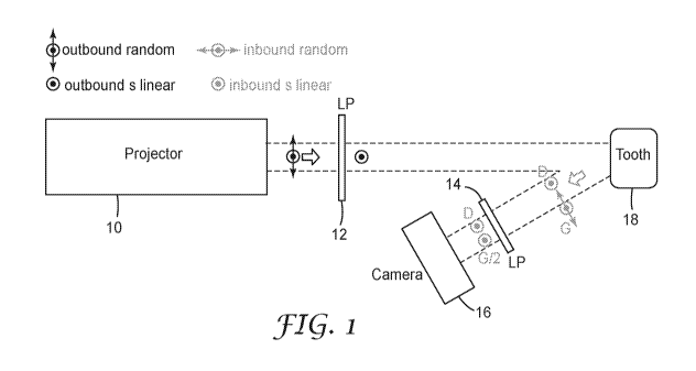
FIG. 1 illustrates an embodiment of an apparatus for projecting a texture
pattern
for 3D imaging. A digital-light-processing (DLP) projector 10 is used to
project a texture
over the field-of-view and depth-of-field of camera 16. Immediately after
exiting the
projector, the light is conditioned by a linear polarizer 12. After reflecting
and scattering
from the object-under-test (for example, tooth 18), the light is filtered by a
second linear
polarizer 14 that is co-aligned with the first linear polarizer 12. The images
are then
captured by camera 16. The embodiment shown in FIG. 1 can optionally be
implemented
without the polarizers 12 and 14.
FIG. 2 illustrates another embodiment of an apparatus for projecting a texture
pattern for 3D imaging. To make the design compact, the projection axis and
camera axis
is made common-path by using a polarizing beam-splitter 22. The light exiting
a DLP
projector 20 is conditioned, which is accomplished by the polarizing beam-
splitter 22.
The unused linear s-state is reflected within the polarizing beam-splitter 22,
upwards and
away from the camera 30 where it can be absorbed/discarded as represented by
block 24.
After exiting the polarizing beam-splitter 22 the linear p-state light passes
through a
quarter-wave retarder (QWR) 28, after which the light is right-hand circular
polarized.
The light can be reflected by a mirror 26 onto the teeth 32 to be imaged. When
the light
directly reflects from the surface of teeth 32, the light is converted to
inbound left-hand
circular polarization. Passing once again through the QWR 28 converts the
light into
linear s-state polarization. Within the polarizing beam-splitter 22, the
linear s-state
reflects towards the camera 30 where images are captured. Any residual inbound
linear p-
state light will be sent back towards the projector where it is discarded.
Polarizing beam-
splitter 22 can be implemented with, for example, a polarizing cube beam-
splitter (PCBS)
3
CA 03092639 2020-08-31
WO 2019/155401
PCT/IB2019/050996
or an irregularly shaped polarizing beam-splitter. The embodiment shown in
FIG. 2 can
optionally be implemented without the QWR 28, in which case the beam-splitter
22 need
not be polarizing and could be implemented with, for example, a plate beam-
splitter.
The DLP projectors 10 and 20 produce a random texture pattern. Projecting a
texture pattern onto a translucent object and then capturing an image of the
scene with the
camera can be described as follows,
I =C=S=P= Texture, (1)
where / is the image frequency spectrum; Texture is the original high contrast
pattern; P is
the transfer function describing the fidelity of the projector; S is the
transfer function of
the object surface; and C is the transfer function of the camera. Furthermore,
all of the
parameters in equation (1) are also functions of spatial frequency and two-
dimensional
spatial coordinates that are orthogonal to the system axis. In an ideal case,
C, S, and P
would be unity over the entire spatial-frequency domain and the image would
perfectly
reproduce the original Texture pattern. However, each of the transfer
functions exhibit a
decreasing amplitude with respect to frequency, ranging from 1 to zero. The
amplitude
describes the transferred contrast level at a given spatial frequency, where
contrast is the
ratio of the difference between the maximum and minimum pixel exposure divided
by the
sum of the maximum and minimum pixel exposure. In effect, the contrast will
decrease as
the pattern becomes finer. The camera is governed by its own modulation
transfer
function (MTF). The surface scatter S can be approximated to resemble a
function
analogous to the camera MTF. The diffusion approximation permits the tooth to
be
characterized with an MTF, depending upon its scattering properties, and thus
be
incorporated into a single MTF that describes the entire imaging system. With
a single
system MTF, an optimum projected texture pattern can be determined.
The projector optics are the first to exert its own MTF onto the initially
pristine
Texture pattern. The C and P parameters are direct consequences of the optical
design of
the camera and projector, respectively, and can be easily modeled from optical
raytracing
software. On the other hand, S depends upon the type of object under test and
will not be
known a priori. To maintain a satisfactory contrast level in the images,
depending on the
S encountered in any particular scene, the characteristics of Texture may need
to adapt and
not remain static. In other words, it may be likely that there is not a single
Texture that is
a ubiquitous solution to capturing adequate images for all teeth. A DLP
projector can be
useful in such circumstances, since the nature of Texture could be rapidly
optimized
4
CA 03092639 2020-08-31
WO 2019/155401
PCT/IB2019/050996
within a few video frames, depending upon feedback gathered from the scene.
Although
not shown in equation (1), the presence of the polarization filters flanks the
S parameter.
For the system in FIG. 1, equation (1) can be written as,
/ C = LP = S = LP = P = Texture, (2)
where the linear polarizers LP have a co-linear alignment. The description for
the system
in FIG. 2 is similar to equation (2) but with the introduction of the quarter-
wave retarder
QWR,
/ C = LP = QWR= S = QWR= LP = P = Texture. (3)
The components LP and QWR are assumed to have no optical wavelength-
dependence.
FIGS. 3A-3F show examples of six different two-dimensional white noise
patterns.
These examples are the pattern files that are sent to the projector, not
actual images
captured by the camera. The initial pattern has 1024x768 pixels, where each
block may
correspond with a single pixel or a group of adjacent pixels. An integer
value, shown
above each pattern, is chosen to assign the integer number of adjacent pixels
into a block
with dark or bright settings. For example, in FIG. 3A 64x64 pixels are
assigned to each
two-dimensional block in the pattern, and in FIG. 3C each block in the pattern
is
comprised of 16x16 pixels.
Each pattern is two-dimensional and not periodic, i.e. not sinusoidal. Rather,
the
pattern is white-noise meaning that the spatial frequency spectrum possesses
signal
ranging from DC out to the upper limit dictated by the pattern cut-off
frequency, i.e. fmax =
1/(2 pixels). The white noise pattern serves two purposes: it provides image
features that
permit good feature-correspondence between images of the stereo-vision camera;
there is
broadband spatial frequency illumination available that permits a measurement
of the
imaging system modulation transfer function; and it permits the measure of the
diffuse
reflectance spatial frequency spectrum of parameter S.
The cameras 16 and 30 can be implemented with, for example, a CMOS digital
image sensor. The image sensor can be partitioned into multiple regions
corresponding
with the optical channels of the apparatus as implemented in a multi-view
intra-oral
scanner. Optionally, multiple image sensors can be used for the channels.
Examples of
multi-view intra-oral scanners are disclosed in US Patent No. 9,591,286, which
is
incorporated herein by reference as if fully set forth.
The DLP projectors can be implemented within an intra-oral scanner containing
the apparatus and can be located, for example, behind or adjacent the image
sensor with
5
CA 03092639 2020-08-31
WO 2019/155401
PCT/IB2019/050996
respect to the object to be imaged. The DLP projectors can be controlled via a
processor
to tune the projected texture pattern before or during imaging, or both before
and during.
Instead of a projector, the system can use a mask to project the pattern. The
systems use
multiple channels to the image sensors with co-axial projecting and imaging.
The projected texture patterns, for example those shown FIGS. 3A-3F, can be
composed of pixels clustered into blocks with the blocks being turned off and
on in a
pseudo-random pattern. For example, each block can have a dark (off) or bright
(on)
setting for the pattern. The translucency of the teeth (or other intra-oral
structures) to be
imaged can determine a size of the blocks in the pattern. As the surface of
the object to be
imaged becomes more translucent or transparent, the blocks can become larger
for the
image sensor to resolve the blocks in the pattern. The pixel size of the
blocks can be fine-
tuned for optimum performance and can overcome the scarcity of features on the
surface
of the object to be imaged, or the translucency of such surface, to
essentially simulate
powder applied to the surface. In particular, the configuration of the blocks,
or other
projected texture pattern, can be sufficient for the image sensor to resolve
features on a
surface of the object to be imaged and possibly optimized for the best or a
desired
resolution of the features by the image sensor.
In the patterns of FIGS. 3A-3F, ideally each adjacent grouping of blocks
(configuration of bright and dark blocks) is unique along each horizontal
direction with
respect to other adjacent grouping of blocks along the same horizontal
direction, but not
necessarily between different horizontal directions. For example, in FIG. 3D
each
adjacent grouping of blocks along a horizontal direction has a different
pattern
(configuration of bright and dark blocks) from each of the other adjacent
grouping of
blocks along the same horizontal direction. Optionally, some adjacent groups
of blocks
along the same horizontal direction can have the same pattern. Also, random
texture
includes pseudo-random texture, since the texture may not be truly random.
The Texture parameter in equation (1) is controlled by the size of the blocks
and
which blocks are turned on (bright) or turned off (dark). The texture patterns
can be
stored as a series of files sent to the projector, for example files
containing the patterns
shown in FIGS. 3A-3F, with each pattern in the series being different from the
other
patterns in the series. The apparatus can cycle through the files sent to the
projector and
evaluate the resulting images to find the best pattern for particular objects
to be imaged.
The other parameters in equation (1), aside from Texture, cannot be directly
controlled.
6
CA 03092639 2020-08-31
WO 2019/155401 PCT/IB2019/050996
FIG. 4 is a graph of the spatial frequency spectrum of a pseudo-random pattern
sent to a projector for block pixel sizes of 2, 8, and 32.
FIG. 5 is a graph of the spatial frequency spectrum of an image after the
transfer
functions of the projection lens, tooth, and camera have been taken into
effect for patterns
with block pixel sizes of 2 and 8. The vertical solid line at 0.07/pixels
represents a cut-off
frequency, the horizontal line at 0.05 represents a noise floor, and both
lines are thresholds
for adequate stereo-vision depth mapping.
7