Note: Descriptions are shown in the official language in which they were submitted.
CA 03092695 2020-08-31
WO 2019/182888
PCT/US2019/022444
DESCRIPTION
HUMAN PD-L2 ANTIBODIES AND METHODS OF USE THEREFOR
PRIORITY CLAIM
[0001] This application claims benefit of priority to U.S. Provisional
Application
Serial No. 62/647,546, filed March 23, 2018, the entire contents of which are
hereby
incorporated by reference.
INCORPORATION OF SEQUENCE LISTING
[0002] The sequence listing that is contained in the file named
"UTFC.P1340W0_5T25.txt", which is 22.9 KB (as measured in Microsoft Windows())
and was created on March 13, 2019, is filed herewith by electronic submission
and is
incorporated by reference herein.
BACKGROUND
1. Field
[0003] The present disclosure relates generally to the field of medicine,
oncology,
and immunology. More particularly, it concerns human antibodies binding to PD-
L2 and
their use in cancer therapies.
2. Description of Related Art
[0004] Programmed death-1 (PD-1) is a cell surface molecule expressed on B and
T cells that regulates the adaptive immune response. The PD-1 receptor on T
cells is
expressed following T cell activation, accumulates over time on the cell
surface, and can
be engaged to attenuate T cell responses as a mechanism of homeostatic
regulation.
Engagement of PD-1 by its ligands PD-Li or PD-L2 transduces a signal that
inhibits T-
cell proliferation, cytokine production, and cytolytic function, marking an
important
checkpoint for cell death.
[0005] PD-Li expression is very tightly regulated by normal cells and is
seldom
expressed in normal tissues but may be rapidly upregulated in a number of
different tissue
types and by tumors in response to interferon-gamma and other inflammatory
mediators
(Dong et al., 2002). PD-L2 expression is highly restricted to antigen
presenting cells,
including dendritic cells and macrophages (Rozali et al., 2012). Exposure of
dendritic
- 1 -
CA 03092695 2020-08-31
WO 2019/182888
PCT/US2019/022444
cells and macrophages to Th2 cytokines, IFNy and Toll Like Receptor ligands
all increase
expression of PD-L2. Human fibroblasts have been shown to express PD-L2,
resulting in
T-cell suppression (Pinchuk et al., 2008). Further, not only normal
fibroblasts, but cancer-
associated fibroblasts constitutively express PD-L2 (Nazareth et al., 2007).
[0006] It is widely known that tumors may adopt normal physiologic checkpoints
for immunomodulation leading to an imbalance between tumor growth and host
surveillance. As they grow, tumors surround themselves with stromal cells
expressing
PD-1 ligands (i.e., PD-Li and PD-L2). When PD-1 expressing T cells encounter
PD-Li
and PD-L2 upon entering the tumor microenvironment, they are rapidly
attenuated and
the tumor escapes immune control. Antibodies blocking the PD-1/PD-L1 binding
interface have been in clinical trials since 2010 (Brahmer et al., 2010).
Companion
studies of PD-Li antibodies have been ongoing since 2012 (Brahmer et al,
2012). As of
yet however, there are no clinical trials for PD-L2 antibodies. These
interactions make the
PD-L2/PD-1 interface an attractive target for therapeutic intervention.
- 2 -
CA 03092695 2020-08-31
WO 2019/182888
PCT/US2019/022444
SUMMARY
[0007] Thus, in accordance with the present disclosure, there is provided an
antibody or antibody fragment comprising clone-paired heavy and light CDR
sequences
from Tables 3 and 4, respectively, that binds selectively to PD-L2. The
antibody or
antibody fragment may be encoded by clone-paired variable sequences as set
forth in
Table 1, may be encoded by light and heavy chain variable sequences having
70%, 80%,
or 90% identity to clone-paired variable sequences as set forth in Table 1, or
may be
encoded by light and heavy chain variable sequences having 95% or greater
identity to
clone-paired sequences as set forth in Table 1. The antibody or antibody
fragment may
comprise light and heavy chain variable sequences according to clone-paired
sequences
from Table 2, may comprise light and heavy chain variable sequences having
70%, 80%
or 90% identity to clone-paired sequences from Table 2, or may comprise light
and heavy
chain variable sequences having 95% or greater identity to clone-paired
sequences from
Table 2.
[0008] There is also provided a method of treating cancer in a subject
comprising
contacting a PD-L2 positive cancer cell in a subject with an antibody as
described above.
The PD-L2 positive cancer cell may be a solid tumor cell, such as a lung
cancer cell,
brain cancer cell, head & neck cancer cell, breast cancer cell, skin cancer
cell, liver cancer
cell, pancreatic cancer cell, stomach cancer cell, colon cancer cell, rectal
cancer cell,
uterine cancer cell, cervical cancer cell, ovarian cancer cell, testicular
cancer cell, skin
cancer cell, esophageal cancer cell, a lymphoma cell, a renal cell carcinoma
cell, or may
be a leukemia or myeloma such as acute myeloid leukemia, chronic myelogenous
leukemia or multiple myeloma.
[0009] The method may further comprise contacting the PD-L2 positive cancer
cell with a second anti-cancer agent or treatment, such as chemotherapy,
radiotherapy,
immunotherapy, hormonal therapy, or toxin therapy. The second anti-cancer
agent or
treatment may inhibit an intracellular PD-L2 function. The second anti-cancer
agent or
treatment may be given at the same time as the first agent, or given before
and/or after the
agent. The PD-L2 positive cancer cell may be a metastatic cancer cell, a
multiply drug
resistant cancer cell or a recurrent cancer cell.
- 3 -
CA 03092695 2020-08-31
WO 2019/182888
PCT/US2019/022444
[0010] The antibody may be a single chain antibody, a single domain antibody,
a
chimeric antibody, or a Fab fragment. The antibody may be a human antibody,
murine
antibody, an IgG, a humanized antibody or a humanized IgG. The antibody or
antibody
fragment may further comprise a label, such as a peptide tag, an enzyme, a
magnetic
particle, a chromophore, a fluorescent molecule, a chemiluminescent molecule,
or a dye.
The antibody or antibody fragment may further comprise an antitumor drug
linked
thereto, such as linked to the antibody or antibody fragment through a
photolabile linker
or an enzymatically-cleaved linker. The antitumor drug may be a toxin, a
radioisotope, a
cytokine or an enzyme. The antibody or antibody fragment may be conjugated to
a
nanoparticle or a liposome
[0011] In another embodiment, there is provided a method of treating a cancer
in
a subject comprising delivering to the subject an antibody or antibody
fragment having
clone-paired heavy and light chain CDR sequences from Tables 3 and 4,
respectively.
The antibody fragment may be a recombinant scFv (single chain fragment
variable)
antibody, Fab fragment, F(ab')2 fragment, or Fv fragment. The antibody may be
an IgG.
The antibody may be is a chimeric antibody.
Delivering may comprise antibody or
antibody fragment administration, or genetic delivery with an RNA or DNA
sequence or
vector encoding the antibody or antibody fragment.
[0012] The antibody or antibody fragment may be encoded by clone-paired light
and heavy chain variable sequences as set forth in Table 1, may be encoded by
clone-
paired light and heavy chain variable sequences having 95% identify to as set
forth in
Table 1, and may be encoded by light and heavy chain variable sequences having
70%,
80%, or 90% identity to clone-paired sequences from Table 1. The antibody or
antibody
fragment may comprise light and heavy chain variable sequences according to
clone-
paired sequences from Table 2, may comprise light and heavy chain variable
sequences
having 70%, 80% or 90% identity to clone-paired sequences from Table 2, or may
comprise light and heavy chain variable sequences having 95% identity to clone-
paired
sequences from Table 2.
[0013] Also provided is a monoclonal antibody, wherein the antibody or
antibody
fragment is characterized by clone-paired heavy and light chain CDR sequences
from
Tables 3 and 4, respectively. The antibody fragment may be a recombinant scFv
(single
- 4 -
CA 03092695 2020-08-31
WO 2019/182888
PCT/US2019/022444
chain fragment variable) antibody, Fab fragment, F(ab')2 fragment, or Fv
fragment. The
antibody may be a chimeric antibody, or an IgG.
[0014] The antibody or antibody fragment may be encoded by clone-paired light
and heavy chain variable sequences as set forth in Table 1, may be encoded by
clone-
paired light and heavy chain variable sequences having 95% identify to as set
forth in
Table 1, and may be encoded by light and heavy chain variable sequences having
70%,
80%, or 90% identity to clone-paired sequences from Table 1. The antibody or
antibody
fragment may comprise light and heavy chain variable sequences according to
clone-
paired sequences from Table 2, may comprise light and heavy chain variable
sequences
having 70%, 80% or 90% identity to clone-paired sequences from Table 2, or may
comprise light and heavy chain variable sequences having 95% identity to clone-
paired
sequences from Table 2.
[0015] In yet another embodiment, there is provided a hybridoma or engineered
cell encoding an antibody or antibody fragment wherein the antibody or
antibody
fragment is characterized by clone-paired heavy and light chain CDR sequences
from
Tables 3 and 4, respectively. The antibody fragment may be a recombinant scFv
(single
chain fragment variable) antibody, Fab fragment, F(ab')2 fragment, or Fv
fragment. The
antibody may be a chimeric antibody or an IgG
[0016] The antibody or antibody fragment may be encoded by clone-paired light
and heavy chain variable sequences as set forth in Table 1, may be encoded by
clone-
paired light and heavy chain variable sequences having 95% identify to as set
forth in
Table 1, and may be encoded by light and heavy chain variable sequences having
70%,
80%, or 90% identity to clone-paired sequences from Table 1. The antibody or
antibody
fragment may comprise light and heavy chain variable sequences according to
clone-
paired sequences from Table 2, may comprise light and heavy chain variable
sequences
having 70%, 80% or 90% identity to clone-paired sequences from Table 2, or may
comprise light and heavy chain variable sequences having 95% identity to clone-
paired
sequences from Table 2.
[0017] A further embodiment comprises a cancer vaccine comprising one or
more antibodies or antibody fragments characterized by clone-paired heavy and
light
chain CDR sequences from Tables 3 and 4, respectively. At least one antibody
fragment
- 5 -
CA 03092695 2020-08-31
WO 2019/182888
PCT/US2019/022444
may be a recombinant scFv (single chain fragment variable) antibody, Fab
fragment,
F(ab')2 fragment, or Fv fragment. At least one of antibody may be a chimeric
antibody,
or an IgG. At least one antibody or antibody fragment may be encoded by clone-
paired
light and heavy chain variable sequences as set forth in Table 1, may be
encoded by
clone-paired light and heavy chain variable sequences having 95% identify to
as set forth
in Table 1, and may be encoded by light and heavy chain variable sequences
having 70%,
80%, or 90% identity to clone-paired sequences from Table 1. At least one
antibody or
antibody fragment may comprise light and heavy chain variable sequences
according to
clone-paired sequences from Table 2, may comprise light and heavy chain
variable
sequences having 70%, 80% or 90% identity to clone-paired sequences from Table
2, or
may comprise light and heavy chain variable sequences having 95% identity to
clone-
paired sequences from Table 2.
[0018] In another embodiment there is provided a method of detecting PD-L2
expressing cells in a subject comprising contacting a sample from said subject
with an
antibody or antibody fragment characterized by clone-paired heavy and light
chain CDR
sequences from Tables 3 and 4, respectively, and detecting a PD-Li or PD-L2
expressing
cell in said sample by binding said antibody or antibody fragment to a cell in
said sample.
The sample may be a body fluid or a tissue sample. The cell may be a cancer
cell, such as
a lymphoma cell, breast cancer cell, or renal cell carcinoma cell. The cell
may be a cell
associated with immune suppression. The cell associated with immune
suppression may
be a non-cancerous cell in a tumor microenvironment, such as a stromal cell or
endothelial cell. Detection may comprise ELISA, RIA, or Western blot. The
method may
further comprise performing the method a second time and determining a change
in
orthopoxyvirus antigen levels as compared to the first assay. The antibody or
antibody
fragment may be encoded by clone-paired light and heavy chain variable
sequences as set
forth in Table 1, may be encoded by clone-paired light and heavy chain
variable
sequences having 95% identify to as set forth in Table 1, and may be encoded
by light
and heavy chain variable sequences having 70%, 80%, or 90% identity to clone-
paired
sequences from Table 1. The antibody or antibody fragment may comprise light
and
heavy chain variable sequences according to clone-paired sequences from Table
2, may
comprise light and heavy chain variable sequences having 70%, 80% or 90%
identity to
clone-paired sequences from Table 2, or may comprise light and heavy chain
variable
sequences having 95% identity to clone-paired sequences from Table 2.
- 6 -
CA 03092695 2020-08-31
WO 2019/182888
PCT/US2019/022444
[0019] Also provided is a method of treating immune suppression in a tumor
microenvironment comprising: delivering to said subject an antibody or
antibody
fragment having clone-paired heavy and light chain CDR sequences from Tables 3
and 4,
respectively. The antibody or antibody fragment may be encoded by clone-paired
light
and heavy chain variable sequences as set forth in Table 1, may be encoded by
clone-
paired light and heavy chain variable sequences having 95% identify to as set
forth in
Table 1, and may be encoded by light and heavy chain variable sequences having
70%,
80%, or 90% identity to clone-paired sequences from Table 1. The antibody or
antibody
fragment may comprise light and heavy chain variable sequences according to
clone-
paired sequences from Table 2, may comprise light and heavy chain variable
sequences
having 70%, 80% or 90% identity to clone-paired sequences from Table 2, or may
comprise light and heavy chain variable sequences having 95% identity to clone-
paired
sequences from Table 2.
[0020] It is contemplated that any method or composition described herein can
be
implemented with respect to any other method or composition described herein.
Other
objects, features and advantages of the present disclosure will become
apparent from the
following detailed description. It should be understood, however, that the
detailed
description and the specific examples, while indicating specific embodiments
of the
disclosure, are given by way of illustration only, since various changes and
modifications
within the spirit and scope of the disclosure will become apparent to those
skilled in the
art from this detailed description.
[0021] As used herein, "essentially free," in terms of a specified component,
is
used herein to mean that none of the specified component has been purposefully
formulated into a composition and/or is present only as a contaminant or in
trace
amounts. The total amount of the specified component resulting from any
unintended
contamination of a composition is preferably below 0.01%. Most preferred is a
composition in which no amount of the specified component can be detected with
standard analytical methods.
[0022] As used herein in the specification and claims, "a" or "an" may mean
one
or more. As used herein in the specification and claims, when used in
conjunction with
the word "comprising", the words "a" or "an" may mean one or more than one. As
used
- 7 -
CA 03092695 2020-08-31
WO 2019/182888
PCT/US2019/022444
herein, in the specification and claim, "another" or "a further" may mean at
least a second
or more.
[0023] As used herein in the specification and claims, the term "about" is
used to
indicate that a value includes the inherent variation of error for the device,
the method
being employed to determine the value, or the variation that exists among the
study
subjects.
[0024] Other objects, features and advantages of the present invention will
become apparent from the following detailed description. It should be
understood,
however, that the detailed description and the specific examples, while
indicating certain
embodiments of the invention, are given by way of illustration only, since
various
changes and modifications within the spirit and scope of the invention will
become
apparent to those skilled in the art from this detailed description.
- 8 -
CA 03092695 2020-08-31
WO 2019/182888
PCT/US2019/022444
BRIEF DESCRIPTION OF THE DRAWINGS
[0025] The following drawings form part of the present specification and are
included to further demonstrate certain aspects of the present invention. The
invention
may be better understood by reference to one or more of these drawings in
combination
with the detailed description of specific embodiments presented herein.
[0026] FIGS. IA-B: Identification of PD-L2 antibodies to block PD-L2
binding to PD-1. Antibody candidates identified as described in the Examples
were
tested for the capacity to bind PD-L2 and block its binding to PD-1. Maximum
fluorescence intensity of Alexafluor 532 labeled PD-1 was measured, with PD-L2
blocking viewed as a reduction in Alexa532 fluorescence in FACS analysis. Anti-
PD-L2
clones 16501, 16425, 16510, and 16478 were evaluated against unstained cells,
control
antibodies, and a commercially available anti-PD-L2 10C12 antibody.
[0027] FIG. 2 - Identification of PD-L2 antibody subclones which bind PD-
L2. Subclones of anti-PD-L2 antibodies (A) 16501 or (C) 16425 were tested
using a PD-
L2:PD-1 assay using CHO-PD-L2 cells capable of stimulating Jurkat T cells
which
produce luciferase in response to activation. Antibodies were added at the
indicated
concentrations. Reactions were performed on the ForteBio Octet platform.
Affinities and
IC5() values for anti-PD-L2 antibodies (C) 16501 and (D) 16425 were calculated
from the
curves.
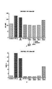
[0028] FIG. 3 - Evaluation of PD-L2 antibodies compared to Keytruda. (Left)
Anti-PD-L2 clone 16501 and subclone 20810 were evaluated for their cell
surface
binding and blocking by detecting Alexafluor intensity of PD-1 cells. (Right)
Anti-PD-L2
clone 1650 and subclone 20810 were evaluated beside clone 20237, Keytruda, and
an
isotype control for their ability to block PD-L2:PD-1 binding in a commercial
PD-L2:PD-
1 binding assay. Antibodies were used at the listed concentrations.
[0029] FIGS. 4A-C - PD-L2 antibodies avidly bind human PD-L2. (FIG. 4A)
Maximum fluorescence intensity was detected using the ForteBio Octet for the
binding
of anti-PD-L2 subclones 16501, 20810, and 20811 to human PD-L2 protein. (FIG.
4B)
Anti-PD-L2 subclones 16425 and 20816 were tested for binding to human PD-L2
protein
and maximum fluorescence intensity was detected. Avidity curves were generated
using
- 9 -
CA 03092695 2020-08-31
WO 2019/182888
PCT/US2019/022444
the ForteBio Octet . (FIG. 4C) Blockade of PD-1-FC binding to human PD-L2 by
anti-
PD-L2 subclones 16501 and 16510.
[0030] FIG. 5 - Equivalence to FDA-approved PD-1/PD-L2 inhibitors.
Candidate antibodies to PD-L2 were assayed using the Promega PD-L2:PD-1
blockade
system. Isotype control, Keytruda, Tecentriq, anti-PD-L2 20810, 20811, and
20816 were
evaluated for their ability to sequester PD-L2 from PD-1. -Fold induction of
the PD-1-
luciferase response is given at the antibody concentrations indicated.
[0031] FIGS. 6A-C - Candidate antibodies are active across multiple human
mixed lymphocyte reactions. Candidate antibodies, FDA approved antibodies,
commercial antibodies, or isotype controls were evaluated in the presence of
induced
dendritic cells and T-cells from separate donors and evaluated by ELISA for IL-
2 or IFN-
y production. (FIG. 6A) Keytruda (anti-PD-1), candidate anti-PD-L2 antibodies
20811
and 20816 and an isotype control were added to the IDCs prior to addition to
CD4+ T
cells. (FIG. 6B) anti-PD-L2 antibody 20814 or an isotype control were added to
the IDCs
prior to addition to CD4+ T cells. (FIG. 6C) Anti-PD-L2 antibody 20814,
Keytruda, PD-
Li monoclonal antibody, an isotype control, or both anti-PD-Li and 20814 were
added to
IDCs prior to subsequent addition to CD4+ T cells.
[0032] FIG. 7 - Candidate antibodies with ADCC are highly active against
human U2940 lymphoma in vivo. Following establishment of PBML xenograft tumors
in SCID mice, mice were treated with either mIgG2a control antibodies,
Herceptin,
Rituxan, or 20810 and tumor volume was assessed on the indicated day.
[0033] FIG. 8 - Candidate antibodies are active against MDA-MB-231.
Following establishment of MDA-MB-231 xenograft tumors in SCID mice, mice were
treated with either mIgG2a control antibodies, Rituxan, Avelumab (anti-PD-L1),
or
20810. Tumor volumes were assessed on the indicated day following
treatment.
[0034] FIG. 9 - aPD-L2 Antibody is Effective in an aPD-L1 Resistant EL4
Lymphoma Mouse Model. Survival was measured for mice injected with anti-PD-Li
resistant EL4 cells expressing PD-L2 and luciferase. 1.5 x 10⁵ EL4 cells
expressing PD-
L2 and luciferase were injected into the mouse tail vein. Mice were treated
with 100 ug
intraperitoneally of the indicated antibodies on days 3, 6, 9, 12, and 15.
Treatment with
- 10 -
CA 03092695 2020-08-31
WO 2019/182888
PCT/US2019/022444
anti-PD-L2 candidate 20810 increased survival rate compared to treatment with
PD-Li
mAb or no treatment.
- 11 -
CA 03092695 2020-08-31
WO 2019/182888
PCT/US2019/022444
DESCRIPTION OF ILLUSTRATIVE EMBODIMENTS
[0035] The inventors have generated monoclonal antibodies with binding
specificity for human PD-L2 protein. As these antibodies have been
demonstrated to bind
to PD-L2, they present an opportunity to block the binding of PD-L2 to PD-1.
They can
also be used to deliver therapeutic payloads to PD-L2 expressing cancer cells.
These and
other aspects of the disclosure are described in even greater detail below.
I. PD-L2
A. Structure
[0036] Programmed death-ligand 2 (PD-L2) is a protein encoded by the CD273
gene. PD-L2 is a 31 kDa protein which may play a major role in immune
suppression
during a variety of events such as, pregnancy, tissue allografts, autoimmune
disease,
cancer and other disease states. The human PD-L2 protein is encoded by the
amino acid
sequence shown below:
MIFLLLMLSLELQLHQIAALFTVTVPKELYIIEHGSNVTLECNFDTGSHVNLGAITASLQKV
ENDTSPHRERATLLEEQLPLGKASFHIPQVQVRDEGQYQCIIIYGVAWDYKYLTLKVKASYR
KINTHILKVPETDEVELTCQATGYPLAEVSWPNVSVPANTSHSRTPEGLYQVTSVLRLKPPP
GRNFSCVFWNTHVRELTLASIDLQSQMEPRTHPTWLLHIFIPFCITAFIFIATVIALRKQLC
QKLYSSKDTTKRPVTTTKREVNSAI (SEQ ID NO: 1)
[0037] PD-L2 is initially produced with a signal peptide corresponding to
amino
acids 1-19 of SEQ ID NO. 1, which is subsequently removed to yield the mature
protein.
The mature PD-L2 protein, corresponding to amino acids 20-273 of SEQ ID NO. 1,
is
comprised of an Ig-like V-domain, an Ig-like C2-type domain, transmembrane
domain,
and a cytoplasmic tail.
B. Function
[0038] PD-L2 is a ligand to its receptor, PD-1. PD-1 may be found on activated
T
cells, B cells, and myeloid cells. Binding of PD-L2 to PD-1 begins an
immunological
cascade which impairs proliferation, cytokine production, cytolytic function
and survival
of the T cell. PD-1 transmits an inhibitor signal that reduces proliferation
of antigen
specific CD8+ T cells and CD4+ helper T-cells. PD-L2 has also been shown to be
an
- 12 -
CA 03092695 2020-08-31
WO 2019/182888
PCT/US2019/022444
independent predictor of response to the PD-1 antibody pembrolizumab across
multiple
cancers (Yearley et al., 2017).
Monoclonal Antibodies and Production Thereof
A. General Methods
[0039] Antibodies to PD-L2 may be produced by standard methods as are well
known in the art (see, e.g., Antibodies: A Laboratory Manual, Cold Spring
Harbor
Laboratory, 1988; U.S. Patent 4,196,265). The methods for generating
monoclonal
antibodies (mAbs) generally begin along the same lines as those for preparing
polyclonal
antibodies. The first step for both these methods is immunization of an
appropriate host or
identification of subjects who are immune due to prior natural infection. As
is well known
in the art, a given composition for immunization may vary in its
immunogenicity. It is
often necessary therefore to boost the host immune system, as may be achieved
by
coupling a peptide or polypeptide immunogen to a carrier. Exemplary and
preferred
carriers are keyhole limpet hemocyanin (KLH) and bovine serum albumin (BSA).
Other
albumins such as ovalbumin, mouse serum albumin or rabbit serum albumin can
also be
used as carriers. Means for conjugating a polypeptide to a carrier protein are
well known
in the art and include glutaraldehyde, m-maleimidobencoyl-N-hydroxysuccinimide
ester,
carbodiimide and bis-biazotized benzidine. As also is well known in the art,
the
immunogenicity of a particular immunogen composition can be enhanced by the
use of
non-specific stimulators of the immune response, known as adjuvants. Exemplary
and
preferred adjuvants include complete Freund's adjuvant (a non-specific
stimulator of the
immune response containing killed Mycobacterium tuberculosis), incomplete
Freund's
adjuvants and aluminum hydroxide adjuvant.
[0040] The amount of immunogen composition used in the production of
polyclonal antibodies varies upon the nature of the immunogen as well as the
animal used
for immunization. A variety of routes can be used to administer the immunogen
(subcutaneous, intramuscular, intradermal, intravenous and intraperitoneal).
The
production of polyclonal antibodies may be monitored by sampling blood of the
immunized animal at various points following immunization. A second, booster
injection,
also may be given. The process of boosting and titering is repeated until a
suitable titer is
achieved. When a desired level of immunogenicity is obtained, the immunized
animal can
- 13 -
CA 03092695 2020-08-31
WO 2019/182888
PCT/US2019/022444
be bled and the serum isolated and stored, and/or the animal can be used to
generate
monoclonal antibodies.
[0041] Following immunization, somatic cells with the potential for producing
antibodies, specifically B lymphocytes (B cells), are selected for use in the
mAb
generating protocol. These cells may be obtained from biopsied spleens or
lymph nodes,
or from circulating blood. The antibody-producing B lymphocytes from the
immunized
animal are then fused with cells of an immortal myeloma cell, generally one of
the same
species as the animal that was immunized or human or human/mouse chimeric
cells.
Myeloma cell lines suited for use in hybridoma-producing fusion procedures
preferably
are non-antibody-producing, have high fusion efficiency, and enzyme
deficiencies that
render then incapable of growing in certain selective media which support the
growth of
only the desired fused cells (hybridomas).
[0042] Any one of a number of myeloma cells may be used, as are known to those
of skill in the art (Goding, pp. 65-66, 1986; Campbell, pp. 75-83, 1984). For
example,
where the immunized animal is a mouse, one may use P3-X63/Ag8, X63-Ag8.653,
NS1/1.Ag 4 1, Sp210-Ag14, FO, NSO/U, MPC-11, MPC11-X45-GTG 1.7 and
S194/5XXO Bul; for rats, one may use R210.RCY3, Y3-Ag 1.2.3, IR983F and 4B210;
and U-266, GM1500-GRG2, LICR-LON-HMy2 and UC729-6 are all useful in
connection with human cell fusions. One particular murine myeloma cell is the
NS-1
myeloma cell line (also termed P3-NS-1-Ag4-1), which is readily available from
the
NIGMS Human Genetic Mutant Cell Repository by requesting cell line repository
number GM3573. Another mouse myeloma cell line that may be used is the
8-azaguanine-resistant mouse murine myeloma SP2/0 non-producer cell line. More
recently, additional fusion partner lines for use with human B cells have been
described,
including KR12 (ATCC CRL-8658; K6H6/B5 (ATCC CRL-1823 SHM-D33 (ATCC
CRL-1668) and HMMA2.5 (Posner et al., 1987). The antibodies in this disclosure
were
generated using the SP2/0/mIL-6 cell line, an IL-6 secreting derivative of the
SP2/0 line.
[0043] Methods for generating hybrids of antibody-producing spleen or lymph
node cells and myeloma cells usually comprise mixing somatic cells with
myeloma cells
in a 2:1 proportion, though the proportion may vary from about 20:1 to about
1:1,
respectively, in the presence of an agent or agents (chemical or electrical)
that promote
the fusion of cell membranes. Fusion methods using Sendai virus have been
described by
- 14 -
CA 03092695 2020-08-31
WO 2019/182888
PCT/US2019/022444
Kohler and Milstein (1975; 1976), and those using polyethylene glycol (PEG),
such as
37% (v/v) PEG, by Gefter et al. (1977). The use of electrically induced fusion
methods
also is appropriate (Goding, pp. 71-74, 1986).
[0044] Fusion procedures usually produce viable hybrids at low frequencies,
about 1 x 10⁻⁶ to 1 x 10⁻⁸. However, this does not pose a problem, as the
viable, fused
hybrids are differentiated from the parental, infused cells (particularly the
infused
myeloma cells that would normally continue to divide indefinitely) by
culturing in a
selective medium. The selective medium is generally one that contains an agent
that
blocks the de novo synthesis of nucleotides in the tissue culture media.
Exemplary and
preferred agents are aminopterin, methotrexate, and azaserine. Aminopterin and
methotrexate block de novo synthesis of both purines and pyrimidines, whereas
azaserine
blocks only purine synthesis. Where aminopterin or methotrexate is used, the
media is
supplemented with hypoxanthine and thymidine as a source of nucleotides (HAT
medium). Where azaserine is used, the media is supplemented with hypoxanthine.
Ouabain is added if the B cell source is an Epstein Barr virus (EBV)
transformed human
B cell line, in order to eliminate EBV transformed lines that have not fused
to the
myeloma.
[0045] The preferred selection medium is HAT or HAT with ouabain. Only cells
capable of operating nucleotide salvage pathways are able to survive in HAT
medium.
The myeloma cells are defective in key enzymes of the salvage pathway, e.g.,
hypoxanthine phosphoribosyl transferase (HPRT), and they cannot survive. The B
cells
can operate this pathway, but they have a limited life span in culture and
generally die
within about two weeks. Therefore, the only cells that can survive in the
selective media
are those hybrids formed from myeloma and B cells. When the source of B cells
used for
fusion is a line of EBV-transformed B cells, as here, ouabain is also used for
drug
selection of hybrids as EBV-transformed B cells are susceptible to drug
killing, whereas
the myeloma partner used is chosen to be ouabain resistant.
[0046] Culturing provides a population of hybridomas from which specific
hybridomas are selected. Typically, selection of hybridomas is performed by
culturing the
cells by single-clone dilution in microtiter plates, followed by testing the
individual clonal
supernatants (after about two to three weeks) for the desired reactivity. The
assay should
- 15 -
CA 03092695 2020-08-31
WO 2019/182888
PCT/US2019/022444
be sensitive, simple and rapid, such as radioimmunoassays, enzyme
immunoassays,
cytotoxicity assays, plaque assays dot immunobinding assays, and the like.
[0047] The selected hybridomas are then serially diluted or single-cell sorted
by
flow cytometric sorting and cloned into individual antibody-producing cell
lines, which
clones can then be propagated indefinitely to provide mAbs. The cell lines may
be
exploited for MAb production in two basic ways. A sample of the hybridoma can
be
injected (often into the peritoneal cavity) into an animal (e.g., a mouse).
Optionally, the
animals are primed with a hydrocarbon, especially oils such as pristane
(tetramethylpentadecane) prior to injection. When human hybridomas are used in
this
way, it is optimal to inject immunocompromised mice, such as SCID mice, to
prevent
tumor rejection. The injected animal develops tumors secreting the specific
monoclonal
antibody produced by the fused cell hybrid. The body fluids of the animal,
such as serum
or ascites fluid, can then be tapped to provide mAbs in high concentration.
The individual
cell lines could also be cultured in vitro, where the mAbs are naturally
secreted into the
culture medium from which they can be readily obtained in high concentrations.
Alternatively, human hybridoma cells lines can be used in vitro to produce
immunoglobulins in cell supernatant. The cell lines can be adapted for growth
in serum-
free medium to optimize the ability to recover human monoclonal
immunoglobulins of
high purity.
[0048] Monoclonal antibodies produced by either means may be further purified,
if desired, using filtration, centrifugation and various chromatographic
methods such as
FPLC or affinity chromatography. Fragments of the monoclonal antibodies of the
disclosure can be obtained from the purified monoclonal antibodies by methods
which
include digestion with enzymes, such as pepsin or papain, and/or by cleavage
of disulfide
bonds by chemical reduction. Alternatively, monoclonal antibody fragments
encompassed
by the present disclosure can be synthesized using an automated peptide
synthesizer.
[0049] It also is contemplated that a molecular cloning approach may be used
to
generate monoclonal antibodies. For this, RNA can be isolated from the
hybridoma line
and the antibody genes obtained by RT-PCR and cloned into an immunoglobulin
expression vector. Alternatively, combinatorial immunoglobulin phagemid
libraries are
prepared from RNA isolated from the cell lines and phagemids expressing
appropriate
antibodies are selected by panning using viral antigens. The advantages of
this approach
- 16-
CA 03092695 2020-08-31
WO 2019/182888
PCT/US2019/022444
over conventional hybridoma techniques are that approximately 104 times as
many
antibodies can be produced and screened in a single round, and that new
specificities are
generated by H and L chain combination which further increases the chance of
finding
appropriate antibodies.
[0050] Yeast-based antibody presentation libraries may be designed rationally,
and antibodies may be selected and/or isolated from such yeast-based antibody
presentation libraries, as disclosed in, for example, W02012/009568;
W02009/036379;
W02010/105256; W02003/074679; U.S. Patent 8,691,730; and U.S. Patent
9,354,228.
The antibodies may then be expressed as full length IgGs from the desired cell
type and
purified.
[0051] Other U.S. patents, each incorporated herein by reference, that teach
the
production of antibodies useful in the present disclosure include U.S. Patent
5,565,332,
which describes the production of chimeric antibodies using a combinatorial
approach;
U.S. Patent 4,816,567 which describes recombinant immunoglobulin preparations;
and
U.S. Patent 4,867,973 which describes antibody-therapeutic agent conjugates.
B. Antibodies of the Present Disclosure
[0052] Antibodies according to the present disclosure may be defined, in the
first
instance, by their binding specificity, i.e., binding to PD-L2. Those of skill
in the art, by
assessing the binding specificity/affinity of a given antibody using
techniques well known
to those of skill in the art, can determine whether such antibodies fall
within the scope of
the instant claims. In one aspect, there are provided monoclonal antibodies
having clone-
paired CDR's from the heavy and light chains as illustrated in Tables 3 and 4,
respectively. Such antibodies may be produced by the clones discussed below in
the
Examples section using methods described herein.
[0053] In a second aspect, the antibodies may be defined by their variable
sequence, which include additional "framework" regions. These are provided in
Tables 1
and 2 that encode or represent full variable regions. Furthermore, the
antibodies
sequences may vary from these sequences, optionally using methods discussed in
greater
detail below. For example, nucleic acid sequences may vary from those set out
above in
that (a) the variable regions may be segregated away from the constant domains
of the
light and heavy chains, (b) the nucleic acids may vary from those set out
above while not
- 17 -
CA 03092695 2020-08-31
WO 2019/182888
PCT/US2019/022444
affecting the residues encoded thereby, (c) the nucleic acids may vary from
those set out
above by a given percentage, e.g., 70%, 75%, 80%, 85%, 90%, 91%, 92%, 93%,
94%,
95%, 96%, 97%, 98% or 99% homology, (d) the nucleic acids may vary from those
set
out above by virtue of the ability to hybridize under high stringency
conditions, as
exemplified by low salt and/or high temperature conditions, such as provided
by about
0.02 M to about 0.15 M NaCl at temperatures of about 50 C to about 70 C, (e)
the amino
acids may vary from those set out above by a given percentage, e.g., 80%, 85%,
90%,
91%, 92%, 93%, 94%, 95%, 96%, 97%, 98% or 99% homology, or (f) the amino acids
may vary from those set out above by permitting conservative substitutions
(discussed
below). Each of the foregoing applies to the nucleic acid sequences set forth
as Table 1
and the amino acid sequences of Table 2.
C. Engineering of Antibody Sequences
[0054] In various embodiments, one may choose to engineer sequences of the
identified antibodies for a variety of reasons, such as improved expression,
improved
cross-reactivity or diminished off-target binding. The following is a general
discussion of
relevant techniques for antibody engineering.
[0055] Hybridomas may be cultured, then cells lysed, and total RNA extracted.
Random hexamers may be used with RT to generate cDNA copies of RNA, and then
PCR
performed using a multiplex mixture of PCR primers expected to amplify all
human
variable gene sequences. PCR product can be cloned into pGEM-T Easy vector,
then
sequenced by automated DNA sequencing using standard vector primers. Assay of
binding and neutralization may be performed using antibodies collected from
hybridoma
supernatants and purified by FPLC, using Protein G columns.
[0056] Recombinant full length IgG antibodies were generated by subcloning
heavy and light chain Fv DNAs from the cloning vector into an IgG plasmid
vector,
transfected into 293 Freestyle cells or CHO cells, and antibodies were
collected an
purified from the 293 or CHO cell supernatant.
[0057] The rapid availability of antibody produced in the same host cell and
cell
culture process as the final cGMP manufacturing process has the potential to
reduce the
duration of process development programs. Lonza has developed a generic method
using
pooled transfectants grown in CDACF medium, for the rapid production of small
- 18 -
CA 03092695 2020-08-31
WO 2019/182888
PCT/US2019/022444
quantities (up to 50 g) of antibodies in CHO cells. Although slightly slower
than a true
transient system, the advantages include a higher product concentration and
use of the
same host and process as the production cell line. Example of growth and
productivity of
GS-CHO pools, expressing a model antibody, in a disposable bioreactor: in a
disposable
bag bioreactor culture (5 L working volume) operated in fed-batch mode, a
harvest
antibody concentration of 2 g/L was achieved within 9 weeks of transfection.
[0058] Antibodies, and antibody libraries from which such antibodies may be
selected and/or isolated, may be rationally designed and synthesized, such as
by the
Adimab technology, as disclosed in, for example, W02012/009568;
W02009/036379;
W02010/105256; W02003/074679; US Patent 8,691,730; and US Patent 9,354,228.
This method of synthesis antibodies requires that the nucleotide sequence
coding for the
desired or designed antibody be inserted into a vector for ectopic expression.
Then the
desired antibodies may be expressed as full chain IgG molecules and purified.
[0059] Antibody molecules will comprise fragments (such as F(ab'), F(ab')2)
that
are produced, for example, by the proteolytic cleavage of the mAbs, or single-
chain
immunoglobulins producible, for example, via recombinant means. Such antibody
derivatives are monovalent. In one embodiment, such fragments can be combined
with
one another, or with other antibody fragments or receptor ligands to form
"chimeric"
binding molecules. Significantly, such chimeric molecules may contain
substituents
capable of binding to different epitopes of the same molecule.
[0060] In related embodiments, the antibody is a derivative of the disclosed
antibodies, e.g., an antibody comprising the CDR sequences identical to those
in the
disclosed antibodies (e.g., a chimeric, or CDR-grafted antibody).
Alternatively, one may
wish to make modifications, such as introducing conservative changes into an
antibody
molecule. In making such changes, the hydropathic index of amino acids may be
considered. The importance of the hydropathic amino acid index in conferring
interactive
biologic function on a protein is generally understood in the art (Kyte and
Doolittle,
1982). It is accepted that the relative hydropathic character of the amino
acid contributes
to the secondary structure of the resultant protein, which in turn defines the
interaction of
the protein with other molecules, for example, enzymes, substrates, receptors,
DNA,
antibodies, antigens, and the like.
- 19 -
CA 03092695 2020-08-31
WO 2019/182888
PCT/US2019/022444
[0061] It also is understood in the art that the substitution of like amino
acids can
be made effectively on the basis of hydrophilicity. U.S. Patent 4,554,101,
incorporated
herein by reference, states that the greatest local average hydrophilicity of
a protein, as
governed by the hydrophilicity of its adjacent amino acids, correlates with a
biological
property of the protein. As detailed in U.S. Patent 4,554,101, the following
hydrophilicity
values have been assigned to amino acid residues: basic amino acids: arginine
(+3.0),
lysine (+3.0), and histidine (-0.5); acidic amino acids: aspartate (+3.0 1),
glutamate
(+3.0 1), asparagine (+0.2), and glutamine (+0.2); hydrophilic, nonionic
amino acids:
serine (+0.3), asparagine (+0.2), glutamine (+0.2), and threonine (-0.4),
sulfur containing
amino acids: cysteine (-1.0) and methionine (-1.3); hydrophobic, nonaromatic
amino
acids: valine (-1.5), leucine (-1.8), isoleucine (-1.8), proline (-0.5 1),
alanine (-0.5), and
glycine (0); hydrophobic, aromatic amino acids: tryptophan (-3.4),
phenylalanine (-2.5),
and tyrosine (-2.3).
[0062] It is understood that an amino acid can be substituted for another
having a
similar hydrophilicity and produce a biologically or immunologically modified
protein. In
such changes, the substitution of amino acids whose hydrophilicity values are
within 2
is preferred, those that are within 1 are particularly preferred, and those
within 0.5 are
even more particularly preferred.
[0063] As outlined above, amino acid substitutions generally are based on the
relative similarity of the amino acid side-chain substituents, for example,
their
hydrophobicity, hydrophilicity, charge, size, and the like. Exemplary
substitutions that
take into consideration the various foregoing characteristics are well known
to those of
skill in the art and include: arginine and lysine; glutamate and aspartate;
serine and
threonine; glutamine and asparagine; and valine, leucine and isoleucine.
[0064] The present disclosure also contemplates isotype modification. By
modifying the Fc region to have a different isotype, different functionalities
can be
achieved. For example, changing to IgGi can increase antibody dependent cell
cytotoxicity, switching to class A can improve tissue distribution, and
switching to class
M can improve valency.
[0065] Modified antibodies may be made by any technique known to those of
skill in the art, including expression through standard molecular biological
techniques, or
- 20 -
CA 03092695 2020-08-31
WO 2019/182888
PCT/US2019/022444
the chemical synthesis of polypeptides. Methods for recombinant expression are
addressed elsewhere in this document.
D. Single Chain Antibodies
[0066] A Single Chain Variable Fragment (scFv) is a fusion of the variable
regions of the heavy and light chains of immunoglobulins, linked together with
a short
(usually serine, glycine) linker. This chimeric molecule retains the
specificity of the
original immunoglobulin, despite removal of the constant regions and the
introduction of
a linker peptide. This modification usually leaves the specificity unaltered.
These
molecules were created historically to facilitate phage display where it is
highly
convenient to express the antigen binding domain as a single peptide.
Alternatively, scFv
can be created directly from subcloned heavy and light chains derived from a
hybridoma.
Single chain variable fragments lack the constant Fc region found in complete
antibody
molecules, and thus, the common binding sites (e.g., protein A/G) used to
purify
antibodies. These fragments can often be purified/immobilized using Protein L
since
Protein L interacts with the variable region of kappa light chains.
[0067] Flexible linkers generally are comprised of helix- and turn-promoting
amino acid residues such as alaine, serine and glycine. However, other
residues can
function as well. Tang et al. (1996) used phage display as a means of rapidly
selecting
tailored linkers for single-chain antibodies (scFvs) from protein linker
libraries. A random
linker library was constructed in which the genes for the heavy and light
chain variable
domains were linked by a segment encoding an 18-amino acid polypeptide of
variable
composition. The scFv repertoire (approx. 5 x 10⁶ different members) was
displayed on
filamentous phage and subjected to affinity selection with hapten. The
population of
selected variants exhibited significant increases in binding activity but
retained
considerable sequence diversity. Screening 1054 individual variants
subsequently yielded
a catalytically active scFv that was produced efficiently in soluble form.
Sequence
analysis revealed a conserved proline in the linker two residues after the VH
C terminus
and an abundance of arginines and prolines at other positions as the only
common
features of the selected tethers.
[0068] The recombinant antibodies of the present disclosure may also involve
sequences or moieties that permit dimerization or multimerization of the
receptors. Such
sequences include those derived from IgA, which permit formation of multimers
in
- 21 -
CA 03092695 2020-08-31
WO 2019/182888
PCT/US2019/022444
conjunction with the J-chain. Another multimerization domain is the Gal4
dimerization
domain. In other embodiments, the chains may be modified with agents such as
biotin/avidin, which permit the combination of two antibodies.
[0069] In a separate embodiment, a single-chain antibody can be created by
joining receptor light and heavy chains using a non-peptide linker or chemical
unit.
Generally, the light and heavy chains will be produced in distinct cells,
purified, and
subsequently linked together in an appropriate fashion (i.e., the N-terminus
of the heavy
chain being attached to the C-terminus of the light chain via an appropriate
chemical
bridge).
[0070] Cross-linking reagents are used to form molecular bridges that tie
functional groups of two different molecules, e.g., a stablizing and
coagulating agent.
However, it is contemplated that dimers or multimers of the same analog or
heteromeric
complexes comprised of different analogs can be created. To link two different
compounds in a step-wise manner, hetero-bifunctional cross-linkers can be used
that
eliminate unwanted homopolymer formation.
[0071] An exemplary hetero-bifunctional cross-linker contains two reactive
groups: one reacting with primary amine group (e.g., N-hydroxy succinimide)
and the
other reacting with a thiol group (e.g., pyridyl disulfide, maleimides,
halogens, etc.).
Through the primary amine reactive group, the cross-linker may react with the
lysine
residue(s) of one protein (e.g., the selected antibody or fragment) and
through the thiol
reactive group, the cross-linker, already tied up to the first protein, reacts
with the
cysteine residue (free sulfhydryl group) of the other protein (e.g., the
selective agent).
[0072] It is preferred that a cross-linker having reasonable stability in
blood will
be employed. Numerous types of disulfide-bond containing linkers are known
that can be
successfully employed to conjugate targeting and therapeutic/preventative
agents. Linkers
that contain a disulfide bond that is sterically hindered may prove to give
greater stability
in vivo, preventing release of the targeting peptide prior to reaching the
site of action.
These linkers are thus one group of linking agents.
[0073] Another cross-linking reagent is SMPT, which is a bifunctional cross-
linker containing a disulfide bond that is "sterically hindered" by an
adjacent benzene
ring and methyl groups. It is believed that steric hindrance of the disulfide
bond serves a
- 22 -
CA 03092695 2020-08-31
WO 2019/182888
PCT/US2019/022444
function of protecting the bond from attack by thiolate anions such as
glutathione which
can be present in tissues and blood, and thereby help in preventing decoupling
of the
conjugate prior to the delivery of the attached agent to the target site.
[0074] The SMPT cross-linking reagent, as with many other known cross-linking
reagents, lends the ability to cross-link functional groups such as the SH of
cysteine or
primary amines (e.g., the epsilon amino group of lysine). Another possible
type of cross-
linker includes the hetero-bifunctional photoreactive phenylazides containing
a cleavable
disulfide bond such as sulfosuccinimidy1-2-(p-azido salicylamido) ethy1-1,3'-
dithiopropionate. The N-hydroxy-succinimidyl group reacts with primary amino
groups
and the phenylazide (upon photolysis) reacts non-selectively with any amino
acid residue.
[0075] In addition to hindered cross-linkers, non-hindered linkers also can be
employed in accordance herewith. Other useful cross-linkers, not considered to
contain or
generate a protected disulfide, include SATA, SPDP and 2-iminothiolane
(Wawrzynczak
& Thorpe, 1987). The use of such cross-linkers is well understood in the art.
Another
embodiment involves the use of flexible linkers.
[0076] U.S. Patent 4,680,338, describes bifunctional linkers useful for
producing
conjugates of ligands with amine-containing polymers and/or proteins,
especially for
forming antibody conjugates with chelators, drugs, enzymes, detectable labels
and the
like. U.S. Patents 5,141,648 and 5,563,250 disclose cleavable conjugates
containing a
labile bond that is cleavable under a variety of mild conditions. This linker
is particularly
useful in that the agent of interest may be bonded directly to the linker,
with cleavage
resulting in release of the active agent. Particular uses include adding a
free amino or free
sulfhydryl group to a protein, such as an antibody, or a drug.
[0077] U.S. Patent 5,856,456 provides peptide linkers for use in connecting
polypeptide constituents to make fusion proteins, e.g., single chain
antibodies. The linker
is up to about 50 amino acids in length, contains at least one occurrence of a
charged
amino acid (preferably arginine or lysine) followed by a proline, and is
characterized by
greater stability and reduced aggregation. U.S. Patent 5,880,270 discloses
aminooxy-
containing linkers useful in a variety of immunodiagnostic and separative
techniques.
- 23 -
CA 03092695 2020-08-31
WO 2019/182888
PCT/US2019/022444
E. Purification
[0078] In certain embodiments, the antibodies of the present disclosure may be
purified. The term "purified," as used herein, is intended to refer to a
composition,
isolatable from other components, wherein the protein is purified to any
degree relative to
its naturally-obtainable state. A purified protein therefore also refers to a
protein, free
from the environment in which it may naturally occur. Where the term
"substantially
purified" is used, this designation will refer to a composition in which the
protein or
peptide forms the major component of the composition, such as constituting
about 50%,
about 60%, about 70%, about 80%, about 90%, about 95% or more of the proteins
in the
composition.
[0079] Protein purification techniques are well known to those of skill in the
art.
These techniques involve, at one level, the crude fractionation of the
cellular milieu to
polypeptide and non-polypeptide fractions. Having separated the polypeptide
from other
proteins, the polypeptide of interest may be further purified using
chromatographic and
electrophoretic techniques to achieve partial or complete purification (or
purification to
homogeneity). Analytical methods particularly suited to the preparation of a
pure peptide
are ion-exchange chromatography, exclusion chromatography; polyacrylamide gel
electrophoresis; isoelectric focusing. Other methods for protein purification
include,
precipitation with ammonium sulfate, PEG, antibodies and the like or by heat
denaturation, followed by centrifugation; gel filtration, reverse phase,
hydroxylapatite and
affinity chromatography; and combinations of such and other techniques.
[0080] In purifying an antibody of the present disclosure, it may be desirable
to
express the polypeptide in a prokaryotic or eukaryotic expression system and
extract the
protein using denaturing conditions. The polypeptide may be purified from
other cellular
components using an affinity column, which binds to a tagged portion of the
polypeptide.
As is generally known in the art, it is believed that the order of conducting
the various
purification steps may be changed, or that certain steps may be omitted, and
still result in
a suitable method for the preparation of a substantially purified protein or
peptide.
[0081] Commonly, complete antibodies are fractionated utilizing agents (i.e.,
protein A) that bind the Fc portion of the antibody. Alternatively, antigens
may be used to
simultaneously purify and select appropriate antibodies. Such methods often
utilize the
selection agent bound to a support, such as a column, filter or bead. The
antibodies is
- 24 -
CA 03092695 2020-08-31
WO 2019/182888
PCT/US2019/022444
bound to a support, contaminants removed (e.g., washed away), and the
antibodies
released by applying conditions (salt, heat, etc.).
[0082] Various methods for quantifying the degree of purification of the
protein
or peptide will be known to those of skill in the art in light of the present
disclosure.
These include, for example, determining the specific activity of an active
fraction, or
assessing the amount of polypeptides within a fraction by SDS/PAGE analysis.
Another
method for assessing the purity of a fraction is to calculate the specific
activity of the
fraction, to compare it to the specific activity of the initial extract, and
to thus calculate
the degree of purity. The actual units used to represent the amount of
activity will, of
course, be dependent upon the particular assay technique chosen to follow the
purification
and whether or not the expressed protein or peptide exhibits a detectable
activity.
[0083] It is known that the migration of a polypeptide can vary, sometimes
significantly, with different conditions of SDS/PAGE (Capaldi et al., 1977).
It will
therefore be appreciated that under differing electrophoresis conditions, the
apparent
molecular weights of purified or partially purified expression products may
vary.
Pharmaceutical Formulations and Treatment of Cancer
A. Cancers
[0084] Cancer results from the outgrowth of a clonal population of cells from
tissue. The development of cancer, referred to as carcinogenesis, can be
modeled and
characterized in a number of ways. An association between the development of
cancer
and inflammation has long-been appreciated. The inflammatory response is
involved in
the host defense against microbial infection, and also drives tissue repair
and
regeneration. Considerable evidence points to a connection between
inflammation and a
risk of developing cancer, i.e., chronic inflammation can lead to dysplasia.
[0085] Cancer cells to which the methods of the present disclosure can be
applied
include generally any cell that expresses PD-L2, and more particularly, that
overexpresses
PD-L2. An appropriate cancer cell can be a breast cancer, lung cancer, colon
cancer,
pancreatic cancer, renal cancer, stomach cancer, liver cancer, bone cancer,
hematological
cancer (e.g., leukemia or lymphoma), neural tissue cancer, melanoma, ovarian
cancer,
testicular cancer, prostate cancer, cervical cancer, vaginal cancer, or
bladder cancer cell.
In addition, the methods of the disclosure can be applied to a wide range of
species, e.g.,
- 25 -
CA 03092695 2020-08-31
WO 2019/182888
PCT/US2019/022444
humans, non-human primates (e.g., monkeys, baboons, or chimpanzees), horses,
cattle,
pigs, sheep, goats, dogs, cats, rabbits, guinea pigs, gerbils, hamsters, rats,
and mice.
Cancers may also be recurrent, metastatic and/or multi-drug resistant, and the
methods of
the present disclosure may be particularly applied to such cancers so as to
render them
resectable, to prolong or re-induce remission, to inhibit angiogenesis, to
prevent or limit
metastasis, and/or to treat multi-drug resistant cancers. At a cellular level,
this may
translate into killing cancer cells, inhibiting cancer cell growth, or
otherwise reversing or
reducing the malignant phenotype of tumor cells.
B. Formulation and Administration
[0086] The present disclosure provides pharmaceutical compositions comprising
anti-PD-L2 antibodies. In a specific embodiment, the term "pharmaceutically
acceptable"
means approved by a regulatory agency of the Federal or a state government or
listed in
the U.S. Pharmacopeia or other generally recognized pharmacopeia for use in
animals,
and more particularly in humans. The term "carrier" refers to a diluent,
excipient, or
vehicle with which the therapeutic is administered. Such pharmaceutical
carriers can be
sterile liquids, such as water and oils, including those of petroleum, animal,
vegetable or
synthetic origin, such as peanut oil, soybean oil, mineral oil, sesame oil and
the like.
Other suitable pharmaceutical excipients include starch, glucose, lactose,
sucrose, saline,
dextrose, gelatin, malt, rice, flour, chalk, silica gel, sodium stearate,
glycerol
monostearate, talc, sodium chloride, dried skim milk, glycerol, propylene
glycol, water,
ethanol and the like.
[0087] The compositions can be formulated as neutral or salt forms.
Pharmaceutically acceptable salts include those formed with anions such as
those derived
from hydrochloric, phosphoric, acetic, oxalic, tartaric acids, etc., and those
formed with
cations such as those derived from sodium, potassium, ammonium, calcium,
ferric
hydroxides, isopropylamine, triethylamine, 2-ethylamino ethanol, histidine,
procaine, etc.
[0088] The antibodies of the present disclosure may include classic
pharmaceutical preparations. Administration of these compositions according to
the
present disclosure will be via any common route so long as the target tissue
is available
via that route. This includes oral, nasal, buccal, rectal, vaginal or topical.
Alternatively,
administration may be by intradermal, subcutaneous, intramuscular,
intraperitoneal or
intravenous injection.
Such compositions would normally be administered as
- 26 -
CA 03092695 2020-08-31
WO 2019/182888
PCT/US2019/022444
pharmaceutically acceptable compositions, described supra. Of particular
interest is
direct intratumoral administration, perfusion of a tumor, or admininstration
local or
regional to a tumor, for example, in the local or regional vasculature or
lymphatic system,
or in a resected tumor bed.
[0089] The active compounds may also be administered parenterally or
intraperitoneally. Solutions of the active compounds as free base or
pharmacologically
acceptable salts can be prepared in water suitably mixed with a surfactant,
such as
hydroxypropylcellulose.
Dispersions can also be prepared in glycerol, liquid
polyethylene glycols, and mixtures thereof and in oils. Under ordinary
conditions of
storage and use, these preparations contain a preservative to prevent the
growth of
microorganisms.
C. Combination Therapies
[0090] In the context of the present disclosure, it also is contemplated that
anti-
PD-L2 antibodies described herein could be used similarly in conjunction with
chemo- or
radiotherapeutic intervention, or other treatments. It also may prove
effective, in
particular, to combine anti-PD-L2 antibodies with other therapies that target
different
aspects of PD-L2 function, such as peptides and small molecules that target
the PD-L2
cytoplasmic domain.
[0091] To kill cells, inhibit cell growth, inhibit metastasis, inhibit
angiogenesis or
otherwise reverse or reduce the malignant phenotype of tumor cells, using the
methods
and compositions of the present disclosure, one would generally contact a
"target" cell
with an anti-PD-L2 antibody according to the present disclosure and at least
one other
agent. These compositions would be provided in a combined amount effective to
kill or
inhibit proliferation of the cell. This process may involve contacting the
cells with the
anti-PD-L2 antibody according to the present disclosure and the other agent(s)
or factor(s)
at the same time. This may be achieved by contacting the cell with a single
composition
or pharmacological formulation that includes both agents, or by contacting the
cell with
two distinct compositions or formulations, at the same time, wherein one
composition
includes the anti-PD-L2 antibody according to the present disclosure and the
other
includes the other agent.
- 27 -
CA 03092695 2020-08-31
WO 2019/182888
PCT/US2019/022444
[0092] Alternatively, the anti-PD-L2 antibody therapy may precede or follow
the
other agent treatment by intervals ranging from minutes to weeks. In
embodiments where
the other agent and the anti-PD-L2 antibody are applied separately to the
cell, one would
generally ensure that a significant period of time did not expire between each
delivery,
such that the agent and expression construct would still be able to exert an
advantageously combined effect on the cell. In such instances, it is
contemplated that one
would contact the cell with both modalities within about 12-24 hours of each
other and,
more preferably, within about 6-12 hours of each other, with a delay time of
only about
12 hours being most preferred. In some situations, it may be desirable to
extend the time
period for treatment significantly, however, where several days (2, 3, 4, 5, 6
or 7) to
several weeks (1, 2, 3, 4, 5, 6, 7 or 8) lapse between the respective
administrations.
[0093] It also is conceivable that more than one administration of either anti-
PD-
L2 antibody or the other agent will be desired. Various combinations may be
employed,
where an anti-PD-L2 antibody according to the present disclosure therapy is
"A" and the
other therapy is "B", as exemplified below:
A/B/A B/A/B B/B/A A/A/B B/A/A A/B/B B/B/B/A B/B/A/B
A/A/B/B A/B/A/B A/B/B/A B/B/A/A B/A/B/A B/A/A/B B/B/B/A
A/A/A/B B/A/A/A A/B/A/A A/A/B/A A/B/B/B B/A/B/B B/B/A/B
[0094] Administration of the therapeutic agents of the present invention to a
patient will follow general protocols for the administration of that
particular secondary
therapy, taking into account the toxicity, if any, of the antibody treatment.
It is expected
that the treatment cycles would be repeated as necessary. It also is
contemplated that
various standard therapies, as well as surgical intervention, may be applied
in
combination with the described cancer therapies.
[0095] The skilled artisan is directed to "Remington's Pharmaceutical
Sciences"
15th Edition, Chapter 33, in particular pages 624-652. Some variation in
dosage will
necessarily occur depending on the condition of the subject being treated. The
person
responsible for administration will, in any event, determine the appropriate
dose for the
individual subject. Moreover, for human administration, preparations should
meet
sterility, pyrogenicity, general safety and purity standards as required by
FDA Office of
Biologics standards.
- 28 -
CA 03092695 2020-08-31
WO 2019/182888
PCT/US2019/022444
1. Chemotherapy
[0096] Cancer therapies also include a variety of combination therapies with
both
chemical and radiation based treatments. Combination chemotherapies include,
for
example, cisplatin (CDDP), carboplatin, procarbazine, mechlorethamine,
cyclophosphamide, camptothecin, ifosfamide, melphalan, chlorambucil, busulfan,
nitrosurea, dactinomycin, daunorubicin, doxorubicin, bleomycin, plicomycin,
mitomycin,
etoposide (VP16), tamoxifen, raloxifene, estrogen receptor binding agents,
taxol,
gemcitabien, navelbine, famesyl-protein transferase inhibitors, transplatinum,
5-fluorouracil, vincristine, vinblastine and methotrexate, Temazolomide (an
aqueous form
of DTIC), or any analog or derivative variant of the foregoing. The
combination of
chemotherapy with biological therapy is known as biochemotherapy. The present
invention contemplates any chemotherapeutic agent that may be employed or kown
in the
art for treating or preventing cancers.
2. Radiotherapy
[0097] Other factors that cause DNA damage and have been used extensively
include what are commonly known as y-rays, X-rays, and/or the directed
delivery of
radioisotopes to tumor cells. Other forms of DNA damaging factors are also
contemplated such as microwaves and UV-irradiation. It is most likely that all
of these
factors effect a broad range of damage on DNA, on the precursors of DNA, on
the
replication and repair of DNA, and on the assembly and maintenance of
chromosomes.
Dosage ranges for X-rays range from daily doses of 50 to 200 roentgens for
prolonged
periods of time (3 to 4 wk), to single doses of 2000 to 6000 roentgens. Dosage
ranges for
radioisotopes vary widely, and depend on the half-life of the isotope, the
strength and
type of radiation emitted, and the uptake by the neoplastic cells.
[0098] The terms "contacted" and "exposed," when applied to a cell, are used
herein to describe the process by which a therapeutic agent and a
chemotherapeutic or
radiotherapeutic agent are delivered to a target cell or are placed in direct
juxtaposition
with the target cell. To achieve cell killing or stasis, both agents are
delivered to a cell in
a combined amount effective to kill the cell or prevent it from dividing.
3. Immunotherapy
[0099] Immunotherapeutics, generally, rely on the use of immune effector cells
and molecules to target and destroy cancer cells. The immune effector may be,
for
- 29 -
CA 03092695 2020-08-31
WO 2019/182888
PCT/US2019/022444
example, an antibody specific for some marker on the surface of a tumor cell.
The
antibody alone may serve as an effector of therapy or it may recruit other
cells to actually
effect cell killing. The antibody also may be conjugated to a drug or toxin
(chemotherapeutic, radionuclide, ricin A chain, cholera toxin, pertussis
toxin, etc.) and
serve merely as a targeting agent. Alternatively, the effector may be a
lymphocyte
carrying a surface molecule that interacts, either directly or indirectly,
with a tumor cell
target. Various effector cells include cytotoxic T-cells and NK cells. The
combination of
therapeutic modalities, i.e., direct cytotoxic activity and inhibition or
reduction of Fordlin
would provide therapeutic benefit in the treatment of cancer.
[00100] Immunotherapy could also be used as part of a combined therapy. The
general approach for combined therapy is discussed below. In one aspect of
immunotherapy, the tumor cell must bear some marker that is amenable to
targeting, i.e.,
is not present on the majority of other cells. Many tumor markers exist and
any of these
may be suitable for targeting in the context of the present invention. Common
tumor
markers include carcinoembryonic antigen, prostate specific antigen, urinary
tumor
associated antigen, fetal antigen, tyrosinase (p97), gp68, TAG-72, HMFG,
Sialyl Lewis
Antigen, MucA, MucB, PLAP, estrogen receptor, laminin receptor, erb B and
p155. An
alternative aspect of immunotherapy is to anticancer effects with immune
stimulatory
effects. Immune stimulating molecules also exist including: cytokines such as
IL-2, IL-4,
IL-12, GM-CSF, gamma-IFN, chemokines such as MIP-1, MCP-1, IL-8 and growth
factors such as FLT3 ligand. Combining immune stimulating molecules, either as
proteins or using gene delivery in combination with a tumor suppressor such as
mda-7 has
been shown to enhance anti-tumor effects (Ju et al., 2000).
[00101] As discussed earlier, examples of immunotherapies currently under
investigation or in use are immune adjuvants (e.g., Mycobacterium bovis,
Plasmodium
falciparum, dinitrochlorobenzene and aromatic compounds) (U.S. Patent
5,801,005; U.S.
Patent 5,739,169; Hui and Hashimoto, 1998; Christodoulides et al., 1998),
cytokine
therapy (e.g., interferons, and; IL-1, GM-CSF and TNF) (Bukowski et al., 1998;
Davidson et al., 1998; Hellstrand et al., 1998) gene therapy (e.g., TNF, IL-1,
IL-2, p53)
(Qin et al., 1998; Austin-Ward and Villaseca, 1998; U.S. Patent 5,830,880 and
U.S.
Patent 5,846,945) and monoclonal antibodies (e.g., anti-ganglioside GM2, anti-
HER-2,
anti-p185) (Pietras et al., 1998; Hanibuchi et al., 1998; U.S. Patent
5,824,311). Herceptin
- 30 -
CA 03092695 2020-08-31
WO 2019/182888
PCT/US2019/022444
(trastuzumab) is a chimeric (mouse-human) monoclonal antibody that blocks the
HER2-
neu receptor. It possesses anti-tumor activity and has been approved for use
in the
treatment of malignant tumors (Dillman, 1999). Combination therapy of cancer
with
herceptin and chemotherapy has been shown to be more effective than the
individual
therapies. Thus, it is contemplated that one or more anti-cancer therapies may
be
employed with the tumor-associated HLA-restricted peptide therapies described
herein.
[00102] In adoptive immunotherapy, the patient's circulating lymphocytes, or
tumor infiltrated lymphocytes, are isolated in vitro, activated by lymphokines
such as IL-
2 or transduced with genes for tumor necrosis, and readministered (Rosenberg
et al.,
1988; 1989). To achieve this, one would administer to an animal, or human
patient, an
immunologically effective amount of activated lymphocytes in combination with
an
adjuvant-incorporated antigenic peptide composition as described herein. The
activated
lymphocytes will most preferably be the patient's own cells that were earlier
isolated from
a blood or tumor sample and activated (or "expanded") in vitro. This form of
immunotherapy has produced several cases of regression of melanoma and renal
carcinoma, but the percentage of responders was few compared to those who did
not
respond.
[00103] A number of different approaches for passive immunotherapy of
cancer exist. They may be broadly categorized into the following: injection of
antibodies
alone; injection of antibodies coupled to toxins or chemotherapeutic agents;
injection of
antibodies coupled to radioactive isotopes; injection of anti-idiotype
antibodies; and
finally, purging of tumor cells in bone marrow.
[00104] Human monoclonal antibodies are employed in passive
immunotherapy, as they produce few or no side effects in the patient. However,
their
application is somewhat limited by their scarcity and have so far only been
administered
intralesionally. Human monoclonal antibodies to ganglioside antigens have been
administered intralesionally to patients suffering from cutaneous recurrent
melanoma (Irie
& Morton, 1986). Regression was observed in six out of ten patients,
following, daily or
weekly, intralesional injections. In another study, moderate success was
achieved from
intralesional injections of two human monoclonal antibodies (Inc et al.,
1989). Possible
therapeutic antibodies include anti-TNF, anti-CD25, anti-CD3, anti-CD20, CTLA-
4-IG,
and anti-CD28.
-31 -
CA 03092695 2020-08-31
WO 2019/182888 PCT/US2019/022444
[00105] It
may be favorable to administer more than one monoclonal antibody
directed against two different antigens or even antibodies with multiple
antigen
specificity. Treatment protocols also may include administration of
lymphokines or other
immune enhancers as described by Bajorin et al. (1988). The development of
human
monoclonal antibodies is described in further detail elsewhere in the
specification.
4. Gene Therapy
[00106] In yet another embodiment, the secondary treatment is a gene therapy
in which a therapeutic polynucleotide is administered before, after, or at the
same time as
the tumor-associated HLA-restricted peptide is administered. Delivery of a
vector
encoding a the tumor-associated HLA-restricted peptide in conjunction with a
second
vector encoding one of the following gene products will have a combined anti-
hyperproliferative effect on target tissues. Alternatively, a single vector
encoding both
genes may be used. A variety of proteins are encompassed within the invention,
some of
which are described below. Various genes that may be targeted for gene therapy
of some
form in combination with the present invention are well known to one of
ordinary skill in
the art and may comprise any gene involved in cancers.
[00107] Inducers of Cellular Proliferation. The proteins that induce cellular
proliferation further fall into various categories dependent on function. The
commonality
of all of these proteins is their ability to regulate cellular proliferation.
For example, a
form of PDGF, the sis oncogene, is a secreted growth factor. Oncogenes rarely
arise from
genes encoding growth factors, and at the present, sis is the only known
naturally-
occurring oncogenic growth factor. In one embodiment of the present invention,
it is
contemplated that anti-sense mRNA directed to a particular inducer of cellular
proliferation is used to prevent expression of the inducer of cellular
proliferation.
[00108] The proteins FMS, ErbA, ErbB and neu are growth factor receptors.
Mutations to these receptors result in loss of regulatable function. For
example, a point
mutation affecting the transmembrane domain of the Neu receptor protein
results in the
neu oncogene. The erbA oncogene is derived from the intracellular receptor for
thyroid
hormone. The modified oncogenic ErbA receptor is believed to compete with the
endogenous thyroid hormone receptor, causing uncontrolled growth.
- 32 -
CA 03092695 2020-08-31
WO 2019/182888
PCT/US2019/022444
[00109] The largest class of oncogenes includes the signal transducing
proteins
(e.g., Src, Abl and Ras). The protein Src is a cytoplasmic protein-tyrosine
kinase, and its
transformation from proto-oncogene to oncogene in some cases, results via
mutations at
tyrosine residue 527. In contrast, transformation of GTPase protein ras from
proto-
oncogene to oncogene, in one example, results from a valine to glycine
mutation at amino
acid 12 in the sequence, reducing ras GTPase activity. The proteins Jun, Fos
and Myc are
proteins that directly exert their effects on nuclear functions as
transcription factors.
[00110] Inhibitors of Cellular Proliferation. The tumor suppressor oncogenes
function to inhibit excessive cellular proliferation. The inactivation of
these genes
destroys their inhibitory activity, resulting in unregulated proliferation.
The most
common tumor suppressors are Rb, p53, p21 and p16. Other genes that may be
employed
according to the present invention include APC, DCC, NF-1, NF-2, WT-1, MEN-I,
MEN-
II, zacl, p73, VHL, C-CAM, MMAC1/PTEN, DBCCR-1, FCC, rsk-3, p27, p27/p16
fusions, and p21/p27 fusions.
[00111] Regulators of Programmed Cell Death. Apoptosis, or programmed
cell death, is an essential process for normal embryonic development,
maintaining
homeostasis in adult tissues, and suppressing carcinogenesis (Kerr et al.,
1972). The Bcl-
2 family of proteins and ICE-like proteases have been demonstrated to be
important
regulators and effectors of apoptosis in other systems. The Bc1-2 protein,
discovered in
association with follicular lymphoma, plays a prominent role in controlling
apoptosis and
enhancing cell survival in response to diverse apoptotic stimuli (Bakhshi et
al., 1985;
Cleary and Sklar, 1985; Cleary et al., 1986; Tsujimoto et al., 1985; Tsujimoto
and Croce,
1986). The evolutionarily conserved Bc1-2 protein now is recognized to be a
member of a
family of related proteins, which can be categorized as death agonists or
death
antagonists.
[00112] Subsequent to its discovery, it was shown that Bc1-2 acts to suppress
cell death triggered by a variety of stimuli. Also, it now is apparent that
there is a family
of Bc1-2 cell death regulatory proteins that share in common structural and
sequence
homologies. These different family members have been shown to either possess
similar
functions to Bc1-2 (e.g., Bc1xL, Bclw, Bcls, Mc1-1, Al, Bfl-1) or counteract
Bc1-2 function
and promote cell death (e.g., Bax, Bak, Bik, Bim, Bid, Bad, Harakiri).
- 33 -
CA 03092695 2020-08-31
WO 2019/182888
PCT/US2019/022444
5. Surgery
[00113] Approximately 60% of persons with cancer will undergo surgery of
some type, which includes preventative, diagnostic or staging, curative and
palliative
surgery. Curative surgery is a cancer treatment that may be used in
conjunction with
other therapies, such as the treatment of the present invention, chemotherapy,
radiotherapy, hormonal therapy, gene therapy, immunotherapy and/or alternative
therapies.
[00114] Curative surgery includes resection in which all or part of cancerous
tissue is physically removed, excised, and/or destroyed. Tumor resection
refers to
physical removal of at least part of a tumor. In addition to tumor resection,
treatment by
surgery includes laser surgery, cryosurgery, electrosurgery, and
microscopically
controlled surgery (Mohs' surgery). It is further contemplated that the
present invention
may be used in conjunction with removal of superficial cancers, precancers, or
incidental
amounts of normal tissue.
[00115] Upon excision of part of all of cancerous cells, tissue, or tumor, a
cavity may be formed in the body. Treatment may be accomplished by perfusion,
direct
injection or local application of the area with an additional anti-cancer
therapy. Such
treatment may be repeated, for example, every 1, 2, 3, 4, 5, 6, or 7 days, or
every 1, 2, 3,
4, and 5 weeks or every 1, 2, 3, 4, 5, 6, 7, 8, 9, 10, 11, or 12 months. These
treatments
may be of varying dosages as well.
IV. Antibody Conjugates
[00116] Antibodies may be linked to at least one agent to form an antibody
conjugate. In order to increase the efficacy of antibody molecules as
diagnostic or
therapeutic agents, it is conventional to link or covalently bind or complex
at least one
desired molecule or moiety. Such a molecule or moiety may be, but is not
limited to, at
least one effector or reporter molecule. Effector molecules comprise molecules
having a
desired activity, e.g., immunosuppression/anti-inflammation. Non-limiting
examples of
such molecules are set out above. Such molecules are optionally attached via
cleavable
linkers designed to allow the molecules to be released at or near the target
site.
[00117] By contrast, a reporter molecule is defined as any moiety which may
be detected using an assay. Non-limiting examples of reporter molecules which
have been
- 34-
CA 03092695 2020-08-31
WO 2019/182888
PCT/US2019/022444
conjugated to antibodies include enzymes, radiolabels, haptens, fluorescent
labels,
phosphorescent molecules, chemiluminescent molecules, chromophores,
photoaffinity
molecules, colored particles or ligands, such as biotin.
[00118] Antibody conjugates are generally preferred for use as diagnostic
agents. Antibody diagnostics generally fall within two classes, those for use
in in vitro
diagnostics, such as in a variety of immunoassays, and those for use in vivo
diagnostic
protocols, generally known as "antibody-directed imaging." Many appropriate
imaging
agents are known in the art, as are methods for their attachment to antibodies
(see, for
e.g., U.S. Patents 5,021,236, 4,938,948, and 4,472,509). The imaging moieties
used can
be paramagnetic ions, radioactive isotopes, fluorochromes, NMR-detectable
substances,
and X-ray imaging agents.
[00119] In the case of paramagnetic ions, one might mention by way of
example ions such as chromium (III), manganese (II), iron (III), iron (II),
cobalt (II),
nickel (II), copper (II), neodymium (III), samarium (III), ytterbium (III),
gadolinium (III),
vanadium (II), terbium (III), dysprosium (III), holmium (III) and/or erbium
(III), with
gadolinium being particularly preferred. Ions useful in other contexts, such
as X-ray
imaging, include but are not limited to lanthanum (III), gold (III), lead
(II), and especially
bismuth (III).
[00120] In the case of radioactive isotopes for therapeutic and/or diagnostic
application, one might mention astatine211, 14carbon, 51chromium, 36ch1orine,
57coba1t,
58coba1t, copper67, 152Eu, gallium67, 3hydrogen, iodine123, iodine125,
iodine"1, indium"1,
59ir0n, 32phosphorus, rhenium"6, rhenium'", 75se1enium, 35sulphur,
technicium99m and/or
yttrium90. 1251 is often being preferred for use in certain embodiments, and
technicium99m
and/or indium' 11 are also often preferred due to their low energy and
suitability for long
range detection. Radioactively labeled monoclonal antibodies may be produced
according
to well-known methods in the art. For instance, monoclonal antibodies can be
iodinated
by contact with sodium and/or potassium iodide and a chemical oxidizing agent
such as
sodium hypochlorite, or an enzymatic oxidizing agent, such as lactoperoxidase.
Monoclonal antibodies may be labeled with technetium99m by ligand exchange
process,
for example, by reducing pertechnate with stannous solution, chelating the
reduced
technetium onto a Sephadex column and applying the antibody to this column.
Alternatively, direct labeling techniques may be used, e.g., by incubating
pertechnate, a
- 35 -
CA 03092695 2020-08-31
WO 2019/182888
PCT/US2019/022444
reducing agent such as SNC12, a buffer solution such as sodium-potassium
phthalate
solution, and the antibody. Intermediary functional groups are often used to
bind
radioisotopes to antibody and exist as metallic ions are
diethylenetriaminepentaacetic acid
(DTPA) or ethylene diaminetetracetic acid (EDTA).
[00121] Among the fluorescent labels contemplated for use as conjugates
include Alexa 350, Alexa 430, AMCA, BODIPY 630/650, BODIPY 650/665, BODIPY-
FL, BODIPY-R6G, BODIPY-TMR, BODIPY-TRX, Cascade Blue, Cy3, Cy5,6-FAM,
Fluorescein Isothiocyanate, HEX, 6-JOE, Oregon Green 488, Oregon Green 500,
Oregon
Green 514, Pacific Blue, REG, Rhodamine Green, Rhodamine Red, Renographin,
ROX,
TAMRA, TET, Tetramethylrhodamine, and/or Texas Red.
[00122] Another type of antibody conjugates contemplated are those intended
primarily for use in vitro, where the antibody is linked to a secondary
binding ligand
and/or to an enzyme (an enzyme tag) that will generate a colored product upon
contact
with a chromogenic substrate. Examples of suitable enzymes include urease,
alkaline
phosphatase, (horseradish) hydrogen peroxidase or glucose oxidase. Preferred
secondary
binding ligands are biotin and avidin and streptavidin compounds. The use of
such labels
is well known to those of skill in the art and are described, for example, in
U.S. Patents
3,817,837, 3,850,752, 3,939,350, 3,996,345, 4,277,437, 4,275,149 and
4,366,241.
[00123] Yet another known method of site-specific attachment of molecules to
antibodies comprises the reaction of antibodies with hapten-based affinity
labels.
Essentially, hapten-based affinity labels react with amino acids in the
antigen binding site,
thereby destroying this site and blocking specific antigen reaction. However,
this may not
be advantageous since it results in loss of antigen binding by the antibody
conjugate.
[00124] Molecules containing azido groups may also be used to form covalent
bonds to proteins through reactive nitrene intermediates that are generated by
low
intensity ultraviolet light (Potter and Haley, 1983). In particular, 2- and 8-
azido analogues
of purine nucleotides have been used as site-directed photoprobes to identify
nucleotide
binding proteins in crude cell extracts (Owens & Haley, 1987; Atherton et al.,
1985). The
2- and 8-azido nucleotides have also been used to map nucleotide binding
domains of
purified proteins (Khatoon et al., 1989; King et al., 1989; Dholakia et al.,
1989) and may
be used as antibody binding agents.
- 36 -
CA 03092695 2020-08-31
WO 2019/182888
PCT/US2019/022444
[00125] Several methods are known in the art for the attachment or conjugation
of an antibody to its conjugate moiety. Some attachment methods involve the
use of a
metal chelate complex employing, for example, an organic chelating agent such
a
diethylenetriaminepentaacetic acid anhydride (DTPA);
ethylenetriaminetetraacetic acid;
N-chloro-p-toluenesulfonamide; and/or tetrachloro-3a-6a-diphenylglycouril-3
attached
to the antibody (U.S. Patents 4,472,509 and 4,938,948). Monoclonal antibodies
may also
be reacted with an enzyme in the presence of a coupling agent such as
glutaraldehyde or
periodate. Conjugates with fluorescein markers are prepared in the presence of
these
coupling agents or by reaction with an isothiocyanate. In U.S. Patent
4,938,948, imaging
of breast tumors is achieved using monoclonal antibodies and the detectable
imaging
moieties are bound to the antibody using linkers such as methyl-p-
hydroxybenzimidate or
N- succinimidy1-3-(4-hydroxyphenyl)propionate.
[00126] In other embodiments, derivatization of immunoglobulins by
selectively introducing sulfhydryl groups in the Fc region of an
immunoglobulin, using
reaction conditions that do not alter the antibody combining site are
contemplated.
Antibody conjugates produced according to this methodology are disclosed to
exhibit
improved longevity, specificity and sensitivity (U.S. Patent 5,196,066,
incorporated
herein by reference). Site-specific attachment of effector or reporter
molecules, wherein
the reporter or effector molecule is conjugated to a carbohydrate residue in
the Fc region
have also been disclosed in the literature (O'Shannessy et al., 1987). This
approach has
been reported to produce diagnostically and therapeutically promising
antibodies which
are currently in clinical evaluation.
V. Immunodetection Methods
[00127] In still further embodiments, there are immunodetection methods for
binding, purifying, removing, quantifying and otherwise generally detecting PD-
L2 and
its associated antigens. Some immunodetection methods include enzyme linked
immunosorbent assay (ELISA), radioimmunoassay (RIA), immunoradiometric assay,
fluoroimmunoassay, chemiluminescent assay, bioluminescent assay, and Western
blot to
mention a few. In particular, a competitive assay for the detection and
quantitation of PD-
L2 antibodies also is provided. The steps of various useful immunodetection
methods
have been described in the scientific literature, such as, e.g., Doolittle and
Ben-Zeev
(1999), Gulbis and Galand (1993), De Jager et al. (1993), and Nakamura et al.
(1987). In
- 37 -
CA 03092695 2020-08-31
WO 2019/182888
PCT/US2019/022444
general, the immunobinding methods include obtaining a sample and contacting
the
sample with a first antibody in accordance with embodiments discussed herein,
as the
case may be, under conditions effective to allow the formation of
immunocomplexes.
[00128] Contacting the chosen biological sample with the antibody under
effective conditions and for a period of time sufficient to allow the
formation of immune
complexes (primary immune complexes) is generally a matter of simply adding
the
antibody composition to the sample and incubating the mixture for a period of
time long
enough for the antibodies to form immune complexes with, i. e. , to bind to PD-
L2 present.
After this time, the sample-antibody composition, such as a tissue section,
ELISA plate,
dot blot or Western blot, will generally be washed to remove any non-
specifically bound
antibody species, allowing only those antibodies specifically bound within the
primary
immune complexes to be detected.
[00129] In general, the detection of immunocomplex formation is well known
in the art and may be achieved through the application of numerous approaches.
These
methods are generally based upon the detection of a label or marker, such as
any of those
radioactive, fluorescent, biological and enzymatic tags. Patents concerning
the use of such
labels include U.S. Patents 3,817,837, 3,850,752, 3,939,350, 3,996,345,
4,277,437,
4,275,149 and 4,366,241. Of course, one may find additional advantages through
the use
of a secondary binding ligand such as a second antibody and/or a biotin/avidin
ligand
binding arrangement, as is known in the art.
[00130] The antibody employed in the detection may itself be linked to a
detectable label, wherein one would then simply detect this label, thereby
allowing the
amount of the primary immune complexes in the composition to be determined.
Alternatively, the first antibody that becomes bound within the primary immune
complexes may be detected by means of a second binding ligand that has binding
affinity
for the antibody. In these cases, the second binding ligand may be linked to a
detectable
label. The second binding ligand is itself often an antibody, which may thus
be termed a
"secondary" antibody. The primary immune complexes are contacted with the
labeled,
secondary binding ligand, or antibody, under effective conditions and for a
period of time
sufficient to allow the formation of secondary immune complexes. The secondary
immune complexes are then generally washed to remove any non-specifically
bound
- 38 -
CA 03092695 2020-08-31
WO 2019/182888
PCT/US2019/022444
labeled secondary antibodies or ligands, and the remaining label in the
secondary immune
complexes is then detected.
[00131] Further methods include the detection of primary immune complexes
by a two-step approach. A second binding ligand, such as an antibody that has
binding
affinity for the antibody, is used to form secondary immune complexes, as
described
above. After washing, the secondary immune complexes are contacted with a
third
binding ligand or antibody that has binding affinity for the second antibody,
again under
effective conditions and for a period of time sufficient to allow the
formation of immune
complexes (tertiary immune complexes). The third ligand or antibody is linked
to a
detectable label, allowing detection of the tertiary immune complexes thus
formed. This
system may provide for signal amplification if this is desired.
[00132] One method of immunodetection uses two different antibodies. A first
biotinylated antibody is used to detect the target antigen, and a second
antibody is then
used to detect the biotin attached to the complexed biotin. In that method,
the sample to
be tested is first incubated in a solution containing the first step antibody.
If the target
antigen is present, some of the antibody binds to the antigen to form a
biotinylated
antibody/antigen complex. The antibody/antigen complex is then amplified by
incubation
in successive solutions of streptavidin (or avidin), biotinylated DNA, and/or
complementary biotinylated DNA, with each step adding additional biotin sites
to the
antibody/antigen complex. The amplification steps are repeated until a
suitable level of
amplification is achieved, at which point the sample is incubated in a
solution containing
the second step antibody against biotin. This second step antibody is labeled,
as for
example with an enzyme that can be used to detect the presence of the
antibody/antigen
complex by histoenzymology using a chromogen substrate. With suitable
amplification, a
conjugate can be produced which is macroscopically visible.
[00133] Another known method of immunodetection takes advantage of the
immuno-PCR (Polymerase Chain Reaction) methodology. The PCR method is similar
to
the Cantor method up to the incubation with biotinylated DNA, however, instead
of using
multiple rounds of streptavidin and biotinylated DNA incubation, the
DNA/biotin/streptavidin/antibody complex is washed out with a low pH or high
salt
buffer that releases the antibody. The resulting wash solution is then used to
carry out a
PCR reaction with suitable primers with appropriate controls. At least in
theory, the
- 39 -
CA 03092695 2020-08-31
WO 2019/182888
PCT/US2019/022444
enormous amplification capability and specificity of PCR can be utilized to
detect a
single antigen molecule.
A. ELISAs
[00134] Immunoassays, in their most simple sense, are binding assays. Certain
preferred immunoassays are the various types of enzyme linked immunosorbent
assays
(ELISAs) and radioimmunoassays (RIA) known in the art. Immunohistochemical
detection using tissue sections is also particularly useful. However, it will
be readily
appreciated that detection is not limited to such techniques, and western
blotting, dot
blotting, FACS analyses, and the like may also be used.
[00135] In one exemplary ELISA, the antibodies of the disclosure are
immobilized onto a selected surface exhibiting protein affinity, such as a
well in a
polystyrene microtiter plate. Then, a test composition suspected of containing
the PD-L2
is added to the wells. After binding and washing to remove non-specifically
bound
immune complexes, the bound antigen may be detected. Detection may be achieved
by
the addition of another anti-PD-L2 antibody that is linked to a detectable
label. This type
of ELISA is a simple "sandwich ELISA." Detection may also be achieved by the
addition
of a second anti-PD-L2 antibody, followed by the addition of a third antibody
that has
binding affinity for the second antibody, with the third antibody being linked
to a
detectable label.
[00136] In another exemplary ELISA, the samples suspected of containing the
PD-L2 antigen are immobilized onto the well surface and then contacted with
anti-PD-L2
antibody. After binding and washing to remove non-specifically bound immune
complexes, the bound anti-PD-L2 antibodies are detected. Where the initial
anti-PD-L2
antibodies are linked to a detectable label, the immune complexes may be
detected
directly. Again, the immune complexes may be detected using a second antibody
that has
binding affinity for the first anti-PD-L2 antibody, with the second antibody
being linked
to a detectable label.
[00137] Irrespective of the format employed, ELISAs have certain features in
common, such as coating, incubating and binding, washing to remove non-
specifically
bound species, and detecting the bound immune complexes. These are described
below.
- 40 -
CA 03092695 2020-08-31
WO 2019/182888
PCT/US2019/022444
[00138] In coating a plate with either antigen or antibody, one will generally
incubate the wells of the plate with a solution of the antigen or antibody,
either overnight
or for a specified period of hours. The wells of the plate will then be washed
to remove
incompletely adsorbed material. Any remaining available surfaces of the wells
are then
"coated" with a nonspecific protein that is antigenically neutral with regard
to the test
antisera. These include bovine serum albumin (BSA), casein or solutions of
milk powder.
The coating allows for blocking of nonspecific adsorption sites on the
immobilizing
surface and thus reduces the background caused by nonspecific binding of
antisera onto
the surface.
[00139] In ELISAs, it is probably more customary to use a secondary or
tertiary detection means rather than a direct procedure. Thus, after binding
of a protein or
antibody to the well, coating with a non-reactive material to reduce
background, and
washing to remove unbound material, the immobilizing surface is contacted with
the
biological sample to be tested under conditions effective to allow immune
complex
(antigen/antibody) formation. Detection of the immune complex then requires a
labeled
secondary binding ligand or antibody, and a secondary binding ligand or
antibody in
conjunction with a labeled tertiary antibody or a third binding ligand.
[00140] "Under conditions effective to allow immune complex
(antigen/antibody) formation" means that the conditions preferably include
diluting the
antigens and/or antibodies with solutions such as BSA, bovine gamma globulin
(BGG) or
phosphate buffered saline (PBS)/Tween. These added agents also tend to assist
in the
reduction of nonspecific background.
[00141] The "suitable" conditions also mean that the incubation is at a
temperature or for a period of time sufficient to allow effective binding.
Incubation steps
are typically from about 1 to 2 to 4 hours or so, at temperatures preferably
on the order of
25 C to 27 C, or may be overnight at about 4 C or so.
[00142] Following all incubation steps in an ELISA, the contacted surface is
washed so as to remove non-complexed material. A preferred washing procedure
includes
washing with a solution such as PBS/Tween, or borate buffer. Following the
formation of
specific immune complexes between the test sample and the originally bound
material,
- 41 -
CA 03092695 2020-08-31
WO 2019/182888
PCT/US2019/022444
and subsequent washing, the occurrence of even minute amounts of immune
complexes
may be determined.
[00143] To provide a detecting means, the second or third antibody will have
an associated label to allow detection. Preferably, this will be an enzyme
that will
generate color development upon incubating with an appropriate chromogenic
substrate.
Thus, for example, one will desire to contact or incubate the first and second
immune
complex with a urease, glucose oxidase, alkaline phosphatase or hydrogen
peroxidase-
conjugated antibody for a period of time and under conditions that favor the
development
of further immune complex formation (e.g., incubation for 2 hours at room
temperature in
a PBS-containing solution such as PBS-Tween).
[00144] After incubation with the labeled antibody, and subsequent to washing
to remove unbound material, the amount of label is quantified, e.g., by
incubation with a
chromogenic substrate such as urea, or bromocresol purple, or 2,2'-azino-di-(3-
ethyl-
benzthiazoline-6-sulfonic acid (ABTS), or H202, in the case of peroxidase as
the enzyme
label. Quantification is then achieved by measuring the degree of color
generated, e.g.,
using a visible spectra spectrophotometer.
B. Western Blot
[00145] The Western blot (alternatively, protein immunoblot) is an analytical
technique used to detect specific proteins in a given sample of tissue
homogenate or
extract. It uses gel electrophoresis to separate native or denatured proteins
by the length
of the polypeptide (denaturing conditions) or by the 3-D structure of the
protein
(native/non-denaturing conditions). The proteins are then transferred to a
membrane
(typically nitrocellulose or PVDF), where they are probed (detected) using
antibodies
specific to the target protein.
[00146] Samples may be taken from whole tissue or from cell culture. In most
cases, solid tissues are first broken down mechanically using a blender (for
larger sample
volumes), using a homogenizer (smaller volumes), or by sonication. Cells may
also be
broken open by one of the above mechanical methods. However, it should be
noted that
bacteria, virus or environmental samples can be the source of protein and thus
Western
blotting is not restricted to cellular studies only. Assorted detergents,
salts, and buffers
may be employed to encourage lysis of cells and to solubilize proteins.
Protease and
- 42 -
CA 03092695 2020-08-31
WO 2019/182888
PCT/US2019/022444
phosphatase inhibitors are often added to prevent the digestion of the sample
by its own
enzymes. Tissue preparation is often done at cold temperatures to avoid
protein
denaturing.
[00147] The proteins of the sample are separated using gel electrophoresis.
Separation of proteins may be by isoelectric point (pI), molecular weight,
electric charge,
or a combination of these factors. The nature of the separation depends on the
treatment
of the sample and the nature of the gel. This is a very useful way to
determine a protein. It
is also possible to use a two-dimensional (2-D) gel which spreads the proteins
from a
single sample out in two dimensions. Proteins are separated according to
isoelectric point
(pH at which they have neutral net charge) in the first dimension, and
according to their
molecular weight in the second dimension.
[00148] In order to make the proteins accessible to antibody detection, they
are
moved from within the gel onto a membrane made of nitrocellulose or
polyvinylidene
difluoride (PVDF). The membrane is placed on top of the gel, and a stack of
filter papers
placed on top of that. The entire stack is placed in a buffer solution which
moves up the
paper by capillary action, bringing the proteins with it. Another method for
transferring
the proteins is called electroblotting and uses an electric current to pull
proteins from the
gel into the PVDF or nitrocellulose membrane. The proteins move from within
the gel
onto the membrane while maintaining the organization they had within the gel.
As a
result of this blotting process, the proteins are exposed on a thin surface
layer for
detection (see below). Both varieties of membrane are chosen for their non-
specific
protein binding properties (i.e., binds all proteins equally well). Protein
binding is based
upon hydrophobic interactions, as well as charged interactions between the
membrane
and protein. Nitrocellulose membranes are cheaper than PVDF, but are far more
fragile
and do not stand up well to repeated probing. The uniformity and overall
effectiveness of
transfer of protein from the gel to the membrane can be checked by staining
the
membrane with Coomassie Brilliant Blue or Ponceau S dyes. Once transferred,
proteins
are detected using labeled primary antibodies, or unlabeled primary antibodies
followed
by indirect detection using labeled protein A or secondary labeled antibodies
binding to
the Fc region of the primary antibodies.
-43 -
CA 03092695 2020-08-31
WO 2019/182888
PCT/US2019/022444
C. Immunohistochemistry
[00149] The antibodies may also be used in conjunction with both fresh-frozen
and/or formalin-fixed, paraffin-embedded tissue blocks prepared for study by
immunohistochemistry (IHC). The method of preparing tissue blocks from these
particulate specimens has been successfully used in previous IHC studies of
various
prognostic factors, and is well known to those of skill in the art (Brown et
al., 1990;
Abbondanzo et al., 1990; Allred et al., 1990).
[00150] Briefly, frozen-sections may be prepared by rehydrating 50 ng of
frozen "pulverized" tissue at room temperature in phosphate buffered saline
(PBS) in
small plastic capsules; pelleting the particles by centrifugation;
resuspending them in a
viscous embedding medium (OCT); inverting the capsule and/or pelleting again
by
centrifugation; snap-freezing in -70 C isopentane; cutting the plastic capsule
and/or
removing the frozen cylinder of tissue; securing the tissue cylinder on a
cryostat
microtome chuck; and/or cutting 25-50 serial sections from the capsule.
Alternatively,
whole frozen tissue samples may be used for serial section cuttings.
[00151] Permanent-sections may be prepared by a similar method involving
rehydration of the 50 mg sample in a plastic microfuge tube; pelleting;
resuspending in
10% formalin for 4 hours fixation; washing/pelleting; resuspending in warm
2.5% agar;
pelleting; cooling in ice water to harden the agar; removing the tissue/agar
block from the
tube; infiltrating and/or embedding the block in paraffin; and/or cutting up
to 50 serial
permanent sections. Again, whole tissue samples may be substituted.
D. Immunodetection Kits
[00152] In still further embodiments, there are immunodetection kits for use
with the immunodetection methods described above. The immunodetection kits
will thus
comprise, in suitable container means, a first antibody that binds to PD-L2
antigen, and
optionally an immunodetection reagent.
[00153] In certain embodiments, the PD-L2 antibody may be pre-bound to a
solid support, such as a column matrix and/or well of a microtitre plate. The
immunodetection reagents of the kit may take any one of a variety of forms,
including
those detectable labels that are associated with or linked to the given
antibody. Detectable
labels that are associated with or attached to a secondary binding ligand are
also
- 44 -
CA 03092695 2020-08-31
WO 2019/182888
PCT/US2019/022444
contemplated. Exemplary secondary ligands are those secondary antibodies that
have
binding affinity for the first antibody.
[00154] Further suitable immunodetection reagents for use in the present kits
include the two-component reagent that comprises a secondary antibody that has
binding
affinity for the first antibody, along with a third antibody that has binding
affinity for the
second antibody, the third antibody being linked to a detectable label. As
noted above, a
number of exemplary labels are known in the art and all such labels may be
employed in
connection with embodiments discussed herein.
[00155] The kits may further comprise a suitably aliquoted composition of the
PD-L2 antigen, whether labeled or unlabeled, as may be used to prepare a
standard curve
for a detection assay. The kits may contain antibody-label conjugates either
in fully
conjugated form, in the form of intermediates, or as separate moieties to be
conjugated by
the user of the kit. The components of the kits may be packaged either in
aqueous media
or in lyophilized form.
[00156] The container means of the kits will generally include at least one
vial,
test tube, flask, bottle, syringe or other container means, into which the
antibody may be
placed, or preferably, suitably aliquoted. The kits will also include a means
for containing
the antibody, antigen, and any other reagent containers in close confinement
for
commercial sale. Such containers may include injection or blow-molded plastic
containers into which the desired vials are retained.
VI. Examples
[00157] The following examples are included to demonstrate preferred
embodiments of the invention. It should be appreciated by those of skill in
the art that the
techniques disclosed in the examples which follow represent techniques
discovered by the
inventor to function well in the practice of the invention, and thus can be
considered to
constitute preferred modes for its practice. However, those of skill in the
art should, in
light of the present disclosure, appreciate that many changes can be made in
the specific
embodiments which are disclosed and still obtain a like or similar result
without departing
from the spirit and scope of the invention.
- 45 -
CA 03092695 2020-08-31
WO 2019/182888
PCT/US2019/022444
Example 1 ¨ Materials and Methods
[00158] Antibody selection, generation, and production. Although additional
detail may be provided in subsequent Examples, the selection, generation, and
production
of the disclosed antibodies were performed generally as follows.
[00159] Materials and methods - Antigens were biotinylated using the EZ-Link
Sulfo-NHS-Biotinylation Kit from Pierce. Goat F(ab')2 anti-human kappa-FITC
(LC-
FITC), ExtrAvidin-PE (EA-PE) and Streptavidin-AF633 (SA-633) were obtained
from
Southern Biotech, Sigma, and Molecular Probes, respectively. Streptavidin
MicroBeads
and MACS LC separation columns were purchased from Miltenyi Biotec. Goat anti-
human IgG-PE (Human-PE) was obtained from Southern Biotech.
[00160]
Naïve Discovery - Eight naïve human synthetic yeast libraries each of
¨109 diversity were propagated as previously described (see, e.g., Xu et al.,
2013;
W02009036379; W02010105256; and W02012009568.) For the first two rounds of
selection, a magnetic bead sorting technique utilizing the Miltenyi MACS
system was
performed, as previously described (see, e.g., Siegel et al., 2004). Briefly,
yeast cells
(-1019 cells/library) were incubated with 3 ml of 10 nM biotinylated Fc fusion-
antigen for
15 mm at 30 C in wash buffer (phosphate-buffered saline (PBS)/0.1% bovine
serum
albumin (BSA)). After washing once with 40 ml ice-cold wash buffer, the cell
pellet was
resuspended in 20 mL wash buffer, and Streptavidin MicroBeads (500 pl) were
added to
the yeast and incubated for 15 mm at 4 C. Next, the yeast were pelleted,
resuspended in
20 mL wash buffer, and loaded onto a Miltenyi LS column. After the 20 mL were
loaded,
the column was washed 3 times with 3 ml wash buffer. The column was then
removed
from the magnetic field, and the yeast were eluted with 5 mL of growth media
and then
grown overnight. The following rounds of selection were performed using flow
cytometry. Approximately 2 x10⁷ yeast were pelleted, washed three times with
wash
buffer, and incubated at 30 C with either 10 nM Fc-fusion antigen or in later
rounds
decreasing concentrations of biotinylated antigen (100 to 1 nM) under
equilibrium
conditions, 100 nM biotinylated antigens of different species (mouse) in order
to obtain
species cross-reactivity, or with a poly-specificity depletion reagent (PSR)
to remove non-
specific antibodies from the selection. For the PSR depletion, the libraries
were
incubated with a 1:10 dilution of biotinylated PSR reagent as previously
described (see,
e.g., Xu et al., 2013). Yeast were then washed twice with wash buffer and
stained with
- 46 -
CA 03092695 2020-08-31
WO 2019/182888
PCT/US2019/022444
LC-FITC (diluted 1:100) and either SA-633 (diluted 1:500) or EAPE (diluted
1:50)
secondary reagents for 15 min at 4 C. After washing twice with wash buffer,
the cell
pellets were resuspended in 0.3 mL wash buffer and transferred to strainer-
capped sort
tubes. Sorting was performed using a FACS ARIA sorter (BD Biosciences) and
sort gates
were determined to select for antibodies with desired characteristics.
Selection rounds
were repeated until a population with all of the desired characteristics was
obtained. After
the final round of sorting, yeast were plated and individual colonies were
picked for
characterization.
[00161] Light chain batch shuffle (LCBS) - The primary discovery also
included a light chain batch diversification protocol from heavy chain
plasmids from the
naïve selections: Heavy chain plasmids from a naïve round four selection
output were
extracted from the yeast and transformed into a light chain library with a
diversity of 5 x
106. Selections were performed with one round of MACS and three rounds of FACS
employing the same conditions as the naïve discovery.
[00162] Antibody Optimization - Optimization of antibodies was performed by
introducing diversities into the heavy chain and light chain variable regions
as described
below. A combination of some of these approaches was used for each antibody.
[00163] CDRH1 and CDRH2 selection: The CDRH3 of a single antibody was
recombined into a premade library with CDRH1 and CDRH2 variants of a diversity
of 1
x 108 and selections were performed with one round of MACS and four rounds of
FACS
as described in the naïve discovery. In the FACS rounds the libraries were
looked at for
PSR binding, species cross-reactivity, antigen cross-reactivity and affinity
pressure, and
sorting was performed in order to obtain a population with the desired
characteristics. For
these selections affinity pressures were applied either by titrating down
biotinylated
monomeric antigen or by preincubating the biotinylated antigen with parental
Fab for 30
minutes and then applying that precomplexed mixture to the yeast library for a
length of
time which would allow the selection to reach an equilibrium. The higher
affinity
antibodies were then able to be sorted.
[00164] VH Mut selection: The heavy chain variable region (VH) was
mutagenized via error prone PCR. The library was then created by transforming
this
mutagenized VH and the heavy chain expression vector into yeast already
containing the
- 47 -
CA 03092695 2020-08-31
WO 2019/182888
PCT/US2019/022444
light chain plasmid of the parent. Selections were performed similar to
previous cycles
using FACS sorting for three rounds. In the FACS rounds the libraries were
looked at for
cross-reactivity and affinity pressure, and sorting was performed in order to
obtain a
population with the desired characteristics.
[00165] CDRL1, CDRL2 and CDRL3 selection: Oligos were ordered from IDT
which comprised the CDRL3 and were variegated via NNK diversity. The CDRL3
oligos
were double-stranded using primers which annealed to the flanking region of
the CDRL3.
These double-stranded CDRL3 oligos were then recombined into a premade library
with
CDRL1 and CDRL2 variants of a diversity of 3 x 10⁵ and selections were
performed with
one round of MACS and three rounds of FACS as described in the naïve
discovery. In the
FACS rounds the libraries were looked at for PSR binding, cross-reactivity,
and affinity
pressure, and sorting was performed in order to obtain a population with the
desired
characteristics. Affinity pressures for these selections were performed as
described above
in the CDRH1 and CDRH2 selection.
[00166] Antibody production and purification - Yeast clones were grown to
saturation and then induced for 48 h at 30 C with shaking. After induction,
yeast cells
were pelleted and the supernatants were harvested for purification. IgGs were
purified
using a Protein A column and eluted with acetic acid, pH 2Ø Fab fragments
were
generated by papain digestion and purified over KappaSelect (GE Healthcare
LifeSciences).
[00167] ForteBio KD measurements - ForteBio affinity measurements were
performed on an Octet RED384 generally as previously described (see, e.g.,
Estep et al.,
High throughput solution-based measurement of antibody-antigen affinity and
epitope
binning. Mabs 5(2), 270-278 (2013)). Briefly, ForteBio affinity measurements
were
performed by loading IgGs on-line onto AHQ sensors. Sensors were equilibrated
off-line
in assay buffer for 30 mm and then monitored on-line for 60 seconds for
baseline
establishment. Sensors with loaded IgGs were exposed to 100 nM antigen for 3
minutes,
and afterwards were transferred to assay buffer for 3 mm for off-rate
measurement. For
monovalent affinity assessment Fabs were used instead of IgGs. For this
assessment the
unbiotinylated Fc fusion antigen was loaded on-line onto the AHQ sensors.
Sensors were
equilibrated off-line in assay buffer for 30 mm and then monitored on-line for
60 seconds
for baseline establishment. Sensors with loaded antigen were exposed to 100 nM
Fab for
- 48 -
CA 03092695 2020-08-31
WO 2019/182888
PCT/US2019/022444
3 minutes, and afterwards they were transferred to assay buffer for 3 mm for
off-rate
measurement. All kinetics were analyzed using the 1:1 binding model.
[00168] ForteBio Epitope Binning/Ligand Blocking - Epitope binning/ligand
blocking was performed using a standard sandwich format cross-blocking assay.
Control
anti-target IgG was loaded onto AHQ sensors and unoccupied Fc-binding sites on
the
sensor were blocked with an irrelevant human IgG1 antibody. The sensors were
then
exposed to 100 nM target antigen followed by a second anti-target antibody or
ligand.
Additional binding by the second antibody or ligand after antigen association
indicates an
unoccupied epitope (non-competitor), while no binding indicates epitope
blocking
(competitor or ligand blocking).
[00169] MSD-SET KD measurements - Equilibrium affinity measurements of
selected high affinity antibodies were performed generally as previously
described
(Estep et al., Mabs, Vol. 5(2), pp. 270-278 (2013)). Briefly, solution
equilibrium titrations
(SET) were performed in PBS + 0.1% IgG-Free BSA (PBSF) with antigen held
constant
at 50 pM and incubated with 3- to 5-fold serial dilutions of Fab starting at
20 nM.
Antibodies (20 nM in PBS) were coated onto standard bind MSD-ECL plates
overnight at
4 C or at room temperature for 30 mm. Plates were then blocked by BSA for 30
min with
shaking at 700 rpm, followed by three washes with wash buffer (PBSF + 0.05%
Tween
20). SET samples were applied and incubated on the plates for 150s with
shaking at 700
rpm followed by one wash. Antigen captured on a plate was detected with
250ng/mL
sulfotag-labeled streptavidin in PBSF by incubation on the plate for 3 mm. The
plates
were washed three times with wash buffer and then read on the MSD Sector
Imager 2400
instrument using lx Read Buffer T with surfactant. The percent free antigen
was plotted
as a function of titrated antibody in Prism and fit to a quadratic equation to
extract the KD.
To improve throughput, liquid handling robots were used throughout MSD-SET
experiments, including SET sample preparation.
[00170] Cell Binding Analysis - Approximately 100,000 cells overexpressing
the antigen were washed with wash buffer and incubated with 100 ul 100 nM IgG
for 5
minutes at room temperature. Cells were then washed twice with wash buffer and
incubated with 100u1 of 1:100 Human-PE for 15 minutes on ice. Cells were then
washed
twice with wash buffer and analyzed on a FACS Canto II analyzer (BD
Biosciences.)
- 49 -
CA 03092695 2020-08-31
WO 2019/182888
PCT/US2019/022444
[00171] Antibody Screening and Characterization. Antibody candidates
generated from the presentation methods described above were tested for the
capacity to
bind to PD-L2 and block its binding to PD-1. 5 ug/mL of antibody was bound to
CHO-
PD-L2 cells and then recombinant PD-1 (RnD Systems) labelled with Alexa 532
(ThermoFisher) was added for 1 hour. PD-1 maximum fluorescence intensity was
measured. Blockade of PD-1 binding was measured by reduction in Alexa 532
fluorescence by flow cytometry. To generate affinity KD to human PD-L2, anti-
PD-L2
Abs were loaded onto Anti-Human Fc Capture (AHC) Biosensors at 100 nM (15
ug/mL), and human PD-L2 protein association and dissociation was tested in
dilution
series from 30-0.37 nM. The binding and release of the analyte (PD-L2) are
recorded by
the Octet instrument in real time and then used to calculate the KD, K., and
Ka.; results
are derived from 2:1 Global Fit Modeling with reference well subtraction. To
generate
affinity KD to murine PD-L2, anti-PD-L2 Abs were covalently immobilized onto
activated Amine Reactive 2nd Generation (AR2G) Biosensors (quenched with 1M
ethanolamine pH 8.5 after protein loading) at 100 nM (15 ug/mL), and mouse PD-
L2
protein association and dissociation was tested in dilution series from 300-1
nM. The
binding and release of the analyte (PD-L2) are recorded by the Octet
instrument in real
time and then used to calculate the KD, K., and Kchs; results are derived from
2:1 Global
Fit Modeling with reference well subtraction.
[00172] Antibody Activity. Varying concentrations of PD-L2 antibodies, with
human IgG1 backbones, were added to CHO cells expressing either human or
murine PD-
L2 (CHO-PD-L2 cells). Binding was detected by addition of anti-human IgG1
secondary
antibody conjugated to phycoerythrin (PE). FACS analysis was performed,
detecting
phycoerythrin to determine fluorescence activity at a variety of antibody
concentrations.
EC50 was calculated using GraphPad Prism software.
[00173] Candidate antibodies prevent PD-1/PD-L2 binding. Candidate PD-
L2 monoclonal antibodies and FDA approved antibodies were assayed using the
Promega
PD-L2: PD-1 blockade system. Varying concentrations of antibody were added to
CHO-
PD-L2 cells. PD-1 effector cells are Jurkat T cells which can be stimulated by
CHO-PD-
L2 cells. The PD-1 effector cells, which produce firefly Luciferase in
response to
activation, were incubated with antibody and CHO cells for 6 hours and then
the results
were read out on a luminometer using the Bio-glo assay kit (Promega) according
to the
- 50 -
CA 03092695 2020-08-31
WO 2019/182888
PCT/US2019/022444
manufacturer's instructions. For competition assays, Biotin-rhPD-1-Fc protein
was added
to the cells and antibodies, followed by addition of Streptavidin-APC
conjugate. Blocking
was assessed as an increase in luciferase signal. Analysis was conducted using
GraphPad
Prism software. For the competition assay, x was transformed to LogX and
analyzed by
non-linear regression (curve fit), dose response inhibition, and
Log(inhibitor) vs response.
[00174] PD-L2 activity in mixed lymphocyte reactions. CD14+ monocytes
were isolated from peripheral blood mononuclear cells using CD14 microbeads.
Cells
were seeded at 1 million/ml in and stimulated with IL-4 and GM-CSF in 10%
FCS/RPMI/P/S cell culture medium. Cells were cultured for 7 days to
differentiate into
immature dendritic cells (IDCs) and varying concentrations of PD-L2 or
commercial
antibodies were added. IDCs were then used to stimulate CD4+ T cells at a
ratio of 10:1
CD4:IDCs. IL-2 and IFN-y were assayed by ELISA following the protocols
provided by
R&D systems.
[00175] Antibody activity against xenograft tumors. U2940 PMBL or MDA-
MB-231 triple negative breast cancer xenograft tumors were established in
immunodeficient mice. Tumors were allowed to reach a volume of 150 mm3. After
reaching 150 mm3, the mice were treated with the antibody therapeutics shown
twice per
week, at a treatment of 10 mg/kg for 3 weeks with 9 mice per treatment group.
Caliper
measurements of tumor width, length, and depth were used to calculate tumor
volume.
[00176] Survival of aPD-L1 Resistant EL4 Lymphoma Mouse Model.
Survival was measured for mice injected with aPD-L1 resistant EL4 cells
expressing PD-
L2 and luciferase. 1.5 x 10⁵ EL4 cells expressing PD-L2 and luciferase were
injected into
the mouse tail vein. Mice were treated with 100 lig intraperitoneally of the
indicated
antibodies on days 3, 6, 9, 12, and 15.
[00177] Epitope binning of PD-L2 antibodies. Binding specificity of a
variety of PD-L2 antibodies were compared using a binding competition assay
conducted
using the ForteBio Octet platform. Target HIS-tagged protein (human PD-L2) is
loaded
onto pre-charged Nickel NTA Biosensors at 1 ug/mL. The first antibody, Ab 1,
is
saturated on the target-loaded biosensor at 100 nM, and a reference (buffer
only) well is
included to determine maximal Ab2 binding signal. The second Ab2 is screened
for
binding signal also at 100 nM, and Abl is included to determine background
self-
-51 -
CA 03092695 2020-08-31
WO 2019/182888
PCT/US2019/022444
blocking signal. Data Analysis HT 9.0 software is used to generate a matrix of
raw signal
response of Ab2 binding that is then transformed to be expressed as a
percentage of the
unblocked Ab2 binding signal. Less than 15% response is considered competitive
blocking.
Example 2 - Results
[00178] Identification of PD-L2 antibodies to block PD-L2 binding to PD-
1. Antibody candidates identified as described in Example 1 above were tested
for the
capacity to bind PD-L2 and block its binding to PD-1. Maximum fluorescence
intensity
of Alexafluor 532 labeled PD-1 was measured, with PD-L2 blocking viewed as a
reduction in Alexa532 fluorescence in FACS analysis. Anti-PD-L2 clones 16501,
16425,
16510, and 16478 were evaluated against unstained cells, control Ig and
antibody, and
commercial antibody 24F. It was found that there was a significant reduction
in PD-L2
binding to PD-1 using clone 16501, 16425, and 16478. Subclones were tested
using a PD-
L2:PD-1 assay using CHO-PD-L2 cells capable of stimulating Jurkat T cells
which
produce luciferase in response to activation (FIG. 2). IC5() values and
affinities were
generated from the PD-L2:PD-1 assay curves. Clones were also evaluated against
Keytruda for ability to block PD-L2:PD-1 binding (FIG. 3). It was found that
each of the
clones evaluated (16501, 20810, 20237) prevented PD-L2 binding to PD-1 better
than
Keytruda, an FDA approved antibody therapeutic (FIG. 3).
[00179] PD-L2 antibodies avidly bind human PD-L2. Avidity curves were
generated for subclones 16425, 16501, 20810, 20811, and 20816 on the ForteBio
Octet
Antibody subclones were immobilized on Anti-Human Fc Capture (AHC) Biosensors
at
100 nM (15 ug/mL), and human PD-L2 protein association and dissociation was
tested in
dilution series from 30-0.37 nM (FIGS. 4A-B). Affinity was calculated for
subclones
20810, 20811, and 20816 from the data (Table 5). Blockade of PD-1-FC binding
to
human PD-L2 by addition of anti-PD-L2 was observed with increasing antibody
concentration (FIG. 4C).
[00180] Equivalence to FDA-approved PD-1/PD-L2. Candidate antibodies to
PD-L2 were assayed using the Promega PD-L2:PD-1 blockade system. It was found
that,
compared to an isotype control, 20810, 20811, and 20816 each caused
significant
sequestration of PD-L2 from PD-1, thereby inducing the production of firefly
luciferase
- 52 -
CA 03092695 2020-08-31
WO 2019/182888
PCT/US2019/022444
(FIG. 5). The candidate antibodies induced luciferase production at least
equivalently to
FDA approved immunotherapeutics: Keytruda and Tecentriq (FIG. 5).
[00181] Candidate antibodies are active across multiple human mixed
lymphocyte reactions. Candidate antibodies were evaluated in the presence of
induced
dendritic cells and T-cells from separate donors. 20811, 20814 and 20816 were
found to
induce production of IL-2 more than the isotype controls. (FIGS. 6A-B).
Treatment with
candidate antibody 20814 induced IFN-y production over isotype control with
treatment
above 10 nM. Combination anti-PD-Li + 20814 treatment significantly induced
IFN-y
production, better than Keytruda or PD-Li mAb alone (FIG. 6C).
[00182] Candidate antibodies with ADCC are highly active against human
U2940 lymphoma in vivo. Following establishment of PBML xenograft tumors in
SCID
mice, mice were treated with either mIgG2a control antibodies, Herceptin,
Rituxan, or
candidate antibody 20810. 20810 was shown to significantly decrease tumor
growth over
the course of treatment when compared to Herceptin, similarly to Rituxan (FIG.
7).
Similarly, 20810 also decreased MDA-MB-231 xenografted tumor growth as well as
or
better than Avelumab or Rituxan (FIG. 8).
[00183] Survival of aPD-L1 Resistant EL4 Lymphoma Mouse Model.
Survival was measured for mice injected with aPD-L1 -resistant EL4 cells
expressing PD-
L2 and luciferase. 1.5 x 10⁵ EL4 cells expressing PD-L2 and luciferase were
injected into
the mouse tail vein. Mice were treated with 100 lig intraperitoneally of the
indicated
antibodies on days 3, 6, 9, 12, and 15. Treatment with anti-PD-L2 candidate
20810
increased survival rate compared to treatment with PD-Li mAb or no treatment
(FIG. 9).
[00184] Candidate antibody binding overlap. Percentages of overlap of
candidate antibodies were evaluated in a tandem competition assay using the
ForteBio
Octet . Candidate antibodies 20810 and 16510 bind very similar epitopes on PD-
L2
based on competition studies (Table 6).
- 53 -
CA 03092695 2020-08-31
WO 2019/182888
PCT/US2019/022444
TABLE 1 ¨ NUCLEIC ACID SEQUENCES FOR ANTIBODY VARIABLE
REGIONS
Clone Variable Sequence Region SEQ
ID NO:
16478 CAGGTGCAGCTGGTGCAGTCTGGGGCTGAGGTGAAGAAGCCTGGGTCCT 2
Heavy CGGTGAAGGTCTCCTGCAAGGCTTCTGGAGGCACCTTCAGCAGCTATGC
TATCAGCTGGGTGCGACAGGCCCCTGGACAAGGGCTTGAGTGGATGGGA
AGCATCATCCCTATCTTTGGTACAGCAAACTACGCACAGAAGTTCCAGG
GCAGAGTCACGATTACCGCGGACGAATCCACGAGCACAGCCTACATGGA
GCTGAGCAGCCTGAGATCTGAGGACACGGCGGTGTACTACTGCGCCAGA
GATGGTGTGACAGCAGCAGCAGCCAGCATCACCTATTACTATGGTATGG
ATGTGTGGGGCCAGGGAACAACTGTCACCGTCTCCTCA
16478 GACATCCAGATGACCCAGTCTCCATCCTCCCTGTCTGCATCTGTAGGAG 3
Light ACAGAGICACCATCACTTGCCAGGCGAGTCAGGACATTAGCAACTATTT
AAATTGGTATCAGCAGAAACCAGGGAAAGCCCCTAAGCTCCTGATCTAC
GATGCATCCAATTTGGAAACAGGGGTCCCATCAAGGTTCAGTGGAAGTG
GATCTGGGACAGATTTTACTTTCACCATCAGCAGCCTGCAGCCTGAAGA
TATTGCAACATATTACTGTCAGCAGGCCGATAACTACTACACTTTTGGC
GGAGGGACCAAGGTTGAGATCAAA
16510 CAGGTGCAGCTGGTGCAGTCTGGGGCTGAGGTGAAGAAGCCTGGGTCCT 4
Heavy CGGTGAAGGTCTCCTGCAAGGCTTCTGGAGGCACCTTCAGCAGCTATGC
TATCAGCTGGGTGCGACAGGCCCCTGGACAAGGGCTTGAGTGGATGGGA
GGGATCATCCCTATCTTTGGTACAGCAAACTACGCACAGAAGTTCCAGG
GCAGAGTCACGATTACCGCGGACGAATCCACGAGCACAGCCTACATGGA
GCTGAGCAGCCTGAGATCTGAGGACACGGCGGTGTACTACTGCGCCAGA
GTGCAAAAACAATATACCTACTTCGACCTATGGGGGAGAGGTACCTTGG
TCACCGTCTCCTCA
16510 GACATCCAGTTGACCCAGTCTCCATCCTCCCTGTCTGCATCTGTAGGAG 5
Light ACAGAGICACCATCACTTGCCAGGCGAGTCAGGACATTAGCAACTITTT
AAATTGGTATCAGCAGAAACCAGGGAAAGCCCCTAAGCTCCTGATCTAC
GATGCATCCAATTTGGAAACAGGGGTCCCATCAAGGTTCAGTGGAAGTG
GATCTGGGACAGATTTTACTTTCACCATCAGCAGCCTGCAGCCTGAAGA
TATTGCAACATATTACTGTCAGCAGCTCGTCGGCGCACCTCCTACTTTT
GGCGGAGGGACCAAGGTTGAGATCAAA
20810 CAGGTGCAGCTGGTGCAGTCTGGGGCTGAGGTGAAGAAGCCTGGGTCCT 6
Heavy CGGTGAAGGTCTCCTGCAAGGCTTCTGGAGGCACCTTCAGCAATTATGC
TATCCATTGGGTGCGACAGGCCCCTGGACAAGGGCTTGAGTGGATGGGA
GGGATCATCCCTATTTTTGGTACAGCATGGTACGCACAGAAGTTCCAGG
GCAGAGTCACGATTACCGCGGACGAATCCACGAGCACAGCCTACATGGA
GCTGAGCAGCCTGAGATCTGAGGACACGGCGGTGTACTACTGCGCCAGA
CTTCCTAGAACATGGTCAGCTTTCGACATATGGGGTCAGGGTACAATGG
TCACCGTCTCCTCA
- 54 -
CA 03092695 2020-08-31
WO 2019/182888
PCT/US2019/022444
20810 GAAATTGTGTTGACACAGTCTCCAGCCACCCTGTCTTTGTCTCCAGGGG 7
Light AAAGAGCCACCCICTCCTGCAGGGCCAGICAGAGTGTTAGCAGCTACTT
AGCCTGGTACCAACAGAAACCTGGCCAGGCTCCCAGGCTCCTCATCTAT
GATGCATCCAACAGGGCCACTGGCATCCCAGCCAGGTTCAGTGGCAGTG
GGTCTGGGACAGACTTCACTCTCACCATCAGCAGCCTAGAGCCTGAAGA
TTTTGCAGTTTATTACTGTCAGCAGGTAGTCACCTTGCCTCCTACTTTT
GGCGGAGGGACCAAGGTTGAGATCAAA
20811 CAGGTGCAGCTGGTGCAGTCTGGGGCTGAGGTGAAGAAGCCTGGGTCCT 8
Heavy CGGTGAAGGTCTCCTGCAAGGCTTCTGGAGGCACCTTCAGCAGCGCGGC
TATCCATTGGGTGCGACAGGCCCCTGGACAAGGGCTTGAGTGGATGGGA
GGGATCATCCCTATCTTTGGTTTGGCACAGTACGCACAGAAGTTCCAGG
GCAGAGTCACGATTACCGCGGACGAATCCACGAGCACAGCCTACATGGA
GCTGAGCAGCCTGAGATCTGAGGACACGGCGGTGTACTACTGCGCCAGA
CTTCCTAGAACATGGTCAGCTTTCGACATATGGGGTCAGGGTACAATGG
TCACCGTCTCCTCA
20811 GAAATTGTGTTGACACAGTCTCCAGCCACCCTGTCTTTGTCTCCAGGGG 9
Light AAAGAGCCACCCICTCCTGCAGGGCCAGICAGAGTGTTAGCAGCTACTT
AGCCTGGTACCAACAGAAACCTGGCCAGGCTCCCAGGCTCCTCATCTAT
GATGCATCCAACAGGGCCACTGGCATCCCAGCCAGGTTCAGTGGCAGTG
GGTCTGGGACAGACTTCACTCTCACCATCAGCAGCCTAGAGCCTGAAGA
TTTTGCAGTTTATTACTGTCAGCAGGTAGTCACCTTGCCTCCTACTTTT
GGCGGAGGGACCAAGGTTGAGATCAAA
20816 CAGGTGCAGCTGGTGCAGTCTGGGGCTGAGGTGAAGAAGCCTGGGTCCT 10
Heavy CGGTGAAGGTCTCCTGCAAGGCTTCTGGAGGCACCTTCAGCAGCTATCT
GATCGCTTGGGTGCGACAGGCCCCTGGACAAGGGCTTGAGTGGATGGGA
GGGATCATCCCTCATTTTGGTGTGGCAAACTACGCACAGAAGTTCCAGG
GCAGAGTCACGATTACCGCGGACGAATCCACGAGCACAGCCTACATGGA
GCTGAGCAGCCTGAGATCTGAGGACACGGCGGTGTACTACTGCGCCAGA
GTCCCTCAGTATCACGGCTACTTCGACCTATGGGGGAGAGGTACCTTGG
TCACCGTCTCCTCA
20816 GACATCCAGATGACCCAGTCTCCATCTTCCGTGTCTGCATCTGTAGGAG 11
Light ACAGAGTCACCATCACTTGTCGGGCGAGTCAGGGTATTAGCAGCTGGTT
AGCCTGGTATCAGCAGAAACCAGGGAAAGCCCCTAAGCTCCTGATCTAT
GCTGCATCCAATTTGCAAAGTGGGGTCCCATCAAGGTTCAGCGGCAGTG
GATCTGGGACAGATTTCACTCTCACCATCAGCAGCCTGCAGCCTGAAGA
TTTTGCAACTTATTACTGTCAGCAGGGAAATAGTTTCCCTACTTTTGGC
GGAGGGACCAAGGTTGAGATCAAA
- 55 -
- 9g -
HANISS
SZIdZSNISOCOXXIVZGEdOrISS IIILZGISSSSSS,321SdASSOrINSVV 10n
TZ X dVMS
dHOOXMVMS S I SO SV213I I IA21GSASVSAS S dS OINO I (I 9180Z
SSAIAILS2ISITICL3A.SHX0dA
21V3 XXAVI GES2:71 S S rIENXVI S S EGVI I I.A.21SOZNOVXMVAS dI IS
OZ SNMErISOS dV021AMVI S S ZISS SVMS SANAS SS dNHAEVS S OArlOAO 9T8OZ
I HANISSS
ZIddrlIAA003XXAVZGEdarlSS I I'LL ,3 GIS SS SS S ,321Vd I SIV2INSVG 10n
61 X Irlr121dVOS dHOOXMYIX S SASO SV213 S ILV21ES dS S 1LVdS OIrIAI LINZ
SSAIANISOSMICL3VSMI21dr1
21V3 XXAVI GES2:71S S rIENXVI S S EGVI I I.A.21SOZNOVXOYIS 'CAH
81 SNMErISOS dV021AMH IVVS S ZISS SVMS SANAS SS dNHAEVS S OArICIA0 LINZ
I HANISSS
ZIddrlIAA003XXAVZGEdarlSS I I'LL ,3 GIS SS SS S ,321Vd I SIV2INSVG 10n
LI X Irlr121dVOS dHOOXMYIX S SASO SV213 S ILV21ES dS S 1LVdS OIrIAI OT8OZ
SSAIANISOSMICL3VSMI21dr1
21V3 XXAVI GES2:71S S rIENXVI S S EGVI I IA21SOZNOVXMVIS /CAH
91 SNMErISOS dV021AMH ZISS SVMS
SANAS SS dNHAEVS S OArICIA0 OT 80Z
I HANISSS
ZIddVSATIOCOXXIVIGEdOrISS IIZIZGISSSSSS,321SdASIErINSVG 10n
SI XifIrDldVMSdHOOXMITIZNISIGOSVOSIIIA2IGSASVS'ISSdSOIrlOIG OTS9T
SSAIAILS2ISWICL3XIXONOA
21V3 XXAVI GES2:71S S rIENXVI S S EGVI I I.A.21SOZNOVXMVIS 'CAH
171 SNMErISOS dV021AMS IVX S S ZISS SVMS SANAS SS dNHAEVS S OArICIA0 OTS9T
HANISS
SZIXXNUVOCOXXIVICEdOrISS IIZIZGISSSSSS,321SdASIErINSVG 10n
ET X dVMS
dHOOXMITIXNIS I GO SVOSI I IA21GSASVS S S dS OINO I (I 8L-1791
S SAIAIISOSMAGNSXXXI I SVVVVIASU
21V3 XXAVI GES2:71S S rIENXVI S S EGVI I I.A.21SOZNOVXMVIS 'CAH
ZT SNMErISOS dV021AMS IVX S S ZISS SVMS SANAS SS dNHAEVS S OArlOAO 8L-1791
:ON
UI Oas aauanbas
aiqugnA auop
SNOI9111 MI011IINV 1104 SIDNIflOIS WHIOUd ¨ Z
11717ZZO/6IOZSI1LIDcl
888Z81/610Z OM
TE-80-0Z0Z S69Z600 VD
-LS-
(-Cc) (0c) (6H
d,3 SNISOO SOrINSITY VrIMSS ISOSW:1 9180Z
(8H (Lb) (9H
d <1-1 IAA IVMSVU 1T-IXS SAS OSW:1
TT8OZ
(cH (E)
d <1-1 IAA IVMSVU 1T-IXS SAS OSW:1
OT8OZ
(ZH (-C) (OH
ddVAI IIMSVU1\71,31\1S I
GOSVO OTS9T
(6E) (8E) (LE)
IXXMUV IIMSVU1\71X1\1S I
GOSVO 8L-1791
( :ON m Oas) ( :ON mOas) ( :ON ui Oas)
111413 Z111413 v-Rm3 Xpocipuv
SIDNII1OIS NIVHD IH-911 HUD ¨ 11IIVI
(9E) (cE) (
/lagXSHX0d.A2:1V SOZNOVX1\IVASZH d
I IS VI rIXS SZIS 9180Z
(EE) (ZE) (TE)
I (-1,3VSMD:Idr12:1V SOZHOVXOYISZ I d I IS HIVVS SZIS TT8OZ
(OE) (6z) (8z)
I (-1,3VSMI2idr12:1V SOZHOVXMVISZ I d I IS HIVX1\ISZIS OT8OZ
(LZ) (9Z) (cZ)
/lagXIXONO.A2:1V SOZNOVX1\IVISZ I d
I IS S IVXSSZIS OTS9T
( (EZ) (ZZ)
AGNSXXXI I SVVVVIASC[2:1V SOZNOVX1\IVISZ I dI IS S IVXSSZIS
8L-1791
(:0W m Oas) (:0W mOas) (:0W m Oas)
CHHUD ZHHUD IHHUD Spoq9uv
SIDNIflOIS NIVHD AAV1H HUD ¨ 1111VI
11717ZZO/6IOZSI1LID.:1
888Z81/610Z OM
TE-80-0Z0Z S69Z600 VD
CA 03092695 2020-08-31
WO 2019/182888
PCT/US2019/022444
TABLE 5¨ AFFINITY MEASUREMENTS OF ANTIBODY BINDING TO PD-L2
75-0 Pr)--i. 2 MP0Ø01.1.i:MMIN.A:,11ANNWOMff.:.:W.MagrMEER.SE2Ø6MM
= - POL2 kg.ti
TABLE 6- ANTIBODY OVERLAP
Competing mAb
E Antibody 20810 16510
20810
T.
16510
* * *
[00185] All of the methods disclosed and claimed herein can be made and
executed without undue experimentation in light of the present disclosure.
While the
compositions and methods of this invention have been described in terms of
preferred
embodiments, it will be apparent to those of skill in the art that variations
may be applied
to the methods and in the steps or in the sequence of steps of the method
described herein
without departing from the concept, spirit and scope of the invention. More
specifically, it
will be apparent that certain agents which are both chemically and
physiologically related
may be substituted for the agents described herein while the same or similar
results would
be achieved. All such similar substitutes and modifications apparent to those
skilled in the
art are deemed to be within the spirit, scope and concept of the invention as
defined by
the appended claims.
- 58 -
CA 03092695 2020-08-31
WO 2019/182888
PCT/US2019/022444
VII. References
[00186] The following references, to the extent that they provide exemplary
procedural or other details supplementary to those set forth herein, are
specifically
incorporated herein by reference.
"Antibodies: A Laboratory Manual," Cold Spring Harbor Press, Cold Spring
Harbor, NY,
1988.
Abbondanzo et al., Am. J. Pediatr. Hematol. Oncol., 12(4), 480-489, 1990.
Allred et al., Arch. Surg., 125(1), 107-113, 1990.
Atherton et al., Biol. of Reproduction, 32, 155-171, 1985.
Austin et al., PLoS Pathog 8, e1002930, 2012.
Brahmer et al., J. Clin. Oncolo., 28:3167-3175, 2010.
Brahmer et al., N. Eng. J. Med, 366:2455-2465; 2012.
Brehin, et al., Virology 371:185-195, 2008.
Brown et al., J. Immunol. Meth., 12;130(1), :111-121, 1990.
Campbell, In: Monoclonal Antibody Technology, Laboratory Techniques in
Biochemistry
and Molecular Biology, Vol. 13, Burden and Von Knippenberg, Eds. pp. 75-83,
Amsterdam, Elsevier, 1984.
Capaldi et al., Biochem. Biophys. Res. Comm., 74(2):425-433, 1977.
Christian et al., Proc Natl Acad Sci USA, 110:18662-18667, 2013.
De Jager et al., Semin. NucL Med. 23(2), 165-179, 1993.
Dholakia et al., J. Biol. Chem., 264, 20638-20642, 1989.
Doolittle and Ben-Zeev, Methods Mol. Biol., 109, :215-237, 1999.
Dong et al, Nat. Medicine, 8:793-800, 2002.
Fric et al., J. Infect. Dis. 207:319-322, 2013.
Gefter et al., Somatic Cell Genet., 3:231-236, 1977.
Goh et al., Clin. Immunol. 149:487-497, 2013.
Gulbis and Galand, Hum. PathoL 24(12), 1271-1285, 1993.
Guo et al., Sci. Transl. Med. 3:99 ra85, 2001.
Kam et al., EMBO Mol. Med. 4, 330-343, 2012b.
Kam et a/. , J. Virol. 86, 13005-13015,2012a.
Kam et al., PLoS One 9, e95647, 2014.
Khatoon et al., Ann. of Neurology, 26, 210-219, 1989.
King et al., J. Biol. Chem., 269, 10210-10218, 1989.
- 59 -
CA 03092695 2020-08-31
WO 2019/182888
PCT/US2019/022444
Kohler and Milstein, Eur. J. Immunol., 6, 511-519, 1976.
Kohler and Milstein, Nature, 256, 495-497, 1975.
Kyte and Doolittle, J. Mol. Biol., 157(1):105-132, 1982.
Lanciotti & Valadere, Emerg Infect Dis 20, 2014.
Lee et al., PLoS Pathog. 7:e1002390, 2011.
Levitt et al., Vaccine 4, 157-162, 1986.
Lum et al., J. Immunol. 190:6295-6302, 2013.
Mainou et al., MBio 4, 2013.
Masrinoul et al., Virology 464-465, 111-117, 2014.
Messer et al., Proc. Natl. Acad. Sci. USA 111:1939-1944, 2014.
Morrison et al., Am J Pathol, 178:32-40, 2011.
Nakamura et al., In: Enzyme Immunoassays: Heterogeneous and Homogeneous
Systems,
Chapter 27, 1987.
Nazareth et al., J. Immunology, 178(9): 5552-5562, 2007.
O'Shannessy et al., J. Immun. Meth., 99, 153-161, 1987.
Paes et al., J. Am. Chem. Soc., 131:6952-6954, 2009.
Pal et al., PLoS Pathog 9, e1003312, 2013.
Persic et al., Gene 187:1, 1997
Pinchuk et al., Gastroenterology, 135(4): 1228-1237, 2008.
Potter and Haley, Meth. Enzymol., 91, 613-633, 1983.
Remington's Pharmaceutical Sciences, 15th Ed., 3:624-652, 1990.
Rozali et al., Clinical and Developmental Immunology; 2012: 656340, 2012.
R.C. Team, R Foundation for Statistical Computing, Vienna, Austria, 2014.
Schilte et al., PLoS NegL Trop. Dis. 7:e2137, 2013.
Selvarajah et al., PLoS Negl. Trop. Dis. 7:e2423, 2013.
Siegel et al., J Immunol Methods, 286(1-2): 141-153, 2004.
Sissoko et al., PLoS NegL Trop. Dis. 3:e389, 2009.
Smith et al., MBio 4, e00873-00813, 2013a.
Sun et al., Elife 2:e00435, 2013.
Sun et al., J. Steroid Biochem., 26(1):83-92, 1987.
Tang et al., J. Biol. Chem., 271:28324-28330, 1996.
Thornburg et al., J. Clin. Invest., 123:4405-4409, 2013.
U.S. Patent 3,817,837
U.S. Patent 3,850,752
- 60 -
CA 03092695 2020-08-31
WO 2019/182888
PCT/US2019/022444
U.S. Patent 3,939,350
U.S. Patent 3,996,345
U.S. Patent 4,196,265
U.S. Patent 4,275,149
U.S. Patent 4,277,437
U.S. Patent 4,366,241
U.S. Patent 4,472,509
U.S. Patent 4,554,101
U.S. Patent 4,680,338
U.S. Patent 4,816,567
U.S. Patent 4,867,973
U.S. Patent 4,938,948
U.S. Patent 5,021,236
U.S. Patent 5,141,648
U.S. Patent 5,196,066
U.S. Patent 5,563,250
U.S. Patent 5,565,332
U.S. Patent 5,856,456
U.S. Patent 5,880,270
Vander Veen et al., Anim Health Res Rev, 13:1-9, 2012.
Voss et al., Nature, 468:709-712, 2010.
Voss et al., Nature, 468:709-712, 2010.
Warter et al., J. Immunol., 186:3258-3264, 2011.
Warter et al., J. Immunol., 186:3258-3264, 2011.
Wawrzynczak & Thorpe, In: Immunoconjugates, Antibody Conuu gates In
Radioimaging
And Therapy Of Cancer, Vogel (Ed.), NY, Oxford University Press, 28, 1987.
Xu et al., PEDS, 26.10: 663-70, 2013.
Yearley et al., Clin Cancer Res, 23: 3158-3167, 2017.
Yu et al., Nature 455:532-536, 2008.
- 61 -