Note: Descriptions are shown in the official language in which they were submitted.
BONE RECONSTRUCTION AND ORTHOPEDIC IMPLANTS
[0001] This application is a division of Canadian Application No. 2,927,549
filed on
October 15, 2014.
RELATED ART
Field of the Invention
[0002] The present disclosure is directed to various aspects of orthopedics
including
bone and tissue reconstruction, patient-specific and mass customized
orthopedic
implants, gender and ethnic specific orthopedic implants, cutting guides,
trauma plates,
bone graft cutting and placement guides, patient-specific instruments,
utilization of
inertial measurement units for anatomical tracking for kinematics and
pathology, and
utilization of inertial measurement units for navigation during orthopedic
surgical
procedures.
INTRODUCTION TO THE INVENTION
[0003] It is a first aspect of the present invention to provide a method of
constructing a
patient-specific orthopedic implant comprising: (a) comparing a patient-
specific
abnormal bone model, derived from an actual anatomy of a patient's abnormal
bone,
with a reconstructed patient-specific bone model, also derived from the
anatomy of the
patient's bone, where the reconstructed patient-specific bone model reflects a
normalized anatomy of the patient's bone, and where the patient-specific
abnormal
bone model reflects an actual anatomy of the patient's bone including at least
one of a
partial bone, a deformed bone, and a shattered bone, wherein the patient-
specific
abnormal bone model comprises at least one of a patient-specific abnormal
point cloud
and a patient-specific abnormal bone surface model, and wherein the
reconstructed
patient-specific bone model comprises at least one of a reconstructed patient-
specific
point cloud and a reconstructed patient-specific bone surface model; (b)
optimizing one
or more parameters for a patient-specific orthopedic implant to be mounted to
the
patient's abnormal bone using data output from comparing the patient-specific
abnormal bone model to the reconstructed patient-specific bone model; and, (c)
generating an electronic design file for the patient-specific orthopedic
implant taking
into account the one or more parameters.
1
CA 3092713 2020-09-02
[0004] In a more detailed embodiment of the first aspect, the method further
includes
fabricating the patient-specific implant using the electronic design file. In
yet another
more detailed embodiment, the method further includes comparing the patient-
specific
abnormal bone model to the reconstructed patient-specific bone model to
identify
missing bone or deformed bone from the patient-specific abnormal bone model,
and
localizing the missing bone or deformed bone onto the reconstructed patient-
specific
bone model. In a further detailed embodiment, the method further includes
generating
the patient-specific abnormal bone model from data representative of the
patient's
abnormal bone, and generating the reconstructed patient-specific bone model
from data
representative of the patient's abnormal bone and from data from a statistical
atlas,
where the statistical atlas data comprises at least one of a point cloud and a
surface
model of a normal bone analogous to the patient's abnormal bone. In still a
further
detailed embodiment, the data representative of the patient's abnormal bone
comprises
at least one of magnetic resonance images, computerized tomography images, X-
ray
images, and ultrasound images. In a more detailed embodiment, the statistical
atlas
data is derived from at least one of magnetic resonance images, computerized
tomography images, X-ray images, and ultrasound images of the normal bone. In
a
more detailed embodiment, the identified missing bone or the deformed bone
comprises
a set of bounding points, and localizing the missing bone or the deformed bone
onto the
reconstructed patient-specific bone model includes associating the set of
bounding
points with the reconstructed patient-specific bone model. In another more
detailed
embodiment, comparing the patient-specific abnormal bone model to the
reconstructed
patient-specific bone model to identify missing bone or deformed bone from the
patient-specific abnormal bone model includes outputting at least two lists of
data,
where the at least two lists of data include a first list identifying the
missing bone or the
deformed bone, and a second list identifying bone in common between the
patient-
specific abnormal bone model and the reconstructed patient-specific bone
model. In
yet another more detailed embodiment, the first list comprises vertices
belonging to the
missing bone or the deformed bone from the patient-specific abnormal bone
model, and
the second list comprises vertices belong to bone in common between the
patient-
specific abnormal bone model and the reconstructed patient-specific bone
model. In
still another more detailed embodiment, the method further includes
determining one
or more patient-specific orthopedic implant fixation locations using data from
the
2
CA 3092713 2020-09-02
patient-specific abnormal bone model and data from the reconstructed patient-
specific
bone model.
[0005] In yet another more detailed embodiment of the first aspect,
determining one or
more patient-specific orthopedic implant fixation locations includes excluding
any
location where the missing bone or the deformed bone has been identified. In
yet
another more detailed embodiment, optimizing one or more parameters for a
patient-
specific orthopedic implant includes using an implant parameterizing template
to
establishing general parameters that are thereafter optimized using the
reconstructed
patient-specific bone model. In a further detailed embodiment, the parameters
include
at least one of angle parameters, depth parameters, curvature parameters, and
fixation
device location parameters. In still a further detailed embodiment, the method
further
comprises constructing an initial iteration of a surface model of the patient-
specific
orthopedic implant. In a more detailed embodiment, constructing the initial
iteration of
the surface model includes combining contours from the patient-specific
abnormal bone
model and contours from the reconstructed patient-specific bone model. In a
more
detailed embodiment, constructing the initial iteration of the surface model
includes
accounting for an intended implantation location for the patient-specific
orthopedic
implant. In another more detailed embodiment, the method further includes
constructing a subsequent iteration of the surface model of the patient-
specific
orthopedic implant. In yet another more detailed embodiment, constructing the
subsequent iteration of the surface model of the patient-specific orthopedic
implant
includes a manual review of the subsequent iteration of the surface model and
the
reconstructed patient-specific bone model to discern if a further iteration of
the surface
model is required. In still another more detailed embodiment, the electronic
design file
includes at least one of a computer aided design file, a computer numerical
control file,
and a rapid manufacturing instruction file.
[0006] In a more detailed embodiment of the first aspect, the method further
comprises
generating an electronic design file for a patient-specific implant placement
guide using
the one or more parameters optimized for the patient-specific orthopedic
implant. In
yet another more detailed embodiment, the method further includes fabricating
the
patient-specific implant placement guide using the electronic design file for
the patient-
specific implant placement guide. In a further detailed embodiment, the one or
more
3
CA 3092713 2020-09-02
parameters optimized for the patient-specific orthopedic implant includes at
least one
of a size parameter, a shape parameter, and a contour parameter. In still a
further
detailed embodiment, at least one contour parameter is in common among the
patient-
specific orthopedic implant and the patient-specific implant placement guide.
In a more
detailed embodiment, the method further comprises designing a patient-specific
implant placement guide to include a surface shape that is a negative of a
surface shape
of the patient's bone where the patient-specific implant placement guide is
intended to
reside. In a more detailed embodiment, the patient-specific abnormal bone
model
comprises at least one of a patient-specific abnormal femur bone model and a
patient-
specific abnormal pelvis bone model derived from an actual anatomy of a
patient's
abnormal hip joint, the reconstructed patient-specific bone model comprises at
least one
of a reconstructed patient-specific femur bone model and a reconstructed
patient-
specific pelvis bone model derived from the anatomy of the patient's hip
joint, the
reconstructed patient-specific model reflects a normalized anatomy from the
patient's
hip joint, and the patient-specific abnormal bone model reflects an actual
anatomy from
the patient's hip joint. In another more detailed embodiment, the patient-
specific
abnormal bone model comprises the patient-specific abnormal femur bone model,
the
reconstructed patient-specific bone model comprises the reconstructed patient-
specific
femur bone model, the reconstructed patient-specific model reflects the
normalized
anatomy from a proximal femur of the patient, the patient-specific abnormal
bone
model reflects the actual anatomy from the proximal femur of the patient, and
the
patient-specific orthopedic implant comprises a femoral stem implant.
100071 In a more detailed embodiment of the first aspect, the patient-specific
abnormal
bone model comprises the patient-specific abnormal pelvis bone model, the
reconstructed patient-specific bone model comprises the reconstructed patient-
specific
pelvis bone model, the reconstructed patient-specific model reflects the
normalized
anatomy from the patient's pelvis, the patient-specific abnormal bone model
reflects
the actual anatomy from the patient's pelvis, and the patient-specific
orthopedic implant
comprises an acetabular cup implant. In yet another more detailed embodiment,
the
electronic design file for the patient-specific orthopedic implant includes at
least one of
a computer aided design file, a computer numerical control file, and a rapid
manufacturing instruction file.
4
CA 3092713 2020-09-02
[0008] It is a second aspect of the present invention to provide a method of
generating
an electronic a reconstructed bone model of an abnormal bone comprising: (a)
utilizing
at least one of a point cloud and a surface model of an abnormal bone, where
the
abnormal bone includes at least one of a partial bone, a deformed bone, and a
shattered
bone, for at least one of identifying a bone from a statistical atlas that is
similar to the
abnormal bone, registering a bone from a statistical atlas to the abnormal
bone, and
morphing surface points on a reconstructed model of the abnormal bone onto at
least
one of the point cloud and the surface model of the abnormal bone; and, (b)
generating
the reconstructed model of the abnormal bone.
[0009] In a more detailed embodiment of the second aspect, the step of
utilizing at least
one of the point cloud and the surface model of an abnormal bone includes
identifying
the statistical atlas bone that is most similar to the abnormal bone. In yet
another more
detailed embodiment, the step of utilizing at least one of the point cloud and
the surface
model of an abnormal bone includes registering the statistical atlas bone to
the abnormal
bone. In a further detailed embodiment, the step of utilizing at least one of
the point
cloud and the surface model of an abnormal bone includes morphing surface
points on
the reconstructed model of the abnormal bone onto at least one of the point
cloud and
the surface model of the abnormal bone. In still a further detailed
embodiment,
identifying the statistical atlas bone that is most similar to the abnormal
bone includes
using one or more similarity metrics to identify the statistical atlas bone.
In a more
detailed embodiment, the statistical atlas includes a plurality of
mathematical
representations, where each of the plurality of mathematical representations
is
representative of a bone. In a more detailed embodiment, the statistical atlas
includes
a plurality of virtual models, where each of the plurality of virtual models
is
representative of a bone. In another more detailed embodiment, the method
further
comprises registering at least one of the point cloud and the surface model of
the
abnormal bone to an identified bone from the statistical atlas that is similar
to the
abnormal bone. In yet another more detailed embodiment, the method further
comprises enhancement of shape parameters between (a) at least one of a point
cloud
and a surface model of an abnormal bone, and (b) an identified bone from the
statistical
atlas that is similar to the abnormal bone. In still another more detailed
embodiment,
enhancement of shape parameters includes interpolating between (a) at least
one of a
CA 3092713 2020-09-02
point cloud and a surface model of an abnormal bone, and (b) an identified
bone from
the statistical atlas that is similar to the abnormal bone, in order to
identify missing bone
or deformed bone in at least one of the point cloud and the surface model of
the
abnormal bone.
[0010] In yet another more detailed embodiment of the second aspect,
enhancement of
the shape parameters results in generating surface points corresponding to the
missing
bone or deformed bone. In yet another more detailed embodiment, the method
further
comprises morphing surface points, having been interpolated from the bone from
the
statistical atlas that is similar to the abnormal bone, with at least one of
the point cloud
and the surface model of the abnormal bone to generate the reconstructed model
of the
abnormal bone. In a further detailed embodiment, the abnormal bone comprises
at least
one of a deformed pelvis section, a shattered pelvis section, and a partial
pelvis section
missing bone, and the reconstructed model of the abnormal bone comprises at
least a
complete pelvis model section having remedied at least one of a bone deformity
in the
deformed pelvis section, a shattered bone comprising part of the shattered
pelvis
section, and a bone absence from the partial pelvis section. In still a
further detailed
embodiment, the complete pelvis model section includes an acetabular cup
anatomy.
In a more detailed embodiment, the abnormal bone comprises at least one of a
deformed
femur section, a shattered femur section, and a partial femur section missing
bone, and
the reconstructed model of the abnormal bone comprises at least a complete
femur
model section having remedied at least one of a bone deformity in the deformed
femur
section, a shattered bone comprising part of the shattered femur section, and
a bone
absence from the partial femur section. In a more detailed embodiment, the
complete
femur model section comprises a proximal femur having neck and ball anatomy.
In
another more detailed embodiment, . In yet another more detailed embodiment,
the
abnormal bone comprises at least one of a deformed humerus section, a
shattered
humerus section, and a partial humerus section missing bone, a deformed ulna
section,
a shattered ulna section, a partial ulna section missing bone, a deformed
radius section,
a shattered radius section, a partial radius section missing bone, a deformed
cranium
section, a shattered cranium section, a partial cranium section missing bone,
a deformed
vertebra section, a shattered vertebra section, and a partial vertebra section
missing
bone, and the reconstructed model of the abnormal bone comprises at least one
of a
6
CA 3092713 2020-09-02
complete humerus model section, a complete ulna model section, a complete
radius
model section a complete cranium model section, and a complete vertebra model
section having remedied at least one of a bone deformity in the deformed ulna
section,
a shattered bone comprising part of the shattered ulna section, a bone absence
from the
partial ulna section, a bone deformity in the deformed radius section, a
shattered bone
comprising part of the shattered radius section, a bone absence from the
partial radius
section, a bone deformity in the deformed cranium section, a shattered bone
comprising
part of the shattered cranium section, a bone absence from the partial cranium
section,
a bone deformity in the deformed vertebra section, a shattered bone comprising
part of
the shattered vertebra section, and a bone absence from the partial vertebra
section.
[0011] It is a third aspect of the present invention to provide a method of
constructing
a mass-customized orthopedic implant comprising: (a) identifying features,
where the
features comprise at least one of landmarks and shape features, across a
statistical atlas
population of bones; (b) generating descriptors relevant to implant design
using the
identified features across the statistical atlas population of bones; (c)
grouping at least
some of the descriptors into a group having similar descriptors; (d)
parameterizing the
group to extract parameters from the group; and, (e) generating an electronic
design file
for a mass-customized orthopedic implant.
[0012] In a more detailed embodiment of the third aspect, the method further
includes
fabricating the mass-customized orthopedic implant using the electronic design
file. In
yet another more detailed embodiment, the identification of features step is
automatically carried out by a software program configured to calculate
landmarks
across a statistical atlas population of bones using location parameters
embedded in a
calculation logic. In a further detailed embodiment, the identification of
features step
is automatically carried out by a software program configured to calculate
shape
features across a statistical atlas population of bones using location
parameters
embedded in a calculation logic. In still a further detailed embodiment, the
descriptors
comprise mathematical descriptors that are calculated across the statistical
atlas
population of bones. In a more detailed embodiment, grouping at least some of
the
descriptors into a group having similar descriptors includes using a
statistical analysis
to establish the group. In a more detailed embodiment, the extracted
descriptors from
the group comprise design parameters for a shape of the mass-customized
orthopedic
7
CA 3092713 2020-09-02
implant. In another more detailed embodiment, the descriptors comprise
mathematical
descriptors, and parameterizing the group to extract descriptors from the
group includes
converting the mathematical descriptors into surface descriptors. In yet
another more
detailed embodiment, the electronic design file for a mass-customized
orthopedic
implant includes a virtual, three-dimensional model of the mass-customized
orthopedic
implant. In still another more detailed embodiment, parameterizing the group
to extract
descriptors from the group includes generating a virtual, three-dimensional
model of
the mass-customized orthopedic implant.
100131 In yet another more detailed embodiment of the third aspect, the method
further
includes extracting three-dimensional cancellous bone features across the
statistical
atlas population of bones and generating a three dimensional bone model for
each bone
within the statistical atlas population of bones that incorporates the
extracted cancellous
bone features unique to that bone. In yet another more detailed embodiment,
the
method further includes conducting a porosity evaluation on each bone within
the
statistical atlas population of bones to determine cancellous bone size and
pore size. In
a further detailed embodiment, the method further includes conducting stress
testing
process that combines cancellous bone size data, pore size data, and surface
descriptor
parameters to generate the electronic design file for the mass-customized
orthopedic
implant. In still a further detailed embodiment, the electronic design file
includes at
least one of a computer aided design file, a computer numerical control file,
and a rapid
manufacturing instruction file. In a more detailed embodiment, the method
further
includes generating an electronic design file for a mass customized implant
placement
guide using at least one of the extracted parameters. In a more detailed
embodiment,
the method further includes fabricating the mass customized implant placement
guide
using the electronic design file for the mass customized implant placement
guide. In
another more detailed embodiment, the statistical atlas population of bones is
ethnic
specific. In yet another more detailed embodiment, the statistical atlas
population of
bones is gender specific. In still another more detailed embodiment, the
statistical atlas
population of bones comprises at least segments of femur bones. In yet another
more
detailed embodiment, the statistical atlas population of bones comprises at
least
segments of pelvis bones.
8
CA 3092713 2020-09-02
[0014] It is a fourth aspect of the present invention to provide a method of
constructing
a mass-customized trauma plate comprising: (a) establish a virtual boundary
for a mass-
customized trauma plate with respect to a virtual three dimensional bone model
template; (b) select a plurality of surface points inside the virtual boundary
corresponding to a surface location on the virtual three dimensional bone
model
template; (c) propagating the plurality of surface points across a statistical
atlas
containing a plurality of virtual three dimensional bone models; (d) using the
plurality
of surface points propagated onto each of the plurality of virtual three
dimensional bone
models to construct a virtual three dimensional bone plate fitted to that
particular bone
model; (e) extracting a plurality of curvatures representative of each virtual
three
dimensional bone plate created; (f) statistically analyze the plurality of
curvatures
extracted to deduce shape parameters for the mass-customized trauma plate;
and, (g)
generate an electronic design file for the mass-customized trauma plate using
the shape
parameters.
[0015] It is a fifth aspect of the present invention to provide a method of
constructing
a patient-specific cutting guide for preparing a bone for an orthopedic
implant
comprising: (a) processing patient-specific bone contours to determine a size
of an
orthopedic implant to be mounted to the patient's bone and the location of the
implant
when mounted relative to the patient's bone; (b) designing a patient-specific
cutting
guide using the size of the orthopedic implant and the location the implant
when
mounted to the patient's bone; and, (c) fabricating a cutting guide that is
patient-specific
that includes a shape that is a negative of the shape of the patient's bone to
which the
cutting guide is configured to be mounted.
BRIEF DESCRIPTION OF THE DRAWINGS
[0016] FIG. 1 is a schematic diagram of an overall process of generating mass
customized and patient-specific molds from a partial anatomy.
[0017] FIG. 2 is a schematic diagram detailing how to add a new anatomical
structure
to a statistical atlas in order to generate correspondence.
[0018] FIG. 3 is a multi-resolution 3D registration algorithm overview
corresponding
to the multi-resolution 3D registration in FIG. 2.
9
CA 3092713 2020-09-02
[0019] FIG. 4 is a multi-scale registration of feature points using multi-
scale features.
[0020] FIG. 5 is a low level break down of multi-resolution registration as
outlined in
FIG. 3.
[0021] FIG. 6 is a graphical representation of capturing variation in
population upon
generation of correspondence
[0022] FIG. 7 is a schematic diagram of a full bone reconstruction process
using partial,
deformed or shattered anatomy.
[0023] FIG. 8 is a schematic diagram of a defect classification process for
generation
of defect templates.
[0024] FIG. 9 is a graphical example of existing AAOS classifications for
acetabular
defects.
[0025] FIG. 10 is a graphical example of existing Paprosky acetabular defect
classification.
[0026] FIG. 11 is a three dimensional model representation of a patient with
severe
pelvis discontinuity on the left. On the right is an example of the three
dimensional
model of the patient's pelvis shown on the left.
[0027] FIG. 12 is a comparison of the reconstructed left model and the
original patient
model, as well as right and left anatomy.
[0028] FIG. 13 is a distance map between a reconstructed model and a mirror
image of
the pelvis model reconstructed.
[0029] FIG. 14 is a patient with complete pelvis discontinuity and results of
reconstruction with rms error of 1.8 mm.
[0030] FIG. 15 are the results of reconstruction on partial skulls and mean
distance map
for reconstruction error.
[0031] FIG. 16 are the results of reconstruction of shattered femur.
CA 3092713 2020-09-02
[0032] FIG. 17 is a schematic diagram of the process of creating a patient-
specific
reconstructive implant.
[0033] FIG. 18 is a schematic diagram of the process for implant generation
depicted
in FIG. 17.
[0034] FIG. 19 is a process flow diagram showing various steps for
reconstruction of
patient full anatomy from partial anatomy and generation of patient specific
cup implant
for pelvis discontinuity.
[0035] FIG. 20 is a graphical representation of a patient-specific placement
guide for a
patient-specific acetabular implant.
[0036] FIG. 21 comprises images studying the relationship between the three
attachment sites of an implant and the cup orientation for mass customization.
[0037] FIG. 22 is a schematic diagram for a method for manufacturing a mass
produced
custom acetabular component using a modular design.
[0038] FIG. 23 is a schematic diagram of a process for generating a patient-
specific hip
stem for reconstructive surgeries.
[0039] FIG. 24 is a schematic diagram of a process for mass customized implant
generation.
[0040] FIG. 25 is a schematic diagram depicting a process for using a
statistical atlas
for generation of both mass customized and patient-specific hip implants.
100411 FIG. 26 is a schematic diagram depicting a process for using a
statistical atlas
for generation of both mass customized and patient-specific hip implant.
[0042] FIG. 27 is a schematic diagram depicting an outline of a process for
designing
population specific hip stem components.
[0043] FIG. 28 is a graphical representation showing where the proximal femur
landmarks are located.
11
CA 3092713 2020-09-02
[0044] FIG. 29 is a 3D model of a femur showing canal waist in the middle of
the femur
and femur waist along the length of the femur.
[0045] FIG. 30 is a graphical representation showing where the proximal femur
axes
are located.
[0046] FIG. 31 is a graphical representation showing where the neck center
calculation
is located.
[0047] FIG. 32 is a graphical representation of two points used to define a
femur
proximal anatomical axis.
[0048] FIG. 33 is a graphical representation of 3D proximal femur
measurements.
[0049] FIG. 34 is shows an exemplary DOIT ratio, which is generally in 2D
(from XR).
[0050] FIG. 35 is a graphical representation of the B/A ratio at the IM
Isthmus.
[0051] FIG. 36 is a graphical representation of IM canal measurements.
[0052] FIG. 37 is a contour and a fitted circle.
[0053] FIG. 38 is a graphical representation of the measurements taken to
obtain the
IM canal femur radii ratio.
[0054] FIG. 39 depicts two femur models showing the effect of the change in
the radii
ratio, with the one on the left having a radii ratio of 0.69, and the one on
the right having
a radii ratio of 0.38.
[0055] FIG. 40 is a graphical representation of medial contours, neck axis and
head
point of a proximal femur before alignment.
[0056] FIG. 41 is a graphical representation of an anatomical axis alignment
with the
Z-direction.
[0057] FIG. 42 is a graphical representation of medial contours aligned using
the
femoral neck pivot point.
12
CA 3092713 2020-09-02
[0058] FIG. 43 is a graphical representation of different models generated
using
interpolation between models to show the smoothness of interpolation.
[0059] FIG. 44 is a graphical and pictorial representation of three
dimensional mapping
of bone density.
[0060] FIG. 45 is an X-ray depiction shown the IM width at 3 levels, and the
proximal
axis, head offset and femur head.
[0061] FIG. 46 is a plot of proximal angle versus head offset.
[0062] FIG. 47 is a plot of proximal angle versus head height.
[0063] FIG. 48 is a plot of head offset versus head height.
100641 FIG. 49 is a proximal angle histogram.
[0065] FIG. 50 is a plot depicting clusters of females and males for head
offset and
calcar diameter.
[0066] FIG. 51 is a plot depicting clusters of females and males for head
offset and
proximal angle.
[0067] FIG. 52 is a head offset histogram.
[0068] FIG. 53 is an IM sizes histogram.
[0069] FIG. 54 is a graphical representation of female measurements with
respect to a
proximal femur.
[0070] FIG. 55 is a graphical representation of male measurements with respect
to a
proximal femur.
[0071] FIG. 56 is a graphical representation of female measurements with
respect to
the greater trochanter height.
[0072] FIG. 57 is a graphical representation of male measurements with respect
to the
greater trochanter height.
13
CA 3092713 2020-09-02
[0073] FIG. 58 IM canal shape difference between gender.
[0074] FIG. 59 Normal Female: T-score 1.1
[0075] FIG. 60 Osteopinia Female: T-score -1.3
[0076] FIG. 61 Osteoporosis Female: T-score -3
[0077] FIG. 62 Interpolated dataset head offsets histogram.
[0078] FIG. 63 dataset Canal Sizes histogram.
[0079] FIG. 64 AP Head height measurement.
[0080] FIG. 65 Head Height Vs AP Head height relative to pivot point.
[0081] FIG. 66 Head Height Vs AP Head height relative to anatomical axis mid-
point.
[0082] FIG. 67 Parameters used for creation of mass customized hip stem
implant
family that accommodates differences in both ethnicity and gender from
clustering.
[0083] FIG. 68. Primary hip stem, assembled and exploded views.
[0084] FIG. 69. Revision hip stem, assembled and exploded views.
[0085] FIG. 70. Isolation of acetabular cup geometry.
[0086] FIG. 71. Acetabular cup anatomical templates.
[0087] FIG. 72. Anatomical acetabular cup and femoral stem ball shape
exhibiting
multiple cup radii.
[0088] FIG. 73. Curvature matching between acetabular cup and femoral head
curvature affects kinematics and constraints.
[0089] FIG. 74. Contours defining cross sectional analysis of acetabular cup
[0090] FIG. 75. Transverse acetabular ligament automatically detected as
method for
cup orientation.
14
CA 3092713 2020-09-02
[0091] FIG. 76. Extracting porous shape and sizes to match bone anatomy from
Micro-
CT.
[0092] FIG. 77. Pet specific implants and cutting guides.
[0093] FIG. 78. Mass customized orthopedic implants for pets using statistical
atlases.
[0094] FIG. 79. Process of generation of patient specific cutting and
placement guides
for hip system.
[0095] FIG. 80. Process of non-rigid registration for creation of patient
specific three
dimensional pelvis and proximal femur models from x-ray.
[0096] FIG. 81. Multiple x-ray views used for reconstruction of pelvis and
proximal
femur.
[0097] FIG. 82. Automatic segmentation of pelvis and proximal femur from MRI
and
CT scans, as described in FIG. 79.
[0098] FIG. 83. Automatic segmentation of complex and shattered anatomy from
MRI
or CT, as outlined in FIG. 79.
[0099] FIG. 84. Process of virtual templating for both acetabular cup and
femoral stem
components.
[0100] FIG. 85. Stem automatic placement using distal fixation.
[0101] FIG. 86. Stem automatic placement using press fit and three contacts.
[0102] FIG. 87. Automatic pelvis landmarking.
[0103] FIG. 88. Automatic calculation of cup orientation and placement.
[0104] FIG. 89. Cup and stem placement evaluation.
[0105] FIG. 90. Assessment of cup and stem placement to ensure overall limb
length
restoration and orientation.
CA 3092713 2020-09-02
[0106] FIG. 91. Preplanning interface for evaluating and modifying implant
placement
and sizing.
[0107] FIG. 92. Process of using patient specific guide for resection and
placement of
femoral stem.
[0108] FIG. 93. Process of using patient specific guide for reaming and
placement of
acetabular cup.
101091 FIG. 94. Mapping of patient specific labrum attachment site, in this
example the
acetabulum, which is used for generation of patient specific guide and locking
mechanism. A statistical atlas, or templates, can be used to determine patient
specific
guide mating sites.
[0110] FIG. 95. Process of creating trauma plates and fixation devices for a
population.
[0111] FIG. 96. Localization of plate shape on atlas mean bone.
[0112] FIG. 97. Propagation of plate loci on entire population, here shown on
a single
instance.
[0113] FIG. 98. Extraction of plate midline curve.
[0114] FIG. 99. Computing 3D radii of curvature for plate midline curve.
[0115] FIG. 100. Calculating plate length.
[0116] FIG. 101. Calculating mid-plate width.
[0117] FIG. 102. Calculating plate cross sectional radii.
[0118] FIG. 103. Determining optimal number of clusters.
[0119] FIG. 104. Plate sizes clustering. Shown in FIG. 95 as "Clustering".
101201 FIG. 105. Parameterization of plate sizes. Shown in FIG. 95 as
"Parameterized
Curves" and "Generate Models".
[0121] FIG. 106. Fitting generated plate on population for evaluation.
16
CA 3092713 2020-09-02
[0122] FIG. 107. 3D surface distance map between plate surface and bone for
evaluating plate fit.
[0123] FIG. 108. Validation of designed plate on cadaver to avoid muscle and
ligament
impingement.
[0124] FIG. 109. Identifying Clavicle Midline Curvature. The Midline curvature
is not
symmetrically "S" shaped, according to a statistical analysis of the
anatomical
population.
[0125] FIG. 110. Superior lateral plate (left), plate midline curve (center)
and midline
plate curvature showing radius of curvature (right).
[0126] FIG. 111. Anterior mid-shaft 7h plate (left), plate midline curve
(center) and
midline plate curvature showing single radius of curvature (right).
[0127] FIG. 112. Superior mid-shaft plate (left), plate midline curve (center)
and
midline plate curvature showing differing radii of curvature (right).
[0128] FIG. 113. Anterior lateral plate (left), plate midline curve (center)
and midline
plate curvature showing differing radii of curvature (right).
[0129] FIG. 114. Anterior mid-shaft long plate (left), plate midline curve
(center) and
midline plate curvature showing differing radii of curvature (right).
[0130] FIG. 115. Process of generating customized plate placement guides for
trauma
reconstructive surgeries.
101311 FIG. 116. A process of generating customized cutting and placement
guide for
reconstructive surgeries using bone grafts.
DETAILED DESCRIPTION
[0132] The exemplary embodiments of the present disclosure are described and
illustrated below to encompass various aspects of orthopedics including bone
and tissue
reconstruction, patient-specific and mass customized orthopedic implants,
gender and
ethnic specific orthopedic implants, cutting guides, trauma plates, bone graft
cutting
17
CA 3092713 2020-09-02
and placement guides, and patient-specific instruments. Of course, it will be
apparent
to those of ordinary skill in the art that the embodiments discussed below are
exemplary
in nature and may be reconfigured without departing from the scope and spirit
of the
present invention. However, for clarity and precision, the exemplary
embodiments as
discussed below may include optional steps, methods, and features that one of
ordinary
skill should recognize as not being a requisite to fall within the scope of
the present
invention.
Full Anatomy Reconstruction
[0133] Referring to FIGS. 1-8, reconstruction of a deformed anatomy or a
partial
anatomy is one of the complex problems facing healthcare providers. Loss of
anatomy
may be the result of birth conditions, tumors, diseases, personal injuries, or
failure of
previous surgeries. As part of providing treatment for various ailments,
healthcare
providers may find it advantageous to reconstruct an anatomy or construct an
anatomy
to facilitate treatment for various conditions that may include, without
limitation,
broken/shattered bones, bone degeneration, orthopedic implant revision, joint
degeneration, and custom instrumentation design. For example, prior art hip
reconstruction solution requires mirroring of the healthy patient anatomy
which may
not be an accurate reflection of the healthy anatomy due to naturally
occurring
asymmetry, as shown in FIG 12-16.
[0134] The present disclosure provides a system and methods for bone and
tissue
reconstruction. In order to carry out this reconstruction, the system and
associated
methods utilizes anatomical images representative of one or more persons.
These
images are processed to create a virtual three dimensional (3D) tissue model
or a series
of virtual 3D tissue models mimicking the proper anatomy in question.
Thereafter, the
system and associated methods are utilized to create a mold and/or other
devices (e.g.,
fixation devices, grafting devices, patient-specific implants, patient-
specific surgical
guides) for use with reconstructive surgery.
[0135] As represented in FIG. 1, an overview of the exemplary system flow
begins with
receiving input data representative of an anatomy. This anatomy may comprise a
partial
anatomy in the case of tissue degeneration or tissue absence resulting from
genetics, or
this anatomy may comprise a deformed anatomy resulting from genetics or
18
CA 3092713 2020-09-02
environmental conditions, or this anatomy may comprise a shattered tissue
resulting
from one or more anatomy breaks. Input anatomical data comprises two
dimensional
(2D) images or three dimensional (3D) surface representations of the anatomy
in
question that may, for example, be in the form of a surface model or point
cloud. In
circumstances where 2D images are utilized, these 2D images are utilized to
construct
a 3D virtual surface representation of the anatomy in question. Those skilled
in the art
are familiar with utilizing 2D images of anatomy to construct a 3D surface
representation. Accordingly, a detailed explanation of this process has been
omitted in
furtherance of brevity. By way of example, input anatomical data may comprise
one
or more of X-rays, computed tomography (CT) scans, magnetic resonance images
(MRIs), or any other imaging data from which a 3D surface representation of
the tissue
in question may be generated.
[0136] Referring to FIG. 45 and Table I, in the context of X-ray images used
to
construct a virtual 3D bone model, it has been discovered that bone rotation
during
imaging plays an important role in correctly constructing the model. In other
words, if
one attempts to compile X-ray images in circumstances where bone rotation has
occurred between images, the X-ray images need to be normalized to account for
this
bone rotation.
[0137] By way of example, in the context of a proximal femur, it has been
discovered
that bone rotation of six and fifteen degrees results in significant changes
to the
measurements extracted from X-ray images. By way of example, these
measurements
include, without limitation, proximal angle, head offset, and intramedullary
canal
width. As reflected in Table I, for the same femur, that was X-ray imaged at
zero
degrees (i.e., a starting point established by the initial X-ray), six degrees
of rotation,
and fifteen degrees of rotation exhibited differences proximal angle, head
offset, and
intramedullary canal width as measured using pixels, where each pixel size was
approximately 0.29 millimeters. In particular, proximal angle increased with
increasing
rotation, as did head offset, but the same was not true for intramedullary
width. In this
exemplary table, three transverse planes were spaced apart along the
longitudinal axis,
where each plane corresponded to a location where the width of the
intramedullary
canal was measured. As reflected in Table I, the widths of the intramedullary
canal for
the same location change depending upon the angle of rotation. Consequently,
as will
19
CA 3092713 2020-09-02
be discussed in more detail hereafter, when constructing a 3D virtual model of
a bone
using X-rays, one must account for rotational deviation to the extent bone
rotation
occurs during imaging.
[0138] It should be understood, however, that the foregoing is an exemplary
description
of anatomies that may be used with the exemplary system and methods and,
therefore,
is in no way intended to limit other anatomies from being used with the
present system
pursuant to the disclosed methods. As used herein, tissue includes bone,
muscle,
ligaments, tendons, and any other definite kind of structural material with a
specific
function in a multicellular organism. Consequently, when the exemplary system
and
methods are discussed in the context of bone, those skilled in the art should
realize the
applicability of the system and methods to other tissue.
[0139] Referring back to FIG. 1, the anatomy data input to the system is
directed to
three modules, two of which involve processing of the anatomy data (full bone
reconstruction module, patient-specific module), while a third (abnormal
database
module) catalogues the anatomy data as part of a database. A first of the
processing
modules, the full bone reconstruction module, processes the input anatomy data
with
data received from the statistical atlas module to generate a virtual, 3D
model of the
bone(s) in question. This 3D model is a full, normal reconstruction of the
bone(s) in
question. A second of the processing modules, the patient-specific module,
processes
the input anatomy data with data received from the full bone reconstruction
module to
generate one or more molds, fixation systems, graft shaping tools, and
renderings, in
addition to one or more final orthopedic implants. A rendering refers to
visualization of
reconstructed anatomy for feedback regarding expected surgical outcome. More
specifically, the patient-specific module is adapted to generate fully
customized
devices, designed to precisely fit patient-specific anatomy, despite severe
deviation of
the patient's anatomy from normal. Moreover, the patient-specific module
utilizes the
virtual 3D reconstructed bone model from the full bone reconstruction module
to
automatically identify anatomical regions and features for device design
parameters
(e.g., fitting region and/or shape). In this fashion, patient-specific data is
used to define
design parameters so that the output instrument and any implant precisely fits
the
specific anatomy of the patient. Exemplary utilizations of the patient-
specific module
will be discussed in greater detail hereafter. In order to understand the
functions and
CA 3092713 2020-09-02
processes of the system in further detail, the following is an explanation of
the modules
of the system starting with the statistical atlas module.
[0140] As shown in FIG. 1 and 2, the statistical atlas module logs virtual, 3D
models
of one or more anatomies (e.g., bones) to capture the inherent anatomical
variability in
a given population. In exemplary form, the atlas logs mathematical
representations of
anatomical features of the one or more anatomies represented as a mean
representation
and variations about the mean representation. By representing the anatomical
features
as mathematical representations, the statistical atlas allows automated
measurements of
anatomies and, as will be discussed in more detail hereafter, reconstruction
of missing
anatomies.
101411 In order to extract anatomical variations across a common anatomy,
input
anatomy data is compared to a common frame of reference across a population,
commonly referred to as a template 3D model or anatomical 3D template model.
This
template 3D model is visually represented on a graphic display as a 3D model
that can
be rotated and otherwise visually manipulated, but comprises a mathematical
representation of anatomical surface features/ representations for all
anatomies across
the statistical atlas for the tissue in question (i.e., for a given bone all
properties of the
bone are shared across the population of the statistical atlas, which is
generated from
the template 3D model). The template 3D model can be a combination of multiple
anatomical representations or a single representative instance and may
represent the
lowest entropy state of the statistical atlas. For each anatomy to be added to
the
statistical atlas (i.e., input anatomy data), an anatomical 3D model is
created and both
the anatomical 3D model and the template 3D model are subjected to a
normalization
process.
101421 During the normalization process, the anatomical 3D model is normalized
relative to the scale of the template 3D model. The normalization process may
involve
scaling one or both of the anatomical 3D model and the template 3D model to
have a
common unit scale. After normalization of the anatomical 3D model and the
template
3D model, the normalized anatomical 3D model and template 3D model are
rendered
scale invariant, so that shape features can be utilized independent of scale
(meaning
21
CA 3092713 2020-09-02
size in this case). After normalization is complete, both 3D models are
processed via a
scale space mapping and feature extraction sequence.
[0143] Scale space mapping and feature extraction is essentially a multi-
resolution
feature extraction process. In particular, this process extracts shape-
specific features at
multiple feature scales. Initially, a plurality of anatomical features is
selected, each
representing features present at a different scale space. Thereafter, for each
scale space
representation of the selected anatomical feature, model specific features are
extracted.
These extracted features are used to draw out robust (as to noise)
registration parameters
between the template 3D model and the anatomical 3D model. Subsequent to this
multi-resolution feature extraction process, the extracted data is processed
via a multi-
resolution 3D registration process.
[0144] Referring to FIGS. 2-5, the multi-resolution 3D registration process
uses the
scale space extracted features to carry out an affine registration calculation
between the
anatomical 3D model and template 3D model in order to register the two models.
In
particular, the anatomical 3D model and template 3D model are processed via a
rigid
registration process. As represented in FIG. 5, this rigid registration
process is
operative to align the anatomical 3D model and template 3D model to ensure
both
models are in the same space and with no pose singularity. In order to align
the 3D
models, the centroids associated with each model are aligned. In addition, the
principle
axes for each 3D model are aligned so that the major direction of both 3D
models is the
same. Finally, the pose difference between the 3D models is minimized by
carrying
out an iterative closest point calculation.
[0145] Post rigid registration, the 3D models are registered using a
similarity
registration process. This process involves aligning the template 3D model and
the
anatomical 3D model in normal scale iteratively by calculating a similarity
transform
that best aligns the normal scale features (i.e., ridges) for both the
template 3D model
and the anatomical 3D model. The iterative similarity alignment algorithm is a
variant
of iterative closest point. Within each iteration rotation, translation and
scale are
calculated between point pairs until convergence. Pair matching or
correspondence
between the two set of points is evaluated using distance query calculated
using Kd-
tree, or some other space partitioning data structure. In particular, the
ridges for both
22
CA 3092713 2020-09-02
models are utilized to carry out a calculate matching point pairs process. In
this
exemplary description, ridges refers to points on a 3D model where a single
principle
curvature has extrema along its curvature lines. As part of the calculate
matching point
pairs process, points are identified on ridges of the 3D models that match one
another.
Next, the ridges of both 3D models are subjected to a similarity
transformation
calculation process where rotation, translation, and scale are calculated that
best align
the ridges of both models. A transform points process follows, which is
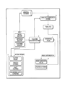
operative to
apply the calculated rotation, translation, and scale to the template 3D model
ridges.
Thereafter, the root mean square error or distance error between each matched
point set
is calculated, followed by calculation of the change in relative root mean
square error
or distance error from the previous process. If the change in relative root
mean square
error or distance error is within a predetermined threshold, then a
transformation
process occurs to apply the final rotation, translation, and scale to the
template 3D
model.
[0146] An articulated registration process follows the similarity registration
process
and receives input data from a scale space features process. In the scale
space feature
process, feature are extracted from the template 3D model and the anatomical
3D model
in different scale spaces. Each scale space is defined by convolving the
original
anatomical 3D model with Gaussian smoothing function.
[0147] The purpose of the articulated registration process is to match "n"
scale space
features of the template 3D model with "m" scale space features calculated on
the
anatomical 3D model. The difference between the number of detected features on
the
template 3D model and the anatomical 3D model is due to anatomical variation.
This
difference in a number of detected features may result in many relationships
between
the template 3D model and the anatomical 3D model. Therefore, a two-way,
mutual
feature matching is performed to accommodate such variation and achieve
accurate
matching between all mutual features. Specifically, feature sets are computed
on the
template 3D model in scale space. In this exemplary process, feature sets are
connected
sets of points that represent a prominent anatomical structure (e.g.,
acetabular cup in
the pelvis, spine process in the lumbar). Likewise, feature sets are computed
on the
anatomical 3D model in scale space. A matching feature pair process matches
the
feature sets computed on the template 3D model to the feature sets on the
anatomical
23
CA 3092713 2020-09-02
3D model using shape descriptors (e.g., curvature, shape index, etc.). The
result of this
process is an "n-m" mapping of feature sets between the template 3D model and
the
anatomical 3D model. If necessary, a regrouping process is carried out to
regroup the
matched feature sets into a single feature set (e.g., if acetabular cup was
detected as two
pieces, this process would regroup the two pieces into one single feature
set).
Thereafter, a calculation process is carried out to calculate the
correspondence between
each point in matched feature sets on the template 3D model and the anatomical
3D
model. An affine calculation transformation process follows in order to
calculate the
rotation, translation, and shear that transform each matched feature set on
the template
3D model to its corresponding feature set on the anatomical 3D model.
Thereafter, the
template 3D model is transformed using the calculated affine transformation
parameters
(i.e., rotation, translation, and shear). Finally, a rigid alignment process
is carried out
to align each matched feature set on the template 3D model and the anatomical
3D
model.
[0148] A non-rigid registration process, occurring after the articulated
registration
process and the normal scale features process, involves matching all surface
vertices on
the template 3D model to vertices on the anatomical 3D model and calculating
initial
correspondence. This correspondence is then used to calculate deformation
fields that
move each vertex on the template 3D model to the matched point on the
anatomical 3D
model. Matching is done between vertices within the same class (i.e., scale
space
feature vertex; normal scale feature vertex, or non-feature vertex). In the
context of the
normal scale features process, shape features are calculated on the template
3D model
and the anatomical 3D model in the original scale space (ridges), meaning the
original
input model.
[0149] Specifically, as part of the non-rigid registration process, the scale
space
features are calculated on the template 3D model (TMssf) and on the anatomical
3D
model (NMssf). Each set of features on the template 3D model and on the
anatomical
3D model are grown using "k" neighbor points. An alignment process is applied
to the
template 3D model scale space features to match its corresponding feature on
the
anatomical 3D model. Given two point clouds, reference (X) and moving (Y), the
goal
is to iteratively align the two point clouds to minimize overall error metric,
under
constraint of a minimum relative root mean squared error and maximum angle
24
CA 3092713 2020-09-02
threshold. A realignment process is carried out to align feature sets on the
template 3D
model with the matching sets on the anatomical 3D model using iterative
closest point
in normal scale. Post realignment, the point correspondence between points in
each
feature set on the template 3D model with the matched feature set on the
anatomical 3D
model is calculated. The matched point on the anatomical 3D model should have
a
surface normal direction close to the template 3D model point. The output is
forwarded
to the calculate deformation fields step.
[01501 Parallel to the scale space features calculation course, template 3D
model
(TMnfp) and anatomical 3D model (NMnfp) non-feature points or the remaining
set of
points on the template 3D model surface that does not belong to either scale
space
features or normal scale features are processed pursuant to a correspondence
calculation
to calculate the point correspondence between non-feature points on the
template 3D
model and non-feature points on the anatomical 3D model. The matched point(s)
on the
new model should have a surface normal direction close to the template model
point.
The output is forwarded to the calculate deformation fields step.
[0151] Also parallel to the scale space features calculation course, normal
scale features
(i.e., ridges) on the template 3D model (TM nsf) are aligned with the normal
scale
features (i.e., ridges) on the anatomical 3D model (NM nsf) using AICP. AICP
is a
variant of the iterative closest point calculation where in each iteration
translation,
rotation, and scale are calculated between matched point sets. After the
alignment
process, a correspondence process is carried out.
[0152] The outputs from scale space features calculation course, the
correspondence
course, and the alignment course are subjected to a deformation process where
the
deformation field is calculated to move each point on the template 3D model to
its
matched point on the anatomical 3D model.
[0153] The output of the non-rigid registration process is a subjected to a
relaxation
process in order to move the vertices of the template 3D model mesh closer to
surface
of the anatomical 3D model after the multi-resolution registration step and
smooth the
output model. In particular, the template 3D model in normal space (TM ns) and
the
anatomical 3D model in normal space (NM ns) are processed via a correspondence
calculation to compute the closest vertices on template 3D model to the
anatomical 3D
CA 3092713 2020-09-02
model using a normal constrained spherical search algorithm. This calculation,
using
the closest vertices for both models, generates a correspondence vector from
each
vertex in the template 3D model and its matched vertices in anatomical 3D
model,
which may result in more than one match point from the anatomical 3D model.
Using
the matched points for each vertex on the template 3D model, the weighted mean
of the
matched points on the anatomical 3D model is calculated based on the Euclidian
distance from the point and matched points. At this point, the template 3D
model is
updated using the weighted average so as to move each point on template 3D
model
using the calculated weighted average distance. After the computed weights
process, a
relaxation process is carried out for every point on template model in order
to find the
closest point on the anatomical 3D model surface and move it to that point.
Finally, a
smoothing operation is performed on the deformed template 3D model to remove
noise.
The resultant registered 3D models (i.e., template and anatomical 3D models)
are then
subjected to a free form deformation process.
101541 The free form deformation process morphs the surface of the template 3D
model
with the surface of the anatomical 3D model. More specifically, the surface of
the
template 3D model is iteratively moved on a weighted point-to-point basis
using
mutually matched points on both the template 3D model surface and the
anatomical 3D
model surface.
101551 Referencing FIGS. 2 and 6, after the free form deformation process, the
anatomical 3D model is subjected to a correspondence calculation process to
determine
the deviation between the anatomical 3D model and the morphed template 3D
model.
This correspondence calculation process refines the template 3D model from the
free
form deformation step to perform a final match of the selected landmark
locations on
the template deformed 3D model and the deformed anatomical 3D model. In this
fashion, the correspondence calculation process calculates and records the
variation in
size and shape between the 3D models, which is recorded as deviation about the
mean
model. The output of this correspondence calculation process is the addition
of a
normalized anatomical 3D model and a revised template 3D model having been
updated
to account for the variations in the anatomical 3D model. In other words, the
output of
the process outlined in FIG. 2 is the normalized anatomical 3D model having
been
modified to have properties (e.g., point correspondence) consistent with the
revised
26
CA 3092713 2020-09-02
template 3D model to facilitate full anatomical reconstruction (e.g., full
bone
reconstruction).
[0156] Referring to FIGS. 1 and 7, inputs from the statistical atlas module
and anatomy
data are directed to a full anatomy reconstruction module. By way of example,
the
anatomy in question may be a bone or multiple bones. It should be noted,
however,
that anatomies other than bone may be reconstructed using the exemplary
hardware,
processes, and techniques described herein. In exemplary form, the full
anatomy
reconstruction module may receive input data as to a partial, deformed, or
shattered
pelvis. Input anatomical data comprises two dimensional (2D) images or three
dimensional (3D) surface representations of the anatomy in question that may,
for
example, be in the form of a surface model or point cloud. In circumstances
where 2D
images are utilized, these 2D images are utilized to construct a 3D surface
representation of the anatomy in question. Those skilled in the art are
familiar with
utilizing 2D images of anatomy to construct a 3D surface representation.
Accordingly,
a detailed explanation of this process has been omitted in furtherance of
brevity. By
way of example, input anatomical data may comprise one or more of X-rays,
computed
tomography (CT) scans, magnetic resonance images (MRIs), or any other imaging
data
from which a 3D surface representation may be generated. As will be discussed
in
more detail hereafter, this input anatomical data may be used, without
limitation, for:
(1) a starting point for identifying the closest statistical atlas 3D bone
model; (2)
registration using a set of 3D surface vertices; and, (3) a final relaxation
step of
reconstruction output.
[0157] As depicted in FIG. 7, the input anatomical data (e.g., bone model of
the patient)
is utilized to identify the anatomical model (e.g., bone model) in the
statistical atlas that
most closely resembles the anatomy of the patient in question. This step is
depicted in
FIG. 3 as finding the closest bone in the atlas. In order to initially
identify a bone model
in the statistical atlas that most closely resembles the patient's bone model,
the patient's
bone model is compared to the bone models in the statistical atlas using one
or more
similarity metrics. The result of the initial similarity metric(s) is the
selection of a bone
model from the statistical atlas that is used as an "initial guess" for a
subsequent
registration step. The registration step registers the patient bone model with
the selected
atlas bone model (i.e., the initial guess bone model) so that the output is a
patient bone
27
CA 3092713 2020-09-02
model that is aligned with the atlas bone model. Subsequent to the
registration step,
the shape parameters for aligned "initial guess" are optimized so that the
shape matches
the patient bone shape.
[0158] Shape parameters, in this case from the statistical atlas, are
optimized so that the
region of non-deformed or existing bone is used to minimize the error between
the
reconstruction and patient bone model. Changing shape parameter values allows
for
representation of different anatomical shapes. This process is repeated, at
different
scale spaces, until convergence of the reconstructed shape is achieved
(possibly
measured as relative surface change between iterations or as a maximum number
of
allowed iterations).
[0159] A relaxation step is performed to morph the optimized tissue to best
match the
original patient 3D tissue model. Consistent with the exemplary case, the
missing
anatomy from the reconstructed pelvis model that is output from the
convergence step
is applied to the patient-specific 3D pelvis model, thereby creating a patient-
specific
3D model of the patient's reconstructed pelvis. More specifically, surface
points on the
reconstructed pelvis model are relaxed (i.e., morphed) directly onto the
patient-specific
3D pelvis model to best match the reconstructed shape to the patient-specific
shape.
The output of this step is a fully reconstructed, patient-specific 3D tissue
model
representing what should be the normal/complete anatomy of the patient.
[0160] Referencing FIG. 1, the abnormal database is utilized as a data input
and
training for the defect classification module. In particular, the abnormal
database
contains data specific to an abnormal anatomical feature that includes an
anatomical
surface representation and related clinical and demographic data.
[0161] Referencing FIGS. 1 and 8, the fully reconstructed, patient-specific 3D
tissue
model representing the normal/complete tissue and input anatomical data (i.e.,
3D
surface representation or data from which a 3D surface representation may be
generated) representing abnormal/incomplete tissue from the abnormal database
are
input to the defect classification module. This anatomical data from the
abnormal
database may be a partial anatomy in the case of tissue degeneration or tissue
absence
resulting from genetics, or this anatomy may be a deformed anatomy resulting
from
genetics or environmental conditions (e.g., surgical revisions, diseases,
etc.), or this
28
CA 3092713 2020-09-02
anatomy may be a shattered tissue resulting from one or more anatomy breaks.
By way
of example, input anatomical data may comprise one or more of X-rays, computed
tomography (CT) scans, magnetic resonance images (MRIs), or any other imaging
data
from which a 3D surface representation may be generated.
[0162] The defect classification module pulls a plurality of abnormal 3D
surface
representations from abnormal database coupled with the normal 3D
representation of
the anatomy in question to create a quantitative defect classification system.
This defect
classification system is used to create "templates" of each defect class or
cluster. More
generally, the defect classification module classifies the anatomical
deficiency into
classes which consist of closely related deficiencies (referring to those with
similar
shape, clinical, appearance, or other characteristics) to facilitate the
generation of
healthcare solutions which address these deficiencies. The instant defect
classification
module uses software and hardware to classify the defects automatically as a
means to
eliminate or reduce discrepancies between pre-operative data and intra-
operative
observer visualization. Traditionally, pre-operative radiographs have been
taken as a
means to qualitatively analyze the extent of anatomical reconstruction
necessary, but
this resulted in pre-operative planning that was hit-or-miss at best.
Currently, intra-
operative observers make the final determination of the extent of anatomy
deficiency
and many times conclude that the pre-operative planning relying on radiographs
was
defective or incomplete. As a result, the instant defect classification module
improves
upon current classification systems by reducing interobserver and
intraobserver
variation related to defect classification and providing quantitative metrics
for
classifying new defect instances.
[0163] As part of the defect classification module t, the module is may take
as input
one or more classification types to be used as an initial state. For example,
in the
context of a pelvis, the defect classification module may use as input defect
features
corresponding to the American Academy of Orthopaedic Surgeons (AAOS) D'Antonio
et al. bone defect classification structure. This structure includes four
different classes
as follows: (1) Type I, corresponding to segmental bone loss; (2) Type II,
corresponding
to cavitary bone loss; (3) Type III, corresponding to combined segmental and
cavitary
bone loss; and, (4) Type IV, corresponding to pelvis discontinuity.
Alternatively, the
defect classification module may be programmed with the Paprosky bone defect
29
CA 3092713 2020-09-02
classification structure. This structure includes three different classes as
follows: (1)
Type I, corresponding to supportive rim with no bone lysis; (2) Type II,
corresponding
to distorted hemispheres with intact supportive columns and less than two
centimeters
of superomedial or lateral migration; and, (3) Type III, corresponding to
superior
migration greater than two centimeters and sever ischial lysis with Kohler's
line broken
or intact. Moreover, the defect classification module may be programmed with
the
Modified Paprosky bone defect classification structure. This structure
includes six
different classes as follows: (1) Type 1, corresponding to supportive rim with
no
component migration; (2) Type 2A, corresponding to distorted hemisphere but
superior
migration less than three centimeters; (3) Type 2B, corresponding to greater
hemisphere
distortion having less than 1/3 rim circumference and the dome remaining
supportive;
(4) Type 2C, corresponding to an intact rim, migration medial to Kohler's
line, and the
dome remains supportive; (5) Type 3A, corresponding to superior migration,
greater
than three centimeters and severe ischial lysis with intact Kohler's line;
and, (6) Type
3B, corresponding to superior migration, greater than three centimeters and
severe
ischial lysis with broken Kohler's line and rim defect greater than half the
circumference. Using the output classification types and parameters, the
defect
classification module compares the anatomical data to that of the
reconstructed data to
discern which of the classification types the anatomical data most closely
resembles,
thereby corresponding to the resulting assigned classification.
[0164] As an initial step, the add to statistical atlas step involves
generating
correspondence between normal atlas 3D bone model and the abnormal 3D bone
model.
More specifically, the 3D bone models are compared to discern what bone in the
normal
3D model is not present in the abnormal 3D model. In exemplary form, the
missing/abnormal bone is identified by comparing points on the surface of each
3D
bone model and generating a list of the discrete points on the surface of the
normal 3D
bone model that are not present on the abnormal 3D bone model. The system may
also
record and list (i.e., identify) those surface points in common between the
two models
or summarily note that unless recorded as points being absent on the abnormal
3D bone
model, all other points are present in common in both bone models (i.e., on
both the
normal and abnormal bone models). Accordingly, the output of this step is the
abnormal 3D bone model with statistical atlas correspondence and a list of
features
CA 3092713 2020-09-02
(points) from the normal atlas 3D bone model indicating if that feature
(point) is present
or missing in the abnormal 3D bone model.
[0165] After generating correspondence between the normal atlas 3D bone model
(generated from the full bone reconstruction module) and the abnormal 3D bone
model
(generated from the input anatomical data), the missing/abnormal regions from
the
abnormal 3D bone model are localized on the normal atlas 3D bone model. In
other
words, the normal atlas 3D bone model is compared to the abnormal 3D bone
model to
identify and record bone missing from the abnormal 3D bone model that is
present in
the normal atlas 3D bone model. Localization may be carried out in a multitude
of
fashions including, without limitation, curvature comparison, surface area
comparisons,
and point cloud area comparisons.
Ultimately, in exemplary form, the
missing/abnormal bone is localized as a set of bounding points identifying the
geometrical bounds of the missing/abnormal region(s).
[0166] Using the bounding points, the defect classification module extracts
features
from the missing/abnormal region(s) using input clinical data. In exemplary
form, the
extracted features may include shape information, volumetric information, or
any other
information used to describe the overall characteristics of the defective
(i.e., missing or
abnormal) area. These features may be refined based on existing clinical data,
such as
on-going defect classification data or patient clinical information not
necessarily related
to the anatomical feature (demographics, disease history, etc.). The output of
this step
is a mathematical descriptor representative of the defective area(s) that are
used in a
subsequent step to group similar tissue (e.g., bone) deformities.
[0167] The mathematical descriptor is clustered or grouped based upon a
statistical
analysis. In particular, the descriptor is statistically analyzed and compared
to other
descriptors from other patients/cadavers to identify unique defect classes
within a given
population. Obviously, this classification is premised upon multiple
descriptors from
multiple patients/cadavers that refine the classifications and identifications
of discrete
groups as the number of patients/cadavers grows. The output from this
statistical
analysis is a set of defect classes that are used to classify new input
anatomical data and
determines the number of templates.
31
CA 3092713 2020-09-02
[0168] The output of the defect classification module is directed to a
template module.
In exemplary form, the template module includes data that is specific as to
each of the
defect classifications identified by the defect classification module. By way
of
example, each template for a given defect classification includes surface
representations
of the defective bone, location(s) of the defect(s), and measurements relating
to the
defective bone. This template data may be in the form of surface shape data,
point
cloud representations, one or more curvature profiles, dimensional data, and
physical
quantity data. Outputs from the template module and the statistical atlas are
utilized by
a mass customization module to design, test, and allow fabrication of mass
customized
implants, fixation devices, instruments or molds. Exemplary utilizations of
the mass
customization module will be discussed in greater detail hereafter.
Patient-Specific Reconstruction Implants
[0169] Referring to FIGS. 1 and 17, an exemplary process and system are
described for
generating patient-specific orthopedic implant guides and associated patient-
specific
orthopedic implants for patients afflicted with partial, deformed, and/or
shattered
anatomies. For purposes of the exemplary discussion, a total hip arthroplasty
procedure
will be described for a patient with a partial anatomy. It should be
understood, however,
that the exemplary process and system are applicable to any orthopedic implant
amenable to patient-specific customization in instances where incomplete or
deformed
anatomy is present. For example, the exemplary process and system are
applicable to
shoulder replacements and knee replacements where bone degeneration (partial
anatomy), bone deformation, or shattered bones are present. Consequently,
though a
hip implant is discussed hereafter, those skilled in the art will understand
the
applicability of the system and process to other orthopedic implants, guides,
tools, etc.
for use with original orthopedic or orthopedic revision surgeries.
[0170] Pelvis discontinuity is a distinct form of bone loss most often
associated with
total hip arthroplasty (THA), in which osteolysis or acetabular fractures can
cause the
superior aspect of the pelvis to become separated from the inferior portion.
The amount
and severity of bone loss and the potential for biological in-growth of the
implant are
some of the factors that can affect the choice of treatment for a particular
patient. In
the case of severe bone loss and loss of pelvic integrity, a custom tri-flange
cup may be
32
CA 3092713 2020-09-02
used. First introduced in 1992, this implant has several advantages over
existing cages.
It can provide stability to pelvic discontinuity, eliminate the need for
structural grafting
and intraoperative contouring of cages, and promote osseointegration of the
construct
to the surrounding bone.
101711 Regardless of the context, whether partial, deformed, and/or shattered
anatomies of the patient are at issue, the exemplary system and process for
generating
patient-specific implants and/or guides utilizes the foregoing exemplary
process and
system of 3D bone model reconstruction (see FIGS. 1-7 and the foregoing
exemplary
discussion of the same) to generate a three dimensional model of the patient's
reconstructed anatomy. More specifically, in the context of total hip
arthroplasty where
pelvis discontinuity is involved, the exemplary patient-specific system
utilizes the
patient pelvis data to generate a 3D model of the patient's complete pelvis,
which is
side specific (right or left). Consequently, a discussion of the system and
process for
utilizing patient anatomy data for a partial anatomy and generating a 3D
reconstructed
model of the patient's anatomy is omitted in furtherance of brevity.
Accordingly, a
description of the process and system for generating patient-specific
orthopedic implant
guides and associated patient-specific orthopedic implants for patients
afflicted with
partial, deformed, and/or shattered anatomies will be described post formation
of the
three dimensional reconstructed model.
101721 Referring specifically to FIGS. 17-19 and 23, after the patient-
specific
reconstructed 3D bone model of the pelvis and femur are generated, both the
incomplete
patient-specific 3D bone model (for pelvis and femur) and the reconstructed 3D
bone
model (for pelvis and femur) are utilized to create the patient-specific
orthopedic
implant and a patient-specific placement guide for the implant and/or its
fasteners. In
particular, the extract defect shape step includes generating correspondence
between
the patient-specific 3D model and the reconstructed 3D model (correspondence
between pelvis models, and correspondence between femur models, but not
between
one femur model and a pelvis model). More specifically, the 3D models are
compared
to discern what bone in the reconstructed 3D model is not present in the
patient-specific
3D model. In exemplary form, the missing/abnormal bone is identified by
comparing
points on the surface of each 3D model and generating a list of the discrete
points on
the surface of the reconstructed 3D model that are not present on the patient-
specific
33
CA 3092713 2020-09-02
3D model. The system may also record and list (i.e., identify) those surface
points in
common between the two models or summarily note that unless recorded as points
being absent on the patient-specific 3D model, all other points are present in
common
in both models (i.e., on both the reconstructed and patient-specific 3D
models).
[0173] Referring to FIG. 18, after generating correspondence between the
reconstructed 3D model (generated from the full bone reconstruction module)
and the
patient-specific 3D model (generated from the input anatomical data), the
missing/abnormal regions from the patient-specific 3D model are localized on
the
reconstructed 3D model. In other words, the reconstructed 3D model is compared
to
the patient-specific 3D model to identify and record bone missing from the
patient-
specific 3D model that is present in the reconstructed 3D model. Localization
may be
carried out in a multitude of fashions including, without limitation,
curvature
comparison, surface area comparisons, and point cloud area comparisons.
Ultimately,
in exemplary form, the missing/abnormal bone is localized and the output
comprises
two lists: (a) a first list identifying vertices corresponding to bone of the
reconstructed
3D model that is absent or deformed in the patient-specific 3D model; and, (b)
a second
list identifying vertices corresponding to bone of the reconstructed 3D model
that is
also present and normal in the patient-specific 3D model.
[0174] Referencing FIGS. 18, 19, and 23, following the extract defect shape
step, an
implant loci step is performed. The two vertices lists from the extract defect
shape step
and a 3D model of a normal bone (e.g., pelvis, femur, etc.) from the
statistical atlas (see
FIGS. 1 and 2, as well as the foregoing exemplary discussion of the same) are
input to
discern the fixation locations for a femoral or pelvic implant. More
specifically, the
fixation locations (i.e., implant loci) are automatically selected so that
each is positioned
where a patient has residual bone. Conversely, the fixation locations are not
selected
in defect areas of the patient's residual bone. In this manner, the fixation
locations are
chosen independent of the ultimate implant design/shape. The selection of
fixation
locations may be automated using shape information and statistical atlas
locations.
[0175] As show in FIG. 18, after the implant loci step, the next step is to
generate
patient-specific implant parameters. In order to complete this step, an
implant
parameterized template is input that defines the implant by a set number of
parameters
34
CA 3092713 2020-09-02
that are sufficient to define the underlying shape of the implant. By way of
example,
in the case of a pelvis reconstruction to replace/augment an absent or
degenerative
acetabulum, the implant parameterized template includes angle parameters for
the
orientation of the replacement acetabular cup and depth parameters to
accommodate
for dimensions of the femoral head. Other parameters for an acetabular implant
may
include, without limitation, the acetabular cup diameter, face orientation,
flange
locations and shapes, location and orientation of fixation screws. In the case
of porous
implants, the location and structural characteristics of the porosity should
be included.
By way of example, in the case of a femoral reconstruction to replace/augment
an
absent or degenerative femur, the implant parameterized template includes
angle
parameters for the orientation of the replacement femoral head, neck length,
head offset,
proximal angle, and cross-sectional analysis of the exterior femur and
intercondylar
channel. Those skilled in the art will understand that the parameters chosen
to define
the underlying shape of the implant will vary depending upon the anatomy being
replaced or supplemented. Consequently, an exhaustive listing of parameters
that are
sufficient to define the underlying shape of an implant is impractical.
Nevertheless, as
depicted in FIGS. 19 for example, the reconstructed 3D pelvis model may be
utilized
to obtain the radius of the acetabular cup, identification of pelvic bone
comprising the
acetabular cup circumferential upper ridge, and identification of the
orientation of the
acetabular cup with respect to the residual pelvis. Moreover, the parameters
may be
refined taking into account the implant loci so that the implant best/better
fits the
patient-specific anatomy.
101761 Subsequent to finalizing the set number of parameters that are
sufficient to
define the underlying shape of the implant, the design of the implant is
undertaken.
More specifically, an initial iteration of the overall implant surface model
is
constructed. This initial iteration of the overall implant surface model is
defined by a
combination of patient-specific contours and estimated contours for the
implanted
region. The estimated contours are determined from the reconstructed 3D bone
model,
missing anatomical bone, and features extracted from the reconstructed 3D bone
model.
These features and the location of the implant site, which can be
automatically
determined, are used to determine the overall implant shape, as depicted for
example in
FIG. 19 for an acetabular cup implant.
CA 3092713 2020-09-02
[0177] Referring back to FIG. 17, the initial iteration of the overall implant
surface
model is processed pursuant to a custom (i.e., patient-specific) planning
sequence. This
custom planning sequence may involve inputs from a surgeon and an engineer as
part
of an iterative review and design process. In particular, the surgeon and/or
engineer
may view the overall implant surface model and the reconstructed 3D bone model
to
determine if changes are needed to the overall implant surface model. This
review may
result in iterations of the overall implant surface model until agreement is
reached
between the engineer and surgeon. The output from this step is the surface
model for
the final implant, which may be in the form of CAD files, CNC machine
encoding, or
rapid manufacturing instructions to create the final implant or a tangible
model.
[0178] Referring to FIGS. 17, 19, and 20, contemporaneous with or after the
design of
the patient-specific orthopedic implant is the design of a patient specific
placement
guide. In the context of an acetabular cup implant, as discussed in exemplary
form
above, one or more surgical instruments can be designed and fabricated to
assist in
placing the patient-specific acetabular cup. Having designed the patient-
specific
implant to have a size and shape to match that of the residual bone, the
contours and
shape of the patient-specific implant may be utilized and incorporated as part
of the
placement guide.
[0179] In exemplary form, the acetabular placement guide comprises three
flanges that
are configured to contact the ilium, ischium, and pubis surfaces, where the
three flanges
are interconnected via a ring. Moreover, the flanges of the placement guide
may take
on the identical shape, size, and contour of the acetabular cup implant so
that the
placement guide will take on the identical position as planned for the
acetabular cup
implant. In other words, the acetabular placement guide is shaped as the
negative
imprint of the patient anatomy (ilium, ischium, and pubis partial surfaces),
just as the
acetabular cup implant is, so that the placement guide fits on the patient
anatomy
exactly. But the implant guide differs from the implant significantly in that
it includes
one or more fixation holes configured to guide drilling for holes and/or
placement of
fasteners. In exemplary form, the placement guide includes holes sized and
oriented,
based on image analysis (e.g., microCT), to ensure proper orientation of any
drill bit or
other guide (e.g., a dowel) that will be utilized when securing the acetabular
cup implant
to the residual pelvis. The number of holes and orientation varies depending
upon the
36
CA 3092713 2020-09-02
residual bone, which impacts the shaped of the acetabular cup implant too.
FIG. 20
depicts an example of a patient-specific placement guide for use in a total
hip
arthroplasty procedure. In another instance, the guide can be made so that it
fits into
the implant and guides only the direction of the fixation screws. In this
form, the guide
is shaped as the negative of the implant, so that it can be placed directly
over the
implant. Nevertheless, the incorporation of at least part of the patient-
specific
reconstructed implant size, shape, and contour is a theme that carries over
regardless of
the intended bone to which the patient-specific implant will be coupled.
[0180] Utilizing the exemplary system and method described herein can provide
a
wealth of information that can result in higher orthopedic placement accuracy,
better
anatomical integration, and the ability to pre-operatively measure true angles
and plane
orientation via the reconstructed three dimensional model.
Creation of Customized Implants Using Massively Customizable Components
[0181] Referring to FIG. 22, an exemplary process and system are described for
generating customized orthopedic implants using massively customizable
components.
For purposes of the exemplary discussion, a total hip arthroplasty procedure
will be
described for a patient with severe acetabular defects. It should be
understood,
however, that the exemplary process and system are applicable to any
orthopedic
implant amenable to mass customization in instances where incomplete anatomy
is
present.
[0182] Severe acetabular defects require specialized procedures and implant
components to repair. One approach is the custom triflange, which a fully
custom
implant consisting of an acetabular cup and three flanges that are attached to
the ilium,
ischium, and pubis. In contrast to the exemplary process and system, prior art
triflange
implants comprise a single complex component, which is cumbersome to
manufacture
and requires that the entire implant be redesigned for every case (i.e.,
completely
patient-specific). The exemplary process and system generates a custom
triflange
implant that makes use of massively customizable components in addition to
fully
custom components in a modular way to allow custom fitting and porosity.
37
CA 3092713 2020-09-02
[0183] A preplanning step in accordance with the exemplary process is
performed to
determine the orientation of the three flanges relative to the cup, the flange
contact
locations, and the acetabular cup orientation and size. This preplanning step
is
conducted in accordance with the "Patient-specific Implants" discussion
immediately
preceding this section. By way of example, specific locations of implant
fixation are
determined pursuant to an implant loci step and using its prefatory data
inputs as
discussed in the immediately preceding section. By way of recall, as part of
this implant
loci step, the two vertices lists from the extract defect shape step and a 3D
model of a
normal pelvis from the statistical atlas (see FIGS. 1 and 2, as well as the
foregoing
exemplary discussion of the same) are input to discern the fixation locations
for the
custom triflange. More specifically, the fixation locations (i.e., implant
loci) are
selected so that each is positioned where a patient has residual bone. In
other words,
the fixation locations are not selected in defect areas of the patient's
residual pelvis. In
this manner, the fixation locations are chosen independent of the ultimate
implant
design/shape.
[0184] After determining the fixation locations, the triflange components
(i.e., flanges)
are designed using the "Patient-specific Implants" discussion immediately
preceding
this section. The flanges are designed to be oriented relative to the
replacement
acetabular cup so that the cup orientation provides acceptable joint
functionality.
Additionally, the contact surfaces of the flanges are contoured to match the
patient's
pelvis anatomy in that the contact surfaces of the triflanges are shaped as a
"negative"
of the pelvis's bony surface. The exemplary process of FIG. 20 utilizes the
final step
of the process depicted in FIG. 17 to rapid prototype the flanges (or use
conventional
computer numerical control (CNC) equipment). After the flanges are fabricated,
further
machining or steps may be performed to provide cavities within which porous
material
may be added to the triflanges.
[0185] One portion of the triflange system that does not need to be a custom
component
is the acetabular cup component. In this exemplary process, a family of
acetabular cups
is initially manufactured and provides the foundation on which to build the
triflange
system. These "blank" cups are retained in inventory for use as needed. If a
particular
porosity for the cup is desired, mechanical features are added to the cup that
allows
38
CA 3092713 2020-09-02
press fitting of porous material into the cup. Alternatively, if a particular
porosity for
the cup is desired, the cup may be coated using one or more porous coatings.
[0186] After the blank cup is formed and any porosity issues are addressed as
discussed
above, the cup is rendered patient-specific by machining the cup to accept the
flanges.
In particular, using the virtual model of the flanges, the system software
constructs
virtual locking mechanisms for the flanges, which are transformed into machine
coding
so that the locking mechanisms are machined into the cup. These locking
mechanisms
allow the cup to be fastened to the flanges so that when the flanges are
mounted to the
patient's residual bone, the cup is properly oriented with respect to the
residual pelvis.
This machining may use conventional CNC) equipment to form the locking
mechanisms into the blank cups.
[0187] Subsequent to fabrication of the locking mechanisms as part of the
blank cup,
the flanges are mounted to the cup using the interface between the locking
mechanisms.
The triflange assembly (i.e., final implant) is subjected to an annealing
process to
promote strong bonding between the components. Post annealing of the triflange
implant, a sterilization process occurs followed by appropriate packaging to
ensure a
sterile environment for the triflange implant.
Creation of Mass Customized Implants
[0188] Referring to FIG. 24, an exemplary process and system are described for
generating mass customized orthopedic implant guides and associated mass
customized
orthopedic implants for patients afflicted with partial, deformed, and/or
shattered
anatomies. For purposes of the exemplary discussion, a total hip arthroplasty
procedure
will be described for a patient needing primary joint replacement. It should
be
understood, however, that the exemplary process and system are applicable to
any
orthopedic implant and guides amenable to mass customization in instances
where
incomplete anatomy is present. For example, the exemplary process and system
are
applicable to shoulder replacements and knee replacements where bone
degeneration
(partial anatomy), bone deformation, or shattered bones are present.
Consequently,
though a hip implant is discussed hereafter, those skilled in the art will
understand the
applicability of the system and process to other orthopedic implants, guides,
tools, etc.
for use with primary orthopedic or orthopedic revision surgeries.
39
CA 3092713 2020-09-02
101891 The exemplary process utilizes input data from a macro perspective and
a micro
perspective. In particular, the macro perspective involves determination of
the overall
geometric shape of the orthopedic implant and corresponding anatomy.
Conversely,
the micro perspective involves accounting for the shape and structure of
cancellous
bone and its porosity.
101901 The macro perspective includes a database communicating with a
statistical
atlas module that logs virtual, 3D models of one or more anatomies (e.g.,
bones) to
capture the inherent anatomical variability in a given population. In
exemplary form,
the atlas logs mathematical representations of anatomical features of the one
or more
anatomies represented as a mean representation and variations about the mean
representation for a given anatomical population. Reference is had to FIG. 2
and the
foregoing discussion of the statistical atlas and how one adds anatomy to the
statistical
atlas of a given population. Outputs from the statistical atlas are directed
to an
automatic landmarking module and to a surface/shape analysis module.
101911 The automatic landmarking module utilizes inputs from the statistical
atlas (e.g.,
regions likely to contain a specific landmark) and local geometrical analyses
to
calculate anatomical landmarks for each instance of anatomy within the
statistical atlas.
This calculation is specific to each landmark. The approximate shape of the
region is
known, for example, and the location of the landmark being searched for is
known
relative to the local shape characteristics. For example, locating the medial
epicondylar
point of the distal femur is accomplished by refining the search based on the
approximate location of medial epicondylar points within the statistical
atlas.
Accordingly, it is known that the medial epicondylar point is the most medial
point
within this search window, so a search for the most medial point is performed
as to each
bone model within the medial epicondylar region defined in the statistical
atlas, with
the output of the search being identified as the medial epicondylar point
landmark.
After the anatomical landmarks are automatically calculated for each virtual,
3D model
within the statistical atlas population, the virtual, 3D models of the
statistical atlas are
directed to a feature extraction module, along with shape/surface analysis
outputs.
101921 The shape/surface outputs come from a shape/surface module also
receiving
inputs from the statistical atlas. In the context of the shape/surface module,
the virtual,
CA 3092713 2020-09-02
3D models within the statistical atlas population are analyzed for
shape/surface features
that are not encompassed by the automatic landmarking. In other words,
features
corresponding to the overall 3D shape of the anatomy, but not belonging to
features
defined in the previous automatic landmarking step are calculated as well. For
example,
curvature data is calculated for the virtual 3D models.
[0193] Outputs from the surface/shape analysis module and the automatic
landmarking
module are directed to a feature extraction module. Using a combination of
landmarks
and shape features, mathematical descriptors (i.e. curvature, dimensions)
relevant to
implant design are calculated for each instance in the atlas. These
descriptors are used
as input to a clustering process.
[0194] The mathematical descriptor is clustered or grouped based upon a
statistical
analysis. In particular, the descriptor is statistically analyzed and compared
to other
descriptors from the remaining anatomy population to identify groups (of
anatomies)
having similar features within the population. Obviously, this clustering is
premised
upon multiple descriptors from multiple anatomies across the population. As
new
instances are presented to the clustering, which were not present in the
initial clustering,
the output clusters are refined to better represent the new population. The
output from
this statistical analysis is a finite number of implants (including implant
families and
sizes) covering all or the vast majority of the anatomical population.
[0195] For each cluster, a parameterization module extracts the mathematical
descriptors within the cluster. The mathematical descriptors form the
parameters (e.g.,
CAD design parameters) for the eventual implant model. The extracted
mathematical
descriptors are fed into an implant surface generation module. This module is
responsible for converting the mathematical descriptors into surface
descriptors to
generate a 3D, virtual model of the anatomy for each cluster. The 3D, virtual
model
complements the micro perspective prior to stress testing and implant
manufacturing.
[0196] On the micro perspective, for each anatomy of a given population, data
is
obtained indicative of structural integrity. In exemplary form, this data for
a bone may
comprise microCT data providing structural information as to the cancellous
bone.
More specifically, the microCT data may comprise images of the bone in
question
(multiple microCT images for multiple bones across a population). These images
are
41
CA 3092713 2020-09-02
thereafter segmented via the extract trabecular bone structure module in order
to extract
the three dimensional geometry of the cancellous bones and create virtual, 3D
models
for each bone within the population. The resulting 3D virtual models are input
to a pore
size and shape module. As depicted graphically in FIG. 76, the 3D virtual
models
include porous size and shape information, which is evaluated by the pore size
and
shape module to determine pore size and size for the cancellous bone. This
evaluation
is useful to analyze the porous size and shape of the bone within the
intramedullary
canal so that the stem of the femoral implant can be treated with a coating or
otherwise
processed to exhibit a porous exterior to promote integration between the
residual bone
of the femur and the femoral implant. The output from this module, in
combination
with the 3D virtual model output from the implant surface generation module,
is
directed to a virtual stress testing module.
[0197] The stress testing module combines implant porosity data from the pore
size
and shape module and implant shape data from the implant surface generation
module
to define the final implant shape model and properties. For example, the shape
and
properties include providing a porous coating for the final implant model that
roughly
matches the cancellous bone porosity for the bone in question. Once the shape
and
properties are incorporated, the final implant model undergoes virtual stress
testing
(finite-element and mechanical analysis) to verify the functional quality of
the model.
To the extent the functional quality is unacceptable, the parameters defining
the implant
shape and porosity are modified until acceptable performance is achieved.
Presuming
the final implant model satisfies the stress testing criteria, the final
implant model is
utilized to generate machine instructions necessary to convert the virtual
model into a
tangible implant (that may be further refined by manufacturing processes known
to
those skilled in the art). In exemplary form, the machine instructions may
include rapid
manufacturing machine instructions to fabricate the final implant through a
rapid
prototyping process (to properly capture porous structure) or a combination of
traditional manufacturing and rapid prototyping.
Creation of Gender/Ethnic Specific Hip Implants
[0198] Referring to FIGS. 25-76, an exemplary process and system are described
for
generating gender and/or ethnic specific implants. For purposes of the
exemplary
42
CA 3092713 2020-09-02
discussion, a total hip arthroplasty procedure will be described for a patient
with
requiring primary joint replacement. It should be understood, however, that
the
exemplary process and system are applicable to any orthopedic implant amenable
to
customization. For example, the exemplary process and system are applicable to
shoulder replacements and knee replacements and other primary joint
replacement
procedures. Consequently, though a hip implant is discussed hereafter, those
skilled in
the art will understand the applicability of the system and process to other
orthopedic
implants, guides, tools, etc. for use with original orthopedic or orthopedic
revision
surgeries.
[0199] The hip joint is composed of the head of the femur and the acetabulum
of the
pelvis. The hip joint anatomy makes it one of the most stable joints in the
body. The
stability is provided by a rigid ball and socket configuration. The femoral
head is almost
spherical in its articular portion that forms two-thirds of a sphere. Data has
shown that
the diameter of the femoral head is smaller for females than males. In the
normal hip,
the center of the femoral head is assumed to coincide exactly with the center
of the
acetabulum and this assumption is used as the basis for the design of most hip
systems.
However, the native acetabulum is not deep enough to cover all of the native
femoral
head. The almost rounded part of the femoral head is spheroidal rather than
spherical
because the uppermost part is flattened slightly. This spheroidal shape causes
the load
to be distributed in a ring-like pattern around the superior pole.
[0200] The geometrical center of the femoral head is traversed by three axes
of the
joint: the horizontal axis; the vertical axis; and, the anterior/posterior
axis. The femoral
head is supported by the neck of the femur, which joints the shaft. The axis
of the
femoral neck is obliquely set and runs superiorly medially and anteriorly. The
angle of
the inclination of the femoral neck to the shaft in the frontal plane is the
neck shaft
angle. In most adults, this angle varies between 90 to 135 degrees and is
important
because it determines the effectiveness of the hip abductors, the length of
the limb, and
the forces imposed on the hip joint.
[0201] An angle of inclination greater than 125 degrees is called coxa valga,
whereas
an angle of inclination less than 125 degrees is called coxa vara. Angles of
inclination
greater than 125 degrees coincide with lengthened limbs, reduced effectiveness
of the
43
CA 3092713 2020-09-02
hip abductors, increased load on the femoral head, and increased stress on the
femoral
neck. In a case of coxa vara, angles of inclination less than 125 degrees
coincide with
shortened the limbs, increased effectiveness of the hip abductors, decreased
load on the
femoral head, and decreased stress on the femoral neck. The femoral neck forms
an
acute angle with the transverse axis of the femoral condyles. This angle faces
medially
and anteriorly and is called angle of anteversion. In adult humans, this angle
averages
approximately 7.5 degrees.
[0202] The acetabulum lies on the lateral aspect of the hip where the ilium,
ischium,
and pubis meet. These three separate bones join into the formation of the
acetabulum,
with the ilium and ischium contributing approximately two-fifths each and the
pubis
one-fifth of the acetabulum. The acetabulum is not a deep enough socket to
cover all
of the femoral head and has both articulating and non-articulating portions.
However,
the acetabular labrum deepens the socket to increase stability. Together with
labrum,
the acetabulum covers slightly more than 50% of the femoral head. Only the
sides of
the acetabulum are lined with articular cartilage, which is interrupted
inferiorly by the
deep acetabular notch. The center part of the acetabular cavity is deeper than
the
articular cartilage and is nonarticular. This center part is called the
acetabular fossae
and is separated from the interface of the pelvic bone by a thin plate. The
acetabular
fossae is a region unique for every patient and is used in creating patient-
specific guide
for reaming and placement of the acetabular cup component. Additionally,
variation of
anatomical features further warrant the need for population specific implant
designs.
[0203] Some of the problems associated with prior art use of cementless
components
can be attributed to the wide variation in size, shape, and orientation of the
femoral
canal. One of the challenges to orthopedic implant design of the femoral stem
is large
variation in the mediolateral and anteroposterior dimensions. There is also
significant
variation in the ratio of the proximal to distal canal size. The different
combination of
various arcs, taper angles, curves, and offsets in the normal population is
staggering.
However, that is not the only problem.
[0204] Ancestral differences in femora morphology and a lack of definite
standards for
modern populations makes designing the proper hip implant system problematic.
For
example, significant differences in anterior curvature, torsion, and cross-
sectional shape
44
CA 3092713 2020-09-02
exist between American Indians, American blacks, and American whites.
Differences
between Asian and Western populations in the femora are found in the anterior
bow of
the femora, where Chinese are more anteriorly bowed and externally rotated
with
smaller intramedullary canals and smaller distal condyles than Caucasian
femora.
Likewise, Caucasian femora are larger than Japanese femora in terms of length
distal
condyle dimensions. Ethnic differences also exist in the proximal femur
mineral bone
density (BMD) and hip axis length between American blacks and whites. The
combined effects of higher BMD, shorter hip axis length, and shorter
intertrochanteric
width may explain the lower prevalence of osteoporotic fractures in American
black
women compared to their white counterparts. Similarly, elderly Asian and
American
black men were found to have thicker cortices and higher BMD than white and
Hispanic
men, which may contribute to greater bone strength in these ethnic groups. In
general,
American blacks have thicker bone cortices, narrower endosteal diameters, and
greater
BMD than American whites.
[0205] Combining the femur and the pelvic ancestral (and ethnic) differences
becomes
even more challenging to primary hip systems. Revision surgery creates more
complexity. Added to these normal anatomic and ethnic variations, the
difficulties
faced by the surgeon who performs revision operation are compounded by: (a)
distortion of the femoral canal caused by bone loss around the originally
placed
prostheses; and, (b) iatrogenic defects produced by the removal of the
components and
cement.
[0206] All of the foregoing factors have led a number of hip surgeons to look
for ways
to improve design of uncemented femoral prostheses. In total hip replacement
(primary
or revision), the ideal is to establish an optimal fit between the femoral
ball and
acetabular cup. The femoral stem neck should have a cruciform cross section to
reduce
stiffness. The stem length should be such that the stem has parallel contact
with the
walls of the femur over two to three internal canal diameters. The proximal
one third
of the stem is porous coated or hydroxylapatite (HA) coated. The stem is
cylindrical
(i.e. not tapered) to control bending loads and to allow transmission of all
rotational and
axial loads proximally. The femoral head position should reproduce the
patient's own
head center, unless it is abnormal.
CA 3092713 2020-09-02
102071 One way to attempt to satisfy these goals is to manufacture femoral
prostheses
individually for each patient. In other words, make a prosthesis that is
specific to a
particular patient rather than trying to reshape the patient's bone to fit a
readymade
prosthesis.
102081 There are some common design rules for patient-specific (or mass
customization) primary and revision hip replacements. Among these design rules
are:
(1) the hip stem should be collarless (except in revision) to allow uniform
distribution
of load to the femur; (2) the hip stem should have a modified rhomboidal cross
section
to maximize fit/fill, but should maintain rotational stability; (3) the hip
stem should be
bowed when necessary to conform to patient's bone; (4) the hip stem should be
inserted
along a curved path, with no gaps between the prosthesis and the bone; (5) the
hip stem
neck should have cruciform cross section to reduce stiffness; (6) the hip stem
length
should be such that the stem has parallel contact with the walls of the femur
over two
to three internal canal diameters; (7) the proximal one third of the hip stem
is porous
coated or hydroxylapatite (HA) coated; (8) the hip stem is cylindrical (i.e.
not tapered)
to control bending loads and to allow transmission of all rotational and axial
loads
proximally; (9) the femoral head position of the hip stem should reproduce the
patient's
own head center, unless it is abnormal.
[0209] The following is an exemplary process and system for generating mass
customized orthopedic implant for patients needing primary joint replacement
taking
into account the gender and/or ethnicity of the patient population. For
purposes of the
exemplary discussion, a total hip arthroplasty procedure will be described for
a patient
with a partial anatomy. It should be understood, however, that the exemplary
process
and system are applicable to any orthopedic implant amenable to mass
customization
in instances where incomplete anatomy is present. For example, the exemplary
process
and system are applicable to shoulder replacements and knee replacements where
bone
degeneration (partial anatomy), bone deformation, or shattered bones are
present.
Consequently, though a femoral component of a hip implant is discussed
hereafter,
those skilled in the art will understand the applicability of the system and
process to
other orthopedic implants, guides, tools, etc. for use with original
orthopedic or
orthopedic revision surgeries.
46
CA 3092713 2020-09-02
[0210] Referring to FIG. 25, an overall process flow is depicted for using a
statistical
atlas for generation of both mass customized and patient-specific hip
implants.
Initially, the process includes the statistical atlas including several
instances of one or
more bones being analyzed. In the exemplary context of a hip implant, the
statistical
atlas includes several instances of bone models for the pelvis bone and the
femur bone.
An articulating surface geometry analysis is conducted at least for the
acetabular
component (i.e., acetabulum) and the proximal femoral component (i.e., femoral
head).
In particular, the articulating surface geometry analysis involves calculation
of
landmarks, measurements, and shape features on each bone from a given
population of
the statistical atlas. In addition, the articulating surface geometry analysis
includes
generating quantitative values, such as statistics, representative of the
calculations.
From these calculations, a distribution of the calculations is plotted and
parsed based
the distribution. For a bell-shaped distribution, for example, it may be
observed that
approximately ninety percent (90%) of the population is grouped so that a non-
patient-
specific implant (e.g., a mass customized implant) may be designed and
adequately fit
this grouping, thereby reducing the costs for patients compared with patient-
specific
implants. For the remaining ten percent (10%) of the population, a patient-
specific
implant may be a better approach.
[0211] In the context of a mass customized implant, the statistical atlas may
be utilized
to quantitatively assess how many different groups (i.e., different implants)
are able to
encompass the overwhelming majority of a given population. These quantitative
assessments may result in clusters of data indicating the general parameters
for a basic
implant design that, while not patient-specific, would be more specific than
an off-the-
shelf alternative.
[0212] In the context of a patient-specific implant, the statistical atlas may
be utilized
to quantitatively assess what a normal bone embodies and differences between
the
patient's bone and a normal bone. More specifically, the statistical atlas may
include
curvature data that is associated with a mean or template bone model. This
template
bone model can then be used to extrapolate what the form of the patient's
correct bone
would be and craft the implant and surgical instruments used to carry out the
implant
procedure.
47
CA 3092713 2020-09-02
[0213] FIG. 26 graphically summarizes the utilization of a statistical atlas
in designing
mass customized and patient-specific hip implants. In the context of the
implant box,
reference is had back to FIGS. 17 and 18 and the associated discussion for
these figures.
Similarly, in the context of the planner box, reference is had back to FIG. 17
and the
associated discussion of the custom planning interface. Finally, in the
context of the
patient-specific guides box, reference is had back to FIG. 19 and the
associated
discussion for this figure.
[0214] As depicted in FIG. 27, a flow chart is depicted for an exemplary
process that
may be utilized to design and fabricate gender and/or ethnic specific hip
implants. In
particular, the process includes utilization of a statistical atlas containing
various
specimens of a proximal femur (i.e., femur including femoral head) that have
been
identified by associated data as being from either a male or a female and the
ethnicity
of the person from which the bone pertains. Moreover, the statistical atlas
module logs
virtual, 3D models of one or more anatomies (e.g., bones) to capture the
inherent
anatomical variability in a given gender and/or ethnic population. In
exemplary form,
the atlas logs mathematical representations of anatomical features of the one
or more
anatomies represented as a mean representation and variations about the mean
representation for a given anatomical population that may have a common gender
and/or ethnicity (or grouped to have one of a plurality of ethnicities for
which
anatomical commonalities exist). Reference is had to FIG. 2 and the foregoing
discussion of the statistical atlas and how one adds anatomy to the
statistical atlas for a
given population. Outputs from the statistical atlas are directed to an
automatic
landmarking module and to a surface/shape analysis module.
[0215] Referring to FIGS. 27-39, the automatic landmarking module utilizes
inputs
from the statistical atlas (e.g., regions likely to contain a specific
landmark) and local
geometrical analyses to calculate anatomical landmarks for each instance of
anatomy
within the statistical atlas. By way of example, various proximal femur
landmarks are
calculated for each 3D virtual model of a femur that include, without
limitation: (1)
femoral head center, which is the center point of a femoral head approximated
by a
sphere; (2) greater trochanter point, which is the point on the greater
trochanter having
the minimum distance to the plane passing through the neck shaft point
perpendicular
to the anatomical neck center line; (3) osteotomy point, which is the point
fifteen
48
CA 3092713 2020-09-02
millimeters from the end of the lesser trochanter (approximately thirty
millimeters from
the lesser trochanter point); (4) neck shaft point, which is the point on the
head sphere
whose tangential plane encloses the minimum femoral neck cross-sectional area;
(5)
femur waist, which is the cross-section with the smallest diameter along the
femur shaft;
(6) intramedullary canal waist, which is the cross-section with the smallest
diameter
along the intramedullary canal; (7) femoral neck pivot point, which is the
point on the
femoral anatomical axis that forms with the femoral head center and the distal
end of
the femoral anatomical axis an angle equal to the femoral neck angle; and, (8)
lesser
trochanter point, which is the point on the lesser trochanter region that most
protrudes
outward. By way of further example, various proximal femur axes are calculated
for
each 3D virtual model of a femur using the identified anatomical landmarks
that
include, without limitation: (a) femoral neck anatomical axis, which is
coaxial with a
line connecting the femur head center with the femur neck center; (b) femoral
neck axis,
which is coaxial with a line joining the femur head center point and the
femoral neck
pivot point; and, (c) femoral anatomical axis, which is coaxial with a line
connecting
two points lying at a distance twenty-three percent and forty percent of the
total femur
length starting from the proximal end of the femur. By way of yet further
example,
various proximal femur measurements are calculated for each 3D virtual model
of a
femur using the identified anatomical landmarks and axes that include, without
limitation: (i) proximal angle, which is the 3D angle between femoral
anatomical axis
and femoral neck anatomical axis; (ii) head offset, which is the horizontal
distance
between the femoral anatomical axis and the femoral head center; (iii) head
height,
which is the vertical distance between the lesser trochanter point (referenced
previously) and femoral head center; (iv) greater trochantor to head center
distance,
which is the distance between the head center and the greater trochanter point
(referenced previously); (v) neck length, which is the distance between the
head center
and the neck-pivot point (referenced previously); (vi) the head radius, which
is the
radius of the sphere fitted to femoral head; (vii) neck diameter, which is the
diameter
of the circle fitted to the neck cross section at plane normal to femoral neck
anatomical
axis and passing through neck center point (referenced previously); (viii)
femoral neck
anteversion transepicondylar angle, which is the angle between the
transepicondylar
axis and femoral neck axis; (ix) femoral neck anteversion posteriorcondylar
angle,
which is the angle between the posteriorcondylar axis and femoral neck axis;
(x) LPFA,
49
CA 3092713 2020-09-02
which is the angle between mechanical axis and vector pointing to the greater
trochanter; (xi) calcar index area, which is defined by the equation: (Z-X)/Z,
where Z
is the femur area at 10 centimeters below the mid lesser trochanter point and
X is the
intramedullary canal area at 10 centimeters below the mid lesser trochanter
point; (xii)
canal calcar ratio area, which is the ratio between the intramedullary canal
area at 3
centimeters below the mid-lesser trochanter level to the intramedullary canal
area at 10
centimeters below the mid-lesser trochanter; (xiii) XYR area, which is the
ratio between
the intramedullary canal area at 3 centimeters below the mid-lesser trochanter
to the
intramedullary canal area at 10 centimeters below the mid-lesser trochanter;
(xiv)
minor/major axes ratio, which is the ratio between the minor axis and major
axis of a
fitted ellipse to the intramedullary canal cross-section at the narrowest
point on
intramedullary canal; and, (xv) femur radii to intramedullary canal radii
ratio, which is
the ratio of circle radii, using circles best fit to the circumference of the
outer
circumference of the femur and intramedullary canal within a plane normal to
the
femoral anatomical axis (this ratio reflects the thickness of the cortical
bone and,
accordingly, cortical bone loss in cases of osteoporosis).
[0216] Referencing FIGS. 27 and 40-42, using the output from the automatic
landmarking module, parameters for the femoral stem are assessed for a given
population. In particular, regardless of whether the population is grouped
based upon
ethnicity, gender, or a combination of the two, the medial contour, neck
angle, and head
offset are assessed.
[0217] In the case of the medial contour, this contour with respect to the
intramedullary
canal for each femur within the population is generated by intersecting the
intramedullary canal with a plane extending through the femoral pivot point
and having
a normal axis perpendicular to both the femoral anatomical axis and the neck
axis
(vectors cross product). After the contours are generated for each femur
within the
population, the population is subdivided into groups using intramedullary
canal size.
When subdivided, the contours may be out of plane, so an alignment process is
carried
out to align all the contours with respect to a common plane (e.g., an X-Z
plane). The
alignment process includes aligning the axis which is normal to both the
femoral neck
axis and anatomical axis to the Y axis then aligning the anatomical axis to
the Z axis.
CA 3092713 2020-09-02
In this fashion, all contours are translated relative to a specific point in
order for the
contours to have a common coordinate frame.
[0218] After the contours have a common coordinate frame, the femoral neck
point is
utilized to verify that the points of the contours are in plane. In
particular, the femoral
neck point is a consistent point that reflects real anatomy and guarantees the
points on
the contours are in plane. By verifying the points of the contour are in
plane, alignment
variability between population femurs can be significantly reduced, which
facilitates
utilization of the contours for head offset and implant angle design.
[0219] Referring to FIG. 43, the statistical atlas may also be useful to
interpolate
between normal and osteoporotic bones. When designing and sizing a femoral
stem,
one of the key considerations is intramedullary canal dimensions. In instances
of
normal bone, with respect to the femur, the intramedullary canal is
significantly
narrower than compared to a femur exhibiting osteoporosis. This narrower
intramedullary canal dimension is the result, at least in part, of bone
thicknesses
(measured transverse to the dominant axis of the femur) decreasing, which
correspondingly results in receding of the interior surface of the femur
delineating the
intramedullary channel. In this method, a synthetic population is created by
interpolating between healthy and severely osteoporotic bone thicknesses and
generating virtual 3D models having said thicknesses. This dataset thusly
contains
bones corresponding to different stages of osteoporosis. This dataset can now
be used
as an input to implant stem design.
[0220] In exemplary form, the statistical atlas includes a population of
normal, non-
osteoporotic bones and osteoporotic bones, in this case the bone is a femur.
Each of
these normal femurs of the atlas is quantified and represented as a 3D virtual
model, in
accordance with the process described herein for adding bones to a statistical
atlas.
Likewise, each of the osteoporotic bones of the atlas is quantified and
represented as a
3D virtual model, in accordance with the process described herein for adding
bones to
a statistical atlas. As part of the 3D models for normal and osteoporotic
bones,
intramedullary canal dimensions are recorded along the longitudinal length of
the
femur. Using atlas point correspondence, the intramedullary canal is
identified on the
atlas bones as spanning a fixed percentage of the overall bone length (say 5%)
proximal
51
CA 3092713 2020-09-02
to the lesser trochanter and a second fixed percentage (say 2%) proximal to
the distal
cortex point. Additionally, points on the external bone surface falling within
these
proximal and distal bounds are used for determining bone thickness, defined as
the
distance from the external point to the nearest point on the TM canal.
[0221] In the context of a proximal femur, FIGS. 46-57 confirm that gender
differences
exist across any ethnic population. As depicted in FIGS. 54 and 55, the
template 3D
model of the statistical atlas for a proximal femur of a woman exhibits
statistical
significant measurements when compared to the template 3D model of a proximal
femur for a male. In particular, the head offset is approximately 9.3% less
for females
than for males. In current implants head offset increases with stem size,
which is
acceptable in normal female cases. But a problem arises when accounting for
head
offset in cases of osteoporosis and osteopinia where the bone loss leads to
increase of
intramedullary canal size, which means larger stem size and larger offset.
Similarly,
the neck diameter and head radius are approximately 11.2% less for females
than for
males. And the neck length is approximately 9.5% less for females than for
males. In
addition, the proximal angle is approximately 0.2% less for females than for
males.
Finally, the femoral head height is approximately 13.3% less for females than
for males.
Consequently, the gender bone data confirms that simply scaling a generic,
femoral
implant (i.e., gender neutral) will not account for differences in bone
geometries and,
hence, a gender based femoral implant is needed.
[0222] Referring to FIGS. 58-63, not only do the dimensions of the proximal
femur
widely vary across gender lines, but so too does the cross-sectional shape of
the femur
along the length of the intramedullary canal. In particular, across a given
population
within a statistical atlas of male and female femurs, males have
intramedullary canal
cross-sections that are closer to circular than females. More specifically,
females have
intramedullary canal cross-sections that are 8.98% more eccentric than for
males. As
will be discussed in more detail hereafter, this gender specific data
comprises part of
the feature extraction data that is plotted to arrive at clusters from which
the number
and general shape parameters are extracted to arrive at the gender specific
femoral
implants.
52
CA 3092713 2020-09-02
[0223] As depicted in FIGS. 64-66, the statistical atlas includes calculations
that
correspond to measurements across a given population of femurs (divided by
gender)
as to the head center offset in the anterior-posterior (AP) direction. In
exemplary form,
AP direction was determined by a vector that points anteriorly perpendicular
to both
the mechanical axis and the posterior condylar axis. Offset was measured
between the
femoral head center and two reference points, with the first reference point
being the
midpoint of the anatomical axis, and the second reference point being the
femur neck
pivot point. In summary, AP head height relative to the neck pivot point and
anatomical
axis midpoint did not exhibit significant differences between male and female
femurs.
Again, this gender specific data comprises part of the feature extraction data
that is
plotted to arrive at clusters from which the number and general shape
parameters are
extracted to arrive at the gender specific femoral implants.
[0224] Referring back to FIGS. 24 and 27, the head center offset, cross-
sectional shape
data of the intramedullary canal, and medial contour data for the femurs
within the
statistical atlas population comprise part of the extracted feature data that
is plotted to
discern the number of clusters present across a given population (one that is
gender
specific, a second that is ethnic specific presuming the statistical atlas
includes data as
to the ethnicity associated with each bone) in order to design a gender and/or
ethnic
specific, mass customized implant consistent with the flow chart and
associated
discussion for FIG. 24. The identified clusters that are gender and/or ethnic
specific
are utilized to extract the parameters necessary to design a mass customized
femoral
implant.
[0225] Referring to FIG. 68, an exemplary mass-customized femoral component in
accordance with the instant disclosure is depicted. In particular, the mass-
customized
femoral component comprises four primary elements that include a ball, neck,
proximal
stem, and distal stem. Each of the primary elements includes an
interchangeable
interface to allow interchangeable balls, necks, and stems with the other
interchangeable elements. In this fashion, if a larger femoral ball is needed,
only the
femoral ball would be exchanged. Likewise, if a greater neck offset was
desired, the
neck element would be exchanged for a different neck element providing the
requisite
offset, while retaining the other three elements if appropriate. In this
manner, the
femoral component can, within certain limits, be customized to fit the patient
without
53
CA 3092713 2020-09-02
necessarily sacrificing the fit or kinematics that would otherwise be
surrendered by
using a one-size-fits-all implant. Accordingly, all of the femoral elements
can be
exchanged for other mass customized elements to better suit the patient
anatomy.
[0226] In this exemplary embodiment, the neck is configured to rotate about
the axis
of the proximal stem so that the rotational orientation of the neck with
respect to the
proximal stem may be adjusted intraoperativly. In
particular, preoperative
measurements may establish the planned rotational position of the neck with
respect to
the proximal stem. Nevertheless, intraoperative considerations such as in-vivo
kinematic testing may result in the surgeon changing the pre-operative
rotational
orientation to provide improved kinematics or avoidance of a particular
impingement.
By way of example, the neck includes a cylindrical stud having an inset
circumferential
groove having a textured surface. This cylindrical stud is received within an
axial
cylindrical channel of the proximal stem. In addition to this cylindrical
channel, a
second channel intersects the cylindrical channel and is shaped to receive a
plate having
a semi-circular groove that is also textured and configured to engage the
textured
surface of the inset circumferential groove. A pair of screws fastened to the
proximal
stem pushes the plate into engagement with the cylindrical stud so that
eventually,
rotational motion of the cylindrical stud with respect to the proximal stem is
no longer
possible. Accordingly, when this fixed engagement is reached, the screws may
be
loosened to allow rotational motion between the cylindrical stud and the
proximal stem,
such as would be necessary to make rotational adjustments intraoperatively.
[0227] Engagement between the neck and ball may be conventional, whereas
engagement between the proximal stem and the distal stem is unconventional. In
particular, the proximal stem includes a distal shank that is threaded and
engaged to be
threadably received within a threaded opening extending into the distal stem.
Accordingly, the proximal stem is mounted to the distal stem by rotation of
the proximal
stem with respect to the distal stem so that the threads of the shank engage
the threads
of the distal stem opening. Rotation of the proximal stem with respect to the
distal stem
is concluded when the proximal stem abuts the distal stem. However, if
rotational
adjustment is necessary between the proximal stem and the distal stem, washers
may
be utilized to provide a spacer corresponding to the correct rotational
adjustment. By
way of further example, if greater rotational adjustment is required, the
washer will be
54
CA 3092713 2020-09-02
greater in thickness, whereas a thinner washer will provide correspondingly
less
rotational adjustment.
[0228] Each of the primary elements may be fabricated in predetermined
alternatives
that account for size and contour variations within a given gender and/or
ethnicity. In
this fashion, the alternatives of the primary elements may be mixed and
matched to
approximate a patient-specific implant that more closely configures to the
anatomy of
the patient than conventional mass customized femoral components, but at a
fraction of
the cost and process utilized to generate a patient-specific femoral implant.
[0229] FIG. 69 depicts a further alternate exemplary mass-customized femoral
component in accordance with the instant disclosure is depicted. In
particular, the
mass-customized femoral component comprises five primary elements that include
a
ball, neck, proximal stem, intermediate stem, and distal stem. Each of the
primary
elements includes an interchangeable interface to allow interchangeable balls,
necks,
and stems with the other interchangeable elements. Those skilled in the art
will
understand that by increasing the number of elements of the mass-customized
femoral
component, akin to stacking slices of the patient's natural femur to reproduce
this bone,
one can increasingly approach the fit of a patient-specific implant by using
mass-
customized elements.
[0230] Similar to the anatomical differences between genders and ethnicities
for the
proximal femur, FIGS. 70-75 confirm that gender and ethnic differences exist
across a
general pelvis population within a statistical atlas. Referring back to FIG.
24, a series
of mass customized acetabular cup implants are designed and fabricated by
using
statistical atlas data (i.e., pelvis population) grouped based upon at least
one of gender
and ethnicity. The grouped atlas data is subjected to an automatic landmarking
process
and a surface/shape analysis process to isolate the geometry of the acetabular
cup within
the population, as depicted graphically in FIG. 70. In addition, as depicted
graphically
in FIGS. 74 and 75, the landmarking (for location of acetabular ligament) and
contour
analysis (for evaluating the contours of the acetabular cup) processes lead to
feature
extraction, from which the anatomical cup implant surfaces are ultimately
generated,
as shown in FIG. 71. This analysis shows that the acetabular cup and femoral
head are
CA 3092713 2020-09-02
not composed of a single radius of curvature, but several radii, as shown in
FIGS. 72
and 73.
Creation of Animal-Specific Implants
[0231] Referring to FIG. 77, an exemplary system and methods for designing and
fabricating an animal-specific (i.e., patient-specific for an animal) implant
and
associated instrumentation is similar to the process depicted and explained
previously
with respect to FIG. 17, which is incorporated herein. As a prefatory matter,
images of
the animal anatomy are taken and automatically segmented to yield a virtual 3D
bone
model. Though graphically depicted as CT scan images, it should be understood
that
other imaging modalities besides CT may be utilized such as, without
limitation, MRI,
ultrasound, and X-ray. The virtual 3D bone model of the affected anatomy is
loaded
into the statistical atlas, in accordance with the previous exemplary
disclosure.
Thereafter, inputs from the statistical atlas are utilized to reconstruct the
bone(s) and
create a reconstructed virtual 3D bone model. Bone landmarks are calculated on
the
surface of the reconstructed virtual 3D bone model to allow determination of
the correct
implant size. Geometry of affected bone is then mapped and converted to
parametric
form, which is then used to create an animal-specific implant that mimics the
residual
anatomical geometry. In addition to the animal-specific implant, animal-
specific
instrumentation is fabricated and utilized for preparation of the animal's
residual bone
and placement of the animal-specific implant.
[0232] Referring to FIG. 78, an exemplary system and methods for designing and
fabricating a mass customized animal implant is similar to the process
depicted and
explained previously with respect to FIG. 24, which is incorporated herein. As
a
prefatory matter, 3D animal bone models from the statistical atlas pertinent
to the
bone(s) in question are subjected to an automatic landmarking and
surface/shape
analysis. The automatic landmarking process uses information stored in the
atlas (e.g.,
regions likely to contain a specific landmark) and local geometrical analyses
to
automatically calculate anatomical landmarks for each 3D animal bone model.
For
each animal bone in question within the statistical atlas, the shape/surface
analysis
directly extracts features the surface geometry of the 3D virtual animal bone
models.
Thereafter, each of the 3D animal bone models have a feature extraction
process carried
56
CA 3092713 2020-09-02
out thereon that uses a combination of landmarks and shape features to
calculate
features relevant to implant design. These features are used as inputs to a
clustering
process, where the animal bone population is divided into groups having
similar
features using a predetermined clustering methodology. Each resulting cluster
represents those instances used to define the shape and size of a single
animal implant.
A parameterization process follows for each cluster center (implant size) in
order to
extract the parameters for an overall implant model (e.g., computer aided
design (CAD)
parameters). Thereafter, using the extracted parameters, the overall implant
surface and
size are generated for each cluster. Depending upon the cluster the animal
patient falls
into, the mass-customized implant is selected from the requisite group and
implanted.
Creation of Patient-Specific Cutting Guides
[0233] Referring to FIGS. 79-94, an exemplary process and system are described
for
integration of multidimensional medical imaging, computer aided design (CAD),
and
computer graphics features for designing patient-specific cutting guides. For
purposes
of exemplary explanation only, the patient-specific cutting guides are
described in the
context of a total hip arthroplasty procedure. Nevertheless, those skilled in
the art will
realize that the exemplary process and system are applicable to any surgical
procedure
for which cutting guides may be utilized.
[0234] As represented in FIG. 79, an overview of the exemplary system flow
begins
with receiving input data representative of an anatomy. Input anatomical data
comprises two dimensional (2D) images or three dimensional (3D) surface
representations of the anatomy in question that may, for example, be in the
form of a
surface model or point cloud. In circumstances where 2D images are utilized,
these 2D
images are utilized to construct a 3D surface representation of the anatomy in
question.
Those skilled in the art are familiar with utilizing 2D images of anatomy to
construct a
3D surface representation. Accordingly, a detailed explanation of this process
has been
omitted in furtherance of brevity. By way of example, input anatomical data
may
comprise one or more of X-rays (taken from at least two views), computed
tomography
(CT) scans, magnetic resonance images (MRIs), or any other imaging data from
which
a 3D surface representation may be generated. In exemplary form, the anatomy
comprises a pelvis and a femur.
57
CA 3092713 2020-09-02
[0235] It should be understood, however, that the following is an exemplary
description
of anatomies that may be used with the exemplary system and in no way is
intended to
limit other anatomies from being used with the present system. As used herein,
tissue
includes bone, muscle, ligaments, tendons, and any other definite kind of
structural
material with a specific function in a multicellular organism. Consequently,
when the
exemplary system and methods are discussed in the context of bones involved
with the
hip joint, those skilled in the art will realize the applicability of the
system and methods
to other tissue.
[0236] The femur and pelvis input anatomy data of the system is directed to
one of two
modules depending upon the type of input data. In the case of X-ray data, the
2D X-
ray images are input to a non-rigid module in order to extract 3d bone
contours. If the
input data is in the form of CT scans or MRI images, these scans/images are
directed to
an auto segmentation module where the scans/images are automatically segmented
to
extract the 3D bone contours (and 3D cartilage contours).
102371 Referring to FIG. 80, the non-rigid module uses the multiple X-ray
images taken
from at least two different views are subjected to one or more pre-processing
steps.
These steps may include one or more of the following: noise reduction and
image
enhancement. The resultant pre-processed X-ray images are subjected to a
calibration
step in order to register the X-ray images. Preferably, the X-ray images have
been taken
in the presence of a fixed position calibration device so that the X-ray
images are
registered with respect to this fixed position calibration device. But when no
fixed
position calibration device is present in the X-ray images, the images may
nonetheless
be calibrated using common detected features across multiple images. From this
calibration process, the output is the position of the anatomy relative to the
imager,
which is identified by the "Pose" reference in FIG. 80.
[0238] The resultant pre-processed X-ray images are subjected to a feature
extraction
step. This feature extraction step comprises one or more computations of image
features utilizing the pre-processed X-ray images. By way of example, these
computations may include gradient features, contours, textural components, or
any
other image derived feature. In this exemplary process, the feature extraction
step
outputs the outline of the anatomy (e.g., bone shape) as represented by the
"Contour"
58
CA 3092713 2020-09-02
reference in FIG. 80, as well as image features as represented by the
"Texture"
reference, derived from the X-ray images. Both the outlined anatomy and image
feature
data is directed to a non-rigid registration step.
[0239] The non-rigid registration step registers the outputs from the feature
extraction
step and the calibration step to a 3D template model of the anatomy in
question from a
statistical atlas. By way of example, the 3D template model is generated
responsive to
non-linear principal components from an anatomical database comprising part of
the
statistical atlas. During the non-rigid registration step, the 3D template
model has its
shape parameters (non-linear principal components) optimized to match the
shape
parameters of the X-ray images resulting from the pose, contour, and texture
data. The
output from the non-rigid registration step is a 3D patient-specific bone
model, which
is directed to a virtual templating module, similar to the 3D patient-specific
bone model
output from the auto segmentation module for CT scans or MRI images.
[0240] Referencing FIG. 83, the auto segmentation process is initialized by
taking the
CT scans or MRI images, for example, and carrying out an automatic
segmentation
sequence. With specific reference to FIG. 82, the automatic segmentation
sequence
includes aligning the scans/images with respect to a base or starting 3D model
of the
anatomy in question. After alignment of the scans/images to the base 3D model,
the
scans/images are processed via an initial deformation process to calculate the
normal
vectors, determine locations of the profile points, linearly interpolate the
intensity
values, filter the resulting profiles using a Savitsky-Golay filter, generate
a gradient of
the profiles, weigh the profiles using a Gaussian weight profile equation,
determine the
maximum profiles, and use these maximum profiles to deform the base 3D model.
The
resulting deformed 3D model is projected onto the template 3D model from a
statistical
atlas for the anatomy in question. Using the parameters of the template 3D
model, the
deformed 3D model is further deformed in a secondary deformation process to
resemble
features unique o the template 3D model. After this latter deformation
process, the
deformed 3D model is compared to the scans/images to discern whether
significant
differences exist.
[0241] In circumstances where significant differences exist between the
deformed 3D
model and the scans/images, the deformed 3D model and the scans/images are
again
59
CA 3092713 2020-09-02
subjected to the initial deformation process followed by the secondary
deformation
process. This looping process is continued until the deformed 3D model is
within a
predetermined tolerance(s) for differences between the deformed 3D model and
the
scans/images.
[0242] After the deformed 3D model has been determined to exhibit less than
significant differences with respect to the previous iteration or a maximum
number of
iterations is achieved, the surface edges of the deformed 3D model as
smoothed,
followed by a higher resolution remeshing step to further smooth the surfaces
to create
a smoothed 3D model. This smoothed 3D model is subjected to an initial
deformation
sequence (identical to the foregoing initial deformation process prior to
surface
smoothing) to generate a 3D segmented bone model.
[0243] Referring back to FIG. 83, the 3D segmented bone model is processed to
generate contours. In particular, the intersection of the 3D segmented bone
model and
the scans/images are calculated, which result in binary contours at each
image/scan
plane.
[0244] The 3D segmented bone model is also processed to generate a statistical
3D
model of the bone appearance that is patient-specific. In particular, the
appearance of
the bone and any anatomical abnormality is modeled based on image information
present in within the contours and external to the contours.
[0245] The bone contours are thereafter reviewed by a user of the segmentation
system.
This user may be a segmentation expert or infrequent user of the segmentation
system
that notices one or more areas of the 3D model that do not correlate with the
segmented
regions. This lack of correlation may exist in the context of a missing region
or a region
that is clearly inaccurate. Upon identification of one or more erroneous
regions, the
user may select a "seed point" on the model indicating the center of the area
where the
erroneous region exists, or manually outlines the missing regions. The
software of the
system uses the seed point to add or subtract from the contour local to the
seed point
using the initial scans/images of the anatomy from CT or MRI. For example, a
user
could select a region where an osteophyte should be present and the software
will
compare the scans/images to the region on the 3D model in order to add the
osteophyte
to the segmentation sequence. Any changes made to the 3D model are ultimately
CA 3092713 2020-09-02
reviewed by the user and verified or undone. This review and revision sequence
may
be repeated as many times as necessary to account for anatomical differences
between
the scans/images and the 3D model. When the user is satisfied with the 3D
model, the
resulting model may be manually manipulated to remove bridges and touch up
areas of
the model as necessary prior to being output to the virtual templating module.
[0246] As shown in FIGS. 79 and 84, the virtual templating module receives 3D
patient-specific models from either or both the auto segmentation module and
the non-
rigid registration module. In the context of a hip joint, the 3D patient-
specific models
include the pelvis and the femur, which are both input to an automatic
landmarking
process. This automatic landmarking step calculates anatomical landmarks
relevant to
implant placement on the femur and pelvis 3D models using regions from similar
anatomy present in a statistical atlas and local geometrical searches.
[0247] In the context of automatic placement of the femoral stem using distal
fixation,
as shown in FIG. 85, the automatic landmarking includes definition of axes on
the
femur and the implant. With respect to the femur, the anatomical femoral axis
(AFA)
is calculated, followed by the proximal anatomical axis (PAA). The proximal
neck
angle (PNA) is then calculated, which is defined as the angle between the AFA
and
PNA. With respect to the femoral implant, the implant axis is along the length
of the
implant stem and the implant neck axis is along the length of the implant
neck. Similar
to the PNA of the femur, the implant angle is defined as the angle between the
implant
axis and the implant neck axis. The implant is then chosen which has an
implant angle
that is closest to the PNA. The implant fitting angle (IFA) is then defined as
the
intersection of the proximal anatomical axis with a vector drawn from the
femoral head
center at the chosen implant angle.
[0248] When using automatic placement of the femoral stem using distal
fixation and
the calculated anatomical landmarks, as shown in FIG. 85, an implant sizing
step
determines/estimates for the appropriate implant sizes for femoral components.
The
implant size is chosen by comparing the width of the implant to the width of
the
intramedullary canal and selecting the implant with the most similar width to
the
intramedullary canal. Thereafter, the system moves forward to an implant
placement
step.
61
CA 3092713 2020-09-02
102491 In the implant placement step for a distal fixation femoral stem, based
on
surgeon preferred surgical technique and previously calculated anatomical
landmarks,
the initial implant position is determined/chosen for all relevant implanted
components.
A resection plane is created to simulate the proximal femur osteotomy and the
implant
fit is assessed. Fit assessment is conducted by analyzing the cross sections
of the aligned
implant and femur intramedullary canal at varying levels along the implant
axis. The
implant is aligned to the femur by aligning the implant axis to the anatomic
femur axis
then translating the implant so that the neck of the implant is in the general
location of
the proximal femur neck. The implant is then rotated about the anatomic femur
axis to
achieve desired anteversion.
102501 As part of this implant placement step, an iterative scheme is utilized
that
includes using an initial "educated guess" as to implant placement as part of
a kinematic
simulation to evaluate the placement of the "educated guess." In exemplary
form, the
kinematic simulation takes the implant (based upon the placement of the
implant
chosen) through a range of motion using estimated or measured joint
kinematics.
Consequently, the kinematic simulation may be used to determine impingement
locations and estimate the resulting range of motion of the implant post
implantation.
In cases where the kinematic simulation results in unsatisfactory data (e.g.,
unsatisfactory range of motion, unsatisfactory mimicking of natural
kinematics, etc.),
another location for implant placement may be utilized, followed by a
kinematic
analysis, to further refine the implant placement until reaching a
satisfactory result.
After the implant position is determined/chosen for all relevant implanted
components,
the template data is forwarded to a jig generation module.
102511 In the context of automatic placement of the femoral stem using press
fit and
three contacts, as shown in FIG. 86, the automatic landmarking includes
definition of
axes on the femur and the implant. With respect to the femur, the anatomical
femoral
axis (AFA) is calculated, followed by the proximal anatomical axis (PAA). The
proximal neck angle (PNA) is then calculated, which is defined as the angle
between
the AFA and PNA. With respect to the femoral implant, the implant axis is
along the
length of the implant stem and the implant neck axis is along the length of
the implant
neck. Similar to the PNA of the femur, the implant angle is defined as the
angle
between the implant axis and the implant neck axis. The implant is then chosen
which
62
CA 3092713 2020-09-02
has an implant angle that is closest to the PNA. The implant fitting angle
(IFA) is then
defined as the intersection of the proximal anatomical axis with a vector
drawn from
the femoral head center at the chosen implant angle.
[0252] When using automatic placement of the femoral stem using press fit,
three
contacts, and the calculated anatomical landmarks, as shown in FIG. 86, an
implant
sizing step determines/estimates for the appropriate implant sizes for pelvis
and femoral
components. The implant size is chosen by aligning the implant to the femur by
aligning the implant axis to the anatomic femur axis. The implant is then
rotated to align
its neck axis with the femoral neck axis. The implant is then translated to be
in an
anatomically proper position within the proximal femur. Thereafter, the system
moves
forward to an implant placement step.
102531 In the implant placement step for a press fit femoral stem, based on
surgeon
preferred surgical technique and previously calculated anatomical landmarks,
the initial
implant position is determined/chosen for all relevant implanted components. A
resection plane is created to simulate the proximal femur osteotomy and the
implant fit
is assessed. Fit assessment is conducted by analyzing a contour of the implant
and
femur intramedullary canal. The contour is created by intersecting the
intramedullary
canal with a plane normal to both anatomical axis and femoral neck axis,
passing
through the point of intersection of the anatomical axis and femur neck axis,
producing
a contour. When the implant and intramedullary canal contours are generated,
only the
implants with widths less than the intramedullary canal width at the same
location are
kept, resulting in many possible correct implant sizes. The group of possible
sizes is
reduced through two strategies reducing mean square distance error between the
implant and the intramedullary canal. The first strategy minimizes the mean
square
error (MSE) or other mathematical error metric of the distance between both
medial
and lateral sides of the implant and the intramedullary canal. The second
strategy
minimizes the MSE of the distance between the lateral side of the implant and
the
intramedullary canal.
[0254] As part of this implant placement step, an iterative scheme is utilized
that
includes using an initial "educated guess" as to implant placement as part of
a kinematic
simulation to evaluate the placement of the "educated guess." In exemplary
form, the
63
CA 3092713 2020-09-02
kinematic simulation takes the implant (based upon the placement of the
implant
chosen) through a range of motion using estimated or measured joint
kinematics.
Consequently, the kinematic simulation may be used to determine impingement
locations and estimate the resulting range of motion of the implant post
implantation.
In cases where the kinematic simulation results in unsatisfactory data (e.g.,
unsatisfactory range of motion, unsatisfactory mimicking of natural
kinematics, etc.),
another location for implant placement may be utilized, followed by a
kinematic
analysis, to further refine the implant placement until reaching a
satisfactory result.
After the implant position is determined/chosen for all relevant implanted
components,
the template data is forwarded to a jig generation module.
[0255] Referring back to FIG. 79, the jig generation module generates a
patient-specific
guide model. More specifically, from the template data and associated planning
parameters, the shape and placement of a patient-specific implant is known
with respect
to the patient's residual bone. Consequently, the virtual templating module,
using the
patient-specific 3D bone model, calculates the position of the implant with
respect to
the patient's residual bone and, thus, provides the jig generation module with
information as to how much of the patient's residual bone is intended to be
retained.
Consistent with this bone retention data, the jig generation module utilizes
the bone
retention data to assign one or more bone cuts to reduce the patient's current
bone to
the residual bone necessary to accept the implant as planned. Using the
intended bone
cut(s), the jig generation module generates a virtual 3D model of a cutting
guide/jig
having a shape configured to mate with the patient's bone in a single location
and
orientation. In other words, the 3D model of the cutting jig is created as a
"negative"
of the anatomical surface of the patient's residual bone so that the tangible
cutting guide
precisely matches the patient anatomy. In this fashion, any guesswork
associated with
positioning of the cutting jig is eliminated. After the jig generation module
generates
the virtual 3D model of the cutting jig, the module outputs machine code
necessary for
a rapid prototyping machine, CNC machine, or similar device to fabricate a
tangible
cutting guide. By way of example, the exemplary cutting jig for resection of
the femoral
head and neck comprises a hollow slot that forms an associated guide to
constrain a
cutting blade within a certain range of motion and maintains the cutting blade
at a
predetermined orientation that replicates the virtual cuts from the surgical
planning and
64
CA 3092713 2020-09-02
templating modules. The jig generation module is also utilized to create a
placement
jig for the femoral stem.
102561 Referring to FIG. 92, subsequent to resecting the femoral head and
neck,
intramedullary reaming followed by femoral stem insertion takes place. In
order to
prepare the femur for insertion of the femoral implant, reaming of the
intramedullary
canal needs to take place along an orientation consistent with the orientation
of the
femoral implant. If the reaming is offset, the orientation of the femoral
implant may be
compromised. To address this concern, the jig generation module generates a
virtual
guide that is a "negative" of the anatomical surface of the patient's residual
or resected
bone so that a rapid prototyping machine, CNC machine, or similar device can
fabricate
the cutting guide that precisely matches the patient anatomy. By way of
example, the
reaming jig may include an axial guide along which the reamer may
longitudinally
traverse. Using this reaming jig, the surgeon performing the reaming operation
is
ensured of reaming in the proper orientation.
102571 The intramedullary canal may receive the femoral stem. Again, to ensure
the
femoral stem is properly positioned both from a rotational perspective and an
angular
perspective within the intramedullary canal, the jig generation module
generates a
femoral stem placement guide. By way of example, the femoral stem placement
guide
concurrently is a "negative" of the anatomical surface of the patient's
residual or
resected bone as well as the top of the femoral stem. In this manner, the
placement
guide slides over the femoral shaft (portion of femoral stem that the femoral
ball is
connected to) and concurrently includes a unique shape to interface with the
patient's
residual or resected bone so that only a single orientation of the femoral
stem is possible
with respect to the patient's femur, thereby ensuring proper implantation of
the femoral
stem consistent with pre-operative planning. It should be noted, however, that
while
the exemplary jigs have been described in the context of a primary hip
implant, those
skilled in the art should understand that the foregoing exemplary process and
system
are not limited to primary hip implants or limited to hip implant or revision
surgical
procedures. Instead, the process and system are applicable to any hip implants
in
addition to surgical procedures involving other areas of the body including,
without
limitation, knee, ankle, shoulder, spine, head, and elbow.
CA 3092713 2020-09-02
[0258] As depicted in FIG. 93, in the context of the acetabulum, the jig
generation
module may generate instructions for fabricating reaming and acetabular
implant
placement guides for the acetabular cup. In particular, from the template data
and
associated planning parameters, the shape and placement of a patient-specific
acetabular implant is known with respect to the patient's residual pelvis.
Consequently,
the virtual templating module, using the patient-specific 3D acetabulum model,
calculates the size and position of the acetabular cup implant with respect to
the
patient's residual bone and, thus, provides the jig generation module with
information
as to how much of the patient's residual pelvis is intended to be retained and
the desired
implant orientation. Consistent with this bone retention data, the jig
generation module
utilizes the bone retention data to assign one or more bone cuts/reaming to
reduce the
patient's current pelvis to the residual bone necessary to accept the
acetabular implant
as planned. Using the intended bone cut(s), the jig generation module
generates a
virtual 3D model of a cutting guide/jig having a shape configured to mate with
two
portions of the patient's pelvis via only one orientation. In other words, the
3D model
of the cutting jig is created as a "negative" of the anatomical surface of the
patient's
pelvis so that the tangible reaming guide precisely matches the patient
anatomy. In this
fashion, any guesswork associated with positioning of the reaming jig is
eliminated.
After the jig generation module generates the virtual 3D model of the reaming
jig, the
module outputs machine code necessary for a rapid prototyping machine, CNC
machine, or similar device to fabricate a tangible reaming jig. By way of
example, the
exemplary acetabular component jig for reaming the acetabulum comprises a four-
piece
structure, where a first piece is configured to be received in the native
acetabulum and
temporarily mount to the second piece until the second piece is secured to the
pelvis
using the first piece as a placement guide. After the second piece is fastened
to the
pelvis, the first piece may be removed. Thereafter, the third piece includes a
cylindrical
or partially cylindrical component that uniquely interfaces with the second
piece to
ensure the reamer can longitudinally traverse with respect to the third piece,
but its
orientation is fixed using a combination of the first and third pieces.
Following
reaming, the reamer is removed and the third piece is removed from the first
piece. The
acetabular cup implant is mounted to the reamed acetabulum using a forth
piece. In
particular, the fourth piece is shaped uniquely to engage the first piece in
only a single
orientation, while at the same time being formed to be received within the
interior of
66
CA 3092713 2020-09-02
the acetabular cup implant. After the implant cup is positioned, both the
first and fourth
pieces are removed. It should also be noted that additional jigs may be
created for
drilling one or more holes into the pelvis to seat the acetabular implant,
where each
drilling jig is mounted in succession to the first piece in order to verify
the orientation
of the drill bit.
Creation of Trauma Plates
[0259] Referring to FIGS. 95-108, an exemplary process and system are
described for
creating bone plates (i.e., trauma plates) across a predetermined population.
Those
skilled in the art are aware that bone is able to undergo regeneration to
repair itself
subsequent to a fracture. Depending on the severity and location of the
fracture, prior
art trauma plates were utilized that often required bending or other
modifications in the
operating room to conform to an irregular bone shape and achieve maximum
contact
between the bone fragments. However, excessive bending decreases the service
life of
the trauma plate, which may lead to bone plate failure and/or trauma plate-
screw
fixation loosening. The instant process and system provides a more accurate
trauma
plate shape to reduce or eliminate having to contour the plate
interoperatively, thereby
increasing plate service life and increasing the time until any bone plate-
screw fixation
loosening occurs.
[0260] The foregoing exemplary explanation for creating trauma plates is
applicable to
any and all bones for which trauma plates may be applied. For purposes of
brevity, the
exemplary explanation describes the system and process for creation of a
trauma plate
for use with the humerus bone. But it should be understood that the process
and system
is equally applicable to other bones of the body and fabrication of
corresponding trauma
plates and is in no way restricted to humerus trauma plates.
[0261] As part of the exemplary process and system for creating trauma plates,
a
statistical bone atlas is created and/or utilized for the bone(s) in question.
By way of
explanation, the bone in question comprises a humerus. Those skilled in the
art are
familiar with statistical atlases and how to construct a statistical atlas in
the context of
one or more bones. Consequently, a detailed discussion of constructing the
statistical
bone atlas has been omitted in furtherance of brevity. Nevertheless, what may
be
unique as to the statistical bone atlas of the exemplary system and process is
67
CA 3092713 2020-09-02
categorizing humeri within the statistical bone atlas based upon gender, age,
ethnicity,
deformation, and/or partial construction. In this fashion, one or more trauma
plates
may be mass customized to one or more of the foregoing categories, where the
one or
more categories establish a particular bone population.
[0262] In exemplary form, the statistical bone atlas includes anatomical data
that may
be in various forms. By way of example, the statistical bone atlas may include
two
dimensional or three dimensional images, as well as information as to bone
parameters
from which measurements may be taken. Exemplary atlas input data may be in the
form of X-ray images, CT scan images, MRI images, laser scanned images,
ultrasound
images, segmented bones, physical measurement data, and any other information
from
which bone models may be created. This input data is utilized by software
accessing
the statistical atlas data to construct three dimensional bone models (or
access three
dimensional bone models having already been created and saved as part of the
statistical
atlas), from which the software is operative to create a mean bone model or
template
bone model in three dimensions.
[0263] Using the template bone model, the software can automatically designate
or
allows manual designation of points upon the exterior surface of the template
bone
model. By way of explanation, in the context of the mean humerus model, a user
of the
software establishes a general boundary shape for the eventual trauma plate by
generally outlining the shape of the trauma plate on the exterior surface of
the humerus
model. The general boundary shape of the trauma plate can also be accomplished
by
the user designating a series of points on the exterior surface of the humerus
model that
correspond to an outer boundary. Once the outer boundary or boundary points
are
established, the software may automatically designate or allows manual
designation of
points on the exterior surface of the humerus model within the established
boundary.
By way of example, the software provides a percent fill operation upon which
the user
can designate that percentage within the boundary of the trauma plate to be
designated
by a series of points, each corresponding to a distinct location on the
exterior of the
humerus model. In addition, the software provides a manual point designation
feature
upon which the user may designate one or more points upon the exterior surface
of the
humerus model within the boundary. It should be noted that in cases where
manual
point designation is utilized, the user need not establish a boundary as a
prefatory matter
68
CA 3092713 2020-09-02
to designating points upon the exterior of the humerus model. Rather, when the
manual
designation of points is completed, the boundary is established by the
outermost points
designated.
[0264] After the designation of points on the exterior surface of the template
bone
model, the localized points are propagated throughout the bone population in
question.
In particular, the localized points are automatically applied to each three
dimensional
bone model within the given population by the software via point
correspondence of
the statistical atlas. By way of example, the given bone population may be
gender and
ethnic specific to comprise humeri from Caucasian women. Using the propagated
points for each bone model of the population, the software fills in the voids
between
points within the boundary using a three dimensional filling process to create
a three
dimensional rendering of the trauma plate for each bone. Thereafter, the
software
calculates the longitudinal midline of the three dimensional rendering of each
trauma
plate via a thinning process.
[0265] The midline of each three dimensional trauma plate rendering comprises
a three
dimensional midline having various curvatures along the length thereof The
software
extracts the three dimensional midline and, using a least square fitting,
determines the
preferred number of radii of curvature that cooperatively best approximate the
predominant curvature of the three dimensional midline. In the context of
humeri, it
has been determined that three radii of curvature accurately approximate the
midline
curvature. But this number may vary depending upon the bone population and the
boundary of the trauma plate. Additional features can be included here as
well, such as
cross-sectional curvature at one or more locations along the length of the
plate, location
of muscles, nerves and other soft tissues to avoid, or any other feature
relevant to
defining plate size or shape. By way of example, the three radii of curvature
for the
midline represent the bend in the trauma plate in the proximal humerus, the
transition
between the humeral shaft and the humeral head, and the curvature of the
humeral shaft.
Each radii of curvature is recorded and a four dimensional feature vector was
applied
to the radii of curvature data to cluster the radii into groups that best fit
the population.
In exemplary form, the cluster data may indicate that multiple trauma plates
are
necessary to properly fit the population. Once the radii of curvature data is
clustered,
the trauma plate dimensions may be finalized.
69
CA 3092713 2020-09-02
[0266] Upon feature extraction related to the plate design, the software
determines the
best number of clusters that fits the population. It must be noted that there
are some
instances where there are two or more clusters that provide local minima as
outlined in
FIG. 100. In order to determine the optimum choice that provides acceptable
error
tolerance as well as reasonable number of plates in each family, the software
generates
three dimensional surface model for the plates in each clusters. Automatic
evaluation
is then performed by placing those plates on the population and computing the
mismatch between the plate and the bone surface. Results of this analysis
allow the
software to pick the optimal number of plates to be used for this specific
population.
The final plate models are then parameterized and screw locations are placed
on each
plate in such a fashion as to avoid muscle and soft tissue locations as well
as maximize
fixation. The width of the screws are determined by the cross sectional
analysis of the
bone at each screw level across the population.
[0267] The instant process and method was validated for the humerus using a
cadaver
study. In particular, CT scans were taken of cadaver humerus bones from
Caucasian
white females. These CT scans were utilized by the software to create separate
three
dimensional models for each humeri. It should be noted that neither the CT
scans nor
the three dimensional models utilized during this validation study were part
of the
statistical atlas and relevant population utilized to create the humeral
trauma plates.
Consequently, the CT scans nor the three dimensional models comprised new data
and
models used to validate the humeral trauma plates designed. After the three
dimensional validation models had been generated, each of the models was
categorized
to a particular cluster (the clusters resulting from designing the humeral
trauma plate
from the design population). Based upon which cluster the validation model was
categorized to, the designed humeral trauma plate for that cluster was fitted
to the
appropriate validation three dimensional humeral bone model and measurements
were
calculated showing any spacing between the exterior surface of the validation
three
dimensional humeral bone model and the underside surface of the humeral trauma
plate.
FIG. 107 depicts a distance map of the trauma plate fitted upon to the
validation three
dimensional humeral bone model to show areas of maximum distance between the
bone
and trauma plate. It can be seen that a majority of the trauma plate is
minimally spaced
from the bone, while areas of less conformity only show spacing that ranges
between
CA 3092713 2020-09-02
0.06 and 0.09 centimeters. Consequently, it was determined at the conclusion
of this
cadaver study that the trauma plates designed pursuant to the foregoing
exemplary
process using the foregoing system had extraordinary contour matching that,
when
applied intraoperatively, obviated the practice of surgeons having to bend or
manually
reshape bone plates.
[0268] In another exemplary instance of this process, trauma plates were
created for
the clavicle. Here, a statistical atlas was created from several clavicle
bones, which
sufficiently captured the variation within Caucasian population. Additionally,
defined
within the statistical atlas were locations relating to muscle attachment
sites. Cross-
sectional contours were extracted at 5% increments along the entire bone, as
well as at
muscle attachment sites and at the clavicle waist. Maximum and minimum
dimensions
of each cross-sectional contour were calculated. In addition, the entire three-
dimensional surface was examined for asymmetry by analyzing the magnitude and
directional differences between homologous points across all bone surfaces in
the
dataset. The results confirm the existing studies on clavicle asymmetry,
namely that the
left clavicle is longer than the right, but the right is thicker than the
left. However, the
patterns of asymmetry differ between males and females. Additionally, the
clavicle
midline does not follow a symmetrical "S" shape, as in existing plate designs.
Males
are significantly asymmetric in all dimensions and at muscle and ligament
attachment
site contours (p<.05), whereas female asymmetry is more variable. We
hypothesize that
this has to do with the absolute and relative differences in male muscle
strength
compared to females. However, an area with no muscle attachments on the
posterior
midshaft was significantly asymmetric in both sexes. From the extracted
features,
clustering was performed to find the family of clavicle plates to optimally
fit the
population. Additionally, screw fixation locations and length can be
determined to
optimally avoid soft tissues (muscle attachments) and prevent additional
fractures or
plate loosening as a result of screws which are too long or too short. Using
the process,
several plates families were designed, as seen in FIGS. 110-114.
Creation of Trauma Plate Placement Guides
[0269] Referring to FIG. 115, an exemplary process and system are described
for
creating trauma plate placement guides that are patient-specific. Those
skilled in the
71
CA 3092713 2020-09-02
art are aware that bone can fracture at one or more locations resulting in
bone fragments
that are separated from one another. As part of reconstructive surgery to
repair the
bone, these fragments are held in a fixed orientation using one or more trauma
plates.
Reconstructive surgeons attempted to piece the bone back together using innate
knowledge rather than patient-specific anatomical fact. Consequently, to the
extent
patient bone anatomy varied from normal, the bone fragments were grossly
distorted,
or the number of bone fragments was large, surgeons would resort to using
prior art
trauma plates and having the bone fragments match the shape of the plate
rather than
vice versa. The instant process and system improves upon prior art trauma
plate
application by creation of trauma plate placement guides and customized trauma
plates
that match the trauma plates to the bone to replicate the original bone shape
and
orientation.
[0270] The exemplary system flow begins with receiving input data
representative of a
fractured anatomy. For purposes of explanation only, the fractured anatomy
comprises
a human skull. It should be noted that the foregoing process and system is
equally
applicable to other anatomies/bones including, without limitation, bones in
the arms,
legs, and torso. In exemplary form, anatomy data input may be in the form of X-
rays,
CT scans, MRIs, or any other imaging data from which bone size and shape may
be
represented.
[0271] The input anatomy data is utilized to construct a three dimensional
virtual model
of the fractured anatomy. By way of example, the input anatomy data comprises
a
computed tomography scan of a fractured skull that is processed by software to
segment
this scan and generate a three dimensional model. Those skilled in the art are
familiar
with how to utilize computed tomography scans to construct three dimensional
virtual
models. Consequently, a detailed description of this aspect of the process has
been
omitted in furtherance of brevity.
[0272] Subsequent to generation of the three dimensional virtual model of the
fractured
skull, the software compares the three dimensional virtual model of the skull
with data
from a statistical atlas to determine areas in the three dimensional virtual
model where
the skull is fractured. In particular, the software utilizes features
extracted from the
surface model of the input anatomy (ex: surface roughness, curvature, shape
index,
72
CA 3092713 2020-09-02
curvedness, neighbor connectivity) to extract areas of fracture sites. The
outline
contours of those fracture sites are then extracted and matched together to
find the
matching fracture sites. Fractured fragments are also matched with the atlas
to indicate
the best location to place the matched fracture sites in order to reconstruct
the normal
anatomy.
[0273] After the software generates a reconstructed three dimensional virtual
model of
the fractured skull, buttresses may be manually and/or automatically
positioned on the
exterior of the reconstructed three dimensional virtual skull model. The
automatic
placement of the buttresses is the result of programmed logic to maximize
stability of
the bone fragments while minimizing the number of buttresses. As used herein,
the
term buttress and plurals thereof refer to any support used to steady bone
fragments
with respect to one another. In certain instances, practical experience by a
surgeon or
other learned user may supplement or supplant to the logic when making use of
the
manual buttress placement feature. In any event, a series of buttresses are
programmed
into the software that allows the software or a user of the software to select
differing
buttresses for differing applications. At the same time, the length of the
buttresses may
be manually or automatically manipulated based upon the dimensions of the
fracture
and bone fragments.
[0274] Subsequent to buttress assignment and placement on the reconstructed
three
dimensional virtual skull model, the software dimensions and contour of each
buttress
is recorded by the software. This recordation includes information necessary
for
fabrication of each buttress or at the very least information helpful to allow
a surgeon
or other learned individual to take existing buttresses and conform each to a
placement
guide. In the context of molding an existing buttress, the software extracts
the contours
of the reconstructed three dimensional virtual skull model to generate
computer-aided
design (CAD) instructions for creation of one or more tangible models
indicative of the
reconstructed three dimensional skull model. These CAD instructions are sent
to a
rapid prototyping machine, which creates the one or more tangible models
indicative
of the reconstructed three dimensional skull model. By
recreating the proper
anatomical surface as a tangible model, each buttress may be applied to the
tangible
model at the target location and manually conformed prior to implantation and
fastening
to the patient's skull.
73
CA 3092713 2020-09-02
[0275] Based upon the location and length of any buttress, the software also
extracts
the contours of the reconstructed three dimensional virtual skull model to
generate
contour data for one or more patient-specific buttress placement guides. In
particular,
a placement guide may be generated for each buttress. In this manner, the
placement
guide includes a surface contour that matches the contour of the patient's
skull in a
single orientation. Given that the location of the buttress is known on the
virtual model
of the reconstructed skull, as is the contour of the adjacent exterior skull
surface, the
software combines the two to create a virtual patient-specific placement
guide. This
virtual guide is output in the form of CAD instructions to a rapid prototyping
machine
for fabrication.
[0276] In this exemplary embodiment, the fabricated patient-specific placement
guide
comprises an elongated handle configured to be gripped by a surgeon. Extending
from
the end of the elongated handle is a block C-shaped contour plate. The
underside of
the contour plate is concave to match the convex topography of the skull at
the location
where the buttress should be positioned. Though not required, the ends (or
another
portion) of the contour plate may be fastened to the buttress, or the contour
plate may
simple provide a working window within which the buttress is aligned and
ultimately
fastened to the skull. Post attachment of the buttress to the skull, the
contour plate may
be removed.
Customized Cutting & Placement Guides, Plates
[0277] Referring to FIG. 116, reconstruction of a deformed, fractured, or
partial
anatomy is one of the complex problems facing healthcare providers. Abnormal
anatomy may be the result of birth conditions, tumors, diseases, or personal
injuries. As
part of providing treatment for various ailments, healthcare providers may
find it
advantageous to reconstruct an anatomy or construct an anatomy to facilitate
treatment
for various conditions that may include, without limitation, broken/shattered
bones,
bone degeneration, orthopedic implant revision, orthopedic initial
implantation, and
disease.
[0278] The present disclosure provides a system and methods for bone and
tissue
reconstruction using bone grafts. In order to carry out this reconstruction,
the system
and associated methods utilizes current anatomy images of a patient to
construct two
74
CA 3092713 2020-09-02
virtual 3D models: (a) a first 3D model representative of the current abnormal
anatomy;
and, (2) a second 3D model representative of the reconstructed anatomy of the
patient.
Reference is had to the prior "Full Anatomy Reconstruction" section for a
detailed
explanation of using patient images (X-rays, CT scans, MRI images, etc.) to
arrive at
virtual models of the patient's abnormal anatomy and reconstructed anatomy.
The
present system and methods builds upon the system described in the "Full
Anatomy
Reconstruction" section to utilize the two 3D virtual models in combination
with
constructing a 3D virtual model of one or more bones from which a bone graft
may be
taken (i.e., a donor bone). As will be described in more detail hereafter, the
3D virtual
models of the patient's reconstructed and abnormal anatomy are analyzed to
generate a
3D virtual model of the bone graft needed for reconstruction. This 3D virtual
graft
model is compared to the 3D virtual model of the donor bone to access one or
more
sites on the donor bone from which a bone graft can be excised. After
determining the
excise location(s), cutting guides and graft placement guides are designed and
fabricated for gathering the grafted bone and mounting the grafted bone to the
site of
reconstruction.
[0279] By way of exemplary explanation, the instant system and methods will be
described in the context of a facial reconstruction, where the donor bone
comprises the
fibula. Those skilled in the art should realize that the instant system and
methods are
applicable to any reconstructive surgical procedure utilizing one or more bone
grafts.
Moreover, while discussing facial reconstruction and the fibula as the bone
donor, those
skilled in the art should understand that the exemplary system and methods may
be used
with donor bones other than the fibula.
[0280] As a prefatory step to discussing the exemplary system and methods for
use
with reconstructive surgical planning and surgical procedures using bone
grafts, it is
presumed that the patient's abnormal anatomy has been imaged and virtual 3D
models
of the patient's abnormal and reconstructed anatomy have been generated
pursuant to
those processes described in the prior "Full Anatomy Reconstruction" section.
Consequently, a detailed discussion of utilizing patient images to generate
both virtual
3D models of the patient's abnormal and reconstructed anatomy has been omitted
in
furtherance of brevity.
CA 3092713 2020-09-02
[0281] After virtual 3D models of the patient's abnormal and reconstructed
anatomy
have been created, the software compares the anatomies and highlights areas of
difference. In particular, the areas in common between the virtual 3D models
denotes
bone that will be retained, whereas areas that differ is indicative of one or
more sites
for reconstruction. The software extracts from the virtual 3D model of the
patient's
reconstructed anatomy those areas not in common and isolates these areas as
separate
3D virtual models of the intended bone graft. The surgeon or other pre-
operative
planner may view the virtual 3D bone graft models and use his judgment as to
the bone
or bones from which the bone grafts might be best excised.
[0282] Regardless as to the logic utilized to initially choose a possible bone
as a graft
candidate, the bone(s) in question is imaged using conventional modalities (X-
ray, CT,
MRI, etc.). Using the processes described in the prior "Full Anatomy
Reconstruction"
section, each imaged bone is segmented and a virtual 3D model of the imaged
bone is
created. This 3D donor bone model is compared to the virtual 3D bone graft
model to
isolate areas in common. In particular, the software compares the surface
contours of
the 3D donor bone model with the surface contours of the virtual 3D bone graft
model
to identify areas in common or having similar curvature. Presuming no areas
are in
common or similar, the process can be restarted by analyzing another possible
donor
bone. In contrast, if one or more areas in common or having similar curvature
exist in
the donor bone, these areas are highlighted on the 3D donor bone model. In
particular,
the highlighted areas mimic the shape of the virtual 3D bone graft model. If
the area in
common is judged to be appropriate for excising the bone graft, the software
virtually
excises the bone graft as a virtual 3D model and applies the bone graft (which
has
contours specific/unique as to the donor bone) to the virtual 3D model of the
patient's
abnormal anatomy to verify potential fit and any areas of the patient's
abnormal
anatomy that may need to be excised as part of the reconstruction. In
circumstances
where application of the virtual 3D model of the excised bone to the virtual
3D model
of the patient's abnormal anatomy results less than satisfactory
reconstruction, the
process may be restarted at the bone selection point or restarted to excise a
different
area of bone. But presuming application of the virtual 3D model of the excised
bone to
the virtual 3D model of the patient's abnormal anatomy results in an
appropriate fit, the
76
CA 3092713 2020-09-02
system moves forward with designing jigs to facilitate excising the bone graft
and
mounting the bone graft to the patient's residual bone.
[0283] In this exemplary embodiment, the system generates and outputs machine
code
necessary for a rapid prototyping machine, CNC machine, or similar device to
fabricate
a bone graft cutting guide and a bone graft placement guide. In order to
generate the
outputs necessary to fabricate the bone graft cutting guide and a bone graft
placement
guide, the system utilizes the virtual 3D model of the excised bone to the
virtual 3D
model of the patient's abnormal anatomy.
[0284] In particular, the virtual 3D model of the excised bone defines the
boundary of
a virtual 3D cutting guide. Moreover, in this exemplary context, a portion of
the fibula
is intended to be excised to provide the bone graft. In order to ensure the
appropriate
portion of the fibula is excised, the virtual 3D cutting guide includes a
window within
which a cutting device (saw, cutting drill, etc.) traverses to create the
appropriately
outlined bone graft. Not only does the virtual 3D cutting guide need to be
shaped to
create the appropriate bone graft outline, but it also needs to be shaped to
ensure
placement of the cutting guide on the patient's donor bone is particularized.
More
specifically, the placement of the cutting guide on the donor bones needs to
concurrently ensure the excised bone includes the correct outline shape and
also
exhibits the correct contours. In this fashion, the underside of the virtual
3D cutting
guide is designed to be the "negative" of the surface of the donor bone where
the cutting
guide will be mounted. Exemplary mounting techniques for securing the cutting
guide
to the donor bone may include, without limitation, screws, dowels, and pins.
In order
to accommodate one or more of these mounting techniques or others, the virtual
3D
cutting guide is also designed to include one or more through orifices besides
the
window within which the surgical cutter traverses. After the design of the
virtual 3D
cutting guide is completed, the system generates and outputs machine code
necessary
for a rapid prototyping machine, CNC machine, or similar device to fabricate
the bone
graft cutting guide, which is followed by fabrication of the actual cutting
guide.
[0285] In addition to the cutting guide, the software also designs one or more
bone graft
placement guides. The bone graft placement guides are patient-specific and
conform
to the anatomy of the patient (both donor bone and residual bone to which the
donor
77
CA 3092713 2020-09-02
bone is mounted) to ensure correct placement of the bone graft with respect to
the
residual bone. In exemplary form, the bone graft placement guide is configured
for a
mandible bone reconstructive procedure. In order to design the bone graft
placement
guides, the software utilizes the virtual 3D model of the excised bone applied
to the
virtual 3D model of the patient's abnormal anatomy to construct a hybrid
model. Using
this hybrid model, joints are identified where the bone graft will interface
with (and
hopefully join via bone growth) the adjacent residual bone. At these joints,
depending
upon various factors, such as surgeon preference, the system identifies bone
graft plate
locations and, for each plate, one or more guides to facilitate correct
placement and
securing of the plates to the bone graft and residual bone.
102861 Those skilled in the art are familiar with conventional mandible bone
plates and,
accordingly, a detailed discussion of general designs of mandible bone plates
has been
omitted in furtherance of brevity. What the present system and methods
accomplish,
unlike conventional systems and methods, is the formation of patient-specific
bone
plates and placement guides that account for the shape of both the residual
bone and
the bone graft. In particular, for each bone plate location identified (either
automatically or manually), the system designed a virtual 3D bone plate and
associated
placement guide. Each virtual 3D bone plate and guide model is overlaid with
respect
to the hybrid 3D model (including bone graft and patient residual bone in
their
reconstructed location) to ensure the underside of each virtual 3D bone plate
and guide
model is the negative of the underlying bone, whether that comprises the bone
graft or
the residual bone. In this manner, the virtual 3D bone plate and guide model
work
together to ensure proper placement of the bone plate and corresponding
engagement
between the bone plate, bone graft, and residual bone. Exemplary mounting
techniques
for securing a bone plate to a bone graft and residual bone may include,
without
limitation, screws, dowels, and pins. In order to accommodate one or more of
these
mounting techniques or others, each virtual 3D bone plate and placement guide
includes
one or more through orifices. After the design of each virtual 3D bone plate
and guide
is completed, the system generates and outputs machine code necessary for a
rapid
prototyping machine, CNC machine, or similar device to fabricate each 3D bone
plate
and guide, which is followed by fabrication of the actual bone plate and
guide.
78
CA 3092713 2020-09-02
[0287] Following from the above description and invention summaries, it should
be
apparent to those of ordinary skill in the art that, while the methods and
apparatuses
herein described constitute exemplary embodiments of the present invention,
the
invention contained herein is not limited to this precise embodiment and that
changes
may be made to such embodiments without departing from the scope of the
invention
as defined by the claims. Additionally, it is to be understood that the
invention is
defined by the claims and it is not intended that any limitations or elements
describing
the exemplary embodiments set forth herein are to be incorporated into the
interpretation of any claim element unless such limitation or element is
explicitly stated.
Likewise, it is to be understood that it is not necessary to meet any or all
of the identified
advantages or objects of the invention disclosed herein in order to fall
within the scope
of any claims, since the invention is defined by the claims and since inherent
and/or
unforeseen advantages of the present invention may exist even though they may
not
have been explicitly discussed herein.
79
CA 3092713 2020-09-02