Note: Descriptions are shown in the official language in which they were submitted.
CA 03093705 2020-09-10
WO 2019/178580
PCT/US2019/022645
ANTI-GUCY2C CHIMERIC ANTIGEN RECEPTOR COMPOSITIONS
AND METHODS.
FIELD OF THE INVENTION
The invention relates to chimeric antigen receptors that bind to guanylyl
cyclase C
and nucleic acid molecules that encode such chimeric antigen receptors. The
invention
also relates to cells that comprise such chimeric antigen receptors, to
methods of making
such chimeric antigen receptors and cells, and to methods of using such cells
to treat
individuals who are suffering from cancer that has cancer cells which express
guanylyl
cyclase C and to protect individuals against cancer that has cancer cells
which express
guanylyl cyclase C.
BACKGROUND OF THE INVENTION
Immunotherapy based upon T cells that express chimeric antigen receptors
(CARs) has become an emerging modality for treating cancer. CARs are fusion
receptors
that comprise a domain which functions to provide 'KA-independent binding of
cell
surface target molecules and a signaling domain that can activate host immune
cells of
various types, typically peripheral blood I cells, which may include
populations of cells
referred to cytotOxic lymphocytes, eytotoxic.T lymphocytes (Ms), Natural
Killer I
cells (NKT) and Natural Killer cells (NK) or helper T cells. That is, while
typically being
introduced into T cells, genetic material encoding CARs may be added to immune
cells
that are not I cells such as NK cells.
Guanylyi cyclase C (also referred to interchangeably as OCC or OUCY2C) is a
membrane-bound receptor that produces the second messenger cGMP following
activation by its hormone ligands guanylin or uroguanylin, regulating
intestinal
homeostasis, tumorigenesis, and obesity. GUCY2C cell surface expression is
confined to
lumina] surfaces of the intestinal epithelium and a subset of hypothalamic
neurons. its
expression is maintained in >95% of colorectal cancer metastases and it is
ectopically
expressed in tumors that evolve from intestinal metaplasia, including
esophageal, gastric,
oral, salivary Wand and pancreatic cancers.
CA 03093705 2020-09-10
WO 2019/178580
PCT/US2019/022645
The inaccessibility of GUCY2C in the apical membranes of polarized epithelial
tissue due to subcellular restriction of GUCY2C, creates a therapeutic
opportunity to
target metastatic lesions of colorectal origin which have lost apical-
basolateral
polarization, without concomitant intestinal toxicity.
A syngeneic, inununocompetent mouse model demonstrated that CAR-T cells
targeting murine GUCY2C were effective against colorectal cancer metastatic to
lung in
the absence of intestinal toxicities. Similarly., other QUCY2C-
tareetedtherapentics;
including antibody-drug conjugates and vaccines, are. safe in preclinical
animal models,
and therapeutic regimens utilizing these platforms are in clinical trials for
metastatic
esophageal, gastric, pancreatic, and colorectal cancers (NCT02202759,
NCT02202785,
Ner01972737).
The safety of these therapeutic regimens, in the context of GUCY2C expression
across the rostral-caudal axis of intestine, reflects compartmentalized
expression of
GUCY2C, enriched in apical, but limited in basolateral, membranes of
epithelial cells.
Systemic radiolabelecl imaging agents conjugated to GUCY2C ligand target
GUCY2C-
expressing metastases without localizing in intestine, confirming the mucosal
compartmentalization of the receptor.
Tumors express up to 10-fold greater amounts. of GWY2C, compared to normal
epithelia cells, potentially creating a quantitative therapeutic window to
discriminate
receptor overexpressing tumors from intestinal epithelium with low/absent
GUCY2C in
basolateral membranes.
U.S. Patent Application Publication 20120251509 Al and U.S. Patent Application
Publication US 2014-0294784 Al, which are each incorporated herein by
reference,
disclose CARsincluding CARs that bind to gutmylyl cyclase C, T cells that
comprise
CA.. s including T cells that comprise CARs that. bind to GUCY2C and target
eells, that
comprise GUCY2C, methods of making chimeric antigen receptors and T cells, and
methods of using T cells that comprise CARS that bind to GUCY2C and target
cells that
comprise GUCY2C to protect individuals against cancer cells that express
GUCY2C and
to treat individuals who are suffering from cancer in which cancer cells
express
GUCY2C.
There is remains a need for improved compositions and methods to protect
individuals against cancer cells that express GUCY2C and to treat. individuals
who are
suffering from cancer in which cancer cells express GUCY2C.
2
CA 03093705 2020-09-10
WO 2019/178580
PCT/US2019/022645
SUMMARY OF THE INVENTION
Proteins comprising an anti-GUCY2C saV sequence are provided. The anti-
GUCY2C scFV sequences may be selected from the group consisting of SEQ ID
NO:8,
SEQ ED NO:9, SEQ ID NO:10, SEQ NO:11, SEQ ID NO:12, SEQ ID NO:13, SEQTD
NO:14 and SEQ ID NO:15.
Proteins comprising the 5F9 anti-OUCY2C seFV sequence and further comprising
a signal sequence, a hinge domain, a transmembrane domain, and a signaling
domain are
provided.
Nucleic acid molecules that encode such proteins are provided. The nucleic
acid
molecules may be operably linked to regulatory elements that can function to
express the
protein in a human cell such as a human T cell. The nucleic acid molecules may
be
incorporated in a nucleic acid vector such as a plasmid or recombinant viral
vector that
can be used. transform human cells into human cells that express the protein.
Human cells comprising the nucleic acid molecules and express the proteins are
provided.
Methods of making such cells are provided.
Methods of treating a patient who has cancer that has cancer cells that
express
GUCY2C and methods of preventing cancer that has cancer cells that express
OUCY2C
in a patient identified as being of increased risk, are provided.
BRIEF DESCRIPTION OF FIGURES
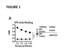
Figure 1 panels A-E. Generation of human GUCY2C-specific-CAR-T cells,
Figure I panel..A.:: Recombinant 5F9 antibody was assessed by 'ELBA for
specific binding
to hOUCY2CECD or BSA (negative control) plated at 1 laginiL. Two-way ANOVA;
****pc0,0001, Figure 1 panel B: Flow cytometry analysis was performed on
parental
C126 mouse colorectal cancer cells or CT26 cells engineered to express hGUCY2C
(CT26.hGUCY2C) and stained with 5F9 antibody. Figure 1 panel C: Schematic of
the
third generation =tine CAR -conatruct Containing murine sequences of the BiP
signal
sequence, 5F9 scFv, CD8a hinge region, the transmembrane and intracellular
domain of
CD28, the intracellular domain of 4-IBB (CDI37), and the intracellular domain
of CD3C
(5F9,m28BBz). The CAR. construct was inserted into the MSCV retroviral plasmid
pMIG
upstream of an 'RES-OFF marker. Figure 1 panel D: Murine CD8+ T cells
transduced
3
CA 03093705 2020-09-10
WO 2019/178580
PCT/US2019/022645
with a retrovirus containing a control (1D3.m28.BISti) CAR or CAR derived from
the. 5F9
antibody (5F9.1n288Bz) were labeled with purified 6xHis-hGUCY2CECD (10
pgfinL),
detected with anti-5xllis-Alexa Fluor 647 conjugate. Flow plots were gated on
live CD8+
cells. Figure! panel E: 6x.His-hGUCY2CECD binding curves for 5F9-derived or
control
(1D3) CARs, gated on live CD8 GFP cells (See data in Figure 5). Combined from
3
independent experiments.
Figure 2 panels AE. halCY2C-specifieCARs mediate antigenAependent
activation and effector functions, In Figure 2 panels A-E, Murine.CD8+ T cells
were left
non-transduced (None) or transduced with control ID3.in28BBz or 5F9.m2888z CAR
constructs as indicated. Figure 2 panel A: Gating strategy tbr all analyses in
Figure 2
panels B-D. Figure 2 panel B: Representative CAR-T cell phenotyping plot based
on
CD45RA and CD621.. Two-way ANOVA; NS: not significant; Bars: mean SD from 2-
3 independent experiments; Tniscm: naïve or T memory stem cells; Tcm: central
memory
I cells; Tern: effector memory T cells; Temra; effector memory T cells
expressing
is CD45RA. (C-fl) 104 CAR-T cells were stimulated for 6 hours with plate-
coated antigen
(BSA or hGUCY2C) or PMA and ionomycin (PMA/ION0). T-cell activation markers
(CD25, CD69, or CD44) and intracellular cytokine production (IFNy, TN:F(411-Z
and
MIPtu) were -then quantified by 'flow eytometry. graphs indicate the mean
SD. Figure
2. panel C refers to activation marker upregulation (MFI) and Figure 2 panel D
refers to
polyfunctional cytokine production (% of CAR+ cells) from 3 independent
experiments.
Figure 2 panel E: Parental CT26 or C.T26.hGUCY2C mouse colorectal cancer cells
in an
E-Plate were treated with CAR-T cells (5:1 E:T ratio), media, or 10% Triton-X
100
(Triton), and the relative electrical impedance was quantified every 15
minutes for 10
hours to quantify cancer cell death (normalized to time74)). Pereeut specific
lysis values
were calculated using impedance values following the addition of media and.
Triton for
normalization (0% and 100% specific lysis, respectively). Two-way ANOVA, B-E:
*p<0.05, **p<0.01, ***p<0.001, ****p<0.0001.
Figure 3 panels A-E. hGUCY2C CAR-T cells provide long-term protection in a
syngentic lung metastasis model_ In Figure 3 panels A-E, BALB/c mice were
injected
with 5x10P CT261GUCY2C cells via the tail vein to establish lung metastases.
Control
(4D5.m28BBz) or 5F9.m2flEtBz-CAR constructs were transduced into murine C1)8+
T
cells. Figure 3 panel. A: Mice were treated 3 days later with 5 Gy total body
irradiation
(TM) followed by I04-107 5F9.m28B8z (144=7-8/group) or 101 control (N=6) CART
cells.
4
CA 03093705 2020-09-10
WO 2019/178580
PCT/US2019/022645
Figure 3 panel rk Mice were treated on day 3(1)3) or day 7(07) with 5 Gy TBI
followed
by 107 control (N=10/group) or 5F9.m28BBz (N=9-10/group) CAR-T cells. Figure 3
panel C: Mice were treated on day 7 with 5 Gy TB1 followed by 107 control
(N=.10) or
5F9.m28BBz (N=12) CAR-T cells on day land day 14. Figure 3 panel D: Mice
treated
.. on day 7 with 5 Gy TB! and PBS or 107 control or 5F9.m28BBz CAR-T cells
were
sacrificed on day 18, lungs stained with India ink, and tumors/lung
enumerated. One-way
ANOV.A; *p4.05. Figure 3 panel E: Surviving mice.fromB and C treated. with.
5F9.m28BBz CAR-T cells or natvemice were challenged with 5x1.05 cr26
7/group) or CT26.hG1TCY2C (N=7/group) cells (re-challenge occurred 16-40 weeks
after
initial challenge). Log-rank Mantel-Cox test Figure 3 panels A-C and E;
"p<0.01,
***r:00)1, ****i.K0.0001. up arrows indicate CAR-I' cell treatment days. Each
panel
indicates an independent experiment
Figure 4 panels A-E. ItGliCY2C CAR-T cells eliminate human colorectal tumor
xenografts. Figure 4 panel A: hGLICY2C expression on TM human colorectal
cancer
cells was quantified by flow cytometry using the recombinant 5F9 antibody. in
Figure 4
panels B-E, Control (103.m28BBz) or 5F9.m28BBz CAR constructs were transduced
into marine CD8+ T cells. Figure 4 panel B: T84 colorectal cancer cells in. an
E-Plate
were treated in duplicate with 5F9-m2813Bz. or control CAR-T cells (5:1 E:T
ratio),
media, or 10% Triton-X 100 (Triton), and the relative electrical impedance was
measured
every 15 minutes for 20 hours to quantify cancer cell death (normalized to
time=0).
Percent specific lysis values were calculated using impedance values following
the
addition of media and. Triton for normalization (0% and 100% specific lysis,
respectively). Two-way ANOVA; "pc:0.01; representative of two independent
experiments. In Figure 4 panels C-E, Immunodeficient NSG.mice were injected
with.
2.5x10⁶ luciferase-expressing T84 colorectal cancer cells via intraperitoneal
injection and
were treated with 107 control (N=5) or 5F9-m281313z (N=4) CAR-T cells on day
14 by
intraperitoneal injection. In Figure 4 panels C-D, Total tumor luminescence
(photons/second) was quantified just prior to T-cell injection and weekly
thereafter. Two-
way ANOVA;-*p<0.05. Fignre.4 panel E: Mice were followed for survival. Log-
rank
Mantel Cox test; *p<0.05.
Figure 5. Detection of 5F9.m28IBBz CAR surface expression. Murine CD8+ T
cells transduced with a retrovirus containing a control m28Bilz CAR or CAR
derived
from the 5F9 antibody (5F9.m28Bliz) upstream of an IRES-GFP marker were
labeled
5
CA 03093705 2020-09-10
WO 2019/178580
PCT/US2019/022645
with purified 6xHisIKILICY2CECD (0-1430 nM) and detected with a5xHis-Alexa-647
conjugate. Flow plots were gated on live CDS+ cells.
Figure 6. hGUCY2C-expressing mouse colorectal cancer cells activate
5F9.m28BBz CAR-T cells. 106 CAR-T cells were stimulated for 6 h with 106
parental
C126, CT26.11GUCY2C colorectal cancer cells or PMA and ionomycin (PMMON0). 'f-
eell activation markers (CD25, CD69, or CD44) were quantified by flow
cytoMetry.
Figure 7, panels -A and B. hOI.J.CY2C-expressing mouse colorectal cancer cells
induce 51:9,m28BBz CAR-T cell eytokine production. 106 CAR-T cells were
stimulated
for 6 h with plate-coated antigen. Figure 7, panel A shows data for BSA,
hGUCY2C, and
PMA and ionomycin (PMA/ION0). Figure 7, panel B shows data for 106 parental
CT26
or CT26.1K1UCY2C colorectal cancer cells or PMA and ionomycin (PMMONO).
Intracellular cytokine production (IFNI, INFa, IL-2 or MIPI et) was quantified
by flow
cytoinetry.
Figure 8 panels A and B. 5F9..m2813Bz CAR-T cells kill hGUCY2C-expressing
mouse colorectal cancer cells. P-galactosidase-expressing C126 (data in Figure
8 panel
A) or C126.hGUCY2C (data in Figure 8 panel B) mouse colorectal cancer cells
were
cultured for 4 h with a range of effector CAR-T cell:target cancer cell ratios
(E:T Ratio).
Specific lysis.was determined by P-galactosidase release into the supernatant
detected by
a luminescent substrate. ****, p<0.0001 (Two-way ANOVA).
Figure 9 panels A and B. 5F9..m28BBz. CAR-T cells do notkill hGUCY2C-
deficient human colorectal tumors. Figure 9, panel A: hGUCY2C expression on.
SW480
human colorectal cancer cells was quantified by flow cytometry using the
recombinant
5F9 antibody. Figure 9, panel B: SW480 eel's in an &Plate were treated with.
5F9.in2813Bzor control I D3õ:m28BBz CART cells, media, or 2,5% Triton,X. .100
(Triton) and the relative electrical impedance was quantified. every 15 min
for 20 h to
quantify cancer cell death (normalized to time=0). Percent specific lysis
values were
calculated using impedance values following the addition of media and Triton
for
normalization (0% and 100% specific lysis, respectively).
Figure 10 panels A-C. Human T cells expresSing.5F9.h28BBz CAR. recognize and
kill-GUCY2C-expressing colorectal cancer cells. Figure 10 panel A: CAR-T cells
expressing a human 5F9 CAR construct (5F9.h28BBZ) were stimulated for 6 hours
with
plate-coated antigen (BSA or hGUCY2C) or PMA and ionomycin (PMA/ION0). The 1'-
cell activation marker CD69 and intracellular cytokines (1FNy. INFa, and IL-
2)0were
6
CA 03093705 2020-09-10
WO 2019/178580
PCT/US2019/022645
then quantified by flow cytometry. In reference to data. in Figure 10 panels B-
C. Parental
(CT26), human GUCY2Cexpressing CT26 (C126.13GUCY2C) mouse colorectal cancer
cells (data shown in Figure 10 panel B), or T84 human colorectal cancer cells
(data
shown in Figure 10 panel C) cultured in an E-Plate were treated with Control
or
5F9.h28BBz CAR-T cells (E:T ratio of 10;1), media, or 23% Triton-X 100 and the
relative electrical impedance was quantified every 15 min to quantify cancer
cell death
(normalized to time=0). Percent specific lysls-values were calculated using
impedance
values following the addition of media andIriton for normalization (0% and
100%
specific lysis, respectively). ***,p<0.001 (Two-way ANOVA).
Figure .11 panels A and B. 5F9.m28BBz CAR-T cells do not kill inGUCY2C-
expressing mouse colorectal cancer cells. CT26 cells expressing il-
galactosidase and
murine OUCY2C (Figure 11 panel A; C126.tuGUCY2C) or human GUCY2C (Figure 11
panel B; CT26,hqUCY2C) were cultured for 4 trwith a range of effector CAR-T
cell :target cancer cell ratios (ET Ratio). Specific lysis was determined by
11-galactosidase
release into the supernatant detected by a luminescent substrate.
****,p<0.0001 (Two-
way ANOVA).
DETAILED DESCRIPTION OF THE PREFERRED EMBODIMENTS
Single chain protein sequences that bind to the extracellular domain of human
(3UCY2C were generated using fragments of the variable light chain and
variable heavy
chain of an anti-GUCY2C antibody that binds to the extracellular domain of
human
GUCY2C. A linker sequence connects the variable light chain fragment to the
variable
heavy chain fragment into a single chain antibody variable fragment fusion
protein
sequence (scFv) that binds to the extracellular domain of human. GUCY2C.
The scPv is a. component in a CAR, which is a larger fusion protein. The CARs
functional components include the immtmoglobulin-derived antigen binding
domain,
antibody sequences i.e. s vFv, which binds to human GUCY2C, a hinge domain
that links
the scIPV to a transmembrane domain that anchors the protein in the cell
membrane of the
cell in Which it is expressed, and the signally domain which functions as
signal
transducingintracellular sequences (also referred to as cytoplasmic sequences)
that
activate the cell upon sav binding to human GUCY2C. The nucleic acid sequences
that
encode the CAR include sequences that encode a signal peptide from a cellular
protein
that facilitate the transport of the translated CAR to the cell membrane. CARs
direct the
7
CA 03093705 2020-09-10
WO 2019/178580
PCT/US2019/022645
recombinant, cells in which they are expressed to. bind to. and; in the case
of recombinant
cytotoxic lymphocytes, recombinant cytotoxic T lymphocytes (CTL.$),
recombinant
Natural Killer T cells (NKT), and recombinant Natural Killer cells.(NK) kill
cells
displaying the antibody-specified target, i.e. GUCY2C. When the CARis
expressed it is
transported to the cell surface and the signal peptide is typically removed.
The mature
CAR functions as a cellular receptor. The scFv and hinge domain are displayed
on the
cell surface where the scFv sequences can be exposed to proteins on. other
cells and bind
to GLICY2C on such cells.. The transmembrance region anchors the CAR in the
cell
membrane and the intracellular sequences function as a signal domain to
transduce a
.. signal in the cell which results in the death of GUCY2C-expressing cell to
which the
CAR-expressing cell is bound.
In some embodiments, the CARs comprise a signal sequence, such as for example
a mammalian or synthetic: signal sequence. In some embodiments, the CARs
comprise a
signal sequence from a membrane-bound protein such as for example .a mammalian
.. membrane-bound protein. in some embodiments, the CARs comprise a signal
sequence
from a membrane-bound protein such as CD8 alpha, CD8 beta, CD4, TCR alpha, TCR
beta, CO3 delta, CO3 epsilon, CD3 gamma, CD28, and Examples of signal
sequences may also be found in membrane bound. any mammalian signal sequence
<http://www.sianalpeptide.deSindex.plip?m=listspdb_manunalia>. In some
embodiments, the CARS comprise a Granulocyte-Macrophage Colony-Stimulating
Factor
(GM-CSF) signal sequence. In some embodiments, the CARs comprise a
Granulocytt.)-
Macrophage Colony-Stimulating Factor (GM-CSF) signal sequence having amino
acids
1-22 of SEQ ID NO2. In some embodiments, the Granulocyte-Macrophage Colony-
Stimulating Factor(GM.-C$F) signal sequence comprises antinoacids- 1-22 of SEQ
ID
NO:2. In some embodiments, the Granulocyte-Macrophage Colony-Stimulating
Factor
(GM-CSF) signal sequence consists essentially of amino acids I-22 of SEQ ID
NO:2. In
some embodiments, the Granulocyte-Macrophage Colony-Stimulating Factor (GM-
CSF)
signal sequence consists of amino acids 1-22 of SEQ ID NO:2. In some
embodiments,
thenueloic acid sequence of the Construct that: encodes the CARs thattoniprise
Granulocyte-Macrophage Colony-Stimulating Factor (GM-CSF)-signal sequence
comprise nucleic acid 1-66 of SEQ ID NO:1. In some embodiments, the nucleic
acid
sequence that encodes the Granulocyte-Tvlacrophage Colony-Stimulating Factor
(GM-
CSF) signal sequence comprises nucleic acid 1-66 of SEQ NO:l. In some
8
CA 03093705 2020-09-10
WO 2019/178580
PCT/US2019/022645
embodiments, the nucleic acid. sequence that encodes the Granulocyte-
Macrophage
Colony-Stimulating Factor (GM-CS F) signal sequence consists essentially of
nucleic acid
1-66 of SEQ ID NO:i. In some embodiments, the nucleic acid sequence that
encodes the
Granulocyte-Macrophage Colony-Stimulating Factor (GM-CSF) signal sequence
consists
of nucleic acid 1-66 of SEQ ID NO: 1.
The anti-GliCY2C binding domain is provided as a single chain chimeric
receptor
that is MHC-independentõ. The antigen-binding domain is derived from an
antibody. In.
some embodiments, CARs comprise anti-auanytyl. cyclase C (also referred to as
GCC or
GLICY2C) single Chain variable fragment (scFv) (preferably a Variable Light
fragment
(GlycineiSerine)4 Linker Variable Heavy fragment) from 5F9. 5F9 is a
hybridoron
expressing a hilly humanized, monoclonal antibody that recognizes the
extracellular
domain of human GUCY2C. The DNA coding sequences of the antibody heavy and
light
chains were used to create a novel scFv for CAR implementation that is
employed in the
creation of anti-GCC CARs, such as for example the 5F9-2813I3z CAR, and
confers
antigen specificity directed towards the GITCY2C molecule.
In some embodiments such as the 5F9-28138z CAR, the anti-G'CC say may be a
5F9 single chain variable fragment (scFv) (Variable Light fragment¨
(Glycine4Serine)4
Linker Variable Heavy fragment). The W9 say may comprise amino acids 25-274 of
SEQ ID NO:2, in some embodiments, the nucleic acid sequence of the construct
that
encodes the CARs that comprise the 5F9 scFv comprise nucleotides 73-822 of SEQ
ID
NO: 1. In some embodiments, the CARs comprise an anti-GCC 5F9 sal,. Amino
acids
25-133 of SEQ ID NO:2 corresponds to the 5F9 Variable Light chain fragment.
Amino
acids 154-274 of SEQ IDNO:2 corresponds to the 5F9 Variable Heavy chain
fragment.
In. some embodiments, the CARs comprise an anti-GCC 5F9 single chain variable
.. fragment (sCR') that corresponds to the 5F9 Variable. Light fragment and
the 5F'9'
Variable Heavy fragment attached to each other with a (GlycineaSerine)n LINKER
in
which (Glycine4Serine) = GGGGS (SEQ ID NO:3) and .n = 2-5.
In some embodiments, the linker contains two (GlycineaSerine) units
((GlYcine4Serine)2) and may referred to as LINKER 045-2 (SEQ ID NO:4). In some
embodiments, the linker contains three (Glycin.a4Serine) units
((GlycinetSerine)3) and
may referred to as LINKER G4S-3 (SEQ ID NO:5). In some embodiments, the linker
contains four (Cilycine4Serine) units ((Glycine4Serine)4.) and may referred to
as LINKER
9
CA 03093705 2020-09-10
WO 2019/178580
PCT/US2019/022645
G4S-4 (SEQ ID "NO:6), In some embodiments, the linker contains five
(Cilyeine4SerittO
units aGlycine4Serine).5) and may referred to as LINKER G4S-5 (SEQ ID NO:7).
The 5F9 variable fragments may be configured from N-terminus to C-terminus in
the order Variable Light Chain fragment-LINKER-Variable::Mavy Chain fragment
or
Variable Heavy Chain fragment-LINKER-Variable Light Chaia:fragment, In:some
embodiments, the CARs comprise an anti-GCC 5E9 scFV configured as [5F9
Variable
Light Chain fragment--(Glycine1Serine)2-5F9 Variable Heavy Chain fragment]
(SEQ ID
NO:8), [51,9 Variable Light Chain fragment--(G1ycine4Serine)3-51,9 Variable
Heavy
Chain fragment] (SEQ ID NO:9), [5E9 Variable Light Chain fragment-
Variable Heavy Chain fragment) (SEQ ID NO:10), or [51,9
Variable Light Chain fragment--(Cilycine4Serine)5--5F9 Variable Heavy Chain
fragment]
(.SEQ IDl0 11) in some embodiments, the CARs comprise an anti-GCC::50:scfy
conli RUT ed a 5E9 N,aripit.ilefle4vy Chain fragment--(G1ycitte4Se1itie)2,-5F9
Variable
Light Chain fragment] (SEQ ID NO:12), [5F9 Variable Heavy Chain fragment--
(Glycine4Serine)3-5F9 Variable Light Chain fragment] (SEQ ID NO:13), [5E9
Variable
Heavy Chain fragment--(Glycine4Serine)4--5F9 Variable Light Chain fragment]
(SEQ ID
NO: I4, or [5F9 Variable Heavy Chain fragment--(Glycinc4Scrinc)5--5F9 Variable
Light
Chain fragment (SEQ ID NO:15).
In some embodiments, the CARs comprise an anti-GCC 5F9FV having:gbh*
acids 25-274 of SEQ ID NO:2. In some embodiments, the 5F9 say comprises amino
acids 25-274 of SEQ ID NO:2, in some embodiments, the 5F9 sav consists
essentially
of amino acids 25-274 of SEQ ID NO:2. In some embodiments, the 5F9 seffv
consists of
amino acids 25-274 of SEQ. ID NO:2. In some embodiment, the nucleic acid
sequence
that encodes the 5F9 say comprises nucleotides 73-822 of SEQ ID NO:l. In some
embodiments, the nucleic acid sequence that encodes the 5F9 scFY consists
essentially of
nucleotides 73-822 of SEQ ID NO:!. In some embodiments, the nucleic acid
sequence
that encodes the 5F9 scFv consists of nucleotides 73-822 of SEQ ID NO:1 ,
In some embodiments, CARs comprise a CD80., 461 -Fc, IgG4-Fc, or CD28
hinge region. In some embodiments, CARs comprise a CD8a hinge region. In some
embodiments, CARs comprise a CD8a hinge region having amino acids 277-336 of
SEQ
ID NO:2. In some embodiments, the CD8a hinge region comprises amino acids 277-
336
CA 03093705 2020-09-10
WO 2019/178580
PCT/US2019/022645
of SEQ ID NO:2. In some embodiments, the CD8a hinge region consists
essentially of
amino acids 277-336 of SEQ ID NO:2. In some embodiments, the CD8a hinge region
consists of amino acids 277-336 of SEQ ID NO:2. In some embodiments, the
nucleic
acid sequence that encodes the C08u hinge region comprises nucleotides 829-
1008 of
SEQ ID NO:I in some embodiments, the nucleic acid sequence that encodes the
CD8a
hinge region consists essentially of nucleotides 829-1008 of SEQ ID NO:l. In
some
embodiments, the nucleic acid sequence that encodes the CD8a hinge region
consists of
nucleotides 829-1008 of SEQ ID NO: 1.
In some embodiments, CARs comprise a CD28, 4-EBB (C0I37), CD2, CD27,
CD30, CD401.õ CD79A, CD79B, CD226, DR3, GITR, HVEM, ICOS, LIGHT, 0X40, or
SLAM transmembrane region.
In some embodiments, CARs comprise a CD28, 4-IBB (CD137), CD2, CD27,
CD30,.CD4OL, CD79.A,CD79B, CD226, DR3., GITR, HVEM,-ICOS, LIGHT, 0X40,.or
SLAM intracellular region.
In some embodiments, CARs comprise both transmembrane and intracellular
(cytoplasmic) sequences from CD28, 4-IBB (CD137), CD2, CD27, CD30, CD4OL,
CD79A, CD793, CD226, .DR3, G1TR, HVEM, ICOS, LIGHT, 0X40, or SLAM. In
some embodiments, CARs comprise CD28 transmembrane and intracellular
sequences.
In some embodiments, -CARs comprise CD28 transmembrane and intracellular
sequences
having amino acids 337-405 of SEQ ID NO:2. In some embodiments, the CO28
transmembrane and intracellular sequences comprises amino acids 337-405 of SEQ
NO:2. In some embodiments, the CD28 transmembrane and intracellular sequences
consists essentially of amino -acids 337-405 of SEQ NO:2. It some embodiments,
the
CD28 transmembrane and. Intracellular sequences. consists of amino acids 337-
405 of
SEQ 'ID NO:2. In some embodiments, the nucleic acid sequence that. encodes
CD28
transmembrane and intracellular sequences comprises nucleotides 1009-1215 of
SEQ ID
NO: 1. In some embodiments, the nucleic acid sequence that encodes CD28
transmembrane and intracellular sequences consists essentially of nucleotides
1009-1215
of SEQ ID NO:1 In-some-embodiments, the nucleic acid sequence encodes CD28
transmembrane and intracellular sequences consists of nucleotides 1009-1215 of
SEQ ID
NO:!.
.1.1
CA 03093705 2020-09-10
WO 2019/178580
PCT/US2019/022645
In some embodiments, CARs comprise intracellular (cytoplasmic) sequences from
Cy-chain associated with CD3 (CD3(;), the CD79-alpha and -beta chains of the B
cell
receptor complex., or certain Fe receptors.
In some embodiments, CARs comprise a) intracellular (cytoplasmic) sequences
.. from one or more of CD28, 4-IRS (CD137), CD2, CD27, 0o30, CD4OL, CD79A,
CD79B, CD226, DR3, GITR, HVEM, ICOS, LIGHT, 0X40, or SLAM intracellular
region in combination with b) intracellular. (cytoplasmic) sequences .from
associated with CD3 (CD30, the CD79-a1Pha and -beta chains of the =B cell
receptor
complex, or certain Fe receptors.
In some embodiments, CARs comprise CD28 transmembrane and intracellular
sequences together with 4-IBB intracellular sequences in combination with CDR;
intracellular sequences.
In some embodiments, CARs comprise CD28 transmembrane and intracellular
sequences having amino acids 337-405 of SEQ. ID NO2. In some embodiments, the
is C1)28 transmembrane and intracellular sequences comprises amino acids
337-405 of SEQ
ID NO:2, in some embodiments, the CD28 transmembrane and intracellular
sequences
consists essentially of amino acids 337-405 of SEQ ID NO:2. In some
embodiments, the
C))28 transmembrane and intracellular sequences .consists.of amino acids 337-
405 of
SEQ ID NO:2, in some embodiments, the nucleic acid sequence that encodes CD28
transmembrane and intracellular sequences comprises nucleotides 1009-1215 of
SEQ ID
NO:1 . In some embodiments, the nucleic acid sequence that encodes CD28
transmembrane and intracellular sequences consists essentially of nucleotides
1009-1215
of SEQ ID NO: I. In some embodiments, the nucleic acid sequence encodes CD28
transmembrane and intracellular sequences consists of nucleotides 1009-1.215
of SEQ ID
In some embodiments, CARs comprise 4-1BB intracellular sequences. In some
embodiments, CARs comprise 4-1BB intracellular sequences having amino acids
406-
444 of SEQ ID NO:2. In some embodiments, CARs comprise 4-1BB intracellular
Sequences comprise amino acids 406-444 of SEQ ID NO:2, In some embodiments, 4-
155 intracellular sequences consists essentially of amino acids 406-444 of SEQ
ID NO:2,
In some embodiments, 4-1BB intracellular sequences consist of amino acids 406
114 of
SEQ 1D NO:2. In some embodiments, the nucleic acid sequence that encodes 4-I
BR
intracellular comprises nucleotides 1216-1332 of SEQ ID NO:!. In some
embodiments,
12
CA 03093705 2020-09-10
WO 2019/178580
PCT/US2019/022645
the nucleic acid sequence. that encodes 4-1138 intracellular consists
essentially of
nucleotides 1216-1332 of SEQ ID NO:!. In some embodiments, the nucleic acid
sequence that encodes 4-188 intracellular consists of nucleotides 1.216-1332
of SEQ ID
NO:1,
In some embodiments, CARs comprise a sequence encoding at least one
imtnunoreceptor tyrosine activation motif (TAM). In some embodiments, CARs
comprise a sequence from a cell signaling molecule that comprises ITAM.s.
Typically 3
1TAMS are present. in such sequences. Examples of cell signaling molecules
that
comprise ITAMS include c-chain associated with CD3 (CD30, the CD79-alpha and -
beta
chains of the B cell receptor complex, and certain Fe receptors. Accordingly,
in some
embodiments, CARs comprise a sequence from a cell signaling molecule such as
CD3.
the CD79-alpha and -beta chains of the 13 cell receptor complex, and certain
Fe receptors
that comprises ITAM.s. The sequences included in the CAR are intracellular
sequences
from such molecules that comprise one of more ITAMs. An ITA1v1 is a conserved
sequence of four amino acids that is repeated twice in the cytoplasmic tails
of certain cell
surface proteins of the immune system. The conserved sequence of four amino
sequence
of an !TAM contains a tyrosine separated from a leucine or isoleucine by any
two other
amino acids (YXXL or YXX1 in which X is independently any amino acid
sequence),
The .ITAM contains a sequence that is typically 14-16 amino acids having the
two four
amino acid conserved sequences separated by between about 6 and 8 amino acids.
The ;-
chain associated with CD3 (CD3) contains 3 1TAM.S. Amino acids 445-557 of SEQ
NO:2 are CD3; intracellular sequences. The ITAMS are located at amino acids
465-479,
504-519 and 535-549. hi seine entbodiments, CARs comprise CD3; intracellular
sequences. In some embodiments. CARs comprise CD3; intracellular sequences
having
amino acids 445-557-of -SEQ ID NO:2. In some embodiments, CD3; intracellular
sequences comprise 445-557 of SEQ ID NO:2. In some embodiments, CD3;
intracellular
sequences consist essentially of 445-557 of SEQ ID NO:2. In some embodiments,
CD3;
intracellular sequences consist of 445-557 of SEQ ID NO:2. In some
embodiments, the
nucleic acid sequence that encodes CD3; intracellular comprises nueledtides
1333-1671
of SEQ ID NO:!. In some embodiments, the nucleic acid sequence that encodes
CD3;
intracellular consists essentially of nucleotides 1333.-1671 of SEQ ID NO:l.
In some
embodiments, the nucleic acid sequence that encodes CD3; intracellular
consists of
nucleotides 1333-1671 of SEQ ID NO:l.
13
CA 03093705 2020-09-10
WO 2019/178580
PCT/US2019/022645
In some embodiments, CARs may comprise an immunoglobulin-derived antigen
binding domain: antibody sequences that bind to GUCY2C fused to a. T cell
signaling
domain such as the CD3zeta signaling chain of the T cell receptor or a T-cell
costimulatory signaling (e.g. CD28) domain linked to a I-cell chain such as
CD3zeta
chain or the gamma-signal-transducing subunit of the Ig Fc receptor complex.
The signaling domain of the CAR comprises sequences derived from a TCR. In
some embodiments, the CAR comprises an extracellular single chain fragment of
antibody variable region that provides antigen binding function fused to a
transmembrane
and cytoplasmic signaling domain such as CD3zeta chain or CD28 signal domain
linked
1.0 to CD3zeta chain. In some embodiments the signaling domain is linked to
the antigen
binding domain by a spacer or hinge. When the fragment of antibody variable
region
binds to GUCY2C, the signaling domain initiates immune cell activation. These
recombinant I cells that express membrane bound chimeric mentors comprising an
extracellular anti-GUCY2C binding domain and intracellular domain derived from
TCRs
which perform signaling functions to stimulate lymphocytes. Some embodiments
provide
anti-MO(2C binding domain is a single chain variable fragment (scFv) that
includes
anti-GUCY2C binding regions of the heavy and light chain variable regions of
an anti-
GUCY2C antibody. A signaling ctomain may include a T-cell .costiMulatory
signaling
(e.g. CD28, 4-I BB (CD137), CD2, CD27, 0)30, CD4OL, CD79A, CD79B, CD226,
DR3, GITR, HVEM, ICOS, LIGHT, 0X40, SLAM) domain and T-cell triggering chain
(e.g. CD3zeta).
In some embodiments, CARs include an affinity tag. Examples of such affinity
tags include: Strep-Tag; Strep-Tagil; Poly(flis); HA; V5; and FLAG-tag. In
some
embodiments, the affinity tag may be located before scFv or between scFv and
hinge
region or after the hinge region. In some embodiments, the affinity tag is
selected from
Strep-Tag, Siren-Tagil, Poly(His), HA; V5, and FLAG-tag, and is located before
scFv or
between scFv and hinge region or after the hinge region.
1.n some embodiments, CARs comprise from N terminus to C terminus, a signal
sequence, the anti-GCC say is a 5F9 single chain variable fragment (stFv), a
hinge
region, a transmembrane region and intracellular sequences from one of more
proteins
and intracellular sequences and an immunoreceptor tyrosine activation motif,
and
optionally an affinity tag.
.14
CA 03093705 2020-09-10
WO 2019/178580
PCT/US2019/022645
In some embodiments, Crns,comprist from N terminus. to C terminus, :4:signal
sequence selected from GM-CSF, CD8 alpha, CD8 beta, CD4, TCR alpha, TCR beta,
CD3 delta, CD3 epsilon, CD3 gamma, CD28, BiP linked to the anti-GCC scfv is a
5F9
single chain variable fragment (scFv) selected from (Variable Light Chain
fragment-
(Glyeine4Serine)-).5 Linker - Variable Heavy Chain fragment) and (Variable
Heavy Chain
fmgment-(Glyeine4Serine)2_5 Linker - Variable Light Chain fragment), linked to
a hinge
region selected from CD8a, IgG IgG4-Fc and CD28 hinge regions, linked to a
transmembrime region selected from a CD8a, IgGI -Fe, IgG4-Fc and CD28
transmembiarteiregim, linked to intracellular sequences selected from CO2$4-
BB
(CD I 37), CD2, 027, (10/28, CD30, CD4OL,, C079A,:CD7913, CD226, DR3, GIlL
Et VENT, 1COS:, LIGHT, 0X40, SLAM tutratellular sequences, linked to an
immunoreeeptot tyrosine activation motif containing sequence selected from
CO3,
CD79-alpha, C079-beta and Fe receptor intracellular sequences that comprise
one or
more ITAMs, optionally linked to an affinity- tag selected from Strep-Tag,
Strep-Tagll,
Poly(iliAtIA; V5, and FLAG-tag.
kwarie embodiments. CARs comprise from N terminus ito:C terminus, :a
Granulocyte-Macrophage Colony-Stimulating Factor (GM-CSF) signal sequence, the
anti-GCC scf:v is a 5F9 single chain variable fragment (say) selected from
[Variable
Light Chain fragment- (Glyeine4Serine) 2 -5 Linker - Variable Heavy Chain
fragment] or
(Variable Heavy Chain fragment-(01yente4Seritie)m Linker - Variable Light
Chain
fragment] MOO, CD28, IgG1 -Fe, or Ig(.14-Fnninge region, a CD8a or (D28
transmembrane and intracellular sequences, 4-I BB intracellular sequences and
CD3,-,
intracellular sequences.
In some embodiments, CARs consist essentially of a ciranulocyte-Macrophage
Colony-Stimulating Factor (GM-CSF) signal sequence, the anti-GCCsen, a::5F9
single
chain variable fragment (seFv) (Variable Light fragment- (Glycine4Serine)4
Linker -
Variable Heavy fragment), a CD8a hinge region, CD28 transmembrane and
intracellular
Sett-peaces, '4.i BB intracellular sequences :and CD3 4; intracellular
sequenees,,
in some embodiments, CARs comprise amino acids 4-22 25-274, 277-336, 337-
405,406-444 and 445-557 of SEQ ID NO;2. In some embodiments, CARs consiSt
essentially of amino acids 1-22, 25-274, 277-336, 337-405, 406-444 and 445-557
of SEQ
ID NO:2. In some embodiments, CARs consist of amino acids 1-22, 25-274, 277-
336,
CA 03093705 2020-09-10
WO 2019/178580
PCT/US2019/022645
337-405, 406-444 and 445-557 ofSEQ ID NO:2, In some embodiments, the nucleic
acid
sequence of the construct that encodes the CARs comprises nucleotides 1-66,.
73422,
829-1008,1009-1215, 121.6-1332 and 1333-1671 of SEQ NO:1 . In some
embodiments, the nucleic acid sequence of the construct that encodes the CARs
consist
S essentially of nucleotides 1-66, 73-822, 829-1008, 1009-1215, 1216-1332
and 1333-1671
of SEQ ID NO: 1. In some embodiments, the nucleic acid sequence of the
construct that
encodes theCAR.s consist of nucleotides 1-66, 73-822, 8294008, 1009-1.215,
121.6-1.332
and 1333-1671 of SEQ NO:!'. In some embodiments, these sequences are linked to
regulatory elements necessary for expression of the coding sequence in a human
cells
such as a human T cell. In some embodiments, a. human cell such as a human T
cell is
transformed with the sequences linked to regulatory elements necessary for
expression of
the coding sequence.
In some embodiments, the CAR is encoded by Gls4.5F9(VL4G4S)4-VH)-CD8a-
CD28tm.ICD-4-18B-CD3z.stop (5F9-2813Bz SEQ ID -NO:1), a novel DNA sequence, a
synthetic receptor that can be expressed by T lymphocytes and infused for the
therapeutic
treatment of human guanylylcyclase C (GUCY2C)-expressing malignancies.
GM.5F9(V1.-(04S)4-VH)-CD8a-CD28tm.ICD-4-113B-CD3z.stop encodes SEQ ID NO:2.
5F9-281$13z -comprises human DNA coding sequences concatenated thusly: (1)
Granulocyte-Macrophage Colony-Stimulating Factor (GM-CSF) signal sequence, (2)
5F9
single chain variable fragment (scFv) (Variable Light fragment-
(Glycine4Serine)4
Linker - Variable Heavy fragment), (3) CD8a hinge region, (4) CD28
transmembrane
domain, (5) CD28 intracellular domain, (6) 4-IBB intracellular domain, and (7)
CD3c
intracellular domain. The CAR is referred to as 5F9-28BBz. In some
embodiments, the
CAR .coutpiises-SEQ ID NO:2. In some embodiments, the CAR consists essentially
of
SEQ ID NO:2. In some embodiments, the CAR consists of SEQ ID NO:2. In some
embodiments, the nucleic acid sequence of the construct that encodes the CARs
consist of
nucleotides comprises SEQ ID NO: 1. In some embodiments, the nucleic acid
sequence
of the construct that encodes the CARs consist of nucleotides consists
essentially of SEQ
ID NO:1 sOme-embodiments, thenucleic acid sequence of the construct that
encodes
the CARs consist of nucleotides consists of SEQ ID NO:l. in some embodiments,
these
sequences are linked to regulatory elements necessaty for expression of the
coding
sequence in a human cell such as a human I cell. In some embodiments, a human
cell
16
CA 03093705 2020-09-10
WO 2019/178580
PCT/US2019/022645
such as a human T cell transformed with the sequences linked to regulatory
elements
necessary for expression of the coding sequence.
In some embodiments, the 5F9-288.B2 SEQ ID NO:1 is linked to regulatory
elements necessary for expression of the coding sequence in a human cell such
as a
human T cell. Regulatory elements necessary for expression Utile coding
sequence in a
human cell such as a human T cell may include a promoter, a polyadenylation
site and
other sequences in 5 and 3' untranslated regions. In some embodiments, SEQ ID
NO:1
is inserted in an expression vector such as a. plasmid such a pVAX, or a
retroviral
expression vector such as a lentiviral vector, or a recombinant DNA viral
vector such a
recombinant adenovirus, recombinant MN, or recombinant vaccinia virus, or as
double
stranded DNA to be used with CRISPRICas9. TA LEN or other transposon
technology
or as messenger RNA.
In some embodiments, CAR coding sequences are introduced ex vivo into cells,
such as T cells, including C04+ and cD8+, invariant Natural Killer T cells,
gamma-delta
T cells, Natural Killer cells, and myeloid cells, including CD34+
hematopoietic stem cells
from peripheral lymphocytes using routine in vitro gene transfer techniques
and materials
such as retroviral vectors. Following gene transfer, the recombinant cells are
cultured to
expand the number of recombinant cells which are administered to a patient.
The
recombinant cells will recognize and bind to cells displaying the antigen
recognized by
the extracellular antibody-derived antigen binding domain. Following
modification, the
cells are expanded ex vivo to obtain large numbers of such cell which are
administered to
the patient have been described. As above, autologous refers to the donor and
recipient of
the cells being the same person. Allogenic refers to the donor and recipient
of the cells
being different people In addition to isolating and expanding populations of
antigen-
specific T cells by ex vivo culturing, the I cells may be modified after
isolating and
before expanding populations by having genetic material added to them that
encodes
proteins such as cytokines, for example 1L-2, 1L-7, and 1L-15.
A plurality of T cells which recognize at least one epitope of GUCY2C may be
obtained by isolating a I cell from a. cell donor, transforming it with a
nucleic acid
molecule that encodes an anti-OUCY2C CAR and, culturing the transformed cell
to
exponentially expand the number of transformed T cells to produce a plurality
of such
cells,
17
CA 03093705 2020-09-10
WO 2019/178580
PCT/US2019/022645
The cell donor may be the individual to whom the expanded population of cells
will be administered, i.e. an autologous cell donor. Alternatively, the T cell
may be
obtained from a cell donor that is a different individual from the individual
to whom the T
cells will be administered, i.e. an allogenic T cell. if an allogenic T cell
is used, it. is
preferred that the cell donor be type matched, that is identified as
expressing the same or
nearly the same set of leukocyte antigens as the recipient.
T cells may be obtained from a cell donor by routine methods including, for
example, isolation from blood fractions, particularly the peripheral blood
monocre cell
component, or from bone marrow samples.
Once T cells are obtained from the cell donor, one or more T cells may be
transformed with a nucleic acid that encodes an anti-GUCY2C CAR which includes
a.
functional binding fragment of an antibody that binds to at least one epitope
of a
GUCY2C and a portion that renders the protein, when expressed in a cell such
as a I cell,
a membrane bound protein.
The nucleic acid molecule that encodes anti-GUCY2C CAR may be obtained by
isolating a B cell that produces antibodies that recognize at least one
epitope of GUCY2C
from an "antibody gene donor" who has such B cells that produce antibodies
that
recognizes at least one epitope of GLICY2C. Such antibody gene donors may have
B
cells that produce antibodies that recognize at least one epitope of a GUCY2C
due to an
immune response that arises from exposure to an immunogen other than by
vaccination
or, such antibody gene donors may be identified as those who have received a
vaccine
which induces production of B cells that produce antibodies that recognize at
least one
epitope of GUCY2C, i.e. a vaccinated antibody genetic donor. The vaccinated
antibody
genetic donor may have been. previously vaccinated or may be administered a
vaccine
specifically as part of an effort to generate such B cells that produce
antibodies that
recognize at least one epitope of GUCY2C for use in a method that comprises
transforming T cells with a nucleic acid molecule that encodes an anti-GUCY2C
CAR,
expanding the cell number, and administering the expanded population of
transformed T
cells to an individual.
The antibody gene donor may be the individual who will be the recipient of the
transformed I cells or a different individual. from the individual who will be
the recipient
of the transformed I cells. The antibody gene donor may be same individual as
the cell
donor or the antibody gene donor may be a different individual than the cell
donor. In
18
CA 03093705 2020-09-10
WO 2019/178580
PCT/US2019/022645
some embodiments, the cell donor is the recipient of the transformed T cells
and the
antibody gene donor is a different individual. In some embodiments, the cell
donor is the
same individual as the antibody gene donor and is a different individual from
the recipient
of the transformed T cells. In some embodiments, the cell donor is the same
individual as
the antibody gene donor and the same individual as the recipient of the
transformed T
cells.
The nucleic acid molecule which encodes anti-GLICY2C CAR comprises a coding
sequence that encodes functional binding, fragment of an antibody that
recognizes at least
one epitope of GLICY2C linked to a protein sequence that provides for the
expressed
to protein to be a membrane bound protein. The coding sequences are linked
so that they
encode a single product that is expressed.
The coding sequence that encodes a functional binding fragment of an antibody
that .recognizes at least one epitope of GUCY2C may be isolated from a B cell
from an
antibody gene donor. Such a B cell may be obtained and the genetic information
isolated.
is In some embodiments, the B cells are used to generate hybrid cells which
express the
antibody and therefore carry the antibody coding sequence. The antibody coding
sequence may be determined, cloned and used to make the abnti-GLICY2C CAR . A
functional binding fragment. of an antibody that recognizes at least one
epitope of
ClUCY2C may include some or all of the antibody protein which when expressed
in the
20 transformed T cells retains its binding activity for at least one
epitope of CII.3CY2C.
The coding sequences for a protein sequence that provides for the expressed
protein to be a membrane bound protein may be derived from membrane bound
cellular
proteins and include the transmembrane domain and, optionally at least a
portion of the
cytoplasmic domain, ant-Vora portion of the extracelhdar domain, and a signal
sequence
25 to transIocate the expressed protein to the cell membrane.
The nucleic acid molecule that encodes the anti-GUCY2C CAR, i.e. the anti-
GLICY2C CAR coding sequence, may be a DNA or RNA The invention relates to
chimeric antigen receptors that bind to guanylyl cyclase C and nucleic acid
molecules that
encode such chimeric antigen receptors. The invention also relates to cells
that comprise
30 such chimeric antigen receptors, to methods of making such chimeric
antigen receptors
and cells, and to methods of using such cells to treat individuals who are
suffering from.
cancer that has cancer cells which express gu.anylylcyclase C and to protect
individuals
against cancer that has cancer cells which express g.uanylyl cyclase C.
19
CA 03093705 2020-09-10
WO 2019/178580
PCT/US2019/022645
Immunotherapy based upon T cells that express chimeric antigen receptors
(CARO has become an emerging modality for treating cancer. CARs are fusion
receptors
that comprise a domain which functions to provide MA-independent binding of
cell
surface target molecules and a signaling domain that can activate host immune
cells of
various types, typically peripheral blood I cells, which may include
populations of cells
referred to cytotoxic lymphocytes, cytotoxic T lymphocytes (CTLs), Natural
Killer T
cells (NKT) and Natural Killer cells (NK.) or helper T cells. That is, while-
typically being
introduced into T cells, genetic material encoding .CARs may be added to
immune cells
that are not I cells such as NK cells.
io Guanylyl cyclase C (also referred to interchangeably as GCC or GUCY2C)
is a
membrane-bound receptor that produces the second messenger cOMP following
activation by its hormone ligands guanylin or uroguanylin, regulating
intestinal
homeostasis,- tuntorigenesis, and obesity. GUCY2C. cell surface expression is
confined to
luminal surfaces of the intestinal epithelium and .a subset of hypothalamic
neurons. Its
is .. expression is maintained in >95% of colorectal cancer metastases and it
is ectopically
expressed in tumors that evolve from intestinal metaplasia, including
esophageal, gastric,
oral, salivary gland and pancreatic cancers.
The inaccessibility of GUCY2C in the apical membranes of polarized epithelial
tissue due to subcellular restriction of GUCY2C, creates a therapeutic
opportunity to
20 target metastatic lesions of colorectal origin which have lost apical-
basolateral
polarization, without concomitant intestinal toxicity.
sy-ngeneic, immunocompetent mouse model demonstrated that CAR-T cells
targeting murine GUCY2C were effective against colorectal cancer metastatic to
lung in
the absence of intestinal to cities. Similarly, other GUCY2C-targeted
therapeutics,
25 including antibody-drug conjugates and vaccines, are safe in preclinical
animal models,
and therapeutic regimens utilizing these platforms are in clinical trials for
metastatic
esophageal, gastric, pancreatic, and colorectal cancers (NCT02202759,
NC102202785,
NCT01972737).
The safety of these therapeutic regimens, in the context of GUCY2C expression.
30 across the rostral-caudal axis of intestine, reflects compartmentalized
expression of
GUCY2C, enriched in apical. but limited in basolateral, membranes of
epithelial cells.
Systemic radiolabeled imaging agents conjugated to GUCY2C ligand target GUCY2C-
CA 03093705 2020-09-10
WO 2019/178580
PCT/US2019/022645
expressing metastases without localizing in intestine,. confirming the mucosal
compartmentalization of the receptor.
Tumors express up to 10-fold greater amounts of GUCY2C, compared to normal
epithelial cells, potentially creating a quantitative therapeutic window to
discriminate
receptor overexpressing tumors from intestinal epithelium with low/absent
GUCY2C in
baso lateral membranes.
U.S. Patent Application Publication 20120251509 Al and U.S. Patent Application
Publication US 2014-0294784 A 1 , which are each incorporated herein by
reference,
disclose CARs including CARs that bind to guanylyl cyclase C, T cells that
comprise
.. CARs including T cells that comprise CARs that bind to GUCY2C and target
cells that
comprise GUCY2C, methods of making chimeric antigen receptors and T cells, and
methods of using T cells that comprise CARs that bind to GUCY2C and target
cells that
comprise GUCY2C to protect individuals against cancer cells that express
GUCY2C -and
to treat individuals who are suffering from cancer in which cancer cells
express
GUCY2C.
There is remains a need for improved compositions and methods to protect
individuals against cancer cells that express GUCY2C and to treat individuals
who are
suffering from cancer in which cancer cells express:GUCY2C.
Proteins comprising an anti-GUCY2C scFV sequence are provided. The anti-
GUCY2C scFV sequences may be selected from the group consisting of SEQ ID
NO:8,
SEQ 'NO:9,
SEQ ID NO:10, SEQ ED NO:!!, SEQ NO:12, SEQ ID NO:13, SEQ ID
NO:14 and SEQ ID NO:13.
Proteins comprising the 5F9 anti-GUCY2C saV sequence and further comprising
a signal sequence, a hinge domain, a transmembrane domain, and. a. signaling
domain are
provided.
Nucleic acid molecules that encode such proteins are provided. The nucleic,
acid
molecules may be operably linked to regulatory elements that can function to
express the
protein in a human cell such as a human I cell. The nucleic acid. Molecules
may be
incorporated in a nucleic acid vector such as a plasmid or recombinant viral
vector that
can be used transform human cells into human cells that express the protein.
Human cells comprising the nucleic acid molecules and express the proteins are
provided.
21
CA 03093705 2020-09-10
WO 2019/178580
PCT/US2019/022645
Methods of making such cells are provided.
Methods of treating a patient who has cancer that has -cancer cells that
express
GUCY2C and methods of preventing cancer that has cancer cells that express.
GUCY2C
in a patient identified as being of increased risk, are provided.
Figure 1 panels A-E. Generation of human GUCY2C-specific CAR-T cells.
(Figure 1 panel A) Recombinant 5F9 antibody was assessed by ELISA for specific
binding to bGLICY2CECD or BSA (negative control) plated at I ggirriL.. Two-way
ANOVA; ****p,0,0001. (Figure 1. panel B) Flow cytometry analysis was
performed on
parental C126 mouse colorectal cancer cells or cm cells engineered to express
hOUCY2C (CT26.hGUCY2C) and stained with 5F9 antibody. (Figure .1 panel C)
Schematic of the third generation murine CAR construct containing murine
sequences of
the BiP signal sequence, 5F9 scFv, CD8ct hinge region, the transmembrane and
intracellular domain of CD28, the intracellular domain of 4-1BB (CDI37), and
the
intracellular domain of CD3C (5F9.m28BBz). The CAR construct was inserted into
the
1.5 MSCV retroviral plasmid pMIG upstream of an IRES-GFP marker. (Figure 1
panel D)
Murine CD8+ T cells transduced with a retrovirus containing a control
(1D3.m281313z)
CAR or CAR. derived from the 5F9 antibody (5F9.m28BI3z) were labeled with
purified
6xHis.-hGUCY2CECD (I QjigituL), detected with anti-5xliis-Alexa Fluor 647
conjugate.
Flow plots were gated on live CM+ cells. (Figure 1 panel E) 6xHis-hGUCY2CECD
binding curves for 5F9-derived or control (1D3) CARs, gated on live CD8+GFP+
cells
(See data in Figure 5). Combined from 3 independent experiments.
Figure 2 panels A-E hGUCY2C-specific CARs mediate antigen-dependent 1-cell
activation and effector functions. (Figure 2 panels A-E) Muritie. CD8+ T cells
were left
non-transduced (None) or transduced with contml ID3.m281311z. or 5F9.m2.8BBz
CAR
constructs as indicated. (Figure 2 panel A) Gating strategy for all analyses
in Figure 2
panels B-13. (Figure 2 panel B) Representative CAR-1 cell phenotyping plot
based on
CD45RA and CD62L. Two-way ANOVA; NS: not significant; Bars: mean 4: SD from 2-
3 independent experiments; Tniscm: naïve or T memory stem cells; Tern: central
memory
I cells; Tern: effector memory 1 cells; Tema: .effector memory I cells
expressing
CD45RA. (C-I)) 106 CAR-T cells were stimulated for 6 hours with plate-coated
antigen
(BSA or hGliCY2C) or .PMA and ionomycin (PMAIION0). T-cell activation markers
(CD25, CD69, or C1344) and intracellular cytokine production (WM+, TN.Frt,
III, and
MI? In) were then quantified by flow cytometry. Graphs indicate the mean 42 SD
(Figure
22
CA 03093705 2020-09-10
WO 2019/178580
PCT/US2019/022645
2 panel C) activation marker upregulation (MFI) and (Figure 2 panel 1))
polyfunctional
cytokine production (i.'41of CAR+ cells) from 3 independent experiments.
(Figure 2 panel
E) Parental C126 or CT26.hGUCY2C mouse colorectal cancer cells in an E-Plate
were
treated with CAR-T cells (5:1 E:T ratio), media, or 10% Triton-X 100 (Triton),
and the
relative electrical impedance was quantified every 15 minutes for 10 hours to
quantify
cancer cell death (normalized to titne=0). Percent specific lysis values were
calculated
using impedance .values following the addition of media and Triton for
normalization (0%
and 100% specific lysise respectively). Two-way ANOVA,.B-E; "`frzØ01,
***p<0.001, ****p<0.0001.
Figure 3 panels A-E. hGUCY2C CAR-T cells provide long-term protection in a
syngeneic lung metastasis model. (Figure 3 panels A-E) BALBic mice were
injected with
5x1 0 CT26.hGUCY2C cells via the tail vein to establish lung metastases.
Control
(4D5.m2883z) or 5179:.m28BBz CAR.constructs were transduced into murin.e. CPS+
T
cells. (Figure 3.panel A) Mice were treated 3 days later with 5 Gy total body
irradiation
1.5 (TBI) followed by 106-10' 5F9.m28BBz (N=7-8/group) or 10 control (N=6)
CART cells.
(Figure 3 panel B) Mice were treated on day 3 (1)3) or day 7 (1)7) with 5 Gy
TBI
followed by 107 control (N=1.0/group) or 5F9.m28BBz (N=9-10/group) CAR-T
cells.
(Figure 3 panel C) Mice were treated on day 7 with 5 Gy TBI followed by 107
control
(N=10). or 5F9.m28BBz (N=12) CAR-T cells on. day 7 and day 14. (Figure 3 panel
D)
Mice treated on day 7 with 5 Gy TBI and PBS or 107 control or 5F9.m2813Bz CAR-
T
cells were sacrificed on day 18, lungs stained with India ink, and tumors/lung
enumerated. One-way AN OVA; *p<0.05, (Figure 3 panel E) Surviving mice from B
and
C treated with 5F9.m28BBz CAR-T cells or naive mice were challenged with 5x I
CT26 (N=4- 7/group)- or C126.1iGUCY2C (N=7/group) cells (re-challenge occurred
16-
40 weeks after initial challenge). Log-rank Mantel-Cox test, Figure 3 panels.
A-C and E;
**p<0,01, ***p<0.001, ****p4}.0001. Up arrows indicate CAR-T cell treatment
days.
Each panel indicates an independent experiment.
Figure 4 panels A-E. hGUCY2C CAR-T cells eliminate human colorectal tumor
xenografts.: (figure 4 panel A) hGUCY2C expression on-T84 human colorectal
cancer
cells was quantified by flow cytometry using the recombinant 5F9 antibody.
(Figure 4
panels B-E) Control (ID3.m28BBz) or 5F9.m28B.Bz CAR constructs were transduced
into murine CDR+ T cells. (Figure 4 panel B) T84 colorectal cancer cells in an
&Plate
were treated in duplicate with 5F9-m28BBz or control CAR-T cells (5:1 E:T
ratio),
23
CA 03093705 2020-09-10
WO 2019/178580
PCT/US2019/022645
media, or 10% Triton-X 100 (Triton), and. the relative electrical impedance
was measured
every 15 minutes .for 20 hours to quantify cancer cell death (normalized to
time.:0).
Percent specific lysis values were calculated using impedance values following
the
addition of media and Triton for normalization. (0% and 100% specific lysis,
respectively). Two-way ANOVA; **p<0.01; representative of two independent
experiments. (Figure 4 panels C-E) Immunodeficient NSG mice were injected with
7...1x1,06 Inciferase-expressing T84 colorectal cancer cells via
intraperitoneal injection and.
were treated with 107 control (N=5) or 5F9-m2.8BBz (N=4) CAR-I tells on day 14
by
intraperitoneal injection. (Figure 4 panels C-D) Total tumor luminescence
(photons/second) was quantified just prior to T-cell injection and weekly
thereafter. Two-
way ANOVA; l'`p<0.05. (Figure 4 panel E) Mice were followed for survival. Log-
rank
Mantel Cox test; *p<0.05.
Figure 5. Detection of 5F9.rn28BBz CAR surface expression. Mtnine C08+ T
cells transduced with a retrovirus containing a control m28BBz CAR. or CAR.
derived
is from the 5F9 antibody (5F9.m28BBz) upstream of an IRES-GFP marker were
labeled
with purified 6xHishGUCY2CECD (0-1430 at) and detected with u5xIiis-Alexa-647
conjugate. Flow plots were gated on live CD8+ cells.
Figure 6. hGLICY2C-expressing mouse colorectal cancer cells activate
5F9.m28BBz CAR-T cells, 106 CAR-T cells were stimulated for 6 h with 10
parental
CT26, CT26.hGlICY2C colorectal cancer cells or PMA and ionomycin (PMAJION0). T-
cell activation markers (CD25, CD69, or CD44) were quantified by flow
cytometry.
Figure 7, panels A and B. hGUCY2C-expressing mouse colorectal cancer cells
induce 5F9.m28BBz CAR-T cell cytokine production. 106 CAR-T cells were
stimulated
.for 6 h. with plate-coated, antigen (Figure 7,-partel .BSA or haTCY.2C) or
106 parental.
CI26 or CT26.11QUCY2C colorectal cancer cells (Figure' 7, panel. B), or PMA
and
ionomycin (PMA/ION0). Intracellular cytokine production (IFN7, TNFa,IL-2 or
MIP1a)
was quantified by flow cytometry.
Figure 8 panels A and B. 5F9.m28BBz CAR-T cells kill hGLICY2C-expressing
mouse colorectal cancer cells. f3-galactosidase-expressing c-r26 or
CT26.11GUCY2C
mouse colorectal cancer cells were cultured tbr 4 h with a range of effector
CAR-T
cell:target cancer cell ratios (E:T Ratio). Specific lysis was determined by
11-galactosidase
release into the supernatant detected by a luminescent substrate. "", p<0.0(x)
I (Two-
way ANOVA).
24
CA 03093705 2020-09-10
WO 2019/178580
PCT/US2019/022645
Figure 9 panels A and B. 5F9.m28BBz CAR-T cells do not kill hGLKN2C-
deficient human colorectal tumors. (Figure 9, panel A) haLTCY2C expression on
SW480
human colorectal cancer cells was quantified by flow cytometry using the
recombinant
5F9 antibody. (Figure 9, panel B) SW480 cells in an E-Plate were treated with
5F9.m288Bz or control ID3.m.28BBz CAR T cells, media, or 23% Triton-X 100
(Triton) and the relative electrical impedance was quantified every 15 min for
20 h to
quantify cancer cell death (normalized to time=0). Percent specific lysiS-
values were
calculated using impedance values following the addition of media and Triton
for
normalization (0% and 100% specific lysis, respectively).
Figure 10 panels A-C. Human T cells expressing 5F9.h28BBz CAR recognize and
kill GLICY2C-expressing colorectal cancer cells. (Figure 10 panel A) CAR-T
cells
expressing a human 5F9 CAR construct (5F9.h28BBz) were stimulated for 6 hours
with
plate-coated antigen (BSA or hGUCY2C) or PMA and ionomycin (pmAnattp). The
cell activation marker CD69 and intracellular cytokines (1FNy; TNFas and 1L-
2).i....;were
then quantified by flow cytometry. (Figure 10 panels B-C) Parental (C126),
human
GUCY2Cexpressing CT26 (CT26.hGUCY2C) mouse colorectal cancer cells, (Figure 10
panel B) or 184 human colorectal cancer cells (Figure 10 panel C) cultured in
an. &Plate
were treated with Control or 5F9.h28Bliz CAR-T cells &Trait) of 10:1)õ media,
or
2.5% Triton-X 100 and the relative electrical impedance was quantified every
15 min to
quantify cancer cell death (normalized to time =0). Percent specific lysis
values were
calculated using impedance values following the addition of media and Triton
for
normalization (0% and 100% specific lysis, respectively). ***,p<0.001 (Two-way
ANOVA).
Figure 11 panels A and B. 5F9.m28BBz.CART cells 4o not kill inGLICY2C-
expressing mouse colorectal cancer cells. CT26 cells expressing fi-
galactosidase and
murine OUCY2C (A; CT26.inGUCY2C) or human GUCY2C (8; CT26.11CiLICY2C)
were cultured for 4 h with a range of effector CAR-T cell:target cancer cell
ratios (E:T
Ratio). Specific lysis was determined by Plalactosidase release into the
supernatant
detected. by a luminescent substrate. ****,p<0.0001. (Two-Way-ANOVA).
Single chain protein, sequences that bind to the extracellular domain of human
OUCY2C were generated using fragments of the variable light chain and variable
heavy
chain of an anti-GUCY2C antibody that binds to the extracellular domain of
human
CA 03093705 2020-09-10
WO 2019/178580
PCT/US2019/022645
GLICY2C. A linker sequence connects the variable light chain fragment to the
variable
heavy chain fragment into a single chain antibody variable fragment. fusion
protein
sequence (scFv) that binds to the extracellular domain of human. GUCY2C.
The say is a component in a CAR., which is a larger fusion protein. The CARs
functional components include the immtmoglobtilin-derived antigen binding
domain,
antibody sequences i.e. svFv, which binds to human GUCY2C, a hinge domain that
links
the scFV to a transmembrane domain that anchors the protein in. the cell
membrane of the
cell in which it is expressed, and the signally domain which functions as
signal
transducing intracellular sequences (also referred to as cytoplasmic
sequences) that
activate the cell upon scFv binding to human GUCY2C. The nucleic acid
sequences that
encode the CAR include sequences that encode a signal peptide from a cellular
protein
that facilitate the transport of the translated CAR to the cell membrane. CARs
direct the
recombinant cells in which they are expressed to bind to and, in the case of
recombinant
cytotoxic lymphocytes, recombinant cytotoxic T lymphocytes (CTI.$),
recombinant
Natural Killer I cells (NKT), and recombinant Natural Killer cells (NK) kill
cells
displaying the antibody-specilled target, i.e. GUCY2C. When the CAR is
expressed it is
transported to the cell surface and the signal peptide is typically removed.
The mature
CAR functions as a cellular receptor. The say and hinge domain are displayed
on the
cell surface. where the scFv sequences can be exposed to proteins on other
cells and bind
to GUCY2C on such cells. The transmembrance region anchors the CAR in the cell
membrane and the intracellular sequences function as a signal domain to
transduce a
signal in the cell which results in the death of GUCY2C-expressing cell to
which the
CAR-expressing cell is bound.
In some embodiments, The CARs comprise a signal sequence, such as for example
a mammalian or synthetic signal sequence. In some embodiments, the CARs
comprise a
signal sequence from a membrane-bound protein such as for example a mammalian
membrane-bound protein. In some embodiments, the CARs comprise a signal
sequence
from a membrane-bound protein such as CD8 alpha, CDS beta, C04, TCR alpha, TCR
beta, CD3 delta, CD3 epsilon, CD3 gamma, CD28, and. Bit'. Examples of signal
sequences may also be found in membrane bound any mammalian signal sequence
<http://www.signalpeptide.de/index.ph.p?m=listspdb_mammalia>. In some
embodiments, the CARs comprise a Granulocyte-Macrophage Colony-Stimulating
Factor
(GM-CSF) signal sequence. In some embodiments, the CARs comprise a Granulocyte-
26
CA 03093705 2020-09-10
WO 2019/178580
PCT/US2019/022645
Macrophage Colony-Stimulating Factor (GM-CSF) signal sequence having amino
acids
1-22 of SEQ ID NO:2. In some embodiments, the Granulocyte-Macrophage Colony-
Stimulating Factor (GM-CSF) signal sequence comprises amino acids 1-22 of SEQ
ID
NO:2. In some embodiments, the Granulocyte-Macrophage Colony-Stimulating
Factor
(GM-CSF) signal sequence consists essentially of amino acids 1-22 of SEQ ID
NO:2. In
some embodiments, the Granulocyte-Macrophage Colony-Stimulating Factor (GM-
CSF)
signal sequence consists of amino.acids 1-22. of SEQ ID NO:2. In some
embodiments,
the nucleic acid sequence of the construct that encodes the CA'Rs that
comprise a.
Granulocyte-Macrophage Colony-Stimulating Factor (GM-CU) signal sequence
comprise nucleic acid 1-66 of SEQ ID NO:1. In some embodiments, the nucleic
acid
sequence that encodes the Granulocyte-Macrophage Colony-Stimulating Factor (GM-
CSF) signal sequence comprises nucleic acid 1-66 of SEQ ID NO:l. In some
embodiments, the nucleic acid sequence that encodes the (3ranulocyte-
Macrophage
Colony-Stimulating Factor (GM-CSF) signal sequence consists essentially of
nucleic acid
1-66 of SEQ ID NO:!. In some embodiments, the nucleic acid sequence that
encodes the
Granulocyte-Macrophage Colony-Stimulating Factor (GM-CSF) signal sequence
consists
of nucleic acid 1-66 of SEQ ID NO:!.
The anti-0UCY2C binding domain is. provided as a single. chain chimeric
receptor
that is MHC-independent. The antigen-binding domain is derived from an
antibody. In
some embodiments, CARs comprise anti-guanylyi cyclase C (also referred to as
GCC or
GUCY2C) single chain variable fragment (scFv) (preferably a Variable Light
fragment -
(Glycine.iSerine)4 Linker - Variable Heavy fragment) from 5F9. 5F9 is a
hybridoma
expressing a fully humanized, monoclonal antibody that recognizes the
extracellular
domain of human GUCY2C. The DNA coding sequences of the antibody heavy and
light
chains were used to create -a novel scFv for CAR implementation that is.
employed in the
creation of anti-GCC CARs, such as for example the 5F9-28BEtz CAR, and confers
antigen specificity directed towards the GUCY2C molecule.
In some embodiments such as the 5F9-28BBz CAR, the anti-GCC sav may be a
5F9 single chain variable fragment (scFv) (Variable Light fragment--
(Glycine4Serine)4
Lit-titer Variable Heavy fragment). The 5F9 say may comprise amino acids 25-
274 of
SEQ ID NO:2. In some embodiments, the nucleic acid sequence of the construct
that
encodes the CARs that comprise the 5F9 scFv comprise nucleotides 73-822 of SEQ
ID
NO: I. In some embodiments, the CARs comprise an anti-GCC 5F9 say. Amino acids
27
CA 03093705 2020-09-10
WO 2019/178580
PCT/US2019/022645
25-133 of SEQ ID NO:2 corresponds to the 5F9 Variable Light chain fragment.
Amino
acids .154-274 of SEQ ID NO:2 corresponds to the 5F9 Variable Heavy chain
fragment.
In some embodiments, the CARs comprise an anti-GCC 5F9 single chain variable
fragment (say') that corresponds to the 5F9 Variable Light fragment and the
5F9
Variable Heavy fragment attached to each other with a (01yeine4Serine)11
LINKER in
which (G1ycine4Serine) GGGGS (SEQ ID NO:3) and .n = 2-5.
In some embodiments, the linker contains two (GlycineiSerine) units
((Glycine4Serine)2) and may referred to as LINKER 045-2 (SEQ NO:4). In some
embodiments, the linker contains three (Glycine4Serine) units
((Glycine4Serine)3) and
may referred to as LINKER G4S-3 (SEQ ID NO:5). In some embodiments, the linker
contains four (Glycine4Serine) units ((Glycirie4Serine)4) and may referred to
as LINKER
G4S-4 (SW ID NO:6), In some embodiments, the linker contains
tiVeiGlycine4Serine)
units ((cilycine4Serine)) and may referred to as LINKER 045-5 (SEQ ED NO:7),
The 51'9 variable fragments may. be -configured from N.terminus to C-terminus
in
the order Variable Light Chain fragment-LINKER-Variable Heavy Chain fragment
or
Variable Heavy Chain fragment-LINKER-Variable Light Chain fragment. In some
embodiments, the CARs comprise an anti-GCC 5F9 scFv configured as [5F9
Variable
Light Chain fragtnent-(Glycine4Serine)2,5F9 Variable Heavy Chain fragment)
(SEQ. ID
NO:8), [5F9 Variable Light Chain fragment-(Glycine4Serine)5F9 Variable Heavy
Chain fragment] (SEQ ID NO:9), (5F9 Variable Light Chain fragment
(Glycine4Serine)4--5F9 Variable Heavy Chain fragment) (SEQ ID NO:10), or [5F9
Variable Light Chain fragment--(Glycine4Serine)5..5F9 Variable Heavy Chain
.fragment
(SEQ ID NO:11). In some embodiments, the CARs comprise an anti-GCC 5F9 schr
configured as [5F9 Variable Heavy Chain fragment--(Glycine4Serine)2--5F9
Variable
Light Chain fragment] (SEQ ID NO:12), [5F9 Variable Heavy Chain fragment-
(Glycine4Serine)3-5F9 Variable Light Chain .fragment] (SEQ ID NO:13), [5F9
Variable
Heavy Chain fragment-(Glycine4Serine)4-5F9 Variable Light Chain fragment] (SEQ
ID
NO:14), or [5F9 Variable Heavy Chain fragment--(Glycine4Serine)5--5F9 Variable
Light
Chain fragment (SEQ ID NO:! 5)
28
CA 03093705 2020-09-10
WO 2019/178580
PCT/US2019/022645
In some embodiments, the CARs comprise an anti-GCC 5F'9 say having amino
acids 25,274 of SEQ ID.N0:2õ In some embodiments, the 5F9 scFv comprises amino
acids 25-.274 of SEQ ID NO:2. In some embodiments, the 5F9 scFv consists
essentially
of amino acids 25-274 of SEQ ID NO:2. In sonic embodiments, the 5F9 scFv
consists of
amino acids 25-274 of SEQ ID NO:2. In some embodiments, the nucleic acid
sequence
that encodes the 5F9 scFv comprises nucleotides 73422 of SEQ ID NO: .1. In
some
embodiments, the nucleic acid sequence that encodes the 5F9 scFv.- consists
essentially of
nucleotides 73-822 of SEQ. ID NO: I. In some embodiments, the nucleic acid
sequence
that encodes the 5F9 scFv consists of nucleotides 73-822 of SEQ ID NO:!.
In some embodiments, CARs comprise a CD8o,, IgG4-Fc, or CD28
binge region. In some embodiments. CARs comprise a CD8a hinge region. In some
embodiments, CARs comprise a CD8a hinge region having amino acids 277-336 of
SEQ
ID NO:2. In some embodiments, the .CD8a hinge region comprises amino acids 277-
336
of SEQ ID NO:2. In some embodiments, the CD8a hinge region consists
essentially of
amino acids 277-336 of SEQ ID NO:2. In some embodiments, the CD8a hinge
region.
consists of amino acids 277-336 of SEQ ID NO:2. In some embodiments, the
nucleic
acid sequence that encodes the CD8u hinge region comprises nucleotides 829-
1008 of
SEQ ID NO:!. In some embodiments, the nucleic acid sequence that encodes the
CD8a
hinge region consists essentially of nucleotides 829-1Q08 of SEQ ID NO: 1. In
some
embodiments, the nucleic acid sequence that encodes the CD8a hinge region
consists of
nucleotides 829-1008 of SEQ ID NO: 1.
In some embodiments, CARs comprise a CD28, 4-11313 (CDI37), CD2, CD27,
CD30, CD4OL, CD79A, CD79B, CD226, DR3, GITR, HVEM, ICOS, LIGHT, 0X40, or
SLAM transmembrane region..
In some embodiments, CARs comprise a CD28, 4-IBB (CDI37), CD.2, CD27,
CD30, CD4OL, CD79A, CD798, CD226, DR3, GITR, HVEM, ICOS, LIGHT, OX40, or
SLAM intracellular region.
in some embodiments, CARs comprise both transmembrane and intracellular
(cytoplasmic) sequences from CD28, 4-1138 (CDI37), CD2, CD27,.CD30, CD4OL,
CD79A, CD79B, CD226, DR:3, GM., HVEM, ICOS, LIGHT, OX4.0 or SLAM. in
some embodiments, CARs comprise CD28 transmembrane and intracellular
sequences.
In some embodiments, CARs comprise CD28 transmembrane and intracellular
sequences
having amino acids 337-405 of SEQ ID NO:2. In some embodiments, the CO28
29
CA 03093705 2020-09-10
WO 2019/178580
PCT/US2019/022645
transmembrane and intracellular sequences comprises amino acids 337-405 of SEQ
ID
NO:2. In some embodiments, the CD28 transmembrane and intracellular sequences
consists essentially of amino acids 337-405 of SEQ ID NO:2. In some
embodiments, the
CD28 transmembrane and intracellular sequences consists of amino acids 337-405
of
SEQ NO:2. In some embodiments, the nucleic acid sequence that encodes CD28
transmembrane and intracellular sequences comprises nucleotides 1009-12.15 of
SEQ ID
NO:1 In. some embodiments, the nucleic acid .sequence that encodes CD28
transmembrane and intracellular sequences consists essentially of nucleotides
1009-1215
of SEQ 113 NO:1. In some embodiments, the nucleic acid sequence encodes CD28
.. transmembrane and intracellular sequences consists of nucleotides .1009-
1215 of SEQ ID
NO:!.
In some embodiments, CARs comprise intracellular (cytoplasmic) sequences from
C-chain associated with CD3 .(CD3C),.the C079-alpha and -beta chains of the 8
cell.
receptor complex, or certain Fe receptors.
In some embodiments. CAR.s comprise a) intracellular (cytoplasmic) sequences
from one or more of CD28, 4-IBB (CD137), CD2, CD27, (D30, CD401, CD79A,
CD7913, CD226, DR.3, GriR, HVEM, ICOS, LIGHT, 0X40, or SLAM intracellular
region in combination with b) intracellular (cytoplasmic) sequences from C-
chain
associated with CD3 (CDR), the CD79-alpha and -beta chains of the B cell
receptor
.. complex, or certain Fe receptors.
In some embodiments, CARs comprise CD28 transmembrane and intracellular
sequences together with 4-IBB intracellular sequences in combination with CD3c
intracellular sequences.
In some embodiments, CARs comprise CD28 transmembrane and intracellular
sequences having amino acids 337-405 of SEQ NO:2. In some embodiments, the
CD28 transmembrane andintracellular sequences comprises amino acids 117-405 of
SEQ
ID NO:2. In some embodiments, the CD28 transmembrane and intracellular
sequences
consists essentially of amino acids 317-405 of SEQ ID NO:2. In some
embodiments, the
CD28 transmembrane and intracellular sequences consists of amino acids 337-405
Of
SEQ ID 'NO:2. In some embodiments, the nucleic acid sequence that encodes.CD28
transmembrane and intracellular sequences comprises nucleotides 10094215 of
SEQ ID
NO: I. In some embodiments, the nucleic acid sequence that encodes CD28
transmembrane and intracellular sequences consists essentially of nucleotides
1009-1215
CA 03093705 2020-09-10
WO 2019/178580
PCT/US2019/022645
of SEQ ID NO:!. In some embodiments, the nucleic acid sequence encodes CO28
transmembrane and intracellular sequences consists of nucleotides 1009-1215 of
SEQ ID
NO:l.
In some embodiments, CARs comprise 4-185 intracellular sequences. In some
embodiments, CARs comprise 4-188 intracellular sequences having amino acids
406-
444 of SEQ ID NO:2. In some embodiments, CARs comprise 4-1BB intracellular
sequences comprise amino acids 406-444 of SEQ NO:2, In some embodiments, 4-
I BB intracellular sequences consists essentially of amino acids 406-444 of
SEQ ID NO:2,
In some embodiments, 4-11313 intracellular sequences consist of amino acids
406-444 of
SEQ ID NO:2. In some embodiments, the nucleic acid sequence that encodes 4-188
intracellular comprises nucleotides 1216-1332 of SEQ ID NO: 1. in some
embodiments,
the nucleic acid sequence that encodes 4-1 BB intracellular consists
essentially of
nucleotides 1216-1332 of SEQ ID NO: 1. In some embodiments, the nucleic acid
sequence that encodes 4-1.913 intracellular consists of nucleotides 1216-133.2
of SEQ ID
NO: l.
In some embodiments, CARs comprise a sequence encoding at least one
immunoreceptor tyrosine activation motif (ITAM). In some embodiments, CARs
comprise a sequence from a cell signaling molecule that comprises IT.AMs.
Typically 31
ITAMS are present in such sequences. Examples of cell signaling molecules that
comprise ITAMs include ry-chain associated with CD3 (CD3?), the CD79-alpha and
-beta
chains of the B cell receptor complex, and certain Fr receptors. Accordingly,
in some
embodiments, CARs comprise a sequence from a cell signaling molecule such as
WU:,
the CD79-tilphaand -beta chains of the B cell receptor complex, and certain Fc
receptors
that comprises IT.AMs. The sequences included in the CAR are intracellular
sequences
from such molecules that comprise one of morelTAMs. An ITAM is a conserved
sequence of four amino acids that is -tweeted twice in the cytoplasmic tails
of certain cell
surface proteins of the immune system. The conserved sequence of four amino
sequence
of an ITAM contains a tyrosine separated from. a leucine or isoleucine by any
two other
amino acids (YXXL or YXXI in which X is independently any amino acid
Sequence).
The ITAM contains a sequence-that is typically 14-16 amino acids having the
two four
amino acid conserved sequences separated by between about 6 and 8 amino acids.
The
chain associated with CD3 (CD3 0 contains 3 !TAMS, Amino acids 445-557 of SEQ
ID
NO:2 are CD3 C intracellular sequences. The ITAMS are located at amino acids
465-479,
31
CA 03093705 2020-09-10
WO 2019/178580
PCT/US2019/022645
504-519 and 535-549. In some embodiments, CARs comprise CD3t. intocellular
sequences. In some embodiments, CARs comprise CD3c intracellular sequences
having
amino acids 445-557 of SEQ ID NO:2. In some embodiments, CD3r, intracellular
sequences comprise 445-557 of SEQ ID NO:2. In some embodiments, CD3C
intracellular
sequences consist essentially of 445-557 of SEQ ID NO:2. In some embodiments.
CD3C
intracellular sequences consist of 445-557 of SEQ. ID NO:2, In some
embodiments, the
nucleic acid sequence that encodes CD3.C, intracellular comprises nucleotides
1333-1671
of SEQ ID NO:!. In some embodiments, the nucleic, acid sequence that encodes
CD3C
intracellular consists essentially of nucleotides 1333-1671 of SEQ ID NO:1, in
some
embodiments, the nucleic acid sequence that encodes CD34 intracellular
consists of
nucleotides 1333-1671 of SEQ ID NO:1,
In some embodiments, CARs may comprise an immunoglobtilin-derived antigen
binding domain, antibody sequences that to
GUCY2C. fused to a T cell signaling
domain such as the CD3zeta signaling chain of the Tull receptor or a 1-cell
costimulatory signaling (e.g. CD28) domain linked to a T-cell chain such as
CD3zeta
chain or the gamma-signal-transducing subunit of the ig Fe receptor complex.
The signaling domain of the CAR. comprises sequences derived from a TCR. In
some embodiments, the CAR comprises an eXtrae.elltilar Single chain, fragment
of
antibody variable region that provides antigen binding function fused to a
transmembrane
and cytoplasmic signaling domain such as CD3zeta chain or CD28 signal domain
linked
to CD3zeta chain. In some embodiments the signaling domain is linked to the
antigen
binding domain by a spacer or hinge. When the fragment of antibody variable
region
binds to GUCY2C, the Signal* domain initiates immune cell activation. These
recombinant T cells that express membrane bound chimeric receptors comprising
an
extracellular anti-GUCY2C binding domain and intracellular domain derived from
TeRs
which perform signaling functions to stimulate lymphocytes. Some embodiments
provide
anti-GUCY2C binding domain is a single chain variable fragment (scFv) that
includes
anti-GUCY2C binding regions of the heavy and light chain variable regions of
an anti-
GOCY2C. -antibody. A Signaling domain mayinclude costimulatory signaling
(e.g. CD28,4-1813 (CD137), CD2, CD27, CD30, CD4OL,.CD79A, CD79B, CD226,
D.R3, GITR, }NEM, ICOS, LIGHT, 0X40, SLAM) domain and T-cell triggering chain
(e.g. CD3zeta).
32
CA 03093705 2020-09-10
WO 2019/178580
PCT/US2019/022645
In some embodiments, CARs include an affinity tag. Examples of such affinity
tags include: Strep-Tag; Strep-Tagil: Poly(Flis); HA; VS; and FLAG-tag. In
some
embodiments, the affinity tag may be located before scFv or between scFv and
hinge
region or after the hinge region. In some embodiments, the affinity tag is
selected from
Strep-Tag, Strep-Tagil, Poly(His), HA; V5, and FLAG-tag, and is located before
scFv or
between scFv and hinge region or after the hinge region.
In some embodiments, CARs comprise from N terminus to C terminus, a signal
sequence, the anti-GCC scFv is a.5F9 simile chain variable fragment (s.eFv), a
hinge
region, a transmembrane region and intracellular sequences from one of more
proteins
and intracellular sequences and an immunoreceptor tyrosine activation motif,
and
optionally an affinity tag.
In some embodiments, CARs comprise from N terminus to C terminus, a signal
sequence selected from GM-CSF, CD 8 alpha, CD8 beta, CO4, TCR. alpha, TCR
beta,
033 delta, 033 epsilon,, CD3 gamma, CD28,. BiP linked to the anti-GCC scFv is
a 5F9
single Chain variable fragment (scFv) selected from (Variable Light Chain,
tragmen t--
(Glycine4Serine)2-5 Linker ¨ Variable Heavy Chain fragment) and (Variable
Heavy Chain
fragment¨(Glycine4Serine)2-5 Linker ¨ Variable Light Chain fragment), linked
to a hinge
region selected from CD8a, IgGl-Fe, IgG4-Fc and 0)28 hinge regions, linked to
a
transmembrane region selected from a CD8a, IgGI-Fc, IgG4-Fe and CD28
transme.mbraneregion, linked to intracellular sequences selected from 03284-
BB
(CDI37), CD2, CD27, CD28, CD3, CD4OL, CD79A, CD7.9B, CD226, DR3, GITRõ
HVEM, ICOS, LIGHT, 0X40, SLAM intracellular sequences, linked to an
immunoreceptor tyrosine activation motif containing sequence selected from
CD3,
CD79-alpha, CD79-beta and Fe receptor intracellular sequences that comprise
one or
more ITAlvls, optionally linked to an affinity tag selected from. Strep-Tag,
Strep-Tagll,
Poly(His), HA; VS, and FLAG-tag.
In some embodiments, CARs comprise from N terminus to C terminus, a
Granulocyte-Macrophage Colony-Stimulating Factor (GM-CSF) signal sequence, the
anti-GCC scFv is a 5F9 single chain variable fragment (scFv) selected from
[Variable
Light Chain fragment¨ (Gly-cine4Serine)24 Linker ¨ Variable Heavy Chain
fragment] or
(Variable Heavy Chain fragment¨ (Glycine4Serine)2,5 Linker Variable Light
Chain
.fragmenti ), a CD8a, CD28, IgGI-Fc, or IgG4-Fc hinge region, a CD8a or CD28
33
CA 03093705 2020-09-10
WO 2019/178580
PCT/US2019/022645
transmembrane and intracellut4r sequences, 4-11:313 intracellular sequences
and CD3t;
intracellular sequences.
In some embodiments, CARs consist essentially of a Gratitdocyte-Macrophage
Colony-Stimulating Factor (GM-CSF) signal sequence, the anti-GCC sclfy is a
5F9 single
chain variable fragment. (scFv) (Variable 'Light fragment- (Glycine:i.Serine)4
Linker --
Variable Heavy fragment), a CD8a hinge region, CD28 transmembrane and
intracellular
sequences, 4-1BB infracellnlar sequences and CD3c; intracellular sequences.
In some embodiments. CARs comprise amino acids 1-22, 25-274, 2.77-336, 337-
405, 406-444 and 445-557 of SEQ ID NO:2.. in some embodiments, CARs consist
essentially of amino acids 1-22, 25-274, 277-336, 337-405, 406-444 and115-557
of SEQ
ID NO:2. in some embodiments, CARs consist of amino acids 1-22, 25-274, 277-
336,
337-405, 406-444 and 445-557 of SEQ ID NO:2. In some embodiments, the nucleic
acid
sequence of the construct that encodes the CARs comprises nucleotides 1-66, 73-
822,
829-1008, 1009-1215, 1216-1332 and 1333-1671 of SEQ. ID Nal. in some
1$ embodiments, the .nucleic acid sequence of the construct that encodes
the CARs consist
essentially of nucleotides -66, 73-822, 829-1008, 1009-121.5, 1216-1332 and
1333-167.1
of SEQ ID NO:1, In some embodiments, the nucleic acid sequence of the
construct that
encodes the CARs consist of nucleotides 1-66, 73-822, 829-1008, 1009-1215,
1216-1332
and 1333-1671 of SEQ ID NO: 1. In some embodiments, these sequences are linked
to
.. regulatory elements necessary for expression of the coding sequence in a
human cells
such as a 'human T cell. In some embodiments, a human cell such as a human T
cell is
transformed with the sequences linked to regulatory elements necessary for
expression of
the coding sequence.
In some embodiments. the CAR is ertCotledbY.GWF9(V.L4G4S)4,VH')-CD8a-
CD28tm.ICD-4-1BB-CD3z.stop (5F9-28BBA - SEQ. ID NO:.1)õ.4'novel DNA
squence,a,..
synthetic .receptor that can be expressed by T lymphocytes and infused for the
therapeutic
treatment of human guanylyl cyclase C (GUCY2C)-expressing malignancies.
G-1\4.5F9(VL-(G4S)4-VH)-CD8a-CD28tm.ICD-4-1BB-CD3z.stop encodes SEQ ID NO:2.
5F9-28BBz comprises human DNA coding sequences concatenated thusly: )
Granulocyte-Macrophage Colony-Stimulating Factor (GM-CSF) signal sequence, (2)
5F9
single chain variable fragment. (sePir) (Variable Light fragment-
(Glycine4Serine)4
Linker - Variable Heavy fragment), (3) CD8a hinge region, (4) CD28
transmembrane
34
CA 03093705 2020-09-10
WO 2019/178580
PCT/US2019/022645
domain, (5) C'D28- intracellular domain, (6) 4-IBB intracellular domain, and
(7) CD3c
intracellular domain. The CAR is referred to as 5F9-28BBz. In some
embodiments, the
CAR comprises SEQ ID NO:2. In some embodiments, the CAR consists essentially
of
SEQ NO:2. In some embodiments, the CAR consists of SEQ ID NO:2. In some
embodiments, the nucleic acid sequence of the construct that encodes the CARs
consist of
nucleotides comprises SEQ ID NO:1 I. In some embodiments, the nucleic acid
sequence
of the construct that encodes the CARs: consist of nucleotides consists
essentially of SEQ
NO:!. In some embodiments, the nucleic acid sequence of the construct that
encodes
the CARS consist of nucleotides consists of SEQ ID NO:!. In some embodiments,
these
sequences are linked to regulatory elements necessary for expression of the
coding
sequence in a human cell such as a human I cell. In some embodiments, a human
cell
such as a human T cell transformed with the sequences linked to regulatory
elements
necessary for expression of the coding. sequence.
In sonic embodiments, the 5F9-28BBz SEQ NO:1 is linked to regulatory
elements necessary for expression of the coding sequence in a human cell such
as a
human T cell. Regulatory elements necessary for expression of the coding
sequence in a
human cell such as a human T cell may include a promoter, a polyadenylation
site and
other sequences.in 5' and 3* untranslated regions. in some embodiments, SEQ ID
NO;1
is inserted in an expression vector such as a plasmid such a PVAX, or
aretroviral
expression vector such as a lentiviral vector, or a recombinant DNA viral
vector such a
recombinant adenoviruse recombinant AAV, or recombinant vaccinia virus, or as
double
stranded DNA to be used with CRISPRICas9. TALENs, or other transposon
technology
or as messenger RNA.
In some embodiments, CAR coding sequences are introduced ex vivo into cells,
such as T. cells, including CD4+ and CD8+, invariant Natural. Killer I cells,
gamma-delta
T cells, Natural Killer cells, and myeloid cells, including C034+
hematopoietic stem cells
from peripheral lymphocytes using routine in vitro gene transfer techniques
and materials
such as retroviral vectors. Following gene transfer, the recombinant cells are
cultured to
expand the number of recombinant cells which are administered to a patient.
The
recombinant, cells will recognize and bind to cells displaying the antigen
recognized by
the extracellular antibody-derived antigen binding domain.. Following
modification, the
cells are expanded ex vivo to obtain large numbers of such cell which are
administered to
the patient have been described. As above, autologous refers to the donor and
recipient of
CA 03093705 2020-09-10
WO 2019/178580
PCT/US2019/022645
the cells being the same person. A llogenic refers to the donor and recipient
of the cells
being different people. In addition to isolating and expanding populations of
antigen-
specific T cells by ex vivo culturing, the T cells may be modified after
isolating and
before expanding populations by having genetic material added to them that
encodes
.. proteins such as cytokines, for example 1L-2, 1L-7, and 1L-15.
.A plurality of T cells which recognize at least one epitope of GUCY2C may be
obtained by isolating a T cell from a cell donor, transforming it with a
nucleic acid
molecule that encodes an anti-OUCY2C CAR. and, culturing the transformed cell
to
exponentially expand the number of transformed I cells to produce a plurality
of such
cells.
The cell donor may be the individual to whom the expanded population of cells
will be administered, i.e. an autolon.ous cell donor. Alternatively, the T
cell may be
obtained from a cell donor that is a different individual from the individual
to whom the T
cells will be administered, i.e. an allogenic T cell If an allogenic I cell is
used, it is
preferred that the cell donor be type matched, that is identified as
expressing the same or
nearly the same set of leukocyte antigens as the recipient.
T cells may be obtained from a cell donor by routine methods including, for
example, isolation from blood. fractions, particularly the peripheral blood
monocyte cell
component, or from bone marrow samples.
Once T cells are obtained from the cell donor, one or morel' cells may be
transformed with a nucleic acid that encodes an anti-GlICY2C CAR which
includes a
functional binding fragment of an antibody that binds to at least one epitope
of a
GLICY2C and a portion that renders the protein, when expressed in a cell such
as a I cell,
a membrane bound protein.
The nucleic acid molecule that encodes anti-GUCY2C CAR may be obtained by
isolating a B cell that produces antibodies that recognize at least one
epitope of CitiCY2C
from an "antibody gene donor" who has such B cells that produce antibodies
that
recognizes at least one epitope of GUCY2C. Such antibody gene donors may have
B
cells that produce antibodies that recognize at least one epitope of a
OUCA.`2C due to an
immune response that arises from exposure to an immunonen other than by
vaccination
or, such antibody gene donors may be identified as those who have meived, a
vaccine
which induces production of B cells that produce antibodies that recognize at
least one
epitope of GLIC.Y2C, i.e. a vaccinated antibody genetic donor. The vaccinated
antibody
36
CA 03093705 2020-09-10
WO 2019/178580
PCT/US2019/022645
genetic donor may have been previously vaccinated or may be administered a
vaccine
specifically as part of an effort to generate such B cells that produce
antibodies that
recognize at least one epitope of GLICY2C for use in a method that comprises
transforming T cells with a nucleic acid molecule that encodes an anti-GLICY2C
CAR,
expanding the cell number, and administering the expanded population of
transformed
cells to an individual.
The antibody gene donor may be the individual who will be the recipient of the
transformed I cells or a different individual from the individual who will be
the recipient
of the transformed I cells. The antibody gene donor may be same individual as
the cell
__ donor or the antibody gene donor may be a different individual than the
cell donor. In
some embodiments, the cell donor is the recipient of the transformed T cells
and the
antibody gene donor is a different individual. In some embodiments, the cell
donor is the
same individual as the antibody gene donor and is a different individual from
the recipient
of the transformed I cells. In some embodiments, the cell, donor is the same
individual as
the antibody gene donor and the same individual as the recipient of the
transformed
cells.
The nucleic acid molecule which encodes anti-GLICY2C CAR. comprises a coding
sequence that encodes Ilinctional binding fragment of an antibody that
recognizes at least
one epitope of GLICY2C linked to a protein sequence that provides for the
expressed
protein to be a membrane bound protein. The coding sequences are linked so
that they
encode a single product that is expressed.
The coding sequence that encodes a functional binding fragment of an antibody
that recognizes at least one epitope of GLICY2C may be isolated from a B cell
from an
antibody gene donor. Such a B cell may be obtained and the genetic information
isolated.
In some embodiments, the B cells are used to generate hybrid cells which
express the
antibody and therefore carry the antibody coding sequence. The antibody coding
sequence may be determined, cloned and used to make the abnti-GUCY2C CAR. A
functional binding fragment of an antibody that recognizes at least one
epitope of
GOCY2C may include some or all of the antibody protein which when expressed in
the
transformed T cells retains its binding activity for at least one epitope of
GUCY2C.
The coding sequences for a protein sequence that provides for the expressed
protein to be a membrane bound protein may be derived from membrane bound
cellular
proteins and include the transmembrane domain and, optionally at least a
portion of the
37
CA 03093705 2020-09-10
WO 2019/178580
PCT/US2019/022645
cytoplas.mic domain, and/or a portion of the extracellular domain, and a
signal sequence
to translocate the expressed protein to the cell membrane.
molecule. The nucleic acid molecule may be operably linked to the regulatory
elements necessary for expression of the coding sequence in a donor cell. in
some
embodiments, the nucleic acid molecule that comprises an anti-GUCY2C CAR
coding
sequence is a plasmid DNA molecule. In some embodiments, the nucleic acid
molecule
that comprises an anti-GUCY2C CAR coding sequence is a plasmid. DNA molecule
that
is an expression vector wherein the coding sequence is operably linked to the
regulatory
elements in the plasmid that are necessary for expression of the anti-GUCY2C
CAR
coding sequence in a donor T cell. In some embodiments, a nucleic acid
molecule that
comprises an anti-GUCY2C CAR coding sequence may be incorporated into viral
particle which is used to infect a donor T cell. Packaging technology for
preparing such
particles is known. The coding sequence incorporated into the particle may be
operable
linked to regulatory elements in the plasmid that are necessary for expression
of the anti-
GUCY2C CAR coding sequence in a donor T cell. In sonic ethbodiments, the
nucleic
acid molecule that comprises an anti-GUCY2C CAR coding sequence is
incorporated
into a viral genome. In some embodiments, the viral genome is incorporated
into viral
particle which is used. to infect, a donor I cell. Viral vectors for
delivering nucleic acid
molecules to cells are well known and include, for example, viral vectors
based upon
vaccine virus, adenovirus, adeno associated virus, pox virus as well as
various
retroviruses. The anti-GUCY2C CAR coding sequence incorporated into the viral
genome may be operable linked to regulatory elements in the plasmid that are
necessary
for expression of the anti-M:1012C CAR coding sequence in a donor I cell.
Upon expression of the nucleic acid. in. the transformed I cells, the
transformed
cells may be tested to identify a I cell that recognizes at. least one epitope
of GUCY2C.
Such transformed T cells may be identified and isolated from the sample using
standard
techniques. The protein that comprises at least one epitope of GUCY2C may be
adhered
to a solid support and contacted with the sample. I cells that remain on the
surftwe after
washing are then further tested. to iderit4 1 cells that which recognize at
least one
epitope of GUCY2C. Affinity isolation methods such as columns, labeled protein
that
binds to the cells, cell sorter technology may also be variously employed. T
cells that
recognize at least one epitope of GUCY2C may also be identified by their
reactivity in
the presence of a protein with at least one epitope of GUCY2C.
38
CA 03093705 2020-09-10
WO 2019/178580
PCT/US2019/022645
Once a I cell is identified as a I cell that recognizes at least one epitope
GLICY2C, it may be clonally expanded using tissue culture techniques with
conditions
that promote and maintain cell growth and division to produce an exponential
number of
identical cells. The expanded population of T cells may be collected for
administration to
a patient.
A plurality of T cells that recognize at least an epitope of GUCY2C. according
to
some embodiments comprise a pharmaceutically acceptable carder in combination
with
the cells. Pharmaceutical formulations comprising cells are well known and may
be
routinely formulated by one having ordinary skill in the art. Suitable
pharmaceutical
carriers are described in Remington's Pharmaceutical Sciences, A. Osol, a
standard
reference text in this field, which is incorporated herein by reference. *The
present
invention relates to pharmaceutical composition for infusion.
In some embodiments, tbr example, the plurality of cells can be formulated as
a
suspension in association with a pharmaceutically acceptable vehicle. Examples
of such
is vehicles are water, saline, Ringer's solution, dextrose solution, and 5%
human serum
albumin. The vehicle may contain additives that maintain isotoMcity (e.g.,
sodium
chloride, mannitol) and chemical stability (e.g., buffers and preservatives).
The vehicle is
sterilized prior to addition of cells by commonly used. techniques.
The plurality of cells may be administered by any means that enables them to
come into contact with cancer cells. Pharmaceutical compositions may be
administered
intravenously for example.
Dosage varies depending upon the nature of the plurality of cells, the age,
health,
and weight of the recipient; nature and extent of symptoms, kind of concurrent
treatment.
.frequencrof treatment, and the effect desired. Generally, x 1.01 to 1 x 10¹²
T cells are
administered although more or fewer may also be administered, such as I x 1W
to 1 x
Typically, 1 x 10¹¹ T cells are administered. The number of cells delivered is
the
amount sufficient to induce a protective or therapeutically response. Those
having
ordinary skill in the art can readily determine the range and optimal. dosage
by routine
methods.
Patients to be treated with the anti-GUCY2C CARs include patients who have
cancer cells that express GUCY2C. In some embodiments, such cancers may be
metastatic colorectal cancer, metastatic or primary stomach, metastatic or
primary
esophageal, metastatic or primary oral, metastatic or primary salivary gland
or metastatic
39
CA 03093705 2020-09-10
WO 2019/178580
PCT/US2019/022645
or primary pancreatic cancer or any other cancer identified as having GUCY2C
expression. In some embodiments, patients suspected of having cancer that
includes
cancer cells which express GUCY2C are treated with anti-GUCY2C CARs. In some
embodiments, prior to treatment with anti-GUCY2C CARs, patients are identified
as
.. metastatic colorectal cancer, metastatic or primary stomach, metastatic or
primary
esophageal, metastatic or primary oral, metastatic or primary salivary gland
or metastatic
or primary pancreatic cancer patients. In some embodiments, prior to treatment
with anti-
GUCY2C CARs, samples of cancer from a patient is tested for GOCY2C expression
and
those patients with cancers that test 'positive for GUCY2C expression are
treated with
anti-GUCY2C CARs. In some embodiments, prior to treatment with anti-GUCY2C
CARS, a patient undergoes surgery to remove a tumor and a sample of the tumor
removed
from the patient is tested for GUCY2C expression and those patients with
cancers that.
test positive for GUCY2C expression are treated with anti-GUCY2C CARs,
The anti-GUCY2C CARs may be useful to prevent cancer in individuals
identified at being at an elevated risk of cancer that has cancer cells that
express
GUCY2C such as metastatic colorectal canceronetastatic or primary stomach,
metastatic
or primary esophageal, metastatic or primary oral, metastatic or primary-
salivary gland or
metastatic or primary pancreatic cancer. An individual may be identified at
being at an
elevated risk of cancer that has cancer cells that. express GUCY2C based upon
family
medical history, genetic background or prior diagnosis of cancer that has
cancer cells that
express GUCY2C such as metastatic colorectal cancer, metastatic or primary
stomach,
metastatic or primary esophageal, metastatic or primary oral, metastatic or
primary
salivary Wand or metastatic or primary pancreatic cancer and treatment
removing the
cancer or treatment resulting in apparent remission or cancer .free status.
EXAMPLES
Example 1
A human GUCY2C-targeted CAR that may be employed in patients with
GUCY2C-expressing malignancies such as metastatic colorectal cancer,
metastatic or
primary stomach, esophageal, oral, salivary gland or pancreatic cancer or
other cancers
that express GUCY2C has been identified. This anti-GUCY2C CAR induced
antigen..
dependent T-cell activation, cytokine production, and cytolytic activity,
Human
GUCY2C-targeted CAR-T cells were effective against metastatic tumors in
CA 03093705 2020-09-10
WO 2019/178580
PCT/US2019/022645
iinmunocompetent, syngeneic mouse models, as well as enograft models of human
colorectal cancer.
Materials and Methods
Cell lines and reagents. cr26 and Pialactosidase-expressing C126.CL25
S .. mouse colorectal cancer cell lines and the human colorectal cancer cell
lines 184 and
5W480 were obtained from .ATCC and large stocks of low-passage cells were
cryopreserved. Cells were authenticated by the original suppliers and.
routinely
authenticated by morphology, growth, antibiotic resistance (where appropriate
GUCY2C
and ii-galactosidase expression, and pattern of metastasis in vivo and
routinely screened
.. for mycoplasma using the Universal Mycoplasma Detection Kit (ATCC, Cat. No.
30-
1012K). Before injection into mice, cells were routinely cultured for <2
weeks. The gene
encoding human GUCY2C was codon-optimized and synthesized (Gene Art, Life
Technologies) and cloned into the retroviral construct pMSCVpuro (Clontech).
CI26.hOUCY2C and CT26.CL25.hGUCY2C were generated by transducing C126 and
.. CT26.CL25 cells with retroviral supernatants encoding bGt./CY2C, followed
by selection
with purotnycin. Retroviral supernatants were produced by transfecting the
Phoenix-Eco
retroviral packaging cell line (Gary Nolan, Stanford University) with pMSCV-
Puro
(Clontech) or hOUCY2C-pMSCV PK and the pCL-Eco (lmgenex.) retroviral
packaging
vector (12). Luciferase containing 184.1Luc cells were generated by
transduction with
.. lentiviral supernatants generated by transfecting 293 FT cells (Invitrogen)
with pLenti4-
V5-GW-luciferase.intro (kindly provided by Andrew Aplin, Thomas Jefferson
University) and the ViraPower Lentiviral Packaging Mix (Invitrogen) according
in
manufacturer inStructions, followed by selection in puromycill The single
chain variable
fragment .(seFv)-from the human GUCY2C-specific antibody 5F9 was cloned intO
the
pR)SE-rIgG-Fc.2 (lL2ss) plasmid (Invivogen), producing a 5F9 say fusion
protein with.
rabbit Fe (5F9-rFc). 5F9-rFc was collected in supernatants of transfected 291F
cells (Life
Technologies), titrated in ELISA plates (Nunc-humuno PolySap) coated with BSA
or
recombinant 6xHis-ttuteed hGLICY2C extmcellular domain (6xHis-hGUCY2CECD)
protein purified under contract from FIEK293-6E cells by GertScript and.
detected with.
HRP-conjugated goat anti-rabbit (Jackson ImmunoResearch). For flow cytometry,
cells
were stained with 5F9-rFc or control supemautnts from untrans.fected 293F
cells diluted
in FACS buffer (1% heat-inactivated FBS in PBS), followed by secondary Alexa
Fluor
488-conjugated anti-rabbit (Life Technologies) in FACS buffer. Cells were
fixed with
41
CA 03093705 2020-09-10
WO 2019/178580
PCT/US2019/022645
2% parafomtaldebyde (PFA; Affyinetrix) and analyzed using the BD LSR 11 flow
cytometer and Flow.to vi 0 software (Tree Star).
Murine CAR-T Cell Generation. Murine CAR. components were employed to
produce a third-generation, codon-optimized retroviral CAR. construct as
previously
described. A codon-optimized scFv sequence derived from the 5F9 human GUCY2C-
specific antibody was cloned into a CAR construct containing murine sequences
of the
BiP signal peptide, CD8a hinge region. CD28 transmembrane and intracellular
domains,
and 4-1BB (CD137) and CD3C intracellular domains, producing the 5F9,m28BBz CAR
construct. CARS derived from the human .ERBB2 (Her2)-specific antibody 41)5 or
mouse CD19-specific antibody 1D3 were used as controls as indicated (Control
m28BBz). CARs were subcloned into the pMSCV-IRES-GFP (pMIG) retroviral vector
(Addgene # 27490). The Phoenix-Eco retroviral packaging cell line (Gary Nolan,
Stanford University) was transfected with CAR-pMIG vectors and the pCL-Eco
retroviral
packaging vector (Imgenex) using the Calcium Phosphate Profectionit Mammalian
Tninsfection System (Promega). Retrovirus-containing supernatants were
collected 48
hours later, filtered through 0.45 p:IVI filters, and aliquots were frozen at -
80cC. Murine
CD8+ T cells were negatively selected from B.AL.Bic splenocytes using the
CD8ct T cell
Isolation Kit. II and LS magnetic columns (Miltenyi Biotec). CDr T cells were
subsequently stimulated with anti-CD3/anti-CD28-coated beads. (T Cell
Activation/Expansion Kit, Miltenyi Biotec) at a 1:1 bead:cell ratio at lx106
cells/mi in
cR.PN41 with 100 Waif, recombinant human 11.2 (NO Repository). The day
following
stimulation, 1,<1 of the culture media was carefully replaced with an equal
volume of
thawed retroviral supernatant in the presence Of 8 uginil polybrene
Spinoculation was performed at room temperature for 90 minutes at 2500rpm.
followed
by incubation at. 37 C for 2.5 hours at: which point cells were pelleted and
resuspended in
fresh media containing 100 U/mL1L2, T cells were expanded for 7-10 days by
daily
dilution to 1x10⁶ cells/m1 with fresh cRPMI and 11,2 at which point they were
used for
functional assays.
Human CAR-T Cell Generation. For studies with human T cells, .PBMCs-were
collected from consenting volunteers in accordance with regulatory and
institutional
requirements. MACS (Stemcell Technologies) purified CD8 T cells were
negatively
selected from individual, normal healthy donor whole blood at >90% purity. CAR
domains employing human sequences were used to produce a third-generation,
codon-
42
CA 03093705 2020-09-10
WO 2019/178580
PCT/US2019/022645
optimized retroviral CAR construct containing the 5F9 human GUCY2C-specific
scPy
and human sequences of the GM-CSF signal peptide, CD8a hinge region, CD28
transmembrane and intracellular domains, and 4-IB.B (CD137) and CD3t;
intracellular
domains producing 5F9128B.Bz (SEQ ID NO:!). CAR-encoding amphotropic y-166
retrovirus production was similar to that with murine I cells, but replaced
pCL-Eco with
the pCL-Ampho packaging plasmid (Imgenex). Retroviral transduction occurred on
day 3
or 4 post-activation with ImmunoCult CD3/CD28 Activator (Stern Cell
Technologies).
Cells underwent flow sorting for GFP-enrichment on day 7,- followed by
experimental use
on day 10. Throughout the duration in culture, human CD8+ T cells were
maintained in
ImmunoCult-XF media (Stemcell Technologies) supplemented with 100 UtmL
recombinant, human IL2 (NCI Repository).
CAR Surface Detection. CAR-transduced I cells were stained with the
LIVE/DEAD Fixable Aqua Dead Cell Stain kit (Invitrogen) in PBS, labeled with
varying
concentrations of 6xilis-hGLICY2CECD for 1 hour in PBS 0.5% BSA, stained with
anti-
5x1-lis-Alexa Fluor 647 conjugate (Qiagen) and anti-CD8b-PE (clone 1-135.17.2,
BD
Biosciences) for 1 hour in PBS 0.5% BSA, fixed with 2% PFA and analyzed using
the
BD LSR II flow cytometer and FlowsIo software v10 (Tree Star). hGUCY2C binding
was
determined by mean fluorescence intensity of A lexa Thor 647 on live CD8+ CAR+
(GFP+) cells. Non-linear regression analysis (GraphPad Prism v6) was used to
determine
the Kay and BITIIIX of GUCY2C-CAR binding.
Mouse T-cell Phenotyping, Activation Markers, and Intracellular Cytokine
Staining. For phenotyping, lx106non-transduced or CAR-transduced mouse I cells
were stained with LIVE/DEAD Fixable Aqua Dead Cell Stain kit (Invitrogen) in
'PBS and
subsequently stained. for surface markers, using. anti-CD8ct-BV570-(clone RPA-
18;
Biolegend), anti-CD45RA-PerCP-Cy.5.5 (clone 14,8; BD Biosciences), and anti-
CD62L--
PE-Cy7 (clone MEL-14; "BD Biosciences) for 30 minutes in PBS 03% BSA. Cells
were
subsequently fixed and permeabilized (BD Cytofix/Cytoperm Kit; BD Biosciences)
with
Cyto.fix/Cytoperm buffer for 20 minutes at 4 C and stained for intracellular
GFP (anti-
GFP-Alexa Fluor 488; Invitrogen) fior 45 minutes in. Perth/Wash. buffer to
identify CAR-
transduced cells. The following subsets were then quantified based on CD45RA
and
CD621.. staining: Tniscm (naive or I memory stem cells; CD62I..,+CD45RA+), Icm
(central memory T cells; CD62L-i-CD45RA-), Tern (effector memory T cells;
CD621..
CD45RA-), and Temra (effector memory I cells expressing CD45RA; CD62L
43
CA 03093705 2020-09-10
WO 2019/178580
PCT/US2019/022645
CD45RA4-).. For activation marker and cytokine analysis, lx100CAR-transduced
mouse
T cells were stimulated for 6 hours in tissue culture plates previously coated
with I
GUCY2C in PBS overnight at 4 C or in tissue culture plates containing 1x10⁶
CT26 or CT26.hGUCY2C cells. As a positive control. CAR-T cells were incubated
for 6
hours with IX Cell Stimulation Cocktail (PMAtIonomycin, eBioscience).
Incubation
included IX Protein Transport Inhibitor Cocktail (eBioscience) when assessing
intracellular cytokines. Cells were stained with LIVE/DEAD Fixable Aqua Dead
Cell
Stain kit (invitroaen) and subsequently stained for surface markers using anti-
CD84-
PerCP-Cy5.5 (clone 53.6-7; BD Biosciences), anti-CD69-PE (clone 111.2F3; BD
Biosciences), anti-CD25-PE (clone PC6I .5, eBioscience), and anti-CD44-APC
(clone
IM7; Biolegend). Intracellular cytokine staining was performed using the BD
Cytofix/Cytoperm Kit (BD Biosciences) and staining with anti-GFP-A1exa488
(Invitrogen), anti-IFNI-APC-Cy7 (clone XMGI .2; BD Bioseiences),-anti-TNFu-PE-
Cy7
(clone MP6-X122; BD Biosciences), ariti4L2-APC (clone JES6-5114; BD
Biosciences)
1.5 and aMiPla-PE (clone 39624; R&D Systems). Cells were fixed in 2% PFA.
and analyzed
on a BD LSR II flow cytometer. Analyses were performed using Flowio v10
software
(Tree Star).
Human T-cell Activation Marker and intracellular Cytokine Staining. For
activation marker and cytokine analysis, I x106 human GUCY2C-directed CAR
transduced human T cells were stimulated tbr 6 hours in tissue culture plates
coated
overnight at 4 C with 10 oglinI. human GUCY2C or BSA control antigen in PBS or
with
IX Cell Stimulation Cocktail (PMAlloriomycin, eBioscience) added at the time
of plating
CAR-T cells. All conditions inCluded. IX Protein Transport inhibitor Cocktail
(eBioscience) at the beginning of the incubation period. Cells were stained
with.
LIVE/DEAD Fixable Aqua Dead Cell Stain kit (Invitrogen) in PBS for 10 minutes
and
subsequently stained for surface markers using anti-CD8-Qdot 800 (clone 3B5,
Invitrogen) and anti-CD69-APC (clone L78, BD Biosciences) in PBS 0.5% BSA for
25
minutes. Intracellular cytokine staining was performed using the BD
Cytofix/Cytopenn
Kit(1.3D Biosciences) consisting of fixation With Cytofix/Cytoperm buffer for
20 minutes
and staining with anti-GFP-Alexa Fluor 488 (Invitrogen), anti-IFNy-BV605
(Clone
4S.B3; Biolegend), anti-INFa-PerCP-C.y5.5 (clone Mab 11; BD Biosciences), and
anti-
112-PE (clone MQ I i 7H i 2; BD Biosciences) in. Si) perm wash buffer for 45
minutes.
44
CA 03093705 2020-09-10
WO 2019/178580
PCT/US2019/022645
Cells were fixed in 2% PFA and analyzed on a BD LSR 11 flow.cytometer.
Analyses were
performed using Flowio v10 software (Tree Star).
TCeft Cytotoxieity Assays. The xCELLigence real-time, cell-mediated
cytotoxicity system (Acea Biosciences Inc.) was utilized for assessment of CAR
T ce11-
mediated cytotoxicity as previously described (12). Briefly, lx104 C126 or
CT26.hGUCY2C or 2.5x10⁴ TM or SW480 cancer cell targets were plated in 150 pi,
of
growth medium in each well of an E-Plate 16 (Acea Biosciences) and grown
overnight in
a 37'C incubator, quantifying electrical impedance every 15 minutes using the
RTCA DP
Analyzer system and RTCA software version 2,0 (Acea Biosciences Inc.),
Approximately
24 hours later for mouse and 6 hours for human T cell experiments, 50 pL of
CAR-i cells
were added (5:1 Ens ratio for mouse T cells or 10:1 E:T ratio for human T
cells), or 50 p.L
of media or 10% Triton-X 100 (Fisher) was added for a final (v/v) of 2.5%
Triton-X 100
as negative and positive controls, respectively. Cell-mediated killing was
quantified over
the next 10-20 hours, reading electrical impedance every 15 minutes.. Percent
specific
lysis values were calculated using GraphPad Prism Software v6 for each
replicate at each
time point, using impedance values following the addition of media and Tritori-
X 100 for
normalization (0% and .100% specific lysis, respectively). Alternatively, the
fl-gal release
T-cell cytotoxicity assay utilized CT26 cancer cell targets expressing fi-
galactosidase
(CT26Ø..25). Cancer cell targets were plated at 2x10 cells/well in a 96-well
plate and
incubated with increasing effector CAR T cell to cancer cell target ratios for
4 hours at
37*C. Released D-galactosidase was measured in the media using the Galacto-
Light Plus
System (Applied Biosystems, Carlsbad, California). Maximum release was
determined.
fronisupernatants of cells that were lysed with supplied lysis buffer. 258 9.4
specific lysis
[(experimental release - spontaneous release/maximum release ¨ spontaneous
release)] x 100.
Metastatic Tumor Models. BALB/c mice and NSG (NOD-scid IfiRrull) mice
were obtained from the NCI Animal Production Program (Frederick, MD) and
Jackson
Labs (Bar Harbor, ME), respectively. Animal protocols were approved by the
Thomas
Jeffersontlniversity Institutional Animal Cate. and We Committee. In -
Synoeneit. mouse
models, BALM mice were injected with 5x10⁵ CT26.hGLICY2C cells in 100 jiL of
PBS
by tail vein injection to establish lung metastases. On indicated days, mice
received a
non-myeloablative dose of 5 Gy total body irradiation in a PanTak, 310kVe x-
ray
machine. Mice received the indicated dose of CAR-T cells produced from CD8's
BALBie
CA 03093705 2020-09-10
WO 2019/178580
PCT/US2019/022645
cells in 100 JAL of PBS by tail vein at the indicated time points. Mice were.
followed for
survival or sacrificed on day 18 after cancer cell iniection and lungs were
stained with
India Ink and fixed in Fekete's solution for tumor enumeration. For re-
challenge
experiments, naive mice or mice cleared of established tumors by CAR-I cells
(referred
to as "surviving mice") received one dose of 5xl05 CT26 or CT26.hGUCT2C via
tail
vein injection. Surviving mice were initially challenged 16-40 weeks prior to
the re-
challenge experiment. In human tumor xenograft models,: NSG (23) mice (SAX.
stock
#005557) were injected with 2.5x10⁶T84.fLuc cells in 100 fd, PBS via
intraperitoneal
injection. Mice received a dose of 107 total (not sorted on CAR) I cells
produced from
CD8'v BALM T cells in .100 tiL PBS via intraperitoneal injection on day 14
after cancer
cell inoculation. Tumor growth was monitored 281 weekly by subcutaneous
injection of a
250 ltL solution of 15 niglinl D-luciferin potassium salt (Gold
Biotechnologies) in PBS
and imaging after :8 minutes of exposure. using the Caliper IVIS Lamina-XR
imaging
station (Perkin. Elmer). Total radiance (photons/second) was quantified using
Living
1.5 image In Vivo Imaging Software (Perkin Elmer).
Results and Discussion
itGUCY2C CAR-T cells
A recombinant antibody (clone 519) specific for Inman Gt./Q(2C (hGLICY2C)
bound to purified liGUCY2C extracellular domain (Figure 1 panel A) and =trine
C126
colorectal cancer cells engineered to express hOUCY2C, but not hGlICY2C
deficient.
CT26 cancer cells (Figure 1 panel B). The 5F9 scFy was used to generate a
third-
generation murine CAR construct (5F9.m28BBz) containing the BiP signal
sequence,
CD8a hinge region, and intraCellular CD28, 4-1BB, and CD3i.; signaling
moieties and
inserted into a retroviral construct (Figure 1, panel C), .Retrnviruses
encoding control.
m281313z or 5F9.m28BBz CARs were used to transduce murine Teens with ¨35-45%
transduction efficiency, quantified by a GFP transduction marker (figure 1
panel D).
hGUCY2C-binding avidity (Kay := 11.2 AM) and CAR expression (Bmax = 957.8
M11),
quantified by incubating CAR-T cells with increasing concentrations of
purified 6xHis-
taped -hGLICY2C.ECD followed by detection with labeled a5xl-lis antibOdy and
assessment by flow cytometry, was comparable to CARs that exhibited functional
reactivity to mouse GUCY2C (12) ((Figure 1 panels D-E and SEQ ID NO:!).
itGlICY2C CAR mediates T-cell activation and effraar fitnction
46
CA 03093705 2020-09-10
WO 2019/178580
PCT/US2019/022645
'Transduction of purified mouse CD8'. T.cells with control m.281313z or
liGtiCY2C
specific 5F9.m.28BBz CAR constructs had no impacton I-cell.phenotype compared
to
non-transduced cells (Figure 2 panel B), producing a mixture of memory arid
effector
phenotypes [Iniscm (CD62L+CD45RA.+), Tcm (CD62L+CD45RA-). Tern (CD62L-
CD45RA-), and Tetnra (CD62L-CD45RA+)] similar to other CAR constructs in CAR-T
cells produced in the presence of1L2. liGUCY2C-specific, but not control, CAR-
T cells
upregulated the activation markers CD25, CD69, and CD44 (Figure 2 panel C) and
produced the effector cytokines 1FNT, INFu. 1L2, and 'MINA (Figure 2 panel D).
when
stimulated with immobilized liGUCY2CECD protein or CI26.hGUCY2C cells (Figure
6
and Figure 7 panels A and B). Activation marker and cytokine responses were
absent
when 5F9.1n2.8BBz CAR-I cells were stimulated with BSA or hGUCY2C-deficient
C126 cells, confirming that I-cell activation by the 5F9.m28BBz CAR is antigen-
dependent ((Figure 2 panels C-D., Figure 6 and Figure 7 panels A-and B),
Although
5F9.m28BBz CAR-T cells were inactive against hGUCY2C deficient C126 cells la
vitro
(Figure 2 panel E), they exhibited time-dependent killing of CI26.hGUCY2C
cells,
quantified by employing an electrical impedance assay (Figure 2 panel E) and
confirmed
in a ii-galactosidase release T-cell cytotoxicity assay (Figure 8 panels A and
B).
kG11(}12C C-4114 eells oppose metastatic colorectal cancer
The endogenous immune system can induce .immunosuppression in the tumor
microenvironment and compete with adoptively transferred T cells for resources
necessary for long-term persistence. In that context, lympho-depletive
conditioning
regimens, such as low-dose total body irradiation (MI) or chemotherapies,
enhance the
efficacy of adoptively transferred I cells by eliminating immtmosuppressive
cells and
reducing competition for homeostatic cytokines., including 1L7 and 11,15. An.
inimunocompetent mouse model and a non-myeloablative dose of 5 Gy total body
irradiation (181) was utilized to mimic clinical treatment regimens,
Immunocompetent
BALBic mice received C126.hGUCY2C cells by tail vein to produce lung
metastases,
followed 3 days later by TB1 and increasing doses of mouse CAR.-T cells
(Figure 3 panel
A). hOUCY2C--targeted-5F9..m28BB; but not Control, CAR-T cells improved
survival of
mice at a dose-of 107I cells (Figure 3 panel A). This dose also was effective
when
administered 7 days after cancer cell. inoculation (Figure 3 panel B), and a
second dose
administered on day 14 further increased median survival compared to a. single
dose on.
day 7 (>150 vs 93.5 days, p<0.05; Figure 3 panel C). Lungs collected 18 days
after cancer
47
CA 03093705 2020-09-10
WO 2019/178580
PCT/US2019/022645
cell inoculation (11 days.atler treatment) contained tumor nodules, confirming
that
control mice succumbed to lung metastases while 5F9.1n2813Bz CAR-T cell
treatment
eliminated macroscopic tumors (Figure 3 panel I)). To determine if surviving
mice
exhibited persistent protection against hGUCY2C-expressing tumors, long term
survivors
.. (161-282 days following initial cancer cell inoculation) were challenged
with either
parental CT26 or CT26.hGUCY2C cells by tail vein injection to examine hGUCY2C-
specific protection (Figure 3 panel E). C1.26 tumors are known to harbor the
gp70
envelope protein derived, from murine leukemia virus that generates protective
gp70-
specific CD8+ T-cell responses in some vaccination regimens. Long-term
surviving and.
.. naïve mice challenged with parental C126 cancer cells exhibited identical
death rates,
indicating that long-term survivors did not produce a protective immune
response to gp70
or other antigens expressed in cr26 cells (Figure 3 panel E). Conversely, long-
term
survivors were protected against C1.261GLICY2C cancer cells compared to naïve
control
mice, indicating that 5.F9.m2813Bz CAR-T cells produce persistent protection
against
.. hGUCY2C-expressing tumors (Figure 3 panel E).
hGUCY2C CAR-T cells recognize human colorectal tumors
Next, it was determined if hGUCY2C CAR-I cells recognized native hGUCY2C
on human colorectal tumors. The recombinant hOUCY2C.;-specific antibody -5E9
stained
hGUCY2C on the surface of GUCY2C-expressing 184 (Time 4 panel A), but not
GUCY2C-deficient SW480 (Figure 9 panel A), human colorectal cancer cells.
Correspondingly, 5F9,m28BBz CAR-T cells lysed T84 (Figure 4 panel B), but not
SW480 (Figure 9 panel B), cancer cells in t,in-o in a time-dependent manner.
Control
CAR-T cells did not kill either human cancer cell type, indicating that
killing was
antigenAependent (Figure 4 panel A and Figure 9 panel. A). Human I cells
expressing a
.. human 5F9 CAR construct (5F9.1128BBz) produced effector cytokines following
GUCY2C stimulation and killed human colorectal cancer cells endogenously
expressing
hGUCY2C (Figure 10 panels A-C). Mouse T cells expressing hGUCY2C-specific
(5F9..m28BBz), but not control. CAR effectively treated T84 human colorectal
tumor
xenografts in a.petitoneal metastases model (Figure 4 panels C-E). Together,
these data
indicated that haiCY2Cspecific CAR constructs produced with the 5F9 scFv can
redirect I cell mediated killing of human colorectal tumors endogenously
expressing
hGUCY2C.
48
CA 03093705 2020-09-10
WO 2019/178580
PCT/US2019/022645
Adoptive 1-cell therapies targeting colorectal tumor antigens have been
limited by
antigen "on-target, off-tumor" toxicities. GUCY2C were previously validated as
a
potential target for CAR-T cell treatment in a completely syngeneic mouse
model in
which CARs targeting mouse GUCY2C promoted antitumor efficacy in the absence
of
toxicities to the normal OUCY2C-expressing intestinal epithelium. Here, a
human
GUCY2C-specific CAR was produced from an antibody that is currently employed
as an
antibody-drug conjugate in clinical trials for GUCY2C-expressing malignancies
(NCT02202759, NC102202785) and. demonstrated its ability to induce T-cell
activation,
effector function, and antitumor efficacy in both syngeneic and human
colorectal tumor
xenogra.ft mouse models using marine T cells. CARs produced from the 5F9 scFv
do not
cross-react with murine GUCY2C (Figure 11 panels A and B), preventing
quantification
of intestinal toxicity in mouse models. Differences in the antigen-recognition
domain of
the CAR. described here and the murine CAR previously describ.ed,.as well as
inherent
differences between mice and humans, suggest caution in GUCY2C. CAR-T cell
1.5 administration to humans, despite murine (IUCY2C CART cell safety data.
Thus,
appropriate safety measures should be considered when translating the use of
GUCY2C
CAR-T cells into the clinic, including transient CAR expression by mRN.A
electroporation or incorporation of suicide genes. Nevertheless, GUCY2C-
targeted CAR-
T cells are an attractive tool for the 1-cell therapy annamentaxium, a
paradigm that is
limited by the lack of suitable antigen targets. Following further development
in human
T-cell systems and translation to human clinical trials, GUCY2C CAR-T cell
therapy may.
potentially transform treatment of metastatic gastrointestinal malignancies, a
disease
setting with limited therapeutic. options that produceS. >140,000 deaths
=Wally in the
Us.
Example 2
Transfer may be combined with various treatments including cytOkine
administration (primal* 1L-2), CAA-directed vaccination and/or antibody
therapy,
chemotherapy, host preparative lymphodepletion with cyclophosphamide and
Iludarabine
total-body irradiation (Tat), among other potential adjunct: treatnients,
References
Maude SL, Frey N, Shaw PA, A.plenc R, Barrett DM, &Mill MI, et al. Chimeric
antigen receptor T cells for sustained remissions in leukemia, N Engl 3 N4ed
2014;371(16):1507-17 doi 10.1056/NESMoal407222.
49
CA 03093705 2020-09-10
WO 2019/178580
PCT/US2019/022645
Lee DW, Kochenderfer IN, Stetter-Stevenson M. Cui YK, Delbrook C,. Feldman
SA, et al. T cells expressing CD19 chimeric antigen receptors for acute
lymphoblastic
leukaemia in children and. young adults: a phase I dose-escalation trial.
Lancet
2015;385(9967):517-28 doi 10.1016/S0140- 6736(14)61403-3.
Louis CU, Savoldo B, Dotti G, Pule M. Yvon E. Myers GD, et al. Antitumor
activity and long-term fate of chimeric antigen receptor-positive T cells in
patients with
neuroblastoma. Blood 201.1.;11$(.23):6050-6 doi.1Ø1182/blood-2011-05-354449.
Parkhurst MR, Yang IC, Langan RC, Dudley ME, Nathan DA, Feldman SA, et at.
'1' cells targeting carcinoembryonk antigen can mediate regression of
metastatic
colorectal cancer but induce severe transient colitis. Mot Ther 2011;19(3):620-
6 doi
10..1038/m1.2010272.
Morgan RA, Yang IC, Kitano M, Dudley ME, Laurencot CM, Rosenberg SA.
Case report pia serious adverse.eyent. following the administration of T cells
transduced
with. a chimeric antigen.receptor recognizing ERBB2. Mol. Ther 2010;18(4):843-
51 doi
m1201024 [pii] 10.1038/mt.2010.24.
Aka AA, Rappaport JA, Pattison AM, Sato T, Snook AE, Waldman SA.
Guanylate cyclase C as a target for prevention, detection, and therapy in
colorectal
cancer. Expert review of clinical pharmacology 2017; I 0(5):549-57 doi
10..1080/17512433.2017.1292124.
Valentino MA, Lin 1"E, Snook .AE, Li P, Kim OW, Marszalowic2 G, et al. A
uroguanylin-GUCY2C endocrine axis regulates feeding in mice. J Clin Invest
2011;121(9)3578-88 doi 10.1172/1C157925.
Carrithers SL, Barber MT, Biswas S. Parkinson Si, Park PK, Goldstein SD,- et
al.
GnanylY1-cyclase C is. a selective Marker for .metastatic. colorectal tiLIMOTS
in.-human
extraintestinal tissues. Proc Nail Mad Sci U S A 1996;93(25):14827-32.
Birbe R, Palazzo .11), Walters R, Weinberg D, Schulz S. Waldman SA. Guanytyl
cyclase C is a marker of intestinal metaplasia, dysplasia, and adenocarcinoma
of the
gastrointestinal tract. Hum Pathol 2005;36(2):170-9 doi
10.1016/j.humpath.2004.12.002.
Wolfe HR. Mendizabal M, Lletme E, Cuthbertson A, Desai V. Pullan S. et al In.
vivo imaging of human colon cancer xenografts in immtmodeficient mice using a
guanylyl cyclase C--specific ligand. I Nucl Med 2002;43(3):3924.
CA 03093705 2020-09-10
WO 2019/178580
PCT/US2019/022645
Coradini P. Casarsa C, rim. S. .Epithelial cell polarity and tumorigenesis:
new
perspectives for cancer detection and treatment. .Acta Pharmacol Sin
2011:32(5):552-64
doi 10.10381aps.2011.20.
Magee MS, Kraft CL, Abraham TS, Baybutt TR, Marszalowicz GP, Li P, et al.
GUCY2C-directed CAR-T cells oppose colorectal cancer metastases without
autoimmunity. Oncoimmunology 2016;5(fte1227897 doi
1Ø1080/2162402x,2016.1227897.
Marszalowicz GP, Snook AE, Magee MS, Merlin. D, Berman-Booty Li),
'Waldman SA. GUCY2C lysosomotropic endocytosis delivers immunotoxin therapy to
metastatic colorectal cancer. Oncotarget 20.14:5(19):9460-71 doi
10.18632/oncotarget.2455,
Snook AE, Li P. Stafford BJ, Faul LI, Huang L. Birbe RC, et al. Lineage
specific
T-cell responses to cancer riltiCOSa antigen oppose-systemic metastases
without mucosal
inflammatory disease. Cancer Res 2009:69(8):3537-44 doi 10.1158/0008-5472.CAN-
08-
3386.
Snook AE, Magee MS, Schulz S. Waldman SA. Selective antigen-specific
CD4(+) T-cell, but not CD8(+) T- or B-cell, tolerance corrupts cancer
immunotherapy.
Eur 3 Immunot 2014:44(7):1956-66 doi 10.10024201444539,
Hodson CA, Ambrogi 10, Scott P.O. Mohler Pi., Ivlilgram SL. Polarized apical
sorting of guanylyl cyclase C is specified by a cytosolic signal. Traffic
2006:7(4):456-64
doi TRA398 [pill 10.1111/j.1600- 0854.2006.00398.x.
Chantey AN, Eenor RW, Alexander-Chacko iT, Zaharia V. Mann EA, Gianriel la
RA. Effect of E. coil heat-stable eaterotoxia on Co Ionic transport in
guanylyl cyclas.e C
receptor-dencientmice. Am I Physiol Gastrointest Liver .Physiol 200-
1:280(2):0216-2.1..
Kuhn M, Ademsann Kõ Jahne S. Forssmann WG, Rechkemmer G.Segmental
differences in the effects of guanylin and Escherichia coli heat stable
enterotoxia on
Cl-
secretion in human gut. The Journal of Physiology 1994;479 (Pt 3):4:3:3-40.
Guarino A, Cohen MB, Overmann G, Thompson MR, Giannella RA. Binding of
E. call heat-stable enterotOtin to rat intestinal brush borders and to
basolateral
membranes. Dig Dis Sci 1987;32(9):1017-26.
Witek. ME, Nielsen K, Walters R, Hyslop T, Palazzo J, Schulz S. et al. The
putative tumor suppressor Cdx2 is overex.pressed by human colorectal
adenocarcinomas.
Clin Cancer Res 2005:11(24 Pt 1):8549-56 doi 10.1158/1078-0432.CCR-05-1624.
51
CA 03093705 2020-09-10
WO 2019/178580
PCT/US2019/022645
Liu X. Jiang S, Fang C. Yang S. Olalere D, Pequignot EC, et al. Affinity-
Tuned
ErbB2 or EGPR Chimeric Antigen Receptor T Cells Exhibit an Increased
Therapeutic
Index against Tumors in Mice. Cancer Res 2015;75(i7):3596-607 doi
1Ø1158/0008-
5472.CAN-15-0159.
Mule JJ, Shu S. Schwarz SL, Rosenberg SA. Adoptive immunotherapy of
established pulmonary metastases with LAK cells and recombinant inierleukin-2.
Science
1984;225(4669):1.48.7-9.
Shultz Li), Lyons BE.. Burzenski LM, Gott B. Chen X. Chaleff S. et al. Human
lymphoid and myeloid cell development in NODILtSz-scid IL2R gamma null mice
engrafted with mobilized human hemopoietic stem cells. J Inummol.
2005;174(10):6477-
89
.Xu XJ, Song DG, Poussin M, Ye Q, Sharma P. Rodriguez-Garcia A, et al.
Multiparameter comparative analysis reveals differential impacts of various
cytokines on
CART cell phenotype and function ex vivo and in vivo. Oncotarget 2016 doi
10.18632/oncotarget.10510.
Muranski P, Boni A, Wrzesinski C, Citrin DE, Rosenberg SA, Childs R. et al.
Increased intensity lymphodepletion and adoptive immunotherapy¨how far can we
go?
Nat Clin PractOncol 2006;414648-$1 doi 1Ø10381ncporic0666.
Gattinoni L. Finkelstein SE, Klebanoff CA, Antony PA, Palmer DC, Spiess Pj, et
al. Removal of homeostatic cytokine sinks by lymphodepletion enhances the
efficacy of
adoptively transferred tumor-specific CD8+ T cells. J Exp Med 2005;202(7):907-
12 doi
10.1084./jem20050732.
Huang AY, Gulden PH., Woods AS, Thomas MC, Tong CD, 'Wang W, et al. The
immunodominant major histocoMpatibility complex classl-restricted antigen of a
murine
colon tumor derives from an endogenous raroviral gene product. Proc Natl Acad
Sei U S
A 1996;93(18).1730-5.
Beatty GE., Haas AR, Maus MV, Torigian DA, Soulen MC, Plesa G, et al.
Mesothelin-specific chimeric antigen receptor mRNA-engineered T cells induce
anti-
tumor activityin solid malignancies. Cancer Immunol .Res 2014;2(4112-20 doi
10.1158/2326-6066.CIR-13-0170.
Casucci M, Bondanza A. Suicide gene therapy to increase the safety of chimeric
antigen receptor-redirected T lymphocytes. .1 Cancer 2011;2:378-82.
52
CA 03093705 2020-09-10
WO 2019/178580
PCT/US2019/022645
Rosenberg SA: Decade Mmitw-,canwiimaltmotherapy: Littering the
mainstream of cancer treatment. Nature reviews Clinical oncology 2014 do.i
10.1038:nrclinonc.2014.174.
53