Note: Descriptions are shown in the official language in which they were submitted.
CA 03093709 2020-09-10
WO 2019/178601 PCT/US2019/022774
PEPTIDE VACCINES AGAINST INTERLEUKIN-31
FIELD OF THE INVENTION
The present invention relates to the field of peptide vaccines and their uses
in clinical and
scientific procedures, including diagnostic procedures. The peptide vaccines
of the present
invention are useful to immunize and/or protect a mammal, such as a cat, dog,
horse, or human,
against an IL-31-mediated disorder.
BACKGROUND OF THE INVENTION
Atopic dermatitis has been defined by the American College of Veterinary
Dermatology task
force as "a genetically-predisposed inflammatory and pruritic allergic skin
disease with
characteristic clinical features" (Olivry, et at. Veterinary Immunology and
Immunopathology
2001; 81:143-146). The task force also recognized that the disease in canines
has been
associated with allergen-specific IgE (Olivry, et at. 2001 supra; Marsella &
Olivry Clinics in
.. Dermatology 2003; 21: 122-133). Severe pruritus, along with secondary
alopecia and
erythema, are the most noticeable and concerning symptoms to pet owners.
The potential factors involved in allergic dermatitis are numerous and poorly
understood.
Components in food may trigger atopic dermatitis (Picco, et al. Vet Dermatol.
2008; 19:150-
155), as well as environmental allergens such as fleas, dust mites, ragweed,
plant extracts, etc.
Genetic factors also play an important role. Although there is no confirmed
breed predilection,
some mode of inheritance is thought to increase predisposition to atopic
dermatitis (Sousa &
Marsella Veterinary Immunology and Immunopathology 2001; 81: 153-157;
Schwartzman, et al.
Clin. Exp. Immunol. 1971; 9:549-569).
The prevalence of atopic dermatitis is estimated to be 10% of the total canine
population
(Marsella & Olivry 2003 supra; Scott, et al. Canadian Veterinary Journal 2002;
43: 601-603;
Hillier Veterinary Immunology and Immunopathology 2001; 81: 147-151).
Globally, about 4.5
million dogs are affected with this chronic and lifelong condition. Incidence
appears to be
increasing. Canine breed and sex predilections have been suspected, but may
vary greatly
depending on geographical region (Hillier, 2001 supra; Picco, et al. 2008
supra).
Feline allergic dermatitis is an inflammatory and pruritic skin condition
thought to be caused by
an abnormal response of the immune system to substances that do not induce a
reaction in
healthy cats. The most consistent feature of feline allergic dermatitis is
chronic recurrent
1
CA 03093709 2020-09-10
WO 2019/178601 PCT/US2019/022774
pruritus. Common clinical presentations of allergic dermatitis in cats include
self-induced
alopecia, miliary dermatitis, eosinophilic granuloma complex lesions
(including plaques,
granulomas, and indolent ulcer), and focused head and neck pruritus
characterized by
excoriations, erosions, and/or ulcers. Breed and sex predilections have not
been demonstrated
and young cats seem more prone to the disease (Hobi et al. Vet Dermatol 2011
22: 406-413;
Ravens et al. Vet Dermatol 2014; 25: 95-102; Buckely In Practice 2017; 39: 242-
254).
Current treatments for cats diagnosed with allergic dermatitis depend on the
severity of the
clinical signs, duration, and owner preferences and include allergen-specific
immunotherapy
and antipruritic drugs such as glucocorticoids and cyclosporines (Buckley,
supra).
lmmunotherapy treatment is effective for some patients but requires frequent
injections, and
clinical improvement may not be seen for 6-9 months (Buckley, supra).
lmmunosuppressive
drugs like glucocorticoids and cyclosporines are generally effective however
long term use often
results in undesirable adverse effects.
Atopic dermatitis in horses is recognized as a potential cause of pruritus.
The role of
environmental allergens in equine atopic dermatitis is becoming better
appreciated. The disease
may be seasonal or non-seasonal, depending on the allergen(s) involved. Age,
breed, and sex
predilections have not been extensively reported. In preliminary work at the
School of Veterinary
Medicine, University of California, Davis (SVM-UCD), the median age at onset
was 6.5 years,
Thoroughbreds were the most common breed, accounting for 25% of the horses,
and males
(usually geldings) were almost twice as prevalent as mares; however, these
data are from only
24 horses, and have not yet been compared with the hospital population at
large. Pruritus, often
directed against the face, distal legs, or trunk, is the most common clinical
sign of equine atopic
dermatitis. Alopecia, erythema, urticaria, and papules may all be present.
Urticarial lesions may
be quite severe, yet nonpruritic. There may be a familial predisposition for
urticarial atopic
dermatitis in the horse. Horses may have a secondary pyoderma, typified by
excess scaling,
small epidermal collarettes, or encrusted papules ("miliary dermatitis").
Diagnosis of atopic
dermatitis is based on clinical signs and the exclusion of other diagnoses,
especially insect
(Culicoides) hypersensitivity (White Clin Tech Equine Pract 2005; 4: 311-313;
Fadok Vet Clin
Equine 2013; 29541-550). Currently, management of atopic dermatitis in horses
is done both
symptomatically, by suppressing the inflammation and the pruritus triggered by
the allergic
response, and by addressing the specific cause (i.e., by identifying the
responsible allergens
and by formulating an allergen-specific vaccine). The symptomatic approach is
typically needed
2
CA 03093709 2020-09-10
WO 2019/178601 PCT/US2019/022774
in the short term to make the patient comfortable and minimize self-trauma.
This approach relies
on the use of a combination of topical and systemic therapies including
antihistamines, essential
fatty acids, pentoxifylline, and glucocorticoids. The primary approach to
environmental allergy
control involves the identification of allergens that trigger the
hypersensitivity reaction. It is
commonly accepted by dermatologists that allergen-specific immunotherapy can
be of help to
atopic horses. However, as a general rule, most horses show improvement only
after the first 6
months of immunotherapy (Marsella Vet Clin Equine 2013; 29: 551-557). Also,
long term use of
immunosuppressive drugs in horses can result in undesirable adverse effects.
Interleukin-31 (IL-31), a cytokine produced by T helper type 2 cells, has been
shown to induce
pruritus in humans, mice, and dogs (Bieber N Engl J Med 2008; 358: 1483-1494;
Dillon et al.
Nat Immunol 2004; 5:752-60; US Patent No. 8,790,651 to Bammert et al.;
Gonzalez et al. Vet
Dermatl. 2013; 24(1): 48-53). IL-31 binds a co-receptor composed of IL-31
receptor A (IL-
31 RA) and the oncostatin M receptor (OSMR) (Dillon et al. 2004 supra and
Bilsborough et al. J
Allergy Clin Immunol. 2006 117(2):418-25). Receptor activation results in
phosphorylation of
STAT through JAK receptor(s). Expression of the co-receptor has been shown in
macrophages,
keratinocytes and in dorsal root ganglia.
Recently, it has been found that IL-31 is involved in dermatitis, pruritic
skin lesions, allergy, and
airway hypersensitivity. Cytopoint@, a canine anti- IL-31 monoclonal antibody
produced by
Zoetis Inc., Parsippany, NJ, has been shown to reduce pruritus and skin
lesions in dogs with
atopic dermatitis (Gonzalez et al. 2013 supra, Michels et al. Vet Dermatol.
2016; Dec; 27(6):
478-e129). It would be desirable to provide for alternative approaches to
prevent and treat IL-
31-mediated disorders in mammals. It would be especially desirable to provide
vaccines to
reduce pruritus and skin lesions in dogs, cats, horses, and humans with atopic
dermatitis.
SUMMARY OF THE INVENTION
In one embodiment, the present invention provides a vaccine composition for
immunizing and/or
protecting a mammal against an IL-31 mediated disorder, wherein the
composition includes: the
combination of a carrier polypeptide and at least one mimotope selected from a
feline IL-31
mimotope, a canine IL-31 mimotope, a horse IL-31 mimotope, or a human IL-31
mimotope; and
an adjuvant.
3
CA 03093709 2020-09-10
WO 2019/178601
PCT/US2019/022774
In one embodiment of the vaccine composition, the canine IL-31 mimotope is
and/or comprises
as part thereof the amino acid sequence SVPADTFECKSF (SEQ ID NO: 186),
SVPADTFERKSF (SEQ ID NO: 187), NSSAILPYFRAIRPLSDKNIIDKIIEQLDKLKF (SEQ ID NO:
192), APTHQLPPSDVRKIILELQPLSRG (SEQ ID NO: 196), TGVPES (SEQ ID NO: 200) or
variants thereof that retain anti-IL-31 binding.
In another embodiment of the vaccine composition, the feline IL-31 mimotope is
and/or
comprises as part thereof the amino acid sequence SMPADNFERKNF (SEQ ID NO:
188), NG
SAILPYFRAIRPLSDKNTIDKIIEQLDKLKF (SEQ ID NO: 193), APAHRLQPSDIRKIILELRPM
SKG (SEQ ID NO: 197), IGLPES (SEQ ID NO: 201) or variants thereof that retain
anti-IL-31
binding.
In a still further embodiment of the vaccine composition, the equine IL-31
mimotope is and/or
comprises as part thereof the amino acid sequence SMPTDNFERKRF (SEQ ID NO:
189), NS
SAILPYFKAISPSLNNDKSLYIIEQLDKLNF (SEQ ID NO: 194), GPIYQLQPKEIQAIIVELQNLS
KK (SEQ ID NO: 198), KGVQKF (SEQ ID NO: 202) or variants thereof that retain
anti-IL-31
binding.
In yet another embodiment of the vaccine composition, the human IL-31 mimotope
is and/or
comprises as part thereof the amino acid sequence SVPTDTHECKRF (SEQ ID NO:
190),
SVPTDTHERKRF (SEQ ID NO: 191), HSPAIRAYLKTIRQLDNKSVIDEIIEHLDKLIF (SEQ ID
NO: 195), LPVRLLRPSDDVQKIVEELQSLSKM (SEQ ID NO: 199), KGVLVS (SEQ ID NO: 203)
or variants thereof that retain anti-IL-31 binding.
In one embodiment, the mimotope contained in the vaccine composition binds to
an anti-IL31
antibody or antigen-binding portion thereof that specifically binds to a
region on a mammalian
IL-31 protein involved with interaction of the IL-31 protein with its co-
receptor. In one
embodiment, the binding of said antibody to said region is impacted by
mutations in a 15H05
epitopebinding region selected from the group consisting of:
a) a region between about amino acid residues 124 and 135 of a feline IL-31
sequence
represented by SEQ ID NO: 157 (Feline IL31_wildtype);
b) a region between about amino acid residues 124 and 135 of a canine IL-31
sequence
represented by SEQ ID NO: 155 (Canine IL31); and
4
CA 03093709 2020-09-10
WO 2019/178601 PCT/US2019/022774
C) a region between about amino acid residues 118 and 129 of an equine IL-31
sequence represented by SEQ ID NO: 165 (Equine _1L31).
In a specific embodiment, the mimotope binds to an anti-IL-31 antibody or
antigen-binding
portion thereof comprising at least one of the following combinations of
complementary
determining region (CDR) sequences:
1) antibody 15H05: variable heavy (VH)-CDR1 of SYTIH (SEQ ID NO: 1), VH-CDR2
of NINPTSGYTENNQRFKD (SEQ ID NO: 2), VH-CDR3 of WGFKYDGEWSFDV
(SEQ ID NO: 3), variable light (VL)-CDR1 of RASQGISIWLS (SEQ ID NO: 4), VL-
CDR2 of KASNLHI (SEQ ID NO: 5), and VL-CDR3 of LQSQTYPLT (SEQ ID NO: 6);
2) antibody ZIL1: variable heavy (VH)-CDR1 of SYGMS (SEQ ID NO: 13), VH-CDR2
of HINSGGSSTYYADAVKG (SEQ ID NO:14), VH-CDR3 of VYTTLAAFWTDNFDY
(SEQ ID NO: 15), variable light (VL)-CDR1 of SGSTNNIGILAAT (SEQ ID NO: 16),
VL-CDR2 of SDGNRPS (SEQ ID NO: 17, and VL-CDR3 of QSFDTTLDAYV (SEQ ID
NO:18);
3) antibody ZIL8: VH-CDR1 of DYAMS (SEQ ID NO: 19), VH-CDR2 of
GIDSVGSGTSYADAVKG (SEQ ID NO: 20), VH-CDR3 of GFPGSFEH (SEQ ID NO:
21), VL-CDR1 of TGSSSNIGSGYVG (SEQ ID NO: 22), VL-CDR2 of YNSDRPS
(SEQ ID NO: 23), VL-CDR3 of SVYDRTFNAV (SEQ ID NO: 24);
4) antibody ZIL9: VH-CDR1 of SYDMT (SEQ ID NO: 25), VH-CDR2 of
DVNSGGTOTAYAVAVKG (SEQ ID NO: 26), VH-CDR3 of LGVRDGLSV (SEQ ID
NO: 27), VL-CDR1 of SGESLNEYYTQ (SEQ ID NO: 28), VL-CDR2 of RDTERPS
(SEQ ID NO: 29), VL-CDR3 of ESAVDTGTLV (SEQ ID NO: 30);
5) antibody ZIL11: VH-CDR1 of TYVMN (SEQ ID NO: 31), VH-CDR2 of
SINGGGSSPTYADAVRG (SEQ ID NO: 32), VH-CDR3 of SMVGPFDY (SEQ ID NO:
33), VL-CDR1 of SGESLSNYYAQ (SEQ ID NO: 34), VL-CDR2 of KDTERPS (SEQ
ID NO: 35), VL-CDR3 of ESAVSSDTIV (SEQ ID NO: 36);
6) antibody ZIL69: VH-CDR1 of SYAMK (SEQ ID NO: 37), VH-CDR2 of
TINNDGTRTGYADAVRG (SEQ ID NO: 38), VH-CDR3 of GNAESGCTGDHCPPY
(SEQ ID NO: 39), VL-CDR1 of SGESLNKYYAQ (SEQ ID NO: 40), VL-CDR2 of
KDTERPS (SEQ ID NO: 41), VL-CDR3 of ESAVSSETNV (SEQ ID NO: 42);
7) antibody ZIL94: VH-CDR1 of TYFMS (SEQ ID NO: 43), VH-CDR2 of
LISSDGSGTYYADAVKG (SEQ ID NO: 44), VH-CDR3 of FWRAFND (SEQ ID NO:
5
CA 03093709 2020-09-10
WO 2019/178601
PCT/US2019/022774
45), VL-CDR1 of GLNSGSVSTSNYPG (SEQ ID NO: 46), VL-CDR2 of DTGSRPS
(SEQ ID NO: 47), VL-CDR3 of SLYTDSDILV (SEQ ID NO: 48);
8) antibody ZIL154: VH-CDR1 of DRGMS (SEQ ID NO: 49), VH-CDR2 of
YIRYDGSRTDYADAVEG (SEQ ID NO: 50), VH-CDR3 of WDGSSFDY (SEQ ID NO:
51), VL-CDR1 of KASQSLLHSDGNTYLD (SEQ ID NO: 52), VL-CDR2 of KVSNRDP
(SEQ ID NO: 53), VL-CDR3 of MQAIHFPLT (SEQ ID NO: 54);
9) antibody ZIL159: VH-CDR1 of SYVMT (SEQ ID NO: 55), VH-CDR2 of
GINSEGSRTAYADAVKG (SEQ ID NO: 56), VH-CDR3 of GDIVATGTSY (SEQ ID
NO: 57), VL-CDR1 of SGETLNRFYTQ (SEQ ID NO: 58), VL-CDR2 of KDTERPS
(SEQ ID NO: 59), VL-CDR3 of KSAVSIDVGV (SEQ ID NO: 60):
10) antibody ZIL171: VH-CDR1 of TYVMN (SEQ ID NO: 61), VH-CDR2 of
SINGGGSSPTYADAVRG (SEQ ID NO: 62), VH-CDR3 of SMVGPFDY (SEQ ID NO:
63), VL-CDR1 of SGKSLSYYYAQ (SEQ ID NO: 64), VL-CDR2 of KDTERPS (SEQ
ID NO: 65), VL-CDR3 of ESAVSSDTIV (SEQ ID NO: 66); or
11) a variant of 1) to 10) that differs from respective parent antibody 15H05,
ZIL1,
ZIL8, ZIL9, ZIL11, ZIL69, ZIL94, ZIL154, ZIL159, or ZIL171 by addition,
deletion,
and/or substitution of one or more amino acid residues in at least one of VH
or VL
CDR1, CDR2, or CDR3.
In some embodiments, the mimotope employed in the vaccine compositions of the
present
invention binds to an anti-IL-31 antibody or antigen-binding portion thereof
which binds to feline
IL-31, wherein the antibody includes a VL chain comprising Framework 2 (FVV2)
changes
selected from the following: an Asparagine in place of Lysine at position 42,
an Isoleucine in
place of Valine at position 43, a Valine in place of Leucine at position 46,
an Asparagine in place
of Lysine at position 49, and combinations thereof, wherein the positions are
in reference to the
numbering of SEQ ID NO: 127 (FEL 15H05 VL1).
In one embodiment of the vaccine compositions described above, the mimotope is
a
constrained mimotope. In a particular embodiment, the constrained mimotope is
a chemically-
linked cyclic peptide.
6
CA 03093709 2020-09-10
WO 2019/178601
PCT/US2019/022774
In some embodiments of the above-described vaccine compositions, the mimotope
is
chemically conjugated to the carrier polypeptide. In other embodiments, the
carrier polypepide
and the mimotope are part of a recombinant fusion protein.
In one embodiment of the vaccine compositions described above, the carrier
polypeptide which
is combined with the mimotope includes a bacterial toxoid or a derivative
thereof, keyhole limpet
hemocyanin (KLH), or a virus-like particle. In one embodiment, the mimotope is
combined with a
bacterial toxoid or derivative selected from tetanus toxoid, a diphtheria
toxoid, a tetanus toxoid,
the outer membrane protein complex from group B N. meningitidis, Pseudomonas
exotoxin, or
.. the nontoxic mutant of diphtheria toxin (CRM197). In another embodiment,
the mimotope is
combined with a virus-like particle selected from HBsAg, HBcAg, E. coli
bacteriophage Qbeta,
Norwalk virus, canine distemper virus (CDV), or influenza HA. In a specific
embodiment, the
mimotope is combined with a carrier polypeptide which comprises or consists of
CRM197.
In one embodiment, the adjuvant contained in the above-described vaccine
compositions of the
present invention is selected from an oil-in-water adjuvant, a polymer and
water adjuvant, a
water-in-oil adjuvant, an aluminum hydroxide adjuvant, a vitamin E adjuvant
and combinations
thereof.
In one embodiment, the adjuvant is a formulation comprising a saponin, a
sterol, a quaternary
ammonium compound, and a polymer. In a specific embodiment, the saponin is
Quil A or a
purified fraction thereof, the sterol is cholesterol, the quaternary ammonium
compound is
dimethyl dioctadecyl ammonium bromide (DDA), and the polymer is polyacrylic
acid.
In another embodiment, the adjuvant comprises the combination of one or more
isolated
immunostimulatory oligonucleotides, a sterol, and a saponin. In a specific
embodiment, the one
or more isolated immunostimulatory oligonucleotides comprises CpG, the sterol
is cholesterol,
and the saponin is Quil A or a purified fraction thereof.
.. The present invention also provides a method of protecting a mammal against
an IL-31
mediated disorder. Such a method includes administering to the mammal a
vaccine composition
according to the present invention. In one embodiment, the mammal to which a
vaccine
according to instant invention is administered is selected from a dog, a cat,
a horse, or a human.
In a particular embodiment, the vaccine composition includes an IL-31 peptide
mimotope which
7
CA 03093709 2020-09-10
WO 2019/178601
PCT/US2019/022774
is administered to the mammal at about 10 pg to about 100 pg per dose or a
corresponding
dose to elicit an equivalent immune response. In one embodiment, the vaccine
composition
includes an IL-31 mimotope which is administered to a mammal, such as a cat,
at about 10 pg
per dose.
In one embodiment, the IL-31-mediated disorder is a pruritic or allergic
condition. In some
embodiments, the pruritic or allergic condition is a pruritic condition
selected from atopic
dermatitis, eczema, psoriasis, scleroderma, and pruritus. In other
embodiments, the pruritic or
allergic condition is an allergic condition selected from allergic dermatitis,
summer eczema,
urticaria, heaves, inflammatory airway disease, recurrent airway obstruction,
airway hyper-
responsiveness, chronic obstruction pulmonary disease, and inflammatory
processes resulting
from autoimmunity. In other embodiments, the IL-31 mediated disorder is tumor
progression. In
some embodiments, the IL-31 mediated disorder is eosinophilic disease or
mastocytomas.
Also provided herein is a method of determining the identity and/or amount of
an anti-IL-31
antibody in a sample. Such a method includes incubating a sample comprising an
anti-IL-31
antibody with at least one mimotope selected from a feline IL-31 mimotope, a
canine IL-31
mimotope, a horse IL-31 mimotope, and a human IL-31 mimotope; and determining
the identity
and/or quantity of the anti-IL-31 in the sample.
In one embodiment, the canine IL-31 mimotope employed in the method to
determine the
identity and/or amount of an anti-IL-31 antibody in the sample is and/or
comprises as part
thereof the amino acid sequence SVPADTFECKSF (SEQ ID NO: 186), SVPADTFERKSF
(SEQ
ID NO: 187), NSSAILPYFRAIRPLSDKNIIDKIIEQLDKLKF (SEQ ID NO: 192),
APTHQLPPSDVRKIILELQPLSRG (SEQ ID NO: 196), TGVPES (SEQ ID NO: 200) or variants
thereof that retain anti-IL-31 binding.
In another embodiment, the feline IL-31 mimotope employed in such a method is
and/or
comprises as part thereof the amino acid sequence SMPADNFERKNF (SEQ ID NO:
188),
NGSAILPYFRAIRPLSDKNTIDKIIEQLDKLKF (SEQ ID NO: 193), APAHRLQPSDIRKIILELRPM
SKG (SEQ ID NO: 197), IGLPES (SEQ ID NO: 201) or variants thereof that retain
anti-IL-31
binding.
8
CA 03093709 2020-09-10
WO 2019/178601
PCT/US2019/022774
In a further embodiment, the equine IL-31 mimotope employed in such a method
is and/or
comprises as part thereof the amino acid sequence SMPTDNFERKRF (SEQ ID NO:
189),
NSSAILPYFKAISPSLNNDKSLYIIEQLDKLNF (SEQ ID NO: 194),
GPIYQLQPKEIQAIIVELONLS KK (SEQ ID NO: 198), KGVQKF (SEQ ID NO: 202) or
variants
thereof that retain anti-IL-31 binding.
In a still further, the human IL-31 mimotope employed in such a method is
and/or comprises as
part thereof the amino acid sequence SVPTDTHECKRF (SEQ ID NO: 190),
SVPTDTHERKRF
(SEQ ID NO: 191), HSPAIRAYLKTIRQLDNKSVIDEIIEHLDKLIF (SEQ ID NO: 195),
LPVRLLRPSDDVQKIVEELQSLSKM (SEQ ID NO: 199), KGVLVS (SEQ ID NO: 203) or
variants
thereof that retain anti-IL-31 binding.
In one embodiment of the above-described diagnostic method, the mimotope is a
capture
reagent bound to a solid surface. In one embodiment, the sample is added to
the mimotope
capture reagent; and secondary detection reagents are then added to quantify
the amount of
the antibody in the sample.
The present invention also provides a method of determining the amount of IL-
31 in a sample
from a mammal. Such a method includes incubating a mammalian sample comprising
IL-31 with
a labeled anti-IL-31 antibody: IL-31 mimotope complex tethered to a solid
surface, wherein the
mimotope in the complex is selected from the group consisting of a feline IL-
31 mimotope, a
canine IL-31 mimotope, a horse IL-31 mimotope, and a human IL-31 mimotope; and
determining the level of the IL-31 in the sample, wherein the labeled anti-IL-
31 antibody in the
complex has an affinity to the mimotope in the complex that is lower than its
affinity to the IL-31
in the sample. In one embodiment of this method, the determining step
comprises measuring
the signal coming from labeled antibody which is liberated from the solid
surface when the IL-31
in the sample binds to the labeled anti-IL-3 antibody of the complex, the
level of IL-31 in the
sample being inversely proportional to the signal.
In one embodiment, the canine IL-31 mimotope employed in the method of
determining the
amount of IL-31 in the sample is and/or comprises as part thereof the amino
acid sequence
SVPADTFECKSF (SEQ ID NO: 186), SVPADTFERKSF (SEQ ID NO: 187),
NSSAILPYFRAIRPLSDKNIIDKIIEQLDKLKF (SEQ ID NO: 192),
9
CA 03093709 2020-09-10
WO 2019/178601 PCT/US2019/022774
APTHQLPPSDVRKIILELQPLSRG (SEQ ID NO: 196), TGVPES (SEQ ID NO: 200) or variants
thereof that retain anti-IL-31 binding.
In another embodiment, the feline IL-31 mimotope employed in such a method is
and/or
comprises as part thereof the amino acid sequence
SMPADNFERKNF (SEQ ID NO: 188), NGSAILPYFRAIRPLSDKNTIDKIIEQLDKLKF (SEQ ID
NO: 193), APAHRLQPSDIRKIILELRPM SKG (SEQ ID NO: 197), IGLPES (SEQ ID NO: 201)
or
variants thereof that retain anti-IL-31 binding.
In yet another embodiment, the equine IL-31 mimotope employed in such a method
is and/or
comprises as part thereof the amino acid sequence SMPTDNFERKRF (SEQ ID NO:
189),
NSSAILPYFKAISPSLNNDKSLYIIEQLDKLNF (SEQ ID NO: 194),
GPIYQLQPKEIQAIIVELQNLS KK (SEQ ID NO: 198), KGVQKF (SEQ ID NO: 202) or
variants
thereof that retain anti-IL-31 binding.
In a still further embodiment, the human IL-31 mimotope employed in such a
method is and/or
comprises as part thereof the amino acid sequence SVPTDTHECKRF (SEQ ID NO:
190),
SVPTDTHERKRF (SEQ ID NO: 191), HSPAIRAYLKTIRQLDNKSVIDEIIEHLDKLIF (SEQ ID
NO: 195), LPVRLLRPSDDVQKIVEELQSLSKM (SEQ ID NO: 199), KGVLVS (SEQ ID NO: 203)
or variants thereof that retain anti-IL-31 binding.
In some embodiments of any of the above-described diagnostic methods of the
invention, the
mimotope binds to an anti-1L31 antibody or antigen-binding portion thereof
that specifically binds
to a region on a mammalian IL-31 protein involved with interaction of the IL-
31 protein with its
co-receptor. In one embodiment of the diagnostic methods of this invention,
the binding of said
antibody to said region is impacted by mutations in a 15H05 epitope binding
region selected
from the group consisting of:
a) a region between about amino acid residues 124 and 135 of a feline IL-31
sequence
represented by SEQ ID NO: 157 (Feline IL31_wildtype);
b) a region between about amino acid residues 124 and 135 of a canine IL-31
sequence
represented by SEQ ID NO: 155 (Canine IL31); and
C) a region between about amino acid residues 118 and 129 of an equine IL-31
sequence represented by SEQ ID NO: 165 (Equine IL31).
CA 03093709 2020-09-10
WO 2019/178601 PCT/US2019/022774
In one specific embodiment of any of the diagnostic methods of the instant
invention, the
mimotope binds to an anti-IL-31 antibody or antigen-binding portion thereof
comprising at least
one of the following combinations of complementary determining region (CDR)
sequences:
1) antibody 15H05: variable heavy (VH)-CDR1 of SYTIH (SEQ ID NO: 1), VH-CDR2
of NINPTSGYTENNQRFKD (SEQ ID NO: 2), VH-CDR3 of WGFKYDGEWSFDV
(SEQ ID NO: 3), variable light (VL)-CDR1 of RASQGISIWLS (SEQ ID NO: 4), VL-
CDR2 of KASNLHI (SEQ ID NO: 5), and VL-CDR3 of LQSQTYPLT (SEQ ID NO: 6);
2) antibody ZIL1: variable heavy (VH)-CDR1 of SYGMS (SEQ ID NO: 13), VH-CDR2
of HINSGGSSTYYADAVKG (SEQ ID NO:14), VH-CDR3 of VYTTLAAFWTDNFDY
(SEQ ID NO: 15), variable light (VL)-CDR1 of SGSTNNIGILAAT (SEQ ID NO: 16),
VL-CDR2 of SDGNRPS (SEQ ID NO: 17, and VL-CDR3 of QSFDTTLDAYV (SEQ ID
NO:18);
3) antibody ZIL8: VH-CDR1 of DYAMS (SEQ ID NO: 19), VH-CDR2 of
GIDSVGSGTSYADAVKG (SEQ ID NO: 20). VH-CDR3 of GFPGSFEH (SEQ ID NO:
21), VL-CDR1 of TGSSSNIGSGYVG (SEQ ID NO: 22), VL-CDR2 of YNSDRPS
(SEQ ID NO: 23), VL-CDR3 of SVYDRTFNAV (SEQ ID NO: 24);
4) antibody ZIL9: VH-CDR1 of SYDMT (SEQ ID NO: 25), VH-CDR2 of
DVNSGGTGTAYAVAVKG (SEQ ID NO: 26), VH-CDR3 of LGVRDGLSV (SEQ ID
NO: 27), VL-CDR1 of SGESLNEYYTQ (SEQ ID NO: 28), VL-CDR2 of RDTERPS
(SEQ ID NO: 29), VL-CDR3 of ESAVDTGTLV (SEQ ID NO: 30);
5) antibody ZIL11: VH-CDR1 of TYVMN (SEQ ID NO: 31), VH-CDR2 of
SINGGGSSPTYADAVRG (SEQ ID NO: 32), VH-CDR3 of SMVGPFDY (SEQ ID NO:
33), VL-CDR1 of SGESLSNYYAQ (SEQ ID NO: 34), VL-CDR2 of KDTERPS (SEQ
ID NO: 35), VL-CDR3 of ESAVSSDTIV (SEQ ID NO: 36);
6) antibody ZIL69: VH-CDR1 of SYAMK (SEQ ID NO: 37), VH-CDR2 of
TINNDGTRTGYADAVRG (SEQ ID NO: 38). VH-CDR3 of GNAESGCTGDHCPPY
(SEQ ID NO: 39), VL-CDR1 of SGESLNKYYAQ (SEQ ID NO: 40), VL-CDR2 of
KDTERPS (SEQ ID NO: 41), VL-CDR3 of ESAVSSETNV (SEQ ID NO: 42);
7) antibody ZIL94: VH-CDR1 of TYFMS (SEQ ID NO: 43), VH-CDR2 of
LISSDGSGTYYADAVKG (SEQ ID NO: 44), VH-CDR3 of FWRAFND (SEQ ID NO:
45), VL-CDR1 of GLNSGSVSTSNYPG (SEQ ID NO: 46), VL-CDR2 of DTGSRPS
(SEQ ID NO: 47), VL-CDR3 of SLYTDSDILV (SEQ ID NO: 48);
11
CA 03093709 2020-09-10
WO 2019/178601
PCT/US2019/022774
8) antibody ZIL154: VH-CDR1 of DRGMS (SEQ ID NO: 49), VH-CDR2 of
YIRYDGSRTDYADAVEG (SEQ ID NO: 50), VH-CDR3 of WDGSSFDY (SEQ ID NO:
51), VL-CDR1 of KASQSLLHSDGNTYLD (SEQ ID NO: 52), VL-CDR2 of KVSNRDP
(SEQ ID NO: 53), VL-CDR3 of MQAIHFPLT (SEQ ID NO: 54);
9) antibody ZIL159: VH-CDR1 of SYVMT (SEQ ID NO: 55), VH-CDR2 of
GINSEGSRTAYADAVKG (SEQ ID NO: 56), VH-CDR3 of GDIVATGTSY (SEQ ID
NO: 57), VL-CDR1 of SGETLNRFYTQ (SEQ ID NO: 58), VL-CDR2 of KDTERPS
(SEQ ID NO: 59), VL-CDR3 of KSAVSIDVGV (SEQ ID NO: 60):
10) antibody ZIL171: VH-CDR1 of TYVMN (SEQ ID NO: 61), VH-CDR2 of
SINGGGSSPTYADAVRG (SEQ ID NO: 62), VH-CDR3 of SMVGPFDY (SEQ ID NO:
63), VL-CDR1 of SGKSLSYYYAQ (SEQ ID NO: 64), VL-CDR2 of KDTERPS (SEQ
ID NO: 65), VL-CDR3 of ESAVSSDTIV (SEQ ID NO: 66); or
11) a variant of 1) to 10) that differs from respective parent antibody 15H05,
ZIL1,
ZIL8, ZIL9, ZIL11, ZIL69, ZIL94, ZIL154, ZIL159, or ZIL171 by addition,
deletion,
and/or substitution of one or more amino acid residues in at least one of VH
or VL
CDR1, CDR2, or CDR3.
In some embodiments, the mimotope employed in the diagnostic methods of the
present
invention binds to an anti-IL-31 antibody or antigen-binding portion thereof
which binds to feline
IL-31, wherein the antibody includes a VL chain comprising Framework 2 (FW2)
changes
selected from the following: an Asparagine in place of Lysine at position 42,
an lsoleucine in
place of Valine at position 43, a Valine in place of Leucine at position 46,
an Asparagine in place
of Lysine at position 49, and combinations thereof, wherein the positions are
in reference to the
numbering of SEQ ID NO: 127 (FEL 15H05_VL1).
Brief Description of the Drawings
Figure 1 is an alignment showing amino acid sequence conservation between IL-
31 from
different species. In particular, a comparison between SEQ ID NO: 155 (canine
IL-31), SEQ ID
NO: 157 (feline IL-31), SEQ ID NO: 165 (equine IL-31), and SEQ ID NO: 181
(human IL-31) is
shown. The percent amino acid sequence identity between canine, feline, horse
and human IL-
31 is also indicated.
12
CA 03093709 2020-09-10
WO 2019/178601 PCT/US2019/022774
Figure 2 details the affinity with which candidate antibodies with CDRs
derived from mouse
origin bind feline and canine IL-31 using surface plasmon resonance (SPR) on a
Biacore
system (Biacore Life Sciences (GE Healthcare), Uppsala, Sweden).
Figure 3 is a table showing potency (IC50 (pg/mI)) of candidate antibodies
with CDRs derived
from mouse origin as measured by canine and feline cellular assays. In
particular, the candidate
antibodies were assessed for their ability to inhibit IL-31-mediated STAT
phosphorylation in
canine DH-82 or feline FCWF4 macrophage-like cells.
Figure 4 shows the results obtained for binding of candidate monoclonal
antibodies with CDRs
of dog origin to various proteins using both an indirect ELISA and Biacore
methods. For the
indirect ELISA, binding (ELISA OD) to wildtype feline IL-31 and a feline IL-31
15H05 mutant
which had mutations in the monoclonal antibody 15H05 epitope region was
assessed. To
confirm binding, biacore analysis was performed using canine, feline, equine,
human, the feline
15H05 mutant, and feline 11E12 mutant IL-31 proteins as surfaces and a single
test
concentration of antibody. The feline IL-31 11E12 mutant had mutations in the
monoclonal
antibody 11E12 epitope region.
Figure 5-Figure 5A shows an alignment of mouse antibody 11E12 VL sequence (SEQ
ID NO:
73) comparing previously disclosed caninized 11E12 sequences designated as
Can 11E12 VL cUn_1 (SEQ ID NO: 182) and CAN 11E12 VL cUn FW2 (SEQ ID NO: 184)
to the felinized versions designated as FEL_11E12 VH1 (SEQ ID NO: 111) and
FEL 11E12 VL1 FW2 (SEQ ID NO: 117). Noted below the alignment in Figure 5A are
dots
showing the positons of relevant changes to Fel 11E12 VL1 that were necessary
to restore
affinity of this antibody to the IL-31 protein. Figure 5B shows an alignment
of the mouse
antibody 15H05 VL sequence designated herein as MU 15H05 VL (SEQ ID NO: 69)
with the
felinized 15H05 VL sequences designated herein as FEI 15H05 VL1 (SEQ ID NO:
127) and
FEI 15H05 VL FW2 (SEQ ID NO: 135). The dots below the alignment in Figure 5 B
indicate
the necessary changes to the felinized 15H05 VL (Fel_15H05 VL1) that were
required to not
only restore, but improve, its affinity to canine and feline IL-31 when
compared to the mouse
and chimeric forms of this antibody.
Figure 6-Figure 6A shows the alignment of wildtype feline IL-31 (SEQ ID NO:
157) with mutants
15H05 (SEQ ID NO: 163) and 11E12 (SEQ ID NO: 161) highlighting the positions
where the
13
CA 03093709 2020-09-10
WO 2019/178601
PCT/US2019/022774
alanine substitutions occur. Figure 6B shows the feline IL-31 homology model
highlighting the
positions of two amino acids involved with binding of antibodies 1 1E12 (site
1) and 15H05 (site
2). Figure 6C is a graph showing the results obtained for binding of
monoclonal antibodies
11E12 and 15H05 to wild-type feline IL-31 and to mutant IL-31 proteins 15H05
(SEQ ID NO:
163) and 11E12 (SEQ ID NO: 161) when the wild-type and these mutants are used
as the
coating antigens.
Figure 7 is of graphs showing competition binding assessments of mAbs 15H05
and 11E12
using Biacore. Figure 7A shows the competition binding data for mouse 15H05
and 11E12
antibodies to canine IL-31. Figure 7B shows the competition binding data for
antibodies 15H05
and 11E12 on a feline IL-31 surface.
Figure 8 is of a graph showing the results obtained for binding of the
individual receptor subunits
of OSMR and IL-31Ra to wild-type feline IL-31 and to mutant IL-31 proteins
15H05 (SEQ ID NO:
163) and 11E12 (SEQ ID NO: 161) when the wild-type and these mutants are used
as the
coating antigens.
Figure 9 is of a graph showing the preliminary efficacy of mouse: feline 11E12
chimera, mouse:
feline 15H05 chimera, and felinized 11E12 (Feline 11E12 1.1) in an IL-31
induced pruritus
model in cats.
Figure 10 is of graphs showing the In vivo evaluation of the efficacy of a
felinized15H05 anti IL-
31 antibody termed ZTS-361 in a cat pruritus challenge model. Figure 10A shows
the baseline
pre-challenge pruritic behavior for the TO1 vehicle placebo and T02 antibody
ZTS-361 groups
from day -7 through day 28 with day zero being the day of antibody
administration to group T02.
Figure 10B shows the efficacy of antibody ZTS-361 demonstrating a significant
reduction in
pruritus observed on days 7 (p< .0001), 21 (p < 0.0027), and 28 (p<0.0238)
following IL-31
challenge when compared to vehicle placebo control.
Figure 11-Figure 11A is of a graph showing the plasma levels of IL-31 in
client owned animals
among dogs with atopic and allergic dermatitis compared to normal laboratory
Figure 11B is of a
graph showing the results of a recent study to determine serum IL-31 levels in
cats with a
presumptive diagnosis of allergic dermatitis (AD) from several different
geographic regions in
14
CA 03093709 2020-09-10
WO 2019/178601 PCT/US2019/022774
the USA. Figure 11C is of a graph showing the pharmacokinetic profile of
canine IL-31 in dogs
following administration of a subcutaneous dose of 1.75 pg/kg canine IL-31.
Figure 12 is of a table showing the results of a full replacement scan of
canine IL-31
encompassing the amino acids outlined in Figure 12. Each position depicted was
individually
replaced in the full length canine IL-31 protein (SEQ ID NO: 155) with one of
the other possible
19 amino acids and binding of antibody 15H05 was assessed using an indirect
ELISA. For
comparison, the corresponding region on feline (SEC ID NO: 157), equine (SEQ
ID NO: 165),
and human IL-31 (SEQ ID NO: 181) are shown.
Figure 13-Figure 13A is of a table showing the sequences and chemical linkers
of various
constrained peptides. Peptide ZTS-561 contains the amino acid sequence N-
TEISVPADTFERKSFILT-C which corresponds to positions 121 through 138 of SEQ ID
NO: 155
with the substitution of Arginine (R) for Cysteine (C) at position number 132.
Peptide ZTS-562
contains the amino acid sequence N-EISVPADTFERKSF-C which corresponds to
positions 122
through 135 of SEQ ID NO: 155 with the substitution of Arginine (R) for
Cysteine (C) at position
number 132. Peptide ZTS-563 contains the amino acid sequence N-
AKVSMPADNFERKNFILT-C which corresponds to positions 121 through 138 of SEQ ID
NO:
157 with the substitution of Threonine (T) for Alanine (A) at position number
138. Peptide ZTS-
564 contains the amino acid sequence N-TEISVPADTFERKSFILT-C which corresponds
to
positions 121 through 138 of SEQ ID NO: 155. Each of peptides ZTS-561, ZTS-
562, ZTS-563,
and ZTS-564 also includes N and C terminal Cysteines as depicted to facilitate
conjugation
chemistry using the free thiol groups. Figure 13B shows the results of an
affinity assessment for
each of peptides ZTS-561, ZTS-562, ZTS-563, and ZTS-564 which had been
independently
conjugated to a carrier polypeptide (CRM-197). For affinity assessment, each
peptide was
independently immobilized to a biacore surface and the KD for the felinized
anti IL-31 15H05
mAb (ZTS-927) was determined.
Figure 14 depicts the study design for an immunogenicity study undertaken to
assess the ability
of CRM-197-conjugated IL-31 mimotopes to generate an epitope-specific immune
response
driven towards the relevant region on the IL-31 protein where antibody 15H05
and other anti-IL-
31 antibodies disclosed herein bind.
CA 03093709 2020-09-10
WO 2019/178601
PCT/US2019/022774
Figure 15 is of graphs showing serum titers generated following vaccination of
dogs with IL-31
15H05 canine and feline mimotopes and full length feline IL-31 protein
organized by treatment
group showing the response at each day serum was taken. Figure 15A depicts the
average
canine antibody titers to full length feline IL-31 protein (SEQ ID NO: 159).
Figure 15B depicts
the average canine antibody titers to the full length feline IL-31 15H05
mutant (SEQ ID NO:
163). Figure 15C depicts the average canine antibody titers to full length
canine IL-31 (SEQ ID
NO: 155). Figure 15D depicts the average canine antibody titers to full length
equine IL-31 (SEQ
ID NO: 165). Figure 15E depicts the average canine antibody titers to full
length human IL-31
(SEQ ID NO: 181).
Figure 16- Figure 16A depicts the design for an immunogenicity study
undertaken to assess the
ability of CRM-197-conjugated full-length canine IL-31 protein or mimotopes to
elicit an immune
response in laboratory beagle dogs. Each mimotope described herein was
designed to
generate an epitope-specific immune response driven towards the relevant
region on the IL-31
protein where antibody 15H05 and other anti-IL-31 antibodies disclosed herein
bind._The
sequences and chemical linkers of various mimotope peptides are shown as
groups T02-T04.
Peptide ZTS-420 contains the amino acid sequence N-TEISVPADTFERKSFILT-C which
corresponds to positions 121 through 138 of SEQ ID NO: 155 with the
substitution of Arginine
(R) for Cysteine (C) at position number 132. Peptide ZTS-421 contains the
amino acid
sequence N- TNISVPTDTHECKRFILT-C which corresponds to positions 122 through
139 of
SEQ ID NO: 181. Peptide ZTS-766 contains the amino acid sequence N-
NSSAILPYFRAIRPLSDKNIIDKIIEQLDKLKF-C which corresponds to positions 83 through
115
of SEQ ID NO: 155. Each of peptides ZTS-420, ZTS-421, and ZTS-766 also
includes N and C
terminal Cysteines as depicted to facilitate conjugation chemistry using the
free thiol groups.
ZTS-766 also contains an additional three amino acid spacer sequence (GSG)
next to the N
terminal cysteine. Figure 16B shows homologous sequences allowing comparison
of the canine
BC helix mimotope (ZTS-766) to the corresponding sequence from feline, equine,
and human
IL-31 and includes the sequence reference number and amino acid positions for
each.
Figure 17 is of graphs showing serum titers generated following vaccination of
dogs with IL-31
15H05 canine and human mimotopes, canine BC helix mimotope, and full length
feline IL-31
protein organized by treatment group showing the response at each day serum
was taken.
Dogs were dosed on days 0, 28, and 56 indicated with arrows. Figure 17A
depicts the average
canine antibody titers to full length canine IL-31 protein (SEQ ID NO: 155).
Figure 17B depicts
16
CA 03093709 2020-09-10
WO 2019/178601
PCT/US2019/022774
the average canine antibody titers to the full length humanIL-31 (SEQ ID NO:
181) on days 0,
42, and 84 for group TO3 only. Dogs in group T03 (human 15H05 mimotope) had no
CRAR to
canine IL-31 (data not shown).
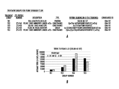
Figure 18- Figure 18A depicts the design for an immunogenicity study
undertaken to assess the
ability of CRM-197-conjugated full-length feline IL-31 protein or mimotopes to
elicit an immune
response in laboratory cats. All treatment groups were formulated with an
adjuvant mixture
including the glycolipid adjuvant Bay R1005 (N-(2-Deoxy-2-L-leucylamino-3-D-
glucopyranosyl)-
N-octadecyldodecanoylamidehydroacetate) as well as CpG oligonucleotides.
Each mimotope described herein was designed to generate an epitope-specific
immune
response driven towards the relevant region on the IL-31 protein where
antibody 15H05 and
other anti-IL-31 antibodies disclosed herein bind._The sequences and chemical
linkers of
various mimotope peptides are shown as groups T02-T05. Peptide ZTS-563
contains the
amino acid sequence N-AKVSMPADNFERKNFILT-C which corresponds to positions 121
through 138 of SEQ ID NO: 157 with the substitution of Threonine (T) for
Alanine (A) at position
number 138. Peptide ZTS-418 contains the amino acid sequence N-
TEVSMPTDNFERKRFILT-C which corresponds to positions 115 through 132 of SEQ ID
NO:
165. Peptide ZTS-423 contains the amino acid sequence N-
NGSAILPYFRAIRPLSDKNTIDKIIEQLDKLKF-C which corresponds to positions 83 through
115
of SEQ ID NO: 157. Peptide ZTS-422 contains the amino acid sequence N-
AKVSMPADNFERKNFILT-C which corresponds to positions 121 through 138 of SEQ ID
NO:
157 with the substitution of Threonine (T) for Alanine (A) at position number
138. Each of
peptides ZTS-563, ZTS-418, ZTS-423, and ZTS-422 also includes N and C terminal
Cysteines
as depicted to facilitate conjugation chemistry using the free thiol groups.
ZTS-422 also
contains an additional aminohexanoic acid linker (Ahx) between the two N
terminal cysteines.
ZTS-423 also contains an additional three amino acid spacer sequence (GSG)
next to the N
terminal cysteine. Figure 18B depicts the average feline antibody titers to
the full length feline
IL-31 (SEQ ID NO: 157) for all treatment groups except T03. Cats in group T03
(equine 15H05
mimotope) had no CRAR to feline IL-31 (data not shown).
Figure 19A is the minimum epitope amino acid sequence bound by anti-canine IL-
31 antibody
M14 according to WO 2018/156367 (Kindred Biosciences, Inc.) The comparison of
multiple
species, sequence reference IDs. and relative amino acid positions are shown.
Figure 19B
shows this minimum amino acid sequence on canine IL-31 highlighted in a black
box. This
17
CA 03093709 2020-09-10
WO 2019/178601
PCT/US2019/022774
figure also shows the alignment of sequence in the surrounding region of the
protein and the
relative positions of the corresponding amino acids in the sequence ID
indicated.
Figure 20 shows a fragment of the IL-31 protein from a loop formed by the
convergence of helix
A with the trailing random coil sequence which shares positional and
structural attributes to the
15H05 loop. Comparison of the amino acid sequences from multiple species and
reference to
the sequence IDs and amino acid positions are shown.
Figure 21A shows the amino acid sequences of three equine IL-31 mimotope
peptides
representing different key epitope regions on the protein. Mimotope 15H05
contains the amino
acid sequence N-TEVSMPTDNFERKRFILT-C which corresponds to positions 115
through 132
of SEQ ID NO: 165. Mimotope BC helix contains the amino acid sequence N-
NSSAILPYFKAISPSLNNDKSLYIIEQLDKLNF-C which corresponds to positions 77 through
109
of SEQ ID NO: 165. Mimotope A helix contains the amino acid sequence N-
GPIYQLQPKEIQAIIVELQNLSKK-C which corresponds to positions 20 through 43 of SEQ
ID
NO: 165. Mimotope 15H05 also includes N and C terminal Cysteines as depicted
to facilitate
conjugation chemistry using the free thiol groups. All three mimotopes contain
an additional
three amino acid spacer sequence (GSG) next to the N biotin group shown as
bold and
underlined in the sequences. The corresponding positions of each amino acid
residue in SEQ
ID NO: 165 are shown. Figure 21B shows the results from a binding assay using
bio-layer
interferometry. The mimotopes indicated were absorbed to streptavidin pins and
used to probe
multiple dilutions of mouse serum. The serum used was from mice vaccinated
with the equine
IL-31 protein (SEQ ID NO: 165) or control serum from mice vaccinated with an
unrelated
protein.
Definitions
Before describing the present invention in detail, several terms used in the
context of the
present invention will be defined. In addition to these terms, others are
defined elsewhere in the
specification, as necessary. Unless otherwise expressly defined herein, terms
of art used in this
specification will have their art-recognized meanings.
As used in the specification and claims, the singular form "a", "an" and "the"
include plural
references unless the context clearly dictates otherwise. For example,
reference to "an
18
CA 03093709 2020-09-10
WO 2019/178601
PCT/US2019/022774
antibody" includes a plurality of such antibodies. As another example,
reference to "a
mimotope'', "an IL-31 mimotope" and the like includes a plurality of such
mimotopes.
As used herein, the term "comprising" is intended to mean that the
compositions and methods
include the recited elements, but not excluding others.
As used herein, the term "vaccine composition" includes at least one antigen
or immunogen in a
pharmaceutically acceptable vehicle useful for inducing an immune response in
a host. Vaccine
compositions can be administered in dosages, and by techniques well known to
those skilled in
the medical or veterinary arts, taking into consideration factors such as the
age, sex, weight,
species and condition of the recipient mammal, and the route of
administration. The route of
administration can be percutaneous, via mucosa! administration (e.g., oral,
nasal, anal, vaginal)
or via a parenteral route (intradermal, transdermal, intramuscular,
subcutaneous, intravenous,
or intraperitoneal). Vaccine compositions can be administered alone, or can be
co-administered
or sequentially administered with other treatments or therapies. Forms of
administration may
include suspensions, syrups or elixirs, and preparations for parenteral,
subcutaneous,
intradermal, intramuscular or intravenous administration (e.g., injectable
administration) such as
sterile suspensions or emulsions. Vaccine compositions may be administered as
a spray, or
mixed in food and/or water, or delivered in admixture with a suitable carrier,
diluent, or excipient
such as sterile water, physiological saline, glucose, or the like. The
compositions can contain
auxiliary substances such as wetting or emulsifying agents, pH buffering
agents, adjuvants,
gelling or viscosity enhancing additives, preservatives, flavoring agents,
colors, and the like,
depending upon the route of administration and the preparation desired.
Standard
pharmaceutical texts, such as "Remington's Pharmaceutical Sciences" (1990),
may be
.. consulted to prepare suitable preparations, without undue experimentation.
The term "immune response" as used herein refers to a response elicited in an
animal or
human. An immune response may refer to cellular immunity (CMI), humoral
immunity, or may
involve both. The present invention also contemplates a response limited to a
part of the
immune system. Usually, an "immunological response" includes, but is not
limited to, one or
more of the following effects: the production or activation of antibodies, B
cells, helper T cells,
suppressor T cells, and/or cytotoxic T cells and/or yd T cells, directed
specifically to an antigen
or antigens included in the composition or vaccine of interest. Preferably,
the host will display
either a therapeutic or protective immunological response, such that
resistance to the disease or
19
CA 03093709 2020-09-10
WO 2019/178601
PCT/US2019/022774
disorder will be enhanced, and/or the clinical severity of the disease
reduced. Such protection
will be demonstrated by either a reduction or lack of symptoms normally
displayed by an
affected host, a quicker recovery time, and/or a lowered antigen (e.g., IL-31)
titer in the affected
host.
The term "protecting" as used herein means conferring a therapeutic
immunological response to
a host mammal, such that resistance to a disease or disorder will be enhanced,
and/or the
clinical severity of the disease reduced in the host mammal.
As used herein, the term "immunogenicity" means capable of producing an immune
response in
a host mammal against an antigen or antigens. This immune response forms the
basis of the
protective immunity elicited by a vaccine against a specific antigen.
As used herein, immunizing, immunization, and the like is the process whereby
a mammal is
made immune or resistant to a disease, typically by the administration of a
vaccine. Vaccines
stimulate the mammal's own immune system to protect the mammal against
subsequent
disease.
An "adjuvant" as used herein means a composition comprised of one or more
substances that
enhances the immune response to an antigen(s). The mechanism of how an
adjuvant operates
is not entirely known. Some adjuvants are believed to enhance the immune
response by slowly
releasing the antigen, while other adjuvants are strongly immunogenic in their
own right, and are
believed to function synergistically.
Epitope, as used herein, refers to the antigenic determinant recognized by the
CDRs of the
antibody. In other words, epitope refers to that portion of any molecule
capable of being
recognized by, and bound by, an antibody. Unless indicated otherwise, the term
"epitope" as
used herein, refers to the region of IL-31 to which an anti-IL-31 agent is
reactive to.
An "antigen" is a molecule or a portion of a molecule capable of being bound
by an antibody
which is additionally capable of being recognized by, and bound by, an
antibody (the
corresponding antibody binding region may be referred to as a paratope). In
general, epitopes
consist of chemically active surface groupings of molecules, for example,
amino acids or sugar
side chains, and have specific three-dimensional structural characteristics as
well as specific
CA 03093709 2020-09-10
WO 2019/178601 PCT/US2019/022774
charge characteristics. Epitopes are the antigenic determinant on a protein
that is recognized
by the immune system. The components of the immune system recognizing epitopes
are
antibodies, T-cells, and B-cells. T-cell epitopes are displayed on the surface
of antigen-
presenting cells (APCs) and are typically 8-11 (MHC class I) or 15 plus (MHC
class II) amino
acids in length. Recognition of the displayed MHC-peptide complex by T-cells
is critical to their
activation. These mechanisms allow for the appropriate recognition of self
versus "non-self"
proteins such as bacteria and viruses. Independent amino acid residues that
are not
necessarily contiguous contribute to interactions with the APC binding cleft
and subsequent
recognition by the T-Cell receptor (Janeway, Travers, Walport, Immunobiology:
The Immune
System in Health and Disease. 5th edition New York: Garland Science; 2001).
Epitopes that
are recognized by soluble antibodies and cell surface associated B-cell
receptors vary greatly in
length and degree of continuity (Sivalingam and Shepherd, Immunol. 2012
Jul;51(3-4):304-309
9). Again even linear epitopes or epitopes found in a continuous stretch of
protein sequence will
often have discontiguous amino acids that represent the key points of contact
with the antibody
paratopes or B-cell receptor. Epitopes recognized by antibodies and B-cells
can be
conformational with amino acids comprising a common area of contact on the
protein in three
dimensional space and are dependent on tertiary and quaternary structural
features of the
protein. These residues are often found in spatially distinct areas of the
primary amino acid
sequence.
A "mimotope" as used herein is a linear or constrained peptide which mimics an
antigen's
epitope. A mimotope may have a primary amino acid sequence capable of
eliciting a T-cell
effector response and/or a three dimensional structure necessary to bind B-
cells resulting in
maturation of an acquired immunological response in an animal. An antibody for
a given
epitope antigen will recognize a mimotope which mimics that epitope. An IL-31
mimotope may
alternatively be referred to herein as an IL-31 peptide mimotope. In some
embodiments, a
mimotope (linear or constrained) for use in the compositions and/or methods of
the present
invention is and/or comprises as part thereof a peptide which is from about 5
amino acid
residues to about 40 amino acid residues in length.
The term "specifically" in the context of antibody binding, refers to high
avidity and/or high
affinity binding of an antibody to a specific antigen, i.e., a polypeptide, or
epitope. In many
embodiments, the specific antigen is an antigen (or a fragment or subfraction
of an antigen)
used to immunize the animal host from which the antibody-producing cells were
isolated.
21
CA 03093709 2020-09-10
WO 2019/178601
PCT/US2019/022774
Antibody specifically binding an antigen is stronger than binding of the same
antibody to other
antigens. Antibodies which bind specifically to a polypeptide may be capable
of binding other
polypeptides at a weak, yet detectable level (e.g., 10% or less of the binding
shown to the
polypeptide of interest). Such weak binding, or background binding, is readily
discernible from
the specific antibody binding to a subject polypeptide, e.g. by use of
appropriate controls. In
general, specific antibodies bind to an antigen with a binding affinity with a
KD of 10-7M or less,
e.g., 10-8M or less (e.g., 10-8M or less, 10100r less, 10-11or less, 10-12 or
less, or 10-18 or less,
etc.).
As used herein, the term "antibody" refers to an intact immunoglobulin having
two light and two
heavy chains. Thus a single isolated antibody or fragment may be a polyclonal
antibody, a
monoclonal antibody, a synthetic antibody, a recombinant antibody, a chimeric
antibody, a
heterochimeric antibody, a caninized antibody, a felinized antibody, a fully
canine antibody, a
fully feline antibody, a fully equine antibody, or a fully human antibody. The
term "antibody"
preferably refers to monoclonal antibodies and fragments thereof (e.g.,
including but not limited
to, antigen-binding portions of the antibody), and immunologic binding
equivalents thereof that
can bind to the IL-31 protein and fragments or modified fragments thereof.
Such fragments and
modified fragments of IL-31 can include the IL-31 peptide mimotopes employed
in the various
embodiments of this invention. For example, an antibody for a given epitope on
IL-31 will
recoginize an IL-31 peptide mimotope which mimics that epitope. The term
antibody is used
both to refer to a homogeneous molecular, or a mixture such as a serum product
made up of a
plurality of different molecular entities.
"Native antibodies" and "native immunoglobulins" are usually heterotetrameric
glycoproteins of
about 150,000 Da!tons, composed of two identical light (L) chains and two
identical heavy (H)
chains. Each light chain is linked to a heavy chain by one covalent disulfide
bond, while the
number of disulfide linkages varies among the heavy chains of different
immunoglobulin
isotypes. Each heavy and light chain also has regularly spaced intrachain
disulfide bridges.
Each heavy chain has at one end a variable domain (VH) followed by a number of
constant
domains. Each light chain has a variable domain at one end (VL) and a constant
domain at its
other end; the constant domain of the light chain is aligned with the first
constant domain of the
heavy chain, and the light-chain variable domain is aligned with the variable
domain of the
heavy chain. Particular amino acid residues are believed to form an interface
between the light-
and heavy-chain variable domains.
22
CA 03093709 2020-09-10
WO 2019/178601 PCT/US2019/022774
The term "antibody fragment" refers to less than an intact antibody structure,
including, without
limitation, an isolated single antibody chain, an Fv construct, a Fab
construct, an Fc construct, a
light chain variable or complementarity determining region (CDR) sequence,
etc. For example,
an antibody fragment can comprise the antigen-binding portion of the antibody.
The term "variable" region comprises framework and CDRs (otherwise known as
hypervariables) and refers to the fact that certain portions of the variable
domains differ
extensively in sequence among antibodies and are used in the binding and
specificity of each
particular antibody for its particular antigen. However, the variability is
not evenly distributed
throughout the variable domains of antibodies. It is concentrated in three
segments called
hypervariable regions both in the light chain and the heavy chain variable
domains. The more
highly conserved portions of variable domains are called the framework region
(FR). The
variable domains of native heavy and light chains each comprise multiple FRs,
largely adopting
a 13-sheet configuration. connected by three hypervariable regions, which form
loops connecting,
and in some cases forming part of, the 13-sheet structure. The hypervariable
regions in each
chain are held together in close proximity by the FRs and, with the
hypervariable regions from
the other chain, contribute to the formation of the antigen-binding site of
antibodies (see Kabat,
et al., Sequences of Proteins of Immunological Interest, 5th Ed. Public Health
Service, National
Institutes of Health, Bethesda, Md. (1991), pages 647-669). The constant
domains are not
involved directly in binding an antibody to an antigen, but exhibit various
effector functions, such
as participation of the antibody in antibody-dependent cellular toxicity.
The term "hypervariable region" when used herein refers to the amino acid
residues of an
antibody which are responsible for antigen binding. The hypervariable region
comprises amino
acid residues from a "complementarity determining region" or "CDR" (Kabat, et
al. (1991),
above) and/or those residues from a "hypervariable loop" (Chothia and Lesk J.
Mol. Biol.
196:901-917 (1987). "Framework" or "FR" residues are those variable domain
residues other
than the hypervariable region residues as herein defined.
Papain digestion of antibodies produces two identical antigen-binding
fragments, called "Fab"
fragments, each with a single antigen-binding site, and a residual "Fe"
fragment, whose name
reflects its ability to crystallize readily. Pepsin treatment yields an
F(ab')2 fragment that has two
antigen-combining sites and is still capable of cross-linking antigen.
23
CA 03093709 2020-09-10
WO 2019/178601
PCT/US2019/022774
"Fv" is the minimum antibody fragment that contains a complete antigen-
recognition and -
binding site. This region consists of a dimer of one heavy chain and one light
chain variable
domain in tight, non-covalent association. It is in this configuration that
the three hypervariable
regions of each variable domain interact to define an antigen-binding site on
the surface of the
VH-VL dimer. Collectively, the six hypervariable regions confer antigen-
binding specificity to the
antibody. However, even a single variable domain (or half of an Fv comprising
only three
hypervariable regions specific for an antigen) has the ability to recognize
and bind antigen,
although at a lower affinity than the entire binding site.
The Fab fragment also contains the constant domain of the light chain and the
first constant
domain (CH1) of the heavy chain. Fab' fragments differ from Fab fragments by
the addition of a
few residues at the carboxyl terminus of the heavy chain CH1 domain including
one or more
cysteine(s) from the antibody hinge region. Fab'-SH is the designation herein
for Fab' in which
the cysteine residue(s) of the constant domains bear a free thiol group.
F(ab') 2 antibody
fragments originally were produced as pairs of Fab' fragments which have hinge
cysteines
between them. Other chemical couplings of antibody fragments are also known.
The "light chains" of antibodies (immunoglobulins) from any vertebrate species
can be assigned
to one of two clearly distinct types, called kappa (K) and lambda (A), based
on the amino acid
sequences of their constant domains.
Depending on the amino acid sequence of the constant domain of their heavy
chains,
immunoglobulins can be assigned to different classes. Presently there are five
major classes of
immunoglobulins: IgA, IgD, IgE, IgG, and IgM, and several of these may be
further divided into
subclasses (isotypes), e.g., IgG1, IgG2, IgG3, IgG4, IgA, and IgA2 (as defined
by mouse and
human designation). The heavy-chain constant domains that correspond to the
different classes
of immunoglobulins are called alpha, delta, epsilon, gamma, and mu,
respectively. The subunit
structures and three-dimensional configurations of different classes of
immunoglobulins are well
known in multiple species. The prevalence of individual isotypes and
functional activities
associated with these constant domains are species-specific and must be
experimentally
defined.
24
CA 03093709 2020-09-10
WO 2019/178601
PCT/US2019/022774
"Monoclonal antibody" as defined herein is an antibody produced by a single
clone of cells (e.g.,
a single clone of hybridoma cells) and therefore a single pure homogeneous
type of antibody.
All monoclonal antibodies produced from the same clone are identical and have
the same
antigen specificity. The term "monoclonal" pertains to a single clone of
cells, a single cell, and
the progeny of that cell.
"Fully canine antibody" as defined herein is a monoclonal antibody produced by
a clone of cells
(typically a CHO cell line) and therefore a single pure homogeneous type of
antibody.
Antibodies identified from single B cells of immunized mammals, such as dogs
are created as
recombinant IgG proteins following identification of their variable domain
sequences. Grafting of
these variable domains onto canine constant domains (heavy chain and light
chain kappa or
lambda constant) results in the generation of recombinant fully canine
antibodies. All fully
canine monoclonal antibodies produced from the same clone are identical and
have the same
antigen specificity. The term "monoclonal" pertains to a single clone of
cells, a single cell, and
the progeny of that cell.
"Fully feline antibody" as defined herein is a monoclonal antibody produced by
a clone of cells
(typically a CHO cell line) and therefore a single pure homogeneous type of
antibody.
Antibodies identified from single B cells of immunized mammals, such as dogs
are created as
recombinant IgG proteins following identification of their variable domain
sequences. Grafting of
these variable domains onto feline constant domains (heavy chain and light
chain kappa or
lambda constant) results in the generation of recombinant fully feline
antibodies. All fully feline
monoclonal antibodies produced from the same clone are identical and have the
same antigen
specificity. The term "monoclonal' pertains to a single clone of cells, a
single cell, and the
progeny of that cell.
"Fully equine antibody" as defined herein is a monoclonal antibody produced by
a clone of cells
(typically a CHO cell line) and therefore a single pure homogeneous type of
antibody.
Antibodies identified from single B cells of immunized mammals, such as dogs
are created as
recombinant IgG proteins following identification of their variable domain
sequences. Grafting of
these variable domains onto equine constant domains (heavy chain and light
chain kappa or
lambda constant) results in the generation of recombinant fully equine
antibodies. All fully
equine monoclonal antibodies produced from the same clone are identical and
have the same
CA 03093709 2020-09-10
WO 2019/178601
PCT/US2019/022774
antigen specificity. The term "monoclonal' pertains to a single clone of
cells, a single cell, and
the progeny of that cell.
"Fully human antibody" as defined herein is a monoclonal antibody produced by
a clone of cells
(typically a CHO cell line) and therefore a single pure homogeneous type of
antibody.
Antibodies identified from single B cells of immunized mammals, such as dogs
are created as
recombinant IgG proteins following identification of their variable domain
sequences. Grafting of
these variable domains onto human constant domains (heavy chain and light
chain kappa or
lambda constant) results in the generation of recombinant fully human
antibodies. All fully
human monoclonal antibodies produced from the same clone are identical and
have the same
antigen specificity. The term "monoclonal" pertains to a single clone of
cells, a single cell, and
the progeny of that cell.
The monoclonal antibodies herein specifically include "chimeric" antibodies
(immunoglobulins)
in which a portion of the heavy and/or light chain is identical with or
homologous to
corresponding sequences in antibodies derived from a particular species, while
the remainder of
the chain(s) is identical with or homologous to corresponding sequences in
antibodies derived
from another species, as well as fragments of such antibodies, so long as they
exhibit the
desired biological activity. Typically, chimeric antibodies are antibodies
whose light and heavy
chain genes have been constructed, typically by genetic engineering, from
antibody variable
and constant region genes belonging to different species. For example, the
variable segments
of the genes from a mouse monoclonal antibody may be joined to canine constant
segments. In
one embodiment of a chimeric mouse:canine IgG, the antigen binding site is
derived from
mouse while the Fc portion is canine.
"Caninized" forms of non-canine (e.g., murine) antibodies are genetically
engineered antibodies
that contain minimal sequence derived from non-canine immunoglobulin.
Caninized antibodies
are canine immunoglobulin sequences (recipient antibody) in which
hypervariable region
residues of the recipient are replaced by hypervariable region residues from a
non-canine
species (donor antibody) such as mouse having the desired specificity,
affinity, and capacity. In
some instances, framework region (FR) residues of the canine immunoglobulin
sequences are
replaced by corresponding non-canine residues. Furthermore, caninized
antibodies may include
residues that are not found in the recipient antibody or in the donor
antibody. These
modifications are made to further refine antibody performance. In general, the
caninized
26
CA 03093709 2020-09-10
WO 2019/178601 PCT/US2019/022774
antibody will include substantially all of at least one, and typically two,
variable domains, in
which all or substantially all of the hypervariable regions correspond to
those of a non-canine
immunoglobulin sequence and all or substantially all of the FRs are those of a
canine
immunoglobulin sequence. The caninized antibody optionally also will comprise
a complete, or
at least a portion of an immunoglobulin constant region (Fc), typically that
of a canine
immunoglobulin sequence. In one embodiment of speciation or caninization of a
mouse IgG,
mouse CDRs are grafted onto canine frameworks.
"Felinized" forms of non-feline (e.g., murine) antibodies are genetically
engineered antibodies
that contain minimal sequence derived from non-feline immunoglobulin.
Felinized antibodies are
feline immunoglobulin sequences (recipient antibody) in which hypervariable
region residues of
the recipient are replaced by hypervariable region residues from a non-feline
species (donor
antibody) such as mouse having the desired specificity, affinity, and
capacity. In some
instances, framework region (FR) residues of the feline immunoglobulin
sequences are replaced
by corresponding non-feline residues. Furthermore, felinized antibodies may
include residues
that are not found in the recipient antibody or in the donor antibody. These
modifications are
made to further refine antibody performance. In general, the felinized
antibody will include
substantially all of at least one, and typically two, variable domains, in
which all or substantially
all of the hypervariable regions correspond to those of a non-feline
immunoglobulin sequence
and all or substantially all of the FRs are those of a feline immunoglobulin
sequence. The
felinized antibody optionally also will comprise a complete, or at least a
portion of an
immunoglobulin constant region (Fc), typically that of a feline immunoglobulin
sequence.
"Equinized" forms of non-equine (e.g., murine) antibodies are genetically
engineered antibodies
that contain minimal sequence derived from non-equine immunoglobulin.
Equinized antibodies
are equine immunoglobulin sequences (recipient antibody) in which
hypervariable region
residues of the recipient are replaced by hypervariable region residues from a
non-equine
species (donor antibody) such as mouse having the desired specificity,
affinity, and capacity. In
some instances, framework region (FR) residues of the equine immunoglobulin
sequences are
replaced by corresponding non-equine residues. Furthermore, equinized
antibodies may include
residues that are not found in the recipient antibody or in the donor
antibody. These
modifications are made to further refine antibody performance. In general, the
equinized
antibody will include substantially all of at least one, and typically two,
variable domains, in
which all or substantially all of the hypervariable regions correspond to
those of a non-equine
27
CA 03093709 2020-09-10
WO 2019/178601 PCT/US2019/022774
immunoglobulin sequence and all or substantially all of the FRs are those of
an equine
immunoglobulin sequence. The equinized antibody optionally also will comprise
a complete, or
at least a portion of an immunoglobulin constant region (Fc), typically that
of an equine
immunoglobulin sequence.
"Humanized" forms of non-human (e.g., murine) antibodies are genetically
engineered
antibodies that contain minimal sequence derived from non-human
immunoglobulin. Humanized
antibodies are human immunoglobulin sequences (recipient antibody) in which
hypervariable
region residues of the recipient are replaced by hypervariable region residues
from a non-
human species (donor antibody) such as mouse having the desired specificity,
affinity, and
capacity. In some instances, framework region (FR) residues of the human
immunoglobulin
sequences are replaced by corresponding non-human residues. Furthermore,
humanized
antibodies may include residues that are not found in the recipient antibody
or in the donor
antibody. These modifications are made to further refine antibody performance.
In general, the
humanized antibody will include substantially all of at least one, and
typically two, variable
domains, in which all or substantially all of the hypervariable regions
correspond to those of a
non-human immunoglobulin sequence and all or substantially all of the FRs are
those of an
human immunoglobulin sequence. The humanized antibody optionally also will
comprise a
complete, or at least a portion of an immunoglobulin constant region (Fc),
typically that of an
human immunoglobulin sequence.
"Fully Canine" antibodies are genetically engineered antibodies that contain
no sequence
derived from non-canine immunoglobulin. Fully canine antibodies are canine
immunoglobulin
sequences (recipient antibody) in which hypervariable region residues are
derived from a
naturally occurring canine antibody (donor antibody) having the desired
specificity, affinity, and
capacity. In some instances, framework region (FR) residues of the canine
immunoglobulin
sequences are replaced by corresponding non-canine residues. Furthermore,
fully canine
antibodies may include residues that are not found in the recipient antibody
or in the donor
antibody, such as including, but not limited to changes in the CDRs to modify
affinity. These
modifications are made to further refine antibody performance. In general, the
fully canine
antibody will include substantially all of at least one, and typically two,
variable domains, in
which all or substantially all of the hypervariable regions correspond to
those of a canine
immunoglobulin sequence and all or substantially all of the FRs are those of
an canine
immunoglobulin sequence. The fully canine antibody optionally also will
comprise a complete, or
28
CA 03093709 2020-09-10
WO 2019/178601
PCT/US2019/022774
at least a portion of an immunoglobulin constant region (Fc), typically that
of canine
immunoglobulin sequence.
"Fully Feline" antibodies are genetically engineered antibodies that contain
no sequence derived
from non-feline immunoglobulin. Fully feline antibodies are feline
immunoglobulin sequences
(recipient antibody) in which hypervariable region residues are derived from a
naturally
occurring feline antibody (donor antibody) having the desired specificity,
affinity, and capacity. In
some instances, framework region (FR) residues of the feline immunoglobulin
sequences are
replaced by corresponding non-feline residues. Furthermore, fully feline
antibodies may include
residues that are not found in the recipient antibody or in the donor
antibody, such as including,
but not limited to changes in the CDRs to modify affinity. These modifications
are made to
further refine antibody performance. In general, the fully feline antibody
will include substantially
all of at least one, and typically two, variable domains, in which all or
substantially all of the
hypervariable regions correspond to those of a feline immunoglobulin sequence
and all or
substantially all of the FRs are those of an feline immunoglobulin sequence.
The fully feline
antibody optionally also will comprise a complete, or at least a portion of an
immunoglobulin
constant region (Fc), typically that of feline immunoglobulin sequence.
"Fully Equine" antibodies are genetically engineered antibodies that contain
no sequence
derived from non-equine immunoglobulin. Fully equine antibodies are equine
immunoglobulin
sequences (recipient antibody) in which hypervariable region residues are
derived from a
naturally occurring equine antibody (donor antibody) having the desired
specificity, affinity, and
capacity. In some instances, framework region (FR) residues of the equine
immunoglobulin
sequences are replaced by corresponding non-equine residues. Furthermore,
fully equine
antibodies may include residues that are not found in the recipient antibody
or in the donor
antibody, such as including, but not limited to changes in the CDRs to modify
affinity. These
modifications are made to further refine antibody performance. In general, the
fully equine
antibody will include substantially all of at least one, and typically two,
variable domains, in
which all or substantially all of the hypervariable regions correspond to
those of a equine
immunoglobulin sequence and all or substantially all of the FRs are those of
an equine
immunoglobulin sequence. The fully equine antibody optionally also will
comprise a complete, or
at least a portion of an immunoglobulin constant region (Fc), typically that
of equine
immunoglobulin sequence.
29
CA 03093709 2020-09-10
WO 2019/178601
PCT/US2019/022774
"Fully Human" antibodies are genetically engineered antibodies that contain no
sequence
derived from non-human immunoglobulin. Fully human antibodies are human
immunoglobulin
sequences (recipient antibody) in which hypervariable region residues are
derived from a
naturally occurring human antibody (donor antibody) having the desired
specificity, affinity, and
capacity. In some instances, framework region (FR) residues of the human
immunoglobulin
sequences are replaced by corresponding non-human residues. Furthermore, fully
human
antibodies may include residues that are not found in the recipient antibody
or in the donor
antibody, such as including, but not limited to changes in the CDRs to modify
affinity. These
modifications are made to further refine antibody performance. In general, the
fully human
antibody will include substantially all of at least one, and typically two,
variable domains, in
which all or substantially all of the hypervariable regions correspond to
those of a human
immunoglobulin sequence and all or substantially all of the FRs are those of a
human
immunoglobulin sequence. The fully human antibody optionally also will
comprise a complete,
or at least a portion of an immunoglobulin constant region (Fc), typically
that of human
immunoglobulin sequence.
The term "heterochimeric" as defined herein, refers to an antibody in which
one of the antibody
chains (heavy or light) is, for example, caninized, felinized, equinized, or
humanized while the
other is chimeric. In one embodiment, a felinized variable heavy chain (where
all of the CDRs
are mouse and all FRs are feline) is paired with a chimeric variable light
chain (where all of the
CDRs are mouse and all FRs are mouse. In this embodiment, both the variable
heavy and
variable light chains are fused to a feline constant region.
The term "variant" as used herein refers to a peptide, polypeptide or a
nucleic acid sequence
encoding a peptide or polypeptide, that has one or more conservative amino
acid variations or
other minor modifications such that the corresponding peptide or polypeptide
has substantially
equivalent function when compared to the wild-type peptide or polypeptide.
Ordinarily, variant
peptide mimotopes for use in the present invention will have at least 30%
identity to the parent
mimotope, more preferably at least 50%, more preferably at least 75%, more
preferably at least
80%, more preferably at least 85%, more preferably at least 90%, and most
preferably at least
95% sequence identity to the parent mimotope.
CA 03093709 2020-09-10
WO 2019/178601
PCT/US2019/022774
A 'variant" anti-IL-31 antibody, refers herein to a molecule which differs in
amino acid sequence
from a "parent" anti-IL-31 antibody amino acid sequence by virtue of addition,
deletion, and/or
substitution of one or more amino acid residue(s) in the parent antibody
sequence and retains at
least one desired activity of the parent anti-IL-31-antibody. Desired
activities can include the
ability to bind the antigen specifically, the ability to reduce, inhibit or
neutralize IL-31 activity in
an animal, and the ability to inhibit IL-31-mediated pSTAT signaling in a cell-
based assay. In
one embodiment, the variant comprises one or more amino acid substitution(s)
in one or more
hypervariable and/or framework region(s) of the parent antibody. For example,
the variant may
comprise at least one, e.g. from about one to about ten, and preferably from
about two to about
five, substitutions in one or more hypervariable and/or framework regions of
the parent antibody.
Ordinarily, the variant will have an amino acid sequence having at least 50%
amino acid
sequence identity with the parent antibody heavy or light chain variable
domain sequences,
more preferably at least 65%, more preferably at least 75%, more preferably at
least 80%, more
preferably at least 85%, more preferably at least 90%, and most preferably at
least 95%
sequence identity. Identity or homology with respect to this sequence is
defined herein as the
percentage of amino acid residues in the candidate sequence that are identical
with the parent
antibody residues, after aligning the sequences and introducing gaps, if
necessary, to achieve
the maximum percent sequence identity. None of N-terminal, C-terminal, or
internal extensions,
deletions, or insertions into the antibody sequence shall be construed as
affecting sequence
identity or homology. The variant retains the ability to bind an IL-31 and
preferably has desired
activities which are superior to those of the parent antibody. For example,
the variant may have
a stronger binding affinity, enhanced ability to reduce, inhibit or neutralize
IL-31 activity in an
animal, and/or enhanced ability to inhibit IL-31-mediated pSTAT signaling in a
cell-based assay.
A "variant" nucleic acid refers herein to a molecule which differs in sequence
from a "parent"
nucleic acid. Polynucleotide sequence divergence may result from mutational
changes such as
deletions, substitutions, or additions of one or more nucleotides. Each of
these changes may
occur alone or in combination, one or more times in a given sequence.
The "parent" antibody herein is one that is encoded by an amino acid sequence
used for the
preparation of the variant. In one embodiment, the parent antibody has a
canine framework
region and, if present, has canine antibody constant region(s). For example,
the parent antibody
may be a caninized or canine antibody. As another example, the parent antibody
may be a
felinized or feline antibody. As yet another example, the parent antibody may
be an equinized
31
CA 03093709 2020-09-10
WO 2019/178601 PCT/US2019/022774
or equine antibody. In another example, the parent antibody may be a humanized
or human
antibody. In a still further example, the parent antibody is a murine
monoclonal antibody.
The term "antigen binding region", "antigen-binding portion", and the like as
used throughout the
specification and claims refers to that portion of an antibody molecule which
contains the amino
acid residues that interact with an antigen and confer on the antibody its
specificity and affinity
for the antigen. The antibody binding region includes the "framework" amino
acid residues
necessary to maintain the proper conformation of the antigen-binding residues.
The antigen-
binding portion of an antibody according to the present invention may
alternatively be referred to
herein as an IL-31-specific peptide or polypeptide or as an anti-IL-31 peptide
or polypeptide, for
example.
The term "isolated" means that the material (e.g., antibody or nucleic acid)
is separated and/or
recovered from a component of its natural environment. Contaminant components
of its natural
environment are materials that would interfere with diagnostic or therapeutic
uses for the
material, and may include enzymes, hormones, and other proteinaceous or
nonproteinaceous
solutes. With respect to nucleic acid, an isolated nucleic acid may include
one that is separated
from the 5' to 3' sequences with which it is normally associated in the
chromosome. In preferred
embodiments, the material will be purified to greater than 95% by weight of
the material, and
most preferably more than 99% by weight. Isolated material includes the
material in situ within
recombinant cells since at least one component of the material's natural
environment will not be
present. Ordinarily, however, isolated material will be prepared by at least
one purification step.
The word "label" when used herein refers to a detectable compound or
composition that is
conjugated directly or indirectly to the antibody, nucleic acid, or mimotope,
for example. The
label may itself be detectable by itself (e.g., radioisotope labels or
fluorescent labels) or, in the
case of an enzymatic label, may catalyze chemical alteration of a substrate
compound or
composition that is detectable.
The terms "nucleic acid", "polynucleotide'', "nucleic acid molecule" and the
like may be used
interchangeably herein and refer to a series of nucleotide bases (also called
"nucleotides") in
DNA and RNA. The nucleic acid may contain deoxyribonucleotides,
ribonucleotides, and/or their
analogs. The term "nucleic acid" includes, for example, single-stranded and
double-stranded
molecules. A nucleic acid can be, for example, a gene or gene fragment, exons,
introns, a DNA
32
CA 03093709 2020-09-10
WO 2019/178601
PCT/US2019/022774
molecule (e.g., cDNA), an RNA molecule (e.g., mRNA), recombinant nucleic
acids, plasmids,
and other vectors, primers and probes. Both 5' to 3' (sense) and 3' to 5'
(antisense)
polynucleotides are included.
A 'subject" or "patient" refers to a mammal in need of treatment that can be
affected by
molecules of the invention. Mammals that can be treated in accordance with the
invention
include vertebrates, with mammals such as canine, feline, equine, and human
mammals being
particularly preferred examples.
A "therapeutically effective amount" (or "effective amount") refers to an
amount of an active
ingredient, e.g., an agent according to the invention, sufficient to effect
beneficial or desired
results when administered to a subject or patient. An effective amount can be
administered in
one or more administrations, applications or dosages. A therapeutically
effective amount of a
composition according to the invention may be readily determined by one of
ordinary skill in the
art. In the context of this invention, a "therapeutically effective amount" is
one that produces an
objectively measured change in one or more parameters associated with
treatment of an IL-31
mediated disorder, such as a pruritic condition or an allergic condition, or
tumor progression,
including clinical improvement in symptoms. Of course, the therapeutically
effective amount will
vary depending upon the particular subject and condition being treated, the
weight and age of
.. the subject, the severity of the disease condition, the particular compound
chosen, the dosing
regimen to be followed, timing of administration, the manner of administration
and the like, all of
which can readily be determined by one of ordinary skill in the art.
As used herein, the term "therapeutic" encompasses the full spectrum of
treatments for a
disease or disorder. A "therapeutic" agent of the invention may act in a
manner that is
prophylactic or preventive, including those that incorporate procedures
designed to target
animals that can be identified as being at risk (pharmacogenetics); or in a
manner that is
ameliorative or curative in nature; or may act to slow the rate or extent of
the progression of at
least one symptom of a disease or disorder being treated.
"Treatment", "treating", and the like refers to both therapeutic treatment and
prophylactic or
preventative measures. Animals in need of treatment include those already with
the disorder as
well as those in which the disorder is to be prevented. The term "treatment"
or "treating" of a
disease or disorder includes preventing or protecting against the disease or
disorder (that is,
33
CA 03093709 2020-09-10
WO 2019/178601 PCT/US2019/022774
causing the clinical symptoms not to develop); inhibiting the disease or
disorder (i.e., arresting
or suppressing the development of clinical symptoms; and/or relieving the
disease or disorder
(i.e., causing the regression of clinical symptoms). As will be appreciated,
it is not always
possible to distinguish between "preventing" and "suppressing" a disease or
disorder since the
ultimate inductive event or events may be unknown or latent. Accordingly, the
term
"prophylaxis" will be understood to constitute a type of "treatment" that
encompasses both
"preventing" and "suppressing." The term "treatment" thus includes
"prophylaxis".
The term "allergic condition" is defined herein as a disorder or disease
caused by an interaction
between the immune system and a substance foreign to the body. This foreign
substance is
termed "an allergen". Common allergens include aeroallergens, such as pollens,
dust, molds,
dust mite proteins, injected saliva from insect bites, etc. Examples of
allergic conditions include,
but are not limited to, the following: allergic dermatitis, summer eczema,
urticaria, heaves,
inflammatory airway disease, recurrent airway obstruction, airway hyper-
responsiveness,
chronic obstructive pulmonary disease, and inflammatory processes resulting
from
autoimmunity, such as Irritable bowel syndrome (IBS).
The term "pruritic condition" is defined herein as a disease or disorder
characterized by an
intense itching sensation that produces the urge to rub or scratch the skin to
obtain relief.
Examples of pruritic conditions include, but are not limited to the following:
atopic dermatitis,
allergic dermatitis, eczema, psoriasis, scleroderma, and pruritus.
As used herein, the terms "cell", "cell line", and "cell culture" may be used
interchangeably. All of
these terms also include their progeny, which is any and all subsequent
generations. It is
understood that all progeny may not be identical due to deliberate or
inadvertent mutations. In
the context of expressing a heterologous nucleic acid sequence, "host cell"
refers to a
prokaryotic or eukaryotic cell (e.g., bacterial cells, yeast cells, mammalian
cells, and insect cells)
whether located in vitro or in vivo. For example, host cells may be located in
a transgenic
animal. Host cell can be used as a recipient for vectors and may include any
transformable
organism that is capable of replicating a vector and/or expressing a
heterologous nucleic acid
encoded by a vector.
A "composition" is intended to mean a combination of active agent and another
compound or
composition which can be inert (e.g., a label), or active, such as an
adjuvant.
34
CA 03093709 2020-09-10
WO 2019/178601
PCT/US2019/022774
As used herein, the terms "pharmaceutically acceptable carrier" and
"pharmaceutically
acceptable vehicle" are interchangeable, and refer to a fluid vehicle for
containing vaccine
antigens that can be injected into a host without adverse effects.
Pharmaceutically acceptable
carriers suitable for use in the invention are well known to those of skill in
the art. Such carriers
include, without limitation, water, saline, buffered saline, phosphate buffer,
alcoholic/aqueous
solutions, emulsions or suspensions. Other conventionally employed diluents,
adjuvants and
excipients, may be added in accordance with conventional techniques. Such
carriers can
include ethanol, polyols, and suitable mixtures thereof, vegetable oils, and
injectable organic
esters. Buffers and pH adjusting agents may also be employed. Buffers include,
without
limitation, salts prepared from an organic acid or base. Representative
buffers include, without
limitation, organic acid salts, such as salts of citric acid, e.g., citrates,
ascorbic acid, gluconic
acid, histidine-HCI, carbonic acid, tartaric acid, succinic acid, acetic acid,
or phthalic acid, Tris,
trimethanmine hydrochloride, or phosphate buffers. Parenteral carriers can
include sodium
chloride solution, Ringer's dextrose, dextrose, trehalose, sucrose, and sodium
chloride, lactated
Ringer's or fixed oils. Intravenous carriers can include fluid and nutrient
replenishers, electrolyte
replenishers, such as those based on Ringer's dextrose and the like.
Preservatives and other
additives such as, for example, antimicrobials, antioxidants, chelating agents
(e.g., EDTA), inert
gases and the like may also be provided in the pharmaceutical carriers. The
present invention is
not limited by the selection of the carrier. The preparation of these
pharmaceutically acceptable
compositions, from the above-described components, having appropriate pH
isotonicity, stability
and other conventional characteristics is within the skill of the art. See,
e.g., texts such as
Remington: The Science and Practice of Pharmacy, 20th ed, Lippincott Williams
& Wilkins,
publ., 2000; and The Handbook of Pharmaceutical Excipients, 4th edit.,
eds. R. C. Rowe et
al, APhA Publications, 2003.
The term "conservative amino acid substitution" indicates any amino acid
substitution for a given
amino acid residue, where the substitute residue is so chemically similar to
that of the given
residue that no substantial decrease in polypeptide function (e.g., enzymatic
activity) results.
Conservative amino acid substitutions are commonly known in the art and
examples thereof are
described, e.g., in U.S. Pat. Nos. 6,790,639, 6,774,107, 6,194,167, or
5,350,576. In a preferred
embodiment, a conservative amino acid substitution will be any one that occurs
within one of
the following six groups
CA 03093709 2020-09-10
WO 2019/178601
PCT/US2019/022774
= 1. Small aliphatic, substantially non-polar residues: Ala, Gly, Pro, Ser,
and Thr;
= 2. Large aliphatic, non-polar residues: Ile, Leu, and Val; Met;
= 3. Polar, negatively charged residues and their amides: Asp and Glu;
= 4. Amides of polar, negatively charged residues: Asn and Gin; His;
= 5. Polar, positively charged residues: Arg and Lys; His; and
= 6. Large aromatic residues: Trp and Tyr; Phe.
In a preferred embodiment, a conservative amino acid substitution will be any
one of the
following, which are listed as Native Residue (Conservative Substitutions)
pairs: Ala
(Ser); Arg (Lys); Asn (Gin; His); Asp (Glu); Gin (Asn); Glu (Asp); Gly (Pro);
His (Asn;
Gin); Ile (Leu; Val); Leu (Ile; Val); Lys (Arg; Gin; Glu); Met (Leu; Ile); Phe
(Met; Leu; Tyr);
Ser (Thr); Thr (Ser); Trp (Tyr); Tyr (Trp; Phe); and Val (Ile; Leu).
Just as a polypeptide may contain conservative amino acid substitution(s), a
polynucleotide
hereof may contain conservative codon substitution(s). A codon substitution is
considered
conservative if, when expressed, it produces a conservative amino acid
substitution, as
described above. Degenerate codon substitution, which results in no amino acid
substitution, is
also useful in polynucleotides according to the present invention. Thus, e.g.,
a polynucleotide
encoding a selected polypeptide useful in an embodiment of the present
invention may be
mutated by degenerate codon substitution in order to approximate the codon
usage frequency
exhibited by an expression host cell to be transformed therewith, or to
otherwise improve the
expression thereof.
DETAILED DESCRIPTION OF THE INVENTION
It should be understood that this invention is not limited to the particular
methodology, protocols,
and reagents, etc., described herein and as such may vary. The terminology
used herein is for
the purpose of describing particular embodiments only, and is not intended to
limit the scope of
the present invention, which is defined solely by the claims.
Unless otherwise defined, scientific and technical terms used in connection
with the vaccine
compositions and antibodies described herein shall have the meanings that are
commonly
understood by those of ordinary skill in the art. Further, unless otherwise
required by context,
.. singular terms shall include pluralities and plural terms shall include the
singular. Generally,
nomenclatures utilized in connection with, and techniques of, cell and tissue
culture, molecular
biology, and protein and oligo- or polynucleotide chemistry and hybridization
described herein
are those well-known and commonly used in the art.
36
Standard techniques are used for recombinant DNA, oligonucleotide synthesis,
and tissue
culture and transfection (e.g., electroporation, lipofection). Enzymatic
reactions and purification
techniques are performed according to manufacturer's specifications or as
commonly
accomplished in the art or as described herein. The foregoing techniques and
procedures are
generally performed according to conventional methods well known in the art
and as described
in various general and more specific references that are cited and discussed
throughout the
present specification, See e.g., Sambrook et al. MOLECULAR CLONING: LAB.
MANUAL (3rd
ed., Cold Spring Harbor Lab. Press, Cold Spring Harbor, N.Y., 2001) and
Ausubel et al. Current
.. Protocols in Molecular Biology (New York: Greene Publishing
Association/Wiley lnterscience),
1993. The nomenclatures utilized in connection with, and the laboratory
procedures and
techniques of, analytical chemistry, synthetic organic chemistry, and
medicinal and
pharmaceutical chemistry described herein are those well-known and commonly
used in the art.
Standard techniques are used for chemical syntheses, chemical analyses,
pharmaceutical
preparation, formulation, and delivery, and treatment of patients.
Other than in the operating examples, or where otherwise indicated, all
numbers expressing
quantities of ingredients or reaction conditions used herein should be
understood as modified in
all instances by the term "about."
All patents and other publications identified herein are referred to for
the purpose of describing and disclosing, for example, the methodologies
described in such
publications that might be used in connection with the present invention.
These publications are
provided solely for their disclosure prior to the filing date of the present
application.
Compositions
The present invention provides for IL-31 mimotopes (peptides) and variants
thereof and their
uses in clinical and scientific procedures, including diagnostic procedures.
As used herein, such
an IL-31 mimotope is a linear or constrained peptide which mimics an antigen's
epitope. An anti-
IL-31 antibody for a given IL-31 epitope antigen will recognize an IL-31
mimotope which mimics
that epitope.
IL-31 mimotopes (peptides) are employed in vaccine compositions according to
the present
invention. Such vaccine compositions are useful for protecting a mammal
against an IL-31
37
Date Recue/Date Received 2022-01-28
CA 03093709 2020-09-10
WO 2019/178601
PCT/US2019/022774
mediated disorder, such as a pruritic or allergic condition. In some
embodiments, the IL-31-
mediated pruritic or allergic condition is a pruritic condition selected from
atopic dermatitis,
eczema, psoriasis, scleroderma, and pruritus. In other embodiments, the IL-31-
mediated pruritic
or allergic condition is an allergic condition selected from allergic
dermatitis, summer eczema,
urticaria, heaves, inflammatory airway disease, recurrent airway obstruction,
airway hyper-
responsiveness, chronic obstruction pulmonary disease, and inflammatory
processes resulting
from autoimmunity. In other embodiments, the IL-31 mediated disorder is tumor
progression. In
some embodiments, the IL-31 mediated disorder is eosinophilic disease or
mastocytomas.
In one embodiment, a vaccine composition according to the present invention
includes the
combination of a carrier polypeptide and at least one mimotope selected from a
feline IL-31
mimotope, a canine IL-31 mimotope, a horse IL-31 mimotope, and a human IL-31
mimotope;
and an adjuvant. In some embodiments, the vaccine compositions of this
invention can include
more than one IL-31 mimotope from a given species, or even a combination of IL-
31 mimotopes
from different species. In some embodiments, a mimotope (linear or
constrained) for use in the
compositions and/or methods of the present invention is and/or comprises as
part thereof a
peptide which is from about 5 amino acid residues to about 40 amino acid
residues in length.
In one embodiment, the at least one mimotope employed in the compositions and
methods of
the instant invention is selected from an IL-31 15H05 mimotope, an IL-31 helix
BC region
mimotope, an IL-31 helix A region mimotope, an IL-31 AB loop region mimotope,
or any
combination thereof.
In one embodiment, the at least one mimotope for use in the compositions and
methods of the
instant invention generates antibodies that are neutralizing the bioactivity
of IL-31.
In another embodiment, the vaccine compositions of this invention are capable
of eliciting a
focused immune response to generate antibodies in the mammal directed to at
least one
neutralizing epitope on IL-31, but not against non-neutralizing epitopes on IL-
31.
38
CA 03093709 2020-09-10
WO 2019/178601 PCT/US2019/022774
In one embodiment, the vaccine composition includes a canine IL-31 mimotope
which is and/or
includes as part thereof the amino acid sequence SVPADTFECKSF (SEQ ID NO:
186),
SVPADTFERKSF (SEQ ID NO: 187), NSSAILPYFRAIRPLSDKNIIDKIIEQLDKLKF (SEQ ID NO:
192), APTHQLPPSDVRKIILELQPLSRG (SEQ ID NO: 196), TGVPES (SEQ ID NO: 200) or
variants thereof that retain anti-IL-31 binding.
In another embodiment, the vaccine composition includes a feline IL-31
mimotope which is
and/or includes as part thereof the amino acid sequence SMPADNFERKNF (SEQ ID
NO: 188),
NGSAILPYFRAIRPLSDKNTIDKIIEQLDKLKF (SEQ ID NO: 193), APAHRLQPSDIRKIILELRPM
.. SKG (SEQ ID NO: 197), IGLPES (SEQ ID NO: 201) or variants thereof that
retain anti-IL-31
binding.
In yet another embodiment, the vaccine composition includes an equine IL-31
mimotope which
is and/or includes as part thereof the amino acid sequence SMPTDNFERKRF (SEQ
ID NO:
189), NSSAILPYFKAISPSLNNDKSLYIIEQLDKLNF (SEQ ID NO: 194),
GPIYQLQPKEIQAIIVELQNLS KK (SEQ ID NO: 198), KGVQKF (SEQ ID NO: 202) or
variants
thereof that retain anti-IL-31 binding.
In a still further embodiment, the vaccine composition includes a human IL-31
mimotope which
is and/or includes as part thereof the amino acid sequence SVPTDTHECKRF (SEQ
ID NO:
190), SVPTDTHERKRF (SEQ ID NO: 191), HSPAIRAYLKTIRQLDNKSVIDEIIEHLDKLIF (SEQ
ID NO: 195), LPVRLLRPSDDVOKIVEELQSLSKM (SEQ ID NO: 199), KGVLVS (SEQ ID NO:
203) or variants thereof that retain anti-IL-31 binding.
In one embodiment, the mimotope employed in the vaccine compositions according
to the
present invention is a constrained mimotope. In one embodiment, such a
constrained mimotope
is a chemically-linked cyclic peptide.
Linear IL-31 mimotopes can be chemically synthesized or recombinantly
produced. Constrained
IL-31 mimotopes such as a chemically-linked cyclic peptide can be chemically
synthesized or
can be made using a combination of chemical synthesis and recombinant
technology.
In some embodiments, the IL-31 mimetope employed in the vaccine composition is
chemically
conjugated to a carrier polypeptide. In other embodiments, the carrier
polypeptide and the
mimotope are part of a recombinant fusion protein.
39
The carrier polypeptide which is combined with the IL-31 mimotope can be or
can include as
part thereof a bacterial toxoid or a derivative thereof, keyhole limpet
hemocyanin (KLH), or a
virus-like particle. By way of non-limiting examples, the bacterial toxoid or
derivative can be a
tetanus toxoid, a diphtheria toxoid, a tetanus toxoid, the outer membrane
protein complex from
group B N. meningitidis, Pseudomonas exotoxin, or the nontoxic mutant of
diphtheria toxin
(CRM197). By way of other non-limiting examples, the virus-like particle can
be HBsAg, HBcAg,
E. coil bacteriophage Qbeta, Norwalk virus, canine distemper virus (CDV), or
influenza HA. In
one preferred embodiment, the IL-31 mimotope is in a combination with a
carrier polypeptide
which includes or consists of CRM197.
The vaccine compositions according to the present invention include at least
one adjuvant or
adjuvant formulation, as will be described in further detail below.
Vaccines of the present invention can be formulated following accepted
convention to include
pharmaceutically acceptable carriers for animals, including humans (if
applicable), such as
standard buffers, stabilizers, diluents, preservatives, and/or solubilizers,
and can also be
formulated to facilitate sustained release. Diluents include water, saline,
dextrose, ethanol,
glycerol, and the like. Additives for isotonicity include sodium chloride,
dextrose, mannitol,
sorbitol, and lactose, among others. Stabilizers include albumin, among
others. Other suitable
vaccine vehicles and additives, including those that are particularly useful
in formulating
modified live vaccines, are known or will be apparent to those skilled in the
art. See, e.g.,
Remington's Pharmaceutical Science, 18th ed., 1990, Mack Publishing.
Vaccines of the present invention can further comprise one or more additional
immunomodulatory components such as, e.g., an adjuvant or cytokine, among
others. Types of
suitable adjuvants for use in the compositions of the present invention
include the following: an
oil-in-water adjuvant, a polymer and water adjuvant, a water-in-oil adjuvant,
an aluminum
hydroxide adjuvant, a vitamin E adjuvant and combinations thereof. Some
specific examples of
adjuvants include, but are not limited to, complete Freund's adjuvant,
incomplete Freund's
adjuvant, Corynebacterium parvum, Bacillus Calmette Guerin, aluminum hydroxide
gel, glucan,
dextran sulfate, iron oxide, sodium alginate, Bacto-Adjuvant, certain
synthetic polymers such as
poly amino acids and co-polymers of amino acids, Block copolymer (CytRx,
Atlanta, Ga.), QS-
Date Recue/Date Received 2022-01-28
CA 03093709 2020-09-10
WO 2019/178601
PCT/US2019/022774
21 (Cambridge Biotech Inc., Cambridge Mass.), SAF-M (Chiron, Emeryville
Calif.),
AMPHIGEN adjuvant, saponin, Quil A or other saponin fraction, monophosphoryl
lipid A, and
Avridine lipid-amine adjuvant (N,N-dioctadecyl-N',N'-- bis(2-hydroxyethyl)-
propanediamine),
"REGRESSIN" (Vetrepharm, Athens, Ga.), paraffin oil, RIBI adjuvant system
(Ribi Inc.,
Hamilton, Mont.), muramyl dipeptide and the like.
Non-limiting examples of oil-in-water emulsions useful in the vaccine of the
invention include
modified SEAM62 and SEAM 1/2 formulations. Modified SEAM62 is an oil-in-water
emulsion
containing 5% (v/v) squalene (Sigma), 1% (v/v) SPAN 85 detergent (ICI
Surfactants), 0.7%
(v/v) TWEEN 80 detergent (ICI Surfactants), 2.5% (v/v) ethanol, 200 pg/ml
Quil A, 100 pg/m1
cholesterol, and 0.5% (v/v) lecithin. Modified SEAM 1/2 is an oil-in-water
emulsion comprising
5% (v/v) squalene, 1% (v/v) SPAN 85 detergent, 0.7% (v/v) Tween 80 detergent,
2.5% (v/v)
ethanol, 100 pg/ml Quil A, and 50 pg/ml cholesterol.
Another example of an adjuvant useful in the compositions of the invention is
SP-oil. As used in
the specification and claims, the term "SP oil" designates an oil emulsion
comprising a
polyoxyethylene-polyoxypropylene block copolymer, squalane, polyoxyethylene
sorbitan
monooleate and a buffered salt solution. Polyoxyethylene-polyoxypropylene
block copolymers
are surfactants that aid in suspending solid and liquid components. These
surfactants are
commercially available as polymers under the trade name Pluronic . The
preferred surfactant is
poloxamer 401 which is commercially available under the trade name Pluronice L-
121. In
general, the SP oil emulsion is an immunostimulating adjuvant mixture which
will comprise
about 1 to 3% vol/vol of block copolymer, about 2 to 6% vol/vol of squalane,
more particularly
about 3 to 6% of squalane, and about 0.1 to 0.5% vol/vol of polyoxyethylene
sorbitan
monooleate, with the remainder being a buffered salt solution.
"Immunomodulators" that can be included in the vaccine include, e.g.,
immunostimulatory
oligonucleotides, one or more interleukins, interferons, or other known
cytokines. In one
embodiment, the adjuvant may be a cyclodextrin derivative or a polyanionic
polymer, such as
those described in U.S. Pat. Nos. 6,165,995 and 6,610,310, respectively.
In one embodiment, the adjuvant is a formulation comprising a saponin, a
sterol, a quaternary
ammonium compound, and a polymer. In a specific embodiment, the saponin is
Quil A or a
41
CA 03093709 2020-09-10
WO 2019/178601
PCT/US2019/022774
purified fraction thereof, the sterol is cholesterol, the quaternary ammonium
compound is
dimethyl dioctadecyl ammonium bromide (DDA), and the polymer is polyacrylic
acid.
In another embodiment, the adjuvant comprises the combination of one or more
isolated
immunostimulatory oligonucleotides, a sterol, and a saponin. In a specific
embodiment, the one
or more isolated immunostimulatory oligonucleotides comprises CpG, the sterol
is cholesterol,
and the saponin is Quil A or a purified fraction thereof. As used herein, the
ZA-01 adjuvant
referred to in the example section includes Quil A (saponin), cholesterol,
CpG, and diluent.
In another embodiment, a useful adjuvant to be employed in the compositions of
this invention
includes CpG-containing immunostimulatory oligonucleotides. CpG-containing
oligonucleotides
are described for example in US Patent NO: 8,580,280. In one specific
embodiment, an
adjuvant for use in the present invention is a mixture including at least one
glycolipid adjuvant
and CpG-containing oligonucleotides. A specific example of a useful adjuvant
is a mixture that
includes the glycolipid adjuvant Bay R1005 (N-(2-Deoxy-2-L-leucylamino-8-D-
glucopyranosyl)-
N-octadecyldodecanoylamidehydroacetate) as well as CpG oligonucleotides.
In one embodiment, the adjuvant or adjuvant mixture is added in an amount of
about 100 pg to
about 10 mg per dose. In another embodiment, the adjuvant/adjuvant mixture is
added in an
amount of about 200 pg to about 5 mg per dose. In yet another embodiment, the
adjuvant/adjuvant mixture is added in an amount of about 300 pg to about 1
mg/dose.
With the advent of methods of molecular biology and recombinant technology, it
is possible to
produce the aforementioned peptides and polypeptides by recombinant means and
thereby
generate gene sequences that code for specific amino acid sequences found in
the peptide or
polypeptide structure. In one embodiment, the peptide is the IL-31 mimotope or
is at least part
of the IL-31 mimotope. In another embodiment, the polypeptide is the carrier
polypeptide which
is present in combination with the IL-31 mimotope. In a still further
embodiment, the polypeptide
is an antibody, such as that to which the IL-31 mimotope employed in the
vaccine composition
or diagnostic methods of this invention binds. Such antibodies can be produced
by either
cloning the gene sequences encoding the polypeptide chains of said antibodies
or by direct
synthesis of said polypeptide chains, with assembly of the synthesized chains
to form active
tetrameric (H2L2) structures with affinity for specific epitopes and antigenic
determinants. This
has permitted the ready production of antibodies having sequences
characteristic of neutralizing
42
CA 03093709 2020-09-10
WO 2019/178601
PCT/US2019/022774
antibodies from different species and sources.
Regardless of the source of the antibodies, or how they are recombinantly-
constructed, or how
they are synthesized, in vitro or in vivo, using transgenic animals, large
cell cultures of
.. laboratory or commercial size, using transgenic plants, or by direct
chemical synthesis
employing no living organisms at any stage of the process, all antibodies have
a similar overall
3 dimensional structure. This structure is often given as H2L2 and refers to
the fact that
antibodies commonly comprise two light (L) amino acid chains and 2 heavy (H)
amino acid
chains. Both chains have regions capable of interacting with a structurally
complementary
antigenic target. The regions interacting with the target are referred to as
'Variable" or "V"
regions and are characterized by differences in amino acid sequence from
antibodies of
different antigenic specificity. The variable regions of either H or L chains
contain the amino acid
sequences capable of specifically binding to antigenic targets.
The "antigen binding region", or "antigen-binding portion" of an antibody
refers to that portion of
an antibody molecule which contains the amino acid residues that interact with
an antigen and
confer on the antibody its specificity and affinity for the antigen. The
antibody binding region
includes the "framework" amino acid residues necessary to maintain the proper
conformation of
the antigen-binding residues. The antigen-binding portion of an antibody
referred to in the
specification and claims may be referred to herein as an IL-31-specific
peptide or polypeptide or
as an anti-IL-31 peptide or polypeptide, for example.
Within the variable regions of the H or L chains that provide for the antigen
binding regions are
smaller sequences dubbed "hypervariable" because of their extreme variability
between
antibodies of differing specificity. Such hypervariable regions are also
referred to as
"complementarity determining regions" or "CDR" regions. These CDR regions
account for the
basic specificity of the antibody for a particular antigenic determinant
structure.
The CDRs represent non-contiguous stretches of amino acids within the variable
regions but,
regardless of species, the positional locations of these critical amino acid
sequences within the
variable heavy and light chain regions have been found to have similar
locations within the
amino acid sequences of the variable chains. The variable heavy and light
chains of all
antibodies each have three CDR regions, each non-contiguous with the others.
43
CA 03093709 2020-09-10
WO 2019/178601
PCT/US2019/022774
In all mammalian species, antibody peptides contain constant (i.e., highly
conserved) and
variable regions, and, within the latter, there are the CDRs and the so-called
"framework
regions" made up of amino acid sequences within the variable region of the
heavy or light chain
but outside the CDRs.
Regarding the antigenic determinate recognized by the CDR regions of the
antibody, this is also
referred to as the "epitope." In other words, epitope refers to that portion
of any molecule
capable of being recognized by, and bound by, an antibody (the corresponding
antibody binding
region may be referred to as a paratope).
An "antigen" is a molecule or a portion of a molecule capable of being bound
by an antibody
which is additionally capable of inducing an animal to produce an antibody
capable of binding to
an epitope of that antigen. An antigen may have one or more than one epitope.
The specific
reaction referred to above is meant to indicate that the antigen will react,
in a highly selective
manner, with its corresponding antibody and not with the multitude of other
antibodies which
may be evoked by other antigens.
The antibodies referred to herein are meant to include both intact
immunoglobulin molecules as
well as portions, fragments, peptides and derivatives thereof such as, for
example, Fab, Fab',
F(ab')2, Fv, Fse, CDR regions, paratopes, or any portion (e.g., a polypeptide)
or peptide
sequence of the antibody that is capable of binding an antigen or epitope. An
antibody is said to
be "capable of binding" a molecule if it is capable of specifically reacting
with the molecule to
thereby bind the molecule to the antibody.
The antibodies referred to herein also include chimeric antibodies,
heterochimeric antibodies,
caninized antibodies, felinized antibodies, equinized antibodies, humanized
antibodies, fully
canine antibodies, fully feline antibodies, fully equine antibodies, fully
human antibodies, as well
as fragments, portions, regions, peptides or derivatives thereof, provided by
any known
technique, such as, but not limited to, enzymatic cleavage, peptide synthesis,
or recombinant
techniques. Such antibodies referred to herein are capable of specifically
binding at least one of
canine IL-31, feline IL-31, equine IL-31, or human IL-31. Antibody fragments
or portions may
lack the Fc fragment of intact antibody, clear more rapidly from the
circulation, and may have
less non-specific tissue binding than an intact antibody. Examples of antibody
fragments may be
44
CA 03093709 2020-09-10
WO 2019/178601
PCT/US2019/022774
produced from intact antibodies using methods well known in the art, for
example by proteolytic
cleavage with enzymes such as papain (to produce Fab fragments) or pepsin (to
produce
F(ab').2 fragments). See, e.g., Wahl et al., 24 J. Nucl. Med. 316-25 (1983).
Portions of
antibodies may be made by any of the above methods, or may be made by
expressing a portion
-- of the recombinant molecule. For example, the CDR region(s) of a
recombinant antibody may
be isolated and subcloned into the appropriate expression vector. See, e.g.,
U.S. Pat. No.
6,680,053.
Clones 15H05, ZIL1, ZIL8, ZIL9, ZIL1 1, ZIL69, ZIL94, ZIL154, ZIL159 and
ZIL171 Nucleotide
-- and Amino Acid Sequences
In some embodiments, the present invention provides for IL-31 mimotopes which
bind to novel
monoclonal antibodies that specifically bind to at least one of canine IL-31,
feline IL-31, or
equine IL-31. Such monoclonal antibodies may be employed in the diagnostic
methods of this
invention together with the IL-31 mimotope. In one embodiment, a monoclonal
antibody referred
-- to in the specification and claims binds to canine IL-31, feline IL-31, or
equine IL-31 and
prevents its binding to, and activation of, its co-receptor complex comprising
IL-31 receptor A
(IL-31Ra) and Oncostatin-M-specific receptor (OsmR or IL-31Rb). Examples of
such
monoclonal antibodies are identified herein as "15H05", "ZIL1", "ZIL8",
"ZIL9'', "ZIL1 1", "ZIL69",
"ZIL94", "ZIL154", "ZIL159" and "ZIL171", which refers to the number assigned
to its clone.
-- Herein, "15H05", "ZIL1", "ZIL8", "ZIL9'', "ZIL1 1", "ZIL69'', "ZIL94",
"ZIL154", "ZIL159" and
"ZIL171"also refers to the portion of the monoclonal antibody, the paratope or
CDRs, that bind
specifically with an IL-31 epitope identified as 15H05, ZIL1, ZIL8, ZIL9,
ZIL11, ZIL69, ZIL94,
ZIL154, ZIL159 and ZIL171 because of its ability to bind the 15H05, ZIL1,
ZIL8, ZIL9, ZIL1 1,
ZIL69, ZIL94, ZIL154, ZIL159 and ZIL171 antibodies, respectively. The several
recombinant,
-- chimeric, heterochimeric, caninized, felinized, equinized, fully canine,
fully feline, and/or fully
equine forms of 15H05, ZIL1, ZIL8, ZIL9, ZIL1 1, ZIL69, ZIL94, ZIL154, ZIL159
and ZIL171
described herein may be referred to by the same name.
In one embodiment, a vaccine composition according to the present invention
includes a
-- mimotope which binds to an anti-1L31 antibody or antigen-binding portion
thereof that
specifically binds to a region on a mammalian IL-31 protein involved with
interaction of the IL-31
protein with its co-receptor. In one embodiment, the binding of said antibody
to said region is
impacted by mutations in a 15H05 epitope binding region selected from: a) a
region between
about amino acid residues 124 and 135 of a feline IL-31 sequence represented
by SEQ ID NO:
CA 03093709 2020-09-10
WO 2019/178601
PCT/US2019/022774
157 (Feline IL31 wildtype); b) a region between about amino acid residues 124
and 135 of a
canine IL-31 sequence represented by SEQ ID NO: 155 (Canine IL31); and c) a
region
between about amino acid residues 118 and 129 of an equine IL-31 sequence
represented by
SEQ ID NO: 165 (Equine 1L31). In one embodiment, the mimotope for use in the
compositions
of the present invention binds to an anti-IL-31 antibody or antigen-binding
portion thereof that
specifically binds to the aforementioned 15H05 epitope region.
In one particular embodiment of a vaccine composition according to this
invention, the
mimotope binds to an anti-IL-31 antibody or antigen-binding portion thereof
which includes at
least one of the following combinations of complementary determining region
(CDR) sequences:
1) antibody 15H05: variable heavy (VH)-CDR1 of SYTIH (SEQ ID NO: 1), VH-CDR2
of
NINPTSGYTENNQRFKD (SEQ ID NO: 2), VH-CDR3 of WGFKYDGEWSFDV (SEQ ID NO: 3),
variable light (VL)-CDR1 of RASQGISIWLS (SEQ ID NO: 4), VL-CDR2 of KASNLHI
(SEQ ID
NO: 5), and VL-CDR3 of LQSQTYPLT (SEQ ID NO: 6);
2) antibody ZIL1: variable heavy (VH)-CDR1 of SYGMS (SEQ ID NO: 13), VH-CDR2
of
HINSGGSSTYYADAVKG (SEQ ID NO:14), VH-CDR3 of VYTTLAAFWTDNFDY (SEC) ID NO:
15), variable light (VL)-CDR1 of SGSTNNIGILAAT (SEQ ID NO: 16), VL-CDR2 of
SDGNRPS
(SEQ ID NO: 17, and VL-CDR3 of QSFDTTLDAYV (SEQ ID NO:18);
3) antibody ZIL8: VH-CDR1 of DYAMS (SEQ ID NO: 19), VH-CDR2 of
GIDSVGSGTSYADAVKG (SEQ ID NO: 20), VH-CDR3 of GFPGSFEH (SEQ ID NO: 21), VL-
CDR1 of TGSSSNIGSGYVG (SEQ ID NO: 22), VL-CDR2 of YNSDRPS (SEQ ID NO: 23), VL-
CDR3 of SVYDRTFNAV (SEQ ID NO: 24);
4) antibody ZIL9: VH-CDR1 of SYDMT (SEQ ID NO: 25), VH-CDR2 of
DVNSGGTGTAYAVAVKG (SEQ ID NO: 26), VH-CDR3 of LGVRDGLSV (SEQ ID NO: 27), VL-
CDR1 of SGESLNEYYTQ (SEQ ID NO: 28). VL-CDR2 of RDTERPS (SEQ ID NO: 29), VL-
CDR3 of ESAVDTGTLV (SEQ ID NO: 30);
5) antibody ZIL11: VH-CDR1 of TYVMN (SEQ ID NO: 31), VH-CDR2 of
SINGGGSSPTYADAVRG (SEQ ID NO: 32), VH-CDR3 of SMVGPFDY (SEQ ID NO: 33), VL-
CDR1 of SGESLSNYYAQ (SEQ ID NO: 34), VL-CDR2 of KDTERPS (SEQ ID NO: 35), VL-
CDR3 of ESAVSSDTIV (SEQ ID NO: 36);
6) antibody ZIL69: VH-CDR1 of SYAMK (SEQ ID NO: 37), VH-CDR2 of
TINNDGTRTGYADAVRG (SEQ ID NO: 38), VH-CDR3 of GNAESGCTGDHCPPY (SEQ ID NO:
46
CA 03093709 2020-09-10
WO 2019/178601
PCT/US2019/022774
39), VL-CDR1 of SGESLNKYYAQ (SEQ ID NO: 40), VL-CDR2 of KDTERPS (SEQ ID NO:
41),
VL-CDR3 of ESAVSSETNV (SEQ ID NO: 42);
7) antibody ZIL94: VH-CDR1 of TYFMS (SEQ ID NO: 43), VH-CDR2 of
LISSDGSGTYYADAVKG (SEQ ID NO: 44), VH-CDR3 of FWRAFND (SEQ ID NO: 45), VL-
CDR1 of GLNSGSVSTSNYPG (SEQ ID NO: 46), VL-CDR2 of DTGSRPS (SEQ ID NO: 47), VL-
CDR3 of SLYTDSDILV (SEQ ID NO: 48);
8) antibody ZIL154: VH-CDR1 of DRGMS (SEQ ID NO: 49), VH-CDR2 of
YIRYDGSRTDYADAVEG (SEQ ID NO: 50), VH-CDR3 of WDGSSFDY (SEQ ID NO: 51), VL-
CDR1 of KASQSLLHSDGNTYLD (SEQ ID NO: 52), VL-CDR2 of KVSNRDP (SEQ ID NO: 53),
VL-CDR3 of MQAIHFPLT (SEQ ID NO: 54);
9) antibody ZIL159: VH-CDR1 of SYVMT (SEQ ID NO: 55), VH-CDR2 of
GINSEGSRTAYADAVKG (SEQ ID NO: 56), VH-CDR3 of GDIVATGTSY (SEQ ID NO: 57), VL-
CDR1 of SGETLNRFYTQ (SEQ ID NO: 58), VL-CDR2 of KDTERPS (SEQ ID NO: 59), VL-
CDR3 of KSAVSIDVGV (SEQ ID NO: 60);
10) antibody ZIL171: VH-CDR1 of TYVMN (SEQ ID NO: 61), VH-CDR2 of
SINGGGSSPTYADAVRG (SEQ ID NO: 62), VH-CDR3 of SMVGPFDY (SEQ ID NO: 63), VL-
CDR1 of SGKSLSYYYAQ (SEQ ID NO: 64), VL-CDR2 of KDTERPS (SEQ ID NO: 65), VL-
CDR3 of ESAVSSDTIV (SEQ ID NO: 66); or
11) a variant of 1) to 10) that differs from respective parent antibody 15H05,
ZIL1, ZIL8, ZIL9,
ZIL1 1, ZIL69, ZIL94, ZIL154, ZIL159, or ZIL171 by addition, deletion, and/or
substitution of one
or more amino acid residues in at least one of VH or VL CDR1, CDR2, or CDR3.
In one embodiment, the IL-31 mimotope used in the vaccine composition binds to
an antibody
which specifically binds feline IL-31, wherein the antibody binds to a region
between about
amino acid residues 125 and 134 of a feline IL-31 sequence represented by SEQ
ID NO: 157
(Feline IL31 wildtype). In some embodiments, such an antibody includes a VL
chain
comprising Framework 2 (FW2) changes selected from the following: an
Asparagine in place of
Lysine at position 42, an lsoleucine in place of Valine at position 43, a
Valine in place of Leucine
at position 46, an Asparagine in place of Lysine at position 49, and
combinations thereof,
wherein the positions are in reference to the numbering of SEQ ID NO: 127 (FEL
15H05 VL1).
In some embodiments, the mimotope binds to an antibody characterized in that:
1) antibody ZIL1 includes at least one of the following:
a) a variable light chain comprising CAN-ZIL1 VL:
47
CA 03093709 2020-09-10
WO 2019/178601
PCT/US2019/022774
QSVLTQPTSVSGSLGQRVTISCSGSTNNIGILAATWYQQLPGKAPKVLVYSDGN
RPSGVPDRFSGSKSGNSATLTITGLQAEDEADYYCQSFDTTLDAYVFGSGTQL
TVL (SEQ ID NO: 77), and
b) a variable heavy chain comprising CAN-ZIL1 VH:
EVOLVESGGDLVKPGGSLRLSCVASGFTFSSYGMSWVRQAPGKGLQWVAHIN
SGGSSTYYADAVKGRFTISRDNAKNTLYLQMNSLRAEDTAVYYCVEVYTTLAAF
WTDNFDYWGQGTLVTVSS (SEQ ID NO: 75);
2) antibody ZIL8 includes at least one of the following:
a) a variable light chain comprising CAN-ZIL8 VL:
QSVLTOPASVSGSLGQKVTISCTGSSSNIGSGYVGWYQQLPGTGPRTLIYYNS
DRPSGVPDRFSGSRSGTTATLTISGLQAEDEADYYCSVYDRTFNAVFGGGT
(SEQ ID NO: 81), and
b) a variable heavy chain comprising CAN-ZIL8 VH:
EVOLVESGGDLVKPAGSLRLSCVASGFTFSDYAMSWVROAPGRGLQWVAGID
SVGSGTSYADAVKGRFTISRDDAKNTLYLQMFNLRAEDTAIYYCASGFPGSFEH
WGQGTLVTVSS (SEQ ID NO: 79);
3) antibody ZIL9 includes at least one of the following:
a) a variable light chain comprising CAN-ZIL9 VL:
SSVLTQPPSVSVSLGQTATISCSGESLNEYYTQWFQQKAGQAPVLVIYRDTER
PSGIPDRFSGSSSGNTHTLTISGARAEDEADYYCESAVDTGTLVFGGGTHLAVL
(SEQ ID NO: 85), and
b) a variable heavy chain comprising CAN-ZIL9 VH:
EVQLVESGGDLVKPPGSLRLSCVASGFTFSSYDMTWVRQAPGKGLQWVADV
NSGGTGTAYAVAVKGRFTISRDNAKKTLYLQMNSLRAEDTAVYYCAKLGVRDG
LSVWGQGTLVTVSS (SEQ ID NO: 83);
4) antibody ZIL11 includes at least one of the following:
a) a variable light chain comprising CAN-ZIL11 VL:
SSVLTQPPSVSVSLGQTATISCSGESLSNYYAQWFQQKAGQAPVLVIYKDTER
PSGIPDRFSGSSSGNTHTLTISGARAEDEADYYCESAVSSDTIVFGGGT (SEQ
ID NO: 89), and
b) a variable heavy chain comprising CAN-ZIL11 VH:
EVOLVESGGDLVKPAGSLRLSCVASGFTFRTYVMNWVROAPGKGLQWVASIN
GGGSSPTYADAVRGRFTVSRDNAQNSLFLOMNSLRAEDTAVYFCVVSMVGPF
DYWGQGTLVTVSS (SEQ ID NO: 87);
48
CA 03093709 2020-09-10
WO 2019/178601 PCT/US2019/022774
5) antibody ZIL69 includes at least one of the following:
a) a variable light chain comprising CAN-ZIL69 VL:
SSVLTQPPSVSVSLGQTATISCSGESLNKYYAQWFQQKAGQAPVLVIYKDTER
PSGIPDRFSGSSAGNTHTLTISGARAEDEADYYCESAVSSETNVFGSGTQLTVL
(SEQ ID NO: 93), and
b) a variable heavy chain comprising CAN-ZIL69 VH:
EVQLVESGGDLVKPAGSLRLSCVASGFTFSSYAMKWVRQAPGKGLQWVATIN
NDGTRTGYADAVRGRFTISKDNAKNTLYLQMDSLRADDTAVYYCTKGNAESGC
TGDHCPPYWGQGTLVTVSS (SEQ ID NO: 91);
6) antibody ZIL94 includes at least one of the following:
a) a variable light chain comprising CAN-ZIL94 VL:
QTVVIQEPSLSVSPGGTVTLTCGLNSGSVSTSNYPGWYQQTRGRTPRTIIYDTG
SRPSGVPNRFSGSISGNKAALTITGAQPEDEADYYCSLYTDSDILVFGGGTHLT
VL (SEQ ID NO: 97), and
b) a variable heavy chain comprising CAN-ZIL94 VH:
EVQLVDSGGDLVKPGGSLRLSCVASGFTFSTYFMSWVRQAPGRGLQWVALIS
SDGSGTYYADAVKGRFTISRDNAKNTLYLQMNSLRAEDTAMYYCAIFWRAFND
WGQGTLVTVSS (SEQ ID NO: 95);
7) antibody ZIL154 includes at least one of the following:
a) a variable light chain comprising CAN-ZIL154_VL:
DIVVTQTPLSLSVSPGETASFSCKASQSLLHSDGNTYLDWFRQKPGQSPQRLIY
KVSNRDPGVPDRFSGSGSGTDFTLRISGVEADDAGLYYCMQAIHFPLIFGAGT
KVELK (SEQ ID NO: 101), and
b) a variable heavy chain comprising CAN-ZIL154_VH:
EVHLVESGGDLVKPWGSLRLSCVASGFTFSDRGMSWVRQSPGKGLQWVAYI
RYDGSRTDYADAVEGRFTISRDNAKNTLYLQMNSLRAEDTAVYYCARWDGSS
FDYWGQGTLVTVSS (SEQ ID NO: 99);
8) antibody ZIL159 includes at least one of the following:
a) a variable light chain comprising CAN-ZIL159_VL:
SNVLTQPPSVSVSLGQTATISCSGETLNRFYTQWFQQKAGQAPVLVIYKDTERP
SGIPDRFSGSSSGNIHTLTISGARAEDEAAYYCKSAVSIDVGVFGGGTHLTVF
(SEQ ID NO: 105), and
b) a variable heavy chain comprising CAN-ZIL159_VH:
EVQLVESGGDLVKPAGSLRLSCVASGFTFSSYVMTWVRQAPGKGLQWVAGIN
49
CA 03093709 2020-09-10
WO 2019/178601
PCT/US2019/022774
SEGSRTAYADAVKGRFTISRDNAKNTLYLQIDSLRAEDTAIYYCATGDIVATGTS
YWGQGTLVTVSS (SEQ ID NO: 103); and
9) antibody ZIL171 includes at least one of the following:
a) a variable light chain comprising CAN-ZIL171_VL:
SSVLTOPPSVSVSLGQTATISCSGKSLSYYYAQWFQQKAGQAPVLVIYKDTERP
SGIPDRFSGSSSGNTHILTISGARAEDEADYYCESAVSSDTIVFGGGTHLTVL
(SEQ ID NO: 109), and
b) a variable heavy chain comprising CAN-ZIL171_VH:
EVQLVESGGDLVKPAGSLRLSCVASGFTFRTYVMNWVRQAPGKGLQWVASIN
GGGSSPTYADAVRGRFTVSRDNAQNSLFLQMNSLRAEDTAIYFCVVSMVGPF
DYWGHGTLVTVSS (SEQ ID NO: 107).
A host cell can be used to produce an antibody described above. Such
antibodies can be used
in the diagnostic procedures described as part of this invention, although the
diagnostic
procedures are not limited to these particular antibodies.
Nucleotide sequences encoding the variable regions of the light and heavy
chains of the anti-IL-
31 antibody can be employed to make the anti-IL-31 antibodies described
herein. Such
nucleotide sequences include, but are not limited to, any nucleotide sequence
that encodes the
amino acid sequence of the15H05, ZIL1, ZIL8, ZIL9, ZIL1 1, ZIL69, ZIL94,
ZIL154, ZIL159 or
ZIL171 antibodies or IL-31-specific polypeptides or peptides thereof. In
addition, in one
embodiment, nucleotide sequences encoding the IL-31 mimotopes can be used to
recombinantly produce the mimotopes alone or as part of a fusion protein
together with the
carrier polypeptide. Alternatively, or in addition, the mimotopes can be
chemically synthesized.
In some embodiments, an isolated nucleic acid can be employed to make a useful
antibody
(such as that used in one of the diagnostic methods described herein), wherein
the nucleic acid
sequence encodes at least one of the following combinations of variable heavy
complementary
determining region (CDR) sequences:
1) 15H05: variable heavy (VH)-CDR1 of SYTIH (SEQ ID NO: 1), VH-CDR2 of
NINPTSGYTENNQRFKD (SEQ ID NO: 2), and VH-CDR3 of WGFKYDGEWSFDV
(SEQ ID NO: 3);
2) ZIL1: VH-CDR1 of SYGMS (SEQ ID NO: 13), VH-CDR2 of HINSGGSSTYYADAVKG
(SEQ ID NO:14), and VH-CDR3 of VYTTLAAFWTDNFDY (SEQ ID NO: 15);
CA 03093709 2020-09-10
WO 2019/178601 PCT/US2019/022774
3) ZIL8: VH-CDR1 of DYAMS (SEQ ID NO: 19), VH-CDR2 of
GIDSVGSGTSYADAVKG (SEQ ID NO: 20), and VH-CDR3 of GFPGSFEH (SEQ ID
NO: 21);
4) ZIL9: VH-CDR1 of SYDMT (SEQ ID NO: 25), VH-CDR2 of
DVNSGGTGTAYAVAVKG (SEQ ID NO: 26), and VH-CDR3 of LGVRDGLSV (SEQ
ID NO: 27);
5) ZIL11: VH-CDR1 of TYVMN (SEQ ID NO: 31), VH-CDR2 of
SINGGGSSPTYADAVRG (SEQ ID NO: 32), and VH-CDR3 of SMVGPFDY (SEQ ID
NO: 33);
6) ZIL69: VH-CDR1 of SYAMK (SEQ ID NO: 37), VH-CDR2 of
TINNDGTRTGYADAVRG (SEQ ID NO: 38), and VH-CDR3 of
GNAESGCTGDHCPPY (SEQ ID NO: 39);
7) ZIL94: VH-CDR1 of TYFMS (SEQ ID NO: 43), VH-CDR2 of
LISSDGSGTYYADAVKG (SEQ ID NO: 44), and VH-CDR3 of FWRAFND (SEQ ID
NO: 45)
8) ZIL154: VH-CDR1 of DRGMS (SEQ ID NO: 49), VH-CDR2 of
YIRYDGSRTDYADAVEG (SEQ ID NO: 50), and VH-CDR3 of WDGSSFDY (SEQ ID
NO: 51);
9) ZIL159: VH-CDR1 of SYVMT (SEQ ID NO: 55), VH-CDR2 of
GINSEGSRTAYADAVKG (SEQ ID NO: 56), and VH-CDR3 of GDIVATGTSY (SEQ
ID NO: 57);
10) ZIL171: VH-CDR1 of TYVMN (SEQ ID NO: 61), VH-CDR2 of
SINGGGSSPTYADAVRG (SEQ ID NO: 62), and VH-CDR3 of SMVGPFDY (SEQ ID
NO: 63), or
11) a variant of 1) to 10) that differs from the CDRs of respective parent
antibody 15H05.
ZIL1, ZIL8, ZIL9, ZIL11, ZIL69, ZIL94, ZIL154, ZIL159, or ZIL171 by addition,
deletion, and/or substitution of one or more amino acid residues in at least
one of VH
CDR1, CDR2, or CDR3.
In another embodiment, the isolated nucleic acid comprises a nucleic acid
sequence encoding
at least one of the following combinations of variable light complementary
determining region
(CDR) sequences:
1) 15H05: variable light (VL)-CDR1 of RASQGISIWLS (SEQ ID NO: 4), VL-CDR2 of
KASNLHI (SEQ ID NO: 5), and VL-CDR3 of LQSQTYPLT (SEQ ID NO: 6);
51
CA 03093709 2020-09-10
WO 2019/178601
PCT/US2019/022774
2) ZIL1: VL-CDR1 of SGSTNNIGILAAT (SEQ ID NO: 16), VL-CDR2 of SDGNRPS
(SEQ ID NO: 17, and VL-CDR3 of QSFDTTLDAYV (SEQ ID NO:18);
3) ZIL8: VL-CDR1 of TGSSSNIGSGYVG (SEQ ID NO: 22), VL-CDR2 of YNSDRPS
(SEQ ID NO: 23), and VL-CDR3 of SVYDRTFNAV (SEQ ID NO: 24);
4) ZIL9: VL-CDR1 of SGESLNEYYTQ (SEQ ID NO: 28), VL-CDR2 of RDTERPS (SEQ
ID NO: 29), and VL-CDR3 of ESAVDTGTLV (SEQ ID NO: 30);
5) ZIL1 1: VL-CDR1 of SGESLSNYYAQ (SEQ ID NO: 34), VL-CDR2 of KDTERPS
(SEQ ID NO: 35), and VL-CDR3 of ESAVSSDTIV (SEQ ID NO: 36);
6) ZIL69: VL-CDR1 of SGESLNKYYAQ (SEQ ID NO: 40), VL-CDR2 of KDTERPS
(SEQ ID NO: 41), and VL-CDR3 of ESAVSSETNV (SEQ ID NO: 42);
7) ZIL94: VL-CDR1 of GLNSGSVSTSNYPG (SEQ ID NO: 46), VL-CDR2 of DTGSRPS
(SEQ ID NO: 47), and VL-CDR3 of SLYTDSDILV (SEQ ID NO: 48);
8) ZIL154: VL-CDR1 of KASQSLLHSDGNTYLD (SEQ ID NO: 52), VL-CDR2 of
KVSNRDP (SEQ ID NO: 53), and VL-CDR3 of MQAIHFPLT (SEQ ID NO: 54);
9) ZIL159: VL-CDR1 of SGETLNRFYTQ (SEQ ID NO: 58), VL-CDR2 of KDTERPS
(SEQ ID NO: 59), and VL-CDR3 of KSAVSIDVGV (SEQ ID NO: 60);
10) ZIL171: VL-CDR1 of SGKSLSYYYAQ (SEQ ID NO: 64), VL-CDR2 of KDTERPS
(SEQ ID NO: 65), and VL-CDR3 of ESAVSSDTIV (SEQ ID NO: 66); or
11) a variant of 1) to 10) that differs from the CDRs of respective parent
antibody 15H05.
ZIL1, ZIL8, ZIL9, ZIL1 1, ZIL69, ZIL94, ZIL154, ZIL159, or ZIL171 by addition,
deletion, and/or substitution of one or more amino acid residues in at least
one of VL
CDR1, CDR2, or CDR3.
In yet another embodiment, the isolated nucleic acid used in the manufacture
of an
antibody described herein comprises a nucleic acid sequence encoding the above-
.. described variable light complementary determining region (CDR) sequences,
as well
as the above-described variable heavy CDR sequences of respective parent
antibody
15H05, ZIL1, ZIL8, ZIL9, ZIL1 1, ZIL69, ZIL94, ZIL1 54, ZIL1 59, or ZIL1 71 or
variants
thereof.
A vector including at least one of the nucleic acids described above can be
used in the
manufacture of these antibodies. As will be described in further detail below,
the nucleic
acid sequence encoding at least one of the above-described combinations of
variable
heavy complementary determining region (CDR) sequences may be contained on the
52
CA 03093709 2020-09-10
WO 2019/178601
PCT/US2019/022774
same vector together with the nucleic acid sequence encoding at least one of
the
above-described combinations of variable light CDR sequences. Alternatively,
the
nucleic acid sequence encoding at least one of the above-described
combinations of
variable light CDR sequences and the nucleic acid sequence encoding at least
one of
the above-described combinations of variable heavy CDR sequences may each be
contained on separate vectors.
Because the genetic code is degenerate, more than one codon can be used to
encode a
particular amino acid. Using the genetic code, one or more different
nucleotide sequences can
be identified, each of which would be capable of encoding the amino acid. The
probability that a
particular oligonucleotide will, in fact, constitute the actual XXX-encoding
sequence can be
estimated by considering abnormal base pairing relationships and the frequency
with which a
particular codon is actually used (to encode a particular amino acid) in
eukaryotic or prokaryotic
cells expressing an anti-IL-31 antibody or IL-31-specific portion thereof.
Such "codon usage
rules" are disclosed by Lathe, et al., 183 J. Molec. Biol. 1-12 (1985). Using
the ''codon usage
rules" of Lathe, a single nucleotide sequence, or a set of nucleotide
sequences that contains a
theoretical "most probable" nucleotide sequence capable of encoding anti-IL-31
sequences can
be identified. It is also intended that the antibody coding regions could also
be provided by
altering existing antibody genes using standard molecular biological
techniques that result in
variants (agonists) of the antibodies and peptides described herein. Such
variants include, but
are not limited to deletions, additions and substitutions in the amino acid
sequence of the anti-
IL-31 antibodies or IL-31-specific polypeptides or peptides (such as antibody
portions or
fragments). Also, variants of the peptide mimotopes described herein can be
made by altering
the nucleotide sequence encoding the parent peptide mimotope.
For example, one class of substitutions is conservative amino acid
substitutions. Such
substitutions are those that substitute a given amino acid in an anti-IL-31
antibody, or IL-31
specific polypeptide or peptide by another amino acid of like characteristics.
Likewise, IL-31
mimotopes which bind to such an antibody or an antigen-binding portion thereof
can include
conservative substitutions or other types of amino acid substitutions.
Typically seen as
conservative substitutions are the replacements, one for another, among the
aliphatic amino
acids Ala, Val, Leu, and Ile; interchange of the hydroxyl residues Ser and
Thr, exchange of the
acidic residues Asp and Glu, substitution between the amide residues Asn and
Gln, exchange
53
CA 03093709 2020-09-10
WO 2019/178601
PCT/US2019/022774
of the basic residues Lys and Arg, replacements among the aromatic residues
Phe, Tyr, and the
like. Guidance concerning which amino acid changes are likely to be
phenotypically silent is
found in Bowie et al., 247 Science 1306-10 (1990).
Variant or agonist anti-IL-31 antibodies or IL-31-specific polypeptides, or
peptides may be fully
functional or may lack function in one or more activities. Likewise, variant
or agonist IL-31
mimotopes may be fully functional or may lack function in one or more
activities. Fully functional
variants typically contain only conservative variations or variations in non-
critical residues or in
non-critical regions. Functional variants can also contain substitution of
similar amino acids that
result in no change or an insignificant change in function. Alternatively,
such substitutions may
positively or negatively affect function to some degree. Non-functional
variants typically contain
one or more non-conservative amino acid substitutions, deletions, insertions,
inversions, or
truncation or a substitution, insertion, inversion, or deletion in a critical
residue or critical region.
Amino acids that are essential for function can be identified by methods known
in the art, such
as site-directed mutagenesis or alanine-scanning mutagenesis. Cunningham et
al., 244 Science
1081-85 (1989). The latter procedure introduces single alanine mutations at
every residue in the
molecule. The resulting mutant molecules are then tested for biological
activity such as epitope
binding or in vitro ADCC activity. Sites that are critical for ligand-receptor
binding can also be
determined by structural analysis such as crystallography, nuclear magnetic
resonance, or
photoaffinity labeling. Smith et al., 224 J. Mol. Biol. 899-904 (1992); de Vos
et al., 255 Science
306-12 (1992).
Moreover, polypeptides often contain amino acids other than the twenty
"naturally occurring"
amino acids. Further, many amino acids, including the terminal amino acids,
may be modified
by natural processes, such as processing and other post-translational
modifications, or by
chemical modification techniques well known in the art. Known modifications
include, but are not
limited to, acetylation, acylation, ADP-ribosylation, amidation, covalent
attachment of flavin,
covalent attachment of a heme moiety, covalent attachment of a nucleotide or
nucleotide
derivative, covalent attachment of a lipid or lipid derivative, covalent
attachment of
phosphotidylinositol, cross-linking, cyclization, disulfide bond formation,
demethylation,
formation of covalent crosslinks, formation of cystine, formation of
pyroglutamate, formylation,
gamma carboxylation, glycosylation, GPI anchor formation, hydroxylation,
iodination,
methylation, myristoylation, oxidation, proteolytic processing,
phosphorylation, prenylation,
54
CA 03093709 2020-09-10
WO 2019/178601 PCT/US2019/022774
racemization, selenoylation, sulfation, transfer-RNA mediated addition of
amino acids to
proteins such as arginylation, and ubiquitination.
Such modifications are well known to those of skill in the art and have been
described in great
detail in the scientific literature. Several particularly common
modifications, glycosylation, lipid
attachment, sulfation, gamma-carboxylation of glutamic acid residues,
hydroxylation and ADP-
ribosylation, for instance, are described in most basic texts, such as
Proteins--Structure and
Molecular Properties (2nd ed., T. E. Creighton, W.H. Freeman & Co., NY, 1993).
Many detailed
reviews are available on this subject, such as by Wold, Posttranslational
Covalent Modification
of proteins, 1-12 (Johnson, ed., Academic Press, NY, 1983); Seifter et al. 182
Meth. Enzymol.
626-46 (1990); and Rattan et al. 663 Ann. NY Acad. Sci. 48-62 (1992).
Accordingly, the IL-31-specific antibodies, polypeptides, and peptides
described herein, as well
as the IL-31 peptide mimotopes described herein also encompass derivatives or
analogs in
which a substituted amino acid residue is not one encoded by the genetic code.
Similarly, the additions and substitutions in the amino acid sequence as well
as variations, and
modifications just described may be equally applicable to the amino acid
sequence of the IL-31
antigen and/or epitope or peptides thereof, and are thus encompassed by the
present invention.
Antibody and Mimotope Derivatives
Included within the scope of this invention are antibody and mimotope
derivatives. A "derivative"
of an antibody or mimotope contains additional chemical moieties not normally
a part of the
protein or peptide. Covalent modifications of the protein or peptide are
included within the scope
of this invention. Such modifications may be introduced into the molecule by
reacting targeted
amino acid residues of the antibody or mimotope with an organic derivatizing
agent that is
capable of reacting with selected side chains or terminal residues. For
example, derivatization
with bifunctional agents, well-known in the art, is useful for cross-linking
the antibody or
fragment or mimotope to a water-insoluble support matrix or to other
macromolecular carriers.
Derivatives also include radioactively labeled monoclonal antibodies or
mimotopes. For
example, with radioactive iodine (1251,131r,
) carbon (14C), sulfur (35S), indium (111 In), tritium (3H) or
the like; conjugates of monoclonal antibodies with biotin or avidin, with
enzymes, such as
horseradish peroxidase, alkaline phosphatase, beta-D-galactosidase, glucose
oxidase,
CA 03093709 2020-09-10
WO 2019/178601
PCT/US2019/022774
glucoamylase, carboxylic acid anhydrase, acetylcholine esterase, lysozyme,
malate
dehydrogenase or glucose 6-phosphate dehydrogenase; and also conjugates of
monoclonal
antibodies with bioluminescent agents (such as luciferase), chemoluminescent
agents (such as
acridine esters) or fluorescent agents (such as phycobiliproteins). Likewise,
the mimotopes can
be labeled in some embodiments.
Another derivative bifunctional antibody is a bispecific antibody, generated
by combining parts
of two separate antibodies that recognize two different antigenic groups. This
may be achieved
by crosslinking or recombinant techniques.
Additionally, moieties may be added to the antibody or a portion thereof or to
the IL-31
mimotopes described herein to increase half-life in vivo (e.g., by lengthening
the time to
clearance from the blood stream. Such techniques include, for example, adding
PEG moieties
(also termed PEGylation), and are well-known in the art. See U.S. Patent.
Appl. Pub. No.
20030031671.
Recombinant Expression of Antibodies, Mimotopes, and Carrier Polypeptides
In some embodiments, the nucleic acids encoding a subject monoclonal antibody
or a fusion
protein containing both the mimotope and the carrier polypeptide are
introduced directly into a
host cell, and the cell is incubated under conditions sufficient to induce
expression of the
encoded antibody or fusion protein. After the subject nucleic acids have been
introduced into a
cell, the cell is typically incubated, normally at 37 C, sometimes under
selection, for a period of
about 1-24 hours in order to allow for the expression of the antibody or
fusion protein carrying
the peptide mimotope and carrier polypeptide. In one embodiment, the antibody
or fusion
protein secreted into the supernatant of the media in which the cell is
growing.
Traditionally, monoclonal antibodies have been produced as native molecules in
murine
hybridoma lines. In addition to that technology, the present invention
provides for recombinant
DNA expression of monoclonal antibodies. This allows the production of
caninized, felinized,
equinized, humanized, fully canine, fully feline, fully equine, and fully
human antibodies, as well
as a spectrum of antibody derivatives and fusion proteins in a host species of
choice.
A nucleic acid sequence encoding at least one anti-IL-31 antibody, portion or
IL-31-specific
polypeptide thereof or a nucleic acid sequence encoding as part thereof at
least one IL-31
56
CA 03093709 2020-09-10
WO 2019/178601 PCT/US2019/022774
peptide mimotope which binds to such an antibody or portion thereof may be
recombined with
vector DNA in accordance with conventional techniques, including blunt-ended
or staggered-
ended termini for ligation, restriction enzyme digestion to provide
appropriate termini, filling in of
cohesive ends as appropriate, alkaline phosphatase treatment to avoid
undesirable joining, and
ligation with appropriate ligases. Techniques for such manipulations are
disclosed, e.g., by
Maniatis et al., MOLECULAR CLONING, LAB. MANUAL, (Cold Spring Harbor Lab.
Press, NY,
1982 and 1989), and Ausubel et al. 1993 supra, may be used to construct
nucleic acid
sequences which encode a monoclonal antibody molecule or antigen binding
region thereof, or
IL-31 peptide mimotope.
A nucleic acid molecule, such as DNA, is said to be "capable of expressing" a
polypeptide if it
contains nucleotide sequences which contain transcriptional and translational
regulatory
information and such sequences are "operably linked" to nucleotide sequences
which encode
the polypeptide. An operable linkage is a linkage in which the regulatory DNA
sequences and
the DNA sequence sought to be expressed are connected in such a way as to
permit gene
expression as anti-IL-31 peptides or antibody portions, or as fusion proteins
carrying IL-31
mimotopes in recoverable amounts. The precise nature of the regulatory regions
needed for
gene expression may vary from organism to organism, as is well known in the
analogous art.
See, e.g., Sambrook et al., 2001 supra; Ausubel et al., 1993 supra.
The present invention accordingly encompasses the expression of an anti-IL-31
antibody or IL-
31-specific polypeptide or peptide, or fusion protein including an IL-31
mimotope, in either
prokaryotic or eukaryotic cells. Suitable hosts include bacterial or
eukaryotic hosts including
bacteria, yeast, insects, fungi, bird and mammalian cells either in vivo, or
in situ, or host cells of
mammalian, insect, bird or yeast origin. The mammalian cell or tissue may be
of human,
primate, hamster, rabbit, rodent, cow, pig, sheep, horse, goat, dog or cat
origin, but any other
mammalian cell may be used.
In one embodiment, the introduced nucleotide sequence will be incorporated
into a plasmid or
viral vector capable of autonomous replication in the recipient host. Any of a
wide variety of
vectors may be employed for this purpose. See, e.g., Ausubel et al., 1993
supra. Factors of
importance in selecting a particular plasmid or viral vector include: the ease
with which recipient
cells that contain the vector may be recognized and selected from those
recipient cells which do
not contain the vector; the number of copies of the vector which are desired
in a particular host;
57
CA 03093709 2020-09-10
WO 2019/178601
PCT/US2019/022774
and whether it is desirable to be able to "shuttle" the vector between host
cells of different
species.
Example prokaryotic vectors known in the art include plasmids such as those
capable of
replication in E. coli (such as, for example, pBR322, ColE1, p50101, pACYC
184, .pi.VX). Such
plasmids are, for example, disclosed by Maniatis et al., 1989 supra; Ausubel
et al, 1993 supra.
Bacillus plasmids include p0194, pC221, pT127, etc. Such plasmids are
disclosed by Gryczan,
in THE MOLEC. BIO. OF THE BACILLI 307-329 (Academic Press, NY, 1982). Suitable
Streptomyces plasmids include pIJ101 (Kendall et al., 169 J. Bacteriol. 4177-
83 (1987)), and
Streptomyces bacteriophages such as .phi.C31 (Chater et at., in SIXTH INT'L
SYMPOSIUM ON
ACTINOMYCETALES BID. 45-54 (Akademiai Kaido, Budapest, Hungary 1986).
Pseudomonas
plasmids are reviewed in John et al., 8 Rev. Infect. Dis. 693-704 (1986);
lzaki, 33 Jpn. J.
Bacteriol. 729-42 (1978); and Ausubel et al., 1993 supra.
Alternatively, gene expression elements useful for the expression of cDNA
encoding anti-IL-31
antibodies or peptides, or fusion proteins as described herein include, but
are not limited to (a)
viral transcription promoters and their enhancer elements, such as the SV40
early promoter
(Okayama et al., 3 Mol. Cell. Biol. 280 (1983)), Rous sarcoma virus LTR
(Gorman et al., 79
Proc. Natl. Acad. Sci., USA 6777 (1982)), and Moloney murine leukemia virus
LTR (Grosschedl
et al., 41 Cell 885 (1985)); (b) splice regions and polyadenylation sites such
as those derived
from the SV40 late region (Okayarea et al., MCB, 3: 280 (1983), and (c)
polyadenylation sites
such as in SV40 (Okayama et al., 1983, supra).
Immunoglobulin cDNA genes can be expressed as described by Weidle et al.,
51(1) Gene 21-
29 (1987), using as expression elements the SV40 early promoter and its
enhancer, the mouse
immunoglobulin H chain promoter enhancers, SV40 late region mRNA splicing,
rabbit S-globin
intervening sequence, immunoglobulin and rabbit S-globin polyadenylation
sites, and SV40
polyadenylation elements.
For immunoglobulin genes comprised of part cDNA, part genomic DNA (Whittle et
al., 1 Protein
Engin. 499-505 (1987)), the transcriptional promoter can be human
cytomegalovirus, the
promoter enhancers can be cytomegalovirus and mouse/human immunoglobulin, and
mRNA
splicing and polyadenylation regions can be the native chromosomal
immunoglobulin
sequences.
58
CA 03093709 2020-09-10
WO 2019/178601
PCT/US2019/022774
In one embodiment, for expression of cDNA genes in rodent cells, the
transcriptional promoter
is a viral LTR sequence, the transcriptional promoter enhancers are either or
both the mouse
immunoglobulin heavy chain enhancer and the viral LTR enhancer, the splice
region contains
an intron of greater than 31 bp, and the polyadenylation and transcription
termination regions
are derived from the native chromosomal sequence corresponding to the
immunoglobulin chain
being synthesized. In other embodiments, cDNA sequences encoding other
proteins are
combined with the above-recited expression elements to achieve expression of
the proteins in
mammalian cells.
Each fused gene can be assembled in, or inserted into, an expression vector.
Recipient cells
capable of expressing the chimeric immunoglobulin chain gene product are then
transfected
singly with an anti-IL-31 peptide or chimeric H or chimeric L chain-encoding
gene, or are co-
transfected with a chimeric H and a chimeric L chain gene. The transfected
recipient cells are
cultured under conditions that permit expression of the incorporated genes and
the expressed
immunoglobulin chains or intact antibodies or fragments are recovered from the
culture.
In one embodiment, the fused genes encoding the anti-IL-31 peptide or chimeric
H and L
chains, or portions thereof are assembled in separate expression vectors that
are then used to
co-transfect a recipient cell. Alternatively the fused genes encoding the
chimeric H and L chains
can be assembled on the same expression vector.
For transfection of the expression vectors and production of the chimeric
antibody, the recipient
cell line may be a myeloma cell. Myeloma cells can synthesize, assemble and
secrete
immunoglobulins encoded by transfected immunoglobulin genes and possess the
mechanism
for glycosylation of the immunoglobulin. Myeloma cells can be grown in culture
or in the
peritoneal cavity of a mouse, where secreted immunoglobulin can be obtained
from ascites
fluid. Other suitable recipient cells include lymphoid cells such as B
lymphocytes of human or
non-human origin, hybridoma cells of human or non-human origin, or
interspecies
heterohybridoma cells.
The expression vector carrying a nucleotide sequence encoding chimeric,
caninized, felinized,
equinized, humanized, fully canine, fully feline, fully equine, or fully human
anti-IL-31 antibody
construct sequences or an IL-31-specific polypeptide or peptide (e.g., antigen-
binding portion of
59
CA 03093709 2020-09-10
WO 2019/178601 PCT/US2019/022774
the antibodies described herein) , or an expression vector carrying a
nucleotide sequence
encoding a fusion protein as described herein, can be introduced into an
appropriate host cell
by any of a variety of suitable means, including such biochemical means as
transformation,
transfection, conjugation, protoplast fusion, calcium phosphate-precipitation,
and application
with polycations such as diethylaminoethyl (DEAE) dextran, and such mechanical
means as
electroporation, direct microinjection, and microprojectile bombardment.
Johnston et al., 240
Science 1538-1541 (1988).
Yeast can provide substantial advantages over bacteria for the production of
immunoglobulin H
and L chains. Yeasts carry out post-translational peptide modifications
including glycosylation. A
number of recombinant DNA strategies now exist which utilize strong promoter
sequences and
high copy number plasmids which can be used for production of the desired
proteins in yeast.
Yeast recognizes leader sequences of cloned mammalian gene products and
secretes peptides
bearing leader sequences (i.e., pre-peptides). Hitzman et al., 11th Intl
Conference on Yeast,
Genetics & Molec. Biol. (Montpelier, France, 1982).
Yeast gene expression systems can be routinely evaluated for the levels of
production,
secretion and the stability of anti-IL-31 peptides, antibody and assembled
murine and chimeric,
heterochimeric, caninized, felinized, equinized, humanized, fully canine,
fully feline, fully equine,
or fully human antibodies, fragments and regions thereof. Any of a series of
yeast gene
expression systems incorporating promoter and termination elements from the
actively
expressed genes coding for glycolytic enzymes produced in large quantities
when yeasts are
grown in media rich in glucose can be utilized. Known glycolytic genes can
also provide very
efficient transcription control signals. For example, the promoter and
terminator signals of the
phosphoglycerate kinase (PGK) gene can be utilized. A number of approaches can
be taken for
evaluating optimal expression plasmids for the expression of cloned
immunoglobulin cDNAs in
yeast. See Vol. II DNA Cloning, 45-66, (Glover, ed.,) IRL Press, Oxford, UK
1985).
Bacterial strains can also be utilized as hosts for the production of antibody
molecules or
peptides, or fusion proteins described by this invention. Plasmid vectors
containing replicon and
control sequences which are derived from species compatible with a host cell
are used in
connection with these bacterial hosts. The vector carries a replication site,
as well as specific
genes which are capable of providing phenotypic selection in transformed
cells. A number of
approaches can be taken for evaluating the expression plasmids for the
production of murine,
CA 03093709 2020-09-10
WO 2019/178601
PCT/US2019/022774
chimeric, heterochimeric, caninized, felinized, equinized, humanized, fully
canine, fully feline,
fully equine, or fully human antibodies, fragments and regions or antibody
chains encoded by
the cloned immunoglobulin cDNAs in bacteria (see Glover, 1985 supra; Ausubel,
1993 supra;
Sambrook, 2001 supra; Colligan et al., eds. Current Protocols in Immunology,
John Wiley &
Sons, NY, NY (1994-2001); Colligan et al., eds. Current Protocols in Protein
Science, John
Wiley & Sons, NY, NY (1997-2001).
Host mammalian cells may be grown in vitro or in vivo. Mammalian cells provide
post-
translational modifications to immunoglobulin protein molecules including
leader peptide
removal, folding and assembly of H and L chains, glycosylation of the antibody
molecules, and
secretion of functional antibody protein.
Mammalian cells which can be useful as hosts for the production of antibody
proteins, in
addition to the cells of lymphoid origin described above, include cells of
fibroblast origin, such as
Vero (ATCC CRL 81) or CHO-K1 (ATCC CRL 61) cells.
Many vector systems are available for the expression of cloned anti-IL-31
peptides H and L
chain genes in mammalian cells (see Glover, 1985 supra). Different approaches
can be
followed to obtain complete H2L2 antibodies. It is possible to co-express H
and L chains in the
same cells to achieve intracellular association and linkage of H and L chains
into complete
tetrameric H2L2 antibodies and/or anti-IL-31 peptides. The co-expression can
occur by using
either the same or different plasmids in the same host. Genes for both H and L
chains and/or
anti-IL-31 peptides can be placed into the same plasmid, which is then
transfected into cells,
thereby selecting directly for cells that express both chains. Alternatively,
cells can be
transfected first with a plasmid encoding one chain, for example the L chain,
followed by
transfection of the resulting cell line with an H chain plasmid containing a
second selectable
marker. Cell lines producing anti-IL-31 peptides and/or H2L2 molecules via
either route could be
transfected with plasmids encoding additional copies of peptides, H, L, or H
plus L chains in
conjunction with additional selectable markers to generate cell lines with
enhanced properties,
such as higher production of assembled H2L2 antibody molecules or enhanced
stability of the
transfected cell lines.
For long-term, high-yield production of recombinant antibodies, stable
expression may be used.
For example, cell lines, which stably express the antibody molecule may be
engineered. Rather
61
CA 03093709 2020-09-10
WO 2019/178601
PCT/US2019/022774
than using expression vectors which contain viral origins of replication, host
cells can be
transformed with immunoglobulin expression cassettes and a selectable marker.
Following the
introduction of the foreign DNA, engineered cells may be allowed to grow for 1-
2 days in
enriched media, and then are switched to a selective media. The selectable
marker in the
recombinant plasmid confers resistance to the selection and allows cells to
stably integrate the
plasmid into a chromosome and grow to form foci which in turn can be cloned
and expanded
into cell lines. Such engineered cell lines may be particularly useful in
screening and evaluation
of compounds/components that interact directly or indirectly with the antibody
molecule.
Once an antibody has been produced, it may be purified by any method known in
the art for
purification of an immunoglobulin molecule, for example, by chromatography
(e.g., ion
exchange, affinity, particularly affinity for the specific antigen after
Protein A, and sizing column
chromatography), centrifugation, differential solubility, or by any other
standard technique for the
purification of proteins. In many embodiments, antibodies are secreted from
the cell into culture
medium and harvested from the culture medium.
Pharmaceutical Applications
The vaccine compositions of the present invention can be used for example in
the treatment of
and/or protection against IL-31-mediated disorders, such as pruritic and/or
allergic conditions in
mammals, such as dogs, cats, horses, and humans. The pharmaceutical
compositions of this
invention are useful for parenteral administration, e.g., subcutaneously,
intramuscularly or
intravenously. Other suitable modes of administration are described herein.
The vaccines of the present invention can be administered either as individual
therapeutic
agents or in combination with other therapeutic agents. They can be
administered alone, but are
generally administered with a pharmaceutical carrier selected on the basis of
the chosen route
of administration and standard pharmaceutical practice.
Administration of the vaccine compositions disclosed herein may be carried out
by any suitable
means, including parenteral injection (such as intraperitoneal, subcutaneous,
or intramuscular
injection), orally, or by topical administration of the vaccines to an airway
surface. Topical
administration to an airway surface can be carried out by intranasal
administration (e.g., by use
of dropper, swab, or inhaler). Topical administration of the vaccines to an
airway surface can
also be carried out by inhalation administration, such as by creating
respirable particles of a
62
CA 03093709 2020-09-10
WO 2019/178601 PCT/US2019/022774
pharmaceutical formulation (including both solid and liquid particles)
containing the vaccines as
an aerosol suspension, and then causing the subject to inhale the respirable
particles. Methods
and apparatus for administering respirable particles of pharmaceutical
formulations are well
known, and any conventional technique can be employed. Oral administration may
be, for
example, in the form of an ingestable liquid or solid formulation.
In some desired embodiments, the vaccines are administered by parenteral
injection. For
parenteral administration, the vaccines can be formulated as a solution,
suspension, emulsion
or lyophilized powder in association with a pharmaceutically acceptable
parenteral vehicle. For
example the vehicle may be a solution of the combination of the mimotope and
the carrier
polypeptide (e.g., mimotope conjugate) or a cocktail thereof dissolved in an
acceptable carrier,
such as an aqueous carrier such vehicles are water, saline, Ringer's solution,
dextrose solution,
trehalose or sucrose solution, or 5% serum albumin, 0.4% saline, 0.3% glycine
and the like.
Liposomes and nonaqueous vehicles such as fixed oils can also be used. These
solutions are
sterile and generally free of particulate matter. These compositions may be
sterilized by
conventional, well known sterilization techniques. The compositions may
contain
pharmaceutically acceptable auxiliary substances as required to approximate
physiological
conditions such as pH adjusting and buffering agents, toxicity adjustment
agents and the like,
for example sodium acetate, sodium chloride, potassium chloride, calcium
chloride, sodium
.. lactate, etc. Also, as described herein, the vaccine compositions of this
invention include an
adjuvant or adjuvant formulation. The concentration of mimotope conjugate in
these vaccine
compositions can vary widely, for example from less than about 0.5%, usually
at or at least
about 1% to as much as 15% or 20% by weight and will be selected primarily
based on fluid
volumes, viscosities, etc., in accordance with the particular mode of
administration selected.
The vehicle or lyophilized powder can contain additives that maintain
isotonicity (e.g., sodium
chloride, mannitol) and chemical stability (e.g., buffers and preservatives).
The formulation is
sterilized by commonly used techniques.
Actual methods for preparing parenterally administrable compositions will be
known or apparent
.. to those skilled in the art and are described in more detail in, for
example, REMINGTON'S
PHARMA. SCI. (15th ed., Mack Pub. Co., Easton, Pa., 1980).
The vaccines of this invention can be lyophilized for storage and
reconstituted in a suitable
carrier prior to use. Any suitable lyophilization and reconstitution
techniques can be employed. It
63
CA 03093709 2020-09-10
WO 2019/178601 PCT/US2019/022774
will be appreciated by those skilled in the art that lyophilization and
reconstitution can lead to
varying degrees of activity loss and that use levels may have to be adjusted
to compensate.
The compositions containing the present IL-31 mimotopes (e.g., IL-31 mimotope
conjugates) or
a cocktail thereof can be administered for prevention of recurrence and/or
therapeutic
treatments for existing disease. Suitable pharmaceutical carriers are
described in the most
recent edition of REMINGTON'S PHARMACEUTICAL SCIENCES, a standard reference
text in
this field of art.
In therapeutic application, compositions are administered to a subject already
suffering from a
disease, in an amount sufficient to cure or at least partially arrest or
alleviate the disease and its
complications. An amount adequate to accomplish this is defined as a
"therapeutically effective
dose" or a "therapeutically effective amount". Amounts effective for this use
will depend upon
the severity of the disease and the general state of the subject's own immune
system. A
therapeutically effective amount of a vaccine composition according to the
invention may be
readily determined by one of ordinary skill in the art.
The dosage administered will, of course, vary depending upon known factors
such as the
pharmacodynamic characteristics of the particular agent, and its mode and
route of
administration; age, health, and weight of the recipient; nature and extent of
symptoms kind of
concurrent treatment, frequency of treatment, and the effect desired.
As a non-limiting example, treatment of IL-31-related pathologies in dogs,
cats, horses, or
humans can be provided as a biweekly or monthly dosage of vaccines of the
present invention
in the dosage range described above.
Single or multiple administrations of the vaccine compositions can be carried
out with dose
levels and pattern being selected by the treating veterinarian or physician.
In any event, the
pharmaceutical formulations should provide a quantity of the vaccine
compositions of this
invention sufficient to effectively treat the subject.
Diagnostic Applications
The present invention also provides the IL-31 mimotopes and anti-IL-31
antibodies for use in
diagnostic methods for detecting IL-31 or anti-IL-31 antibodies in mammalian
samples,
64
CA 03093709 2020-09-10
WO 2019/178601
PCT/US2019/022774
including, but not limited to, samples from mammals known to be or suspected
of having a
puritic and/or allergic condition.
For example, the present invention provides a method of determining the
identity and/or amount
of an anti-IL-31 antibody in a sample. This method includes incubating a
sample including an
anti-IL-31 antibody with at least one IL-31 mimotope such as a feline IL-31
mimotope, a canine
IL-31 mimotope, a horse IL-31 mimotope, or a human IL-31 mimotope; and
determining the
identity and/or quantity of the anti-IL-31 in the sample.
In one embodiment, the canine IL-31 mimotope employed in the method to
determine the
identity and/or amount of an anti-IL-31 antibody in the sample is and/or
comprises as part
thereof the amino acid sequence
SVPADTFECKSF (SEQ ID NO: 186), SVPADTFERKSF (SEQ ID NO: 187),
NSSAILPYFRAIRPLSDKNIIDKIIEOLDKLKF (SEQ ID NO: 192),
APTHQLPPSDVRKIILELOPLSRG (SEQ ID NO: 196), TGVPES (SEQ ID NO: 200) or variants
thereof that retain anti-IL-31 binding.
In another embodiment, the feline IL-31 mimotope employed in such a method is
and/or
comprises as part thereof the amino acid sequence SMPADNFERKNF (SEQ ID NO:
188),
NGSAILPYFRAIRPLSDKNTIDKIIEQLDKLKF (SEQ ID NO: 193), APAHRLQPSDIRKIILELRPM
SKG (SEQ ID NO: 197), IGLPES (SEQ ID NO: 201) or variants thereof that retain
anti-IL-31
binding.
In a further embodiment, the equine IL-31 mimotope employed in such a method
is and/or
comprises as part thereof the amino acid sequence SMPTDNFERKRF (SEQ ID NO:
189),
NSSAILPYFKAISPSLNNDKSLYIIEQLDKLNF (SEQ ID NO: 194),
GPIYQLQPKEIQAIIVELQNLS KK (SEQ ID NO: 198), KGVQKF (SEQ ID NO: 202) or
variants
thereof that retain anti-IL-31 binding.
In a still further, the human IL-31 mimotope employed in such a method is
and/or comprises as
part thereof the amino acid sequence SVPTDTHECKRF (SEQ ID NO: 190),
SVPTDTHERKRF
(SEQ ID NO: 191), HSPAIRAYLKTIRQLDNKSVIDEIIEHLDKLIF (SEQ ID NO: 195),
LPVRLLRPSDDVQKIVEELQSLSKM (SEQ ID NO: 199), KGVLVS (SEQ ID NO: 203) or
variants
thereof that retain anti-IL-31 binding.
CA 03093709 2020-09-10
WO 2019/178601
PCT/US2019/022774
In one embodiment of the above-described diagnostic method, the mimotope is a
capture
reagent bound to a solid surface. In one embodiment, the sample is added to
the mimotope
capture reagent; and secondary detection reagents are then added to quantify
the amount of
the antibody in the sample.
The present invention also provides a method of determining the amount of IL-
31 in a sample
from a mammal. Such a method will have utility for detecting IL-31 from
multiple species. Such
a method includes incubating a mammalian sample comprising IL-31 with a
labeled anti-IL-31
antibody: IL-31 mimotope complex tethered to a solid surface, wherein the
mimotope in the
complex is selected from the group consisting of a feline IL-31 mimotope, a
canine IL-31
mimotope, a horse IL-31 mimotope, and a human IL-31 mimotope; and determining
the level of
the IL-31 in the sample, wherein the labeled anti-IL-31 antibody in the
complex has an affinity to
the mimotope in the complex that is lower than its affinity to the IL-31 in
the sample. In one
embodiment of this method, the determining step comprises measuring the signal
coming from
labeled antibody which is liberated from the solid surface when the IL-31 in
the sample binds to
the labeled anti-IL-3 antibody of the complex, the level of IL-31 in the
sample being inversely
proportional to the signal.
In one embodiment, the canine IL-31 mimotope employed in the method of
determining the
amount of IL-31 in the sample is and/or comprises as part thereof the amino
acid sequence
SVPADTFECKSF (SEQ ID NO: 186), SVPADTFERKSF (SEQ ID NO: 187),
NSSAILPYFRAIRPLSDKNIIDKIIEQLDKLKF (SEQ ID NO: 192),
APTHOLPPSDVRKIILELOPLSRG (SEQ ID NO: 196), TGVPES (SEQ ID NO: 200) or variants
thereof that retain anti-IL-31 binding.
In another embodiment, the feline IL-31 mimotope employed in such a method is
and/or
comprises as part thereof the amino acid sequence SMPADNFERKNF (SEQ ID NO:
188),
NGSAILPYFRAIRPLSDKNTIDKIIEQLDKLKF (SEQ ID NO: 193), APAHRLOPSDIRKIILELRPM
SKG (SEQ ID NO: 197), IGLPES (SEQ ID NO: 201) or variants thereof that retain
anti-IL-31
binding.
66
CA 03093709 2020-09-10
WO 2019/178601
PCT/US2019/022774
In yet another embodiment, the equine IL-31 mimotope employed in such a method
is and/or
comprises as part thereof the amino acid sequence SMPTDNFERKRF (SEQ ID NO:
189),
NSSAILPYFKAISPSLNNDKSLYIIEQLDKLNF (SEQ ID NO: 194),
GPIYQLQPKEIQAIIVELONLS KK (SEQ ID NO: 198), KGVQKF (SEQ ID NO: 202) or
variants
thereof that retain anti-IL-31 binding.
In a still further embodiment, the human IL-31 mimotope employed in such a
method is and/or
comprises as part thereof the amino acid sequence SVPTDTHECKRF (SEQ ID NO:
190),
SVPTDTHERKRF (SEQ ID NO: 191), HSPAIRAYLKTIROLDNKSVIDEIIEHLDKLIF (SEQ ID
NO: 195), LPVRLLRPSDDVQKIVEELQSLSKM (SEQ ID NO: 199), KGVLVS (SEQ ID NO: 203)
or variants thereof that retain anti-IL-31 binding.
In some embodiments of any of the diagnostic methods of the invention, the
mimotope binds to
an anti-1L31 antibody or antigen-binding portion thereof that specifically
binds to a region on a
mammalian IL-31 protein involved with interaction of the IL-31 protein with
its co-receptor. In
one embodiment of the diagnostic methods of this invention, the binding of
said antibody to said
region is impacted by mutations in a 15H05 epitope binding region selected
from the group
consisting of:
a) a region between about amino acid residues 124 and 135 of a feline IL-31
sequence
represented by SEQ ID NO: 157 (Feline IL31_wildtype);
b) a region between about amino acid residues 124 and 135 of a canine IL-31
sequence
represented by SEQ ID NO: 155 (Canine IL31); and
c) a region between about amino acid residues 118 and 129 of an equine IL-31
sequence represented by SEQ ID NO: 165 (Equine IL31).
In one embodiment of the diagnostic methods of the present invention the
mimotope binds to an
anti-IL31 antibody or antigen-binding portion thereof that specifically binds
to the
aforementioned 15H05 epitope region.ln one specific embodiment of any of the
diagnostic
methods of the instant invention, the mimotope binds to an anti-IL-31 antibody
or antigen-
binding portion thereof comprising at least one of the following combinations
of complementary
determining region (CDR) sequences:
67
CA 03093709 2020-09-10
WO 2019/178601 PCT/US2019/022774
1) antibody 15H05: variable heavy (VH)-CDR1 of SYTIH (SEQ ID NO: 1), VH-CDR2
of NINPTSGYTENNQRFKD (SEQ ID NO: 2), VH-CDR3 of WGFKYDGEWSFDV
(SEQ ID NO: 3), variable light (VL)-CDR1 of RASQGISIWLS (SEQ ID NO: 4), VL-
CDR2 of KASNLHI (SEQ ID NO: 5), and VL-CDR3 of LQSQTYPLT (SEQ ID NO: 6);
2) antibody ZIL1: variable heavy (VH)-CDR1 of SYGMS (SEQ ID NO: 13), VH-CDR2
of HINSGGSSTYYADAVKG (SEQ ID NO:14), VH-CDR3 of VYTTLAAFWTDNFDY
(SEQ ID NO: 15), variable light (VL)-CDR1 of SGSTNNIGILAAT (SEQ ID NO: 16),
VL-CDR2 of SDGNRPS (SEQ ID NO: 17, and VL-CDR3 of QSFDTTLDAYV (SEQ ID
NO:18);
3) antibody ZIL8: VH-CDR1 of DYAMS (SEQ ID NO: 19), VH-CDR2 of
GIDSVGSGTSYADAVKG (SEQ ID NO: 20). VH-CDR3 of GFPGSFEH (SEQ ID NO:
21), VL-CDR1 of TGSSSNIGSGYVG (SEQ ID NO: 22), VL-CDR2 of YNSDRPS
(SEQ ID NO: 23), VL-CDR3 of SVYDRTFNAV (SEQ ID NO: 24);
4) antibody ZIL9: VH-CDR1 of SYDMT (SEQ ID NO: 25), VH-CDR2 of
DVNSGGTGTAYAVAVKG (SEQ ID NO: 26), VH-CDR3 of LGVRDGLSV (SEQ ID
NO: 27), VL-CDR1 of SGESLNEYYTQ (SEQ ID NO: 28), VL-CDR2 of RDTERPS
(SEQ ID NO: 29), VL-CDR3 of ESAVDTGTLV (SEQ ID NO: 30);
5) antibody ZIL11: VH-CDR1 of TYVMN (SEQ ID NO: 31), VH-CDR2 of
SINGGGSSPTYADAVRG (SEQ ID NO: 32), VH-CDR3 of SMVGPFDY (SEQ ID NO:
33), VL-CDR1 of SGESLSNYYAO (SEQ ID NO: 34), VL-CDR2 of KDTERPS (SEQ
ID NO: 35), VL-CDR3 of ESAVSSDTIV (SEQ ID NO: 36);
6) antibody ZIL69: VH-CDR1 of SYAMK (SEQ ID NO: 37), VH-CDR2 of
TINNDGTRTGYADAVRG (SEQ ID NO: 38), VH-CDR3 of GNAESGCTGDHCPPY
(SEQ ID NO: 39), VL-CDR1 of SGESLNKYYAQ (SEQ ID NO: 40), VL-CDR2 of
KDTERPS (SEQ ID NO: 41), VL-CDR3 of ESAVSSETNV (SEQ ID NO: 42);
7) antibody ZIL94: VH-CDR1 of TYFMS (SEQ ID NO: 43), VH-CDR2 of
LISSDGSGTYYADAVKG (SEQ ID NO: 44), VH-CDR3 of FWRAFND (SEQ ID NO:
45), VL-CDR1 of GLNSGSVSTSNYPG (SEQ ID NO: 46), VL-CDR2 of DTGSRPS
(SEQ ID NO: 47), VL-CDR3 of SLYTDSDILV (SEQ ID NO: 48);
8) antibody ZIL154: VH-CDR1 of DRGMS (SEQ ID NO: 49), VH-CDR2 of
YIRYDGSRTDYADAVEG (SEQ ID NO: 50), VH-CDR3 of WDGSSFDY (SEQ ID NO:
51), VL-CDR1 of KASQSLLHSDGNTYLD (SEQ ID NO: 52), VL-CDR2 of KVSNRDP
(SEQ ID NO: 53), VL-CDR3 of MQAIHFPLT (SEQ ID NO: 54);
68
CA 03093709 2020-09-10
WO 2019/178601
PCT/US2019/022774
9) antibody ZIL159: VH-CDR1 of SYVMT (SEQ ID NO: 55), VH-CDR2 of
GINSEGSRTAYADAVKG (SEQ ID NO: 56), VH-CDR3 of GDIVATGTSY (SEQ ID
NO: 57), VL-CDR1 of SGETLNRFYTQ (SEQ ID NO: 58), VL-CDR2 of KDTERPS
(SEQ ID NO: 59), VL-CDR3 of KSAVSIDVGV (SEQ ID NO: 60):
10) antibody ZIL171: VH-CDR1 of TYVMN (SEQ ID NO: 61), VH-CDR2 of
SINGGGSSPTYADAVRG (SEQ ID NO: 62), VH-CDR3 of SMVGPFDY (SEQ ID NO:
63), VL-CDR1 of SGKSLSYYYAQ (SEQ ID NO: 64), VL-CDR2 of KDTERPS (SEQ
ID NO: 65), VL-CDR3 of ESAVSSDTIV (SEQ ID NO: 66); or
11) a variant of 1) to 10) that differs from respective parent antibody 15H05,
ZIL1,
ZIL8, ZIL9, ZIL11, ZIL69, ZIL94, ZIL154, ZIL159, or ZIL171 by addition,
deletion,
and/or substitution of one or more amino acid residues in at least one of VH
or VL
CDR1, CDR2, or CDR3.
In some embodiments, the mimotope employed in the diagnostic methods of the
present
invention binds to an anti-IL-31 antibody or antigen-binding portion thereof
which binds to feline
IL-31, wherein the antibody includes a VL chain comprising Framework 2 (FW2)
changes
selected from the following: an Asparagine in place of Lysine at position 42,
an Isoleucine in
place of Valine at position 43, a Valine in place of Leucine at position 46,
an Asparagine in place
of Lysine at position 49, and combinations thereof, wherein the positions are
in reference to the
numbering of SEQ ID NO: 127 (FEL 15H05_VL1).
Anti-IL-31 antibodies, polypeptides, and/or peptides of the present invention,
and IL-31 peptide
mimotopes are useful for immunoassays which detect or quantitate IL-31, or
anti-IL-31
antibodies, in a sample. An immunoassay for IL-31 typically comprises
incubating a clinical or
biological sample in the presence of a detectably labeled high affinity (or
high avidity) anti-IL-31
antibody, polypeptide, or peptide of the present invention capable of
selectively binding to IL-31,
and detecting the labeled polypeptide, peptide or antibody which is bound in a
sample. In a
preferred embodiment, an IL-31 mimotope is bound to a solid surface and is
used to capture a
labeled anti-IL-31 antibody, such that a labeled anti-IL-31 antibody:IL-31
mimotope complex
becomes tethered to the solid surface. The labeled anti-IL-31 antibody in the
complex has an
affinity to the mimotope in the complex that is lower than its affinity to the
IL-31 in the sample.
The level of IL-31 in the sample can therefore be determined by measuring the
signal coming
from labeled antibody which is liberated from the solid surface when the IL-31
in the sample
binding to the labeled anti-IL-31 antibody of the anti-IL-31 antibody:IL-31
mimotope complex. In
69
CA 03093709 2020-09-10
WO 2019/178601 PCT/US2019/022774
this instance, the level of IL-31 in the sample is inversely proportional to
the signal. Various
clinical assay procedures are well known in the art. See, e.g., IMMUNOASSAYS
FOR THE 80'S
(Voller et al., eds., Univ. Park, 1981). Such samples include tissue biopsy,
blood, serum, and
fecal samples, or liquids collected from animal subjects and subjected to
ELISA analysis as
described below.
In some embodiments, the binding of antigen to antibody is detected without
the use of a solid
support. For example, the binding of antigen to antibody can be detected in a
liquid format.
In other embodiments, an IL-31 peptide mimotope, or an anti-IL-31 antibody,
polypeptide, or
peptide can, for example, be fixed to nitrocellulose, or another solid support
which is capable of
immobilizing cells, cell particles or soluble proteins. The support can then
be washed with
suitable buffers followed by treatment with the detectably labeled IL-31-
specific polypeptide,
peptide or antibody. The solid phase support can then be washed with the
buffer a second time
to remove unbound polypeptide, peptide or antibody. The amount of bound label
on the solid
support can then be detected by known method steps.
"Solid phase support" or "carrier" refers to any support capable of binding
polypeptide, peptide,
antigen, or antibody. Well-known supports or carriers, include glass,
polystyrene, polypropylene,
polyethylene, polyvinylidenefluoride (PVDF), dextran, nylon, amylases, natural
and modified
celluloses, polyacrylamides, agaroses, and magnetite. The nature of the
carrier can be either
soluble to some extent or insoluble for the purposes of the present invention.
The support
material can have virtually any possible structural configuration so long as
the coupled molecule
is capable of binding to the IL-31 peptide mimotope, IL-31 or an anti-IL-31
antibody. It is
envisioned that the IL-31 mimotope bound to the support may itself be
conjugated to a carrier
polypeptide, if desired. Thus, the support configuration can be spherical, as
in a bead, or
cylindrical, as in the inside surface of a test tube, or the external surface
of a rod. Alternatively,
the surface can be flat, such as a sheet, culture dish, test strip, etc. For
example, supports may
include polystyrene beads. Those skilled in the art will know many other
suitable carriers for
binding antibody, polypeptide, peptide or antigen, or can ascertain the same
by routine
experimentation.
Well known method steps can determine binding activity of a given lot of
mimotope or anti-IL-31
polypeptide, peptide and/or antibody. Those skilled in the art can determine
operative and
CA 03093709 2020-09-10
WO 2019/178601 PCT/US2019/022774
optimal assay conditions by routine experimentation.
Detectably labeling an IL-31-specific polypeptide, peptide and/or antibody as
well as labeling of
an IL-31 peptide mimotope (or conjugate thereof) can be accomplished by
several different
methods, including linking to an enzyme for use in an enzyme immunoassay
(EIA), or enzyme-
linked immunosorbent assay (ELISA). The linked enzyme reacts with the exposed
substrate to
generate a chemical moiety which can be detected, for example, by
spectrophotometric,
fluorometric or by visual means. Enzymes which can be used to detectably label
the IL-31-
specific antibodies or mimotopes described herein include, but are not limited
to, malate
dehydrogenase, staphylococcal nuclease, delta-5-steroid isomerase, yeast
alcohol
dehydrogenase, alpha-glycerophosphate dehydrogenase, triose phosphate
isomerase,
horseradish peroxidase, alkaline phosphatase, asparaginase, glucose oxidase,
beta-
galactosidase, ribonuclease, urease, catalase, glucose-6-phosphate
dehydrogenase,
glucoamylase and acetylcholinesterase.
By radioactively labeling the IL-31-specific antibodies or mimotopes, it is
possible to detect IL-31
through the use of a radioimmunoassay (RIA). See Work et al., LAB. TECHNIQUES
&
BIOCHEM. 1N MOLEC. Bio. (No. Holland Pub. Co., NY, 1978). The radioactive
isotope can be
detected by such means as the use of a gamma counter or a scintillation
counter or by
autoradiography. Isotopes which are particularly useful for the purpose of the
present invention
include: 3H, 1251, 13,1, 35s, 14,-,
and 1251.
It is also possible to label the IL-31-specific antibodies or mimotopes with a
fluorescent
compound. When the fluorescent labeled antibody is exposed to light of the
proper wave length,
its presence can then be detected due to fluorescence. Among the most commonly
used
fluorescent labeling compounds are fluorescein isothiocyanate, rhodamine,
phycoerythrin,
phycocyanin, allophycocyanin, o-phthaldehyde and fluorescamine.
The IL-31-specific antibodies or mimotopes can also be detectably labeled
using fluorescence-
emitting metals such a 125Eu, or others of the lanthanide series. These metals
can be attached
to the IL-31--specific antibody or mimotope using such metal chelating groups
as
diethylenetriaminepentaacetic acid (DTPA) or ethylenediamine-tetraacetic acid
(EDTA).
The IL-31-specific antibodies also can be detectably labeled by coupling to a
chemiluminescent
71
CA 03093709 2020-09-10
WO 2019/178601
PCT/US2019/022774
compound. The presence of the chemiluminescently labeled antibody or mimotope
is then
determined by detecting the presence of luminescence that arises during the
course of a
chemical reaction. Examples of useful chemiluminescent labeling compounds are
luminol,
isoluminol, theromatic acridinium ester, imidazole, acridinium salt and
oxalate ester.
Likewise, a bioluminescent compound can be used to label the mimotope, or IL-
31-specific
antibody, portion, fragment, polypeptide, or derivative thereof.
Bioluminescence is a type of
chemiluminescence found in biological systems in which a catalytic protein
increases the
efficiency of the chemiluminescent reaction. The presence of a bioluminescent
protein is
determined by detecting the presence of luminescence. Important bioluminescent
compounds
for purposes of labeling are luciferin, luciferase and aequorin.
Detection of the mimotope, IL-31-specific antibody, portion, fragment,
polypeptide, or derivative
can be accomplished by a scintillation counter, for example, if the detectable
label is a
radioactive gamma emitter, or by a fluorometer, for example, if the label is a
fluorescent
material. In the case of an enzyme label, the detection can be accomplished by
colorometric
methods which employ a substrate for the enzyme. Detection can also be
accomplished by
visual comparison of the extent of enzymatic reaction of a substrate in
comparison with similarly
prepared standards.
For the purposes of the present invention, the IL-31 which is detected by the
above assays can
be present in a biological sample. Any sample containing IL-31 may be used.
For example, the
sample is a biological fluid such as, for example, blood, serum, lymph, urine,
feces,
inflammatory exudate, cerebrospinal fluid, amniotic fluid, a tissue extract or
homogenate, and
the like. The invention is not limited to assays using only these samples,
however, it being
possible for one of ordinary skill in the art, in light of the present
specification, to determine
suitable conditions which allow the use of other samples.
In situ detection can be accomplished by removing a histological specimen from
an animal
subject, and adding a labeled antibody (alone or in a complex with an IL-31
mimotope described
herein) to such a specimen. It is also envisioned that the antibody in the
complex may comprise
only a portion of the antibody. The antibody (or portion thereof) may be
provided by applying or
by overlaying the labeled antibody (or portion) to a biological sample.
Through the use of such a
procedure, it is possible to determine not only the presence of IL-31 but also
the distribution of
72
CA 03093709 2020-09-10
WO 2019/178601 PCT/US2019/022774
IL-31 in the examined tissue. Using the present invention, those of ordinary
skill will readily
perceive that any of a wide variety of histological methods (such as staining
procedures) can be
modified in order to achieve such in situ detection.
The mimotope, antibody, fragment or derivative of the present invention can be
adapted for
utilization in an immunometric assay, also known as a "two-site" or "sandwich"
assay. In a
typical immunometric assay, a quantity of unlabeled antibody (or fragment of
antibody) is bound
to a solid support that is insoluble in the fluid being tested and a quantity
of detectably labeled
soluble antibody is added to permit detection and/or quantification of the
ternary complex
formed between solid-phase antibody, antigen, and labeled antibody.
The antibodies or antibody:mimotope complexes may be used to quantitatively or
qualitatively
detect the IL-31 in a sample or to detect presence of cells that express the
IL-31. This can be
accomplished by immunofluorescence techniques employing a fluorescently
labeled antibody or
antibody:mimotope complex (see below) coupled with fluorescence microscopy,
flow cytometric,
or fluorometric detection. For diagnostic purposes, the antibodies may either
be labeled or
unlabeled. Unlabeled antibodies can be used in combination with other labeled
antibodies
(second antibodies) that are reactive with the antibody, such as antibodies
specific for canine or
feline immunoglobulin constant regions. Alternatively, the antibodies can be
directly labeled. A
wide variety of labels may be employed, such as radionuclides, fluors,
enzymes, enzyme
substrates, enzyme cofactors, enzyme inhibitors, ligands (particularly
haptens), etc. Numerous
types of immunoassays, such as those discussed previously are available and
are well known
to those skilled in the art.
In one embodiment, the diagnostic method for detecting IL-31 is a lateral flow
immunoassay
test. This is also known as the immunochromatographic assay, Rapid
ImmunoMigration (RIMTm)
or strip test. Lateral flow immunoassays are essentially immunoassays adapted
to operate
along a single axis to suit the test strip format. A number of variations of
the technology have
been developed into commercial products, but they all operate according to the
same basic
principle. A typical test strip consists of the following components: (1)
sample pad ¨ an
absorbent pad onto which the test sample is applied; (2) conjugate or reagent
pad ¨ this
contains antibodies specific to the target analyte conjugated to colored
particles (usually
colloidal gold particles, or latex microspheres); (3) reaction membrane ¨
typically a hydrophobic
nitrocellulose or cellulose acetate membrane onto which anti-target analyte
antibodies are
73
CA 03093709 2020-09-10
WO 2019/178601 PCT/US2019/022774
immobilized in a line across the membrane as a capture zone or test line (a
control zone may
also be present, containing antibodies specific for the conjugate antibodies);
and (4) wick or
waste reservoir ¨ a further absorbent pad designed to draw the sample across
the reaction
membrane by capillary action and collect it. The components of the strip are
usually fixed to an
-- inert backing material and may be presented in a simple dipstick format or
within a plastic
casing with a sample port and reaction window showing the capture and control
zones.
There are two main types of lateral flow immunoassay used in microbiological
testing: double
antibody sandwich assays and competitive assays. In the double antibody
sandwich format, the
sample migrates from the sample pad through the conjugate pad where any target
analyte
present will bind to the conjugate. The sample then continues to migrate
across the membrane
until it reaches the capture zone where the target/conjugate complex will bind
to the immobilized
antibodies producing a visible line on the membrane. The sample then migrates
further along
the strip until it reaches the control zone, where excess conjugate will bind
and produce a
second visible line on the membrane. This control line indicates that the
sample has migrated
across the membrane as intended. Two clear lines on the membrane is a positive
result. A
single line in the control zone is a negative result. Competitive assays
differ from the double
antibody sandwich format in that the conjugate pad contains antibodies that
are already bound
to the target analyte, or to an analogue of it. If the target analyte is
present in the sample it will
therefore not bind with the conjugate and will remain unlabelled. As the
sample migrates along
the membrane and reaches the capture zone an excess of unlabelled analyte will
bind to the
immobilized antibodies and block the capture of the conjugate, so that no
visible line is
produced. The unbound conjugate will then bind to the antibodies in the
control zone producing
a visible control line. A single control line on the membrane is a positive
result. Two visible lines
in the capture and control zones is a negative result. However, if an excess
of unlabelled target
analyte is not present, a weak line may be produced in the capture zone,
indicating an
inconclusive result. There are a number of variations on lateral flow
technology. The capture
zone on the membrane may contain immobilized antigens or enzymes - depending
on the target
analyte - rather than antibodies. It is also possible to apply multiple
capture zones to create a
multiplex test. For example, commercial test strips able to detect both EHEC
Shiga toxins ST1
and ST2 separately in the same sample have been developed.
Importantly, the mimotopes and antibodies described herein may be helpful in
diagnosing a
pruritic and/or allergic in dogs, cats, or horses. More specifically, the
antibody in the
74
CA 03093709 2020-09-10
WO 2019/178601
PCT/US2019/022774
antibody:mimotope complex may bind to IL-31 in the sample and help identify
the
overexpression of IL-31 in mammals, including companion animals. Thus, the
antibodies
described herein, which can be used in conjunction with the mimotope, may
provide an
important immunohistochemistry tool. In one embodiment, an assay design is
conceived here
whereby an IL-31 mimotope (peptide) is used to capture an antibody of the
present invention
that is labeled for detection in an assay. This captured antibody would have
an affinity to the
attached mimotope that is lower that the affinity of native circulating IL-31
in a host species. In
this embodiment, incubation of the fluid derived from the host species is
incubated with the
labeled antibody: mimotope complex that is tethered to a solid surface. The
presence of IL-31
in the test fluid derived from the host species will have a higher affinity to
the antibody, thus
liberating the labeled antibody from the solid surface where it can be removed
during wash
steps. The level of IL-31 in the test fluid can thus be correlated to the lack
of signal that appears
on the mimotope-bound surface. It is conceived that such an assay would have
utility to
measure IL-31 in a research or clinical setting for use as a diagnostic test.
The antibodies and mimotopes described herein may be used on arrays, highly
suitable for
measuring gene expression profiles.
Kits
Also included within the scope of the present invention are kits for
practicing the subject
therapeutic methods and diagnostic methods. In one embodiment, a kit according
to the present
invention at least includes a vaccine composition of the present invention. In
one embodiment, a
vaccine of the present invention may be provided, usually in a lyophilized
form, in a container. In
another embodiment, a kit according to the present invention can include the
components
necessary to carry out the diagnostic methods of this invention. For example,
a kit of this
invention may include as one of its components an IL-31 mimotope as described
herein, such
as a feline IL-31 mimotope, a canine IL-31 mimotope, a horse IL-31 mimotope,
or a human IL-
31 mimotope. Such a mimotope may already be bound to a solid surface. A kit
according to the
present invention can also include antibodies. The antibodies, which may be
conjugated to a
label or toxin, or unconjugated, are typically included in the kits with
buffers, such as Tris,
phosphate, carbonate, etc., stabilizers, biocides, inert proteins, e.g., serum
albumin, or the like.
Generally, these materials will be present in less than 5% wt. based on the
amount of active
antibody, and usually present in total amount of at least about 0.001% wt.
based again on the
antibody concentration. Frequently, it will be desirable to include an inert
extender or excipient
CA 03093709 2020-09-10
WO 2019/178601 PCT/US2019/022774
to dilute the active ingredients, where the excipient may be present in from
about 1% to 99% wt.
of the total composition. Where a second antibody capable of binding to the
primary antibody is
employed in an assay, this will usually be present in a separate vial. The
second antibody is
typically conjugated to a label and formulated in an analogous manner with the
antibody
.. formulations described above. The kits will generally also include a set of
instructions for use.
In one embodiment, a kit according to the present invention is a test strip
kit (lateral flow
immunoassay kit) useful for detecting IL-31, such as canine, feline, equine,
or human IL-31
protein in a sample. Such a test strip will typically include a sample pad
onto which the test
sample is applied; a conjugate or reagent pad containing an antibody or
antibody:mimotope
specific to canine, feline, equine, or human IL-31, wherein the antibody or
antibody:mimotope
complex is conjugated to colored particles (usually colloidal gold particles);
a reaction
membrane onto which anti-IL-31 antibodies or an antibody:mimotope complex are
immobilized
in a line across the membrane as a capture zone or test line (a control zone
may also be
present, containing antibodies or an antibody:mimotope complex specific for
the conjugate
antibodies); and a further absorbent pad designed to draw the sample across
the reaction
membrane by capillary action and collect it. The test strip kit will generally
also include
directions for use.
The invention will now be described further by the non-limiting examples
below. In the example
section below and in the figures, any data presented for antibodies containing
"11E12" in their
designation is for purposes of comparison with the antibodies of the present
invention.
Examples
1. Example 1
1.1. Production of canine Interleukin 31 (cIL-31) from Chinese Hamster Ovary
(CHO) cells
The Interleukin 31 protein varies in amino acid sequence conservation among
homologous
species (Figure 1) but is believed to have common structural architecture with
other members of
the type I cytokine family (Boulay et al. 2003, Immunity. Aug; 19(2):159-
632003; Dillon et al.
2004 Nat Immunol. Jul; 5(7):752-60). This up-down bundle topology is
significant to the mode of
receptor recognition shared by these cytokines (Dillon et al. supra,
Cornelissen et al. 2012 Eur J
Cell Biol. Jun-Jul; 91(6-7):552-66). With variation in IL-31 protein sequence
identities between
different species, it is not possible to predict if antibodies raised against
one species will cross-
76
CA 03093709 2020-09-10
WO 2019/178601
PCT/US2019/022774
react with others given different epitope propensities and local amino acid
compositions. As a
consequence, multiple forms of IL-31 protein were considered for this work
representing
multiple species and expression systems. The canine IL-31 protein (cIL-31) was
produced to
use as an immunogen and a reagent to test affinity and potency of antibody
hits. Recombinant
cIL-31 was created in CHO cells using the CHROMOS ACE (Artificial Chromosome
Expression)
system (Chromos Molecular Systems, Inc., Burnaby, British Columbia) to
generate the secreted
canine IL-31 protein having the sequence of (SEQ ID NO: 155; Canine IL31), the
corresponding nucleotide sequence for which is (SEQ ID NO: 156; Canine IL31).
Conditioned
medium from 400 ml of cell culture (CHO cell line) was obtained and dialyzed
against 10
volumes of QA buffer (20 mM Iris pH 8.0, 20 mM NaCI) for 4.5 hours. Dialyzed
medium was
0.2 pm filtered and loaded at 1 ml/min onto a SOURCETM 0 column (GE
Healthcare, Uppsala,
Sweden) pre-equilibrated with QA buffer. Protein was eluted using a multi-step
linear gradient.
The majority of cIL-31 remained in the flow through (FT) fraction, a small
amount of cIL-31
eluted early in the gradient. Identity of the protein was previously confirmed
by Western
immunoblotting, and Mass-Spectrometry (MS) analysis of a tryptic digest.
Protein in the FT
fraction was concentrated 4-5 fold and dialyzed overnight against Phosphate
Buffered Saline
(PBS) at 4 C. Stability of the protein was checked following dialysis into
PBS. No precipitation
was observed, and no proteolysis was observed after several days at 4 C. De-
glycosylation
experiments using N-glycosidase F resulted in the protein condensing down to a
single band of
-15 kDa on SDS-PAGE. Protein concentration was determined using a
bicinchoninic assay
(BCA assay) with Bovine Serum Albumin (BSA) as a standard (ThermoFisher
Scientific, Inc.,
Rockford, IL). The protein solution was split into aliquots, snap frozen
(liquid N2) and stored at -
80 C.
1.2. Transient expression of wildtype and mutant feline Interleukin 31 (fIL-
31) from CHO
Cells
To aid in the identification of antibodies with the appropriate epitope
binding property, wildtype
and mutant feline IL-31 proteins were expressed in a mammalian expression
system for
production, purification, and assessment in affinity and cell-based assays.
The binding site of
antibody 11E12 on IL-31 was described previously (US Patent No. 8,790,651 to
Bammert, et
al.). Characterization of the novel binding site on IL-31 recognized by
antibody 15H05 is
described herein. The wildtype designation is full length feline IL-31 protein
with no changes to
the native amino acid residues. Mutant proteins were designated by their
corresponding
antibodies name (11E12 and 15H05) referring to mutations in amino acids in the
IL-31 protein
77
CA 03093709 2020-09-10
WO 2019/178601
PCT/US2019/022774
that (when altered) affect the binding to each respective antibody.
Identification of the
appropriate mutations required for the feline IL-31 151-105 protein are
described below in section
1.10. The objective was to change amino acids in the IL-31 epitope and observe
a loss of
binding phenotype to each respective antibody. A comparison can then be made
during
screening to see if new candidate antibodies bind to the wildtype protein and
not to the mutant.
New antibody hits can then be binned according to binding to the same or
similar epitope as
antibody 11E12 or 15H05.
Expression constructs were codon optimized and synthesized for expression in
Chinese
Hamster Ovarian (CHO) cells. The synthesized genes were cloned into pD2529
(ATUM vector)
for transient expression use. Wildtype feline IL-31 protein is represented by
(SEQ ID NO: 157;
Feline IL31 wildtype), the corresponding nucleotide sequence for which is (SEQ
ID NO: 158;
Feline IL31 wildtype). Mutant feline IL-31 11E12 protein is represented by
(SEQ ID NO: 161;
Feline IL31 11E12 mutant), the corresponding nucleotide sequence for which is
(SEQ ID NO:
162; Feline IL31 11E12 mutant). Mutant feline IL-31 15H05 protein is
represented by (SEQ ID
NO: 163; Feline IL31 15H05 mutant), the corresponding nucleotide sequence for
which is
(SEQ ID NO: 164; Feline IL31 15H05 mutant). Recombinant feline IL-31 proteins
were
expressed in ExpiCHO-STM cells (Thermo Fisher Scientific, Inc., Rockford, IL)
by following the
manufacturer's maximum titer protocol for transient CHO expression. Twelve
days post
transfection the cells were centrifuged and filtered to capture the secreted
protein in the
conditioned media. For each construct (wild type and mutants), 120 mL of
conditioned media
(from CHO cell culture, 0.2 mm filtered) was adjusted to 30 mS/cm with the
addition of NaCI, 5
mM imidazole, and pH 7.4. Each media sample was combined with 5 mL of HisPur
Cobalt resin
(ThermoFisher Scientific, Inc., Rockford, IL) which had been equilibrated with
5 mM imidazole,
20 mM sodium phosphate, 300 mM NaCI, pH 7.4. Each sample and resin was allowed
to mix at
4 C, overnight. Resins were collected (and separated from the unbound
fraction) by pouring
through BioRad Econcolumns (Bio-Rad, Hercules, CA). Resins were washed with 5
x 5 mL of
buffer (as above) and then eluted with 5 x 5 mL of 500 mM imidazole in the
same buffer.
Fractions were evaluated by SDS-PAGE. Concentration was measured by a BOA
protein assay
using standard methods.
1.3. Production of feline Interleukin 31 (f1L-31) from E. coif
Recombinant feline IL-31 protein was generated in an E. coli expression host
to use as an
assay reagent and for in vivo challenge studies to induce a pruritic response
in cats. The gene
78
CA 03093709 2020-09-10
WO 2019/178601 PCT/US2019/022774
representing feline IL-31 was synthesized for optimal expression in E. coll.
Expression
constructs were created with the full length feline IL-31 gene containing an N-
terminal 6-His tag
for detection and purification. This feline IL-31 protein is represented by
(SEQ ID NO: 159;
Feline IL-31 E coli), the corresponding nucleotide sequence for which is (SEQ
ID NO: 160;
Feline IL-31 E coli). Sequence confirmed plasmids were used to transform E.
coil BL21 DE3
(Invitrogen Corp., Carlsbad, CA) and subsequent protein expression carried
out.
Cell paste (262.3 g) from E. coil was lysed as follows: The cell paste was
resuspended in 500
mL of 50 mM Tris pH 8, filtered through a stainless mesh filter to remove
particles, and then
lysed via two passes through a microfluidizer at 1300 psi. The lysate (-1200
mL volume) was
divided into four bottles, centrifuged at 12,000 g for 20 minutes at 10 C. The
supernatant was
decanted and discarded. Each pellet was washed by suspension in 300 mL of 5 mM
EDTA,
0.5% Triton X-100, pH 9.0, and then centrifugation at 12,000 g, 50 minutes, at
10 C. The
supernatant was decanted and discarded. The washed pellets were stored at -20
C until folding
and isolation.
Prior to isolation, one of the pellets was washed with water to remove the
residual detergent,
and then centrifuged at 10,000 g, 20 minutes, at 4 C. Again, the supernatant
was decanted.
Finally, the washed pellet was solubilized in 60 mL of 50 mM sodium phosphate,
300 mM NaCI,
6 M guanidine-HCI, 5 mM imidazole, pH 7.4. The pellet was allowed to mix at
room
temperature for approximately 25 minutes before centrifuging again at 10,000g,
20 minutes, and
4 C. This time, the supernatant was decanted and kept for further processing.
The pellet
(resuspended in water to original volume) was set aside for SDS-PAGE only.
Crude IMAC
(immobilized metal affinity chromatography) was performed to increase purity
prior to folding. In
this case, 15 mL of Ni-NTA Superflow (Qiagen Inc, Germantown, MD, Cat #30450,
pre-
equilibrated in the same buffer) was added to the clarified supernatant and
allowed to mix at
room temperature for approximately 90 minutes. The unbound fraction was
decanted and set
aside for SDS-PAGE. The IMAC resin was washed with 5 mM imidazole, 50 mM
sodium
phosphate, 300 mM NaCI, 6 M guanidine-HCI, pH 7.4 (same as solubilization
buffer). The resin
was eluted with (first 7.5 mL and then multiples of 15 mL, monitoring protein
elution by Bradford
assay) 200 mM imidazole, 50 mM sodium phosphate, 300 mM NaCI, 6 M guanidine-
HCI, pH
7.4. Elution fractions containing protein (as per Bradford) were pooled (125
mL) for further
processing.
79
CA 03093709 2020-09-10
WO 2019/178601
PCT/US2019/022774
The IL-31 protein was folded as follows. The IL-31 was reduced by the addition
of dithiothreitol
to a final of 10 mM, and allowed to mix at room temperature for 2 hours. The
diluted sample
was then diluted drop-wise into 2500 mL (20X volume) of PBS + 1 M NaCI with
rapid stirring.
The theoretical concentration of urea should have been approximately 0.4 M at
this point. The
remainder of the urea was removed slowly by dialysis against 3 exchanges (4L
each) of PBS, at
4 C overnight. Following dialysis, the sample was 0.2 pm filtered to remove
any
unfolded/precipitated protein.
The sample was further purified by a second round of IMAC, this time with a
linear gradient
elution. Fifteen mL of Ni-NTA Superflow resin was added to the sample and
allowed to bind
batch-wise by stirring (with a suspended stir bar) overnight at 4 C. Again,
the unbound fraction
was decanted and set aside. The Ni-NTA Superf low resin was packed in an XK16
column (GE
Healthcare Lifesciences, Marlborough, MA) and hooked up to an AKTA brand
chromatography
system (GE Healthcare Lifesciences, Marlborough, MA). The column was then
washed with 50
mM Tris, 300 mM NaCI, pH 8.2 and the eluted via a 150 mL linear gradient from
0 to 500 mM
imidazole, each in wash buffer. Fractions were analyzed by SDS-PAGE. Fractions
having
sufficient purity of IL-31 were pooled and buffer exchanged again by dialysis
against 3
exchanges (2L each) of PBS, at 4 C, overnight. Finally, the folded and
purified sample was
collected from dialysis, sterile filtered, concentration measured aliquoted,
snap-froze in a dry-
ice/isopropanol bath, and stored at -80 C.
1.4. Method to determine affinity of anti-IL-31 antibodies for IL-31 using
surface plasmon
resonance
The affinity with which candidate mAbs bind canine and feline IL-31 was
determined using
surface plasmon resonance (SPR) on a Biacore system (Biocore Life Sciences (GE
Healthcare), Uppsala, Sweden). To avoid affinity differences associated with
differential surface
preparation that can occur when immobilizing antibodies to surfaces; a
strategy was employed
where IL-31 was directly conjugated to the surface. Immobilization was
obtained by amine
coupling 5 pg/mL IL-31 using N-hydroxysuccinimide (NHS)/1-Ethyl-3-(3-
dimethylaminopropyl)
carbodiimide (EDC) chemistry. Chips were quenched with ethanolamine and the
affinity with
which all candidate mAbs bound to the immobilized IL-31 was evaluated. All
curves were fit to a
1:1 model. Affinity constants (KD) less than 1 x 10 -11M (1E-11 M) are below
the lower limit of
CA 03093709 2020-09-10
WO 2019/178601 PCT/US2019/022774
quantitation of detection for the instrument. Results for affinity
measurements are described
herein.
1.5. Method to determine potency of anti-IL-31 antibodies assessed by
inhibition of canine
and feline IL-31 induced pSTAT3 signaling in canine and feline macrophage
cells
To identify candidates with inhibitory activity, antibodies were assessed for
their ability to affect
IL-31-mediated STAT3 phosphorylation in either a canine or feline cell-based
assay. STAT3
phosphorylation was determined in canine DH-82 (ATCC CRL-10389Tm) or feline
FCWF4
macrophage-like cells (ATCC CRL-2787). DH82 and FCWF4 cells were primed with
canine
interferon gamma (R&D Systems, Minneapolis, MN) at 10 ng/mL for 24 hours or
feline interferon
gamma (R&D Systems, Minneapolis, MN) at 125 ng/mL for 96 hours, respectively,
to increase
receptor expression. Both cell types were serum starved for 2 hours prior to
IL-31 and mAb
treatment. Using two independent methods, all candidate mAbs were evaluated
for their ability
to inhibit either 1 pg/mL canine or 0.2 pg/mL feline IL-31 induced STAT3
phosphorylation.
Assays were also run to demonstrate cross-reactivity of canine and feline
cytokines and cross-
functionality of the antibodies ability to inhibit signaling in both species.
To ensure complex
formation, a one hour co-incubation of mAb and IL-31 cytokine prior to cell
stimulation was
completed. IL-31 cell stimulation was carried out for five minutes. STAT3
phosphorylation was
measured using AlphaLISA SureFire ULTRATm technology (Perkin Elmer, Waltham,
MA). In the
case where antibody concentration and purity are unknown, hybridoma
supernatants were
qualitatively measured for their ability to inhibit STAT3 phosphorylation
following a 1 hour co-
incubation with 1 mg/ml canine or 0.2 mg/ml feline IL-31. The potency of
individual monoclonal
antibodies defined by their ability to inhibit IL-31 mediated STAT3
phosphorylation in these
assays was considered the key selection criteria for further advancement of
select antibodies.
The term potency refers to the IC50 value calculated from these assays and is
the concentration
of the antibody where signaling induced by IL-31 is reduced to one half its
maximal value.
Increased potency described herein correlates to a lower IC50 value.
1.6. Identification of mouse and canine monoclonal antibodies recognizing
canine and feline
Interleukin 31
Mice and dogs were immunized with recombinant canine IL-31 (SEQ ID No. 155)
for the
purpose of identifying antibodies. Serum antibody titers from immunized
animals were
determined using an enzyme linked immunosorbent assay (ELISA). Canine or
feline IL-31 (50
ng/well) was immobilized to polystyrene microplates and used as a capture
antigen. Serum
81
CA 03093709 2020-09-10
WO 2019/178601
PCT/US2019/022774
from immunized animals was diluted in phosphate buffered saline with 0.05%
tween-20 (PBST).
The presence of anti-IL-31 antibodies was detected with an appropriate
secondary HRP labeled
antibody. Following addition of a chromogenic substrate (SureBlue Reserve TMB
1-Component
Microwell Peroxidase Substrate, KPL, Inc., Gaithersburg, MD) and a ten minute
incubation at
room temperature (RT) the reaction was stopped with the addition of 100 1_ of
0.1 N HCI. The
absorbance of each well was determined at an optical density (OD) of 450 rm.
Antibodies were
selected for their ability to bind canine and feline IL-31 using an ELISA. In
some cases, further
characterization was performed at the time of selection using an ELISA with a
mutant form of
the feline IL-31 protein as a capture antigen. Cells producing antibodies with
desired binding
and inhibitory properties were chosen for sequence analysis of RNA transcripts
representing the
variable heavy (VH) and variable light (VL) IgG chains.
In the case of mouse antibodies, donor splenocytes from a single responsive CF-
1 mouse were
used for fusion and hybridoma supernatants were screened for antibodies that
bind to either
canine or feline IL-31 proteins by ELISA. This resulted in the identification
of a single mouse
antibody, Mu-15H05, having a sub-nanomolar affinity to both species of IL-31
(Figure 2A).
Mouse anti IL-31 15H05 was further subcloned to generate a hybridoma producing
homogeneous antibody and for sequencing of the variable heavy and light
chains. The mouse
anti IL-31 variable sequences determined for antibody 15H05 are as follows,
15H05 variable
heavy chain (SEQ ID NO: 67; MU-15H05-VH), the corresponding nucleotide
sequence for which
is (SEQ ID NO: 68; MU-15H05-VH), 15H05 variable light chain (SEQ ID NO: 69; MU-
15H05-
VL), the corresponding nucleotide sequence for which is (SEQ ID NO: 70; MU-
15H05-VL).
In addition to mouse antibody 15H05, further consideration was given to mouse-
derived
antibody 11E12 that was previously described in US Patent No. 8,790,651 to
Bammert, et al.
Described herein are data showing the ability of antibody 11E1 2 to bind both
canine and feline
IL-31 proteins with high affinity. The ability of 11E1 2 to bind feline IL-31
made this antibody a
suitable candidate for felinization and potential therapeutic use in cats. The
mouse anti IL-31
variable sequences previously determined for antibody 11E1 2 are as follows,
11E12 variable
heavy chain (SEQ ID NO: 71; MU-11E1 2-VH), the corresponding nucleotide
sequence for which
is (SEQ ID NO: 72; MU-11E12-VH), 11E12 variable light chain (SEQ ID NO: 73; MU-
11E1 2-VL),
the corresponding nucleotide sequence for which is (SEQ ID NO: 74; MU-11E12-
VL).
Dogs having elevated anti IL-31 titers following vaccination were selected for
analysis of B-cell
populations producing antibodies with desired phenotypes. B-cells were derived
from PBMCs,
82
CA 03093709 2020-09-10
WO 2019/178601
PCT/US2019/022774
bone marrow, spleen, or lymph nodes for further analysis. Single B-cells were
segregated into
individual wells and assayed for presence of secreted IgGs capable of binding
wildtype, 11E12
mutant, and 15H05 mutant forms of canine IL-31 (AbCellera, Vancouver, BC)
using methods
described in U52012/0009671A1, U52016/0252495A1, US 9,188,593, WO 2015/176162
A9,
and WO 2016/123692 Al.
This screening strategy is based on known regions of the IL-31 protein that
are critical for
binding and signal transduction through its co-receptor complex. Selection of
these mutant
proteins for screening is described in section 1.2 of this application.
Sequencing of the variable
heavy and light IgG domains was carried following an RT-PCR reaction from
individual
candidate B-cells. These screens resulted in the identification of nine canine
antibodies
selected for further evaluation. These canine anti IL-31 variable sequences
are as follows, ZIL1
variable heavy chain (SEQ ID NO:75; CAN-ZIL1 VH), the corresponding nucleotide
sequence
for which is (SEQ ID NO: 76; CAN-ZIL1 VH), ZIL1 variable light chain (SEQ ID
NO: 77; CAN-
IS .. ZIL1 VL), the corresponding nucleotide sequence for which is (SEQ ID NO:
78; CAN-ZIL1 VL);
ZIL8 variable heavy chain (SEQ ID NO:79; CAN-ZIL8 VH), the corresponding
nucleotide
sequence for which is (SEQ ID NO: 80; CAN-ZIL8 VH), ZIL8 variable light chain
(SEQ ID NO:
81; CAN-ZIL8 VL), the corresponding nucleotide sequence for which is (SEQ ID
NO: 82; CAN-
ZIL8 VL); ZIL9 variable heavy chain (SEQ ID NO:83; CAN-ZIL9 VH), the
corresponding
.. nucleotide sequence for which is (SEQ ID NO: 84; CAN-ZIL9_VH), ZIL9
variable light chain
(SEQ ID NO: 85; CAN-ZIL9 VL), the corresponding nucleotide sequence for which
is (SEQ ID
NO: 86; CAN-ZIL9_VL); ZIL11 variable heavy chain (SEQ ID NO:87; CAN-ZIL11 VH),
the
corresponding nucleotide sequence for which is (SEQ ID NO: 88; CAN-ZIL11 VH),
ZIL11
variable light chain (SEQ ID NO: 89; CAN-ZIL11 VL), the corresponding
nucleotide sequence
for which is (SEQ ID NO: 90; CAN-ZIL11 VL); ZIL69 variable heavy chain (SEQ ID
NO:91;
CAN-ZIL69_VH), the corresponding nucleotide sequence for which is (SEQ ID NO:
92; CAN-
ZIL69 VH), ZIL69 variable light chain (SEQ ID NO: 93; CAN-ZIL69 VL), the
corresponding
nucleotide sequence for which is (SEQ ID NO: 94; CAN-ZIL69 VL); ZIL94 variable
heavy chain
(SEQ ID NO:95; CAN-ZIL94 VH), the corresponding nucleotide sequence for which
is (SEQ ID
NO: 96; CAN-ZIL94 VH), ZIL94 variable light chain (SEQ ID NO: 97; CAN-ZIL94
VL), the
corresponding nucleotide sequence for which is (SEQ ID NO: 98; CAN-ZIL94 VL);
ZIL154
variable heavy chain (SEQ ID NO:99; CAN-ZIL154 VH), the corresponding
nucleotide
sequence for which is (SEQ ID NO: 100; CAN-ZIL154_VH), ZIL1 54 variable light
chain (SEQ ID
NO: 101; CAN-ZIL154_VL), the corresponding nucleotide sequence for which is
(SEQ ID NO:
83
CA 03093709 2020-09-10
WO 2019/178601 PCT/US2019/022774
102; CAN-ZIL154 VL); ZIL159 variable heavy chain (SEQ ID NO:103; CAN-ZIL159
VH), the
corresponding nucleotide sequence for which is (SEQ ID NO: 104; CAN-ZIL159
VH), ZIL159
variable light chain (SEQ ID NO: 105; CAN-ZIL159 VL), the corresponding
nucleotide sequence
for which is (SEQ ID NO: 106; CAN-ZIL159 VL); ZIL171 variable heavy chain (SEQ
ID NO:107;
.. CAN-ZIL171 VH), the corresponding nucleotide sequence for which is (SEQ ID
NO: 108; CAN-
ZIL171 VH), ZIL171 variable light chain (SEQ ID NO: 109; CAN-ZIL171_VL), the
corresponding
nucleotide sequence for which is (SEQ ID NO: 110; CAN-ZIL171 VL).
The aforementioned nine monoclonal antibodies which were selected for further
characterization may be referred to elsewhere in the specification, figures,
or claims as ZIL1,
ZIL8, ZIL8, ZIL11, ZIL69, ZIL94, ZIL154, ZIL159, and ZIL171.
1.7. Construction of recombinant chimeric and fully canine antibodies
Antibody variable domains are responsible for antigen binding. Grafting of the
full variable
.. domain onto respective constant region is expected to have little or no
impact on the antibody's
ability to bind the IL-31 immunogen. To simultaneously confirm that the
correct sequence of the
heavy and light chain variable regions were identified and to produce
homogenous material,
expression vectors were designed to produce recombinant chimeric or fully
canine antibodies in
mammalian expression systems. Chimeric antibodies described here consist of
the variable
.. sequence (both CDR and framework) from the host species antibody grafted
onto the respective
heavy and light constant regions of a feline or canine IgG molecule (for
example; mouse
variable : canine constant is referred to as mouse:canine chimera). Fully
canine antibodies
described here consist of the variable sequence (both CDR and framework) from
the host
species antibody (canine) grafted on to the respective heavy and light chain
constant regions of
the canine IgG molecule. Synthetic DNA sequences were constructed for the
variable heavy
(VH) and variable light (VL) sequences of selected antibodies. These sequences
contain unique
restriction endonuclease sites, a Kozak consensus sequence and, an N-terminal
secretion
leader to facilitate expression and secretion of the recombinant antibody from
a mammalian cell
line.
For mouse: feline chimeras, each respective variable region was cloned into a
mammalian
expression plasmid containing either the feline IgG heavy (SEQ ID NO: 173;
Feline HC AlleleA 1) the corresponding nucleotide sequence for which is (SEQ
ID NO: 174;
Feline HC AlleleA 1) or light chain (SEQ ID NO: 175; Feline LC Kappa G minus)
the
84
CA 03093709 2020-09-10
WO 2019/178601 PCT/US2019/022774
corresponding nucleotide sequence for which is (SEQ ID NO: 176; Feline LC
Kappa G minus)
constant regions. For mouse: canine chimeras or fully canine antibodies, each
mouse or canine
variable region was cloned into a mammalian expression plasmid containing
either the canine
IgG heavy (SEQ ID NO: 177; Canine HC 65 1) the corresponding nucleotide
sequence for
which is (SEQ ID NO: 178; Canine HC 65_1) or light chain (SEQ ID NO: 179;
Canine LC_Kappa) the corresponding nucleotide sequence for which is (SEQ ID
NO: 180;
Canine LC Kappa) constant regions. The plasmids encoding each heavy and light
chain,
under the control of the CMV promoter, were co-transfected into HEK 293 cells
using standard
methods. Following six days of expression, chimeric mAbs were purified from
50m1 of
transiently transfected HEK293FS cell supernatants using MabSelect Sure
protein A resin (GE
Healthcare, Uppsala, Sweden) according to standard methods for protein
purification. Eluted
fractions were pooled, concentrated to -500 pl using a 10,000 nominal MW
cutoff Nanosep
Omega centrifugal device (Pall Corp., Port Washington, NY), dialyzed overnight
at 4 C in lx
PBS, pH7.2 and stored at 4 C. for further use. Affinity and cell based
potency of select
recombinant antibodies are described below.
Figure 2 details the affinity of antibodies with CDRs derived from mouse
origin using biacore.
Figure 2a shows the affinity of mouse anti IL-31 antibodies 11E12 and 15H05
and the
corresponding affinities of the feline and canine chimeric forms to both
feline and canine IL-31
surfaces. These observations confirm the correct sequence for both mouse
antibodies and
indicate conversion to the chimeric form results in antibodies with equivalent
or higher affinity
when compared to the mouse parent with the exception of the mouse:feline 15H05
chimera
which lost some affinity to both IL-31 species as a result of its conversion
to the chimeric form.
Fully mouse and chimeric forms of antibodies 11E12 and 15H05 were also tested
for activity in
the canine and feline cellular assays described in section 1.5. Figure 3 shows
the results for
these assays. Mouse antibodies 11E12 and 15H05 were tested for activity
against canine and
feline cell types using both canine and feline IL-31 to stimulate signaling.
The potency of both
mouse antibodies was comparable against both canine and feline cells using the
feline cytokine
with the exception of 15H05 against feline IL-31 in feline FCWF4 cells that
shows a slight
increase in 1050. Mouse 15H05 was capable of blocking canine IL-31 signaling
in both feline
and canine cells with the potency in the canine assay being slightly higher.
These results
indicate that the respective epitopes recognized by these antibodies exists on
both canine and
feline IL-31 and binding of these antibodies is capable of neutralizing
receptor-mediated cellular
signaling in a relevant cell line from both species.
CA 03093709 2020-09-10
WO 2019/178601 PCT/US2019/022774
Figure 3 also describes the potency of select chimeras in both cellular
assays. Conversion of
mouse antibodies to feline and canine chimeras had minimal impact on the
potency against
feline IL-31 in the feline potency assay (IC50 range 1.15 ¨ 3.45 ug/m1).
Similar results were
observed when these chimeras were tested against feline IL-31 signaling on the
canine DH82
cell line with a slight increase in potency (I050 - 0.71 pg/ml) observed for
the 15H05 mouse:
canine chimera. In general there was an increase in 1050 values against canine
IL-31 in both
canine and feline cell types. The mouse: feline 15H05 chimera was slightly
less potent in this
assay format compared to the mouse: canine form (IC50 28.61 vs. 12.49 pg/ml).
Consistent
with observations for the mouse antibodies, conversion to canine and feline
chimeric forms
resulted in minimal changes in potency.
Antibodies described above that were identified from single B cells of
immunized dogs were
constructed as recombinant IgG proteins following identification of their
variable domain
sequences. Grafting of these variable domains onto the canine heavy chain Fc
(65_i isotype)
resulted in the generation of recombinant fully canine antibodies. It was of
interest to identify
additional canine antibodies that bound wildtype feline IL-31 and who's
binding was decreased
to the feline IL-31 15H05 mutant (i.e. are directed at the 15H05 epitope).
These antibodies
obtained from this alternate source (canine vs. mouse) provide additional
paratopes (the portion
of the antibody which recognizes the IL-31 protein, includes CDRs) recognizing
the 15H05
epitope thus increasing the diversity of antibodies with different physical
properties to select
from.
Figure 4 shows the results obtained for binding of these recombinant canine
antibodies to
various proteins using both ELISA and Biacore methods. For the indirect ELISA
method,
antibody binding to wildtype and feline IL-31 15H05 mutant proteins was
assessed. All nine
canine monoclonal antibodies (ZIL1, ZIL8, ZIL8, ZIL11, ZIL69, ZIL94, ZIL154,
ZIL159, and
ZIL171) were capable of binding to wildtype feline IL-31 and binding was
impacted by mutations
in the mAb 15H05 epitope region confirming the correct binding phenotype
determined during
the initial screening used to identify them. In comparison, the 11E12 antibody
bound to the wild-
type feline IL-31 and its binding was not impacted by the mutations in the
15H05 epitope region
as evidenced by the data in Figure 4. To confirm binding, biacore analysis was
performed using
canine, feline, equine, human, feline 15H05 mutant, and feline 11E12 mutant IL-
31 proteins as
surfaces and a single test concentration of antibody. Similar to ELISA
observations, all
86
CA 03093709 2020-09-10
WO 2019/178601 PCT/US2019/022774
antibodies tested bound to wildtype feline IL-31. In agreement with the data
described above in
this section, mouse antibodies 11E12 and 15H05 both bound to canine and feline
IL-31
surfaces. Three additional antibodies where shown to have this dual binding
property, ZIL69
(partial canine binding), ZIL94, and ZIL159. From this group of nine fully
canine antibodies, only
ZI L1 and ZIL9 cross-reacted with equine IL-31. Of note, antibody 15H05 was
the only one of all
assayed herein that bound to canine, feline, and equine IL-31 indicating some
level of epitope
conservation across the three species. In contrast, none of the antibodies
described herein
bound to human IL-31. Additional biacore surfaces were used to verify ELISA
observations
showing differential binding of antibodies to wildtype feline IL-31 and two
proteins with mutations
in the 15H05 (15H05 mutant) or 11E12 (11E12 mutant) epitopes. As expected,
control mouse
antibody 11E12 bound to the 151-105 IL-31 mutant and did not bind to the 11E1
2 IL-31 mutant
due to mutations in the epitope. Likewise mouse 15H05 did not bind to the
15H05 mutant and
retained binding to the 11E12 IL-31 mutant further distinguishing the separate
binding epitopes
recognized by these two antibodies. In agreement with the ELISA results, all
fully canine
antibodies were impacted by the 15H05 mutation with the exception of ZIL94,
ZIL154, and
Z1L1 71 (partially affected). Differing results can be attributed to
differences in the two assay
methodologies. In addition, binding of three antibodies was also shown to be
impacted by the
11E1 2 mutation; ZIL1 (partially effected), Z1L8, and ZIL159. These results
indicate the epitope
recognized by these antibodies is impacted by changes in both regions of the
IL-31 protein.
Taken together these results support the characterization nine antibodies
derived from canine B
cells sharing binding to a region on the feline IL-31 protein that is
recognized by antibody
15H05.
1.8. Felinization of the murine 11E1 2 and 15H05 antibodies and optimization
of binding
affinities
The generation of anti-drug antibodies (ADAs) can been associated with loss of
efficacy for any
biotherapeutic protein including monoclonal antibodies. Comprehensive
evaluation of the
literature has shown that speciation of monoclonal antibodies can reduce the
propensity for
mAbs to be immunogenic although examples of immunogenic fully human mAbs and
non-
immunogenic chimeric mAbs can be found. To help mitigate risks associated with
ADA
formation for the anti-IL-31 monoclonal antibodies provided herein, a
felinization strategy was
employed. This felinization strategy is based on identifying the most
appropriate feline germline
antibody sequence for CDR grafting. Following extensive analysis of all
available feline
germline sequences for both the variable heavy and light chain, germline
candidates were
87
CA 03093709 2020-09-10
WO 2019/178601
PCT/US2019/022774
selected based on their homology to the mouse mAbs, and the CDRs from the
mouse
progenitor mAbs were used to replace native feline CDRs. The objective was to
retain high
affinity and cell-based activity using feline antibody frameworks to minimize
the potential of
immunogenicity in vivo. Felinized mAbs were expressed and characterized for
their affinity to
feline IL-31 and their potency in cell-based assays. In the event that a
felinized antibody loses
its ability to bind IL-31, a systematic dissection was undertaken to identify;
1) the chain
responsible for the loss of function, 2) the framework responsible for the
loss of function and 3)
the amino acid(s) responsible for the loss function.
Synthetic nucleotide constructs representing the felinized variable heavy and
light chains of
mAbs 11E12 and 15H05 were made. Following subcloning of each variable chain
into plasmids
containing the respective feline heavy or kappa constant region, plasmids were
co-transfected
for antibody expression in HEK 293 cells. Initial attempts at felinization of
antibody 11E12
focused on utilization of a single feline VH framework (SEQ ID NO: 111;
FEL_11E12_VH1) the
corresponding nucleotide sequence for which is (SEQ ID NO: 112; FEL 11E12 VH1)
paired
independently with VL frameworks (SEQ ID NO: 113; FEL 11E12_VL1) the
corresponding
nucleotide sequence for which is (SEQ ID NO: 114; FEL 11E1 2 VL1) and (SEQ ID
NO: 115;
FEL 11E12 VL2) the corresponding nucleotide sequence for which is (SEQ ID NO:
116;
FEL 11E12 VL2) to form Feline 11E12 1.1 and Feline 11E12 1.2 respectively.
This attempt at
speciation resulted in a loss of affinity with Feline 11E12 1.1 to both the
feline and canine IL-31
proteins and a total loss of binding with the Feline 11E1 2 1.2 mAb when
compared to the mouse
form of the antibody (Figure 2b). Potency of these speciated antibodies was
tested in the
canine DH82 and Feline FCWF4 cell assays using the feline IL-31 cytokine.
Felinized 11E12
1.1 had approximately a two-fold decrease in potency against feline IL-31 in
the feline FCWF
assay when compared to the mouse version of the antibody. In agreement with
the loss of
affinity for felinized 11E12 1.2, a complete loss of cellular potency was
observed for this
antibody (Figure 3). Based on previous experience during caninization of the
mAb 11E12
ortholog, a similar strategy was undertaken in attempt to restore the affinity
loss to felinization
(US Patent No. 8,790,651 to Bammert, et al.). Substitution of the felinized
framework 2 (FW2)
region of Feline 11E12 VL1 with the mouse FW2 from (SEQ ID NO: 73; Mu 11E12
VL) the
corresponding nucleotide sequence for which is (SEQ ID NO: 74; Mu 11E1 2 VL)
was done to
generate Feline 11E12 VL1 FW2. In addition, a single substitution at position
46 of the feline VL
(K46Q) was performed to generate (SEQ ID NO: 119; FEL 11E12 VL1 K46Q) the
corresponding nucleotide sequence for which is (SEQ ID NO: 120; FEL 11E12 VL1
K46Q).
88
CA 03093709 2020-09-10
WO 2019/178601 PCT/US2019/022774
Pairing of the above VLs with Fell 1E12 VH1 resulted in Feline 11E12 1.1 FW2
and Feline
11E12 1.1 K460 respectively. Changing FW2 resulted in a restoration of
affinity for Feline
11E12 1.1 FW2 to the feline IL-31 protein resulting in a KD equivalent to that
of the mouse and
chimeric form (Figure 2A and 2B). These changes however had a detrimental
effect on Feline
11E12 1.1 FW2s affinity to the canine IL-31 protein indicating a clear
distinction in the nature of
antibody 11E12s ability to bind this epitope on the feline and canine
cytokine. The single amino
acid substitution in Feline 11E12 1.1 K46Q was unable to influence affinity of
this antibody.
Increased affinity of antibody 11E12 1.1 FW2 for the feline IL-31 protein
resulted in increased
potency against the feline cytokine in the canine DH82 assay (Figure 3).
Felinization efforts with mouse antibody 15H05 focused on the combinations of
three feline VH
frameworks with three feline VL frameworks for a total of 9 felinized mAbs.
FEL 15H05 VH1
(SEQ ID NO: 121; FEL 15H05 VH1) the corresponding nucleotide sequence for
which is (SEQ
ID NO: 122; FEL 15H05 VH1) was combined with (SEQ ID NO: 127; FEL 15H05 VL1)
the
corresponding nucleotide sequence for which is (SEQ ID NO: 128; FEL 15H05
VL1), (SEQ ID
NO: 129; FEL 15H05 VL2) the corresponding nucleotide sequence for which is
(SEQ ID NO:
130; FEL 15H05 VL2), and (SEQ ID NO: 131; FEL 15H05 VL3) the corresponding
nucleotide
sequence for which is (SEQ ID NO: 132; FEL 15H05 VL3) to create Feline 15H05
1.1, Feline
15H05 1.2, and Feline 15H05 1.3 respectively. FEL_15H05 VH2 (SEQ ID NO: 123;
FEL 15H05 VH2) the corresponding nucleotide sequence for which is (SEQ ID NO:
124;
FEL 15H05 VH2) was combined with (SEQ ID NO: 127; FEL 15H05 VL1) the
corresponding
nucleotide sequence for which is (SEQ ID NO: 128; FEL 15H05 VL1), (SEQ ID NO:
129;
FEL 15H05 VL2) the corresponding nucleotide sequence for which is (SEQ ID NO:
130;
FEL 15H05 VL2), and (SEQ ID NO: 131; FEL 15H05 VL3) the corresponding
nucleotide
sequence for which is (SEQ ID NO: 132; FEL 15H05 VL3) to create Feline 15H05
2.1, Feline
15H05 2.2, and Feline 15H05 2.3 respectively. FEL_15H05 VH3 (SEQ ID NO: 125;
FEL 15H05 VH3) the corresponding nucleotide sequence for which is (SEQ ID NO:
126;
FEL 15H05 VH3) was combined with (SEQ ID NO: 127; FEL 15H05 VL1) the
corresponding
nucleotide sequence for which is (SEQ ID NO: 128; FEL 15H05 VL1), (SEQ ID NO:
129;
FEL 15H05 VL2) the corresponding nucleotide sequence for which is (SEQ ID NO:
130;
FEL 15H05 VL2), and (SEQ ID NO: 131; FEL 15H05 VL3) the corresponding
nucleotide
sequence for which is (SEQ ID NO: 132; FEL 15H05 VL3) to create Feline 15H05
3.1, Feline
15H05 3.2, and Feline 15H05 3.3 respectively. Similar to observations with
antibody 11E12, the
first attempt at felinization of antibody 15H05 resulted in a loss of affinity
to the feline IL-31
89
CA 03093709 2020-09-10
WO 2019/178601 PCT/US2019/022774
protein when compared to mouse 15H05 and a neutral affect when compared to the
15H05
mouse feline chimera (Figure 2A and 2C). Similar to observations with
felinized antibody 11E12
binding to canine IL-31, certain combinations of feline 15H05 VH and VL
frameworks had a
neutral to positive impact on affinity to canine IL-31 (See figure 20 Feline
15H05 1.1, 2.2, and
3.2).
In an effort to restore the affinity of felinized antibody 15H05, each
felinized 15H05 VI-I was
paired with the mouse 15H05 VL to generate heterochimeric antibodies. FEL
15H05 VH1
(SEQ ID NO: 121; FEL 15H05 VH1) the corresponding nucleotide sequence for
which is (SEQ
ID NO: 122; FEL 15H05 VH1) was combined with MU 15H05 VL (SEQ ID NO: 69;
MU_15H05_VL) the corresponding nucleotide sequence for which is (SEQ ID NO:
70;
MU_15H05_VL) to generate Feline 15H05 VH1 mouse VL. FEL 15H05 VH2 (SEQ ID NO:
123; FEL 15H05 VH2) the corresponding nucleotide sequence for which is (SEQ ID
NO: 124;
FEL 15H05 VH2) was combined with MU 15H05 VL (SEQ ID NO: 69; MU 15H05 VL) the
corresponding nucleotide sequence for which is (SEQ ID NO: 70; MU 15H05 VL) to
generate
Feline 15H05 VH2 mouse VL. FEL 15H05 VH3 (SEQ ID NO: 125; FEL 15H05 VH3) the
corresponding nucleotide sequence for which is (SEQ ID NO: 126; FEL 15H05 VH3)
was
combined with MU 15H05 VL (SEQ ID NO: 69; MU 15H05 VL) the corresponding
nucleotide
sequence for which is (SEQ ID NO: 70; MU 15H05 VL) to generate Feline 15H05 VI-
13 mouse
VL. These felinized VH mouse VL heterochimeras were analyzed for their
affinity to canine and
feline IL-31. Pairing of felinized 15H05 VH1 and VH3 with mouse 15H05 VL
restored the affinity
to feline IL-31 to equivalent or better than the mouse and chimeric forms.
This trend in
improved affinity was also observed to the canine IL-31 protein (Figure 2A and
2C).
To further dissect the positions in the 15H05 frameworks responsible for
affinity loss, a single
felinized VH of 15H05 (FEL 15H05 VH1) was used to pair with individual
framework
substitutions from mouse 15H05 VL. FEL 15H05 VH1 (SEQ ID NO: 122; FEL 15H05
VH1)
the corresponding nucleotide sequence for which is (SEQ ID NO: 123; FEL 15H05
VH1) was
combined independently with FEL_15H05 VL1 FW1 (SEQ ID NO: 133;
FEL 15H05 VL1 FW1) the corresponding nucleotide sequence for which is (SEQ ID
NO: 134;
FEL 15H05 VL1 FW1), FEL 15H05 VL1 FW2 (SEQ ID NO: 135; FEL 15H05 VL1_FW2) the
corresponding nucleotide sequence for which is (SEQ ID NO: 136; FEL 15H05
VL1_FW2), and
FEL 15H05 VL1 FW3 (SEQ ID NO: 137; FEL 15H05_VL1 FW3) the corresponding
nucleotide sequence for which is (SEQ ID NO: 138; FEL 15H05 VL1 FW3) to create
Feline
CA 03093709 2020-09-10
WO 2019/178601
PCT/US2019/022774
15H05 1.1 FW1, Feline 15H05 1.1 FW2, and Feline 15H05 1.1 FW3 respectively.
Substitution
of mouse 15H05 FW1 onto Feline 15H05 1.1 was detrimental to the affinity to
both feline and
canine IL-31, however, when mouse FW2 or FW3 were substituted on Feline 15H05
1.1,
excellent affinity was achieved to canine and feline IL-31 with the FW2 being
superior for both
species (Figure 2C). Additional pairwise framework substitutions were
performed to determine
the extent of affinity modulation by this approach. FEL 15H05 VH1 (SEQ ID NO:
121;
FEL 15H05 VH1) the corresponding nucleotide sequence for which is (SEQ ID NO:
122;
FEL 15H05 VH1) was combined independently with FEL 15H05_VL1 FW1 2 (SEQ ID NO:
139; FEL 15H05 VL1_FW1 FW2) the corresponding nucleotide sequence for which is
(SEQ
ID NO: 140; FEL 15H05 VL1 FW1 FW2), FEL 15H05 VL1 FW2 3 (SEQ ID NO: 143;
FEL 15H05 VL1 FW2_FW3) the corresponding nucleotide sequence for which is (SEQ
ID NO:
144; FEL 15H05 VL1_FW2 FW3), and FEL_15H05_VL1 FW1 3 (SEQ ID NO: 141;
FEL 15H05 VL1 FW1 FW3) the corresponding nucleotide sequence for which is (SEQ
ID NO:
142; FEL 15H05 VIA_FW1 FW3) to give Feline 15H05 1.1 FW1 2, Feline 15H05 1.1
FW2 3,
and Feline 15H05 1.1 FW1 _3 respectively. Interestingly, the substitution of
mouse FW1 alone
was detrimental to affinity while combinations of FW1 with FW2 or FW3 resulted
in good affinity
to both feline and canine IL-31 (Figure 2C).
Finally an attempt was made to minimize the number of backmutations in the
feline frameworks
beginning with the most promising combinations of felinized VH and VL
sequences. For this,
FEL 15H05 VH1 (SEQ ID NO: 121; FEL 15H05 VH1) the corresponding nucleotide
sequence
for which is (SEQ ID NO: 122; FEL 15H05 VH1) was combined independently with
FEL 15H05 VL1 FW2_K42N (SEQ ID NO: 145; FEL_15H05 VL1 FW2 K42N) the
corresponding nucleotide sequence for which is (SEQ ID NO: 146;
FEL 15H05 VL1 FW2 K42N), FEL 15H05 VL1 FW2 V43I (SEQ ID NO: 147;
FEL 15H05 VL1 FW2_V43I) the corresponding nucleotide sequence for which is
(SEQ ID NO:
148; FEL 15H05 VL1_FW2 V43I), FEL 15H05 VL1_FW2 L46V (SEQ ID NO: 149;
FEL 15H05 VL1 FW2 L46V) the corresponding nucleotide sequence for which is
(SEQ ID
NO: 150; FEL 15H05 VL1 FW2_L46V), FEL_15H05_VL1 FW2 Y49N (SEQ ID NO: 151;
FEL 15H05 VL1 FW2_Y49N) the corresponding nucleotide sequence for which is
(SEQ ID
NO: 152; FEL 15H05 VL1 FW2_Y49N), and FEL 15H05 VIA_FW2 K42N V43I (SEQ ID
NO: 153; FEL 15H05 VL1 FW2_K42N V431) the corresponding nucleotide sequence
for
which is (SEQ ID NO: 154; FEL 15H05 VL1 FW2 K42N V43I) to give Feline 15H05
1.1
K42N, Feline 15H05 1.1 V43I, Feline 15H05 1.1 L46V, Feline 15H05 1.1 Y49N, and
Feline
91
CA 03093709 2020-09-10
WO 2019/178601
PCT/US2019/022774
15H05 1.1 K42N V43I respectively. While the substitution of the entire mouse
FW2 framework
onto Felinized 15H05 VL1 resulted in an antibody with excellent affinity to
canine and feline IL-
31 (Figure 20, Feline 15H05 1.1 FW2), the individual backmutations of FW2
amino acid
residues had a neutral or detrimental effect indicating all 4 substitutions
are necessary to
.. maintain the optimal tertiary structure for positioning of the CDRs on the
IL-31 epitope.
Increased affinity of felinized 15H05 1.1 FW2 to feline and canine IL-31 lead
to the selection of
this antibody for further work.
Figure 5A shows an alignment of mouse antibody 11E12 VL sequence comparing
previously
referenced caninized 11E12 sequence to the felinized versions. Noted below the
alignment are
dots showing the positons of relevant changes to Fel 11E12 VL1 that were
necessary to
restore affinity of this antibody to the IL-31 protein. Likewise figure 5B
shows the necessary
changes to the felinized 15H05 VL (Fel 15H05 VL1) that were required to not
only restore, but
improve, its affinity to canine and feline IL-31 when compared to the mouse
and chimeric forms
of this antibody.
1.9. Generation of cell lines expressing felinized anti IL-31 antibodies from
Glutamine
synthetase (GS) plasmids
Felinized 15H05 1.1 FW2 was chosen as a candidate for the generation of stable
cell lines that
.. will produce a homogenous supply of the antibody for further
characterization. The genes
encoding the felinized heavy and light chains for cell line production were
cloned into GS
plasmids pEE 6.4 and pEE 12.4 respectively (Lonza, Basel, Switzerland). The
resulting
plasmids were digested according to the manufacturer's protocol and ligated
together to form a
single mammalian expression plasmid. For ZTS-927, the heavy chain is (SEQ ID
NO: 121;
FEL 15H05 VH1) the corresponding nucleotide sequence for which is (SEQ ID NO:
122;
FEL 15H05 VH1) combined with feline IgG heavy chain constant (SEQ ID NO: 171;
Feline HC AlleleA wt) the corresponding nucleotide sequence for which is (SEQ
ID NO: 172;
Feline HC AlleleA wt). For ZTS-927, the light chain is (SEQ ID NO: 135; FEL-
15H05-
VL1 FW2) the corresponding nucleotide sequence for which is (SEQ ID NO: 136;
EEL-15H05-
VL1 FW2) combined with feline IgG light chain constant (SEQ ID NO: 175:
Feline LC Kappa G minus) the corresponding nucleotide sequence for which is
(SEQ ID NO:
176; Feline LC Kappa G minus). For ZTS-361, the heavy chain is (SEQ ID NO:
121;
FEL 15H05 VH1) the corresponding nucleotide sequence for which is (SEQ ID NO:
122;
FEL 15H05 VH1) combined with feline IgG heavy chain constant (SEQ ID NO: 173;
92
CA 03093709 2020-09-10
WO 2019/178601 PCT/US2019/022774
Feline HC AlleleA 1) the corresponding nucleotide sequence for which is (SEQ
ID NO: 174;
Feline HC AlleleA 1). For ZTS-361, the light chain is (SEQ ID NO: 135; FEL-
15H05-
VL1 FW2) the corresponding nucleotide sequence for which is (SEQ ID NO: 136;
FEL-15H05-
VL1 FW2) combined with feline IgG light chain constant (SEQ ID NO: 175;
Feline LC Kappa G minus) the corresponding nucleotide sequence for which is
(SEQ ID NO:
176; Feline LC Kappa_G minus).
To demonstrate transient production of antibody, each plasmid was used to
transfect HEK 293
cells and expression was carried out in various size cultures. Protein was
isolated from
conditioned HEK medium using Protein A affinity chromatography according to
standard protein
purification methods. Medium was loaded onto chromatographic resin and eluted
by pH
shift. Eluted protein was pH adjusted, dialyzed, and sterile filtered prior to
use. ZTS-361 was
subsequently used for evaluation in the cat pruritus model to evaluate in vivo
efficacy.
Antibodies produced from a single GS plasmid, ZTS-927 and ZTS-361, were tested
for affinity
and potency. Figure 2D shows the results for the affinity assessment of these
antibodies using
biacore. The affinity of ZTS-927 and ZTS-361 to feline IL-31 is highly
consistent with that of the
mouse and chimeric form of the progenitor mouse mAb 15H05. The potency of
these two
antibodies was determined against canine and feline IL-31 using both canine
and feline cell
assays (Figure 3). Consistent with previous observations the IC50 values were
proportionally
higher when using the canine form of IL-31 with both cell types. The IC50
values for ZTS-927
and ZTS-361 against feline IL-31 were also highly consistent with values
derived from the
chimeric and mouse form of the antibody indicating the final felinized version
of mAb 15H05
produced form a single GS plasmid was suitable for cell line development.
.. For generation of a stable cell line producing candidate antibodies, the GS
plasmid was linearized
prior to transfection with the restriction enzyme, Pvul, which cuts at a
single site in the plasmid
backbone. GS-CHOK1SV (clone 144E12) cells were transfected with linearized
plasmid DNA via
electroporation. Following transfection, cells were plated in 48-well plates
(48WP) in order to
generate stable pools. When pools were at least 50% confluent in the 48WPs,
100p1 of
supernatant was analyzed for IgG expression using the ForteBio Octet and
protein A biosensors
(Pall ForteBio, Fremont, CA). The best expressing clones were scaled up into 6
well-plates (6
WP) and then into 125mL shake flasks (SF). Once cells adapted to suspension
culture in 125mL
flasks, 2 vials of each cell line pool were banked for LN storage. Since
manufacturing cell lines
must be clonal, the top 3 highest expressing pools were subcloned by limiting
dilution in 96 well
93
CA 03093709 2020-09-10
WO 2019/178601 PCT/US2019/022774
culture plates. In order to prove clonality and avoid a second round of
limiting dilution, 96 well
plates were imaged using Molecular Devices Clone-Select Imager (CSI)
(Molecular Devices LLC,
San Jose, CA) which captures images of single-cells and their subsequent
growth. Clones were
selected based on successful CSI images, growth and production in 96WPs.
In order to assess cell culture growth and productivity, the top expressing
pools were further
evaluated in a 14-day fed batch in 125mL SFs. Cells were seeded in platform
media and feeds
consisting of Life Technologies' CD CHO plus 4 amino acids, proprietary feed
CDF v6.2, and 10%
glucose. Following the 14-day Fed-Batch, pools were centrifuged and the CD CHO
produced
mAB was isolated by filtering the supernatant via a 0.20 urn Polyethersulfone
(PES) membrane
prior to purification.
A typical purification consists of two liters of conditioned medium (from CHO
cell culture, 0.2 pm
filtered) loaded onto a 235 mL column of MabSelect (GE healthcare, cat #17-
5199-02). The
column had been pre-equilibrated with PBS. The sample was loaded at a
residence time of
>2.5 minutes. Following load, the column was washed again with PBS, and then
with 25 mM
sodium acetate, pH -neutral. The column was eluted with 25 mM acetic acid, pH
3.6, and then
stripped with 250 mM acetic acid, 250 mM sodium chloride, pH -2.2. Fractions
(50 mL) were
collected during the elution and strip steps. UV absorbance at A280 was
monitored throughout.
Peak fractions were pooled, pH adjusted to -5.5 with the addition of 20 mM
sodium acetate,
and then dialyzed against three exchanges of buffer. The dialysate was
collected, sterile
filtered, and stored at 4 C.
1.10. Identification of the epitope on IL-31 recognized by antibody 15H05
Knowledge of the epitope on IL-31 that is recognized by an antibody is
critical to understanding
the mechanism by which it neutralizes the cytokine from binding to the IL-
31Ra: OSMR co-
receptor. In addition, knowing the epitope enables (but is not limited to)
optimization of antibody
binding affinity and design of peptide epitope mimetics (mimotopes) which can
have great utility
as analytical capture reagents and as a subunit vaccines to elicit a relevant
focused immune
response. A multistep process using CLIPS (Chemical Linkage of Peptides onto
Scaffolds)
technology (Timmerman et al. J Mol Recognit. 2007; 20(5): 283-299) was used to
identify and
optimize a peptide capable of binding to the paratope of mAb 15H05 (Pepscan,
Lelystad
Netherlands). The affinity of mAb 15H05 to both canine and feline IL-31
proteins is high (Figure
2, MU-15H05) so the primary sequence of both IL-31 species was considered
relevant to this
94
CA 03093709 2020-09-10
WO 2019/178601
PCT/US2019/022774
effort. A peptide microarray library representing the canine IL-31 protein was
created and used
to identify peptides capable of binding mAb 15H05 using an indirect ELISA.
Following
identification of peptides whose primary amino acid sequences represent the
binding region of
mAb 15H05 on IL-31, a focused full replacement analysis was performed using
peptides
representing a segment of IL-31 and replacing each of the 12 amino acids in
this mAb 15H05
binding region with the 19 other possible amino acid residues at each
position. This analysis
was essential to identify the key amino acid residues on IL-31 involved with
mAb 15H05 binding
and also demonstrated where substitutions on the canine primary sequence lead
to an
enhancement of antibody binding.
The amino acids on the canine IL-31 protein that are recognized by antibody
11E12 were
described previously (US Patent No. 8,790,651 to Bammert, et al.). Therein
were described the
mutational analysis of the canine IL-31 protein showing positions on the
canine IL-31 protein
that affect binding of mAb 11E12 when converted to alanine. Based on the full
replacement
analysis described for mAb 15H05 above and previous knowledge of the binding
epitope of
11E12, mutant forms of the feline IL-31 protein were created by substituting
alanine for two key
residues on the epitope recognized by each antibody (mutants described in
section 1.2 above).
Mutations for each epitope were named according to the antibody that
recognizes the site of the
mutation (mutant 11E12 and 15H05 vs. the native wt protein sequence).
Figure 6A shows the alignment of wildtype feline IL-31 (SEQ ID NO: 157) with
mutants 15H05
(SEQ ID NO: 163) and 11E12 (SEQ ID NO: 161) highlighting the positions where
the alanine
substitutions occur. IL-31 belongs to the IL-6 family of cytokines with the
four helical bundles
possessing an up and down architecture (OATH database, Dawson et al. 2017
Nucleic Acids
Res. 2017 Jan 4; 45 (Database issue): D289¨D295). A homology model was
generated based
on the human IL-6 structure 1P9M (Boulanger et al. 2003 Science. Jun 27;
300(5628):2101-4)
using the MOE software (Chemical Computing Group, Montreal, QC, Canada).
Figure 6B
shows the feline IL-31 homology model highlighting the positions of the amino
acids involved
with binding of antibodies 11E12 (site 1) and 15H05 (site 2). The binding
sites for each
.. antibody appear to be located at separate positions on the IL-31 protein.
To determine the impact of mAbs 11E12 and 15H05 ability to bind these mutant
forms of feline
IL-31, an indirect ELISA was run using the mutants directly coated on to an
immunoassay plate.
Figure 60 shows the results for this ELISA demonstrating that mAbs 11E12 and
15H05 are
CA 03093709 2020-09-10
WO 2019/178601
PCT/US2019/022774
capable of binding to wiltype feline IL-31 in this assay format. When mutant
11E12 is used as a
capture protein, binding of mAb 11E12 is highly attenuated and binding of mAb
15H05 is
partially attenuated. Previous analysis of the 11E12 epitope on canine IL-31
(described in US
Patent No. 8,790,651 to Bammert et al.) indicated that 4 amino acid residues
impact binding of
the mAb when mutated to alanine so the mutation of 2 residues, in this case,
may not be
enough to completely eliminate the high affinity binding of mAb 11E12 using
this ELISA format.
The minor attenuation of mAb 15H05s binding to the 11E12 mutant is likely due
to translational
effects of the mutations from movement of the two front helices effecting the
15H05 binding site.
The mutations designed to affect mAb 15H05s binding (mutant 15H05) show a
complete loss of
mAb 1505s ability to bind this IL-31 mutant by ELISA. Unlike the 11E12 mutant,
the changes in
the random coil recognized by mAb 15H05 (mutant 15H05) had no impact on mAb
11E12
binding further supporting the distinction between the two epitopes (Figure
6C).
1.11. Competition binding assessment of mAbs 15H05 and 11E12 using biacore
To further characterize the IL-31 epitopes bound by mAbs 15H05 and 11E12,
blocking
experiments were performed using biacore where the surface containing the IL-
31 protein was
generated followed by sequential addition of antibodies. Figure 7 shows the
relative binding of
each antibody to IL-31 following capture of 11E12 or 15H05. The columns
labeled HBS-EP
(assay buffer) indicate the maximum signal obtained from each antibody binding
to the IL-31
surface alone without competition. Figure 7A shows the competition binding
data for mouse
15H05 and 11E12 antibodies to canine IL-31. These results clearly indicate
that antibodies
15H05 and 11E12 are capable of binding to canine IL-31 in the presence of one
another
indicating they recognized distinct epitopes on the protein. The sensograms
related to figure 7A
show the disassociation kinetics of both antibodies are very slow on this
newly formed biacore
surface therefore no additional occupation of binding sites can occur with
addition of the same
antibody (data not shown).
Figure 7B shows the competition binding data for antibodies 15H05 and 11E12 on
a feline IL-31
surface again showing no overlap in the epitope recognized. Binding of
additional antibody in
the presence of the same antibody is a result of the increased off rate due to
the poorer quality
of the surface used. Increased off rates can be seen and compared to the KD
values from
newly formed feline IL-31 surfaces in Figure 2.
96
CA 03093709 2020-09-10
WO 2019/178601 PCT/US2019/022774
These results further support the epitope mapping data in section 1.10
indicating a distinct
epitope is recognized by the CDRs contained in antibody MU-15H05 when compared
to MU-
11E12. The epitope recognized by antibody 15H05 is distinct from antibody
11E12 described in
(US Patent No. 8,790,651 to Bammert, et al.) and is a novel target on the IL-
31 protein for
neutralization of this cytokines activity in multiple species. These findings
highlight the distinct
spatial relationship of binding sites described in the feline IL-31 homology
model (Figure 6B)
and support the hypothesis that this face of the cytokine is critical for
interaction with the IL-
31Ra:OSMR receptor complex.
1.12. Synthesis and characterization of soluble feline IL-31 co-receptor (IL-
31 RA and
OSMR)
The human IL-31 heteromeric receptor co-complex, consisting of IL31Ra and OSMR
subunits,
was shown to be required for IL31-mediated intercellular activation of the JAK-
STAT pathway
and having involvement in atopic skin disease (Dillon et al. 2004 Nat lmmunol.
Jul; 5(7):752-60,
Dreuw et al. 2004 J Biol Chem. 279:36112-36120; and Diveu et al. 2004 Eur
Cytokine Netw.
15:291-302). The human IL-31 Ra subunit was later described as the initial
binding event that
occurs when IL-31 is in contact with cell surface receptors and this event is
a pre-requisite for
the recruitment of OSMR with subsequent formation of a high affinity co-
receptor complex (Le
Saux et al. 2010 J Biol Chem. Jan 29;285(5):3470-7). We describe here evidence
that the
feline IL-31 protein is capable of binding to both OSMR and the IL-31 Ra
independently. This
observation is novel and has important implications to understanding how the
IL-31 protein
interacts with the IL-31Ra:OSMR co-receptor and to the biological role of IL-
31 as it interacts
independently with individual subunits.
To enable understanding of how IL-31 binds to its co-receptor and to
characterize the inhibitory
properties of identified antibodies, two receptor forms were synthesized. The
individual IL-31
receptor subunit IL-31 Ra (SEQ ID NO: 169; Feline IL31Ra HIgG1 Fc X1_Fn3) the
corresponding nucleotide sequence for which is (SEQ ID NO: 170;
Feline IL31Ra HIgG1 Fc X1_Fn3), and OSMR- (SEQ ID NO: 167; Feline OSMR hIgG1
Fc)
the corresponding nucleotide sequence for which is (SEQ ID NO: 168;
Feline OSMR hIgG1 Fc) were both constructed as human IgG1 Fc fusions. By
homology to
the human homologs, the cytokine binding, fibronectin Ill, and Ig-like domains
were
identified. To evaluate the individual receptor subunits, the extracellular
domains of OSMR and
the IL-31Ra (with its expected N-terminal proximal fibronectin III domain)
were generated as
97
CA 03093709 2020-09-10
WO 2019/178601
PCT/US2019/022774
human IgG1 Fc fusions, both employing their native signal peptides. All
synthetic cassettes
were cloned into pcDNA3.1, expressed in the ExpiCHO system and purified as
described
above.
To analyze the ability of these receptor forms to bind wildtype and mutant IL-
31 proteins, and
indirect ELISA was run by coating 100 ul of each respective protein on an
immulon 2HB plate (1
pg/ml) overnight in carb/bicarb buffer (sigma 03041-100CAP) at 4C. The ELISA
plates were
then blocked with 5% NFDM blocking buffer in PBST for 1 hour at room
temperature followed by
binding of multiple concentrations of each receptor construct at room for 1
hour. Following
washing with PBST, the presence of the bound receptor (Fc fusion) was
identified using mouse
anti-human IgG1 (Lifetech A10684, 1:500 dilution) for 1 hour at room
temperature. The wells
were again washed with PBST and developed with KPL sureblue 3,3',5,5'-tetra-
methylbenzidine
(TMB) microwell substrate. Figure 8 shows the results for this indirect ELISA
using wildtype and
mutant forms of the feline IL-31 protein as a capture. These data demonstrate
the ability of the
wildtype feline IL-31 to independently bind to the IL-31Ra and OSMR receptor
subunits. These
observations are in contrast to previous reports indicating the IL-31 protein
initially binds to the
IL-31 Ha subunit and further recruits OSMR to the site. As the biological role
of IL-31 is still
being determined, it is of great importance to understand the dynamics of
receptor binding and
the potential consequences to attenuation of its role in diseases such as
atopic dermatitis. For
this reason, consideration was further given to these observations when
characterizing
antibodies the bind to epitopes capable of disrupting the ability of IL-31 to
recognize IL-31Ra
and OSMR.
In section 1.2 we describe the attenuated binding of antibodies 11E12 and
15H05 to mutants
with key amino acids in their binding sites converted to alanine (mutant 11E12
and 15H05
respectively). It was therefore of great interest to understand the impact of
these mutations on
the ability to bind to the individual IL-31Ra and OSMR receptor subunits.
Figure 8 shows that
mutation in either the 11E12 or 15H05 binding site completely disrupts IL-31Ra
and OSMRs
ability to bind indicating both antibodies bind epitopes that are necessary
for interaction of IL-31
with both receptor subunits. Lack of binding could also be due to changes in
the confirmation of
IL-31 resulting from mutation however these mutants are still capable of
binding to antibody
which suggests this is not the case. This key finding supports the ability of
both antibodies
11E12 and 15H05 (and derivatives) recognizing epitopes on IL-31 that
neutralize the cytokines
ability to signal through its co-receptor and further block cell association
of the cytokine to either
98
CA 03093709 2020-09-10
WO 2019/178601
PCT/US2019/022774
receptor during this process. These data support the identification of
antibodies that are
capable of removing IL-31 from circulation and rendering it unable to bind to
cell surface or
soluble receptor forms.
1.13. In vivo evaluation of chimeric antibodies in a feline IL-31 pruritus
challenge model
The ability of an antibody to effectively neutralize its target can be
assessed in vitro through
examination of binding to a relevant epitope on the target protein with the
appropriate affinity
and potency in a cell based assays that allow extrapolation to in vivo
potency. Described above
are the steps taken to characterize two series of antibodies generated from
the mouse
progenitor mAbs 11E12 and 15H05. Section 1.7 describes the generation of
mouse: feline
chimeric forms of mAbs 11E12 and 15H05 with a resulting affinity to canine and
feline IL-31 that
are comparable to the original mouse monoclonal antibody (figure 2A). The
mouse: feline
chimeric forms of 11E1 2 and 15H05 also had comparable IC50 values showing
inhibition of
feline IL-31 induced pSTAT3 signaling in canine and feline macrophage cells
(Figure 3). During
the felinization process in section 1.8, mouse mAb 11E12 was converted to the
felinized version
(Feline 11E12 1.1) with subsequent loss of affinity to canine and feline IL-31
(Figure 3) and loss
of potency against feline IL-31 signaling in canine and feline cells (Figure
3). Prior to
optimization of the felinized 11E12 and 15H05 antibodies described in section
1.8, it was of
interest to understand the ability of these preliminary felinized and chimeric
forms to neutralize
the pruritic activity of feline IL-31 in a cat challenge model. Of interest
was the
pharmacodynamic effect of these different antibodies on neutralization of
pruritus and to
understand any correlation to affinity, cellular potency, or epitope
recognition that may influence
efficacy. Going forward a range of cellular potency that correlates to in vivo
efficacy in the
pruritus challenge model could be predictive of further optimization necessary
using in vitro
.. assays.
To test the preliminary efficacy of mouse: feline 11E12 chimera, mouse: feline
15H05 chimera,
and felinized 11E1 2 (Feline 11E12 1.1), an IL-31 induced pruritus model in
cats was developed.
Following an intravenous dose of 0.5 pg/kg feline IL-31 (SEQ ID NO: 159;
Feline IL-31 E coli),
the corresponding nucleotide sequence for which is (SEQ ID NO: 160; Feline IL-
31_E coli),
cats will portray transient pruritic behavior that includes (but is not
limited to) licking, chewing,
scratching, and head or body shaking. Rubbing up against the cage was not
considered a
pruritic activity. Pruritic observations take place by a trained investigator
for 30 minutes prior to
administration of the IL-31 protein and for 1 hour following. For this study,
a baseline challenge
99
CA 03093709 2020-09-10
WO 2019/178601
PCT/US2019/022774
with feline IL-31 was performed up to 1 month prior to dosing with antibody.
On day zero, a 0.5
mg/kg antibody dose was combined with 0.5 pg/kg of feline IL-31 at room
temperature for 60
minutes prior to injecting the pre-bound complex into each animal. A "no mAb"
control was
included for a control. The dose of mAb represents a gross molar excess of
antibody to
cytokine. Pruritic activity was monitored as described on days 0, 7, and 21.
Results in Figure 9
show significant improvement (p<0.05) in pruritus scores with mAb mouse:
feline 15H05
chimera at days 0, 7, and 21 when compared to the placebo control. Although
the mouse: feline
11E12 chimera showed an initial trend in efficacy at day zero, it did not
achieve a significant
reduction in pruritus at any timepoint when compared to vehicle placebo.
Feline 11E12 1.1 did
not reduce pruritus at day zero and showed no trend in efficacy when compared
to vehicle
placebo so further IL-31 challenges on days 7 and 21 were not performed.
Taken together these results show a clear delineation between the activities
of these antibodies
with the lack of efficacy for feline 11E12 1.1 at preventing pruritic behavior
in the cat induced by
IL-31. The loss of affinity and potency of feline 11E12 1.1 likely resulted in
the lack of in vivo
efficacy. When comparing the efficacy outcome of mouse: feline 11E12 chimera
and mouse:
feline 15H05 chimera the distinction is more subtle. The chimeric forms of
both mAbs have a
comparable KD value to their mouse progenitor with the affinity of mouse:
feline 11E12 being
slightly superior to both feline and canine IL-31 (Figure 2A). This increased
affinity however
does not translate directly to increased potency as the mouse: feline 15H05
chimera has an
approximately 2-fold increased IC50 to that of mouse: feline 11E12 chimera
against feline IL-31
induced pSTAT3 signaling in feline FCWF4 cells (Figure 3). These data suggest
that the
manner in which antibody 15H05 CDRs recognize feline IL-31 is superior at
neutralizing the
cytokines ability to signal through its co-receptor in turn making it more
effective at blocking
pruritus in cats. The differences in 1050s observed in these cellular assays
offers a promising
means to predict in vivo potency and to discriminate subtle differences in
epitope recognition
both within and between series of antibodies.
1.14. In vivo evaluation of the efficacy of felinized 15H05 anti IL-31
antibodies in a cat
pruritus challenge model
Based on the positive efficacy outcome using the mouse: feline 15H05 chimera
described
above, further work was done to increase the affinity and potency of felinized
15H05 (described
above in section 1.8). Systematic substitution of the variable light chain
feline frameworks in
antibody feline 15H05 1.1 lead to the identification of Feline 15H05 1.1 FW2
having increased
100
CA 03093709 2020-09-10
WO 2019/178601
PCT/US2019/022774
affinity to both feline and canine IL-31 compared to mouse 15H05 (Figure 2).
Combination of
the heavy and light chains of Feline 15H05 1.1 FW2 into a single plasmid lead
to the formation
of ZTS-927 and ZTS-361 antibodies following production from HEK and CHO
expression
systems. The affinity and potency of both antibodies resulting from expression
from a single
plasmid are also described in figures 2 and 3 respectively.
The efficacy of the fully felinized anti feline IL-31 mAb ZTS-361 was assessed
for its ability to
neutralize pruritic behavior in an IL-31 induced in vivo cat model. Figure 10A
shows the
baseline pre-challenge pruritic behavior for the TO1 vehicle placebo and T02
antibody ZTS-361
groups from day -7 through day 28 with day zero being the day of antibody
administration to
group T02. As shown in this graph, the variance of pruritic behavior scored
for both TO1 and
T02 groups prior to IL-31 challenge varied little with the number of pruritic
events observed
between 0 and 10 within the 30 minute observation period. This study differed
from the
preliminary feline challenge model described above in section 1.13 in that on
day zero cats were
dosed with 4 mg/kg ZTS-361 subcutaneously without combination with feline IL-
31 to generate
a pre-bound complex. This represents a more rigorous assessment of efficacy as
antibody
ZTS-361 will be in circulation for seven days prior to the first IL-31
challenge requiring the
antibody to have sufficient exposure to bind and neutralize circulating IL-31.
.. For this study, pruritic behavior was assessed on days 7, 21, and 28 for 1
hour following a 0.5
pg/kg intravenous challenge of the IL-31 protein. Figure 10B shows the
efficacy of antibody
ZTS-361 demonstrating a significant reduction in pruritus observed on days 7
(p< .0001), 21 (p
<0.0027), and 28 (p<0.0238) following IL-31 challenge when compared to vehicle
placebo
control. Data from this challenge model support previous observations
demonstrating the
.. efficacy of mouse: feline 15H05 chimera and support the cell-based potency
and relevance of
the epitope on feline IL-31 recognized by the 15H05 CDRs. These data further
support the
ability of antibody ZTS-361 to neutralize pruritus induced by feline IL-31 in
vivo and suggest this
antibody may serve as a therapeutic in the treatment of IL-31 mediated disease
in cats including
atopic dermatitis.
Recent data examining the plasma levels of IL-31 in client owned animals shows
an increased
amount of the cytokine in circulation among dogs with atopic and allergic
dermatitis compared to
normal laboratory beagles (Figure 11A). A recent study was performed to
determine serum IL-
31 levels in cats with a presumptive diagnosis of allergic dermatitis (AD)
from several different
101
CA 03093709 2020-09-10
WO 2019/178601
PCT/US2019/022774
geographic regions in the USA. Figure 11B shows the results from this
assessment indicating
that, like dogs with atopic and allergic dermatitis, 73 cats surveyed with
this presumptive
diagnosis had mean circulating IL-31 levels of 8799 fg/ml compared to 205
fg/m1 in the 17 age-
matched control cats. To understand the levels of canine IL-31 in a previous
model
development study, the pharmacokinetic profile of canine IL-31 was analyzed in
dogs following
administration of a subcutaneous dose of 1.75 pg/kg. Figure 11C shows peak
plasma levels
within the first hour reaching a maximum of about 30 ng/ml and a maintained
level of about 400
pg/mlat three hours. Based on these findings it is reasonable to believe that
an intravenous
dose of 0.5 pg/kg feline IL-31 used in this feline model will result in a
circulating amount that is
far excessive to that observed in the naturally occurring disease state for
dogs and cats.
2. Example 2- Characterization and Use of IL-31 Mimotopes in Vaccines and in
Diagnostics
2.1. Amino acid residues on Canine IL-31 that are involved with antibody 15H05
binding
As described in section 1.10 of this application, a full replacement scan of
the canine IL-31
protein was performed encompassing the amino acids outlined in figure 12. Each
position
described within this section of IL-31 was individually replaced in the full
length protein with one
of the other possible 19 amino acids and binding of antibody 15H05 was
assessed using an
indirect ELISA. Those substitutions having no impact on binding resulted in an
ELISA signal
equivalent to (or higher) than the with IL-31 protein, while substitutions
impacting antibody
binding resulted in a lower (or no) signal from the assay. As detailed in
figure 12, some
positions on canine IL-31 were tolerant of certain substitutions of the amino
acids indicated
(SEQ ID NO: 155; positions 124, 125, 129, and 132-135) while others were not
(SEQ ID NO:
155; positions 126, 127, 128, 130, and 131). For comparison, the corresponding
region on
feline (SEQ ID NO: 157), equine (SEQ ID NO: 165), and human IL-31 (SEQ ID NO:
181) are
shown. One can extrapolate the mutational observations in canine IL-31 to the
other species for
design of homologous peptides. This fine positional mapping of the epitope
region on canine IL-
31 allowed for the design of both linear and constrained peptides that mimic
the binding site on
the IL-31 protein recognized by antibody 15H05. These results in conjunction
with the feline IL-
31 model described in section 1.10 (figure 6B) indicate the epitope recognized
by mAb 15H05 is
a consecutive region of amino acids that form the convergence of a random coil
leading into the
fourth helical domain of the cytokine. Presentation of this epitope allows
binding of mAb 15H05
to both linear and constrained peptide representations with greater affinity
associated with the
constrained form. Mutational data described above (section 1.12) further
emphasizes the
102
CA 03093709 2020-09-10
WO 2019/178601 PCT/US2019/022774
important positioning of this epitope (and the 11E12 epitope previously
described in US Patent
No. US 8,790,651 to Bammert et al.) on the face of the IL-31 protein with
respect to binding to
the co-receptor complex.
2.2. Characterization of peptide mimetics representing the epitope on IL-31
recognized by
antibody 15H05
As mentioned previously, the design of peptide epitope mimetics (mimotopes)
can have great
utility as analytical capture reagents and as subunit vaccines to elicit a
relevant focused immune
response to generate antibodies in an animal directed at a neutralizing
epitope on IL-31.
Towards this goal, canine and feline peptides were designed and characterized
for their affinity
to antibody ZTS-927 and their ability to block antibody ZTS-361s ability to
inhibit IL-31 mediated
receptor signaling on feline FCFW4 cells. The construction of antibodies ZTS-
927 and ZTS-361
(both containing CDRs from mouse antibody 15H05) are described above in
section 1.9.
Peptide ZTS-561 contains the amino acid sequence N-TEISVPADTFERKSFILT-C which
corresponds to positions 121 through 138 of SEQ ID NO: 155 with the
substitution of Arginine
(R) for Cysteine (C) at position number 132. Peptide ZTS-561 also includes N
and C terminal
Cysteines to facilitate conjugation chemistry using the free thiol groups.
Peptide ZTS-562
contains the amino acid sequence N-EISVPADTFERKSF-C which corresponds to
positions 122
through 135 of SEQ ID NO: 155 with the substitution of Arginine (R) for
Cysteine (C) at position
number 132. Peptide ZTS-562 also includes N and C terminal Cysteines to
facilitate
conjugation chemistry using the free thiol groups. Peptide ZTS-563 contains
the amino acid
sequence N- AKVSMPADNFERKNFILT-C which corresponds to positions 121 through
138 of
SEQ ID NO: 157 with the substitution of Threonine (T) for Alanine (A) at
position number 138.
Peptide ZTS-563 also includes N and C terminal Cysteines to facilitate
conjugation chemistry
using the free thiol groups. Peptide ZTS-564 contains the amino acid sequence
N-
TEISVPADTFERKSFILT-C which corresponds to positions 121 through 138 of SEQ ID
NO:
155. Peptide ZTS-564 also includes N and C terminal Cysteines to facilitate
conjugation
chemistry using the free thiol groups. A multistep process using CLIPS
technology (Timmerman
et al. J Mol Recognit. 2007; 20(5): 283-299) was used to identify and optimize
these four
peptides capable of binding to the paratope of mAb 15H05 (Pepscan, Lelystad
Netherlands).
For the purpose of generating immunogens, these four peptides (depicted in
Figure 13A) were
independently conjugated to a carrier protein which is an inactive mutant (non-
toxic) form of
diphtheria toxin (CRM197) using standard cross-linking chemistry. For affinity
assessment,
each peptide was independently immobilized to a biacore surface and the KD for
the felinized
103
CA 03093709 2020-09-10
WO 2019/178601 PCT/US2019/022774
anti IL-31 15H05 mAb (ZTS-927) was determined (Figure 13B). All four peptides
bound ZTS-
927 with nanomolar affinity indicating they are close representations of the
binding site on full
length IL-31. To assess the potency of each peptide, a dose titration of
conjugated or
unconjugated peptides were co-incubated at 37 C for 1 hour with 0.2pM (6.5
g/m1) of mAb
ZTS-361 prior to addition of feline IL-31 on FCWF-4 (feline macrophage-like
cells). IC50 values
were calculated using increasing concentrations of peptide (x-axis) versus the
percent effect (y-
axis) defined as the ability of the peptide to bind and block mAb ZTS-361
inhibition of IL-31
protein mediated STAT3 phosphorylation in feline FCWF-4 macrophages. Peptide
ZTS-564
had reduced solubility in solution which likely resulted in inefficient
conjugation, low epitope
density, and poor potency. Peptide ZTS-561 had poor potency in the conjugated
form but
maintained a good potency when unconjugated (IC60 -1.7 pg/ml). ZTS-562 and ZTS-
563 both
demonstrated excellent potency unconjugated with 1050s of 1.046 pg/ml and
1.742 pg/ml
respectively. The potency declined approximately 3-fold following conjugation
with 1050s for
ZTS-562 and ZTS-563 of 3.024 ug/m1 and 3.384 pg/ml respectively (Figure 13B).
The ability of
these peptides to block the high affinity binding of mAb ZTS-361 to the IL-31
protein was highly
promising and gave further evidence to support their utility as epitope
mimetics (further referred
to as IL-31 15H05 mimotopes) of a relevant epitope on IL-31. These IL-31 15H05
mimotopes
were further explored for their utility as an immunogens to elicit an anti-IL-
31 immune response.
2.3. Study design for generating serum titers to IL-31 following immunization
of beagle dogs
with IL-31 15H05 canine and feline mimotopes and full length feline IL-31
protein
An immunogenicity study was undertaken to assess the ability of CRM-197-
conjugated IL-31
15H05 mimotopes to generate an epitope-specific immune response driven towards
the
relevant region on the IL-31 protein where antibody 15H05 binds and
neutralizes the cytokines
ability to activate the IL-31Ra:OSMR co-receptor. The study design is depicted
in Figure 14.
Purebred male beagle dogs were subcutaneously administered the conjugated IL-
31 15H05
mimotopes adjuvanted with ZA-01. The diagram below shows the experimental
design by
group. Control groups were included containing ZA-01 adjuvant CRM-197 alone
(T01) and
CRM-197 conjugated feline IL-31 (SEQ ID NO: 159; Feline IL-31 E coli), the
corresponding
nucleotide sequence for which is (SEQ ID NO: 160; Feline IL-31_E coli) in ZA-
01 (T02). 10
pg(dose of each adjuvanted mimotope or control was administered subcutaneously
on days 0,
28, and 56 (0.5 ml of a 20 pg/ml solution). Blood for serum was taken on day 0
(pre-dose), 7,
12, 28 (pre-dose) 35, 42, 49, 56 (pre-dose), 63, 70, 77, and 84. In addition,
on days 35 and 84,
approximately 40 mls of blood was collected from each animal into lithium
heparin tubes and
104
CA 03093709 2020-09-10
WO 2019/178601
PCT/US2019/022774
processed for PBMC isolation using a standard method. PBMCs were cryopreserved
following
isolation until further evaluation of antigen-specific B-cells.
2.4. Serum titers generated following vaccination of dogs with IL-31 15H05
canine and feline
mimotopes and full length feline IL-31 protein
Serum titers from each study day indicated in section 2.3 above were assessed
for each animal.
Titers were determined using an indirect ELISA where a full length IL-31
protein was used as
the capture material for each respective assay. Serum was assayed from each
study group for
binding to feline, feline 15H05 mutant, canine, equine, and human IL-31
proteins. The objective
.. was to understand the immune response elicited by feline IL-31 protein (SEQ
ID NO: 159;
Feline IL-31 E coli), or 15H05 peptide mimotopes, against multiple species of
IL-31 having a
range of amino acid sequence identities to one other (Figure 1A). Treatment
group 2 (full-length
Feline IL-31 CRM) represents the adaptive immune response to multiple epitopes
spanning the
entire protein sequence. Using the full protein as an immunogen will generate
antibodies that
are both neutralizing and non-neutralizing to the bioactivity of IL-31.
Previous work in mice
describing identification of neutralizing antibodies to IL-31 signaling
indicates that the
percentage of these antibodies are small and therefore the majority of the
polyclonal response
to the full length protein will be that of a non-neutralizing type (US8790651
to Bammert, et al.).
As a vaccine approach, generation of non-neutralizing antibodies to IL-31 may
have adverse
effects on safety and efficacy. Non-neutralizing antibodies may result in
increased amounts of
bioactive IL-31 in circulation resulting from bound antibody cytokine
complexes. These
complexes can allow monomeric or aggregated forms of IL-31 to exist in
circulation allowing
availability of the receptor binding portion of the IL-31 to interact with the
IL-31:0SMR co-
receptor. Increased pSTAT signaling resulting from increased IL-31 in
circulation will
exacerbate pruritic activity in a disease state like atopic dermatitis
(Gonzales et al. Vet
Dermatol. 2013 Feb; 24(1):48-53.e11-2).
Figure 15 A-E shows the average titer results to each respective IL-31 protein
organized by
treatment group showing the response at each day serum was taken. Serum titers
were
.. examined to IL-31 using multiple species of the protein to understand the
extent of Cross-
Reactive Antibody Response (CRAR) that may occur. The maximum dilution tested
for each
serum sample was 1:50,000 so when the titer exceeded this value it was
designated as 50,000.
For clarity, the following description of figures will proceed according to
the individual treatment
groups response to each IL-31 protein used for the capture ELISA.
105
CA 03093709 2020-09-10
WO 2019/178601
PCT/US2019/022774
For dogs vaccinated with full-length feline IL-31 (T02), it was expected that
the polyclonal serum
generated would bind multiple species given the high percent identity between
homologs.
Figure 15A shows the canine antibody titers generated that bind to feline IL-
31. Analysis of the
.. T02 group shows a moderate and sustained antibody response was generated to
the full length
feline IL-31 protein following the third dose which persisted to the
termination of the study at day
84. When examining the T02 group response against the feline 15H05 mutant
protein (SEQ ID
NO: 163; Feline IL31 15H05 mutant), a similar profile is seen with titers even
further elevated
on day 63, 70, and 77 (figure 15B). Figure 150 shows the titers to canine IL-
31. When looking
at titers from the 102 group we examine the extent to which the vaccinated
dogs mounted a
CRAR to the canine IL-31 protein. No response was observed prior to the third
dose of feline
IL-31 CRM. Following dose 3 on day 56, a transient CRAR can be observed from
days 63-77
returning to near baseline by day 84. The magnitude of the anti-canine
response was similar to
the anti-feline response however the duration of was shorter. Interestingly,
the CRAR to horse
and human IL-31 was negligible to minor respectively (Figures 15D and E, days
63 and 84 for
human). In summary, the dog's immune response to feline IL-31 CRM was most
robust and
persistent against the feline IL-31 protein itself. Feline and canine IL-31
share a 76 % amino
acid identity to each other which appears to be a sufficiently high enough
level for a CRAR to
occur to the canine protein. Horse and human IL-31 have a 57 and 49% identity
to feline
respectively yielding only a minor CRAR in the case of human protein titers.
IL-31 15H05 mimotope ZTS-561 represents the binding site on canine IL-31
recognized by
antibody 15H05. Antibody responses from dogs vaccinated with ZTS-561 CRM are
described
as T03 in figures 15 A-E. The objective here was to assess the antibody
response to this
specific region on IL-31 known to be involved with the cytokines interaction
with its receptor.
Focusing the immune response to a specific epitope will ensure antibodies are
directed to an
area on the protein that will result in neutralizing its biological activity.
ZTS-561 CRM is a
constrained 20-mer representing the portion of the canine IL-31 protein
recognized by
antibodies with identical CDRs to murine antibody 15H05 (SEQ ID NO: 67; MU
15H05 VH), the
corresponding nucleotide sequence for which is (SEQ ID NO: 68; MU 15H05 VH)
paired with
VL (SEQ ID NO: 69; MU 15H05_VL), the corresponding nucleotide sequence for
which is (SEQ
ID NO: 70; MU 15H05 VL). Mimotope ZTS-561 failed to produce a strong anti-
feline IL-31
response throughout the entire study (figure 15A) including to the feline IL-
31 15H05 mutant
(figure 15B). In contrast, the dogs immune response to ZTS-561 CRM against the
canine IL-31
106
CA 03093709 2020-09-10
WO 2019/178601 PCT/US2019/022774
protein was very strong beginning at day 35 following the second injection and
persisting
through the termination of the study at day 84 (figure 15C). The CRAR elicited
by ZTS-561 to
equine IL-31 was negligible and to human only a small response was observed on
day 56 of the
study (figure 15 D and E). It is interesting to note that even though the
feline and canine
proteins share a high degree of identity in this region of the protein (Figure
1), a species-specific
immune response was directed toward the dog IL-31 protein following
vaccination of dogs with
the canine 15H05 mimotope.
ZTS-562 CRM is a constrained 16-mer truncated version of ZTS-561 again
representing the
portion of the canine IL-31 protein recognized by antibodies with identical
CDRs to murine
antibody 15H05 (SEQ ID NO: 67; MU 15H05 VH), the corresponding nucleotide
sequence for
which is (SEQ ID NO: 68; MU 15H05 VH) paired with VL (SEQ ID NO: 69; MU
15H05_VL),
the corresponding nucleotide sequence for which is (SEQ ID NO: 70; MU 15H05
VL). Data for
the dog's response to ZTS-562 are found as the T04 group in figures 15 A-E.
Interesting the
.. CRAR elicited by this shorter version was more pronounced resulting in
modest anti-feline titers
on days 35 through 70 (figure 15A). Some response was also observed to the
mutant 15H05
IL-31 protein between days 35 and 63 (figure 15B). The anti-canine IL-31
response elicited by
this mimotope was outstanding beginning on day 35 following the second dose
and persisting
through the termination of the study on day 84. Consistent with other results
with canine
peptide ZTS-561 CRM, ZTS-562 CRM had no CRAR with the equine and human
proteins.
ZTS-563 CRM is a constrained 18-mer and is the only mimotope representing the
portion of the
feline IL-31 protein recognized by antibodies with identical CDRs to murine
antibody 15H05
(SEQ ID NO: 67; MU 15H05 VH), the corresponding nucleotide sequence for which
is (SEQ ID
.. NO: 68; MU 15H05 VH) paired with VL (SEQ ID NO: 69; MU 15H05 VL), the
corresponding
nucleotide sequence for which is (SEQ ID NO: 70; MU 15H05 VL). Data for the
dog's response
to ZTS-563 are found as the 105 group in figures 15 A-E. Consistent with
previous
observations of a species-specific response to the canine mimotope, the cat
mimotope (ZTS-
563) elicited a feline-specific anti-1L31 response in the dog. Figure 15A
shows the anti-feline IL-
31 titer response to vaccination with ZTS-563 reaching greater than 1:50,000
on day 35 and
sustained at this level through day 77 falling modestly at day 84. Comparing
the T05 (ZTS-563)
treatment group between figures 15A and 15B one can clearly see the difference
in titer
between feline IL-31 and the feline IL-31 15H05 mutant. The time dependent
decrease in titer
to the mutant protein (when compared to the wildtype protein) indicates that a
significant portion
107
CA 03093709 2020-09-10
WO 2019/178601
PCT/US2019/022774
of the immune response is directed to a very specific portion of the protein
represented by
mimotope ZTS-563. Remarkably, the anti-canine IL-31 response was modest to
low, further
supporting the species-specific response generated by the dogs immune system,
to subtle
differences in the amino acid sequences of the two species. Vaccination with
ZTS-563 is the
only mimotope that generated a CRAR in dog to the equine IL-31 protein. These
observations
demonstrate subtle changes in the mimotope sequence can lead to species
specificity and may
also impart a cross-species immunogenic response. Understanding these
properties is
beneficial to the design of an IL-31 directed vaccine for single or multi-
species use using this
technology.
Lastly is ZTS-564 CRM, a constrained 18-mer identical to ZTS-561 however
utilizing an
alternate linker, mT2b (figure 15A). ZTS-564 CRM represents the portion of the
canine IL-31
protein recognized by antibodies with identical CDRs to murine antibody 15H05
(SEQ ID NO:
67; MU 15H05_VH), the corresponding nucleotide sequence for which is (SEQ ID
NO: 68;
MU_15H05_VH) paired with VL (SEQ ID NO: 69; MU 15H05 VL), the corresponding
nucleotide
sequence for which is (SEQ ID NO: 70; MU 15H05 VL). Data for the dog's
response to ZTS-
564 are found as the TOG group in figures 15 A-E. Consistent with other
observations there is
little to no dog anti-feline IL-31 response elicited by this mimotope (figure
15A and 15B). The
anti-canine IL-31 response generated by ZTS-564 was very robust. Figure 15C
shows of all the
.. treatment groups in this study, TOG (ZTS-564) is the only one generating an
immune response
against canine IL-31 following a single dose. The anti-canine IL-31 titers
generated following
the second and third dose resulted in a maximal assay response (greater than
1:50000) at days
35 through the termination of the study at day 84. There was no CRAR to equine
IL-31
observed however this mimotope produced the only consistent response to human
IL-31
observed among the treatment groups. It is noteworthy that such subtle
differences in the linker
chemistry and perhaps the more defined constraints of the mT2b linker provide
a more precise
directed anti IL-31 response potentially alleviating the need for more
frequent dosing.
Data from this study indicates that peptide mimetics representing the binding
site of the
neutralizing anti-IL-31 antibody 15H05 are capable of eliciting an immune
response in an animal
and this immune response is directed against the epitope recognized by the
antibody 15H05
CDRs. It is conceived from these data that further characterization of this
anti-serum using
recombinant IL-31 co-receptors can be used to define the IL-31 neutralizing
fraction generated
108
CA 03093709 2020-09-10
WO 2019/178601 PCT/US2019/022774
during this polyclonal response. These results further suggest the utility of
such an approach for
use as a vaccine against an IL-31 mediated disorder like atopic dermatitis.
2.5. Identification of IL-31 neutralizing antibodies from individual B-cells
isolated from
plasma cells of beagle dogs immunized with IL-31 15H05 mimotopes
As described above, blood was harvested on days 35 and 84 of the study for the
purpose of
isolating PBMCs. PBMCs from a single T05 dog vaccinated with ZTS-563 CRM (the
feline
mimotope) were used for further evaluation of antibody positive B-cells due to
the robust 15H05
epitope-directed response (Figure 15A and 15B). Activated memory B-cells were
screened for
those cells secreting antibodies using an anti-canine IgG Fc antibody coupled
to a bead.
Secreted IgGs were simultaneously assessed for their ability to bind wildtype
feline IL-31 and
bind to the feline IL-31 15H05 mutant. These primary screening results lead to
the selection of
7 hits from this PBMC cell population. Of these 7 hits, 3 did not bind to the
15H05 mutant
indicating these B-cells are making antibodies with the closest recognition of
the 15H05 epitope
as a result of immunization with the IL-31 15H05 mimotope ZTS-563 (data not
shown).
Following sequencing of the variable heavy and light chains for these 7 hits,
recombinant fully
canine versions were constructed, expressed in HEK cells, and purified as
previously described
herein. Re-screening of these 7 recombinant canine IgGs resulted in only a
single hit (ZIL1)
that retained binding to the feline IL-31 protein (figure 4). Furthermore,
binding of ZIL1 to the IL-
31 15H05 mutant is decreased, using ELISA and Biacore methods, indicating this
antibody
binds to a common epitope region as antibody 15H05. The additional hits
derived directly from
canine B-cells in section 1.6 and in figure 4 (ZIL8 ¨ ZIL171) were from dogs
immunized with full
length feline IL-31 and from other tissue sources previously described herein.
Only the ZIL1
antibody was derived from PBMCs following vaccination with a peptide mimotope.
An important aspect of these findings is the ability to identify mAb 15H05
epitope-specific
antibody secreting B-cells in the circulation of a dog following immunization
with a peptide
mimotope from the IL-31 protein. These results validate the use of a peptide
mimicking the
epitope region on IL-31 known to be relevant to the mode of inhibitory action
of antibody 15H05.
As antibodies with CDRs derived from mAb 15H05 are capable of preventing IL-31
mediated
pruritus in vivo, these results further support the concept of immunization
with such a mimetic
designed to generate an epitope-focused immune response as a vaccine approach
for the
prevention of IL-31 mediated diseases such as atopic dermatitis.
109
CA 03093709 2020-09-10
WO 2019/178601 PCT/US2019/022774
2.6. Use of IL-31 15H05 mimotope as a capture reagent to measure the identity
and potency
of antibody 15H05 and its dervatives
An IL-31 15H05 mimotope represents the binding site on the IL-31 protein that
is recognized by
the CDRs from the mouse progenator mAb 15H05. As mentioned previously it is
desirable to
have such a reagent to use in an ELISA (or other) assay format to monitor the
antibody
production process throughout manufacturing. It is conceived herein that such
mimotopes
described would have such utility where a production lot of an anti-IL-31
antibody is made and a
peptide mimotope would be used for an analytical assay to verify the identity
and quantity of
antibody produced.
2.7. Use of IL-31 15H05 mimotope as a diagnostic to measure antibody levels or
to
determine IL-31 levels in a host species
It is further conceived herein the use of a peptide mimotope as described to
be used as an
analytical assay reagent to measure the amount of circulating antibody in a
host following
treatment of a therapeutic for an IL-31 mediated disorder such as atopic
dermatitis. Body fluid
from an animal is added directly to the mimotope which is bound to a solid
surface and then
appropriate secondary detection reagents are added to quantify the level of
antibody.
Additionally, an assay design is conceived here whereby a mimotope is used to
capture an
antibody that is labeled for detection in an assay. This captured antibody
would have an affinity
to the attached mimotope that is lower that the affinity of native circulating
IL-31 in a host
species. In this embodiment, incubation of the fluid derived from the host
species is incubated
with the labeled antibody: mimotope complex that is tethered to a solid
surface. The presence
of IL-31 in the test fluid derived from the host species will have a higher
affinity to the antibody,
thus liberating the labeled antibody from the solid surface where it can be
removed during wash
steps. The level of IL-31 in the test fluid can thus be correlated to the lack
of signal that appears
on the mimotope-bound surface. It is conceived that such an assay would have
utility to
measure IL-31 in a research or clinical setting for use as a diagnostic test.
2.8. Serum titers to IL-31 following immunization of beagle dogs with IL-31
mimotopes and
full length canine IL-31 protein
A second serology study was performed using purebred neutered male beagles
like the study
design described herein in section 2.3 however in this study different
mimotopes were
compared. Purebred male beagle dogs were subcutaneously administered the
conjugated IL-
31 mimotopes adjuvanted with ZA-01. A control group was included containing
CRM-197
110
CA 03093709 2020-09-10
WO 2019/178601 PCT/US2019/022774
conjugated canine IL-31 (SEQ ID NO: 155; Canine IL-31), the corresponding
nucleotide
sequence for which is (SEQ ID NO: 156; Canine IL-31) in ZA-01 (T01). 10
pg/dose of each
adjuvanted mimotope or control was administered subcutaneously on days 0, 28,
and 56 (0.5 ml
of a 20 pg/ml solution). Blood for serum was taken on day -1, 0 (pre-dose),
14,28 (pre-dose),
42, 56 (pre-dose), 70, and 84. In addition, on days -1, 35 and 63,
approximately 40 mls of blood
was collected from each animal into lithium heparin tubes and processed for
PBMC isolation
using a standard method. PBMCs were cryopreserved following isolation until
further evaluation
of antigen-specific B-cells.
.. Treatment group 1 (full-length Canine IL-31 CRM) represents the adaptive
immune response to
multiple epitopes spanning the entire protein sequence like the rationale
described in section
2.4 using full length feline protein. Figure 16A shows a table outlining the
study treatment
groups. In addition to the full-length protein described for T01, two
mimotopes representing the
15H05 epitope on canine (T02, ZTS-420) and human (103, ZTS-421) were used. ZTS-
420 is
like ZTS-561 described previously in the feline serology study however ZTS-420
is constrained
by a disulphide bond formed by cysteines at the N and C terminus of the
peptide compared to
the mT2a linker found in ZTS-561. ZTS-421 is the homologous region on the
human IL-31
protein (SEQ ID NO: 181; Human IL-31) constrained by linking the N and C
termini with the
mT2b linker described in figure 13A. Reference to the key amino acid sequences
involved with
antibody 15H05 binding can be found in figure 12. A forth group was included
to explore the
immunogenic potential of a key antibody binding region on canine IL-31
previously described
using two antibodies known to neutralize IL-31 mediated pSTAT signaling and IL-
31 mediated
pruritus in dogs (US8790651 to Bammert, etal.). This region on feline IL-31 is
highlighted in the
homology model shown in figure 66. The accepted model of IL-31 is as a four-
helical domain
cytokine with the helices forming an alternating up and down topology. For
further descriptions,
the structure of the IL-31 protein will be described regarding these four
helices on canine IL-31
(SEQ ID NO: 155; Canine IL-31), the corresponding nucleotide sequence for
which is (SEQ ID
NO: 156; Canine IL-31) with respect to the corresponding positions on
homologous IL-31
proteins from other species (Figure 1). Helix A is composed of the sequence
from about amino
acid 33 to 59, helix B is composed of the sequence from about amino acid 83 to
98, helix C is
composed of the sequence from about amino acid 101 to 114, and helix D is
composed of the
sequence from about amino acid 129 to 156. A defined loop region exists
between about amino
acid 97 to 101. A loop following helix A exists from about amino acid 57 to 62
and a loop
preceding helix D from about amino acid 126 to 129. Any intervening sequence
lacking
111
CA 03093709 2020-09-10
WO 2019/178601
PCT/US2019/022774
predicted secondary structure will be referred to as random coil. Treatment
group 4 (ZTS-766)
is a mimotope representing helices B and C of canine IL-31 and includes an N-
terminal cysteine
residue to facilitate coupling the CRM-197 carrier protein. The peptide
sequence alignment of
this region of the IL-31 protein is shown figure 16 B comparing canine,
feline, equine, and
human proteins with the corresponding sequence reference number and amino acid
position
annotated.
Titers were determined using an indirect ELISA where a full-length IL-31
protein was used as
the capture material for each respective assay. Serum was assayed from each
study group for
.. binding to canine and human IL-31 proteins (Figures 17A and 17B
respectively). Dogs
vaccinated with CRM-197 conjugated full length canine IL-31 protein (T01)
showed a modest
increase in titer at day 42 following the second dose on day 28. This group
showed a
diminishing titer response for the duration of the study even after a third
dose at day 56. Given
the general response to all epitopes on the IL-31 protein (both neutralizing
and non-
neutralizing), coupled with the poor titers, it is unlikely that vaccination
with a full-length IL-31
protein represents a viable candidate for vaccine development. Group 2 from
this study (ZTS-
420) is a canine 15H05 mimotope with like ZTS-561 described in section 2.4
however ZTS-420
is constrained by a disulphide bond between cysteines added at the N and C
terminus of the
peptide in contrast to the mT2a linker on ZTS-561. This mimotope failed to
produce a robust
immune response when compared to the mT2a constrained form (compare figure 17A
to 15C).
A modest increase in titer is observed on day 84 following the third dose on
day 56. It is
possible the disulphide cyclization is inadequate or modified during CRM-197
conjugation
resulting in sub optimal presentation of the immunogen to the immune cells in
dogs. Group 4
(ZTS-766) representing the helices B and C of canine IL-31 produced the most
robust response
with titers appearing following the second dose on day 28 and increasing out
to day 84 at the
completion of the study. Given the IL-31 neutralizing capacity of the
antibodies recognizing this
sequence previously described, this mimotope represents a promising vaccine
candidate for
prevention of IL-31 mediated disorders. Treatment group 3 (ZTS-421) is the
15H05 epitope
using the human IL-31 sequence in this region of the mimotope. Interestingly,
none of the dogs
vaccinated with this mimotope generated a response against the canine IL-31
protein (data not
shown) however an immune response was observed to the human IL-31 protein
following the
second and third doses (Figure 17B). This is remarkable specificity of the dog
anti human IL-31
response given the similarity in sequence between the core epitope region of
the 15H05
mimotope (Figure 12).
112
CA 03093709 2020-09-10
WO 2019/178601 PCT/US2019/022774
2.9. Serum titers to IL-31 following immunization of laboratory cats with IL-
31 feline and
equine mimotopes and full length feline IL-31 protein
A serology study was performed using laboratory cats like the study design
described herein in
section 2.3 however in this study feline and equine mimotopes were compared.
Laboratory cats
were subcutaneously administered the conjugated IL-31 mimotopes adjuvanted
with a mixture
including the glycolipid adjuvant Bay R1005 (N-(2-Deoxy-2-L-leucylamino-p-D-
glucopyranosyl)-
N-octadecyldodecanoylamidehydroacetate) as well as CpG oligonucleotides. A
control group
was included containing CRM-197 conjugated feline IL-31 (SEQ ID NO: 157;
Feline IL31 wildtype), the corresponding nucleotide sequence for which is (SEQ
ID NO: 158;
Feline IL-31 wildtype) in an adjuvant mixture including the glycolipid
adjuvant Bay R1005 (N-
(2-Deoxy-2-L-leucylamino-8-D-glucopyranosyl)-N-
octadecyldodecanoylamidehydroacetate) as
well as CpG oligonucleotides. (101). 10 pg/dose of each adjuvanted mimotope or
control was
administered subcutaneously on days 0, 28, and 56 (0.5 ml of a 20 pg/ml
solution). Blood for
serum was taken on day -14, 0 (pre-dose), 28 (pre-dose), 42, 56 (pre-dose),
70, and 84. In
addition, on days 35 and 63, approximately 40 mls of blood was collected from
each animal into
lithium heparin tubes and processed for PBMC isolation using a standard
method. PBMCs
were cryopreserved following isolation until further evaluation of antigen-
specific B-cells. The
study treatment groups are outlined in figure 18A. ZTS-563 (T02) is described
herein in section
2.4 as an immunogen that was used in the previous dog serology study. ZTS-563
is an mT2a
constrained 15H05 mimotope conjugated to CRM-197. 103 (ZTS-418) is a 15H05
mimotope
with equine sequence (compare the homologous canine version ZTS-420 in figure
16A).
Treatment group 4 is ZTS-423, a mimotope peptide representing the BC helix
described in
section 2.8 with feline IL-31 sequence. Treatment group 5 is ZTS-422, a feline
15H05
mimotope with an aminohexanoic acid (Ahx) mT2b linker. Figure 18B shows the
results for the
serum antibody response of treatment groups 101, T02,104 and 105 (T03 had no
CRAR to
feline IL-31 protein, data not shown) on days -14, 42, and 84 to full length
feline IL-31 using an
indirect ELISA. Once again, the conjugated form of the full length IL-31
protein (101) showed
the poorest antibody response in cats with titers to full length IL-31 never
exceeding 1:20000 for
the duration of the study. T02 (ZTS-563) had a modest response throughout the
study with a
dose-dependent increase out to day 84 indicating this presentation of the
feline 15H05 epitope
may be a suitable vaccine. The average titer in cats to full length feline IL-
31 protein following
three doses of ZTS-423 (T04) are dose-dependentally increased to greater than
1:100,000
following the second and third dose indicating an outstanding immune response
to a highly
113
CA 03093709 2020-09-10
WO 2019/178601
PCT/US2019/022774
relevant epitope region. ZTS-422 (T05) representing the 15H05 mimotope with
the AhX mT2b
linker shows also shows a robust immune response in cats with titers exceeding
1:100000
following the second and third dose. The 15H05 epitope in this form is clearly
a relevant
presentation of this region of the IL-31 protein and represents a promising
vaccine mimotope to
neutralize in vivo IL-31 activity.
2.10.
Sequence and structural considerations for the appropriate design of mimotopes
for use as vaccines
Described herein are several peptide representations of epitopes on the IL-31
protein
(mimotopes) with unique sequences corresponding to the amino acids of their
species of origin.
The ultimate objective in vaccine design is the portrayal of epitopes whereby
the immune
system recognizes them and generates a robust and specific response. Vaccine
design is
facilitated by, but not limited to, the addition of carrier proteins like CRM-
197 and formulation
with adjuvants. Described herein are examples of epitopes that have been
identified based on
the properties of the antibodies they are bound by. Key epitopes are those
areas on the IL-31
protein which, when bound by an antibody, are not able to further engage the
IL-31 RA: OSMR
receptor complex and therefore are not able to elicit a pSTAT signaling
response in cell culture
or in vivo. Blockade of IL-31 mediated receptor signaling is therefore an
approach to prevent
and/or treat IL-31 mediated disorders like atopic dermatitis.
Mapping of antibody binding sites on proteins using mutational techniques is
an effective way to
identify key residues involved with antibody: antigen recognition as was
previously described for
IL-31 in US8790651 to Bammert, etal. Building upon this knowledge enabled the
design of
canine and feline BC helix mimotopes which are described herein as effective
immunogens
eliciting robust anti IL-31 responses in dogs and cat. Another method using a
GST canine IL-31
fusion protein was described recently to map the anti-canine IL-31 antibody
M14 (WO
2018/156367 (Kindred Biosciences, Inc.). These authors sought to define a
minimum epitope
sequence recognized by the M14 antibody comprised of the amino acids PSDX,
X2KI (SEQ ID
NO 155, amino acids 34-40) where X is any amino acid. Description of this
sequence with
comparison to homologous IL-31 species can be found in figure 19A. A further
description,
including the flanking sequence surrounding the above, is described figure
19B. Following
identification of a minimum binding fragment indicated in figure 196 (grey
shaded box around
amino acids 34-42) the authors generated alanine substitutions at each
position using a GST
fusion of this peptide fragment. From these data, the above M14 minimum
binding fragment
114
CA 03093709 2020-09-10
WO 2019/178601 PCT/US2019/022774
was described. The fundamental flaw with this approach is that the nature of
the described
binding fragment is dependent upon its structure in the context of a GST
fusion protein. While
not being bound to a single theory, it is believed that the amino acid
sequence recognized by
the M14 antibody is part of an ordered alpha helical domain described herein
as helix A. Alpha
helices in peptides and proteins exist, but are not limited to, the
coordination of hydrogen
bonding patterns between the oxygen of carbonyl and nitrogen of amine backbone
groups
(Corey-Pauling rules, a dictionary of chemistry, 2008). The minimum binding
fragment
described for the M14 antibody is not believed to represent an adequate
description of the
epitope as no evidence is given to binding properties in the absence of a GST
scaffold.
Furthermore, the composition of the sequence surrounding, and including, the
reported M14
epitope contains an abundance of nonpolar amino acids (I, L, V, P, G, A, M)
(Figure 19B). The
physical properties of these amino acids in a peptide will result in aqueous
insolubility and a
disordered secondary structure in the absence of intervening polar or charged
amino acids. It is
therefore conceived herein that the minimum binding fragment for the M14
antibody described
in WO 2018/156367 (Kindred Biosciences, Inc.) is dependent upon properties
conferred by the
GST fusion product and not inherent to the peptide itself.
Several peptide presentations of IL-31 epitopes are described herein whose
properties exist as
independent peptides in the absence of a fusion protein. This was exemplified
in section 2.2
(figure 13B) showing the binding and inhibitory properties of the 15H05 class
of mimotopes in
both a conjugated and unconjugated form. In addition to secondary structural
features of
peptides, primary amino acid sequence represents another key aspect of vaccine
design
necessary for appropriate presentation on the surface a T-cells. The
appropriate amino acids
sequences, in conjunction with a carrier protein having B and T cell epitopes,
will elicit an
immune response direct to key areas on the IL-31 protein. Multiple areas on
the IL-31 have
been described herein as being suitable to elicit a directed immune response
in dogs and cats.
The success of vaccine mimotopes depends on the factors described herein and
are ultimately
determined in vivo by the effectiveness of the response. However, based on the
!earnings from
several epitope regions on IL-31 described here, it is conceived that other
such epitopes may
.. exist which would make suitable vaccine mimotopes. Antibody 15H05
recognizes a loop
preceding helix D illustrated as site 2 on figure 6B. It is conceived that
other loops on the
protein maybe represent epitopes accessible by antibodies. As an example, the
loop formed by
the convergence of helix A with the trailing random coil sequence shares such
positional and
structural attributes as the 15H05 loop. This AB loop is described in figure
20 with comparison
115
CA 03093709 2020-09-10
WO 2019/178601 PCT/US2019/022774
of the primary amino acid sequences from multiple species. Not wishing to be
limited to this as
a single example, it is believed that other such regions on the protein may
share immunogenic
2.11. Serum titers to equine IL-31 mimotopes following vaccination of
mice with full
length equine IL-31 protein
Mice were immunized with full length equine IL-31 (SEQ ID NO: 165; Equine_IL-
31), the
corresponding nucleotide sequence for which is (SEQ ID NO: 166; Canine IL-31)
conjugated to
CRM-197 like the method described in section 1.6 of this application. Biotin
conjugated
peptides representing three epitope regions described herein were designed for
use with a bio-
layer interferometry binding assay (Octet, ForteBio). Description of these
peptides are
described in figure 21A. Each peptide contains an N terminal biotin with a
three-amino acid
spacer sequence (GSG) annotated with bold underlined text in the figure. The
corresponding
amino acid sequence position number from SEQ ID NO 165 are also indicated in
the figure.
The 15H05 mimotope includes two terminal cysteine residues (also highlighted
in bold and
underlined) to facilitate cyclization by a disulphide bond. Figure 21B shows
the results for a
biolayer interferometry where the peptides described in figure 21A are
immobilized to
streptavidin coated pins and then used to probe multiple dilutions of mouse
anti equine IL-31 or
control mouse serum. Control mouse serum was from a mouse vaccinated with an
unrelated
protein. The response, described here as the amplitude of the signal following
120 seconds of
antiserum association, is represented on the y axis of the figure. From these
data, the immune
response resulting from presentation of epitopes on the full equine IL-31
protein can be
assessed. In addition, the ability of these IL-31 mimotopes to be recognized
by those immune
responses can be assessed through binding. These data indicate that all three
mimotope
peptides described (15H05, BC helix, and A helix) are recognized as relevant
immunogens from
processing and presentation of the equine IL-31 protein in vivo. Minimal
signal was observed
with binding of the control serum to each mimotope except for the A helix
which showed some
dilution dependent signal. Like presentation of mimotopes described herein
which elicit immune
responses to full length protein, this experiment describes a reciprocal
validation of these
epitopes where the immune response is validated from the protein against the
mimotope.
116