Note: Descriptions are shown in the official language in which they were submitted.
CA 03093745 2020-09-11
WO 2019/175198 PCT/EP2019/056197
ANTIBODIES
Field of Invention
The present invention relates to antibodies binding to 5T4, including
bispecific antibodies binding to
5T4 and CD3. The invention further provides pharmaceutical compositions
comprising the antibodies
and use of the antibodies for therapeutic and diagnostic procedures, in
particular in cancer therapy.
Background
5T4 (also known as trophoblast glycoprotein [TPBG] or Wnt-activated inhibitory
factor 1 [WAIF1]) is a
72 kDa, single-pass transmembrane protein that contains 8 leucine-rich repeats
(LRR) and 7 potential
N-glycosylation sites (Zhao et al., 2014 Structure 22, 612-620).
5T4 expression is limited in normal adult tissues, except for placenta
(Southall et al., 1990 Br J Cancer
61, 89-95). 5T4 is expressed in many human cancers, including renal, cervical,
ovarian, lung, prostate
and colon cancer (Stern and Harrop, 2017 Cancer Immunol Immunother 66, 415-
426; Southall et al.,
1990 Br J Cancer 61, 89-95). 5T4 expression in tumor cells drives tumor
development by 1) facilitating
epithelial-to-mesenchymal transition (Damelin et al., 2011 Cancer Res 71, 4236-
4246; Carsberg et al.,
1996 Int J Cancer 68, 84-92), and 2) inhibition of the canonical Wnt/beta-
catenin signaling pathway
and activation of the non-canonical Wnt pathway (Kagermeier-Schenk et al.,
2011 Dev Cell 21, 1129-
1143).
5T4-targeting antibodies and 5T4-targeting therapies have clinical activity in
several cancers known
to express 5T4 (including colorectal, lung and renal cancer). For example,
naptumomab estafenatox
is a recombinant fusion protein that consist of the 5T4-Fab moiety genetically
fused to the
engineered superantigen variant SEA/E-120. It is currently in clinical trials
as an immunotherapy for
non-small cell lung cancer (NSCLC), renal cell (RCC) and pancreatic cancer
(see e.g. Eisen, et al., 2014
Curr Oncol Rep 16, 370). Furthermore, TroVax is a modified vaccinia Ankara
that expresses 5T4
constructs (MVA-5T4), which shows clinical benefit in colorectal, prostate and
renal cancer (see e.g.
Stern and Harrop, 2017 Cancer Immunol Immunother 66, 415-426; Scurr et al.,
2017 JAMA Oncol 12,
10). Further anti-5T4 antibodies have been described in W02007106744,
W003038098,
W02011048369, W02013041687, W02017072207.
While significant progress has been made on eradication of cancer, there is
still a need for further
improvement of antibody-based cancer therapy.
1
CA 03093745 2020-09-11
WO 2019/175198 PCT/EP2019/056197
Summary of Invention
It is an object of the present invention to provide an antibody comprising at
least one antigen-
binding region capable of binding to 5T4 (Trophoblast glycoprotein), wherein
the antibody is able to
block binding to 5T4 of an antibody comprising a variable heavy chain (VH)
region comprising the
.. sequence set forth in SEQ ID NO: 5, and a variable light chain (VL) region
comprising the sequence set
forth in SEQ ID NO: 9 [059].
The antibody may in particular be a bispecific antibody and may further
comprise an antigen binding
region of an antibody that binds to CD3, such as human CD3E (epsilon), such as
human CD3E (epsilon)
as specified in SEQ ID NO: 4.
.. In another aspect, the present invention relates to a nucleic acid
construct comprising
a) a nucleic acid sequence encoding a heavy chain sequence of an antibody
comprising an
antigen-binding region capable of binding to 5T4 as defined herein, and/or
b) a nucleic acid sequence encoding a light chain sequence of an antibody
comprising an
antigen-binding region capable of binding to 5T4 as defined herein.
In another aspect, the present invention relates to an expression vector
comprising
a) a nucleic acid sequence encoding a heavy chain sequence of an antibody
comprising an
antigen-binding region capable of binding to 5T4 as defined hererin, and/or
b) a nucleic acid sequence encoding a light chain sequence of an antibody
comprising an
antigen-binding region capable of binding to 5T4 as defined herein.
In another aspect, the present invention relates to a cell comprising a
nucleic acid construct or an
expression vector as defined herein.
In another aspect, the present invention relates to a composition comprising
an antibody according
to the invention.
In another aspect, the present invention relates to a pharmaceutical
composition comprising an
antibody as defined herein and a pharmaceutically acceptable carrier.
In another aspect, the present invention relates to an antibody as defined
herein for use as a
medicament, such as for use in the treatment of a disease.
2
CA 03093745 2020-09-11
WO 2019/175198 PCT/EP2019/056197
In another aspect, the present invention relates to a method of treating a
disease or disorder, the
method comprising administering an antibody, a composition or pharmaceutical
composition as
defined herein, to a subject in need thereof.
In another aspect, the present invention relates to methods for producing an
antibody as defined
herein.
In another aspect, the present invention relates to a kit-of-parts, comprising
an antibody as defined
herein; and instructions for use of said kit.
In another aspect, the present invention relates to an anti-idiotypic
antibody, which binds to the
antigen-binding region capable of binding to 5T4 of the antibody as defined
herein.
Brief Description of Figures
Figure 1: Antibody displacement of IgG1-5T4-059-FEAR, IgG1-5T4-207-FEAR and
IgG1-5T4-226-FEAR
in combination with laG1-5T4-A3-F405L. Antibody displacement was determined by
biolayer
interferometry on an Octet HTX instrument (ForteBio). IgG1-5T4-A3-F405L was
immobilized on the
biosensor and loaded with human 5T4ECDHis (mature protein of SEQ ID NO. 99).
Subsequently, the
loaded biosensors were exposed to IgG1-5T4-A3-F405L, IgG1-5T4-H8-FEAR, IgG1-
5T4-059-FEAR,
IgG1-5T4-207-FEAR or IgG1-5T4-226-FEAR. The figure shows the association
responses (500 s) upon
exposure to the second antibodies. A-C. IgG1-5T4-A3-F405L showed no binding to
the immobilized
IgG1-5T4-A3-F405L-5T4ECDHis complex, indicating cross-block (self-block) with
IgG1-5T4-A3-F405L.
IgG1-5T4-H8-FEAR antibodies showed an increase in mass (indicating binding to
the immobilized
IgG1-5T4-A3-F405L-5T4ECDHis complex) and hence no cross-block with IgG1-5T4-A3-
F405L. A. IgG1-
5T4-059-FEAR, B. IgG1-5T4-207-FEAR and C. IgG1-5T4-226-FEAR all showed an
initial increase in mass
(indicating binding of the antibodies to the immobilized IgG1-5T4-A3-F405L-
5T4ECDHis complex)
followed by a rapid decrease in mass. This behavior of the antibodies is
indicative of antibody
displacement (Abdiche YN, et al. (2017) Antibodies Targeting Closely Adjacent
or Minimally
Overlapping Epitopes Can Displace One Another. PLoS ONE 12(1): e0169535.
doi:10.1371/journal.pone.0169535) .
Figure 2: Simultaneous binding of 5T4 antibodies to membrane-bound 5T4
measured with flow
cytometry. 5T4 antibodies IgG1-5T4-H8-FEAR, IgG1-5T4-207-FEAR and IgG1-5T4-226-
FEAR were
conjugated to fluorescein isothiocyanate (FITC) and added at a concentration
of 2 ug/mL to 5T4-
.. expressing SK-OV-3 cells in presence of 10 ug/mL unconjugated IgG1-5T4-H8-
FEAR, IgG1-5T4-A1-
3
CA 03093745 2020-09-11
WO 2019/175198 PCT/EP2019/056197
F405L, IgG1-5T4-A3-F4051_, IgG1-b12, IgG1-5T4-207-FEAR or IgG1-5T4-226-FEAR.
Percentage binding
of FITC-labeled antibodies was calculated and depicted as mean percentage
binding standard
deviation (SD).
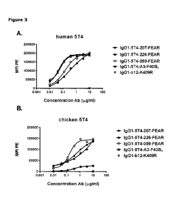
Figure 3: Binding of 5T4 antibodies to HEK-293 cells transfected with full
length human and chicken
5T4. HEK-293 cells transiently transfected with full length human 5T4 (SEQ ID
NO: 1) (A) or chicken
5T4 (SEQ ID NO: 3) (B) were incubated with various concentrations of IgG1-5T4-
A3-F4051_, IgG1-5T4-
059-FEAR, IgG1-5T4-207-FEAR or IgG1-5T4-226-FEAR antibodies. After incubation
with R-
Phycoerythrin (PE)-conjugated goat-anti-human IgG F(ab')2, the mean
fluorescence intensity (MFI)
was determined by flow cytometry. As negative control, IgG1-b12-K409R (10
ug/mL) was included.
Figure 4: Internalization capacity of monovalent 5T4 antibodies. Bispecific,
toxin-conjugated
antibodies that recognize 5T4 with one Fab-arm while recognizing an irrelevant
antigen (HIV-1 gp120,
which is not expressed on tumor cells) with the second Fab-arm, were generated
by controlled Fab-
arm exchange of unconjugated 5T4 antibodies with (HIV-1 gp120-specific) IgG1-
b12 antibodies that
had been conjugated with one Duostatin-3 molecule per antibody. MDA-MB-468 (A)
and HCC1954
(B) cells were incubated with increasing concentrations of antibodies, as
indicated. Cell viability was
measured after 5 days. Data are presented as mean percentage viable cells of
three replicate
experiments. As negative control, monospecific, bivalent IgG1-b12 conjugated
with Duostatin-3
(IgG1-b12-vcDuo3) was included.
Figure 5(1): Binding of CD3x5T4 bispecific antibodies to full length human and
cynomolaus monkey
5T4 transfected into HEK-293 cells. Binding of monovalent and bivalent 5T4
antibodies was analysed
using HEK-293 cells transiently transfected with full length human (left
panels) or cynomolgus
monkey 5T4 (right panels). Cells were incubated with increasing concentrations
of antibodies, as
indicated. After secondary labelling with FITC conjugated goat-anti-human IgG
F(ab')2, binding was
analysed by flow cytometry. As negative control antibody, IgG1-b12-K409R (3
ug/mL) was included.
Data are presented as mean fluorescence intensity (MFI) values of two
technical replicates SD. A.
Binding of bsIgG1-huCD3-H101G-FEALx5T4-207-FEAR and IgG1-5T4-207-FEAR. B.
Binding of bsIgG1-
huCD3-H101G-FEALx5T4-226-FEAR and IgG1-5T4-226-FEAR. C. Binding of bsIgG1-
huCD3-H101G-
FEALx5T4-059-FEAR and IgG1-5T4-059-FEAR. D. Binding of bsIgG1-huCD3-H101G-
FEALx5T4-H8-FEAR
and IgG1-5T4-H8-FEAR.
Figure 5(11): Binding of bispeafic CD3x5T4 antibodies to cynomolgus monkey and
human 5T4
transfected into HEK-293 cells. Mono- and bivalent binding of 5T4 antibodies
was analysed using
HEK-293 cells transiently transfected with human 5T4 (left panels) or with
cynomolgus monkey 5T4
4
CA 03093745 2020-09-11
WO 2019/175198 PCT/EP2019/056197
(right panels). Cells were incubated with increasing concentrations of
antibodies, as indicated. After
secondary labelling with phycoerythrin (PE)-conjugated goat-anti-human IgG
F(ab')2, binding was
analysed by flow cytometry. A. Binding of bsIgG1-huCD3-H101G-FEALx5T4-207-FEAR
and IgG1-5T4-
207-FEAR; B. Binding of bsIgG1-huCD3-H101G-FEALx5T4-226-FEAR and IgG1-5T4-226-
FEAR; C.
Binding of bsIgG1-huCD3-H101G-FEALx5T4-059-FEAR and IgG1-5T4-059-FEAR; D.
Binding of bsIgG1-
huCD3-H101G-FEALx5T4-106-FEAR and IgG1-5T4-106-FEAR; E. Binding of bsIgG1-
huCD3-H101G-
FEALx5T4-076-FEAR and IgG1-5T4-076-FEAR; F. Binding of bsIgG1-huCD3-H101G-
FEALx5T4-085-FEAR
and IgG1-5T4-085-FEAR; G. Binding of bsIgG1-huCD3-H101G-FEALx5T4-127-FEAR and
IgG1-5T4-127-
FEAR; H. Binding of bsIgG1-huCD3-H101G-FEALx5T4-A1-FEAR and IgG1-5T4-A1-FEAR;
I. Binding of
bsIgG1-huCD3-H101G-FEALx5T4-A3-FEAR and IgG1-5T4-A3-FEAR.
Figure 6(1): Binding of CD3x5T4 bispecific and 5T4 monospecific antibodies to
5T4-positive human
tumor cells. Mono- and bivalent binding of 5T4 antibodies to HeLa cells (left
panels) or MDA-MB-231
cells (right panels) was determined by flow cytometry. Cells were incubated
with increasing
concentrations of antibodies. After secondary labelling with FITC-conjugated
goat-anti-human IgG
F(ab')2, the MFI was determined by flow cytometry. A. Binding of bsIgG1-huCD3-
H101G-FEALx5T4-
207-FEAR and IgG1-5T4-207-FEAR antibodies to HeLa cells (left panel) or MDA-MB-
231 cells (right
panel). B. Binding of bsIgG1-huCD3-H101G-FEALx5T4-059-FEAR and IgG1-5T4-059-
FEAR antibodies to
HeLa cells (left panel) or MDA-MB-231 cells (right panel). C. Binding of
bsIgG1-huCD3-H101G-
FEALx5T4-226-FEAR and IgG1-5T4-226-FEAR antibodies to HeLa cells (left panel)
or MDA-MB-231
cells (right panel). IgG1-b12-K409R (3 ug/mL) was included as negative control
(open circles).Figure
6(11): Binding of CD3x5T4 bispeafic and 5T4 monospeafic antibodies to HeLa
cells. Mono- and bivalent
binding of 5T4 antibodies to HeLa cells was determined by flow cytometry.
Cells were incubated with
increasing concentrations of antibodies. After secondary labelling with
Phycoerythrin (PE)-conjugated
goat-anti-human IgG F(ab')2, the mean fluorescence intensity (MFI) was
determined by flow
cytometry. A. Binding of bsIgG1-huCD3-H101G-FEALx5T4-207-FEAR and IgG1-5T4-207-
FEAR; B.
Binding of bsIgG1-huCD3-H101G-FEALx5T4-226-FEAR and IgG1-5T4-226-FEAR; C.
Binding of bsIgG1-
huCD3-H101G-FEALx5T4-059-FEAR and IgG1-5T4-059-FEAR; D. Binding of bsIgG1-
huCD3-H101G-
FEALx5T4-106-FEAR and IgG1-5T4-106-FEAR; E. Binding of bsIgG1-huCD3-H101G-
FEALx5T4-085-FEAR
and IgG1-5T4-085-FEAR; F. Binding of bsIgG1-huCD3-H101G-FEALx5T4-127-FEAR and
IgG1-5T4-127-
FEAR; G. Binding of bsIgG1-huCD3-H101G-FEALx5T4-A1-FEAR and IgG1-5T4-A1-FEAR;
H. Binding of
bsIgG1-huCD3-H101G-FEALx5T4-A3-FEAR and IgG1-5T4-A3-FEAR
5
CA 03093745 2020-09-11
WO 2019/175198 PCT/EP2019/056197
Figure 6(111): Binding of CD3x5T4 bispecific and 5T4 monospeafic antibodies to
MDA-MB-231 cells.
Mono- and bivalent binding of 5T4 antibodies to MDA-MB-231 cells was
determined by flow
cytometry. Cells were incubated with increasing concentrations of antibodies.
After secondary
labelling with PE-conjugated goat-anti-human IgG F(a1312, the mean
fluorescence intensity (MFI) was
determined by flow cytometry. A. Binding of bsIgG1-huCD3-H101G-FEALx5T4-207-
FEAR and IgG1-
5T4-207-FEAR; B. Binding of bsIgG1-huCD3-H101G-FEALx5T4-226-FEAR and IgG1-5T4-
226-FEAR; C.
Binding of bsIgG1-huCD3-H101G-FEALx5T4-059-FEAR and IgG1-5T4-059-FEAR; D.
Binding of bsIgG1-
huCD3-H101G-FEALx5T4-106-FEAR and IgG1-5T4-106-FEAR; E. Binding of bsIgG1-
huCD3-H101G-
FEALx5T4-085-FEAR and IgG1-5T4-085-FEAR; F. Binding of bsIgG1-huCD3-H101G-
FEALx5T4-127-FEAR
and IgG1-5T4-127-FEAR; G. Binding of bsIgG1-huCD3-H101G-FEALx5T4-A1-FEAR and
IgG1-5T4-A1-
FEAR; H. Binding of bsIgG1-huCD3-H101G-FEALx5T4-A3-FEAR and IgG1-5T4-A3-FEAR.
Figure 7(1): Induction of cytotoxicity in vitro by CD3x5T4 bispecific
antibodies in MDA-MB-231 cells
using purified T cells as effector cells. MDA-MB-231 cells were incubated with
increasing
concentrations of CD3x5T4 bispecific antibodies or monospecific, bivalent 5T4
antibodies and
.. isolated T cells as effector cells in an Effector:Target cell (E:T) ratio
of 8:1. Purified T cells obtained
from two different donors were used for this experiment, donor A (left panels)
and donor B (right
panels). Cytotoxicity was determined by measuring the percentage of viable MDA-
MB-231 cells after
72 hrs of incubation (% viable cells = [absorbance sample ¨ absorbance
staurosporine-treated target
cells]/[absorbance untreated target cells ¨ absorbance staurosporine-treated
target cells] x 100). A.
Cytotoxicity induced in the presence of bsIgG1-huCD3-FEALx5T4-207-FEAR, bsIgG1-
huCD3-H101G-
FEALx5T4-207-FEAR and IgG1-5T4-207-FEAR; B. Cytotoxicity induced in the
presence of bsIgG1-
huCD3-FEALx5T4-226-FEAR, bsIgG1-huCD3-H101G-FEALx5T4-226-FEAR and IgG1-5T4-226-
FEAR; C.
Cytotoxicity induced in the presence of bsIgG1-huCD3-FEALx5T4-059-FEAR, bsIgG1-
huCD3-H101G-
FEALx5T4-059-FEAR and IgG1-5T4-059-FEAR.
Figure 7(11): IC50 values of cytotoxicity induced in vitro by CD3x5T4
bispecific antibodies in MDA-MB-
231 cells using purified T cells as effector cells. IC50 values of the T-cell
mediated cytotoxicity induced
by bsIgG1-huCD3-FEALx5T4-207-FEAR, bsIgG1-huCD3-H101G-FEALx5T4-207-FEAR,
bsIgG1-huCD3-
FEALx5T4-226-FEAR, bsIgG1-huCD3-H101G-FEALx5T4-226-FEAR, bsIgG1-huCD3-FEALx5T4-
059-FEAR
or bsIgG1-huCD3-H101G-FEALx5T4-059-FEAR in MDA-MB-231 cells were analyzed
using GraphPad
Prism V7.02 software. Data are presented as mean IC50 values of two different
donors SD.
Figure 8(1): Induction of cytotoxicity by CD3x5T4 bispecific antibodies in MDA-
MB-231 cells using T
cells as effector cells in vitro. MDA-MB-231 cells were incubated with
increasing concentrations of
6
CA 03093745 2020-09-11
WO 2019/175198 PCT/EP2019/056197
CD3x5T4 bispecific antibodies or 5T4 homodimers and isolated T cells as
effector cells in an E:T ratio
of 8:1. Three different donors were used for this experiment. Data shown are
mean %
survival standard error of the mean (SEM) of three donors tested. A. T-cell-
mediated cytotoxicity
(decrease in survival) induced in the presence of bsIgG1-huCD3-FEALx5T4-207-
FEAR, bsIgG1-huCD3-
H101G-FEALx5T4-207-FEAR and IgG1-5T4-207-FEAR; B. T-cell-mediated cytotoxicity
induced in the
presence of bsIgG1-huCD3-FEALx5T4-226-FEAR, bsIgG1-huCD3-H101G-FEALx5T4-226-
FEAR and IgG1-
5T4-226-FEAR; C. T-cell-mediated cytotoxicity induced in the presence of
bsIgG1-huCD3-FEALx5T4-
059-FEAR, bsIgG1-huCD3-H101G-FEALx5T4-059-FEAR and IgG1-5T4-059-FEAR; D. T-
cell-mediated
cytotoxicity induced in the presence of bsIgG1-huCD3-FEALx5T4-106-FEAR, bsIgG1-
huCD3-H101G-
FEALx5T4-106-FEAR and IgG1-5T4-106-FEAR; E. T-cell-mediated cytotoxicity
induced in the presence
of bsIgG1-huCD3-FEALx5T4-A1-FEAR, bsIgG1-huCD3-H101G-FEALx5T4-A1-FEAR and IgG1-
5T4-A1-
FEAR; F. T-cell-mediated cytotoxicity induced in the presence of bsIgG1-huCD3-
FEALx5T4-A3-FEAR,
bsIgG1-huCD3-H101G-FEALx5T4-A3-FEAR and IgG1-5T4-A3-FEAR.
Figure 8(11): IC50 values of cytotoxicity induced by CD3x5T4 bispecific
antibodies in MDA-M8-231 cells
using T cells as effector cells in vitro. IC50 values of the T-cell-mediated
cytotoxicity induced CD3x5T4
bispecific antibodies in MDA-MB-231 cells were analyzed using GraphPad Prism
V7.02 software. Data
are presented as mean IC50 values of three different donors SD. A. IC50
values of the T-cell-
mediated cytotoxicity induced by bsIgG1-huCD3-FEALx5T4-207-FEAR, bsIgG1-huCD3-
FEALx5T4-226-
FEAR, bsIgG1-huCD3-FEALx5T4-059-FEAR, bsIgG1-huCD3-FEALx5T4-106-FEAR, bsIgG1-
huCD3-
FEALx5T4-A1-FEAR and bsIgG1-huCD3-FEALx5T4-A3-FEAR; B. IC50 values of the T-
cell-mediated
cytotoxicity induced by bsIgG1-huCD3-H101G-FEALx5T4-207-FEAR, bsIgG1-huCD3-
H101G-FEALx5T4-
226-FEAR, bsIgG1-huCD3-H101G-FEALx5T4-059-FEAR, bsIgG1-huCD3-H101G-FEALx5T4-
106-FEAR,
bsIgG1-huCD3-H101G-FEALx5T4-A1-FEAR and bsIgG1-huCD3-H101G-FEALx5T4-A3-FEAR.
Figure 9(1): In vitro T-cell activation by CD3x5T4 bispecific antibodies in
the presence of MDA-M8-231
cells. MDA-MB-231 cells were incubated with increasing concentrations of
CD3x5T4 bispecific
antibodies and monospecific, bivalent 5T4 antibodies, as indicated, and
isolated T cells as effector
cells in an E:T ratio of 8:1. The expression of three T cell activation
markers (PD1 [upper panels],
CD25 [middle panels] and CD69 [lower panels]) was analyzed by flow cytometry.
Two different
donors were used for this experiment, donor A (closed symbols) and donor B
(open symbols). A. T-
.. cell activation induced in the presence of bsIgG1-huCD3-FEALx5T4-207-FEAR,
bsIgG1-huCD3-H101G-
FEALx5T4-207-FEAR and IgG1-5T4-207-FEAR; B. T-cell activation induced in the
presence of bsIgG1-
huCD3-FEALx5T4-226-FEAR, bsIgG1-huCD3-H101G-FEALx5T4-226-FEAR and IgG1-5T4-226-
FEAR; C. T-
7
CA 03093745 2020-09-11
WO 2019/175198 PCT/EP2019/056197
cell activation induced in the presence of bsIgG1-huCD3-FEALx5T4-059-FEAR,
bsIgG1-huCD3-H101G-
FEALx5T4-059-FEAR and IgG1-5T4-059-FEAR.
Figure 9(11): EC50 values of in vitro T-cell activation by CD3x5T4 bispecific
antibodies in the presence
of MDA-MB-231 cells. EC50 values of in vitro T-cell activation markers (PD1,
CD25 and CD69) induced
by bsIgG1-huCD3-FEALx5T4-207-FEAR, bsIgG1-huCD3-H101G-FEALx5T4-207-FEAR,
bsIgG1-huCD3-
FEALx5T4-226-FEAR, bsIgG1-huCD3-H101G-FEALx5T4-226-FEAR, bsIgG1-huCD3-FEALx5T4-
059-FEAR
or bsIgG1-huCD3-H101G-FEALx5T4-059-FEAR in the presence of MDA-MB-231 cells
were analyzed
using GraphPad Prism V7.02 software. Data are presented as mean of two
different donors SD.
Figure 10(I): In vitro T-cell activation by CD3x5T4 bispecific antibodies in
the presence of MDA-MB-
231 cells. MDA-MB-231 cells were incubated with increasing concentrations of
CD3x5T4 bispecific
antibodies and 5T4 homodimers and isolated T cells as effector cells in an E:T
ratio of 8:1. T-cell
activation was measured by an increase in % CD69+ cells within the CD4+ (left
panels) and CD8+ (right
panels) T cell populations. Three different donors were used for this
experiment; data shown are
mean % CD69 upregulation SEM of three donors tested. A. T-cell activation
induced in the presence
of bsIgG1-huCD3-FEALx5T4-207-FEAR, bsIgG1-huCD3-H101G-FEALx5T4-207-FEAR and
IgG1-5T4-207-
FEAR; B. T-cell activation induced in the presence of bsIgG1-huCD3-FEALx5T4-
226-FEAR, bsIgG1-
huCD3-H101G-FEALx5T4-226-FEAR and IgG1-5T4-226-FEAR; C. T-cell activation
induced in the
presence of bsIgG1-huCD3-FEALx5T4-059-FEAR, bsIgG1-huCD3-H101G-FEALx5T4-059-
FEAR and IgG1-
5T4-059-FEAR; D. T-cell activation induced in the presence of bsIgG1-huCD3-
FEALx5T4-106-FEAR,
bsIgG1-huCD3-H101G-FEALx5T4-106-FEAR and IgG1-5T4-106-FEAR; E. T-cell
activation induced in the
presence of bsIgG1-huCD3-FEALx5T4-A1-FEAR, bsIgG1-huCD3-H101G-FEALx5T4-A1-FEAR
and IgG1-
5T4-A1-FEAR; F. T-cell activation induced in the presence of bsIgG1-huCD3-
FEALx5T4-A3-FEAR,
bsIgG1-huCD3-H101G-FEALx5T4-A3-FEAR and IgG1-5T4-A3-FEAR.
Figure 10(II): EC50 values of in vitro T-cell activation by CD3x5T4 bispecific
antibodies in the presence
.. of MDA-MB-231 cells. EC50 values of T-cell activation markers (increase in
% of CD69+ [A-B], CD25+ [C-
D] and PD1+ [E-F], CD25 and CD69 cells within the CD4+ and CD8+ T cell
populations) induced in vitro
by CD3x5T4 bispecific antibodies in the presence of MDA-MB-231 cells were
analyzed using
GraphPad Prism V7.02 software. Data are presented as mean of three different
donors SD. A. EC50
values of the CD69 upregulation induced by bsIgG1-huCD3-FEALx5T4-207-FEAR,
bsIgG1-huCD3-
FEALx5T4-226-FEAR, bsIgG1-huCD3-FEALx5T4-059-FEAR, bsIgG1-huCD3-FEALx5T4-106-
FEAR, bsIgG1-
huCD3-FEALx5T4-A1-FEAR and bsIgG1-huCD3-FEALx5T4-A3-FEAR; B. EC50 values of
the CD69
upregulation induced by bsIgG1-huCD3-H101G-FEALx5T4-207-FEAR, bsIgG1-huCD3-
H101G-FEALx5T4-
8
CA 03093745 2020-09-11
WO 2019/175198 PCT/EP2019/056197
226-FEAR, bsIgG1-huCD3-H101G-FEALx5T4-059-FEAR, bsIgG1-huCD3-H101G-FEALx5T4-
106-FEAR,
bsIgG1-huCD3-H101G-FEALx5T4-A1-FEAR and bsIgG1-huCD3-H101G-FEALx5T4-A3-FEAR.
C. EC50
values of the CD25 upregulation induced by bsIgG1-huCD3-FEALx5T4-207-FEAR,
bsIgG1-huCD3-
FEALx5T4-226-FEAR, bsIgG1-huCD3-FEALx5T4-059-FEAR, bsIgG1-huCD3-FEALx5T4-106-
FEAR, bsIgG1-
huCD3-FEALx5T4-A1-FEAR and bsIgG1-huCD3-FEALx5T4-A3-FEAR; D. EC50 values of
the CD25
upregulation induced by bsIgG1-huCD3-H101G-FEALx5T4-207-FEAR, bsIgG1-huCD3-
H101G-FEALx5T4-
226-FEAR, bsIgG1-huCD3-H101G-FEALx5T4-059-FEAR, bsIgG1-huCD3-H101G-FEALx5T4-
106-FEAR,
bsIgG1-huCD3-H101G-FEALx5T4-A1-FEAR and bsIgG1-huCD3-H101G-FEALx5T4-A3-FEAR.
E. EC50
values of the PD1 upregulation induced by bsIgG1-huCD3-FEALx5T4-207-FEAR,
bsIgG1-huCD3-
FEALx5T4-226-FEAR, bsIgG1-huCD3-FEALx5T4-059-FEAR, bsIgG1-huCD3-FEALx5T4-106-
FEAR, bsIgG1-
huCD3-FEALx5T4-A1-FEAR and bsIgG1-huCD3-FEALx5T4-A3-FEAR; F. EC50 values of
the PD1
upregulation induced by bsIgG1-huCD3-H101G-FEALx5T4-207-FEAR, bsIgG1-huCD3-
H101G-FEALx5T4-
226-FEAR, bsIgG1-huCD3-H101G-FEALx5T4-059-FEAR, bsIgG1-huCD3-H101G-FEALx5T4-
106-FEAR,
bsIgG1-huCD3-H101G-FEALx5T4-A1-FEAR and bsIgG1-huCD3-H101G-FEALx5T4-A3-FEAR.
Figure 11: T cell cytokine release induced by CD3x5T4 bispecific antibodies in
the presence of 5T4-
positive tumor cells. MDA-MB-231 cells were incubated with 0.2 ug/mL CD3x5T4
bispecific antibodies
(bsIgG1-huCD3-FEALx5T4-207-FEAR, bsIgG1-huCD3-H101G-FEALx5T4-207-FEAR, bsIgG1-
huCD3-
FEALx5T4-226-FEAR, bsIgG1-huCD3-H101G-FEALx5T4-226-FEAR, bsIgG1-huCD3-FEALx5T4-
059-FEAR
or bsIgG1-huCD3-H101G-FEALx5T4-059-FEAR) and 5T4 monospecific antibodies (IgG1-
5T4-207-FEAR,
IgG1-5T4-226-FEAR or IgG1-5T4-059-FEAR) and isolated T cells as effector cells
in an E:T ratio of 8:1.
Release of cytokines was analyzed by U-PLEX assay. A. Concentration of IL-10,
IL-13 and TNF in the
supernatant of T cell (derived from donor A)-tumor cell co-cultures, after 72
h of incubation with
CD3x5T4 bispecific antibodies or 5T4 monospecific antibodies. B. Concentration
of IL-10, IL-13 and
TNF in the supernatant of T cell (derived from donor B)-tumor cell co-
cultures, after 72 h of
incubation with CD3x5T4 bispecific antibodies or 5T4 monospecific antibodies.
Figure 12: Induction of cytotoxicity in vitro by CD3x5T4 bispecific antibodies
in SK-OV-3 cells using
PBMCs as effector cells at varying E:T ratios. SK-OV-3 cells were incubated
with increasing
concentrations of bsIgG1-huCD3-FEALx5T4-207-FEAR (left panels) or bsIgG1-huCD3-
H101G-
FEALx5T4-207-FEAR (right panels) and PBMCs as effector cells in an E:T ratio
of 1:2, 1:1, 2:1, 4:1, 8:1
and 12:1. Cytotoxicity was determined by measuring the percentage of viable SK-
OV-3 cells after 72
h of incubation (% viable cells = [absorbance sample ¨ absorbance
staurosporine-treated target
9
CA 03093745 2020-09-11
WO 2019/175198 PCT/EP2019/056197
cells]/[absorbance untreated target cells - absorbance staurosporine-treated
target cells] x 100).
PBMCs from two different donors were used for this experiment: A. donor C and
B. donor D.
Figure 13: Induction of cytotoxicity in SK-OV-3 cells in vitro by CD3x5T4
bispecific antibodies using T
cells as effector cells at varying E:T ratios. SK-OV-3 cells were incubated
with increasing
concentrations of bsIgG1-huCD3-FEALx5T4-207-FEAR (left panels) or bsIgG1-huCD3-
H101G-
FEALx5T4-207-FEAR (right panels) and isolated T cells as effector cells in an
E:T ratio of 1:2, 1:1, 2:1,
4:1 and 8:1. The efficiency of cytotoxicity was determined by measuring the
percentage of viable SK-
OV-3 cells after 72 h of incubation (% viable cells = [absorbance sample -
absorbance staurosporine-
treated target cells]/[absorbance untreated target cells - absorbance
staurosporine-treated target
cells] x 100). T cells from two different donors were used for this
experiment: A. donor E and B.
donor F.
Figure 14. Anti-tumor activity of CD3x5T4 bispecific antibodies in a MDA-MB-
231 xenograft model in
NSG-HIS mice. A. Average tumor size in the MDA-MB-231 xenograft model in NSG-
HIS mice after
treatment with PBS (vehicle control), 0.5 mg/kg bsIgG1-huCD3-FEALx5T4-207-FEAR
or 0.5 mg/kg
bsIgG1-huCD3-H101G-FEALx5T4-207-FEAR. Tumor size was assessed by caliper
measurement. Error
bars indicate SEM. B. Percentage of NSG-HIS mice injected with MDA-MB-231
cells with a tumor size
< 500 mm3 after treatment with PBS, bsIgG1-huCD3-FEALx5T4-207-FEAR or bsIgG1-
huCD3-H101G-
FEALx5T4-207-FEAR.
Figure 15: Binding of directly FITC-labeled 5T4-specific antibodies to human
5T4 variants with single
alanine mutations at positions 32 to 355 of human 5T4 ECD, as determined by
flow cytometry.
Binding was expressed as Z-score (fold change), as a measure for change in
binding compared to a
non-cross blocking 5T4-specific control antibody (bsIgG1-5T4-A1-F405Lxb12-FEAR-
FITC) used for
normalization. The number on the x-axis refers to the amino acid positions in
human 5T4 (SEQ ID: 1).
Residues where the Z-score in binding was lower than - 1.5 (indicated by the
dotted line) were
considered 'loss of binding mutants'. Residues with a positive Z-score in
binding are loss of binding
residues for the non-cross blocking 5T4 specific control antibody (bsIgG1-5T4-
A1-67F-F405Lxb12-
FEAR-FITC). Residues on aa position 38, 45, 49, 51, 54, 62, 64, 66, 68, 71,
72, 77, 91, 104, 108, 110,
112, 118, 121, 122, 135, 137, 155, 161, 167, 171, 201, 202, 205, 208, 218,
231, 269, 279, 298, 300,
303, 323, 324, 340 and 344 were not evaluated, as these positions contained
either endogenous
alanines or cysteines. Data shown are Z-scores for binding of (A) bsIgG1-b12-
FEALx5T4-059-FEAR-
FITC, (B) bsIgG1-b12-FEALx5T4-207-FEAR-FITC, (C) bsIgG1-b12-FEALx5T4-226-FEAR-
FITC, and (D)
bsIgG1-5T4-A3-F405Lxb12-FEAR-FITC. Buried residues with a Z-score just below -
1.5 that were
CA 03093745 2020-09-11
WO 2019/175198 PCT/EP2019/056197
predicted to be spatially separated from the majority of surface-exposed loss
of binding residues
were excluded (for bIgG1-b12-FEALx5T4-207-FEAR-FITC: L281 [Z-score: -1.57] and
P326 [Z-
score: -1.54]; and for bsIgG1-b12-FEALx5T4-226-FEAR-FITC: L273 [Z-score: -
1.58], L281 [Z-score:-
1.65], N294 [Z-score:-1.57], L309 [Z-score:-1.63] and P326 [Z-score:-1.67]).
Figure 16(1): Induction of cytotoxicity in vitro by CD3x5T4 bispecific
antibodies in tumor cells of
different indications using T cells as effector cells. Tumor cells were
incubated with increasing
concentrations of bsIgG1-huCD3-H101G-FEALx5T4-207-FEAR or control antibodies
(bsIgG1-huCD3-
H101G-FEALxb12-FEAR, bsIgG1-b12-FEALx5T4-207-FEAR) and isolated T cells as
effector cells in an
E:T ratio of 4:1. Cytotoxicity (decrease in survival) was determined by
measuring the percentage of
viable tumor cells after 72 h of incubation. Data shown are mean % survival
SEM of duplicate wells
from one representative donor out of at least three donors tested. A.
Cytotoxicity (decrease in
survival) induced in pancreas cancer cell lines; B. Cytotoxicity (decrease in
survival) induced in
cervical cancer cell lines.
Figure 16(II): IC50 values of cytotoxicity induced in vitro by CD3x5T4
bispecific antibodies in tumor cell
lines of different indications using T cells as effector cells. IC50 values of
the T-cell-mediated
cytotoxicity induced by bsIgG1-huCD3-H101G-FEALx5T4-207-FEAR in tumor cells of
the indicated
indications were analyzed using GraphPad Prism V7.02 software. Data are
presented as mean IC50
values of at least three different donors (see Table 10) SD.
Figure 17(1): In vitro T-cell activation by CD3x5T4 bispecific antibodies in
the presence of tumor cells of
different indications. Tumor cells were incubated with increasing
concentrations of bsIgG1-huCD3-
H101G-FEALx5T4-207-FEAR or control antibodies (bsIgG1-huCD3-H101G-FEALxb12-
FEAR, bsIgG1-
b12-FEALx5T4-207-FEAR and isolated T cells as effector cells in an E:T ratio
of 4:1 for 72 h. T-cell
activation was measured by the upregulation of CD69 (% of CD69+ cells) within
CD4+ (left panels) and
CD8+ (right panels) T-cell populations. Data shown are mean % CD69+ cells SD
of duplicate wells
.. from one representative donor out of at least three donors tested. A. T-
cell activation induced by
CD3x5T4 bispecific antibodies in the presence of pancreas cancer cell line
BxPc-3; B. T-cell activation
induced by CD3x5T4 bispecific antibodies in the presence of pancreas cancer
cell line PANC-1; C. T-
cell activation induced by CD3x5T4 bispecific antibodies in the presence of
cervical cancer cell line
SiHa; D. T-cell activation induced by CD3x5T4 bispecific antibodies in the
presence of cervical cancer
cell line Ca Ski.
Figure 17(11): EC50 values of in vitro T-cell activation by CD3x5T4 bispecific
antibodies in with the
presence of tumor cell lines of different indications. EC50 values of the T-
cell activation (% of CD69+
11
CA 03093745 2020-09-11
WO 2019/175198 PCT/EP2019/056197
cells within CD4+ and CD8+ T-cell populations) induced by bsIgG1-huCD3-H101G-
FEALx5T4-207-FEAR
in co-culture with tumor cell lines of the different indications were analyzed
using GraphPad Prism
V7.02 software. Data are presented as mean EC50 values of at least three
different donors (see Table
10) SD. A. EC50 values of CD4+ T-cell activation induced by bsIgG1-huCD3-
H101G-FEALx5T4-207-
FEAR in the presence of the indicated tumor cell lines; B. EC50 values of CD8+
T-cell activation
induced by bsIgG1-huCD3-H101G-FEALx5T4-207-FEAR in the presence of the
indicated tumor cell
lines.
Detailed Description
Definitions
The term "antibody" as used herein is intended to refer to an immunoglobulin
molecule, a fragment
of an immunoglobulin molecule, or a derivative of either thereof, which has
the ability to specifically
bind to an antigen under typical physiological and/or tumor-specific
conditions with a half-life of
significant periods of time, such as at least about 30 minutes, at least about
45 minutes, at least
about one hour, at least about two hours, at least about four hours, at least
about 8 hours, at least
about 12 hours, at least about 24 hours or more, at least about 48 hours or
more, at least about 3, 4,
5, 6, 7 or more days, etc., or any other relevant functionally-defined period
(such as a time sufficient
to induce, promote, enhance, and/or modulate a physiological response
associated with antibody
binding to the antigen and/or time sufficient for the antibody to be
internalized). The binding region
(or binding domain which may be used herein, both having the same meaning)
which interacts with
an antigen, comprises variable regions of both the heavy and light chains of
the immunoglobulin
molecule. The constant regions of the antibodies (Abs) may mediate the binding
of the
immunoglobulin to host tissues or factors, including various cells of the
immune system (such as
effector cells) and components of the complement system such as C1q, the first
component in the
classical pathway of complement activation.
In the context of the present invention, the term "antibody" includes a
monoclonal antibody (mAb),
an antibody-like polypeptide, such as a chimeric antibody and a humanized
antibody, as well as an
'antibody fragment' or a 'fragment thereof' retaining the ability to
specifically bind to the antigen
(antigen-binding fragment) provided by any known technique, such as enzymatic
cleavage, peptide
synthesis, and recombinant techniques, and retaining the ability to be
conjugated to a toxin. An
antibody as defined according to the invention can possess any isotype unless
the disclosure herein is
otherwise limited.
12
CA 03093745 2020-09-11
WO 2019/175198 PCT/EP2019/056197
As indicated above, the term antibody as used herein, unless otherwise stated
or clearly contradicted
by context, includes fragments of an antibody that retain the ability to
specifically interact, such as
bind, to the antigen. It has been shown that the antigen-binding function of
an antibody may be
performed by fragments of a full-length antibody. Examples of binding
fragments encompassed
within the term "antibody" include (i) a Fab' or Fab fragment, a monovalent
fragment consisting of
the ligh chain variable domain (VL), heavy chain variable domain (VH), light
chain constant region (CL)
and heavy chain constant region domain 1 (CH1) domains, or a monovalent
antibody as described in
WO 2007/059782; (ii) F(ab')2 fragments, bivalent fragments comprising two Fab
fragments linked by
a disulfide bridge at the hinge region; (iii) an Ed fragment consisting
essentially of the VH and CH1
domains; (iv) an Fy fragment consisting essentially of the VL and VH domains
of a single arm of an
antibody, (v) a dAb fragment Ward et al., Nature 341, 544-546 (1989), which
consists essentially of a
VH domain and is also called domain antibody Holt et al; Trends Biotechnol.
2003 Nov;21(11):484-90;
(vi) camelid or nanobodies Revets et al; Expert Opin Biol Ther. 2005
Jan;5(1):111-24 and (vii) an
isolated complementarity determining region (CDR). Furthermore, although the
two domains of the
Fy fragment, VL and VH, are coded for by separate genes, they may be joined,
using recombinant
methods, by a synthetic linker that enables them to be made as a single
protein chain in which the VL
and VH regions pair to form monovalent molecules (known as single chain
antibodies or single chain
Fy (scFv), see for instance Revets et al; Expert Opin Biol Ther. 2005
Jan;5(1):111-24 and Bird et al.,
Science 242, 423-426 (1988). Such single chain antibodies are encompassed
within the term antibody
unless otherwise noted or clearly indicated by context. Although such
fragments are generally
included within the meaning of antibody, they collectively and each
independently are unique
features of the present invention, exhibiting different biological properties
and utility. These and
other useful antibody fragments in the context of the present invention are
discussed further herein.
An antibody can be produced in and collected from different in vitro or ex
vivo expression or
production systems, for example from recombinantly modified host cells, from
hybridomas or
systems that use cellular extracts supporting in vitro transcription and/or
translation of nucleic acid
sequences encoding the antibody. It is to be understood that a multitude of
different antibodies, the
antibodies being as defined in the context of the present invention, is one
that can be provided by
producing each antibody separately in a production system as mentioned above
and thereafter
mixing the antibodies, or by producing several antibodies in the same
production system.
The term "immunoglobulin heavy chain" or "heavy chain of an immunoglobulin" as
used herein is
intended to refer to one of the heavy chains of an immunoglobulin. A heavy
chain is typically
13
CA 03093745 2020-09-11
WO 2019/175198 PCT/EP2019/056197
comprised of a heavy chain variable region (abbreviated herein as VH) and a
heavy chain constant
region (abbreviated herein as CH) which defines the isotype of the
immunoglobulin. The heavy chain
constant region typically is comprised of three domains, CH1, CH2, and CH3.
The term
"immunoglobulin" as used herein is intended to refer to a class of
structurally related glycoproteins
consisting of two pairs of polypeptide chains, one pair of light (L) low
molecular weight chains and
one pair of heavy (H) chains, all four potentially inter-connected by
disulfide bonds. The structure of
immunoglobulins has been well characterized (see for instance Fundamental
Immunology Ch. 7
(Paul, W., ed., 2nd ed. Raven Press, N.Y. (1989)). Within the structure of the
immunoglobulin, the
two heavy chains are inter-connected via disulfide bonds in the so-called
"hinge region". Equally to
the heavy chains, each light chain is typically comprised of several regions;
a light chain variable
region (abbreviated herein as VL) and a light chain constant region. The light
chain constant region
typically is comprised of one domain, CL. Furthermore, the VH and VL regions
may be further
subdivided into regions of hypervariability (or hypervariable regions which
may be hypervariable in
sequence and/or form of structurally defined loops), also termed
complementarity determining
regions (CDRs), interspersed with regions that are more conserved, termed
framework regions (FRs).
Each VH and VL is typically composed of three CDRs and four FRs, arranged from
amino-terminus to
carboxy-terminus in the following order: FR1, CDR1, FR2, CDR2, FR3, CDR3, FR4.
CDR sequences are
defined according to IMGT (see Lefranc MP. et al., Nucleic Acids Research, 27,
209-212, 1999] and
Brochet X. Nucl. Acids Res. 36, W503-508 (2008)).
When used herein, the terms "half molecule", "Fab-arm" and "arm" refer to one
heavy chain-light
chain pair. When a bispecific antibody is described to comprise a half-
molecule antibody "derived
from" a first antibody, and a half-molecule antibody "derived from" a second
antibody, the term
"derived from" indicates that the bispecific antibody was generated by
recombining, by any known
method, said half-molecules from each of said first and second antibodies into
the resulting bispecific
antibody. In this context, "recombining" is not intended to be limited by any
particular method of
recombining and thus includes all of the methods for producing bispecific
antibodies described
herein below, including for example recombining by half-molecule exchange, as
well as recombining
at nucleic acid level and/or through co-expression of two half-molecules in
the same cells.
The term "antigen-binding region" or "binding region" as used herein, refers
to a region of an
antibody which is capable of binding to the antigen. The antigen can be any
molecule, such as a
polypeptide, e.g. present on a cell, bacterium, or virion. The terms "antigen"
and "target" may,
unless contradicted by the context, be used interchangeably in the context of
the present invention.
14
CA 03093745 2020-09-11
WO 2019/175198 PCT/EP2019/056197
The terms "antigen-binding region" and "antigen-binding site" may, unless
contradicted by the
context, be used interchangeably in the context of the present invention.
The term "blocks binding" or "blocking the binding of an antibody" or "cross-
blocking binding" or
"cross-blocks binding" refers to the situation where one antibody bound to a
specific antigen
prevents binding of the second antibody to the same antigen and vice versa. In
the absence of the
other antibody, each antibody has the ability to bind to the antigen as
determined by a significant
binding response, whereas one of the antibodies lacks a binding response when
the other antibody is
present. The ability of one antibody to block the binding of another antibody
may be determined by
biolayer interferometry in a classical sandwich epitope binning assay format,
for instance as
described in Example 3 in the present application and by Abdiche et al.
(Abdiche YN, Malashock DS,
Pinkerton A, Pons J. Exploring blocking assays using Octet, Prote0n, and
Biacore biosensors. Anal
Biochem. 2009; 386(2): 172-180). Briefly, in a sandwich epitope binning assay,
an antibody in solution
is tested for binding to its specific antigen that is first captured via an
immobilized antibody. In the
context of the present invention, one antibody does not block the binding of
another antibody if it is
capable of "displacing" the other antibody, according to the definition of
"displacement" below. The
terms "blocks binding" and "blocking the binding of an antibody" and "cross-
blocking binding" and
"cross-blocks binding" may, unless contradicted by the context, be used
interchangeably in the
context of the present invention. Preferably, the ability of one antibody to
block the binding of
another antibody is determined using full-length antibodies.
The term "displacement" or "ability to displace" or "displacing" refers to the
situation wherein two
antibodies perturb one another's binding to an antigen by kinetically altering
one another's binding
to their specific antigen via the formation of a transient trimolecular
complex, which rapidly collapses
by retaining one antibody to the antigen and displacing the other. Antibody
displacement is defined
in Abdiche et al., 2017 (Abdiche YN, Yeung AY, Ni I, Stone D, Miles A,
Morishige W, et al. (2017)
Antibodies Targeting Closely Adjacent or Minimally Overlapping Epitopes Can
Displace One Another.
PLoS ONE 12(1): e0169535. doi:10.1371/journal.pone.0169535). Antibody
displacement may be
determined by biolayer interferometry using real-time label-free biosensors in
a classical sandwich
assay format as described in Abdiche et al. 2017 and Example 4 in the present
application.Preferably,
antibody displacement is determined using antibodies which are in the IgG
format.
The term "binding" as used herein refers to the binding of an antibody to a
predetermined antigen or
target, typically with a binding affinity corresponding to a KD of 1E8 M or
less, e.g. 5E7 M or less, 1E7
M or less, such as 5E8 M or less, such as 1E8 M or less, such as 5E9 M or
less, or such as 1E9 M or
CA 03093745 2020-09-11
WO 2019/175198 PCT/EP2019/056197
less, when determined by biolayer interferometry using the antibody as the
ligand and the antigen
as the analyte and binds to the predetermined antigen with an affinity
corresponding to a KD that is
at least ten-fold lower, such as at least 100-fold lower, for instance at
least 1,000-fold lower, such as
at least 10,000-fold lower, for instance at least 100,000-fold lower than its
affinity for binding to a
non-specific antigen (e.g., BSA, casein) other than the predetermined antigen
or a closely-related
antigen.
The term "KD" (M), as used herein, refers to the dissociation equilibrium
constant of a particular
antibody-antigen interaction, and is obtained by dividing kd by ka.
The term "kd" (5ec-1), as used herein, refers to the dissociation rate
constant of a particular antibody-
antigen interaction. Said value is also referred to as the kaff value or off-
rate.
The term "ka" (M-1 x 5ec-1), as used herein, refers to the association rate
constant of a particular
antibody-antigen interaction. Said value is also referred to as the kan value
or on-rate.
The term "5T4" as used herein, refers to the protein entitled 5T4, which is
also referred to as
trophoblast glycoprotein, 5T4 oncofetal antigen, 5T4 oncofetal trophoblast
glycoprotein, TPBG,
WAIF1 and M6P1. It is 72-80 kDa transmembrane protein with an extensively N-
linked glycosylated
core. In humans (Homo sapiens), the 5T4 protein has the amino acid sequence
shown in SEQ ID NO: 1
(Human Trophoblast glycoprotein: Uniprot accession no. 013641). In the amino
acid sequence
shown in SEQ ID NO: 1, amino acid residues 1-31 are a signal peptide, and
amino acid residues 32-
420 are the mature polypeptide. In cynomolgus monkey (Macaca fascicularis),
the 5T4 protein has
the amino acid sequence shown in SEQ ID NO: 2 (Uniprot accession no. Q4R8Y9).
In the amino acid
sequence shown in SEQ ID NO: 2, amino acid residues 1-34 are a signal peptide,
and amino acid
residues 35-420 are the mature polypeptide. In chicken (Gallus gal/us), the
5T4 protein has the amino
acid sequence shown in SEQ ID NO: 3 (Uniprot accession no. R4GM46). In the
sequence shown in
SEQ ID NO: 3, amino acid residues 1-27 are a signal peptide, and amino acid
residues 28-379 are the
mature polypeptide.
The term "CD3" as used herein, refers to the human Cluster of Differentiation
3 protein which is part
of the T-cell co-receptor protein complex and is composed of four distinct
chains. CD3 is also found in
other species, and thus, the term "CD3" is not limited to human CD3 unless
contradicted by context.
In mammals, the complex contains a CD3y (gamma) chain (human CD3y chain
UniProtKB/Swiss-Prot
No P09693, or cynomolgus monkey CD3y UniProtKB/Swiss-Prot No Q951_17), a CD36
(delta) chain
(human CD36 UniProtKB/Swiss-Prot No P04234, or cynomolgus monkey CD36
UniProtKB/Swiss-Prot
16
CA 03093745 2020-09-11
WO 2019/175198 PCT/EP2019/056197
No 095LI8), two CD3E (epsilon) chains (human CD3E UniProtKB/Swiss-Prot No
P07766; amino acid
residues 1-22 is a signal peptide and amino acid residues 23-207 is the mature
CD3E polypeptide,
which is identified herein as SEQ ID NO: 4; cynomolgus monkey CD3E
UniProtKB/Swiss-Prot No
095L15; or rhesus monkey CD3E UniProtKB/Swiss-Prot No G7NCB9), and a CD3-chain
(zeta) chain
(human CD3 UniProtKB/Swiss-Prot No P20963, cynomolgus monkey CD3
UniProtKB/Swiss-Prot No
Q09TKO). These chains associate with a molecule known as the T-cell receptor
(TCR) and generate an
activation signal in T lymphocytes. The TCR and CD3 molecules together
comprise the TCR complex.
The term "antibody binding region" refers to a region of the antigen, which
comprises the epitope to
which the antibody binds. An antibody binding region may be determined by
epitope binning using
biolayer interferometry, by alanine scan, or by shuffle assays (using antigen
constructs in which
regions of the antigen are exchanged with that of another species and
determining whether the
antibody still binds to the antigen or not). The amino acids within the
antibody binding region that
are involved in the interaction with the antibody may be determined by
hydrogen/deuterium
exchange mass spectrometry and by crystallography of the antibody bound to its
antigen.
The term "epitope" means an antigenic determinant which is specifically bound
by an antibody.
Epitopes usually consist of surface groupings of molecules such as amino
acids, sugar side chains or a
combination thereof and usually have specific three dimensional structural
characteristics, as well as
specific charge characteristics. Conformational and non-conformational
epitopes are distinguished in
that the binding to the former but not the latter is lost in the presence of
denaturing solvents. The
epitope may comprise amino acid residues which are directly involved in the
binding, and other
amino acid residues, which are not directly involved in the binding, such as
amino acid residues
which are effectively blocked or covered by the antibody when it is bound to
the antigen (in other
words, the amino acid residue is within or closely adjacent to the footprint
of the specific antibody).
The terms "monoclonal antibody", "monoclonal Ab", "monoclonal antibody
composition", "mAb", or
the like, as used herein refer to a preparation of antibody molecules of
single molecular composition.
A monoclonal antibody composition displays a single binding specificity and
affinity for a particular
epitope. Accordingly, the term "human monoclonal antibody" refers to
antibodies displaying a single
binding specificity which have variable and constant regions derived from
human germline
immunoglobulin sequences. The human monoclonal antibodies may be produced by a
hybridoma
which includes a B cell obtained from a transgenic or transchromosomal non-
human animal, such as
a transgenic mouse, having a genome comprising a human heavy chain transgene
and a light chain
transgene, fused to an immortalized cell. Monoclonal antibodies may also be
produced from
17
CA 03093745 2020-09-11
WO 2019/175198 PCT/EP2019/056197
recombinantly modified host cells, or systems that use cellular extracts
supporting in vitro
transcription and/or translation of nucleic acid sequences encoding the
antibody.
The term "isotype" as used herein refers to the immunoglobulin class (for
instance IgG1, IgG2, IgG3,
IgG4, IgD, IgA, IgE, or IgM) or any allotypes thereof, such as IgG1m(za) and
IgG1m(f)) that is encoded
by heavy chain constant region genes. Further, each heavy chain isotype can be
combined with
either a kappa (lc) or lambda (2) light chain.
The term "full-length antibody" when used herein, refers to an antibody (e.g.,
a parent or variant
antibody) comprising one or two pairs of heavy and light chains, each
containing all heavy and light
chain constant and variable domains that are normally found in a heavy chain-
light chain pair of a
wild-type antibody of that isotype. In a full length variant antibody, the
heavy and light chain
constant and variable domains may in particular contain amino acid
substitutions that improve the
functional properties of the antibody when compared to the full length parent
or wild type antibody.
A full-length antibody according to the present invention may be produced by a
method comprising
the steps of (i) cloning the CDR sequences into a suitable vector comprising
complete heavy chain
sequences and complete light chain sequence, and (ii) expressing the complete
heavy and light chain
sequences in suitable expression systems. It is within the knowledge of the
skilled person to produce
a full-length antibody when starting out from either CDR sequences or full
variable region sequences.
Thus, the skilled person would know how to generate a full-length antibody
according to the present
invention.
The term "human antibody", as used herein, is intended to include antibodies
having variable and
framework regions derived from human germline immunoglobulin sequences and a
human
immunoglobulin constant domain. The human antibodies of the invention may
include amino acid
residues not encoded by human germline immunoglobulin sequences (e.g.,
mutations, insertions or
deletions introduced by random or site-specific mutagenesis in vitro or by
somatic mutation in vivo).
However, the term "human antibody", as used herein, is not intended to include
antibodies in which
CDR sequences derived from the germline of another non-human species, such as
a mouse, have
been grafted onto human framework sequences.
The term "humanized antibody" as used herein, refers to a genetically
engineered non-human
antibody, which contains human antibody constant domains and non-human
variable domains
modified to contain a high level of sequence homology to human variable
domains. This can be
achieved by grafting of the six non-human antibody complementarity-determining
regions (CDRs),
which together form the antigen binding site, onto a homologous human acceptor
framework region
18
CA 03093745 2020-09-11
WO 2019/175198 PCT/EP2019/056197
(FR) (see W092/22653 and EP0629240). In order to fully reconstitute the
binding affinity and
specificity of the parental antibody, the substitution of framework residues
from the parental
antibody (i.e. the non-human antibody) into the human framework regions (back-
mutations) may be
required. Structural homology modeling may help to identify the amino acid
residues in the
framework regions that are important for the binding properties of the
antibody. Thus, a humanized
antibody may comprise non-human CDR sequences, primarily human framework
regions optionally
comprising one or more amino acid back-mutations to the non-human amino acid
sequence, and
fully human constant regions. Optionally, additional amino acid modifications,
which are not
necessarily back-mutations, may be applied to obtain a humanized antibody with
preferred
characteristics, such as affinity and biochemical properties.
The term "Fc region" as used herein, refers to a region comprising, in the
direction from the N- to C-
terminal end of the antibody, at least a hinge region, a CH2 region and a CH3
region. An Fc region of
the antibody may mediate the binding of the immunoglobulin to host tissues or
factors, including
various cells of the immune system (such as effector cells) and components of
the complement
system.
The term "hinge region" as used herein refers to the hinge region of an
immunoglobulin heavy chain.
Thus, for example the hinge region of a human IgG1 antibody corresponds to
amino acids 216-230
according to the Eu numbering as set forth in Kabat Kabat, E.A. et al.,
Sequences of proteins of
immunological interest. 5th Edition - US Department of Health and Human
Services, NIH publication
.. No. 91-3242, pp 662,680,689 (1991). However, the hinge region may also be
any of the other
subtypes as described herein.
The term "CH1 region" or "CH1 domain" as used herein refers to the CH1 region
of an
immunoglobulin heavy chain. Thus, for example the CH1 region of a human IgG1
antibody
corresponds to amino acids 118-215 according to the Eu numbering as set forth
in Kabat (ibid).
However, the CH1 region may also be any of the other subtypes as described
herein.
The term "CH2 region" or "CH2 domain" as used herein refers to the CH2 region
of an
immunoglobulin heavy chain. Thus, for example the CH2 region of a human IgG1
antibody
corresponds to amino acids 231-340 according to the Eu numbering as set forth
in Kabat (ibid).
However, the CH2 region may also be any of the other subtypes as described
herein.
The term "CH3 region" or "CH3 domain" as used herein refers to the CH3 region
of an
immunoglobulin heavy chain. Thus for example the CH3 region of a human IgG1
antibody
19
CA 03093745 2020-09-11
WO 2019/175198 PCT/EP2019/056197
corresponds to amino acids 341-447 according to the Eu numbering as set forth
in Kabat (ibid).
However, the CH3 region may also be any of the other subtypes as described
herein.
The term "Fc-mediated effector functions," as used herein, is intended to
refer to functions that are
a consequence of binding a polypeptide or antibody to its target or antigen on
a cell membrane
.. wherein the Fc-mediated effector function is attributable to the Fc region
of the polypeptide or
antibody. Examples of Fc-mediated effector functions include (i) C1q binding,
(ii) complement
activation, (iii) complement-dependent cytotoxicity (CDC), (iv) antibody-
dependent cell-mediated
cytotoxity (ADCC), (v) Fc-gamma receptor (FcgR)-binding, (vi) antibody-
dependent, FcyR-mediated
antigen crosslinking, (vii) antibody-dependent cellular phagocytosis (ADCP),
(viii) complement-
dependent cellular cytotoxicity (CDCC), (ix) complement-enhanced cytotoxicity,
(x) binding to
complement receptor of an opsonized antibody mediated by the antibody, (xi)
opsonisation, and (xii)
a combination of any of (i) to (xi).
The term "inertness", "inert" or "non-activating" as used herein, refers to an
Fc region which is at
least not able to bind any FcyR, induce Fc-mediated cross-linking of FcyRs, or
induce FcyR-mediated
cross-linking of target antigens via two Fc regions of individual antibodies,
or is not able to bind C1q.
The inertness of an Fc region of an antibody, may be tested using the antibody
in a monospecific or
bispecific format.
The term "full-length" when used in the context of an antibody indicates that
the antibody is not a
fragment, but contains all of the domains of the particular isotype normally
found for that isotype in
nature, e.g. the VH, CH1, CH2, CH3, hinge, VL and CL domains for an IgG1
antibody.
The term "monovalent antibody", in the context of the present invention,
refers to an antibody
molecule that can interact with a specific epitope on an antigen, with only
one antigen binding
domain (e.g. one Fab arm). In the context of a bispecific antibody,
"monovalent antibody binding"
refers to the binding of the bispecific antibody to one specific epitope on an
antigen with only one
antigen binding domain (e.g. one Fab arm).
The term "monospecific antibody" in the context of the present invention,
refers to an antibody that
has binding specificity to one epitope only. The antibody may be a
monospecific, monovalent
antibody (i.e. carrying only one antigen binding region) or a monospecifc,
bivalent antibody (i.e. an
antibody with two identical antigen binding regions).
CA 03093745 2020-09-11
WO 2019/175198 PCT/EP2019/056197
The term "bispecific antibody" refers to an antibody having two non-identical
antigen binding
domains, e.g. two non-identical Fab-arms or two Fab-arms with non-identical
CDR regions. In the
context of this invention, bispecific antibodies have specificity for at least
two different epitopes.
Such epitopes may be on the same or different antigens or targets. If the
epitopes are on different
antigens, such antigens may be on the same cell or different cells, cell types
or structures, such as
extracellular matrix or vesicles and soluble protein. A bispecific antibody
may thus be capable of
crosslinking multiple antigens,e.g. two different cells.
The term "bivalent antibody" refers to an antibody that has two antigen
binding regions, which bind
to epitopes on one or two targets or antigens or binds to one or two epitopes
on the same antigen.
Hence, a bivalent antibody may be a monospecific, bivalent antibody or a
bispecific, bivalent
antibody.
The term "amino acid" and "amino acid residue" may herein be used
interchangeably, and are not to
be understood limiting. Amino acids are organic compounds containing amine (-
NH2) and carboxyl (-
COOH) functional groups, along with a side chain (R group) specific to each
amino acid. In the context
of the present invention, amino acids may be classified based on structure and
chemical
characteristics. Thus, classes of amino acids may be reflected in one or both
of the following tables:
Main classification based on structure and general chemical characterization
of R group
Class Amino acid
Acidic Residues D and E
Basic Residues K, R, and H
Hydrophilic Uncharged Residues S, T, N, and Q
Aliphatic Uncharged Residues G, A, V, L, and I
Non-polar Uncharged Residues C, M, and P
Aromatic Residues F, Y, and W
Alternative Physical and Functional Classifications of Amino Acid Residues
Class Amino acid
Hydroxyl group containing residues S and T
Aliphatic residues I, L, V, and M
Cycloalkenyl-associated residues F, H, W, and Y
Hydrophobic residues A, C, F, G, H, I, L, M, R, T, V, W,
and Y
21
CA 03093745 2020-09-11
WO 2019/175198 PCT/EP2019/056197
Negatively charged residues D and E
Polar residues C, D, E, H, K, N, Q, R, S, and T
Positively charged residues H, K, and R
Small residues A, C, D, G, N, P, S, T, and V
Very small residues A, G, and S
Residues involved in turn formation A, C, D, E, G, H, K, N, Q, R, S, P,
and T
Flexible residues Q, T, K, S, G, P, D, E, and R
Substitution of one amino acid for another may be classified as a conservative
or non-conservative
substitution. In the context of the invention, a "conservative substitution"
is a substitution of one
amino acid with another amino acid having similar structural and/or chemical
characteristics, such
substitution of one amino acid residue for another amino acid residue of the
same class as defined in
any of the two tables above: for example, leucine may be substituted with
isoleucine as thay are
both aliphatic, branched hydrophobes. Similarly, aspartic acid may be
substituted with glutamic acid
since they are both small, negatively charged residues.
In the context of the present invention, a substitution in an antibody is
indicated as:
Original amino acid ¨ position ¨ substituted amino acid;
Referring to the well-recognized nomenclature for amino acids, the three
letter code, or one letter
code, is used, including the codes "Xaa" or "X" to indicate any amino acid
residue. Thus, Xaa or X may
typically represent any of the 20 naturally occurring amino acids. The term
"naturally occurring" as
used herein refers to any one of the following amino acid residues; glycine,
alanine, valine, leucine,
isoleucine, serine, threonine, lysine, arginine, histidine, aspartic acid,
asparagine, glutamic acid,
glutamine, proline, tryptophan, phenylalanine, tyrosine, methionine, and
cysteine. Accordingly, the
notation "K409R" or "Lys409Arg" means, that the antibody comprises a
substitution of Lysine with
Arginine in amino acid position 409.
Substitution of an amino acid at a given position to any other amino acid is
referred to as:
Original amino acid ¨ position; or e.g. "K409"
For a modification where the original amino acid(s) and/or substituted amino
acid(s) may comprise
more than one, but not all amino acid(s), the more than one amino acid may be
separated by "," or
"/". E.g. the substitution of Lysine with Arginine, Alanine, or Phenylalanine
in position 409 is:
"Lys409Arg,Ala,Phe" or "Lys409Arg/Ala/Phe" or "K409R,A,F" or "K409R/A/F" or
"K409 to R, A, or F".
22
CA 03093745 2020-09-11
WO 2019/175198 PCT/EP2019/056197
Such designation may be used interchangeably in the context of the invention
but have the same
meaning and purpose.
Furthermore, the term "a substitution" embraces a substitution into any one or
the other nineteen
natural amino acids, or into other amino acids, such as non-natural amino
acids. For example, a
substitution of amino acid K in position 409 includes each of the following
substitutions: 409A, 409C,
409D, 409E, 409F, 409G, 409H, 4091, 409L, 409M, 409N, 4090, 409R, 409S, 409T,
409V, 409W, 409P,
and 409Y. This is, by the way, equivalent to the designation 409X, wherein the
X designates any
amino acid other than the otiginal amino acid. These substitutions may also be
designated K409A,
K409C, etc. or K409A,C, etc. or K409A/C/etc. The same applies by analogy to
each and every position
mentioned herein, to specifically include herein any one of such
substitutions.
The antibody according to the invention may also comprise a deletion of an
amino acid residue. Such
deletion may be denoted "del", and includes, e.g., writing as K409del. Thus,
in such embodiments,
the Lysine in position 409 has been deleted from the amino acid sequence.
The term "host cell", as used herein, is intended to refer to a cell into
which an expression vector has
been introduced. It should be understood that such terms are intended to refer
not only to the
particular subject cell, but also to the progeny of such a cell. Because
certain modifications may occur
in succeeding generations due to either mutation or environmental influences,
such progeny may
not, in fact, be identical to the parent cell, but are still included within
the scope of the term "host
cell" as used herein. Recombinant host cells include, for example,
transfectomas, such as CHO cells,
HEK-293 cells, Expi293F cells, PER.C6 cells, NSO cells, and lymphocytic cells,
and prokaryotic cells such
as E. coli and other eukaryotic hosts such as plant cells and fungi.
The term "transfectoma", as used herein, includes recombinant eukaryotic host
cells expressing the
antibody or a target antigen, such as CHO cells, PER.C6 cells, NSO cells, HEK-
293 cells, Expi293F cells,
plant cells, or fungi, including yeast cells.
For purposes of the present invention, the sequence identity between two amino
acid sequences is
determined using the Needleman-Wunsch algorithm (Needleman and Wunsch, 1970,
J. Mol. Biol. 48:
443-453) as implemented in the Needle program of the EMBOSS package (EMBOSS:
The European
Molecular Biology Open Software Suite, Rice et al., 2000, Trends Genet. 16:
276-277), preferably
version 5Ø0 or later. The parameters used are gap open penalty of 10, gap
extension penalty of 0.5,
.. and the EBLOSUM62 (EMBOSS version of BLOSUM62) substitution matrix. The
output of Needle
23
CA 03093745 2020-09-11
WO 2019/175198 PCT/EP2019/056197
labeled "longest identity" (obtained using the -nobrief option) is used as the
percent identity and is
calculated as follows:
(Identical Residues x 100)/(Length of Alignment - Total Number of Gaps in
Alignment).
The retention of similar residues may also or alternatively be measured by a
similarity score, as
determined by use of a BLAST program (e.g., BLAST 2.2.8 available through the
NCB! using standard
settings BLOSUM62, Open Gap=11 and Extended Gap=1). Suitable variants
typically exhibit at least
about 45%, such as at least about 55%, at least about 65%, at least about 75%,
at least about 85%, at
least about 90%, at least about 95%, or more (e.g., about 99%) similarity to
the parent sequence.
The term "internalized" or "internalization" as used herein, refers to a
biological process in which
molecules such as the antibody according to the present invention, are
engulfed by the cell
membrane and drawn into the interior of the cell. Internalization may also be
referred to as
"endocytosis".
Antibodies
In a first aspect, the present invention provides an antibody comprising at
least one antigen-binding
region capable of binding to 5T4 (Trophoblast glycoprotein), wherein the
antibody is able to block
binding to 5T4 of an antibody selected from the group consisting of:
a) an antibody comprising a VH region comprising the sequence set forth in SEQ
ID NO: 5
and a VL region comprising the sequence set forth in SEQ ID NO: 9 [059],
b) an antibody comprising a VH region comprising the sequence set forth in SEQ
ID NO: 12
and a VL region comprising the sequence set forth in SEQ ID NO: 16 [076],
c) an antibody comprising a VH region comprising the sequence set forth in SEQ
ID NO: 19
and a VL region comprising the sequence set forth in SEQ ID NO: 23 [085],
d) an antibody comprising a VH region comprising the sequence set forth in SEQ
ID NO: 26
and a VL region comprising the sequence set forth in SEQ ID NO: 30 [106],
e) an antibody comprising a VH region comprising the sequence set forth in SEQ
ID NO: 33
and a VL region comprising the sequence set forth in SEQ ID NO: 37 [127],
f) an antibody comprising a VH region comprising the sequence set forth in SEQ
ID NO: 40
and a VL region comprising the sequence set forth in SEQ ID NO: 44 [207]; and
g) an antibody comprising a VH region comprising the sequence set forth in SEQ
ID NO: 47
and a VL region comprising the sequence set forth in SEQ ID NO: 51 [226].
24
CA 03093745 2020-09-11
WO 2019/175198 PCT/EP2019/056197
In particular, the invention provides an antibody comprising at least one
antigen-binding region
capable of binding to 5T4 (Trophoblast glycoprotein), wherein the antibody is
able to block binding to
5T4 of an antibody comprising a variable heavy chain (VH) region comprising
the sequence set forth
in SEQ ID NO: 5, and a variable light chain (VL) region comprising the
sequence set forth in SEQ ID
NO: 9 [059].
The antibody may in particular beable to block binding to 5T4 of an antibody
selected from the group
consisting of:
a) an antibody comprising a variable heavy chain (VH) region comprising the
sequence set
forth in SEQ ID NO: 40 and a variable light chain (VL) region comprising the
sequence set
forth in SEQ ID NO: 44 [207],
b) an antibody comprising a variable heavy chain (VH) region comprising the
sequence set
forth in SEQ ID NO: 47 and a variable light chain (VL) region comprising the
sequence set
forth in SEQ ID NO: 51 [226]; and
an antibody comprising a variable heavy chain (VH) region comprising the
sequence set forth in SEQ
ID NO: 5 and a variable light chain (VL) region comprising the sequence set
forth in SEQ ID NO: 9
[059].
In particular embodiments of the invention, the antibody is able to block
binding to 5T4 of an
antibody selected from the group consisting of:
a) an antibody comprising a variable heavy chain (VH) region comprising the
sequence set
forth in SEQ ID NO: 40 and a variable light chain (VL) region comprising the
sequence set
forth in SEQ ID NO: 44 [207]; and
b) an antibody comprising a variable heavy chain (VH) region comprising the
sequence set
forth in SEQ ID NO: 47 and a variable light chain (VL) region comprising the
sequence set
forth in SEQ ID NO: 51 [226]
The antibodies according to the invention are characterized by having
specificity for or having the
ability to bind human (Homo sapiens) 5T4. Hence, 5T4 as referred to herein may
in particular be
human 5T4, such as the mature polypeptide of SEQ ID NO: 1.
In further embodiments, the antibodies of the invention are characterized by
having specificity for or
having the ability to bind to cynomolgus monkey (Macaca fascicularis) 5T4,
such as specificity for or
the ability to bind to both human and cynomolgus monkey 5T4. Cynomolgus monkey
5T4 may in
particular be the mature polypeptide of SEQ ID NO: 2.
CA 03093745 2020-09-11
WO 2019/175198 PCT/EP2019/056197
In still further embodiments, the antibodies according to the invention have
specificity for or have
the ability to bind to chicken (Gallus gal/us) 5T4, such as specificity for or
the ability to bind to human
5T4 and chicken 5T4 or such as specificity for or the ability to bind to
human, cynomolgus monkey
and chicken 5T4, wherein chicken 5T4 in particular may have the amino acid
sequence of the mature
polypeptide of SEQ ID NO: 3.
Accordingly, the antibodies of the invention may have specificity for or be
able to bind to human 5T4
such as the mature polypeptide of SEQ ID NO: 1 and cynomologus monkey 5T4,
such as the mature
polypeptide of SEQ ID NO: 2.
Further, the antibodies according to the invention may have specificity for or
be able to bind to
human 5T4, such as the mature polypeptide of SEQ ID NO: 1, cynomologus monkey
5T4 such as the
mature polypeptide of SEQ ID NO: 2 and chicken 5T4, such as the mature
polypeptide of SEQ ID NO:
3.
The antibodies according to the invention may be able to bind human 5T4,
cynomolgus monkey
and/or chicken 5T4, with a binding affinity that corresponds to a KD value of
1E-7 M or less, such as a
KD value of about 1E-7 M or less, 5E-8 M or less, about 5E-8 M or less, 1E-8 M
or less, about 1E-8 M
or less, 5E-9 M or less, about 5E-9 M or less, such as 1E-9 M or less or such
as about 1E-9 M or less,
such as with a binding affinity corresponding to a KD value which is within
the range of 1E-7 to 5E-10
M, such as within the range of about 1E-7 to about 5E-10 M, such as 1E-7 to 1E-
9 M, such as about
1E-7 to about 1E-9 M, such as 5E-8 to 5E-10 M, such as about 5E-8 to about 5E-
10 M, such as 5E-8 to
1E-9 M, such as about 5E-8 to about 1E-9 M, such as 1E-8 to 5E-10 M, such as
about 1E-8 to about
5E-10 M, such as 1E-8 to 1E-9 M, such as about 1E-8 to about 1E-9 M, such as
1E-8 to 5E-9 M or such
as about 1E-8 to about 5E-9 M.
While it is within the capacity of the skilled person to determine the
affinity of an antibody for
binding to its target, the binding affinity of the antibodies according to the
invention for 5T4 may in
particular be determined by biolayer interferometry, optionally as set forth
in Example 2 herein.
More specifically, the binding affinity of an antibody according to the
invention may determined
using a procedure, such as a biolayer interferometry procedure, comprising the
steps of:
I) Immobilizing the antibody at an amount of 1 ug/mL for 600
seconds on an anti-human
IgG Fc Capture biosensor;
II) Determining association over a time period of 200 seconds and dissociation
over a
time period of 1000 seconds of 5T4ECDHis (mature protein of SEQ ID NO: 99) or
26
CA 03093745 2020-09-11
WO 2019/175198 PCT/EP2019/056197
cynomolgus monkey 5T4 (mature protein of SEQ ID NO: 2, or recombinant
cynomolgus
monkey 5T4 protein (Cusabio; cat. no. CSB-MP024093M0V), using 2-fold dilution
series ranging from 100 nM to 1.56 nM.
III) Referencing the data to a buffer control (0 nM).
The binding affinity of an antibody according to the invention may in
particular be determined using
an antibody as defined in any one of the preceding claims, which is a
monospecific, bivalent
antibody, such as an antibody which is a full length IgG1.
In further embodiments of the invention, the antibody recognizes or binds to
an epitope or antibody
binding region or binding site on 5T4, said binding site or epitope or
antibody binding region being
__ recognized by any one of the antibodies selected from the group consisting
of:
a) an antibody comprising a VH region comprising the sequence set forth in SEQ
ID NO: 5
and a VL region comprising the sequence set forth in SEQ ID NO: 9 [059],
b) an antibody comprising a VH region comprising the sequence set forth in SEQ
ID NO: 12
and a VL region comprising the sequence set forth in SEQ ID NO: 16 [076],
c) an antibody comprising a VH region comprising the sequence set forth in SEQ
ID NO: 19
and a VL region comprising the sequence set forth in SEQ ID NO: 23 [085],
d) an antibody comprising a VH region comprising the sequence set forth in SEQ
ID NO: 26
and a VL region comprising the sequence set forth in SEQ ID NO: 30 [106],
e) an antibody comprising a VH region comprising the sequence set forth in SEQ
ID NO: 33
and a VL region comprising the sequence set forth in SEQ ID NO: 37 [127],
f) an antibody comprising a VH region comprising the sequence set forth in SEQ
ID NO: 40
and a VL region comprising the sequence set forth in SEQ ID NO: 44 [207]; and
g) an antibody comprising a VH region comprising the sequence set forth in SEQ
ID NO: 47
and a VL region comprising the sequence set forth in SEQ ID NO: 51 [226].
In still further embodiments, the antibody according to the invention
recognizes or binds to an
antibody binding region, a binding site or epitope on 5T4, which is not an
antibody binding region, a
binding site or epitope bound by, or is different from an antibody binding
region, a binding site or
epitope bound by, an antibody selected from the group consisting of:
a) an antibody comprising a VH region comprising the sequence set forth in SEQ
ID NO: 87
and a VL region comprising the sequence set forth in SEQ ID NO: 88 [H8],
b) an antibody comprising a VH region comprising the sequence set forth in SEQ
ID NO: 83
and a VL region comprising the sequence set forth in SEQ ID NO: 84 [Al]; and
27
CA 03093745 2020-09-11
WO 2019/175198 PCT/EP2019/056197
c) An antibody comprising a VH region comprising the sequence set forth in SEQ
ID NO: 85
and a VL region comprising the sequence set forth in SEQ ID NO: 86 [A3].
In other embodiments, the binding of the antibody according to the invention
to 5T4 is blocked by
binding to 5T4 of an antibody comprising a variable heavy chain (VH) region
comprising the sequence
set forth in SEQ ID NO: 85 and a variable light chain (VL) region comprising
the sequence set forth in
SEQ ID NO: 86 [A3]. An antibody comprising the VH and VL sequences set forth
in SEQ ID NOs 85 and
86 respectively, is antibody A3, one of three murine 5T4 antibodies disclosed
in W02007106744.
Rephrase: antibody A3 with a single aa substitution. In the CDR sequences?
In still other embodiments, the antibody according to the invention shows
displacement of an
antibody bound to 5T4 or to His-tagged extracellular domain of 5T4 (e.g.
5T4ECDHis/mature protein
of SEQ ID NO: 99), said antibody bound to 5T4 comprising a variable heavy
chain (VH) region
comprising the sequence set forth in SEQ ID NO: 85 and a variable light chain
(VL) region comprising
the sequence set forth in SEQ ID NO: 86 [A3]. This displacement behavior
indicates that the antibody
of the invention binds to an epitope, which is different from the epitope
bound by antibody A3, but
may be adjacent to or even overlapping with the epitope bound by A3.
"Displacement" or the ability to displace a bound antibody may be determined
in a biolayer
interferometry assay, such as in an assay performed as described in Example 4
of the present
application.
"Cross-blocking", or the ability of an antibody as defined according to the
invention to block binding
of another antibody to 5T4 may be determined by the use of a fluorescence-
activated cell sorting
(FACS) assay, such as in an assay performed as described in Example 5.
In particular, "cross-blocking", or the ability of an antibody according to
the invention to block
binding of another antibody to 5T4, is determined as the ability of an
unconjugated antibody to block
binding of a conjugated antibody, and is optionally determined in a procedure
comprising the steps
of:
i) Providing a set of samples, each sample comprising a mixture
of human ovary
adenocarcinoma SK-OV-3 cells, an antibody which binds to 5T4 and is conjugated
to
fluorescein isothiocyanate (FITC), and an excess of unconjugated antibody
targeting
5T4,
ii) Incubating the samples for 30 minutes at 4 C, and thereafter subjecting
the samples
to centrifugation,
28
CA 03093745 2020-09-11
WO 2019/175198
PCT/EP2019/056197
iii) Removing the supernatant from each sample and resuspending the cells
in buffer
and determining mean fluorescence intensity (MFI) of FITC using a flow
cytometer;
and
iv) Calculating the percentage of binding as following:
The difference in MFI between cells incubated with a mixture of FITC-
conjugated
antibodies and unconjugated antibodies and cells incubated without FITC-
conjugated
or unconjugated antibodies, multiplied by 100, and subsequently divided by the
difference in MFI between cells incubated with a mixture of FITC-conjugated
antibodies and IgG-b12 antibodies and cells incubated without FITC-conjugated
or
unconjugated antibodies.
While the skilled person will be familiar with suitable technologies for
determining the ability of an
antibody to block the binding of another antibody to its target, or to
displace another antibody
bound to its target, the present application discloses procedures suitable for
determining blocking of
binding and displacement. Hence, in some embodiments, the ability of an
antibody according to the
invention to block binding of another antibody to 5T4 or to displace another
antibody bound to 5T4,
may be determined using biolayer interferometry, such as in biolayer
interferometry performed as
described in Example 3.
In particular, the ability of an antibody according to the invention to block
binding of another
antibody to 5T4, or to displace another antibody bound to 5T4 is determined
using biolayer
interferometry may be determined in a procedure comprising the steps of:
i) Immobilizing the antibody according to the invention, at an amount of 20
ug/mL in 10
mM sodium acetate buffer to an activated Amine-Reactive 2nd Generation
biosensor,
ii) Quenching the biosensor with the immobilized antibody in ethanolamine
pH 8.5,
iii) Immersing the biosensor with the immobilized antibody in a composition
comprising
3.6 ug/mL (100 nM) of human 5T4ECDHis (mature protein of SEQ ID NO: 99) for a
time
period of 500 seconds, and then
iv) Immersing the biosensor with the immobilized antibody and 5T4ECDHis in
a
composition comprising 10 ug/mL of the other antibody targeting 5T4 and
determining
the association response over a time period of 500 seconds;
wherein steps i)-iv) are performed at a temperature of 30 C and with shaking
at 1000 rpm.
29
CA 03093745 2020-09-11
WO 2019/175198 PCT/EP2019/056197
The antibodies provided herein may bind to an epitope or antibody binding
region on human 5T4
comprising the amino acid residues R73, Y92 and R94; the numbering of each
amino acid residue
referring to its position in SEQ ID NO: 1.
Also provided herein are antibodies, which bind to an epitope or antibody
binding region on human
5T4 comprising the amino acid residues S69, R73, Y92 and R94; the numbering of
each amino acid
residue referring to its position in SEQ ID NO: 1.
Further provided herein are antibodies, which bind to an epitope or antibody
binding region on
human 5T4 comprising the amino acid residues R73, T74, Y92, R94 and N95; the
numbering of each
amino acid residue referring to its position in SEQ ID NO: 1.
.. Based on the results provided in Example 16 herein it is hypothesized,
without any wish to be bound
by theory, that any one or more of these amino acid residues (i.e. S69, R73,
T74, Y92, R94 and N95)
is/are directly involved in binding of the antibody, such as by way of non-
covalent interactions; e.g
with amino acid residues within the CDR sequences of the antibody. The
hypothesis is supported by
the fact that these residues were identified as being surface-exposed on the
structure of 5T4 (4cnm;
provided in the RCSB PDB Protein Data Bank; DOI: 10.2210/pdb4CNM/pdb) as
published in Zhao, Y.,
Malinauskas, T., Harlos, K., & Jones, E. Y. (2014). Structural insights into
the inhibition of Wnt
signaling by cancer antigen 5T4/Wnt-activated inhibitory factor 1. Structure,
22(4), 612-620.
One or more of the following additional amino acid residues may be involved
binding of the
antibody, such as indirectly involved in binding, e.g. by impacting protein
folding and/or positioning
.. of one or more amino acid residues directly involved in binding of the
antibody: L89, F111, L117,
F138, L144, D148, N152; the numbering of each amino acid residue referring to
its position in SEQ ID
NO: 1. In particular, L89, F111, L117, F138, L144 have been identified as part
of a hydrophobic core
within 5T4 as described by Zhao et al., Structure, 22(4), 612-620.
Further, the antibody disclosed herein may to an epitope or antibody binding
region on human 5T4
within which amino acid residues R73, Y92 and R94 are directly involved in
binding the antibody, and
wherein one or more of amino acid residues F111, F138, L144 and D148 are
indirectly involved in
said binding; the numbering of each amino acid residue referring to its
position in SEQ ID NO: 1.
The antibody provided herein may bind to an epitope or antibody binding region
on human 5T4
within which amino acid residues S69, R73, Y92 and R94 are directly involved
in binding the antibody,
.. and wherein one or more of amino acid residues F111, F138, and D148 are
indirectly involved in said
binding; the numbering of each amino acid residue referring to its position in
SEQ ID NO: 1.
CA 03093745 2020-09-11
WO 2019/175198 PCT/EP2019/056197
Also, the present disclosure provides antibodies which bind to an epitope or
antibody binding region
on human 5T4 within which amino acid residues R73, T74, Y92, R94 and N95 are
directly involved in
binding the antibody, and wherein amino acid residue F138 is indirectly
involved in said binding; the
numbering of each amino acid residue referring to its position in SEQ ID NO:
1.
The amino acid residues comprised by said epitope or antibody binding region
and optionally the one
or more additional amino acid residues which are indirectly involved in
binding may be identified by
alanine scanning of human 5T4 having the amino acid sequence set forth in SEQ
ID NO: 1 or the
mature polypeptide sequence of SEQ ID NO: 1, or by alanine scanning of of a
polypeptide comprising
amino acid residues 32-355 of SEQ ID NO: 1.
The alanine scanning may in particular be performed as set forth or
essentially as set forth in
Example 16 herein.
Further, the alanine scanning may be performed by a procedure comprising the
steps of:
i) Expressing mutant human 5T4 polypeptides in which all amino acid residues
in the
extracellular domain of human 5T4 (corresponding to amino acid residues 32-355
of SEQ
ID NO: 1), except cysteines and alanines, are individually substituted with
alanine, and
wild type 5T4 polypeptides (amino acid residues 32-355 of SEQ ID NO: 1)
individually in
human embryonic kidney cells, e.g. HEK 293 cells, such that for each mutant or
wild type
5T4 a sample comprising 70-90.000 cells, such as 80.000 cells is provided,
ii) Incubating the cells in each sample with 20 uL of said antibody conjugated
to fluorescein
isothiocyanate (FITC)-conjugated antibody (3 ug/mL; in FACS buffer) for 40
minutes at
room temperature, and subsequently washing each sample twice in 150-180 uL
FACS
buffer (phosphate-buffered saline [PBS; Lonza, cat. no. 6E17-517] + 0.1% [w/v]
BSA
[Roche, cat. no. 10735086001] + 0.02% [w/v] sodium azide [NaN3; EMELCA
Bioscience,
cat. no. 41920044-3]) and resuspending the cells in each sample in 30 uL FACS
buffer,
iii) Determining, for each sample, the average amount of antibody bound per
cell as the
geometric mean of the fluorescence intensity (gMFI) for the viable, single
cell population
in said sample and normalizing the data for each test antibody against the
binding
intensity of a non-cross blocking 5T4-specific control antibody using the
equation:
g 111F 'Test Ab )
Normalized gMF1õ position = L01110(
.9MFI Control Ab
wherein 'aa position' refers to the position that was mutated into an alanine,
31
CA 03093745 2020-09-11
WO 2019/175198 PCT/EP2019/056197
wherein the Z-score is calculated to express loss or gain of binding of the
antibody,
according to the calculation:
Normalized gMF1õposition ¨ p.
Z - score(f old change) =
o-
wherein and a are the mean and standard deviation, respectively, of the
Normalized
gMFI calculated from all mutants,
wherein data is excluded from the analysis if the gMFI of the control antibody
for a
particular 5T4 mutant is lower than the mean gMFIcontra Ab - 2.5 x SD of the
mean
gMFIControl Ab (from all mutants); and optionally
wherein data is excluded from the analysis if a residue binds with a Z-score
just below -
1.5 (e.g. between -1.5 and -1.8, such as between -1.5 and -1.7 or such as
between -1.5
and -1.6) and that residue is predicted to be buried and spatially separated
from the
majority of residues, which are predicted to be surface-exposed and for which
loss of
binding or reduced binding is determined.
A suitable non-cross blocking 5T4-specific control antibody to be used in step
iii) is a bispecific
antibody comprising
- an antigen-binding region, which comprises a VH sequence as set forth in SEQ
ID NO: 83
and a VL sequence as set forth in SEQ ID NO: 84 [Al]; and
-
an antigen binding region, which comprises a VH sequence as set forthin SEQ
ID NO: 97
and a VL sequence as set forth in SEQ ID NO: 98 [812].
The present invention provides antibodies which bind to 5T4 such that there is
loss of binding or
binding is reduced if any one or more of the amino acid residues R73, Y92 and
R94 is/are substituted
with alanine; the numbering of each amino acid residue referring to its
position in SEQ ID NO: 1.
In particular, the antibodies may bind to 5T4 such that there is loss of
binding or binding is reduced if
any one or more of the amino acid residues S69, R73, Y92 and R94 is/are
substituted with alanine;
the numbering of each amino acid residue referring to its position in SEQ ID
NO: 1..
Further, the antibodies may bind to 5T4 such that there is loss of binding or
binding is reduced if any
one or more of the amino acid residues R73, T74, Y92, R94 and N95 is/are
substituted with alanine;
the numbering of each amino acid residue referring to its position in SEQ ID
NO: 1.
32
CA 03093745 2020-09-11
WO 2019/175198 PCT/EP2019/056197
Also, the antibodies disclosed herein may bind to 5T4 such that there is loss
of binding or binding is
reduced if any one or more of the amino acid residues: L89, F111, L117, F138,
L144, D148, N152
is/are substituted with alanine; the numbering of each amino acid residue
referring to its position in
SEQ ID NO: 1.
Further, the antibodies may bind to 5T4 such that there is loss of binding or
binding is reduced if any
one or more of the amino acid residues R73, Y92, R94, F111, F138, L144 and
D148 is/are substituted
with alanine; the numbering of each amino acid residue referring to its
position in SEQ ID NO: 1.
The antibodies may bind to 5T4 such that there is loss of binding or binding
is reduced if any one or
more of the amino acid residues S69, R73, Y92, R94, F111, F138, and D148
is/are substituted with
alanine; the numbering of each amino acid residue referring to its position in
SEQ ID NO: 1.
In other embodiments, the antibodies of the invention may bind to 5T4 such
that there is loss of
binding or binding is reduced if any one or more of the amino acid residues
R73, T74, Y92, R94, N95
and F138 is/are substituted with alanine; the numbering of each amino acid
residue referring to its
position in SEQ ID NO: 1.
The the effect of any of the alanine substitutions provided above may be
determined by alanine
scanning of a polypeptide comprising amino acid residues 32-355 of SEQ ID NO:
1.
In particular, the effect of the alanine substitutions may be determined by a
procedure as set forth or
essentially as set forth in Example 16 herein.
Loss of binding may be defined as a Z-score in binding being lower than 1.5;
the Z-score optionally
being calculated as set forth or essentially as set forth in Example 16
herein.
The effect of any of the alanine substitutions may be determined by a
procedure comprising the
steps of:
i) Expressing mutant human 5T4 polypeptides in which all amino acid residues
in the
extracellular domain of human 5T4 (corresponding to amino acid residues 32-355
of SEQ
ID NO: 1), except cysteines and alanines, are individually substituted with
alanine, and
wild type 5T4 polypeptides individually in human embryonic kidney cells, e.g.
HEK 293
cells, such that for each mutant or wild type 5T4 a sample comprising 70-
90.000 cells,
such as 80.000 cells is provided,
ii) Incubating the cells in each sample with 20 uL of said antibody conjugated
to fluorescein
isothiocyanate (FITC)-conjugated antibody (3 ug/mL; in FACS buffer) for 40
minutes at
33
CA 03093745 2020-09-11
WO 2019/175198 PCT/EP2019/056197
room temperature, and subsequently washing each sample twice in 150-180 uL
FACS
buffer (phosphate-buffered saline [PBS; Lonza, cat. no. 6E17-517] + 0.1% [w/v]
BSA
[Roche, cat. no. 10735086001] + 0.02% [w/v] sodium azide [NaN3; EMELCA
Bioscience,
cat. no. 41920044-3]) and resuspending the cells in each sample in 30 uL FACS
buffer,
iii) Determining, for each sample, the average amount of antibody bound per
cell as the
geometric mean of the fluorescence intensity (gMFI) for the viable, single
cell population
in said sample and normalizing the data for each test antibody against the
binding
intensity of a non-cross blocking 5T4-specific control antibody using the
equation:
9MF/TestAb )
Normalized gMF1õposition = Log 10(
gMFI Control Ab
wherein 'aa position' refers to the position that was mutated into an alanine,
wherein the Z-score is calculated to express loss or gain of binding of the
antibody,
according to the calculation:
Normalized gMF1õ position ¨ p.
Z ¨ score(f old change) =
o-
wherein and a are the mean and standard deviation, respectively, of the
Normalized
gMFI calculated from all mutants,
wherein data is excluded from the analysis if the gMFI of the control antibody
for a
particular 5T4 mutant is lower than the mean gMFIcontra Ab - 2.5 x SD of the
mean
gMFIControl Ab (from all mutants); and optionally
wherein data is excluded from the analysis if a residue binds with a Z-score
just below -
1.5 (e.g. between -1.5 and -1.8, such as between -1.5 and -1.7 or such as
between -1.5
and -1.6) and that residue is predicted to be buried and spatially separated
from the
majority of residues, which are predicted to be surface-exposed and for which
loss of
binding or reduced binding is determined.
A suitable non-cross blocking 5T4-specific control antibody in step iii) of
the preocedure above is a
bispecific antibody comprising
-
an antigen-binding region, which comprises a VH sequence as set forth in SEQ
ID NO: 83 and
a VL sequence as set forth in SEQ ID NO: 84 [Al]; and
34
CA 03093745 2020-09-11
WO 2019/175198 PCT/EP2019/056197
-
an antigen binding region, which comprising a VH sequence as set forthin SEQ
ID NO: 97 and
a VL sequence as set forth in SEQ ID NO: 98 [B12].
The antibody according to the invention may be characterized by having reduced
internalization
capacity as shown by reduced cytotoxicity when conjugated to a cytotoxic
moiety as compared to a
likewise conjugated antibody comprising a variable heavy chain (VH) region
comprising the sequence
set forth in SEQ ID NO: 87 and a variable light chain (VL) region comprising
the sequence set forth in
SEQ ID NO: 88 [H8]. An antibody comprising the VH and VL sequences set forth
in SEQ ID Nos: 87 and
88 respectively, may be murine 5T4 antibody mAb5T4, also called the H8
antibody, (Shaw et al.
(2002), Biochem. J. 363: 137-45, W098/55607). Various chimeric or humanized
versions of antibody
H8 are disclosed in W006/031653.
Cytotoxicity or internalization of 5T4 antibodies that monovalently bind 5T4
may be determined
using a procedure as set forth in Example 7 in the present application. In
particular, cytotoxicity may
be determined in an assay comprising the steps of:
i) Providing an toxin-conjugated bispecific antibody that monovalently binds
5T4,
comprising a first-Fab arm of an antibody as defined in any one of the
preceding claims
and a second Fab arm capable of binding to HIV viral protein gp120 (HIV-1
gp120),
wherein the HIV-1 gp120-specific Fab-arm is conjugated to Duostatin-3,
ii) Incubating 5T4-positivie breast cancer cells MDA-MB-468 (ATCC clone HTB-
132) or
HCC1954 (ATCC clone CRL-1338) with said bispecific antibody that monovalently
binds
5T4 for 5 days at 37 C; and
iii) Determining the viability of the cells.
IgG-b12 is a HIV-1 gp120 specific antibody (Barbas, CF. J Mol Biol. 1993 Apr
5; 230(3):812-23).
Sequences of the heavy chain (VH) and light chain variable regions (VL) are
set forth in SEQ ID NOs:
97 and 98, respectively.
In certain embodiments, the antibody of the invention is one, wherein said
antigen-binding region,
which is capable of binding to 5T4 comprises a heavy chain variable region
(VH) selected from the
group consisting of:
a) a heavy chain variable region (VH) comprising the CDR1, CDR2, and CDR3
sequences of
SEQ ID NOs.: 6, 7 and 8 [059],
b) a heavy chain variable region (VH) comprising the CDR1, CDR2, and CDR3
sequences of
SEQ ID NOs.: 13, 14 and 15 [076],
CA 03093745 2020-09-11
WO 2019/175198 PCT/EP2019/056197
c) a heavy chain variable region (VH) comprising the CDR1, CDR2, and CDR3
sequences of
SEQ ID NOs.: 20, 21 and 22 [085],
d) a heavy chain variable region (VH) comprising the CDR1, CDR2, and CDR3
sequences of
SEQ ID NOs.: 27, 28 and 29 [106],
e) a heavy chain variable region (VH) comprising the CDR1, CDR2, and CDR3
sequences of
SEQ ID NOs.: 34, 35 and 36 [127],
f) a heavy chain variable region (VH) comprising the CDR1, CDR2, and CDR3
sequences of
SEQ ID NOs.: 41, 42 and 43 [207],
g) a heavy chain variable region (VH) comprising the CDR1, CDR2, and CDR3
sequences of
SEQ ID NOs.: 48, 49 and 50 [226]; and
h) a heavy chain variable region (VH) comprising CDR1, CDR2 and CDR3
sequences, said
CDR1, CDR2 and CDR3 sequences comprising in total, at the most 1, 2, 3, 4, 5,
6, 7, 8, 9 or
at the most 10 amino acid substitutions, when compared to the CDR1, CDR2 and
CDR3
sequences defined in any one of a) to g).
In other embodiments, the antibody according to the invention is one, wherein
said antigen-binding
region capable of binding to 5T4 comprises a heavy chain variable region (VH)
selected from the
group consisting of:
a) a heavy chain variable region (VH) comprising the CDR1, CDR2, and CDR3
sequences of
SEQ ID NOs.: 6, 7 and 8 [059],
b) a heavy chain variable region (VH) comprising the CDR1, CDR2, and CDR3
sequences of
SEQ ID NOs.: 41, 42 and 43 [207];
c) a heavy chain variable region (VH) comprising the CDR1, CDR2, and CDR3
sequences of
SEQ ID NOs.: 48, 49 and 50 [226]; and
d) a heavy chain variable region (VH) comprising CDR1, CDR2 and CDR3
sequences, said
CDR1, CDR2 and CDR3 sequences comprising in total, at the most 1, 2, 3, 4, 5,
6, 7, 8, 9 or
at the most 10 amino acid substitutions, when compared to the CDR1, CDR2 and
CDR3
sequences defined in any one of a) to c).
In particular, the antibody according to the invention may be one, wherein
said antigen-binding
region capable of binding to 5T4 comprises a heavy chain variable region (VH)
comprising CDR1,
CDR2, and CDR3 sequences of SEQ ID NOs.: 6, 7 and 8 [059].
Alternatively, the antibody according to the invention may be one, wherein
said antigen-binding
region capable of binding to 5T4 comprises a heavy chain variable region (VH)
selected from the
36
CA 03093745 2020-09-11
WO 2019/175198 PCT/EP2019/056197
group consisting of: a heavy chain variable region (VH) comprising CDR1, CDR2,
and CDR3 sequences
of SEQ ID NOs.: 41, 42 and 43 [207].
Also, the antibody according to the invention may be one, wherein said antigen-
binding region
capable of binding to 5T4 comprises a heavy chain variable region (VH)
selected from the group
consisting of: a heavy chain variable region (VH) comprising CDR1, CDR2, and
CDR3 sequences of SEQ
ID NOs.: 48, 49 and 50 [226].
In other embodiments, the antibody according to the invention is one, wherein
said antigen-binding
region capable of binding to 5T4 comprises a heavy chain variable region (VH)
and a light chain
variable region (VL) selected from the group consisting of:
a) a heavy chain variable region (VH) comprising the CDR1, CDR2, and CDR3
sequences of
SEQ ID NOs.: 6, 7 and 8, respectively, and a light chain variable region (VL)
comprising the
CDR1, CDR2, and CDR3 sequences of SEQ ID NO: 10, AAS and SEQ ID NO: 11,
respectively
[059],
b) a heavy chain variable region (VH) comprising the CDR1, CDR2, and CDR3
sequences of
SEQ ID NOs.: 13, 14 and 15, respectively; and a light chain variable region
(VL) comprising
the CDR1, CDR2, and CDR3 sequences of SEQ ID NO: 17, DAS and SEQ ID NO:18,
respectively [076],
c) a heavy chain variable region (VH) comprising the CDR1, CDR2, and CDR3
sequences of
SEQ ID NOs.: 20, 21 and 22, respectively; and a light chain variable region
(VL) comprising
the CDR1, CDR2, and CDR3 sequences of SEQ ID NO: 24, DAS and SEQ ID NO: 25,
respectively [085],
d) a heavy chain variable region (VH) comprising the CDR1, CDR2, and CDR3
sequences of
SEQ ID NOs.: 27, 28 and 29, respectively; and a light chain variable region
(VL) comprising
the CDR1, CDR2, and CDR3 sequences of SEQ ID NO: 31, DVS and SEQ ID NO: 32,
respectively [106],
e) a heavy chain variable region (VH) comprising the CDR1, CDR2, and CDR3
sequences of
SEQ ID NOs.: 34, 35 and 36, respectively; and a light chain variable region
(VL) comprising
the CDR1, CDR2, and CDR3 sequences of SEQ ID NO: 38, DAS and SEQ ID NO: 39,
respectively [127],
f) a heavy chain variable region (VH) comprising the CDR1, CDR2, and CDR3
sequences of
SEQ ID NOs.: 41, 42 and 43, respectively, and a light chain variable region
(VL) comprising
37
CA 03093745 2020-09-11
WO 2019/175198 PCT/EP2019/056197
the CDR1, CDR2, and CDR3 sequences of SEQ ID NO: 45, DAS and SEQ ID NO: 46,
respectively [207],
g) a heavy chain variable region (VH) comprising the CDR1, CDR2, and CDR3
sequences of
SEQ ID NOs.: 48, 49 and 50, respectively, and a light chain variable region
(VL) region
comprising the CDR1, CDR2, and CDR3 sequences of SEQ ID NO: 52, DAS and SEQ ID
NO:
53, respectively [226]; and
h) a heavy chain variable region (VH) comprising CDR1, CDR2 and CDR3
sequences, said
CDR1, CDR2 and CDR3 sequences comprising in total, at the most 1, 2, 3, 4, 5,
6, 7, 8, 9 or
at the most 10 amino acid substitutions, when compared to the CDR1, CDR2 and
CDR3
sequences defined in any one of a) to g).
The antibody according to the invention may be an antibody, wherein the six
complementarity-
determining regions (CDRs) of the antigen binding region(s) capable of binding
to 5T4 comprise, in
total, at the most 1, 2, 3, 4, 5, 6, 7, 8, 9 or at the most 10 amino acid
substitutions, when compared
to
i) the CDR sequences of SEQ ID NOs: 6, 7, 8, 10, AAS and SEQ ID NO: 11
[059],
ii) the CDR sequences of SEQ ID NOs.: 41, 42, 43, 45, DAS and SEQ ID NO: 46
[207]; or
iii) the CDR sequences of SEQ ID NOs.: 48, 49, 50, 52, DAS and SEQ ID NO:
53 [226].
Preferably 1, such as 2, 3, 4, 5, 6, 7, 8, 9 or 10 of the said amino acid
substitutions is/are conservative
amino acid substitution(s).
The antibody may in particular comprise one or two heavy chain variable
regions in which the
complementarity-determining region 3 (CDR3) comprises six consecutive amino
acid residues of the
sequence set forth in SEQ ID NO: 102 (YYGMDV) [059, 207, 226]. These six
consecutive amino acid
residues may be the most C-terminal amino acid residues within the CDR3.
The antibody according to the invention may be an antibody, wherein said
antigen-binding region
capable of binding to 5T4 comprises one or two heavy chain variable region(s)
(VH) comprising the
CDR1 sequence of SEQ ID NO: 41 (GGSFSGYY), the CDR2 sequence of SEQ ID NO: 103
(IDH5X15T), and
the CDR3 sequence of SEQ ID NO: 104 (AX2WFGELX3X4YYYGMDV), and a light chain
variable region
(VL) comprising the CDR1 sequence of SEQ ID NO: 105 (QSVSSX5), the CDR2
sequence DAS, and the
CDR3 sequence of SEQ ID NO: 46 (QQRSNWPLT), and wherein X1 is G or E, X2 is A
or G, X3 is W or Y, X4
is D or H and X5 is Y or F [207, 226].
38
CA 03093745 2020-09-11
WO 2019/175198 PCT/EP2019/056197
The antibody according to the invention may be one, wherein said antigen-
binding region capable of
binding to 5T4 comprises a heavy chain variable region (VH) comprising the
CDR1, CDR2, and CDR3
sequences of SEQ ID NOs.: 6, 7, and 8, respectively, and a light chain
variable region (VL) comprising
the CDR1, CDR2, and CDR3 sequences of SEQ ID NO: 10, AAS and SEQ ID NO: 11,
respectively [059].
Alternatively, the antibody according to the invention may be one, wherein
said antigen-binding
region capable of binding to 5T4 comprises a heavy chain variable region (VH)
comprising the CDR1,
CDR2, and CDR3 sequences of SEQ ID NOs.: 41, 42 and 43, respectively, and a
light chain variable
region (VL) comprising the CDR1, CDR2, and CDR3 sequences of SEQ ID NO: 45,
DAS and SEQ ID NO:
46, respectively [207].
Additionally, the antibody according to the invention may be one, wherein said
antigen-binding
region capable of binding to 5T4 comprises a heavy chain variable region (VH)
comprising the CDR1,
CDR2, and CDR3 sequences of SEQ ID NOs.: 48, 49 and 50, respectively, and a
light chain variable
region (VL) comprising the CDR1, CDR2, and CDR3 sequences of SEQ ID NO: 52,
DAS and 53,
respectively [226].
In some embodiments, the antibody according to the invention is an antibody,
wherein said antigen-
binding region capable of binding to 5T4 comprises a heavy chain variable
region (VH) selected from
the group consisting of:
a) a heavy chain variable region (VH) comprising the sequence of SEQ ID NO: 5
or a
sequence having at least 90%, at least 95%, at least 97%, or at least 99%
amino acid
sequence identity to the sequence of SEQ ID NO: 5 [059],
b) a heavy chain variable region (VH) comprising the sequence of SEQ ID NO: 12
or a
sequence having at least 90%, at least 95%, at least 97%, or at least 99%
amino acid
sequence identity to the sequence of SEQ ID NO: 12 [076],
c) a heavy chain variable region (VH) comprising the sequence of SEQ ID NO: 19
or a
sequence having at least 90%, at least 95%, at least 97%, or at least 99%
amino acid
sequence identity to the sequence of SEQ ID NO: 19 [085],
d) a heavy chain variable region (VH) comprising the sequence of SEQ ID NO: 26
or a
sequence having at least 90%, at least 95%, at least 97%, or at least 99%
amino acid
sequence identity to the sequence of SEQ ID NO: 26 [106],
e) a heavy chain variable region (VH) comprising the sequence of SEQ ID NO: 33
or a
sequence having at least 90%, at least 95%, at least 97%, or at least 99%
amino acid
sequence identity to the sequence of SEQ ID NO: 33 [127],
39
CA 03093745 2020-09-11
WO 2019/175198 PCT/EP2019/056197
f) a heavy chain variable region (VH) comprising the sequence of SEQ ID NO: 40
or a
sequence having at least 90%, at least 95%, at least 97%, or at least 99%
amino acid
sequence identity to the sequence of SEQ ID NO: 40 [207]; and
g) a heavy chain variable region (VH) comprising the sequence of SEQ ID NO: 47
or a
sequence having at least 90%, at least 95%, at least 97%, or at least 99%
amino acid
sequence identity to the sequence of SEQ ID NO: 47 [226].
The antibody according to the invention may in particular be an antibody,
wherein said antigen-
binding region capable of binding to 5T4 comprises a heavy chain variable
region (VH) comprising the
sequence of SEQ ID NO: 5 or a sequence having at least 90%, at least 95%, at
least 97%, or at least
99% amino acid sequence identity to the sequence of SEQ ID NO: 5 [059].
Also, the antibody according to the invention may be one, wherein said antigen-
binding region
capable of binding to 5T4 comprises a heavy chain variable region (VH)
comprising the sequence of
SEQ ID NO: 40 or a sequence having at least 90%, at least 95%, at least 97%,
or at least 99% amino
acid sequence identity to the sequence of SEQ ID NO: 40 [207].
Additionally, the antibody according to the invention may be an antibody,
wherein said antigen-
binding region capable of binding to 5T4 comprises a heavy chain variable
region (VH) comprising the
sequence of SEQ ID NO: 47 or a sequence having at least 90%, at least 95%, at
least 97%, or at least
99% amino acid sequence identity to the sequence of SEQ ID NO: 47 [226].
In other embodiments, the antibody according to the invention is an antibody,
wherein said antigen-
binding region capable of binding to 5T4 comprises a heavy chain variable
region (VH) and a light
chain variable region (VL) selected from the group consisting of:
a) a heavy chain variable region (VH) comprising the sequence of SEQ ID NO: 5
or a
sequence having at least 90%, at least 95%, at least 97%, or at least 99%
amino acid
sequence identity to the sequence of SEQ ID NO: 5, and a light chain variable
region (VL)
comprising the sequence of SEQ ID NO: 9 or a sequence having at least 90%, at
least 95%,
at least 97%, or at least 99% amino acid sequence identity to the sequence of
SEQ ID NO:
9 [059],
b) a heavy chain variable region (VH) comprising the sequence of SEQ ID NO: 12
or a
sequence having at least 90%, at least 95%, at least 97%, or at least 99%
amino acid
sequence identity to the sequence of SEQ ID NO: 12, and a light chain variable
region (VL)
comprising the sequence of SEQ ID NO: 16 or a sequence having at least 90%, at
least
CA 03093745 2020-09-11
WO 2019/175198 PCT/EP2019/056197
95%, at least 97%, or at least 99% amino acid sequence identity to the
sequence of SEQ ID
NO: 16 [076],
c) a heavy chain variable region (VH) comprising the sequence of SEQ ID NO: 19
or a
sequence having at least 90%, at least 95%, at least 97%, or at least 99%
amino acid
sequence identity to the sequence of SEQ ID NO: 19, and a light chain variable
region (VL)
comprising the sequence of SEQ ID NO: 23 or a sequence having at least 90%, at
least
95%, at least 97%, or at least 99% amino acid sequence identity to the
sequence of SEQ ID
NO: 23 [085],
d) a heavy chain variable region (VH) comprising the sequence of SEQ ID NO: 26
or a
sequence having at least 90%, at least 95%, at least 97%, or at least 99%
amino acid
sequence identity to the sequence of SEQ ID NO: 26, and a light chain variable
region (VL)
comprising the sequence of SEQ ID NO: 30 or a sequence having at least 90%, at
least
95%, at least 97%, or at least 99% amino acid sequence identity to the
sequence of SEQ ID
NO: 30 [106],
e) a heavy chain variable region (VH) comprising the sequence of SEQ ID NO: 33
or a
sequence having at least 90%, at least 95%, at least 97%, or at least 99%
amino acid
sequence identity to the sequence of SEQ ID NO: 33, and a light chain variable
region (VL)
comprising the sequence of SEQ ID NO: 37 or a sequence having at least 90%, at
least
95%, at least 97%, or at least 99% amino acid sequence identity to the
sequence of SEQ ID
NO: 37 [127],
f) a heavy chain variable region (VH) comprising the sequence of SEQ ID NO: 40
or a
sequence having at least 90%, at least 95%, at least 97%, or at least 99%
amino acid
sequence identity to the sequence of SEQ ID NO: 40, and a light chain variable
region (VL)
comprising the sequence of SEQ ID NO: 44 or a sequence having at least 90%, at
least
95%, at least 97%, or at least 99% amino acid sequence identity to the
sequence of SEQ ID
NO: 44 [207],
g) a heavy chain variable region (VH) comprising the sequence of SEQ ID NO: 47
or a
sequence having at least 90%, at least 95%, at least 97%, or at least 99%
amino acid
sequence identity to the sequence of SEQ ID NO: 47, and a light chain variable
region (VL)
comprising the sequence of SEQ ID NO: 51 or a sequence having at least 90%, at
least
95%, at least 97%, or at least 99% amino acid sequence identity to the
sequence of SEQ ID
NO: 51 [226].
41
CA 03093745 2020-09-11
WO 2019/175198 PCT/EP2019/056197
In one embodiment, the at least one binding region comprises a variable heavy
chain (VH) region and
a variable light chain (VL) region having at most 10 mutations or
substitutions, at most 5 mutations or
substitutions, such as at most 4 mutations or substitutions, such as at most 3
mutations or
substitutions, such as at most 2 mutations or substitutions, such as at most 1
mutation or
substitution, across said heavy chain variable region (VH) and light chain
variable region (VL) region
selected from the group consisting of:
a) a heavy chain variable region (VH) comprising or consisting of the sequence
of SEQ ID NO:
5, and a light chain variable region (VL) comprising or consisting of the
sequence of SEQ ID
NO: 9 [059],
b) a heavy chain variable region (VH) comprising or consisting of the sequence
of SEQ ID NO:
12, and a light chain variable region (VL) comprising or consisting of the
sequence of SEQ
ID NO: 16 [076],
c) a heavy chain variable region (VH) comprising or consisting of the sequence
of SEQ ID NO:
19, and a light chain variable region (VL) comprising or consisting of the
sequence of SEQ
ID NO: 23 [085],
d) a heavy chain variable region (VH) comprising or consisting of the sequence
of SEQ ID NO:
26, and a light chain variable region (VL) comprising or consisting of the
sequence of SEQ
ID NO: 30 [106],
e) a heavy chain variable region (VH) comprising or consisting of the sequence
of SEQ ID NO:
33, and a light chain variable region (VL) comprising or consisting of the
sequence of SEQ
ID NO: 37 [127],
f) a heavy chain variable region (VH) comprising or consisting of the
sequence of SEQ ID NO:
40, and a light chain variable region (VL) comprising or consisting of the
sequence of SEQ
ID NO: 44 [207],
g) a heavy chain variable region (VH) comprising or consisting of the sequence
of SEQ ID NO:
47, and a light chain variable region (VL) comprising or consisting of the
sequence of SEQ
ID NO: 51 [226].
In some embodiments of the present disclosure, the at most 10 mutations or
substitutions, at most 5
mutations or substitutions, such as at most 4 mutations or substitutions, such
as at most 3 mutations
or substitutions, such as at most 2 mutations or substitutions, such as at
most 1 mutation or
substitution are allowed across the full length of the variable heavy chain
and the entire variable light
chain. In other embodiments, the at most 10 mutations or substitutions, at
most 5 mutations or
substitutions, such as at most 4 mutations or substitutions, such as at most 3
mutations or
42
CA 03093745 2020-09-11
WO 2019/175198 PCT/EP2019/056197
substitutions, such as at most 2 mutations or substitutions, such as at most 1
mutation or
substitution may not be within any of the 6 CDR sequences in the said variable
heavy chain and the
variable light chain.
The up to 10 mutations or substitutions may be distributed across the full
length of the variable
heavy chain and the variable light chain of each binding region. Some or all
of the mutations or
substitutions may be conservative substitutions in which one amino acid
residue is substituted with
an amino acid residue of the same class as indicated under the definition
"amino acid" herein above;
for instance an acidic amino acid being substituted for another acidic amino
acid residue, and an
aromatic residue may be substituted for another aromatic residue. It may be
preferred that 35% or
more, 50% or more, 60% or more, 70% or more, 75% or more, 80% or more, 85% or
more, 90% or
more, 92% or more, 93% or more or 94% or more of the substitutions in the
variant are conservative
amino acid residue replacements.
In particular, some or all of the mutations or substitutions may be with amino
acid residue(s) each
having the same physical or functional properties as the respective amino acid
residue which they
substitute. Amino acid residues sharing physical and functional properties are
provided under the
definition "amino acid" herein above; for instance a under the definition
"amino acid" herein above;
for instance a hydrophobic residue may be substituted for another hydrophobic
amino acid residue
or a cycloalkenyl-associated residue may be substituted for another
cycloalkenyl-associated residue.
Antibodies comprising substitutions or mutations as disclosed above may in
particular be functional
variants of the VL regions, VH regions, or one or more CDRs defined above with
reference to
sequences identifiers. A functional variant of a VL, VH, or CDR used in the
context of the antibodies of
the present invention still allows the antibody to retain at least a
substantial proportion (at least
about 50%, 60%, 70%, 80%, 90%, 95%, 99% or more) of the affinity and/or the
specificity/selectivity
of the parent antibody, and in some cases such an 5T4 antibody may even be
associated with greater
affinity, selectivity and/or specificity than the parent antibody.
In further embodiments of the invention, the antibody is one, wherein said
antigen-binding region
capable of binding to 5T4 comprises a heavy chain variable region (VH) and a
light chain variable
region (VL) selected from the group consisting of:
a) a heavy chain variable region (VH) comprising or consisting of the sequence
of SEQ ID NO:
5, and a light chain variable region (VL) comprising or consisting of the
sequence of SEQ ID
NO: 9 [059],
43
CA 03093745 2020-09-11
WO 2019/175198 PCT/EP2019/056197
b) a heavy chain variable region (VH) comprising or consisting of the sequence
of SEQ ID NO:
12, and a light chain variable region (VL) comprising or consisting of the
sequence of SEQ
ID NO: 16 [076],
c) a heavy chain variable region (VH) comprising or consisting of the sequence
of SEQ ID NO:
19, and a light chain variable region (VL) comprising or consisting of the
sequence of SEQ
ID NO: 23 [085],
d) a heavy chain variable region (VH) comprising or consisting of the sequence
of SEQ ID NO:
26, and a light chain variable region (VL) comprising or consisting of the
sequence of SEQ
ID NO: 30 [106],
e) a heavy chain variable region (VH) comprising or consisting of the sequence
of SEQ ID NO:
33, and a light chain variable region (VL) comprising or consisting of the
sequence of SEQ
ID NO: 37 [127],
f) a heavy chain variable region (VH) comprising or consisting of the
sequence of SEQ ID NO:
40, and a light chain variable region (VL) comprising or consisting of the
sequence of SEQ
ID NO: 44 [207]; and
g) a heavy chain variable region (VH) comprising or consisting of the sequence
of SEQ ID NO:
47, and a light chain variable region (VL) comprising or consisting of the
sequence of SEQ
ID NO: 51 [226].
The antibody of the invention may be a full-length antibody, such as a full
length IgG1 antibody.
Further, the antibody of the invention may be a monovalent antibody.
Alternatively, the antibody
according to the invention may be a bivalent antibody.
In other embodiments, the antibody provided according to the present invention
is a monospecific
antibody.
Alternatively, the antibody according to the present disclosure may be a
bispecific antibody.
It is further within the scope of the present disclosure to provide an
antibody as defined above, the
antibody comprising an antigen binding region of an antibody that binds to
CD3, such as human CD3E
(epsilon), such as human CD3E (epsilon) as specified in SEQ ID NO: 4.
In particular, the present disclosure provides a bispecific antibody
comprising a first antigen binding
region of an antibody as disclosed above, and a second binding region which
binds to CD3, such as
human CD3 as defined above.
44
CA 03093745 2020-09-11
WO 2019/175198 PCT/EP2019/056197
Examples of bispecific antibody molecules which may be used in the present
invention include but
are not limited to (i) a single antibody that has two arms comprising
different antigen-binding
regions, (ii) a single chain antibody that has specificity to two different
epitopes, e.g., via two scFvs
linked in tandem by an extra peptide linker; (iii) a dual-variable-domain
antibody (DVD-IgTm), where
each light chain and heavy chain contains two variable domains in tandem
through a short peptide
linkage Wu et al., Generation and Characterization of a Dual Variable Domain
Immunoglobulin (DVD-
lgTM) Molecule, In: Antibody Engineering, Springer Berlin Heidelberg (2010);
(iv) a chemically-linked
bispecific (Fab')2 fragment; (v) a Tandab , which is a fusion of two single
chain diabodies resulting in
a tetravalent bispecific antibody that has two binding sites for each of the
target antigens; (vi) a
flexibody, which is a combination of scFvs with a diabody resulting in a
multivalent molecule; (vii) a
so called "dock and lock" molecule (Dock-and-Lock ), based on the
"dimerization and docking
domain" in Protein Kinase A, which, when applied to Fabs, can yield a
trivalent bispecific binding
protein consisting of two identical Fab fragments linked to a different Fab
fragment; (viii) a so-called
Scorpion molecule, comprising, e.g., two scFvs fused to both termini of a
human Fab-arm; and (ix) a
diabody.
In one embodiment, the bispecific antibody of the present invention is a
diabody, a cross-body, such
as CrossMabs, or a bispecific antibody obtained via a controlled Fab arm
exchange (such as described
in WO 2011/131746).
Examples of different classes of bispecific antibodies include but are not
limited to (i) IgG-like
molecules with complementary CH3 domains to force heterodimerization; (ii)
recombinant IgG-like
dual targeting molecules, wherein the two sides of the molecule each contain
the Fab fragment or
part of the Fab fragment of at least two different antibodies; (iii) IgG
fusion molecules, wherein full
length IgG antibodies are fused to extra Fab fragment or parts of Fab
fragment; (iv) Fc fusion
molecules, wherein single chain Fy molecules or stabilized diabodies are fused
to heavy-chain
constant-domains, Fc-regions or parts thereof; (v) Fab fusion molecules,
wherein different Fab-
fragments are fused together, fused to heavy-chain constant-domains, Fc-
regions or parts thereof;
and (vi) ScFv-and diabody-based and heavy chain antibodies (e.g., domain
antibodies, Nanobodies )
wherein different single chain Fy molecules or different diabodies or
different heavy-chain antibodies
(e.g. domain antibodies, Nanobodies ) are fused to each other or to another
protein or carrier
molecule fused to heavy-chain constant-domains, Fc-regions or parts thereof.
Examples of IgG-like molecules with complementary CH3 domains molecules
include but are not
limited to the Triomab (Trion Pharma/Fresenius Biotech, WO/2002/020039), the
Knobs-into-Holes
CA 03093745 2020-09-11
WO 2019/175198 PCT/EP2019/056197
(Genentech, W09850431;), CrossMAbs (Roche, W02011117329) and the
electrostatically-matched
(Amgen, EP1870459 and W02009089004; Chugai, U5201000155133; Oncomed,
W02010129304 ),
the LUZ-Y (Genentech), DIG-body and PIG-body (Pharmabcine), the Strand
Exchange Engineered
Domain body (SEEDbody)(EMD Serono, W02007110205), the BicIonics (Merus),
FcAAdp (Regeneron,
WO 2010/015792), bispecific IgG1 and IgG2 (Pfizer/Rinat, W011143545),
Azymetric scaffold
(Zymeworks/Merck, W02012058768), mAb-Fy (Xencor, W02011028952), bivalent
bispecific
antibodies (Roche WO 2009/080254) and DuoBody molecules (Genmab A/S, WO
2011/131746).
Examples of recombinant IgG-like dual targeting molecules include but are not
limited to Dual
Targeting (DT)-Ig (GSK/Domantis), Two-in-one Antibody (Genentech), Cross-
linked Mabs (Karmanos
Cancer Center), mAb2 (F-Star, W02008003116), ZybodiesTM (Zyngenia), approaches
with common
light chain (Crucell/Merus, US 7,262,028), KXBodies (NovImmune) and CovX-body
(CovX/Pfizer).
Examples of IgG fusion molecules include but are not limited to Dual Variable
Domain (DVD)-lgTM
(Abbott, US 7,612,181), Dual domain double head antibodies (Unilever; Sanofi
Aventis,
W020100226923), IgG-like Bispecific (ImClone/Eli Lilly), Ts2Ab (MedImmune/AZ)
and BsAb
(Zymogenetics), HERCULES (Biogen Idec, U5007951918), scFy fusion (Novartis),
scFy fusion
(Changzhou Adam Biotech Inc, CN 102250246) and TvAb (Roche, W02012025525,
W02012025530).
Examples of Fc fusion molecules include but are not limited to ScFv/Fc Fusions
(Academic
Institution), SCORPION (Emergent BioSolutions/Trubion, Zymogenetics/BMS), Dual
Affinity
Retargeting Technology (Fc-DARTTm) (MacroGenics, W02008157379, W02010/080538)
and
Dual(ScFv)2-Fab (National Research Center for Antibody Medicine ¨ China).
Examples of Fab fusion bispecific antibodies include but are not limited to
F(ab)2 (Medarex/AMGEN),
Dual-Action or Bis-Fab (Genentech), Dock-and-Lock (DNL) (ImmunoMedics),
Bivalent Bispecific
(Biotecnol) and Fab-hi (UCB-Celltech).
Examples of scFv-, diabody-based and domain antibodies include but are not
limited to Bispecific T
Cell Engager (BiTE ) (Micromet, Tandem Diabody (TandabTm) (Affimed), Dual
Affinity Retargeting
Technology (DART) (MacroGenics), Single-chain Diabody (Academic), TCR-like
Antibodies (AIT,
ReceptorLogics), Human Serum Albumin ScFy Fusion (Merrimack) and COMBODY
(Epigen Biotech),
dual targeting nanobodies (Ablynx), dual targeting heavy chain only domain
antibodies.
The antibody according to the present disclosure may in particular be an
antibody, wherein the
antigen binding region that binds to CD3 comprises
46
CA 03093745 2020-09-11
WO 2019/175198 PCT/EP2019/056197
a heavy chain variable region (VH) comprising the CDR1, CDR2, and CDR3
sequences of SEQ
ID NOs.: 54, 55 and 56, respectively; [huCD3-H1L1] (W02015001085 (Genmab
A/S));
and, optionally
a light chain variable region (VL) comprising the CDR1, CDR2, and CDR3
sequences of SEQ ID
NO: 58, GTN and 59, respectively [huCD3-H1L1].
Also disclosed are antibodies wherein the antigen binding region that binds to
CD3 comprises
a heavy chain variable region (VH) comprising the sequence of SEQ ID NO: 57,
or a
sequence having at least 90%, at least 95%, at least 97%, or at least 99%
amino acid
sequence identity to the sequence of SEQ ID NO: 57 [huCD3-H1L1];
and, optionally
a light chain variable region (VL) comprising the sequence of SEQ ID NO: 60 or
a sequence
having at least 90%, at least 95%, at least 97%, or at least 99% amino acid
sequence identity
to the sequence of SEQ ID NO: 60, [huCD3-H1L1].
The present disclosure further provides an antibody, wherein
the antigen-binding region capable of binding to 5T4 comprises a heavy chain
variable
region (VH) comprising CDR1, CDR2, and CDR3 sequences of SEQ ID NOs.: 6, 7 and
8,
respectively, and a light chain variable region (VL) comprising the CDR1,
CDR2, and CDR3
sequences of SEQ ID NO: 10, AAS and 11, respectively [059];
and
the antigen-binding region capable of binding to CD3 comprises a heavy chain
variable
region (VH) comprising CDR1, CDR2, and CDR3 having the sequences as set forth
in SEQ ID
NOs: 54, 55 and 56, respectively, and a light chain variable region (VL)
comprising CDR1,
CDR2, and CDR3 having the sequences as set forth in SEQ ID NO: 58, the
sequence GTN, and
the sequence as set forth in SEQ ID NO: 59 [huCD3-H1L1], respectively.
Also, the disclosure provides an antibody, wherein
the antigen-binding region capable of binding to 5T4 comprises a heavy chain
variable
region (VH) comprising the CDR1, CDR2, and CDR3 sequences of SEQ ID NOs.: 41,
42 and 43,
respectively, and a light chain variable region (VL) comprising the CDR1,
CDR2, and CDR3
sequences of SEQ ID NOs.: 45, DAS and 46, respectively [207];
and
the antigen-binding region capable of binding to CD3 comprises a heavy chain
variable
region (VH) comprising CDR1, CDR2, and CDR3 having the sequences as set forth
in SEQ ID
47
CA 03093745 2020-09-11
WO 2019/175198 PCT/EP2019/056197
NOs: 54, 55 and 56, respectively, and a light chain variable region (VL)
comprising CDR1,
CDR2, and CDR3 having the sequences as set forth in SEQ ID NO: 58, the
sequence GTN, and
the sequence as set forth in SEQ ID NO: 59 [huCD3-H1L1], respectively.
Also, disclosure provides an antibody, wherein
the antigen-binding region capable of binding to 5T4 comprises a heavy chain
variable
region (VH) comprising the CDR1, CDR2, and CDR3 sequences of SEQ ID NOs.: 48,
49 and 50,
respectively, and a light chain variable region (VL) comprising the CDR1,
CDR2, and CDR3
sequences of SEQ ID NO: 52, DAS and SEQ ID NO: 53, respectively [226 - VH + VL
CDR1, -2
and -3 sequences];
and
the antigen-binding region capable of binding to CD3 comprises a heavy chain
variable
region (VH) comprising CDR1, CDR2, and CDR3 having the sequences as set forth
in SEQ ID
NOs: 54, 55 and 56, respectively, and a light chain variable region (VL)
comprising CDR1,
CDR2, and CDR3 having the sequences as set forth in SEQ ID NO: 58, the
sequence GTN, and
the sequence as set forth in SEQ ID NO: 59 [huCD3-H1L1], respectively.
The antigen binding region that binds to CD3, may bind with an equilibrium
dissociation constant KD
within the range of 200 ¨ 1000 nM, such as within the range of 300 ¨ 1000 nM,
within the range of
400 ¨ 1000 nM, within the range of 500 ¨ 1000 nM, within the range of 300 ¨
900 nM within the
range of 400 ¨ 900 nM, within the range of 400 ¨ 700 nM, within the range of
500 ¨ 900 nM, within
.. the range of 500 ¨ 800 nM, within the range of 500 ¨ 700 nM, within the
range of 600 ¨ 1000 nM,
within the range of 600 ¨ 900 nM, within the range of 600 ¨ 800 nM, or such as
within the range of
600 ¨ 700 n M.
In further embodiments, the antibody disclosed herein has a lower human CD3E
binding affinity than
an antibody having an antigen-binding region comprising a VH sequence as set
forth in SEQ ID NO:
57, and a VL sequence as set forth in SEQ ID NO: 60 [huCD3-H1L1], preferably
wherein said affinity is
at least 2-fold lower, e.g. at least 5-fold lower, such as at least 10-fold
lower, e.g. at least 20-fold
lower, at least 30-fold lower, at least 40-fold lower, at least 45-fold lower,
at least 50-fold lower, at
least 55-fold lower, or such as at least 60-fold lower.
In particular, the antigen binding region that binds to CD3 may bind with an
equilibrium dissociation
constant KD within the range of 1 ¨ 100 nM, such as within the range of 5 ¨
100 nM, within the range
of 10¨ 100 nM, within the range of 1 ¨ 80 nM, within the range of 1 ¨ 60 nM
within the range of 1 ¨
48
CA 03093745 2020-09-11
WO 2019/175198 PCT/EP2019/056197
40 nM, within the range of 1 ¨ 20 nM, within the range of 5 ¨ 80 nM, within
the range of 5 ¨ 60 nM,
within the range of 5 ¨40 nM, within the range of 5 ¨ 20 nM, within the range
of 10 ¨ 80 nM, within
the range of 10 ¨ 60 nM, within the range of 10 ¨ 40 nM, or such as within the
range of 10¨ 20 nM.
The affinity with which the antibody according to the invention bind to CD3
may be determined by
biolayer interferometry, using a modification of the procedure described above
or as set forth in
Example 2 herein, in which the antibody is immobilized on a human IgG Fc
Capture biosensor and
association and dissociation of the CD3E27-GSKa (mature protein of SEQ ID NO:
101) to the
immobilize antibody is determined. . Further, the affinity with which the
antibody according to the
invention bind to CD3 may be determined by biolayer interferometry as provided
in Example 9
herein.
Antibodies binding CD3, in particular human CD3, with reduced affinity are
provided in WO
2017/009442, and it is to be understood that any of these antibodies may serve
as the basis for
generating antibodies according to the present invention which in addition to
the ability to bind 5T4
also have the ability to bind CD3 with reduced affinity. Hence, in further
embodiments, the antibody
according to the invention is an antibody, wherein
the antigen binding region that binds to CD3 comprises a heavy chain variable
(VH) region
comprising a CDR1 sequence, a CDR2 sequence and a CDR3 sequence,
the heavy chain variable (VH) region, when compared to a heavy chain variable
(VH) region
comprising the sequence set forth in SEQ ID NO: 57, has an amino acid
substitution in one of
the CDR sequences, the substitution being at a position selected from the
group consisting
of: T31, N57, H101, G105, 5110 and Y114, the positions being numbered
according to the
sequence of SEQ ID NO: 57; and
the wild type light chain variable (VL) region comprises the CDR1, CDR2 and
CDR3 sequences
set forth in SEQ ID NOs: 58, GTN and SEQ ID NO: 59, respectively.
It is preferred that the CDR1, CDR2 and CDR3 sequences of the heavy chain
variable (VH) region of
the antigen binding region that binds to CD3 comprise, in total, at the most
1, 2, 3, 4 or 5 amino acid
substitutions, when compared to the sequence set forth in SEQ ID NO: 57.
The amino acid sequences of the CDR1, CDR2 and CDR3 of the heavy chain
variable (VH) region of
the antigen binding region that binds to CD3 may have at least 95% sequence
identity, such as at
least 96% sequence identity, at least 97% sequence identity, at least 98%
sequence identity or at
least 99% sequence identity to the amino acid sequences of the CDR1, CDR2 and
CDR3 of the wild
49
CA 03093745 2020-09-11
WO 2019/175198 PCT/EP2019/056197
type heavy chain variable (VH) region, sequence identity being calculated
based on an aligning an
amino acid sequence consisting of the sequences of the CDR1, CDR2 and CDR3 of
the heavy chain
variable (VH) region of the antigen binding region that binds to CD3 with an
amino acid sequence
comprising the sequences of the CDR1, CDR2 and CDR3 of the wild type heavy
chain variable (VH)
region.
In particular, the antigen binding region that binds to CD3 may comprise a
mutation selected from
the group consisting of: T31M, T31P, N57E, H101G, H101N, G105P, 5110A, 5110G,
Y114M, Y114R,
Y114V, the positions being numbered according to the reference sequence of SEQ
ID NO: 57.
In certain embodiments, the antibody according to the invention is an
antibody, wherein when said
.. antibody is a bispecific antibody, which is devoid of, or has reduced Fc-
mediated effector function
("inert" antibody), and comprises an antigen binding region of an antibody
that binds to CD3, then
the antibody:
a) is capable of mediating concentration-dependent cytotoxicity of SK-OV-3
cells, when
using purified peripheral blood mononuclear cells (PBMCs) or T cells as
effector cells
e.g. when assayed as described in Example 14 herein,
b) is capable of mediating concentration-dependent cytotoxicity of MDA-MB-231
cells,
when using purified T cells as effector cells e.g. when assayed as described
in Example
13 herein,
c) is capable of activating T cells in vitro in the presence of MDA-MB-231
tumor cells;
e.g. when assayed as described in Example 13 (II) herein
d) is capable of activating T-cells in vitro in the presence of BxPC-3, PANC-
1, Ca Ski
and/or SiHa tumor cells; e.g. when assayed as described in Example 17 herein,
e) is capable of inducing cytotoxicity of BxPC-3, PANC-1, Ca Ski and/or SiHa
tumor cells
when using purified T cells as effector cells e.g. when assayed as described
in Example
17 herein; and/or
f) shows anti-tumor activity, such as inhibition of tumor growth or delayed
tumor
outgrowth, in a humanized immune hematopoietic stem cell reconstitution mouse
xenograft model, such as NOD.Cg-Prkdc'd 112relwil/Sz.1 inoculated with human
MDA-
MB-231 tumor cells; e.g. when determined as described in Example 15; and
Further, the antibody according to the invention is an antibody that, when
assessed by flow
cytometry or ELISA, does not bind leukocyte FcyRs, and does not induce CD3-
antibody dependent,
FcyR-mediated CD3-crosslinking in absence of target (5T4)-specific tumor cells
by binding to C1q.
CA 03093745 2020-09-11
WO 2019/175198 PCT/EP2019/056197
A more detailed disclosure of antibodies with reduced or no Fc-mediated
effector function ("inert"
antibodies) can be found herein below.
The ability of the antibody to mediate concentration-dependent cytotoxicity of
SK-OV-3 cells is
determined in an in vitro cytotoxicity assay comprising the steps of:
i) isolating PBMCs or T cells from healthy human donor buffy coats,
ii) providing
a first set of samples, wherein each sample comprises PBMCs and human ovary
adenocarcinoma SK-OV-3 cells, and wherein the ratios PBMC5:SK-OV-3 cells in
said
samples are 1:2, 1:1, 2:1, 4:1, 8:1, and 12:1; and
a second set of samples, wherein each sample comprises T cells and human ovary
adenocarcinoma SK-OV-3 cells and wherein the ratios of T cells:SK-OV-3 cells
in said
samples are 1:2, 1:1, 2:1, 4:1 and 8:1
iii) adding the antibody to each set of samples at concentrations ranging from
0.0128 ng/mL
to 1000 ng/mL and incubating the samples for 72 hours at 37 C; and then
iv) assessing the viability of the SK-OV-3 cells using Resazurin (7-Hydroxy-3H-
phenoxazin-3-
one 10-oxide).
The ability to activate T cells in vitro in the presence of MDA-MB-231 tumor
cells may be determined
in an assay comprising the steps of:
i) Isolating T cells from healthy human donor buffy coats,
ii) Providing a set of samples, wherein each sample comprises T-cells and
human breast
adenocarcinoma MDA-MB-231 cells and wherein the ratio of T-cells: MDA-MB-231
cells in
said samples is 8:1,
iii) adding the antibody to the set of samples at concentrations ranging from
0.0128 ng/mL to
1000 ng/mL and incubating the samples for 72 h at 37 C,
iv) staining the Tcells with fluorescent-labeled antibodies against T-cell
activation markers,
such as CD69-APC, CD25-PE-Cy7 and CD279/PD 1-By 604 antibodies, by incubation
with
said antibodies for 30 min at 4 C; and
v) analyzing the T cells by flow cytometry.
APC anti-human CD69 (CD69-APC) antibodies are commercially available, for
instance from
BioLegend (Cat. #s 310909 and 310910). CD25 Monoclonal Antibody, PE-Cyanine7
(CD25-PE-Cy7) is
also commercially available, for instance from ThermoFisher Scientific (Cat. #
25-0259-42) and from
51
CA 03093745 2020-09-11
WO 2019/175198 PCT/EP2019/056197
BD Biosciences (Cat. # 557741). Finally, CD279/PD 1-By 604 antibodies may be
obtained
commercially from Genscript (Cat. # A01828).
The activation of T cells in vitro in the presence of BxPC-3, PANC-1, Ca Ski
and/or SiHa tumor cells
may be determined in an procedure comprising the steps of:
i) Providing T cells isolated from healthy human donor buffy coats,
ii) Providing a set of samples, wherein each sample comprises said T cells and
BxPC-3, PANC-
1, Ca Ski or SiHa tumor cells and wherein the ratio of T cells: tumor cells in
said samples is
4:1,
iii) adding the antibody to the set of samples at concentrations ranging from
0.0128 ng/mL to
5000 ng/mL (such as 5-fold dilutions) and incubating the samples for 72 hours
at 37 C,
iv) collecting from each sample 110 uL supernatant containing T cells and
staining the T cells
with fluorescent-labeled antibodies against T-cell markers, such as CD3-
eFluor450, CD4-
APC-eFluor780, DC8-AF700, and with antibodies against T-cell markers, such as
69-APC,
CD25-PE-Cy7 and CD279/PD1-BV604 antibodies, by incubation with said antibodies
for 30
minutes at 4 C; and
v) analyzing the samples by flow cytometry.
The ability to induce cytotoxicity of BxPC-3, PANC-1, Ca Ski and/or SiHa tumor
cells may be
determined in a procedure comprising the steps of
i) Providing T cells isolated from healthy human donor buffy coats,
ii) Providing a set of test samples and control samples, wherein each sample
comprises said
T-cells and BxPC-3, PANC-1, Ca Ski or SiHa tumor cells which have been allowed
to adhere
to the bottom of a 96-well tissue culture plate and wherein the ratio of T-
cells: tumor cells
in said samples is 4:1,
iii) adding the antibody to the set of test samples at concentrations ranging
from 0.0128
ng/mL to 5000 ng/mL (such as 5-fold dilutions), while the control samples
remain
untreated or are incubated with 5 uM staurosporin, and incubating all samples
for 72
hours at 37 C,
iv) Incubating the adherent cells in 10% (w/w) 7-hydroxy-3H-phenoxazin-3-one
10-oxide
(Resazurin) in RPMI-1640 medium supplemented with 10% (w/w) donor bovine serum
with iron and penecilin/streptomycin at 37 C for 4 hours,
52
CA 03093745 2020-09-11
WO 2019/175198 PCT/EP2019/056197
v)
Measuring the absorbance of the cells; setting the absorbance of the cells
incubated with
staurosporin as 0% viability and the untreated cells as 100% viability and
calculating the
percentage viable cells as
[absorbance sample - absorbance staurosporine treated cells]
x100% viable cells =
[absorbance untreated cells - absorbance staurosporine treated cells]
The antibody of the invention may in particular be an antibody, wherein the
antigen-binding region
capable of binding to CD3 comprises:
a) a heavy chain variable region (VH) comprising CDR1, CDR2, and CDR3 having
the
sequences as set forth in SEQ ID NOs: 61, 55, and 56 [VH CDR1-T31P + Wild type
VH
CDRs 2,3], respectively, and a light chain variable region (VL) comprising
CDR1, CDR2,
and CDR3 having the sequences as set forth in SEQ ID NO: 58, the sequence GTN,
and
the sequence as set forth in SEQ ID NO: 59, respectively [Wild type VL CDRs
1,2,3], or
b) a heavy chain variable region (VH) comprising CDR1, CDR2, and CDR3 having
the
sequences as set forth in SEQ ID NOs: 63, 55, and 56 [VH CDR1-T31M + Wild type
VH
CDRs 2,3], respectively, and a light chain variable region (VL) comprising
CDR1, CDR2,
and CDR3 having the sequences as set forth in SEQ ID NO: 58, the sequence GTN,
and
the sequence as set forth in SEQ ID NO: 59, respectively [Wild type VL CDRs
1,2,3],
respectively, or
c) a heavy chain variable region (VH) comprising CDR1, CDR2, and CDR3 having
the
sequences as set forth in SEQ ID NOs: 54, 65, and 56 [VH CDR-N57E + Wild type
VH CDRs
1,3], respectively, and a light chain variable region (VL) comprising CDR1,
CDR2, and
CDR3 having the sequences as set forth in SEQ ID NO: 58, the sequence GTN, and
the
sequence as set forth in SEQ ID NO: 59, respectively [Wild type VL CDRs
1,2,3],
respectively, or
d) a heavy chain variable region (VH) comprising CDR1, CDR2, and CDR3 having
the
sequences as set forth in SEQ ID NOs: 54, 55, and 67 [Wild type VH CDRs 1,2 +
VH CDR3-
H101G], respectively, and a light chain variable region (VL) comprising CDR1,
CDR2, and
CDR3 having the sequences as set forth in SEQ ID NO: 58, the sequence GTN, and
the
sequence as set forth in SEQ ID NO: 59, respectively [Wild type VL CDRs 1,2,3]
,
respectively.
e) a heavy chain variable region (VH) comprising CDR1, CDR2, and CDR3 having
the
sequences as set forth in SEQ ID NOs: 54, 55, and 69 [Wild type VH CDRs 1,2 +
VH CDR3-
53
CA 03093745 2020-09-11
WO 2019/175198 PCT/EP2019/056197
H101N1], respectively, and a light chain variable region (VL) comprising CDR1,
CDR2, and
CDR3 having the sequences as set forth in SEQ ID NO: 58, the sequence GTN, and
the
sequence as set forth in SEQ ID NO: 59, respectively [Wild type VL CDRs 1,2,3]
,
respectively;
f) a heavy chain variable region (VH) comprising CDR1, CDR2, and CDR3
having the
sequences as set forth in SEQ ID NOs: 54, 55, and 71 [Wild type VH CDRs 1,2 +
VH CDR3-
G105P], respectively, and a light chain variable region (VL) comprising CDR1,
CDR2, and
CDR3 having the sequences as set forth in SEQ ID NO: 58, the sequence GTN, and
the
sequence as set forth in SEQ ID NO: 59, respectively [Wild type VL CDRs
1,2,3],
respectively;
g) a heavy chain variable region (VH) comprising CDR1, CDR2, and CDR3 having
the
sequences as set forth in SEQ ID NOs: 54, 55, and 73 [Wild type VH CDRs 1,2 +
VH CDR3-
S110A], respectively, and a light chain variable region (VL) comprising CDR1,
CDR2, and
CDR3 having the sequences as set forth in SEQ ID NO: 58, the sequence GTN, and
the
sequence as set forth in SEQ ID NO: 59, respectively [Wild type VL CDRs
1,2,3],
respectively, or
h) a heavy chain variable region (VH) comprising CDR1, CDR2, and CDR3 having
the
sequences as set forth in SEQ ID NOs: 54, 55, and 75 [Wild type VH CDRs 1,2 +
VH CDR3-
S110G], respectively, and a light chain variable region (VL) comprising CDR1,
CDR2, and
CDR3 having the sequences as set forth in SEQ ID NO: 58, the sequence GTN, and
the
sequence as set forth in SEQ ID NO: 59, respectively [Wild type VL CDRs
1,2,3],
respectively,
i) a heavy chain variable region (VH) comprising CDR1, CDR2, and CDR3
having the
sequences as set forth in SEQ ID NOs: 54, 55, and 77 [Wild type VH CDRs 1,2 +
VH CDR3-
Y114V], respectively, and a light chain variable region (VL) comprising CDR1,
CDR2, and
CDR3 having the sequences as set forth in SEQ ID NO: 58, the sequence GTN, and
the
sequence as set forth in SEQ ID NO: 59, respectively [Wild type VL CDRs
1,2,3],
respectively, or
j) a heavy chain variable region (VH) comprising CDR1, CDR2, and CDR3
having the
sequences as set forth in SEQ ID NOs: 54, 55, and 79 [Wild type VH CDRs 1,2 +
VH CDR3-
Y114M], respectively, and a light chain variable region (VL) comprising CDR1,
CDR2, and
CDR3 having the sequences as set forth in SEQ ID NO: 58, the sequence GTN, and
the
54
CA 03093745 2020-09-11
WO 2019/175198 PCT/EP2019/056197
sequence as set forth in SEQ ID NO: 59, respectively [Wild type VL CDRs
1,2,3],
respectively, or
k) a heavy chain variable region (VH) comprising CDR1, CDR2, and CDR3 having
the
sequences as set forth in SEQ ID NOs: 54, 55, and 81 [Wild type VH CDRs 1,2 +
VH CDR3-
Y114R], respectively, and a light chain variable region (VL) comprising CDR1,
CDR2, and
CDR3 having the sequences as set forth in SEQ ID NO: 58, the sequence GTN, and
the
sequence as set forth in SEQ ID NO: 59, respectively [Wild type VL CDRs
1,2,3],
respectively.
In certain embodiments, the antigen-binding region capable of binding to CD3 a
heavy chain variable
region (VH) comprising CDR1, CDR2, and CDR3 having the sequences as set forth
in SEQ ID NOs: 54,
55, and 67 [Wild type VH CDRs 1,2 + VH CDR3-H101G], respectively, and a light
chain variable region
(VL) comprising CDR1, CDR2, and CDR3 having the sequences as set forth in SEQ
ID NO: 58, the
sequence GTN, and the sequence as set forth in SEQ ID NO: 59, respectively
[Wild type VL CDRs
1,2,3], respectively.
Further, the present invention provides an antibody as defined above, wherein
the antigen-binding region capable of binding to 5T4 comprises a heavy chain
variable
region (VH) comprising CDR1, CDR2, and CDR3 sequences of SEQ ID NOs.: 6, 7 and
8,
respectively, and a light chain variable region (VL) comprising the CDR1,
CDR2, and CDR3
sequences of SEQ ID NO: 10, AAS and SEQ ID NO: 11, respectively [059];
and
the antigen-binding region capable of binding to CD3 comprises a heavy chain
variable
region (VH) comprising CDR1, CDR2, and CDR3 having the sequences as set forth
in SEQ ID
NOs: 54, 55, and 67 [Wild type VH CDRs 1,2 + VH CDR3-H101G], respectively, and
a light
chain variable region (VL) comprising CDR1, CDR2, and CDR3 having the
sequences as set
forth in SEQ ID NO: 58, the sequence GTN, and the sequence as set forth in SEQ
ID NO: 59,
respectively [Wild type VL CDRs 1,2,3], respectively.
Also, the invention provides an antibody as defined above, wherein
the antigen-binding region capable of binding to 5T4 comprises a heavy chain
variable
region (VH) comprising the CDR1, CDR2, and CDR3 sequences of SEQ ID NOs.: 41,
42 and 43,
respectively, and a light chain variable region (VL) comprising the CDR1,
CDR2, and CDR3
sequences of SEQ ID NO: 45, DAS and 46, respectively [207];
and
CA 03093745 2020-09-11
WO 2019/175198 PCT/EP2019/056197
the antigen-binding region capable of binding to CD3 comprises a heavy chain
variable
region (VH) comprising CDR1, CDR2, and CDR3 having the sequences as set forth
in SEQ ID
NOs: 54, 55, and 67 [Wild type VH CDRs 1,2 + VH CDR3-H101G], respectively, and
a light
chain variable region (VL) comprising CDR1, CDR2, and CDR3 having the
sequences as set
forth in SEQ ID NO: 58, the sequence GTN, and the sequence as set forth in SEQ
ID NO: 59,
respectively [Wild type VL CDRs 1,2,3], respectively.
Further, the invention provides an antibody as defined above, wherein
the antigen-binding region capable of binding to 5T4 comprises a heavy chain
variable
region (VH) comprising the CDR1, CDR2, and CDR3 sequences of SEQ ID NOs.: 48,
49 and 50,
respectively, and a light chain variable region (VL) comprising the CDR1,
CDR2, and CDR3
sequences of SEQ ID NO: 52, DAS and 53, respectively [226];
and
the antigen-binding region capable of binding to CD3 comprises a heavy chain
variable
region (VH) comprising CDR1, CDR2, and CDR3 having the sequences as set forth
in SEQ ID
NOs: 54, 55, and 67 [Wild type VH CDRs 1,2 + VH CDR3-H101G], respectively, and
a light
chain variable region (VL) comprising CDR1, CDR2, and CDR3 having the
sequences as set
forth in SEQ ID NO: 58, the sequence GTN, and the sequence as set forth in SEQ
ID NO: 59,
respectively [Wild type VL CDRs 1,2,3], respectively.
In the antibody according to the invention, the antigen-binding region capable
of binding to human
CD3 may comprise a VH sequence and a VL sequence selected from the group
consisting of:
a) a VH sequence as set forth in SEQ ID NO: 62 [VH T31P] and a VL sequence
as set forth in
SEQ ID NO: 60,
b) a VH sequence as set forth in SEQ ID NO: 64 [VH T31M] and a VL sequence
as set forth in
SEQ ID NO: 60,
c) a VH sequence as set forth in SEQ ID NO: 66 [VH N57E] and a VL sequence as
set forth in
SEQ ID NO: 60,
d) a VH sequence as set forth in SEQ ID NO: 68 [VH H101G] and a VL sequence
as set forth
in SEQ ID NO: 60,
e) a VH sequence as set forth in SEQ ID NO: 70 [VH H101N] and a VL sequence
as set forth
in SEQ ID NO: 60,
f) a VH sequence as set forth in SEQ ID NO: 72 [VH G105P] and a VL sequence
as set forth
in SEQ ID NO: 60,
56
CA 03093745 2020-09-11
WO 2019/175198 PCT/EP2019/056197
g) a VH sequence as set forth in SEQ ID NO: 74 [VH S110A] and a VL sequence
as set forth
in SEQ ID NO: 60,
h) a VH sequence as set forth in SEQ ID NO: 76 [VH S110G] and a VL sequence
as set forth
in SEQ ID NO: 60,
i) a VH
sequence as set forth in SEQ ID NO: 78 [VH Y114V] and a VL sequence as set
forth
in SEQ ID NO: 60,
j) a VH sequence as set forth in SEQ ID NO: 80 [VH Y114M] and a VL sequence
as set forth
in SEQ ID NO: 60; and
k) a VH sequence as set forth in SEQ ID NO: 82 [VH Y1148] and a VL sequence
as set forth
in SEQ ID NO: 60.
In particular, the antibody according to the invention may be an antibody,
wherein the antigen-
binding region capable of binding to human CD3 comprises a VH sequence as set
forth in SEQ ID NO:
68 [VH H101G] and a VL sequence as set forth in SEQ ID NO: 60.
In some embodiments, the antibody according to the invention is one, wherein
the antigen-binding region capable of binding to 5T4 comprises a heavy chain
variable
region (VH) comprising the sequence of SEQ ID NO: 5 or a sequence having at
least 90%, at
least 95%, at least 97%, or at least 99% amino acid sequence identity to the
sequence of
SEQ ID NO: 5 [059];
and
the antigen-binding region capable of binding to human CD3 comprises a VH
sequence as
set forth in SEQ ID NO: 68 [VH H101G] and a VL sequence as set forth in SEQ ID
NO: 60.
In other embodiments, the antibody according to the invention is one, wherein
the antigen-binding region capable of binding to 5T4 comprises a heavy chain
variable
region (VH) comprising the sequence of SEQ ID NO: 40 or a sequence having at
least 90%, at
least 95%, at least 97%, or at least 99% amino acid sequence identity to the
sequence of
SEQ ID NO: 40 [207];
and
the antigen-binding region capable of binding to human CD3 comprises a VH
sequence as
set forth in SEQ ID NO: 68 [VH H101G] and a VL sequence as set forth in SEQ ID
NO: 60.
In still other embodiments, the antibody according to the invention is one,
wherein
the antigen-binding region capable of binding to 5T4 comprises a heavy chain
variable
region (VH) comprising the sequence of SEQ ID NO: 47 or a sequence having at
least 90%, at
57
CA 03093745 2020-09-11
WO 2019/175198 PCT/EP2019/056197
least 95%, at least 97%, or at least 99% amino acid sequence identity to the
sequence of
SEQ ID NO: 47 [226];
and
the antigen-binding region capable of binding to human CD3 comprises a VH
sequence as
set forth in SEQ ID NO: 68 [VH H101G] and a VL sequence as set forth in SEQ ID
NO: 60.
As will be well-known to the skilled person, each antigen-binding region of an
antibody generally
comprises a heavy chain variable region (VH) and a light chain variable region
(VL), and each of the
variable regions comprises three CDR sequences, CDR1, CDR2 and CDR3,
respectively, and four
framework sequences, FR1, FR2, FR3 and FR4, respectively. This structure may
also be found in the
.. antibodies according to the present invention. Further, the antibodies
according to the invention
may comprise two heavy chain constant regions (CH), and two light chain
constant regions (CL).
In particular embodiments, the antibody according to the invention comprises a
first and a second
heavy chain, such as a first and second heavy chain each comprising at least a
hinge region, a CH2
and CH3 region. Stable, heterodimeric antibodies can be obtained at high yield
for instance by so-
called Fab-arm exchange as provided in WO 2008/119353 and WO 2011/131746, on
the basis of two
homodimeric starting proteins containing only a few, asymmetrical mutations in
the CH3 regions.
Hence, in some embodiments of the invention, the antibody a first heavy chain
wherein at least one
of the amino acids at the positions corresponding to positions selected from
the group consisting of
T366, L368, K370, D399, F405, Y407 and K409 in a human IgG1 heavy chain has
been substituted, and
a second heavy chain wherein at least one of the amino acids in the positions
corresponding to a
position selected from the group consisting of T366, L368, K370, D399, F405,
Y407, and K409 in a
human IgG1 heavy chain has been substituted, wherein said substitutions of
said first and said
second heavy chains are not in the same positions, and wherein the amino acid
positions are
numbered according to EU numbering.
In particular embodiments, the invention provides an antibody, wherein the
amino acid in the
position corresponding to K409 in a human IgG1 heavy chain is R in said first
heavy chain, and the
amino acid in the position corresponding to F405 in a human IgG1 heavy chain
is L in said second
heavy chain, or vice versa.
In some embodiments, the antibody according to the present invention
comprises, in addition to the
antigen-binding regions, an Fc region consisting of the Fc sequences of the
two heavy chains. The
first and second Fc sequence may each be of any isotype, including any human
isotype, such as an
58
CA 03093745 2020-09-11
WO 2019/175198 PCT/EP2019/056197
IgG1, IgG2, IgG3, IgG4, IgE, IgD, IgM, or IgA isotype or a mixed isotype.
Preferably, the Fc region is a
human IgG1, IgG2, IgG3, IgG4 isotype or a mixed isotype, such as a human IgG1
isotype.
Antibodies according to the present invention may comprise modifications in
the Fc region to render
the antibody an inert, or non-activating, antibody. Hence, in the antibodies
disclosed herein, one or
both heavy chains may be modified so that the antibody induces Fc-mediated
effector function to a
lesser extent relative to an antibody which is identical, except for
comprising non-modified first and
second heavy chains. The Fc-mediated effector function may be measured by
determining Fc-
mediated CD69 expression on T cells (i.e. CD69 expression as a result of CD3
antibody-mediated, Fcy
receptor-dependent CD3 crosslinking), by binding to Fcy receptors, by binding
to C1q, or by induction
of Fc-mediated cross-linking of FcyRs. In particular, the heavy chain constant
sequences may be
modified so that the Fc-mediated CD69 expression is reduced by at least 50%,
at least 60%, at least
70%, at least 80%, at least 90%, at least 99% or 100% when compared to a wild-
type (unmodified)
antibody, wherein said Fc-mediated CD69 expression is determined in a PBMC-
based functional
assay, e.g. as described in Example 3 of W02015001085. Modifications of the
heavy and light chain
constant sequences may also result in reduced binding of C1q to said antibody.
As compared to an
unmodified antibody the reduction may be by at least 70%, at least 80%, at
least 90%, at least 95%,
at least 97%, or 100% and the C1q binding may be determined by ELISA. Further,
the Fc region which
may be modified so that said antibody mediates reduced Fc-mediated T-cell
proliferation compared
to an unmodified antibody by at least 50%, at least 60%, at least 70%, at
least 80%, at least 90%, at
least 99% or 100%, wherein said T-cell proliferation is measured in a PBMC-
based functional assay.
Examples of amino acid positions that may be modified, e.g. in an IgG1 isotype
antibody, include
positions L234 and L235. Hence, the antibody according to the invention may
comprises a first and a
second heavy chain, and wherein in both the first and the second heavy chain,
the amino acid
residues at the positions corresponding to positions L234 and L235 in a human
IgG1 heavy chain
according to EU numbering are F and E, respectively.
In addition, a D265A amino acid substitution can decrease binding to all Fcy
receptors and prevent
ADCC (Shields et al., 2001, J. Biol. Chem. (276):6591-604). Therefore, the
antibody according to the
invention may comprise a first and a second heavy chain, wherein in both the
first and the second
heavy chain, the amino acid residue at the position corresponding to position
D265 in a human IgG1
heavy chain according to EU numbering is A. Further embodiments of the
invention provide
antibodies wherein, in at least one, such as in both, of said first and second
heavy chains the amino
acids in the positions corresponding to positions L234, L235, and D265 in a
human IgG1 heavy chain,
59
CA 03093745 2020-09-11
WO 2019/175198 PCT/EP2019/056197
are F, E, and A, respectively. In the present application antibodies, which
have the combination of
three amino acid substitutions L234F, L235E and D265A and in addition the
K409R or the F405L
mutation disclosed herein above are termed with the suffix "FEAR" or "FEAL",
respectively.
The amino acid sequence of the wild type IgG1 heavy chain constant region is
identified herein as
SEQ ID NO: 89. Consistent with the embodiments disclosed above, the antibody
of the invention may
comprise an IgG1 heavy chain constant region carrying the F405L substitution
and having the amino
acid sequence set forth in SEQ ID NO: 90 and/or an IgG1 heavy chain constant
region carrying the
K409R substitution and having the amino acid sequence set forth in SEQ ID NO:
94.
The amino acid sequence of an IgG1 heavy chain constant region carrying the
L234F, L235E and
D265A substitutions is identified herein as SEQ ID NO: 91. The amino acid
sequence of an IgG1 heavy
chain constant region carrying the L234F, L235E, D265A and F405L substitutions
is identified herein
as SEQ ID NO: 92. The amino acid sequence of an IgG1 heavy chain constant
region carrying the
L234F, L235E, D265A and K409R substitutions is identified herein as SEQ ID NO:
93.
The present invention further provides an antibody, wherein
a) the antigen-binding region(s) capable of binding to 5T4 is/are
humanized, and/or
b) the antigen-binding region capable of binding to CD3, if
present, is humanized.
Also, the invention provides an antibody, wherein
a) the antigen-binding region(s) capable of binding to 5T4 is/are human,
and/or
b) the antigen-binding region capable of binding to CD3, if present, is
human.
Further, the invention provides an antibody, wherein
a) the antigen-binding region(s) capable of binding to 5T4 is/are chimeric,
and/or
b) the antigen-binding region capable of binding to CD3, if present, is
chimeric.
In some embodiments of the invention, the antibody comprises a kappa (k) light
chain. The sequence
of in particular embodiments of the invention concerning bispecific
antibodies, the kappa light chain
comprises the CDR1, -2 and -3 sequences of a 5T4 antibody light chain as
disclosed above.
In further embodiments of the invention, the antibody according to any one of
the preceding claims,
wherein said antibody comprises a lambda (X) light chain. In particular
embodiments of the invention
concerning bispecific antibodies, the lambda light chain comprises the CDR1, -
2 and -3 sequences of a
CD3 antibody light chain as disclosed above, in particular a the CDR1, -2 and -
3 sequences of a CD3
antibody having reduced affinity for CD3 as disclosed above. The amino acid
sequence of a kappa
CA 03093745 2020-09-11
WO 2019/175198 PCT/EP2019/056197
light chain constant region is included herein as SEQ ID NO: 95 and the amino
acid sequence of a
lambda light chain constant region is included herein as SEQ ID NO: 96.
In particular embodiments, the antibody comprises a lambda (X) light chain and
a kappa (k) light
chain; e.g. an antibody with a heavy chain and a lambda light chain which
comprise the binding
region capable of binding to CD3, and a heavy chain and a kappa light chain
which comprise the
binding region capable of binding to 5T4.
Immunoconiugates
In another aspect, the invention provides an immunoconjugate or antibody-drug
conjugate (ADC)
comprising the antibody defined above, and a therapeutic moiety, such as a
cytotoxic agent, a
chemotherapeutic drug, a cytokine, an immunosuppressant, antibiotic, or a
radioisotope. In general,
the skilled person will have at his disposition numerous cytotoxic agents,
chemotherapeutic drugs,
cytokines, immunosuppressants, antibiotics and radioisotopes, the optimal
choice of therapeutic
moiety depending on the desired application of the immunoconjugate. For
certain applications the
preferred cytotoxic agent may be a microtubule-disrupting agent, such as a
duostatin, e.g. Duostatin-
3.
Nucleic acid constructs
A further aspect of the invention provides nucleic acid construct comprising
a) a nucleic acid sequence encoding a heavy chain sequence of an antibody
comprising an
antigen-binding region capable of binding to 5T4 as defined herein before,
and/or
b) a nucleic acid sequence encoding a light chain sequence of an antibody
comprising an
antigen-binding region capable of binding to 5T4 as defined herein before.
The nucleic acid construct may further comprise
a) a nucleic acid sequence encoding a heavy chain sequence of an
antibody comprising an
antigen-binding region capable of binding to CD3 as defined herein before;
and/or
b) a nucleic acid sequence encoding a light chain sequence of an antibody
comprising an
antigen-binding region capable of binding to CD3 as defined herein before.
61
CA 03093745 2020-09-11
WO 2019/175198 PCT/EP2019/056197
Expression vectors
Another aspect of the invention provides an expression vector comprising
nucleic acid sequences
encoding heavy and/or light chain sequences of an antibody according to the
invention. In particular,
the expression vector may comprise:
a) a nucleic acid sequence encoding a heavy chain sequence of an antibody
comprising an
antigen-binding region capable of binding to 5T4 as defined herein before,
and/or
b) a nucleic acid sequence encoding a light chain sequence of an antibody
comprising an
antigen-binding region capable of binding to 5T4 as defined herein before.
The expression vector may further comprise:
a) a nucleic acid sequence encoding a heavy chain sequence of an antibody
comprising an
antigen-binding region capable of binding to CD3 as defined herein before;
and/or
b) a nucleic acid sequence encoding a light chain sequence of an antibody
comprising an
antigen-binding region capable of binding to CD3 as defined herein before.
In a further embodiment, the expression vector further comprises a nucleic
acid sequence encoding
the constant region of a light chain, a heavy chain or both light and heavy
chains of an antibody, e.g.
a human IgG1,k monoclonal antibody.
An expression vector in the context of the present invention may be any
suitable vector, including
chromosomal, non-chromosomal, and synthetic nucleic acid vectors (a nucleic
acid sequence
comprising a suitable set of expression control elements). Examples of such
vectors include
derivatives of SV40, bacterial plasmids, phage DNA, baculovirus, yeast
plasmids, vectors derived from
combinations of plasmids and phage DNA, and viral nucleic acid (RNA or DNA)
vectors. In one
embodiment, an anti-5T4 antibody-encoding nucleic acid is comprised in a naked
DNA or RNA vector,
including, for example, a linear expression element (as described in for
instance Sykes and Johnston,
Nat Biotech 17, 355-59 (1997)), a compacted nucleic acid vector (as described
in for instance US
6,077, 835 and/or WO 00/70087), a plasmid vector such as pBR322, pUC 19/18, or
pUC 118/119, a
"midge" minimally-sized nucleic acid vector (as described in for instance
Schakowski et al., Mol Ther
3, 793-800 (2001)), or as a precipitated nucleic acid vector construct, such
as a CaPO4-precipitated
construct (as described in for instance WO 00/46147, Benvenisty and Reshef,
PNAS USA 83, 9551-55
(1986), Wigler et al., Cell 14, 725 (1978), and Coraro and Pearson, Somatic
Cell Genetics 7, 603
(1981)). Such nucleic acid vectors and the usage thereof are well known in the
art (see for instance
US 5,589,466 and US 5,973,972).
62
CA 03093745 2020-09-11
WO 2019/175198 PCT/EP2019/056197
In one embodiment, the vector is suitable for expression of the anti-5T4
antibody in a bacterial cell.
Examples of such vectors include expression vectors such as BlueScript
(Stratagene), pIN vectors Van
Heeke & Schuster, J Biol Chem 264, 5503 5509 (1989), pET vectors (Novagen,
Madison WI) and the
like).
An expression vector may also or alternatively be a vector suitable for
expression in a yeast system.
Any vector suitable for expression in a yeast system may be employed. Suitable
vectors include, for
example, vectors comprising constitutive or inducible promoters such as alpha
factor, alcohol oxidase
and PGH (reviewed in: F. Ausubel et al., ed. Current Protocols in Molecular
Biology, Greene
Publishing and Wiley InterScience New York (1987), and Grant et al., Methods
in Enzymol 153, 516
544 (1987)).
A nucleic acid construct and/or vector may also comprises a nucleic acid
sequence encoding a
secretion/localization sequence, which can target a polypeptide, such as a
nascent polypeptide chain,
to the periplasmic space or into cell culture media. Such sequences are known
in the art, and include
secretion leader or signal peptides, organelle targeting sequences (e. g.,
nuclear localization
sequences, ER retention signals, mitochondrial transit sequences, chloroplast
transit sequences),
membrane localization/anchor sequences (e. g., stop transfer sequences, GPI
anchor sequences), and
the like.
In an expression vector of the invention, anti-5T4 antibody-encoding nucleic
acids may comprise or
be associated with any suitable promoter, enhancer, and other expression-
facilitating elements.
Examples of such elements include strong expression promoters (e.g., human CMV
IE
promoter/enhancer as well as RSV, 5V40, 5L3-3, MMTV, and HIV LTR promoters),
effective poly (A)
termination sequences, an origin of replication for plasmid product in E.
coli, an antibiotic resistance
gene as selectable marker, and/or a convenient cloning site (e.g., a
polylinker). Nucleic acids may also
comprise an inducible promoter as opposed to a constitutive promoter such as
CMV IE (the skilled
artisan will recognize that such terms are actually descriptors of a degree of
gene expression under
certain conditions).
In one embodiment, the anti-5T4-antibody-encoding expression vector may be
positioned in and/or
delivered to a host cell or host animal via a viral vector.
Cells and host cells
In a further aspect, the invention provides a cell comprising a nucleic acid
construct as defined herein
above, or an expression vector as defined herein above. It is to be understood
that the cell may have
63
CA 03093745 2020-09-11
WO 2019/175198 PCT/EP2019/056197
been obtained by transfecting a host cell with said nucleic acid construct or
expression vector, such
as a recombinant host cell.
The host cell may be of human origin, such as a human embryonic kidney (HEK)
cell, such as a
HEK/Expi cell. Alternatively, it may be of rodent origin, such as a Chinese
hamster ovary cell, such as a
CHO/N50 cell. Further, the host cell may be of bacterial origin.
The cell may comprise a nucleic acid sequence encoding an antibody of the
invention or parts thereof
stably integrated into the cellular genome. Alternatively, the cell may
comprise a non-integrated
nucleic acid, such as a plasmid, cosmid, phagemid, or linear expression
element, which comprises a
sequence coding for expression of an anti-5T4 antibody of the invention or a
part thereof. In
particular, the host cell may comprise a non-integrated nucleic acid, such as
a plasmid, cosmid,
phagemid, or linear expression element, which comprises a sequence coding for
expression of an
anti-5T4 antibody or a part thereof.
Compositions
A still further aspect of the invention provides a composition comprising an
antibody; e.g. a bispecific
antibody or an immunoconjugate as defined in the above. The composition may be
a pharmaceutical
composition comprising the antibody, bispecific antibody or immunoconjugate
and a
pharmaceutically acceptable carrier.
The pharmaceutical compositions may be formulated with the carrier, excipient
and/or diluent as
well as any other components suitable fo pharmaceutical compositions,
including known adjuvants,
in accordance with conventional techniques such as those disclosed in
Remington: The Science and
Practice of Pharmacy, 19th Edition, Gennaro, Ed., Mack Publishing Co., Easton,
PA, 1995. The
pharmaceutically acceptable carriers or diluents as well as any known
adjuvants and excipients
should be suitable for the antibody or antibody conjugate of the present
invention and the chosen
mode of administration. Suitability for carriers and other components of
pharmaceutical
compositions is determined based on the lack of significant negative impact on
the desired biological
properties of the chosen compound or pharmaceutical composition of the present
invention (e.g.,
less than a substantial impact [10% or less relative inhibition, 5% or less
relative inhibition, etc.] upon
antigen binding).
A pharmaceutical composition of the present invention may include diluents,
fillers, salts, buffers,
detergents (e. g., a nonionic detergent, such as Tween-20 or Tween-80),
stabilizers (e.g., sugars or
64
CA 03093745 2020-09-11
WO 2019/175198 PCT/EP2019/056197
protein-free amino acids), preservatives, tissue fixatives, solubilizers,
and/or other materials suitable
for inclusion in a pharmaceutical composition.
The actual dosage levels of the active ingredients in the pharmaceutical
compositions of the present
invention may be varied so as to obtain an amount of the active ingredient
which is effective to
achieve the desired therapeutic response for a particular patient,
composition, and mode of
administration, without being toxic to the patient. The selected dosage level
will depend upon a
variety of pharmacokinetic factors including the activity of the particular
compositions of the present
invention employed, or the amide thereof, the route of administration, the
time of administration,
the rate of excretion of the particular compound being employed, the duration
of the treatment,
other drugs, compounds and/or materials used in combination with the
particular compositions
employed, the age, sex, weight, condition, general health and prior medical
history of the patient
being treated, and like factors well known in the medical arts.
Pharmaceutically acceptable carriers include any and all suitable solvents,
dispersion media, coatings,
antibacterial and antifungal agents, isotonicity agents, antioxidants and
absorption-delaying agents,
and the like that are physiologically compatible with a compound of the
present invention.
Examples of suitable aqueous and non-aqueous carriers which may be employed in
the
pharmaceutical compositions of the present invention include water, saline,
phosphate buffered
saline, ethanol, dextrose, polyols (such as glycerol, propylene glycol,
polyethylene glycol, and the
like), and suitable mixtures thereof, vegetable oils, such as olive oil, corn
oil, peanut oil, cottonseed
oil, and sesame oil, carboxymethyl cellulose colloidal solutions, tragacanth
gum and injectable
organic esters, such as ethyl oleate, and/or various buffers. Other carriers
are well known in the
pharmaceutical arts.
Pharmaceutically acceptable carriers include sterile aqueous solutions or
dispersions and sterile
powders for the extemporaneous preparation of sterile injectable solutions or
dispersion. The use of
such media and agents for pharmaceutically active substances is known in the
art. Except insofar as
any conventional media or agent is incompatible with the active compound, use
thereof in the
pharmaceutical compositions of the present invention is contemplated.
Pharmaceutical compositions of the present invention may also comprise
pharmaceutically
acceptable antioxidants for instance (1) water-soluble antioxidants, such as
ascorbic acid, cysteine
hydrochloride, sodium bisulfate, sodium metabisulfite, sodium sulfite and the
like; (2) oil-soluble
antioxidants, such as ascorbyl palmitate, butylated hydroxyanisole (BHA),
butylated hydroxytoluene
CA 03093745 2020-09-11
WO 2019/175198 PCT/EP2019/056197
(BHT), lecithin, propyl gallate, alpha-tocopherol, and the like; and (3) metal-
chelating agents, such as
citric acid, ethylenediamine tetraacetic acid (EDTA), sorbitol, tartaric acid,
phosphoric acid, and the
like.
Pharmaceutical compositions of the present invention may also comprise
isotonicity agents, such as
sugars, polyalcohols, such as mannitol, sorbitol, glycerol or sodium chloride
in the compositions.
The pharmaceutical compositions of the present invention may also contain one
or more adjuvants
appropriate for the chosen route of administration such as preservatives,
wetting agents, emulsifying
agents, dispersing agents, preservatives or buffers, which may enhance the
shelf life or effectiveness
of the pharmaceutical composition. The compounds of the present invention may
be prepared with
carriers that will protect the compound against rapid release, such as a
controlled release
formulation, including implants, transdermal patches, and micro-encapsulated
delivery systems. Such
carriers may include gelatin, glyceryl monostearate, glyceryl distearate,
biodegradable,
biocompatible polymers such as ethylene vinyl acetate, polyanhydrides,
polyglycolic acid, collagen,
poly-ortho esters, and polylactic acid alone or with a wax, or other materials
well known in the art.
Methods for the preparation of such formulations are generally known to those
skilled in the art, see
e.g. Sustained and Controlled Release Drug Delivery Systems, J.R. Robinson,
ed., Marcel Dekker, Inc.,
New York, 1978.
In one embodiment, the compounds of the present invention may be formulated to
ensure proper
distribution in vivo. Pharmaceutically acceptable carriers for parenteral
administration include sterile
aqueous solutions or dispersions and sterile powders for the extemporaneous
preparation of sterile
injectable solutions or dispersion. The use of such media and agents for
pharmaceutically active
substances is known in the art. Except in so far as any conventional media or
agent is incompatible
with the active compound, use thereof in the pharmaceutical compositions of
the present invention
is contemplated. Other active or therapeutic compounds may also be
incorporated into the
compositions.
Pharmaceutical compositions for injection must typically be sterile and stable
under the conditions of
manufacture and storage. The composition may be formulated as a solution,
micro-emulsion,
liposome, or other ordered structure suitable to high drug concentration. The
carrier may be an
aqueous or a non-aqueous solvent or dispersion medium containing for instance
water, ethanol,
polyols (such as glycerol, propylene glycol, polyethylene glycol, and the
like), and suitable mixtures
thereof, vegetable oils, such as olive oil, and injectable organic esters,
such as ethyl oleate. The
proper fluidity may be maintained, for example, by the use of a coating such
as lecithin, by the
66
CA 03093745 2020-09-11
WO 2019/175198 PCT/EP2019/056197
maintenance of the required particle size in the case of dispersion and by the
use of surfactants. In
many cases, it will be preferable to include isotonic agents, for example,
sugars, polyalcohols such as
glycerol, mannitol, sorbitol, or sodium chloride in the composition. Prolonged
absorption of the
injectable compositions may be brought about by including in the composition
an agent that delays
absorption, for example, monostearate salts and gelatin. Sterile injectable
solutions may be prepared
by incorporating the active compound in the required amount in an appropriate
solvent with one or
a combination of ingredients e.g. as enumerated above, as required, followed
by sterilization
microfiltration. Generally, dispersions are prepared by incorporating the
active compound into a
sterile vehicle that contains a basic dispersion medium and the required other
ingredients e.g. from
those enumerated above. In the case of sterile powders for the preparation of
sterile injectable
solutions, examples of methods of preparation are vacuum drying and freeze-
drying (Iyophilization)
that yield a powder of the active ingredient plus any additional desired
ingredient from a previously
sterile-filtered solution thereof.
Sterile injectable solutions may be prepared by incorporating the active
compound in the required
amount in an appropriate solvent with one or a combination of ingredients
enumerated above, as
required, followed by sterilization microfiltration. Generally, dispersions
are prepared by
incorporating the active compound into a sterile vehicle that contains a basic
dispersion medium and
the required other ingredients from those enumerated above. In the case of
sterile powders for the
preparation of sterile injectable solutions, examples of methods of
preparation are vacuum-drying
and freeze-drying (Iyophilization) that yield a powder of the active
ingredient plus any additional
desired ingredient from a previously sterile-filtered solution thereof.
The pharmaceutical composition of the present invention may contain one
antibody, bispecific
antibody or antibody-drug conjugate (ADC) of the present invention, a
combination of an antibody, a
bispecific antibody or ADC according to the invention with another therapeutic
compound, or a
combination of compounds of the present invention.
The pharmaceutical composition may be administered by any suitable route and
mode. Suitable
routes of administering a compound of the present invention in vivo and in
vitro are well known in
the art and may be selected by those of ordinary skill in the art.
In one embodiment, the pharmaceutical composition of the present invention is
administered
parenterally; i.e. by a mode of administration other than enteral and topical
administration; usually
by injection, and include epidermal, intravenous, intramuscular, intra-
arterial, intrathecal,
intracapsular, intra-orbital, intracardiac, intradermal, intraperitoneal,
intratendinous, transtracheal,
67
CA 03093745 2020-09-11
WO 2019/175198 PCT/EP2019/056197
subcutaneous, subcuticular, intra-articular, subcapsular, subarachnoid,
intraspinal, intracranial,
intrathoracic, epidural and intrasternal injection and infusion. In
particular, the pharmaceutical
composition of the present invention may be administered by intravenous or
subcutaneous injection
or infusion.
Uses and therapeutical applications
The present invention further provides an antibody, such as a bispecific
antibody, or an
immunoconjugate or antibody-drug conjugate (ADC) as defined herein for use as
a medicament. The
anti-5T4 antibodies or immunoconjugates of the present invention can be used
in the treatment or
prevention of a disease or disorder involving cells expressing 5T4. In
particular, the bispecific
antibodies according to the invention; i.e. antibodies which comprise antigen
binding regions capable
of binding 5T4 and CD3 may be useful in therapeutic settings in which specific
targeting and T cell-
mediated killing of cells that express 5T4 is desired, and they may be more
efficient compared to a
regular anti-5T4 antibody in certain such indications and settings.
In one embodiment, the antibody, such as the bispecific antibody, or
immunoconjugate or antibody-
drug conjugate (ADC) of the present invention is disclosed herein for use in
the treatment of cancer.
The antibody, such as the bispecific antibody, or the immunoconjugate or
antibody-drug conjugate
(ADC) may in particular be use in treatment of a cancer, wherein the cancer is
characterized by
expression of 5T4 in at least some of the tumor cells.
The cancer may in particular be selected from the group consisting of
kidney/renal cancer, breast
cancer, colorectal cancer, prostate cancer, ovarian
cancer, bladder cancer,
uterine/endometrial/cervical cancer, lung cancer, gastro-intestinal cancer,
stomach cancer,
pancreatic cancer, thyroid cancer, head and neck cancer, lymphoma, acute
myeloid leukemia.
Additionally, the invention relates to the use of an antibody according to the
invention for the
manufacture of a medicament, such as a medicament for the treatment of cancer,
e.g. a cancer
selected from the group consisting of kidney/renal cancer, breast cancer,
colorectal cancer, prostate
cancer, ovarian cancer, bladder cancer, uterine/endometrial/cervical cancer,
lung cancer, gastro-
intestinal cancer, stomach cancer, pancreatic cancer, thyroid cancer, head and
neck cancer,
lymphoma, acute myeloid leukemia.
In a further aspect, the invention provides method of treating a disease, the
method comprising
administering an antibody, an immunoconjugate, a composition, such as a
pharmaceutical
68
CA 03093745 2020-09-11
WO 2019/175198 PCT/EP2019/056197
composition or antibody-drug conjugate (ADC) according to the invention to a
subject in need
thereof.
In particular embodiments of the invention, said method is for treatment of a
cancer. The method of
the invention may in particular comprise the steps of:
a) selecting a subject suffering from a cancer comprising tumor cells
expressing 5T4 and/or a
cancer known to express 5T4; and
b) administering to the subject the antibody, such as the bispecific antibody,
the
pharmaceutical composition or the antibody-drug conjugate (ADC) of the present
invention.
The cancer may in particular be selected from the group consisting of
kidney/renal cancer, breast
cancer, colorectal cancer, prostate cancer, ovarian cancer, bladder cancer,
uterine/endometrial/
cervical cancer, lung cancer, gastro-intestinal cancer, stomach cancer,
pancreatic cancer, thyroid
cancer, head and neck cancer, lymphoma, acute myeloid leukemia.
Dosage regimens in the above methods of treatment and uses are adjusted to
provide the optimum
desired response (e.g., a therapeutic response). For example, a single bolus
may be administered,
several divided doses may be administered over time or the dose may be
proportionally reduced or
increased as indicated by the exigencies of the therapeutic situation.
Parenteral compositions may be
formulated in dosage unit form for ease of administration and uniformity of
dosage.
The efficient dosages and the dosage regimens for the antibodies depend on the
disease or condition
to be treated and may be determined by the persons skilled in the art. An
exemplary, non-limiting
range for a therapeutically effective amount of a compound of the present
invention is about 0.001 -
10 mg/kg, such as about 0.001 - 5 mg/kg, for example about 0.001 - 2 mg/kg,
such as about 0.001 - 1
mg/kg, for instance about 0.001, about 0.01, about 0.1, about 1 or about 10
mg/kg. Another
exemplary, non-limiting range for a therapeutically effective amount of an
antibody of the present
invention is about 0.1 - 100 mg/kg, such as about 0.1 - 50 mg/kg, for example
about 0.1 - 20 mg/kg,
such as about 0.1 - 10 mg/kg, for instance about 0.5, about such as 0.3, about
1, about 3, about 5, or
about 8 mg/kg.
A physician having ordinary skill in the art may readily determine and
prescribe the effective amount
of the pharmaceutical composition required. For example, the physician or
veterinarian could start
doses of the antibody employed in the pharmaceutical composition at levels
lower than that
required to achieve the desired therapeutic effect and gradually increase the
dosage until the desired
69
CA 03093745 2020-09-11
WO 2019/175198 PCT/EP2019/056197
effect is achieved. In general, a suitable daily dose of an antibody of the
present invention will be
that amount of the compound which is the lowest dose effective to produce a
therapeutic effect.
Administration may e.g. be parenteral, such as intravenous, intramuscular or
subcutaneous. In one
embodiment, the antibodies may be administered by infusion in a weekly dosage
of calculated by
mg/m2. Such dosages can, for example, be based on the mg/kg dosages provided
above according to
the following: dose (mg/kg) x 70: 1.8. Such administration may be repeated,
e.g., 1 to 8 times, such
as 3 to 5 times. The administration may be performed by continuous infusion
over a period of from 2
to 24 hours, such as from 2 to 12 hours. In one embodiment, the antibodies may
be administered by
slow continuous infusion over a long period, such as more than 24 hours, to
reduce toxic side effects.
In one embodiment, the antibodies may be administered in a weekly dosage of
calculated as a fixed
dose for up to 8 times, such as from 4 to 6 times when given once a week. Such
regimen may be
repeated one or more times as necessary, for example, after 6 months or 12
months. Such fixed
dosages can, for example, be based on the mg/kg dosages provided above, with a
body weight
estimate of 70 kg. The dosage may be determined or adjusted by measuring the
amount of antibody
of the present invention in the blood upon administration by for instance
taking out a biological
sample and using anti-idiotypic antibodies which target the 5T4 antigen
antigen-binding region of the
antibodies of the present invention.
In one embodiment, the antibodies may be administered as maintenance therapy,
such as, e.g., once
a week for a period of 6 months or more.
An antibody may also be administered prophylactically to reduce the risk of
developing cancer, delay
the onset of the occurrence of an event in cancer progression, and/or reduce
the risk of recurrence
when a cancer is in remission.
The antibodies of the invention may also be administered in combination
therapy, i.e., combined
with other therapeutic agents relevant for the disease or condition to be
treated. Accordingly, in one
embodiment, the antibody-containing medicament is for combination with one or
more further
therapeutic agents, such as a cytotoxic, chemotherapeutic or anti-angiogenic
agent.
Antibody production
Also provided herein is a method for producing the antibody, such as the
bispecific antibody of the
invention. In particular, there is provided a method for producing the
antibody of the invention,
comprising the steps of
a) culturing a host cell comprising an expression vector as
defined herein; and
CA 03093745 2020-09-11
WO 2019/175198 PCT/EP2019/056197
b) and purifying said antibody from the culture medium.
In embodiments of the invention, wherein the antibody comprises a binding
region capable of
binding to 5T4 and a binding region capable of binding to CD3, the antibody
may be produced using a
method comprising the steps of
a) Providing an antibody capable of binding to 5T4 by culturing a host cell
comprising an
expression vector as defined herein under conditions allowing expression of
the antibody
capable of binding to 5T4, and purifying the antibody capable of binding to
5T4 from the
culture medium;
b) Providing an antibody capable of binding to CD3 by culturing a host cell
comprising an
expression vector comprising
I) a nucleic acid sequence encoding a heavy chain sequence of an antibody
comprising
an antigen-binding region capable of binding to CD3 as defined herein above;
and
II) a nucleic acid sequence encoding a light chain sequence of an antibody
comprising an
antigen-binding region capable of binding to CD3 as defined herein above;
under conditions allowing expression of the antibody capable of binding to
CD3, and
purifying the antibody capable of binding to CD3 from the culture medium;
c) incubating said antibody capable of binding to 5T4 together with said
antibody capable of
binding to CD3 under reducing conditions sufficient to allow cysteines in the
hinge region to
undergo disulfide-bond isomerization, and
d) obtaining said antibody.
Kits
The invention further provides a kit-of-parts comprising an antibody as
disclosed above, such as a kit
for use as a companion diagnostic/for identifying within a population of
patients, those patients
which have a propensity to respond to treatment with an antibody as defined
herein above or an
immunoconjugate or antibody-drug conjugate (ADC) as defined herein above, or
for predicting
efficacy or anti-tumor activity of said antibody or immunoconjugate or ADC
when used in treatment
of a patient, the kit comprising an antibody as defined above; and
instructions for use of said kit.
Anti-id iotypic antibodies
In a further aspect, the invention relates to an anti-idiotypic antibody which
binds to an antibody
comprising at least one antigen-binding region capable of binding to 5T4, i.e.
an antibody according
71
CA 03093745 2020-09-11
WO 2019/175198 PCT/EP2019/056197
to the invention as described herein. In particular embodiments, the anti-
idiotypic antibody binds to
the antigen-binding region capable of binding to 5T4.
An anti-idiotypic (Id) antibody is an antibody which recognizes unique
determinants generally
associated with the antigen-binding site of an antibody. An anti-Id antibody
may be prepared by
immunizing an animal of the same species and genetic type as the source of an
anti-5T4 monoclonal
antibody with the monoclonal antibody against which an anti-Id is being
prepared. The immunized
animal typically can recognize and respond to the idiotypic determinants of
the immunizing antibody
by producing an antibody to these idiotypic determinants (the anti-Id
antibody). Such antibodies are
described in for instance US 4,699,880. Such antibodies are further features
of the present invention.
An anti-Id antibody may also be used as an "immunogen" to induce an immune
response in yet
another animal, producing a so-called anti-anti-Id antibody. An anti-anti-Id
antibody may be
epitopically identical to the original monoclonal antibody, which induced the
anti-Id antibody. Thus,
by using antibodies to the idiotypic determinants of a monoclonal antibody, it
is possible to identify
other clones expressing antibodies of identical specificity. Anti-Id
antibodies may be varied (thereby
producing anti-Id antibody variants) and/or derivatized by any suitable
technique, such as those
described elsewhere herein with respect to 5T4-specific antibodies of the
present invention. For
example, a monoclonal anti-Id antibody may be coupled to a carrier such as
keyhole limpet
hemocyanin (KLH) and used to immunize BALB/c mice. Sera from these mice
typically will contain
anti-anti-Id antibodies that have the binding properties similar, if not
identical, to an
original/parental anti-5T4 antibody.
Sequences
SEQ ID NO: Name Domain Sequence
1 Human 5T4 ORE MPGGCSRGPAAGDGRLRLARLALVLLGWVSSSSPTSS
ASS FSSSAP F LASAVSAQP P LP DQC PA LCE CS EAA RTVK
CVN RN LTEVPTDLPAYVRN LFLTG NCILAVLPAGAFARR
PPLAELAALNLSGSRLDEVRAGAFEHLPSLRQLDLSHNP
LADLSPFAFSGSNASVSAPSPLVELILNHIVPPEDERQNR
SFEGMVVAALLAGRALQGLRRLELASNHFLYLPRDVLA
QLPSLRHLDLSNNSLVSLTYVSFRNLTHLESLHLEDNALK
VLH NGTLAELQGLPH I RVFLDN NPWVCDCH MADMVT
WLKETEVVQGKDRLTCAYPEKMRNRVLLELNSADLDC
DPI LPPSLQTSYVFLGIVLALIGAI FLLVLYLN RKGIKKWM
HNIRDACRDHMEGYHYRYEINADPRLTNLSSNSDV
72
CA 03093745 2020-09-11
WO 2019/175198 PCT/EP2019/056197
2 Cynomolgus ORE MPGGCSRGPAAGDGRLRLARLALVLLGWVSSSSSTSSA
monkey 5T4 SSSSSSAPFLASAASAQPPLPDQCPALCECSEAARTVKC
VN RN LTEVPTDLPLYVRN LFLTGNCILAVLPAGAFARRP
PLAELAALNLSGSRLDEVRGGAFEH LPSLRCILDLSH NPL
AYLSPFAFSGSNASISAPSPLVELILN HIVPPDDKRQN RS
FEG MVAAALVAG RALQG LH LLELASN HFLYLPRDVLAQ
LPSLRYLDLSN NSLVSLTYVSF RN LTHLESLH LEDNALKV
LH NGTLAELQGLPHVRVFLDN NPWVCDCH MAD MVT
WLKQTGVVQGKDRLTCAFPEKM RN RVLLELNSADLDC
DPI LPPSLQTSYVFLG IVLALIGAI FLLVLYLN RKG I KKWM
HNIRDACRDHMEGYHYRYEINADPRLTNLSSNSDV
3 Chicken 5T4 ORE MPGREAERRGALCLGLLLHALLGCGSAQPPAACPAPCE
CS EAAKTVKCVN KN LTEVPPDLPPYVRNLFITGN RLGRL
PAGALSAPRLAELGSLNLSGNH LRAVEAGALAALPALR
QLDLGGNPLAELSPLAFGRASPLEELALRGALREQGALL
GLADLLQAGALRN LSRLELADNGLLLLPTGM LGALPAL
RHLDLSNNSLVGLRNVSFQGLVRLQSLNLSDNSLGVLR
NGTLAQWRGLPALRRISLSH NTWVCDCAIEDMVAWL
KESDQVEGKEALSCAFPEKMAGRALLKLNTSELNCSAP
VDVPSQLQTSYVFLGIVLALIGAIFLLVLYLN RKG I KKWM
HNIRDACRDHMEGYHYRYEINADPRLTNLSSNSDV
4 Mature Human Mature QDGNEEMGGITQTPYKVSISGTTVILTCPQYPGSEILW
CD3E (epsilon) protein QHNDKNIGGDEDDKNIGSDEDHLSLKEFSELEQSGYYV
CYPRGSKPEDAN FYLYLRARVCENCMEMDVMSVATIV
IVDICITGGLLLLVYYWSKN RKAKAKPVTRGAGAGGRQ
RGQNKERPPPVPN PDYEPIRKGQRDLYSGLNQRRI
HC_5T4-059 VH QVQLVESGGGVVQPGRSLRLSCAVSGFTFSSYDMNW
VRQAPG KG LEWVTFISYDGSN KYNADSVKG RFTISRDN
SKNTLYLQMNSLRAEDTAVYYCARDSYSRSWYGDYYG
MDVWGQGTTVTVSS
6 HC_5T4-059 VH_CDR1 GFTFSSYD
7 HC_5T4-059 VH_CDR2 ISYDGSNK
8 HC_5T4-059 VH_CDR3 ARDSYSRSWYGDYYGMDV
9 LC_5T4-059 VL DIQMTQSPSSLSASVGDRVTITCRASQGISSWLAWYQ
QKPEKAPKSLIYAASSLQSGVPSRFSGSGSGTDFTLTISSL
QPEDFATYYCQQYNSYPLTFGGGTKVEIK
LC_5T4-059 VL_CDR1 QGISSW
LC_5T4-059 VL_CDR2 AAS
11 LC_5T4-059 VL_CDR3 QQYNSYPLT
12 HC_5T4-076 VH QVQLVQSGAEVKKPGASVKVSCKASGYTFTSYGISWVR
QAPGQGLEWMGWISAYNGNTNYAQKLQGRVTMTT
DTSTRTAYMELRSLRSDDTAVYYCARDPGYFDWLYGD
YWGQGTLVTVSS
13 HC_5T4-076 VH_CDR1 GYTFTSYG
14 HC_5T4-076 VH_CDR2 ISAYNGNT
HC_5T4-076 VH_CDR3 ARDPGYFDWLYGDY
73
CA 03093745 2020-09-11
WO 2019/175198 PCT/EP2019/056197
16 LC_5T4-076 VL AIQLTQSPSSLSASVGDRVTITCRASQGISSALAWYQQK
PGKAPKLLIYDASSLESGVPSRFSGSGSGTDFTLTISSLQP
EDFATYYCQQFNSYPRTFGQGTKVEIK
17 LC_5T4-076 VL_CDR1 QGISSA
LC_5T4-076 VL_CDR2 DAS
18 LC_5T4-076 VL_CDR3 QQFNSYPRT
19 HC_5T4-085 VH EVQLLESGGGLVQPGGSLRLSCAASGFTFSSYAMSWV
RQAPGKGLEWVSAISGSGGSTYNADSVKGRFTIFRDNS
KNTLYLHMNSLRAEDTAVYYCARDPGYNNVEYLDHW
GQGTLVTVSS
20 HC_5T4-085 VH_CDR1 GFTFSSYA
21 HC_5T4-085 VH_CDR2 ISGSGGST
22 HC_5T4-085 VH_CDR3 ARDPGYNNVEYLDH
23 LC_5T4-085 VL AIQLTQSPSSLSASVGDRVTITCRASQGISSALAWYQQK
PGKAPKLLIYDASSLESGVPSRFSGSGSGTDFTLTISSLQP
EDFATYYCQQFNSYPLTFGGGTKVEIK
24 LC_5T4-085 VL_CDR1 QGISSA
LC_5T4-085 VL_CDR2 DAS
25 LC_5T4-085 VL_CDR3 QQFNSYPLT
26 HC_5T4-106 VH EVQLVQSGAEVKKPGESLKISCKGSGYRFTSYWIGWVR
QMPGKGLEWMGIIYPGDSDARYSPSFQGQVTISADKSI
STAYLQWSSLKASDTGMYYCARSVLFDYWGQGTLVTV
SS
27 HC_5T4-106 VH_CDR1 GYRFTSYW
28 HC_5T4-106 VH_CDR2 IYPGDSDA
29 HC_5T4-106 VH_CDR3 ARSVLFDY
30 LC_5T4-106 VL AIQLTQSPSSLSASVGDRVTITCRASQGISSALAWYQQK
PGKAPKLLIYDVSNLESGVPSRFSGSGSGTDFTLTISSLQ
PEDFATYYCQQFNSYPHTFGQGTKLEIK
31 LC_5T4-106 VL_CDR1 QGISSA
LC_5T4-106 VL_CDR2 DVS
32 LC_5T4-106 VL_CDR3 QQFNSYPHT
33 HC_5T4-127 VH EVQLLESRGGLVQPGGSLRLSCAASGFTFSSYAMSWVR
QAPGKGLEWVSTISGSGGSTYYADSVKGRFTISRDNSK
KTLYLQMNSLRAEDTAVYYCAKDWGSGSYPAEYFQH
WGQGTLVTVSS
34 HC_5T4-127 VH_CDR1 GFTFSSYA
35 HC_5T4-127 VH_CDR2 ISGSGGST
36 HC_5T4-127 VH_CDR3 AKDWGSGSYPAEYFQH
37 LC_5T4-127 VL EIVLTQSPATLSLSPGERATLSCRASQSVSSYLAWYQQK
PGQAPRLLIYDASNRATGIPARFSGSGSGTDFTLTISSLE
PEDFAVYYCQQRSNWLMYTFGQGTKLEIK
38 LC_5T4-127 VL_CDR1 QSVSSY
LC_5T4-127 VL_CDR2 DAS
39 LC_5T4-127 VL_CDR3 QQRSNWLMYT
74
CA 03093745 2020-09-11
WO 2019/175198 PCT/EP2019/056197
40 HC_5T4-207 VH QVQLQQWGAGLLKPSETLSLTCAVYGGSFSGYYWTWI
RQPPGKGLEWIGEIDHSESTNYNPSLKSRVTISVDTSKN
QFSLKLSSVTAADTAVYYCAGWFGELYHYYYGMDVW
GQGTTVTVSS
41 HC_5T4-207 VH_CDR1 GGSFSGYY
42 HC_5T4-207 VH_CDR2 IDHSEST
43 HC_5T4-207 VH_CDR3 AGWFGELYHYYYGMDV
44 LC_5T4-207 VL EIVLTQSPATLSLSPGERATLSCRASQSVSSYLAWYQQK
PGQAPRLLIYDASNRATGIPARFSGSGSGTDFTLTISSLE
PEDFAVYYCQQRSNWPLTFGGGTKVEIK
45 LC_5T4-207 VL_CDR1 QSVSSY
LC_5T4-207 VL_CDR2 DAS
46 LC_5T4-207 VL_CDR3 QQRSNWPLT
47 HC_5T4-226 VH QVQLQQWGAGLLKPSETLSLTCAVYGGSFSGYYWSWI
RQPPGKGLEWIGEIDHSGSTNYNPSLKSRVTISVDTSKN
QFSLKLSSVTAADTAVYYCAAWFGELWDYYYGMDVW
GQGTTVTVSS
48 HC_5T4-226 VH_CDR1 GGSFSGYY
49 HC_5T4-226 VH_CDR2 IDHSGST
50 HC_5T4-226 VH_CDR3 AAWFGELWDYYYGMDV
51 LC_5T4-226 VL EIVLTQSPATLSLSPGERATLSCRASQSVSSFLAWYQQK
PGQAPRLLIYDASNRATGIPARFSGSGSGTDFTLTISSLE
PEDFAVYYCQQRSNWPLTFGQGTRLEIK
52 LC_5T4-226 VL_CDR1 QSVSSF
LC_5T4-226 VL_CDR2 DAS
53 LC_5T4-226 VL_CDR3 QQRSNWPLT
54 VH_huCD3- VH_CDR1 GFTFNTYA
H1L1_CDR1
55 VH_huCD3- VH_CDR2 IRSKYNNYAT
H1L1_CDR2
56 VH_huCD3- VH_CDR3 VRHGNFGNSYVSWFAY
H1L1_CDR3
57 VH_huCD3-H1L1 VH EVKLVESGGGLVQPGGSLRLSCAASGFTFNTYAMNWV
RQAPGKGLEWVARIRSKYNNYATYYADSVKDRFTISRD
DSKSSLYLQMNNLKTEDTAMYYCVRHGNFGNSYVSW
FAYWGQGTLVTVSS
58 VL_huCD3- VL_CDR1 TGAVTTSNY
H1L1_CDR1
VL_huCD3- VL_CDR2 GTN
H1L1_CDR2
59 VL_huCD3- VL_CDR3 ALWYSNLWV
H1L1_CDR3
60 VL_huCD3-H1L1 VL QAVVTQEPSFSVSPGGTVTLTCRSSTGAVTTSNYANW
VQQTPGQAFRGLIGGTNKRAPGVPARFSGSLIGDKAAL
TITGAQADDESIYFCALWYSNLWVFGGGTKLTVL
61 VH CDR1-T31P VH_CDR1 GFTFNPYA
HC_T31P CDR1
CA 03093745 2020-09-11
WO 2019/175198 PCT/EP2019/056197
62 VH T31P full VH EVKLVESGGGLVQPGGSLRLSCAASGFTFNPYAMNWV
length sequence RQAPGKGLEWVARIRSKYNNYATYYADSVKDRFTISRD
HC_T31P DSKSSLYLQMNNLKTEDTAMYYCVRHGNFGNSYVSW
FAYWGQGTLVTVSS
63 VH CDR1-T31M VH_CDR1 GFTFNMYA
HC_T31M CDR1
64 VH T31M full VH EVKLVESGGGLVQPGGSLRLSCAASGFTFNMYAMNW
length sequence VRQAPGKGLEWVARIRSKYNNYATYYADSVKDRFTISR
HC_T31M DDSKSSLYLQMNNLKTEDTAMYYCVRHGNFGNSYVS
WFAYWGQGTLVTVSS
65 VH CDR2-N57E VH_CDR2 IRSKYNEYAT
HC_N57E CDR2
66 VH N57E full VH EVKLVESGGGLVQPGGSLRLSCAASGFTFNTYAMNWV
length sequence RQAPGKGLEWVARIRSKYNEYATYYADSVKDRFTISRD
HC_N57E DSKSSLYLQMNNLKTEDTAMYYCVRHGNFGNSYVSW
FAYWGQGTLVTVSS
67 VH_huCD3- VH_CDR3 VRGGNFGNSYVSWFAY
H1L1-
H101G_CDR3
HC_H101G CDR3
68 VH_huCD3- VH EVKLVESGGGLVQPGGSLRLSCAASGFTFNTYAMNWV
H1L1-H101G RQAPGKGLEWVARIRSKYNNYATYYADSVKDRFTISRD
HC_H101G DSKSSLYLQMNNLKTEDTAMYYCVRGGNFGNSYVSW
FAYWGQGTLVTVSS
69 VH CDR3-H101N VH_CDR3 VRNGNFGNSYVSWFAY
HC_H101N CDR3
70 VH H101N full VH EVKLVESGGGLVQPGGSLRLSCAASGFTFNTYAMNWV
length sequence RQAPGKGLEWVARIRSKYNNYATYYADSVKDRFTISRD
HC_H101N DSKSSLYLQMNNLKTEDTAMYYCVRNGNFGNSYVSW
FAYWGQGTLVTVSS
71 VH CDR3-G105P VH_CDR3
HC_G105P CDR3 VRHGNFPNSYVSWFAY
72 VH G105P full VH EVKLVESGGGLVQPGGSLRLSCAASGFTFNTYAMNWV
length sequence RQAPGKGLEWVARIRSKYNNYATYYADSVKDRFTISRD
HC_G105P DSKSSLYLQMNNLKTEDTAMYYCVRHGNFPNSYVSWF
AYWGQGTLVTVSS
73 VH CDR3-S110A VH_CDR3 VRHGNFGNSYVAWFAY
HC_S110A CDR3
74 VH S110A full VH EVKLVESGGGLVQPGGSLRLSCAASGFTFNTYAMNWV
length sequence RQAPGKGLEWVARIRSKYNNYATYYADSVKDRFTISRD
HC_S110A DSKSSLYLQMNNLKTEDTAMYYCVRHGNFGNSYVAW
FAYWGQGTLVTVSS
75 VH CDR3-S110G VH_CDR3
HC_S110G CDR3 VRHGNFGNSYVGWFAY
76 VH S110G full VH EVKLVESGGGLVQPGGSLRLSCAASGFTFNTYAMNWV
length sequence RQAPGKGLEWVARIRSKYNNYATYYADSVKDRFTISRD
HC_S110G DSKSSLYLQMNNLKTEDTAMYYCVRHGNFGNSYVGW
FAYWGQGTLVTVSS
76
CA 03093745 2020-09-11
WO 2019/175198 PCT/EP2019/056197
77 VH CDR3-Y114V VH_CDR3 VRHGNFGNSYVSWFAV
HC_Y114V CDR3
78 VH Y114V full VH EVKLVESGGGLVQPGGSLRLSCAASGFTFNTYAM NWV
length sequence RQAPG KG LEWVARI RSKYN NYATYYADSVKDRFTISRD
HC_Y114V DSKSSLYLQM NN LKTEDTAMYYCVRHGNFGNSYVSW
FAVWGQGTLVTVSS
79 VH CDR3-Y114M VH_CDR3 VRHGNFGNSYVSWFAM
HC_Y114M
CDR3
80 VH Y114M full VH EVKLVESGGGLVQPGGSLRLSCAASGFTFNTYAM NWV
length sequence RQAPG KG LEWVARI RSKYN NYATYYADSVKDRFTISRD
HC_Y114M DSKSSLYLQM NN LKTEDTAMYYCVRHGNFGNSYVSW
FAMWGQGTLVTVSS
81 VH CDR3-Y114R VH_CDR3 VRHGNFGNSYVSWFAR
HC_Y114R CDR3
82 VH Y114R full VH EVKLVESGGGLVQPGGSLRLSCAASGFTFNTYAM NWV
length sequence RQAPG KG LEWVARI RSKYN NYATYYADSVKDRFTISRD
HC_Y114R DSKSSLYLQM NN LKTEDTAMYYCVRHGNFGNSYVSW
FARWGQGTLVTVSS
83 HC_5T4-A1 VH QIQLVQSG PE LKKPG ETVKISCKASGYTFTN FGMNWVK
QGPGEGLKWMGWINTNTGEPRYAEEFKGRFAFSLETT
ASTAYLQIN NLKN EDTATYFCARDWDGAYFFDYWGQ
GTTLTVSS
84 LC_5T4-A1 VL SIVMTQTPKFLLVSAGDRVTITCKASQSVSN DVAWYQ
QKPGQSPKLLIN FATN RYTGVPN RFTGSGYGTDFTFTIS
TVQAEDLALYFCQQDYSSPWTFGGGTKLEIK
85 HC_5T4-A3 VH EVQLVESGGGLVQPKGSLKLSCAASGFTFNTYAMNWV
RQAPG KG LEWVARI RSKSNNYATYYADSVKDRFTISRD
DSQSMLYLQM NN LKTEDTAMYYCVRQWDYDVRAM
NYWGQGTSVTVSS
86 LC_5T4-A3 VL D IVMTQS H I FMSTSVGDRVSITCKASQDVDTAVAWYQ
QKPGQSPKLLIYWASTRLTGVPDRFTGSGSGTDFTLTIS
NVQSEDLADYFCQQYSSYPYTFGGGTKLEIK
87 HC_5T4-H8 VH EVQLQQSGPDLVKPGASVKISCKASGYSFTGYYMHWV
KQS HG KS LE WIG RI N P N NGVTLYN QKF KD KAI LTVD KS
STTAYMELRSLTSEDSAVYYCARSTMITNYVMDYWGQ
VTSVTVSS
88 LC_5T4-H8 VL SIVMTQTPTFLLVSAGDRVTITCKASQSVSN DVAWYQ
QKPGQSPTLLISYTSSRYAGVPDRFIGSGYGTDFTFTISTL
QAEDLAVYFCQQDYNSPPTFGGGTKLEIK
77
CA 03093745 2020-09-11
WO 2019/175198 PCT/EP2019/056197
89 IgG1-Fc Constant ASTKGPSVFPLAPSSKSTSGGTAALGCLVKDYFPEPVTV
SWNSGALTSGVHTFPAVLOSSGLYSLSSVVTVPSSSLGT
QTYICNVNHKPSNTKVDKRVEPKSCDKTHTCPPCPAPE
LLGGPSVFLFPPKPKDTLMISRTPEVTCVVVDVSHEDPE
VKFNWYVDGVEVHNAKTKPREEQYNSTYRVVSVLTVL
HQDWLNGKEYKCKVSNKALPAPIEKTISKAKGQPREPQ
VYTLPPSREEMTKNQVSLTCLVKGFYPSDIAVEWESNG
OPENNYKTTPPVLDSDGSFFLYSKLTVDKSRWQQGNV
FSCSVMHEALHNHYTQKSLSLSPGK
90 IgG1-Fc_F405L Constant ASTKGPSVFPLAPSSKSTSGGTAALGCLVKDYFPEPVTV
SWNSGALTSGVHTFPAVLOSSGLYSLSSVVTVPSSSLGT
QTYICNVNHKPSNTKVDKRVEPKSCDKTHTCPPCPAPE
LLGGPSVFLFPPKPKDTLMISRTPEVTCVVVDVSHEDPE
VKFNWYVDGVEVHNAKTKPREEQYNSTYRVVSVLTVL
HQDWLNGKEYKCKVSNKALPAPIEKTISKAKGQPREPQ
VYTLPPSREEMTKNQVSLTCLVKGFYPSDIAVEWESNG
OPENNYKTTPPVLDSDGSFLLYSKLTVDKSRWQQGNV
FSCSVMHEALHNHYTQKSLSLSPGK
91 IgG1-Fc_FEA Constant ASTKGPSVFPLAPSSKSTSGGTAALGCLVKDYFPEPVTV
SWNSGALTSGVHTFPAVLOSSGLYSLSSVVTVPSSSLGT
QTYICNVNHKPSNTKVDKRVEPKSCDKTHTCPPCPAPE
FEGGPSVFLFPPKPKDTLMISRTPEVTCVVVAVSHEDPE
VKFNWYVDGVEVHNAKTKPREEQYNSTYRVVSVLTVL
HQDWLNGKEYKCKVSNKALPAPIEKTISKAKGQPREPQ
VYTLPPSREEMTKNQVSLTCLVKGFYPSDIAVEWESNG
OPENNYKTTPPVLDSDGSFFLYSKLTVDKSRWQQGNV
FSCSVMHEALHNHYTQKSLSLSPGK
92 IgG1-Fc_FEAL Constant ASTKGPSVFPLAPSSKSTSGGTAALGCLVKDYFPEPVTV
SWNSGALTSGVHTFPAVLOSSGLYSLSSVVTVPSSSLGT
QTYICNVNHKPSNTKVDKRVEPKSCDKTHTCPPCPAPE
FEGGPSVFLFPPKPKDTLMISRTPEVTCVVVAVSHEDPE
VKFNWYVDGVEVHNAKTKPREEQYNSTYRVVSVLTVL
HQDWLNGKEYKCKVSNKALPAPIEKTISKAKGQPREPQ
VYTLPPSREEMTKNQVSLTCLVKGFYPSDIAVEWESNG
OPENNYKTTPPVLDSDGSFLLYSKLTVDKSRWQQGNV
FSCSVMHEALHNHYTQKSLSLSPGK
93 IgG1-Fc_FEAR Constant ASTKGPSVFPLAPSSKSTSGGTAALGCLVKDYFPEPVTV
SWNSGALTSGVHTFPAVLOSSGLYSLSSVVTVPSSSLGT
QTYICNVNHKPSNTKVDKRVEPKSCDKTHTCPPCPAPE
FEGGPSVFLFPPKPKDTLMISRTPEVTCVVVAVSHEDPE
VKFNWYVDGVEVHNAKTKPREEQYNSTYRVVSVLTVL
HQDWLNGKEYKCKVSNKALPAPIEKTISKAKGQPREPQ
VYTLPPSREEMTKNQVSLTCLVKGFYPSDIAVEWESNG
OPENNYKTTPPVLDSDGSFFLYSRLTVDKSRWQQGNV
FSCSVMHEALHNHYTQKSLSLSPGK
78
CA 03093745 2020-09-11
WO 2019/175198 PCT/EP2019/056197
94 IgG1-Fc_K409R Constant ASTKGPSVFPLAPSSKSTSGGTAALGCLVKDYFPEPVTV
SWNSGALTSGVHTFPAVLOSSGLYSLSSVVTVPSSSLGT
QTYICNVNHKPSNTKVDKRVEPKSCDKTHTCPPCPAPE
LLGGPSVFLFPPKPKDTLMISRTPEVTCVVVDVSHEDPE
VKFNWYVDGVEVHNAKTKPREEQYNSTYRVVSVLTVL
HQDWLNGKEYKCKVSNKALPAPIEKTISKAKGQPREPQ
VYTLPPSREEMTKNQVSLTCLVKGFYPSDIAVEWESNG
OPENNYKTTPPVLDSDGSFFLYSRLTVDKSRWQQGNV
FSCSVMHEALHNHYTQKSLSLSPGK
95 Kappa Constant RTVAAPSVFIFPPSDEQLKSGTASVVCLLNNFYPREAKV
QWKVDNALQSGNSQESVTEQDSKDSTYSLSSTLTLSKA
DYEKHKVYACEVTHQGLSSPVTKSFNRGEC
96 Lambda Constant GQPKAAPSVTLFPPSSEELQANKATLVCLISDFYPGAVT
VAWKADSSPVKAGVETTTPSKQSNNKYAASSYLSLTPE
QWKSHRSYSCQVTHEGSTVEKTVAPTECS
97 b12_VH VH QVQLVQSGAEVKKPGASVKVSCQASGYRFSNFVIHWV
RQAPGQRFEWMGWINPYNGNKEFSAKFQDRVTFTA
DTSANTAYMELRSLRSADTAVYYCARVGPYSWDDSPQ
DNYYMDVWGKGTTVIVSS
98 b12_VL VL EIVLTQSPGTLSLSPGERATFSCRSSHSIRSRRVAWYQH
KPGQAPRLVIHGVSNRASGISDRFSGSGSGTDFTLTITR
VEPEDFALYYCQVYGASSYTFGQGTKLERK
99 5T4ECDHis ORE MPGGCSRGPAAGDGRLRLARLALVLLGWVSSSSPTSS
ASSFSSSAPFLASAVSAQPPLPDQCPALCECSEAARTVK
CVNRNLTEVPTDLPAYVRNLFLTGNCILAVLPAGAFARR
PPLAELAALNLSGSRLDEVRAGAFEHLPSLRQLDLSHNP
LADLSPFAFSGSNASVSAPSPLVELILNHIVPPEDERQNR
SFEGMVVAALLAGRALQGLRRLELASNHFLYLPRDVLA
QLPSLRHLDLSNNSLVSLTYVSFRNLTHLESLHLEDNALK
VLH NGTLAE LQG LPH I RVFLDN N PWVCDCH MADMVT
WLKETEVVQGKDRLTCAYPEKMRNRVLLELNSADLDC
DPILPPSLQTSHHHHHHHH
100 5T4ECD91- ORE MPGGCSRGPAAGDGRLRLARLALVLLGWVSSSSPTSS
FcRbHis ASSFSSSAPFLASAVSAQPPLPDQCPALCECSEAARTVK
CVNRNLTEVPTDLPAAPSTCSKPTCPPPELLGGPSVFIFP
PKPKDTLMISRTPEVTCVVVDVSQDDPEVQFTWYINN
EQVRTARPPLREQQFNSTIRVVSTLPIAHQDWLRGKEF
KCKVHNKALPAPIEKTISKARGQPLEPKVYTMGPPREEL
SSRSVSLTCMINGFYPSDISVEWEKNGKAEDNYKTTPA
VLDSDGSYFLYSKLSVPTSEWQRGDVFTCSVMHEALH
N HYTQKS IS RS PG KH HHHHHHH
101 CD3E27-GSKa ORE MWWRLWWLLLLLLLLWPMVWAQDGNEEMGGITQT
PYKVSISGTTVILTGGGGSGGGGSGGGGSEIVLTQSPAT
LSLSPGERATLSCRASQSVSSYLAWYQQKPGQAPRLLIY
DASNRATGIPARFSGSGSGTDFTLTISSLEPEDFAVYYCQ
QRSNWPITFGQGTRLEIKRTVAAPSVFIFPPSDEQLKSG
TASVVCLLNNFYPREAKVQWKVDNALQSGNSQESVTE
QDSKDSTYSLSSTLTLSKADYEKHKVYACEVTHQGLSSP
VTKSFNRGE
79
CA 03093745 2020-09-11
WO 2019/175198 PCT/EP2019/056197
102 HC_574-059 VH-CDR3 YYGMDV
HC_5T4-207 C-term
HC_5T4-226
103 HC_5T4-207 VH-CDR-2 IDHSX1ST; X1 is G or E
HC_5T4-226
104 HC_5T4-207 VH-CDR-3 AX2WFGELX3X4YYYGMDV; X2 is A or G, X3
is W or Y,
HC_5T4-226 X4 is D or H
105 HC_5T4-207 VL-CDR-1 QSVSSX5; X5 is Y or F
HC_5T4-226
The present invention is further illustrated by the following examples which
should not be construed
as further limiting.
Examples
Example 1 - Generation of 5T4 antibodies and screenings materials
Expression constructs for 5T4
The following codon-optimized constructs for expression of various full length
5T4 variants were
generated: human (Homo sapiens) 5T4 (Uniprot accession no. Q13641), cynomolgus
monkey
(Macaca fascicularis) 5T4 (Uniprot accession no. Q4R8Y9), and chicken (Gallus
gal/us) 5T4 (Uniprot
accession no. R4GM46). In addition, the following codon-optimized constructs
for various 5T4
extracellular domain (ECD) variants were generated: the ECD of human 5T4 (aa 1-
355 from Uniprot
accession no. 013641) with a C-terminal His tag (5T4ECDHis)(SEQ ID NO: 99),
and the ECD of human
5T4 (aa 1-91) fused to rabbit Fc domain and C-terminal His-tag (5T4ECD91-
FcRbHis). In SEQ ID NO:
99, amino acid residues 1-31 are a signal peptide; hence the mature 5T4ECDHis
protein corresponds
to amino acid residues 32- 363 of SEQ ID NO: 99. Likewise, amino acid residues
1-31 of SEQ ID NO:
100 are a signal peptide and the mature 5T4ECD91-FcRbHis protein corresponds
to amino acid
residues 32-327 of SEQ ID NO: 100.
The constructs contained suitable restriction sites for cloning and an optimal
Kozak (GCCGCCACC)
sequence (Kozak, M., Gene 1999;234(2):187-208). The full length human 5T4 and
cynomolgus
monkey 5T4 codon-optimized constructs were cloned in the mammalian expression
vector pcDNA3.3
(Invitrogen). The full length chicken 5T4 codon-optimized constructs was
cloned in pSB, a mammalian
expression vector containing Sleeping Beauty inverter terminal repeats
flanking an expression
cassette consisting of a CMV promoter and HSV-TK polyA signal.
CA 03093745 2020-09-11
WO 2019/175198 PCT/EP2019/056197
Generation of HEK-293F cell lines transiently expressing full length human,
cynomolgus or chicken 5T4
FreestyleTM 293-F (a HEK-293 subclone adapted to suspension growth and
chemically defined
Freestyle medium [HEK-293F]) cells were obtained from Invitrogen (cat. no.
R790-07) and transfected
with the codon-optimized constructs described supra, using 293fectin
(Invitrogen, cat. no. 12347-
.. 019) according to the manufacturer's instructions.
Purification of His-tagged 5T4
5T4ECDHis (mature protein of SEQ ID NO: 99) was expressed in HEK-293F cells as
described supra.
5T4ECD91-FcRbHis was expressed using the Expi293F expression platform (Thermo
Fisher Scientific,
.. Waltham, MA, USA, cat. no. A14527) essentially as described by the
manufacturer.
The His-tag enables purification with immobilized metal affinity
chromatography. In this process, a
chelator fixed onto the chromatographic resin is charged with Co2+ cations.
Supernatants containing
the His-tagged protein were incubated with the resin in batch mode (i.e.
solution). The His-tagged
protein binds strongly to the resin beads, while other proteins present in the
culture supernatant do
not bind or bind weakly compared to the His-tagged proteins. After incubation,
the beads were
retrieved from the supernatant and packed into a column. The column was washed
in order to
remove weakly bound proteins. The strongly bound His-tagged proteins were then
eluted with a
buffer containing imidazole, which competes with the binding of His to Co2+.
The eluent was removed
by buffer exchange on a desalting column.
Immunization
For generation of antibodies IgG1-5T4-207 and IgG1-5T4-226, HCo17-BalbC
transgenic mice (Bristol-
Myers Squibb, New York, NY, USA) were immunized alternatingly
intraperitoneally (IP) and
subcutaneously (SC) with 20 lig of the 5T4ECDHis protein in Sigma adjuvant
system (Sigma-Aldrich,
St. Louis, MO, USA, cat. no. S6322) with an interval of 14 days. In total 8
immunizations were
performed: 4 IP and 4 SC.
For generation of antibodies IgG1-5T4-076 and IgG1-5T4-059, HCo12-BalbC (IgG1-
5T4-076) and
HCo20-BalbC (IgG1-5T4-059) transgenic mice (Bristol-Myers Squibb) were
immunized alternatingly IP
and SC with 20 lig of the 5T4ECDHis protein in Sigma adjuvant system with an
interval of 14 days. In
total 8 immunizations were performed: 4 IP and 4 SC.
81
CA 03093745 2020-09-11
WO 2019/175198 PCT/EP2019/056197
For generation of antibody IgG1-5T4-085, HCo17-BalbC transgenic mice were
immunized
alternatingly IP and SC with 20 lig of the 5T4ECDHis protein and 20 lig of the
5T4ECD91-FcRbHis
mature protein in Sigma adjuvant system with an interval of 14 days. In total
8 immunizations were
performed: 4 IP and 4 Sc.
For generation of antibodies IgG1-5T4-106 and IgG1-5T4-127, HCo12-BalbC (IgG1-
5T4-106) and
HCo17-BalbC (IgG1-5T4-127) transgenic mice were immunized alternatingly IP and
SC with 20 lig of
the 5T4ECD91-FcRbHis mature protein in Sigma adjuvant system with an interval
of 14 days. In total
8 immunizations were performed: 4 IP and 4 SC.
Mice with at least two sequential 5T4 specific antibody titers in the antigen
specific screening
Fluorometric Micro volume Assay Technology (FMAT) as described below, were
boosted with 10 lig
of 5T4ECDHis or 10 lig 5T4ECD91-FcRbHis (in PBS injected intravenously) and
splenocytes and lymph
node cells of these mice were fused 3-4 days later.
Homogeneous antigen specific screening assay
The presence of 5T4 antibodies in sera of immunized mice or HuMAb (human
monoclonal antibody)
hybridoma or transfectoma culture supernatant was determined by homogeneous
antigen specific
screening assays using FMAT (Applied Biosystems, Foster City, CA, USA). For
this, a combination of 4
cell based assays was used.
Sera from immunized mice, or hybridoma or transfectoma culture supernatant
samples were
analyzed for binding of human antibodies to HEK-293F cells transiently
expressing human 5T4, HEK-
293F cells transiently expressing cynomolgus monkey 5T4, streptavidin-coated
polystyrene particles
(0.5% w/v; 6.7 um; Spherotech, Lake Forest, IL, USA, cat. no. SVP-60-5) coated
with 5T4ECD91-
FcRBHis, and HEK-293 wild-type cells (negative control).
Samples were added to the cells to allow binding to 5T4. Subsequently, binding
of HuMAb was
detected using a fluorescent conjugate (AffiniPure Goat Anti-Human IgG Fc
gamma-Alexa Fluor 647;
Jackson ImmunoResearch, cat no. 109-605-098). IgG1-5T4-H8-F405L was used as a
positive control
and ChromPure Human IgG, whole molecule (Jackson ImmunoResearch, cat no. 009-
000-003) was
used as negative control. The samples were scanned using an ImageXpress Velos
(Molecular devices,
LLC, Sunnyvale, CA, USA) and total fluorescence was used as read-out. Samples
were stated positive
when counts were higher than 50 and counts x fluorescence was at least three
times higher than the
negative control.
82
CA 03093745 2020-09-11
WO 2019/175198 PCT/EP2019/056197
HuMAb hybridoma generation
HuMAb mice with sufficient antigen-specific titer development (described
above) were sacrificed and
the spleen and lymph nodes flanking the abdominal aorta and vena cava were
collected. Fusion of
splenocytes and lymph node cells to a mouse myeloma cell line (5P2.0 cells)
was done by
electrofusion using a CytoPulse CEEF 50 Electrofusion System (Cellectis,
Paris, France), essentially
according to the manufacturer's instructions. Next, the antigen-positive
primary wells were sub-
cloned using the ClonePix system (Genetix, Hampshire, UK). To this end,
specific primary well
hybridomas were seeded in semisolid medium made from 40% CloneMedia (Genetix,
Hampshire, UK)
and 60% HyQ 2x complete media (Hyclone, Waltham, USA). The subclones were
retested for 5T4
binding according to the antigen-specific binding assay as described above and
scanned using the
IsoCyte system (Molecular Devices). IgG levels were measured using an Octet
system (Fortebio,
Menlo Park, USA) in order to select the best producing clone per primary well
for further expansion.
Further expansion and culturing of the resulting HuMAb hybridomas were done
based upon standard
protocols (e.g. as described in Coligan J.E., Bierer, B.E., Margulies, D.H.,
Shevach, E.M. and Strober,
W., eds. Current Protocols in Immunology, John Wiley & Sons, Inc., 2006).
Sequence analysis of the 5T4 antibody variable domains and cloning in
expression vectors
Total RNA was prepared from 2 to 5x10⁶ hybridoma cells and 5'-RACE-
complementary DNA (cDNA)
was prepared from 100 ng total RNA, using the SMART RACE cDNA Amplification
kit (Clontech),
according to the manufacturer's instructions. VH and VL coding regions were
amplified by PCR and
cloned directly, in frame, in the p33G1f and p33Kappa expression vectors
(pcDNA3.3 based vectors
with codon optimized human IgG1m(f) and Kappa constant domains, respectively),
by ligation
independent cloning (Aslanidis, C. and P.J. de Jong, Nucleic Acids Res
1990;18(20): 6069-74). The
variable domains from these expression vectors were sequenced and CDRs were
annotated
according to IMGT definitions (Lefranc MP. et al., Nucleic Acids Research, 27,
209-212, 1999 and
Brochet X. Nucl. Acids Res. 36, W503-508 (2008)). Clones with a correct Open
Reading Frame (ORF)
were expressed and tested for binding to the antigen. A lead panel was ordered
as codon optimized
sequences (GeneArt, Thermo Fisher Scientific) and produced with the Expi293
expression system
according to manufacturer's instructions (Thermo Fisher Scientific). The
antibodies in these
supernatants were purified and used for functional characterization. The
sequences of the resulting
lead clones are shown in the table above.
83
CA 03093745 2020-09-11
WO 2019/175198 PCT/EP2019/056197
5T4 control antibodies
In some of the Examples comparison antibodies against 5T4 were used (IgG1-5T4-
H8, IgG1-5T4-A3
and IgG1-5T4-A1) that have been previously described in W02007/106744. The
codon optimized
antibody encoding sequences were synthesized and cloned in pCDNA3.3 expression
vectors (Thermo
Fisher Scientific).
IgG1-b12 antibody
In some of the Examples the antibody b12, an HIV-1 gp120 specific antibody
(Barbas, CF. J Mol Biol.
1993 Apr 5; 230(3):812-23) was used as a negative control. The codon optimized
antibody encoding
sequences for this control antibody were synthesized and cloned into pCDNA3.3
expression vectors
(Thermo Fisher Scientific). The sequence of the variable heavy chain (VH)
region and the sequence of
the variable light chain (VL) region are included herein as SEQ ID NOs.: 97
and 98, respectively.
Example 2 ¨ Determination of the binding affinities of 5T4 specific antibodies
using biolayer
interferometry
Affinities of the 5T4 antibodies for recombinant 5T4 protein were determined
using label-free
biolayer interferometry on an Octet HTX instrument (ForteBio, Portsmouth, UK).
5T4 antibodies (1
pg/mL) were immobilized for 600 seconds on anti-human IgG Fc Capture
biosensors (ForteBio). After
a baseline measurement (100 s), the association (200 s) and dissociation (1000
s) of human
5T4ECDHis (mature protein of SEQ ID NO: 99) or recombinant cynomolgus monkey
5T4 protein
(Cusabio; cat. no. CSB-MP024093M0V) in Sample Diluent (ForteBio) was
determined using a 2-fold
dilution series (ranging from 100 nM to 1.56 nM) starting at 3.58 pg/mL (100
nM) human 5T4ECDHis
or 3.99 pg/mL (100 nM) cynomolgus 5T4, while shaking at 1000 rpm at 30 C. Data
were analyzed
with Data Analysis Software v9Ø0.12 (ForteBio). Values of reference wells
containing only Sample
Diluent during the association and dissociation steps were subtracted from
values of wells containing
antigen, for each antibody separately. The Y-axis was aligned to the last 10 s
of the baseline and
Interstep Correction alignment to dissociation as well as Savitzky-Golay
filtering was applied.
Responses < 0.05 nm were excluded from analysis. The data were fitted using
the 1:1 model and a
global full fit with 200 s association time and 1000 s or 50 s dissociation
time as Window of Interest.
The fit with the full dissociation time (1000 s) as Window of Interest was
used by default. Based on
the R2 value and visual inspection of the fit, a dissociation time of 50s was
used as Window of
Interest for IgG1-5T4-127-FEAR.
84
CA 03093745 2020-09-11
WO 2019/175198 PCT/EP2019/056197
Table 1 shows the association rate constant ka (1/Ms), dissociation rate
constant kd (1/s) and
equilibrium dissociation constant KD (M) of the 5T4 antibodies for human
5T4ECDHis determined by
biolayer interferometry. A range of affinities of the antibodies to human 5T4
was measured ranging
from 1.3 x 10⁻⁹¨ 2.7 x 10⁻⁸ M. The response of IgG1-5T4-085-FEAR was lower
than 0.05 nm, which
prevented proper fitting of the data (low R2 values for these fits).
Furthermore, the response of IgG1-
5T4-076-FEAR could not be fitted properly. These data are shown in italics.
Table 2 shows the association rate constant ka (1/Ms), dissociation rate
constant kd (1/s) and
equilibrium dissociation constant KD (M) for cynomolgus monkey 5T4 determined
with biolayer
interferometry. A range of affinities of the antibodies to cynomolgus monkey
5T4 was measured
ranging from 1.1 x 10⁻⁹ ¨ 4.1 x 10⁻⁸ M. The responses of IgG1-5T4-085-FEAR,
IgG1-5T4-106-FEAR and
IgG1-5T4-H8-FEAR were lower than 0.05 nm, which prevented proper fitting of
the data (low R2
values for these fits). Furthermore, the response of IgG1-5T4-076-FEAR could
not be fitted properly.
These data are shown in italics.
Table 1: Binding affinities of monospecific, bivalent 5T4 antibodies to human
5T4 extracellular
domain as determined by label-free biolayer interferometry.
On-rate Off-rate
Antibody KD (M)
ka (1/Ms) kd (1/s)
IgG1-5T4-059-FEAR 2.1E+05 3.2E-04 1.5E-09
IgG1-5T4-076-FEAR No fit
IgG1-5T4-085-F EAR Response <0.05 nm
IgG1-5T4-106-FEAR 2.1E+05 1.2E-03 5.5E-09
IgG1-5T4-127-FEAR 5.8E+05 1.6E-02 2.7E-08
IgG1-5T4-207-FEAR 2.7E+05 6.8E-04 2.6E-09
IgG1-5T4-226-FEAR 3.3E+05 8.1E-04 2.5E-09
IgG1-5T4-H8-FEAR 2.2E+05 2.9E-04 1.3E-09
Table 2: Binding affinities of monospecific, bivalent 5T4 antibodies to
cynomolgus monkey 5T4
extracellular domain as determined by label-free biolayer interferometry.
CA 03093745 2020-09-11
WO 2019/175198 PCT/EP2019/056197
On-rate Off-rate
Antibody Ko (M)
ka (11MS) kd (1/s)
IgG1-5T4-059-FEAR 1.6E+05 2.8E-04 1.8E-09
IgG1-5T4-076-FEAR NO fit
IgG1-5T4-085-FEAR Response <0.05 nm
IgG1-5T4-106-FEAR Response <0.05 nm
IgG1-5T4-127-FEAR 3.7E+05 1.5E-02 4.1E-08
IgG1-5T4-207-FEAR 1.4E+05 8.7E-04 6.3E-09
IgG1-5T4-226-FEAR 1.4E+05 1.5E-03 1.1E-08
IgG1-5T4-H8-FEAR Response <0.05 nm
Example 3 - Cross-block of 5T4 antibodies determined by biolayer
interferometry
Antibody cross-block analysis (epitope binning) was performed using biolayer
interferometry on an
Octet HTX instrument (ForteBio). 5T4 antibodies (20 ug/mL in 10 mM sodium
acetate buffer pH 6.0,
ForteBio) were immobilized on Amine-Reactive 2nd Generation (AR2G) biosensors
(ForteBio)
according to the manufacturer's instructions. After a baseline measurement
(100 s) in Sample Diluent
(ForteBio), biosensors containing immobilized antibodies were loaded for 500 s
with human
5T4ECDHis (mature protein of SEQ ID NO: 99) 100 nM (3.6 ug/mL). Next, the
association response of
a second 5T4 antibody (10 pg/mL) was determined for 500 s. Biosensors were
regenerated by 3
times 5 s exposure to 10 mM glycine pH 2.5 followed by Sample Diluent, and the
measurement was
repeated with a new set of second 5T4 antibodies starting from the baseline
step. Each biosensor
was used four times. Measurements were performed at 30 C using a shaker speed
of 1000 rpm. Data
were analyzed using Data Analysis Software v9Ø0.12 (ForteBio). The Y-axis
was aligned to the
association step and Savitzky-Golay filtering was applied. The response of
Sample Diluent during the
association step was subtracted from the association response of the second
antibody in order to
correct for the dissociation of 5T4ECDHis from the immobilized antibody. The
corrected association
responses were plotted in a matrix format. In general, responses > 0.1 nm were
considered non-
blocking antibody pairs (white), while responses between -0.1 and 0.1 nm were
considered to be
blocking antibody pairs (dark grey). For some antibody pairs the second
antibody showed an initial
86
CA 03093745 2020-09-11
WO 2019/175198 PCT/EP2019/056197
positive response, followed by a decrease in signal. This was considered to be
antibody displacement
(light grey), i.e. the second antibody displacing the interaction between the
first antibody and the
antigen (Abdiche YN, Yeung AY, Ni I, Stone D, Miles A, Morishige W, et al.
(2017) Antibodies Targeting
Closely Adjacent or Minimally Overlapping Epitopes Can Displace One Another.
PLoS ONE 12(1):
e0169535. doi:10.1371/journal.pone.0169535). In some cases, the data curves
needed visual
inspection by an expert to assign blocking, non-blocking or displacement
properties to antibody
pairs.
Cross-block experiments were performed for antibodies IgG1-5T4-059-FEAR, IgG1-
5T4-076-FEAR,
IgG1-5T4-085-FEAR, IgG1-5T4-106-FEAR, IgG1-5T4-127-FEAR, IgG1-5T4-207-FEAR,
IgG1-5T4-226-
FEAR, and prior art antibodies IgG1-5T4-H8-FEAR, IgG1-5T4-A1-F405L and IgG1-
5T4-A3-F405L. The
results are summarized in Table 3.
None of the antibodies (except IgG1-5T4-A1-F405L itself) blocked binding of
IgG1-5T4-A1-F405L to
5T4ECDHis. Antibodies IgG1-5T4-076-FEAR, IgG1-5T4-085-FEAR, IgG1-5T4-127-FEAR,
IgG1-5T4-106-
FEAR, IgG1-5T4-059-FEAR, IgG1-5T4-207-FEAR and IgG1-5T4-226-FEAR (as well as
IgG1-5T4-H8-FEAR
itself) blocked binding of IgG1-5T4-H8-FEAR to 5T4ECDHis. Antibodies IgG1-5T4-
076-FEAR, IgG1-5T4-
085-FEAR, and IgG1-5T4-127-FEAR (as well as IgG1-5T4-A3-F405L itself) also
blocked binding of IgG1-
5T4-A3-F405L to 5T4ECDHis, while antibodies IgG1-5T4-106-FEAR and IgG1-5T4-H8-
FEAR did not
block binding of IgG1-5T4-A3-F405L to 5T4ECDHis. Antibodies IgG1-5T4-059-FEAR,
IgG1-5T4-207-
FEAR and IgG1-5T4-226-FEAR showed antibody displacement in combination with
IgG1-5T4-A3-
F405L, which is described in more detail in Example 4.
Table 3: Antibody cross-block as determined by biolayer interferometry.
The first column shows the immobilized antibodies and the first row shows the
antibodies in
solution. Corrected association responses of the antibodies in solution are
shown. Cross-block of
antibodies is indicated by the dark grey color, displacing antibody
combinations are indicated by light
grey color and an asterisk. Non-blocking antibody combinations are unmarked
(transparent
background).
87
CA 03093745 2020-09-11
WO 2019/175198 PCT/EP2019/056197
Al A3 076 085 127 106 H8 059 207 226
Al IBMI 0.76 0.36 0.72 0.87 0.85 0.89 0.91 0.86 0.86
A3 0.69 0.01 0.00 0.00 0.01 0.57 0.50 * * *
076 0.04 0.00 -0.01-0.02-0.02-0.02 0.1 0.05 0.05
085 0.07 -0.01-0.01-0.01 0.00 -0.01 -0.04 0.08 0.07
0.01
127 0.15 0.01-0.02-0.01-0.01-0.01 -0.05 0.16 0.16
0.02
106 0.79 0.56 -0.03-0.04-0.02-0.02 -0.03-0.03-0.02
0.02
H8 0.64 0.49 -0.02-0.02-0.01-0.01 0.00 -0.02-0.01-0.01
059 0.96 * 0.00-0.02-0.10 0.01 0.01 0.01 0.02 0.01
207 1.29 * 1.22 1.03 1.29 -0.01 -0.02-0.02-0.02
0.01
226 1.56 * 1.47 1.35 1.51 -0.02 -0.02-0.02-0.02
0.01
Example 4 - Antibody displacement of IgG1-5T4-059-FEAR, IgG1-5T4-207-FEAR and
IgG1-5T4-226-
FEAR in combination with IgG1-5T4-A3-F4051_
Antibody displacement was demonstrated using biolayer interferometry on an
Octet HTX instrument
(ForteBio). IgG1-5T4-A3-F4051_ (20 pg/mL in 10 mM sodium acetate buffer pH
6.0, ForteBio) was
immobilized on Amine-Reactive 2nd Generation (AR2G) biosensors (ForteBio)
according to the
manufacturer's instructions. After a baseline measurement (100 s) in Sample
Diluent (ForteBio),
biosensors containing immobilized IgG1-5T4-A3-F4051_ antibodies were loaded
for 500 s with human
5T4ECDHis (mature protein of SEQ ID NO: 99) 100 nM (3.6 ug/mL). Next, the
association response of
a second 5T4 antibody (IgG1-5T4-059-FEAR, IgG1-5T4-207-FEAR or IgG1-5T4-226-
FEAR; 10 pg/mL) or
Sample Diluent (buffer control) was determined for 500 s. The experiment was
performed at 30 C
using a shaker speed of 1000 rpm. Data was analyzed using Data Analysis
Software v9Ø0.12
(ForteBio). The buffer control response was subtracted from the responses of
the second antibodies
88
CA 03093745 2020-09-11
WO 2019/175198 PCT/EP2019/056197
to correct for the dissociation of human 5T4ECDHis from the immobilized IgG1-
5T4-A3-F405L, the Y-
axis was aligned to the association step and Savitzky-Golay filtering was
applied.
As shown in Figure 1, IgG1-5T4-A3-F405L did not show binding, indicating cross-
block (self-block)
with IgG1-5T4-A3-F405L. IgG1-5T4-H8-FEAR showed binding to 5T4ECDHis and hence
no cross-block
with IgG1-5T4-A3-F405L. IgG1-5T4-059-FEAR, IgG1-5T4-207-FEAR and IgG1-5T4-226-
FEAR initially
showed a positive response (indicating binding to the IgG1-5T4-A3-F405L-
5T4ECDHis complex instead
of cross-blocking with IgG1-5T4-A3-F405L), followed by a decrease in response
that dropped below
the self-block response of IgG1-5T4-A3-F405L. This demonstrates loss of mass
from the IgG1-5T4-A3-
F405L-5T4ECDHis complex, indicating that IgG1-5T4-059-FEAR, IgG1-5T4-207-FEAR
and IgG1-5T4-
226-FEAR induce dissociation of human 5T4ECDHis from IgG1-5T4-A3-F405L upon
binding to the
complex. This phenomenon has been described as antibody displacement and
indicates that the
epitopes are closely adjacent or minimally overlapping (Abdiche YN, Yeung AY,
Ni I, Stone D, Miles A,
Morishige W, et al. (2017) Antibodies Targeting Closely Adjacent or Minimally
Overlapping Epitopes
Can Displace One Another. PLoS ONE 12(1): e0169535.
doi:10.1371/journal.pone.0169535)). This
indicates that antibodies IgG1-5T4-059-FEAR, IgG1-5T4-207-FEAR and IgG1-5T4-
226-FEAR bind to a
distinct epitope on 5T4 as compared to IgG1-5T4-A3-F405L.
Example 5 - Simultaneous binding of 5T4 antibodies to membrane-bound 5T4
measured with flow
cytometry
Binding of IgG1-5T4-207-FEAR and IgG1-5T4-226-FEAR antibodies to membrane-
bound 5T4 in the
presence of IgG1-5T4-A1-F405L and IgG1-5T4-A3-F405L was assessed by flow
cytometry. IgG1-5T4-
H8-FEAR, IgG1-5T4-207-FEAR and IgG1-5T4-226-FEAR were conjugated to
fluorescein isothiocyanate
(FITC, Thermo Fisher Scientific) according to manufacturer's instructions. SK-
OV-3 cells (50,000 cells
per condition), which express approximately 20,000 5T4 molecules/cell, were
incubated with
mixtures of 10 pg/mL unconjugated 5T4 antibodies (IgG1-5T4-H8-FEAR, IgG1-5T4-
A1-F405L, IgG1-
.. 5T4-A3-F405L, IgG1-b12, IgG1-5T4-207-FEAR or IgG1-5T4-226-FEAR) and 2 pg/mL
FITC-conjugated
5T4 antibodies (IgG1-5T4-H8-FEAR-FITC, IgG1-5T4-207-FEAR-FITC and IgG1-5T4-226-
FEAR-FITC).
Table 4 shows an overview of the tested combinations. After 30 min incubation
at 4 C, cells were
centrifuged at 1200 RPM for 5 min, and the supernatant was discarded. The
cells were resuspended
in 100 uL FACS-buffer supplemented with 1:4000 Topro-3-iodine (Molecular
Probes). Mean
fluorescence intensity (MFI) of the FITC signal was measured using a flow
cytometer (FACS Fortessa,
BD Biosciences). Percentage of binding was calculated using the following
formula:
89
CA 03093745 2020-09-11
WO 2019/175198 PCT/EP2019/056197
([MFI of cells with Ab-FITC and unconjugated Ab ¨ MFI of cells without Ab-FITC
or unconjugated AID] * 100)
(MFI of cells with Ab-FITC and isotype control ¨ MFI of cells without Ab-FITC
or unconjugated Ab)
Figure 2 shows that binding of IgG1-5T4-H8-FEAR-FITC, IgG1-5T4-207-FEAR-FITC
and IgG1-5T4-226-
FEAR-FITC was blocked in presence of their unconjugated counterpart. However,
binding of IgG1-
5T4-207-FEAR-FITC and IgG1-5T4-226-FEAR-FITC to membrane-bound 5T4 was still
observed in the
presence of unconjugated IgG1-5T4-Al-F405L, IgG1-5T4-A3-F405L or IgG1-b12, and
was comparable
to binding of IgG1-5T4-H8-FEAR-FITC to membrane-bound 5T4 in the presence of
unconjugated IgG1-
5T4-Al-F405L, IgG1-5T4-A3-F405L or IgG1-b12. This demonstrates that antibodies
IgG1-5T4-H8-FEAR,
IgG1-5T4-207-FEAR and IgG1-5T4-226-FEAR bind to a distinct epitope on 5T4 as
compared to
antibodies IgG1-5T4-Al-F405L and IgG1-5T4-A3-F405L.
Table 4: Overview of antibody combinations used in flow cytometry experiment.
FITC-labeled antibody (2 ug/mL) Unconjugated antibody (10 pg/mL)
1 IgG1-5T4-H8-FEAR-FITC IgG1-5T4-H8-FEAR
2 IgG1-5T4-H8-FEAR-FITC IgG1-5T4-A3-F405L
3 IgG1-5T4-H8-FEAR-FITC IgG1-5T4-207-FEAR
4 IgG1-5T4-H8-FEAR-FITC IgG1-5T4-226-FEAR
5 IgG1-5T4-H8-FEAR-FITC IgG1-5T4-A1-F405L
6 IgG1-5T4-H8-FEAR-FITC IgG1-b12
7 IgG1-5T4-207-FEAR-FITC IgG1-5T4-H8-FEAR
8 IgG1-5T4-207-FEAR-FITC IgG1-5T4-A3-F405L
9 IgG1-5T4-207-FEAR-FITC IgG1-5T4-207-FEAR
10 IgG1-5T4-207-FEAR-FITC IgG1-5T4-226-FEAR
11 IgG1-5T4-207-FEAR-FITC IgG1-5T4-A1-F405L
12 IgG1-5T4-207-FEAR-FITC IgG1-b12
13 IgG1-5T4-226-FEAR-FITC IgG1-5T4-H8-FEAR
14 IgG1-5T4-226-FEAR-FITC IgG1-5T4-A3-F405L
IgG1-5T4-226-FEAR-FITC IgG1-5T4-207-FEAR
16 IgG1-5T4-226-FEAR-FITC IgG1-5T4-226-FEAR
17 IgG1-5T4-226-FEAR-FITC IgG1-5T4-A1-F405L
18 IgG1-5T4-226-FEAR-FITC IgG1-b12
Example 6 - Binding of 5T4 antibodies to HEK-293 cells transfected with human
or chicken 5T4
Binding of 5T4 antibodies to HEK-293 cells transiently transfected with full
length human or chicken
15 5T4 (generated as described in Example 1) was analyzed by flow
cytometry. Cells (5 x 10⁴ cells/well)
were incubated in polystyrene 96-well round-bottom plates (Greiner bio-one,
cat. no. 650180) with
serial dilutions of 5T4 antibodies (range 0.01 to 10 pg/mL in 3-fold dilution
steps) in 50 uL PBS/0.1%
BSA/0.02% azide (staining buffer) at 4 C for 30 min. After washing twice in
staining buffer, cells were
CA 03093745 2020-09-11
WO 2019/175198 PCT/EP2019/056197
incubated in 50 uL R-Phycoerythrin (PE)-conjugated goat-anti-human IgG F(ab')2
(1:500 in staining
buffer; Jackson ImmunoResearch Laboratories, Inc., West Grove, PA, cat. no.
109-116-098) at 4 C for
30 min. Cells were washed twice in staining buffer, re-suspended in 20 uL
staining buffer and
analyzed on an iQue screener (Intellicyt Corporation, USA). Binding curves
were analyzed by non-
linear regression (sigmoidal dose-response with variable slope) using Graph
Pad Prism V7.02 software
(GraphPad Software, San Diego, CA, USA).
Figure 3A shows dose-dependent binding of IgG1-5T4-207-FEAR, IgG1-5T4-226-
FEAR, IgG1-5T4-059-
FEAR and IgG1-5T4-A3-F405L to HEK-293 cells transfected with full length human
5T4. Figure 3B
shows that while dose-dependent binding of IgG1-5T4-207-FEAR, IgG1-5T4-226-
FEAR and IgG1-5T4-
059-FEAR to HEK-293 cells transfected with full length chicken 5T4 was
observed, IgG1-5T4-A3-F405L
showed minimal binding to HEK-293 cells transfected with full length chicken
5T4. The negative
control antibody, IgG1-b12-K409R, did not show binding to HEK-293 cells
transfected with full length
human or chicken 5T4 at a concentration of 10 pg/mL.
Example 7 - Internalization capacity of 5T4 antibodies in tumor cells
Experiments were performed to characterize the internalization capacity of
monovalent 5T4
antibodies. Intracellular payload delivery and resulting cytotoxicity were
used as a read out for
internalization of the 5T4 antibodies upon target binding. Bispecific, toxin-
conjugated antibodies that
recognize 5T4 with one Fab-arm while recognizing an irrelevant antigen (HIV-1
gp120, which is not
expressed on tumor cells) with the second Fab-arm, were generated by
controlled Fab-arm exchange
of unconjugated 5T4 antibodies with (HIV-1 gp120-specific) IgG1-b12 antibodies
that had been
conjugated with the microtubule-disrupting agent Duostatin-3. The resulting
bispecific Duostatin-3
conjugated antibodies carry 1 toxin molecule per antibody (drug-antibody ratio
1). Serial dilutions
(0.00152-10 pg/mL, 3-fold) of Duostatin-3 conjugated bispecific antibodies
that monovalently bind
5T4, were added to MDA-MB-468 (mammary cancer cell line, ATCC, clone HTB-132)
or HCC1954
(mammary cancer cell line, ATCC, clone CRL-2338) cells seeded in flat-bottom
96-well tissue culture
plates (5,000 cells/well; Greiner-bio-one, The Netherlands, cat. no. 655180).
The cells were incubated
for 5 days at 37 C, after which cell viability was assessed using a CellTiter-
Glo Luminescent Cell
Viability Assay (Promega, USA, cat. no. G7570) according to manufacturer's
instructions. Cytotoxicity
curves were analyzed using non-linear regression (sigmoidal dose-response with
variable slope) using
Graph Pad Prism V7.02 software (Graph Pad Software, San Diego, CA, USA).
91
CA 03093745 2020-09-11
WO 2019/175198 PCT/EP2019/056197
Figure 4 shows the cytotoxic capacity of Duostatin-3 conjugated bispecific
antibodies that
monovalently bind 5T4 in MDA-MB-468 (A) or HCC1954 cells (B). BsIgG1-5T4-H8-
FEARxb12-vcDuo3
was highly capable of inducing cytotoxicity, indicative of an effective
internalization capacity of the
antibody. In contrast, bsIgG1-5T4-076-FEARxb12-vcDuo3, bsIgG1-5T4-085-FEARxb12-
vcDuo3 and
bsIgG1-5T4-127-FEARxb12-vcDuo3 did not induce any cytotoxicity; dose response
curves were
similar to that of the non-binding IgG1-b12-vcDuo3 control antibody. This
indicates poor
internalization of those antibodies upon binding to membrane-bound 5T4. BsIgG1-
5T4-059-
FEARxb12-vcDuo3, bsIgG1-5T4-106-FEARxb12-vcDuo3, bsIgG1-5T4-207-FEARxb12-
vcDuo3, and
bsIgG1-5T4-226-FEARxb12-vcDuo3 induced intermediate cytotoxicity in both
tested cell lines,
indicating that these monovalent 5T4 antibodies induced internalization but to
a lesser extent than
bsIgG1-5T4-H8-FEARxb12-vcDuo3.
Example 8 - Humanized CD3 antibodies for the generation of CD3x5T4 bispecific
antibodies
The generation of humanized antibody IgG1-huCD3-H1L1 is described in Example 1
of
W02015/001085. IgG1-huCD3-H1L1 is referred to herein as 'IgG1-huCD3'. Antibody
IgG1-huCD3-
H1L1-FEAL is a variant hereof with amino acid substitutions in the Fc domain
that prevent
interactions with IgG Fc receptors (Fc gamma receptors [FcyR]) and complement,
in addition to a
mutation that allows the generation of bispecific antibodies through
controlled Fab-arm exchange:
L234F, L235E, D265A and F405L, as described herein above. It has previously
been demonstrated
that these mutation have no effect on target binding of the antibodies in
which they are introduced
(see e.g. US 2015/0337049)
The generation of humanized antibody IgG1-huCD3-H1L1-H101G is described in
Example 2 of
W02017/009442. IgG1-huCD3-H1L1-H101G will be referred to as 'IgG1-huCD3-
H101G'. Antibody
IgG1-huCD3-H101G-FEAL is a variant hereof with amino acid substitutions L234F,
L235E, D265A and
F405L, as described herein above.
Example 9 - CD3 binding affinity determination using biolayer interferometry
Binding affinities of selected CD3 antibodies, including IgG1-huCD3 and IgG1-
huCD3-H101G, were
determined as described in Example 7 of W02017/009442.
In short, binding affinities of selected CD3 antibodies in an IgG1-huCD3-FEAL
format to for
recombinant soluble CD3E (CD3E27-G5Ka) (mature protein of SEQ ID NO: 101) were
determined
using biolayer interferometry on a ForteBio Octet HTX (ForteBio). Anti-human
Fc capture biosensors
(ForteBio, cat. no. 18-5060) were loaded for 600 s with hIgG (1 mg/mL). After
a baseline
92
CA 03093745 2020-09-11
WO 2019/175198 PCT/EP2019/056197
measurement (200 s), the association (1000 s) and dissociation (2000 s) of
CD3E27-GSKa was
determined, using a CD3E27-GSKa concentration range of 27.11 pg/mL - 0.04
pg/mL (1000 nM - 1.4
nM) with three-fold dilution steps (sample diluent, ForteBio, cat. no. 18-
5028). For calculations, the
theoretical molecular mass of CD3E27-GSKa based on the amino acid sequence was
used, i.e. 27.11
kDa. Experiments were carried out while shaking at 1000 rpm and at 30 C. Each
antibody was tested
in at least two independent experiments. Data was analyzed with ForteBio Data
Analysis Software
v8.1, using the 1:1 model and a global full fit with 1000 s association time
and 100 s dissociation
time. Data traces were corrected by subtraction of a reference curve (antibody
on biosensor,
measurement with sample diluent only), the Y-axis was aligned to the last 10 s
of the baseline, and
interstep correction as well as Savitzky-Golay filtering was applied. Data
traces with a response <0.05
nm were excluded from analysis.
Table 5 shows the association rate constant ka (1/Ms), dissociation rate
constant kd (1/s) and
equilibrium dissociation constant KD (M) for recombinant CD3E determined by
biolayer
interferometry. IgG1-huCD3-FEAL showed a relatively high (KD: 15 nM) binding
affinity to
recombinant CD3E compared to IgG1-huCD3-H101G-FEAL (KD: 638 nM).
Table 5: Binding affinities of monospecific, bivalent CD3 antibodies to
recombinant CD3E as
determined by label-free biolayer interferometry
On-rate Off-rate
Antibody KD ( n M )
ka (1/Ms) kd (1/s)
IgG1-huCD3-FEAL 2.7E+05 4.0E-03 15
IgG1-huCD3-H101G-FEAL 3.0E+04 2.0E-02 683
Example 10 - Generation of bispecific antibodies by 2-MEA-induced Fab-arm
exchange
Bispecific antibodies were generated in vitro using the DuoBody platform
technology, i.e. 2-MEA-
induced Fab-arm exchange as described in W02011147986, W02011131746 and
W02013060867
(Genmab) and Labrijn et al. (Labrijn et al., PNAS 2013, 110: 5145-50; Gramer
et al., MAbs 2013, 5:
962- 973). To enable the production of bispecific antibodies by this method,
IgG1 molecules carrying
a single mutation in the CH3 domain were generated: in one parental IgG1
antibody the F405L
mutation (i.e. the CD3 antibodies), in the other parental IgG1 antibody the
K4098 mutation (i.e. the
5T4 or control, HIV-1 gp120-specific, antibodies). In addition to these
mutations, the parental IgG1
93
CA 03093745 2020-09-11
WO 2019/175198 PCT/EP2019/056197
antibodies included substitutions that result in a Fc domain that is unable to
interact with IgG Fc
receptors (Fc gamma receptors) and complement: L234F, L235E, D265A (FEA).
To generate bispecific antibodies, the two parental antibodies were mixed in
equal mass amounts in
PBS buffer (Phosphate Buffered Saline; 8.7 mM HP042-, 1.8 mM H2PO4-, 163.9 mM
Na, 140.3 mM Cl-,
pH 7.4). 2-mercaptoethylamine-HCI (2-MEA) was added to a final concentration
of 75 mM and the
reaction mixture was incubated at 31 C for 5 h. The 2-MEA was removed by
dialysis into PBS buffer
using 10 kDa molecular-weight cutoff Slide-A-Lyzer carriages (Thermo Fisher
Scientific) according to
the manufacturer's protocol in order to allow re-oxidation of the inter-chain
disulfide bonds and
formation of intact bispecific antibodies.
The following antibodies were used in the examples:
CD3 antibodies
IgG1-huCD3-FEAL (having the VH and VL sequences set forth in SEQ ID NO: 57 and
SEQ ID NO: 60).
IgG1-huCD3-H101G-FEAL (having the VH and VL sequences set forth in SEQ ID NO:
68 and SEQ ID NO:
60)
5T4 antibodies
IgG1-5T4-207-FEAR (having the VH and VL sequences set forth in SEQ ID NO: 40
and SEQ ID NO: 44)
IgG1-5T4-226-FEAR (having the VH and VL sequences set forth in SEQ ID NO: 47
and SEQ ID NO: 51)
IgG1-5T4-059-FEAR (having the VH and VL sequences set forth in SEQ ID NO: 5
and SEQ ID NO: 9)
IgG1-5T4-076-FEAR (having the VH and VL sequences set forth in SEQ ID NO: 12
and SEQ ID NO: 16)
IgG1-5T4-085-FEAR (having the VH and VL sequences set forth in SEQ ID NO: 19
and SEQ ID NO: 23)
IgG1-5T4-106-FEAR (having the VH and VL sequences set forth in SEQ ID NO: 26
and SEQ ID NO: 30)
IgG1-5T4-127-FEAR (having the VH and VL sequences set forth in SEQ ID NO: 33
and SEQ ID NO: 37)
IgG1-5T4-H8-FEAR (based on 5T4 antibody H8 from Wyeth (WO 2007/106744 and
U52010/0173382);
having the VH and VL sequences set forth in SEQ ID NO: 87 and SEQ ID NO: 88)
IgG1-5T4-Al-F405L (based on 5T4 antibody Al from Wyeth (WO 2007/106744 and
U58044178);
having the VH and VL sequences set forth in SEQ ID NO: 83 and SEQ ID NO: 84)
IgG1-5T4-Al-FEAR (based on 5T4 antibody Al from Wyeth (WO 2007/106744 and
U58044178);
having the VH and VL sequences set forth in SEQ ID NO: 83 and SEQ ID NO: 84)
94
CA 03093745 2020-09-11
WO 2019/175198 PCT/EP2019/056197
IgG1-5T4-A3-F405L (based on 5T4 antibody A3 from Wyeth (WO 2007/106744 and
US8759495);
haying the VH and VL sequences set forth in SEQ ID NO: 85 and SEQ ID NO: 86)
IgG1-5T4-A3-FEAR (based on 5T4 antibody A3 from Wyeth (WO 2007/106744 and
US8759495);
haying the VH and VL sequences set forth in SEQ ID NO: 85 and SEQ ID NO: 86)
Bispecific antibodies
bsIgG1-huCD3-H101G-FEALx5T4-207-FEAR
bsIgG1-huCD3-H101G-FEALx5T4-226-FEAR
bsIgG1-huCD3-H101G-FEALx5T4-059-FEAR
bsIgG1-huCD3-H101G-FEALx5T4-106-FEAR
bsIgG1-huCD3-H101G-FEALx5T4-076-FEAR
bsIgG1-huCD3-H101G-FEALx5T4-085-FEAR
bsIgG1-huCD3-H101G-FEALx5T4-127-FEAR
bsIgG1-huCD3-H101G-FEALx5T4-A1-FEAR
bsIgG1-huCD3-H101G-FEALx5T4-A3-FEAR
bsIgG1-huCD3-H101G-FEALx5T4-H8-FEAR
bsIgG1-huCD3-H101G-FEALxb12-FEAR
bsIgG1-huCD3-FEALx5T4-207-FEAR
bsIgG1-huCD3-FEALx5T4-226-FEAR
bsIgG1-huCD3-FEALx5T4-059-FEAR
bsIgG1-huCD3-FEALx5T4-106-FEAR
bsIgG1-huCD3-FEALx5T4-H8-FEAR
bsIgG1-huCD3-FEALx5T4-A1-FEAR
bsIgG1-huCD3-FEALx5T4-A3-FEAR
bsIgG1-b12-FEALx5T4-207-FEAR
Fluorescein isothiocyan ate (FITC)-labeled bispecific antibodies
bsIgG1-b12-FEALx5T4-059-FEAR-FITC
bsIgG1-b12-FEALx5T4-207-FEAR-FITC
bsIgG1-b12-FEALx5T4-226-FEAR-FITC
bsIgG1-5T4-A1-F405Lxb12-FEAR-FITC
bsIgG1-5T4-A3-F405Lxb12-FEAR-FITC
CA 03093745 2020-09-11
WO 2019/175198 PCT/EP2019/056197
Duostatin-3 conjugated bispecific antibodies
BsIgG1-5T4-H8-FEARxb12-vcDuo3
bsIgG1-5T4-076-FEARxb12-vcDuo3
bsIgG1-5T4-085-FEARxb12-vcDuo3
bsIgG1-5T4-127-FEARxb12-vcDuo3
BsIgG1-5T4-059-FEARxb12-vcDuo3
bsIgG1-5T4-106-FEARxb12-vcDuo3
bsIgG1-5T4-207-FEARxb12-vcDuo3
bsIgG1-5T4-226-FEARxb12-vcDuo3.
Non-binding control antibodies
IgG-b12 is a HIV-1 gp120 specific antibody (Barbas, CF. J Mol Biol. 1993 Apr
5; 230(3):812-23) that is
used in some of the examples as negative, non-binding, control second arm for
bispecific antibodies.
IgG1-b12-F405L is a variant hereof with the substitution F405L.
IgG1-b12-FEAL is a variant hereof with substitutions that result in a Fc
domain that is unable to
interact with IgG Fc receptors (Fc gamma receptors) and complement, in
addition to a mutation that
allows the generation of bispecific antibodies through controlled Fab-arm
exchange: L234F, L235E,
D265A and F405L.
IgG1-b12-K409R is a variant hereof with the substitution K409R.
IgG1-b12-FEAR is a variant hereof with substitutions that result in a Fc
domain that is unable to
interact with IgG Fc receptors (Fc gamma receptors) and complement, in
addition to a mutation that
allows the generation of bispecific antibodies through controlled Fab-arm
exchange: L234F, L235E,
D265A and K409R.
Example 11 - Binding of CD3x5T4 bispecific antibodies to cynomolgus monkey and
human 5T4
expressed in HEK-293 cells
Binding of bispecific, monovalent CD3x5T4 antibodies and monospecific,
bivalent 5T4 antibodies to
the plasma membrane of HEK-293 cells transiently transfected with human 5T4 or
with cynomolgus
monkey (Macaca fascicularis) 5T4 (generated as described in Example 1) was
analyzed by flow
cytometry.
Cells (3x10⁴ cells/well) were incubated in polystyrene 96-well round-bottom
plates (Greiner bio-one,
cat. no. 650180) with serial dilutions of antibodies (ranging from 0.0137 to
10 pg/mL in 3-fold
96
CA 03093745 2020-09-11
WO 2019/175198 PCT/EP2019/056197
dilution steps) in 100 uL PBS/0.1% BSA/0.02% azide (staining buffer) at 4 C
for 30 min. Experiments
were performed in technical duplicate. After washing twice in staining buffer,
cells were incubated in
50 uL secondary antibody at 4 C for 30 min. As a secondary antibody, FITC-
conjugated goat-anti-
human IgG F(ab')2 (Southern Biotech, USA, cat. no. 2043-02) diluted 1:200 in
staining buffer, was
used in all experiments. Cells were washed twice in staining buffer, re-
suspended in 30 uL staining
buffer and analyzed on an iQue Screener (Intellicyt Corporation, USA). Binding
curves were analyzed
using non-linear regression (sigmoidal dose-response with variable slope)
using GraphPad Prism
V7.02 software (GraphPad Software, San Diego, CA, USA).
Figure 5(1) (left panels) shows that bispecific antibodies bsIgG1-huCD3-H101G-
FEALx5T4-207-FEAR
(A), bsIgG1-huCD3-H101G-FEALx5T4-226-FEAR (B), bsIgG1-huCD3-H101G-FEALx5T4-059-
FEAR (C) and
bsIgG1-huCD3-H101G-FEALx5T4-H8-FEAR (D), that monovalently bind 5T4, display
dose-dependent
binding to HEK-293 cells transfected with human 5T4, which was comparable to
binding of
monospecific, bivalent 5T4 antibodies IgG1-5T4-207-FEAR, IgG1-5T4-226-FEAR,
IgG1-5T4-059-FEAR
and IgG1-5T4-H8-FEAR, respectively.
Figure 5(1) (right panels) shows the that bispecific antibodies bsIgG1-huCD3-
H101G-FEALx5T4-207-
FEAR (A), bsIgG1-huCD3-H101G-FEALx5T4-226-FEAR (B), and bsIgG1-huCD3-H101G-
FEALx5T4-059-
FEAR (C), that monovalently bind 5T4, display dose-dependent binding to HEK-
293 cells transfected
with cynomolgus monkey 5T4, which was comparable to binding of monospecific,
bivalent 5T4
antibodies IgG1-5T4-207-FEAR, IgG1-5T4-226-FEAR and IgG1-5T4-059-FEAR,
respectively. BsIgG1-
huCD3-H101G-FEALx5T4-H8-FEAR and IgG1-5T4-H8-FEAR show poor binding to
cynomolgus monkey
5T4, which is in line with Example 2 and experiments described in
W02007/106744. As negative
control, IgG1-b12-K409R (3 ug/mL) was included in these experiments, which
showed no binding to
HEK-293 cells transfected with either human or cynomolgus monkey 5T4.
In a second experiment, the staining was performed as described above with
minor
adjustments. The cells were incubated with serial dilutions of antibodies
ranging from 0.000128 to 10
ug/mL, in 5-fold dilution steps. As a secondary antibody, Phycoerythrin (PE)-
conjugated goat-anti-
human IgG F(ab')2 (Jackson Immunoresearch, UK, cat. no. 109-116-098) diluted
1:200 in staining
buffer, was used.
Figure 5(11) shows that antibodies bsIgG1-huCD3-H101G-FEALx5T4-207-FEAR and
IgG1-5T4-
207-FEAR (A), bsIgG1-huCD3-H101G-FEALx5T4-226-FEAR and IgG1-5T4-226-FEAR (B),
bsIgG1-huCD3-
H101G-FEALx5T4-059-FEAR and IgG1-5T4-059-FEAR (C), bsIgG1-huCD3-H101G-FEALx5T4-
106-FEAR
97
CA 03093745 2020-09-11
WO 2019/175198 PCT/EP2019/056197
and IgG1-5T4-106-FEAR (D), bsIgG1-huCD3-H101G-FEALx5T4-076-FEAR and IgG1-5T4-
076-FEAR (E),
bsIgG1-huCD3-H101G-FEALx5T4-085-FEAR and IgG1-5T4-085-FEAR (F), bsIgG1-huCD3-
H101G-
FEALx5T4-127-FEAR and IgG1-5T4-127-FEAR (G), bsIgG1-huCD3-H101G-FEALx5T4-A1-
FEAR and IgG1-
5T4-A1-FEAR (H), bsIgG1-huCD3-H101G-FEALx5T4-A3-FEAR and IgG1-5T4-A3-FEAR (1)
display dose-
dependent binding to HEK-293 cells transfected with human 5T4 (left panels) as
well as HEK-293 cells
with cynomolgus monkey 5T4 (right panels). Again, the binding curves of the
bivalent, monospecific
and bispecific, monovalent antibodies display a similar trend between human
and cynomolgus 5T4.
Example 12 - Binding of CD3x5T4 bispecific antibodies to 5T4-positive human
tumor cells
Binding of CD3x5T4 bispecific antibodies to the 5T4-expressing human tumor
cell lines HeLa (cervix
adenocarcinoma; ATCC, cat. no. CCL-2) and MDA-MB-231 (breast adenocarcinoma;
ATCC, cat. no.
HTB-26) cell line was analyzed by flow cytometry. Neither HeLa nor MDA-MB-231
cells express CD3.
Cells (3x10⁴ cells/well) were incubated in polystyrene 96-well round-bottom
plates (Greiner bio-one,
cat. no. 650180) with serial dilutions of antibodies (range 0.000152 to 3
pg/mL in 3-fold dilution
steps) in 100 uL PBS/0.1% BSA/0.02% azide (staining buffer) at 4 C for 30 min.
After washing twice in
staining buffer, cells were incubated in 50 uL secondary antibody at 4 C for
30 min. As a secondary
antibody, Fluorescein isothiocyanate (FITC)-conjugated goat-anti-human IgG
F(abl (Southern
Biotech, USA, cat. no. 2043-02) diluted 1:400 in staining buffer, was used for
the first experiment.
Next, cells were washed twice in staining buffer, re-suspended in 120 uL
staining buffer and analyzed
on a BD LSRFortessa FACS (BD Biosciences, USA). Binding curves were analyzed
using non-linear
regression (sigmoidal dose-response with variable slope) using GraphPad Prism
V7.02 software
(GraphPad Software, San Diego, CA, USA).
Figure 6(1) (left panels) shows that the CD3x5T4 bispecific antibodies bsIgG1-
huCD3-H101G-
FEALx5T4-207-FEAR (A) and bsIgG1-huCD3-H101G-FEALx5T4-059-FEAR (B) display
dose-dependent
binding to HeLa cells, with higher maximum binding than the monospecific,
bivalent 5T4 antibodies
IgG1-5T4-207-FEAR and IgG1-5T4-059-FEAR. For bsIgG1-huCD3-H101G-FEALx5T4-226-
FEAR (C) the
maximum binding was similar to that of the monospecific, bivalent 5T4 antibody
IgG1-5T4-226-FEAR
on HeLa cells.
Figure 6(1) (right panels) shows that the CD3x5T4 bispecific antibodies bsIgG1-
huCD3-H101G-
FEALx5T4-207-FEAR (A), bsIgG1-huCD3-H101G-FEALx5T4-059-FEAR (B) and bsIgG1-
huCD3-H101G-
FEALx5T4-226-FEAR (C) display dose-dependent binding to MDA-MB-231 cells, with
higher maximum
binding than the monospecific, bivalent 5T4 antibodies IgG1-5T4-207-FEAR, IgG1-
5T4-226-FEAR and
98
CA 03093745 2020-09-11
WO 2019/175198 PCT/EP2019/056197
IgG1-5T4-059-FEAR. The negative control antibody that was included in these
experiments, IgG1-b12-
K409R (3 pg/mL), did not show binding to HeLa and MDA-MB-231 cells.
In a second experiment, the staining was performed as described above with
minor adjustments.
The cells were incubated with serial dilutions of antibodies, ranging from
0.000128 to 10 pg/mL, in 5-
fold dilution steps. As a secondary antibody, Phycoerythrin (PE)-conjugated
goat-anti-human IgG
F(ab')2 (Jackson Immunoresearch, UK, cat. no. 109-116-098) diluted 1:200 in
staining buffer, was
used.
Figures 6(11) and 6(111) show that antibodies bsIgG1-huCD3-H101G-FEALx5T4-207-
FEAR and IgG1-5T4-
207-FEAR, bsIgG1-huCD3-H101G-FEALx5T4-226-FEAR and IgG1-5T4-226-FEAR, bsIgG1-
huCD3-H101G-
FEALx5T4-059-FEAR and IgG1-5T4-059-FEAR, bsIgG1-huCD3-H101G-FEALx5T4-106-FEAR
and IgG1-
5T4-106-FEAR, bsIgG1-huCD3-H101G-FEALx5T4-085-FEAR and IgG1-5T4-085-FEAR,
bsIgG1-huCD3-
H101G-FEALx5T4-127-FEAR and IgG1-5T4-127-FEAR, bsIgG1-huCD3-H101G-FEALx5T4-A1-
FEAR and
IgG1-5T4-A1-FEAR, bsIgG1-huCD3-H101G-FEALx5T4-A3-FEAR and IgG1-5T4-A3-FEAR
display dose-
dependent binding to HeLa and MDA-MB-231 tumor cells. In general, bsIgG1-huCD3-
H101G-
FEALx5T4-207-FEAR, bsIgG1-huCD3-H101G-FEALx5T4-226-FEAR, bsIgG1-huCD3-H101G-
FEALx5T4-
059-FEAR, bsIgG1-huCD3-H101G-FEALx5T4-106-FEAR, IgG1-5T4-207-FEAR, IgG1-5T4-
226-FEAR, IgG1-
5T4-059-FEAR, IgG1-5T4-106-FEAR, IgG1-5T4-085-FEAR and IgG1-5T4-127-FEAR
display binding at
lower antibody concentrations compared to bsIgG1-huCD3-H101G-FEALx5T4-A1-FEAR,
bsIgG1-
huCD3-H101G-FEALx5T4-A3-FEAR, IgG1-5T4-A1-FEAR and IgG1-5T4-A3-FEAR.
Example 13 - Induction of T-cell activation, cytokine release and cytotoxicity
in vitro by CD3x5T4
bispecific antibodies using purified T cells as effector cells
CD3x5T4 bispecific antibodies were tested in an in vitro cytotoxicity assay
using 5T4-positive tumor
cell lines as target cells and purified T cells as effector cells. T cells
were derived from healthy human
donor buffy coats (Sanquin, Amsterdam, The Netherlands) and isolated using the
RosetteSep human
T cell enrichment cocktail (Stemcell Technologies, France, cat. no. 15061)
according to the
manufacturer's instructions. To determine the percentage of viable T cells
after isolation (either total
T cells, CD4+ T cells or CD8+ T cells), a sample of the isolated T cells (2.5
x105 cells per condition) was
stained for 30 min at 4 C in a U-well 96-well plate (Cellstar, cat. no.
650180) using the following
antibodies: Pacific Blue-anti-CD3 (eBiosciences, clone OKT3), APC-Cy-anti-CD4
(eBiosciences, clone
OKT4), AF700-anti-CD8 (Biolegend, clone RPA-T8) and viability marker FVS 510
(BD Biosciences) in
100 uL PBS/0.1% BSA/0.02% azide (staining buffer). Next, cells were washed
twice in staining buffer,
99
CA 03093745 2020-09-11
WO 2019/175198 PCT/EP2019/056197
re-suspended in 120 uL staining buffer and analyzed on a BD LSRFortessa FACS
(BD Biosciences, USA).
The percentages of CD3+, CD3+CD4+ and CD3+CD8+ T cells for each of the donors
used in the
cytotoxicity experiment are described in Table 6.
Table 6: Ratio CD3+, CD4+ and CD8+ T cells per donor
Donor % CD3+ of viable cells % CD4+ within CD3+ % CD8+ withinCD3+
cells cells
A 91.2 84.2 11.8
B 77.8 78.3 18
C 97.6 78.1 19.6
D 92.6 77.3 15.5
E 99.2 78.4 20.3
MDA-MB-231 cells (16,000 cells/well) were seeded into flat bottom 96-well
plates (Greiner-bio-one,
The Netherlands, cat. no. 655180) and left to adhere for 4 hours at 37 C. T
cells were added to tumor
cells at an E:T ratio = 8:1. Serial dilutions of bispecific CD3x5T4 antibodies
or monospecific, bivalent
5T4 antibodies were added (final concentration ranging from 1000 to 0.0128
ng/mL; 5-fold dilutions)
and plates were incubated for 72 hours at 37 C. Next, 110 uL supernatants
containing T cells were
transferred to U-bottom 96 Well culture plates (CellStar, cat. no. 650180).
Plates were centrifuged
(300 x g) for 3 min at 4 C, after which 75 uL of supernatant was transferred
to a new plate for
cytokine production measurement, and T cells were kept to assess T cell
activation markers
(described below). Cytokine production induced by 0.2 ug/mL CD3x5T4 bispecific
antibodies was
analyzed by a multiplex U-plex assay (MeSo Scale Discovery, USA, cat. no.
K15049K) according to
manufacturer's instructions.
T cells were stained for T-cell markers CD3 (1:200; eBioscience, clone OKT3,
conjugated to
eFluor450), CD4 (1:50; eBioscience, clone OKT4, conjugated to APC-eFluor780),
CD8 (1:100;
Biolegend, clone RPA-T8, conjugated to AF700) and T-cell activation markers
CD69 (1:50; BD
Biosciences, clone AB2439, conjugated to APC), CD25 (1:50; eBioscience, clone
BC96, conjugated to
PE-Cy7) and CD279/PD1 (1:50; Biolegend, clone EH12.2H7, conjugated to BV605).
Single stained
samples with Ultracomp beads (5 L; Invitrogen, cat. no. 01-2222-42) were used
for compensation
adjustments of the flow cytometer. After 30 min of incubation at 4 C, plates
were washed three
times with PBS/0.1% BSA/0.02% azide (staining buffer). Cells were resuspended
in 120 uL staining
buffer and analyzed using a FACS Fortessa (BD Biosciences). Data were
processed using Flow.lo (BD
Biosciences).
100
CA 03093745 2020-09-11
WO 2019/175198 PCT/EP2019/056197
In parallel, the viability of the tumor cells was assessed using Resazurin (7-
Hydroxy-3H-phenoxazin-3-
one 10-oxide). The adherent tumor cells were washed twice with PBS and
incubated with 10%
Resazurin (150 L; Life Technologies, The Netherlands, cat. no. DAL1100) in
RPMI-1640 (Lonza,
Switzerland, cat. no. BE12-115F) medium containing 10% donor bovine serum with
iron (Life
Technologies, The Netherlands, cat. no. 10371-029) and pen/strep (Lonza, cat.
no. DE17-603E) for 4 h
at 37 C. The absorbance was measured with an Envision multilabel plate reader
(PerkinElmer, US).
The absorbance of staurosporine-treated (Sigma-Aldrich, US, cat. no. S6942)
tumor cell samples
wasset as 0% viability and the absorbance of untreated tumor cell samples was
set as 100% viability.
The 'percentage viable cells' was calculated as follows:
% viable cells = ([absorbance sample ¨ absorbance staurosporine-treated target
cells]/[absorbance
untreated target cells ¨ absorbance staurosporine treated target cells]) x
100.
Dose-response curves, EC50 and IC50 values were analyzed using non-linear
regression (sigmoidal
dose-response with variable slope) using GraphPad Prism V7.02 software
(GraphPad Software, San
Diego, CA, USA).
Figure 7(1) shows that bsIgG1-huCD3-FEALx5T4-207-FEAR, bsIgG1-huCD3-H101G-
FEALx5T4-207-FEAR,
bsIgG1-huCD3-FEALx5T4-226-FEAR, bsIgG1-huCD3-H101G-FEALx5T4-226-FEAR, bsIgG1-
huCD3-
FEALx5T4-059-FEAR and bsIgG1-huCD3-H101G-FEALx5T4-059-FEAR induced dose-
dependent
cytotoxicity (shown as decrease in % viable cells) in the 5T4-positive tumor
cell line MDA-MB-231.
Donor-to-donor variation was observed, but T cells of both donors induced
maximum kill in the
presence of 1 ug/mL CD3x5T4 bispecific antibody. Monospecific, bivalent
antibodies IgG1-5T4-207-
FEAR, IgG1-5T4-226-FEAR and IgG1-5T4-059-FEAR did not induce cytotoxicity.
IC50 values calculated
from the graphs are presented in Figure 7(11). The IC50 value of bsIgG1-huCD3-
FEALx5T4-207-FEAR
and bsIgG1-huCD3-FEALx5T4-059-FEAR were lower compared to bsIgG1-huCD3-H101G-
FEALx5T4-
207-FEAR and bsIgG1-huCD3-H101G-FEALx5T4-059-FEAR, respectively. In contrast,
the IC50 value of
bsIgG1-huCD3-FEALx5T4-226-FEAR was comparable to bsIgG1-huCD3-H101G-FEALx5T4-
226-FEAR.
Figure 8(1) shows that bsIgG1-huCD3-FEALx5T4-207-FEAR, bsIgG1-huCD3-H101G-
FEALx5T4-207-FEAR,
bsIgG1-huCD3-FEALx5T4-226-FEAR, bsIgG1-huCD3-H101G-FEALx5T4-226-FEAR, bsIgG1-
huCD3-
FEALx5T4-059-FEAR, bsIgG1-huCD3-H101G-FEALx5T4-059-FEAR, bsIgG1-huCD3-FEALx5T4-
106-FEAR,
bsIgG1-huCD3-H101G-FEALx5T4-106-FEAR, bsIgG1-huCD3-FEALx5T4-A1-FEAR, bsIgG1-
huCD3-H101G-
FEALx5T4-A1-FEAR, bsIgG1-huCD3-FEALx5T4-A3-FEAR and bsIgG1-huCD3-H101G-
FEALx5T4-A3-FEAR
induced T-cell mediated cytotoxicity (shown as decrease in tumor cell
survival) in MDA-MB-231 cell
line. Bivalent, monospecific antibodies IgG1-5T4-207-FEAR, IgG1-5T4-226-FEAR,
IgG1-5T4-059-FEAR,
101
CA 03093745 2020-09-11
WO 2019/175198 PCT/EP2019/056197
IgG1-5T4-106-FEAR, IgG1-5T4-A1-FEAR and IgG1-5T4-A3-FEAR did not induce T-cell-
mediated
cytotoxicity. IC50 values calculated from the graphs are presented in Figure
8(11). IC50 values of the T-
cell mediated cytotoxicity induced by bsIgG1-huCD3-FEALx5T4-207-FEAR, bsIgG1-
huCD3-FEALx5T4-
226-FEAR, bsIgG1-huCD3-FEALx5T4-059-FEAR and bsIgG1-huCD3-FEALx5T4-106-FEAR
are lower than
the IC50 values of bsIgG1-huCD3-FEALx5T4-A1-FEAR and bsIgG1-huCD3-FEALx5T4-A3-
FEAR. Also,
IC50 values of the T-cell mediated cytotoxicity induced by bsIgG1-huCD3-H101G-
FEALx5T4-207-FEAR,
bsIgG1-huCD3-H101G-FEALx5T4-226-FEAR, bsIgG1-huCD3-H101G-FEALx5T4-059-FEAR and
bsIgG1-
huCD3-H101G-FEALx5T4-106-FEAR are lower than the IC50 values of bsIgG1-huCD3-
H101G-
FEALx5T4-A1-FEAR and bsIgG1-huCD3-H101G-FEALx5T4-A3-FEAR.
T-cell activation was determined by flow cytometry through staining for
activation markers PD1,
CD25 and CD69 (Figure 9(1)). Monospecific, bivalent antibodies IgG1-5T4-207-
FEAR, IgG1-5T4-226-
FEAR and IgG1-5T4-059-FEAR did not induce upregulation of these T-cell
activation markers, while
bispecific antibodies bsIgG1-huCD3-FEALx5T4-207-FEAR, bsIgG1-huCD3-H101G-
FEALx5T4-207-FEAR,
bsIgG1-huCD3-FEALx5T4-226-FEAR, bsIgG1-huCD3-H101G-FEALx5T4-226-FEAR, bsIgG1-
huCD3-
FEALx5T4-059-FEAR and bsIgG1-huCD3-H101G-FEALx5T4-059-FEAR induced dose-
dependent
upregulation of PD1, CD25 and CD69. EC50 values calculated from the graphs are
represented in
Figure 9(11). The EC50 values for upregulation of PD1, CD25 and CD69 by bsIgG1-
huCD3-FEALx5T4-207-
FEAR and bsIgG1-huCD3-FEALx5T4-059-FEAR were lower compared to bsIgG1-huCD3-
H101G-
FEALx5T4-207-FEAR and bsIgG1-huCD3-H101G-FEALx5T4-059-FEAR, respectively. The
EC50 values for
upregulation of CD25 and CD69 by bsIgG1-huCD3-FEALx5T4-226-FEAR were lower
compared to
bsIgG1-huCD3-H101G-FEALx5T4-226-FEAR, while the EC50 value for PD1
upregulation was
comparable between bsIgG1-huCD3-FEALx5T4-226-FEAR and bsIgG1-huCD3-H101G-
FEALx5T4-226-
FEAR.
Figure 10(1) shows that bsIgG1-huCD3-FEALx5T4-207-FEAR, bsIgG1-huCD3-H101G-
FEALx5T4-207-
FEAR, bsIgG1-huCD3-FEALx5T4-226-FEAR, bsIgG1-huCD3-H101G-FEALx5T4-226-FEAR,
bsIgG1-huCD3-
FEALx5T4-059-FEAR, bsIgG1-huCD3-H101G-FEALx5T4-059-FEAR, bsIgG1-huCD3-FEALx5T4-
106-FEAR,
bsIgG1-huCD3-H101G-FEALx5T4-106-FEAR, bsIgG1-huCD3-FEALx5T4-A1-FEAR, bsIgG1-
huCD3-H101G-
FEALx5T4-A1-FEAR, bsIgG1-huCD3-FEALx5T4-A3-FEAR and bsIgG1-huCD3-H101G-
FEALx5T4-A3-FEAR
induced T-cell activation (exemplified in Figure 10(1) by increase in % CD69+
T cells within the CD4+
and CD8+ T cell populations) when incubated with the MDA-MB-231 cell line,
while the bivalent,
monospecific antibodies IgG1-5T4-207-FEAR, IgG1-5T4-226-FEAR, IgG1-5T4-059-
FEAR, IgG1-5T4-106-
FEAR, IgG1-5T4-A1-FEAR and IgG1-5T4-A3-FEAR did not induce T-cell activation.
EC50 values of three
102
CA 03093745 2020-09-11
WO 2019/175198 PCT/EP2019/056197
T-cell activation markers are shown in Figure 10(11). In general, the EC50
values of the T-cell activation
(increase in % CD69+, CD25+ and PD1+ cells within the CD4+ and CD8+ T cell
populations) induced by
bsIgG1-huCD3-FEALx5T4-207-FEAR, bsIgG1-huCD3-FEALx5T4-226-FEAR, bsIgG1-huCD3-
FEALx5T4-
059-FEAR and bsIgG1-huCD3-FEALx5T4-106-FEAR are lower than the EC50 values of
bsIgG1-huCD3-
FEALx5T4-A1-FEAR and bsIgG1-huCD3-FEALx5T4-A3-FEAR. Also, EC50 values of T-
cell activation
(increase in % of CD69+, CD25+ and PD1+ T cells within the CD4+ and CD8+ T
cell populations) induced
by bsIgG1-huCD3-H101G-FEALx5T4-207-FEAR, bsIgG1-huCD3-H101G-FEALx5T4-226-FEAR,
bsIgG1-
huCD3-H101G-FEALx5T4-059-FEAR and bsIgG1-huCD3-H101G-FEALx5T4-106-FEAR are
lower than the
EC50 values of bsIgG1-huCD3-H101G-FEALx5T4-A1-FEAR and bsIgG1-huCD3-H101G-
FEALx5T4-A3-
FEAR.
Production of the cytokines IL-10, IL-13 and TNF after exposure of co-cultures
of T cells and MDA-
MB-231 cells to 0.2 ug/mL CD3x5T4 bispecific antibodies was measured in
culture supernatant, by
multiplex U-plex assay. Figure 11 shows the cytokine levels in the supernatant
of T cell-tumor cell co-
cultures, after incubation with bispecific antibodies. Experiments were
performed using T cells from
two different healthy donors; Figure 11A shows the results from co-cultures
with T cells derived from
donor A, Figure 11B shows the results from co-cultures with T cells derived
donor B. Bispecific
antibodies bsIgG1-huCD3-FEALx5T4-207-FEAR, bsIgG1-huCD3-H101G-FEALx5T4-207-
FEAR, bsIgG1-
huCD3-FEALx5T4-226-FEAR, bsIgG1-huCD3-H101G-FEALx5T4-226-FEAR, bsIgG1-huCD3-
FEALx5T4-
059-FEAR and bsIgG1-huCD3-H101G-FEALx5T4-059-FEAR all induced cytokine
release, although the
cytokine levels in T cell-tumor cell co-cultures incubated with CD3x5T4
bispecific antibodies
containing a IgG1-huCD3-H101G-FEAL-derived CD3-specific Fab-arm were lower
than cytokine levels
in co-cultures that had been incubated with bispecific antibodies containing a
IgG1-huCD3-FEAL-
derived CD3-specific Fab-arm. The monospecific antibodies IgG1-5T4-207-FEAR,
IgG1-5T4-226-FEAR
and IgG1-5T4-059-FEAR did not induce any cytokine release.
Example 14 - Induction of cytotoxicity in vitro by CD3x5T4 bispecific
antibodies using PBMCs or
purified T cells as effector cells at varying effector to target ratios
To determine the efficiency of the T-cell-mediated kill of bispecific
antibodies bsIgG1-huCD3-H101G-
FEALx5T4-207-FEAR and bsIgG1-huCD3-H101G-FEALx5T4-207-FEAR in more detail, a
cytotoxicity
assay was performed as described in Example 13, with varying effector to
target cell (E:T) ratios. In
addition, either peripheral blood mononuclear cells (PBMCs) or isolated T
cells were used as effector
cells. The ovarian cancer cell line SK-OV-3 (9,000 cells/well, ATCC, cat. no.
HTB-77) was used as target
cell line. PBMCs were isolated from 40 mL of buffy coat of human blood
(Sanquin) using a Ficoll
103
CA 03093745 2020-09-11
WO 2019/175198 PCT/EP2019/056197
gradient (Lonza; lymphocyte separation medium, cat. no. 17-829E) according to
the manufacturer's
instructions. T cells were isolated as described in Example 13. For PBMCs, the
following E:T ratios
were used: 1:2, 1:1, 2:1, 4:1, 8:1 and 12:1. For isolated T cells, the
following E:T ratios were used: 1:2,
1:1, 2:1, 4:1 and 8:1. In each experiment, effector cells from two separate
donors were used. Table 7
provides an overview of the percentage of CD3+, CD3+CD4+ and CD3+CD8+T cells
in the PM BC or T-cell
isolates for each of the donors (determined as described in Example 13).
Table 7: Ratio CD3+, CD4+ and CD8+ T cells per donor.
Donor % CD3 with viable cell % CD4+ within CD3+
cells % CD8+ within CD3+ cells
population
C (PBMCs) 75 56.8 28.9
D (PBMCs) 60 63.2 32
E (T cells) 98.3 59.6 31.6
F (T cells) 97.2 70 26.4
As shown in Figure 12, using effector cells from two different donors, E:T
ratios from 4:1 to 12:1
.. resulted in efficient PBMC-mediated kill of the SK-OV-3 cells in the
presence of bsIgG1-huCD3-
FEALx5T4-207-FEAR or bsIgG1-huCD3-H101G-FEALx5T4-207-FEAR. At E:T ratios of
2:1 and lower,
maximum kill of the SK-OV-3 cells was not achieved at the highest antibody
concentration used (1000
ng/mL). A similar result was observed when isolated T cells were used as
effector cells (Figure 13).
Using effector cells from two different donors, an E:T ratio of 4:1 and 8:1
resulted in maximum T-cell-
mediated kill of the SK-OV-3 cells in the presence of bsIgG1-huCD3-FEALx5T4-
207-FEAR or bsIgG1-
huCD3-H101G-FEALx5T4-207-FEAR at the highest antibody concentration used (1000
ng/mL),
whereas lower E:T ratios were not sufficient to induce maximum kill. The
efficacy of the T-cell-
mediated kill induced by bsIgG1-huCD3-FEALx5T4-207-FEAR and bsIgG1-huCD3-H101G-
FEALx5T4-
207-FEAR is thus dependent on a sufficiently high E:T ratio.
Example 15 - Anti-tumor activity of CD3x5T4 bispecific antibodies in a
humanized immune system
mouse xenograft model
The in vivo anti-tumor efficacy of the CD3x5T4 bispecific antibodies bsIgG1-
huCD3-FEALx5T4-207-
FEAR and bsIgG1-huCD3-H101G-FEALx5T4-207-FEAR was evaluated in humanized (tail
vein injected
CD34+ hematopoietic stem cells [HSC] at an age of 3- 4 weeks) NOD.Cg-Prkdc'd
112relwil/Sz.1 (NSG-
.. HIS) mice (obtained from The Jackson Laboratory) that were inoculated
subcutaneously with human
MDA-MB-231 tumor cells. Humanization of the immune system of NSG-HIS mice was
confirmed 16
weeks post-engraftment by flow cytometry. Subsequently, NSG-HIS mice were
randomized in three
104
CA 03093745 2020-09-11
WO 2019/175198 PCT/EP2019/056197
groups (8 mice per group), based on HSC donor (#5239 or #2328) and the
percentage of human CD3+
T cells within the human CD45+ population in peripheral blood (mean %hCD45+
and %hCD3+ cells
respectively; 42% hCD45+ and 39% hCD3+ for the PBS group, 34% hCD45+ and 25
%hCD3+ for the
bsIgG1-huCD3-FEALx5T4-207-FEAR group, and 36% hCD45+ and 29% hCD3+ for the
bsIgG1-huCD3-
H101G-FEALx5T4-207-FEAR). 5x10⁶ MDA-MB-231 cells (in 100 uL PBS) were injected
subcutaneously
(SC) in the flank of the mice; this was indicated as day 0 in the study. At
day 14, 18, 21 and 25, the
mice were injected intravenously (IV) with either 0.5 mg/kg antibody or PBS.
Treatment groups are
shown in Table 8. Tumor growth was evaluated twice per week (starting at day
14) using a caliper.
Tumor volumes (mm3) were calculated from caliper measurements as 0.52 x
(length) x (width)2.
The results are shown in Figure 14. Figure 14A shows that both bsIgG1-huCD3-
FEALx5T4-207-FEAR
(p<0.01) and bsIgG1-huCD3-H101G-FEALx5T4-207-FEAR (p<0.05) efficiently
inhibited tumor growth
based on Mann-Whitney statistical analysis at day 43 compared to the control
group. Furthermore,
statistical analysis of the tumor-free survival curves (Kaplan Meier plot,
using a tumor size < 500 mm3
as a cut-off) using a Mantel Cox test demonstrated that the difference in
tumor-free survival was
statistically different, showing increased tumor-free survival in animals
treated with bsIgG1-huCD3-
FEALx5T4-207-FEAR (p<0.001) or bsIgG1-huCD3-H101G-FEALx5T4-207-FEAR (p <
0.001) compared to
the untreated animals (Figure 14B).
Table 8. Treatment groups.
Antibody Dose Treatment days Animals per
group
PBS - 14, 18, 21, 25 8
bsIgG1-huCD3-FEALx5T4-207-FEAR 0.5 mg/kg 14, 18, 21, 25 8
bsIgG1-huCD3-H101G-FEALx5T4-207-FEAR 0.5 mg/kg 14, 18, 21, 25 8
105
CA 03093745 2020-09-11
WO 2019/175198 PCT/EP2019/056197
Example 16. Determination of the contribution of 5T4 amino acid residues to
antibody binding using
alanine scanning.
Library design
A human 514 (Uniprot ID Q13641) single residue alanine library was synthesized
(GeneArt, Thermo Fisher
Scientific), in which all amino acid residues in the extracellular domain of
human 514 were individually mutated
to alanine, except for positions already containing an alanine or cysteine. To
minimize the chance of structural
disruption of the antigen, cysteines were not mutated. The library was cloned
in the pMAC expression vector
containing a CMV/TK-polyA expression cassette, an Ampicillin resistance gene
and a pBR322 replication origin.
Library production and screening
The wild type 5T4 and alanine mutants were expressed individually in FreeStyle
HEK293 cells
according to the manufacturer's instructions (Thermo Fisher Scientific, cat.
no. 12347-019). One day
post transfection, the cells were harvested. Approximately 80,000 cells were
incubated with 20 L
FITC-conjugated antibody (3 pg/mL; in FACS buffer (PBS [Lonza, cat. no. 6E17-
517] + 0.1% [w/v] BSA
[Roche, cat. no. 10735086001] + 0.02% [w/v] sodium azide [NaN3; EMELCA
Bioscience, cat. no.
41920044-3]); Table 9) at room temperature for 40 min. Subsequently, cells
were washed twice by
centrifugation using 150-180 L FACS buffer. Cells were resuspended in 30 L
FACS buffer and stored
at 4 C until analysis by flow cytometry using an iQue screener (Intellicyt
Corporation).
The entire experiment was performed twice yielding duplicate measurements.
Table 9: Antibodies used in determination of the contribution of 5T4 amino
acid residues in antibody
binding using alanine scanning. Antibodies monovalently binding to 5T4 were
labeled with FITC
(Thermo Fisher Scientific, cat. no. 46425), prior to performing the
experiment. IgG1-5T4-Al-F405L
and IgG1-5T4-A3-F405L are surrogate Al and A3 antibodies, respectively, that
were cloned into the
human IgG1 backbone containing the F405L mutations. Hence, the surrogate Al
antibody has a
variable region identical to that of the Al antibody disclosed in
W02007106744. Likewise, the A3
surrogate antibody has a variable region identical to that of the A3 antibody
disclosed in
W02007106744. In both antibodies, the Fc domain carries the F405L
substitution.
Antibody Test or control antibody
bsIgGl-b12-FEALx5T4-059-FEAR-FITC Test antibody
bsIgGl-b12-FEALx5T4-207-FEAR-FITC Test antibody
bsIgGl-b12-FEALx5T4-226-FEAR-FITC Test antibody
106
CA 03093745 2020-09-11
WO 2019/175198 PCT/EP2019/056197
bsIgG1-5T4-A3-F405Lxb12-FEAR-FITC Test antibody
bsIgG1-5T4-A1-F405Lxb12-FEAR-FITC Control antibody used for
normalization
Data analysis
For every sample, the average amount of antibody bound per cell was determined
as the geometric
mean of the fluorescence intensity (gMFI) for the viable, single cell
population. The gMFI is
influenced by the affinity of the antibody for the 5T4 mutant and the
expression level of the 5T4
mutant per cell. Since specific alanine mutations can impact the surface
expression level of the
mutant 5T4, and to correct for expression differences for each 5T4 mutant in
general, data for each
test antibody were normalized against the binding intensity of a non-cross
blocking 5T4-specific
control antibody, using the following equation:
Normalized gMFIõ position = Logio( gMFITest Ab )
9MF I Control Ab
In which 'aa position' refers to the position that was mutated into an
alanine; and the Z-score was
calculated to express loss or gain of binding of the antibodies, according to
the following calculation:
Normalized gMFIaa position ¨ it
Z ¨ score(fold change) =
o-
Where and a are the mean and standard deviation of the Normalized gMFI
calculated from all
mutants.
If the gMFI of the control antibody for a particular 5T4 mutant was lower than
the mean gMFIControl
Ab - 2.5 x SD of the mean gMFIControl Ab (from all mutants), data were
excluded from analysis (it
was assumed that expression levels for those 5T4 mutants were not sufficient
to draw conclusions).
This was the case for amino acid W at position 296 (SEQ ID NO: 1).
Results
Figure 15 shows the binding results of the tested antibodies to human 5T4
variants with single
alanine mutations in the [CD: positions 32 to 355 (according to SEQ ID NO: 1
). The results indicate
that antibody bsIgG1-b12-FEALx5T4-059-FEAR-FITC showed loss of binding when aa
R at position 73,
T at position 74, Y at position 92, R at position 94, N at position 95 or F at
position 138 of human 5T4
were mutated to an alanine. This suggests that binding of antibody IgG1-5T4-
059-04-FEAR is at least
dependent on aa R73, T74, Y92, R94, N95, F138 of human 5T4 (SEQ ID NO: 1),
antibody bsIgG1-b12-
107
CA 03093745 2020-09-11
WO 2019/175198 PCT/EP2019/056197
FEALx5T4-207-FEAR-FITC showed loss of binding when aa S at position 69, R at
position 73, Y at
position 92, R at position 94, F at position 111, F at position 138, D at
position 148 of human 5T4
were mutated to an alanine. This suggests that binding of antibody IgG1-5T4-
207-FEAR is at least
dependent on aa S69, R73, Y92, R94, F111, F138 and D148 of human 5T4 (SEQ ID
NO: 1),antibody
bsIgG1-b12-FEALx5T4-226-FEAR-FITC showed loss of binding when aa R at position
73, Y at position
92, R at position 94, F at position 111, F at position 138, L at position 144
or D at position 148 of
human 5T4 were mutated to an alanine. This suggests that binding of antibody
IgG1-5T4-226-FEAR is
at least dependent on aa R73, Y92, R94, F111, F138, L144 and D148 of human 5T4
(SEQ ID NO: 1),
antibody bsIgG1-5T4-A3-F405Lxb12-FEAR-FITC showed loss of binding when aa D at
position 60, Q at
position 61, D at position 88, L at position 89, Y at position 92, F at
position 111, P at position 115, L
at position 117, F at position 138, D at position 148 or N at position 152 of
human 5T4 were mutated
to an alanine. This suggests that binding of antibody IgG1-5T4-A3-FEAR is at
least dependent on aa
D60, 061, D88, L89, Y92, F111, P115, L117, F138, D148 and N152 of human 5T4
(SEQ ID NO: 1).
Some amino acids might be indirectly involved in binding. For example,
mutating a hydrophobic
residue to alanine might impact the local folding and affect the positioning
of directly interacting
residues (Zhao et al., 2014 Structure 22, 612-620). Based on structural data
(human 5T4 crystal
structure 4cnm; RCSB protein databank) the following residues are buried and
therefore expected to
indirectly contribute to binding to:
= antibody bsIgG1-b12-FEALx5T4-059-04-FEAR-FITC: F138,
= antibody bsIgG1-b12-FEALx5T4-207-FEAR-FITC: F111, F138, D148,
= antibody bsIgG1-b12-FEALx5T4-226-FEAR-FITC: F111, F138, L144, D148,
= antibody bsIgG1-5T4-A3-F405Lxb12-FEAR-FITC: L89, F111, L117, F138, D148,
N152.
Since only surface-exposed residues can directly interact with the antibody,
the following residues
are expected to directly interact with:
= antibody bsIgG1-b12-FEALx5T4-059-FEAR-FITC: R73, T74, Y92, R94 and N95,
= antibody bsIgG1-b12-FEALx5T4-207-FEAR-FITC: S69, R73, Y92 and R94,
= antibody bsIgG1-b12-FEALx5T4-226-FEAR-FITC: R73, Y92 and R94,
= antibody bsIgG1-5T4-A3-F405Lxb12-FEAR-FITC: D60, 061, D88, Y92 and P115.
Together, these results propose that antibodies IgG1-5T4-059, IgG1-5T4-207 and
IgG1-5T4-226 all
bind by direct interaction with amino acid residues R73, Y92 and R94. The
results also indicate that
antibodies IgG1-5T4-059, IgG1-5T4-207 and IgG1-5T4-226 each bind to a epitope
which is different
108
CA 03093745 2020-09-11
WO 2019/175198 PCT/EP2019/056197
from but partially overlapping with the epitope bound by IgG1-5T4-A3. This is
in line with the
displacement behavior described in Example 3 and 4.
Example 17: Induction of T-cell activation and cytotoxicity by CD3x5T4
bispecific antibodies in cell
lines of different indications in vitro
CD3x5T4 bispecific antibodies were tested in an in vitro cytotoxicity assay
using tumor cell lines of
pancreas and cervical cancer as target cells and purified T cells as effector
cells. For each indication
(pancreas cancer and cervical cancer) two representative cell lines were
selected. The tumor cell
lines used in the in vitro cytotoxicity assay are summarized in Table 10. T
cells were derived from
human donor buffy coats (Sanquin, Amsterdam, The Netherlands) and isolated
using the RosetteSep
human T cell enrichment cocktail (Stemcell Technologies, France, cat. no.
15061) according to
manufacturer's instructions. For each cell line, at least three different
donors were tested in the in
vitro cytotoxicity assay and T-cell activation analysis, as summarized in
Table 10.
Table 10: Tumor cell lines used for in vitro cytotoxicity assay
Tumor cell line Indication ATCC clone no. cytotox (n) T-cell
activation (n)
BxPC-3 Pancreas CRL-1687 3 3
PANC-1 Pancreas CRL-1469 9 4
Ca Ski Cervical CRL-1550 5 3
SiHa Cervical HTB-35 3 3
Tumor cells (16,000 cells/well) were seeded into flat-bottom 96-well plates
(Greiner Bio-One, The
Netherlands, cat. no. 655180) and left to adhere at 37 C for 4 h. T cells were
added to tumor cells at
an E:T ratio = 4:1. Serial dilutions of bsIgG1-huCD3-H101G-FEALx5T4-207-FEAR
or control antibodies
(bsIgG1-huCD3-H101G-FEALxb12-FEAR, bsIgG1-b12-FEALx5T4-207-FEAR) were added
(final
concentration ranging from 5000 to 0.0128 ng/mL; 5-fold dilutions) and plates
were incubated at
37 C for 72 h. Next, 110 uL supernatants containing T cells were transferred
to round-bottom 96-well
culture plates (CellStar, cat. no. 650180) and centrifuged (300 x g) at 4 C
for 3 min. T cells were
stained for T-cell markers by incubation with CD3-eFluor450 (1:200;
eBioscience, clone OKT3), CD4-
APC-eFluor780 (1:50; eBioscience, clone OKT4), CD8-AF700 (1:100; Biolegend,
clone RPA-T8) and T-
cell activation markers CD69-APC (1:50; BD Biosciences, clone AB2439), CD25-PE-
Cy7 (1:50;
eBioscience, clone BC96) and CD279/PD1-BV605 (1:50; Biolegend, clone EH12.2H7)
diluted in 50 uL
109
CA 03093745 2020-09-11
WO 2019/175198 PCT/EP2019/056197
PBS/0.1% BSA/0.02% azide (staining buffer). Single stained samples with
Ultracomp beads (5 L;
Invitrogen, cat. no. 01-2222-42) were used for compensation adjustments of the
flow cytometer.
After 30 min of incubation at 4 C, plates were washed three times with
staining buffer. Cells were
resuspended in 120 uL staining buffer and analyzed using a FACS Fortessa (BD
Biosciences). Data
were processed using Flow.lo (version 10, BD Biosciences).
In parallel, the viability of the tumor cells was assessed using Resazurin (7-
Hydroxy-3H-phenoxazin-3-
one 10-oxide). The adherent tumor cells were washed twice with PBS and
incubated with 10%
Resazurin (150 L; Life Technologies, The Netherlands, cat. no. DAL1100) in
RPMI-1640 medium
(Lonza, Switzerland, cat. no. BE12-115F) supplemented with 10% donor bovine
serum with iron (Life
Technologies, The Netherlands, cat. no. 10371-029) and pen/strep (Lonza, cat.
no. DE17-603E) at
37 C for 4 h. The absorbance was measured with an Envision multilabel plate
reader (PerkinElmer,
US). The absorbance of staurosporine-treated (Sigma-Aldrich, US, cat. no.
S6942) cells were set as 0%
viability and the absorbance of untreated cells were set as 100% viability.
The 'percentage viable
cells' was calculated as follows:
% viable cells = ([absorbance sample ¨ absorbance staurosporine-treated target
cells]/[absorbance untreated target cells ¨ absorbance staurosporine treated
target cells]) x 100.
Cytotoxicity curves, T-cell activation curves, IC50 (cytotoxicity) and EC50 (T-
cell activation) values
were analyzed using non-linear regression (sigmoidal dose-response with
variable slope) using
Graph Pad Prism V7.02 software (Graph Pad Software, San Diego, CA, USA).
Figure 16(1) shows that bsIgG1-huCD3-H101G-FEALx5T4-207-FEAR induced
cytotoxicity in a range of
cell lines of different indications, while the control bispecific antibodies
(bsIgG1-huCD3-H101G-
FEALxb12-FEAR, bsIgG1-b12-FEALx5T4-207-FEAR) targeting only the tumor cells or
the T cells did not
show any cytotoxicity. Figure 16(11) shows the mean IC50 values for each of
the cell lines tested with
different donors (at least n=3). Figure 17(1) shows the T-cell activation
induced by bsIgG1-huCD3-
H101G-FEALx5T4-207-FEAR in a range of cell lines of different indications as
measured by the
upregulation of CD69 on CD4+ and CD8+ T cells (% of CD69+ cells within the
CD4+ or CD8+ population).
The control bispecific antibodies (bsIgG1-huCD3-H101G-FEALxb12-FEAR, bsIgG1-
b12-FEALx5T4-207-
FEAR) targeting only the tumor cells or the T cells, did not induce any T-cell
activation. Figure 17(11)
shows the mean EC50 values for each of the cell lines tested with different
donors (at least n=3).
These data indicate that bsIgG1-huCD3-H101G-FEALx5T4-207-FEAR can specifically
induce T-cell
mediated cytotoxicity and T-cell activation in pancreas and cervical cancer ,
while control bispecific
110
CA 03093745 2020-09-11
WO 2019/175198 PCT/EP2019/056197
antibodies bsIgG1-huCD3-H101G-FEALxb12-FEAR and bsIgG1-b12-FEALx5T4-207-FEAR
do not induce
T-cell activation and T-cell mediated cytotoxicity.
References
1. Zhao et al., 2014 Structure 22, 612-620
2. Southall et al., 1990 Br J Cancer 61, 89-95
3. Stern and Harrop, 2017 Cancer Immunol Immunother 66, 415-426; Southall et
al., 1990 Br J
Cancer 61, 89-95
4. Damelin et al., 2011 Cancer Res 71, 4236-4246; Carsberg et al., 1996 Int J
Cancer 68, 84-92
5. Carsberg et al., 1996 Int J Cancer 68, 84-92
6. Kagermeier-Schenk et al., 2011 Dev Cell 21, 1129-1143
7. Eisen, et al., 2014 Curr Oncol Rep 16, 370
8. Stern and Harrop, 2017 Cancer Immunol Immunother 66, 415-426; Scurr et al.,
2017 JAMA
Oncol 12, 10
9. Scurr et al., 2017 JAMA Oncol 12, 10)
10. W02007106744
11. W003038098
12. W02011048369
13. W02013041687
14. W02017072207
15. Abdiche YN, Yeung AY, Ni I, Stone D, Miles A, Morishige W, et al. (2017)
Antibodies Targeting
Closely Adjacent or Minimally Overlapping Epitopes Can Displace One Another.
PLoS ONE
12(1): e0169535. doi:10.1371/journal.pone.0169535)
16. WO 2007/059782; Genmab A/S
17. Ward et al., Nature 341, 544-546 (1989)
18. Holt et al; Trends Biotechnol. 2003 Nov;21(11):484-90
19. Revets et al; Expert Opin Biol Ther. 2005 Jan;5(1):111-24
20. Bird et al., Science 242, 423-426 (1988)
21. Huston et al., PNAS USA 85, 5879-5883 (1988)
22. Fundamental Immunology Ch. 7 (Paul, W., ed., 2nd ed. Raven Press, N.Y.
(1989)
23. Lefranc MP. et al., Nucleic Acids Research, 27, 209-212, 1999
24. Brochet X. Nucl. Acids Res. 36, W503-508 (2008)
111
CA 03093745 2020-09-11
WO 2019/175198 PCT/EP2019/056197
25. Abdiche YN, Malashock DS, Pinkerton A, Pons J. Exploring blocking assays
using Octet,
Prote0n, and Biacore biosensors. Anal Biochem. 2009; 386(2): 172-180)
26. W092/22653
27. EP0629240
28. Kabat, E.A. et al., Sequences of proteins of immunological interest. 5th
Edition - US
Department of Health and Human Services, NIH publication No. 91-3242, pp
662,680,689
(1991)
29. Rice et al., 2000, Trends Genet. 16: 276-277
30. W02007106744
31. Shaw et al. (2002), Biochem. J. 363: 137-45, W098/55607
32. W006/031653
33. Wu et al., Generation and Characterization of a Dual Variable Domain
Immunoglobulin (DVD-
lgTM) Molecule, In: Antibody Engineering, Springer Berlin Heidelberg (2010)
34. WO 2011/131746; Genmab A/S
35. WO/2002/020039; Trion Pharma/Fresenius Biotech
36. W09850431; Genetech
37. W02011117329; Roche
38. EP1870459; Amgen
39. W02009089004; Amgen
40. U5201000155133; Chugai
41. W02010129304; Oncomed
42. W02007110205; EMD Serono
43. WO 2010/015792; Regeneron
44. W011143545; Pfizer/Rinat
45. W02012058768: Zymeworks/Merck
46. W02011028952; Xencor
47. WO 2009/080254; Roche
48. W02008003116; F-Star
49. US 7,262,028; Crucell/Merus
50. US 7,612,181; Abbott
51. W020100226923; Unilever, Sanofi Aventis
52. U5007951918; Biogen Idec
53. CN 102250246; Changzhou Adam Biotech Inc
112
CA 03093745 2020-09-11
WO 2019/175198 PCT/EP2019/056197
54. W02012025525; Roche
55. W02012025530; Roche
56. W02008157379; Macrogenics
57. W02010/080538; Macrogenics
58. WO 2008/119353
59. WO 2011/131746
60. W02015001085
61. Shields et al., 2001, J. Biol. Chem. (276):6591-604)
62. Sykes and Johnston, Nat Biotech 17, 355-59 (1997)
63. US 6,077, 835
64. WO 00/70087
65. Schakowski et al., Mol Ther 3, 793-800 (2001)
66. WO 00/46147
67. Benvenisty and Reshef, PNAS USA 83, 9551-55 (1986)
68. Wigler et al., Cell 14, 725 (1978)
69. Coraro and Pearson, Somatic Cell Genetics 7, 603 (1981)
70. US 5,589,466
71. US 5,973,972
72. Van Heeke & Schuster, J Biol Chem 264, 5503-5509 (1989)
73. Remington: The Science and Practice of Pharmacy, 19th Edition, Gennaro,
Ed., Mack
Publishing Co., Easton, PA, 1995
74. Sustained and Controlled Release Drug Delivery Systems, J.R. Robinson,
ed., Marcel Dekker,
Inc., New York, 1978
75. US 4,699,880
76. Kozak, M., Gene 1999;234(2):187-208)
77. Coligan J.E., Bierer, B.E., Margulies, D.H., Shevach, E.M. and Strober,
W., eds. Current
Protocols in Immunology, John Wiley & Sons, Inc., 2006)
78. Aslanidis, C. and P.J. de Jong, Nucleic Acids Res 1990;18(20): 6069-74
79. Lefranc MP. et al., Nucleic Acids Research, 27, 209-212, 1999
80. Brochet X. Nucl. Acids Res. 36, W503-508 (2008)
81. W02007/106744
82. W02015/001085
83. W02017/009442
113
CA 03093745 2020-09-11
WO 2019/175198
PCT/EP2019/056197
84. W02011147986,
85. W02011131746
86. W02013060867
87. Labrijn et al., PNAS 2013, 110: 5145-50
88. Gramer et al., MAbs 2013, 5: 962- 973
89. Barbas, CF. J Mol Biol. 1993 Apr 5; 230(3):812-23
90. W02007/106744
91. US 2015/0337049
114