Note: Descriptions are shown in the official language in which they were submitted.
CA 03094191 2020-09-16
WO 2019/231531 PCT/US2019/024227
BONE FIXATION IMPLANT AND METHOD OF IMPLANTATION
CROSS-REFERENCE TO RELATED APPLICATIONS
[0001] This application claims priority to U.S. Provisional Patent
Application No.
62/678,530, filed on May 31, 2018, the entirety of which is incorporated
herein by reference.
FIELD
[0002] This disclosure relates generally to medical devices, and more
specifically to
implants for correcting bone deformity.
BACKGROUND
[0003] Hallux valgus deformities in the human foot relate to a condition
in which the first
(great) toe has a deviated position leaning in towards the second toe. The
first metatarsal
deviates towards the mid-sagittal plane, and the great toe deviates away from
the mid-sagittal
plane. This is often accompanied by a bump due to a swollen bursal sac or a
bony anomaly on
the metatarsophalangeal joint.
[0004] A variety of non-surgical methods are used to treat hallux valgus,
but in cases of
continued pain or visible deformity, the patient may seek a surgical
correction of the condition.
Surgical methods may include removing the bony enlargement of the first
metatarsal, realigning
the first metatarsal bone relative to the adjacent metatarsal bone, and/or
straightening the great
toe relative to the first metatarsal and adjacent toes.
[0005] One such method of treating hallux valgus deformities is known as
a Lapidus
procedure. In a Lapidus procedure the first tarsal-metatarsal joint is fused
to decrease the
movement of the joint. This straightens the first metatarsal and toe to reduce
or eliminate the
hallux valgus deformity.
SUMMARY
[0006] In one embodiment, an implant is configured to attach a first bone
to a second
bone. The implant includes an intramedullary portion, an extramedullary
portion, and an
1
CA 03094191 2020-09-16
WO 2019/231531 PCT/US2019/024227
intermediate portion. The intramedullary portion is configured for insertion
into the first bone
and has a longitudinal axis and an intramedullary fastener hole that extends
through the
intramedullary portion. The extramedullary portion is configured for contact
with the second
bone and has a bone facing surface and an extramedullary fastener hole
extending through the
extramedullary portion. The bone facing surface is configured to abut a
surface of the second
bone and is spaced apart from the longitudinal axis. The intermediate portion
extends between
the intramedullary portion and the extramedullary portion and has a
compression fastener hole
having a compression fastener hole axis extending therethrough. The
compression fastener hole
axis is disposed at an oblique angle with respect to the longitudinal axis.
[0007] In another aspect, an implant system is configured to attach a
first bone to a
second bone. The implant system includes a first fastener, a second fastener,
a compression
screw, and an implant. The implant includes an intramedullary portion, an
extramedullary
portion, and an intermediate portion. The intramedullary portion is configured
for insertion into
the first bone and has a longitudinal axis and an intramedullary fastener hole
that extends
through the intramedullary portion. The extramedullary portion is configured
for contact with
the second bone and has a bone facing surface and an extramedullary fastener
hole extending
through the extramedullary portion. The bone facing surface is configured to
abut a surface of
the second bone and is spaced apart from the longitudinal axis. The
intermediate portion extends
between the intramedullary portion and the extramedullary portion and has a
compression
fastener hole having a compression fastener hole axis extending therethrough.
The compression
fastener hole axis is disposed at an acute angle with respect to the
longitudinal axis.
[0008] In another aspect, a method of securing a metatarsal bone to a
tarsal bone is
provided. The method includes creating an incision to access a tarsal-
metatarsal joint. The
method further includes forming a longitudinal hole in the metatarsal bone.
The method further
includes inserting an intramedullary portion of an implant into the
longitudinal hole. The
intramedullary portion has a longitudinal axis and an intramedullary fastener
hole. The implant
also has an extramedullary portion having an extramedullary fastener hole. The
extramedullary
portion has a bone facing surface spaced apart from the longitudinal axis. The
implant also has
an intermediate portion extending between the intramedullary portion and the
extramedullary
portion. The intramedullary portion has a compression fastener hole. The
method further
includes forming a first drill hole in the metatarsal bone. The method further
includes inserting a
2
CA 03094191 2020-09-16
WO 2019/231531 PCT/US2019/024227
first fastener into the first drill hole and the intramedullary fastener hole
to attach the
intramedullary portion to the metatarsal bone. The method further includes
forming a
compression drill hole through the metatarsal bone and into the tarsal bone.
The method further
includes inserting a compression screw into the compression fastener hole and
the compression
drill hole. The method further includes reducing a distance between the
metatarsal bone and the
tarsal bone. The method further includes forming a second drill hole in the
tarsal bone. The
method further includes inserting a second fastener through the extramedullary
fastener hole and
the second drill hole to attach the extramedullary portion to the tarsal bone.
BRIEF DESCRIPTION OF THE DRAWINGS
[0009] These and other features and advantages of the bone fixation
implants and
methods of implantation described herein will be more fully disclosed in, or
rendered obvious
by, the following detailed description of the preferred embodiments, which is
to be considered
together with the accompanying drawings wherein like numbers refer to like
parts and further
wherein:
[0010] FIG. 1 shows a top view of an implant, according to one
embodiment.
[0011] FIG. 2 shows a side view of the implant of FIG. 1.
[0012] FIG. 3 shows a side cross-sectional view of the implant of FIG. 1.
[0013] FIG. 4 shows a top view of an target guide, according to one
embodiment.
[0014] FIG. 5 shows a side view of the target guide of FIG. 4.
[0015] FIG. 6 shows a side cross-sectional view of the target guide of
FIG. 4.
[0016] FIG. 7 shows a top view of the target guide of FIG. 4 engaged with
the implant of
FIG. 1.
[0017] FIG. 8 shows a side view of the target guide of FIG. 4 engaged
with the implant
of FIG. 1.
[0018] FIG. 9 shows a side cross-sectional view of the target guide of
FIG. 4 engaged
with the implant of FIG. 1.
[0019] FIG. 10 shows a side view of a drill guide configured for use with
the target guide
of FIG. 4.
[0020] FIG. 11 shows a cross-sectional view of the drill guide of FIG.
10.
3
CA 03094191 2020-09-16
WO 2019/231531 PCT/US2019/024227
[0021] FIG. 12 shows a side view of the implant of FIG. 1 implanted in a
metatarsal
bone.
[0022] FIG. 13 shows a perspective view of the implant of FIG. 1
implanted in a
metatarsal bone.
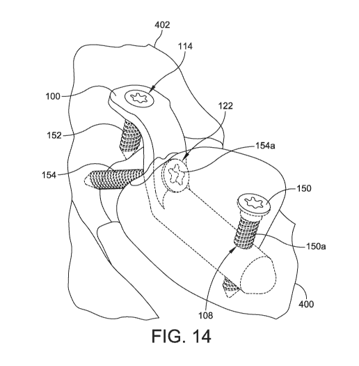
[0023] FIG. 14 shows another perspective view of the implant of FIG. 1
implanted in a
metatarsal bone.
[0024] FIG. 15 is a flow chart of a method of treatment using the implant
of FIG. 1 and
the target guide of FIG. 4.
DETAILED DESCRIPTION
[0025] This description of preferred embodiments is intended to be read
in connection
with the accompanying drawings, which are to be considered part of the entire
written
description of this invention. The drawing figures are not necessarily to
scale and certain
features of the invention may be shown exaggerated in scale or in somewhat
schematic form in
the interest of clarity and conciseness. In the description, relative terms
such as "horizontal,"
"vertical," "up," "down," "top," and "bottom" as well as derivatives thereof
(e.g., "horizontally,"
"downwardly," "upwardly," etc.) should be construed to refer to the
orientation as then described
or as shown in the drawing figure under discussion. These relative terms are
for convenience of
description and normally are not intended to require a particular orientation.
Terms including
"inwardly" versus "outwardly," "longitudinal" versus "lateral" and the like
are to be interpreted
relative to one another or relative to an axis of elongation, or an axis or
center of rotation, as
appropriate. Terms concerning attachments, coupling and the like, such as
"connected" and
"interconnected," refer to a relationship wherein structures are secured or
attached to one another
either directly or indirectly through intervening structures, as well as both
movable or rigid
attachments or relationships, unless expressly described otherwise.
[0026] This disclosure provides an implant and a target guide for
surgical fixation of the
first tarsal-metatarsal joint as well as methods for implantation and securing
of the implant. The
implant is suitable for correction of hallux valgus deformity of the first
metatarsal and can also
be used in the correction of analogous deformities in other joints. Although
the drawings show
application of the implant and target guide to treat a first metatarsal for
correction of hallux
4
CA 03094191 2020-09-16
WO 2019/231531 PCT/US2019/024227
valgus, the implant and target guide can be sized and configured to treat
other bones, and can
also be used in a variety of procedures.
[0027] In one embodiment, as shown in FIGS. 1-3, an implant 100 includes
an
intramedullary portion 102, an extramedullary portion 104, and an intermediate
portion 106. In
the embodiment shown, the intramedullary portion 102, the extramedullary
portion 104, and the
intermediate portion 106 are integrally formed from a monolithic component. In
another
embodiment, one or more of the intramedullary portion 102, the extramedullary
portion 104, and
the intermediate portion 106 are separate components that are joined using
fasteners, adhesive,
welding, or any other appropriate technique. The implant 100 is configured to
join a first bone
400 to a second bone 402, as shown in FIGS. 12-14 and as described further
herein. It should be
noted that the implant 100 can be used on either the left or right foot.
[0028] The intramedullary portion 102 is configured for insertion into a
first bone 400,
such as the first metatarsal. The intramedullary portion 102 has a
longitudinal axis 107, which
can be a central axis. In one embodiment, the intramedullary portion 102
includes a cylindrical
portion. The distal end of the intramedullary portion 102 can include a
chamfer 104a to assist
with insertion into the first bone. In alternative embodiments, the entire
intramedullary portion
102 is tapered (i.e., the intramedullary portion forms a portion of a cone).
Alternatively, the
distal end of the intramedullary portion 102 can taper to an edge (i.e.,
forming a triangular
prism). The taper may be bi-lateral (i.e., tapering from both the top and
bottom of the
intramedullary portion 102) or unilateral (i.e., tapering from only the top or
bottom of the
intramedullary portion 102). In other embodiments, the distal end of the
intramedullary portion
102 is pyramidally shaped.
[0029] Although the cross-sectional geometry of intramedullary portion
102 is shown as
being cylindrical, in other embodiments the cross-sectional geometry of
intramedullary portion
102 may be polygonal (e.g., triangular, rectangular, pentagonal, etc.) and/or
include one or more
protrusions or flat surfaces formed thereon to resist rotation of the implant
100 relative to the
first bone segment or fragment. In some embodiments, the intramedullary
portion 102 may be
completely or partially threaded. In some embodiments, the intramedullary
portion 102 may
include one or more fins or protrusions extending outwardly therefrom to
resist rotation of the
implant 100 relative to the bone segment, section, or fragment.
CA 03094191 2020-09-16
WO 2019/231531 PCT/US2019/024227
[0030] An intramedullary fastener hole 108 extends through the
intramedullary portion
102. In various embodiments, the intramedullary fastener hole 108 may be
cylindrical or slotted.
The intramedullary fastener hole 108 has an intramedullary fastener hole axis
110, shown in FIG.
3. In one embodiment, the intramedullary fastener hole axis 110 is
substantially orthogonal to the
longitudinal axis 107 and is oriented such that the intramedullary fastener
hole axis 110 extends
in a substantially superior-inferior orientation when the implant 100 is
implanted. In another
embodiment, the intramedullary fastener hole axis 110 forms an oblique angle
with the
longitudinal axis 107. For example, the intramedullary fastener hole axis 110
can be oriented in
a superior-proximal to inferior-distal orientation with respect to the
longitudinal axis 107. In one
embodiment, the intramedullary fastener hole 108 is threaded. By providing a
threaded
intramedullary fastener hole 108, movement of a fastener engaged with the
intramedullary
fastener hole 108 relative to the implant 100 is minimized. This can reduce or
eliminate fretting
of the fastener and/or implant. In another embodiment, the intramedullary
fastener hole 108 is
unthreaded. Providing a press or slip fit between the fastener and the
intramedullary fastener
hole 108 can also minimize relative movement. In at least one embodiment, the
intramedullary
portion 102 includes multiple fastener holes.
[0031] The extramedullary portion 104 is configured for contact with a
second bone 402
(FIGS. 12-14), such as a tarsal bone. The extramedullary portion 104 includes
a bone facing
surface 112, shown in FIGS. 2 and 3, offset from the longitudinal axis 107.
The bone facing
surface 112 is configured to abut a surface of the second bone when the
implant 100 is
implanted, as shown in FIGS. 12-14. In one embodiment, the bone facing surface
112 is offset
from the intramedullary portion 102 such that when the intramedullary portion
102 is inserted
into the first metatarsal the bone facing surface 112 sits atop the tarsal
bone. The bone facing
surface 112 is spaced from the longitudinal axis 107 a distance 113 that is
greater than one half
the width of the intramedullary portion 102 (i.e., greater than the radius of
the intramedullary
portion 102). The distance 113 can be configured to provide the desired offset
based on the
severity of the deformity. In some embodiments, a kit containing multiple
implants is provided,
the implants having a variety of offset distances 113. For example, the
implants can have an
offset distance 113 of 2mm, 4mm, 6mm, 8mm, or any other appropriate value. The
increment of
offset distances between implants can be any appropriate or desired increment.
The bone facing
surface 112 can be in contact with the tarsal bone or, alternatively, a gap
can be present between
6
CA 03094191 2020-09-16
WO 2019/231531 PCT/US2019/024227
the bone facing surface 112 and the tarsal bone. In one embodiment, the bone
facing surface 112
is substantially flat. In another embodiment, the bone facing surface 112 is
contoured to
conform to the tarsal bone. For example, the bone facing surface 112 can be at
least partially
concave.
[0032] An extramedullary fastener hole 114 extends through the
extramedullary portion
104. The extramedullary fastener hole 114 has an extramedullary fastener hole
axis 116. In one
embodiment, the extramedullary fastener hole axis 116 extends in a
substantially superior-
inferior orientation. In one embodiment, the intramedullary fastener hole axis
110 and the
extramedullary fastener hole axis 116 are substantially parallel. In at least
one embodiment, the
extramedullary portion 104 includes multiple fastener holes.
[0033] The intermediate portion 106 extends between the intramedullary
portion 102 and
the extramedullary portion 104. As shown in FIG. 2, the intermediate portion
106 is inclined
relative to the longitudinal axis 107 such that the superior surface 126 of
the intermediate portion
106 forms an angle 118 with the longitudinal axis 107. In one embodiment, the
angle 118 is
about 60 . In another embodiment, the angle 118 is between about 55 and about
65 . In
another embodiment, the angle 118 is between about 45 and about 75 . As
described above
with respect to the distance 113 between the longitudinal axis 107 and the
extramedullary portion
104, the angle 118 can be selected based on the severity of the deformity. As
the distance 113 is
increased to accommodate more severe deformities, the angle 118 may be
increased as well. A
kit having a plurality of implants can be provided having a variety of angles.
In one
embodiment, the implant 100 includes a fillet 120 at the intersection of the
extramedullary
portion 104 and the intermediate portion 106 to provide a smooth contour for
contact with the
tarsal bone.
[0034] A compression fastener hole 122 extends through the intermediate
portion 106.
The compression fastener hole 122 has a compression fastener hole axis 124. In
one
embodiment, the compression fastener hole axis 124 is orthogonal to the
superior surface 126 of
the intermediate portion 106. In another embodiment, the compression fastener
hole axis 124
forms a non-perpendicular angle with the superior surface 126. The compression
fastener hole
122 is configured to receive a compression screw therein such that the shaft
of the compression
screw extends into the tarsal bone. The compression fastener hole axis 124
forms an oblique
angle 132 with the longitudinal axis 107. For example, in one embodiment, the
angle 132 is
7
CA 03094191 2020-09-16
WO 2019/231531 PCT/US2019/024227
about 35 . In another embodiment, the angle 132 is between about 30 and about
40 . In
another embodiment, the angle 132 is between about 25 and about 45 . In one
embodiment, the
compression fastener hole axis 124 also forms an oblique angle with respect to
the
intramedullary fastener hole axis 110 and the extramedullary screw hole axis.
[0035] The compression fastener hole 122 includes a shoulder 128
extending into the
hole such that a counterbore is formed therein. In one embodiment, as will be
described further
below, as the compression screw is installed in the tarsal bone, the head of
the compression
screw contacts the shoulder 128, thereby pulling the metatarsal and tarsal
bones toward one
another.
[0036] As shown in FIG. 14, the intramedullary fastener hole 108 is
configured to
receive a fastener 150 that extends through a portion of the first metatarsal.
The fastener 150 can
be a screw, pin, nail, k-wire, rod, or any other appropriate fastener. As
shown, in one
embodiment, the fastener 150 is a screw and the shaft 150a of the screw
extends through the
intramedullary fastener hole 108. The fastener 150 fixes the implant 100 to
the metatarsal to
restrict movement of the implant 100 relative to the metatarsal. As mentioned
above, the
intramedullary fastener hole 108 can be threaded to engage a threaded shaft
150a of the fastener
150. Alternatively, the fastener 150 can be self-tapping such that it forms
threads in the
intramedullary fastener hole 108 as it is inserted.
[0037] Further, the extramedullary fastener hole 114 is configured to
receive a fastener
152 that extends into the tarsal bone. This secures the implant 100 to the
tarsal bone. In one
embodiment, the extramedullary fastener hole 114 allows for variable angle
alignment. In some
embodiments, polyaxial screws such as 3Di locking screws or non-locking screws
sold by
Wright Medical Technology, Inc. of Memphis, TN may be utilized. For example,
in one
embodiment, the fastener 152 is a locking screw and the shaft of the screw can
form an angle of
up to about 15 in any direction with respect to the extramedullary fastener
hole axis 116.
[0038] The compression fastener hole 122 is configured to receive a
compression
fastener 154, as shown in FIG. 14. The compression fastener 154 is configured
to pass through
the compression fastener hole 122 and engage the tarsal bone. In one
embodiment, the
compression fastener 154 is a screw. In one embodiment, the head 154a of the
compression
fastener 154 contacts the shoulder 128 as the compression fastener 154 is
inserted. As the
compression fastener 154 is tightened, it draws the metatarsal bone towards
the tarsal bone. In
8
CA 03094191 2020-09-16
WO 2019/231531 PCT/US2019/024227
other words, the space between the metatarsal and tarsal bones is reduced,
compressing the bones
together. The amount of compression can be controlled by the surgeon by
controlling the
amount that the compression fastener 154 is turned within the compression
fastener hole 122
(and, thereby, the metatarsal bone).
[0039] In some embodiments, the compression fastener 154 is an
interfragmentary
fastener. In such embodiments, the threaded portion of the compression
fastener 154 may
engage both the metatarsal and tarsal bones. In other embodiments, the
threaded portion of the
compression fastener 154 engages only the tarsal bone.
[0040] In one embodiment, as described in more detail below, the
compression fastener
154 is installed prior to the fastener 152 in extramedullary portion 104. As a
result, when the
fastener 152 is inserted into the tarsal bone, the relative positions of the
metatarsal and tarsal
bones are fixed.
[0041] The implant 100 can comprise a metal, such as titanium, stainless
steel, or CoCr.
In some embodiments, the implant 100 can comprise a metal substrate coated
with or having an
additional layer of hydroxyapatite (HA), titanium plasma spray (TPS) / vacuum
plasma spray
(VPS), roughened surface of resorbable blast media (RBM), a bioactive glass,
an antimicrobial
or antibiotic, or strontium. Alternatively, the implant 100 can comprise a
metal substrate with a
composite coating or composite layer including HA on plasma, beads, an
irregular sintered
coating or TPS on an RBM-prepared substrate. In other embodiments, the metal
substrate can
have a porous coating, such as spherical bead, asymmetrical powder or an
irregular particle
coating.
[0042] In some embodiments, the metal substrate of implant 100 comprises
a degradable
(resorbable) material, such as a magnesium alloy, which may contain lithium,
aluminum, rare
earth metals (e.g., neodymium or cerium), manganese, zinc or other metals. In
other
embodiments, the resorbable material can include, but is not limited to
polymer materials
including a polylactide, polyglycolide, polycaprolactone, polyvalerolactone,
polycarbonates,
polyhydroxy butyrates, poly ortho esters, polyurethanes, polyanhydrides, and
combinations and
copolymers thereof, for example.
[0043] In some embodiments, the implant 100 comprises a biologic
material. The
biologic material can be a combination of Medical grade f3-TCP granules and
rhPDGF-BB
solution, such as AUGMENT bone graft material sold by Wright Medical
Technology, Inc. of
9
CA 03094191 2020-09-16
WO 2019/231531 PCT/US2019/024227
Memphis, TN. The biologic material can be applied, sprayed, or inserted at the
wound site for
bone in-growth, or can be provided as a coating on the implants or any or all
portions of the
implant system. In some embodiments, the biologic material is a coating
containing
osteoinductive or osteoconductive biological components. In some embodiments,
the biologic
material can include bone morphogenetic factors, i.e., growth factors whose
activity are specific
to bone tissue including, but not limited to, demineralized bone matrix (DBM),
bone protein
(BP), bone morphogenetic protein (BMP), and mixtures and combinations thereof.
Additionally,
formulations for promoting the attachment of endogenous bone may comprise bone
marrow
aspirate, bone marrow concentrate, and mixtures and combinations thereof.
[0044] FIGS. 4-6 show a target guide 200 that includes a coupling portion
202 and an
arm 204. The target guide 200 can be integrally constructed from a monolithic
component.
Alternatively, the target guide 200 can be constructed of two or more separate
components that
are joined using fasteners, adhesive, or any other appropriate means. The
target guide 200 is
suitable for guiding drills to form fastener holes in a bone and for insertion
of the intramedullary
portion 102 into the first bone 400. The target guide 200 can also be used to
rotate the implant
100 and metatarsal to achieve the desired alignment of the metatarsal and
tarsal bone, as will be
described in more detail herein. The use of a target guide to both guide a
drill as well as provide
the desired rotation simplifies the correction of the deformity and eliminates
the need for
additional fixtures or tools.
[0045] The coupling portion 202 is configured to couple to the
extramedullary portion
104. In one embodiment, the coupling portion 202 includes an insert 206 having
a threaded end
206a for coupling to the extramedullary fastener hole 114. The coupling
portion 202 also
includes a flange 207 extending therefrom and configured to contact the
lateral and medial sides
of the extramedullary portion 104 to align the implant 100 to the target guide
200. The implant
100 can also be aligned to the target guide 200 through any other appropriate
means, such as
through the use of one or more pins. The target guide 200 is coupled to the
implant 100 prior to
implantation and is used to guide insertion of the implant 100 into the
metatarsal.
[0046] In one embodiment, the insert 206 includes a first flange 214 and
a second flange
216. Further, the coupling portion 202 includes a pin hole 218. After
inserting the insert 206 in
the coupling portion 202, a pin 220 is inserted in the pin hole 218 to retain
the insert 206 within
the coupling portion 202, as shown in FIG. 6. The distance between the first
flange 214 and the
CA 03094191 2020-09-16
WO 2019/231531 PCT/US2019/024227
second flange 216 allows the threaded end 206a to be engaged and disengaged
from the
extramedullary fastener hole 114. In addition, the insert 206 is freely
rotatable within the
coupling portion 202 to allow for rotational engagement with the
extramedullary fastener hole
114. The pin 220 can be press-fit within the pin hole 218.
[0047] The target guide described above is only exemplary and is not
limiting. For
example, in a variation of the target guide (not shown), the insert 206 is not
pre-assembled within
the coupling portion 202, and the pin 220 is omitted. The surgeon or
technician can assemble the
insert 206 (or a drill guide having the same outer diameter as the insert 206)
inside the coupling
portion 202 before use. With a removable insert 206 or drill guide, the
surgeon can remove the
insert 206 or drill guide and implant the fastener 152 (FIGS. 13 and 14)
through the coupling
portion 202 of the target guide 200, without first removing the target guide
200. This provides
greater flexibility in surgical technique and procedures.
[0048] FIGS. 7-9 show the target guide 200 coupled to the implant 100.
The arm 204
includes an intramedullary guide aperture 208 and a compression guide aperture
210. As shown
in FIG. 9, when the target guide 200 is connected to the implant 100, the
intramedullary guide
aperture 208 is configured to be aligned with the intramedullary fastener hole
108 of the implant
100. The compression guide aperture 210 is configured to align with the
compression fastener
hole 122 of the implant 100. As will be described further below, the
intramedullary 208 and
compression 210 guide apertures can guide a drill bit as it forms holes
through the metatarsal or
tarsal bones.
[0049] FIGS. 10 and 11 show an example of a drill guide 300 suitable for
use with the
target guide 200. FIG. 10 is a plan view of the drill guide 300, and FIG. 11
is a cross-section of
the drill guide 300, taken along section line 11-11 of FIG. 10.
[0050] In FIGS. 10 and 11, the drill guide 300 has an outer surface 310
with an outer
diameter 302 sized to be slidably received in the intramedullary guide
aperture 208 and/or the
compression guide aperture 210 of target guide 200. The drill guide 300 has a
first portion with
a bore 312 having a first inner diameter 318. The drill guide 300 has a second
portion with a
bore 314 having a second inner diameter 320 less than the first inner diameter
318. The second
inner diameter 320 is sized to slidably receive and align a drill that
penetrates the drill guide 300
and the first 400 and/or second 402 bones. The first inner diameter 318 of the
bore 312 of drill
guide 300 is sized larger than the second inner diameter 320, to avoid
friction between the drill
11
CA 03094191 2020-09-16
WO 2019/231531 PCT/US2019/024227
and the sidewall of bore 312. The drill guide 300 has a taper section 316
between (and
connecting) the bore 312 and the bore 314, for guiding the drill 350 into the
bore 314. The drill
guide 300 may also have a knob 322 with a larger diameter than the outer
surface 310. The knob
322 acts as a stop to prevent the drill guide 300 from falling through the arm
204. The knob 322
can have a gripping surface, such as ridges, grooves, splines, or a knurled,
patterned or textured
surface.
[0051] In some embodiments, the surgeon inserts a longitudinal k-wire
(not shown) in
the metatarsal 400 and uses a cannulated reamer (not shown) to form a
longitudinal
intramedullary opening in the metatarsal 400 concentric with the longitudinal
k-wire. The
surgeon removes the longitudinal k-wire from the longitudinal intramedullary
opening and
inserts the intramedullary portion 102 of the implant 100 into the
longitudinal intramedullary
opening.
[0052] In some embodiments, the surgeon applies a force to the target
guide 200 or a k-
wire or drill inserted in the target guide 200, resulting in application of a
moment to rotate the
implant 100 and the metatarsal about the longitudinal axis 107 of the
intramedullary portion 102
of implant 100. Although the surgeon can apply the force directly to the
target guide 200, in
some instances the surgeon may wish to grasp a drill or k-wire and use the
drill or k-wire as a joy
stick during the rotation. The surgeon applies the force to rotate the implant
100 until the
metatarsal rotates through a desired angle. After rotation, the extramedullary
portion 104 of the
implant 100 and the tarsal bone 402 are properly aligned with respect to the
metatarsal 400.
[0053] In another embodiment, shown in FIG. 15, a method 1200 of fixing a
first bone to
a second bone is provided. At step 1202, the surgeon creates an incision that
provides access to
the tarsal-metatarsal joint. The incision can be formed with any appropriate
surgical tool, such
as a scalpel. Next, at step 1203, the joint is prepared by removing cartilage,
tissue, and/or other
material in order to provide access to the first and second bones. Various
tools can be used to
prepare the joint.
[0054] At step 1204, the surgeon forms the longitudinal hole in the
metatarsal bone (for
receiving the intramedullary portion of the implant). Optionally, prior to
forming the
longitudinal hole, a k-wire can be inserted into the metatarsal to define the
orientation of the
longitudinal hole. A cannulated reamer, guided by the k-wire, can be used to
form the
longitudinal hole. After forming the longitudinal hole, the k-wire is removed.
12
CA 03094191 2020-09-16
WO 2019/231531 PCT/US2019/024227
[0055] At step 1206, the surgeon attaches the target guide to the
extramedullary fastener
hole in the extramedullary portion of the implant (by engaging the threaded
end of the insert of
the target guide with the threads of the extramedullary fastener hole).
Alternatively, the surgeon
can obtain a pre-packaged or previously assembled construct comprising an
implant attached to
the threaded end of the insert of a target guide.
[0056] At step 1208, the surgeon inserts the intramedullary portion of
the implant into the
longitudinal intramedullary opening in the proximal section of the first
metatarsal. During the
insertion, the surgeon may grip the target guide to push the implant into the
opening. When the
insertion is completed, the extramedullary portion of the implant has a bone
facing surface facing
radially inward toward the first longitudinal axis.
[0057] At step 1210, the surgeon forms a first drill hole in the first
metatarsal. In some
embodiments, the surgeon inserts a k-wire through the body of the target guide
prior to forming
the first drill hole in order to guide the drill as it forms the first
fastener hole.
[0058] At step 1212, a first fastener is inserted into the first drill
hole and through the
intramedullary portion of the implant to secure the implant to the metatarsal.
In one
embodiment, the first fastener is a screw.
[0059] At step 1214, after inserting the first fastener, the surgeon
applies a force to the
target guide to rotate the implant and the first metatarsal about the first
longitudinal axis in situ to
correct a hallux valgus deformity. Because the implant is fixed to the
metatarsal by the first
fastener, rotation of the implant results in a corresponding rotation of the
metatarsal. Hence, no
additional tools or fixtures are required to impart the desired rotation.
[0060] In other embodiments, the implant and metatarsal are rotated prior
to insertion of
the first fastener. For example, after forming the first drill hole, the
surgeon may maintain the
drill in the first drill hole and use the drill like a joy stick to manipulate
and rotate the implant
and the first metatarsal to achieve the desired rotation angle.
[0061] At step 1216, the surgeon forms a compression drill hole through
the metatarsal
and tarsal bone using the target guide and a drill guide. Optionally, prior to
forming the
compression drill hole, a k-wire is inserted to guide the orientation of the
compression drill hole.
In such an embodiment, the drill may be cannulated to allow the drill to pass
over the k-wire.
After forming the compression drill hole, the k-wire can be removed.
13
CA 03094191 2020-09-16
WO 2019/231531 PCT/US2019/024227
[0062] At step 1218, after forming the compression drill hole, the
surgeon inserts a
fastener through the compression fastener hole in the implant and into the
compression drill hole
in the tarsal bone. In some embodiments, the compression screw has a cannula,
and the inserting
step comprises inserting the compression screw in the compression fastener
hole with the k-wire
extending through the cannula of the compression screw.
[0063] At step 1220, the surgeon forms a drill hole in the tarsal bone.
The drill hole can
be formed using a drill guided by the insert of the target guide.
Alternatively, a dedicated drill
guide can be used to guide the drill. In some embodiments, a k-wire is
inserted through the
extramedullary fastener hole and into the tarsal bone prior to forming the
drill hole in order to
guide the drill.
[0064] At step 1222, a fastener is inserted through the extramedullary
fastener hole and
into the drill hole. In some embodiments, the target guide is removed from the
implant prior to
insertion of the second fastener.
[0065] Optionally, the method may further include making a second
incision to provide
clearance for insertion of the compression screw and making a third incision
to provide clearance
for insertion of the first fastener.
[0066] Although the devices, kits, systems, and methods have been
described in terms of
exemplary embodiments, they are not limited thereto. Rather, the appended
claims should be
construed broadly, to include other variants and embodiments of the devices,
kits, systems, and
methods, which may be made by those skilled in the art without departing from
the scope and
range of equivalents of the claimed devices, kits, systems, and methods.
14