Note: Descriptions are shown in the official language in which they were submitted.
CA 03094235 2020-09-16
WO 2019/197600
PCT/EP2019/059391
-1-
Her2-targeting Antigen Binding Molecules comprising 4-1BBL
FIELD OF THE INVENTION
The invention relates to Her2 targeting 4-1BB agonists, in particular 4-1BBL
trimer-
containing antigen binding molecules comprising an antigen binding domain
capable of specific
binding to Her2 and their use in the treatment of cancer. The invention
further relates to
methods of producing these molecules and to methods of using the same.
BACKGROUND
4-1BB (CD137), a member of the TNF receptor superfamily, was first identified
as an
inducible molecule expressed by activated by T cells (Kwon and Weissman, 1989,
Proc Natl
Acad Sci USA 86, 1963-1967). Subsequent studies demonstrated that many other
immune cells
also express 4-1BB, including NK cells, B cells, NKT cells, monocytes,
neutrophils, mast cells,
dendritic cells (DCs) and cells of non-hematopoietic origin such as
endothelial and smooth
muscle cells (Vinay and Kwon, 2011, Cell Mol Immunol 8, 281-284). Expression
of 4-1BB in
different cell types is mostly inducible and driven by various stimulatory
signals, such as T-cell
receptor (TCR) or B-cell receptor triggering, as well as signaling induced
through co-stimulatory
molecules or receptors of pro-inflammatory cytokines (Diehl et al., 2002, J
Immunol 168, 3755-
3762; Zhang et al., 2010, Clin Cancer Res 13, 2758-2767).
4-1BB ligand (4-1BBL or CD137L) was identified in 1993 (Goodwin et al., 1993,
Eur J
Immunol 23, 2631-2641). It has been shown that expression of 4-1BBL was
restricted on
professional antigen presenting cells (APC) such as B-cells, DCs and
macrophages. Inducible
expression of 4-1BBL is characteristic for T-cells, including both c43 and y6
T-cell subsets, and
endothelial cells (Shao and Schwarz, 2011, J Leukoc Biol 89, 21-29).
Co-stimulation through the 4-1BB receptor (for example by 4-1BBL ligation)
activates
multiple signaling cascades within the T cell (both CD4+ and CD8+ subsets),
powerfully
augmenting T cell activation (Bartkowiak and Curran, 2015). In combination
with TCR
triggering, agonistic 4-1BB-specific antibodies enhance proliferation of T-
cells, stimulate
lymphokine secretion and decrease sensitivity of T-lymphocytes to activation-
induced cells
death (Snell et al., 2011, Immunol Rev 244, 197-217). This mechanism was
further advanced as
the first proof of concept in cancer immunotherapy. In a preclinical model
administration of an
DK / 28.03.2019
CA 03094235 2020-09-16
WO 2019/197600
PCT/EP2019/059391
-2-
agonistic antibody against 4-1BB in tumor bearing mice led to potent anti-
tumor effect (Melero
et al., 1997, Nat Med 3, 682-685). Later, accumulating evidence indicated that
4-1BB usually
exhibits its potency as an anti-tumor agent only when administered in
combination with other
immunomodulatory compounds, chemotherapeutic reagents, tumor-specific
vaccination or
radiotherapy (Bartkowiak and Curran, 2015, Front Oncol 5, 117).
Signaling of the TNFR-superfamily needs cross-linking of the trimerized
ligands to engage
with the receptors, so does the 4-1BB agonistic antibodies which require wild
type Fc-binding
(Li and Ravetch, 2011, Science 333, 1030-1034). However, systemic
administration of 4-1BB-
specific agonistic antibodies with the functionally active Fc domain resulted
in influx of CD8+ T-
cells associated with liver toxicity (Dubrot et al., 2010, Cancer Immunol
Immunother 59, 1223-
1233) that is diminished or significantly ameliorated in the absence of
functional Fc-receptors in
mice. In the clinic, an Fc-competent 4-1BB agonistic Ab (BMS-663513)
(NCT00612664) caused
a grade 4 hepatitis leading to termination of the trial (Simeone and Ascierto,
2012, J
Immunotoxicol 9, 241-247). Therefore, there is a need for effective and safer
4-1BB agonists.
The human epidermal growth factor receptor-2 (Her2; ErbB2) is a receptor
tyrosine kinase
and a member of the epidermal growth factor receptor (EGFR) family of
transmembrane
receptors. Her2 is overexpressed in a range of tumor types and it has been
implicated in disease
initiation and progression. It is associated with poor prognosis. For example,
overexpression of
Her2 is observed in approximately 30% of human breast cancers and it is
implicated in the
aggressive growth and poor clinical outcomes associated with these tumors
(Slamon et al (1987)
Science 235:177-182).
The humanized anti-Her2 monoclonal antibody trastuzumab (CAS 180288-69-1,
HERCEPTINO, huMAb4D5-8, rhuMAb Her2, Genentech) targets the extracellular
domain of
HER-2 (US 5677171; US 5821337; US 6054297; US 6165464; US 6339142; US 6407213;
US
6639055; US 6719971; US 6800738; US 7074404; Coussens et al (1985) Science
230:1 132-9;
Slamon et al (1989) Science 244:707-12; Slamon et al (2001) New Engl. J . Med.
344:783-792).
Trastuzumab has been shown to inhibit the proliferation of human tumor cells
that overexpress
HER-2 and is a mediator of antibody-dependent cellular cytotoxicity, ADCC
(Hudziak et al
(1989) Mol Cell Biol 9:1 165-72; Lewis et al (1993) Cancer Immunol Immunother;
37:255-63;
Baselga et al (1998) Cancer Res. 58:2825-2831; Hotaling et al (1996)
[abstract]. Proc. Annual
Meeting Am Assoc Cancer Res; 37:471; Pegram MD, et al (1997) [abstract]. Proc
Am Assoc
Cancer Res; 38:602; Sliwkowski et al (1999) Seminars in Oncology 26(4), Suppl
12:60- 70;
Yarden Y. and Sliwkowski, M. (2001) Nature Reviews: Molecular Cell Biology,
Macmillan
Magazines, Ltd., Vol. 2:127-137).
CA 03094235 2020-09-16
WO 2019/197600
PCT/EP2019/059391
-3-
HERCEPTINO (trastuzumab, Genentech Inc.) was approved in 1998 for the
treatment of
of patients with Her2-overexpressing metastatic breast cancers (Baselga et al,
(1996) J. Clin.
Oncol. 14:737-744). In 2006, the FDA approved HERCEPTINO as part of a
treatment regimen
containing doxorubicin, cyclophosphamide and paclitaxel for the adjuvant
treatment of patients
with Her2-positive, node-positive breast cancer.
Pertuzumab (also known as recombinant humanized monoclonal antibody 2C4,
rhuMAb
2C4, PERJETAO, Genentech, Inc, South San Francisco) is another antibody
treatment targeting
Her2. Pertuzumab is a Her dimerization inhibitor (HDI) and functions to
inhibit the ability of
Her2 to form active heterodimers or homodimers with other Her receptors (such
as EGFR/Her 1,
Her2, Her3 and Her4). See, for example, Harari and Yarden Oncogene 19:6102-14
(2000);
Yarden and Sliwkowski. Nat Rev Mol Cell Biol 2:127-37 (2001); Sliwkowski, Nat
Struct Biol
10:158-9 (2003); Cho et al. Nature 421:756-60 (2003); and Malik et al., Pro Am
Soc Cancer Res
44:176-7 (2003); US 7560111. PERJETAO was first approved in 2012 in
combination with
trastuzumab and docetaxel for the treatment of patients with advanced or late-
stage (metastatic)
Her2-positive breast cancer. The combination therapy using trastuzumab and
pertuzumab is
meanwhile also approved for the neoadjuvant (before surgery) treatment of f
Her2-positive,
locally advanced, inflammatory, or early stage breast cancer and for adjuvant
(after surgery)
treatment of Her2-positive early breast cancer (EBC) at high risk of
recurrence. The mechanisms
of action of Perj eta and Herceptin are believed to complement each other, as
both bind to the
Her2 receptor, but to different places. The combination of Perj eta and
Herceptin is thought to
provide a more comprehensive, dual blockade of HER signaling pathways, thus
preventing
tumor cell growth and survival.
Bispecific, bivalent Her2 antibodies that are directed against domains II, III
and IV of
human ErbB2 are disclosed in WO 2012/143523. Bispecific HER-2 antibodies
comprising
optimized variants of the antibodies rhuMab 2C4 and hu4D5, called Herceptarg,
have been
described in WO 2015/091738.
Although the therapeutic efficacy of trastuzumab in breast carcinoma is well
demonstrated,
there are many patients who do not benefit from trastuzumab because of
resistance. Given the
lack of an effective anti-Her2 therapy in specific cancers expressing low
levels of Her2, the
resistance to the current therapies, and the prevalence of Her2 related
cancers, new therapies are
required to treat such cancers.
The new antigen binding molecules of the present invention combine an anti-
Her2 antigen
binding domain with a moiety that is capable of forming a costimulatory 4-1BB
ligand timer
and that is sufficiently stable to be pharmaceutically useful. Fusion proteins
composed of a
binding specificity for CD137 and a binding specificity for Her2/neu are
disclosed in WO
CA 03094235 2020-09-16
WO 2019/197600
PCT/EP2019/059391
-4-
2016/177802. These molecules are antibody-lipocalin mutein fusion
polypeptides, meaning that
a lipocalin mutein with binding specificity for CD137 is fused to an anti-Her2
antibody.
Lipocalin muteins (anticalins) are non-antibody scaffolds and the conversion
of such modalities
into differentiated drugs has been challenging (Vazquez-Lombardi et al. 2015,
Drug Discovery
Today 20, 1271-1283). Compared to antibodies challenges could arise in view of
different serum
half-life, tissue penetration and immunogenicity. Thus, there is still a need
for drug candidates
with improved properties that are based on antibody technology or human-like
proteins.
SUMMARY OF THE INVENTION
The new antigen binding molecules of the present invention combine an anti-
Her2 antigen
binding domain with a moiety that is capable of forming a costimulatory 4-1BBL
trimer and that
is sufficiently stable to be pharmaceutically useful. Surprisingly, antigen
binding molecules of
the invention provide a trimeric and thus biologically active human 4-1BB
ligand, although one
of the trimerizing 4-1BBL ectodomains is located on another polypeptide than
the other two 4-
1BBL ectodomains of the molecule. Targeted by the anti-Her2 antigen binding
domain the
antigen binding molecules of the present invention have an increased activity
on the tumor site,
comprise the natural human 4-1BB ligand and should thus impose less safety
issues compared to
conventional 4-1BB agonistic antibodies or more artificial fusion proteins.
In one aspect, the invention provides a 4-1BBL trimer-containing antigen
binding molecule
comprising
(a) an antigen binding domain capable of specific binding to Her2,
(b) a first and a second polypeptide that are linked to each other by a
disulfide bond,
wherein the antigen binding molecule is characterized in that the first
polypeptide comprises two
ectodomains of 4-1BBL or a fragment thereof that are connected to each other
by a peptide
linker and in that the second polypeptide comprises one ectodomain of 4-1BBL
or a fragment
thereof, and
(c) an Fc domain composed of a first and a second subunit capable of stable
association.
In a particular aspect, the invention provides a 4-1BBL trimer-containing
antigen binding
molecule, wherein the ectodomain of 4-1BBL or a fragment thereof comprises the
amino acid
sequence selected from the group consisting of SEQ ID NO:1, SEQ ID NO: 2, SEQ
ID NO:3,
SEQ ID NO:4, SEQ ID NO:5, SEQ ID NO: 6, SEQ ID NO:7 and SEQ ID NO:8,
particularly the
amino acid sequence of SEQ ID NO:1 or SEQ ID NO:5.
In a further aspect, the invention provides a 4-1BBL trimer-containing antigen
binding
molecule, comprising
(a) an antigen binding domain capable of specific binding to Her2,
(b) a first and a second polypeptide that are linked to each other by a
disulfide bond,
CA 03094235 2020-09-16
WO 2019/197600
PCT/EP2019/059391
-5-
wherein the antigen binding molecule is characterized in that the first
polypeptide comprises the
amino acid sequence selected from the group consisting of SEQ ID NO:9, SEQ ID
NO:10, SEQ
ID NO:11 and SEQ ID NO:12 and in that the second polypeptide comprises the
amino acid
sequence selected from the group consisting of SEQ ID NO:1, SEQ ID NO:5, SEQ
ID NO:3 and
SEQ ID NO:4, and
(c) an Fc domain composed of a first and a second subunit capable of stable
association.
In one aspect, the Fc domain is an IgG, particularly an IgG1 Fc domain or an
IgG4 Fc
domain. More particularly, the Fc domain is an IgG1 Fc domain. In a particular
aspect, the Fc
domain comprises a modification promoting the association of the first and
second subunit of the
.. Fc domain. In a particular aspect, the invention provides a 4-1BBL trimer-
containing antigen
binding molecule, wherein the Fc domain comprises knob-into-hole modifications
promoting
association of the first and the second subunit of the Fc domain. In a
specific aspect, the
invention provides a 4-1BBL trimer-containing antigen binding molecule,
wherein the first
subunit of the Fc domain comprises the amino acid substitutions 5354C and
T366W (numbering
according to Kabat EU index) and the second subunit of the Fc domain comprises
the amino acid
substitutions Y349C, T3665, L368A and Y407V (numbering according to Kabat EU
index).
In another aspect, the invention is concerned with a 4-1BBL trimer-containing
antigen
binding molecule as defined herein before, comprising (c) an Fc domain
composed of a first and
a second subunit capable of stable association, wherein the Fc domain
comprises one or more
amino acid substitution that reduces binding to an Fc receptor, in particular
towards Fcy receptor.
In particular, the Fc domain comprises amino acid substitutions at positions
234 and 235 (EU
numbering according to Kabat) and/or 329 (EU numbering according to Kabat) of
the IgG heavy
chains. Particularly, provided is a 4-1BBL trimer-containing antigen binding
molecule, wherein
the Fc domain is an IgG1 Fc domain comprising the amino acid substitutions the
amino acid
substitutions L234A, L235A and P329G (numbering according to Kabat EU index).
In one aspect, the 4-1BBL trimer-containing antigen binding molecule is one,
wherein
wherein the antigen binding domain capable of specific binding to Her2 is a
Fab molecule
capable of specific binding to Her2. In another aspect, the antigen binding
domain capable of
specific binding to Her2 is a cross-over Fab molecule or a scFV molecule
capable of specific
.. binding to Her2.
In one aspect, the invention provides a 4-1BBL trimer-containing antigen
binding molecule
as described herein before, wherein the 4-1BBL trimer-containing antigen
binding molecule
comprises one Fab domain capable of specific binding to Her2, meaning that it
comprises
monovalent binding towards Her2.
CA 03094235 2020-09-16
WO 2019/197600
PCT/EP2019/059391
-6-
In a further aspect, provided is a 4-1BBL trimer-containing antigen binding
molecule,
wherein wherein the antigen binding domain capable of specific binding to Her2
comprises
(a) a VH domain comprising (i) CDR-H1 comprising the amino acid sequence of
SEQ ID
NO:13, (ii) CDR-H2 comprising the amino acid sequence of SEQ ID NO:14, and
(iii) CDR-H3
comprising the amino acid sequence of SEQ ID NO:15, and a VL domain comprising
(iv) CDR-
Li comprising the amino acid sequence of SEQ ID NO:16, (v) CDR-L2 comprising
the amino
acid sequence of SEQ ID NO:17, and (vi) CDR-L3 comprising the amino acid
sequence of SEQ
ID NO:18, or
(b) a VH domain comprising (i) CDR-H1 comprising the amino acid sequence of
SEQ ID
NO :21, (ii) CDR-H2 comprising the amino acid sequence of SEQ ID NO :22, and
(iii) CDR-H3
comprising the amino acid sequence of SEQ ID NO:23, and a VL domain comprising
(iv) CDR-
Li comprising the amino acid sequence of SEQ ID NO:24, (v) CDR-L2 comprising
the amino
acid sequence of SEQ ID NO:25, and (vi) CDR-L3 comprising the amino acid
sequence of SEQ
ID NO:26, or
(c) a VH domain comprising (i) CDR-H1 comprising the amino acid sequence of
SEQ ID
NO:29, (ii) CDR-H2 comprising the amino acid sequence of SEQ ID NO:30, and
(iii) CDR-H3
comprising the amino acid sequence of SEQ ID NO:31, and a VL domain comprising
(iv) CDR-
Li comprising the amino acid sequence of SEQ ID NO:32, (v) CDR-L2 comprising
the amino
acid sequence of SEQ ID NO:33, and (vi) CDR-L3 comprising the amino acid
sequence of SEQ
.. ID NO:34.
In a further aspect, the 4-1BBL trimer-containing antigen binding molecule of
the
invention comprises
(a) a VH domain comprising an amino acid sequence of SEQ ID NO:19 and a VL
domain
comprising an amino acid sequence of SEQ ID NO:20, or
(b) a VH domain comprising an amino acid sequence of SEQ ID NO:27 and a VL
domain
comprising an amino acid sequence of SEQ ID NO:28, or
(c) a VH domain comprising an amino acid sequence of SEQ ID NO:35 and a VL
domain
comprising an amino acid sequence of SEQ ID NO:36.
In a further aspect, provided is a 4-1BBL trimer-containing antigen binding
molecule,
wherein the antigen binding molecule comprises
a first heavy chain and a first light chain, both comprising a Fab molecule
capable of specific
binding to Her2,
a second heavy chain comprising the constant domains and two ectodomains of a
4-i BBL or a
fragment thereof connected to each other by a first peptide linker fused at
its C-terminus by a
second peptide linker to a second heavy or light chain,
and a second light chain comprising a constant domain and one ectodomain of 4-
1BBL or a
fragment thereof fused at its C-terminus by a third peptide linker to a second
light or heavy
CA 03094235 2020-09-16
WO 2019/197600
PCT/EP2019/059391
-7-
chain, respectively. More particularly, provided is a 4-1BBL trimer-containing
antigen binding
molecule, wherein the first peptide comprising two ectodomains of 4-1BBL or a
fragment
thereof connected to each other by a first peptide linker is fused at its C-
terminus by a second
peptide linker to a CL domain that is part of a heavy chain, and the second
peptide comprising
one ectodomain of said 4-1BBL or a fragment thereof is fused at its C-terminus
by a third
peptide linker to a CH1 domain that is part of a light chain.
In a particular aspect, the invention relates to a 4-1BBL trimer-containing
antigen binding
molecule as defined above, wherein the peptide linker is (G4S)2, i.e. a
peptide linker of SEQ ID
NO:68. In one aspect, the peptide linker in all instances is (G45)2.
Provided is further a 4-1BBL trimer-containing antigen binding molecule,
wherein in the
CL domain adjacent to the TNF ligand family member the amino acid at position
123 (EU
numbering) has been replaced by arginine (R) and the amino acid at position
124 (EU
numbering) has been substituted by lysine (K), and wherein in the CH1 domain
adjacent to the
TNF ligand family member the amino acids at position 147 (EU numbering) and at
position 213
(EU numbering) have been substituted by glutamic acid (E).
In another aspect, provided is a 4-1BBL trimer-containing antigen binding
molecule,
wherein the antigen binding molecule comprises
(i) a first heavy chain comprising the VH domain comprising the amino acid
sequence of SEQ
ID NO:19 and a first light chain comprising the VL domain comprising the amino
acid sequence
of SEQ ID NO:20 or
a first heavy chain comprising the VH domain comprising the amino acid
sequence of SEQ ID
NO:27 and a first light chain comprising the VL domain comprising the amino
acid sequence of
SEQ ID NO:28, or
a first heavy chain comprising the VH domain comprising the amino acid
sequence of SEQ ID
NO:35 and a first light chain comprising the VL domain comprising the amino
acid sequence of
SEQ ID NO:36,
(ii) a second heavy chain comprising the amino acid sequence selected from the
group consisting
of SEQ ID NO:37, SEQ ID NO:39, SEQ ID NO:41 and SEQ ID NO:43, and
(iii) a second light chain comprising the amino acid sequence selected from
the group consisting
of SEQ ID NO:38, SEQ ID NO:40, SEQ ID NO:42 and SEQ ID NO:44.
In a particular aspect, provided is a 4-1BBL trimer-containing antigen binding
molecule
comprising
(a) a first heavy chain comprising the amino acid sequence of SEQ ID NO:45, a
first light chain
comprising the amino acid sequence of SEQ ID NO:46, a second heavy chain
comprising the
amino acid sequence of SEQ ID NO:37 and a second light chain comprising the
amino acid
CA 03094235 2020-09-16
WO 2019/197600
PCT/EP2019/059391
-8-
sequence of SEQ ID NO:38, or
(b) a first heavy chain comprising the amino acid sequence of SEQ ID NO:47, a
first light chain
comprising the amino acid sequence of SEQ ID NO:48, a second heavy chain
comprising the
amino acid sequence of SEQ ID NO:37 and a second light chain comprising the
amino acid
sequence of SEQ ID NO:38, or
(c) a first heavy chain comprising the amino acid sequence of SEQ ID NO:49, a
first light chain
comprising the amino acid sequence of SEQ ID NO:50, a second heavy chain
comprising the
amino acid sequence of SEQ ID NO:37 and a second light chain comprising the
amino acid
sequence of SEQ ID NO:38.
According to another aspect of the invention, there is provided an isolated
nucleic acid
molecule encoding a 4-1BBL trimer-containing antigen binding molecule as
defined herein
before. The invention further provides a vector, particularly an expression
vector, comprising the
isolated nucleic acid molecule of the invention and a host cell comprising the
isolated nucleic
acid or the vector of the invention. In some embodiments the host cell is an
eukaryotic cell,
particularly a mammalian cell.
In another aspect, provided is a method for producing the 4-1BBL trimer-
containing
antigen binding molecule of the invention, comprising culturing the host cell
of the invention
under conditions suitable for expression of the 4-1BBL trimer-containing
antigen binding
molecule, and isolating the 4-1BBL trimer-containing antigen binding molecule.
The invention
also encompasses a 4-1BBL trimer-containing antigen binding molecule produced
by the method
of the invention.
The invention further provides a pharmaceutical composition comprising the 4-
1BBL
trimer-containing antigen binding molecule of the invention and at least one
pharmaceutically
acceptable excipient. In another aspect, a pharmaceutical composition is
provided comprising the
4-1BBL trimer-containing antigen binding molecule of the invention and at
least one
pharmaceutically acceptable excipient, further comprising an additional
therapeutic agent, e.g. a
chemotherapeutic agent and/ or other agents for use in cancer immunotherapy.
In a further
aspect, provided is a pharmaceutical composition further comprising a T-cell
activating anti-CD3
bispecific antibody, in particular an anti-Her2/anti-CD3 bispecific antibody.
Also encompassed by the invention is the 4-1 BBL trimer-containing antigen
binding
molecule of the invention, or the pharmaceutical composition of the invention,
for use as a
medicament. In one aspect is provided the 4-1 BBL trimer-containing antigen
binding molecule
of the invention, or the pharmaceutical composition of the invention, for use
in the treatment of a
disease in an individual in need thereof In a specific embodiment, provided is
the 4-1 BBL
trimer-containing antigen binding molecule of the invention, or the
pharmaceutical composition
CA 03094235 2020-09-16
WO 2019/197600
PCT/EP2019/059391
-9-
of the invention, for use in the treatment of cancer. In another aspect,
provided is the 4-1BBL
trimer-containing antigen binding molecule of the invention, or the
pharmaceutical composition
of the invention, for use in up-regulating or prolonging cytotoxic T cell
activity. In another
aspect, provided is the 4-1BBL trimer-containing antigen binding molecule of
the invention, or
the pharmaceutical composition of the invention, for use in the treatment of
cancer, wherein the
the 4-1BBL trimer-containing antigen binding molecule is used in combination
with another
therapeutic agent, in particular a T-cell activating anti-CD3 bispecific
antibody. In one aspect,
the T-cell activating anti-CD3 bispecific antibody is administered
concurrently with, prior to, or
subsequently to the 4-1BBL trimer-containing antigen binding molecule.
Also provided is the use of the 4-1BBL trimer-containing antigen binding
molecule of the
invention for the manufacture of a medicament for the treatment of a disease
in an individual in
need thereof, in particular for the manufacture of a medicament for the
treatment of cancer, as
well as a method of treating a disease in an individual, comprising
administering to said
individual a therapeutically effective amount of a composition comprising the
4-1BBL trimer-
.. containing antigen binding molecule as disclosed herein in a
pharmaceutically acceptable form.
In a specific aspect, the disease is cancer. Further provided is the use of
the 4-1BBL trimer-
containing antigen binding molecule of the invention for the manufacture of a
medicament for
the treatment of cancer, wherein the 4-1BBL trimer-containing antigen binding
molecule is used
in combination with a T-cell activating anti-CD3 bispecific antibody, in
particular an anti-
Her2/anti-CD3 antibody. Furthermore, provided is a method for treating an
individual having
cancer comprising administering to the subject an effective amount of the 4-
1BBL trimer-
containing antigen binding molecule of the invention, or a pharmaceutical
composition thereof,
and an effective amount of a T-cell activating anti-CD3 bispecific antibody,
in particular an anti-
Her2/anti-CD3 antibody. Also provided is a method of up-regulating or
prolonging cytotoxic T
cell activity in an individual having cancer, comprising administering to the
individual an
effective amount of the 4-1BBL trimer-containing antigen binding molecule of
the invention, or
the pharmaceutical composition of the invention. In any of the above
embodiments the
individual is preferably a mammal, particularly a human.
BRIEF DESCRIPTION OF THE DRAWINGS
Figure 1 shows the components for the assembly of the monovalent Her2-
targeting split
trimeric 4-1BB ligand Fc fusion antigen binding molecules. Fig. lA shows the
dimeric 4-1BB
ligand that is fused at the C-terminus to a human IgGl-CL domain with
mutations E123R and
Q124K (charged variant) and Fig. 1B shows the monomeric 4-1BB ligand fused at
its C-
terminus to a human IgGl-CH1 domain with mutations K147E and K213E (charged
variant).
CA 03094235 2020-09-16
WO 2019/197600
PCT/EP2019/059391
-10-
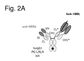
Figure 2A illustrates schematically the structure of the monovalent Her2-
targeting split
trimeric 4-1BB ligand Fc (kih) fusion antigen binding molecule comprising CH-
CL cross with
charged residues. The thick black point stands for the knob-into-hole
modification. * symbolizes
amino acid modifications in the CH1 and CL domain (so-called charged variant).
Figure 2B
.. illustrates the mouse surrogate, i.e. a bispecific 4-1BB antibody with
bivalent binding for mouse
4-1BB and monovalent binding for Her2 (anti -4-1BB/anti-Her2 moIgG1 DDKK DAPG,
termed
mu4-1BB-Her2). The thick black point stands for the DD/KK knob-into-hole
modification. The
DAPG mutations abolish the crosslinking of the fusion protein via mouse Fcy
receptors or the
binding of complement but allow binding to FcRn, so that the molecule remains
its antibody like
pharmacokinetics..
Figure 3A shows the setup of the SPR experiments for simultaneous binding of
the Her2-
targeting split trimeric 4-1BB ligand-containing antigen binding molecules of
the invention. The
simultaneous binding of Her2(PER)-4-1BBL (Analyte 1) to immobilized human 4-
1BB and
human Her2 (analyte 2) is shown in Fig. 3B. Simultaneous binding to human 4-
1BB and human
Her2 of Her2(aff PER)-4-1BBL is shown in Fig. 3C.
Figures 4A to 4D show the binding of Her2-targeting 4-1BB split trimeric
ligand Fc
fusion antigen binding molecules to Her2 expressed on the cell surface by
human breast cancer
cell line SK-Br3 (Fig. 4A and 4C) or human gastric carcinoma cell line NCI-N87
(Fig. 4B and
4D). Her2-targeting split 4-1BBL antigen binding molecules displaying the Her2
binders
.. pertuzumab (PER) or affinity-matured pertuzumab (aff-PER) or the fusion
protein Her2 (TRAS)-
anticalin 4-1BB huIgG4 (as described in patent W02016/177802) or previously
described
agonistic anti-human 4-1BB antibodies anti-human 4-1BB clone 20H4.9 huIgG4
(described in
U57659384 B2) or anti-human 4-1BB clone MOR-7480 huIgG2 (described in WO
2012/032433) or control molecules as indicated in the legend were incubated
with Her2
expressing cell lines SK-Br3 (Fig. 4A and 4C) or NCI-N87 (Fig. 4B and 4D) at
different
concentrations as indicated in the X-axis. Afterwards excessive and not bound
molecules were
washed of and bound molecules were detected with a secondary binding PE-
conjugated anti-
human Fc-fragment specific goat IgG F(a1302 fragment. The median of
fluorescence intensity
(MFI) was measured by flow cytometry and indicates the affinity (monovalent
binders) or
avidity (bivalent binders) of the tested molecules in a dose dependent manner.
Values are
baseline corrected by subtracting the blank control (e.g. staining with 2nd
detection fragment
only), shown is the mean +/- SEM.
Figures 5A and 5B illustrate the binding of Her2-targeting split trimeric 4-
1BB ligand Fc
fusion antigen binding molecules to Her2 expressed by human breast cancer cell
line KPL-4 the
cell surface. Her2-targeting split 4-1BBL antigen binding molecules comprising
the Her2 binders
PER, aff-PER or trastuzumab (TRAS), the fusion protein Her2 (TRAS)-anticalin 4-
1BB huIgG4
CA 03094235 2020-09-16
WO 2019/197600
PCT/EP2019/059391
-11-
(as described in patent W02016/177802) or previously described agonistic anti-
human 4-1BB
antibodies 20H4.9 huIgG4 or MOR-7480 huIgG2 or control molecules as indicated
in the legend
were incubated with Her2 expressing cell lines KL-4 at different
concentrations as indicated in
the X-axis. Afterwards excessive and not bound molecules were washed of and
bound molecules
were detected with a secondary binding PE-conjugated anti-human Fc-fragment
specific goat
IgG F(abµ)2 fragment. The median of fluorescence intensity (MFI) was measured
by flow
cytometry and indicates the affinity (monovalent binders) or avidity (bivalent
binders) of the
tested molecules in a dose dependent manner. Values are baseline corrected by
subtracting the
blank control (e.g. staining with 2nd detection fragment only), shown is the
mean +/- SEM.
Figure 6 shows a scheme that illustrates the general principal of the NFKB
activation assay
with human 4-1BB expressing Jurkat reporter cell line. Crosslinking of human 4-
1BB, expressed
on the reporter cells, induces NFKB activation and NFKB-mediated Luciferase
expression. After
lysis of the cells, luciferase can catalyze the oxidation of Luciferin to
Oxyluciferin. This
chemical reaction correlates positively with the strength of NFKB-mediated
luciferase expression
and can be measured by the strength of light emission (units of released
light).
The NFKB-mediated luciferase activity in a Jurkat-hu4-1BB-NFkB-1uc2 reporter
cell line
is shown in Figures 7A to 7F. In 96-well plates Jurkat-hu4-1BB-NFkB-1uc2
reporter cells were
incubated with different concentrations (indicated in the x-axis) of Her2
(PER)-4-1BBL or Her2
(PER)-4-1BBL molecules or the fusion protein Her2 (TRAS)-anticalin 4-1BB
huIgG4 or
agonistic anti-human 4-1BB antibodies 20H4.9 huIgG4 or MOR-7480 huIgG2 or the
control
molecules as indicated in the legend. The results in the absence of Her2+
cells are shown in Fig
7A and 7D, in the presence of human Her2+ breast cancer cell line SK-Br3 in
Fig. 7B and 7E or
in the presence of Her2+ human gastric cancer cell line NCI-N87 in Fig. 7C and
7F. Reporter
cells were incubated with the Her2-expressing tumor cells in an 1:5 ratio for
6 h. Afterwards
cells were washed, lysed and incubated with Luciferin in a detection buffer.
Luciferase-catalyzed
oxidation of luciferin was detected via light emission as units of released
light (y-axis). Shown is
the mean +/- SEM. All values are baseline corrected by subtracting the
baseline light emission.
The results of a second experiment comparing Her2 (PER)-4-1BBL with Her2
(TRAS)-4-
1BBL are shown in Figures 8A to 8H. In 348-well plates Jurkat-hu4-1BB-NFkB-
1uc2 reporter
cells were incubated with different concentrations (indicated in the x-axis)
of Her2 (PER)-4-
1BBL or Her2 (TRAS)-4-1BBL or the fusion protein Her2 (TRAS)-anticalin 4-1BB
huIgG4 or
agonistic anti-human 4-1BB antibodies 20H4.9 huIgG4 or MOR-7480 huIgG2 or
control
molecules as indicated in the legend. Shown is the NFKB-mediated luciferase
expression in a
Jurkat-hu4-1BB-NFKB-1uc2 reporter cell line in the absence (Fig. 8A and 8E) or
the presence of
human Her2+ breast cancer cell line SK-Br3 (Fig. 8B and 8F), in the presence
of human breast
cancer cell line KPL-4 (Fig. 8C and 8G) or Her2+ human gastric cancer cell
line NCI-N87 (FIG.
CA 03094235 2020-09-16
WO 2019/197600
PCT/EP2019/059391
-12-
8D and 8H) when given in a reporter cell line to tumor cell line 1:5 ratio for
6 h. Cells were
washed, lysed and incubated with Luciferin in a detection buffer. Luciferase-
catalyzed oxidation
of luciferin was detected via light emission as units of released light (y-
axis). Shown is the mean
+/- SEM. All values are baseline corrected by subtracting the baseline light
emission.
Figure 9 shows a scheme that illustrates the general principal of the
activation assay with
human PBMCs as described in Example 3.2.2. T cells are activated by 2 nM
agonistic CD3
antibody and co-stimulated with different concentrations of agonistic 4-1BB
molecules in the
presence of Her2-expressing gastric carcinoma NCI-N87 cells. The content/well
comprised 50
Gy irradiated 2x10⁴ NCI-N87 cells, 7.5x10⁴ CFSE-labelled human PBMCs, 2 nM
agonistic anti-
human CD3 human IgG wt (clone V9) and different concentrations of Her2-
targeting 4-1BB
agonistic molecules (here shown as Her2-4-1BBL). Cells were incubated for 4
days and then T
cell activation was determined my flow cytometry.
The results are shown as activation of CD8+ T cells in Figures 10A to 10F.
Resting
PBMCs isolated from a buffy coat of a healthy donor were activated with 2 nM
agonistic CD3
antibody and co-stimulated with different concentrations of agonistic 4-1BB
molecules as
indicated in the x-axis and in the legend in presence of Her2-expressing
gastric carcinoma NCI-
N87 cells for 4 days. Cells were gated on living CD8+ T cells and analyzed for
their frequency of
CD25+ (Fig. 10A and 10D), Granzyme &ugh (Fig. 10B and 10E) or proliferating
(low CFSE
MFI) CD8+ T cells (Fig. 10C and 10F). Shown is the mean +/- SD.
The activation of CD4+ T cells is shown in Figures 11A to 11F. Resting PBMCs
isolated
from a buffy coat of a healthy donor were activated with 2 nM agonistic CD3
antibody and co-
stimulated with different concentrations of agonistic 4-1BB molecules as
indicated in the x-axis
in the presence of Her2-expressing gastric carcinoma NCI-N87 cells for 4 days.
Cells were gated
on living CD4+ T cells and analyzed for their frequency of CD25+ (Fig. 11A and
11D),
Granzyme &ugh (Fig. 11B and 11E) or proliferating (low CFSE MFI) CD4+ T cells
(Fig. 11C
and 11F). Shown is the mean +/- SD.
Figure 12 shows a scheme that illustrates the general principal of the
activation assay with
mouse splenocytes as described in Example 3.2.3. T cells are activated by 0.5
iug/mL (-3.6 nM)
agonistic anti-mouse CD3 Armenian hamster IgG antibody (clone 1452C11) and co-
stimulated
with different concentrations of agonistic mouse surrogate mu4-1BB-Her2 in the
presence of
Her2-expressing human breast cancer cell line KPL-4. The content/well
comprised 50 Gy
irradiated 2x10⁴ KPL-4 cells, 15x104 violet proliferation dye-labelled mouse
splenocytes, 0.5
iug/mL (-3.6 nM) agonistic anti-mouse CD3 Armenian hamster IgG antibody (clone
1452C11)
and different concentrations of mouse surrogate mu4-1BB-Her2 or an untargeted
control mu4-
CA 03094235 2020-09-16
WO 2019/197600
PCT/EP2019/059391
-13-
1BB muIgG1 DAPG. Cells were incubated for 3 days and then T cell activation
was determined
my flow cytometry.
The results are shown as activation of mouse CD8+ and CD4+ T cells in Figures
13A to
13D. Resting mouse splenocytes isolated from C57BL/6 spleens were activated
with 0.5 g/mL
(-3.6 nM) agonistic anti-mouse CD3 Armenian hamster IgG antibody (clone
1452C11) and co-
stimulated with different concentrations of mouse surrogate mu4-1BB-Her2 or
untargeted
control as indicated in the x-axis and in the legend in presence of Her2-
expressing human breast
cancer KPL-4 cells for 3 days. Cells were gated on living CD8+ or CD4+ T cells
and analyzed for
their frequency of CD25+ expression (Fig. 13A and 13C) or proliferating (low
violet
proliferation dye MFI) (Fig. 13B and 13D). Shown is the mean +/- SD of
technical triplicates per
point.
In Figure 14A T cell activation of the combination of anti-Her2/anti-CD3
bispecific
antibody (Her2 TDB) with Her2(PER)-4-1BBL and the T cell activation of the
single agents is
shown. A robust T cell activation was induced by Her2 TDB alone as well as by
the combination
of both agents. Target cell killing of the combination of anti-Her2/anti-CD3
bispecific antibody
(Her2 TDB) with Her2(PER)-4-1BBL and the single agents is shown in Figure 14B.
The results of the T cell proliferation assay for the combination of anti-
Her2/anti-CD3
bispecific antibody (Her2 TDB) with Her2(PER)-4-1BBL are shown in Figures 15A
and 15B.
Addition of HER2-4-1BBL substantially enhanced anti-HER2/CD3-TDB induced T
cell
proliferation/survival in vitro.
Figures 16A to 16D show the tumor growth kinetics (linear scale) as observed
in immune-
competent mice that were implanted with human HER2 expressing Fo5 tumor
allografts and
treated with vehicle (Fig. 16A), Her2 TDB alone (Fig. 14B), mu 4-1BB-Her2
mouse surrogate
alone (Fig. 14C) and the combination of Her2 TDB and mu 4-1BB-Her2 (Fig. 14D).
The
individual tumor growth kinetics of each animal for all treatment groups are
shown. CR means
no dectable tumor at the end of the study, PR means that at least 50% of tumor
shrinkage is
observed compared to day 0. Four of seven mice (57%) treated in combination
with mu4-1BB-
Her2 agonist demonstrated complete responses without detectable tumors in the
end of study
(CR = 57%).
DETAILED DESCRIPTION OF THE INVENTION
Definitions
Unless defined otherwise, technical and scientific terms used herein have the
same
meaning as generally used in the art to which this invention belongs. For
purposes of interpreting
CA 03094235 2020-09-16
WO 2019/197600
PCT/EP2019/059391
-14-
this specification, the following definitions will apply and whenever
appropriate, terms used in
the singular will also include the plural and vice versa.
As used herein, the term "antigen binding molecule" refers in its broadest
sense to a
molecule that specifically binds an antigenic determinant. Examples of antigen
binding
.. molecules are antibodies, antibody fragments and scaffold antigen binding
proteins.
The term "antigen binding domain" refers to the part of an antigen binding
molecule that
comprises the area which specifically binds to and is complementary to part or
all of an antigen.
Where an antigen is large, an antigen binding molecule may only bind to a
particular part of the
antigen, which part is termed an epitope. An antigen binding domain may be
provided by, for
example, one or more variable domains (also called variable regions).
Preferably, an antigen
binding domain comprises an antibody light chain variable region (VL) and an
antibody heavy
chain variable region (VH).
As used herein, the term "antigen binding domain capable of specific binding
to Her2"
or "moiety capable of specific binding to Her2" refers to a polypeptide
molecule that specifically
binds to Her2. In one aspect, the antigen binding domain is able to activate
or inhibit signaling
through Her2. In a particular aspect, the antigen binding domain is able to
direct the entity to
which it is attached (e.g. the 4-1BBL trimer) to a target site, for example to
a specific type of
tumor cell bearing Her2. Antigen binding domains capable of specific binding
to Her2 include
antibodies and fragments thereof as further defined herein. In relation to an
antibody or fragment
thereof, the term "moiety capable of specific binding to a target cell
antigen" refers to the part of
the molecule that comprises the area which specifically binds to and is
complementary to part or
all of an antigen. A moiety capable of specific antigen binding may be
provided, for example, by
one or more antibody variable domains (also called antibody variable regions).
Particularly, a
moiety capable of specific antigen binding comprises an antibody light chain
variable region
(VL) and an antibody heavy chain variable region (VH).
The term "antibody" herein is used in the broadest sense and encompasses
various
antibody structures, including but not limited to monoclonal antibodies,
polyclonal antibodies,
monospecific and multispecific antibodies (e.g., bispecific antibodies), and
antibody fragments
so long as they exhibit the desired antigen-binding activity.
The term "monoclonal antibody" as used herein refers to an antibody obtained
from a
population of substantially homogeneous antibodies, i.e., the individual
antibodies comprising
the population are identical and/or bind the same epitope, except for possible
variant antibodies,
e.g. containing naturally occurring mutations or arising during production of
a monoclonal
antibody preparation, such variants generally being present in minor amounts.
In contrast to
polyclonal antibody preparations, which typically include different antibodies
directed against
CA 03094235 2020-09-16
WO 2019/197600
PCT/EP2019/059391
-15-
different determinants (epitopes), each monoclonal antibody of a monoclonal
antibody
preparation is directed against a single determinant on an antigen.
The term "monospecific" antibody as used herein denotes an antibody that has
one or
more binding sites each of which bind to the same epitope of the same antigen.
The term
"bispecific" means that the antigen binding molecule is able to specifically
bind to at least two
distinct antigenic determinants. Typically, a bispecific antigen binding
molecule comprises two
antigen binding sites, each of which is specific for a different antigenic
determinant. In certain
embodiments the bispecific antigen binding molecule is capable of
simultaneously binding two
antigenic determinants, particularly two antigenic determinants expressed on
two distinct cells.
The term "valent" as used within the current application denotes the presence
of a
specified number of binding sites in an antigen binding molecule. As such, the
terms
"monovalent", "bivalent", "tetravalent", and "hexavalent" denote the presence
of one binding
site, two binding sites, four binding sites, and six binding sites,
respectively, in an antigen
binding molecule.
The terms "full length antibody", "intact antibody", and "whole antibody" are
used herein
interchangeably to refer to an antibody having a structure substantially
similar to a native
antibody structure. "Native antibodies" refer to naturally occurring
immunoglobulin molecules
with varying structures. For example, native IgG-class antibodies are
heterotetrameric
glycoproteins of about 150,000 daltons, composed of two light chains and two
heavy chains that
are disulfide-bonded. From N- to C-terminus, each heavy chain has a variable
region (VH), also
called a variable heavy domain or a heavy chain variable domain, followed by
three constant
domains (CH1, CH2, and CH3), also called a heavy chain constant region.
Similarly, from N- to
C-terminus, each light chain has a variable region (VL), also called a
variable light domain or a
light chain variable domain, followed by a light chain constant domain (CL),
also called a light
chain constant region. The heavy chain of an antibody may be assigned to one
of five types,
called a (IgA), 6 (IgD), 8 (IgE), y (IgG), or u (IgM), some of which may be
further divided into
subtypes, e.g. yl (IgG1), y2 (IgG2), y3 (IgG3), y4 (IgG4), al (IgAl) and a2
(IgA2). The light
chain of an antibody may be assigned to one of two types, called kappa (x) and
lambda (X), based
on the amino acid sequence of its constant domain.
An "antibody fragment" refers to a molecule other than an intact antibody that
comprises
a portion of an intact antibody that binds the antigen to which the intact
antibody binds.
Examples of antibody fragments include but are not limited to Fv, Fab, Fab',
Fab'-SH, F(ab')2;
diabodies, triabodies, tetrabodies, cross-Fab fragments; linear antibodies;
single-chain antibody
molecules (e.g. scFv); and single domain antibodies. For a review of certain
antibody fragments,
see Hudson et al., Nat Med 9, 129-134 (2003). For a review of scFv fragments,
see e.g.
CA 03094235 2020-09-16
WO 2019/197600
PCT/EP2019/059391
-16-
Pliickthun, in The Pharmacology of Monoclonal Antibodies, vol. 113, Rosenburg
and Moore
eds., Springer-Verlag, New York, pp. 269-315 (1994); see also WO 93/16185; and
U.S. Patent
Nos. 5,571,894 and 5,587,458. For discussion of Fab and F(ab')2 fragments
comprising salvage
receptor binding epitope residues and having increased in vivo half-life, see
U.S. Patent No.
5,869,046. Diabodies are antibody fragments with two antigen-binding sites
that may be bivalent
or bispecific, see, for example, EP 404,097; WO 1993/01161; Hudson et al., Nat
Med 9, 129-134
(2003); and Hollinger et al., Proc Natl Acad Sci USA 90, 6444-6448 (1993).
Triabodies and
tetrabodies are also described in Hudson et al., Nat Med 9, 129-134 (2003).
Single-domain
antibodies are antibody fragments comprising all or a portion of the heavy
chain variable domain
or all or a portion of the light chain variable domain of an antibody. In
certain embodiments, a
single-domain antibody is a human single-domain antibody (Domantis, Inc.,
Waltham, MA; see
e.g. U.S. Patent No. 6,248,516 B1). Antibody fragments can be made by various
techniques,
including but not limited to proteolytic digestion of an intact antibody as
well as production by
recombinant host cells (e.g. E. coli or phage), as described herein.
Papain digestion of intact antibodies produces two identical antigen-binding
fragments,
called "Fab" fragments containing each the heavy- and light-chain variable
domains and also the
constant domain of the light chain and the first constant domain (CH1) of the
heavy chain. As
used herein, Thus, the term "Fab fragment" refers to an antibody fragment
comprising a light
chain fragment comprising a VL domain and a constant domain of a light chain
(CL), and a VH
domain and a first constant domain (CH1) of a heavy chain. Fab' fragments
differ from Fab
fragments by the addition of a few residues at the carboxy terminus of the
heavy chain CH1
domain including one or more cysteins from the antibody hinge region. Fab'-SH
are Fab'
fragments in which the cysteine residue(s) of the constant domains bear a free
thiol group.
Pepsin treatment yields an F(ab')2 fragment that has two antigen-combining
sites (two Fab
.. fragments) and a part of the Fc region.
The term "cross-Fab fragment" or "xFab fragment" or "crossover Fab fragment"
refers to
a Fab fragment, wherein either the variable regions or the constant regions of
the heavy and light
chain are exchanged. Two different chain compositions of a crossover Fab
molecule are possible
and comprised in the bispecific antibodies of the invention: On the one hand,
the variable regions
of the Fab heavy and light chain are exchanged, i.e. the crossover Fab
molecule comprises a
peptide chain composed of the light chain variable region (VL) and the heavy
chain constant
region (CH1), and a peptide chain composed of the heavy chain variable region
(VH) and the
light chain constant region (CL). This crossover Fab molecule is also referred
to as CrossFab
(\gym. On the other hand, when the constant regions of the Fab heavy and light
chain are
exchanged, the crossover Fab molecule comprises a peptide chain composed of
the heavy chain
variable region (VH) and the light chain constant region (CL), and a peptide
chain composed of
CA 03094235 2020-09-16
WO 2019/197600
PCT/EP2019/059391
-17-
the light chain variable region (VL) and the heavy chain constant region
(CH1). This crossover
Fab molecule is also referred to as CrossFab (CLCH1).
A "single chain Fab fragment" or "scFab" is a polypeptide consisting of an
antibody heavy
chain variable domain (VH), an antibody constant domain 1 (CH1), an antibody
light chain
variable domain (VL), an antibody light chain constant domain (CL) and a
linker, wherein said
antibody domains and said linker have one of the following orders in N-
terminal to C-terminal
direction: a) VH-CH1-linker-VL-CL, b) VL-CL-linker-VH-CH1, c) VH-CL-linker-VL-
CH1 or
d) VL-CH1-linker-VH-CL; and wherein said linker is a polypeptide of at least
30 amino acids,
preferably between 32 and 50 amino acids. Said single chain Fab fragments are
stabilized via the
natural disulfide bond between the CL domain and the CH1 domain. In addition,
these single
chain Fab molecules might be further stabilized by generation of interchain
disulfide bonds via
insertion of cysteine residues (e.g. position 44 in the variable heavy chain
and position 100 in the
variable light chain according to Kabat numbering).
A "crossover single chain Fab fragment" or "x-scFab" is a is a polypeptide
consisting of
an antibody heavy chain variable domain (VH), an antibody constant domain 1
(CH1), an
antibody light chain variable domain (VL), an antibody light chain constant
domain (CL) and a
linker, wherein said antibody domains and said linker have one of the
following orders in N-
terminal to C-terminal direction: a) VH-CL-linker-VL-CH1 and b) VL-CH1-linker-
VH-CL;
wherein VH and VL form together an antigen-binding site which binds
specifically to an antigen
and wherein said linker is a polypeptide of at least 30 amino acids. In
addition, these x-scFab
molecules might be further stabilized by generation of interchain disulfide
bonds via insertion of
cysteine residues (e.g. position 44 in the variable heavy chain and position
100 in the variable
light chain according to Kabat numbering).
A "single-chain variable fragment (scFv)" is a fusion protein of the variable
regions of
the heavy (VII) and light chains (VL) of an antibody, connected with a short
linker peptide of ten
to about 25 amino acids. The linker is usually rich in glycine for
flexibility, as well as serine or
threonine for solubility, and can either connect the N-terminus of the VH with
the C-terminus of
the VL, or vice versa. This protein retains the specificity of the original
antibody, despite removal
of the constant regions and the introduction of the linker. scFv antibodies
are, e.g. described in
Houston, J.S., Methods in Enzymol. 203 (1991) 46-96). In addition, antibody
fragments
comprise single chain polypeptides having the characteristics of a VH domain,
namely being
able to assemble together with a VL domain, or of a VL domain, namely being
able to assemble
together with a VH domain to a functional antigen binding site and thereby
providing the antigen
binding property of full length antibodies.
CA 03094235 2020-09-16
WO 2019/197600
PCT/EP2019/059391
-18-
"Scaffold antigen binding proteins" are known in the art, for example,
fibronectin and
designed ankyrin repeat proteins (DARPins) have been used as alternative
scaffolds for antigen-
binding domains, see, e.g., Gebauer and Skerra, Engineered protein scaffolds
as next-generation
antibody therapeutics. Curr Opin Chem Biol 13:245-255 (2009) and Stumpp et
al., Darpins: A
new generation of protein therapeutics. Drug Discovery Today 13: 695-701
(2008). In one aspect
of the invention, a scaffold antigen binding protein is selected from the
group consisting of
CTLA-4 (Evibody), Lipocalins (Anticalin), a Protein A-derived molecule such as
Z-domain of
Protein A (Affibody), an A-domain (Avimer/Maxibody), a serum transferrin
(trans-body); a
designed ankyrin repeat protein (DARPin), a variable domain of antibody light
chain or heavy
chain (single-domain antibody, sdAb), a variable domain of antibody heavy
chain (nanobody,
aVH), VNAR fragments, a fibronectin (AdNectin), a C-type lectin domain
(Tetranectin); a
variable domain of a new antigen receptor beta-lactamase (VNAR fragments), a
human gamma-
crystallin or ubiquitin (Affilin molecules); a kunitz type domain of human
protease inhibitors,
microbodies such as the proteins from the knottin family, peptide aptamers and
fibronectin
(adnectin). CTLA-4 (Cytotoxic T Lymphocyte-associated Antigen 4) is a CD28-
family receptor
expressed on mainly CD4+ T-cells. Its extracellular domain has a variable
domain- like Ig fold.
Loops corresponding to CDRs of antibodies can be substituted with heterologous
sequence to
confer different binding properties. CTLA-4 molecules engineered to have
different binding
specificities are also known as Evibodies (e.g. US7166697B1). Evibodies are
around the same
size as the isolated variable region of an antibody (e.g. a domain antibody).
For further details
see Journal of Immunological Methods 248 (1-2), 31-45 (2001). Lipocalins are a
family of
extracellular proteins which transport small hydrophobic molecules such as
steroids, bilins,
retinoids and lipids. They have a rigid beta-sheet secondary structure with a
number of loops at
the open end of the conical structure which can be engineered to bind to
different target antigens.
.. Anticalins are between 160-180 amino acids in size, and are derived from
lipocalins. For further
details see Biochim Biophys Acta 1482: 337-350 (2000), US7250297B1 and
US20070224633.
An afflbody is a scaffold derived from Protein A of Staphylococcus aureus
which can be
engineered to bind to antigen. The domain consists of a three-helical bundle
of approximately 58
amino acids. Libraries have been generated by randomization of surface
residues. For further
.. details see Protein Eng. Des. Sel. 2004, 17, 455-462 and EP 1641818A1.
Avimers are
multidomain proteins derived from the A-domain scaffold family. The native
domains of
approximately 35 amino acids adopt a defined disulfide bonded structure.
Diversity is generated
by shuffling of the natural variation exhibited by the family of A-domains.
For further details see
Nature Biotechnology 23(12), 1556 - 1561 (2005) and Expert Opinion on
Investigational Drugs
.. 16(6), 909-917 (June 2007). A transferrin is a monomeric serum transport
glycoprotein.
Transferrins can be engineered to bind different target antigens by insertion
of peptide sequences
in a permissive surface loop. Examples of engineered transferrin scaffolds
include the Trans-
body. For further details see J. Biol. Chem 274, 24066-24073 (1999). Designed
Ankyrin Repeat
CA 03094235 2020-09-16
WO 2019/197600
PCT/EP2019/059391
-19-
Proteins (DARPins) are derived from Ankyrin which is a family of proteins that
mediate
attachment of integral membrane proteins to the cytoskeleton. A single ankyrin
repeat is a 33
residue motif consisting of two alpha-helices and a beta-turn. They can be
engineered to bind
different target antigens by randomizing residues in the first alpha-helix and
a beta-turn of each
repeat. Their binding interface can be increased by increasing the number of
modules (a method
of affinity maturation). For further details see J. Mol. Biol. 332, 489-503
(2003), PNAS 100(4),
1700-1705 (2003) and J. Mol. Biol. 369, 1015-1028 (2007) and US20040132028A1.
A single-
domain antibody is an antibody fragment consisting of a single monomeric
variable antibody
domain. The first single domains were derived from the variable domain of the
antibody heavy
chain from camelids (nanobodies or VHFI fragments). Furthermore, the term
single-domain
antibody includes an autonomous human heavy chain variable domain (aVH) or
VNAR fragments
derived from sharks. Fibronectin is a scaffold which can be engineered to bind
to antigen.
Adnectins consists of a backbone of the natural amino acid sequence of the
10th domain of the
repeating units of human fibronectin type III (FN3). Three loops at one end of
the .beta.-
15 sandwich can be engineered to enable an Adnectin to specifically
recognize a therapeutic target
of interest. For further details see Protein Eng. Des. Sel. 18, 435- 444
(2005), US20080139791,
W02005056764 and US6818418B1. Peptide aptamers are combinatorial recognition
molecules
that consist of a constant scaffold protein, typically thioredoxin (TrxA)
which contains a
constrained variable peptide loop inserted at the active site. For further
details see Expert Opin.
Biol. Ther. 5, 783-797 (2005). Microbodies are derived from naturally
occurring microproteins
of 25-50 amino acids in length which contain 3-4 cysteine bridges - examples
of microproteins
include KalataBI and conotoxin and knottins. The microproteins have a loop
which can
beengineered to include upto 25 amino acids without affecting the overall fold
of the
microprotein. For further details of engineered knottin domains, see
W02008098796.
Lipocalins are a family of extracellular proteins which transport small
hydrophobic
molecules such as steroids, bilins, retinoids and lipids. They have a rigid
beta-sheet secondary
structure with a number of loops at the open end of the conical structure
which can be engineered
to bind to different target antigens. Anticalins are between 160-180 amino
acids in size, and are
derived from lipocalins. For further details see Biochim Biophys Acta 1482:
337-350 (2000),
Biodrugs 19(5), 279-288 (2005), US7250297B1 and US20070224633.
An "antigen binding molecule that binds to the same epitope" as a reference
molecule
refers to an antigen binding molecule that blocks binding of the reference
molecule to its antigen
in a competition assay by 50% or more, and conversely, the reference molecule
blocks binding
of the antigen binding molecule to its antigen in a competition assay by 50%
or more.
As used herein, the term "antigenic determinant" is synonymous with "antigen"
and
"epitope," and refers to a site (e.g. a contiguous stretch of amino acids or a
conformational
CA 03094235 2020-09-16
WO 2019/197600
PCT/EP2019/059391
-20-
configuration made up of different regions of non-contiguous amino acids) on a
polypeptide
macromolecule to which an antigen binding moiety binds, forming an antigen
binding moiety-
antigen complex. Useful antigenic determinants can be found, for example, on
the surfaces of
tumor cells, on the surfaces of virus-infected cells, on the surfaces of other
diseased cells, on the
surface of immune cells, free in blood serum, and/or in the extracellular
matrix (ECM). The
proteins useful as antigens herein can be any native form the proteins from
any vertebrate source,
including mammals such as primates (e.g. humans) and rodents (e.g. mice and
rats), unless
otherwise indicated. In a particular embodiment the antigen is a human
protein. Where reference
is made to a specific protein herein, the term encompasses the "full-length",
unprocessed protein
as well as any form of the protein that results from processing in the cell.
The term also
encompasses naturally occurring variants of the protein, e.g. splice variants
or allelic variants.
The term "capable of specific binding to Her2" refers to an antigen binding
molecule that
is capable of binding to Her2 with sufficient affinity such that the antigen
binding molecule is
useful as a diagnostic and/or therapeutic agent in targeting Her2. The antigen
binding molecule
includes but is not limited to, antibodies, Fab molecules, crossover Fab
molecules, single chain
Fab molecules, Fv molecules, scFv molecules, single domain antibodies, and VH
and scaffold
antigen binding protein. In one aspect, the extent of binding of an anti-Her2
antigen binding
molecule to an unrelated, non-Her2 protein is less than about 10% of the
binding of the antigen
binding molecule to Her2 as measured, e.g., by surface plasmon resonance
(SPR). In particular,
an antigen binding molecule that is capable of specific binding to Her2 has a
dissociation
constant (1(d) of < 1
10-8M or less, e.g. from 10-8M to 10-13M, e.g., from 10-9M to 10-13 M). In
certain aspects, an
anti-Her2 antigen binding molecule binds to Her2 from different species. In
particular, the anti-
Her2 antigen binding molecule binds to human and cynomolgus Her2.
The term "epitope" denotes the site on an antigen, either proteinaceous or non-
proteinaceous, to which an anti-[[PRO]] antibody binds. Epitopes can be formed
from
contiguous amino acid stretches (linear epitope) or comprise non-contiguous
amino acids
(conformational epitope), e.g., coming in spatial proximity due to the folding
of the antigen, i.e.
by the tertiary folding of a proteinaceous antigen. Linear epitopes are
typically still bound by an
antibody after exposure of the proteinaceous antigen to denaturing agents,
whereas
conformational epitopes are typically destroyed upon treatment with denaturing
agents. An
epitope comprises at least 3, at least 4, at least 5, at least 6, at least 7,
or 8-10 amino acids in a
unique spatial conformation.
The "epitope 4D5" or "4D5 epitope" or "4D5" is the region in the extracellular
domain of
HER2 to which the antibody 4D5 (ATCC CRL 10463) and trastuzumab bind. This
epitope is
close to the transmembrane domain of HER2, and within domain IV of HER2. To
screen for
CA 03094235 2020-09-16
WO 2019/197600
PCT/EP2019/059391
-21-
antibodies which bind to the 4D5 epitope, a routine cross-blocking assay such
as that described
in Antibodies, A Laboratory Manual, Cold Spring Harbor Laboratory, Ed Harlow
and David
Lane (1988), can be performed. Alternatively, epitope mapping can be performed
to assess
whether the antibody binds to the 4D5 epitope of HER2 (e.g. any one or more
residues in the
region from about residue 550 to about residue 610, inclusive, of human HER2
(SEQ ID NO:
54).
The "epitope 2C4" or "2C4 epitope" is the region in the extracellular domain
of HER2 to
which the antibody 2C4 binds. In order to screen for antibodies which bind to
the 2C4 epitope, a
routine cross-blocking assay such as that described in Antibodies, A
Laboratory Manual, Cold
Spring Harbor Laboratory, Ed Harlow and David Lane (1988), can be performed.
Alternatively,
epitope mapping can be performed to assess whether the antibody binds to the
2C4 epitope of
HER2. Epitope 2C4 comprises residues from domain II in the extracellular
domain of HER2.
The 2C4 antibody and pertuzumab bind to the extracellular domain of HER2 at
the junction of
domains I, II and III (Franklin et al. Cancer Cell 5:317-328 (2004)).
By "specific binding" is meant that the binding is selective for the antigen
and can be
discriminated from unwanted or non-specific interactions. The ability of an
antigen binding
molecule to bind to a specific antigen can be measured either through an
enzyme-linked
immunosorbent assay (ELISA) or other techniques familiar to one of skill in
the art, e.g. Surface
Plasmon Resonance (SPR) technique (analyzed on a BIAcore instrument)
(Liljeblad et al., Glyco
J 17, 323-329 (2000)), and traditional binding assays (Heeley, Endocr Res 28,
217-229 (2002)).
In one embodiment, the extent of binding of an antigen binding molecule to an
unrelated protein
is less than about 10% of the binding of the antigen binding molecule to the
antigen as measured,
e.g. by SPR. In certain embodiments, an molecule that binds to the antigen has
a dissociation
constant (Kd) of < 1 [tM, < 100 nM, < 10 nM, < 1 nM, < 0.1 nM, < 0.01 nM, or <
0.001 nM (e.g.
10-8 M or less, e.g. from 10-8 M to 10-13 M, e.g. from 10-9 M to 10-13 M).
"Affinity" or "binding affinity" refers to the strength of the sum total of
non-covalent
interactions between a single binding site of a molecule (e.g. an antibody)
and its binding partner
(e.g. an antigen). Unless indicated otherwise, as used herein, "binding
affinity" refers to intrinsic
binding affinity which reflects a 1:1 interaction between members of a binding
pair (e.g.
antibody and antigen). The affinity of a molecule X for its partner Y can
generally be represented
by the dissociation constant (Kd), which is the ratio of dissociation and
association rate constants
(koff and kon, respectively). Thus, equivalent affinities may comprise
different rate constants, as
long as the ratio of the rate constants remains the same. Affinity can be
measured by common
methods known in the art, including those described herein. A particular
method for measuring
affinity is Surface Plasmon Resonance (SPR).
CA 03094235 2020-09-16
WO 2019/197600
PCT/EP2019/059391
-22-
A "target cell antigen" as used herein refers to an antigenic determinant
presented on the
surface of a target cell, for example a T-cell or B-cell, a cell in a tumor
such as a cancer cell or a
cell of the tumor stroma. In certain aspects, the target cell antigen is an
antigen on the surface of
cancer cell. In one aspect, the target cell antigen is Her2.
The term "Her2", also known as "ErbB2", "ErbB2 receptor", or "c-Erb-B2",
refers to any
native, mature HER2 which results from processing of a HER2 precursor protein
in a cell. The
term includes HER2 from any vertebrate source, including mammals such as
primates (e.g.
humans and cynomolgus monkeys) and rodents (e.g., mice and rats), unless
otherwise indicated.
The term also includes naturally occurring variants of HER2, e.g., splice
variants or allelic
variants. The amino acid sequence of an exemplary human HER2 protein is shown
in SEQ ID
NO:54.
A "T-cell antigen" as used herein refers to an antigenic determinant presented
on the
surface of a T lymphocyte, particularly a cytotoxic T lymphocyte.
A "T cell activating therapeutic agent" as used herein refers to a therapeutic
agent
capable of inducing T cell activation in a subject, particularly a therapeutic
agent designed for
inducing T-cell activation in a subject. Examples of T cell activating
therapeutic agents include
bispecific antibodies that specifically bind an activating T cell antigen,
such as CD3, and a target
cell antigen, such as CEA or Folate Receptor.
An "activating T cell antigen" as used herein refers to an antigenic
determinant expressed
by a T lymphocyte, particularly a cytotoxic T lymphocyte, which is capable of
inducing or
enhancing T cell activation upon interaction with an antigen binding molecule.
Specifically,
interaction of an antigen binding molecule with an activating T cell antigen
may induce T cell
activation by triggering the signaling cascade of the T cell receptor complex.
An exemplary
activating T cell antigen is CD3.
The term "CD3" refers to any native CD3 from any vertebrate source, including
mammals
such as primates (e.g. humans), non-human primates (e.g. cynomolgus monkeys)
and rodents
(e.g. mice and rats), unless otherwise indicated. The term encompasses "full-
length,"
unprocessed CD3 as well as any form of CD3 that results from processing in the
cell. The term
also encompasses naturally occurring variants of CD3, e.g., splice variants or
allelic variants. In
one embodiment, CD3 is human CD3, particularly the epsilon subunit of human
CD3 (CD38).
The amino acid sequence of human CD38 is shown in UniProt (www.uniprot.org)
accession no.
P07766 (version 144), or NCBI (www.ncbi.nlm.nih.gov/) RefSeq NP 000724.1. See
also SEQ
ID NO: 85. The amino acid sequence of cynomolgus [Macaca fascicularis] CD38 is
shown in
NCBI GenBank no. BAB71849.1. See also SEQ ID NO: 86.
CA 03094235 2020-09-16
WO 2019/197600
PCT/EP2019/059391
-23-
The term "variable domain" or "variable region" refers to the domain of an
antibody
heavy or light chain that is involved in binding the antigen binding molecule
to antigen. The
variable domains of the heavy chain and light chain (VH and VL, respectively)
of a native
antibody generally have similar structures, with each domain comprising four
conserved
framework regions (FRs) and three hypervariable regions (HVRs). See, e.g.,
Kindt et al., Kuby
Immunology, 6th ed., W.H. Freeman and Co., page 91 (2007). A single VH or VL
domain may
be sufficient to confer antigen-binding specificity.
The term "hypervariable region" or "HVR" as used herein refers to each of the
regions of
an antigen binding variable domain which are hypervariable in sequence and
which determine
antigen binding specificity, for example "complementarity determining regions"
("CDRs").
Generally, antigen binding domains comprise six CDRs: three in the VH (CDR-H1,
CDR-H2,
CDR-H3), and three in the VL (CDR-L1, CDR-L2, CDR-L3). Exemplary CDRs herein
include:
(a) hypervariable loops occurring at amino acid residues 26-32 (L1), 50-52
(L2), 91-96
(L3), 26-32 (H1), 53-55 (H2), and 96-101 (H3) (Chothia and Lesk, J. Mot. Biol.
196:901-917
(1987));
(b) CDRs occurring at amino acid residues 24-34 (L1), 50-56 (L2), 89-97 (L3),
31-35b
(H1), 50-65 (H2), and 95-102 (H3) (Kabat et al., Sequences of Proteins of
Immunological
Interest, 5th Ed. Public Health Service, National Institutes of Health,
Bethesda, MD (1991)); and
(c) antigen contacts occurring at amino acid residues 27c-36 (L1), 46-55 (L2),
89-96 (L3),
30-35b (H1), 47-58 (H2), and 93-101 (H3) (MacCallum et al. J. Mol. Biol. 262:
732-745 (1996)).
Unless otherwise indicated, the CDRs are determined according to Kabat et al.,
supra. One
of skill in the art will understand that the CDR designations can also be
determined according to
Chothia, supra, McCallum, supra, or any other scientifically accepted
nomenclature. Kabat et at.
also defined a numbering system for variable region sequences that is
applicable to any antibody.
One of ordinary skill in the art can unambiguously assign this system of
"Kabat numbering" to
any variable region sequence, without reliance on any experimental data beyond
the sequence
itself As used herein, "Kabat numbering" refers to the numbering system set
forth by Kabat et
al., U.S. Dept. of Health and Human Services, "Sequence of Proteins of
Immunological Interest"
(1983). Unless otherwise specified, references to the numbering of specific
amino acid residue
positions in an antibody variable region are according to the Kabat numbering
system.
As used herein, the term "affinity matured" in the context of antigen binding
molecules
(e.g., antibodies) refers to an antigen binding molecule that is derived from
a reference antigen
binding molecule, e.g., by mutation, binds to the same antigen, preferably
binds to the same
epitope, as the reference antibody; and has a higher affinity for the antigen
than that of the
CA 03094235 2020-09-16
WO 2019/197600
PCT/EP2019/059391
-24-
reference antigen binding molecule. Affinity maturation generally involves
modification of one
or more amino acid residues in one or more CDRs of the antigen binding
molecule. Typically,
the affinity matured antigen binding molecule binds to the same epitope as the
initial reference
antigen binding molecule.
"Framework" or "FR" refers to variable domain residues other than
hypervariable region
(HVR) residues. The FR of a variable domain generally consists of four FR
domains: FR1, FR2,
FR3, and FR4. Accordingly, the HVR and FR sequences generally appear in the
following
sequence in VH (or VL): FR1-H1(L1)-FR2-H2(L2)-FR3-H3(L3)-FR4.
An "acceptor human framework" for the purposes herein is a framework
comprising
the amino acid sequence of a light chain variable domain (VL) framework or a
heavy chain
variable domain (VH) framework derived from a human immunoglobulin framework
or a human
consensus framework, as defined below. An acceptor human framework "derived
from" a human
immunoglobulin framework or a human consensus framework may comprise the same
amino
acid sequence thereof, or it may contain amino acid sequence changes. In some
embodiments,
the number of amino acid changes are 10 or less, 9 or less, 8 or less, 7 or
less, 6 or less, 5 or less,
4 or less, 3 or less, or 2 or less. In some embodiments, the VL acceptor human
framework is
identical in sequence to the VL human immunoglobulin framework sequence or
human
consensus framework sequence.
The term "chimeric" antibody refers to an antibody in which a portion of the
heavy and/or
light chain is derived from a particular source or species, while the
remainder of the heavy and/or
light chain is derived from a different source or species.
The "class" of an antibody refers to the type of constant domain or constant
region
possessed by its heavy chain. There are five major classes of antibodies: IgA,
IgD, IgE, IgG, and
IgM, and several of these may be further divided into subclasses (isotypes),
e.g. IgGi, IgG2,
IgG3, IgG4, IgAi, and IgA2. The heavy chain constant domains that correspond
to the different
classes of immunoglobulins are called a, 6, 8, y, and ii. respectively.
The terms "constant region derived from human origin" or "human constant
region"
denote a constant heavy chain region of a human antibody of the subclass IgGl,
IgG2, IgG3, or
IgG4 and/or a constant light chain kappa or lambda region. Such constant
regions are well
known in the state of the art and e.g. described by Kabat, E.A., et al.,
Sequences of Proteins of
Immunological Interest, 5th ed., Public Health Service, National Institutes of
Health, Bethesda,
MD (1991) (see also e.g. Johnson, G., and Wu, T.T., Nucleic Acids Res. 28
(2000) 214-218;
Kabat, E.A., et al., Proc. Natl. Acad. Sci. USA 72 (1975) 2785-2788). Unless
otherwise specified
herein, numbering of amino acid residues in the constant region is according
to the EU
numbering system, also called the EU index of Kabat, as described in Kabat,
E.A. et al.,
CA 03094235 2020-09-16
WO 2019/197600
PCT/EP2019/059391
-25-
Sequences of Proteins of Immunological Interest, 5th ed., Public Health
Service, National
Institutes of Health, Bethesda, MD (1991), NIH Publication 91-3242.
A "humanized" antibody refers to a chimeric antibody comprising amino acid
residues
from non-human HVRs and amino acid residues from human FRs. In certain
embodiments, a
humanized antibody will comprise substantially all of at least one, and
typically two, variable
domains, in which all or substantially all of the HVRs (e.g., CDRs) correspond
to those of a non-
human antibody, and all or substantially all of the FRs correspond to those of
a human antibody.
A humanized antibody optionally may comprise at least a portion of an antibody
constant region
derived from a human antibody. A "humanized form" of an antibody, e.g., a non-
human
antibody, refers to an antibody that has undergone humanization. Other forms
of "humanized
antibodies" encompassed by the present invention are those in which the
constant region has
been additionally modified or changed from that of the original antibody to
generate the
properties according to the invention, especially in regard to Clq binding
and/or Fc receptor
(FcR) binding.
A "human" antibody is one which possesses an amino acid sequence which
corresponds to
that of an antibody produced by a human or a human cell or derived from a non-
human source
that utilizes human antibody repertoires or other human antibody-encoding
sequences. This
definition of a human antibody specifically excludes a humanized antibody
comprising non-
human antigen-binding residues.
The term "Fc domain" or "Fc region" herein is used to define a C-terminal
region of an
antibody heavy chain that contains at least a portion of the constant region.
The term includes
native sequence Fc regions and variant Fc regions. In one embodiment, a human
IgG heavy
chain Fc region extends from Cys226, or from Pro230, to the carboxyl-terminus
of the heavy
chain. However, antibodies produced by host cells may undergo post-
translational cleavage of
one or more, particularly one or two, amino acids from the C-terminus of the
heavy chain.
Therefore an antibody produced by a host cell by expression of a specific
nucleic acid molecule
encoding a full-length heavy chain may include the full-length heavy chain, or
it may include a
cleaved variant of the full-length heavy chain. This may be the case where the
final two C-
terminal amino acids of the heavy chain are glycine (G446) and lysine (K447,
numbering
according to Kabat EU index). Therefore, the C-terminal lysine (Lys447), or
the C-terminal
glycine (Gly446) and lysine (Lys447), of the Fc region may or may not be
present. Amino acid
sequences of heavy chains including an Fc region are denoted herein without C-
terminal glycine-
lysine dipeptide if not indicated otherwise. In one embodiment, a heavy chain
including an Fc
region as specified herein, comprised in an antibody according to the
invention, comprises an
additional C-terminal glycine-lysine dipeptide (G446 and K447, numbering
according to EU
index of Kabat). In one embodiment, a heavy chain including an Fc region as
specified herein,
CA 03094235 2020-09-16
WO 2019/197600
PCT/EP2019/059391
-26-
comprised in an antibody according to the invention, comprises an additional C-
terminal glycine
residue (G446, numbering according to EU index of Kabat). Unless otherwise
specified herein,
numbering of amino acid residues in the Fc region or constant region is
according to the EU
numbering system, also called the EU index, as described in Kabat et al.,
Sequences of Proteins
of Immunological Interest, 5th Ed. Public Health Service, National Institutes
of Health,
Bethesda, MD, 1991. An IgG Fc region comprises an IgG CH2 and an IgG CH3
domain. The
"CH2 domain" of a human IgG Fc region usually extends from an amino acid
residue at about
position 231 to an amino acid residue at about position 340. In one
embodiment, a carbohydrate
chain is attached to the CH2 domain. The CH2 domain herein may be a native
sequence CH2
domain or variant CH2 domain. The "CH3 domain" comprises the stretch of
residues C-terminal
to a CH2 domain in an Fc region (i.e. from an amino acid residue at about
position 341 to an
amino acid residue at about position 447 of an IgG). The CH3 region herein may
be a native
sequence CH3 domain or a variant CH3 domain (e.g. a CH3 domain with an
introduced
"protuberance" ("knob") in one chain thereof and a corresponding introduced
"cavity" ("hole")
.. in the other chain thereof; see US Patent No. 5,821,333, expressly
incorporated herein by
reference). Such variant CH3 domains may be used to promote heterodimerization
of two non-
identical antibody heavy chains as herein described.
The "knob-into-hole" technology is described e.g. in US 5,731,168; US
7,695,936;
Ridgway et al., Prot Eng 9, 617-621 (1996) and Carter, J Immunol Meth 248, 7-
15 (2001).
Generally, the method involves introducing a protuberance ("knob") at the
interface of a first
polypeptide and a corresponding cavity ("hole") in the interface of a second
polypeptide, such
that the protuberance can be positioned in the cavity so as to promote
heterodimer formation and
hinder homodimer formation. Protuberances are constructed by replacing small
amino acid side
chains from the interface of the first polypeptide with larger side chains
(e.g. tyrosine or
tryptophan). Compensatory cavities of identical or similar size to the
protuberances are created
in the interface of the second polypeptide by replacing large amino acid side
chains with smaller
ones (e.g. alanine or threonine). The protuberance and cavity can be made by
altering the nucleic
acid encoding the polypeptides, e.g. by site-specific mutagenesis, or by
peptide synthesis. In a
specific embodiment a knob modification comprises the amino acid substitution
T366W in one
of the two subunits of the Fc domain, and the hole modification comprises the
amino acid
substitutions T3665, L368A and Y407V in the other one of the two subunits of
the Fc domain. In
a further specific embodiment, the subunit of the Fc domain comprising the
knob modification
additionally comprises the amino acid substitution 5354C, and the subunit of
the Fc domain
comprising the hole modification additionally comprises the amino acid
substitution Y349C.
Introduction of these two cysteine residues results in the formation of a
disulfide bridge between
the two subunits of the Fc region, thus further stabilizing the dimer (Carter,
J Immunol Methods
248, 7-15 (2001)). The numbering is according to EU index of Kabat et al,
Sequences of Proteins
CA 03094235 2020-09-16
WO 2019/197600
PCT/EP2019/059391
-27-
of Immunological Interest, 5th Ed. Public Health Service, National Institutes
of Health,
Bethesda, MD, 1991.
A "region equivalent to the Fc region of an immunoglobulin" is intended to
include
naturally occurring allelic variants of the Fc region of an immunoglobulin as
well as variants
having alterations which produce substitutions, additions, or deletions but
which do not decrease
substantially the ability of the immunoglobulin to mediate effector functions
(such as antibody-
dependent cellular cytotoxicity). For example, one or more amino acids can be
deleted from the
N-terminus or C-terminus of the Fc region of an immunoglobulin without
substantial loss of
biological function. Such variants can be selected according to general rules
known in the art so
as to have minimal effect on activity (see, e.g., Bowie, J. U. et al., Science
247:1306-10 (1990)).
The term "effector functions" refers to those biological activities
attributable to the Fc
region of an antibody, which vary with the antibody isotype. Examples of
antibody effector
functions include: Clq binding and complement dependent cytotoxicity (CDC), Fc
receptor
binding, antibody-dependent cell-mediated cytotoxicity (ADCC), antibody-
dependent cellular
phagocytosis (ADCP), cytokine secretion, immune complex-mediated antigen
uptake by antigen
presenting cells, down regulation of cell surface receptors (e.g. B cell
receptor), and B cell
activation.
An "activating Fc receptor" is an Fc receptor that following engagement by an
Fc region
of an antibody elicits signaling events that stimulate the receptor-bearing
cell to perform effector
functions. Activating Fc receptors include FcyRIIIa (CD16a), FcyRI (CD64),
FcyRIIa (CD32),
and FcaRI (CD89). A particular activating Fc receptor is human FcyRIIIa (see
UniProt accession
no. P08637, version 141).
The term "TNF ligand family member" or "TNF family ligand" refers to a
proinflammatory cytokine. Cytokines in general, and in particular the members
of the TNF
ligand family, play a crucial role in the stimulation and coordination of the
immune system. At
present, nineteen cyctokines have been identified as members of the TNF
(tumour necrosis
factor) ligand superfamily on the basis of sequence, functional, and
structural similarities. All
these ligands are type II transmembrane proteins with a C-terminal
extracellular domain
(ectodomain), N-terminal intracellular domain and a single transmembrane
domain. The C-
terminal extracellular domain, known as TNF homology domain (THD), has 20-30%
amino acid
identity between the superfamily members and is responsible for binding to the
receptor. The
TNF ectodomain is also responsible for the TNF ligands to form trimeric
complexes that are
recognized by their specific receptors. Members of the TNF ligand family are
selected from the
group consisting of Lymphotoxin a (also known as LTA or TNFSF1), TNF (also
known as
TNFSF2), LTI3 (also known as TNFSF3), OX4OL (also known as TNFSF4), CD4OL
(also
CA 03094235 2020-09-16
WO 2019/197600
PCT/EP2019/059391
-28-
known as CD154 or TNFSF5), FasL (also known as CD95L, CD178 or TNFSF6), CD27L
(also
known as CD70 or TNFSF7), CD3OL (also known as CD153 or TNFSF8), 4-1BBL (also
known
as TNFSF9), TRAIL (also known as APO2L, CD253 or TNFSF10), RANKL (also known
as
CD254 or TNFSF11), TWEAK (also known as TNFSF12), APRIL (also known as CD256
or
TNFSF13), BAFF (also known as CD257 or TNFSF13B), LIGHT (also known as CD258
or
TNFSF14), TL1A (also known as VEGI or TNFSF15), GITRL (also known as TNFSF18),
EDA-Al (also known as ectodysplasin Al) and EDA-A2 (also known as
ectodysplasin A2). The
term refers to any native TNF family ligand from any vertebrate source,
including mammals
such as primates (e.g. humans), non-human primates (e.g. cynomolgus monkeys)
and rodents
(e.g. mice and rats), unless otherwise indicated. The term "costimulatory TNF
ligand family
member" or "costimulatory TNF family ligand" refers to a subgroup of TNF
ligand family
members, which are able to costimulate proliferation and cytokine production
of T-cells. These
TNF family ligands can costimulate TCR signals upon interaction with their
corresponding TNF
receptors and the interaction with their receptors leads to recruitment of
TNFR-associated factors
(TRAF), which initiate signalling cascades that result in T-cell activation.
Costimulatory TNF
family ligands are selected from the group consisting of 4-1BBL, OX4OL, GITRL,
CD70,
CD3OL and LIGHT, more particularly the costimulatory TNF ligand family member
is 4-1BBL.
As described herein before, 4-1BBL is a type II transmembrane protein and one
member of
the TNF ligand family. Complete or full length 4-1BBL having the amino acid
sequence of SEQ
ID NO:69 has been described to form trimers on the surface of cells. The
formation of timers is
enabled by specific motives of the ectodomain of 4-1BBL. Said motives are
designated herein as
"trimerization region". The amino acids 50-254 of the human 4-1BBL sequence
(SEQ ID
NO:70) form the extracellular domain of 4-1BBL, but even fragments thereof are
able to form
the trimers. In specific embodiments of the invention, the term "ectodomain of
4-1BBL or a
fragment thereof" refers to a polypeptide having an amino acid sequence
selected from SEQ ID
NO:4 (amino acids 52-254 of human 4-1BBL), SEQ ID NO:1 (amino acids 71-254 of
human 4-
1BBL), SEQ ID NO:3 (amino acids 80-254 of human 4-1BBL) and SEQ ID NO:2 (amino
acids
85-254 of human 4-1BBL) or a polypeptide having an amino acid sequence
selected from SEQ
ID NO:5 (amino acids 71-248 of human 4-1BBL), SEQ ID NO:8 (amino acids 52-248
of human
4-1BBL), SEQ ID NO:7 (amino acids 80-248 of human 4-1BBL) and SEQ ID NO:6
(amino
acids 85-248 of human 4-1BBL), but also other fragments of the ectodomain
capable of
trimerization are included herein.
An "ectodomain" is the domain of a membrane protein that extends into the
extracellular
space (i.e. the space outside the target cell). Ectodomains are usually the
parts of proteins that
.. initiate contact with surfaces, which leads to signal transduction. The
ectodomain of TNF ligand
family member as defined herein thus refers to the part of the TNF ligand
protein that extends
into the extracellular space (the extracellular domain), but also includes
shorter parts or
CA 03094235 2020-09-16
WO 2019/197600
PCT/EP2019/059391
-29-
fragments thereof that are responsible for the trimerization and for the
binding to the
corresponding TNF receptor. The term "ectodomain of a TNF ligand family member
or a
fragment thereof" thus refers to the extracellular domain of the TNF ligand
family member that
forms the extracellular domain or to parts thereof that are still able to bind
to the receptor
(receptor binding domain).
The term "peptide linker" refers to a peptide comprising one or more amino
acids,
typically about 2 to 20 amino acids. Peptide linkers are known in the art or
are described herein.
Suitable, non-immunogenic linker peptides are, for example, (G45),, (5G4)n or
G4(5G4). peptide
linkers, wherein "n" is generally a number between 1 and 10, typically between
1 and 4, in
particular 2, i.e. the peptides selected from the group consisting of GGGGS
(SEQ ID NO:71),
GGGGSGGGGS (SEQ ID NO:68), SGGGGSGGGG (SEQ ID NO:72), (G45)3 or
GGGGSGGGGSGGGGS (SEQ ID NO:73), GGGGSGGGGSGGGG or G4(5G4)2 (SEQ ID
NO:74), and (G45)4 or GGGGSGGGGSGGGGSGGGGS (SEQ ID NO:75), but also include
the
sequences GSPGSSSSGS (SEQ ID NO:76), GSGSGSGS (SEQ ID NO:77), GSGSGNGS (SEQ
ID NO:78), GGSGSGSG (SEQ ID NO:79), GGSGSG (SEQ ID NO:80), GGSG (SEQ ID
NO:81), GGSGNGSG (SEQ ID NO:82), GGNGSGSG (SEQ ID NO:83) and GGNGSG (SEQ
ID NO:84). Peptide linkers of particular interest are (G45)1 or GGGGS (SEQ ID
NO:71), (G45)2
or GGGGSGGGGS (SEQ ID NO:68) and (G45)3 (SEQ ID NO:73).
The term "amino acid" as used within this application denotes the group of
naturally
occurring carboxy a-amino acids comprising alanine (three letter code: ala,
one letter code: A),
arginine (arg, R), asparagine (asn, N), aspartic acid (asp, D), cysteine (cys,
C), glutamine (gln,
Q), glutamic acid (glu, E), glycine (gly, G), histidine (his, H), isoleucine
(ile, I), leucine (leu, L),
lysine (lys, K), methionine (met, M), phenylalanine (phe, F), proline (pro,
P), serine (ser, S),
threonine (thr, T), tryptophan (trp, W), tyrosine (tyr, Y), and valine (val,
V).
A "fusion polypeptide" or "fusion protein" as used herein refers to a single
chain
polypeptide composed of an antibody fragment and a peptide that is not derived
from an
antibody. In one aspect, a fusion polypeptide is composed of one or two
ectodomains of 4-1BBL
or a fragment thereof fused to a part of antigen binding domain or Fc part.
The fusion may occur
by directly linking the N or C-terminal amino acid of the antigen binding
moiety via a peptide
linker to the C- or N-terminal amino acid of the ectodomain of said 4-1BBL or
fragment thereof
By "fused" or "connected" is meant that the components (e.g. a polypeptide and
an
ectodomain of said TNF ligand family member) are linked by peptide bonds,
either directly or
via one or more peptide linkers.
"Percent (%) amino acid sequence identity" with respect to a reference
polypeptide
(protein) sequence is defined as the percentage of amino acid residues in a
candidate sequence
CA 03094235 2020-09-16
WO 2019/197600
PCT/EP2019/059391
-30-
that are identical with the amino acid residues in the reference polypeptide
sequence, after
aligning the sequences and introducing gaps, if necessary, to achieve the
maximum percent
sequence identity, and not considering any conservative substitutions as part
of the sequence
identity. Alignment for purposes of determining percent amino acid sequence
identity can be
achieved in various ways that are within the skill in the art, for instance,
using publicly available
computer software such as BLAST, BLAST-2, ALIGN. SAWI or Megalign (DNASTAR)
software. Those skilled in the art can determine appropriate parameters for
aligning sequences,
including any algorithms needed to achieve maximal alignment over the full
length of the
sequences being compared. For purposes herein, however, % amino acid sequence
identity
values are generated using the sequence comparison computer program ALIGN-2.
The ALIGN-
2 sequence comparison computer program was authored by Genentech, Inc., and
the source code
has been filed with user documentation in the U.S. Copyright Office,
Washington D.C., 20559,
where it is registered under U.S. Copyright Registration No. TXU510087. The
ALIGN-2
program is publicly available from Genentech, Inc., South San Francisco,
California, or may be
compiled from the source code. The ALIGN-2 program should be compiled for use
on a UNIX
operating system, including digital UNIX V4.0D. All sequence comparison
parameters are set by
the ALIGN-2 program and do not vary. In situations where ALIGN-2 is employed
for amino
acid sequence comparisons, the % amino acid sequence identity of a given amino
acid sequence
A to, with, or against a given amino acid sequence B (which can alternatively
be phrased as a
given amino acid sequence A that has or comprises a certain % amino acid
sequence identity to,
with, or against a given amino acid sequence B) is calculated as follows:
100 times the fraction X/Y
where X is the number of amino acid residues scored as identical matches by
the sequence
alignment program ALIGN-2 in that program's alignment of A and B, and where Y
is the total
number of amino acid residues in B. It will be appreciated that where the
length of amino acid
sequence A is not equal to the length of amino acid sequence B, the % amino
acid sequence
identity of A to B will not equal the % amino acid sequence identity of B to
A. Unless
specifically stated otherwise, all % amino acid sequence identity values used
herein are obtained
as described in the immediately preceding paragraph using the ALIGN-2 computer
program.
In certain embodiments, amino acid sequence variants of the TNF ligand trimer-
containing antigen binding molecules provided herein are contemplated. For
example, it may be
desirable to improve the binding affinity and/or other biological properties
of the TNF ligand
trimer-containing antigen binding molecules. Amino acid sequence variants of
the TNF ligand
trimer-containing antigen binding molecules may be prepared by introducing
appropriate
modifications into the nucleotide sequence encoding the molecules, or by
peptide synthesis.
Such modifications include, for example, deletions from, and/or insertions
into and/or
CA 03094235 2020-09-16
WO 2019/197600
PCT/EP2019/059391
-3 1 -
substitutions of residues within the amino acid sequences of the antibody. Any
combination of
deletion, insertion, and substitution can be made to arrive at the final
construct, provided that the
final construct possesses the desired characteristics, e.g., antigen-binding.
Sites of interest for
substitutional mutagenesis include the HVRs and Framework (FRs). Conservative
substitutions
are provided in Table B under the heading "Preferred Substitutions" and
further described below
in reference to amino acid side chain classes (1) to (6). Amino acid
substitutions may be
introduced into the molecule of interest and the products screened for a
desired activity, e.g.,
retained/improved antigen binding, decreased immunogenicity, or improved ADCC
or CDC.
TABLE A
Original Exemplary Preferred
Residue Substitutions Substitutions
Ala (A) Val; Leu; Ile Val
Arg (R) Lys; Gin; Asn Lys
Asn (N) Gin; His; Asp, Lys; Arg Gin
Asp (D) Glu; Asn Glu
Cys (C) Ser; Ala Ser
Gin (Q) Asn; Glu Asn
Glu (E) Asp; Gin Asp
Gly (G) Ala Ala
His (H) Asn; Gin; Lys; Arg Arg
Ile (I) Leu; Val; Met; Ala; Phe; Norleucine Leu
Leu (L) Norleucine; Ile; Val; Met; Ala; Phe Ile
Lys (K) Arg; Gin; Asn Arg
Met (M) Leu; Phe; Ile Leu
Phe (F) Trp; Leu; Val; Ile; Ala; Tyr Tyr
Pro (P) Ala Ala
Ser (S) Thr Thr
Thr (T) Val; Ser Ser
Trp (W) Tyr; Phe Tyr
Tyr (Y) Trp; Phe; Thr; Ser Phe
Val (V) Ile; Leu; Met; Phe; Ala; Norleucine Leu
Amino acids may be grouped according to common side-chain properties:
CA 03094235 2020-09-16
WO 2019/197600
PCT/EP2019/059391
-32-
(1) hydrophobic: Norleucine, Met, Ala, Val, Leu, Ile;
(2) neutral hydrophilic: Cys, Ser, Thr, Asn, Gln;
(3) acidic: Asp, Glu;
(4) basic: His, Lys, Arg;
(5) residues that influence chain orientation: Gly, Pro;
(6) aromatic: Trp, Tyr, Phe.
Non-conservative substitutions will entail exchanging a member of one of these
classes for
another class.
The term "amino acid sequence variants" includes substantial variants wherein
there are
amino acid substitutions in one or more hypervariable region residues of a
parent antigen binding
molecule (e.g. a humanized or human antibody). Generally, the resulting
variant(s) selected for
further study will have modifications (e.g., improvements) in certain
biological properties (e.g.,
increased affinity, reduced immunogenicity) relative to the parent antigen
binding molecule
and/or will have substantially retained certain biological properties of the
parent antigen binding
molecule. An exemplary substitutional variant is an affinity matured antibody,
which may be
conveniently generated, e.g., using phage display-based affinity maturation
techniques such as
those described herein. Briefly, one or more CDR residues are mutated and the
variant antigen
binding molecules displayed on phage and screened for a particular biological
activity (e.g.
binding affinity). In certain embodiments, substitutions, insertions, or
deletions may occur within
one or more CDRs so long as such alterations do not substantially reduce the
ability of the
antigen binding molecule to bind antigen. For example, conservative
alterations (e.g.,
conservative substitutions as provided herein) that do not substantially
reduce binding affinity
may be made in CDRs. A useful method for identification of residues or regions
of an antibody
that may be targeted for mutagenesis is called "alanine scanning mutagenesis"
as described by
Cunningham and Wells (1989) Science, 244:1081-1085. In this method, a residue
or group of
target residues (e.g., charged residues such as Arg, Asp, His, Lys, and Glu)
are identified and
replaced by a neutral or negatively charged amino acid (e.g., alanine or
polyalanine) to determine
whether the interaction of the antibody with antigen is affected. Further
substitutions may be
introduced at the amino acid locations demonstrating functional sensitivity to
the initial
substitutions. Alternatively, or additionally, a crystal structure of an
antigen-antigen binding
molecule complex to identify contact points between the antibody and antigen.
Such contact
residues and neighboring residues may be targeted or eliminated as candidates
for substitution.
Variants may be screened to determine whether they contain the desired
properties.
Amino acid sequence insertions include amino- and/or carboxyl-terminal fusions
ranging
in length from one residue to polypeptides containing a hundred or more
residues, as well as
intrasequence insertions of single or multiple amino acid residues. Examples
of terminal
CA 03094235 2020-09-16
WO 2019/197600
PCT/EP2019/059391
-33-
insertions include a 4-1BBL trimer-containing antigen binding molecule with an
N-terminal
methionyl residue. Other insertional variants of the molecule include the
fusion to the N- or C-
terminus to a polypeptide which increases the serum half-life of the 4-1BBL
trimer-containing
antigen binding molecule.
In certain embodiments, the 4-1BBL trimer-containing antigen binding molecules
provided
herein are altered to increase or decrease the extent to which the antibody is
glycosylated.
Glycosylation variants of the molecules may be conveniently obtained by
altering the amino acid
sequence such that one or more glycosylation sites is created or removed.
Where the 4-1BBL
trimer-containing antigen binding molecule comprises an Fc region, the
carbohydrate attached
thereto may be altered. Native antibodies produced by mammalian cells
typically comprise a
branched, biantennary oligosaccharide that is generally attached by an N-
linkage to Asn297 of
the CH2 domain of the Fc region. See, e.g., Wright et al. TIB TECH 15:26-32
(1997). The
oligosaccharide may include various carbohydrates, e.g., mannose, N-acetyl
glucosamine
(GlcNAc), galactose, and sialic acid, as well as a fucose attached to a GlcNAc
in the "stem" of
the biantennary oligosaccharide structure. In some embodiments, modifications
of the
oligosaccharide in 4-1BBL ligand trimer-containing antigen binding molecule
may be made in
order to create variants with certain improved properties. In one aspect,
variants of 4-1BBL
trimer-containing antigen binding molecules are provided having a carbohydrate
structure that
lacks fucose attached (directly or indirectly) to an Fc region. Such
fucosylation variants may
have improved ADCC function, see e.g. US Patent Publication Nos. US
2003/0157108 (Presta,
L.) or US 2004/0093621 (Kyowa Hakko Kogyo Co., Ltd). Further variants of the 4-
1BBL
trimer-containing antigen binding molecules of the invention include those
with bisected
oligosaccharides, e.g., in which a biantennary oligosaccharide attached to the
Fc region is
bisected by GlcNAc. Such variants may have reduced fucosylation and/or
improved ADCC
function., see for example WO 2003/011878 (Jean-Mairet et al.); US Patent No.
6,602,684
(Umana et al.); and US 2005/0123546 (Umana et al.). Variants with at least one
galactose
residue in the oligosaccharide attached to the Fc region are also provided.
Such antibody variants
may have improved CDC function and are described, e.g., in WO 1997/30087
(Patel et al.); WO
1998/58964 (Raju, S.); and WO 1999/22764 (Raju, S.).
In certain embodiments, it may be desirable to create cysteine engineered
variants of the
4-1BBL trimer-containing antigen binding molecule of the invention, e.g.,
"thioMAbs," in which
one or more residues of the molecule are substituted with cysteine residues.
In particular
embodiments, the substituted residues occur at accessible sites of the
molecule. By substituting
those residues with cysteine, reactive thiol groups are thereby positioned at
accessible sites of the
antibody and may be used to conjugate the antibody to other moieties, such as
drug moieties or
linker-drug moieties, to create an immunoconjugate. In certain embodiments,
any one or more of
the following residues may be substituted with cysteine: V205 (Kabat
numbering) of the light
CA 03094235 2020-09-16
WO 2019/197600
PCT/EP2019/059391
-34-
chain; A118 (EU numbering) of the heavy chain; and S400 (EU numbering) of the
heavy chain
Fc region. Cysteine engineered antigen binding molecules may be generated as
described, e.g., in
U.S. Patent No. 7,521,541.
In certain aspects, the 4-1BBL trimer-containing antigen binding molecules
provided
herein may be further modified to contain additional non-proteinaceous
moieties that are known
in the art and readily available. The moieties suitable for derivatization of
the antibody include
but are not limited to water soluble polymers. Non-limiting examples of water
soluble polymers
include, but are not limited to, polyethylene glycol (PEG), copolymers of
ethylene
glycol/propylene glycol, carboxymethylcellulose, dextran, polyvinyl alcohol,
polyvinyl
pyrrolidone, poly-1, 3-dioxolane, poly-1,3,6-trioxane, ethylene/maleic
anhydride copolymer,
polyaminoacids (either homopolymers or random copolymers), and dextran or
poly(n-vinyl
pyrrolidone)polyethylene glycol, propropylene glycol homopolymers,
prolypropylene
oxide/ethylene oxide co-polymers, polyoxyethylated polyols (e.g., glycerol),
polyvinyl alcohol,
and mixtures thereof. Polyethylene glycol propionaldehyde may have advantages
in
manufacturing due to its stability in water. The polymer may be of any
molecular weight, and
may be branched or unbranched. The number of polymers attached to the antibody
may vary,
and if more than one polymer is attached, they can be the same or different
molecules. In
general, the number and/or type of polymers used for derivatization can be
determined based on
considerations including, but not limited to, the particular properties or
functions of the antibody
to be improved, whether the bispecific antibody derivative will be used in a
therapy under
defined conditions, etc. In another aspect, conjugates of an antibody and non-
proteinaceous
moiety that may be selectively heated by exposure to radiation are provided.
In one embodiment,
the non-proteinaceous moiety is a carbon nanotube (Kam, N.W. et al., Proc.
Natl. Acad. Sci.
USA 102 (2005) 11600-11605). The radiation may be of any wavelength, and
includes, but is
not limited to, wavelengths that do not harm ordinary cells, but which heat
the non-proteinaceous
moiety to a temperature at which cells proximal to the antibody-non-
proteinaceous moiety are
killed.
In another aspect, immunoconjugates of the 4-1BBL trimer-containing antigen
binding
molecules provided herein maybe obtained. An "immunoconjugate" is an antibody
conjugated
to one or more heterologous molecule(s), including but not limited to a
cytotoxic agent.
The term "nucleic acid molecule" or "polynucleotide" includes any compound
and/or
substance that comprises a polymer of nucleotides. Each nucleotide is composed
of a base,
specifically a purine- or pyrimidine base (i.e. cytosine (C), guanine (G),
adenine (A), thymine
(T) or uracil (U)), a sugar (i.e. deoxyribose or ribose), and a phosphate
group. Often, the nucleic
acid molecule is described by the sequence of bases, whereby said bases
represent the primary
structure (linear structure) of a nucleic acid molecule. The sequence of bases
is typically
CA 03094235 2020-09-16
WO 2019/197600
PCT/EP2019/059391
-35-
represented from 5' to 3'. Herein, the term nucleic acid molecule encompasses
deoxyribonucleic
acid (DNA) including e.g., complementary DNA (cDNA) and genomic DNA,
ribonucleic acid
(RNA), in particular messenger RNA (mRNA), synthetic forms of DNA or RNA, and
mixed
polymers comprising two or more of these molecules. The nucleic acid molecule
may be linear
or circular. In addition, the term nucleic acid molecule includes both, sense
and antisense
strands, as well as single stranded and double stranded forms. Moreover, the
herein described
nucleic acid molecule can contain naturally occurring or non-naturally
occurring nucleotides.
Examples of non-naturally occurring nucleotides include modified nucleotide
bases with
derivatized sugars or phosphate backbone linkages or chemically modified
residues. Nucleic acid
molecules also encompass DNA and RNA molecules which are suitable as a vector
for direct
expression of an antibody of the invention in vitro and/or in vivo, e.g., in a
host or patient. Such
DNA (e.g., cDNA) or RNA (e.g., mRNA) vectors, can be unmodified or modified.
For example,
mRNA can be chemically modified to enhance the stability of the RNA vector
and/or expression
of the encoded molecule so that mRNA can be injected into a subject to
generate the antibody in
vivo (see e.g., Stadler ert al, Nature Medicine 2017, published online 12 June
2017,
doi:10.1038/nm.4356 or EP 2 101 823 B1).
By "isolated" nucleic acid molecule or polynucleotide is intended a nucleic
acid molecule,
DNA or RNA, which has been removed from its native environment. For example, a
recombinant polynucleotide encoding a polypeptide contained in a vector is
considered isolated
for the purposes of the present invention. Further examples of an isolated
polynucleotide include
recombinant polynucleotides maintained in heterologous host cells or purified
(partially or
substantially) polynucleotides in solution. An isolated polynucleotide
includes a polynucleotide
molecule contained in cells that ordinarily contain the polynucleotide
molecule, but the
polynucleotide molecule is present extrachromosomally or at a chromosomal
location that is
different from its natural chromosomal location. Isolated RNA molecules
include in vivo or in
vitro RNA transcripts of the present invention, as well as positive and
negative strand forms, and
double-stranded forms. Isolated polynucleotides or nucleic acids according to
the present
invention further include such molecules produced synthetically. In addition,
a polynucleotide or
a nucleic acid may be or may include a regulatory element such as a promoter,
ribosome binding
site, or a transcription terminator.
By a nucleic acid or polynucleotide having a nucleotide sequence at least, for
example,
95% "identical" to a reference nucleotide sequence of the present invention,
it is intended that the
nucleotide sequence of the polynucleotide is identical to the reference
sequence except that the
polynucleotide sequence may include up to five point mutations per each 100
nucleotides of the
reference nucleotide sequence. In other words, to obtain a polynucleotide
having a nucleotide
sequence at least 95% identical to a reference nucleotide sequence, up to 5%
of the nucleotides
in the reference sequence may be deleted or substituted with another
nucleotide, or a number of
CA 03094235 2020-09-16
WO 2019/197600
PCT/EP2019/059391
-36-
nucleotides up to 5% of the total nucleotides in the reference sequence may be
inserted into the
reference sequence. These alterations of the reference sequence may occur at
the 5' or 3'
terminal positions of the reference nucleotide sequence or anywhere between
those terminal
positions, interspersed either individually among residues in the reference
sequence or in one or
more contiguous groups within the reference sequence. As a practical matter,
whether any
particular polynucleotide sequence is at least 80%, 85%, 90%, 95%, 96%, 97%,
98% or 99%
identical to a nucleotide sequence of the present invention can be determined
conventionally
using known computer programs, such as the ones discussed above for
polypeptides (e.g.
ALIGN-2).
The term "expression cassette" refers to a polynucleotide generated
recombinantly or
synthetically, with a series of specified nucleic acid elements that permit
transcription of a
particular nucleic acid in a target cell. The recombinant expression cassette
can be incorporated
into a plasmid, chromosome, mitochondrial DNA, plastid DNA, virus, or nucleic
acid fragment.
Typically, the recombinant expression cassette portion of an expression vector
includes, among
other sequences, a nucleic acid sequence to be transcribed and a promoter. In
certain
embodiments, the expression cassette of the invention comprises polynucleotide
sequences that
encode bispecific antigen binding molecules of the invention or fragments
thereof.
The term "vector" or "expression vector" is synonymous with "expression
construct" and
refers to a DNA molecule that is used to introduce and direct the expression
of a specific gene to
which it is operably associated in a target cell. The term includes the vector
as a self-replicating
nucleic acid structure as well as the vector incorporated into the genome of a
host cell into which
it has been introduced. The expression vector of the present invention
comprises an expression
cassette. Expression vectors allow transcription of large amounts of stable
mRNA. Once the
expression vector is inside the target cell, the ribonucleic acid molecule or
protein that is
encoded by the gene is produced by the cellular transcription and/or
translation machinery. In
one embodiment, the expression vector of the invention comprises an expression
cassette that
comprises polynucleotide sequences that encode bispecific antigen binding
molecules of the
invention or fragments thereof
The terms "host cell", "host cell line," and "host cell culture" are used
interchangeably and
refer to cells into which exogenous nucleic acid has been introduced,
including the progeny of
such cells. Host cells include "transformants" and "transformed cells," which
include the primary
transformed cell and progeny derived therefrom without regard to the number of
passages.
Progeny may not be completely identical in nucleic acid content to a parent
cell, but may contain
mutations. Mutant progeny that have the same function or biological activity
as screened or
selected for in the originally transformed cell are included herein. A host
cell is any type of
cellular system that can be used to generate the bispecific antigen binding
molecules of the
CA 03094235 2020-09-16
WO 2019/197600
PCT/EP2019/059391
-37-
present invention. Host cells include cultured cells, e.g. mammalian cultured
cells, such as CHO
cells, BHK cells, NSO cells, SP2/0 cells, YO myeloma cells, P3X63 mouse
myeloma cells, PER
cells, PER.C6 cells or hybridoma cells, yeast cells, insect cells, and plant
cells, to name only a
few, but also cells comprised within a transgenic animal, transgenic plant or
cultured plant or
.. animal tissue.
An "effective amount" of an agent refers to the amount that is necessary to
result in a
physiological change in the cell or tissue to which it is administered.
A "therapeutically effective amount" of an agent, e.g. a pharmaceutical
composition,
refers to an amount effective, at dosages and for periods of time necessary,
to achieve the desired
.. therapeutic or prophylactic result. A therapeutically effective amount of
an agent for example
eliminates, decreases, delays, minimizes or prevents adverse effects of a
disease.
An "individual" or "subject" is a mammal. Mammals include, but are not limited
to,
domesticated animals (e.g. cows, sheep, cats, dogs, and horses), primates
(e.g. humans and non-
human primates such as monkeys), rabbits, and rodents (e.g. mice and rats).
Particularly, the
individual or subject is a human.
The term "pharmaceutical composition" refers to a preparation which is in such
form as
to permit the biological activity of an active ingredient contained therein to
be effective, and
which contains no additional components which are unacceptably toxic to a
subject to which the
formulation would be administered.
A "pharmaceutically acceptable excipient" refers to an ingredient in a
pharmaceutical
composition, other than an active ingredient, which is nontoxic to a subject.
A pharmaceutically
acceptable excipient includes, but is not limited to, a buffer, a stabilizer,
or a preservative.
The term "package insert" is used to refer to instructions customarily
included in
commercial packages of therapeutic products, that contain information about
the indications,
usage, dosage, administration, combination therapy, contraindications and/or
warnings
concerning the use of such therapeutic products.
As used herein, "treatment" (and grammatical variations thereof such as
"treat" or
"treating") refers to clinical intervention in an attempt to alter the natural
course of the individual
being treated, and can be performed either for prophylaxis or during the
course of clinical
pathology. Desirable effects of treatment include, but are not limited to,
preventing occurrence or
recurrence of disease, alleviation of symptoms, diminishment of any direct or
indirect
pathological consequences of the disease, preventing metastasis, decreasing
the rate of disease
progression, amelioration or palliation of the disease state, and remission or
improved prognosis.
CA 03094235 2020-09-16
WO 2019/197600
PCT/EP2019/059391
-38-
In some embodiments, the molecules of the invention are used to delay
development of a disease
or to slow the progression of a disease.
The term "cancer" as used herein refers to proliferative diseases, such as
lymphomas,
carcinoma, lymphoma, blastoma, sarcoma, leukemia, lymphocytic leukemias, lung
cancer, non-
small cell lung (NSCL) cancer, bronchioloalviolar cell lung cancer, bone
cancer, pancreatic
cancer, skin cancer, cancer of the head or neck, cutaneous or intraocular
melanoma, uterine
cancer, ovarian cancer, rectal cancer, cancer of the anal region, stomach
cancer, gastric cancer,
colorectal cancer (CRC), pancreatic cancer, breast cancer, triple-negative
breast cancer , uterine
cancer, carcinoma of the fallopian tubes, carcinoma of the endometrium,
carcinoma of the
cervix, carcinoma of the vagina, carcinoma of the vulva, Hodgkin's Disease,
cancer of the
esophagus, cancer of the small intestine, cancer of the endocrine system,
cancer of the thyroid
gland, cancer of the parathyroid gland, cancer of the adrenal gland, sarcoma
of soft tissue, cancer
of the urethra, cancer of the penis, prostate cancer, cancer of the bladder,
cancer of the kidney or
ureter, renal cell carcinoma, carcinoma of the renal pelvis, mesothelioma,
hepatocellular cancer,
biliary cancer, neoplasms of the central nervous system (CNS), spinal axis
tumors, brain stem
glioma, glioblastoma multiforme, astrocytomas, schwanomas, ependymonas,
medulloblastomas,
meningiomas, squamous cell carcinomas, pituitary adenoma and Ewings sarcoma,
melanoma,
multiple myeloma, B-cell cancer (lymphoma), chronic lymphocytic leukemia
(CLL), acute
lymphoblastic leukemia (ALL), hairy cell leukemia, chronic myeloblastic
leukemia, including
refractory versions of any of the above cancers, or a combination of one or
more of the above
cancers.
A "Her2-positive" cancer comprises cancer cells which have higher than normal
levels of
Her2. Examples of Her2-positive cancer include Her2-positive breast cancer and
Her2-positive
gastric cancer. Optionally, Her2-positive cancer has an immunohistochemistry
(IHC) score of 2+
or 3+ and/or an in situ hybridization (ISH) amplification ratio >2Ø
The term "early stage breast cancer (EBC)" or "early breast cancer" is used
herein to refer
to breast cancer that has not spread beyond the breast or the axillary lymph
nodes. This includes
ductal carcinoma in situ and stage I, stage IIA, stage JIB, and stage IIIA
breast cancers.
Reference to a tumor or cancer as a "Stage 0", "Stage I", "Stage II", "Stage
III", or "Stage
IV", and various sub-stages within this classification, indicates
classification of the tumor or
cancer using the Overall Stage Grouping or Roman Numeral Staging methods known
in the art.
Although the actual stage of the cancer is dependent on the type of cancer, in
general, a Stage 0
cancer is an in situ lesion, a Stage I cancer is small localized tumor, a
Stage II and III cancer is a
local advanced tumor which exhibits involvement of the local lymph nodes, and
a Stage IV
CA 03094235 2020-09-16
WO 2019/197600
PCT/EP2019/059391
-39-
cancer represents metastatic cancer. The specific stages for each type of
tumor is known to the
skilled clinician.
The term "metastatic breast cancer" means the state of breast cancer where the
cancer cells
are transmitted from the original site to one or more sites elsewhere in the
body, by the blood
vessels or lymphatics, to form one or more secondary tumors in one or more
organs besides the
breast.
An "advanced" cancer is one which has spread outside the site or organ of
origin, either by
local invasion or metastasis. Accordingly, the term "advanced" cancer includes
both locally
advanced and metastatic disease.
A "recurrent" cancer is one which has regrown, either at the initial site or
at a distant site,
after a response to initial therapy, such as surgery. A "locally recurrent"
cancer is cancer that
returns after treatment in the same place as a previously treated cancer. An
"operable" or
"resectable" cancer is cancer which is confined to the primary organ and
suitable for surgery
(resection). A "non-resectable" or "unresectable" cancer is not able to be
removed (resected) by
surgery.
4-1BBL trimer-containing antigen binding molecules of the invention
The invention provides novel 4-1BBL trimer-containing antigen binding
molecules with
particularly advantageous properties such as producibility, stability, binding
affinity, biological
activity, targeting efficiency, reduced toxicity and reduced immunicity.
In a first aspect, the invention provides a 4-1BBL trimer-containing antigen
binding
molecule comprising
(a) an antigen binding domain capable of specific binding to Her2,
(b) a first and a second polypeptide that are linked to each other by a
disulfide bond,
wherein the antigen binding molecule is characterized in that the first
polypeptide comprises two
ectodomains of 4-1BBL or a fragment thereof that are connected to each other
by a peptide
linker and in that the second polypeptide comprises one ectodomain of 4-1BBL
or a fragment
thereof, and
(c) an Fc domain composed of a first and a second subunit capable of stable
association.
In a further aspect, provided is a 4-1BBL trimer-containing antigen binding
molecule as
defined herein before, comprising
(a) an antigen binding domain capable of specific binding to Her2, and
(b) a first and a second polypeptide that are linked to each other by a
disulfide bond,
wherein the antigen binding molecule is characterized in that
CA 03094235 2020-09-16
WO 2019/197600
PCT/EP2019/059391
-40-
(i) the first polypeptide contains a CH1 or CL domain and the second
polypeptide contains
a CL or CH1 domain, respectively, wherein the second polypeptide is linked to
the first
polypeptide by a disulfide bond between the CH1 and CL domain, and wherein the
first
polypeptide comprises two ectodomains of 4-1BBL or a fragment thereof that are
connected to each other and to the CH1 or CL domain by a peptide linker and
wherein
the second polypeptide comprises one ectodomain of said 4-1BBL or a fragment
thereof
connected via a peptide linker to the CL or CH1 domain of said polypeptide, or
(ii) the first polypeptide contains a CH3 domain and the second polypeptide
contains a
CH3 domain, respectively, and wherein the first polypeptide comprises two
ectodomains of a 4-1BBL or a fragment thereof that are connected to each other
and to
the C-terminus of the CH3 domain by a peptide linker and wherein the second
polypeptide comprises only one ectodomain of said 4-1BBL or a fragment thereof
connected via a peptide linker to C-terminus of the CH3 domain of said
polypeptide, or
(iii) the first polypeptide contains a VH-CL or a VL-CH1 domain and the second
polypeptide contains a VL-CH1 domain or a VH-CL domain, respectively, wherein
the
second polypeptide is linked to the first polypeptide by a disulfide bond
between the
CH1 and CL domain, and wherein the first polypeptide comprises two ectodomains
of
4-1BBL or a fragment thereof that are connected to each other and to to VH or
VL by a
peptide linker and wherein the second polypeptide comprises one ectodomain of
said
TNF ligand family member or a fragment thereof connected via a peptide linker
to VL
or VH of said polypeptide, and
(c) an Fc domain composed of a first and a second subunit capable of stable
association.
In another aspect, provided is a 4-1BBL trimer-containing antigen binding
molecule of as
defined herein before, comprising
(a) an antigen binding domain capable of specific binding to Her2, and
(b) a first and a second polypeptide that are linked to each other by a
disulfide bond,
wherein the antigen binding molecule is characterized in that
(i) the first polypeptide contains a CH1 or CL domain and the second
polypeptide contains
a CL or CH1 domain, respectively, wherein the second polypeptide is linked to
the first
polypeptide by a disulfide bond between the CH1 and CL domain, and wherein the
first
polypeptide comprises two ectodomains of 4-1BBL or a fragment thereof that are
connected to each other and to the CH1 or CL domain by a peptide linker and
wherein
the second polypeptide comprises one ectodomain of said 4-1BBL or a fragment
thereof
connected via a peptide linker to the CL or CH1 domain of said polypeptide, or
(ii) the first polypeptide contains a CH3 domain and the second polypeptide
contains a
CH3 domain, respectively, and wherein the first polypeptide comprises two
CA 03094235 2020-09-16
WO 2019/197600
PCT/EP2019/059391
-41-
ectodomains of 4-1BBL or a fragment thereof that are connected to each other
and to
the C-terminus of the CH3 domain by a peptide linker and wherein the second
polypeptide comprises only one ectodomain of said 4-1BBL or a fragment thereof
connected via a peptide linker to C-terminus of the CH3 domain of said
polypeptide,
and
(c) an Fc domain composed of a first and a second subunit capable of stable
association.
In one aspect, the ectodomain of 4-1BBL comprises the amino acid sequence
selected from
the group consisting of SEQ ID NO:1, SEQ ID NO: 2, SEQ ID NO:3, SEQ ID NO:4,
SEQ ID
NO:5, SEQ ID NO: 6, SEQ ID NO:7 and SEQ ID NO:8, particularly the amino acid
sequence of
SEQ ID NO:1 or SEQ ID NO:5. More particularly, the ectodomain of 4-1BBL
comprises the
amino acid sequence of SEQ ID NO:1 or SEQ ID NO:5. Most particularly, the
ectodomain of 4-
1BBL comprises the amino acid sequence of SEQ ID NO:5. In particular, provided
is a 4-1BBL
trimer-containing antigen binding molecule of as defined herein before,
wherein all three
ectodomains of 4-1BBL or a fragment thereof are identical.
In a further aspect, the 4-1BBL trimer-containing antigen binding molecule of
the
invention comprises
(a) an antigen binding domain capable of specific binding to Her2,
(b) a first and a second polypeptide that are linked to each other by a
disulfide bond,
wherein the antigen binding molecule is characterized in that the first
polypeptide comprises the
amino acid sequence selected from the group consisting of SEQ ID NO:9, SEQ ID
NO:10, SEQ
ID NO:11 and SEQ ID NO:12 and in that the second polypeptide comprises the
amino acid
sequence selected from the group consisting of SEQ ID NO:1, SEQ ID NO:5, SEQ
ID NO:3 and
SEQ ID NO:4, and
(c) an Fc domain composed of a first and a second subunit capable of stable
association.
In one aspect, the 4-1BBL trimer-containing antigen binding molecule of the
invention
comprises
(a) an antigen binding domain capable of specific binding to Her2,
(b) a first and a second polypeptide that are linked to each other by a
disulfide bond,
wherein the antigen binding molecule is characterized in that the first
polypeptide comprises the
amino acid sequence of SEQ ID NO:10 and in that the second polypeptide
comprises the amino
acid sequence of SEQ ID NO:5, and
(c) an Fc domain composed of a first and a second subunit capable of stable
association.
In a further aspect, the 4-1BBL trimer-containing antigen binding molecule of
the
invention comprises
(a) an antigen binding domain capable of specific binding to Her2,
CA 03094235 2020-09-16
WO 2019/197600
PCT/EP2019/059391
-42-
(b) a first and a second polypeptide that are linked to each other by a
disulfide bond,
wherein the antigen binding molecule is characterized in that the first
polypeptide comprises the
amino acid sequence of SEQ ID NO:9 and in that the second polypeptide
comprises the amino
acid sequence of SEQ ID NO:1, and
(c) an Fc domain composed of a first and a second subunit capable of stable
association.
In another aspect, the 4-1BBL trimer-containing antigen binding molecule of
the invention
comprises
(a) an antigen binding domain capable of specific binding to Her2,
(b) a first polypeptide containing a CH1 or CL domain and a second polypeptide
containing a CL
or CH1 domain, respectively, wherein the second polypeptide is linked to the
first polypeptide
by a disulfide bond between the CH1 and CL domain,
and wherein the antigen binding molecule is characterized in that the first
polypeptide comprises
two ectodomains of 4-1BBL or fragments thereof that are connected to each
other and to the
CH1 or CL domain by a peptide linker and in that the second polypeptide
comprises only one
ectodomain of 4-1BBL or a fragment thereof connected by a peptide linker to
the CL or CH1
domain of said polypeptide.
In one aspect, provided is a 4-1BBL trimer-containing antigen binding molecule
comprising
(a) an antigen binding domain capable of specific binding to Her2,
(b) a first polypeptide containing a CH1 domain and a second polypeptide
containing a CL
domain, wherein the second polypeptide is linked to the first polypeptide by a
disulfide bond
between the CH1 and CL domain,
and wherein the antigen binding molecule is characterized in that the first
polypeptide comprises
two ectodomains of 4-1BBL or a fragment thereof that are connected to each
other and to the
CH1 domain by a peptide linker and in that the second polypeptide comprises
one ectodomain of
4-1BBL or a fragment thereof connected via a peptide linker to the CL domain
of said
polypeptide.
In another aspect, the invention provides a 4-1BBL trimer-containing antigen
binding
molecule comprising
(a) one antigen binding domain capable of specific binding to Her2, and
(b) a first and a second polypeptide that are linked to each other by a
disulfide bond,
wherein the antigen binding molecule is characterized in that the first
polypeptide comprises two
ectodomains of 4-1BBL or a fragment thereof that are connected to each other
by a peptide
linker and in that the second polypeptide comprises one ectodomain of 4-1BBL
or a fragment
thereof, and
(c) an Fc domain composed of a first and a second subunit capable of stable
association
CA 03094235 2020-09-16
WO 2019/197600
PCT/EP2019/059391
-43-
In yet another aspect, the invention provides a 4-1BBL trimer-containing
antigen binding
molecule comprising
(a) more than one antigen binding domain capable of specific binding to Her2,
and
(b) a first and a second polypeptide that are linked to each other by a
disulfide bond,
wherein the antigen binding molecule is characterized in that the first
polypeptide comprises two
ectodomains of 4-1BBL or a fragment thereof that are connected to each other
by a peptide
linker and in that the second polypeptide comprises one ectodomain of 4-1BBL
or a fragment
thereof, and
(c) an Fc domain composed of a first and a second subunit capable of stable
association.
In one aspect, the invention provides a 4-1BBL trimer-containing antigen
binding molecule
comprising
(a) two antigen binding domains capable of specific binding to Her2, and
(b) a first and a second polypeptide that are linked to each other by a
disulfide bond,
wherein the antigen binding molecule is characterized in that the first
polypeptide comprises two
ectodomains of 4-1BBL or a fragment thereof that are connected to each other
by a peptide
linker and in that the second polypeptide comprises one ectodomain of 4-1BBL
or a fragment
thereof, and
(c) an Fc domain composed of a first and a second subunit capable of stable
association.
In a further aspect, the invention provides a 4-1BBL trimer-containing antigen
binding
molecule as defined herein before, wherein antigen binding domain capable of
specific binding
to Her2 is selected from the group consisting of an antibody, an antibody
fragment and a scaffold
antigen binding protein.
In one aspect, provided is a 4-1BBL trimer-containing antigen binding molecule
as
described herein before, wherein the antigen binding domain capable of
specific binding to Her2
is selected from the group consisting of an antibody fragment, a Fab molecule,
a crossover Fab
molecule, a single chain Fab molecule, a Fv molecule, a scFv molecule, a
single domain
antibody, or aVH and a scaffold antigen binding protein. In one aspect, the
antigen binding
domain capable of specific binding to Her2 is an aVH or a scaffold antigen
binding protein.
In a particular aspect, provided is a 4-1BBL trimer-containing antigen binding
molecule,
wherein the antigen binding domain capable of specific binding to Her2 is a
Fab molecule or a
crossover Fab molecule capable of specific binding to Her2. In particular, the
antigen binding
domain capable of specific binding to Her2 is a Fab capable of specific
binding to Her2.
In a further aspect, provided is a 4-1BBL trimer-containing antigen binding
molecule
according to the invention, wherein a peptide comprising two ectodomains of 4-
1BBL or a
fragment thereof connected to each other by a first peptide linker is fused at
its C-terminus to the
CA 03094235 2020-09-16
WO 2019/197600
PCT/EP2019/059391
-44-
CH1 domain of a heavy chain by a second peptide linker and wherein one
ectodomain of said 4-
1BBL or a fragment thereof is fused at the its C-terminus to the CL domain on
a light chain by a
third peptide linker.
In another aspect, provided is a 4-1BBL trimer-containing antigen binding
molecule
according to the invention, wherein a peptide comprising two ectodomains of 4-
1BBL or a
fragment thereof connected to each other by a first peptide linker is fused at
its C-terminus to the
CL domain of a heavy chain by a second peptide linker and wherein one
ectodomain of said 4-
1BBL or a fragment thereof is fused at the its C-terminus to the CH1 domain on
a light chain by
a third peptide linker.
In a further aspect, the invention is concerned with a 4-1BBL trimer-
containing antigen
binding molecule according to the invention, wherein a peptide comprising two
ectodomains of a
4-1BBL or a fragment thereof connected to each other by a first peptide linker
is fused at its C-
terminus to the CL domain of a light chain by a second peptide linker and
wherein one
ectodomain of said 4-1BBL or a fragment thereof is fused at the its C-terminus
to the CH1
domain of the heavy chain by a third peptide linker.
In a particular aspect, the invention relates to a 4-1BBL trimer-containing
antigen binding
molecule as defined above, wherein the peptide linker is (G4S)2. In one
aspect, the first peptide
linker is (G4S)2(SEQ ID NO:68), the second peptide linker is (G45)2 (SEQ ID
NO:68) and the
third peptide linker is (G45)2 (SEQ ID NO:68).
In another aspect, the 4-1BBL trimer-containing antigen binding molecule as
defined
herein before comprises an Fc domain composed of a first and a second subunit
capable of stable
association.
In particular, the 4-1BBL trimer-containing antigen binding molecule of the
invention
comprises (a) a Fab molecule capable of specific binding to Her2, wherein the
Fab heavy chain
is fused at the C-terminus to the N-terminus of a CH2 domain in the Fc domain
and (c) an Fc
domain composed of a first and a second subunit capable of stable association.
In a further aspect, the Fc domain is an IgG, particularly an IgG1 Fc domain
or an IgG4 Fc
domain. More particularly, the Fc domain is an IgG1 Fc domain. In a particular
aspect, the Fc
domain comprises a modification promoting the association of the first and
second subunit of the
Fc domain.
Fc domain modifications reducing Fc receptor binding and/or effector function
The Fc domain of the 4-1BBL trimer-containing antigen binding molecules of the
invention consists of a pair of polypeptide chains comprising heavy chain
domains of an
CA 03094235 2020-09-16
WO 2019/197600
PCT/EP2019/059391
-45-
immunoglobulin molecule. For example, the Fc domain of an immunoglobulin G
(IgG) molecule
is a dimer, each subunit of which comprises the CH2 and CH3 IgG heavy chain
constant
domains. The two subunits of the Fc domain are capable of stable association
with each other.
The Fc domain confers favorable pharmacokinetic properties to the antigen
binding
molecules of the invention, including a long serum half-life which contributes
to good
accumulation in the target tissue and a favorable tissue-blood distribution
ratio. At the same time
it may, however, lead to undesirable targeting of the bispecific antibodies of
the invention to
cells expressing Fc receptors rather than to the preferred antigen-bearing
cells. Accordingly, in
particular aspects, the Fc domain of the 4-1BBL trimer-containing antigen
binding molecule of
the invention exhibits reduced binding affinity to an Fc receptor and/or
reduced effector
function, as compared to a native IgG1 Fc domain. In one aspect, the Fc does
not substantially
bind to an Fc receptor and/or does not induce effector function. In a
particular aspect the Fc
receptor is an Fcy receptor. In one aspect, the Fc receptor is a human Fc
receptor. In a specific
aspect, the Fc receptor is an activating human Fcy receptor, more specifically
human FcyRIIIa,
FcyRI or FcyRIIa, most specifically human FcyRIIIa. In one aspect, the Fc
domain does not
induce effector function. The reduced effector function can include, but is
not limited to, one or
more of the following: reduced complement dependent cytotoxicity (CDC),
reduced antibody-
dependent cell-mediated cytotoxicity (ADCC), reduced antibody-dependent
cellular
phagocytosis (ADCP), reduced cytokine secretion, reduced immune complex-
mediated antigen
uptake by antigen-presenting cells, reduced binding to NK cells, reduced
binding to
macrophages, reduced binding to monocytes, reduced binding to
polymorphonuclear cells,
reduced direct signaling inducing apoptosis, reduced dendritic cell
maturation, or reduced T cell
priming.
In certain aspects, one or more amino acid modifications may be introduced
into the Fc
region of a 4-1BBL trimer-containing antigen binding molecule provided herein,
thereby
generating an Fc region variant. The Fc region variant may comprise a human Fc
region
sequence (e.g., a human IgGl, IgG2, IgG3 or IgG4 Fc region) comprising an
amino acid
modification (e.g. a substitution) at one or more amino acid positions.
In a particular aspect, the invention provides a 4-1BBL trimer-containing
antigen binding
molecule comprising
(a) an antigen binding domain capable of specific binding to Her2,
(b) a first and a second polypeptide that are linked to each other by a
disulfide bond,
wherein the antigen binding molecule is characterized in that the first
polypeptide comprises two
ectodomains of 4-1BBL or a fragment thereof that are connected to each other
by a peptide
linker and in that the second polypeptide comprises one ectodomain of 4-1BBL
or a fragment
thereof, and
CA 03094235 2020-09-16
WO 2019/197600
PCT/EP2019/059391
-46-
(c) an Fe domain composed of a first and a second subunit capable of stable
association, wherein
the Fe domain comprises one or more amino acid substitution that reduces
binding to an Fe
receptor, in particular towards Fey receptor.
In one aspect, the Fe domain of the 4-1BBL trimer-containing antigen binding
molecule of
the invention comprises one or more amino acid mutation that reduces the
binding affinity of the
Fe domain to an Fe receptor and/or effector function. Typically, the same one
or more amino
acid mutation is present in each of the two subunits of the Fe domain. In
particular, the Fe
domain comprises an amino acid substitution at a position of E233, L234, L235,
N297, P331 and
P329 (EU numbering). In particular, the Fe domain comprises amino acid
substitutions at
positions 234 and 235 (EU numbering) and/or 329 (EU numbering) of the IgG
heavy chains.
More particularly, provided is a trimeric TNF family ligand-containing antigen
binding molecule
according to the invention which comprises an Fe domain with the amino acid
substitutions
L234A, L235A and P329G ("P329G LALA", EU numbering) in the IgG heavy chains.
The
amino acid substitutions L234A and L235A refer to the so-called LALA mutation.
The "P329G
LALA" combination of amino acid substitutions almost completely abolishes Fey
receptor
binding of a human IgG1 Fe domain and is described in International Patent
Appl. Publ. No. WO
2012/130831 Al which also describes methods of preparing such mutant Fe
domains and
methods for determining its properties such as Fe receptor binding or effector
functions. "EU
numbering" refers to the numbering according to EU index of Kabat et al,
Sequences of Proteins
of Immunological Interest, 5th Ed. Public Health Service, National Institutes
of Health,
Bethesda, MD, 1991.
Fe domains with reduced Fe receptor binding and/or effector function also
include those
with substitution of one or more of Fe domain residues 238, 265, 269, 270,
297, 327 and 329
(U.S. Patent No. 6,737,056). Such Fe mutants include Fe mutants with
substitutions at two or
more of amino acid positions 265, 269, 270, 297 and 327, including the so-
called "DANA" Fe
mutant with substitution of residues 265 and 297 to alanine (US Patent No.
7,332,581).
In another aspect, the Fe domain is an IgG4 Fe domain. IgG4 antibodies exhibit
reduced
binding affinity to Fe receptors and reduced effector functions as compared to
IgG1 antibodies.
In a more specific aspect, the Fe domain is an IgG4 Fe domain comprising an
amino acid
substitution at position S228 (Kabat numbering), particularly the amino acid
substitution 5228P.
In a more specific aspect, the Fe domain is an IgG4 Fe domain comprising amino
acid
substitutions L235E and 5228P and P329G (EU numbering). Such IgG4 Fe domain
mutants and
their Fey receptor binding properties are also described in WO 2012/130831.
Mutant Fe domains can be prepared by amino acid deletion, substitution,
insertion or
modification using genetic or chemical methods well known in the art. Genetic
methods may
CA 03094235 2020-09-16
WO 2019/197600
PCT/EP2019/059391
-47-
include site-specific mutagenesis of the encoding DNA sequence, PCR, gene
synthesis, and the
like. The correct nucleotide changes can be verified for example by
sequencing.
Binding to Fc receptors can be easily determined e.g. by ELISA, or by Surface
Plasmon
Resonance (SPR) using standard instrumentation such as a BIAcore instrument
(GE Healthcare),
and Fc receptors such as may be obtained by recombinant expression. A suitable
such binding
assay is described herein. Alternatively, binding affinity of Fc domains or
cell activating
bispecific antigen binding molecules comprising an Fc domain for Fc receptors
may be evaluated
using cell lines known to express particular Fc receptors, such as human NK
cells expressing
FcyIlla receptor.
Effector function of an Fc domain, or bispecific antibodies of the invention
comprising an
Fc domain, can be measured by methods known in the art. A suitable assay for
measuring ADCC
is described herein. Other examples of in vitro assays to assess ADCC activity
of a molecule of
interest are described in U.S. Patent No. 5,500,362; Hellstrom et al. Proc
Natl Acad Sci USA 83,
7059-7063 (1986) and Hellstrom et al., Proc Natl Acad Sci USA 82, 1499-1502
(1985); U.S.
Patent No. 5,821,337; Bruggemann et al., J Exp Med 166, 1351-1361 (1987).
Alternatively, non-
radioactive assays methods may be employed (see, for example, ACTITm non-
radioactive
cytotoxicity assay for flow cytometry (CellTechnology, Inc. Mountain View,
CA); and CytoTox
96 non-radioactive cytotoxicity assay (Promega, Madison, WI)). Useful
effector cells for such
assays include peripheral blood mononuclear cells (PBMC) and Natural Killer
(NK) cells.
.. Alternatively, or additionally, ADCC activity of the molecule of interest
may be assessed in
vivo, e.g. in a animal model such as that disclosed in Clynes et al., Proc
Natl Acad Sci USA 95,
652-656 (1998).
In some embodiments, binding of the Fc domain to a complement component,
specifically
to Cl q, is reduced. Accordingly, in some embodiments wherein the Fc domain is
engineered to
have reduced effector function, said reduced effector function includes
reduced CDC. Clq
binding assays may be carried out to determine whether the bispecific
antibodies of the invention
is able to bind Clq and hence has CDC activity. See e.g., Clq and C3c binding
ELISA in WO
2006/029879 and WO 2005/100402. To assess complement activation, a CDC assay
may be
performed (see, for example, Gazzano-Santoro et al., J Immunol Methods 202,
163 (1996);
Cragg et al., Blood 101, 1045-1052 (2003); and Cragg and Glennie, Blood 103,
2738-2743
(2004)).
In a particular aspect, the Fc domain comprises a modification promoting the
association of
the first and second subunit of the Fc domain.
CA 03094235 2020-09-16
WO 2019/197600
PCT/EP2019/059391
-48-
Fc domain modifications promoting heterodimerization
In one aspect, the 4-1BBL trimer-containing antigen binding molecules of the
invention
comprise (a) an antigen binding domain capable of specific binding to Her2,
(b) a first and a second polypeptide that are linked to each other by a
disulfide bond,
wherein the antigen binding molecule is characterized in that the first
polypeptide comprises two
ectodomains of 4-1BBL or a fragment thereof that are connected to each other
by a peptide
linker and in that the second polypeptide comprises one ectodomain of 4-1BBL
or a fragment
thereof, and (c) an Fc domain composed of a first and a second subunit capable
of stable
association. Thus, they comprise different moieties, fused to one or the other
of the two subunits
of the Fc domain that are typically comprised in two non-identical polypetide
chains ("heavy
chains"). Recombinant co-expression of these polypeptides and subsequent
dimerization leads to
several possible combinations of the two polypeptides. To improve the yield
and purity of the 4-
1BBL trimer-containing antigen binding molecules in recombinant production, it
will thus be
advantageous to introduce in the Fc domain of the 4-1BBL trimer-containing
antigen binding
molecules of the invention a modification promoting the association of the
desired polypeptides.
Accordingly, the Fc domain of the 4-1BBL trimer-containing antigen binding
molecules of
the invention comprises a modification promoting the association of the first
and the second
subunit of the Fc domain. The site of most extensive protein-protein
interaction between the two
subunits of a human IgG Fc domain is in the CH3 domain of the Fc domain. Thus,
said
modification is particularly in the CH3 domain of the Fc domain.
In a specific aspect, said modification is a so-called "knob-into-hole"
modification,
comprising a "knob" modification in one of the two subunits of the Fc domain
and a "hole"
modification in the other one of the two subunits of the Fc domain. Thus, in a
particular aspect,
the invention relates to a 4-1BBL trimer-containing antigen binding molecule
as described herein
before which comprises an IgG molecule, wherein the Fc part of the first heavy
chain comprises
a first dimerization module and the Fc part of the second heavy chain
comprises a second
dimerization module allowing a heterodimerization of the two heavy chains of
the IgG molecule
and the first dimerization module comprises knobs and the second dimerization
module
comprises holes according to the knob into hole technology.
The knob-into-hole technology is described e.g. in US 5,731,168; US 7,695,936;
Ridgway
et al., Prot Eng 9, 617-621 (1996) and Carter, J Immunol Meth 248, 7-15
(2001). Generally, the
method involves introducing a protuberance ("knob") at the interface of a
first polypeptide and a
corresponding cavity ("hole") in the interface of a second polypeptide, such
that the
protuberance can be positioned in the cavity so as to promote heterodimer
formation and hinder
homodimer formation. Protuberances are constructed by replacing small amino
acid side chains
CA 03094235 2020-09-16
WO 2019/197600
PCT/EP2019/059391
-49-
from the interface of the first polypeptide with larger side chains (e.g.
tyrosine or tryptophan).
Compensatory cavities of identical or similar size to the protuberances are
created in the
interface of the second polypeptide by replacing large amino acid side chains
with smaller ones
(e.g. alanine or threonine).
Accordingly, in a particular aspect, in the CH3 domain of the first subunit of
the Fc domain
of the 4-1BBL trimer-containing antigen binding molecules of the invention an
amino acid
residue is replaced with an amino acid residue having a larger side chain
volume, thereby
generating a protuberance within the CH3 domain of the first subunit which is
positionable in a
cavity within the CH3 domain of the second subunit, and in the CH3 domain of
the second
subunit of the Fc domain an amino acid residue is replaced with an amino acid
residue having a
smaller side chain volume, thereby generating a cavity within the CH3 domain
of the second
subunit within which the protuberance within the CH3 domain of the first
subunit is positionable.
The protuberance and cavity can be made by altering the nucleic acid encoding
the
polypeptides, e.g. by site-specific mutagenesis, or by peptide synthesis.
In a specific aspect, in the CH3 domain of the first subunit of the Fc domain
the threonine
residue at position 366 is replaced with a tryptophan residue (T366W), and in
the CH3 domain of
the second subunit of the Fc domain the tyrosine residue at position 407 is
replaced with a valine
residue (Y407V). More particularly, in the second subunit of the Fc domain
additionally the
threonine residue at position 366 is replaced with a serine residue (T366S)
and the leucine
.. residue at position 368 is replaced with an alanine residue (L368A). More
particularly, in the
first subunit of the Fc domain additionally the serine residue at position 354
is replaced with a
cysteine residue (S354C), and in the second subunit of the Fc domain
additionally the tyrosine
residue at position 349 is replaced by a cysteine residue (Y349C). The
introduction of these two
cysteine residues results in the formation of a disulfide bridge between the
two subunits of the Fc
domain. The disulfide bridge further stabilizes the dimer (Carter, J Immunol
Methods 248, 7-15
(2001)).
In an alternative aspect, a modification promoting association of the first
and the second
subunit of the Fc domain comprises a modification mediating electrostatic
steering effects, e.g.
as described in PCT publication WO 2009/089004. Generally, this method
involves replacement
of one or more amino acid residues at the interface of the two Fc domain
subunits by charged
amino acid residues so that homodimer formation becomes electrostatically
unfavorable but
heterodimerization electrostatically favorable.
Modifications in the CH1/CL domains
CA 03094235 2020-09-16
WO 2019/197600
PCT/EP2019/059391
-50-
To further improve correct pairing, the 4-1BBL trimer-containing antigen
binding
molecules can contain different charged amino acid substitutions (so-called
"charged residues").
These modifications are introduced in the crossed or non-crossed CH1 and CL
domains. In a
particular aspect, the invention relates to a 4-1BBL trimer-containing antigen
binding molecule,
.. wherein in one of CL domains the amino acid at position 123 (EU numbering)
has been replaced
by arginine (R) and the amino acid at position 124 (EU numbering) has been
substituted by
lysine (K) and wherein in one of the CH1 domains the the amino acids at
position 147 (EU
numbering) and at position 213 (EU numbering) have been substituted by
glutamic acid (E).
More particularly, the invention relates to a 4-1BBL trimer-containing antigen
binding
molecule, wherein in the CL domain adjacent to the TNF ligand family member
the amino acid
at position 123 (EU numbering) has been replaced by arginine (R) and the amino
acid at position
124 (EU numbering) has been substituted by lysine (K), and wherein in the CH1
domain
adjacent to the TNF ligand family member the amino acids at position 147 (EU
numbering) and
at position 213 (EU numbering) have been substituted by glutamic acid (E).
Thus, in a particular aspect, provided is a 4-1BBL trimer-containing antigen
binding
molecule comprising
(a) an antigen binding domain capable of specific binding to Her2,
(b) a first polypeptide containing a CL domain comprising the amino acid
mutations E123R and
Q124K and a second polypeptide containing a CH1 domain comprising the amino
acid mutations
.. K147E and K213E, wherein the second polypeptide is linked to the first
polypeptide by a
disulfide bond between the CH1 and CL domain,
and wherein the antigen binding molecule is characterized in that the first
polypeptide comprises
two ectodomains of 4-1BBL or a fragment thereof that are connected to each
other and to the CL
domain by a peptide linker and in that the second polypeptide comprises one 4-
1BBL or a
.. fragment thereof connected via a peptide linker to the CH1 domain of said
polypeptide; and
(c) an Fc domain composed of a first and a second subunit capable of stable
association.
In one aspect, the invention provides a 4-1 BBL trimer-containing antigen
binding
molecule, wherein in the CL domain adjacent to the TNF ligand family member
the amino acid
at position 123 (EU numbering) has been replaced by arginine (R) and the amino
acid at position
124 (EU numbering) has been substituted by lysine (K), and wherein in the CH1
domain
adjacent to the TNF ligand family member the amino acids at position 147 (EU
numbering) and
at position 213 (EU numbering) have been substituted by glutamic acid (E).
These modifications
lead to so-called charged residues with advantageaous properties that avoid
undesired effects
such as for example mispairing.
CA 03094235 2020-09-16
WO 2019/197600
PCT/EP2019/059391
-51-
In particular, the CL domain comprises the amino acid mutations E123R and
Q124K and
the CH1 domain comprises the amino acid mutations K147E and K213E.
Particular 4-1BBL trimer-containing antigen binding molecules
The invention provides a 4-1BBL trimer-containing antigen binding molecule
that
comprises an antigen binding domain capable of specific binding to Her2. In a
particular aspect,
the 4-1BBL trimer-containing antigen binding molecule comprises one moiety
capable of
specific binding to Her2, meaning the 4-1BBL trimer-containing antigen binding
molecule is
monovalent. In another aspect, the invention provides a 4-1BBL trimer-
containing antigen
binding molecule comprising two moieties capable of specific binding to Her2,
meaning the 4-
1BBL trimer-containing antigen binding molecule is bivalent.
In one aspect, the invention provides a 4-1BBL trimer-containing antigen
binding
molecule, wherein the antigen binding domain capable of specific binding to
Her2 comprises
(a) a VH domain comprising (i) CDR-H1 comprising the amino acid sequence of
SEQ ID
NO:13, (ii) CDR-H2 comprising the amino acid sequence of SEQ ID NO:14, and
(iii) CDR-H3
comprising the amino acid sequence of SEQ ID NO:15, and a VL domain comprising
(iv) CDR-
Li comprising the amino acid sequence of SEQ ID NO:16, (v) CDR-L2 comprising
the amino
acid sequence of SEQ ID NO:17, and (vi) CDR-L3 comprising the amino acid
sequence of SEQ
ID NO:18, or
(b) a VH domain comprising (i) CDR-H1 comprising the amino acid sequence of
SEQ ID
NO :21, (ii) CDR-H2 comprising the amino acid sequence of SEQ ID NO :22, and
(iii) CDR-H3
comprising the amino acid sequence of SEQ ID NO:23, and a VL domain comprising
(iv) CDR-
Li comprising the amino acid sequence of SEQ ID NO:24, (v) CDR-L2 comprising
the amino
acid sequence of SEQ ID NO:25, and (vi) CDR-L3 comprising the amino acid
sequence of SEQ
ID NO:26, or
(c) a VH domain comprising (i) CDR-H1 comprising the amino acid sequence of
SEQ ID
NO:29, (ii) CDR-H2 comprising the amino acid sequence of SEQ ID NO:30, and
(iii) CDR-H3
comprising the amino acid sequence of SEQ ID NO:31, and a VL domain comprising
(iv) CDR-
Li comprising the amino acid sequence of SEQ ID NO:32, (v) CDR-L2 comprising
the amino
acid sequence of SEQ ID NO:33, and (vi) CDR-L3 comprising the amino acid
sequence of SEQ
ID NO:34.
In one aspect, the invention provides a 4-1BBL trimer-containing antigen
binding
molecule, wherein the antigen binding domain capable of specific binding to
Her2 comprises a
VH domain comprising (i) CDR-H1 comprising the amino acid sequence of SEQ ID
NO:13, (ii)
CDR-H2 comprising the amino acid sequence of SEQ ID NO:14, and (iii) CDR-H3
comprising
the amino acid sequence of SEQ ID NO:15, and a VL domain comprising (iv) CDR-
L1
CA 03094235 2020-09-16
WO 2019/197600
PCT/EP2019/059391
-52-
comprising the amino acid sequence of SEQ ID NO:16, (v) CDR-L2 comprising the
amino acid
sequence of SEQ ID NO:17, and (vi) CDR-L3 comprising the amino acid sequence
of SEQ ID
NO:18.
In another aspect, provided is a 4-1BBL trimer-containing antigen binding
molecule,
wherein the antigen binding domain capable of specific binding to Her2
comprises a VH domain
comprising (i) CDR-H1 comprising the amino acid sequence of SEQ ID NO :21,
(ii) CDR-H2
comprising the amino acid sequence of SEQ ID NO:22, and (iii) CDR-H3
comprising the amino
acid sequence of SEQ ID NO:23, and a VL domain comprising (iv) CDR-L1
comprising the
amino acid sequence of SEQ ID NO:24, (v) CDR-L2 comprising the amino acid
sequence of
SEQ ID NO:25, and (vi) CDR-L3 comprising the amino acid sequence of SEQ ID
NO:26.
In a further aspect, provided is a 4-1BBL trimer-containing antigen binding
molecule,
wherein the antigen binding domain capable of specific binding to Her2
comprises a VH domain
comprising (i) CDR-H1 comprising the amino acid sequence of SEQ ID NO:29, (ii)
CDR-H2
comprising the amino acid sequence of SEQ ID NO:30, and (iii) CDR-H3
comprising the amino
acid sequence of SEQ ID NO:31, and a VL domain comprising (iv) CDR-L1
comprising the
amino acid sequence of SEQ ID NO:32, (v) CDR-L2 comprising the amino acid
sequence of
SEQ ID NO:33, and (vi) CDR-L3 comprising the amino acid sequence of SEQ ID
NO:34.
In a further aspect, the antigen binding domain capable of specific binding to
Her2
comprises a heavy chain variable region comprising an amino acid sequence that
is at least about
95%, 96%, 97%, 98%, 99% or 100% identical to the amino acid sequence of SEQ ID
NO:19 and
a light chain variable region comprising an amino acid sequence that is at
least about 95%, 96%,
97%, 98%, 99% or 100% identical to the amino acid sequence of SEQ ID NO:20.
In another aspect, the antigen binding domain capable of specific binding to
Her2
comprises a heavy chain variable region comprising an amino acid sequence that
is at least about
95%, 96%, 97%, 98%, 99% or 100% identical to the amino acid sequence of SEQ ID
NO:27 and
a light chain variable region comprising an amino acid sequence that is at
least about 95%, 96%,
97%, 98%, 99% or 100% identical to the amino acid sequence of SEQ ID NO:28.
In a further aspect, the antigen binding domain capable of specific binding to
Her2
comprises a heavy chain variable region comprising an amino acid sequence that
is at least about
95%, 96%, 97%, 98%, 99% or 100% identical to the amino acid sequence of SEQ ID
NO:35 and
a light chain variable region comprising an amino acid sequence that is at
least about 95%, 96%,
97%, 98%, 99% or 100% identical to the amino acid sequence of SEQ ID NO:36.
In a further aspect, the invention provides a a 4-1BBL trimer-containing
antigen binding
molecule, wherein the antigen binding domain capable of specific binding to
Her2 comprises
CA 03094235 2020-09-16
WO 2019/197600
PCT/EP2019/059391
-53-
(a) a VH domain comprising an amino acid sequence of SEQ ID NO:19 and a VL
domain
comprising an amino acid sequence of SEQ ID NO:20, or
(b) a VH domain comprising an amino acid sequence of SEQ ID NO:27 and a VL
domain
comprising an amino acid sequence of SEQ ID NO:28, or
(c) a VH domain comprising an amino acid sequence of SEQ ID NO:35 and a VL
domain
comprising an amino acid sequence of SEQ ID NO:36.
In one aspect, provided is a 4-1BBL trimer-containing antigen binding molecule
as defined
herein before, wherein the antigen binding domain capable of specific binding
to Her2 comprises
a variable heavy chain comprising an amino acid sequence of SEQ ID NO:19 and a
variable light
chain comprising an amino acid sequence of SEQ ID NO:20 or wherein the antigen
binding
domain capable of specific binding to Her2 comprises a variable heavy chain
comprising an
amino acid sequence of SEQ ID NO:35 and a variable light chain comprising an
amino acid
sequence of SEQ ID NO:36.
In a particular aspect, the antigen binding domain capable of specific binding
to Her2
comprises a heavy chain variable region comprising the amino acid sequence of
SEQ ID NO:19
and a light chain variable region comprising the amino acid sequence of SEQ ID
NO:20. In
another particular aspect, the antigen binding domain capable of specific
binding to Her2
comprises a heavy chain variable region comprising the amino acid sequence of
SEQ ID NO:35
and a light chain variable region comprising the amino acid sequence of SEQ ID
NO:36. In a
specific aspect, the antigen binding domain capable of specific binding to
Her2 comprises a VH
domain consisting of amino acid sequence of SEQ ID NO:27 and a VL domain
consisting of the
amino acid sequence of SEQ ID NO:28.
In a further aspect, the 4-1BBL trimer-containing antigen binding molecule of
the
invention comprises
(i) a first heavy chain comprising the VH domain comprising the amino acid
sequence of SEQ
ID NO:19 and a first light chain comprising the VL domain comprising the amino
acid sequence
of SEQ ID NO:20 or
a first heavy chain comprising the VH domain comprising the amino acid
sequence of SEQ ID
NO:27 and a first light chain comprising the VL domain comprising the amino
acid sequence of
SEQ ID NO:28, or
a first heavy chain comprising the VH domain comprising the amino acid
sequence of SEQ ID
NO:35 and a first light chain comprising the VL domain comprising the amino
acid sequence of
SEQ ID NO:36,
(ii) a second heavy chain comprising the amino acid sequence selected from the
group consisting
of SEQ ID NO:37, SEQ ID NO:39, SEQ ID NO:41 and SEQ ID NO:43, and
CA 03094235 2020-09-16
WO 2019/197600
PCT/EP2019/059391
-54-
(iii) a second light chain comprising the amino acid sequence selected from
the group consisting
of SEQ ID NO:38, SEQ ID NO:40, SEQ ID NO:42 and SEQ ID NO:44.
In a particular aspect, the 4-1BBL trimer-containing antigen binding molecule
of the
invention comprises
(a) an antigen binding domain of specific binding to Her2 comprising a heavy
chain variable
region comprising the amino acid sequence of SEQ ID NO:19 and a light chain
variable region
comprising the amino acid sequence of SEQ ID NO:20 or a heavy chain variable
region
comprising the amino acid sequence of SEQ ID NO:27 and a light chain variable
region
comprising the amino acid sequence of SEQ ID NO:28 or a heavy chain variable
region
comprising the amino acid sequence of SEQ ID NO:35 and a light chain variable
region
comprising the amino acid sequence of SEQ ID NO:36, and
(b) a first and a second polypeptide that are linked to each other by a
disulfide bond,
wherein the antigen binding molecule is characterized in that the first
polypeptide comprises the
the amino acid sequence of SEQ ID NO:10 and the second polypeptide comprises
the amino acid
sequence of SEQ ID NO:5.
In a particular aspect, provided is a 4-1BBL trimer-containing antigen binding
molecule,
wherein the antigen binding molecule comprises
(i) a first heavy chain comprising the VH domain comprising the amino acid
sequence of SEQ
ID NO:19 and a first light chain comprising the VL domain comprising the amino
acid sequence
of SEQ ID NO:20 or
a first heavy chain comprising the VH domain comprising the amino acid
sequence of SEQ ID
NO:27 and a first light chain comprising the VL domain comprising the amino
acid sequence of
SEQ ID NO:28, or
a first heavy chain comprising the VH domain comprising the amino acid
sequence of SEQ ID
NO:35 and a first light chain comprising the VL domain comprising the amino
acid sequence of
SEQ ID NO:36,
(ii) a second heavy chain comprising the amino acid sequence selected from the
group consisting
of SEQ ID NO:37, SEQ ID NO:39, SEQ ID NO:41 and SEQ ID NO:43, and
(iii) a second light chain comprising the amino acid sequence selected from
the group consisting
of SEQ ID NO:38, SEQ ID NO:40, SEQ ID NO:42 and SEQ ID NO:44.
In another aspect, the invention provides a 4-1BBL trimer-containing antigen
binding
molecule, wherein the antigen binding molecule comprises
(a) a first heavy chain comprising the amino acid sequence of SEQ ID NO:45, a
first light chain
comprising the amino acid sequence of SEQ ID NO:46, a second heavy chain
comprising the
amino acid sequence of SEQ ID NO:37 and a second light chain comprising the
amino acid
sequence of SEQ ID NO:38, or
CA 03094235 2020-09-16
WO 2019/197600
PCT/EP2019/059391
-55-
(b) a first heavy chain comprising the amino acid sequence of SEQ ID NO:47, a
first light chain
comprising the amino acid sequence of SEQ ID NO:48, a second heavy chain
comprising the
amino acid sequence of SEQ ID NO:37 and a second light chain comprising the
amino acid
sequence of SEQ ID NO:38, or
(c) a first heavy chain comprising the amino acid sequence of SEQ ID NO:49, a
first light chain
comprising the amino acid sequence of SEQ ID NO:50, a second heavy chain
comprising the
amino acid sequence of SEQ ID NO:37 and a second light chain comprising the
amino acid
sequence of SEQ ID NO:38.
Polynucleotides
The invention further provides isolated nucleic acid molecules encoding a 4-
1BBL trimer-
containing antigen binding molecule as described herein or a fragment thereof
The isolated polynucleotides encoding 4-1BBL trimer-containing antigen binding
molecules of the invention may be expressed as a single polynucleotide that
encodes the entire
antigen binding molecule or as multiple (e.g., two or more) polynucleotides
that are co-
.. expressed. Polypeptides encoded by polynucleotides that are co-expressed
may associate
through, e.g., disulfide bonds or other means to form a functional antigen
binding molecule. For
example, the light chain portion of an immunoglobulin may be encoded by a
separate
polynucleotide from the heavy chain portion of the immunoglobulin. When co-
expressed, the
heavy chain polypeptides will associate with the light chain polypeptides to
form the
immunoglobulin.
In some aspects, the isolated nucleic acid molecule encodes the entire 4-1BBL
trimer-
containing antigen binding molecule according to the invention as described
herein. In
parrticular, the isolated polynucleotide encodes a polypeptide comprised in
the 4-1BBL trimer-
containing antigen binding molecule according to the invention as described
herein.
In one aspect, the present invention is directed to isolated nucleic acid
molecules encoding
a 4-1BBL trimer-containing antigen binding molecule, wherein the nucleic acid
molecule
comprises (a) a sequence that encodes an antigen binding domain capable of
specific binding to a
Her2, (b) a sequence that encodes a polypeptide comprising two ectodomains of
4-1BBL or a
fragment thereof that are connected to each other by a peptide linker and (c)
a sequence that
encodes a polypeptide comprising one ectodomain of said 4-1BBL or a fragment
thereof.
In another aspect, provided is an isolated polynucleotide encoding a 4-1BB
ligand trimer-
containing antigen binding molecule, wherein the polynucleotide comprises (a)
a sequence that
encodes a moiety capable of specific binding to Her2, (b) a sequence that
encodes a polypeptide
comprising two ectodomains of 4-1BBL or two fragments thereof that are
connected to each
CA 03094235 2020-09-16
WO 2019/197600
PCT/EP2019/059391
-56-
other by a peptide linker and (c) a sequence that encodes a polypeptide
comprising one
ectodomain of 4-1BBL or a fragment thereof
In certain aspects, the polynucleotide or nucleic acid is DNA. In other
embodiments, a
polynucleotide of the present invention is RNA, for example, in the form of
messenger RNA
(mRNA). RNA of the present invention may be single stranded or double
stranded.
Recombinant Methods
4-1BBL trimer-containing antigen binding molecules of the invention may be
obtained, for
example, by solid-state peptide synthesis (e.g. Merrifield solid phase
synthesis) or recombinant
production. For recombinant production one or more polynucleotide encoding the
4-1BBL
trimer-containing antigen binding molecule or polypeptide fragments thereof,
e.g., as described
above, is isolated and inserted into one or more vectors for further cloning
and/or expression in a
host cell. Such polynucleotide may be readily isolated and sequenced using
conventional
procedures. In one aspect of the invention, a vector, preferably an expression
vector, comprising
one or more of the polynucleotides of the invention is provided. Methods which
are well known
to those skilled in the art can be used to construct expression vectors
containing the coding
sequence of the 4-1BBL trimer-containing antigen binding molecule (fragment)
along with
appropriate transcriptional/translational control signals. These methods
include in vitro
recombinant DNA techniques, synthetic techniques and in vivo
recombination/genetic
recombination. See, for example, the techniques described in Maniatis et al.,
MOLECULAR
CLONING: A LABORATORY MANUAL, Cold Spring Harbor Laboratory, N.Y. (1989); and
Ausubel et al., CURRENT PROTOCOLS IN MOLECULAR BIOLOGY, Greene Publishing
Associates and Wiley Interscience, N.Y. (1989). The expression vector can be
part of a plasmid,
virus, or may be a nucleic acid fragment. The expression vector includes an
expression cassette
into which the polynucleotide encoding the 4-1BBL trimer-containing antigen
binding molecule
or polypeptide fragments thereof (i.e. the coding region) is cloned in
operable association with a
promoter and/or other transcription or translation control elements. As used
herein, a "coding
region" is a portion of nucleic acid which consists of codons translated into
amino acids.
Although a "stop codon" (TAG, TGA, or TAA) is not translated into an amino
acid, it may be
considered to be part of a coding region, if present, but any flanking
sequences, for example
promoters, ribosome binding sites, transcriptional terminators, introns, 5'
and 3' untranslated
regions, and the like, are not part of a coding region. Two or more coding
regions can be present
in a single polynucleotide construct, e.g. on a single vector, or in separate
polynucleotide
constructs, e.g. on separate (different) vectors. Furthermore, any vector may
contain a single
coding region, or may comprise two or more coding regions, e.g. a vector of
the present
invention may encode one or more polypeptides, which are post- or co-
translationally separated
into the final proteins via proteolytic cleavage. In addition, a vector,
polynucleotide, or nucleic
CA 03094235 2020-09-16
WO 2019/197600
PCT/EP2019/059391
-57-
acid of the invention may encode heterologous coding regions, either fused or
unfused to a
polynucleotide encoding the 4-1BBL trimer-containing antigen binding molecule
of the
invention or polypeptide fragments thereof, or variants or derivatives
thereof. Heterologous
coding regions include without limitation specialized elements or motifs, such
as a secretory
signal peptide or a heterologous functional domain. An operable association is
when a coding
region for a gene product, e.g. a polypeptide, is associated with one or more
regulatory
sequences in such a way as to place expression of the gene product under the
influence or control
of the regulatory sequence(s). Two DNA fragments (such as a polypeptide coding
region and a
promoter associated therewith) are "operably associated" if induction of
promoter function
.. results in the transcription of mRNA encoding the desired gene product and
if the nature of the
linkage between the two DNA fragments does not interfere with the ability of
the expression
regulatory sequences to direct the expression of the gene product or interfere
with the ability of
the DNA template to be transcribed. Thus, a promoter region would be operably
associated with
a nucleic acid encoding a polypeptide if the promoter was capable of effecting
transcription of
that nucleic acid. The promoter may be a cell-specific promoter that directs
substantial
transcription of the DNA only in predetermined cells. Other transcription
control elements,
besides a promoter, for example enhancers, operators, repressors, and
transcription termination
signals, can be operably associated with the polynucleotide to direct cell-
specific transcription.
Suitable promoters and other transcription control regions are disclosed
herein. A variety
of transcription control regions are known to those skilled in the art. These
include, without
limitation, transcription control regions, which function in vertebrate cells,
such as, but not
limited to, promoter and enhancer segments from cytomegaloviruses (e.g. the
immediate early
promoter, in conjunction with intron-A), simian virus 40 (e.g. the early
promoter), and
retroviruses (such as, e.g. Rous sarcoma virus). Other transcription control
regions include those
derived from vertebrate genes such as actin, heat shock protein, bovine growth
hormone and
rabbit d-globin, as well as other sequences capable of controlling gene
expression in eukaryotic
cells. Additional suitable transcription control regions include tissue-
specific promoters and
enhancers as well as inducible promoters (e.g. promoters inducible
tetracyclins). Similarly, a
variety of translation control elements are known to those of ordinary skill
in the art. These
include, but are not limited to ribosome binding sites, translation initiation
and termination
codons, and elements derived from viral systems (particularly an internal
ribosome entry site, or
IRES, also referred to as a CITE sequence). The expression cassette may also
include other
features such as an origin of replication, and/or chromosome integration
elements such as
retroviral long terminal repeats (LTRs), or adeno-associated viral (AAV)
inverted terminal
repeats (ITRs).
Polynucleotide and nucleic acid coding regions of the present invention may be
associated
with additional coding regions which encode secretory or signal peptides,
which direct the
CA 03094235 2020-09-16
WO 2019/197600
PCT/EP2019/059391
-58-
secretion of a polypeptide encoded by a polynucleotide of the present
invention. For example, if
secretion of the 4-1BBL trimer-containing antigen binding molecule or
polypeptide fragments
thereof is desired, DNA encoding a signal sequence may be placed upstream of
the nucleic acid
encoding a 4-1BBL trimer-containing antigen binding molecule of the invention
or polypeptide
fragments thereof According to the signal hypothesis, proteins secreted by
mammalian cells
have a signal peptide or secretory leader sequence which is cleaved from the
mature protein once
export of the growing protein chain across the rough endoplasmic reticulum has
been initiated.
Those of ordinary skill in the art are aware that polypeptides secreted by
vertebrate cells
generally have a signal peptide fused to the N-terminus of the polypeptide,
which is cleaved
from the translated polypeptide to produce a secreted or "mature" form of the
polypeptide. In
certain embodiments, the native signal peptide, e.g. an immunoglobulin heavy
chain or light
chain signal peptide is used, or a functional derivative of that sequence that
retains the ability to
direct the secretion of the polypeptide that is operably associated with it.
Alternatively, a
heterologous mammalian signal peptide, or a functional derivative thereof, may
be used. For
example, the wild-type leader sequence may be substituted with the leader
sequence of human
tissue plasminogen activator (TPA) or mouse 13-glucuronidase.
DNA encoding a short protein sequence that could be used to facilitate later
purification
(e.g. a histidine tag) or assist in labeling the fusion protein may be
included within or at the ends
of the polynucleotide encoding a 4-1BBL trimer-containing antigen binding
molecule of the
invention or polypeptide fragments thereof
In a further aspect of the invention, a host cell comprising one or more
polynucleotides of
the invention is provided. In certain embodiments a host cell comprising one
or more vectors of
the invention is provided. The polynucleotides and vectors may incorporate any
of the features,
singly or in combination, described herein in relation to polynucleotides and
vectors,
respectively. In one aspect, a host cell comprises (e.g. has been transformed
or transfected with)
a vector comprising a polynucleotide that encodes (part of) a 4-1BBL trimer-
containing antigen
binding molecule of the invention of the invention. As used herein, the term
"host cell" refers to
any kind of cellular system which can be engineered to generate the fusion
proteins of the
invention or fragments thereof Host cells suitable for replicating and for
supporting expression
of antigen binding molecules are well known in the art. Such cells may be
transfected or
transduced as appropriate with the particular expression vector and large
quantities of vector
containing cells can be grown for seeding large scale fermenters to obtain
sufficient quantities of
the antigen binding molecule for clinical applications. Suitable host cells
include prokaryotic
microorganisms, such as E. coli, or various eukaryotic cells, such as Chinese
hamster ovary cells
(CHO), insect cells, or the like. For example, polypeptides may be produced in
bacteria in
particular when glycosylation is not needed. After expression, the polypeptide
may be isolated
from the bacterial cell paste in a soluble fraction and can be further
purified. In addition to
CA 03094235 2020-09-16
WO 2019/197600
PCT/EP2019/059391
-59-
prokaryotes, eukaryotic microbes such as filamentous fungi or yeast are
suitable cloning or
expression hosts for polypeptide-encoding vectors, including fungi and yeast
strains whose
glycosylation pathways have been "humanized", resulting in the production of a
polypeptide
with a partially or fully human glycosylation pattern. See Gerngross, Nat
Biotech 22, 1409-1414
(2004), and Li et al., Nat Biotech 24, 210-215 (2006).
Suitable host cells for the expression of (glycosylated) polypeptides are also
derived from
multicellular organisms (invertebrates and vertebrates). Examples of
invertebrate cells include
plant and insect cells. Numerous baculoviral strains have been identified
which may be used in
conjunction with insect cells, particularly for transfection of Spodoptera
frugiperda cells. Plant
cell cultures can also be utilized as hosts. See e.g. US Patent Nos.
5,959,177, 6,040,498,
6,420,548, 7,125,978, and 6,417,429 (describing PLANTIBODIESTm technology for
producing
antibodies in transgenic plants). Vertebrate cells may also be used as hosts.
For example,
mammalian cell lines that are adapted to grow in suspension may be useful.
Other examples of
useful mammalian host cell lines are monkey kidney CV1 line transformed by
5V40 (COS-7);
human embryonic kidney line (293 or 293T cells as described, e.g., in Graham
et al., J Gen Virol
36, 59 (1977)), baby hamster kidney cells (BHK), mouse sertoli cells (TM4
cells as described,
e.g., in Mather, Biol Reprod 23, 243-251 (1980)), monkey kidney cells (CV1),
African green
monkey kidney cells (VERO-76), human cervical carcinoma cells (HELA), canine
kidney cells
(MDCK), buffalo rat liver cells (BRL 3A), human lung cells (W138), human liver
cells (Hep
G2), mouse mammary tumor cells (MMT 060562), TRI cells (as described, e.g., in
Mather et al.,
Annals N.Y. Acad Sci 383, 44-68 (1982)), MRC 5 cells, and F54 cells. Other
useful mammalian
host cell lines include Chinese hamster ovary (CHO) cells, including dhfr- CHO
cells (Urlaub et
al., Proc Natl Acad Sci USA 77, 4216 (1980)); and myeloma cell lines such as
YO, NSO, P3X63
and Sp2/0. For a review of certain mammalian host cell lines suitable for
protein production, see,
e.g., Yazaki and Wu, Methods in Molecular Biology, Vol. 248 (B.K.C. Lo, ed.,
Humana Press,
Totowa, NJ), pp. 255-268 (2003). Host cells include cultured cells, e.g.,
mammalian cultured
cells, yeast cells, insect cells, bacterial cells and plant cells, to name
only a few, but also cells
comprised within a transgenic animal, transgenic plant or cultured plant or
animal tissue. In one
embodiment, the host cell is a eukaryotic cell, preferably a mammalian cell,
such as a Chinese
Hamster Ovary (CHO) cell, a human embryonic kidney (HEK) cell or a lymphoid
cell (e.g., YO,
NSO, Sp20 cell). Standard technologies are known in the art to express foreign
genes in these
systems. Cells expressing a polypeptide comprising either the heavy or the
light chain of an
immunoglobulin, may be engineered so as to also express the other of the
immunoglobulin
chains such that the expressed product is an immunoglobulin that has both a
heavy and a light
chain.
In one aspect, a method of producing a 4-1BBL trimer-containing antigen
binding
molecule of the invention or polypeptide fragments thereof is provided,
wherein the method
CA 03094235 2020-09-16
WO 2019/197600
PCT/EP2019/059391
-60-
comprises culturing a host cell comprising polynucleotides encoding the 4-1BBL
timer-
containing antigen binding molecule of the invention or polypeptide fragments
thereof, as
provided herein, under conditions suitable for expression of the 4-1BBL trimer-
containing
antigen binding molecule of the invention or polypeptide fragments thereof,
and recovering the
4-1BBL trimer-containing antigen binding molecule of the invention or
polypeptide fragments
thereof from the host cell (or host cell culture medium).
In the 4-1BBL trimer-containing antigen binding molecule of the invention, the
components (at least one moiety capable of specific binding to a target cell
antigen, one
polypeptide comprising two ectodomains of a TNF ligand family member or
fragments thereof
and a polypeptide comprising one ectodomain of said 4-1BBL family member or a
fragment
thereof) are not genetically fused to each other. The polypeptides are
designed such that its
components (two ectodomains of a TNF ligand family member or fragments thereof
and other
components such as CH or CL) are fused to each other directly or through a
linker sequence. The
composition and length of the linker may be determined in accordance with
methods well known
in the art and may be tested for efficacy. Examples of linker sequences
between different
components of the antigen binding molecules of the invention are found in the
sequences
provided herein. Additional sequences may also be included to incorporate a
cleavage site to
separate the individual components of the fusion protein if desired, for
example an
endopeptidase recognition sequence.
In certain embodiments the moieties capable of specific binding to a target
cell antigen
(e.g. Fab fragments) forming part of the antigen binding molecule comprise at
least an
immunoglobulin variable region capable of binding to an antigen. Variable
regions can form part
of and be derived from naturally or non-naturally occurring antibodies and
fragments thereof
Methods to produce polyclonal antibodies and monoclonal antibodies are well
known in the art
(see e.g. Harlow and Lane, "Antibodies, a laboratory manual", Cold Spring
Harbor Laboratory,
1988). Non-naturally occurring antibodies can be constructed using solid phase-
peptide
synthesis, can be produced recombinantly (e.g. as described in U.S. patent No.
4,186,567) or can
be obtained, for example, by screening combinatorial libraries comprising
variable heavy chains
and variable light chains (see e.g. U.S. Patent. No. 5,969,108 to McCafferty).
Any animal species of immunoglobulin can be used in the invention. Non-
limiting
immunoglobulins useful in the present invention can be of murine, primate, or
human origin. If
the fusion protein is intended for human use, a chimeric form of
immunoglobulin may be used
wherein the constant regions of the immunoglobulin are from a human. A
humanized or fully
human form of the immunoglobulin can also be prepared in accordance with
methods well
known in the art (see e. g. U.S. Patent No. 5,565,332 to Winter). Humanization
may be achieved
by various methods including, but not limited to (a) grafting the non-human
(e.g., donor
CA 03094235 2020-09-16
WO 2019/197600
PCT/EP2019/059391
-61-
antibody) CDRs onto human (e.g. recipient antibody) framework and constant
regions with or
without retention of critical framework residues (e.g. those that are
important for retaining good
antigen binding affinity or antibody functions), (b) grafting only the non-
human specificity-
determining regions (SDRs or a-CDRs; the residues critical for the antibody-
antigen interaction)
onto human framework and constant regions, or (c) transplanting the entire non-
human variable
domains, but "cloaking" them with a human-like section by replacement of
surface residues.
Humanized antibodies and methods of making them are reviewed, e.g., in Almagro
and
Fransson, Front Biosci 13, 1619-1633 (2008), and are further described, e.g.,
in Riechmann et
al., Nature 332, 323-329 (1988); Queen et al., Proc Natl Acad Sci USA 86,
10029-10033 (1989);
US Patent Nos. 5,821,337, 7,527,791, 6,982,321, and 7,087,409; Jones et al.,
Nature 321, 522-
525 (1986); Morrison et al., Proc Natl Acad Sci 81, 6851-6855 (1984); Morrison
and 0i, Adv
Immunol 44, 65-92 (1988); Verhoeyen et al., Science 239, 1534-1536 (1988);
Padlan, Molec
Immun 31(3), 169-217 (1994); Kashmiri et al., Methods 36, 25-34 (2005)
(describing SDR (a-
CDR) grafting); Padlan, Mol Immunol 28, 489-498 (1991) (describing
"resurfacing");
Dall'Acqua et al., Methods 36, 43-60 (2005) (describing "FR shuffling"); and
Osbourn et al.,
Methods 36, 61-68 (2005) and Klimka et al., Br J Cancer 83, 252-260 (2000)
(describing the
"guided selection" approach to FR shuffling). Particular immunoglobulins
according to the
invention are human immunoglobulins. Human antibodies and human variable
regions can be
produced using various techniques known in the art. Human antibodies are
described generally
in van Dijk and van de Winkel, Curr Opin Pharmacol 5, 368-74 (2001) and
Lonberg, Curr Opin
Immunol 20, 450-459 (2008). Human variable regions can form part of and be
derived from
human monoclonal antibodies made by the hybridoma method (see e.g. Monoclonal
Antibody
Production Techniques and Applications, pp. 51-63 (Marcel Dekker, Inc., New
York, 1987)).
Human antibodies and human variable regions may also be prepared by
administering an
immunogen to a transgenic animal that has been modified to produce intact
human antibodies or
intact antibodies with human variable regions in response to antigenic
challenge (see e.g.
Lonberg, Nat Biotech 23, 1117-1125 (2005). Human antibodies and human variable
regions may
also be generated by isolating Fv clone variable region sequences selected
from human-derived
phage display libraries (see e.g., Hoogenboom et al. in Methods in Molecular
Biology 178, 1-37
(O'Brien et al., ed., Human Press, Totowa, NJ, 2001); and McCafferty et al.,
Nature 348, 552-
554; Clackson et al., Nature 352, 624-628 (1991)). Phage typically display
antibody fragments,
either as single-chain Fv (scFv) fragments or as Fab fragments.
In certain aspects, the moieties capable of specific binding to a target cell
antigen (e.g. Fab
fragments) comprised in the antigen binding molecules of the present invention
are engineered to
have enhanced binding affinity according to, for example, the methods
disclosed in PCT
publication WO 2012/020006 (see Examples relating to affinity maturation) or
U.S. Pat. Appl.
Publ. No. 2004/0132066. The ability of the antigen binding molecules of the
invention to bind to
CA 03094235 2020-09-16
WO 2019/197600
PCT/EP2019/059391
-62-
a specific antigenic determinant can be measured either through an enzyme-
linked
immunosorbent assay (ELISA) or other techniques familiar to one of skill in
the art, e.g. surface
plasmon resonance technique (Liljeblad, et al., Glyco J 17, 323-329 (2000)),
and traditional
binding assays (Heeley, Endocr Res 28, 217-229 (2002)). Competition assays may
be used to
identify an antigen binding molecule that competes with a reference antibody
for binding to a
particular antigen. In certain embodiments, such a competing antigen binding
molecule binds to
the same epitope (e.g. a linear or a conformational epitope) that is bound by
the reference antigen
binding molecule. Detailed exemplary methods for mapping an epitope to which
an antigen
binding molecule binds are provided in Morris (1996) "Epitope Mapping
Protocols", in Methods
in Molecular Biology vol. 66 (Humana Press, Totowa, NJ). In an exemplary
competition assay,
immobilized antigen is incubated in a solution comprising a first labeled
antigen binding
molecule that binds to the antigen and a second unlabeled antigen binding
molecule that is being
tested for its ability to compete with the first antigen binding molecule for
binding to the antigen.
The second antigen binding molecule may be present in a hybridoma supernatant.
As a control,
immobilized antigen is incubated in a solution comprising the first labeled
antigen binding
molecule but not the second unlabeled antigen binding molecule. After
incubation under
conditions permissive for binding of the first antibody to the antigen, excess
unbound antibody is
removed, and the amount of label associated with immobilized antigen is
measured. If the
amount of label associated with immobilized antigen is substantially reduced
in the test sample
relative to the control sample, then that indicates that the second antigen
binding molecule is
competing with the first antigen binding molecule for binding to the antigen.
See Harlow and
Lane (1988) Antibodies: A Laboratory Manual ch.14 (Cold Spring Harbor
Laboratory, Cold
Spring Harbor, NY).
4-1BBL trimer-containing antigen binding molecules of the invention prepared
as
described herein may be purified by art-known techniques such as high
performance liquid
chromatography, ion exchange chromatography, gel electrophoresis, affinity
chromatography,
size exclusion chromatography, and the like. The actual conditions used to
purify a particular
protein will depend, in part, on factors such as net charge, hydrophobicity,
hydrophilicity etc.,
and will be apparent to those having skill in the art. For affinity
chromatography purification an
antibody, ligand, receptor or antigen can be used to which the 4-1BBL trimer-
containing antigen
binding molecule binds. For example, for affinity chromatography purification
of fusion proteins
of the invention, a matrix with protein A or protein G may be used. Sequential
Protein A or G
affinity chromatography and size exclusion chromatography can be used to
isolate an antigen
binding molecule essentially as described in the Examples. The purity of the 4-
1BBL timer-
containing antigen binding molecule or fragments thereof can be determined by
any of a variety
of well-known analytical methods including gel electrophoresis, high pressure
liquid
chromatography, and the like. For example, the 4-1BBL trimer-containing
antigen binding
CA 03094235 2020-09-16
WO 2019/197600
PCT/EP2019/059391
-63-
molecules expressed as described in the Examples were shown to be intact and
properly
assembled as demonstrated by reducing and non-reducing SDS-PAGE.
Assays
The antigen binding molecules provided herein may be identified, screened for,
or
characterized for their physical/chemical properties and/or biological
activities by various assays
known in the art. Biological activity may include, e.g., the ability to
enhance the activation
and/or proliferation of different immune cells especially T-cells. E.g. they
enhance secretion of
immunomodulating cytokines. Other immunomodulating cytokines which are or can
be
enhanced are e.g IL2, Granzyme B etc. Biological activity may also include,
cynomolgus
binding crossreactivity, as well as binding to different cell types. Antigen
binding molecules
having such biological activity in vivo and/or in vitro are also provided.
1. Affinity assays
The affinity of the 4-1BBL trimer-containing antigen binding molecule provided
herein for
4-1BB (CD137) can be determined in accordance with the methods set forth in
the Examples by
surface plasmon resonance (SPR), using standard instrumentation such as a
BIAcore instrument
(GE Healthcare), and receptors or target proteins such as may be obtained by
recombinant
expression. The affinity of the 4-1BBL trimer-containing antigen binding
molecule for HER2
can also be determined by surface plasmon resonance (SPR), using standard
instrumentation
such as a BIAcore instrument (GE Healthcare), and receptors or target proteins
such as may be
obtained by recombinant expression. A specific illustrative and exemplary
embodiment for
measuring binding affinity is described in Example 4. According to one aspect,
KD is measured
by surface plasmon resonance using a BIACOREO T100 machine (GE Healthcare) at
25 C.
2. Binding assays and other assays
Binding of the 4-1BBL trimer-containing antigen binding molecule provided
herein to the
corresponding receptor expressing cells may be evaluated using cell lines
expressing the
particular receptor or target antigen, for example by flow cytometry (FACS).
In one aspect, fresh
peripheral blood mononuclear cells (PBMCs) expressing 4-1BB can be used in the
binding
assay. These cells are used directly after isolation (naïve PMBCs) or after
stimulation (activated
PMBCs). In another aspect, activated mouse splenocytes (expressing 4-1BB) can
be used to
demonstrate the binding of the 4-1BBL trimer-containing antigen binding
molecule of the
invention to 4-1BB expressing cells.
In a further aspect, cell lines expressing Her2 were used to demonstrate the
binding of the
antigen binding molecules to this target cell antigen.
CA 03094235 2020-09-16
WO 2019/197600
PCT/EP2019/059391
-64-
In another aspect, competition assays may be used to identify an antigen
binding molecule
that competes with a specific antibody or antigen binding molecule for binding
to Her2 or 4-
1BB, respectively. In certain embodiments, such a competing antigen binding
molecule binds to
the same epitope (e.g., a linear or a conformational epitope) that is bound by
a specific anti-Her2
antibody or a specific 4-1BB antibody. Detailed exemplary methods for mapping
an epitope to
which an antibody binds are provided in Morris (1996) "Epitope Mapping
Protocols," in
Methods in Molecular Biology vol. 66 (Humana Press, Totowa, NJ).
3. Activity assays
In one aspect, assays are provided for identifying 4-1BBL trimer-containing
antigen
.. binding molecules that bind to Her2 and to 4-1BB having biological
activity. Biological activity
may include, e.g., agonistic signalling through 4-1BB on cancer cells
expressing Her2. 4-1BBL
trimer-containing antigen binding molecules identified by the assays as having
such biological
activity in vitro are also provided.
In certain aspects, a 4-1BBL trimer-containing antigen binding molecule of the
invention is
.. tested for such biological activity. Assays for detecting the biological
activity of the molecules of
the invention are those described in Example 3. Furthermore, assays for
detecting cell lysis (e.g.
by measurement of LDH release), induced apoptosis kinetics (e.g. by
measurement of Caspase
3/7 activity) or apoptosis (e.g. using the TUNEL assay) are well known in the
art. In addition, the
biological activity of such complexes can be assessed by evaluating their
effects on survival,
proliferation and lymphokine secretion of various lymphocyte subsets such as
NK cells, NKT-
cells or y6 T-cells or assessing their capacity to modulate phenotype and
function of antigen
presenting cells such as dendritic cells, monocytes/macrophages or B-cells.
Pharmaceutical Compositions, Formulations and Routes of Administation
In a further aspect, the invention provides pharmaceutical compositions
comprising any of
the 4-1BBL trimer-containing antigen binding molecules provided herein, e.g.,
for use in any of
the below therapeutic methods. In one embodiment, a pharmaceutical composition
comprises
any of the 4-1BBL trimer-containing antigen binding molecules provided herein
and at least one
pharmaceutically acceptable excipient. In another embodiment, a pharmaceutical
composition
comprises any of the 4-1BBL trimer-containing antigen binding molecules
provided herein and
.. at least one additional therapeutic agent, e.g., as described below.
Pharmaceutical compositions of the present invention comprise a
therapeutically effective
amount of one or more 4-1BBL trimer-containing antigen binding molecules
dissolved or
dispersed in a pharmaceutically acceptable excipient. The phrases
"pharmaceutical or
pharmacologically acceptable" refers to molecular entities and compositions
that are generally
CA 03094235 2020-09-16
WO 2019/197600
PCT/EP2019/059391
-65-
non-toxic to recipients at the dosages and concentrations employed, i.e. do
not produce an
adverse, allergic or other untoward reaction when administered to an animal,
such as, for
example, a human, as appropriate. The preparation of a pharmaceutical
composition that contains
at least one 4-1BBL trimer-containing antigen binding molecule and optionally
an additional
.. active ingredient will be known to those of skill in the art in light of
the present disclosure, as
exemplified by Remington's Pharmaceutical Sciences, 18th Ed. Mack Printing
Company, 1990,
incorporated herein by reference. In particular, the compositions are
lyophilized formulations or
aqueous solutions. As used herein, "pharmaceutically acceptable excipient"
includes any and all
solvents, buffers, dispersion media, coatings, surfactants, antioxidants,
preservatives (e.g.
antibacterial agents, antifungal agents), isotonic agents, salts, stabilizers
and combinations
thereof, as would be known to one of ordinary skill in the art.
Parenteral compositions include those designed for administration by
injection, e.g.
subcutaneous, intradermal, intralesional, intravenous, intraarterial
intramuscular, intrathecal or
intraperitoneal injection. For injection, the 4-1BBL trimer-containing antigen
binding molecules
of the invention may be formulated in aqueous solutions, preferably in
physiologically
compatible buffers such as Hanks' solution, Ringer's solution, or
physiological saline buffer. The
solution may contain formulatory agents such as suspending, stabilizing and/or
dispersing
agents. Alternatively, the fusion proteins may be in powder form for
constitution with a suitable
vehicle, e.g., sterile pyrogen-free water, before use. Sterile injectable
solutions are prepared by
incorporating the fusion proteins of the invention in the required amount in
the appropriate
solvent with various of the other ingredients enumerated below, as required.
Sterility may be
readily accomplished, e.g., by filtration through sterile filtration
membranes. Generally,
dispersions are prepared by incorporating the various sterilized active
ingredients into a sterile
vehicle which contains the basic dispersion medium and/or the other
ingredients. In the case of
sterile powders for the preparation of sterile injectable solutions,
suspensions or emulsion, the
preferred methods of preparation are vacuum-drying or freeze-drying techniques
which yield a
powder of the active ingredient plus any additional desired ingredient from a
previously sterile-
filtered liquid medium thereof. The liquid medium should be suitably buffered
if necessary and
the liquid diluent first rendered isotonic prior to injection with sufficient
saline or glucose. The
composition must be stable under the conditions of manufacture and storage,
and preserved
against the contaminating action of microorganisms, such as bacteria and
fungi. It will be
appreciated that endotoxin contamination should be kept minimally at a safe
level, for example,
less that 0.5 ng/mg protein. Suitable pharmaceutically acceptable excipients
include, but are not
limited to: buffers such as phosphate, citrate, and other organic acids;
antioxidants including
ascorbic acid and methionine; preservatives (such as octadecyldimethylbenzyl
ammonium
chloride; hexamethonium chloride; benzalkonium chloride; benzethonium
chloride; phenol,
butyl or benzyl alcohol; alkyl parabens such as methyl or propyl paraben;
catechol; resorcinol;
CA 03094235 2020-09-16
WO 2019/197600
PCT/EP2019/059391
-66-
cyclohexanol; 3-pentanol; and m-cresol); low molecular weight (less than about
10 residues)
polypeptides; proteins, such as serum albumin, gelatin, or immunoglobulins;
hydrophilic
polymers such as polyvinylpyrrolidone; amino acids such as glycine, glutamine,
asparagine,
histidine, arginine, or lysine; monosaccharides, disaccharides, and other
carbohydrates including
glucose, mannose, or dextrins; chelating agents such as EDTA; sugars such as
sucrose, mannitol,
trehalose or sorbitol; salt-forming counter-ions such as sodium; metal
complexes (e.g. Zn-protein
complexes); and/or non-ionic surfactants such as polyethylene glycol (PEG).
Aqueous injection
suspensions may contain compounds which increase the viscosity of the
suspension, such as
sodium carboxymethyl cellulose, sorbitol, dextran, or the like. Optionally,
the suspension may
also contain suitable stabilizers or agents which increase the solubility of
the compounds to
allow for the preparation of highly concentrated solutions. Additionally,
suspensions of the
active compounds may be prepared as appropriate oily injection suspensions.
Suitable lipophilic
solvents or vehicles include fatty oils such as sesame oil, or synthetic fatty
acid esters, such as
ethyl cleats or triglycerides, or liposomes.
Active ingredients may be entrapped in microcapsules prepared, for example, by
coacervation techniques or by interfacial polymerization, for example,
hydroxymethylcellulose
or gelatin-microcapsules and poly-(methylmethacylate) microcapsules,
respectively, in colloidal
drug delivery systems (for example, liposomes, albumin microspheres,
microemulsions, nano-
particles and nanocapsules) or in macroemulsions. Such techniques are
disclosed in Remington's
Pharmaceutical Sciences (18th Ed. Mack Printing Company, 1990). Sustained-
release
preparations may be prepared. Suitable examples of sustained-release
preparations include
semipermeable matrices of solid hydrophobic polymers containing the
polypeptide, which
matrices are in the form of shaped articles, e.g. films, or microcapsules. In
particular
embodiments, prolonged absorption of an injectable composition can be brought
about by the
use in the compositions of agents delaying absorption, such as, for example,
aluminum
monostearate, gelatin or combinations thereof
Exemplary pharmaceutically acceptable excipients herein further include
insterstitial drug
dispersion agents such as soluble neutral-active hyaluronidase glycoproteins
(sHASEGP), for
example, human soluble PH-20 hyaluronidase glycoproteins, such as rHuPH20
(HYLENEXO,
Baxter International, Inc.). Certain exemplary sHASEGPs and methods of use,
including
rHuPH20, are described in US Patent Publication Nos. 2005/0260186 and
2006/0104968. In one
aspect, a sHASEGP is combined with one or more additional
glycosaminoglycanases such as
chondroitinases.
Exemplary lyophilized antibody formulations are described in US Patent No.
6,267,958.
Aqueous antibody formulations include those described in US Patent No.
6,171,586 and
W02006/044908, the latter formulations including a histidine-acetate buffer.
CA 03094235 2020-09-16
WO 2019/197600
PCT/EP2019/059391
-67-
In addition to the compositions described previously, the fusion proteins may
also be
formulated as a depot preparation. Such long acting formulations may be
administered by
implantation (for example subcutaneously or intramuscularly) or by
intramuscular injection.
Thus, for example, the fusion proteins may be formulated with suitable
polymeric or
hydrophobic materials (for example as an emulsion in an acceptable oil) or ion
exchange resins,
or as sparingly soluble derivatives, for example, as a sparingly soluble salt.
Pharmaceutical compositions comprising the fusion proteins of the invention
may be
manufactured by means of conventional mixing, dissolving, emulsifying,
encapsulating,
entrapping or lyophilizing processes. Pharmaceutical compositions may be
formulated in
conventional manner using one or more physiologically acceptable carriers,
diluents, excipients
or auxiliaries which facilitate processing of the proteins into preparations
that can be used
pharmaceutically. Proper formulation is dependent upon the route of
administration chosen.
The 4-1BBL trimer-containing antigen binding molecules may be formulated into
a
composition in a free acid or base, neutral or salt form. Pharmaceutically
acceptable salts are
salts that substantially retain the biological activity of the free acid or
base. These include the
acid addition salts, e.g. those formed with the free amino groups of a
proteinaceous composition,
or which are formed with inorganic acids such as for example, hydrochloric or
phosphoric acids,
or such organic acids as acetic, oxalic, tartaric or mandelic acid. Salts
formed with the free
carboxyl groups can also be derived from inorganic bases such as for example,
sodium,
potassium, ammonium, calcium or ferric hydroxides; or such organic bases as
isopropylamine,
trimethylamine, histidine or procaine. Pharmaceutical salts tend to be more
soluble in aqueous
and other protic solvents than are the corresponding free base forms.
The composition herein may also contain more than one active ingredients as
necessary for
the particular indication being treated, preferably those with complementary
activities that do not
adversely affect each other. Such active ingredients are suitably present in
combination in
amounts that are effective for the purpose intended.
In one aspect, the pharmaceutical compositions may comprise any of the 4-1BBL
timer-
containing antigen binding molecules provided herein and at least one
additional therapeutic
agent. In one aspect, the pharmaceutical compositions may comprise any of the
4-1BBL timer-
containing antigen binding molecules provided herein and a T-cell activating
anti-CD3 bispecific
antibody, in particular an anti-Her2/anti-CD3 antibody.
In one aspect, the anti-Her2/anti-CD3 antibody comprises a first antigen
binding domain
that binds to CD3, and a second antigen binding domain that binds to Her2. In
a particular aspect
the second binding domain binding to Her2 binds to a different epitope on Her2
than the 4-1BBL
trimer-containing antigen binding molecule.
CA 03094235 2020-09-16
WO 2019/197600
PCT/EP2019/059391
-68-
In one aspect, the anti-Her2/anti-CD3 bispecific antibody as used herein
comprises a first
antigen binding domain comprising a heavy chain variable region (VHCD3)
comprising CDR-H1
sequence of SEQ ID NO:91, CDR-H2 sequence of SEQ ID NO:92, and CDR-H3 sequence
of
SEQ ID NO:93; and/or a light chain variable region (VLCD3) comprising CDR-L1
sequence of
SEQ ID NO:94, CDR-L2 sequence of SEQ ID NO:95, and CDR-L3 sequence of SEQ ID
NO:96.
More particularly, the anti-Her2/anti-CD3 bispecific antibody comprises a
first antigen binding
domain comprising a heavy chain variable region (VHCD3) that is at least 90%,
95%, 96%, 97%,
98%, or 99% identical to the amino acid sequence of SEQ ID NO:97 and/or a
light chain
variable region (VLCD3) that is at least 90%, 95%, 96%, 97%, 98%, or 99%
identical to the
amino acid sequence of SEQ ID NO:98. In a further aspect, the anti-Her2/anti-
CD3 bispecific
antibody comprises a heavy chain variable region (VHCD3) comprising the amino
acid sequence
of SEQ ID NO:97 and/or a light chain variable region (VLCD3) comprising the
amino acid
sequence of SEQ ID NO:98.
In one aspect, the anti-Her2/anti-CD3 bispecific antibody comprises a second
antigen
binding domain that binds to the same epitope as the antibody 4D5 (humanized
version thereof
known as trastuzumab). In another aspect, the anti-Her2/anti-CD3 bispecific
antibody comprises
a second antigen binding domain that binds to the same epitope as the antibody
2C4 (humanized
version thereof known as pertuzumab). In yet another aspect, anti-Her2/anti-
CD3 bispecific
antibody comprises a second antigen binding domain that binds to the same
epitope as the
antibody 7C2 (U.S. Patent No. 9,518,118).
In another aspect, the anti-Her2/anti-CD3 bispecific antibody comprises a
second antigen
binding domain comprising
(a) a heavy chain variable region (VHHer2) comprising CDR-H1 sequence of SEQ
ID NO :21,
CDR-H2 sequence of SEQ ID NO:22, and CDR-H3 sequence of SEQ ID NO:23, and/or a
light
chain variable region (WHer2) comprising CDR-L1 sequence of SEQ ID NO:24, CDR-
L2
sequence of SEQ ID NO:25, and CDR-L3 sequence of SEQ ID NO:26, or
(b) a heavy chain variable region (VuHer2) comprising CDR-H1 sequence of SEQ
ID NO:13,
CDR-H2 sequence of SEQ ID NO:14, and CDR-H3 sequence of SEQ ID NO:15, and/or a
light
chain variable region (WHer2) comprising CDR-L1 sequence of SEQ ID NO:16, CDR-
L2
sequence of SEQ ID NO:17, and CDR-L3 sequence of SEQ ID NO:18, or
(c) a heavy chain variable region (VuHer2) comprising CDR-H1 sequence of SEQ
ID NO:99,
CDR-H2 sequence of SEQ ID NO:100, and CDR-H3 sequence of SEQ ID NO:101, and/or
a
light chain variable region (WHer2) comprising CDR-L1 sequence of SEQ ID
NO:102, CDR-
L2 sequence of SEQ ID NO:103, and CDR-L3 sequence of SEQ ID NO:104.
In one aspect, the anti-Her2/anti-CD3 bispecific comprises a second antigen
binding
domain comprising a heavy chain variable region (VuHer2) that is at least 90%,
95%, 96%, 97%,
CA 03094235 2020-09-16
WO 2019/197600
PCT/EP2019/059391
-69-
98%, or 99% identical to the amino acid sequence of SEQ ID NO:27 and/or a
light chain
variable region (VLHer2) that is at least 90%, 95%, 96%, 97%, 98%, or 99%
identical to the
amino acid sequence of SEQ ID NO:28. In a further aspect, the anti-Her2/anti-
CD3 bispecific
comprises a second antigen binding domain comprising a heavy chain variable
region (VHHer2)
comprising the amino acid sequence of SEQ ID NO:27 and/or a light chain
variable region
(VLHer2) comprising the amino acid sequence of SEQ ID NO:28. In another
aspect, the anti-
Her2/anti-CD3 bispecific comprises a second antigen binding domain comprising
a heavy chain
variable region (VHHer2) that is at least 90%, 95%, 96%, 97%, 98%, or 99%
identical to the
amino acid sequence of SEQ ID NO:19 and/or a light chain variable region
(VLHer2) that is at
least 90%, 95%, 96%, 97%, 98%, or 99% identical to the amino acid sequence of
SEQ ID
NO:20. In a further aspect, the anti-Her2/anti-CD3 bispecific comprises a
second antigen binding
domain comprising a heavy chain variable region (VHHer2) comprising the amino
acid sequence
of SEQ ID NO:19 and/or a light chain variable region (VLCEA) comprising the
amino acid
sequence of SEQ ID NO:20. In another aspect, the anti-Her2/anti-CD3 bispecific
comprises a
second antigen binding domain comprising a heavy chain variable region
(VHHer2) that is at
least 90%, 95%, 96%, 97%, 98%, or 99% identical to the amino acid sequence of
SEQ ID
NO:105 and/or a light chain variable region (VLHer2) that is at least 90%,
95%, 96%, 97%, 98%,
or 99% identical to the amino acid sequence of SEQ ID NO:106. In a further
aspect, the anti-
Her2/anti-CD3 bispecific comprises a second antigen binding domain comprising
a heavy chain
variable region (VHHer2) comprising the amino acid sequence of SEQ ID NO:105
and/or a light
chain variable region (VLCEA) comprising the amino acid sequence of SEQ ID
NO:106.
In one aspect, the anti-Her2/anti-CD3 bispecific antibody comprises a first
antigen binding
domain comprising a heavy chain variable region (VHCD3) comprising the amino
acid sequence
of SEQ ID NO:97 and/or a light chain variable region (VLCD3) comprising the
amino acid
sequence of SEQ ID NO:98 and a second antigen binding domain comprising a
heavy chain
variable region (VHHer2) comprising the amino acid sequence of SEQ ID NO:27
and/or a light
chain variable region (VLHer2) comprising the amino acid sequence of SEQ ID
NO:28.
The formulations to be used for in vivo administration are generally sterile.
Sterility may
be readily accomplished, e.g., by filtration through sterile filtration
membranes.
Therapeutic methods and compositions
Any of the 4-1BBL trimer-containing antigen binding molecules provided herein
may be
used in therapeutic methods.
For use in therapeutic methods, 4-1BBL trimer-containing antigen binding
molecules of
the invention can be formulated, dosed, and administered in a fashion
consistent with good
medical practice. Factors for consideration in this context include the
particular disorder being
CA 03094235 2020-09-16
WO 2019/197600
PCT/EP2019/059391
-70-
treated, the particular mammal being treated, the clinical condition of the
individual patient, the
cause of the disorder, the site of delivery of the agent, the method of
administration, the
scheduling of administration, and other factors known to medical
practitioners.
In one aspect, 4-1BBL trimer-containing antigen binding molecules of the
invention for
use as a medicament are provided. In further aspects, 4-1BBL trimer-containing
antigen binding
molecules of the invention for use in treating a disease, in particular for
use in the treatment of
cancer, are provided. In certain aspects, 4-1BBL trimer-containing antigen
binding molecules of
the invention for use in a method of treatment are provided. In one aspect,
the invention provides
a 4-1BBL trimer-containing antigen binding molecule as described herein for
use in the
treatment of a disease in an individual in need thereof. In certain aspects,
the invention provides
a 4-1BBL trimer-containing antigen binding molecule for use in a method of
treating an
individual having a disease comprising administering to the individual a
therapeutically effective
amount of the fusion protein. In certain aspects, the disease to be treated is
Her2-positive cancer.
Examples of Her2-positive cancers include breast cancer, ovarian cancer,
gastric cancer, bladder
cancer, salivary gland, endometrial cancer, pancreatic cancer and non-small-
cell lung cancer
(NSCLC). In one aspect, the Her2-positive cancer is Her2+ positive breast
cancer, for instance
early or locally advanced Her2+ positive breast cancer. Thus, a 4-1BBL trimer-
containing
antigen binding molecule as described herein for use in the treatment of these
cancers is
provided. The subject, patient, or "individual" in need of treatment is
typically a mammal, more
specifically a human.
In another aspect, provided is a 4-1BBL trimer-containing antigen binding
molecule as
described herein for use in the treatment of infectious diseases, in
particular for the treatment of
viral infections. In a further aspect, provided is a 4-1BBL trimer-containing
antigen binding
molecule as described herein for use in the treatment of autoimmune diseases
such as for
example Lupus disease.
In a further aspect, the invention relates to the use of a 4-1BBL trimer-
containing antigen
binding molecule in the manufacture or preparation of a medicament for the
treatment of a
disease in an individual in need thereof In one aspect, the medicament is for
use in a method of
treating a disease comprising administering to an individual having the
disease a therapeutically
effective amount of the medicament. In certain embodiments the disease to be
treated is a
proliferative disorder, particularly cancer. Thus, in one aspect, the
invention relates to the use of
a 4-1 BBL trimer-containing antigen binding molecule of the invention in the
manufacture or
preparation of a medicament for the treatment of cancer, in particular Her2-
positive cancers.
Examples of Her2-positive cancers include breast cancer, ovarian cancer,
gastric cancer, bladder
cancer, salivary gland, endometrial cancer, pancreatic cancer and non-small-
cell lung cancer
(NSCLC). In certain aspect, cancers to be treated are Her2-positive breast
cancer, in particular
CA 03094235 2020-09-16
WO 2019/197600
PCT/EP2019/059391
-71-
Her2-positive metastatic breast cancer. A skilled artisan may recognize that
in some cases the 4-
1BBL trimer-containing antigen binding molecule may not provide a cure but may
only provide
partial benefit. In some aspects, a physiological change having some benefit
is also considered
therapeutically beneficial. Thus, in some aspects, an amount of 4-1BBL trimer-
containing
antigen binding molecule that provides a physiological change is considered an
"effective
amount" or a "therapeutically effective amount".
In a further aspect, the invention provides a method for treating a disease in
an individual,
comprising administering to said individual a therapeutically effective amount
of a 4-1BBL
trimer-containing antigen binding molecule of the invention. In one aspect a
composition is
administered to said individual, comprising a fusion protein of the invention
in a
pharmaceutically acceptable form. In certain aspects, the disease to be
treated is a proliferative
disorder. In a particular aspect, the disease is cancer. In certain aspects,
the method further
comprises administering to the individual a therapeutically effective amount
of at least one
additional therapeutic agent, e.g. an anti-cancer agent if the disease to be
treated is cancer. An
"individual" according to any of the above embodiments may be a mammal,
preferably a human.
For the prevention or treatment of disease, the appropriate dosage of a 4-1BBL
trimer-
containing antigen binding molecule of the invention (when used alone or in
combination with
one or more other additional therapeutic agents) will depend on the type of
disease to be treated,
the route of administration, the body weight of the patient, the type of
antigen binding molecule,
the severity and course of the disease, whether the fusion protein is
administered for preventive
or therapeutic purposes, previous or concurrent therapeutic interventions, the
patient's clinical
history and response to the fusion protein, and the discretion of the
attending physician. The
practitioner responsible for administration will, in any event, determine the
concentration of
active ingredient(s) in a composition and appropriate dose(s) for the
individual subject. Various
dosing schedules including but not limited to single or multiple
administrations over various
time-points, bolus administration, and pulse infusion are contemplated herein.
The 4-1BBL trimer-containing antigen binding molecule is suitably administered
to the
patient at one time or over a series of treatments. Depending on the type and
severity of the
disease, about 1 g/kg to 15 mg/kg (e.g. 0.1 mg/kg ¨ 10 mg/kg) of 4-1BBL
trimer-containing
antigen binding molecule can be an initial candidate dosage for administration
to the patient,
whether, for example, by one or more separate administrations, or by
continuous infusion. One
typical daily dosage might range from about 1 g/kg to 100 mg/kg or more,
depending on the
factors mentioned above. For repeated administrations over several days or
longer, depending on
the condition, the treatment would generally be sustained until a desired
suppression of disease
symptoms occurs. One exemplary dosage of the fusion protein would be in the
range from about
0.005 mg/kg to about 10 mg/kg. In other examples, a dose may also comprise
from about 1
CA 03094235 2020-09-16
WO 2019/197600
PCT/EP2019/059391
-72-
[tg/kg body weight, about 5 1..tg/kg body weight, about 10 1..tg/kg body
weight, about 50 [tg/kg
body weight, about 100 jig/kg body weight, about 200 jig/kg body weight, about
350 jig/kg body
weight, about 500 jig/kg body weight, about 1 mg/kg body weight, about 5 mg/kg
body weight,
about 10 mg/kg body weight, about 50 mg/kg body weight, about 100 mg/kg body
weight, about
200 mg/kg body weight, about 350 mg/kg body weight, about 500 mg/kg body
weight, to about
1000 mg/kg body weight or more per administration, and any range derivable
therein. In
examples of a derivable range from the numbers listed herein, a range of about
5 mg/kg body
weight to about 100 mg/kg body weight, about 5 jig/kg body weight to about 500
mg/kg body
weight etc., can be administered, based on the numbers described above. Thus,
one or more
doses of about 0.5 mg/kg, 2.0 mg/kg, 5.0 mg/kg or 10 mg/kg (or any combination
thereof) may
be administered to the patient. Such doses may be administered intermittently,
e.g. every week or
every three weeks (e.g. such that the patient receives from about two to about
twenty, or e.g.
about six doses of the fusion protein). An initial higher loading dose,
followed by one or more
lower doses may be administered. However, other dosage regimens may be useful.
The progress
of this therapy is easily monitored by conventional techniques and assays.
The 4-1BBL trimer-containing antigen binding molecules of the invention will
generally
be used in an amount effective to achieve the intended purpose. For use to
treat or prevent a
disease condition, the 4-1BBL trimer-containing antigen binding molecules of
the invention, or
pharmaceutical compositions thereof, are administered or applied in a
therapeutically effective
amount. Determination of a therapeutically effective amount is well within the
capabilities of
those skilled in the art, especially in light of the detailed disclosure
provided herein.
For systemic administration, a therapeutically effective dose can be estimated
initially from
in vitro assays, such as cell culture assays. A dose can then be formulated in
animal models to
achieve a circulating concentration range that includes the IC50 as determined
in cell culture.
Such information can be used to more accurately determine useful doses in
humans.
Initial dosages can also be estimated from in vivo data, e.g., animal models,
using
techniques that are well known in the art. One having ordinary skill in the
art could readily
optimize administration to humans based on animal data.
Dosage amount and interval may be adjusted individually to provide plasma
levels of the
4-1BBL trimer-containing antigen binding molecules which are sufficient to
maintain
therapeutic effect. Usual patient dosages for administration by injection
range from about 0.1 to
50 mg/kg/day, typically from about 0.5 to 1 mg/kg/day. Therapeutically
effective plasma levels
may be achieved by administering multiple doses each day. Levels in plasma may
be measured,
for example, by HPLC.
CA 03094235 2020-09-16
WO 2019/197600
PCT/EP2019/059391
-73-
In cases of local administration or selective uptake, the effective local
concentration of the
4-1BBL trimer-containing antigen binding molecule may not be related to plasma
concentration.
One skilled in the art will be able to optimize therapeutically effective
local dosages without
undue experimentation.
A therapeutically effective dose of the 4-1BBL trimer-containing antigen
binding
molecules described herein will generally provide therapeutic benefit without
causing substantial
toxicity. Toxicity and therapeutic efficacy of a fusion protein can be
determined by standard
pharmaceutical procedures in cell culture or experimental animals. Cell
culture assays and
animal studies can be used to determine the LD5o (the dose lethal to 50% of a
population) and the
ED50 (the dose therapeutically effective in 50% of a population). The dose
ratio between toxic
and therapeutic effects is the therapeutic index, which can be expressed as
the ratio LD50/ED50.
4-1BBL trimer-containing antigen binding molecules that exhibit large
therapeutic indices are
preferred. In one embodiment, the 4-1BBL trimer-containing antigen binding
molecule
according to the present invention exhibits a high therapeutic index. The data
obtained from cell
culture assays and animal studies can be used in formulating a range of
dosages suitable for use
in humans. The dosage lies preferably within a range of circulating
concentrations that include
the ED50 with little or no toxicity. The dosage may vary within this range
depending upon a
variety of factors, e.g., the dosage form employed, the route of
administration utilized, the
condition of the subject, and the like. The exact formulation, route of
administration and dosage
can be chosen by the individual physician in view of the patient's condition
(see, e.g., Fingl et al.,
1975, in: The Pharmacological Basis of Therapeutics, Ch. 1, p. 1, incorporated
herein by
reference in its entirety).
The attending physician for patients treated with fusion proteins of the
invention would
know how and when to terminate, interrupt, or adjust administration due to
toxicity, organ
dysfunction, and the like. Conversely, the attending physician would also know
to adjust
treatment to higher levels if the clinical response were not adequate
(precluding toxicity). The
magnitude of an administered dose in the management of the disorder of
interest will vary with
the severity of the condition to be treated, with the route of administration,
and the like. The
severity of the condition may, for example, be evaluated, in part, by standard
prognostic
evaluation methods. Further, the dose and perhaps dose frequency will also
vary according to the
age, body weight, and response of the individual patient.
Other agents and treatments
The 4-1BBL trimer-containing antigen binding molecules of the invention may be
administered in combination with one or more other agents in therapy. For
instance, a fusion
protein of the invention may be co-administered with at least one additional
therapeutic agent.
CA 03094235 2020-09-16
WO 2019/197600
PCT/EP2019/059391
-74-
The term "therapeutic agent" encompasses any agent that can be administered
for treating a
symptom or disease in an individual in need of such treatment. Such additional
therapeutic agent
may comprise any active ingredients suitable for the particular indication
being treated,
preferably those with complementary activities that do not adversely affect
each other. In certain
embodiments, an additional therapeutic agent is another anti-cancer agent.
Such other agents are suitably present in combination in amounts that are
effective for the
purpose intended. The effective amount of such other agents depends on the
amount of 4-1BBL
trimer-containing antigen binding molecule used, the type of disorder or
treatment, and other
factors discussed above. The 4-1BBL trimer-containing antigen binding
molecules are generally
.. used in the same dosages and with administration routes as described
herein, or about from 1 to
99% of the dosages described herein, or in any dosage and by any route that is
empirically/clinically determined to be appropriate.
Such combination therapies noted above encompass combined administration
(where two
or more therapeutic agents are included in the same or separate compositions),
and separate
administration, in which case, administration of the 4-1BBL trimer-containing
antigen binding
molecule of the invention can occur prior to, simultaneously, and/or
following, administration of
the additional therapeutic agent and/or adjuvant.
Thus, in one aspect a 4-1BBL trimer-containing antigen binding molecule as
described
herein for use in the treatment of cancer, in particular Her2 positive cancer
is provided, wherein
the 4-1BBL trimer-containing antigen binding molecule is used in combination
with a T-cell
activating anti-CD3 bispecific antibody, in particular anti-Her2/anti-CD3
bispecific antibody.
In one aspect, the anti-Her2/anti-CD3 antibody comprises a first antigen
binding domain
that binds to CD3, and a second antigen binding domain that binds to Her2. In
a particular aspect
the second binding domain binding to Her2 binds to a different epitope on Her2
than the 4-1BBL
.. trimer-containing antigen binding molecule.
In one aspect, the anti-Her2/anti-CD3 bispecific antibody as used herein
comprises a first
antigen binding domain comprising a heavy chain variable region (VHCD3)
comprising CDR-H1
sequence of SEQ ID NO:91, CDR-H2 sequence of SEQ ID NO:92, and CDR-H3 sequence
of
SEQ ID NO:93; and/or a light chain variable region (VLCD3) comprising CDR-L1
sequence of
SEQ ID NO:94, CDR-L2 sequence of SEQ ID NO:95, and CDR-L3 sequence of SEQ ID
NO:96.
More particularly, the anti-Her2/anti-CD3 bispecific antibody comprises a
first antigen binding
domain comprising a heavy chain variable region (VHCD3) that is at least 90%,
95%, 96%, 97%,
98%, or 99% identical to the amino acid sequence of SEQ ID NO:97 and/or a
light chain
variable region (VLCD3) that is at least 90%, 95%, 96%, 97%, 98%, or 99%
identical to the
amino acid sequence of SEQ ID NO:98. In a further aspect, the anti-Her2/anti-
CD3 bispecific
CA 03094235 2020-09-16
WO 2019/197600
PCT/EP2019/059391
-75-
antibody comprises a heavy chain variable region (VHCD3) comprising the amino
acid sequence
of SEQ ID NO:97 and/or a light chain variable region (VLCD3) comprising the
amino acid
sequence of SEQ ID NO:98.
In one aspect, the anti-Her2/anti-CD3 bispecific antibody comprises a second
antigen
binding domain that binds to the same epitope as the antibody 4D5 (humanized
version thereof
known as trastuzumab). In another aspect, the anti-Her2/anti-CD3 bispecific
antibody comprises
a second antigen binding domain that binds to the same epitope as the antibody
2C4 (humanized
version thereof known as pertuzumab). In yet another aspect, anti-Her2/anti-
CD3 bispecific
antibody comprises a second antigen binding domain that binds to the same
epitope as the
antibody 7C2 (U.S. Patent No. 9,518,118).
In another aspect, the anti-Her2/anti-CD3 bispecific antibody comprises a
second antigen
binding domain comprising
(a) a heavy chain variable region (VHHer2) comprising CDR-H1 sequence of SEQ
ID NO :21,
CDR-H2 sequence of SEQ ID NO:22, and CDR-H3 sequence of SEQ ID NO:23, and/or a
light
chain variable region (VLHer2) comprising CDR-L1 sequence of SEQ ID NO:24, CDR-
L2
sequence of SEQ ID NO:25, and CDR-L3 sequence of SEQ ID NO:26, or
(b) a heavy chain variable region (VHHer2) comprising CDR-H1 sequence of SEQ
ID NO:13,
CDR-H2 sequence of SEQ ID NO:14, and CDR-H3 sequence of SEQ ID NO:15, and/or a
light
chain variable region (VLHer2) comprising CDR-L1 sequence of SEQ ID NO:16, CDR-
L2
.. sequence of SEQ ID NO:17, and CDR-L3 sequence of SEQ ID NO:18, or
(c) a heavy chain variable region (VHHer2) comprising CDR-H1 sequence of SEQ
ID NO:99,
CDR-H2 sequence of SEQ ID NO:100, and CDR-H3 sequence of SEQ ID NO:101, and/or
a
light chain variable region (VLHer2) comprising CDR-L1 sequence of SEQ ID
NO:102, CDR-
L2 sequence of SEQ ID NO:103, and CDR-L3 sequence of SEQ ID NO:104.
In one aspect, the anti-Her2/anti-CD3 bispecific comprises a second antigen
binding
domain comprising a heavy chain variable region (VHHer2) that is at least 90%,
95%, 96%, 97%,
98%, or 99% identical to the amino acid sequence of SEQ ID NO:27 and/or a
light chain
variable region (VLHer2) that is at least 90%, 95%, 96%, 97%, 98%, or 99%
identical to the
amino acid sequence of SEQ ID NO:28. In a further aspect, the anti-Her2/anti-
CD3 bispecific
comprises a second antigen binding domain comprising a heavy chain variable
region (VHHer2)
comprising the amino acid sequence of SEQ ID NO:27 and/or a light chain
variable region
(VLHer2) comprising the amino acid sequence of SEQ ID NO:28. In another
aspect, the anti-
Her2/anti-CD3 bispecific comprises a second antigen binding domain comprising
a heavy chain
variable region (VHHer2) that is at least 90%, 95%, 96%, 97%, 98%, or 99%
identical to the
.. amino acid sequence of SEQ ID NO:19 and/or a light chain variable region
(VLHer2) that is at
least 90%, 95%, 96%, 97%, 98%, or 99% identical to the amino acid sequence of
SEQ ID
CA 03094235 2020-09-16
WO 2019/197600
PCT/EP2019/059391
-76-
NO:20. In a further aspect, the anti-Her2/anti-CD3 bispecific comprises a
second antigen binding
domain comprising a heavy chain variable region (VHHer2) comprising the amino
acid sequence
of SEQ ID NO:19 and/or a light chain variable region (VLCEA) comprising the
amino acid
sequence of SEQ ID NO:20. In another aspect, the anti-Her2/anti-CD3 bispecific
comprises a
second antigen binding domain comprising a heavy chain variable region
(VHHer2) that is at
least 90%, 95%, 96%, 97%, 98%, or 99% identical to the amino acid sequence of
SEQ ID
NO:105 and/or a light chain variable region (VLHer2) that is at least 90%,
95%, 96%, 97%, 98%,
or 99% identical to the amino acid sequence of SEQ ID NO:106. In a further
aspect, the anti-
Her2/anti-CD3 bispecific comprises a second antigen binding domain comprising
a heavy chain
variable region (VHHer2) comprising the amino acid sequence of SEQ ID NO:105
and/or a light
chain variable region (VLCEA) comprising the amino acid sequence of SEQ ID
NO:106.
In one aspect, the anti-Her2/anti-CD3 bispecific antibody comprises a first
antigen binding
domain comprising a heavy chain variable region (VHCD3) comprising the amino
acid sequence
of SEQ ID NO:97 and/or a light chain variable region (VLCD3) comprising the
amino acid
sequence of SEQ ID NO:98 and a second antigen binding domain comprising a
heavy chain
variable region (VHHer2) comprising the amino acid sequence of SEQ ID NO:27
and/or a light
chain variable region (VLHer2) comprising the amino acid sequence of SEQ ID
NO:28.
In a further aspect, the 4-1BBL trimer-containing antigen binding molecule is
used in
combination with a T-cell activating anti-CD3 bispecific antibody and the T-
cell activating anti-
CD3 bispecific antibody is administered concurrently with, prior to, or
subsequently to the 4-
1BBL trimer-containing antigen binding molecule.
In a further aspect, provided is the use of the 4-1BBL trimer-containing
antigen binding
molecule for the manufacture of a medicament for the treatment of cancer,
wherein the 4-1BBL
trimer-containing antigen binding molecule is used in combination with a T-
cell activating anti-
CD3 bispecific antibody, in particular an anti-Her2/anti-CD3 bispecific
antibody. In certain
aspects, the disease to be treated is Her2-positive cancers. Examples of Her2-
positive cancers
include breast cancer, ovarian cancer, gastric cancer, bladder cancer,
salivary gland, endometrial
cancer, pancreatic cancer and non-small-cell lung cancer (NSCLC). In certain
aspects, cancers to
be treated are Her2-positive breast cancer, in particular Her2-positive
metastatic breast cancer.
In a further aspect, the invention provides a method for treating cancer in an
individual,
comprising administering to said individual a therapeutically effective amount
of a 4-1BBL
trimer-containing antigen binding molecule of the invention and an effective
amount a T-cell
activating anti-CD3 bispecific antibody, in particular an anti-Her2/anti-CD3
bispecific antibody
as defined above. In certain aspects, the method is for Her2-positive cancers.
Examples of Her2-
positive cancers include breast cancer, ovarian cancer, gastric cancer,
bladder cancer, salivary
CA 03094235 2020-09-16
WO 2019/197600
PCT/EP2019/059391
-77-
gland, endometrial cancer, pancreatic cancer and non-small-cell lung cancer
(NSCLC). In one
aspect, the method is for treating Her2-positive metastatic breast cancer.
Articles of Manufacture
In another aspect of the invention, an article of manufacture containing
materials useful for
the treatment, prevention and/or diagnosis of the disorders described above is
provided. The
article of manufacture comprises a container and a label or package insert on
or associated with
the container. Suitable containers include, for example, bottles, vials,
syringes, IV solution bags,
etc. The containers may be formed from a variety of materials such as glass or
plastic. The
container holds a composition which is by itself or combined with another
composition effective
for treating, preventing and/or diagnosing the condition and may have a
sterile access port (for
example the container may be an intravenous solution bag or a vial having a
stopper that is
pierceable by a hypodermic injection needle). At least one active agent in the
composition is a 4-
1BBL trimer-containing antigen binding molecule of the invention.
The label or package insert indicates that the composition is used for
treating the condition
of choice. Moreover, the article of manufacture may comprise (a) a first
container with a
composition contained therein, wherein the composition comprises a 4-1BBL
trimer-containing
antigen binding molecule of the invention; and (b) a second container with a
composition
contained therein, wherein the composition comprises a further cytotoxic or
otherwise
therapeutic agent. The article of manufacture in this embodiment of the
invention may further
comprise a package insert indicating that the compositions can be used to
treat a particular
condition.
Alternatively, or additionally, the article of manufacture may further
comprise a second (or
third) container comprising a pharmaceutically-acceptable buffer, such as
bacteriostatic water for
injection (BWFI), phosphate-buffered saline, Ringer's solution and dextrose
solution. It may
further include other materials desirable from a commercial and user
standpoint, including other
buffers, diluents, filters, needles, and syringes.
Table B (Sequences):
SEQ
ID NO: Description Sequence
1 Human (hu) 4-1BBL (71-254)
REGPELS PDDPAGLLDLRQGMFAQLVAQNVLL I DGPLS
WYSDPGLAGVSLTGGLSYKEDTKELVVAKAGVYYVFFQ
LELRRVVAGEGSGSVSLALHLQPLRSAAGAAALALTVD
LPPAS SEARNSAFGFQGRLLHLSAGQRLGVHLHTEARA
RHAWQLTQGATVLGLFRVT PE I PAGLPS PRSE
2 hu 4-1BBL (85-254)
LDLRQGMFAQLVAQNVLL I DGPL SWYS DPGLAGVS LTG
GLSYKEDTKELVVAKAGVYYVFFQLELRRVVAGEGSGS
VS LALHLQPLRSAAGAAALALTVDL PPAS SEARNSAFG
CA 03094235 2020-09-16
WO 2019/197600 PCT/EP2019/059391
-78-
SEQ
Description
ID NO: Sequence
FQGRLLHLSAGQRLGVHLHTEARARHAWQLTQGATVLG
LFRVT PE I PAGLPS PRSE
3 hu 4-1BBL (80-254) DPAGLLDLRQGMFAQLVAQNVLL I
DGPLSWYSDPGLAG
VS LTGGL S YKEDTKELVVAKAGVYYVFFQLELRRVVAG
EGSGSVSLALHLQPLRSAAGAAALALTVDLPPAS SEAR
NSAFGFQGRLLHLSAGQRLGVHLHTEARARHAWQLTQG
ATVLGLFRVT PE I PAGLPS PRSE
4 hu 4-1BBL (52-254) PWAVSGARAS PGSAAS PRLREGPELS
PDDPAGLLDLRQ
GMFAQLVAQNVLL I DGPLSWYSDPGLAGVSLTGGLSYK
EDTKELVVAKAGVYYVFFQLELRRVVAGEGSGSVSLAL
HLQPLRSAAGAAALALTVDLPPAS SEARNSAFGFQGRL
LHLSAGQRLGVHLHTEARARHAWQLTQGATVLGLFRVT
PE I PAGLPS PRSE
Human (hu) 4-1BBL (71-248) REGPELS PDDPAGLLDLRQGMFAQLVAQNVLL I DGPLS
WYSDPGLAGVSLTGGLSYKEDTKELVVAKAGVYYVFFQ
LELRRVVAGEGSGSVSLALHLQPLRSAAGAAALALTVD
LPPAS SEARNSAFGFQGRLLHLSAGQRLGVHLHTEARA
RHAWQLTQGATVLGLFRVT PE I PAGL
6 hu 4-1BBL (85-248) LDLRQGMFAQLVAQNVLL I DGPL SWYS DPGLAGVS
LTG
GLSYKEDTKELVVAKAGVYYVFFQLELRRVVAGEGSGS
VS LALHLQPLRSAAGAAALALTVDL PPAS SEARNSAFG
FQGRLLHLSAGQRLGVHLHTEARARHAWQLTQGATVLG
LFRVT PE I PAGL
7 hu 4-1BBL (80-248) DPAGLLDLRQGMFAQLVAQNVLL I
DGPLSWYSDPGLAG
VS LTGGL S YKEDTKELVVAKAGVYYVFFQLELRRVVAG
EGSGSVSLALHLQPLRSAAGAAALALTVDLPPAS SEAR
NSAFGFQGRLLHLSAGQRLGVHLHTEARARHAWQLTQG
ATVLGLFRVT PE I PAGL
8 hu 4-1BBL (52-248) PWAVSGARAS PGSAAS PRLREGPELS
PDDPAGLLDLRQ
GMFAQLVAQNVLL I DGPLSWYSDPGLAGVSLTGGLSYK
EDTKELVVAKAGVYYVFFQLELRRVVAGEGSGSVSLAL
HLQPLRSAAGAAALALTVDLPPAS SEARNSAFGFQGRL
LHLSAGQRLGVHLHTEARARHAWQLTQGATVLGLFRVT
PE I PAGL
9 dimeric hu 4-1BBL (71-254) REGPELS PDDPAGLLDLRQGMFAQLVAQNVLL I
DGPLS
connected by (G4S)2 linkerWYSDPGLAGVSLTGGLSYKEDTKELVVAKAGVYYVFFQ
LELRRVVAGEGSGSVSLALHLQPLRSAAGAAALALTVD
LPPAS SEARNSAFGFQGRLLHLSAGQRLGVHLHTEARA
RHAWQLTQGATVLGLFRVT PE I PAGLPS PRSEGGGGSG
GGGSREGPELS PDDPAGLLDLRQGMFAQLVAQNVLL I D
GPLSWYSDPGLAGVSLTGGLSYKEDTKELVVAKAGVYY
VFFQLELRRVVAGEGSGSVSLALHLQPLRSAAGAAALA
LTVDLPPAS SEARNSAFGFQGRLLHLSAGQRLGVHLHT
EARARHAWQLTQGATVLGLFRVT PE I PAGLPS PRSE
dimeric hu 4-1BBL (71-248) REGPELS PDDPAGLLDLRQGMFAQLVAQNVLL I DGPLS
connected by (G4S)2 linker WYSDPGLAGVSLTGGLSYKEDTKELVVAKAGVYYVFFQ
LELRRVVAGEGSGSVSLALHLQPLRSAAGAAALALTVD
LPPAS SEARNSAFGFQGRLLHLSAGQRLGVHLHTEARA
RHAWQLTQGATVLGLFRVT PE I PAGLGGGGSGGGGSRE
GPELS PDDPAGLLDLRQGMFAQLVAQNVLL I DGPLSWY
SDPGLAGVSLTGGLSYKEDTKELVVAKAGVYYVFFQLE
LRRVVAGEGSGSVSLALHLQPLRSAAGAAALALTVDLP
PAS SEARNSAFGFQGRLLHLSAGQRLGVHLHTEARARH
AWQLTQGATVLGLFRVT PE I PAGL
11 dimeric hu 4-1BBL (80-254) DPAGLLDLRQGMFAQLVAQNVLL I
DGPLSWYSDPGLAG
connected by (G4S)2 linker VS LTGGL S
YKEDTKELVVAKAGVYYVFFQLELRRVVAG
EGSGSVSLALHLQPLRSAAGAAALALTVDLPPAS SEAR
NSAFGFQGRLLHLSAGQRLGVHLHTEARARHAWQLTQG
CA 03094235 2020-09-16
WO 2019/197600 PCT/EP2019/059391
-79-
Description Sequence
ID NO:
ATVLGLFRVT PE I PAGL PS PRSEGGGGSGGGGSDPAGL
LDLRQGMFAQLVAQNVLL I DGPL SWYS DPGLAGVS LTG
GLSYKEDTKELVVAKAGVYYVFFQLELRRVVAGEGSGS
VS LALHLQPLRSAAGAAALALTVDL PPAS SEARNSAFG
FQGRLLHLSAGQRLGVHLHTEARARHAWQLTQGATVLG
LFRVT PE I PAGL PS PRSE
12 dimeric hu 4-1BBL (52-254) PWAVSGARAS PGSAAS PRLREGPELS
PDDPAGLLDLRQ
connected by (G45)2 linker GMFAQLVAQNVLL I DGPL SWYS DPGLAGVS
LTGGL SYK
EDTKELVVAKAGVYYVFFQLELRRVVAGEGSGSVSLAL
HLQPLRSAAGAAALALTVDLPPAS SEARNSAFGFQGRL
LHLSAGQRLGVHLHTEARARHAWQLTQGATVLGLFRVT
PE I PAGL PS PRSEGGGGSGGGGS PWAVSGARAS PGSAA
S PRLREGPELS PDDPAGLLDLRQGMFAQLVAQNVLL I D
GPLSWYSDPGLAGVSLTGGLSYKEDTKELVVAKAGVYY
VFFQLELRRVVAGEGSGSVSLALHLQPLRSAAGAAALA
LTVDLPPAS SEARNSAFGFQGRLLHLSAGQRLGVHLHT
EARARHAWQLTQGATVLGLFRVT PE I PAGL PS PRSE
13 heavy chain CDR-H1, pertuzumab GFTFTDYTMD
14 heavy chain CDR-H2, pertuzumab DVNPNSGGS I YNQRFKG
15 heavy chain CDR-H3, pertuzumab NLGPSFYFDY
16 light chain CDR-L1, pertuzumab KASQDVS I GVA
17 light chain CDR-L2, pertuzumab SASYRYT
18 light chain CDR-L3, pertuzumab QQYY I YPYT
19 heavy chain variable domain VH,
EVQLVESGGGLVQPGGSLRLSCAASGFTFTDYTMDWVR
pertuzumab QAPGKGLEWVADVNPNSGGS I YNQRFKGRFTL
SVDRSK
NTLYLQMNSLRAEDTAVYYCARNLGPSFYFDYWGQGTL
VT VS S
20 light chain variable domain VL, D I QMTQS PS SLSASVGDRVT I TCKASQDVS
I GVAWYQQ
pertuzumab KPGKAPKLL I YSASYRYTGVPSRFSGSGSGTDFTLT
I S
SLQPEDFATYYCQQYY I YPYTFGQGTKVE IK
21 heavy chain CDR-H1, trastuzumab GFN IKDTY I H
22 heavy chain CDR-H2, trastuzumab RI YPTNGYTRYADSVKG
23 heavy chain CDR-H3, trastuzumab WGGDGFYAMDY
24 light chain CDR-L1, trastuzumab RA S Q DVN TAVA
25 light chain CDR-L2, trastuzumab SAS FLYS
26 light chain CDR-L3, trastuzumab QQHYTTPPT
27 heavy chain variable domain VH, EVQLVE SGGGLVQPGGS LRL SCAASGFN I
KDTY I HWVR
trastuzumab QAPGKGLEWVARI YPTNGYTRYADSVKGRFT I
SADTSK
NTAYLQMNSLRAEDTAVYYCSRWGGDGFYAMDYWGQGT
LVTVS S
28 light chain variable domain VL, D I QMTQS PS SLSASVGDRVT I
TCRASQDVNTAVAWYQQ
trastuzumab KPGKAPKLL I YSAS FLYSGVPSRFSGSRSGTDFTLT
I S
SLQPEDFATYYCQQHYTTPPTFGQGTKVE IK
29 heavy chain CDR-H1, aff. GFTFNDYTMD
pertuzumab
30 heavy chain CDR-H2, aff. DVNPNSGGS IVNRRFKG
pertuzumab
31 heavy chain CDR-H3,aff NLGPFFYFDY
pertuzumab
32 light chain CDR-L1,aff. KASQDVSTAVA
pertuzumab
CA 03094235 2020-09-16
WO 2019/197600 PCT/EP2019/059391
-80-
Description Sequence
ID NO:
33 light chain CDR-L2, aff. SAS FRYT
pertuzumab
34 light chain CDR-L3, aff. QQHYTTPPT
pertuzumab
35 heavy chain variable domain VH,
EVQLVESGGGLVQPGGSLRLSCAASGFTFNDYTMDWVR
aff. pertuzumab QAPGKGLEWVADVNPNSGGS IVNRRFKGRFTLSVDRSK
NTLYLQMNSLRAEDTAVYYCARNLGPFFYFDYWGQGTL
VT VS S
36 light chain variable domain VL, aff. D I QMTQS PS SLSASVGDRVT I
TCKASQDVSTAVAWYQQ
pertuzumab KPGKAPKLL I YSAS FRYTGVPSRFSGSRSGTDFTLT
I S
SLQPEDFATYYCQQHYTTPPTFGQGTKVE IK
37 Dimeric 4-1BB ligand (71-248) ¨ see Table 1
CL* Fc knob chain
38 Monomeric 4-1BB ligand (71-248)- see Table 1
CH1*
39 Dimeric 4-1BB ligand (71-248) ¨ REGPELS PDDPAGLLDLRQGMFAQLVAQNVLL I
DGPL S
CL Fc knob chain WYSDPGLAGVSLTGGLSYKEDTKELVVAKAGVYYVFFQ
LELRRVVAGEGSGSVSLALHLQPLRSAAGAAALALTVD
LPPAS SEARNSAFGFQGRLLHLSAGQRLGVHLHTEARA
RHAWQLTQGATVLGLFRVT PE I PAGLGGGGSGGGGSRE
GPELS PDDPAGLLDLRQGMFAQLVAQNVLL I DGPL SWY
SDPGLAGVSLTGGLSYKEDTKELVVAKAGVYYVFFQLE
LRRVVAGEGSGSVSLALHLQPLRSAAGAAALALTVDLP
PAS SEARNSAFGFQGRLLHLSAGQRLGVHLHTEARARH
AWQLTQGATVLGLFRVT PE I PAGLGGGGSGGGGSRTVA
APSVF I FPPS DEQLKSGTASVVCLLNNFYPREAKVQWK
VDNALQS GN S QE SVTEQDS KDS TYS LS S T LTL S KADYE
KHKVYACEVTHQGLS S PVTKS FNRGECDKTHTC PPC PA
PEAAGGPSVFLFPPKPKDTLMI SRTPEVTCVVVDVSHE
DPEVKFNWYVDGVEVHNAKTKPREEQYNSTYRVVSVLT
VLHQDWLNGKEYKCKVSNKALGAP I EKT I SKAKGQPRE
PQVYTLPPCRDELTKNQVSLWCLVKGFYPSDIAVEWES
NGQPENNYKTTPPVLDSDGSFFLYSKLTVDKSRWQQGN
VFSCSVMHEALHNHYTQKSLSLSP
40 Monomeric 4-1BB ligand (71-248)- REGPELS PDDPAGLLDLRQGMFAQLVAQNVLL I
DGPL S
CH1 WYSDPGLAGVSLTGGLSYKEDTKELVVAKAGVYYVFFQ
LELRRVVAGEGSGSVSLALHLQPLRSAAGAAALALTVD
LPPAS SEARNSAFGFQGRLLHLSAGQRLGVHLHTEARA
RHAWQLTQGATVLGLFRVT PE I PAGLGGGGSGGGGSAS
TKGPSVFPLAPS SKS T SGGTAALGCLVKDYFPE PVTVS
WNSGALTSGVHTFPAVLQS SGLYSLS SVVTVPS S SLGT
QTY I CNVNHKPSNTKVDKKVE PKSC
41 Dimeric 4-1BB ligand (71-254) ¨ REGPELS PDDPAGLLDLRQGMFAQLVAQNVLL I
DGPL S
CL* Fc knob chain WYSDPGLAGVSLTGGLSYKEDTKELVVAKAGVYYVFFQ
LELRRVVAGEGSGSVSLALHLQPLRSAAGAAALALTVD
LPPAS SEARNSAFGFQGRLLHLSAGQRLGVHLHTEARA
RHAWQLTQGATVLGLFRVT PE I PAGL PS PRSEGGGGSG
GGGSREGPELS PDDPAGLLDLRQGMFAQLVAQNVLL ID
GPLSWYSDPGLAGVSLTGGLSYKEDTKELVVAKAGVYY
VFFQLELRRVVAGEGSGSVSLALHLQPLRSAAGAAALA
LTVDLPPAS SEARNSAFGFQGRLLHLSAGQRLGVHLHT
EARARHAWQLTQGATVLGLFRVT PE I PAGL PS PRSEGG
GGSGGGGSRTVAAPSVF I FPPS DRKLKSGTASVVCLLN
NFYPREAKVQWKVDNALQSGNSQESVTEQDSKDSTYSL
S STLTLSKADYEKHKVYACEVTHQGLS S PVTKSFNRGE
CDKTHTCPPCPAPEAAGGPSVFLFPPKPKDTLMI SRTP
CA 03094235 2020-09-16
WO 2019/197600 PCT/EP2019/059391
-81-
Description Sequence
ID NO:
EVTCVVVDVS HE D PEVKFNWYVDGVEVHNAKTKPREEQ
YNS TYRVVSVLTVLHQDWLNGKEYKCKVSNKALGAP I E
KT I SKAKGQPREPQVYTLPPCRDELTKNQVSLWCLVKG
FYPSDIAVEWESNGQPENNYKTTPPVLDSDGSFFLYSK
LTVDKSRWQQGNVFSCSVMHEALHNHYTQKSLSLSP
42 Monomeric 4-1BB ligand (71-254)- REGPELS PDDPAGLLDLRQGMFAQLVAQNVLL I
DGPL S
CH1* WYSDPGLAGVSLTGGLSYKEDTKELVVAKAGVYYVFFQ
LELRRVVAGEGSGSVSLALHLQPLRSAAGAAALALTVD
LPPAS SEARNSAFGFQGRLLHLSAGQRLGVHLHTEARA
RHAWQLTQGATVLGLFRVT PE I PAGL PS PRSEGGGGSG
GGGSASTKGPSVFPLAPS SKS T SGGTAALGCLVEDYFP
EPVTVSWNSGALTSGVHTFPAVLQS SGLYSLS SVVTVP
S S S LGTQTY I CNVNHKPSNTKVDEKVE PKSC
43 Dimeric 4-1BB ligand (71-254) - REGPELS PDDPAGLLDLRQGMFAQLVAQNVLL I
DGPL S
CL Fc knob chain WYSDPGLAGVSLTGGLSYKEDTKELVVAKAGVYYVFFQ
LELRRVVAGEGSGSVSLALHLQPLRSAAGAAALALTVD
LPPAS SEARNSAFGFQGRLLHLSAGQRLGVHLHTEARA
RHAWQLTQGATVLGLFRVT PE I PAGL PS PRSEGGGGSG
GGGSREGPELS PDDPAGLLDLRQGMFAQLVAQNVLL ID
GPLSWYSDPGLAGVSLTGGLSYKEDTKELVVAKAGVYY
VFFQLELRRVVAGEGSGSVSLALHLQPLRSAAGAAALA
LTVDLPPAS SEARNSAFGFQGRLLHLSAGQRLGVHLHT
EARARHAWQLTQGATVLGLFRVT PE I PAGL PS PRSEGG
GGSGGGGSRTVAAPSVF I FPPS DEQLKSGTASVVCLLN
NFYPREAKVQWKVDNALQSGNSQESVTEQDSKDSTYSL
S STLTLSKADYEKHKVYACEVTHQGLS S PVTKSFNRGE
CDKTHTCPPCPAPEAAGGPSVFLFPPKPKDTLMI SRTP
EVTCVVVDVS HE D PEVKFNWYVDGVEVHNAKTKPREEQ
YNS TYRVVSVLTVLHQDWLNGKEYKCKVSNKALGAP I E
KT I SKAKGQPREPQVYTLPPCRDELTKNQVSLWCLVKG
FYPSDIAVEWESNGQPENNYKTTPPVLDSDGSFFLYSK
LTVDKSRWQQGNVFSCSVMHEALHNHYTQKSLSLSP
44 Monomeric 4-1BB ligand (71-254) REGPELS PDDPAGLLDLRQGMFAQLVAQNVLL I
DGPL S
-CH1 WYSDPGLAGVSLTGGLSYKEDTKELVVAKAGVYYVFFQ
LELRRVVAGEGSGSVSLALHLQPLRSAAGAAALALTVD
LPPAS SEARNSAFGFQGRLLHLSAGQRLGVHLHTEARA
RHAWQLTQGATVLGLFRVT PE I PAGL PS PRSEGGGGSG
GGGSASTKGPSVFPLAPS SKS T SGGTAALGCLVKDYFP
EPVTVSWNSGALTSGVHTFPAVLQS SGLYSLS SVVTVP
SS S LGTQTY I CNVNHKPSNTKVDKKVE PKSC
45 anti-Her2 (PER) Fc hole chain see Table 1
46 anti-Her2 (PER) light chain see Table 1
47 anti-Her2 (TRAS) Fc hole chain see Table 2
48 anti-Her2 (TRAS) light chain see Table 2
49 anti-Her2 (aff-PER) Fc hole chain see Table 3
50 anti-Her2 (aff-PER) light chain see Table 3
51 human 4-1BB ECD, aa 24-186 of LQDPC SNC PAGTFCDNNRNQ I C S PC PPNS
FS SAGGQRT
Q07011 CD I CRQCKGVFRTRKEC S
STSNAECDCTPGFHCLGAGC
SMCEQDCKQGQELTKKGCKDCCFGTFNDQKRG I CRPWT
NC S LDGKSVLVNGTKERDVVCGPS PADLS PGAS SVT PP
APAREPGHS PQ
52 see Table 5
Fc hole chain
CA 03094235 2020-09-16
WO 2019/197600
PCT/EP2019/059391
-82-
ID NO: SEQ
Description Sequence
53
human 4-1BB antigen Fc knob see Table 5
chain
54 human Her2, UniProt Acc. No.
MELAALCRWG LLLALLPPGA ASTQVCTGTD
P04626-1 MKLRLPASPE THLDMLRHLY QGCQVVQGNL
ELTYLPTNAS LSFLQDIQEV QGYVLIAHNQ
VRQVPLQRLR IVRGTQLFED NYALAVLDNG
DPLNNTTPVT GASPGGLREL QLRSLTEILK
GGVLIQRNPQ LCYQDTILWK DIFHKNNQLA
LTLIDTNRSR ACHPCSPMCK GSRCWGESSE
DCQSLTRTVC AGGCARCKGP LPTDCCHEQC
AAGCTGPKHS DCLACLHFNH SGICELHCPA
LVTYNTDTFE SMPNPEGRYT FGASCVTACP
YNYLSTDVGS CTLVCPLHNQ EVTAEDGTQR
CEKCSKPCAR VCYGLGMEHL REVRAVTSAN
IQEFAGCKKI FGSLAFLPES FDGDPASNTA
ETLEEITGYL YISAWPDSLP DLSVFQNLQV
IRGRILHNGA YSLTLQGLGI SWLGLRSLRE
LGSGLALIHH NTHLCFVHTV PWDQLFRNPH
QALLHTANRP EDECVGEGLA CHQLCARGHC
WGPGPTQCVN CSQFLRGQEC VEECRVLQGL
PREYVNARHC LPCHPECQPQ NGSVTCFGPE
ADQCVACAHY KDPPFCVARC PSGVKPDLSY
MPIWKFPDEE GACQPCPINC THSCVDLDDK
GCPAEQRASP LTSIISAVVG ILLVVVLGVV
FGILIKRRQQ KIRKYTMRRL LQETELVEPL
TPSGAMPNQA QMRILKETEL RKVKVLGSGA
FGTVYKGIWI PDGENVKIPV AIKVLRENTS
PKANKEILDE AYVMAGVGSP YVSRLLGICL
TSTVQLVTQL MPYGCLLDHV RENRGRLGSQ
DLLNWCMQIA KGMSYLEDVR LVHRDLAARN
VLVKSPNHVK ITDFGLARLL DIDETEYHAD
GGKVPIKWMA LESILRRRFT HQSDVWSYGV
TVWELMTFGA KPYDGIPARE IPDLLEKGER
LPQPPICTID VYMIMVKCWM IDSECRPRFR
ELVSEFSRMA RDPQRFVVIQ NEDLGPASPL
DSTFYRSLLE DDDMGDLVDA EEYLVPQQGF
FCPDPAPGAG GMVHHRHRSS STRSGGGDLT
LGLEPSEEEA PRSPLAPSEG AGSDVFDGDL
GMGAAKGLQS LPTHDPSPLQ RYSEDPTVPL
PSETDGYVAP LTCSPQPEYV NQPDVRPQPP
SPREGPLPAA RPAGATLERP KTLSPGKNGV
VKDVFAFGGA VENPEYLTPQ GGAAPQPHPP
PAFSPAFDNL YYWDQDPPER GAPPSTFKGT
PTAENPEYLG LDVPV
55 4-1BB antibody 20H4.9 IgG4,
MKHLWFFLLL VAAPRWVLSQ VQLQQWGAGL
Heavy chain LKPSETLSLT CAVYGGSFSG YYWSWIRQSP
EKGLEWIGEI NHGGYVTYNP SLESRVTISV
DTSKNQFSLK LSSVTAADTA VYYCARDYGP
GNYDWYFDLW GRGTLVTVSS ASTKGPSVFP
LAPCSRSTSE STAALGCLVK DYFPEPVTVS
WNSGALTSGV HTFPAVLQSS GLYSLSSVVT
VPSSSLGTKT YTCNVDHKPS NTKVDKRVES
KYGPPCPPCP APEFLGGPSV FLFPPKPKDT
LMISRTPEVT CVVVDVSQED PEVQFNWYVD
GVEVHNAKTK PREEQFNSTY RWSVLTVLHQ
DWLNGKEYKC KVSNKGLPSS IEKTISKAKG
CA 03094235 2020-09-16
WO 2019/197600 PCT/EP2019/059391
-83-
Description Sequence
ID NO:
QPREPQVYTL PPSQEEMTKN QVSLTCLVKG
FYPSDIAVEW ESNGQPENNY KTTPPVLDSD
GSFFLYSRLT VDKSRWQEGN VFSCSVMHEA
LHNHYTQKSL SLSL
56 4-1BB antibody 20H4.9 IgG4, EIVLTQSPAT LSLSPGERAT LSCRASQSVS
Light chain SYLAWYQQKP GQAPRLLIYD ASNRATGIPA
RFSGSGSGTD FTLTISSLEP EDFAVYYCQQ
RSNWPPALTF CGGTKVEIKR TVAAPSVFIF
PPSDEQLKSG TASVVCLLNN FYPREAKVQW
KVDNALQSGN SQESVTEQDS KDSTYSLSST
LTLSKADYEK HKVYACEVTH QGLSSPVTKS
FNRGEC
57 4-1BB antibody M0R7480 IgG2, EVQLVQSGAEVKKPGESLRISCKGSGYSFSTYWISWVR
Heavy chain QMPGKGLEWMGKIYPGDSYTNYSPSFQGQVTISADKSI
STAYLQWSSLKASDTAMYYCARGYGIFDYWGQGTLVTV
SSASTKGPSVFPLAPCSRSTSESTAALGCLVKDYFPEP
VTVSWNSGALTSGVHTFPAVLQSSGLYSLSSVVTVPSS
NFGTQTYTCNVDHKPSNTKVDKTVERKCCVECPPCPAP
PVAGPSVFLFPPKPKDTLMISRTPEVTCVVVDVSHEDP
EVQFNWYVDGVEVHNAKTKPREEQFNSTFRVVSVLTVV
HQDWLNGKEYKCKVSNKGLPAPIEKTISKTKGQPREPQ
VYTLPPSREEMTKNQVSLTCLVKGFYPSDIAVEWESNG
QPENNYKTTPPMLDSDGSFFLYSKLTVDKSRWQQGNVF
SCSVMHEALHNHYTQKSLSLSP
58 4-1BB antibody M0R7480 IgG2, SYELTQPPSVSVSPGQTASITCSGDNIGDQYAHWYQQK
Light chain PGQSPVLVIYQDKNRPSGIPERFSGSNSGNTATLTISG
TQAMDEADYYCATYTGFGSLAVFGGGTKLTVLGQPKAA
PSVTLFPPSSEELQANKATLYCLISDFYPGAVTVAWKA
DSSPVKAGVETTTPSKQSNNKYAASSYLSLTPEQWKSH
RSYSCQVTHEGSTVEKTVAPTECS
59 Her2 (TRAS)-anticalin-4-1BB EVQLVESGGGLVQPGGSLRLSCAASGFNIKDTYIHWVR
human IgG4 heavy chain QAPGKGLEWVARIYPTNGYTRYADSVKGRFTISADTSK
NTAYLQMNSLRAEDTAVYYCSRWGGDGFYAMDYWGQGT
LVTVSSASTKGPSVFPLAPCSRSTSESTAALGCLVKDY
FPEPVTVSWNSGALTSGVHTFPAVLQSSGLYSLSSVVT
VPSSSLGTKTYTCNVDHKPSNTKVDKRVESKYGPPCPP
CPAPEAAGGPSVFLFPPKPKDTLMISRTPEVTCVVVDV
SQEDPEVQFNWYVDGVEVHNAKTKPREEQFNSTYRVVS
VLTVLHQDWLNGKEYKCKVSNKGLPSSIEKTISKAKGQ
PREPQVYTLPPSQEEMTKNQVSLTCLVKGFYPSDIAVE
WESNGQPENNYKTTPPVLDSDGSFFLYSRLTVDKSRWQ
EGNVFSCSVMHEALHNHYTQKSLSLSLGKGGGGSGGGG
SGGGGSQDSTSDLIPAPPLSKVPLQQNFQDNQFHGKWY
VVGQAGNIRLREDKDPIKMMATIYELKEDKSYDVTMVK
FDDKKCMYDIWTFVPGSQPGEFTLGKIKSFPGHTSSLV
RVVSTNYNQHAMVFFKFVFQNREEFYITLYGRTKELTS
ELKENFIRFSKSLGLPENHIVFPVPIDQCIDG
60 Her2 (TRAS)-anticalin-4-1BB DIQMTQSPSSLSASVGDRVTITCRASQDVNTAVAWYQQ
human IgG4 light chain KPGKAPKLLIYSASFLYSGVPSRFSGSRSGTDFTLTIS
SLQPEDFATYYCQQHYTTPPTFGQGTKVEIKRTVAAPS
VFIFPPSDEQLKSGTASVVCLLNNFYPREAKVQWKVDN
ALQSGNSQESVTEQDSKDSTYSLSSTLTLSKADYEKHK
VYACEVTHQGLSSPVTKSFNRGEC
61 DP47 Fc hole chain EVQLLESGGGLVQPGGSLRLSCAASGFTFSSYAMSWVR
QAPGKGLEWVSAISGSGGSTYYADSVKGRFTISRDNSK
NTLYLQMNSLRAEDTAVYYCAKGSGFDYWGQGTLVTVS
SASTKGPSVFPLAPSSKSTSGGTAALGCLVKDYFPEPV
TVSWNSGALTSGVHTFPAVLQSSGLYSLSSVVTVPSSS
CA 03094235 2020-09-16
WO 2019/197600 PCT/EP2019/059391
-84-
SEQ
Description S
ID NO: equence
LGTQTY I CNVNHKPSNTKVDKKVE PKSCDKTHTC PPC P
APEAAGGPSVFLEPPKPKDTLMI SRTPEVTCVVVDVSH
EDPEVKFNWYVDGVEVHNAKTKPREEQYNSTYRVVSVL
TVLHQDWLNGKEYKCKVSNKALGAP I EKT I SKAKGQPR
EPQVCTLPPSRDELTKNQVSLSCAVKGFYPSDIAVEWE
SNGQPENNYKTTPPVLDSDGSFFLVSKLTVDKSRWQQG
NVFSCSVMHEALHNHYTQKSLSLSP
62 DP47 light chain E IVLTQS PGTLSLS PGERATLSCRASQSVS S
SYLAWYQ
QKPGQAPRLL I YGAS SRATG I PDRFSGSGSGTDFTLT I
SRLEPEDFAVYYCQQYGS S PLTFGQGTKVE I KRTVAAP
SVF I EPPS DEQLKSGTASVVCLLNNFYPREAKVQWKVD
NALQS GN S QE SVTEQDS KDS TYS LS S T LTL S KADYEKH
KVYACEVTHQGLS S PVTKSFNRGEC
63 DP47-anticalin-4-1BB human IgG4 EVQLLESGGGLVQPGGSLRLSCAASGFTFS SYAMSWVR
heavy chain QAPGKGLEWVSAI SGSGGSTYYADSVKGRFT I
SRDNSK
NTLYLQMNSLRAEDTAVYYCAKGSGFDYWGQGTLVTVS
SAS TKGPSVFPLAPC SRS T SE S TAALGCLVKDYFPE PV
TVSWNSGALTSGVHTFPAVLQS SGLYSLS SVVTVPS S S
LGTKT YTCNVDHKP SNTKVDKRVE S KYGP PC P PC PA PE
AAGGPSVFLEPPKPKDTLMI SRTPEVTCVVVDVSQEDP
EVQFNWYVDGVEVHNAKTKPREEQFNSTYRVVSVLTVL
HQDWLNGKEYKCKVSNKGLPS S I EKT I SKAKGQPREPQ
VYTLPPSQEEMTKNQVSLTCLVKGFYPSDIAVEWESNG
QPENNYKTTPPVLDSDGSFFLYSRLTVDKSRWQEGNVF
SC SVMHEALHNHYTQKS LS LS LGKGGGGSGGGGSGGGG
SQDS T S DL I PAPPLSKVPLQQNFQDNQFHGKWYVVGQA
GN I RLREDKDP I KMMAT I YELKEDKSYDVTMVKFDDKK
CMYDIWTFVPGSQPGEFTLGKIKSFPGHTS SLVRVVST
NYNQHAMVFFKFVFQNREEFY I TLYGRTKELTSELKEN
F IRFSKS LGLPENH IVFPVP I DQC I DG
64 DP47-anticalin-4-1BB human IgG4 E IVLTQS PGTLSLS PGERATLSCRASQSVS S
SYLAWYQ
light chain QKPGQAPRLL I YGAS SRATG I
PDRFSGSGSGTDFTL T I
SRLEPEDFAVYYCQQYGS S PLTFGQGTKVE I KRTVAAP
SVF I EPPS DEQLKSGTASVVCLLNNFYPREAKVQWKVD
NALQS GN S QE SVTEQDS KDS TYS LS S T LTL S KADYEKH
KVYACEVTHQGLS S PVTKSFNRGEC
65 Her2 (TRAS) human IgG1 P329G EVQLVE SGGGLVQPGGS LRL SCAASGFN I KDTY
I HWVR
LALA, Heavy chain QAPGKGLEWVARI YPTNGYTRYADSVKGRFT I
SADTSK
NTAYLQMNSLRAEDTAVYYCSRWGGDGFYAMDYWGQGT
LVTVS SAS TKGPSVFPLAPS SKS T SGGTAALGCLVKDY
FPEPVTVSWNSGALTSGVHTFPAVLQS SGLYSLS SVVT
VPS S S LGTQTY I CNVNHKPSNTKVDKKVE PKSCDKTHT
CPPCPAPEAAGGPSVFLEPPKPKDTLMI SRTPEVTCVV
VDVSHEDPEVKFNWYVDGVEVHNAKTKPREEQYNS TYR
VVSVLTVLHQDWLNGKEYKCKVSNKALGAP I EKT I SKA
KGQPRE PQVYTLPPSRDELTKNQVS LTCLVKGFYPS D I
AVEWESNGQPENNYKTTPPVLDSDGSFFLYSKLTVDKS
RWQQGNVFSCSVMHEALHNHYTQKSLSLSP
66 Her2 (PER) human IgG1 P329G EVQLVESGGGLVQPGGSLRLSCAASGFTFTDYTMDWVR
LALA, Heavy chain QAPGKGLEWVADVNPNSGGS I YNQRFKGRFTL
SVDRSK
NTLYLQMNSLRAEDTAVYYCARNLGPSFYFDYWGQGTL
VTVS SAS TKGPSVFPLAPS SKS T SGGTAALGCLVKDYF
PE PVTVSWNSGALT SGVHTFPAVLQS SGLYSLS SVVTV
P55 S LGTQTY I CNVNHKPSNTKVDKKVE PKSCDKTHTC
PPCPAPEAAGGPSVFLEPPKPKDTLMI SRTPEVTCVVV
DVSHEDPEVKFNWYVDGVEVHNAKTKPREEQYNSTYRV
VSVLTVLHQDWLNGKEYKCKVSNKALGAP I EKT I SKAK
CA 03094235 2020-09-16
WO 2019/197600 PCT/EP2019/059391
-85-
Description Sequence
ID NO:
GQPREPQVYTLPPSRDELTKNQVSLTCLVKGFYPSDIA
VEWESNGQPENNYKTTPPVLDSDGSFFLYSKLTVDKSR
WQQGNVFSCSVMHEALHNHYTQKSLSLSP
67 DP47 human IgG1 P329G LALA, EVQLLESGGGLVQPGGSLRLSCAASGFTFSSYAMSWVR
Heavy chain QAPGKGLEWVSAISGSGGSTYYADSVKGRFTISRDNSK
NTLYLQMNSLRAEDTAVYYCAKGSGFDYWGQGTLVTVS
SASTKGPSVFPLAPSSKSTSGGTAALGCLVKDYFPEPV
TVSWNSGALTSGVHTFPAVLQSSGLYSLSSVVTVPSSS
LGTQTYICNVNHKPSNTKVDKKVEPKSCDKTHTCPPCP
APEAAGGPSVFLFPPKPKDTLMISRTPEVTCVVVDVSH
EDPEVKFNWYVDGVEVHNAKTKPREEQYNSTYRVVSVL
TVLHQDWLNGKEYKCKVSNKALGAPIEKTISKAKGQPR
EPQVYTLPPSRDELTKNQVSLTCLVKGFYPSDIAVEWE
SNGQPENNYKTTPPVLDSDGSFFLYSKLTVDKSRWQQG
NVFSCSVMHEALHNHYTQKSLSLSP
68 (G45)2 peptide linker GGGGSGGGGS
69 human 4-1BBL (UniProt no. MEYASDASLD PEAPWPPAPR ARACRVLPWA
P41273) LVAGLLLLLL LAAACAVFLA CPWAVSGARA
SPGSAASPRL REGPELSPDD PAGLLDLRQG
MFAQLVAQNV LLIDGPLSWY SDPGLAGVSL
TGGLSYKEDT KELVVAKAGV YYVFFQLELR
RVVAGEGSGS VSLALHLQPL RSAAGAAALA
LTVDLPPASS EARNSAFGFQ GRLLHLSAGQ
RLGVHLHTEA RARHAWQLTQ GATVLGLFRV
TPEIPAGLPS PRSE
70 human 4-1BBL(50-254) ACPWAVSGARASPGSAASPRLREGPELSPDDPAGLLDL
RQGMFAQLVAQNVLLIDGPLSWYSDPGLAGVSLTGGLS
YKEDTKELVVAKAGVYYVFFQLELRRVVAGEGSGSVSL
ALHLQPLRSAAGAAALALTVDLPPASSEARNSAFGFQG
RLLHLSAGQRLGVHLHTEARARHAWQLTQGATVLGLFR
VTPEIPAGLPSPRSE
71 Peptide linker G4S GGGGS
72 Peptide linker (SG4)2 SGGGGSGGGG
73 Peptide linker (G4S)3 GGGGSGGGGSGGGGS
74 Peptide linker G4(SG4)2 GGGGSGGGGSGGGG
75 Peptide linker (G4S)4 GGGGSGGGGSGGGGSGGGGS
76 Peptide linker GSPGSSSSGS
77 Peptide linker GSGSGSGS
78 Peptide linker GSGSGNGS
79 Peptide linker GGSGSGSG
80 Peptide linker GGSGSG
81 Peptide linker GGSG
82 Peptide linker GGSGNGSG
83 Peptide linker GGNGSGSG
84 Peptide linker GGNGSG
85 human CD38 UniProt No. P07766
86 cynomolgus CD3 8 NCBI GenBank no. BAB71849.1 Uniprot
Q05LI5
87 VHCH1(EE) (MU137-1) Fc-KK (mu4- See Table 4A
1BB-Her2)
88 VLCH1 (2C4) VHCH1(EE) (MU137- See Table 4A
1) Fc-DD (mu4-1BB-Her2)
89 VLCL(RK)-Light chain (MU137-1) See Table 4A
(mu4-1BB-Her2)
CA 03094235 2020-09-16
WO 2019/197600
PCT/EP2019/059391
-86-
Description Sequence
ID NO:
90 VHCL-Light chain (2C4) See Table 4A
91 heavy chain CDR-H1, CD3 NYYIH
92 heavy chain CDR-H2, CD3 WIYPGDGNTK YNEKFKG
93 heavy chain CDR-H3, CD3 DSYSNYYFDY
94 light chain CDR-L1, CD3 KSSQSLLNSR TRKNYLA
95 light chain CDR-L2, CD3 WASTRES
96 light chain CDR-L3, CD3 TQSFILRT
97 heavy chain variable domain VH, EVQLVQSGAE VKKPGASVKV
SCKASGYTFT
CD3 NYYIHWVRQA PGQGLEWIGW IYPGDGNTKY
NEKFKGRATL TADTSTSTAY LELSSLRSED
TAVYYCARDS YSNYYFDYWG QGTLVTVSS
98 light chain variable domain VL, DIVMTQSPDS LAVSLGERAT
INCKSSQSLL
CD3 NSRTRKNYLA WYQQKPGQPP KLLIYWASTR
ESGVPDRFSG SGSGTDFTLT ISSLQAEDVA
VYYCTQSFIL RTFGQGTKVE IK
99 heavy chain CDR-H1, Her2 (7C2) GYWMN
100 heavy chain CDR-H2, Her2 (7C2) MIHPSDSEIR ANQKFRD
101 heavy chain CDR-H3, Her2 (7C2) GTYDGGFEY
102 light chain CDR-L1, Her2 (7C2) RASQSVSGSR FTYMH
103 light chain CDR-L2, Her2 (7C2) YASILES
104 light chain CDR-L3, Her2 (7C2) QHSWEIPPWT
105 heavy chain variable domain VH, QVQLQQPGAE LVRPGASVKL
SCKASGYSFT
Her2 (7C2) GYWMNWLKQR PGQGLEWIGM IHPSDSEIRA
NQKFRDKATL TVDKSSTTAY MQLSSPTSED
SAVYYCARGT YDGGFEYWGQ GTTLTVSS
106 light chain variable domain VL, DIVLTQSPAS LVVSLGQRAT
ISCRASQSVS
Her2 (7C2) GSRFTYMHWY QQKPGQPPKL LIKYASILES
GVPARFSGGG SGTDFTLNIH PVEEDDTATY
YCQHSWEIPP WTFGGGTKLE IK
General information regarding the nucleotide sequences of human immuno
globulins light
and heavy chains is given in: Kabat, E.A., et al., Sequences of Proteins of
Immunological
Interest, 5th ed., Public Health Service, National Institutes of Health,
Bethesda, MD (1991).
Amino acids of antibody chains are numbered and referred to according to the
EU numbering
systems according to Kabat (Kabat, E.A., et al., Sequences of Proteins of
Immunological
Interest, 5th ed., Public Health Service, National Institutes of Health,
Bethesda, MD (1991)) as
defined above.
***
CA 03094235 2020-09-16
WO 2019/197600
PCT/EP2019/059391
-87-
EXAMPLES
The following are examples of methods and compositions of the invention. It is
understood
that various other embodiments may be practiced, given the general description
provided above.
Recombinant DNA techniques
Standard methods were used to manipulate DNA as described in Sambrook et al.,
Molecular cloning: A laboratory manual; Cold Spring Harbor Laboratory Press,
Cold Spring
Harbor, New York, 1989. The molecular biological reagents were used according
to the
manufacturer's instructions. General information regarding the nucleotide
sequences of human
immunoglobulin light and heavy chains is given in: Kabat, E.A. et al., (1991)
Sequences of
Proteins of Immunological Interest, Fifth Ed., NIH Publication No 91-3242.
DNA sequencing
DNA sequences were determined by double strand sequencing.
Gene synthesis
Desired gene segments were either generated by PCR using appropriate templates
or were
synthesized by Geneart AG (Regensburg, Germany) from synthetic
oligonucleotides and PCR
products by automated gene synthesis. In cases where no exact gene sequence
was available,
oligonucleotide primers were designed based on sequences from closest
homologues and the
genes were isolated by RT-PCR from RNA originating from the appropriate
tissue. The gene
segments flanked by singular restriction endonuclease cleavage sites were
cloned into standard
cloning / sequencing vectors. The plasmid DNA was purified from transformed
bacteria and
concentration determined by UV spectroscopy. The DNA sequence of the subcloned
gene
fragments was confirmed by DNA sequencing. Gene segments were designed with
suitable
restriction sites to allow sub-cloning into the respective expression vectors.
All constructs were
designed with a 5'-end DNA sequence coding for a leader peptide which targets
proteins for
secretion in eukaryotic cells.
Cell culture techniques
Standard cell culture techniques were used as described in Current Protocols
in Cell
Biology (2000), Bonifacino, J.S., Dasso, M., Harford, J.B., Lippincott-
Schwartz, J. and Yamada,
K.M. (eds.), John Wiley & Sons, Inc.
CA 03094235 2020-09-16
WO 2019/197600
PCT/EP2019/059391
-88-
Protein purification
Proteins were purified from filtered cell culture supernatants referring to
standard
protocols. In brief, antibodies were applied to a Protein A Sepharose column
(GE healthcare) and
washed with PBS. Elution of antibodies was achieved at pH 2.8 followed by
immediate
neutralization of the sample. Aggregated protein was separated from monomeric
antibodies by
size exclusion chromatography (Superdex 200, GE Healthcare) in PBS or in 20 mM
Histidine,
150 mM NaCl pH 6Ø Monomeric antibody fractions were pooled, concentrated (if
required)
using e.g., a MILLIPORE Amicon Ultra (30 MWCO) centrifugal concentrator,
frozen and stored
at -20 C or -80 C. Part of the samples were provided for subsequent protein
analytics and
analytical characterization e.g. by SDS-PAGE, size exclusion chromatography
(SEC) or mass
spectrometry.
SDS-PAGE
The NuPAGEO Pre-Cast gel system (Invitrogen) was used according to the
manufacturer's
instruction. In particular, 10% or 4-12% NuPAGEO Novex0 Bis-TRIS Pre-Cast gels
(pH 6.4)
and a NuPAGEO MES (reduced gels, with NuPAGEO Antioxidant running buffer
additive) or
MOPS (non-reduced gels) running buffer was used.
Analytical size exclusion chromatography
Size exclusion chromatography (SEC) for the determination of the aggregation
and
oligomeric state of antibodies was performed by HPLC chromatography. Briefly,
Protein A
.. purified antibodies were applied to a Tosoh TSKgel G3000SW column in 300 mM
NaCl, 50 mM
KH2PO4/K2HPO4, pH 7.5 on an Agilent HPLC 1100 system or to a Superdex 200
column (GE
Healthcare) in 2 x PBS on a Dionex HPLC-System. The eluted protein was
quantified by UV
absorbance and integration of peak areas. BioRad Gel Filtration Standard 151-
1901 served as a
standard.
Example 1
Generation and Production of Her2-targeting 4-1BB agonistic antigen binding
molecules
1.1. Generation and Production of Her2-targeting 4-1BB ligand trimer-
containing Fc
fusion antigen binding molecules
The Her2 binders used to target the trimeric 4-1BB ligand were pertuzumab
(termed in the
text below PER), trastuzumab (TRAS) and affinity matured pertuzumab (aff-PER)
as described
in WO 2015/091738.
CA 03094235 2020-09-16
WO 2019/197600
PCT/EP2019/059391
-89-
The variable region of heavy and light chain DNA sequences encoding a binder
specific
for Her2, were subcloned in frame with either the constant heavy chain of the
hole or the
constant light chain of human IgGl.
The DNA sequence encoding part of the ectodomain (amino acid 71-248) of human
4-1BB
ligand was synthetized according to the P41273 sequence of Uniprot database.
A polypeptide containing two ectodomains of 4-1BB ligand, separated by (G4S)2
linkers,
and fused to the human IgGl-CL domain, was cloned as depicted in Figure 1A:
human 4-1BB
ligand, (G4S)2 connector, human 4-1BB ligand, (G4S)2 connector, human CL.
A polypeptide containing one ectodomain of 4-1BB ligand and fused to the human
IgGl-
CH domain, was cloned as described in Figure 1B: human 4-1BB ligand, (G4S)2
connector,
human CH.
To improve correct pairing the following mutations were introduced in the
crossed CH-CL.
In the dimeric 4-1BB ligand fused to human CL the mutations E123R and Q124K
were
introduced. In the monomeric 4-1BB ligand fused to human CH1, the mutations
K147E and
K213E were cloned into the human CH1 domain as described in International
Patent Appl. Publ.
No. WO 2015/150447.
The variable region of heavy and light chain DNA sequences encoding the
binders specific
for Her2 (PER, TRAS and aff-PER) were subcloned in frame with either the
constant heavy
chain of the hole or the constant light chain of human IgGl.
In the Fc domain the P329G, L234A and L235A mutations were introduced in the
constant
region of the knob and hole heavy chains to abrogate binding to Fc gamma
receptors according
to the method described in International Patent Appl. Publ. No. WO
2012/130831. Combination
of the dimeric ligand-Fc knob chain containing the S354C/T366W mutations, the
monomeric
CH1 fusion, the targeted anti-Her2-Fc hole chain containing the
Y349C/T366S/L368A/Y407V
.. mutations and the anti-Her2 light chain allowed the generation of a
heterodimer, which includes
an assembled trimeric 4-1BB ligand and a Her2 binding Fab (Figure 2A).
Table 1 shows the amino acid sequences of the monovalent anti-Her2(PER) split
trimeric
4-1BB ligand Fc (kih) fusion antigen binding molecule containing CH1-CL
crossover and
charged residues. The molecule is called Her2(PER)-4-1BBL.
Table 2 shows the amino acid sequences of the monovalent anti-Her2(TRAS) split
trimeric
4-1BB ligand Fc (kih) fusion antigen binding molecule containing CH1-CL
crossover and
charged residues. The molecule is called Her2(TRAS)-4-1BBL
CA 03094235 2020-09-16
WO 2019/197600 PCT/EP2019/059391
-90-
Table 3 shows the amino acid sequences of the monovalent Her2(aff-PER) split
trimeric 4-
1BB ligand Fc (kih) fusion antigen binding molecule containing CH1-CL
crossover and charged
residues. The molecule is called Her2(aff-PER)-4-1BBL
Table 1: Amino acid sequences of Her2(PER)-4-1BBL containing CH1-CL crossover
and
charged residues (*for charged residues)
SEQ ID
NO: Description Sequences
37 Dimeric hu 4- REGPELS PDDPAGLL DLRQGMFAQLVAQNVLL I
DGPLSWYSDPG
1BBL (71-248)¨ LAGVS LT GGLSYKE DTKELVVAKAGVYYVFFQLELRRVVAGEGS
CL* Fc knob GSVSLALHLQPLRSAAGAAALALTVDLPPASSEARNSAFGFQGR
chain LLHLSAGQRLGVHLHTEARARHAWQLTQGATVLGLFRVT PE I PA
GLGGGGSGGGGSREGPELSPDDPAGLLDLRQGMFAQLVAQNVLL
I DGPL SWYS DPGLAGVS LT GGLSYKE DTKELVVAKAGVYYVFFQ
LELRRVVAGEGSGSVSLALHLQPLRSAAGAAALALTVDLPPASS
EARNSAFGFQGRLLHLSAGQRLGVHLHTEARARHAWQLTQGATV
LGLFRVT PE I PAGLGGGGSGGGGSRTVAAPSVF I FPPSDRKLKS
GTASVVCLLNNFYPREAKVQWKVDNALQSGNSQESVTEQDSKDS
TYSLS ST LT LSKADYEKHKVYACEVTHQGLS SPVTKS FNRGECD
KTHTCPPCPAPEAAGGP SVFL FP PKPKDT LMI SRT PEVTCVVVD
VSHEDPEVKFNWYVDGVEVHNAKTKPREEQYNSTYRVVSVLTVL
HQDWLNGKEYKCKVSNKALGAP I EKT I SKAKGQ PREPQVYT LP P
CRDELTKNQVSLWCLVKGFYPSDIAVEWESNGQPENNYKTT PPV
LDS DGSFFLYSKLTVDKSRWQQGNVFSCSVMHEALHNHYTQKSL
SLSP
38 Monomeric hu 4- REGPELS PDDPAGLL DLRQGMFAQLVAQNVLL I
DGPLSWYSDPG
1BBL (71-248) ¨ LAGVS LT GGLSYKE DTKELVVAKAGVYYVFFQLELRRVVAGEGS
CH1* GSVSLALHLQPLRSAAGAAALALTVDLPPASSEARNSAFGFQGR
LLHLSAGQRLGVHLHTEARARHAWQLTQGATVLGLFRVT PE I PA
GLGGGGSGGGGSASTKGPSVFPLAPSSKSTSGGTAALGCLVEDY
F PE PVTVSWNS GALT SGVHTFPAVLQSSGLYSLSSVVTVPSSSL
GTQTY I CNVNHKP SNTKVDEKVE PKSC
45 anti-Her2 (PER) EVQLVE S GGGLVQ PGGS LRLS CAAS GFT FT
DYTMDWVRQAPGKG
Fc hole chain LEWVADVNPNS GGS I YNQRFKGRFT LSVDRSKNTLYLQMNS
LRA
E DTAVYYCARNLGPS FY FDYWGQGT LVTVSSAS TKGP SVFPLAP
SSKST SGGTAALGCLVKDYFPEPVTVSWNSGALTSGVHT FPAVL
Q SSGLYSLS SVVTVP SS SLGTQTY I CNVNHKPSNTKVDKKVEPK
SCDKT HT CP PCPAPEAAGGPSVFLFPPKPKDTLMI SRTPEVTCV
VVDVS HE DPEVKFNWYVDGVEVHNAKTKPREEQYNSTYRVVSVL
TVLHQDWLNGKEYKCKVSNKALGAP I EKT I SKAKGQPREPQVCT
L PP SRDELTKNQVSL SCAVKGFY PS DIAVEWESNGQPENNYKT T
PPVLDSDGSFFLVSKLTVDKSRWQQGNVFSCSVMHEALHNHYTQ
KSL SL SP
46 anti-Her2 (PER) D I QMTQS PS SL SASVGDRVT I TCKASQ DVS I
GVAWYQQKPGKAP
light chain KLL IYSASYRYTGVPSRFSGSGSGT DFTLT I SSLQ PE
DFATYYC
QQYY I YPYT FGQGTKVE IKRTVAAPSVFI FP PS DEQLKSGTASV
VCLLNNFYPREAKVQWKVDNALQ SGNSQE SVTEQDSKDS TY SL S
STLTLSKADYEKHKVYACEVTHQGLSSPVTKSFNRGEC
CA 03094235 2020-09-16
WO 2019/197600 PCT/EP2019/059391
-91-
Table 2: Amino acid sequences of Her2 (TRAS)-4-1BBL containing CH1-CL
crossover and
charged residues (*for charged residues)
SEQ ID
NO: Description Sequences
37 Dimeric hu 4- see Table 1
1BBL (71-248)¨
CL* Fe knob
chain
38 Monomeric hu 4- see Table 1
1BBL (71-248) ¨
CH1*
47 anti-Her2 (TRAS) EVQLVE S GGGLVQ PGGS LRLS CAAS GFNI KDTY I
HWVRQAPGKG
Fe hole chain LEWVARIYPTNGYTRYADSVKGRFT I SADTSKNTAYLQMNSLRA
EDTAVYYCSRWGGDGFYAMDYWGQGTLVTVSSASTKGPSVFPLA
PSSKSTSGGTAALGCLVKDYFPEPVTVSWNSGALT SGVHTFPAV
LQS SGLY SL SSVVTVPS SSLGTQTY I CNVNHKP SNTKVDKKVE P
KSCDKTHTCPPCPAPEAAGGP SVFL FP PKPKDT LMI SRT PEVTC
VVVDVSHEDPEVKFNWYVDGVEVHNAKTKPREEQYNSTYRVVSV
LTVLHQDWLNGKEYKCKVSNKALGAP I EKT I SKAKGQ PRE PQVC
T LP PSRDELTKNQVSLSCAVKGFYP SDIAVEWE SNGQ PENNYKT
T PPVL DS DGSFFLVSKLTVDKSRWQQGNVFSCSVMHEALHNHYT
QKSLSLSP
48 anti-Her2 (TRAS) D I QMTQS PS SL SASVGDRVT I
TCRASQDVNTAVAWYQQKPGKAP
light chain KLL IYSASFLYSGVPSRFSGSRSGT DFTLT I SSLQ PE
DFATYYC
QQHYT T P PT FGQGTKVE IKRTVAAPSVFI FP PS DEQLKSGTASV
VCLLNNFYPREAKVQWKVDNALQ SGNSQE SVTEQDSKDS TY SL S
STLTLSKADYEKHKVYACEVTHQGLSSPVTKSFNRGEC
Table 3: Amino acid sequences of Her2 (aff-PER)-4-1BBL containing CH1-CL
crossover
and charged residues (*for charged residues)
SEQ ID
NO: Description Sequences
37 Dimeric hu 4- see Table 1
1BBL (71-248)¨
CL* Fe knob
chain
38 Monomeric hu 4- see Table 1
1BBL (71-248) ¨
CH1*
49 anti-Her2 (aff- EVQLVESGGGLVQPGGSLRLSCAASGFTFNDYTMDWVRQAPGKG
PER) Fe hole LEWVADVNPNSGGSIVNRRFKGRFTLSVDRSKNTLYLQMNSLRA
chain EDTAVYYCARNLGPFFYFDYWGQGTLVTVSSASTKGPSVFPLAP
SSKST SGGTAALGCLVKDYFPEPVTVSWNSGALTSGVHT FPAVL
Q SSGLYSLS SVVTVP SS SLGTQTYI CNVNHKPSNTKVDKKVEPK
SCDKT HT CP PCPAPEAAGGPSVFLFPPKPKDTLMI SRTPEVTCV
CA 03094235 2020-09-16
WO 2019/197600
PCT/EP2019/059391
-92-
VVDVS HE DPEVKFNWYVDGVEVHNAKTKPREEQYNSTYRVVSVL
TVLHQ DWLNGKEYKCKVSNKALGAP I EKT I SKAKGQPREPQVCT
L PP SRDELTKNQVSL SCAVKGFY PS DIAVEWESNGQPENNYKT T
PPVLDSDGSFFLVSKLTVDKSRWQQGNVFSCSVMHEALHNHYTQ
KSL SL SP
50 anti-Her2 (aff- DIQMTQS PS SL SASVGDRVT I
TCKASQDVSTAVAWYQQKPGKAP
PER) light chain KLL IYSASFRYTGVPSRFSGSRSGT DFTLT I SSLQ PE DFATYYC
QQHYT T P PT FGQGTKVE IKRTVAAPSVFI FP PS DEQLKSGTASV
VCLLNNFYPREAKVQWKVDNALQ SGNSQE SVTEQDSKDS TY SL S
STLTLSKADYEKHKVYACEVTHQGLSSPVTKSFNRGEC
The bispecific constructs were produced by co-transfecting HEK293-EBNA cells
with the
mammalian expression vectors using polyethylenimine. The cells were
transfected with the
corresponding expression vectors in a 1:1:1:1 ("vector 4-1BBL Fc-knob chain":
"vector 4-1BBL
light chain" :"vector Fc-hole chain ": "vector light chain").
Production was performed in shake flasks using HEK293 EBNA cells. For
transfection,
cells were centrifuged and medium replaced by pre-warmed CD CHO medium.
Expression
vectors were mixed in CD CHO medium, PEI was added, the solution vortexed and
incubated
for 10 minutes at room temperature. Afterwards, cells were mixed with the
DNA/PEI solution,
.. transferred to shake flask and incubated for 3 hours at 37 C in an
incubator with a 5% CO2
atmosphere. After the incubation, Excell medium with supplements was added.
One day after
transfection 12% Feed was added. After culturing for 7 days, the cell
supernatant was collected
by centrifugation. The solution was sterile filtered, supplemented with sodium
azide to a final
concentration of 0.01 % (w/v), and kept at 4 C.
Secreted proteins were purified from cell culture supernatants by affinity
chromatography
using Protein A, followed by size exclusion chromatography. For affinity
chromatography, the
supernatant was loaded on a Protein A MabSelect SuRe column (GE Healthcare)
equilibrated
with 20 mM sodium phosphate, 20 mM sodium citrate pH 7.5. Unbound protein was
removed by
washing with equilibration buffer. The bound protein was eluted using a step
elution created with
20 mM sodium citrate, 100 mM sodium chloride, 100 mM glycine, pH 3Ø The pH
of collected
fractions was adjusted by adding 1/10 (v/v) of 0.5 M sodium phosphate pH8Ø
The protein was
concentrated and filtered prior to loading on a HiLoad Superdex 200 column (GE
Healthcare)
equilibrated with 20 mM histidine, 140 mM sodium chloride, pH 6Ø
The protein concentration of purified constructs was determined by measuring
the OD at
280 nm, using the molar extinction coefficient calculated on the basis of the
amino acid
sequence. Purity and molecular weight of the bispecific constructs were
analyzed by CE-SDS in
the presence and absence of a reducing agent (Invitrogen, USA) using a
LabChipGXII (Caliper).
The aggregate content of bispecific constructs was analyzed using a TSKgel
G3000 SW XL
CA 03094235 2020-09-16
WO 2019/197600
PCT/EP2019/059391
-93-
analytical size-exclusion column (Tosoh) equilibrated in a 25 mM K2HPO4, 125
mM NaCl,
200mM L-Arginine Monohydrocloride, 0.02 % (w/v) NaN3, pH 6.7 running buffer at
25 C.
Table 4 summarizes the yield and final monomer content of the HER2 targeted
and
untargeted 4-1BB ligand trimer-containing Fc (kih) fusion antigen binding
molecules and control
molecules.
Table 4: Biochemical analysis of Her2 targeted 4-1BB ligand trimer-containing
Fc (kih)
fusion antigen binding molecules
Monomer Yield CE-SDS
Molecule ['IA] [mg/1] (non-
red)
(SEC)
Her2 (PER) 4-1-BBL 98 3.5 89
Her2 (TRAS) 4-1BBL 100 2.8 97
Her2 (aff-PER) 4-1BBL 98 16.4 93
1.2. Generation and Production of Her2-targeting 4-1BB agonistic antigen
binding
molecule as mouse surrogate (mu4-1BB-Her2)
As the mu 4-1BBL naturally forms a dimer (A. Brita et at. 2018) and not a
trimer as in
humans, a bispecific agonistic 4-1BB antibody with bivalent binding for mouse
4-1BB and
monovalent binding for Her2, also termed 2+1, has been prepared as illustrated
in Figure 2B.
In this example the HC1 of the construct was comprised of the following
components,
VHCH1 of an anti-mouse 4-1BB (clone MU137-1) followed by CH2 and CH3. HC2 was
comprised of VLCH1 of anti-Her2 (clone 2C4, Adams CW et al, Cancer Immunol
Immunother,
55(6), 2006, pp. 717-727, cross Fab) followed by VHCH1 of an anti-mouse 4-1BB
(clone
MU137-1) and CH2 and CH3. The mutations promoting heterodimerization described
by
Gunasekaran et al., J. Biol. Chem. 2010,19637-19646, namely E356K and D399K
(termed KK)
and K392D and K409D (termed DD), were introduced in HC1 and HC2, respectively.
Furthermore, DAPG mutations were introduced in the constant regions of the
heavy chains
to abrogate binding to mouse Fc gamma receptors according to the method
described e.g. in
Baudino et al. J. Immunol. (2008), 181, 6664-6669, or in WO 2016/030350 Al.
Briefly, the
D265A and P329G mutations have been introduced in the constant region of the
Fc-DD and Fc-
KK heavy chains to abrogate binding to Fc gamma receptors (numbering according
to Kabat EU
index; i.e. D265A, P329G).
CA 03094235 2020-09-16
WO 2019/197600 PCT/EP2019/059391
-94-
Combination of the Fc-DD with the Fc-KK chain allowed generation of a
heterodimer,
which includes one Her2 binding Fab and two 4-1BB binding Fabs. To improve
correct pairing,
the following mutations were introduced in the CH-CL of the anti-4-1BB Fab:
E123R and
Q124K in CL and K147E and K213E in CH1.
The amino acid sequences for the 2+1 anti-4-1BB(MU137-1), anti-Her2 (2C4)
antibody
can be found in Table 4A.
Table 4A: Amino acid sequences of 2+1 anti-mu 4-1BB anti-Her2 (2C4) antibody
DAPG
DD1(1( (mu4-1BB-Her2)
SEQ ID
NO: Description Sequences
87 VHCH1(EE) DVQLVESGGGLVQPGRSLKLSCAASGFIFSYFDMAWVRQAPTKG
(MU137-1) Fc- LEWVASISPSGDIPYYRDSVKGRFTVSRENAKSSLYLQMDSLRS
KK EDTATYYCARRSYGGYSELDYWGQGVMVTVSSAKTTPPSVYPLA
PGSAAQTNSMVTLGCLVEGYFPEPVTVTWNSGSLSSGVHTFPAV
LQSDLYTLSSSVTVPSSTWPSQTVTCNVAHPASSTKVDEKIVPR
DCGCKPCICTVPEVSSVFIFPPKPKDVLTITLTPKVTCVVVAIS
KDDPEVQFSWFVDDVEVHTAQTKPREEQINSTFRSVSELPIMHQ
DWLNGKEFKCRVNSAAFGAPIEKTISKTKGRPKAPQVYTIPPPK
KQMAKDKVSLTCMITNFFPEDITVEWQWNGQPAENYKNTQPIMK
TDGSYFVYSKLNVQKSNWEAGNTFTCSVLHEGLHNHHTEKSLSH
SP
88 VLCH1 (2C4) DTVMTQSHKIMSTSVGDRVSITCKASQDVSIGVAWYQQRPGQSP
VHCH1(EE) KLLIYSASYRYTGVPDRFTGSGSGTDFTFTISSVQAEDLAVYYC
(MU137-1) Fc- QQYYIYPYTFGGGTKLEIKSSAKTTPPSVYPLAPGSAAQTNSMV
DD TLGCLVKGYFPEPVTVTWNSGSLSSGVHTFPAVLQSDLYTLSSS
VTVPSSTWPSQTVTCNVAHPASSTKVDKKIVPRDCGGGGSGGGG
SDVQLVESGGGLVQPGRSLKLSCAASGFIFSYFDMAWVRQAPTK
GLEWVASISPSGDIPYYRDSVKGRFTVSRENAKSSLYLQMDSLR
SEDTATYYCARRSYGGYSELDYWGQGVMVTVSSAKTTPPSVYPL
APGSAAQTNSMVTLGCLVEGYFPEPVTVTWNSGSLSSGVHTFPA
VLQSDLYTLSSSVTVPSSTWPSQTVTCNVAHPASSTKVDEKIVP
RDCGCKPCICTVPEVSSVFIFPPKPKDVLTITLTPKVTCVVVAI
SKDDPEVQFSWFVDDVEVHTAQTKPREEQINSTFRSVSELPIMH
QDWLNGKEFKCRVNSAAFGAPIEKTISKTKGRPKAPQVYTI PPP
KEQMAKDKVSLTCMITNFFPEDITVEWQWNGQPAENYDNTQPIM
DTDGSYFVYSDLNVQKSNWEAGNTFTCSVLHEGLHNHHTEKSLS
HSP
89 VLCL(RK)-Light DIQMTQSPASLSASLEEIVTITCQASQDIGNWLAWYHQKPGKSP
chain (MU137-1) QLLIYGTSSLADGVPSRFSGSSSGSQYSLKISRLQVEDIGIYYC
LQAYGAPWTFGGGTKLELKRADAAPTVSIFPPSSRKLTSGGASV
VCFLNNFYPKDINVKWKIDGSERQNGVLNSWTDQDSKDSTYSMS
STLTLTKDEYERHNSYTCEATHKTSTSPIVKSFNRNEC
90 VHCL-Light EVQLQQSGPELVKPGTSVKISCKASGFTFTDYTMDWVKQSHGKS
chain (2C4) LEWIGDVNPNSGGSIYNQRFKGKASLTVDRSSRIVYMELRSLTF
EDTAVYYCARNLGPSFYFDYWGQGTTLTVSSASDAAPTVSIFPP
SSEQLTSGGASVVCFLNNFYPKDINVKWKIDGSERQNGVLNSWT
CA 03094235 2020-09-16
WO 2019/197600
PCT/EP2019/059391
-95-
DQDSKDS TY SMSS TLTLTKDEYERHNSYT CEAT HKT STS P IVKS
FNRNEC
Table 4B: Biochemical analysis of 2+1 anti-mu 4-1BB anti-Her2 (2C4) antibody
Monomer Yield CE-SDS
Molecule ['IA] [mg/1] (non-
red)
(SEC)
2+1 H2H anti-mu 4-1BB anti-Her2 (2C4)
100 2.5 100
muIgG1 DAPG DDKI( (mu4-1BB-Her2)
Example 2
Functional Characterization of Her2-targeting split trimeric 4-1BB ligand Fc
fusion
antigen binding molecules by surface plasmon resonance
Preparation of 4-1BB Fc (kih) fusion molecule
A DNA sequence encoding the ectodomain of human 4-1BB (amino acids 24 to 186
of
human 4-1BB according to Q07011, SEQ ID NO :51) were subcloned in frame with
the human
IgG1 heavy chain CH2 and CH3 domains on the knob. An AcTEV protease cleavage
site was
introduced between an antigen ectodomain and the Fc of human IgGl. An Avi tag
for directed
biotinylation was introduced at the C-terminus of the antigen-Fc knob.
Combination of the
antigen-Fc knob chain containing the 5354C/T366W mutations, with a Fc hole
chain containing
the Y349C/T3665/L368A/Y407V mutations allows generation of a heterodimer which
includes
a single copy of 4-1BB ectodomain containing chain, thus creating a monomeric
form of Fc-
linked antigen. Table 5 shows the amino acid sequences of the antigen Fc-
fusion construct.
Table 5: Amino acid sequences of monomeric human 4-1BB Fc(kih) fusion molecule
SEQ ID Antigen Sequence
NO:
52 Fc hole chain DKT HT CP PCPAPELLGGPSVFLFPPKPKDTLMI
SRTPEVTCVVV
DVS HE DPEVKFNWYVDGVEVHNAKTKPREEQYNSTYRVVSVLTV
LHQ DWLNGKEYKCKVSNKALPAP I EKT I SKAKGQPREPQVCTLP
P SRDELTKNQVSL SCAVKGFY PS DIAVEWESNGQPENNYKT T P P
VLDSDGSFFLVSKLTVDKSRWQQGNVFSCSVMHEALHNHYTQKS
L SL SP
CA 03094235 2020-09-16
WO 2019/197600
PCT/EP2019/059391
-96-
53 human 4-1BB LQDPCSNCPAGT FCDNNRNQI CS PCPPNS FS
SAGGQRTCDI CRQ
CKGVFRTRKECSSTSNAECDCTPGFHCLGAGCSMCEQDCKQGQE
antigen Fe knob LTKKGCKDCCFGT FNDQKRGI
CRPWTNCSLDGKSVLVNGTKERD
chain VVCGPSPADLSPGASSVTPPAPAREPGHSPQVDEQLYFQGGSPK
SADKT HT CP PCPAPELLGGPSVFLFPPKPKDTLMI SRTPEVTCV
VVDVS HE DPEVKFNWYVDGVEVHNAKTKPREEQYNSTYRVVSVL
TVLHQDWLNGKEYKCKVSNKALPAP I EKT I SKAKGQPREPQVYT
L PPCRDELTKNQVSLWCLVKGFY PS DIAVEWESNGQPENNYKT T
PPVLDSDGSFFLYSKLTVDKSRWQQGNVFSCSVMHEALHNHYTQ
KSLSLSPGKSGGLNDI FEAQKIEWHE
All 4-1BB-Fc-fusion molecule encoding sequences were cloned into a plasmid
vector,
which drives expression of the insert from an MPSV promoter and contains a
synthetic polyA
signal sequence located at the 3' end of the CDS. In addition, the vector
contains an EBV OriP
sequence for episomal maintenance of the plasmid.
For preparation of the biotinylated monomeric antigen/Fc fusion molecule,
exponentially
growing suspension HEK293 EBNA cells were co-transfected with three vectors
encoding the
two components of fusion protein (knob and hole chains) as well as BirA, an
enzyme necessary
for the biotinylation reaction. The corresponding vectors were used at a 2 : 1
: 0.05 ratio
("antigen ECD-AcTEV- Fe knob": "Fe hole" : "BirA").
For protein production in 500 ml shake flasks, 400 million HEK293 EBNA cells
were
seeded 24 hours before transfection. For transfection cells were centrifuged
for 5 minutes at 210
g, and the supernatant was replaced by pre-warmed CD CHO medium. Expression
vectors were
resuspended in 20 mL of CD CHO medium containing 200 [tg of vector DNA. After
addition of
540 ut, of polyethylenimine (PEI), the solution was vortexed for 15 seconds
and incubated for 10
minutes at room temperature. Afterwards, cells were mixed with the DNA/PEI
solution,
transferred to a 500 mL shake flask and incubated for 3 hours at 37 C in an
incubator with a 5 %
CO2 atmosphere. After the incubation, 160 mL of F17 medium was added and cells
were
cultured for 24 hours. One day after transfection, 1 mM valproic acid and 7 %
Feed 1 with
supplements were added to the culture. After 7 days of culturing, the cell
supernatant was
collected by spinning down cells for 15 min at 210 g. The solution was sterile
filtered (0.22 [tm
filter), supplemented with sodium azide to a final concentration of 0.01 %
(w/v), and kept at 4 C.
Secreted proteins were purified from cell culture supernatants by affinity
chromatography
using Protein A, followed by size exclusion chromatography. For affinity
chromatography, the
supernatant was loaded on a HiTrap ProteinA HP column (CV = 5 mL, GE
Healthcare)
equilibrated with 40 mL 20 mM sodium phosphate, 20 mM sodium citrate pH 7.5.
Unbound
protein was removed by washing with at least 10 column volumes of 20 mM sodium
phosphate,
20 mM sodium citrate, 0.5 M sodium chloride containing buffer (pH 7.5). The
bound protein was
eluted using a linear pH-gradient of sodium chloride (from 0 to 500 mM)
created over 20 column
CA 03094235 2020-09-16
WO 2019/197600
PCT/EP2019/059391
-97-
volumes of 20 mM sodium citrate, 0.01 % (v/v) Tween-20, pH 3Ø The column was
then washed
with 10 column volumes of 20 mM sodium citrate, 500 mM sodium chloride, 0.01 %
(v/v)
Tween-20, pH 3Ø
The pH of collected fractions was adjusted by adding 1/40 (v/v) of 2M Tris,
pH8Ø The
protein was concentrated and filtered prior to loading on a HiLoad Superdex
200 column (GE
Healthcare) equilibrated with 2mM MOPS, 150 mM sodium chloride, 0.02 % (w/v)
sodium
azide solution of pH 7.4.
Recombinant human Her2 (ECD of ErbB2 protein, amino acids 23 to 652 of SEQ ID
NO:54, UniProt Acc. No. P04626-1) is commercially available (e.g. from abcam,
Cat No.
ab168896) and was used for the determination of binding to Her2.
Determination of simultaneous binding of Her2-targeting split trimeric 4-1BB
ligand Fc
fusion antigen binding molecules
The capacity of binding simultaneously human 4-1BB Fc(kih) and human Her2 was
assessed by surface plasmon resonance (SPR). All SPR experiments were
performed on a
Biacore T200 at 25 C with HBS-EP as running buffer (0.01 M HEPES pH 7.4, 0.15
M NaCl, 3
mM EDTA, 0.005% Surfactant P20, Biacore, Freiburg/Germany). Biotinylated human
4-1BB
Fc(kih) was directly coupled to a flow cell of a streptavidin (SA) sensor
chip. Immobilization
levels up to 450 resonance units (RU) were used.
The Her2-targeting split trimeric 4-1BB ligand Fc fusion constructs were
passed at a
concentration range of 200 nM with a flow of 30 [LL/minute through the flow
cells over 90
seconds and dissociation was set to zero sec. Human Her2 was injected as
second analyte with a
flow of 30 [LL/minute through the flow cells over 90 seconds at a
concentration of 500 nM
(Figure 3A). The dissociation was monitored for 120 sec. Bulk refractive index
differences were
corrected for by subtracting the response obtained in a reference flow cell,
where no protein was
immobilized.
As can be seen in the graphs of Figure 3B and 3C, the bispecific constructs
could bind
simultaneously human 4-1BB and human Her2.
Example 3
Functional characterization of the Her2-targeting split trimeric 4-1BB ligand
Fc fusion
antigen binding molecules
For the functional assays we tested the above described monovalent Her2-
targeting split
trimeric 4-1BB ligand Fc fusion antigen binding molecules, namely Her2 (PER)-4-
1BBL, Her2
CA 03094235 2020-09-16
WO 2019/197600
PCT/EP2019/059391
-98-
(aff-PER)-4-1BBL and Her2 (TRAS)-4-1BBL against two previously described
agonistic anti-
human 4-1BB antibodies, i.e. anti-human 4-1BB clone 20H4.9 human IgG4
(described in patents
EP1670828B1 and US7659384(B2), antibody with heavy chains of SEQ ID NO:55 and
light
chains of SEQ ID NO:56) and anti-human 4-1BB clone M0R7480 human IgG2
(described in
patent application W02012/032433, antibody with heavy chains of SEQ ID NO:57
and light
chains of SEQ ID NO:58), and a previously described fusion polypeptide Her2
(TRAS)-
anticalin-4-1BB human IgG4 (fusion polypeptide of SEQ ID Nos 59 and 60 as
described in
patent application W02016/177802).
Furthermore, different control molecules like untargeted DP47-4-1BBL (molecule
comprising the amino acid sequences of SEQ ID Nos:37, 28, 61 and 62 and
described as Control
D in WO 2016/075278 Al, a germline control, termed DP47, not binding to the
antigen was used
to replace the antigen binding domain), DP47-anticalin-4-1BB human IgG4
(fusion polypeptide
of SEQ ID NO:63 and 64 prepared in analogy to the molecules described in
W02016/17780),
Her2 (TRAS) human IgG1 P329G LALA (antibody with heavy chains of SEQ ID NO:65
and
light chains of SEQ ID NO:48), Her2 (PER) human IgG1 P329G LALA (antibody with
heavy
chains of SEQ ID NO:66 and light chains of SEQ ID NO:46) and a non-targeted
DP47 human
IgG1 P329G LALA (antibody with heavy chains of SEQ ID NO:67 and light chains
of SEQ ID
NO:62) were used. These IgG1 antibodies comprise the Pro329Gly, Leu234Ala and
Leu235Ala
mutations, to abrogate binding to Fc gamma receptors.
3.1. Binding to Her2-expressing tumor cells
To test binding to cell surfaced expressed Her2, different human Her2-
expressing tumor
cells were used: human breast cancer cell line SK-Br3 (ATCC HTB-30), human
breast cancer
cell line KPL-4 (Kawasaki Medical School) and human gastric cancer cell line
NCI-N87 (ATCC
CRL-5822).
0.2 x 10⁶ tumor cells resuspended in DPBS (Gibco by Life Technologies, Cat.
No.
14190-326) were added to each well of a round-bottom suspension cell 96-well
plates (greiner
bio-one, cellstar, Cat.-No. 650185). Cells were washed once with 200 4 DPBS
and pellets were
resuspended. 100 4/well of 4 C cold DPBS buffer containing 1:5000 diluted
Fixable Viability
Dye eFluor 450 (eBioscience, Cat.-No. 65-0863-18) were added and plates were
incubated for
30 minutes at 4 C. Cells were washed once with 200 4 4 C cold DPBS buffer and
resuspended
in 50 4/well of 4 C cold FACS buffer (DPBS supplied with 2 % FBS, 5 mM EDTA
pH8
(Amresco, Cat.-No. E177) and 7.5 mM Sodium azide (Sigma-Aldrich S2002)
containing titrated
concentrations of the human split 4-1BB ligand molecules called Her2 (PER)-4-
1BBL or Her2
(aff-PER)-4-1BBL or Her2 (TRAS)-4-1BBL as well as the control molecule, i.e.
non-targeted
DP47-4-1BBL, or anti-human 4-1BB antibodies called anti-human 4-1BB clone
20H4.9 huIgG4
CA 03094235 2020-09-16
WO 2019/197600
PCT/EP2019/059391
-99-
or anti-human 4-1BB clone MOR-7480 huIgG2 or fusion proteins Her2 (TRAS)-
anticalin-4-1BB
huIgG4 or its untargeted control DP47-anticalin-4-1BB huIgG4 or Her2 (TRAS)
huIgG1 P329G
LALA or Her2 (PER) huIgG1 P329G LALA or DP47 huIgG1 P329G LALA. The cells were
incubated for 1 hour at 4 C and afterwards washed several times with cold
FACS buffer to
remove non bound antibodies. Cells were further stained with 50 4/well of 4 C
cold FACS
buffer 5 g/mL PE-conjugated AffiniPure anti-human IgG F(a1302-fragment-
specific goat
F(a1302 fragment (Jackson ImmunoResearch, Cat. No. 109 116 097) for 30 minutes
at 4 C.
Cells were washed twice with 200 L 4 C FACS buffer and cells were fixed in
50 4/well
DPBS containing 1 % Formaldehyde (Sigma, HT501320-9.5L) for at least 10 min.
Afterwards
cells were resuspended in 100 ILLL 4 C FACS buffer and acquired using the
MACS Quant
Analyzer 10 flow cytometer (Miltenyi Biotech) coupled to a Cytomat
(ThermoFisher). Data was
analyzed using FlowJo Version 10 (FLowJo LLC), Microsoft Excel and Graph Pad
Prism
Version 6 (Graph Pad Software Inc.).
As shown in Figures 4A to 4D, Her2 (PER)-4-1BBL and Her2 (aff-PER)-4-1BBL show
a
similar binding to human Her2 expressed by the human tumor cells SK-Br3 or NCI-
N87,
whereby the Her2 (aff-PER)-4-1BBL molecule shows slightly higher EC50 values
as listed in
Table 6. The antibody Her2 (PER) huIgG P329G LALA binds ¨ different to Her2
(PER)-4-
1BBL ¨ bivalent instead of monovalent. This is reflected by a slightly lower
MFI (less
antibodies/cell surface as the antibody can occupy two instead of one Her2
molecule) as shown
in Figure 4 and a slighter lower EC50 value reflecting the avidity effect as
listed in Table 6. The
bivalent trastuzumab binding fusion protein Her2 (TRAS)-anticalin-4-1BB huIgG4
reflects a
lower MFI than the bivalent binding Her2 (PER) huIgG P329G LALA and higher
EC50 values
(shown in Figure 4 and Table 6). This reflects the lower affinity and avidity
of the trastuzumab
(TRAS) binder compared to the pertuzumab (PER) binder.
Table 6: ECso values of binding curves to Herr tumor cell lines shown in
Figure 4
Her2 (PER) Her2 (TRAS)-
Her2 (aff- Her2 (PER)-
PER )-4- 1BBL 4 - 1BBL
huIgG1 anticalin 4-1BB
PGLALA
huIgG4
EC50 [nM] on SK-Br3 cells 7.3 2.4 1.3 4.1
EC50 [nM] on NCI-N87 cells 4.3 2.9 2.1 5.3
As shown in Figures 5A and 5B, Her2 (PER)-4-1BBL and Her2 (aff-PER)-4-1BBL
show
a similar binding to human Her2 expressed by the human breast cancer cell line
KPL-4. Her2
(Tras)-4-1BBL has a lower MFI (Figure 5A) and a lightly higher EC50 value
(Table 7) than
Her2 (PER)-4-1BBL and Her2 (aff-PER)-4-1BBL, reflecting the lower affinity of
trastuzumab
(TRAS) compared to pertuzumab (PER). This is only shown for the monovalent
Her2 binding
constructs as the bivalent Her2-binding Her2 (TRAS) huIgG1 PG LALA and Her2
(PER)
CA 03094235 2020-09-16
WO 2019/197600
PCT/EP2019/059391
-100-
huIgG1 PG LALA molecules (displaying avidity) binding similar (Figure 5B and
Table 7). The
bivalent Her2-binding molecule Her2 (TRAS)-anticalin-4-1BB huIgG4 showed a
lower MFI
than Her2 (Tras)-4-1BBL (Figure 5A) but lower EC50 (Table 6) ¨ this reflects
the difference
between affinity (monovalent binding) and avidity (bivalent binding) of the
two Her2 (TRAS)-
targeted molecules.
Table 7: ECso values of binding curves to Herr tumor cell line KPL-4 shown in
Figure 5
EC50 [nM] on KPL-4 cells
Her2 (PER)-4-1BBL 0.23
Her2 (aff-PER)-4-1BBL 0.35
Her2 (TRAS)-4-1BBL 0.54
Her2 (TRAS)-anticalin 4-1BB huIgG4 0.34
Her2 (PER) huIgG1 PGLALA 0.12
Her2 (TRAS) huIgG1 PGLALA 0.16
3.2 Biological Activity Assays
3.2.1 NFxB activation in Jurkat cells expressing human 4-1BB and a NFxB-luc
reporter cassette
Jurkat-hu4-1BB-NFKB-1uc2 reporter cell line was ordered and received from
Promega
(CS196004) and cultured in RPMI 1640 (GIBCO by Life Technologies, Cat.-No.
42401-042)
supplied with 10 % FCS (GIBCO by Life Technologies, Cat.-No. 16000-044), 2 mM
GlutaMAX-I (GIBCO by Life Technologies, Cat.-No. 35050-038), 1 mM Sodium-
Pyruvate
(SIGMA-Aldrich, Cat.-No. S8636), 0.1 mM MEM-non essential amino acid solution
(SIGMA-
Aldrich, Cat.-No. M7145), 25 mM HEPES (Sigma Life Science, Cat.-No. H0887),
600 jig/ml G-
418 (Roche, Cat.-No. 04727894001) and 400 jig/ml Hygromycin B (Roche, Cat.-No.
10843555001). To set up the activation assay, Jurkat-hu4-1BB-NFkB-1uc2 were
resuspended in
RPMI 1640 supplied with 10 % FCS and 2 mM GlutaMAX-I (further referred as
assay medium)
and 20'000 cells in 100 uL were seeded in each well of a tissue-culture
treated flat bottom white
96-well plate (Huber lab, greiner bio-one Cat.-No. 655083) or 2'000 cells in
10 uL were seeded
in each well of a tissue-culture treated flat bottom white 384-well plate
(Corning, Cat.-No.
3826). Afterwards 50 iut (96-well plate) or 10 iut (384-well plate) of either
assay medium or
CA 03094235 2020-09-16
WO 2019/197600
PCT/EP2019/059391
-10 1-
Her2-expressing tumor cells (either SK-Br3, KLP-4 or NCI-N87, 100'000
cells/well in 96-well
plate or 10'000 cells/well in 384-well plate) were added. Finally 50 uL (96-
well plate) or 10 uL
(384-well plate) assay medium containing different titrated concentrations of
monovalent Her2-
targeting split trimeric 4-1BB ligand Fc fusion antigen binding molecules
called Her2 (PER)-4-
1BBL, Her2(aff-PER)-4-1BBL or Her2 (TRAS)-4-1BBL or non-targeted control DP47-
4-1BBL
or anti-human 4-1BB antibodies 20H4.9 huIgG4 or MOR-7480 huIgG2 or the fusion
proteins
Her2 (TRAS)-anticalin-4-1BB huIgG4 or its untargeted control DP47-anticalin-4-
1BB huIgG4
or the control antibodies Her2 (TRAS) huIgG1 P329G LALA or Her2 (PER) huIgG1
P329G
LALA or DP47 huIgG1 P329G LALA were added. Plates were incubated for 6 h at 37
C and
5% CO2 in a cell culture incubator.
To detect luciferase activity using 96-well plates, plates were washed twice
with 200
uL/well DPBS. 40 1 fresh prepared Reporter Lysis Buffer (Promega, Cat-No:
E3971) were
added to each well and the plate were stored over night at -20 C. The next
day frozen plates and
detection buffer (Luciferase 1000 Assay System, Promega, Cat. No. E4550) were
thawed to
room temperature. 100 uL of detection buffer were added to each well and
plates were measured
as fast as possible using a Tecan microplate reader (Tecan) with 500ms
integration time, no
filter, collecting all wave length and top reading.
To detect luciferase activity using 384-well plates, to each well freshly to
RT thawed 6 uL
One-Glo Luciferase (Promega, Cat.-No. E6110) were added and plates were
measured as fast as
possible using a Tecan microplate reader (Tecan) with 500 ms integration time,
no filter,
collecting all wave length and top reading. Light emission emitting due to
luciferase-mediated
Luciferin oxidation was detected as units of released light (URLs). Data was
analyzed using
Microsoft Excel and Graph Pad Prism Version 6 (Graph Pad Software Inc.).
In Figure 6, the setup of the Jurkat-hu4-1BB-NFKB-1uc2 activation assay is
illustrated. As
shown in Figures 7A to 7F, Her2 (PER)-4-1BBL and Her2 (aff-PER)-4-1BBL display
a similar
activity in the presence of Her2-expressing tumor cells (Figure 7B and 7C),
whereas the fusion
protein Her2(TRAS)-anticalin 4-1BB huIgG1 shows less activity potential ¨
displayed in less
area under the curve as well as a higher EC50 value listed in Table 8. All
three Her2 targeted 4-
1BB agonistic molecules do not induce any activation of the reporter cell line
Jurkat-hu4-1BB-
NFkB-1uc2 in the absence of Her2-expressing cells (Figure 7A). Only the
agonistic anti-human
4-1BB clone 20H4.9 huIgG4 was able to induce Jurkat-hu4-1BB-NFKB-1uc2
activation ¨
independent of the presence or absence of Her2-expressing tumor cells (Figures
7D, 7E and 7F).
Table 8: ECso values of NFxB-activation-induced Luciferase activity-curves
shown in
Figure 7
CA 03094235 2020-09-16
WO 2019/197600
PCT/EP2019/059391
-102-
Her2 (TRAS)- Anti-
hu
Her2(PER)-4- Her2 (aff-
anticalin-4-1BB 4-1BB clone
1BBL PER)-4-1BBL
huIgG4 20H4.9 huIgG4
EC50 [nM] with
n.d. n.d. n.d. 0.16
no Her2 + cells
EC50 [nM] on
0.02 0.03 0.23 0.20
SK-Br3 cells
EC50 [nM] on
0.03 0.05 0.32 0.14
NCI-N87 cells
In Figures 8A to 8D, Her2 (PER)-4-1BBL is compared with Her2 (TRAS)-4-1BBL.
Due
to the lower affinity binder trastuzumab (TRAS) the Her2 (TRAS)-4-1BBL
displays less activity
than Her2 (PER)-4-1BBL (Figures 8B, 8C and 8D). This is displayed in a lower
area under the
curve and a higher EC50 value as displayed in Table 9. In the absence of Her2
+ tumor cells both
molecules did not activate the reporter cell line Jurkat-hu4-1BB-NFkB-1uc2
(Figure 8A). Again
the fusion protein Her2 (TRAS)-anticalin 4-1BB huIgG1 shows less activity than
Her2 (PER)-4-
1BBL or Her2 (TRAS)-4-1BBL shown by lower area under the curve, lower plateau
values and
a higher EC50 (shown in Figure 8B-8D and Table 9). Similar as in Figures 7,
also in this
experiment only the agonistic anti-human 4-1BB clone 20H4.9 huIgG4 was able to
induce
Jurkat-hu4-1BB-NFkB-1uc2 activation - independent of the presence or absence
of Her2-
expressing tumor cells (Figures 8E-8H).
Table 9: ECso values of NFxB-activation-induced Luciferase activity-curves
shown in
Figure 8
Her2 (TRAS)- Anti-hu
Her2(PER)-4- Her2 (TRAS)-4- .
EC50 [nM] anticalm-4-1BB 4-1BB clone
1BBL 1BBL
huIgG4 20H4.9 huIgG4
no Her-2+ cells n.d. 0.02 0.02 0.07
SK-Br3 0.01 0.07 0.28 0.01
KPL-4 0.01 0.13 0.62 0.02
NCI-N87 0.02 0.16 0.43 0.02
3.2.2 Activation assay of human PBMCs in the presence of Her2-expressing tumor
cell line NCI-N87
For Her2-binding-mediated crosslinking the Her2-expressing adherent human
gastric
carcinoma cell line NCI-N87 was used. NCI-N87 cells were washed with DPBS
(Gibco by Life
Technologies, Cat. No. 14190 326) and treated with enzyme-free, PBS-based Cell
Dissociation
Buffer (Gibco by Life Technologies, Cat.-No. 13151-014) for 15 minutes at 37
C. Cells were
CA 03094235 2020-09-16
WO 2019/197600
PCT/EP2019/059391
-103-
harvested and resuspended in T cell medium consisting of RPMI 1640 supplied
with 10 % FCS,
2 mM GlutaMAX-I, 1 mM Sodium-Pyruvate (SIGMA-Aldrich, Cat.-No. S8636), 1% MEM-
non
essential Aminoacid Solution (SIGMA-Aldrich, Cat.-No. M7145) and 50 uMP-
Mercaptoethanol
(Sigma-Aldrich, Cyt.-No. M3148) and irradiated with 50 Gy (X-Ray Irradiator RS
2000, Rad
source).
2 x iO4 NCI-N87 cells in 50 ilL T cell medium were seeded to each well of a
round bottom
tissue culture 96-well plate (TTP, Cat.-No. 92697). 501AL of T cell medium
containing different
titrated concentrations of human monovalent Her2-targeting split trimeric 4-
1BB ligand Fc
fusion antigen binding molecules Her2 (PER)-4-1BBL, Her2(aff-PER)-4-1BBL or
Her2
(TRAS)-4-1BBL or non-targeted control molecule DP47-4-1BBL or agonistic anti-
human 4-
1BB antibodies anti-human 4-1BB clone 20H4.9 huIgG4 or anti-human 4-1BB clone
MOR-7480
huIgG2 or fusion proteins Her2 (TRAS)-anticalin-4-1BB huIgG4 or its untargeted
control DP47-
anticalin-4-1BB huIgG4 or the control antibodies Her2 (TRAS) huIgG1 P329G
LALA, Her2
(PER) huIgG1 P329G LALA or DP47 huIgG1 P329G LALA were added. Human PBMCs
isolated from a buffy coat of a healthy donor were labeled in 37 C warm DPBS
containing 40
nM CFDA-SE (SIGMA-Aldrich, Cat.-No. 21888-25MG-F) for 15 min at 37 C. CFSE-
labeling
was stopped by adding fetal bovine serum (FBS), PBMCs were washed twice and
resuspended in
T cell medium to a final concentration of 1.5 x 10⁶ cells/mL. 50 ilL of this
PBMC cell solution
were seeded to each well to add 7.5 x 10⁴ CFSE-labeled PBMCs well. Finally a
stock solution of
T cell medium containing 8 nM agonistic anti-human CD3 human IgG1 clone V9 was
prepared
and 50 4/well were added to each well giving a final concentration of 2 nM
anti-human CD3
human IgG1 clone V9.
Plates were incubated for 4 days at 37 C and 5% CO2 in a cell incubator. Cells
were
washed with DPBS and stained with 100 4/well DPBS containing 1:1000 diluted
LIVE/DEAD
Fixable Aqua Dead Cell Stain Kit (Molecular Probes by Life Technology, Cat.-
No. L34957) for
min at 4 C. Cells were washed once with 200 4/well DPBS and stained with 50
ilL FACS
buffer (DPBS supplied with 2 % FBS, 5 mM EDTA pH8 (Amresco, Cat. No. E177) and
7.5 mM
Sodium azide (Sigma-Aldrich S2002)) containing 0.1 ilg/mL PerCP-Cy5.5-
conjugated anti-
human CD137 mouse IgG1 K (clone 4B4-1, BioLegend, Cat.-No. 309814), 0.1 ilg/mL
PE/Cy7-
30 conjugated anti-human PD-1 mouse IgG1 K (clone EH12.2H7, BioLegend, Cat.-
No. 329918),
0.03 ilg/mL APC-conjugated anti-human CD25 mouse IgG1 K (clone BC96,
BioLegend, Cat.-
No. 302610), 0.06 ilg/mL APC/Cy7-conjugated anti-human CD8 Mouse IgG1 K (clone
RPA-T8,
BioLegend, Cat.-No. 3301016), BV421-conjugated anti-human CD4 Mouse IgG1 K
(clone RPA-
T4, BioLegend, Cat.-No. 300532) for 30 min at 4 C. Cells were washed twice
with 200 4/well
DPBS and incubated for 30 min at 4 C with 50 4/well freshly prepared FoxP3
Perm/Fix buffer
(eBioscience Cat.-No. 00-5123). Cells were washed twice with 200 ilL/well
DPBS, resuspended
in 50 4/well freshly prepared Perm-buffer (eBioscience Cat.-No 00-8333)
supplied with PE-
CA 03094235 2020-09-16
WO 2019/197600 PCT/EP2019/059391
-104-
conjugated 1:250 diluted anti-human Granzyme B mouse IgG1 K (clone GB11, Lot
4269803, BD
Pharmingen, Cat.-No. 561142) and incubated for 1 h at 4 C. Plates were washed
twice with 200
uL/well DPBS and cells were fixed for 15 min with DPBS containing 1 %
Formaldehyde
(Sigma, HT501320-9.5L). Cells were resuspended in 100 uL/well FACS-buffer and
acquired
using the MACS Quant Analyzer X (Miltenyi Biotech). Data was analyzed using
FlowJo V10
(FlowJo, LLC), Microsoft Excel and GraphPad Prism 6 (GraphPad Software, Inc).
In Figure 9, the setup of the PBMC activation assay is illustrated. As shown
in Figures
10A to 1OF and Figures 11A to 11F, Her2 (PER)-4-1BBL induced the strongest
activation of
CD8 and CD4 T cells indicated by the upregulation of IL-2Ra (CD25) (Figure 10A
and 11A),
intracellular increase of granzyme B (Figure 10B and 11B) and increased
proliferation (Figure
10C and 11C). Her2 (TRAS)-4-1BBL induced as well a strong activation of CD8
(Figures 10A-
10C) and CD4 T cells (Figures 11A-11C), however with higher EC50 values. The
fusion
polypeptide Her2(TRAS)-anticalin 4-1BB huIgG4 mediated less T cell activation
displayed
mainly in much lower frequency of CD25+, Granzyme Bhigh and proliferating CD8
(Figures
10A-10C) and CD4 T cells (Figures 11A-11C). The agonistic anti-human 4-1BB
clone 20H4.9
huIgG4 antibody displays again some activation potential, however not as
potent as the Her2-
targeted 4-1BB agonist polypeptides (Figures 10D-10F and Figures 11D-11F).
EC50 values and
area under the curve values above background are listed in Table 10 and Table
11.
Table 10: ECso values of CD8 and CD4 T cell activation curves shown in Figure
10 and 11
Anti-hu
EC50 [nM] Her2 (TRAS)- Anti-hu
Her2(PER)- Her2 (TRAS)- . . 4-1BB
clone
PBMC activation anticalm-4-1BB 4-1BB clone
4-1BBL 4-1BBL
MOR-7480 hu
assay huIgG4 20H4.9 huIgG4
IgG2
% CD25+ CD8 0.01 0.06 n.d. n.d.
n.d.
% GnzBlugh CD8 0.02 0.19 0.19 n.d. n.d.
% proliferating 0.01 0.04 0.05 ¨0.03
n.d.
CD8
% CD25+ CD4 0.02 0.08 n.d. n.d.
n.d.
% GnzBlugh CD4 0.01 0.19 0.21 n.d. n.d.
% proliferating 0.02 0.04 0.05 0.01
0.13
CD4
Table 11: Area under the curve above background values of CD8 and CD4 T cell
activation
curves shown in Figure 10 and 11
Anti-hu
AUC above Her2(PER)- Her2 (TRAS)- Her2 (TRAS)- Anti-hu. 4-1BB
clone
background 4-1BBL 4-1BBL
anticalin-4-1BB 4-1BB clone
MOR-7480 hu
huIgG4 20H4.9 huIgG4
IgG2
CA 03094235 2020-09-16
WO 2019/197600 PCT/EP2019/059391
-105-
% CD25+ CD8 10 8 4 n.d.
n.d.
% GnzBlugh CD8 122 63 58 n.d. n.d.
% proliferating 38 27 13 16
0.1
CD8
% CD25+ CD4 81 64 15 n.d.
n.d.
% GnzBlugh CD4 55 27 23 n.d. n.d.
% proliferating 25 20 12 10
12
CD4
3.2.3 Activation assay of of mouse splenocytes in the presence of Her2-
expressing
tumor cell line KPL-4
For Her2-binding-mediated crosslinking the Her2-expressing adherent human
breast
cancer cell line KPL-4 was used. KPL-4 cells were washed with DPBS (Gibco by
Life
Technologies, Cat. No. 14190 326) and treated with enzyme-free, PBS-based Cell
Dissociation
Buffer (Gibco by Life Technologies, Cat.-No. 13151-014) for 15 minutes at 37
C. Cells were
harvested and resuspended in T cell medium consisting of RPMI 1640 supplied
with 10 % FCS,
2 mM GlutaMAX-I, 1 mM Sodium-Pyruvate (SIGMA-Aldrich, Cat.-No. S8636), 1% MEM-
non
essential Aminoacid Solution (SIGMA-Aldrich, Cat.-No. M7145) and 50 uMP-
Mercaptoethanol
(Sigma-Aldrich, Cyt.-No. M3148) and irradiated with 50 Gy (X-Ray Irradiator RS
2000, Rad
source).
2 x 10⁴ irradiated KPL-4 cells in 50 ilL T cell medium were seeded to each
well of a round
bottom tissue culture 96-well plate (TTP, Cat.-No. 92697). 501AL of T cell
medium containing
different titrated concentrations of mouse surrogate mu4-1BB-Her2 or the
untargeted control
mu4-1BB muIgG1 DAPG were added. Mouse splenocytes were isolated from freshly
collected
spleens of C57BL/6 mice (Octo Dissociator, Miltenyi Biotech following
manufacture's
protocol). Afterwards erythrocytes were lysed by an incubation for 10 min at
room temperture in
ACK lysis buffer (0.15M NH4CL, 10 mM KHCO3, 0.1mM EDTA in ddH20, pH 7.2).
Lysis was
stopped by adding T cell medium and cells were washed with DPBS. Mouse
splenocytes were
labeled by incubation in 37 C warm DPBS containing 0.5 ILIM CellTrace violet
proliferation dye
(Molecular Probes by Life Technologies, Cat.-No. C34557) for 15 min at 37 C.
Labeling was
stopped by adding fetal bovine serum (FBS), mouse splenocytes were washed
twice and
resuspended in T cell medium to a final concentration of 3 x 10⁶ cells/mL. 50
ilL of this mouse
splenocyte cell solution were seeded to each well to add 15 x 104 violet
proliferation dye-labeled
mouse splenocytes per well. Finally a stock solution of T cell medium
containing 2 iug/mL
agonistic anti-mouse CDR armenian hamster IgG1 clone 1452C11 (BioLegend Cat.-
No.
100331) was prepared and 50 ilL/well were added to each well giving a final
concentration of
0.5 iug/mL agonistic anti-mouse CDR armenian hamster IgG1 clone 1452C11.
CA 03094235 2020-09-16
WO 2019/197600
PCT/EP2019/059391
-106-
Plates were incubated for 3 days at 37 C and 5% CO2 in a cell incubator. Cells
were
washed with DPBS and stained with 100 uL/well DPBS containing 1:1000 diluted
LIVE/DEAD
Fixable Aqua Dead Cell Stain Kit (Molecular Probes by Life Technology, Cat.-
No. L34957) for
30 min at 4 C. Cells were washed once with 200 uL/well DPBS and stained with
50 uL FACS
buffer (DPBS supplied with 2 % FBS, 5 mM EDTA pH8 (Amresco, Cat. No. E177) and
7.5 mM
Sodium azide (Sigma-Aldrich S2002)) containing 0.67 g/mL anti-mouse CD8a-APC-
Cy7
(BioLegend, Cat.-No. 100714, clone 53-6.7, rat IgG2a K), 0.67 g/mL anti-mouse
CD4-APC
(BioLegend, Cat.-No. 100412 clone GK1.5, rat IgG2b, K), 0.67 g/mL anti-mouse
CD137-PE
(BioLegend, Cat.-No. 106106, clone 17B5, Syrian Hamster IgG), 0.67 g/mL anti-
mouse CD25-
PerCP-Cy5.5 (BioLegend, Cat.-No. 101912, clone 3C7, Rat IgG2b, K) for 30 min
at 4 C. Cells
were washed twice with 200 4/well DPBS and incubated for 30 min at 4 C with 50
L/well
freshly prepared FoxP3 Perm/Fix buffer (eBioscience Cat.-No. 00-5123). Cells
were washed
twice with 200 L/well DPBS, resuspended in 50 4/well freshly prepared Perm-
buffer
(eBioscience Cat.-No 00-8333) supplied with 10 iLig/mL anti-mouse Eomes-
AlexaFluor 488
(eBioscience, Cat.-No. 53-4875-82, clone Danl lmag, ratIgG2a) and incubated
for 1 h at 4 C.
Plates were washed twice with 200 L/well DPBS and cells were fixed for 15 min
with DPBS
containing 1 % Formaldehyde (Sigma, HT501320-9.5L). Cells were resuspended in
100 L/well
FACS-buffer and acquired using the MACS Quant Analyzer X (Miltenyi Biotech).
Data was
analyzed using FlowJo V10 (FlowJo, LLC), Microsoft Excel and GraphPad Prism 6
(GraphPad
Software, Inc).
In Figure 12, the setup of the mouse splenocyte activation assay is
illustrated. As shown in
Figures 13A to 13D mouse surrogate mu4-1BB-Her2 induced activation of CD8 and
CD4 T
cells indicated by the upregulation of IL-2Ra (CD25) (Figure 13A and 13C) and
increased
proliferation (Figure 13B and 13D). In contrast, non-targeted agonistic mu4-
1BB muIgG1
DAPG did not induce such an activation. Therefore, this synergistic
costimulatory effect is
strongly dependent on Her2-crosslinking. EC50 values and area under the curve
values above
background are listed in Table 12 and Table 13.
Table 12: ECso values of mouse CD8 and CD4 T cell activation curves shown in
Figure 13
EC50 [nM]
mu4-1BB-Her2
Mouse splenocyte activation assay
% CD25+ CD8 0.35
% proliferating CD8 ¨0.03
% CD25+ CD4 0.36
% proliferating CD4 ¨0.1
CA 03094235 2020-09-16
WO 2019/197600
PCT/EP2019/059391
-107-
Table 13: Area under the curve of mouse CD8 and CD4 T cell activation curves
shown in
Figure 14
AUC mu4-1BB muIgG1 mu4-1BB-Her2
Mouse splenocyte activation assay DAPG
% CD25+ CD8 53 64
% proliferating CD8 354 380
% CD25+ CD4 70 80
% proliferating CD4 208 279
Example 4
In vitro testing of the combination of Her2(PER)-4-1BBL and Her2/CD3
bispecific
antibody
To test the combination of Her2(PER)-4-1BBL and Her2/CD3 bispecific antibody
(Her2
TDB, Junttila et al., 2014), we generated a 4-1BBL bispecific molecule that
binds to Her2 using
pertuzumab (2C4) derived Fab (Figure 2A). Trastuzumab based Her2/CD3
bispecific antibody
(Her2-TDB) binds to domain IV of Her2 (Cho et al., 2003), whereas Her2(PER)-4-
1BBL binds
to domain II of HER2 (Franklin et al., 2004) non-competitively. Her2 TDB was
produced as
described in WO 2015/095392 Al. The mouse surrogate is a muIgG2a HER2 TDB with
the
"knob" arm being murine anti-HER2(hu4D5) and the "hole" arm being chimeric
anti-murine
CD3(2C11) (Leo et al. Proc Natl Acad Sci USA. 84: 1374-1378, 1987).
Human Peripheral blood mononuclear cells (PBMC) and CD8 + T cell isolation:
Human PBMC were separated from the blood of healthy volunteers using
Lymphoprep
medium (STEMCELL Technologies). CD8 + cells were extracted from PBMC using
human
CD8 + Isolation Kit from Miltenyi Biotec (#130-094-156) by negative selection.
In vitro T cell activation:
All antibodies for flow cytometry cell staining were from BD Biosciences (San
Jose, CA).
Human CD8+ cells and human breast cancer cell line SKBR3 cells (in 3:1 ratio)
were incubated
in the presence of test article for 24 hours in flat-bottom 96 well plate
(BD). After incubation,
cells were transferred to a new V-bottom 96 well plate. Cells were stained
with anti CD8-FITC,
anti CD69-PE, and anti CD25-APC. CD69 and CD25 surface expression was detected
on CD8+
T cells by flow cytometry. The percentage of CD8+CD69+CD25+ was reported as T
cell
activation.
In vitro target cell killing:
CA 03094235 2020-09-16
WO 2019/197600
PCT/EP2019/059391
-108-
Effector cells (human CD8+ cells) and target cells (SKBR3, at density of
20,000 cells per
well) were incubated in 3:1 ratio in the presence of test article for 48 hours
in black, clear-
bottomed 96 well plates. In the end of incubation, cell supernatant was
discarded and plates were
washed 2x with PBS. 100uL Cell Titer-Glow Luminescent Cell Viability reagent
(Promega
.. cat#G7570) was added and plates were read on luminometer as described in
the instructions.
In vitro T cell proliferation & survival:
CD8+ T cell proliferation response was detected by measuring
carboxyfluorescein
succinimidyl ester (CFSE) fluorescence intensity dilution. In brief, Human
CD8+ T cells were
labeled with CFSE and co-cocultured with SKBR3 target cells and tested
articles. HER2 TDB
was used at 100 ng/ml and 4-1BB agonist was used at 1000 ng/ml. An aliquot of
the cells from
day 0 (fresh isolated and labeled CD8+), day 3 and day 7 were analyzed by flow
cytometry for
CFSE fluorescence intensity. The survival of CD8+ T cells were measured by
number of live
CD8+ T cells in day 0, day 3 and day 7.
As expected, single agent Her2(PER)-4-1BBL did not induce T cell activation or
target cell
killing in vitro (Figures 14A and 14B, respectively). Anti-HER2/CD3-TDB
induced robust T cell
activation and tumor cell killing, but this was not substantially enhanced by
co-treatment with
Her2(PER)-4-1BBL. In contrast, addition of Her2(PER)-4-1BBL substantially
enhanced anti-
HER2/CD3-TDB induced T cell proliferation/survival in vitro (Figures 15A and
15B).
Example 5
In vivo anti-tumor efficacy of of the combination of mouse surrogate Her2(2C4)-
4-1BB and
Her2/CD3 bispecific antibody
Anti-tumor activity of the murine 4-1BB agonists was tested in immune-
competent mice
that were implanted with human HER2 expressing Fo5 tumor allografts (Lewis
Phillips et al.,
2008). Single agent HER2 TDB treatment typically results in transient
responses in treatment of
this model and complete responses are rare even with high dose levels (Li et
al., 2018). TDB co-
treatment with mu4-1BB Her2 did not result in significant improvement of
responses, but four of
seven mice (57%) treated with the combination of Her2 TDB and mu4-1BB-Her2
demonstrated
complete responses without detectable tumors in the end of study (Figure 16D).
Anti-tumor efficacy - Fo5 tumor allograft model:
A 1 cm incision was made in the skin just rostral to the third mammary fat pad
on female
FVB WT mice. A pocket for the tumor was made into the No. 2/3 mammary fat pad
and a 2x2
mm MMTV-Her2-transgenic Founder #5 (Fo5) tumor section (Lewis Phillips et al.,
2008) was
placed into the pocket. The skin was closed using wound clips. Wound clips
were removed at 7-
CA 03094235 2020-09-16
WO 2019/197600
PCT/EP2019/059391
-109-
days post-surgery and mice were monitored for appearance of palpable tumors.
When tumor
volumes grew to an average of ¨190 mm3, they were placed into treatment
cohorts with an
equivalent tumor volume average size.
Tumor-bearing mice were divided in groups of 7 animals per group (N=7). One
group was
5 treated with 0.5 mg/kg i.v. doses of HER2 TDB twice weekly (qwx2) on days
0, 7 and 14.
Another group was treated with 10 mg/kg doses of mu4-1-BB-Her(2C4) (mouse
surrogate) twice
weekly (qwx2) injected i.v. on day 0 and i.p. on day 7 and a further group was
treated with 0.5
mg/kg i.v. doses of Her2 TDB twice weekly (qwx2) on days 0, 7 and 14 and 10
mg/kg doses of
mu4-1-BB Her2(2C4) (mouse surrogate) twice weekly (qwx2) injected i.v. on day
0 and i.p. on
10 day 7. The control group was only treated with vehicle.
***
Citations:
Ascierto, P. A., E. Simeone, M. Sznol, Y. X. Fu, and I. Melero (2010),
Clinical experiences with
anti-CD137 and anti-HER2 therapeutic antibodies. Semin Onco137:508-516.
Aggarwal B.B. (2003), Signalling pathways of the TNF superfamily: a double-
edged sword. Nat.
Rev. Immunol. 3(9),745-56.
Banner D. et al (1993), Crystal structure of the soluble human 55 kd TNF
receptor-human TNF
beta complex: implications for TNF receptor activation. Cell 73, 431-445.
Bodmer J., Schneider P. and Tschopp, J. (2002), The molecular architecture of
the TNF
superfamily. Trends in Biochemical Sciences 27(1), 19-26.
Broll, K., Richter, G., Pauly, S., Hofstaedter, F., and Schwarz, H. (2001).
CD137 expression in
tumor vessel walls. High correlation with malignant tumors. Am J Clin Pathol
115, 543-549.
Buechele, C., Baessler, T., Schmiedel, B.J., Schumacher, C.E., Grosse-Hovest,
L., Rittig, K., and
Salih, H.R. (2012). 4-1BB ligand modulates direct and Rituximab-induced NK-
cell reactivity in
chronic lymphocytic leukemia. Eur J Immunol 42, 737-748.
Chen S., Lee L., Fisher T., Jessen B., Elliott M., Evering W., Logronio K., Tu
K.H., Tsaparikos
K., Li X., Wang H., Ying C., Xiong M.., Van Arsdale T., and. Lin J.C. (2015),
Combination of
4-1BB Agonist and PD-1 Antagonist Promotes Antitumor Effector/Memory CD8 T
Cells in a
Poorly Immunogenic Tumor Model. Cancer Immunology Research 3(2), 149-160.
Published
online 11 November 2014.
CA 03094235 2020-09-16
WO 2019/197600
PCT/EP2019/059391
-110-
Cho, H. S., Mason, K., Ramyar, K. X., Stanley, A. M., Gabelli, S. B., Denney,
D. W., Jr., and
Leahy, D. J. (2003). Structure of the extracellular region of HER2 alone and
in complex with the
Herceptin Fab. Nature 421, 756-760
Choi, B.K., Kim, Y.H., Kwon, P.M., Lee, S.C., Kang, S.W., Kim, M.S., Lee,
M.J., and Kwon,
B.S. (2009). 4-1BB functions as a survival factor in dendritic cells. J
Immunol 182, 4107-4115.
Cuadros, C., Dominguez, A.L., Lollini, P.L., Croft, M., Mittler, R.S.,
Borgstrom, P., and
Lustgarten, J. (2005). Vaccination with dendritic cells pulsed with apoptotic
tumors in
combination with anti-0X40 and anti-4-1BB monoclonal antibodies induces T cell-
mediated
protective immunity in Her-2/neu transgenic mice. Int J Cancer 116, 934-943.
Curran, M.A., Kim, M., Montalvo, W., Al-Shamkhani, A., and Allison, J.P.
(2011). Combination
CTLA-4 blockade and 4-1BB activation enhances tumor rejection by increasing T-
cell
infiltration, proliferation, and cytokine production. PLoS One 6, e19499.
Diehl, L., van Mierlo, G.J., den Boer, A.T., van der Voort, E., Fransen, M.,
van Bostelen, L.,
Krimpenfort, P., Melief, C.J., Mittler, R., Toes, R.E., and Offringa, R.
(2002). In vivo triggering
through 4-1BB enables Th-independent priming of CTL in the presence of an
intact CD28
costimulatory pathway. J Immunol 168, 3755-3762.
Dubrot, J., Milheiro, F., Alfaro, C., Palazon, A., Martinez-Forero, I., Perez-
Gracia, J.L., Morales-
Kastresana, A., Romero-Trevejo, J.L., Ochoa, M.C., Hervas-Stubbs, S., et al.
(2010). Treatment
with anti-CD137 mAbs causes intense accumulations of liver T cells without
selective antitumor
immunotherapeutic effects in this organ. Cancer Immunol Immunother 59, 1223-
1233.
Franklin, M. C., Carey, K. D., Vajdos, F. F., Leahy, D. J., de Vos, A. M., and
Sliwkowski, M. X.
(2004). Insights into ErbB signaling from the structure of the ErbB2-
pertuzumab complex.
Cancer Cell 5, 317-328.
Futagawa, T., Akiba, H., Kodama, T., Takeda, K., Hosoda, Y., Yagita, H., and
Okumura, K.
(2002). Expression and function of 4-1BB and 4-1BB ligand on murine dendritic
cells. Int
Immunol 14, 275-286.
Guo, Z., Cheng, D., Xia, Z., Luan, M., Wu, L., Wang, G., and Zhang, S. (2013).
Combined TIM-
3 blockade and CD137 activation affords the long-term protection in a murine
model of ovarian
cancer. J Transl Med 11, 215.
Heinisch, I.V., Daigle, I., Knopfli, B., and Simon, H.U. (2000). CD137
activation abrogates
granulocyte-macrophage colony-stimulating factor-mediated anti-apoptosis in
neutrophils. Eur J
Immunol 30, 3441-3446.
CA 03094235 2020-09-16
WO 2019/197600
PCT/EP2019/059391
-111-
Hornig, N., Kermer, V., Frey, K., Diebolder, P., Kontermann, R.E., Mueller, D.
(2012),
Combination of a bispecific antibody and costimulatory antibody-ligand fusion
proteins for
targeted cancer immunotherapy. J. Immunother. 35, 418-429.
Ju, S.A., Cheon, S.H., Park, S.M., Tam, N.Q., Kim, Y.M., An, W.G., and Kim,
B.S. (2008).
Eradication of established renal cell carcinoma by a combination of 5-
fluorouracil and anti-4-
1BB monoclonal antibody in mice. Int J Cancer 122, 2784-2790.
Juntala, T. T., Li, J., Johnston, J., Hristopoulos, M., Clark, R., Ellerman,
D., Wang, B. E., Li, Y.,
Mathieu, M., Li, G., et al. (2014). Antitumor efficacy of a bispecific
antibody that targets HER2
and activates T cells. Cancer Res 74, 5561-5571.
Kienzle, G., and von Kempis, J. (2000). CD137 (ILA/4-1BB), expressed by
primary human
monocytes, induces monocyte activation and apoptosis of B lymphocytes. Int
Immunol 12, 73-
82.
Kim, D.H., Chang, W.S., Lee, Y.S., Lee, K.A., Kim, Y.K., Kwon, B.S., and Kang,
C.Y. (2008).
4-1BB engagement costimulates NKT cell activation and exacerbates NKT cell
ligand-induced
airway hyperresponsiveness and inflammation. J Immunol 180, 2062-2068.
Kim, Y.H., Choi, B.K., Oh, H.S., Kang, W.J., Mittler, R.S., and Kwon, B.S.
(2009). Mechanisms
involved in synergistic anticancer effects of anti-4-1BB and cyclophosphamide
therapy. Mol
Cancer Ther 8, 469-478.
Kwon, B.S., and Weissman, S.M. (1989). cDNA sequences of two inducible T-cell
genes. Proc
Natl Acad Sci U S A 86, 1963-1967.
Lee, H., Park, H.J., Sohn, H.J., Kim, J.M., and Kim, S.J. (2011).
Combinatorial therapy for liver
metastatic colon cancer: dendritic cell vaccine and low-dose agonistic anti-4-
1BB antibody co-
stimulatory signal. J Surg Res 169, e43-50.
Levitsky, V., de Campos-Lima, P.O., Frisan, T., and Masucci, M.G. (1998). The
clonal
composition of a peptide-specific oligoclonal CTL repertoire selected in
response to persistent
EBV infection is stable overtime. J Immunol 161, 594-601.
Lewis Phillips, G. D., Li, G., Dugger, D. L., Crocker, L. M., Parsons, K. L.,
Mai, E., Blattler, W.
A., Lambert, J. M., Chari, R. V., Lutz, R. J., et al. (2008). Targeting HER2-
positive breast cancer
with trastuzumab-DM1, an antibody-cytotoxic drug conjugate. Cancer Res 68,
9280-9290.
Li, F., and Ravetch, J.V. (2011). Inhibitory Fcgamma receptor engagement
drives adjuvant and
anti-tumor activities of agonistic CD40 antibodies. Science 333, 1030-1034.
CA 03094235 2020-09-16
WO 2019/197600
PCT/EP2019/059391
-112-
Li, J., Ybarra, R., Mak, J., Herault, A., De Almeida, P., Arrazate, A., Ziai,
J., Totpal, K., Junttila,
M. R., Walsh, K. B., and Juntala, T. T. (2018). IFNgamma-induced Chemokines
Are Required
for CXCR3-mediated T-Cell Recruitment and Antitumor Efficacy of Anti-HER2/CD3
Bispecific
Antibody. Clinical Cancer Research 24, 6447-6458.
.. Lin, W., Voskens, C.J., Zhang, X., Schindler, D.G., Wood, A., Burch, E.,
Wei, Y., Chen, L.,
Tian, G., Tamada, K., et al. (2008). Fc-dependent expression of CD137 on human
NK cells:
insights into "agonistic" effects of anti-CD137 monoclonal antibodies. Blood
112, 699-707.
Melero, I., Johnston, J.V., Shufford, W.W., Mittler, R.S., and Chen, L.
(1998). NK1.1 cells
express 4-1BB (CDw137) costimulatory molecule and are required for tumor
immunity elicited
by anti-4-1BB monoclonal antibodies. Cell Immunol 190, 167-172.
Melero, I., Shuford, W.W., Newby, S.A., Aruffo, A., Ledbetter, J.A.,
Hellstrom, K.E., Mittler,
R.S., and Chen, L. (1997). Monoclonal antibodies against the 4-1BB T-cell
activation molecule
eradicate established tumors. Nat Med 3, 682-685.
Merchant, A.M., Zhu, Z., Yuan, J.Q., Goddard, A., Adams, C.W., Presta, L.G.,
and Carter, P.
.. (1998). An efficient route to human bispecific IgG. Nat Biotechnol 16, 677-
681.
Morales-Kastresana, A., Sanmamed, M.F., Rodriguez, I., Palazon, A., Martinez-
Forero, I.,
Labiano, S., Hervas-Stubbs, S., Sangro, B., Ochoa, C., Rouzaut, A., et al.
(2013). Combined
immunostimulatory monoclonal antibodies extend survival in an aggressive
transgenic
hepatocellular carcinoma mouse model. Clin Cancer Res 19, 6151-6162.
.. Mueller, D., Frey, K., Kontermann, R.E. (2008), A novel antibody-4-1BB1
fusion protein for
targeted costimulation in cancer immunotherapy, J. Immunother. 31, 714-722.
Murillo, 0., Dubrot, J., Palazon, A., Anna, A., Azpilikueta, A., Alfaro, C.,
Solano, S., Ochoa,
M.C., Berasain, C., Gabari, I., et al. (2009). In vivo depletion of DC impairs
the anti-tumor effect
of agonistic anti-CD137 mAb. Eur J Immunol 39, 2424-2436.
Narazaki, H., Zhu, Y., Luo, L., Zhu, G., and Chen, L. (2010). CD137 agonist
antibody prevents
cancer recurrence: contribution of CD137 on both hematopoietic and
nonhematopoietic cells.
Blood 115, 1941-1948.
Nishimoto, H., Lee, S.W., Hong, H., Potter, K.G., Maeda-Yamamoto, M.,
Kinoshita, T.,
Kawakami, Y., Mittler, R.S., Kwon, B.S., Ware, C.F., et al. (2005).
Costimulation of mast cells
by 4-1BB, a member of the tumor necrosis factor receptor superfamily, with the
high-affinity
IgE receptor. Blood 106, 4241-4248.
CA 03094235 2020-09-16
WO 2019/197600
PCT/EP2019/059391
-113-
Olofsson, P.S., Soderstrom, L.A., Wagsater, D., Sheikine, Y., Ocaya, P., Lang,
F., Rabu, C.,
Chen, L., Rudling, M., Aukrust, P., et al. (2008). CD137 is expressed in human
atherosclerosis
and promotes development of plaque inflammation in hypercholesterolemic mice.
Circulation
117, 1292-1301.
Palazon, A., Teijeira, A., Martinez-Forero, I., Hervas-Stubbs, S., Roncal, C.,
Penuelas, I.,
Dubrot, J., Morales-Kastresana, A., Perez-Gracia, J.L., Ochoa, M.C., et al.
(2011). Agonist anti-
CD137 mAb act on tumor endothelial cells to enhance recruitment of activated T
lymphocytes.
Cancer Res 71, 801-811.
Taraban, V. Y., Rowley, T. F., O'Brien, L., Chan, H. T., Haswell, L. E.,
Green, M. H., Tuft, A.
L., Glennie, M. J., and Al-Shamkhani, A. (2002). Expression and costimulatory
effects of the
TNF receptor superfamily members CD134 (0X40) and CD137 (4-1BB), and their
role in the
generation of anti-tumor immune responses. Eur J Immunol 32, 3617-3627.
Schwarz, H., Valbracht, J., Tuckwell, J., von Kempis, J., and Lotz, M. (1995).
ILA, the human 4-
1BB homologue, is inducible in lymphoid and other cell lineages. Blood 85,
1043-1052.
Shao, Z., and Schwarz, H. (2011). CD137 ligand, a member of the tumor necrosis
factor family,
regulates immune responses via reverse signal transduction. J Leukoc Biol 89,
21-29.
Shi, W., and Siemann, D.W. (2006). Augmented antitumor effects of radiation
therapy by 4-1BB
antibody (BMS-469492) treatment. Anticancer Res 26, 3445-3453.
Shindo Y., Yoshimura K., Kuramasu A., Watanabe Y., Ito H.; Kondo T., et al.
(2015).
Combination Immunotherapy with 4-1BB activation and PD-1 blockade enhances
antitumor
efficacy in a mouse model of subcutaneous tumor. Anticancer Res. 35(1), 129-
136.
Simeone, E., and Ascierto, P.A. (2012). Immunomodulating antibodies in the
treatment of
metastatic melanoma: the experience with anti-CTLA-4, anti-CD137, and anti-
HER2. J
Immunotoxicol 9, 241-247.
Snell, L.M., Lin, G.H., McPherson, A.J., Moraes, T.J., and Watts, T.H. (2011).
T-cell intrinsic
effects of GITR and 4-1BB during viral infection and cancer immunotherapy.
Immunol Rev 244,
197-217.
Stagg, J., Loi, S., Divisekera, U., Ngiow, S.F., Duret, H., Yagita, H., Teng,
M.W., and Smyth,
M.J. (2011). Anti-ErbB-2 mAb therapy requires type I and II interferons and
synergizes with
anti-PD-1 or anti-CD137 mAb therapy. Proc Natl Acad Sci U S A 108, 7142-7147.
CA 03094235 2020-09-16
WO 2019/197600
PCT/EP2019/059391
-114-
Teng, M.W., Sharkey, J., McLaughlin, N.M., Exley, M.A., and Smyth, M.J.
(2009). CD1d-based
combination therapy eradicates established tumors in mice. J Immunol 183, 1911-
1920.
von Kempis, J., Schwarz, H., and Lotz, M. (1997). Differentiation-dependent
and stimulus-
specific expression of ILA, the human 4-1BB-homologue, in cells of mesenchymal
origin.
Osteoarthritis Cartilage 5, 394-406.
Wei, H., Zhao, L., Li, W., Fan, K., Qian, W., Hou, S., Wang, H., Dai, M.,
Hellstrom, I.,
Hellstrom, K.E., and Guo, Y. (2013). Combinatorial PD-1 blockade and CD137
activation has
therapeutic efficacy in murine cancer models and synergizes with cisplatin.
PLoS One 8, e84927.
Wei H., Zhao L., Hellstrom I., Hellstrom K.E. and Guo Y. (2014). Dual
targeting of CD137 co-
stimulatory and PD-1 co-inhibitory molecules for ovarian cancer immunotherapy,
OncoImmunology, 3:4, e28248, DOI: 10.4161/onci.28248.
Wilcox, R.A., Chapoval, A.I., Gorski, K.S., Otsuji, M., Shin, T., Flies, D.B.,
Tamada, K.,
Mittler, R.S., Tsuchiya, H., Pardoll, D.M., and Chen, L. (2002). Cutting edge:
Expression of
functional CD137 receptor by dendritic cells. J Immunol 168, 4262-4267.
Wilcox, R.A., Tamada, K., Flies, D.B., Zhu, G., Chapoval, A.I., Blazar, B.R.,
Kast, W.M., and
Chen, L. (2004). Ligation of CD137 receptor prevents and reverses established
anergy of CD8+
cytolytic T lymphocytes in vivo. Blood 103, 177-184.
Zhang, N., Sadun, R.E., Arias, R.S., Flanagan, M.L., Sachsman, S.M., Nien, Y,
Khawli, L.A.,
Hu, P., Epstein, A.L. (2007). Targeted and untargeted CD137L fusion proteins
for the
immunotherapy of experimental solid tumors. Clin. Cancer Res. 13, 2758-2767.
Zhang, X., Voskens, C.J., Sallin, M., Maniar, A., Montes, C.L., Zhang, Y.,
Lin, W., Li, G.,
Burch, E., Tan, M., et al. (2010). CD137 promotes proliferation and survival
of human B cells. J
Immunol 184, 787-795.
***