Note: Descriptions are shown in the official language in which they were submitted.
CA 03094521 2020-09-18
WO 2019/186296 PCT/IB2019/051717
1
STENT
TECHINCAL FIELD
[own] The present subject matter relates, generally, to medical devices and,
particularly but not exclusively, to medical devices for deployment in a
lumen.
BACKGROUND
[0002] A stent is a tubular support frame made of a biocompatible metal,
biostable
polymer, biodegradable material, non-metals, bio-resorbable material or shape-
memory alloys. The stent may be used in the lumen of humans as well as non-
human
animals, such as primates, horses, cows, pigs and sheep. Physiologically, the
stent may
be placed inside the lumen of any space, such as an artery, vein, bile duct,
urinary tract,
alimentary tract, tracheobronchial tree, cerebral aqueduct or genitourinary
system,
and may be balloon-expandable or self-expandable for being deployed in the
lumen.
For example, the stent may be deployed in blood vessels or organs, to prevent
the
lumen from collapsing. Therefore, in an example, the stent may be used in
arteries,
such as coronary, superficial femoral, and iliac, at a narrowed site to expand
the vessel
and to circumferentially support the vessel wall, to remedy blockages and/or
narrowing of arteries that may otherwise cause obstruction of blood flow.
[0003] The stents are deployed at a target site using catheter-based
procedures or
similar interventional procedures into the intravascular region. The stent
arrives at the
target site in an initial crimped state and expands or is expanded, as the
case may be,
to a final state for deployment. In the process, the stent securely fixes
inside the lumen
against a wall of the lumen and provides the radial support to the lumen. For
example,
in case of a blood vessel, the stent expands the vessel from a clogged
condition, thereby
facilitating the recovery of blood flow in the clogged blood vessel and
preventing elastic
recoil and collapsing of the blood vessel. In said example, in addition, the
stent also
prevents local dissection of the blood vessel along a medial layer.
CA 03094521 2020-09-18
WO 2019/186296 PCT/IB2019/051717
2
BRIEF DESCRIPTION OF DRAWINGS
[0004] The detailed description is provided with reference to the accompanying
figures. It should be noted that the description and the figures are merely
examples of
the present subject matter, and are not meant to represent the subject matter
itself.
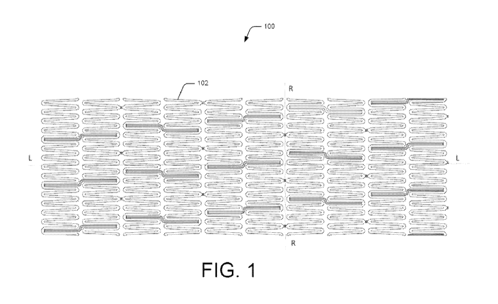
[0005] FIG. 1 illustrates a detailed view of a section of a medical device in
a crimped
state, according to an embodiment of the present subject matter.
[0006] FIG. 2 illustrates a magnified view of a tubular support structure of
the
medical device, according to an embodiment of the present subject matter.
[00 07] FIG. 3A and 3B illustrates a magnified perspective view of the medical
device in an expanded state, according to an embodiment of the present subject
matter.
[0008] FIG. 4 illustrates a magnified view of connections formed in the
tubular
support structure, according to an embodiment of the present subject matter.
[00 09] FIG. 5 illustrates the detailed view of a section of the medical
device,
according to another embodiment of the present subject matter.
[ooio] FIG. 6 illustrates anchor members of the tubular support structure of
the
medical device, according to an embodiment of the present subject matter.
[o on] FIG. 7A and 7B illustrates a perspective view of a section of the
medical
device showing the tubular support structure attached with an end-stopper,
according
to yet another embodiment of the present subject matter.
[0012] Throughout the drawings, identical reference numbers designate similar,
but not necessarily identical, elements. The figures are not necessarily to
scale, and the
size of some parts may be exaggerated to more clearly illustrate the example
shown.
Moreover, the drawings provide examples and/or implementations consistent with
the description; however, the description is not limited to the examples
and/or
implementations provided in the drawings.
DETAILED DESCRIPTION
[0013] Generally, stents are designed in order to have certain inherent
properties
for effective operation. For example, the stents should be highly flexible to
navigate
through tortuous route inside the lumen and it should have sufficient
stiffness and
rigidity in the crimped state to be easily pushed through calcified lesions in
the lumen.
CA 03094521 2020-09-18
WO 2019/186296 PCT/IB2019/051717
3
In addition, the stent should be able to take the shape and conform to the
shape of the
artery during deployment, and at the same time, should have sufficient radial
strength
and rigidity to provide adequate radial support to the artery to avoid
prolapse after
deployment. The stent should have a good expansion ratio with low recoil.
Further,
the design of the stent should be such that it allows the stent to be crimped
without
compromising with the design of the stent, for instance, allowing the stent to
retain its
original axial length after deployment.
[0014] In addition, stents are designed such that the design either restricts
or
accommodates the stress generated in the stent due to different mechanical
forces
applied on it at the time of deploying the stent or after the deployment. Most
commonly, these mechanical forces are elongation, compression, torsional
movement,
bending movement and other physiological conditions e.g. blood flow (after the
deployment). The combined effect of these forces, beyond a safe value, leads
to
fracturing of the joints in deployed stent and the fractured joints can give
rise to many
clinical complications e.g. recoiling, overlapping of adjacent ringlets and
restenosis.
Hence, fracture resistance is a major safety aspect of stent designing which
can be
enhanced by having design features that restrict or manage stress generation
or stress
concentration at potential locations to the minimum level by virtue of their
design.
[0015] In order to meet the abovementioned criteria, the stents have different
constructions, designs and properties each of which attempt to address
different
properties or a combination of the properties as mentioned above. However, due
to
design compromises, most of the stents meet only limited no. of properties and
objectives outlined above, resulting in restricted utility and effectiveness.
[oo16] For example, the tubular support frame of the stent may have many
ringlet
segments. The ringlet segments are formed by segment struts which are of a
specific
design and are sequentially arranged and joined in an endless manner to form a
ring
along the tubular shape of the stent. Further, these ringlet segments are
joined
longitudinally through connectors or connecting ties. The design of these
connectors
and their positioning between two adjoining ringlet segments substantially
affects the
design and performance of the stent either during the deploying of stent or
post-
deployment of the stent or both.
[0017] In another case, a stent can have a wave-like ringlet design without
using
straight struts. Further, in the present case, the stent is designed such that
the ringlet
segment or segment struts are wide at a mid-section. In addition, the stent
may also
CA 03094521 2020-09-18
WO 2019/186296 PCT/IB2019/051717
4
include connecting tie-bars which are also formed as wave-like structure and
connect
peak-to-peak or valley-to-valley of the ringlets, at an offset. As a result,
while the stent
is highly flexible and has good stress distribution, the design compromises on
longitudinal stiffness of the stent.
[0018] In another design, the stent utilizes long connectors to enhance
flexibility,
increased scaffolding and ability to absorb torsional forces. The stent
includes angled
(plate-shaped) struts and straight or angled connectors connecting the ringlet
segments peak-to-peak at an offset. This structure can provide increased
scaffolding,
but increased stiffness in circumferential direction and overall stress in the
stent
during crimping.
[0019] In yet another design, the stent is a helical stent with bio-resorbable
connectors. The connectors connect the crowns/ringlets in peak-to-peak fashion
and
can be of various shapes, such as curved, wave-like, or straight. The
connectors
connect the ringlets peak-to-peak at an angle or curved at 90 on both ends.
In
addition, the stent also includes straight connectors which connect valley-to-
peak in
the adjacent ringlets. This design, though, provides varying degree of
flexibility to the
stent, the stent lacks longitudinal stiffness. In addition, the flexibility of
the connecting
members may change after implantation. In addition, the straight connectors
are to
be released out of plane during bending, say due to bending of the balloon
catheter
used for deployment, and may pose a risk of damage to the lumen.
[0020] The present subject matter describes a medical device, such as a stent,
that
is designed to have a high degree of flexibility, significant radial strength,
fracture
resistance, and negligible axial length loss after deployment. The medical
device, in
accordance with the present subject matter, can have optimum levels of
scaffolding,
flexibility, and radial strength. At the same time, torsional forces in the
medical device
are balanced which is helpful in trackability and makes the medical device
safe.
[0021] According to one aspect of the present subject matter, the medical
device
includes a tubular support structure comprising which is formed of a plurality
of
ringlets which are arranged sequentially along a common longitudinal axis
thereof. In
simpler language, the ringlets have coaxial central longitudinal axes. Each
ringlet is
formed by of plurality of crowns connected along a circumferential direction
and, in
turn, each crown is formed by two straight struts arranged in V-shaped
configuration.
The medical device further includes a plurality of long connecting elements,
and at
least one long connecting element connects adjacent ringlets. The long
connecting
CA 03094521 2020-09-18
WO 2019/186296 PCT/IB2019/051717
element is Z-shaped and consecutive long connecting elements which connect
adjacent ringlets form a mirror-reflection of each other about a radial plane
of
reflection. The radial plane of reflection can be a plane perpendicular to the
common
longitudinal axis of the ringlets of the medical device. Such a design
provides a set of
mechanical properties which allow easy insertion and manoeuvring of the
medical
device into lumens of small diameter having tortuous anatomy.
[0022] According to an aspect of the present subject matter, the long
connecting
element is of Z-shaped configuration and connects the valleys of adjacent
ringlets, i.e.
one end of the long connecting element is connected to a valley-type formation
formed
in one ringlet and the other end of the long connecting element is connected
to a
similar valley-type formation in the adjacent ringlet. The valley type
configuration can
be formed between two struts connected in V-shape in the crown. In an example,
two
adjacent ringlets are connected through long connecting elements at an offset
between
them.
[0023] The long connecting element can be formed of two long sections and a
short
section. In an example, the length of the short section can be equal to or
greater than
the shorter circumferential distance between one valley in one ringlet and the
other
valley in the adjacent ringlet connected by the long connecting element, as
explained
above. In addition, the short section connects the ends of the two long
sections is such
a way to form an obtuse angle between the short section and the long section,
thereby
forming the Z-shape of the long connecting element. As a design element of the
medical device, the angle between the short section and the long section of
the long
connecting member is decided at the time of fabrication and remains fixed
while
crimping or expanding the tubular support structure. In an example, the angle
can be
between 91 and i6o , and the angles between one long section and the short
section
and the other long section and the short section can be substantially same.
[0024] As a result of such configuration of the long connecting element and
the
unchanging nature of the angle of the long connecting element, the length of
the
tubular support structure does not change axially after the tubular support
structure
is released to the normal state from the crimped state, for instance, in self-
expansion
operation. This feature of retaining original axial length of the tubular
support
structure after the deployment of the medical device provides enhanced
accuracy in
treatment of the lumen.
CA 03094521 2020-09-18
WO 2019/186296 PCT/IB2019/051717
6
[0025] Further, in case the medical device is deployed by balloon expansion
mechanism, the design of the long connecting element, in accordance with the
present
subject matter restricts or delays the axial contraction of the tubular
support structure.
Accordingly, the axial length of the tubular support structure may change in
such a
case, but the change is negligible. For instance, the axial length of the
tubular support
structure may change less than 5% of its original length after the deployment
(after
expansion), while being deployed using the balloon-expansion mechanism. In
addition, at the time of fabrication, different expansion and flexural
properties can be
obtained by designing the angle between the short and long sections of the
long
connecting element.
[0026] Therefore, a properly selected and designed angle in the long
connecting
element improves safety and performance of the medical device. Angle present
in the
long connecting element provides improved trackability while the tubular
support
structure is being maneuvered through the lumen to reach the target site and
also
provides stability and radial stiffness too. Additionally, the angle present
in the long
connecting element provides flexibility to the tubular support structure and
helps in
addressing the variations in length due to crimping or expansion.
[0027] As mentioned previously, two consecutive Z-shaped long connecting
elements are opposite or mirror-reflection to each other about a radial plane
of
reflection. In other words, the direction of the Z-shape of any two
consecutive long
connecting element, i.e., between any two consecutive ringlets is a mirror-
reflection
about the plane passing perpendicular to the longitudinal axis of the tubular
support
structure of the medical device. Such a design of the medical device provides
stability,
safety, trackability, fracture resistance, flexibility in crimped state, and
also facilitates
for negligible or no variation in the length of medical device structure
either in crimped
or expanded state.
[0028] Optionally, the medical device can further include a plurality of short
connecting elements to supplement the long connecting elements in connecting
the
ringlets in the medical device. The short connecting elements and long
connecting
elements can connect specific points on crowns of one ringlet to specific
points on
crowns of the adjacent ringlet. The short connecting elements restricts
flexibility but
bring higher bending stiffness to the tubular support structure of the medical
device.
[0029] According to an example, the short connecting elements can connect
peaks
of two adjacent ringlets, i.e. one end of the short connecting element is
connected to a
CA 03094521 2020-09-18
WO 2019/186296 PCT/IB2019/051717
7
peak formed a ringlet and other end is connected to a similar peak formed in
the
adjacent ringlet. In an example, the short connecting elements connect to
ringlets in
such a way that there is no offset between the connected peaks, i.e., the
short
connecting element can connect in-line peaks. Accordingly, the short
connecting
element is substantially parallel to the common longitudinal axis and can be
at a right
angle with the radial plane.
[0030] In another example, one or more long connecting element from among the
plurality of long connecting elements and one or more short connecting element
from
among the plurality of long connecting elements are connected to a common
crown.
In said example, a single short connecting element and a single long
connecting
element can be connected at the same point where one side of the crown forms a
valley
for the long connecting element and the opposite side of the same crown forms
a peak
for the short connecting element on the opposite side.
[0031] During fabrication, flexural and strength related properties of the
medical
device can be customized by defining specific number of short connecting
elements, if
present in the design, in the tubular support structure and the long
connecting
elements present between ringlets. Accordingly, the tubular support structure
can be
easily crimped while having high flexibility. In the crimped state, the
medical device
can be mounted on a catheter and guided through the vessel or organ to the
targeted
vessel part for deployment. After reaching the deployment state, the tubular
support
structure is self-expanded or balloon-expanded to its final state.
[0032] Further, according to an example, two adjacent ringlets can be
connected
only by short connecting elements or long connecting elements. In other words,
each
ringlet is connected with adjacent ringlets through at least one connecting
element,
which can either be a short connecting element or a long connecting element.
Further,
the connecting elements can connect the ringlets alternatively or
continuously, i.e., the
long or short connecting element connect adjacent ringlets alternatively or
they
connect adjacent ringlets continuously.
[0033] In addition, the long connecting elements or the long connecting
elements
along with short connecting elements aid in minimizing the stress generation
or stress
concentration at potential locations due to different mechanical forces
applied on the
medical device at the time of deploying it or after its deployment. The
mechanical
forces may be, for example, elongation, compression, torsional movement,
bending
movement and other physiological conditions, for instance, blood flow (after
the
CA 03094521 2020-09-18
WO 2019/186296 PCT/IB2019/051717
8
deployment). The presence of the long connecting elements or the long
connecting
elements along with short connecting elements restrict the stress generated or
stress
concentration or both in the stent due to the abovementioned factors. The
properties
of the stent, such as radial strength, fracture resistance, flexibility,
bending strength,
and stability, can be achieved by selecting combinations of long and short
connecting
elements in the medical device and by customizing the density of connecting
elements
between two ringlets and along the length of the medical device.
[0034] In addition, width of the straight struts forming the crowns, the long
connecting elements, and the short connecting elements measured in the
circumferential direction of the tubular support frame remain constant along a
length
of the particular element. Also, the thickness of the straight struts, the
long connecting
elements, and the short connecting elements measured in the radial direction
of the
tubular support frame also remains constant along the length.
[0035] The above aspects are further illustrated in the figures and described
in the
corresponding description below. It should be noted that the description and
figures
merely illustrate principles of the present subject matter. Therefore, various
arrangements that encompass the principles of the present subject matter,
although
not explicitly described or shown herein, may be devised from the description
and are
included within its scope.
[0036] FIG. 1 illustrates a developed view of a medical device loo showing a
section
of a tubular support structure 102 in an initial, crimped state according to
an
embodiment. In an example, the medical device loo can be a stent. The medical
device
100, according to the present subject matter, can be placed inside the lumen
of human
or animal, such as an artery, vein, bile duct, urinary tract, alimentary
tract,
tracheobronchial tree, cerebral aqueduct or genitourinary system.
Specifically, the
medical device 100 can be used in femoral artery, superficial femoral artery,
popliteal
artery, tibial artery, genicular artery, cerebral artery, carotid artery,
vertebral artery,
subclavian artery, radial artery, brachial artery, axillary artery, coronary
artery,
peripheral artery, iliac artery or neuro-arteries. For example, the medical
device can
be used to remedy stenosis in superficial femoral artery.
[0037] The tubular support structure 102, according to the present subject
matter,
can be formed of close cell, open cell or hybrid configuration. Further, the
tubular
support structure 102 is made of a material selected from metal, non-metal,
alloy,
polymer, biodegradable, bioresorbable material or a combination of two or more
CA 03094521 2020-09-18
WO 2019/186296 PCT/IB2019/051717
9
thereof. For example, all deformable, medically possible metal, metal alloy
can be used
and include but are not limited to Stainless steel, Cobalt alloys, pure Iron,
Nickel-
titanium alloys, Tantalum, Niobium, Nickel alloys, Magnesium alloys, Zinc
alloys,
L6o5, MP25N, and Nitinol. For instance, the material used for the medical
device 100
deployable through balloon-expansion mechanism is selected from Cobalt
Chromium,
Stainless Steel, Magnesium, Platinum, bioresorbable polymer or a combination
of two
or more thereof. On the other hand, in said example, the material used for the
medical
device loo capable of self-expanding operation is mainly a shape-memory alloy
e.g.
Nitinol.
[0038] In addition, examples of polymers that can be used to fabricate the
medical
device loo in accordance with the present subject matter include but are not
limited
to polymers of L-lactide, Glycolide or combinations of thereof,
poly(hydroxybutyrate),
polyorthoesters, poly anhydrides, poly(glycolic acid), poly(glycolide), poly(L-
lactic
acid), poly(L-lactide), poly(D-lactic acid), poly(D-lactide),
poly(caprolactone),
poly(trimethylene carbonate), polyester amide, polyesters, polyolefins,
polycarbonates, polyoxymethylenes, polyimides, polyethers, and copolymers and
combinations thereof.
[0039] Further, the tubular support structure 102 can carry a biocompatible
material, which in one case, can be a layer of the biocompatible material
coated on the
tubular support structure 102 using any coating technique. The biocompatible
material can be a drug-eluting biocompatible material.
[0040] Structure of the tubular support structure 102 will now be described
with
respect to Fig. 2, 3A, and 3B. FIG. 2 shows a magnified view of the medical
device 100
showing a section of the tubular support structure 102 shown in Fig. 1.
Further, Fig.
3a illustrates a perspective view of the tubular support structure 102 and
Fig. 3h
illustrates front view of the tubular support structure 102. According to an
example,
the tubular support structure 102 may be tubular in shape and is made of
multiple
ringlets for example 202, 204, 206 and 208. The ringlets for example, 202,
204, 206
and 208, are made of endless sequence of multiple crowns 210. In an example,
each
ringlet is formed of four to fifteen crowns 210. The tubular support structure
102 can
be of different lengths and diameters. The length of the tubular support
structure 102
depends on the number of ringlets 202 and the diameter of the tubular support
structure 102 depend on the number of crowns 210 in each ringlet 202.
Depending on
the treatment required for a particular vessel or organ; the number of
ringlets and
CA 03094521 2020-09-18
WO 2019/186296 PCT/IB2019/051717
number of crowns in each ringlet can be customized to prepare a suitable
support
structure for a specific vessel or organ treatment. In one example, the
ringlets 202 are
arranged sequentially in longitudinal axis direction and adjacent ringlets for
example
204, 206 and 208 are connected through long connecting elements 212 or short
connecting elements 214 in continuous manner. The crowns 210 are formed of
struts
216 where struts are arranged in a V-shaped configuration 218 as shown in Fig.
3A.
The width of the struts 216, long connecting elements 212, short connecting
elements
214 measured in the circumferential direction of the tubular support structure
102
remain constant along their lengths. Also, the thickness of the struts 216,
long
connecting elements 212 and short connecting elements 214 measured in the
radial
direction of the tubular support structure 102 remains constant.
[0041] Short connecting elements 214 and long connecting elements 212 connect
crowns of one ringlet to crowns of another adjacent ringlet in a predefined
manner. In
one example, short connecting elements 214 connect two adjacent ringlets in a
peak-
to-peak configuration whereas long connecting elements 212 connect two
adjacent
ringlets in a valley-to-valley configuration. Additionally, no two adjacent
ringlets can
have both type of connecting elements. Two adjacent ringlets for example 204,
206
can be connected only by short connecting elements 214 or long connecting
elements
212. Additionally, the ringlets will be mandatorily connected with adjacent
ringlets
through at least one connecting element where the connecting element can be a
short
connecting element 214 or a long connecting element 212.
[0042] In one implementation, if two adjacent ringlets 202, 204 are connected
through the long connecting elements 212 then the next two adjacent ringlets
204, 206
are connected using the short connecting elements 214. This design involving
alternatively using both type of connecting elements between the ringlets 204
is
followed across the length (longitudinal axis) of the tubular support
structure 102
except the ends of the tubular support structure 102. If required, at the ends
of the
tubular support structure 102, the last one to three ringlets 202 from the end
are
connected using the short connecting elements 214 in peak-to-peak
configuration.
This is also shown in FIG. 4. The peak-to-peak connecting configuration
between these
ringlets 204 can be at all the peaks or at alternative peaks. However, it is
possible to
use either the long connecting elements 212 or short connecting elements 214
in
continuous manner or in blocks. In continuous manner, all the ringlets 202,
204, 206,
and 208 will be connected through either the long connecting elements 212 or
short
CA 03094521 2020-09-18
WO 2019/186296 PCT/IB2019/051717
11
connecting elements 214. In blocks manner, some ringlets will be connected
through
one type of connecting elements and followed by this block, some others will
be
connected through another type of connecting element.
[0043] According to an aspect, as depicted in FIG. 1, FIG. 2, FIG. 3A, Fig.
3B, and
FIG. 4, the long connecting elements 212 is of a Z-shaped configuration 220
and
connects the ringlets 202, 204, 206, and 208 in valley-to-valley configuration
i.e. one
end of the long connecting elements 212 is connected to a valley-type
formation
formed between two struts 216 connected in a V-shaped configuration 218 in the
crown 4 of a ringlet 202 and the other end of the long connecting elements 212
is
connected to a similar valley formation in an adjacent ringlet 204. The long
connecting
elements 212 is formed of two long sections 402 and a short section 404. The
short
section 404 connects the ends of the two long sections 402 in such a way to
form an
obtuse angle 0 between the short section 404 and the long section 402.
[0044] As a design element, angle 0 is decided at the time of fabrication and
remains fixed while crimping or expanding the tubular support structure. This
angle 0
can be designed between 91 and 16o , including both the angles. Due to this
shape of
the long connecting elements 212 and unchanging nature of this angle 0 present
in the
long connecting elements 212; the length of the tubular support structure does
not
change axially after the tubular support structure is deployed in self-
expansion
mechanism. This feature of retaining original axial length of the tubular
support
structure, after the deployment of the tubular support structure, provides
enhanced
accuracy in treatment of the vessel or organ. In balloon expansion mechanism,
the
long connecting element 212 restricts or delays the change in axial length of
the tubular
support structure 102. Hence, in balloon-expansion mechanism, the length of
the
tubular support structure 102 contracts but minimally.
[0045] Similarly, the struts 216 also form an angle 01 with the long section
402 of
the long connecting elements 212. This angle 01 is not fixed and increases or
decreases
depending on crimping or expansion. Combination of long connecting elements
212
with unchanging angle 0 and expandable/ crimpable peak/valley brings a desired
set
of required properties in a tubular support structure. Long connecting element
212
provides structural stability, flexibility whereas V-shaped peak/valley
provides
expandability. A straight long connecting element 212 has poor stress
distribution and
can pose a risk to the vessel during deployment of the balloon catheter.
Having an
angle in the long connecting elements 212 provides improved safety while the
tubular
CA 03094521 2020-09-18
WO 2019/186296 PCT/IB2019/051717
12
support structure 102 is being maneuvered through the vessel to the target
site. In
addition, the unchanging angle 0 in the long connecting element helps in
managing
the stress generation or stress concentration due to different mechanical
forces
applied on the tubular support structure 102 at the time of deploying it or
after its
deployment. Most commonly, these mechanical forces are elongation,
compression,
torsional movement, bending movement and other physiological conditions e.g.
blood
flow (after the deployment)
[0046] FIG. 5 depicts another embodiment of the tubular support structure 102,
according to the present subject matter, where the adjacent ringlets 202, 204
can be
connected using only the long connecting elements 212 in a valley-to-valley
configuration. Optionally, the last two ringlets 206 and 208 can be connected
using
the short connecting elements 214 in a peak-to-peak configuration. The peak-to-
peak
connecting configuration between these ringlets can be at all the peaks or at
alternative
peaks.
[0047] As seen in FIGS. 2,4 and 5, as one moves along the tubular support
structure
in longitudinal direction; placement of the long connecting elements 212 is in
such a
way so that the long connecting elements 212 between two ringlets 202, 204 is
mirror
reflection of the long connecting element 212 present in next two ringlets
206, 208.
This arrangement reduces strain development in one direction and bring greater
stability, safety, radial stiffness, flexibility, fracture resistance and
trackability. If all
long connecting elements 212 are in one direction, it will bring an inherent
tendency
in the tubular support structure to deflect to one side which is not a
required property
and also poses risk to the patient.
[0048] As shown is FIGS. 1, 2, 3A, 3B, 4, 5 and 6, the short connecting
elements 214
connect two adjacent ringlets in peak-to-peak manner i.e. one end of the short
connecting element 214 is connected to a peak formed at adjoining point of two
struts
216 in the ringlet 202 and the other end is connected to a similar peak formed
in the
adjacent ringlet 204. These two peaks are in same line longitudinally i.e.
there is no
offset. Hence, the short connecting elements 214 are at right angle to the
radial axis
and parallel to the longitudinal axis. In another embodiment, the short
connecting
elements 214 and long connecting elements 212 can adjoin a common point where
two
struts 216 will form a valley for the long connecting element 212 and the same
two
struts will form a peak for the short connecting element 214. The short
connecting
elements 214 provide low flexibility and high bending stiffness to the tubular
support
CA 03094521 2020-09-18
WO 2019/186296 PCT/IB2019/051717
13
structure. During fabrication, flexural and strength related properties of a
stent can be
customized by defining specific number of short connecting elements 214 and
long
connecting elements 212 present in the stent between ringlets. Their specific
combination will give specific set of properties. In specific cases, short
connecting
elements 214 are not included in the tubular support structure and the desired
set of
properties are obtained from presence of long connecting elements 212 only in
the
tubular support structure.
[0049] Additionally, FIG. 3A and 3B show expanded views of the tubular support
structure, according to an example. While Fig. 3A illustrates a perspective
view of the
tubular support structure of the medical device 100, Fig. 3B illustrates a two-
dimensional view of the tubular support structure of the medical device loo.
In Fig.
3A and 3B, it can be seen that in expanded state, shape of long connecting
element is
unchanged. Also, there is no sign of buckling too whereas the crown formed due
to V-
shaped adjoining of struts 216 expands and increase the diameter of the
tubular
support structure.
[0050] FIG. 6 illustrates another embodiment of the medical device 100, where
the
tubular support structure 102 has at least one anchor member 602 attached to
either
one end or both the ends of the tubular support structure 102. The anchor
member
602 prevents the tubular support structure 102 from moving axially or radially
from
the target site. In one case, the medical device 100 has multiple anchor
members 602
at one end of the tubular support structure 102. In another case, the medical
device
100 can have multiple anchor members at both the ends of the tubular support
structure 102. The anchor members 602 at one or both ends of the tubular
support
structure 102 can have radiopaque markers to aid the physician in positioning
the
medical device under fluoroscopic imaging.
[oo51] FIG. 7A and 7B show an end-stopper 702 that is used with a tubular
support
structure 102, according to an example. While Fig. 7A illustrates a
perspective view of
the tubular support structure 102 with the end-stopper 702, Fig. 7B
illustrates a two-
dimensional view of the tubular support structure 102 with the end-stopper
702. The
end-stopper 702 can be hollow, circular and can have a uniform diameter, for
example,
for uniform load distribution. In an example, the end-stopper 702 can be used
with
medical device loo which are manufactured to be used as self-expanding stents.
Fig.
7A shows the end-stopper 702 at the time of deployment, when a self-expandable
support structure is being deployed in a vessel or organ. The end-stopper 702
is a
CA 03094521 2020-09-18
WO 2019/186296 PCT/IB2019/051717
14
hollow, circular structure of uniform diameter that is attached to an inner
tube of a
catheter and helps is uniform load transfer to the tubular support structure
102 at the
time of deployment. The end-stopper 702 has peripheral slots or grooves 704 to
accommodate anchor members 602 of the tubular support structure (also shown in
Fig. 6). These slots or grooves 704 helps in better and uniform load transfer
to the
tubular support structure at the time of deploying the stent at the target
site.
[0052] In addition, the present subject matter also envisages a method for
fabricating the medical device loo as explained above. For the manufacturing
of the
medical device loo, the method can involve, firstly, loading a medically clean
and
approved work-piece in a designing instrument. According to one example of the
present subject matter, the work-piece or the specimen can be in shape of a
hollow
circular tube, a film, or a sheet. Then the required design of the medical
device loo is
set-up or uploaded in the designing instrument, such as a computer-numerical
controlled (CNC) machine for manufacturing. Subsequently, the required design
is
carved out of the work-piece to fabricate the medical device 100, such as a
tubular
support structure or a stent. In one example, the fabrication technique used
in the
designing instrument is selected from laser fabrication, chemical-etching,
photochemical-etching or electro-discharge machining. For instance, the
medical
device 100 is fabricated by slitting a metallic hollow circular tube with a
laser beam,
the laser beam following a predefined cutting contour to produce the design of
the
medical device 100, as has been explained in the foregoing description of the
present
subject matter. Once the medical device loo has been manufactured, the
undesired
material is removed from the surface of the medical device loo for finishing.
The
cleaned and finished medical device 100 can then be polished or coated with an
appropriate coating. For example, it can be coated with an anti-reactive agent
which
prevents it from reacting with the atmosphere where either the medical device
100 is
stored or deployed. Additionally or alternatively, the medical device loo can
be
covered with a medicinal substance, depending on the purpose, mode, and
location of
deployment of the medical device loo. Further, the tubular support structure
102 can
be manufactured using 3D printing technique or additive manufacturing. 3D
printing
technique can be selected from Stereolithography (SLA), Digital light
processing
(DLP), Fused deposition modelling (FDM), Selective laser sintering (SLS),
Selective
laser melting (SLM), Electronic beam melting (EBM), Laminated object
manufacturing (LOM), Polyjet technology or a combination of thereof.
CA 03094521 2020-09-18
WO 2019/186296 PCT/IB2019/051717
[0053] Overall, the medical device 100 has high radial stiffness, zero or
minimal
axial length loss after deployment, enhanced flexibility and better bending
stiffness.
This ensures excellent and uniform bracing of the medical device 100 with the
wall of
the lumen, thereby providing effective support. The medical device loo,
according to
the present subject matter, can, therefore, be easily crimped and expanded
through
balloon-expandable delivery mechanism or self-expanded delivery mechanism. For
example, the design supports easy crimping of the tubular support structure
102
during the deployment process. However, the inherent flexibility and stability
due to
the design helps in easy movement of the medical device loo along the tortuous
paths
of a vessels during the implantation, with a higher safety level for both the
patient and
the physician.
[0054] Although design and application of the medical device 100 are
described, it
is to be understood that the present subject matter is not limited to the
specific features
or methods described. Rather, the specific features and methods are disclosed
as
implementations of the medical device 100.