Note: Descriptions are shown in the official language in which they were submitted.
METHODS AND DEVICES FOR KNEE SURGERY WITH INERTIAL SENSORS
Field
[0001] The present disclosure is directed to devices, methods, and techniques
related to computer
aided surgery and computer planned surgery.
Background
[0002] Computer aided surgery has been shown to improve precision of
orthopedic surgery in
most large joints, specifically hip and knee joints. Conventional knee
replacement surgery requires
several surgical trays with a plethora of unique surgical instruments, where
each surgical tray is
expensive, heavy, and requires sterilization for reuse. Technology has been
developed in the field
of computer aided surgery to reduce the number of instruments while
maintaining or slightly
improving precision. Current configurations of such technologies include
surgical robotics, optical
navigation, and inertial sensor-based instrumentation.
[0003] Each of these current systems has inherent advantages and
disadvantages. For example,
robotic systems are expensive, bulky, and often require significant time to
setup, tear down, and
execute the surgical procedure. Optical systems are also expensive, suffer
from line-of-sight
issues, and require similar time as robotics to setup and tear down. Inertial
systems, in most of
their current forms, require significant manual instrumentation to constrain
the degrees of freedom
due to shortcomings in the technology, often requiring at least one or two
instrument trays.
Therefore, there is a need for technology to improve surgical precision
without the cost, space
constraints, and capital equipment requirements of current technologies.
Summary
[0004] What is disclosed herein are techniques, methods, and devices as part
of a computer aided
surgical navigation system for the knee joint surgeries and instrumentation to
support the same so
that the navigation system may be delivered in a "just-in-time" manner with
minimal
instrumentation.
[0005] It is a first aspect of the present invention to provide a use of
navigating a cutting instrument
via a computer system for carrying out one of a cut and pin placement, the use
comprising: (a)
mounting a patient-specific anatomical mapper (PAM) to a human in a single
known location and
1
Date Recue/Date Received 2021-06-30
orientation, where the PAM includes a surface precisely and correctly mating
with a human surface
correctly in only a single location and orientation; (b) mounting a reference
inertial measurement
unit (IMU) to the human; (c) operatively coupling a guide to the PAM, where
the guide includes
an instrument inertial measurement unit (IMU) and one of a cutting slot and a
pin orifice; (d)
outputting data from the reference IMU and the instrument IMU indicative of
changes in position
and orientation of the guide with respect to the human; (e) repositioning the
guide with respect to
the human to a position and an orientation consistent with a plan for carrying
out the one of a cut
and pin placement; and, (f) visually displaying feedback concerning the
position and orientation
of the guide with respect to the human using data output from the reference
IMU and the instrument
IMU, which data is processed by a computer program and the computer program
directs the
visually displayed feedback.
[0006] In a more detailed embodiment of the first aspect, the PAM is mounted
to at least one of a
tibia and a femur. In yet another more detailed embodiment, the reference IMU
is mounted to at
least one of a tibia and a femur. In a further detailed embodiment,
operatively coupling the guide
to the PAM includes using a mechanical connection comprising at least two
joints to allow
repositioning of the cutting guide independent of the PAM. In still a further
detailed embodiment,
the at least two joints comprise at least one of a revolute joint and a
spherical joint. In a more
detailed embodiment, the at least two joints comprise a pair of revolute
joints and a spherical joint.
In a more detailed embodiment, the method further includes registering the
reference IMU with
respect to the instrument IMU while at least one of the reference IMU and the
instrument IMU is
in a known position and orientation with respect to the human. In another more
detailed
embodiment, the instrument IMU is mountable to the guide in only a single
known location and
orientation. In yet another more detailed embodiment, the reference IMU is
mountable to the
patient in a plurality of locations and orientations. In still another more
detailed embodiment,
registering the reference IMU with respect to the instrument IMU includes
holding the IMUs
stationary with respect to one another for a predetermined period of time.
[0007] In yet another more detailed embodiment of the first aspect,
repositioning the guide with
respect to the human includes repositioning the guide with respect to the PAM.
In yet another
more detailed embodiment, the method further includes performing an evaluation
using a load
measuring device operatively coupled to the instrument IMU to assess knee
joint laxity. In a
further detailed embodiment, the plan comprises a plan for placing the pin. In
still a further
2
Date Recue/Date Received 2021-06-30
detailed embodiment, visually displaying feedback includes displaying a
virtual model of patient
anatomy and reference indicia reflecting a position of the guide with respect
to the patient anatomy.
In a more detailed embodiment, visually displaying feedback includes
displaying a virtual model
of patient anatomy and reference indicia reflecting an intended location for
the cut on the virtual
model. In a more detailed embodiment, the method further includes mounting at
least one pin to
a resected aspect of the patient, orienting a static cutting guide with
respect to the patient using the
at least one pin, and mounting the static cutting guide to the patient post
orienting the static cutting
guide. In another more detailed embodiment, the method further includes
mounting at least one
pin to a resected aspect of the patient, orienting a guide foot with respect
to the patient using the
at least one pin, mounting the guide foot to the patient post orienting the
guide foot, discontinuing
the operative coupling between the PAM and guide, and operatively coupling the
guide to the
guide foot.
[0008] It is a second aspect of the present invention to provide a surgical
equipment system
comprising: (a) a first inertial measurement unit (IMU) having a gyroscope, an
accelerometer, and
a magnetometer; (b) a second inertial measurement unit (IMU) having a
gyroscope, an
accelerometer, and a magnetometer, the second IMU configured to be mounted to
a reference
device, where the reference device is configured to be mounted to patient
anatomy; (c) a patient-
specific anatomical mapper (PAM) that includes a surface precisely and
correctly mating with a
patient anatomy surface in only a single location and orientation, where the
PAM is adapted to be
mounted to the patient anatomy surface; and, (d) a guide configured to be
operatively coupled to
the PAM when in use, the guide including at least one of a cutting slot and a
pin orifice, the guide
configured to couple to the first IMU in a predetermined known position and
orientation.
[0009] In a more detailed embodiment of the second aspect, the system further
includes a
controller including software having preloaded at least one virtual anatomical
model of the patient
anatomy and a pre-operative surgical plan indicating the position and
orientation of an intended
bone resection with respect to the at least one virtual anatomical model of
the patient anatomy, the
controller configured to be communicatively coupled to the first and second
IMUs to receive IMU
data and to translate the received IMU data to determine the position and
orientation of the guide
with respect to the patient anatomy and output instructions for a display to
visually represent the
virtual anatomical model of the patient anatomy and provide guidance as to
whether the guide is
positioned with respect to the patient anatomy consistent with the pre-
operative surgical plan to
3
Date Recue/Date Received 2021-06-30
achieve the intended bone resection. In yet another more detailed embodiment,
the patient-specific
anatomical mapper is configured to engage at least one of a proximal tibia and
a distal femur. In
a further detailed embodiment, the patient-specific anatomical mapper
comprises a first tibia PAM
and a second femur PAM. In still a further detailed embodiment, the system
further includes a
mechanical connection operative to couple the guide to the PAM, the mechanical
connection
including at least one joint. In a more detailed embodiment, the at least one
joint comprises at
least two joints. In a more detailed embodiment, the at least two joints
include a revolute joint and
a spherical joint. In another more detailed embodiment, the at least two
joints include a pair of
revolute joints. In yet another more detailed embodiment, the mechanical
connection is configured
to concurrently mount to a first predetermined location of the PAM and a
second predetermined
location of the guide to assume a registration position and orientation. In
still another more
detailed embodiment, the system further includes a load measuring device
configured to couple to
the first IMU in a known position and orientation.
[0010] In yet another more detailed embodiment of the second aspect, the load
measuring device
comprises at least one of a plurality of piezoresistive sensors, a plurality
of capacitive sensors, and
a plurality of piezoelectric based strain sensors. In yet another more
detailed embodiment, the
system further comprises an orthopedic implant placement device configured to
couple to the first
IMU in a known position and orientation. In a further detailed embodiment, the
orthopedic implant
placement device is configured to couple to an orthopedic implant in a
predetermined location and
orientation, where the orthopedic implant comprises at least one of an
orthopedic trial and a final
orthopedic implant. In still a further detailed embodiment, the orthopedic
implant comprises at
least one of a tibial implant and a femoral implant as part of at least one of
a knee replacement
surgery or a knee revision surgery. In a more detailed embodiment, the system
further includes a
display communicatively coupled to the controller, the display operative to
visually represent the
virtual anatomical model of the patient anatomy and provide guidance as to
whether the guide is
positioned with respect to the patient anatomy consistent with the pre-
operative surgical plan to
achieve the intended bone resection. In a more detailed embodiment, the
display comprises a
plurality of display windows. In another more detailed embodiment, each of the
plurality of
display windows is associated with a stand-alone screen. In yet another more
detailed
embodiment, the system further comprises an orthopedic implant comprising at
least one of a final
orthopedic implant and an orthopedic trial. In still another more detailed
embodiment, the final
4
Date Recue/Date Received 2021-06-30
orthopedic implant comprises a component of a total knee joint replacement or
a partial knee joint
replacement.
[0011] In a more detailed embodiment of the second aspect, the final
orthopedic implant comprises
at least one of a patient-specific femoral component and a patient-specific
tibial component of a
total knee replacement. In yet another more detailed embodiment, the system
further includes a
guide foot configured to be operatively coupled to the guide when the guide
foot is mounted to the
patient anatomy in order to facilitate at least one bone cut.
[0012] It is a third aspect of the present invention to provide a use of
inertial measurement units
to facilitate three dimensional tracking of a surgical tool, via a computer
system, the use
comprising: (a) mounting a first inertial measurement unit (IMU) to a first
mammalian tissue so
that the first IMU is not repositionable with respect to the first mammalian
tissue; (b) operatively
coupling a second inertial measurement unit (IMU) to the first mammalian
tissue by using a
patient-specific anatomical mapper (PAM) having a surface precisely and
correctly mating with a
surface of the first mammalian tissue in only a single location and
orientation, the second IMU
being repositionable with respect to the first mammalian tissue; (c)
registering the position and
orientation of the second IMU with respect to the first mammalian tissue and
the first IMU while
the PAM is mounted to the first mammalian tissue; (d) mounting the second IMU
to a surgical
tool; and, (e) tracking a position and an orientation of the surgical tool and
first mammalian tissue
in three dimensions while the second IMU is mounted to the surgical tool and
repositionably
coupled to the PAM.
[0013] In a more detailed embodiment of the third aspect, the method further
comprises visually
displaying feedback concerning the position and orientation of the surgical
tool with respect to the
first mammalian tissue using data output from the first and second IMUs, which
data is processed
by a computer program and the computer program directs the visually displayed
feedback. In yet
another more detailed embodiment, the feedback comprises a virtual model of
the first mammalian
tissue and first indicia on the virtual model indicating the relative real-
world position of the
surgical tool with respect to the first mammalian tissue. In a further
detailed embodiment, the
feedback further comprises a second indicia on the virtual model indicating an
intended position
of the surgical tool with respect to the first mammalian tissue consistent
with a predetermined plan.
In still a further detailed embodiment, the PAM is mounted to at least one of
a tibia and a femur.
In a more detailed embodiment, the first mammalian tissue comprises at least
one of a tibia and a
Date Recue/Date Received 2021-06-30
femur. In a more detailed embodiment, the surgical tool is operatively coupled
to the PAM. In
another more detailed embodiment, operatively coupling the surgical tool to
the PAM includes
using a mechanical connection comprising at least two joints to allow
repositioning of the surgical
tool independent of the PAM. In yet another more detailed embodiment, the at
least two joints
comprise at least one of a revolute joint and a spherical joint. In still
another more detailed
embodiment, the at least two joints comprise a pair of revolute joints and a
spherical joint.
[0014] In yet another more detailed embodiment of the third aspect, the second
IMU is mountable
to the surgical tool in only a single known location and orientation. In yet
another more detailed
embodiment, the first IMU is mountable to the first mammalian tissue in a
plurality of locations
and orientations. In a further detailed embodiment, the position and
orientation of the second IMU
with respect to the first mammalian tissue includes holding the first and
second IMUs stationary
with respect to one another for a predetermined period of time. In still a
further detailed
embodiment, the method further includes performing an evaluation using a load
measuring device
operatively coupled to the second IMU to assess knee joint laxity.
[0015] It is a fourth aspect of the present invention to provide a surgical
equipment kit for a knee
replacement or revision procedure comprising: (a) a first inertial measurement
unit (IMU) having
a gyroscope, an accelerometer, and a magnetometer; (b) a second inertial
measurement unit (IMU)
having a gyroscope, an accelerometer, and a magnetometer; (c) a tibial patient-
specific anatomical
mapper (PAM) that includes a surface precisely and correctly mating with a
tibial surface in only
a single location and orientation, where the tibial PAM is configured to be
mounted to the tibial
surface; and, (d) a femoral patient-specific anatomical mapper (PAM) that
includes a surface
precisely and correctly mating with a femoral surface in only a single
location and orientation,
where the femoral PAM is adapted to be mounted to the femoral surface, where
the second IMU
is configured to be operatively coupled to at least one of the tibial PAM and
the femoral PAM.
[0016] In a more detailed embodiment of the fourth aspect, the kit further
includes a cutting guide
configured to be repositionably coupled to at least one of the tibial PAM and
the femoral PAM,
the cutting guide including at least one of a cutting slot and a pin orifice,
the guide configured to
couple to the first IMU in a predetermined known position and orientation. In
yet another more
detailed embodiment, the kit further includes a mechanical connection
comprising at least two
joints to operatively couple the cutting guide to at least one of the tibial
PAM and the femoral
PAM. In a further detailed embodiment, the orthopedic implant comprises a non-
patient-specific
6
Date Recue/Date Received 2021-06-30
implant. In still a further detailed embodiment, the non-patient-specific
implant includes a femoral
condyle and a tibial tray insert. In a more detailed embodiment, the non-
patient-specific implant
includes a femoral implant having a pair of condyles and a tibial tray insert
having a pair of condyle
receivers. In a more detailed embodiment, the kit further includes a reference
housing configured
to be rigidly mounted to at least one of a tibia and a femur, the reference
housing configured to
mount to the first IMU correctly in only a single position and orientation. In
another more detailed
embodiment, the kit further includes a 4-in-1 static cutting block. In yet
another more detailed
embodiment, the kit further includes a 4-in-1 reconfigurable cutting block. In
still another more
detailed embodiment, the kit further includes a physical memory device upon
which is stored
computer readable code that, when executed by a computer, is operative to
provide surgical
navigation guidance consistent with a pre-operative plan.
[0017] In yet another more detailed embodiment of the fourth aspect, the mass
customized implant
includes a femoral implant having a pair of condyles and a tibial tray insert
having a pair of condyle
receivers. In yet another more detailed embodiment, the at least two joints
comprise at least one
of a revolute joint and a spherical joint. In a further detailed embodiment,
the at least two joints
comprise a pair of revolute joints and a spherical joint. In still a further
detailed embodiment, the
kit further includes an orthopedic implant configured to replace at least a
portion of a knee joint.
In a more detailed embodiment, the orthopedic implant comprises a patient-
specific implant. In a
more detailed embodiment, the patient-specific implant includes a femoral
condyle and a tibial
tray insert. In another more detailed embodiment, the patient-specific implant
includes a femoral
implant having a pair of condyles and a tibial tray insert having a pair of
condyle receivers. In yet
another more detailed embodiment, the orthopedic implant comprises a mass
customized implant.
In still another more detailed embodiment, the mass customized implant
includes a femoral
condyle and a tibial tray insert. In yet another more detailed embodiment, the
kit includes a copy
of an internet address that may be accessed to provide stored computer
readable code that, when
executed by a computer, is operative to provide surgical navigation guidance
consistent with a pre-
operative plan.
[0018] It is a fifth aspect of the present invention to provide a surgical
equipment kit for a knee
replacement or revision procedure comprising: (a) a tibial patient-specific
anatomical mapper
(PAM) that includes a surface precisely and correctly mating with a tibial
surface in only a single
location and orientation, where the tibial PAM is configured to be mounted to
the tibial surface;
7
Date Recue/Date Received 2021-06-30
and, (b) a femoral patient-specific anatomical mapper (PAM) that includes a
surface precisely and
correctly mating with a femoral surface in only a single location and
orientation, where the femoral
PAM is configured to be mounted to the femoral surface.
[0019] It is a fifth aspect of the present invention to provide a surgical
navigation system
comprising: (a) a tibial patient-specific anatomical mapper (PAM) that
includes a surface precisely
and correctly mating with a tibial surface in only a single location and
orientation, where the tibial
PAM is adapted to be mounted to the tibial surface; (b) a femoral patient-
specific anatomical
mapper (PAM) that includes a surface precisely and correctly mating with a
femoral surface in only
a single location and orientation, where the femoral PAM is adapted to be
mounted to the femoral
surface; (c) a first inertial measurement unit (IMU) having a gyroscope, a
plurality of
accelerometers, and a magnetometer; (d) a first transmitter communicatively
coupled to the first
IMU; (e) a second inertial measurement unit (IMU) having a gyroscope, a
plurality of
accelerometers, and a magnetometer; (f) a second transmitter communicatively
coupled to the
second IMU; (g) a first signal receiver communicatively coupled to the first
and second
transmitters; (h) a cutting guide adapted to be operatively coupled to at
least one of the tibial PAM
and the femoral PAM, the guide including at least one of a cutting slot and a
pin orifice, the cutting
guide configured to couple to the first IMU correctly in only a single
position and orientation; (i) a
controller communicatively coupled to the first signal receiver, the
controller including software
having access to a virtual model of patient anatomy and a pre-operative
surgical plan indicating
intended resection cuts with respect to the virtual model; and
a visual display communicatively coupled to the controller, wherein the
controller software is
adapted to process data from the first and second IMUs to determine position
and orientation of the
cutting guide with respect to the patient anatomy and output instructions for
the visual display to
visually represent the virtual model of the patient anatomy and provide
guidance as to whether the
cutting guide is positioned with respect to the patient anatomy consistent
with the pre-operative
surgical plan to achieve the intended resection cuts.
[0020] In a more detailed embodiment of the fifth aspect, the tibial PAM is
configured to engage a
proximal portion of a tibia and the femoral PAM is configured to engage a
distal portion of the
femur. In a further detailed embodiment, the system further includes a
mechanical connection
operative to couple the cutting guide to at least one of the tibial PAM and
the femoral PAM, the
mechanical connection including at least one joint. In still a further
detailed
8
Date Recue/Date Received 2021-11-17
embodiment, the at least one joint comprises at least two joints. In a more
detailed embodiment,
the at least two joints include a revolute joint and a spherical joint. In a
more detailed embodiment,
the at least two joints include a pair of revolute joints. In another more
detailed embodiment, the
mechanical connection is configured to concurrently mount to a first
predetermined location of at
least one of the tibial PAM and the femoral PAM and a second predetermined
location of the
cutting guide to assume a registration position and orientation.
[0021] In a more detailed embodiment of the fifth aspect, the system further
includes a load
measuring device configured to couple to the first IMU in a known position and
orientation. In
yet another more detailed embodiment, the load measuring device comprises at
least one of a
plurality of piezoresistive sensors, a plurality of capacitive sensors, and a
plurality of piezoelectric
based strain sensors. In a further detailed embodiment, the system further
includes an orthopedic
implant placement device configured to couple to the first IMU in a known
position and
orientation. In still a further detailed embodiment, the orthopedic implant
placement device is
configured to couple to an orthopedic implant in a predetermined location and
orientation, where
the orthopedic implant comprises at least one of an orthopedic trial and a
final orthopedic implant.
In a more detailed embodiment, the orthopedic implant comprises at least one
of a tibial implant
and a femoral implant as part of at least one of a knee replacement surgery or
a knee revision
surgery. In a more detailed embodiment, the visual display comprises a
plurality of display
windows. In another more detailed embodiment, each of the plurality of display
windows is
associated with a stand-alone screen. In yet another more detailed embodiment,
the system further
includes an orthopedic implant comprising at least one of a final orthopedic
implant and an
orthopedic trial. In still another more detailed embodiment, the final
orthopedic implant comprises
a component of a total knee joint replacement or a partial knee joint
replacement. In yet another
more detailed embodiment, the final orthopedic implant comprises at least one
of a patient-specific
femoral component and a patient-specific tibial component of a total knee
replacement. In yet
another more detailed embodiment, the system further includes a guide foot
configured to be
operatively coupled to the cutting guide when the guide foot is mounted to the
patient anatomy in
order to facilitate at least one bone cut.
[0022] It is a sixth aspect of the present invention to provide a use of a
cutting guide to make a
femoral bone cut consistent with a pre-operative surgical plan, comprising
repositioning a cutting
guide using navigation guidance displayed on a visual display, the cutting
guide including a first
9
Date Recue/Date Received 2021-06-30
inertial measurement unit (IMU), the cutting guide operatively coupled to a
femoral patient-
specific anatomical mapper (PAM) that includes a surface precisely and
correctly mating with a
femoral surface in only a single location and orientation, where the
navigation guidance includes
at least one of a virtual model of the cutting guide and a virtual model of a
patient femur, as well
as an indication regarding a three dimensional position of the cutting guide
with respect to the
patient femur using data from the first IMU, where the navigation guidance
also includes guidance
for repositioning the cutting guide to make a femoral bone cut consistent with
a pre-operative
surgical plan
[0023] In a more detailed embodiment of the sixth aspect, the method further
includes
repositioning the cutting guide using navigation guidance displayed on the
visual display, the
cutting guide including the first inertial measurement unit (IMU), the cutting
guide operatively
coupled to a tibial patient-specific anatomical mapper (PAM) that includes a
surface precisely and
correctly mating with a tibial surface in only a single location and
orientation, where the navigation
guidance includes at least one of the virtual model of the cutting guide and a
virtual model of a
patient tibia, as well as an indication regarding the three dimensional
position of the cutting guide
with respect to the patient tibia using data from the first IMU, where the
navigation guidance also
includes guidance for repositioning the cutting guide to make a tibial bone
cut consistent with the
pre-operative surgical plan. In yet another more detailed embodiment, the
method further includes
mounting the femoral PAM surface to the patient femoral surface in the correct
single location and
orientation, coupling a second inertial measurement unit (IMU) to the patient
femur, and
registering the first and second IMUs with respect to one another. In a
further detailed
embodiment, the method further includes mounting the tibial PAM surface to the
patient tibial
surface in the correct single location and orientation, coupling a second
inertial measurement unit
(IMU) to the patient tibia, and registering the first and second IMUs with
respect to one another.
In still a further detailed embodiment, the navigation guidance includes only
the virtual model of
the cutting guide. In a more detailed embodiment, the navigation guidance
includes only the
virtual model of the patient femur. In a more detailed embodiment, the
navigation guidance
includes at least one of the virtual model of the patient femur and the
virtual model of the patient
tibia, in addition to a first cutting line representing the real-world
position of the cutting guide and
a second cutting line representing an intended pre-operative plan position of
the cutting guide for
making at least one of the femoral bone cut and the tibial bone cut.
Date Recue/Date Received 2021-06-30
[0024] In a further detailed embodiment, the method further includes making
the femoral bone cut
using a surgical saw guided by the cutting guide, and repositioning the
cutting guide using
navigation guidance displayed on the visual display, the cutting guide
including the first inertial
measurement unit (IMU) and being operatively coupled to the femoral patient-
specific anatomical
mapper (PAM), where the navigation guidance includes at least one of the
virtual model of the
cutting guide and the virtual model of the patient femur, as well as an
indication regarding the
three dimensional position of the cutting guide with respect to the patient
femur using data from
the first IMU, where the navigation guidance also includes guidance for
repositioning the cutting
guide to make a subsequent femoral bone cut consistent with the pre-operative
surgical plan. In
still a further detailed embodiment, the method further includes making the
femoral bone cut using
a surgical saw guided by the cutting guide, where the femoral bone cut is a
distal resection, and
repositioning the cutting guide using navigation guidance displayed on the
visual display, the
cutting guide including the first inertial measurement unit (IMU) and being
operatively coupled to
the femoral patient-specific anatomical mapper (PAM), where the navigation
guidance includes at
least one of the virtual model of the cutting guide and the virtual model of
the patient femur, as
well as an indication regarding the three dimensional position of the cutting
guide with respect to
the patient femur using data from the first IMU, where the navigation guidance
also includes
guidance for repositioning the cutting guide to drill holes into the resected
femur consistent with
the pre-operative surgical plan. In a more detailed embodiment, the method
further includes
drilling holes into the resected femur femoral bone using a surgical drill
guided by the cutting
guide, inserting surgical pins into the drill holes, repositioning a 4-in-1
cutting guide against the
resected femur using the inserted surgical pins for alignment, and making at
least one femoral
resection cut using guidance from the 4-in-1 cutting guide. In a more detailed
embodiment, the
method further includes drilling holes into the resected femur femoral bone
using a surgical drill
guided by the cutting guide, inserting surgical pins into the drill holes,
repositioning a fixed
position cutting guide against the resected femur using the inserted surgical
pins for alignment,
and making at least one femoral resection cut using guidance from the fixed
position cutting guide.
[0025] According to another broad aspect, there is provided a surgical
equipment kit for a knee
replacement or revision procedure, the kit comprising: a first inertial
measurement unit (IMU)
comprising a gyroscope, an accelerometer and a magnetometer; a second inertial
measurement
unit (IMU) comprising a gyroscope, an accelerometer and a magnetometer; a
tibial patient-specific
11
Date Recue/Date Received 2021-06-30
anatomical mapper (PAM) comprising a surface precisely and correctly mating
with a tibial
surface in only a single location and orientation, the tibial PAM being
adapted to be mounted to
the tibial surface; a femoral patient-specific anatomical mapper (PAM)
comprising a surface
precisely and correctly mating with a femoral surface in only a single
location and orientation, the
femoral PAM being adapted to be mounted to the femoral surface; a cutting
guide adapted to be
repositionably coupled to one of the tibial PAM and the femoral PAM, the
cutting guide
comprising one of a cutting slot and a pin orifice, the cutting guide being
adapted to couple to the
first IMU in a predetermined known position and orientation; and a mechanical
connection
comprising two joints to operatively couple the cutting guide to one of the
tibial PAM and the
femoral PAM; wherein the second IMU is adapted to be operatively coupled to
one of the tibial
PAM and the femoral PAM.
[0026] According to a further broad aspect, there is provided a surgical
equipment kit for a knee
replacement or revision procedure, the kit comprising: at least one of (i) a
tibial patient-specific
anatomical mapper (PAM) comprising a surface precisely and correctly mating
with a tibial
surface in only a single location and orientation, the tibial PAM being
adapted to be mounted to
the tibial surface and (ii) a femoral patient-specific anatomical mapper (PAM)
comprising a
surface precisely and correctly mating with a femoral surface in only a single
location and
orientation, the femoral PAM being adapted to be mounted to the femoral
surface; a first inertial
measurement unit (IMU) comprising a gyroscope, an accelerometer and a
magnetometer; a second
inertial measurement unit (IMU) comprising a gyroscope, an accelerometer and a
magnetometer;
a cutting guide adapted to be repositionably coupled to one of the tibial PAM
and the femoral
PAM, the cutting guide comprising one of a cutting slot and a pin orifice, the
cutting guide being
adapted to couple to the first IMU in a predetermined known position and
orientation; and a
mechanical connection comprising two joints to operatively couple the cutting
guide to one of the
tibial PAM and the femoral PAM; wherein the second IMU is adapted to be
operatively coupled
to one of the tibial PAM and the femoral PAM.
Brief description of the drawings
[0027] FIG. 1 is a diagram depicting portions of an exemplary image guided
surgical system in
accordance with the instant disclosure.
12
Date Recue/Date Received 2021-06-30
[0028] FIG. 2 is a diagram depicting an overview of an exemplary sequence in
accordance with
the instant disclosure where pre-operative images are eventually converted
into surgical kits and
surgical guidance instructions.
[0029] FIG. 3 is an elevated perspective view of a distal femur showing
exemplary components
of the image guided surgical system mounted thereto.
[0030] FIG. 4 is an elevated perspective view of a distal femur showing
exemplary and alternate
exemplary components of the image guided surgical system mounted thereto.
[0031] FIG. 5 is a series of illustrations correlating trigonometry with the
possible locations of a
distal femoral resection plane.
[0032] FIG. 6 is an end view of a distal femur showing an exemplary patient
anatomical mapper
mounted thereto, as well as identifying the dimension that is medial-lateral,
as well as the
dimension that is anterior-posterior.
[0033] FIG. 7 is a graphical illustration of several different patient
anatomical surfaces from a
distal femur taken across a population within an anatomical statistical atlas
and how using a generic
model, the model can be deformed to be patient-specific when creating a
patient anatomical
mapper.
[0034] FIG. 8 are profile and overhead views of the same exemplary cutting
guide and mechanical
connection in accordance with the instant disclosure.
[0035] FIG. 9 are frontal and overhead views of the same alternate exemplary
cutting guide and
mechanical connection in accordance with the instant disclosure.
[0036] FIG. 10 is an overhead view of a further alternate exemplary cutting
guide in accordance
with the instant disclosure.
[0037] FIG. 11 is an overhead view of an exemplary pin guide and mechanical
connection in
accordance with the instant disclosure.
[0038] FIG. 12 is a compilation of graphics reflecting how automatic landmarks
within a statistical
atlas may be identified.
[0039] FIG. 13 is a distal end view of three superimposed femurs showing the
differences in
medio-lateral width of the distal resection for a total knee arthroplasty
procedure.
[0040] FIG. 14 comprises a series of distal end view of femurs from a
statistical atlas showing
how much bone is removed for a distal resection cut for different sized
femurs.
13
Date Recue/Date Received 2021-06-30
[0041] FIG. 15 is superimposed planar view showing how changes in resection
depth at the distal
end of the femur result in progressively more bone being removed.
[0042] FIG. 16 is a statistical distribution across a statistical atlas
population showing how medio-
lateral resection width varies across the population.
[0043] FIG. 17 is a distal femur showing an exemplary cutting guide
repositionable among three
positions, where a plurality of further positions are possible, and showing
how the position of the
cutting guide can be changed by pivoting about a lower revolute joint.
[0044] FIG. 18 is a screen shot from a display in accordance with the instant
system and disclosure
showing a virtual distal femur model and a dotted line showing the pre-
operative intended location
of the resection with respect to the model.
[0045] FIG. 19 is a distal femur showing an exemplary cutting guide
repositionable among a
plurality of positions, where a plurality of further positions are possible,
and showing how the
position of the cutting guide can be changed by repositioning an upper
spherical joint.
[0046] FIG. 20 is a screen shot from a display in accordance with the instant
system and disclosure
showing a virtual distal femur model and a first dotted line showing the pre-
operative intended
location of the resection with respect to the model, as well as a second
dotted line showing the
actual position of the cutting guide slot with respect to the patient anatomy.
[0047] FIG. 21 is an elevated perspective view from the distal end of a femur
with components in
accordance with the instant disclosure mounted thereto and points of reference
for mathematical
calculations in accordance with the instant disclosure.
[0048] FIG. 22 is a side view of a femur with components in accordance with
the instant disclosure
mounted thereto and points of reference for mathematical calculations in
accordance with the
instant disclosure, specific to a lower joint.
[0049] FIG. 23 is a side view of a femur with components in accordance with
the instant disclosure
mounted thereto and points of reference for mathematical calculations in
accordance with the
instant disclosure, specific to an upper joint.
[0050] FIG. 24 is an elevated perspective view of a femur with components in
accordance with
the instant disclosure mounted thereto and points of reference for
mathematical calculations in
accordance with the instant disclosure, specific to an upper spherical joint.
14
Date Recue/Date Received 2021-06-30
[0051] FIG. 25 is an elevated perspective view of a distal end of the femur
showing components
in accordance with the instant disclosure mounted thereto and being used to
guide a surgical saw
as part of making a distal femoral resection cut.
[0052] FIG. 26 is an elevated perspective view of a distal end of the femur
showing components
in accordance with the instant disclosure mounted thereto after making the
distal femoral resection
cut.
[0053] FIG. 27 is an elevated perspective view of a distal end of the femur
showing components
in accordance with the instant disclosure mounted thereto and having the
cutting guide
repositioned in anticipation of surgical pin placement into the resected femur
after making the
distal femoral resection cut.
[0054] FIG. 28 is an elevated perspective view of a distal end of the femur
showing components
in accordance with the instant disclosure mounted thereto, after making the
distal femoral resection
cut, in anticipation of surgical pin placement into the resected femur.
[0055] FIG. 29 is a screen shot from a display in accordance with the instant
system and disclosure
showing a first virtual distal femur model and a first dotted line showing the
pre-operative intended
location of the resection with respect to the model, as well as a second
dotted line showing the
actual position of the cutting guide slot with respect to the patient anatomy
(for both the anterior
cut and the posterior cut), as well as a second virtual model from a profile
view showing the distal
resection and areas of the femur yet to be resected.
[0056] FIG. 30 is an elevated perspective view of a distal end of the femur
showing components
in accordance with the instant disclosure mounted thereto and having a 4-in-1
cutting guide to be
mounted to the resected femur using pins installed as depicted in FIG. 28.
[0057] FIG. 31 is a profile view of a distal end of a femur post making five
resection cuts in
accordance with a TKA pre-operative plan.
[0058] FIG. 32 is an elevated perspective view of a distal end of the femur
showing components
in accordance with the instant disclosure dismounted therefrom.
[0059] FIG. 33 is an elevated perspective view of a distal end of the femur
showing components
in accordance with the instant disclosure mounted thereto, including a guide
foot that replaces the
PAM.
Date Recue/Date Received 2021-06-30
[0060] FIG. 34 is an elevated perspective view of a distal end of the femur
showing components
in accordance with the instant disclosure mounted thereto and being used to
guide a surgical saw
as part of making an anterior femoral resection cut.
[0061] FIG. 35 is a screen shot from a display in accordance with the instant
system and disclosure
showing a first virtual distal femur model and a first dotted line showing the
pre-operative intended
location of the anterior resection with respect to the model, as well as a
second dotted line showing
the actual position of the cutting guide slot with respect to the patient
anatomy.
[0062] FIG. 36 is an elevated perspective view of a distal end of the femur
showing components
in accordance with the instant disclosure mounted thereto and being used to
guide a surgical saw
as part of making a posterior femoral resection cut.
[0063] FIG. 37 is an elevated perspective view of a distal end of the femur
showing components
in accordance with the instant disclosure mounted thereto and being used to
guide a surgical saw
as part of making an anterior chamfer femoral resection cut.
[0064] FIG. 38 is an elevated perspective view of a distal end of the femur
showing components
in accordance with the instant disclosure mounted thereto and being used to
guide a surgical saw
as part of making a posterior chamfer femoral resection cut.
[0065] FIG. 39 is a frontal view of a proximal end of the tibia showing
components in accordance
with the instant disclosure mounted thereto and having the cutting guide
repositioned in
anticipation of making the proximal tibial resection cut.
[0066] FIG. 40 is an elevated perspective view of the tibia and components of
FIG. 39.
[0067] FIG. 41 is an elevated perspective view of an exemplary placement
device, mounted to a
tibial trial, in accordance with the instant disclosure.
[0068] FIG. 42 is an elevated perspective view of a load measurement device in
accordance with
the instant disclosure.
[0069] FIG. 43 is a diagram depicting exemplary components that may comprise a
surgical kit in
accordance with the instant disclosure.
Detailed description of embodiments
[0072] Variants, examples and preferred embodiments of the invention are
described
hereinbelow. The exemplary embodiments of the present disclosure are described
and illustrated
below to encompass exemplary devices, methods, and techniques related to
computer aided
16
Date Recue/Date Received 2021-06-30
surgery and computer planned surgery. Of course, it will be apparent to those
of ordinary skill
in the art that the embodiments discussed below are exemplary in nature and
may be reconfigured
without departing from the scope and spirit of the present invention. However,
for clarity and
precision, the exemplary embodiments as discussed below may include optional
steps, methods,
17
Date Recue/Date Received 2021-06-30
18
Date Re9ue/Date Received 2021-06-30
CA 03094852 2020-09-22
WO 2019/246357 PCT/US2019/038164
and features that one of ordinary skill should recognize as not being a
requisite to fall within the
scope of the present invention.
[0073] Referencing FIGS. 1-3, an image guided surgical system 100 in
accordance with the
instant disclosure for use by a surgeon 101 or other personnel may comprise a
workstation 102
that includes a computer/controller and associated software 104
communicatively coupled to one
or more visual displays 106 and input devices 109 (e.g., keyboard, mouse,
etc.) and surgical
instruments 170, 190 to facilitate surgical navigation related to an
orthopedic replacement or
revision surgery. In exemplary form, the instant surgery will involve a total
knee arthroplasty
replacement or revision procedure. Nevertheless, those skilled in the art will
understand that the
exemplary techniques, systems, software, and components may be used as part of
any orthopedic
replacement or revision surgical procedure and by no means are limited to the
knee.
[0074] In this exemplary embodiment, the associated software 104 includes
surgical navigation
software making use of tissue models (that may include bone and soft tissue
models) 114 that may
be specific to the patient 110. By way of example, imaging of the patient 110
may be undertaken
during or in advance of surgery using any of the known imaging modalities 112
sufficient for
producing one or more patient-specific virtual tissue models 114 including,
but not limited to, X-
ray, fluoroscopy, ultrasound, CT, MRI. From the data output using at least one
of the imaging
modalities 112, one or more patient-specific virtual tissue models 114 may be
created using any
of various methods known to those skilled in the art of bone reconstruction.
For example, for knee
surgeries, exemplary patient-specific virtual tissue models may include, but
are not limited to,
bones of femur, tibia, and patella, cartilage associated with one or more of
these bones, and
connective ligament tissue. As part of the virtual tissue models 114, the
software 104 may be
uploaded with data reflecting the relative positions of the bones with respect
to one another so that
static poses of the models are available over a range of motion and, in
addition or in the alternative,
dynamic images of the models are available to show virtual motion of the
models with respect to
one another across a range of motion. These dynamic images may be extracted
directly from
certain modalities, such as, without limitation, fluoroscopy, or may be
extrapolated using computer
simulation software making use of a plethora of static poses across a range of
motion.
[0075] The exemplary software 104 may make use of the virtual tissue models
114 to create or
incorporate a pre-operative surgical plan to achieve the knee replacement or
revision. As part of
19
CA 03094852 2020-09-22
WO 2019/246357 PCT/US2019/038164
an exemplary surgical plan, virtual models 120 of one or more orthopedic
implants may be loaded
or created and then test fit onto the virtual tissue models 114 in order to
identify the sizing of the
implant(s), the bone cuts (resection cuts) needing to be made, and the proper
placement of the
eventual orthopedic implant(s). By way of example, the exemplary software 104
incorporates a
static planner 122 that allows fitting of a virtual model of an orthopedic
implant 120 onto at least
one of the patient's virtual bone models 114 in order to assess fit, sizing,
identification of
anatomical landmarks, and bone cut positions for receiving the eventual
implant. As part of this
static planner 122, once a virtual implant is chosen and its position is
finalized with respect to the
virtual tissue models 114, the planner may calculate the position of the bone
cuts (for the actual
patient bone) needed to effectuate implantation of the orthopedic implant.
This static planner 122
is contrasted with an available dynamic planner 124 as part of the software
102, which allows
concurrent repositioning of the virtual tissue models 114 and the orthopedic
implant models 120
as a unified unit so that one may assess kinematic factors for determining
implant type, shape, size,
and position on the resected patient bone. Those skilled in the art are
familiar with kinematic
considerations surgeons utilize to differentiate between orthopedic implants
and the factors a
surgeon uses to choose an orthopedic implant using kinematic data. As part of
this dynamic
planner 124, once a virtual implant is chosen and its position is finalized
with respect to the virtual
tissue models 114, the planner may calculate the position of the bone cuts
(for the actual patient
bone) needed to effectuate implantation of the orthopedic implant.
[0076] After the pre-operative surgical plan is created or uploaded, one may
use the preoperative
plan to create custom instrumentation for the femur, tibia, and/or patella,
that includes, without
limitation, patient anatomical mappers (PAMs) 130 and cutting guides 190.
[0077] A PAM 130 comprises a patient-specific device that matches the patient
anatomy in only
a single known position and orientation and may be mounted to the patient
using surgical pins 210
By way of example, the PAM 130 may have one surface with a negative geometry
precisely mating
with the patient anatomy (in other words, the surface shape of the PAM
precisely follows the
surface, including shape changes, of the patient anatomy, so that a patient
trough would reflect a
PAM crest, while a patient crest would reflect a PAM trough). By utilizing a
PAM that fits to the
patient anatomy in only a single location and orientation, instrumentation or
other parts having
known geometries (size, width, length, height, etc.) may be attached to the
PAM to facilitate
localization of position and orientation of the instrumentation or other parts
within a frame of
CA 03094852 2020-09-22
WO 2019/246357 PCT/US2019/038164
reference utilized by the surgical navigation software. In other words,
because one knows the
exact position and orientation of the PAM with respect to a patient anatomy
(e.g., a bone), any
structure (having known dimensions) rigidly mounted to the PAM will also have
a known position
and orientation with respect to the patient anatomy. And a PAM may be utilized
in combination
with a cutting guide 190.
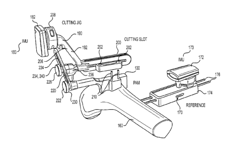
[0078] In accordance with the instant disclosure, exemplary cutting guides 190
may be aligned
and positioned with the aid of the PAM 130. By way of introduction, an
exemplary cutting guide
190 may be repositionably mounted to the PAM 130 so that the PAM is used for
its reference
position to know the position and orientation of the cutting guide with
respect to a patient's bone
Conversely, or in addition, the cutting guide 190 may be disengaged from the
PAM 130. In such
an instance, the PAM 130 may be coupled to a pinner having orifices configured
to receive an
alignment pin in only a single orientation. Using the pinner, once correctly
positioned, two or
more pins are inserted into a patient's bone so that the pins align with
orifices of an exemplary
cutting guide (disjoined from the PAM 130). In this manner, an exemplary
cutting guide may be
aligned by sliding over the pins in order to align the cutting guide to make
one or more bone cuts
[0079] The pre-operative surgical plan may also be used to create computer
instructions, referred
to herein as a patient case file or surgical plan, that may be loaded into an
associated surgical
navigation software application 104 to facilitate real-time guidance of the
relevant surgical
instrumentation. In addition, the instrumentation and instruments needed for
surgery, which may
be created or chosen using the static 122 and/or dynamic planner 124, may be
manufactured,
packaged, sterilized, and assembled into a kit 500 for delivery in a just-in-
time manner.
[0080] Referring to FIG. 3, a distal femur 160 of the patient 110 may include
a rigid reference 170
attached to a patient bone, either via an existing surgical incision or
percutaneously. In exemplary
form, the rigid reference 170 comprises a component of the image guided
surgical system 100 and
may include a housing 174 mounted to a pair of pins 176 fastened to the femur
160. The rigid
reference 170 facilitates tracking of a patient bone 160 by the housing 174
coupling with or
including an inertial measurement unit device 172 or other tracking device
that communicates
(whether wired or wi rel essly) with the surgical navigation workstation 102.
In exemplary form,
an inertial measurement unit (IMUs) device 172 may include an inertial
measurement unit (IMU)
173, a battery, and a wireless transmitter contained within a single housing,
where the device 172
may be operative to create and transmit data to the surgical navigation
software application 104.
21
CA 03094852 2020-09-22
WO 2019/246357 PCT/US2019/038164
Each IMU 173, 183 may consist of at least one triaxial accelerometer, one
triaxial magnetometer,
and one triaxial gyroscope. In this manner, the IMU 173, 183 generates data
indicative of
acceleration in three orthogonal axes, magnetic data, and gyroscopic data,
which the surgical
navigation software application 104 uses to determine changes in position and
orientation of the
IMU. Accordingly, by having the IMU 173 rigidly mounted to the bone (e.g.,
femur 160) using
the rigid reference 170, changes in position and orientation of the IMU can be
quickly and
accurately attributed to changes in position and orientation for the bone.
Thus, by knowing how
the IMU 173 is being repositioned as a function of time, the surgical
navigation software
application 104 is also able to determine changes in position and orientation
of the bone over the
same time period. As will be discussed hereafter, by initializing the IMU
device 172 of the rigid
reference 170 with respect to a second IMU device 182 associated with a
cutting guide 190, a
relative position of the cutting guide with respect to the patient bone can be
determined by the
surgical navigation software application 104.
[0081] Turning back to FIG. 3, an exemplary cutting guide 190 in accordance
with the instant
disclosure is configured to be repositionably mounted to a PAM 130 in order to
guide a surgeon
in making one or more bone cuts. This exemplary cutting guide 190 may be used
for each of the
femoral and tibial resections as part of a total knee arthroplasty.
[0082] In this exemplary embodiment, the cutting guide 190 includes a guide
body 192 having at
least one cutting slot 200 for guiding a surgical sagittal saw or similar tool
250 (see FIG. 25) along
a planar path to make one or more bone cuts. The guide body 192 may also,
separate from or in
addition to the slot 200, delineate one or more though orifices 202 sized to
allow throughput of a
surgical pin 210. By way of example, each surgical pin 210 may be mounted to
the patient's bone
and be utilized to guide and couple to a fixed position cutting block 300 (see
FIG. 30). In this
exemplary embodiment, the guide body 192 includes a neck 206 terminating at a
receiver 208
configured to have mounted thereto the second inertial measurement device 182.
[0083] By way of example, the second inertial measurement device 182 may
include an inertial
measurement unit (IMU) 183, a battery, and a wireless transmitter contained
within a single
housing, where the device 182 may be operative to create and transmit data to
the surgical
navigation software application 104.
22
CA 03094852 2020-09-22
WO 2019/246357 PCT/US2019/038164
[0084] As disclosed herein, each IMU 173, 183 may comprise three gyroscopes,
three
accelerometers, and three Hall-effect magnetometers (set of three, tri-axial
gyroscopes,
accelerometers, magnetometers) that may be integrated into a single circuit
board or comprised of
separate boards of one or more sensors (e.g., gyroscope, accelerometer,
magnetometer) in order to
output data concerning three directions perpendicular to one another (e.g., X,
Y, Z directions). In
this manner, each IMU 173, 183 may be operative to generate 21 voltage or
numerical outputs
from the three gyroscopes, three accelerometers, and three magnetometers. In
exemplary form,
each IMU 173, 183 may include a sensor board and a processing board, with a
sensor board
including an integrated sensing module consisting of three accelerometers,
three gyroscopic
sensors and three magnetometers (LSM9DS, ST-Microelectronics) and two
integrated sensing
modules consisting of three accelerometers, and three magnetometers (L5M303,
ST-
Microelectronics). In particular, the IMU 173, 183 may also include angular
momentum sensors
measuring rotational changes in space for at least three axes: pitch (up and
down), yaw (left and
right) and roll (clockwise or counter-clockwise rotation). In this manner, the
IMUs 173, 183
generates data indicative of acceleration in three orthogonal axes, magnetic
data, and gyroscopic
data, which the surgical navigation software application 104 uses to determine
changes in position
and orientation of each IMU.
[0085] By having the IMU 183 rigidly mounted to the cutting guide 190, changes
in position and
orientation of each IMU 173, 183 can be quickly and accurately attributed to
changes in position
and orientation of the cutting guide with respect to the patient bone. Thus,
by knowing how the
IMU 183 is being repositioned as a function of time, the surgical navigation
software application
104 is also able to determine changes in position and orientation of the
cutting guide 190 over the
same time period. As will be discussed hereafter, by initializing the IMU 173
of the rigid reference
170 with respect to the second IMU 183 associated with the cutting guide 190,
a relative position
of the cutting guide with respect to the patient bone can be determined by the
surgical navigation
software application 104.
[0086] By way of example, the cutting guide 190 may have any number of known
positions, such
that when the cutting slot 200 is placed into one of these known positions,
the position of the
cutting slot 200 is known relative to the PAM 130 In order to repositionably
mount the cutting
guide 190 to the PAM 130, a mechanical connection 220 exists therebetween that
may include one
23
CA 03094852 2020-09-22
WO 2019/246357 PCT/US2019/038164
or more joints. In exemplary form, the mechanical connection 220 includes a
lower joint 222, an
adjuster 224, and an upper joint 226.
[0087] By way of example, the lower joint 222 may be at or near the connection
of the PAM 130
to the cutting slot 200. The lower joint 222 may comprise a revolute joint
including a bolt or screw
230 (optionally spring loaded) that may be tightened to selectively inhibit
rotation of the adjuster
224 with respect to the PAM 130 and, accordingly, in a coarse sense adjust the
position of the
cutting slot 200. Alternatively, the lower joint 222 may be any joint or
motion activated device
(motor driven) that allows selective repositioning of the adjuster 224 with
respect to the PAM 130
so that, when desired, repositioning of the adjuster with respect to the PAM
is substantially
inhibited.
[0088] By way of example, the adjuster 224 may comprise an oblong or extended
ring at least a
portion of the lower joint 222 engages to fix and release the position of the
adjuster with respect
to the lower joint. Similarly, the adjuster 224 is also mounted to the upper
joint 226 which, in
exemplary form, may comprise a revolute joint 234.
[0089] In exemplary form, the revolute joint 234 may include a bolt or screw
240 (optionally
spring loaded) that may be tightened to selectively inhibit rotation of the
adjuster 224 with respect
to the cutting guide 190. Alternatively, the upper joint 226 may be any joint
or motion activated
device (motor driven) that allows selective repositioning of the adjuster 224
with respect to the
cutting guide 190 so that, when desired, repositioning of the adjuster with
respect to the cutting
guide is substantially inhibited. In addition to the revolute joint 234, the
upper joint 226 may also
include a spherical joint 236. In this fashion, when the spherical joint is
not locked, the cutting
guide 190 may be angularly repositioned with respect to the adjuster 224 (and
PAM 130) up to 45
degrees with respect to an axis extending parallel to the rotational axis of
the revolute joint 234
As will be discussed in more detail hereafter, the adjustability of the
spherical joint 236 may be
utilized to adjust the yams or valgus nature of a distal femoral bone cut.
[0090] Turning to FIG. 4, an alternate exemplary rigid reference 270, that may
be used in lieu of
or in addition to the rigid reference 170 of FIG. 3, comprises a reference
housing 272, that includes
an inertial measurement unit, mounted to a pair of pins 276 fastened to the
femur 160. This
alternate exemplary rigid reference 270 facilitates tracking of a patient bone
160 by
24
CA 03094852 2020-09-22
WO 2019/246357 PCT/US2019/038164
communicatively coupling (whether wired 273 or wirelessly) the reference
housing 272 (with the
IMU 274) with a reference transmitter located within a housing 278 that also
houses a power
supply (e.g., battery). In exemplary form, the relatively small size of the
reference housing 272
allows it to be mounted to the patient bone 160 without requiring an
additional incision or larger
incision to access the surgical site of the joint replacement or revision. In
other words, this
alternate exemplary rigid reference 270 provides a size advantage (smaller)
over the other rigid
reference 170 by not requiring the transmitter and power supply be rigidly
mounted to the patient
bone. For example, the IMU is operative to create and convey data to the
transmitter, which passes
the data onto the surgical navigation software application 104. The IMU 274
may consist of at
least one triaxial accelerometer, one triaxial magnetometer, and one triaxial
gyroscope. In this
manner, the IMU 274 generates data indicative of acceleration in three
orthogonal axes, magnetic
data, and gyroscopic data, which the surgical navigation software application
104 uses to
determine changes in position and orientation of the IMU. Accordingly, by
having the IMU 274
rigidly mounted to the bone (e.g., femur 160) using the rigid reference 270,
changes in position
and orientation of the IMU can be quickly and accurately attributed to changes
in position and
orientation for the bone. Thus, by knowing how the MU 274 is being
repositioned as a function
of time, the surgical navigation software application 104 is also able to
determine changes in
position and orientation of the bone over the same time period. As will be
discussed hereafter, by
initializing the IMU device 274 of the rigid reference 270 with respect to the
second IMU device
182 associated with the cutting guide 190, a relative position of the cutting
guide with respect to
the patient bone can be determined by the surgical navigation software
application 104.
[0091] Referencing FIG. 1 again, the workstation 102 running the surgical
navigation software
104 is operative to process sensor data from the IMUs 173/274, 183 and convert
this sensor data
to information relating to a resection plane location relative to the patient
anatomy. In addition,
the surgical navigation software 104 is operative to provide visualization to
a surgeon via the one
or more visual displays 106. In exemplary form, visualization may include 3D
virtual tissue
models 114, 3D virtual models of the cutting guide 190 or cutting slot 200,
projections, text, or
any other forms of communicating the orientation and position of the cutting
slot relative to the
patient anatomy. The information communicated as part of the visualization may
be updated at a
minimum of ten frames per second so that the information being displayed may
be considered near
real-time or real-time.
CA 03094852 2020-09-22
WO 2019/246357 PCT/US2019/038164
[0092] Any or all of the components of the cutting guide 190 may be disposable
for single-use
Alternatively, any or all of the components of the cutting guide 190 may be
reusable and amenable
to resterilization. In any event, any or all of the components of the cutting
guide 190, PAM 130,
and rigid references 170, 270 may be fabricated from numerous materials such
as, without
limitation, polymers, metals, and composites, and may be fabricated using
techniques including,
but not limited to, additive manufacturing, injection molding, machine
milling, and casting
Assembly and connection of individual components of cutting guide 190, PAM
130, and rigid
references 170, 270 may be performed by any means available, such as
appropriate press fitting,
locking, utilization of external fixation devices such as set screws,
adhesives, welding, or other
methods known to those skilled in mechanical assemblies to secure components
to one another.
While various components of the cutting guide 190, PAM 130, and rigid
references 170, 270 may
have been discussed separately herein, it is understood that any or all the
components may be
integrated or separable.
[0093] Referring to FIG. 5, to provide real-time feedback as to the position
and orientation of the
cutting slot 200, 'Mils 173/274, 183 are operative to generate data indicative
of orientation and
position, which is communicated to the surgical navigation software 104
running on the
workstation 102. The following is a discussion of how orientation and position
of the cutting slot
200 are determined by the surgical navigation software 104 when teamed with
known dimensions
for the surgical equipment (e.g., cutting guide 190, PAM 130) in an exemplary
procedure for a
total knee arthroplasty (TKA).
[0094] IMUs 173/274, 183 in accordance with the instant disclosure may measure
orientation
about an x-axis, y-axis, and z-axis, but may not directly measure translation.
In order to determine
translation of the 'Mils, one may use external sensors or have the IMUs
initialized using a starting
position and orientation that is known with respect to a real-world object
(e.g., a bone). For
example, the external sensors may comprise linear positioning sensors (e.g.
linear variable
displacement transformer, linear motion encoder, ultrasonic ranging, or
optical ranging) to provide
translation information.
[0095] In exemplary form, as discussed hereafter, the instant disclosure may
make use of an
initialization position where the IMUs 173/274, 183 are rigidly mounted to the
cutting guide 190
26
CA 03094852 2020-09-22
WO 2019/246357 PCT/US2019/038164
and PAM 130, respectively, so that the relative position and orientation of
the cutting guide with
respect to the PAM is known (and the relative position and orientation of the
IMUs 173/274, 183
is also known). By way of further example, this initialization position may
have cutting slot 200
aligned along the same plane as the PAM. After establishing this
initialization position, the cutting
guide 190 may be repositioned with respect to the PAM 130 to carry out the
femoral bone cuts
established via the pre-operative surgical plan.
[0096] In the context of the instant disclosure, pre-operative surgical
planning will establish the
depth (e.g., location) of the distal bone cut for a TKA, as well as the
placement of the PAM 130
on the patient bone 160. As depicted in FIG. 5, with the depth of the distal
bone cut known,
identified as "x", and the starting position B known from the placement of the
PAM 130 with
respect to the bone 160, two pieces of information are required in order to
position the cutting
guide 190 correctly to effectuate the distal cut: (1) the angle a; and (2) the
distance AB. The
distance AB is a function of known instrument dimensions (this is the linear
distance from the
center of the PAM to the center of the cutting slot 200), where the distance
AB is constant in
accordance with the instant disclosure and does not change as the cutting
guide 190 is rotated about
the PAM 130 via the lower joint 222. As a result, using trigonometry, one can
calculate the angle
a from the equation of FIG. 5. And knowing this angle a, the surgical
navigation software 104
tracks the angular change of the cutting slot 200, via the IMU 173, 274,
relative to the patient bone
using the IMU 183 of the PAM 130, so that when cutting slot is positioned at
angle a, the surgical
navigation software informs the surgeon the cutting slot is positioned in
accordance with the pre-
operative surgical plan, so that the surgeon may carry out the distal femoral
bone cut. In case the
cutting slot 200 is not aligned with angle a, the surgical navigation software
provides feedback to
the surgeon indicating how the cutting slot should be repositioned to achieve
angle a. As will be
discussed hereafter, the joints 226, 236 associated with the cutting guide 190
may be repositioned
to adjust for varus/valgus, flexion/extension, and other known degrees of
freedom.
[0097] In accordance with the instant disclosure, knowing the instrument (PAM
130, cutting guide
190) dimensions is important for calculating the relative position and
orientation of the instruments
dynamically during a surgical procedure, such as TKA. For example, each of the
PAM 130, cutting
guide 190, and cutting slot 200 may be appropriately sized to facilitate
performing the desired
surgery, preferably with minimal modification to the standard incision or
minimally invasive
27
CA 03094852 2020-09-22
WO 2019/246357 PCT/US2019/038164
incision. Appropriate dimensions for each component (e.g., the PAM 130,
cutting guide 190, and
cutting slot 200) may be selected prior to surgery in many ways. For example,
each component
may be made in a patient-specific manner, where all dimensions are selected to
best match the
patient and the surgical plan. Because patient-specific manufacturing may not
be cost effective,
another option is to choose dimensions based on population analysis. Those
skilled in the art of
orthopedic instrumentation will be familiar with sizing based on population
analysis.
[0098] In general, a dimension of the anatomy is measured across several
samples ¨ a population
¨ so that the range and variation of measurement is known across the samples.
If desired, the
population may be subdivided so that the range and variation of the
measurement within each
population subdivision is known. Methods of performing this subdivision
include, but are not
necessarily limited to, building regression models, unsupervised or supervised
clustering, mixture
modeling, partitioning, or any other methodology. In such a way, the best set
of dimensions, or
sizes, may be chosen for each component. This process may be performed in an
automatic or
semi-automatic way using statistical geometrical models or machine learning
methods.
[0099] Using the known dimensions of component parts of each instrument,
including post
assembly, one can determine the allowable working volume of the surgical
instrument ¨
specifically the reachable cut orientation and positions ¨ using methods
familiar to those skilled in
the art of robotic manipulators and forward kinematics. For example, the D en
avi t¨Hartenb erg
(DH) parameters of each joint are known given the type of j oint ¨ revolute,
spherical, or any other
¨ and the known dimensions of each linkage of the mechanical connection 220 as
outlined above.
From this information the DH convention may be used to establish the
appropriate series of
transformations between the first revolute joint 222 and the cutting slot 200.
By calculating the
end position and orientation at all or most of the allowable range of the
variables for each joint,
the working volume of the cutting slot 200 can be calculated for each of the
steps in the surgical
procedure. This convention may be used to verify that the chosen component
dimensions are
sufficient to achieve the desired surgical plan FIGS 6-15 show some possible
critical dimensions
of component parts of the system 100 as well as examples of population
variations, which serve
as inputs to dimensional choice.
28
CA 03094852 2020-09-22
WO 2019/246357 PCT/US2019/038164
[0100] Referencing FIG. 6, the PAM 130 may include critical dimensions, in
addition to the
patient specific features, comprising the (mediolateral) ML width and
(anteroposterior) AP height
In this exemplary disclosure, the PAM 130 sets the center of rotation for the
cutting guide 190,
where the location of the PAM 130 can be selected to optimize the accuracy and
performance of
the system 100. The AP and ML dimensions should be carefully chosen so that
the mechanical
connection 220 coupling the PAM 130 to the cutting guide 190 clears both the
intended incision
and the medial aspect of the femora without causing impingement of the cutting
guide with the
bone or soft tissue once assembled. Similarly, the length of the cutting guide
190, as well as the
location of the locking positions via the mechanical connection 220, should be
selected to facilitate
each of the procedural steps ¨ allowing the cutting slot 200 to be properly
positioned and
manipulated without impingement.
[0101] Referencing FIG. 7, an exemplary method for establishing and optimizing
the mating site
of the PAM 130 on the patient bone 160 includes utilization of a trained human
expert as part of
the pre-operative plan or, in addition to or in lieu of, using artificial
intelligence (Al). Al learns
the design constraints with regard to accuracy and the population morphology
using measurements
or surface geometry extracted from population statistics (i.e. statistical
atlas), and outputs a
location of the PAM 130 optimized for accuracy to achieve the desired plan.
[0102] Turning to FIGS. 8 and 9, an exemplary cutting guide 190 in accordance
with the instant
disclosure may include a cutting slot 200 having dimensions that may be,
configured in part, based
upon the intended saw blade a surgeon anticipates using during the TKA, in
order to appropriately
capture the saw blade. In many TKA procedures, surgeons will utilize an
oscillating tip saw to
remove bone from the distal femur to prepare the femur to accept an orthopedic
implant
Exemplary saw blades for an oscillating tip saw may have a thickness of
approximately 1.19
millimeters, though other thickness may be used from time-to-time. In this
manner, the width of
the cutting slot 200 (in the AP direction) may be slightly greater than the
thickness of the intended
saw blade. The more precise the tolerance between the cutting slot 200 width
and the saw blade
results in greater precision that the bone cut of the blade is coplanar with
the slot. In addition to
establishing the AP width of the cutting slot 200, the ML length of the
cutting slot should be chosen
to allow resection of the entire distal, posterior and anterior surfaces as
dictated by the pre-
operative surgical plan.
29
CA 03094852 2020-09-22
WO 2019/246357 PCT/US2019/038164
[0103] Referring to FIG. 10, an alternate exemplary cutting guide 290 includes
a guide body 292
that replaces the cutting slot 200 with at least two guide pin holes 294. In
this alternate exemplary
cutting guide 290, the guide pin holes 294 match corresponding holes of a
separate cutting block
300 (see FIG. 30) so that the guide 290 can be utilized to position two or
more surgical pins 210
that are then utilized to align the cutting block. In other words, the cutting
guide 290 is utilized by
the surgeon to know where the drill holes and correspondingly fasten surgical
pins 210 to the
patient's bone. By way of further example, the pin holes drilled (or the
position of the surgical
pins themselves) into the patient's bone may be of the same distance from one
another as the pin
holes on a conventional distal cutting block 300. Post drilling of the pin
holes, the guide 290 may
be replaced with a separate cutting block, which is aligned to the patient's
bone using the surgical
pins. By way of example, exemplary conventional cutting blocks are available
from Smith &
Nephew, Zimmer, DePuy, and Stryker.
[0104] Looking at FIG. 11, the exemplary cutting guide 190 may include a blade
support
attachment 195 selectively coupled to the guide body 192 in order to provide
stability to the
surgical blade extending through the cutting slot 200.
[0105] FIGS. 12-16 reflect an exemplary process for automatic landmarking of
the patient bone
model 114, this this case the femur, using the pre-operative surgical planning
software 104. As
depicted in FIG. 12, the software 104 is operative to use statistical atlas
automatic landmarking to
compute the location of the mechanical axis, the distal resection point, which
are both used to
compute the suggested, preferred femoral distal resection plane 156. FIG. 13
depicts extraction
and measurement of the medio-lateral width of the resection across a given
population of the
statistical atlas. The extracted and measured medio-lateral widths are used to
create the design
envelope for the distal cutting slot 200 dimensions (representatives from the
statistical atlas
population are depicted, with blue reflecting the largest size, green
depicting a medium size, and
red reflecting the smallest size). FIG. 14 shows the distal femoral resection
cuts made to
representatives of the statistical atlas. FIG. 15 reflects the relationship
between resected medio-
lateral dimensions and changes in distal resection plane 156 depth. For
instance, the yellow color
reflects positioning the resection plane 156 4 millimeters more distal than
the planned or suggested
resection plane location, whereas red reflects positioning the resection plane
at the planned or
suggested location and, finally, light blue reflects positioning the resection
plane 4 millimeters
CA 03094852 2020-09-22
WO 2019/246357 PCT/US2019/038164
more proximal than the planned or suggested resection plane location. Finally,
FIG. 16 depicts
medio-lateral widths across the statistical atlas population to establish
cutting slot 200 dimensions
that capture most or all of a given population.
[0106] Turning to FIGS. 17-20, as an early step in surgical navigation,
registration is undertaken
to align the image guided surgical system 100 to the patient bone 160. As part
of establishing
registration, the PAM 130 may be aligned to the patient bone 160 so that the
patient specific
surface(s) of the PAM match and precisely contact the patient anatomy in only
a single orientation
and position. Upon positioning the PAM 130 on the patient bone 160 so the PAM
occupies the
single orientation and position matching precisely the topography of the
tissue (e.g., bone), the
PAM may be mounted to the patient tissue (e.g., bone) using one or more
surgical pins 210 or
screws that are received within holes that may be drilled in to the patient
tissue. In this fashion,
the PAM 130 is rigidly affixed to the patient tissue so that as the tissue is
repositioned, so too is
the PAM. In addition to mounting the PAM 130 to patient tissue, the rigid
reference 170 is also
mounted to patient tissue. As discussed herein, the cutting guide 190 is
repositionably mounted to
the PAM 130 via the mechanical connection.
[0107] As depicted in FIGS. 17 and 18, in exemplary form, the cutting guide
190 is mounted to
the PAM 130 in a known registration position and orientation using the
mechanical connection
220, which is in turn a known position and orientation relative to the patient
bone 160 (e.g., the
femur) by way of the PAM. More specifically, as depicted in FIG. 17, the lower
joint 222 couples
the PAM 130 to the adjuster 224, and the upper joint 226 couples the cutting
guide 190 to the
adjuster. In particular, the cutting guide 190 is oriented so that a dominant
longitudinal axis of the
cutting slot 200 is parallel to a dominant longitudinal axis of the cutting
guide so the axes are co-
planar. In addition, a spacing is set between the cutting guide 190 and the
PAM 130, along the
adjuster 224 using the joints 222, 226, that corresponds to a predetermined
spacing that is known.
In this manner, the position of the cutting guide 190 in solid lines is the
predetermined position
with respect to the PAM 130 It should be noted that by adjusting the revolute
lower joint 222, the
cutting guide 190 may be rotated about the PAM 130 as depicted in phantom
lines. When the
cutting guide 190 is mounted to the PAM 130, via the mechanical connection
220, and assumes
the known registration position (and when the rigid reference 170 is mounted
to the patient tissue),
data from the IMUs 173, 183 is recorded by the image guided surgical system
100 to establish a
31
CA 03094852 2020-09-22
WO 2019/246357 PCT/US2019/038164
point of reference. More specifically, data from the Wills 173, 183 is
processed to determine
changes in position and orientation of the cutting guide 190 with respect to
the patient bone 160.
In this fashion, future motions of the patient bone 160 are tracked
independently using the MU
173 of the rigid reference 170, while motions of the cutting guide 190 are
tracked separately using
the tracking IMU 183. As a result, as depicted in FIG. 18, the image guided
surgical system 100
displays a virtual bone model 114 of the patient's bone 160 along with a
phantom line 185 denoting
the position and orientation of the cutting slot 200 (that may be color
highlighted (e.g., green)) to
differentiate between a position of the cutting slot that is or is not
consistent with a pre-operative
surgical plan establishing the position and orientation of a bone cutting
plane.
[0108] Turning to FIGS. 19 and 20, establishing registration of the cutting
guide 190 with respect
to the patient bone may also include repositioning of the cutting guide with
respect to the
mechanical connection 220 using the upper spherical joint 236. In exemplary
form, the upper
spherical joint 236 allows the guide body 192 to selectively allow the guide
body (and cutting slot
200) to be angularly repositioned with respect to the adjuster 224 (and PAM
130) up to 45 degrees
with respect to an axis extending parallel to the rotational axis of the
revolute joint 234. In this
manner, the spherical joint 236 allows for yams or valgus adjustment of the
cutting slot 200. By
way of example, the solid line position of the cutting guide 190 body 192 is
representative of the
registration position, whereas the phantom lines are representative of
possible changes in angular
orientation that the guide body 192 may occupy with respect to the rotational
axis of the revolute
joint 234. Because the IMU 183 is rigidly mounted to the guide body 192,
changes in the position
and orientation of the cutting slot 200 are correspondingly reflected in
changes in position and
orientation of the IMU 183, which sends its data to the image guided surgical
system 100. The
image guided surgical system uses the data from the IMU 183, along with
knowing the dimensions
of the guide body 192 and the position of the spherical joint 236 with respect
thereto, to calculate
the position and orientation of the cutting slot 200. As a result, as depicted
in FIG. 20, the image
guided surgical system 100 displays a virtual bone model 114 of the patient's
bone 160 along with
a pair of phantom lines 187, 189 denoting the position and orientation of the
cutting slot 200 (that
may be color highlighted (e.g., white 189)) with respect to the position and
orientation of the
intended cutting slot (that may be color highlighted (e.g., green 187)) to
differentiate between a
position and/or orientation of the cutting slot that is or is not consistent
with a pre-operative
surgical plan establishing the position and orientation of a bone cutting
plane. Post registration,
32
CA 03094852 2020-09-22
WO 2019/246357 PCT/US2019/038164
the image guided surgical system 100 may be utilized to facilitate one or more
bone cuts at a distal
end of the femur as part of a TKA.
[0109] Referring to FIG. 22, a TKA surgery may include a distal femoral
resection. After
registration of the image guided surgical system 100 as previously described,
the cutting guide 190
may be repositioned with respect to the PAM using one or both of the joints
222, 226. By way of
example, the lower revolute joint 222 may be manipulated so as to allow the
cutting guide 190 to
rotation around the PAM 130 via a rotational axis extending through the
bolt/screw 230 in
preparation for the distal femoral resection. In exemplary form, the image
guided surgical system
100 may be operative to process data from the IMUs 173, 183 and display
virtual bone model 114
of the patient's bone 160 and the relative updated position and orientation of
the cutting slot 200
from calculating the relative position and orientation of the cutting guide
190 with respect to the
patient's bone. In the context of the lower joint 222, because only a single
revolute joint is used,
the one or more visual displays 106 may show a "reachable" region, or the
allowable range of bone
that may be cut by manipulating the upper spherical joint. In particular,
using trigonometry, the
image guided surgical system 100 calculates the distal-to-proximal distance
"depthi" by taking the
know distance "liu 1" between the first and second joints 222, 226 and
multiplying by the sine 0,
where angle 0 is the angle between the registration position of the cutting
guide 190 and the current
position of the cutting guide. Using data from the IMUs 173, 183, the image
guided surgical
system 100 is operative to calculate the position of the cutting guide and,
correspondingly,
calculate angle 0. Using the calculated angle 0, the image guided surgical
system 100 then
calculates "depthi" and depicts the virtual bone model 114 of the patient's
bone 160 and the
relative updated position and orientation of the cutting slot 200. In this
fashion, the surgeon is able
to determine whether the cutting guide 190 should be further rotated with
respect to the PAM 130
in accordance with the pre-operative surgical plan to make the correct distal
femoral bone cut
Presuming the "depthi" of the femoral bone cut is reached, the surgeon may
lock the lower joint
222 in position and focus on repositioning the upper joint 226.
[0110] Turning to FIG. 23, the upper joint 226 may include a revolute joint
234 and a spherical
joint 236. Each may be repositioned to adjust the position of the cutting
guide 190 with respect to
the PAM 130. In exemplary form, the cutting guide 190 may be allowed to rotate
around a
rotational axis extending through the bolt/screw 240. Rotation about the
bolt/screw 240 may be
used to correct for (or make adjustments to) the flexion and extension angle
for resection. In
33
CA 03094852 2020-09-22
WO 2019/246357 PCT/US2019/038164
particular, using trigonometry, the image guided surgical system 100
calculates the distal-to-
proximal distance "depth2" by taking the know distance "lRi 2", between the
second joint 226 and
the center of the cutting guide body 192, and multiplying by the sine a, where
angle a is the angle
between the registration position of the cutting guide 190 and the current
position of the cutting
guide with respect to upper joint 226. Using data from the IMUs 173, 183, the
image guided
surgical system 100 is operative to calculate the position of the cutting
guide 190 and,
correspondingly, calculate angle G. Using the calculated angle CS, the image
guided surgical system
100 then calculates "depth2" and depicts the virtual bone model 114 of the
patient's bone 160 and
the relative updated position and orientation of the cutting slot 200. In this
fashion, the surgeon is
able to determine whether the cutting guide 190 should be further rotated with
respect to the second
joint 226 in accordance with the pre-operative surgical plan to make the
correct distal femoral bone
cut. Presuming the "depth2" of the femoral bone cut is reached, the surgeon
may lock the upper
revolute joint 234 in position and focus on repositioning the spherical joint
236.
[0111] As depicted in FIG. 24, adjustment of the spherical joint 236 allows
for rotation of the
cutting slot 200 to accommodate for varus and valgus angular adjustments. In
other words,
unlocking the spherical j oint 236 allows the cutting slot 200 to be
manipulated so that the
resection depth, vanis orientation, and flexion orientation of the cut is
acceptable relative to a
pre-operative plan. In exemplary form, the cutting guide 190 may be allowed to
rotate around a
sphere of the spherical joint 236. In particular, using trigonometry, the
image guided surgical
system 100 calculates medial compartment offset and lateral compartment offset
using the
following equations:
PMKSIMMtt)
dPrthnt1141.,Mit, asina)
deoftõõ6õ.:1= &ink + depth2+ depthim
depth3-.,
depthmt,,õ = d.epthm, + depthx.
where:
"lRi 3M" is the length of the guide body 192 across the medial compartment;
34
CA 03094852 2020-09-22
WO 2019/246357 PCT/US2019/038164
"1R1 3L" is the length of the guide body 192 across the lateral compartment;
angle "X" is the angle between the registration position and the angular
offset.
[0112] Referring to FIGS. 22-26, presuming the "depth2" of the femoral bone
cut is reached, the
surgeon may lock the upper revolute joint 234. Accordingly, to get the medial
depth offset
"depth3m", the known length of "lut 3M" is multiplied by sine X. Likewise, to
get the lateral depth
offset "depth3L", the known length of "lRi 31_," is multiplied by sine X. In
order to calculate the
actual resection depth in the medial compartment, "depthi" and "depth2" and
"depth3m" are
summed (see FIGS. 22-24). Similarly, to calculate the actual resection depth
in the lateral
compartment, "depthi" and "depth2" and "clepth3L" are summed (see FIGS. 22-
24). Using data
from the Mils 173, 183, the image guided surgical system 100 is operative to
calculate the
foregoing. In this fashion, the surgeon is able to determine whether the
cutting guide 190 should
be further rotated about the spherical joint 236 in accordance with the pre-
operative surgical plan
to make the correct distal femoral bone cut. Once the position and orientation
of the cutting guide
190 is acceptable, the joints 222, 226, 236 may be locked and the distal
femoral resection cut may
be undertaken, as depicted in FIGS. 25 and 26, with a surgeon controlling a
surgical saw 250.
[0113] Referring to FIGS. 27-30, after the distal femoral resection cut is
completed, the cutting
guide 190 and PAM 130 may be used to facilitate placement of fixation devices
(e.g., surgical pins
210) that will guide and engage a fixed position cutting block 300. In femoral
TKA surgical
procedures, having five bone cuts, the remaining cuts (besides the distal
femoral resection) are the
anterior, posterior, and two chamfer cuts. To facilitate these four bone cuts,
one may make use of
a conventional instrument, referred to as the 4-in-1 cutting block 300. This
cutting block 300,
which is fixed to the distal end of a patient's resected femur 160, includes
two or more openings
configured to receive two or more surgical pins 210 extending from the
resected distal surface. In
this fashion, the surgical pins 210 are operative to align the cutting block
300 with respect to the
distal femur and to guide the cutting block into position against the resected
femur surface. In
addition to the openings configured to receive the surgical pins 210, the
cutting block 300 includes
four or more cutting slots, each cutting slot belonging to one of the four
mentioned remaining bone
cuts. It should be noted, however, that different knee implants may require
different cutting
positions and even different numbers of bone cuts. Nevertheless, the exemplary
devices and
CA 03094852 2020-09-22
WO 2019/246357 PCT/US2019/038164
methods disclosed herein may be applied to any cutting guide (e.g., smart or
dumb) whether placed
by physical alignment guides or via computer feedback or control.
[0114] As depicted in FIGS. 28 and 29, in order to prepare the resected femur
for using the 4-in-
1 cutting block 300, the image guided surgical system 100 provides
instructions via the one or
more visual displays 106 for repositioning the cutting guide 190 so that the
guide body 192 is
positioned against the exposed surface of the resected distal femur 107 based
upon data from the
IMUs 173, 183. In particular, the guide body 192 is positioned so that one or
more though orifices
202 are aligned with intended locations of the resected femur 107 so that a
surgical drill may
extend through the orifices and drill out holes within the femur. The pin
holes dictate the internal-
external rotation and anterior-posterior positioning of the remaining bone
cuts. Post hole creation,
two or more surgical pins 210 are placed, one in each hole, optionally using
the guide body orifices
202 to align the surgical pins into position so that the surgical pins extend
into the resected femur
and extend distally generally perpendicular to the resected, femoral planar
surface. After the
surgical pins 210 are mounted to the resected femur, the cutting guide 190 and
PAM 130 may be
removed from the surgical site.
[0115] Turning to FIGS. 30 and 31, with the surgical pins 210 in position on
the resected femur
107, a 4-in-1 cutting block 300 is aligned with respect to the distal resected
femur 107 so that two
or more openings of the cutting block 300 are configured to receive the two or
more surgical pins
210 so that the cutting block may be repositioned against the exposed bone
surface of the distal
femoral resection cut. With the cutting block aligned using the surgical pins
210 and against the
resected distal femur surface, the surgeon may lock the cutting block 300 in
position. Thereafter,
the surgeon may reposition a surgical blade through the respective slots 302
of the block 300 to
make the anterior, posterior, and two chamfer distal femur cuts. After
completion of the bone cuts
(see FIG. 31), the block 300 and surgical pins 210 may be removed from the
distal femur in
anticipation of orthopedic trial fitting. While the foregoing exemplary
surgical procedure makes
use of a 4-in-1 cutting block 300 to make the anterior, posterior, and two
chamfer distal femur
cuts, it is also within the scope of the disclosure to utilize the cutting
guide 190, the PAM 130, and
a guide foot 260.
[0116] Referencing FIGS. 32-37, after the distal femoral resection cut is
completed, the cutting
guide 190 and PAM 130 may be used to facilitate placement of fixation devices
(e.g., surgical pins
210) that will guide and engage the guide foot 260. As discussed herein,
femoral TKA surgical
36
CA 03094852 2020-09-22
WO 2019/246357 PCT/US2019/038164
procedures generally include five bone cuts, four of which remain after the
distal femoral resection
has been completed. To facilitate these four bone cuts, one may make use of
the guide foot 260
that replaces the PAM 130 as the anchor to which the mechanical connection 220
and the cutting
guide 190 are mounted.
[0117] As depicted in FIG. 32, after the PAM 130, mechanical connection 220,
and cutting guide
190 have been utilized to position the surgical pins 210, the foregoing
components may be removed
from the surgical site. In exemplary form, the PAM 130 is replaced with a
guide foot 260 that
connects to the mechanical connection 220 just as the PAM did, so that the
guide foot, mechanical
connection, and cutting guide 190 are mounted to one another. In exemplary
form, the guide foot
260 includes two or more orifices configured to receive, respectively, the
surgical pins 260
extending from the resected portion of the femur 107. The orifices of the
guide foot 260 are
configured to receive the surgical pins 210 in only a single orientation so
that, when the guide foot
receives the surgical pins and is repositioned against the resected femur 107
and affixed in position,
the image guided surgical system 100 knows precisely the position and
orientation of the guide
foot with respect to the femur.
[0118] In this exemplary embodiment, the image guided surgical system 100 is
programmed with
the precise dimensions of the guide foot 260 so that when the guide foot is in
a registration position,
the position of the cutting guide 190 with respect to the femur is known. In
other words, the cutting
guide 190 is mounted to the guide foot 260 in a known registration position
and orientation using
the mechanical connection 220, which is in turn a known position and
orientation relative to the
patient bone 160 (e.g., the femur) by way of the guide foot.
[0119] As discussed herein, the lowed oint 222 couples the guide foot 260 to
the adjuster 224, and
the upper joint 226 couples the cutting guide 190 to the adjuster. In
particular, the cutting guide
190 is oriented so that a dominant longitudinal axis of the cutting slot 200
is parallel to a dominant
longitudinal axis of the cutting guide so the axes are co-planar. In addition,
a spacing is set between
the cutting guide 190 and the guide foot 260, along the adjuster 224 using the
joints 222, 226, that
corresponds to a predetermined spacing that is known. In this manner, the
position of the cutting
guide 190 is known with respect to the guide foot 260. When the cutting guide
190 is mounted to
the guide foot 260, via the mechanical connection 220, and assumes the known
registration
position (and when the rigid reference 170 is mounted to the patient tissue),
data from the IMUs
173, 183 is recorded by the image guided surgical system 100 to establish a
point of reference for
37
CA 03094852 2020-09-22
WO 2019/246357 PCT/US2019/038164
the cutting guide 190 having a known position and orientation with respect to
the resected femur.
And, as discussed hereafter, this point of reference is utilized by the image
guided surgical system
100 to track and inform a user (e.g., a surgeon) concerning the position and
orientation of the
cutting guide to facilitate utilizing the cutting guide to perform the
anterior, posterior, and two
chamfer distal femur cuts.
[0120] Referring to FIGS. 34 and 35, data from the IMUs 173, 183 is processed
by the image
guided surgical system 100 to determine changes in position and orientation of
the cutting guide
190 with respect to the patient bone 160. In this fashion, motions of the
patient bone 160 are
tracked independently using the IMU 173 of the rigid reference 170, while
motions of the cutting
guide 190 are tracked separately using the tracking IMU 183. As a result, the
image guided
surgical system 100 displays a virtual bone model 114 of the patient's bone
160 along with a
phantom line 310 color coded (e.g., green) to confirm that the intended cut
line is in accordance
with a pre-operative surgical plan. In this exemplary sequence, the surgeon
may reposition the
cutting guide 190 by loosening and tightening the revolute joints 222, 234 and
the spherical joint
236 in order to position and orient the cutting slot 200 to make the requisite
cuts in accordance
with the pre-operative surgical plan, namely the anterior cut as depicted in
FIG . 34. The one or
more visual displays 106 are updated in real-time or near real-time to depict
the bone model 114
consistent with the position and orientation the patient's actual bone with
respect to the projected
cutting line, which passes through the cutting slot 200. Consequently, when
the cutting guide 190
is positioned and oriented consistent with the pre-operative surgical plan,
the surgeon may visually
confirm the position using the one or more visual displays 106 and carry out
the bone cut by using
a surgical saw 250 having a blade received within the cutting slot 200. This
process is repeated
by repositioning the cutting guide 190 via loosening and tightening the
revolute joints 222, 234
and the spherical joint 236 in order to reposition and reorient the cutting
slot 200 to also make the
posterior cut (see FIG. 36), the anterior chamfer cut (see FIG. 37), and the
posterior chamfer cut
(see FIG. 38). After making the last four (or so) bone cuts using the cutting
guide 190, the cutting
guide, mechanical connection 220, and the guide foot 260 may be removed from
the surgical pins
210 and away from the surgical site. Likewise, the surgical pins 210 may be
removed from the
distal resected femur to accommodate orthopedic trial test fitting. But the
instant embodiments
can also be used with bone cuts beyond the distal femoral cuts.
38
CA 03094852 2020-09-22
WO 2019/246357 PCT/US2019/038164
[0121] Referencing FIGS. 39 and 40, it is also in accordance with the instant
disclosure that the
image guided surgical system 100 be utilized to guide bone cuts beyond those
of the distal femur.
By way of example, the image guided surgical system 100 may be utilized to
carry out the tibia
resection as part of a TKA procedure (revision or replacement). For example,
the image guided
surgical system 100 may make use of the same workstation 102, software 104,
visual displays 106,
mechanical connections 220, and surgical instruments 170, 190. But what
differs are the patient-
specific virtual tissue models 114 (which comprise a proximal tibia, rather
than a distal femur
discussed above) and the PAM 130 (PAM for femur is not the same as the PAM for
the tibia).
[0122] In this exemplary discussion, the PAM 130 is configured to have at
least one surface with
a geometry matching the negative of the patient's proximal tibia (in other
words, the surface shape
of the PAM precisely follows the surface, including shape changes, of the
patient's proximal tibia).
By utilizing a PAM 130 that fits to the patient's tibia in only a single
location and orientation,
instrumentation or other parts having known geometries (size, width, length,
height, etc.) may be
attached to the PAM to facilitate localization of position and orientation of
the instrumentation or
other parts within a frame of reference utilized by the surgical navigation
software 104. In other
words, because one knows the exact position and orientation of the PAM 130
with respect to a
patient's tibia, any structure (having known dimensions) rigidly mounted to
the PAM will also
have a known position and orientation with respect to the patient's tibia. In
this fashion, the PAM
130 operates to correlate the virtual frame of reference with the real-world
frame of reference.
[0123] In the context of the instant disclosure, virtual pre-operative
surgical planning may
establish the position and orientation of the proximal tibia resection bone
cut for a TKA, as well
as the placement of the PAM 130 on the patient's tibia 162. As part of this
pre-operative surgical
planning, the image guided surgical system 100 makes use of a registration to
align the image
guided surgical system to the patient 110. As part of establishing
registration, the PAM 130 is
aligned to the patient so that the patient specific surface(s) of the PAM
match and precisely contact
the patient's tibia in only a single orientation and position. Upon
positioning the PAM 130 on the
patient's tibia 162 so the PAM occupies the single orientation and position
matching precisely the
topography of the tibia, the PAM may be mounted to the tibia using one or more
surgical pins 132
or screws that are received within holes that may be drilled into the tibia.
In this fashion, the PAM
130 is rigidly affixed to the tibia so that as the tibia is repositioned, so
too is the PAM. In addition
to mounting the PAM 130 to patient tissue, a rigid reference (not shown) is
also mounted to the
39
CA 03094852 2020-09-22
WO 2019/246357 PCT/US2019/038164
tibia. Similarly, as discussed before, the cutting guide 190 is repositionably
mounted to the PAM
130 via the mechanical connection 220.
[0124] In exemplary form, the cutting guide 190 is mounted to the PAM 130 in a
known
registration position and orientation using the mechanical connection 220,
which is in turn a known
position and orientation relative to the patient bone (e.g., the tibia 162) by
way of the PAM.
Consistent with the prior discussion, the lower joint 222 couples the PAM 130
to the adjuster 224,
and the upper joint 226 couples the cutting guide 190 to the adjuster. In
particular, the cutting
guide 190 may be oriented so that a dominant longitudinal axis of the cutting
slot 200 is parallel
to a dominant longitudinal axis of the cutting guide so the axes are co-
planar. In addition, a spacing
is set between the cutting guide 190 and the PAM 130, along the adjuster 224
using the joints 222,
226, that corresponds to a predetermined spacing that is known. It should be
noted that by
adjusting the revolute lower joint 222, the cutting guide 190 may be rotated
about the PAM 130.
When the cutting guide 190 is mounted to the PAM 130, via the mechanical
connection 220, and
assumes the known registration position (and when the rigid reference 170 is
mounted to the
patient tissue), data from the IMUs 173, 183 is recorded by the image guided
surgical system 100
to establish a point of reference. More specifically, data from the IMUs 173,
183 is processed to
determine changes in position and orientation of the cutting guide 190 with
respect to the patient
bone 162. In this fashion, future motions of the tibia 162 are tracked
independently using the IMU
173 of the rigid reference 170, while motions of the cutting guide 190 are
tracked separately using
the tracking IlIVIU 183. As a result, the image guided surgical system 100
displays a virtual bone
model 114 of the tibia along with a visual reference denoting the position and
orientation of the
cutting slot 200 (that may be color highlighted (e.g., green)) to
differentiate between a position of
the cutting slot that is or is not consistent with a pre-operative surgical
plan establishing the
position and orientation of a bone cutting plane.
[0125] Because the IMU 183 is rigidly mounted to the guide body 192, changes
in the position
and orientation of the cutting slot 200 are correspondingly reflected in
changes in position and
orientation of the IMU 183, which sends its data to the image guided surgical
system 100. The
image guided surgical system uses the data from the IMU 183, along with
knowing the dimensions
of the guide body 192 to calculate the position and orientation of the cutting
slot 200. As a result,
the image guided surgical system 100 may display a virtual bone model 114 of
the patient's tibia
CA 03094852 2020-09-22
WO 2019/246357 PCT/US2019/038164
162 along with a pair of phantom lines denoting the position and orientation
of the cutting slot 200
(that may be color highlighted (e.g., white)) with respect to the position and
orientation of the
intended cutting slot (that may be color highlighted (e.g., green)) to
differentiate between a position
and/or orientation of the cutting slot that is or is not consistent with a pre-
operative surgical plan
establishing the position and orientation of the tibia resection cut. Post
registration, the image
guided surgical system 100 may be utilized to facilitate the tibia resection
cut
[0126] Referring again to FIGS 39 and 40, the cutting guide 190 may be
repositioned with respect
to the PAM 130 using one or both of the joints 222, 226. By way of example,
the lower revolute
joint 222 may be manipulated so as to allow the cutting guide 190 to rotation
around the PAM 130
via a rotational axis extending through the bolt/screw 230 in preparation for
the distal femoral
resection. In exemplary form, the image guided surgical system 100 may be
operative to process
data from the IMUs 173, 183 and display virtual bone model 114 of the
patient's tibia 162 and the
relative updated position and orientation of the cutting slot 200 from
calculating the relative
position and orientation of the cutting guide 190 with respect to the
patient's tibia. In the context
of the lower joint 222, because only a single revolute joint is used, the one
or more visual displays
106 may show a "reachable" region, or the allowable range of proximal tibia
that may be cut by
manipulating the upper spherical joint. In particular, as discussed herein,
using trigonometry, the
image guided surgical system 100 the position of the cutting slot 200 using
data from the IMUs
173, 183. Upon reaching the appropriate position, as confirmed by the visual
displays 106, the
surgeon may utilize a surgical saw blade (not shown) extending into the
cutting slot 200 in order
to remove the proximal section of the tibia. After making the tibia resection
cut, the rigid reference
170, PAM 130, mechanical connections 220, and cutting guide 190 may be removed
from the
surgical site. In the alternative, the rigid reference 170 may be maintained
as part of positioning
an orthopedic trial or permanent implant.
[0127] Turning to FIG. 41, it is also within the scope of the disclosure to
remove the second inertial
measurement device 182 from the cutting guide 190 and mount the inertial
measurement device
to a placement device 400. In such a circumstance, the second inertial
measurement device 182
may be mounted to a placement device 400, to which is mounted an orthopedic
trial 410 (e.g., a
tibial trial plate) or a final orthopedic implant. It should be noted that
while the following example
is described with respect to a tibial trail 410, any orthopedic trial or final
implant for any j oint (e.g.,
41
CA 03094852 2020-09-22
WO 2019/246357 PCT/US2019/038164
knee, hip, shoulder, ankle, etc.) may be similarly used in conjunction with
the placement device
400.
[0128] In exemplary form, the placement device 400 has known dimensions and
may accept the
second inertial measurement device 182 in only a single orientation and
position. As a result, the
when the second inertial measurement device 182 is mounted to the placement
device 400, the
image guided surgical system 100 may realize this mounting automatically or
rely on a manual
input to tell the system that the second inertial measurement device is now
mounted to the
placement device. Either way, the image guided surgical system 100 uses the
registration position
and orientation of the second inertial measurement device 182 (when it was
mounted to the cutting
guide 190) to calculate the position and orientation of the IMU 183 in real-
time. Because the
position and orientation of the IMU 183 with respect to the second inertial
measurement device
182 is constant, and the second inertial measurement device can only be
mounted to the placement
device 400 in a single position and orientation, by calculating the position
and orientation of the
IMU, the image guided surgical system 100 is operative to calculate the
position and orientation
of the placement device 400.
[0129] In this exemplary embodiment, the placement device 400 may only be
mounted to the
orthopedic trial or final implant in a predetermined position and orientation,
where the image
guided surgical system 100 includes CAD files or similar data for each
orthopedic trial or final
implant that may be utilized during the TKA procedure. In this manner, the
image guided surgical
system 100, by knowing the position and orientation of the placement device,
and knowing which
orthopedic trial or implant is mounted to the placement device (whether
automatically or via
manual input), calculates the relative position of the orthopedic trial or
implant with respect to the
patient bone (e.g., tibia). As a result, the surgeon may be guided as to the
position and orientation
of the orthopedic trial or implant in accordance with a pre-operative surgical
plan. By guiding the
surgeon concerning placement and orientation of the final implant and/or
orthopedic trail, the
surgeon is able to more precisely position and orient the implant/trial. If
the implant/trial does not
appear to confirm, the surgeon may make professional judgments concerning
whether further bone
cuts are necessary, whether a different size implant/trail is necessary, and
whether a different
implant altogether is necessary.
[0130] With reference to FIG. 42, it is also within the scope of the
disclosure to remove the second
inertial measurement device 182 from the cutting guide 190 (or other surgical
device it is mounted
42
CA 03094852 2020-09-22
WO 2019/246357 PCT/US2019/038164
to) and mount the inertial measurement device to a tensioning device 420
(while retaining the rigid
reference 170 mounted to a patient bone). In this exemplary circumstance, the
tensioning device
420 may comprise a wireless tensioner or load measurement device. By way of
example, the load
measurement device 420 may comprise a multitude of piezoresistive, capacitive,
and/or
piezoelectric based strain sensors. These sensors may be configured into an
array of sensors for
mapping the location of high strain on the joint surface. In exemplary form,
the load measurement
device 420 may have a surface that can be flat or surfaced to match the
articulating surface of the
joint component. Moreover, the load measurement device 420 may include a
microcomputer
and/or a wireless transmitter for data communication.
[0131] In exemplary form, the load measurement device 420, in the context of
TKA, may be
placed after the femoral and tibia resection to evaluate the tightness of the
joint. As part of
evaluating the tightness of the joint, the joint may be taken through a range
of motion while an
orthopedic trial or the final implant is in place. More specifically, when
teamed with IIVIU data,
the load measurement device 420 may provide joint tightness information along
with the flexion
angles calculated from IMU data, not to mention the overall position and
orientation of the device
using the IMU data.
[0132] In exemplary form, the load measurement device 420 has known dimensions
and may
accept the second inertial measurement device 182 in only a single orientation
and position. As a
result, the when the second inertial measurement device 182 is mounted to the
load measurement
device 420, the image guided surgical system 100 may realize this mounting
automatically or rely
on a manual input to tell the system that the second inertial measurement
device is now mounted
to the load measurement device. Either way, the image guided surgical system
100 uses the
registration position and orientation of the second inertial measurement
device 182 (when it was
mounted to the cutting guide 190) to calculate the position and orientation of
the IlVIU 183 in real-
time. Because the position and orientation of the IMU 183 with respect to the
second inertial
measurement device 182 is constant, and the second inertial measurement device
can only be
mounted to the load measurement device 420 in a single position and
orientation, by calculating
the position and orientation of the IMU, the image guided surgical system 100
is operative to
calculate the position and orientation of the load measurement device 420.
[0133] In this exemplary embodiment, the image guided surgical system 100, by
knowing the
position and orientation of the IMU 182, calculates the relative position of
the load measurement
43
CA 03094852 2020-09-22
WO 2019/246357 PCT/US2019/038164
device 420 with respect to the patient bone (e.g., tibia). As a result, the
surgeon may receive
feedback from the load measurement device 420 indicative of whether the joint
loading is or is not
consistent with a pre-operative surgical plan. By guiding the surgeon
concerning joint tightness,
the surgeon is able address any concerns by professional judgments concerning
whether further
bone cuts are necessary, whether a different size implant/trail is necessary,
and whether a different
implant altogether is necessary.
[0134] By way of summary, the exemplary disclosed steps for carrying out a TKA
replacement or
revision surgery may include one or more of the following, without limitation,
in any order: (a)
mount PAM 130 to femur 160; (b) mount reference IMU 173 to femur 160; (c)
mount instrument
IMU 183 to cutting guide 190; (d) register IMUs 173, 183 with respect to one
another, where at
least one IMU is in a known position with respect to the patient bone 160; (e)
reposition the cutting
guide 190 with respect to the PAM 130 (femur specific) (that may include
repositioning the
revolute joints 222, 234 and spherical joint 236) using IMU guidance to
position the cutting slot
200 to guide a distal femoral resection cut consistent with a pre-operative
surgical plan; (f) make
the distal femoral resection cut; (g) reposition the cutting guide 190 with
respect to the PAM 130
(tibia specific) (that may include repositioning the revolute joints 222,234
and spherical joint 236)
using IMU guidance to position the cutting slot 200 to guide a proximal tibial
resection cut
consistent with a pre-operative surgical plan; (h) make the proximal tibial
resection cut; (i) perform
evaluation(s) with guided IMU load measurement device to determine any needed
resection
alterations and appropriate component rotation, (j) using IMU guidance and
using display(s) to
show user real-time or near real-time updates on pin positions, posterior
resections, anterior
notching, internal/external rotation, (1) reposition the cutting guide to 4-in-
1 pin position, (2)
unlock lower revolute joint, rotate until desired pin proximity is achievable,
(3) lock bottom
revolute joint, unlock spherical joint and reposition until desired pin
position is achievable, (4)
lock all joints when acceptable position achieved, (5) drill surgical pin
holes, (6) mount surgical
pins to resected femur using the drilled holes; (k) remove PAM 130, mechanical
connections 220,
and cutting guide 190; (1) mount a multi-cut cutting guide to the resected
distal femur using the
surgical pins as guides; (m) mount a guide foot 260 to the resected distal
femur using the surgical
pins as guides, where the guide foot is ultimately mounted to a repositionable
cutting guide 190,
(n) adjust cut slot for each of posterior, posterior chamfer, anterior chamfer
and anterior cuts on
the distal femur; (o) make each of each of posterior, posterior chamfer,
anterior chamfer and
44
CA 03094852 2020-09-22
WO 2019/246357 PCT/US2019/038164
anterior cuts on the distal femur (that may include using a surgical saw); (p)
position orthopedic
trial components on the resected femur and tibia to verify component size and
placement position,
(q) place final orthopedic components on the resected femur and tibia.
[0135] Referring to FIG. 43, it is also within the scope of the instant
disclosure to provide a kit
500 that includes one or more of the components disclosed herein, in addition
to final orthopedic
implants (and optionally trial orthopedic implants). Given the need for
instrument and inventory
reduction in orthopedic surgery, specifically in primary knee arthroplasty, it
is desirable that the
kit 500 be deliverable in a "just-in-time" or made-to-order manner to reduce
needed shelf space at
healthcare facilities and instrument/inventory costs for implant
manufacturers. Each component of
the kit 500 may be delivered sterile or non-sterile depending on customer
requirements.
[0136] As part of an exemplary kit 500, the kit may include a non-patient-
specific package 510
comprising one or more of the following: two or more IIVIU devices 172, 182, a
rigid reference
housing 174, the mechanical connections 220, the cutting guide 190, the guide
foot 260, a
placement device 400, and a tensioning device 420. The foregoing exemplary
package 510
components of the kit 500 are anticipated to be single use (i.e., disposable),
but could also be re-
sterilized and reused as multi-use components. In this fashion, the kit 500
may or may not include
the non-patient-specific package 510, particularly where a surgeon is reusing
components from a
prior kit. In addition to non-patient-specific components, the kit 500 may
include various patient-
specific components.
[0137] By way of example, the kit 500 may include a patient-specific package
520 comprising one
or more of the following: a distal femur PAM 130, a proximal tibia PAM 130,
patient-specific
orthopedic implants and optionally orthopedic trials (e.g., femoral component,
tibial tray, tibial
tray insert, etc.). The foregoing exemplary package 520 components of the kit
500 are anticipated
to be used for only a single surgical procedure (i.e., disposable).
[0138] By way of further example, the kit 500 may include an optional package
530 including
components that a surgeon or hospital may anticipate using as part of the
surgical procedure,
whether or not the components are single use or reusable. In exemplary form,
the optional package
530 may include one or more of the following. surgical pins, surgical drill
bit(s), static multi-cut
bone cutting guide (e.g., 4-in-1 cutting block 300), reconfigurable multi-cut
bone cutting guide
without navigation, and non-patient-specific orthopedic implants and
optionally orthopedic trials
(e.g., femoral component, tibial tray, tibial tray insert, etc.). The
foregoing exemplary package
530 components of the kit 500 are anticipated to be single use (i.e.,
disposable), but could also be
re-sterilized and reused as multi-use components.
101391 By way of even further example, the kit 500 may include one or more of
the packages 510,
520, 530 and, when including a patient-specific package 520, may be
manufactured and delivered
in a just-in-time fashion. Moreover, as part of the kit, before or at the time
of surgery, a surgical
plan may be prepared and made available to the surgical navigation software
104, wirelessly or
via USB or similar portable memory, and used with the kit 500 components to
execute the desired
surgical procedure such as, without limitation, TKA.
101401 Following from the above description, it should be apparent to those of
ordinary skill in
the art that, while the methods and apparatuses herein described constitute
exemplary
embodiments of the present invention, the invention described herein is not
limited to any precise
embodiment and that changes may be made to such embodiments without departing
from the scope
of the invention as defined by the claims. Additionally, it is to be
understood that the invention is
defined by the claims and it is not intended that any limitations or elements
describing the
exemplary embodiments set forth herein are to be incorporated into the
interpretation a any claim
element unless such limitation or element is explicitly stated. Likewise, it
is to be understood that
it is not necessary to meet any or all of the identified advantages or objects
of the invention
disclosed herein in order to fall within the scope of any claims, since the
invention is defined by
the claims and since inherent and/or unforeseen advantages of the present
invention may exist even
though they may not have been explicitly discussed herein.
46
Date Recue/Date Received 2020-10-21