Note: Descriptions are shown in the official language in which they were submitted.
DELIVERY SYSTEMS FOR PROSTHETIC HEART VALVE
FIELD
[001] The present disclosure concerns embodiments of delivery systems for
implanting
prosthetic heart valves.
BACKGROUND
[002] Prosthetic cardiac valves have been used for many years to treat cardiac
valvular
disorders. The native heart valves (such as the aortic, pulmonary and mitral
valves) serve critical
functions in assuring the forward flow of an adequate supply of blood through
the cardiovascular
system. These heart valves can be rendered less effective by congenital,
inflammatory or
infectious conditions. Such damage to the valves can result in serious
cardiovascular
compromise or death. For many years the definitive treatment for such
disorders was the
surgical repair or replacement of the valve during open heart surgery, but
such surgeries are
prone to many complications. More recently a transvascular technique has been
developed for
introducing and implanting a prosthetic heart valve using a flexible catheter
in a manner that is
less invasive than open heart surgery.
[003] In this technique, a prosthetic valve is mounted in a crimped state on
the end portion of a
flexible catheter and advanced through a blood vessel of the patient until the
prosthetic valve
reaches the implantation site. The prosthetic valve at the catheter tip is
then expanded to its
functional size at the site of the defective native valve such as by inflating
a balloon on which the
prosthetic valve is mounted. Alternatively, the prosthetic valve can have a
resilient, self-
expanding stent or frame that expands the prosthetic valve to its functional
size when it is
advanced from a delivery sheath at the distal end of the catheter.
[004] A prosthetic valve that has a relatively large profile or diameter in
the compressed state
can inhibit the physician's ability to advance the prosthetic valve through
the femoral artery or
vein. More particularly, a smaller profile allows for treatment of a wider
population of patients,
with enhanced safety. Thus, a need exists for delivery devices that can
minimize the overall
- 1 -
Date Recue/Date Receievd 2020-10-02
crimp profile of the prosthetic valve for the delivery of the prosthetic valve
through the patient's
vasculature.
[005] Relatively long delivery devices, such as used for transfemoral delivery
of a prosthetic
valve, can inhibit the physician's ability to position the prosthetic valve
precisely at the desired
implantation site because the forces applied to the handle at one end of the
delivery device can
cause unwanted movement of the prosthetic valve at the opposite end of the
delivery device.
Thus, a need exists for delivery devices that allow a physician to accurately
control the
positioning of the prosthetic valve at the desired implantation location.
[006] When introducing a delivery device into the body, an introducer sheath
typically is
inserted first and then the delivery device is inserted through the introducer
sheath and into the
body. If the prosthetic valve is mounted on a balloon catheter, the prosthetic
valve can contact
the inner surface of the introducer sheath and may become dislodged from its
preferred location
on the balloon catheter, depending on the size of the crimped valve. Thus, a
need exists for
delivery devices that can better retain the crimped valve at its desired
location on the balloon
catheter as it is advanced through an introducer sheath.
SUMMARY
[007] Described herein are systems and methods for delivering prosthetic
devices, such as
prosthetic heart valves, through the body and into the heart for implantation
therein. The
prosthetic devices delivered with the delivery systems disclosed herein are,
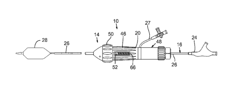
for example, radially
expandable from a radially compressed state mounted on the delivery system to
a radially
expanded state for implantation using an inflatable balloon (or equivalent
expansion device) of
the delivery system. Exemplary delivery routes through the body and into the
heart include
transfemoral routes, transapical routes, and transaortic routes, among others.
Although the
devices and methods disclosed herein are particular suited for implanting
prosthetic heart valves
(e.g., a prosthetic aortic valve or prosthetic mitral valve), the disclosed
devices and methods can
be adapted for implanting other types of prosthetic valves within the body
(e.g., prosthetic
- 2 -
Date Recue/Date Receievd 2020-10-02
venous valves) or other types of expandable prosthetic devices adapted to be
implanted in
various body lumens.
[008] In some embodiments, a delivery apparatus for implanting a prosthetic,
transcatheter
heart valve via a patient's vasculature includes an adjustment device for
adjusting the position of
a balloon relative to a crimped prosthetic valve (and/or vice versa). A
balloon catheter can
extend coaxially with a guide (or flex) catheter, and a balloon member at the
distal end of the
balloon catheter can be positioned proximal or distal to a crimped prosthetic
valve. The balloon
member and the crimped prosthetic valve can enter the vasculature of a patient
through an
introducer sheath and, once the balloon member and the crimped prosthetic
valve reach a
suitable location in the body, the relative position of the prosthetic valve
and balloon member
can be adjusted so that the balloon member is positioned within the frame of
the prosthetic valve
so that the prosthetic valve eventually can be expanded at the treatment site.
Once the crimped
prosthetic valve is positioned on the balloon, the prosthetic valve is
advanced to the vicinity of
the deployment location (i.e., the native aortic valve) and the adjustment
device can further be
used to accurately adjust or "fine tune" the position of the prosthetic valve
relative to the desired
deployment location.
[009] An exemplary method of implanting a radially compressible and expandable
prosthetic
device (e.g., a prosthetic heart valve) in the heart comprises: (a)
introducing a delivery device
into the body of a patient, the delivery device comprising a handle portion,
an elongated shaft
extending from the handle portion, the shaft having a distal end portion
mounting an inflatable
balloon and a prosthetic heart valve in a radially compressed state; (b)
advancing the distal end
portion of the delivery device toward the native heart valve until the
prosthetic valve is within or
adjacent the annulus of the native heart valve; (c) positioning the prosthetic
heart valve at a
desired implantation position within the annulus of the native by rotating an
adjustment device
coupled to the handle portion and the shaft to cause the shaft and the
prosthetic valve to move
distally and/or proximally relative to the handle portion until the prosthetic
heart valve is at the
desired implantation position; and (d) after the prosthetic heart valve has
been moved to the
- 3 -
Date Recue/Date Receievd 2020-10-02
desired implantation position, inflating the balloon to cause the prosthetic
heart valve to radially
expand and engage the annulus of the native heart valve.
[010] An exemplary delivery apparatus for implantation of a prosthetic device
(e.g., a
prosthetic heart valve) in the heart comprises an elongated shaft comprising a
proximal end
portion and a distal end portion, an inflatable balloon, and a valve mounting
member. The
balloon is mounted on the distal end portion of the shaft. The valve mounting
member is
disposed on the distal end portion of the shaft within the balloon and is
configured to facilitate
frictional engagement between the prosthetic heart valve and the balloon when
the prosthetic
heart valve is mounted in a radially compressed state on the balloon and
surrounding the
mounting member. The mounting member comprises at least one longitudinally
extending fluid
passageway though which an inflation fluid in the balloon can flow.
[011] In some embodiments, the at least one fluid passageway has first and
second openings
adjacent first and second ends of the prosthetic heart valve, respectively.
When the prosthetic
valve is mounted on the balloon in a crimped state, the inflation fluid in the
balloon can flow
from a first region of the balloon proximal to the first end of the prosthetic
valve, inwardly
through the first opening, through the fluid passageway, outwardly through the
second opening
and into a second region of the balloon distal to the second end of the
prosthetic valve.
[012] Another exemplary delivery apparatus for implantation of a prosthetic
device (e.g., a
prosthetic heart valve) in the heart comprises a handle portion and an
elongated shaft extending
from the handle portion. The shaft comprises a proximal end portion coupled to
the handle
portion and a distal end portion configured to mount a prosthetic heart valve
in a radially
compressed state. The apparatus also comprises a sliding member disposed on
the proximal end
portion of the shaft. The handle portion comprising a rotatable member that is
operatively
coupled to the sliding member so as to cause translational movement of the
sliding member upon
rotation of the rotatable member. A shaft engagement member is disposed on the
shaft and
couples the shaft to the sliding member. The shaft engagement member is
configured to be
manipulated between a first state and a second state. In the first state, the
shaft can move freely
- 4 -
Date Recue/Date Receievd 2020-10-02
in the longitudinal direction relative to the sliding member and the rotatable
member. In the
second state, the shaft engagement member frictionally engages the shaft and
prevents rotational
and longitudinal movement of the shaft relative to the sliding member such
that rotation of the
rotatable member causes corresponding longitudinal movement of the sliding
member and the
shaft. When a prosthetic device is mounted on the distal end of the shaft and
the shaft
engagement member is manipulated to engage the shaft, the rotatable member can
be used to
adjust the location of the prosthetic device relative to its desired
implantation location within the
heart.
[013] In some embodiments, the shaft engagement member comprises a collet
disposed on the
shaft. The collet can have flexible fingers that can be forced to frictionally
engage and retain the
shaft relative to the sliding member so that the rotatable member can be used
to adjust the
position of the prosthetic device mounted on the distal end portion of the
shaft.
[014] Another exemplary delivery device for implantation of a prosthetic
device (e.g., a
prosthetic heart valve) within the heart, such as via a transapical or
transaortic route, comprises
an inflatable balloon, a proximal stop, and a distal stop. The stops are
configured to limit
longitudinal movement of the prosthetic device relative to the balloon while
the prosthetic device
is mounted over the balloon in the radially compressed state between the
proximal stop and the
distal stop. The proximal stop and the distal stop each comprise an end
portion positioned within
the balloon and configured to be positioned adjacent the prosthetic device
when the prosthetic
device is radially compressed between the proximal and distal stops. Each of
the stop end
portions comprises at least one longitudinally extending slot that allows the
respective stop end
portion to be radially compressed to a smaller diameter. The at least one
longitudinally
extending slot in each stop end portion can also be configured to allow a
balloon-inflation fluid
to flow radially through the respective stop and into the region of the
balloon extending through
the prosthetic valve.
[015] In some embodiments, when a prosthetic device is mounted on the delivery
device in the
radially compressed state, the proximal stop and the distal stop are
configured to allow a balloon-
- 5 -
Date Recue/Date Receievd 2020-10-02
inflation fluid to flow from a proximal portion of the balloon, through the at
least one slot in the
proximal stop, through an intermediate portion of the balloon positioned
within the prosthetic
device, through the at least one slot in the distal stop, and into a distal
portion of the balloon.
[016] In some embodiments, a proximal end of the balloon is attached to the
proximal stop and
a distal end of the balloon is attached to the distal stop.
[017] In some embodiments, the delivery device further comprises an outer
shaft having a
lumen and an inner shaft extending through the lumen of the outer shaft, with
the proximal stop
attached to a distal end of the outer shaft and positioned around the inner
shaft and the distal stop
attached to an outer surface of the inner shaft.
[018] In some embodiments, the proximal stop further comprises a proximal
portion attached
to the distal end of the outer shaft and to a proximal end of the balloon, and
an intermediate
portion extending between the proximal portion and the end portion, the
intermediate portion
having an outer diameter that is less than an outer diameter of the proximal
portion and less than
the diameter of the end portion.
[019] In some embodiments, the proximal stop is attached to the distal end of
the outer shaft
and further comprises at least one fluid passageway that allows an inflation
fluid to flow through
the at least one passageway and into the balloon.
[020] In some embodiments, the distal stop further comprises a distal portion
attached to a
distal end of the balloon and an intermediate portion extending between the
distal portion and the
end portion, the intermediate portion having an outer diameter that is less
than an outer diameter
of the distal portion and less than the diameter of the end portion.
[021] In some embodiments, the end portion of each stop decreases in diameter
in a direction
extending away from the prosthetic device.
[022] In some embodiments, the delivery device further comprises a nosecone
attached to a
distal end of the distal stop.
- 6 -
Date Recue/Date Receievd 2020-10-02
[023] In some embodiments, at least one of the stop end portions comprises at
least three
longitudinal slots that allow the stop end portion to be radially compressed
to a smaller diameter
when the prosthetic device is crimped onto the delivery device.
[024] An exemplary method of implanting a prosthetic heart valve within the
heart comprises:
(a) introducing a distal end portion of a delivery device into the native
aortic valve of the heart, a
distal end portion of the delivery device comprising an inflatable balloon, a
proximal stop and a
distal stop positioned at least partially within the balloon, and a radially
expandable prosthetic
heart valve mounted over the balloon and between the proximal stop and the
distal stop in a
radially compressed state; (b) inflating the balloon to radially expand the
prosthetic heart valve
within the native aortic valve, wherein the balloon is inflated with an
inflation fluid that flows
radially through the proximal and distal stops; (c) deflating the balloon; and
(d) retracting the
delivery device from the heart.
[025] In some embodiments, the proximal stop is positioned adjacent to a
proximal end of the
prosthetic heart valve and the distal stop is positioned adjacent to a distal
end of the prosthetic
heart valve, such that the prosthetic device is longitudinally contained
between the proximal and
distal stops during introduction of the prosthetic heart valve through an
introducer sheath into the
body.
[026] In some embodiments, inflating the balloon comprises causing the
inflation fluid to flow:
(i) through a first passageway in the proximal stop and into a proximal
portion of the balloon; (ii)
from the proximal portion of the balloon, through a second passageway in the
proximal stop, and
into an intermediate portion of the balloon within the prosthetic device; and
(iii) from the
intermediate portion of the balloon, through a passageway in the distal stop,
and into a distal
portion of the balloon.
[027] In some embodiments, prior to introducing the delivery device into the
heart, the
prosthetic heart valve is crimped to the radially compressed state onto
delivery device while the
proximal stop and the distal stop are simultaneously radially compressed. The
prosthetic heart
valve can have a first outer diameter in the radially compressed state and the
proximal stop and
- 7 -
Date Recue/Date Receievd 2020-10-02
distal stop can be compressed from a second outer diameter to about the first
outer diameter
during the crimping. When compressive pressure is released after the crimping,
the proximal
stop and distal stop can be configured to resiliently expand from about the
first outer diameter to
about the second outer diameter.
[028] An exemplary system for delivering a prosthetic device into a patient
comprises an
introducer sheath configured to be inserted partially into a patient, a loader
configured to be
inserted into a proximal end the introducer sheath, and a delivery device
configured to be passed
through the loader and the introducer sheath into the patient carrying a
prosthetic device to be
implanted in the patient. The loader comprises a flush port for selectively
introducing fluid into
the loader and a bleed port for selectively releasing fluid from within the
loader, and both the
flush port and the bleed port are sealed with the same resiliently flexible
annular sealing member.
The sealing member can comprise a push tab that extends radially through the
bleed port, such
that the bleed port is configured to be selectively opened by depressing the
push tab in the
radially inward direction.
[029] The foregoing and other objects, features, and advantages of the
invention will become
more apparent from the following detailed description, which proceeds with
reference to the
accompanying figures.
BRIEF DESCRIPTION OF THE DRAWINGS
[030] FIG. 1 is a side view of a delivery apparatus for implanting a
prosthetic heart valve,
according to one embodiment.
[031] FIG. 2A is a cross-sectional view of the handle of the delivery
apparatus of FIG. 1.
[032] FIG. 2B is another cross-sectional view of the handle of the delivery
apparatus of FIG. 1.
[033] FIG. 3 is side view of a section of the handle and a section of the
distal end portion of the
delivery apparatus of FIG. 1.
[034] FIG. 4 is a side view of the distal end portion of the delivery
apparatus of FIG. 1.
- 8 -
Date Recue/Date Receievd 2020-10-02
[035] FIG. 5 is a side view of the distal end portion of the delivery
apparatus of FIG. 1 showing
the balloon in an inflated state.
[036] FIG. 6 is an enlarged perspective view of a collet used in the handle of
the delivery
apparatus of FIG. 1.
[037] FIG. 7 is a cross-sectional view of the collet of FIG. 6.
[038] FIG. 8 is an enlarged side view of a mounting member for a prosthetic
heart valve.
[039] FIGS. 9-11 are enlarged, cross-sectional views of the distal end portion
of the delivery
apparatus of FIG. 1, showing the inflation of a balloon for deployment of a
prosthetic heart valve
on the balloon.
[040] FIG. 12 is a perspective view of an alternative embodiment of a mounting
member for a
prosthetic heart valve.
[041] FIG. 13 is a side view of the mounting member of FIG. 12 shown partially
in section.
[042] FIG. 14 is an end view of the mounting member of FIG. 12.
[043] FIGS. 15-17 are enlarged, cross-sectional views of the distal end
portion of a delivery
apparatus containing the mounting member of FIG. 12, and showing the inflation
of a balloon for
deployment of a prosthetic heart valve on the balloon.
[044] FIG. 18 is an exploded perspective view of the handle of a delivery
apparatus, according
to another embodiment.
[045] FIG. 19 is an enlarged perspective view of the collet, pusher element,
spring, ring, and
washer of the handle shown in FIG. 18.
[046] FIG. 20 is a cross-sectional view of the handle of the delivery
apparatus of FIG. 18.
[047] FIG. 21 is another cross-sectional view of the handle of the delivery
apparatus of FIG. 18.
- 9 -
Date Recue/Date Receievd 2020-10-02
[048] FIG. 22 is a perspective view of the inner shaft, or slider, of the
handle shown in FIG. 18.
[049] FIG. 23 is an enlarged side view of the inner nut of the handle shown in
FIG. 18.
[050] FIG. 24 is an enlarged cross-sectional view of the inner nut shown in
FIG. 23.
[051] FIGS. 25-27 are enlarged top, perspective and end views, respectively,
of the rotatable
knob of the handle shown in FIG. 18.
[052] FIG. 28 is an enlarged perspective view of the indicator ring of the
handle shown in FIG.
18.
[053] FIGS. 29-31 are cross-sectional views of the distal end portion of a
delivery apparatus for
a prosthetic heart valve, according to another embodiment, having two
inflatable balloons for
deploying a prosthetic valve.
[054] FIG. 32 is a side view of a delivery apparatus for a prosthetic heart
valve, an introducer,
and a loader device, according to another embodiment.
[055] FIG. 33 is an enlarged, cross-sectional view of the distal end portion
of the delivery
apparatus of FIG. 32.
[056] FIG. 34 is a cross-sectional view of the introducer of FIG. 32.
[057] FIG. 35 is a cross-sectional view of the loader of FIG. 32.
[058] FIG. 36 is a perspective view of the handle of the delivery apparatus
shown in FIG. 32.
[059] FIG. 37 is a partially exploded, perspective view of the handle of FIG.
36.
[060] FIG. 38 is a perspective view of the handle of FIG. 36, shown with a
portion of the outer
housing cut away for purposes of illustration.
[061] FIG. 39 is an exploded, perspective view of the handle of FIG. 36.
- 10 -
Date Recue/Date Receievd 2020-10-02
[062] FIG. 40 is a perspective view of another embodiment of a handle that can
be used in the
delivery apparatus of FIG. 32.
[063] FIG. 41 is a perspective of the handle of FIG. 40, with a portion of the
outer housing and
some internal components removed for purposes of illustration.
[064] FIG. 42 is an exploded, perspective view of the handle of FIG. 40.
[065] FIG. 43 is a perspective view of another embodiment of a handle that can
be used in the
delivery apparatus of FIG. 32.
[066] FIG. 44 is a perspective of the handle of FIG. 43, with a portion of the
outer housing and
some internal components removed for purposes of illustration.
[067] FIG. 45 is an exploded, perspective view of the handle of FIG. 43.
[068] FIG. 46 is a perspective view of a delivery apparatus for a prosthetic
heart valve,
according to another embodiment.
[069] FIG. 47 is an enlarged, cross-sectional view of the distal end portion
of the delivery
apparatus of FIG. 46.
[070] FIG. 47A is an enlarged, cross-sectional view of the distal end portion
of the delivery
apparatus of FIG. 46 showing a prosthetic heart valve mounted in a crimped
state on the balloon
of the delivery apparatus.
[071] FIG. 48 is a perspective view of the handle of the delivery apparatus of
FIG. 46, with a
portion of the outer housing removed for purposes of illustration.
[072] FIG. 49 is a perspective view of an introducer, according to another
embodiment.
[073] FIG. 50 is an enlarged, cross-sectional view of the proximal housing
portion of the
introducer shown in FIG. 49.
- 11 -
Date Recue/Date Receievd 2020-10-02
[074] FIG. 51 is a perspective view of a loader, according to another
embodiment.
[075] FIG. 52 is a cross-sectional view of the loader shown in FIG. 51.
[076] FIG. 53 is a perspective view of the loader of FIG. 51 shown inserted
into the introducer
of FIG. 49.
[077] FIG. 54 is a perspective view of the button valve of the loader shown in
FIG. 51.
[078] FIG. 55 is a top plan view of the button valve shown in FIG. 51.
[079] FIG. 56 is a perspective view of a prosthetic heart valve, according to
one embodiment.
[080] FIG. 57 is a side elevation view of the prosthetic heart valve of FIG.
56.
DETAILED DESCRIPTION
[081] In particular embodiments, a delivery apparatus for implanting a
prosthetic, transcatheter
heart valve via a patient's vasculature includes an adjustment device for
adjusting the position of
a balloon relative to a crimped prosthetic valve (and/or vice versa). A
balloon catheter can
extend coaxially with a guide (or flex) catheter, and a balloon member at the
distal end of the
balloon catheter can be positioned proximal or distal to a crimped prosthetic
valve. As described
below in more detail, the balloon member and the crimped prosthetic valve can
enter the
vasculature of a patient through an introducer sheath and, once the balloon
member and the
crimped prosthetic valve reach a suitable location in the body, the relative
position of the
prosthetic valve and balloon member can be adjusted so that the balloon member
is positioned
within the frame of the prosthetic valve so that the prosthetic valve
eventually can be expanded
at the treatment site. Once the crimped prosthetic valve is positioned on the
balloon, the
prosthetic valve is advanced to the vicinity of the deployment location (i.e.,
the native aortic
valve) and the adjustment device can further be used to accurately adjust or
"fine tune" the
position of the prosthetic valve relative to the desired deployment location.
[082] FIG. 1 shows a delivery apparatus 10 adapted to deliver a prosthetic
heart valve 12
(shown schematically in FIGS. 9-11) (e.g., a prosthetic aortic valve) to a
heart, according to one
- 12 -
Date Recue/Date Receievd 2020-10-02
embodiment. The apparatus 10 generally includes a steerable guide catheter 14
(FIG. 3), and a
balloon catheter 16 extending through the guide catheter 14. The guide
catheter can also be
referred to as a flex catheter or a main catheter. The use of the term main
catheter should be
understood, however, to include flex or guide catheters, as well as other
catheters that do not
have the ability to flex or guide through a patient's vasculature.
[083] The guide catheter 14 and the balloon catheter 16 in the illustrated
embodiment are
adapted to slide longitudinally relative to each other to facilitate delivery
and positioning of
prosthetic valve 12 at an implantation site in a patient's body, as described
in detail below.
[084] The guide catheter 14 includes a handle portion 20 and an elongated
guide tube, or shaft,
22 extending from handle portion 20 (FIG. 3). FIG. 1 shows the delivery
apparatus without the
guide catheter shaft 22 for purposes of illustration. FIG. 3 shows the guide
catheter shaft 22
extending from the handle portion 20 over the balloon catheter. The balloon
catheter 16 includes
a proximal portion 24 (FIG. 1) adjacent handle portion 20 and an elongated
shaft 26 that extends
from the proximal portion 24 and through handle portion 20 and guide tube 22.
The handle
portion 20 can include a side arm 27 having an internal passage which fluidly
communicates
with a lumen defined by the handle portion 20.
[085] An inflatable balloon 28 is mounted at the distal end of balloon
catheter 16. As shown in
FIG. 4, the delivery apparatus 10 is configured to mount the prosthetic valve
12 in a crimped
state proximal to the balloon 28 for insertion of the delivery apparatus and
prosthetic valve into a
patient's vasculature, which is described in detail in U.S. Publication No.
2009/0281619 (U.S.
Application No. 12/247,846, filed October 8, 2008), which is incorporated
herein by reference.
Because prosthetic valve 12 is crimped at a location different from the
location of balloon 28
(e.g., in this case prosthetic valve 12 desirably is crimped proximal to
balloon 28), prosthetic
valve 12 can be crimped to a lower profile than would be possible if
prosthetic valve 12 was
crimped on top of balloon 28. This lower profile permits the surgeon to more
easily navigate the
delivery apparatus (including crimped valve 12) through a patient's
vasculature to the treatment
location. The lower profile of the crimped prosthetic valve is particularly
helpful when
- 13 -
Date Recue/Date Receievd 2020-10-02
navigating through portions of the patient's vasculature which are
particularly narrow, such as
the iliac artery. The lower profile also allows for treatment of a wider
population of patients,
with enhanced safety.
[086] A nose piece 32 (FIG. 4) can be mounted at the distal end of the
delivery apparatus 10 to
facilitate advancement of the delivery apparatus 10 through the patient's
vasculature to the
implantation site. In some instances, it may be useful to have nose piece 32
connected to a
separate elongated shaft so that nose piece 32 can move independently of other
elements of
delivery apparatus 10. Nose piece 32 can be formed of a variety of materials,
including various
plastic materials.
[087] As can be seen in FIG. 5, the balloon catheter 16 in the illustrated
configuration further
includes an inner shaft 34 (FIG. 2A) that extends from proximal portion 24 and
coaxially through
the outer balloon catheter shaft 26 and the balloon 28. The balloon 28 can be
supported on a
distal end portion of inner shaft 34 that extends outwardly from the outer
shaft 26 with a
proximal end portion 36 of the balloon secured to the distal end of outer
shaft 26 (e.g., with a
suitable adhesive) (FIG. 5). The outer diameter of inner shaft 34 is sized
such that an annular
space is defined between the inner and outer shafts along the entire length of
the outer shaft. The
proximal portion 24 of the balloon catheter can be formed with a fluid
passageway (not shown)
that is fluidly connectable to a fluid source (e.g., saline) for inflating the
balloon. The fluid
passageway is in fluid communication with the annular space between inner
shaft 34 and outer
shaft 26 such that fluid from the fluid source can flow through fluid
passageway, through the
space between the shafts, and into balloon 28 to inflate the same and deploy
prosthetic valve 12.
[088] The proximal portion 24 also defines an inner lumen that is in
communication with a
lumen 38 of the inner shaft 34 that is sized to receive guide wire (not shown)
that can extend
coaxially through the inner shaft 34 and the nose cone 32.
[089] The inner shaft 34 and outer shaft 26 of the balloon catheter can be
formed from any of
various suitable materials, such as nylon, braided stainless steel wires, or a
polyether block
amide (commercially available as Pebax ). The shafts 26, 34 can have
longitudinal sections
- 14 -
Date Recue/Date Receievd 2020-10-02
formed from different materials in order to vary the flexibility of the shafts
along their lengths.
The inner shaft 34 can have an inner liner or layer formed of Teflon to
minimize sliding friction
with a guide wire.
[090] The distal end portion of the guide catheter shaft 22 comprises a
steerable section 68
(FIG. 3), the curvature of which can be adjusted by the operator to assist in
guiding the apparatus
through the patient's vasculature, and in particular, the aortic arch. The
handle 20 in the
illustrated embodiment comprises a distal handle portion 46 and a proximal
handle portion 48.
The distal handle portion 46 functions as a mechanism for adjusting the
curvature of the distal
end portion of the guide catheter shaft 22 and as a flex indicating device
that allows a user to
measure the relative amount of flex of the distal end of the guide catheter
shaft 22. In addition,
the flex indicating device provides a visual and tactile response at the
handle the device, which
provides a surgeon with an immediate and direct way to determine the amount of
flex of the
distal end of the catheter.
[091] The distal handle portion 46 can be operatively connected to the
steerable section 68 and
functions as an adjustment mechanism to permit operator adjustment of the
curvature of the
steerable section via manual adjustment of the handle portion. Explaining
further, the handle
portion 46 comprises a flex activating member 50, an indicator pin 52, and a
cylindrical main
body, or housing 54. As shown in FIGS. 2A and 2B, the flex activating member
50 comprises an
adjustment knob 56 and a shaft 58 extending proximally from the knob into the
housing 54. A
proximal end portion of the guide catheter shaft 22 extends into and is fixed
within the central
lumen of the housing 54. An inner sleeve 70 surrounds a portion of the guide
catheter shaft 22
inside the housing 54. A threaded slide nut 72 is disposed on and is slidable
relative to the
sleeve 70. The slide nut 72 is formed with external threads that mate with
internal threads 60 of
the shaft 58.
[092] The slide nut 72 can be formed with two slots formed on the inner
surface of the nut and
extending the length thereof The sleeve 70 can be formed with longitudinally
extending slots
that are aligned with the slots of the slide nut 72 when the slide nut is
placed on the sleeve.
- 15 -
Date Recue/Date Receievd 2020-10-02
Disposed in each slot is a respective elongated nut guide, which can be in the
form of an
elongated rod or pin 76. The nut guides 76 extend radially into respective
slots in the slide
nut 72 to prevent rotation of the slide nut 72 relative to the sleeve 70. By
virtue of this
arrangement, rotation of the adjustment knob 56 (either clockwise or
counterclockwise) causes
the slide nut 72 to move longitudinally relative to the sleeve 70 in the
directions indicated by
double-headed arrow 74.
[093] One or more pull wires 78 (FIG. 2A) couple the adjustment knob 56 to the
steerable
section 68 to adjust the curvature of the steerable section upon rotation of
the adjustment knob.
For example, the proximal end portion of the pull wire 78 can extend into and
can be secured to a
retaining pin, such as by crimping the pin around the proximal end of the pull
wire, which pin is
disposed in a slot in the slide nut 72. The pull wire extends from the pin,
through the slot in the
slide nut, a slot in the sleeve 70, and into and through a pull wire lumen in
the shaft 22. The
distal end portion of the pull wire is secured to the distal end portion of
the steerable section 68.
[094] The pin, which retains the proximal end of the pull wire 78, is captured
in the slot in the
slide nut 72. Hence, when the adjustment knob 56 is rotated to move the slide
nut 72 in the
proximal direction, the pull wire also is moved in the proximal direction. The
pull wire pulls the
distal end of the steerable section 68 back toward the handle portion, thereby
bending the
steerable section and reducing its radius of curvature. The friction between
the adjustment knob
56 and the slide nut 72 is sufficient to hold the pull wire taut, thus
preserving the shape of the
bend in the steerable section if the operator releases the adjustment knob 56.
When the
adjustment knob 56 is rotated in the opposite direction to move the slide nut
72 in the distal
direction, tension in the pull wire is released. The resiliency of the
steerable section 68 causes
the steerable to return its normal, non-deflected shape as tension on the pull
wire is decreased.
Because the pull wire is not fixedly secured to the slide nut 72 (the pin can
move within the slot
in the nut), movement of the slide nut in the distal direction does not push
on the end of the pull
wire, causing it to buckle. Instead, the pin is allowed to float within the
slot of the slide nut 72
when the knob 56 is adjusted to reduce tension in the pull wire, preventing
buckling of the pull
wire.
- 16 -
Date Recue/Date Receievd 2020-10-02
[095] In particular embodiments, the steerable section 68 in its non-deflected
shape is slightly
curved and in its fully curved position, the steerable section generally
conforms to the shape of
the aortic arch. In other embodiments, the steerable section can be
substantially straight in its
non-deflected position.
[096] The distal handle portion 46 can have other configurations that are
adapted to adjust the
curvature of the steerable section 68. One such alternative handle
configuration is shown in co-
pending U.S. Patent Application No. 11/152,288 (published under Publication
No.
US2007/0005131), which is incorporated herein by reference in its entirety.
Additional details
relating to the steerable section and handle configuration discussed above can
be found in U.S.
Patent Application No. 11/852977 (published as U.S. Publication No.
US2008/0065011), which
is incorporated herein by reference in its entirety.
[097] The shaft 58 also includes an externally threaded surface portion 62. As
shown in
FIG. 2B, a base portion 64 of the indicator pin 52 mates with the externally
threaded surface
portion 62 of the shaft 58. The shaft 58 extends into the main body 54 and the
indicator pin 52 is
trapped between the externally threaded surface portion 62 and the main body
54, with a portion
of the indicator pin 52 extending into a longitudinal slot 66 of the handle.
As the knob 56 rotated
to increase the curvature of the distal end of the guide catheter shaft 22,
the indicator pin 52
tracks the external threaded portion 62 of the flex activating member and
moves in the proximal
direction inside of the slot 66. The greater the amount of rotation of the
knob 56, the further
indicator pin 52 moves towards the proximal end of the proximal handle portion
46. Conversely,
rotating the knob 56 in the opposite direction decreases the curvature of the
distal end of the
guide catheter shaft 22 (i.e., straightens the guide catheter shaft) and
causes corresponding
movement of the indicator pin 52 toward the distal end of the distal handle
portion 46.
[098] The outer surface of the main body 54 of the distal handle portion 46
can include visual
indicia adjacent the slot 66 that indicate the amount of flex of the distal
end of the guide catheter
shaft 22, based on the position of the indicator pin 52 relative to the visual
indicia. Such indicia
can identify the amount of flex in any of a variety of manners. For example,
the outer surface of
- 17 -
Date Recue/Date Receievd 2020-10-02
the main body 54 can include a series of numbers (e.g., 0 to 10) adjacent the
slot that indicate
the amount of curvature of the guide catheter shaft 22 based on the position
of the indicator pin
52 relative to the number scale.
[099] As described above, when the delivery apparatus is introduced into the
vasculature of the
patient, a crimped prosthetic valve 12 is positioned proximal to the balloon
28 (FIG. 4). Prior to
expansion of the balloon 28 and deployment of prosthetic valve 12 at the
treatment site, the
prosthetic valve 12 is moved relative to the balloon (or vice versa) to
position the crimped
prosthetic valve on the balloon for deploying (expanding) the prosthetic
valve. As discussed
below, the proximal handle portion 48 serves as an adjustment device that can
be used to move
the balloon 28 proximally into position within the frame of prosthetic valve
12, and further to
accurately position the balloon and the prosthetic valve at the desired
deployment location.
[0100] As shown in FIG. 2A and 2B, the proximal handle portion 48 comprises an
outer housing
80 and an adjustment mechanism 82. The adjustment mechanism 82, which is
configured to
adjust the axial position of the balloon catheter shaft 26 relative to the
guide catheter shaft 22,
comprises an adjustment knob 84 and a shaft 86 extending distally into the
housing 80. Mounted
within the housing 80 on the balloon catheter shaft 26 is an inner support 88,
which in turn
mounts an inner shaft 90 (also referred to as a slider or sliding mechanism)
(also shown in FIG.
22). The inner shaft 90 has a distal end portion 92 formed with external
threads that mate with
internal threads 94 that extend along the inner surface of the adjustment
mechanism 82. The
inner shaft 90 further includes a proximal end portion 96 that mounts a
securement mechanism
98, which is configured to retain the position of the balloon catheter shaft
26 relative to the
proximal handle portion 48 for use of the adjustment mechanism 82, as further
described below.
The inner shaft 90 can be coupled to the inner support 88 such that rotation
of shaft 86 causes the
inner shaft 90 to move axially within the handle. For example, the inner
support 88 can have an
axially extending rod or rail that extends into slot formed in the inner
surface of the inner shaft
90. The rod or rail prevents rotation of the inner shaft 90 but allows it to
move axially upon
rotation of the shaft 86.
- 18 -
Date Recue/Date Receievd 2020-10-02
[0101] The securement mechanism 98 includes internal threads that mate with
external threads
of the proximal end portion 96 of the inner shaft. Mounted within the proximal
end portion 96
on the balloon catheter shaft 26 is a pusher element 100 and a shaft
engagement member in the
form of a collet 102. The collet 102 is configured to be manipulated by the
securement
mechanism between a first state in which collet allows the balloon catheter
shaft to be moved
freely in the longitudinal and rotational directions and a second state in
which the collet
frictionally engages the balloon catheter shaft and prevents rotational and
longitudinal movement
of the balloon catheter shaft relative to the inner shaft 90, as further
described below.
[0102] As best shown in FIGS. 6 and 7, the collet 102 comprises a distal end
portion 104, an
enlarged proximal end portion 106, and a lumen 108 that receives the balloon
catheter shaft 26.
A plurality of axially extending, circumferentially spaced slots 110 extend
from the proximal end
of the collet to a location on the distal end portion 104, thereby forming a
plurality of flexible
fingers 112. The proximal end portion can be formed with a tapered end surface
114 that
engages a corresponding tapered end surface of the pusher element 100 (FIG.
2A).
[0103] As noted above, the securement mechanism 98 is operable to restrain
movement of the
balloon catheter shaft 26 (in the axial and rotational directions) relative to
the proximal handle
portion 48. Explaining further, the securement mechanism 98 is movable between
a proximal
position (shown in FIGS. 2A and 2B) and a distal position closer to the
adjacent end of the
knob 84. In the proximal position, the collet 102 applies little, if any,
force against the balloon
catheter shaft 26, which can slide freely relative to the collet 102, the
entire handle 20, and the
guide catheter shaft 22. When the securement mechanism 98 is rotated so as to
move to its distal
position closer to knob 84, the securement mechanism urges pusher element 100
against the
proximal end of the collet 102. The tapered surface of the pusher element
pushes against the
corresponding tapered surface 114 of the collet, forcing fingers 112 radially
inward against the
outer surface of the balloon catheter shaft 26. The holding force of the
collet 102 against the
balloon catheter shaft locks the balloon catheter shaft relative to the inner
shaft 90. In the locked
position, rotation of the adjustment knob 84 causes the inner shaft 90 and the
balloon catheter
- 19 -
Date Recue/Date Receievd 2020-10-02
shaft 26 to move axially relative to the guide catheter shaft 22 (either in
the proximal or distal
direction, depending on the direction the knob 84 is rotated).
[0104] The adjustment knob 84 can be utilized to position the prosthetic valve
12 on the balloon
28 and/or once the prosthetic valve 12 is on the balloon, to position the
prosthetic valve and the
balloon at the desired deployment site within the native valve annulus. One
specific method for
implanting the prosthetic valve 12 in the native aortic valve is as follows.
The prosthetic valve
12 initially can be crimped on a mounting region 120 (FIGS. 4 and 5) of the
balloon catheter
shaft 26 immediately adjacent the proximal end of the balloon 28 or slightly
overlapping the
proximal end of the balloon. The proximal end of the prosthetic valve can abut
the distal end
122 of the guide catheter shaft 22 (FIG. 4), which keeps the prosthetic valve
in place on the
balloon catheter shaft as the delivery apparatus and prosthetic valve are
inserted through an
introducer sheath. The prosthetic valve 12 can be delivered in a transfemoral
procedure by first
inserting an introducer sheath into the femoral artery and pushing the
delivery apparatus through
the introducer sheath into the patient's vasculature.
[0105] After the prosthetic valve 12 is advanced through the narrowest
portions of the patient's
vasculature (e.g., the iliac artery), the prosthetic valve 12 can be moved
onto the balloon 28. For
example, a convenient location for moving the prosthetic valve onto the
balloon is the
descending aorta. The prosthetic valve can be moved onto the balloon, for
example, by holding
the handle portion 46 steady (which retains the guide catheter shaft 22 in
place), and moving the
balloon catheter shaft 26 in the proximal direction relative to the guide
catheter shaft 22. As the
balloon catheter shaft is moved in the proximal direction, the distal end 122
of the guide catheter
shaft pushes against the prosthetic valve, allowing the balloon 28 to be moved
proximally
through the prosthetic valve in order to center the prosthetic valve on the
balloon, as depicted in
FIG. 9. The balloon catheter shaft can include one or more radiopaque markers
to assist the user
in positioning the prosthetic valve at the desired location on the balloon.
The balloon catheter
shaft 26 can be moved in the proximal direction by simply sliding/pulling the
balloon catheter
shaft in the proximal direction if the securement mechanism 98 is not engaged
to retain the shaft
26. For more precise control of the shaft 26, the securement mechanism 98 can
be engaged to
- 20 -
Date Recue/Date Receievd 2020-10-02
retain the shaft 26, in which case the adjustment knob 84 is rotated to effect
movement of the
shaft 26 and the balloon 28.
[0106] As shown in FIG. 5, the delivery apparatus can further include a
mounting member 124
secured to the outer surface of the shaft 34 within the balloon 28. The
mounting member helps
retain the prosthetic valve in place on the balloon by facilitating the
frictional engagement
between the prosthetic valve and the outer surface of the balloon. The
mounting member 124
helps retain the prosthetic valve in place for final positioning of the
prosthetic valve at the
deployment location, especially when crossing the native leaflets, which
typically are calcified
and provide resistance against movement of the prosthetic valve. The nose cone
32 can include a
proximal portion 126 inside the balloon to assist in positioning the
prosthetic valve. The
proximal portion 126 desirably comprises a tapered member that has a maximum
diameter at its
proximal end adjacent the distal end of the prosthetic valve (FIG. 9) and
tapers in a direction
toward the distal end of the nosecone 32. The tapered member 126 serves as a
transition section
between the nosecone and the prosthetic valve as the prosthetic valve is
pushed through the
calcified native leaflets by shielding the distal end of the prosthetic valve
from contacting the
native leaflets. Although FIG. 9 shows the prosthetic valve having a crimped
diameter slightly
larger than the diameter of the tapered member 126 at its proximal end, the
tapered member 126
can have a diameter at its proximal end that is the same as or slightly larger
than the diameter of
the crimped prosthetic valve, or at least the same as or slightly larger than
the diameter of the
metal frame of the crimped prosthetic valve.
[0107] As shown in FIG. 9, the prosthetic valve desirably is positioned on the
balloon for
deployment such that the distal end of the prosthetic valve is slightly spaced
from the nose cone
portion 126. When the prosthetic valve is positioned as shown in FIG. 9, the
guide catheter shaft
22 can be moved proximally relative to the balloon catheter shaft 26 so that
the guide catheter
shaft is not covering the inflatable portion of the balloon 28, and therefore
will not interfere with
inflation of the balloon.
- 21 -
Date Recue/Date Receievd 2020-10-02
[0108] As the prosthetic valve 12 is guided through the aortic arch and into
the ascending aorta,
the curvature of the steerable section 68 can be adjusted (as explained in
detail above) to help
guide or steer the prosthetic valve through that portion of the vasculature.
As the prosthetic
valve is moved closer toward the deployment location within the aortic
annulus, it becomes
increasingly more difficult to control the precise location of the prosthetic
valve by pushing or
pulling the handle portion 20 due to the curved section of the delivery
apparatus. When pushing
or pulling the handle portion 20, slack is removed from the curved section of
the delivery
apparatus before the pushing/pulling force is transferred to the distal end of
the delivery
apparatus. Consequently, the prosthetic valve tends to "jump" or move
abruptly, making precise
positioning of the prosthetic valve difficult.
[0109] For more accurate positioning of the prosthetic valve within the aortic
annulus, the
prosthetic valve 12 is placed as close as possible to its final deployment
location (e.g., within the
aortic annulus such that an inflow end portion of the prosthetic valve is in
the left ventricle and
an outflow end portion of the prosthetic valve is in the aorta) by
pushing/pulling the handle 20,
and final positioning of the prosthetic valve is accomplished using the
adjustment knob 84. To
use the adjustment knob 84, the securement mechanism 98 is placed in its
locked position, as
described above. Then, the handle 20 is held steady (which retains the guide
catheter shaft 22 in
place) while rotating the adjustment knob 84 to move the balloon catheter
shaft 26, and thus the
prosthetic valve, in the distal or proximal directions. For example, rotating
the knob in a first
direction (e.g., clockwise), moves the prosthetic valve proximally into the
aorta, while rotating
the knob in a second, opposite direction (e.g., counterclockwise) advances the
prosthetic valve
distally toward the left ventricle. Advantageously, operation of the
adjustment knob 84 is
effective to move the prosthetic valve in a precise and controlled manner
without sudden, abrupt
movements as can happen when pushing or pulling the delivery apparatus for
final positioning.
[0110] When the prosthetic valve is at the deployment location, the balloon 28
is inflated to
expand the prosthetic valve 12 (as depicted in FIG. 11) so as to contact the
native annulus. The
expanded prosthetic valve becomes anchored within the native aortic annulus by
the radial
outward force of the valve's frame against the surrounding tissue.
- 22 -
Date Recue/Date Receievd 2020-10-02
[0111] The mounting member 124 within the balloon is configured to allow the
inflation fluid
(e.g., saline) to flow unobstructed from the proximal end of the balloon to
the distal end of the
balloon. As best shown in FIG. 8, for example, the mounting member 124
comprises a coiled
wire (e.g., a metal coil) having a first section 124a, a second section 124b,
a third section 124c, a
fourth section 124d, and a fifth section 124e. When the prosthetic valve 12 is
positioned on the
balloon for deployment, the second section 124b is immediately adjacent the
proximal end of the
prosthetic valve and the fourth section 124d is immediately adjacent the
distal end of the
prosthetic valve. The first and fifth sections 124a, 124e, respectively, which
are at the proximal
and distal ends of the mounting member, respectively, are secured to the
balloon catheter shaft.
The second, third, and fourth sections 124b, 124c, and 124d, respectively, are
relatively larger in
diameter than the first and fifth sections and are spaced radially from the
outer surface of the
balloon catheter shaft. As can be seen, the second section 124b and the fourth
section 124d are
formed with spaces between adjacent coils. The third section can be formed
with smaller spaces
(or no spaces) between adjacent coils to maximize the surface area available
to retain the
prosthetic valve on the balloon during final positioning of the prosthetic
valve at the deployment
location.
[0112] Referring to FIG. 10, the spacing between coils of the second and
fourth sections 124b,
124d allows the inflation fluid to flow radially inwardly through the coils of
the second section
124b, axially through the lumen of the third section 124c, radially outwardly
through the coils of
the fourth section 124d, into the distal section of the balloon, in the
direction of arrows 128. The
nose cone portion 126 also can be formed with one or more slots 130 that allow
the inflation
fluid to flow more easily past the proximal nose cone portion 126 into the
distal section of the
balloon. In the illustrated embodiment, the proximal nose cone portion 126 has
three
circumferentially spaced slots 130. Since the inflation fluid can pressurize
and inflate the
proximal and distal sections of the balloon at substantially the same rate,
the balloon can be
inflated more evenly for controlled, even expansion of the prosthetic valve.
[0113] FIGS. 12-14 illustrate a mounting member 140 according to another
embodiment. The
mounting member 140 comprises a cylindrical inner wall 142, a cylindrical
outer wall 144, and a
- 23 -
Date Recue/Date Receievd 2020-10-02
plurality of angularly spaced ribs 146 separating the inner and outer walls.
The inner wall 142 is
secured to the outer surface of the shaft 34 within the balloon. In particular
embodiments, the
mounting member 140 can be made of a relatively rigid material (e.g.,
polyurethane or another
suitable plastic) that does not radially compress when the prosthetic valve is
moved onto the
balloon. As shown in FIG. 16, during inflation of the balloon, inflation fluid
in the proximal
section of the balloon can flow through the spaces 148 between the inner and
outer walls of the
mounting member, through one or more slots 130 in the proximal nose cone
portion 126, and
into the distal section of the balloon, in the direction of arrows 128.
[0114] It should be noted that the location of the threaded portions of the
adjustment mechanism
82 and the inner shaft 90 can be reversed. That is, adjustment mechanism 82
can have an
externally threaded portion that engages an internally threaded portion of the
inner shaft 90. In
addition, for embodiments where the balloon 28 is initially positioned
proximal to the prosthetic
valve 12, the adjustment mechanism 82 can be used to move the balloon distally
relative to the
crimped prosthetic valve in order to center the prosthetic valve on the
balloon for deployment.
[0115] FIGS. 56 and 57 show a prosthetic heart valve 700, according to another
embodiment.
The heart valve 700 comprises a frame, or stent, 702 and a leaflet structure
704 supported by the
frame. In particular embodiments, the heart valve 700 is adapted to be
implanted in the native
aortic valve and can be implanted in the body using, for example, the delivery
apparatus 10
described above. The prosthetic valve 700 can also be implanted within the
body using any of
the other delivery apparatuses described herein. Thus, the frame 702 typically
comprises a
plastically expandable material, such as stainless steel, a nickel based alloy
(e.g., a nickel-cobalt-
chromium alloy), polymers, or combinations thereof. In other embodiments, the
prosthetic valve
12, 700 can be a self-expandable prosthetic valve with a frame made from a
self-expanding
material, such as Nitinol. When the prosthetic valve is a self-expanding
valve, the balloon of the
delivery apparatus can be replaced with a sheath or similar restraining device
that retains the
prosthetic valve in a radially compressed state for delivery through the body.
When the
prosthetic valve is at the implantation location, the prosthetic valve can be
released from the
- 24 -
Date Recue/Date Receievd 2020-10-02
sheath, and therefore allowed to expand to its functional size. It should be
noted that any of the
delivery apparatuses disclosed herein can be adapted for use with a self-
expanding valve.
[0116] FIG. 18 is an exploded, perspective view of the distal end section of
an alternative
embodiment of a delivery device, indicated at 10'. The delivery device 10'
shares many
similarities with the delivery device 10, and therefore components of the
delivery device 10' that
are the same as those in the delivery device 10 are given the same reference
numerals and are not
described further. One difference between the delivery device 10 and the
delivery device 10' is
that the latter includes a different mechanism for locking/securing the
balloon catheter shaft 26
relative to the adjustment knob 84.
[0117] Referring to FIGS. 18 and 19, the locking mechanism for the balloon
catheter shaft
comprises an adjustment knob 150 housing an inner nut 152, a washer 154 and a
ring 156
disposed inside the inner nut 152, a biasing member in the form of a coiled
spring 158, a pusher
element 160, and a shaft engagement member in the form of a collet 102. As
best shown in
FIGS. 20 and 21, the inner nut 152 includes inner threads 162 (FIG. 24) that
engage the external
threads of the distal end portion 96 of the inner shaft 90 (FIG. 22). The
pusher element 160
includes a proximal shaft 164 and an enlarged distal end portion 166 that
bears against the
proximal end portion 106 of the collet 102. The spring 158 is disposed on the
shaft 164 of the
pusher element 160 and has a proximal end that bears against the ring 156 and
a distal end that
bears against the distal end portion 166 of the pusher element 160.
[0118] Referring to FIGS. 25-27, the adjustment knob 150 is formed with a
plurality of
longitudinally extending, circumferentially spaced projections 168 on the
inner surface of the
knob. A distal portion of the knob 150 includes one or more radially extending
projections 170
for gripping by a user and a proximal portion of the knob comprises a semi-
annular portion 172.
The knob 150 extends co-axially over the inner nut 152 with the projections
168 mating with
respective grooves 174 on the outer surface of the nut 152 such that rotation
of the knob causes
corresponding rotation of the nut 152.
- 25 -
Date Recue/Date Receievd 2020-10-02
[0119] The delivery device 10' can be used in the manner described above in
connection with
the delivery device 10 to deliver a prosthetic valve in the vicinity of the
implantation site. To
restrain movement of the balloon catheter shaft 26 for fine positioning of the
prosthetic valve, the
knob 150 is rotated, which in turn causes rotation of the inner nut 152. The
inner nut 152 is
caused to translate in the distal direction along the external threads on the
distal end portion 96 of
the shaft 90. As the nut 152 is moved distally, the nut 152 pushes against the
ring 156, which in
turn pushes against the spring 158. The spring 158 presses against the distal
end portion 166 of
the pusher element 160, urging the pusher element against the collet 102. The
pushing force of
the pusher element 160 against the collet causes the fingers 112 of the collet
to frictionally
engage the balloon catheter shaft 26, thereby retaining the balloon catheter
shaft relative to the
inner shaft 90. In the locked position, rotation of the adjustment knob 84
causes the inner shaft
90 and the balloon catheter shaft 26 to move axially relative to the guide
catheter shaft 22 (either
in the proximal or distal direction, depending on the direction the knob 84 is
rotated).
[0120] The biasing force of the spring 158 desirably is sufficient to lock the
collet against the
balloon catheter shaft with a relatively small degree of rotation of the knob
150, such as less than
360 degrees rotation of the knob. In the illustrated embodiment, the knob 150
is rotated less than
180 degrees from an unlocked position (in which the collet does not retain the
balloon catheter
shaft) to a locked position (in which the collet frictionally engages and
retains the balloon
catheter shaft). Conversely, rotating the knob 150 in the opposite direction
from the locked
position to the unlocked position through the same degree of rotation allows
the spring 158 to
release the biasing force against the pusher element and the collet so as to
permit axial movement
of the balloon catheter shaft relative to the collet.
[0121] As best shown in FIG. 21, an indicator ring 176 is disposed on the
shaft 90 adjacent the
proximal end of the knob 84. The indicator ring 176 sits within the semi-
annular wall 172 of the
knob 150 and includes an indicator tab 178 that extends into the annular space
between the ends
180 (FIG. 27) of the semi-annular wall 172. As best shown in FIG. 25, the
outer surface of the
knob 150 can include visual indicia that indicate whether the balloon catheter
shaft 26 is in a
locked state relative to the adjustment knob 84. In the illustrated
implementation, for example, a
- 26 -
Date Recue/Date Receievd 2020-10-02
first indicia 182a is located adjacent one end 180 of the semi-annular wall
172 and a second
indicia 182b is located adjacent the other end 180 of the semi-annular wall
172. The first indicia
182a is a graphical representation of a closed lock (indicating that the
balloon catheter shaft is in
a locked state) and the second indicia 182b is a graphical representation of
an open lock
(indicating that the balloon catheter shaft is in an unlocked state). However,
it should be
understood that the indicia can take various other forms (text and/or
graphics) to indicate the
locked and unlocked states.
[0122] Since the indicator ring 176 is fixed rotationally relative to the knob
150, the indicator tab
178 limits rotation of the knob 150 through an arc length defined by the
annular space between
the ends 180 of the semi-annular wall 172 (about 170 degrees in the
illustrated embodiment).
When the knob 150 is rotated in a first direction (counterclockwise in the
illustrated example),
the indicator tab 178 will contact the wall end 180 adjacent indicia 182b and
prevent further
rotation of the knob 150. In this position, the collet 102 does not
frictionally engage the balloon
catheter shaft 26, which can be moved freely relative to the proximal handle
portion 48. When
the knob 150 is rotated in a second direction (clockwise in the illustrated
example), the indicator
tab 178 will contact the wall end 180 adjacent indicia 182a and prevent
further rotation of the
knob 150. In this position, the collet 102 is caused to frictionally engage
the balloon catheter
shaft in the manner described above to restrain axial and rotational movement
of the balloon
catheter shaft relative to the proximal handle portion 48.
[0123] FIGS. 29-31 show the distal end portion of a balloon catheter 200,
according to another
embodiment, that can be used to implant an intraluminal implant, such as a
stent or a stented
prosthetic valve. The features of the balloon catheter 200 can be implemented
in the delivery
apparatuses disclosed herein (e.g., apparatus 10 of FIG. 1). In the figures, a
prosthetic valve is
shown schematically and is identified by reference numeral 202. The balloon
catheter 200
includes a balloon catheter shaft 204. The proximal end of the shaft 204 is
mounted to a handle
(not shown) and the distal end of the shaft mounts a balloon assembly 206.
- 27 -
Date Recue/Date Receievd 2020-10-02
[0124] The balloon assembly 206 comprises an inner balloon 208 disposed inside
an outer
balloon 210. The inner balloon 208 is shaped to control expansion of the
prosthetic valve 202
while the outer balloon is shaped to define the final expanded shape of the
prosthetic valve. For
example, as shown in FIG. 30, the inner balloon 208 can have a "dog bone"
shape when inflated,
having bulbous end portions that taper inwardly to form a generally
cylindrical center portion of
a reduced diameter. The shape of the inner balloon 208 helps maintain the
position of the
prosthetic valve relative to the balloon as the prosthetic valve is expanded
due to the larger end
portions that restrict movement of the prosthetic valve in the axial
directions. The distal end
portion of the shaft 204 can have openings to allow an inflation fluid to flow
from the lumen of
the shaft 204 into the inner balloon 208.
[0125] The inner balloon 208 can be formed with small pores or openings that
are sized to
permit suitable inflation of the inner balloon and allow the inflation fluid
to flow outwardly into
the space between the two balloons to inflate the outer balloon, as indicated
by arrows 212.
After the inner balloon is inflated, which partially expands the prosthetic
valve 202 (FIG. 30), the
inflation fluid begins inflating the outer balloon 210 (FIG. 31). Inflation of
the outer balloon
further expands the prosthetic valve 202 to its final desired shape (e.g.,
cylindrical as shown in
FIG. 31) against the surrounding tissue. In such a two-stage expansion of the
prosthetic valve
202, the position of the prosthetic valve relative to the shaft 204 can be
controlled due to the
inner balloon, which limits axial movement of the prosthetic valve during its
initial expansion.
[0126] In an alternative embodiment, in lieu of or in addition to the pores or
holes in the inner
balloon, the inner balloon can be configured to burst at a predetermined
pressure (e.g., 1-5 bars)
after it is inflated to a desired size. After the inner balloon ruptures, the
inflation fluid can begin
inflating the outer balloon.
[0127] FIG. 32 discloses a delivery system 300, according to another
embodiment, that can be
used to implant an expandable prosthetic valve. The delivery system 300 is
specifically adapted
for use in introducing a prosthetic valve into a heart in a transapical
procedure, which is
disclosed in co-pending Application No. 12/835,555, filed July 13, 2010 (U.S.
Publication No.
- 28 -
Date Recue/Date Receievd 2020-10-02
2011/0015729), which is incorporated herein by reference. In a transapical
procedure, a
prosthetic valve is introduced into the left ventricle through a surgical
opening in the apex of the
heart. The delivery system 300 similarly can be used for introducing a
prosthetic valve into a
heart in a transaortic procedure. In a transaortic procedure, a prosthetic
valve is introduced into
the aorta through a surgical incision in the ascending aorta, such as through
a partial J-
sternotomy or right parasternal mini-thoracotomy, and then advanced through
the ascending
aorta toward heart.
[0128] The delivery system comprises a balloon catheter 302, an introducer
304, and a
loader 306. The balloon catheter 302 comprises a handle 308, an outer flush
shaft 310 extending
from the handle, an articulating main shaft 312 extending from the handle 308
coaxially through
the outer shaft 310, an inner shaft 313 extending from the handle coaxially
through the
articulating shaft 312, an inflatable balloon 314 mounted on the shaft 312,
and a nose cone 316
mounted on the inner shaft 313 distal to the balloon.
[0129] As best shown in FIG. 33, a pusher element, or stop member, 318 is
mounted on the shaft
312 within the proximal portion of the balloon and the nose cone is formed
with a stop member
320 that extends into the distal portion of the balloon. The spacing between
the distal end of the
pusher element 318 and the proximal end of the stop member 320 defines an
annular space sized
to partially receive a prosthetic valve that is crimped on the balloon. In
use, the prosthetic valve
is crimped onto the balloon between the pusher element 318 and the stop member
320 such that
the proximal end of the prosthetic valve can abut the pusher element and the
distal end of the
prosthetic valve can abut the stop member (depicted in the embodiment shown in
FIG. 47A). In
this manner, these two elements assist in retaining the position of the
prosthetic valve on the
balloon as it is inserted through the introducer 304.
[0130] As shown in FIG. 32, the introducer 304 comprises an introducer housing
322 and a
distal sheath 324 extending from the housing 322. The introducer 304 is used
introduce or insert
the balloon catheter 302 into a patient's body. As shown in FIG. 34, the
introducer housing 322
houses one or more valves 326 and includes a proximal cap 328 for mounting the
loader. The
- 29 -
Date Recue/Date Receievd 2020-10-02
loader 306 provides a coupling between the balloon catheter and the
introducer. The loader 306
includes two retaining arms 330 that engage the proximal cap 328 of the
introducer. The manner
of using a loader to assist in inserting a balloon catheter and prosthetic
valve into an introducer is
described below with respect to the embodiment shown in FIGS. 51-53.
[0131] The construction of the handle 308 is shown in FIGS. 36-39. The handle
308 includes a
housing 332, which houses a mechanism for effecting controlled deflection, or
articulation, of
balloon catheter shaft 312. The mechanism in the illustrated embodiment
comprises a shaft 334,
a sliding mechanism 336, a spring 338, and proximal and distal rack gears 340,
342, respectively.
The proximal end portion of the shaft 334 is formed with external threads that
engage internal
threads of two threaded nuts 364a, 364b inside the handle. The shaft 334 can
rotate within the
handle but is restricted from translational movement within the handle. The
nuts 364 desirably
have opposite threads and are disposed on respective portions of the shaft 334
that have
corresponding external threads. For example, the proximal nut 364a can have
left-handed
threads and is disposed on left-handed threads on the shaft, while the distal
nut 364b can have
right-handed threads and is disposed on right-handed threads on the shaft.
This causes the nuts
364 to translate in opposite directions along the threads of the shaft 334
upon its rotation. As
best shown in FIG. 39, each nut 364 has a pair of radially extending flanges
380 on diametrically
opposite sides of the nut. The inside of the housing is formed with a pair of
elongated slots 382
(one of which is shown in FIG. 39) on opposing inside surfaces of the housing.
The opposing
flanges 380 on each nut 364 can extend into respective slots 382, which
prevent rotation of the
nuts upon rotation of the shaft 334. In this manner, the nuts 364 are caused
to move lengthwise
of the shaft 334 upon its rotation.
[0132] The distal end portion of the shaft 334 supports a proximal spur gear
344, a distal spur
gear 346, a proximal clutch 348, and a distal clutch 350. The shaft 334 has a
flat 366 that
engages corresponding flats on center bores of the clutches 348, 350, which
provides for rotation
of the shaft when one of the clutches is engaged and rotated by a respective
spur gear, as
described below. The sliding mechanism 336 includes a user-engageable actuator
352, an
elongate arm 354 extending from actuator 352, and proximal and distal rings
356, 358,
- 30 -
Date Recue/Date Receievd 2020-10-02
respectively, mounted on the distal end portion of the arm 354. Mounted on the
shaft 334 and
held between the rings is a coil spring 360.
[0133] Two pull wires (not shown) extend from the handle through the balloon
catheter
shaft 312 on diametrically opposite sides of the balloon catheter shaft to its
distal end portion. A
first pull wire has a proximal end secured to the proximal nut 364a inside the
handle and a distal
end that is secured to the distal end portion of the balloon catheter shaft
312. A second pull wire
has a proximal end secured to the distal nut 364b inside the handle and a
distal end that is
secured to the distal end portion of the balloon catheter shaft 312 on a
diametrically opposite side
from the securement location of the first pull wire.
[0134] The housing 332 is configured to actuate the deflection (articulation)
mechanism inside
the handle when it is squeezed by the hand of a user. For example, the housing
332 can
comprise a lower housing section 368 and an upper housing section 370, which
can be comprised
of two separable housing sections 370a, 370b for ease of assembly. Referring
to FIG. 36, the
lower housing section 368 is mounted to the upper housing section 370 in a
manner that permits
the two sections to move toward and apart from each other a limited distance
when squeezed by
a user's hand, as indicated by arrow 374. The torsion spring 338 has one arm
376a that bears
against the inner surface of the upper housing portion 370 and another arm
376b that bears
against the inner surface of the lower housing portion 368 to resiliently urge
the two housing
portions apart from each other. As such, squeezing the handle moves the upper
and lower
housing portions together and releasing manual pressure allows the housing
portions to move
apart from each other a limited amount under the spring force. In an
alternative embodiment, a
portion of the housing can be made of a flexible or deformable material that
can deform when
squeezed by the hand of a user in order to actuate the deflection mechanism.
[0135] The deflection mechanism works in the following manner. Squeezing the
handle 332
causes the rack gears 340, 342 to move in opposite directions perpendicular to
shaft 334 (due to
movement of the upper and lower housing sections), which in turn causes
rotation of the
corresponding spur gears 344, 346 in opposite directions. The sliding
mechanism 336 can be
- 31 -
Date Recue/Date Receievd 2020-10-02
manually moved between a proximal position, a neutral (intermediate) position,
and a distal
position. When the sliding mechanism is in the neutral position (FIG. 36), the
clutches are
disengaged from their respective spur gears, such that rotation of the spur
gears does not rotate
the shaft 334. However, sliding the sliding mechanism 336 distally to a distal
position pushes
the coil spring 360 against the distal clutch 350 to engage the distal spur
gear 346. While the
sliding mechanism is held in the distal position, the handle is squeezed and
the resulting rotation
of the distal spur gear 346 is transmitted to the shaft 334 to rotate in the
same direction, which in
turn causes the nuts 364 to move in opposite directions along the shaft 334
(e.g., toward each
other). Translation of the nuts 364 in opposite directions applies tension to
the first pull wire and
introduces slack to the second pull wire, causing the balloon catheter shaft
312 to bend or deflect
in a first direction. The face of the clutch 350 that engages spur gear 346 is
formed with
teeth 362 that cooperate with corresponding features of the gear to rotate the
clutch and shaft 334
when the handle is squeezed, and allow the gear to spin or rotate relative to
the clutch when
manual pressure is removed from the handle. In this manner, the balloon
catheter shaft bends a
predetermined amount corresponding to each squeeze of the handle. The
deflection of the
balloon catheter shaft can be controlled by repeatedly squeezing the handle
until the desired
degree of deflection is achieved.
[0136] The balloon catheter shaft 312 can be deflected in a second direction,
opposite the first
direction by sliding the sliding mechanism 336 in the proximal direction,
which pushes the coil
spring 360 against the proximal clutch 348 to engage the proximal spur gear
344. While holding
the sliding mechanism in the proximal position and squeezing the handle, the
proximal spur
gear 344 rotates the proximal clutch 348 in the same direction. Rotation of
the proximal clutch is
transmitted to the shaft 334 to rotate in the same direction, resulting in
translation of the nuts 364
in opposite directions (e.g., if the nuts move toward each other when the
sliding mechanism is in
the distal position, then the nuts move away from each other when the sliding
mechanism is in
the proximal position). The proximal clutch 348 is similarly formed with teeth
362 that engage
the proximal spur gear 344 and cause rotation of the proximal clutch and shaft
334 only when the
handle is squeezed but not when manually pressure is removed from the handle.
In any case,
- 32 -
Date Recue/Date Receievd 2020-10-02
movement of the threaded nuts 364 applies tension to the second pull wire and
introduces slack
to the first pull wire, causing the balloon catheter shaft 312 to bend in the
opposite direction.
[0137] FIGS. 40-42 show an alternative embodiment of a handle, indicated at
400, that can be
incorporated in the balloon catheter 302 (in place of handle 308). The handle
400 comprises a
housing 402, which can be formed from two halves 402a, 402b for ease of
assembly. Two
wheels, or rotatable knobs, 404a, 404b are positioned on opposite sides of the
handle. The knobs
are mounted on opposite ends of a shaft 406 having gear teeth 408. A
rotatable, hollow
cylinder 410 extends lengthwise inside of the handle in a direction
perpendicular to shaft 406.
The cylinder 410 includes external gear teeth 412 that engage the gear teeth
408 on shaft 406.
The inner surface of the cylinder 410 is formed with internal threads 414,
which can include
right-handed and left-handed threads. A proximal threaded nut 416a and a
distal threaded
nut 416b are disposed inside of the cylinder 410 and are mounted for sliding
movement on a
rail 418 that extends co-axially through the cylinder. The nuts 416a, 416b
have external threads
that are threaded in opposite directions and engage the corresponding right-
handed and left-
handed threads on the inner surface of the cylinder 410. The rail 418 has a
flat 420 that engages
corresponding flats on the inner bores of the nuts 416a, 416b, which allows
the nuts to translate
along the length of the rail without rotating.
[0138] First and second pull wires (not shown) are provided and secured to
respective nuts 416a,
416b and the distal end of the balloon catheter shaft 312 as previously
described. Deflection of
the balloon catheter shaft 312 in first and second opposing directions can be
accomplished by
rotating the knobs 404a, 404b (which rotate together) clockwise and
counterclockwise. For
example, rotating the knobs clockwise produces rotation of the cylinder 410
via gear teeth 408
engaging gear teeth 412. Rotation of cylinder 410 causes the nuts 416a, 416b
to move in
opposite directions along the rail 418 (e.g., toward each other). Translation
of the nuts in
opposite directions applies tension to the first pull wire and introduces
slack to the second pull
wire, causing the balloon catheter shaft 312 to bend or deflect in a first
direction. Rotating the
knobs counterclockwise produces rotation of the cylinder 410 in a direction
opposite its initial
rotation mentioned above. Rotation of cylinder 410 causes the nuts 416a, 416b
to move in
- 33 -
Date Recue/Date Receievd 2020-10-02
opposite directions along the rail 418 (e.g., away each other). Translation of
the nuts in opposite
directions applies tension to the second pull wire and introduces slack to the
first pull wire,
causing the balloon catheter shaft 312 to bend or deflect in a second
direction, opposite the first
direction.
[0139] The handle 400 can optional include a pusher actuation mechanism 422
that is configured
to move a pusher device adjacent the distal end of the balloon catheter. The
pusher device
extends partially over the balloon and holds the prosthetic valve in place on
the balloon as the
prosthetic valve and balloon catheter are inserted through the introducer. A
pusher device is
disclosed in co-pending Application No. 12/385,555, which is incorporated
herein by reference.
The actuation mechanism 422 is pivotably connected to a linkage arm 424, which
in turn is
pivotably connected to a proximal holder 426 of the pusher device (not shown).
The pusher
device can extend from the proximal holder 426 to the balloon 314. Moving the
actuation
mechanism 422 to a distal position moves the pusher device in a position
partially extending
over the balloon 314 and holding the prosthetic valve in place on the balloon
for insertion
through the introducer 304. Moving the actuation mechanism 422 to a proximal
position moves
the pusher device proximally away from the balloon and the prosthetic valve
once inside the
heart so that the balloon can be inflated for deployment of the prosthetic
valve. If a movable
pusher device is not used (as in the illustrated balloon catheter 302), then
the pusher actuation
mechanism 422 would not be needed. For example, in lieu of or in addition to
such a pusher
device, stop members 318, 320 inside the balloon can be used to retain the
position of the
prosthetic valve on the balloon (FIGS. 33 and 47A).
[0140] FIGS. 43-45 show another embodiment of a handle, indicated at 500, that
can be
incorporated in the balloon catheter 302 (in place of handle 308). The handle
500 comprises a
housing 502, which can be formed from multiple housing sections, including
first and second
distal housing portions 504, 506, respectively, that form a distal housing
space, and first and
second proximal housing portions 508, 510, respectively, that form a proximal
housing space.
The housing houses a proximal cylinder 512 and a distal cylinder 514, which
house proximal and
distal nuts 516, 518, respectively. The nuts are disposed on a rail 520 that
extends co-axially
- 34 -
Date Recue/Date Receievd 2020-10-02
through the cylinders 512, 514. The cylinders 512, 514 have opposing internal
threads, e.g., the
proximal cylinder can have right-handed threads and the distal cylinder can
have left-handed
threads. The cylinders 512, 514 are secured to each other end-to-end (e.g.,
with a frictional fit
between the distal end of the proximal cylinder and the proximal end of the
distal cylinder) so
that both rotate together. In other embodiments, the cylinders 512, 514 can be
formed as a single
cylinder having left-handed and right-handed threads as used in the handle 400
described above.
[0141] A user-engageable, rotatable knob 522 is mounted on the outside of the
housing 502 and
engages the proximal cylinder 512 (e.g., through an annular gap in the
housing) such that
rotation of the knob 522 causes corresponding rotation of the cylinders 512,
514. The deflection
mechanism of this embodiment works in a manner similar to that shown in FIGS.
40-42 to
alternatively apply tension and introduce slack in first and second pull wires
(not shown) secured
to the nuts 516, 518, respectively. For example, rotating the knob 522 in a
first direction causes
the nuts to translate in opposite directions along the rail 520 (e.g., toward
each other), which is
effective to apply tension to the first pull wire and introduce slack to the
second pull wire,
causing the balloon catheter shaft 312 to bend or deflect in a first
direction. Rotating the
knob 522 in a second direction causes the nuts to translate in opposite
directions (e.g., away from
each other), which is effective to apply tension to the second pull wire and
introduce slack to the
first pull wire, causing the balloon catheter shaft 312 to bend or deflect in
a second direction,
opposite the first bending direction.
[0142] FIG. 46 discloses a delivery apparatus 600, according to another
embodiment, that can
be used to implant an expandable prosthetic heart valve. The delivery
apparatus 600 is
specifically adapted for use in introducing a prosthetic valve into a heart in
a transapical or
transaortic procedure. A delivery system for implanting a prosthetic heart
valve can comprise
the delivery apparatus 600, an introducer 602 (FIGS. 49-50), and a loader 604
(FIGS. 51-52).
[0143] Referring to FIGS. 46-47, the delivery apparatus 600 in the illustrated
form is a balloon
catheter comprising a handle 606, a steerable shaft 608 extending from the
handle 606, an inner
shaft 610 extending from the handle 606 coaxially through the steerable shaft
608, an inflatable
- 35 -
Date Recue/Date Receievd 2020-10-02
balloon 612 extending from the distal end of the steerable shaft 608, a
proximal shoulder, or stop
member, 614 extending from the distal end of the steerable shaft 608 into the
proximal end
region of the balloon, a nose cone 616 mounted on the distal end of the inner
shaft 610, and a
distal shoulder, or stop member, 618 mounted on the inner shaft 610 within the
distal end region
of the balloon. The distal stop member 618 can be an integral extension of the
nose cone 616 as
shown. The proximal stop member 614 can have a proximal end portion 620
secured to the
outside surface of the distal end portion of the steerable shaft 608. The
balloon 612 can have a
proximal end portion 622 and a distal end portion 624, with the proximal end
portion 622 being
secured to the outer surfaces of the shaft 608 and/or the end portion 620 of
the proximal stop 614
and the distal end portion 624 being secured to the outer surface of a distal
end portion 626 of the
distal stop member 618.
[0144] As best shown in FIG. 47, the proximal end portion 620 of the proximal
stop member
614 includes one or more openings 646 for inflation fluid formed in the
annular wall between the
outer surface of the inner shaft 610 and the inner surface of the outer shaft
608. The openings
646 allow inflation fluid to flow outwardly from the space between the inner
shaft 610 and the
outer shaft 608 into the balloon in the distal direction.
[0145] The proximal stop member 614 has a distal end portion 628 in form of a
substantially
cone-shaped member, and the distal stop member 618 has a proximal end portion
630 of the
same shape. The spacing between the cone-shaped members 628, 630 defines an
annular space
sized to at least partially receive a prosthetic valve that is crimped on the
balloon. In use, as
shown in FIG. 47A, the prosthetic valve 12 is crimped onto the balloon between
the cone-shaped
members 628, 630 such that the prosthetic valve is retained on the balloon
between the cone-
shaped members as the prosthetic valve is advanced through the introducer.
Desirably, the
spacing between the cone-shaped members 628, 630 is selected such that the
prosthetic valve is
slightly wedged between the cone-shaped members with the non-inflated balloon
extending
between the proximal end of the prosthetic valve and the proximal member 628
and between the
distal end of the prosthetic valve and the distal member 630. In addition, the
maximum diameter
of the members 628, 630 at their ends adjacent the ends of the prosthetic
valve desirably is about
- 36 -
Date Recue/Date Receievd 2020-10-02
the same as or slightly greater than the outer diameter of the frame of the
prosthetic valve 12
when crimped onto the balloon.
[0146] As further shown in FIG. 47, each of the cone-shaped members 628, 630
desirably is
formed with one or more slots 632. In the illustrated embodiment, each of the
cone-shaped
members 628, 630 has three such slots 632 that are equally angularly spaced in
the
circumferential direction. The slots 632 facilitate radial compression of the
cone-shaped
members 628, 630, which is advantageous during manufacturing of the delivery
device and
during crimping of the prosthetic valve. In particular, the proximal and
distal ends 622, 624 of
the balloon may be relatively smaller than the maximum diameter of the cone-
shaped members
628, 630. Thus, to facilitate insertion of the cone-shaped members 628, 630
into the balloon
during the assembly process, they can be radially compressed to a smaller
diameter for insertion
into the balloon and then allowed to expand once inside the balloon. When the
prosthetic valve
is crimped onto the balloon, the inside surfaces of the crimping device (such
as the surfaces of
crimping jaws) may contact the cone-shaped members 628, 630 and therefore will
radially
compress the cone-shaped members along with the prosthetic valve. Typically,
the prosthetic
valve will undergo a small amount of recoil (radial expansion) once removed
from the crimping
device. Due to the compressibility cone-shaped members 628, 630, the
prosthetic valve can be
fully compressed to a crimped state in which the metal frame of the prosthetic
valve has an outer
diameter equal to or less than the maximum diameter of the cone-shaped members
(accounting
for recoil of the prosthetic valve).
[0147] The slots 632 in the cone-shaped members 628, 630 also allow inflation
fluid to flow
radially inwardly through the cone-shaped members and through the region of
the balloon
extending through the crimped prosthetic valve in order to facilitate
expansion of the balloon.
Thus, inflation fluid can flow from a proximal region of the balloon, inwardly
though slots 632
in proximal stop member 628, through the region of the balloon extending
through the prosthetic
valve, outwardly through slots 632 in distal stop 630, and into a distal
region of the balloon.
Another advantage of the distal stop member 618 is that it serves a transition
region between the
nose cone and the prosthetic valve. Thus, when the prosthetic valve is
advanced through the
- 37 -
Date Recue/Date Receievd 2020-10-02
leaflets of a native valve, the distal stop member 618 shields the distal end
of the prosthetic valve
from contacting the surrounding tissue, which can otherwise dislodge or
prevent accurate
positioning of the prosthetic valve prior to deployment.
[0148] The construction of the handle 606 is shown in FIG. 48. The handle 606
comprises a
housing 634, which can be formed from multiple housing sections. The housing
634 houses a
mechanism for effecting controlled articulation/deflection of the shaft 608.
The mechanism in
the illustrated embodiment comprises a threaded shaft 636, and a threaded nut
638 disposed on
the shaft. The proximal end portion of the shaft 636 is formed with external
threads that engage
internal threads of the threaded nut 638. The shaft 636 can rotate within the
handle but is
restricted from translational movement within the handle. The nut 638 has
opposing flanges 640
(one of which is shown in FIG. 48), which extend into respective slots formed
on the inside
surfaces of the housing to prevent rotation of the nut. In this manner, the
nut 638 translates along
the threads of the shaft 636 upon rotation of the shaft.
[0149] The distal end portion of the shaft 636 supports user-engageable,
rotatable knob 642.
The shaft 636 is coupled to the knob 12 such that rotation of the knob causes
corresponding
rotation of the shaft 636. A pull wire 644 extends from the handle through the
balloon catheter
shaft 608 on one side of the balloon catheter shaft to its distal end portion.
The pull wire 644 has
a proximal end secured to the threaded nut 638 inside the handle and a distal
end that is secured
to the distal end portion of the balloon catheter shaft 608. The articulation
mechanism of this
embodiment works by rotating the knob 642 in one direction, which causes the
threaded nut 638
to translate along the shaft 636, which is effective to apply tension to the
pull wire causing the
balloon catheter shaft 608 to bend or articulate in a predetermined direction.
Rotating the knob
642 in the opposite direction causes to the nut 638 to translate in the
opposite direction, thereby
releasing tension in the pull wire, which allows the shaft 608 to deflect in
the opposite direction
under its own resiliency. In alternative embodiments, another threaded nut and
respective pull
wire can be provided in the housing to allow for bi-directional steering of
the shaft 608, as
described above in connection with the embodiments of FIGS. 36-45.
- 38 -
Date Recue/Date Receievd 2020-10-02
[0150] FIG. 49 is a perspective view of the introducer 602, which comprises an
introducer
housing assembly 650 and a sheath 652 extending from the housing assembly 650.
The
introducer 602 is used to introduce or insert the delivery apparatus 600 into
a patient's body. In
a transapical procedure, for example, the sheath 652 is inserted through
surgical incisions in the
chest and the apex of the heart to position the distal end of the sheath in
the left ventricle (such as
when replacing the native aortic valve). The introducer 602 serves as a port
or entry point for
inserting the delivery apparatus into the body with minimal blood loss. As
shown in FIG. 50, the
introducer housing 650 houses one or more valves 654, and includes a distal
cap 656 to secure
sheath 652 to the housing 650 and a proximal cap 658 for mounting the loader
604.
[0151] FIGS. 51-52 are respective and cross-sectional views of the loader 604,
which is used to
protect the crimped prosthesis during insertion into the introducer 602. The
loader 604 in the
illustrated configuration comprises a distal loader assembly 660 and a
proximal loader assembly
662. The distal loader assembly 660 and proximal loader assembly 662 can be
secured to each
other by mating female and male threads 680 and 682, respectively. The distal
loader assembly
660 comprises a loader tube 664 and a loader distal cap 666. The proximal
loader assembly 662
comprises a loader housing 668, a button valve 670, a washer 672, two disc
valves 674, and a
proximal loader cap 676. The distal loader cap 666 can be formed with a lip
684 that is
configured to engage the proximal cap 658 of the introducer 602 as shown in
FIG. 53.
[0152] In use, the proximal loader assembly 662 (apart from the distal loader
assembly 660) can
be placed on the balloon catheter shaft 608 prior to placing the prosthetic
valve on the balloon
and the crimping the prosthetic valve to avoid passing the crimped prosthetic
valve through the
sealing members 674 inside the housing 668. After the prosthetic valve is
crimped onto the
balloon, the distal loader assembly 660 is slid over the crimped prosthetic
valve and secured to
the proximal loader assembly 662 (by screwing threads 682 into threads 680).
As shown in FIG.
53, the loader tube 664 (while covering the crimped prosthetic valve) can then
be inserted into
and through the introducer housing 650 so as to extend through the internal
sealing members 654
(FIG. 50). The loader tube 664 therefore prevents direct contact between the
sealing members
654 of the introducer and the crimped prosthetic valve. The loader 604 can be
secured to the
- 39 -
Date Recue/Date Receievd 2020-10-02
introducer 602 by pressing the annular lip 684 of the loader into the proximal
cap 658 of the
introducer. After insertion of the loader tube into the introducer, the
prosthetic valve can be
advanced from the loader tube, through the sheath 652, and into a region with
the patient's body
(e.g., the left ventricle).
[0153] As best shown in FIG. 53, the proximal cap 658 of the introducer
comprises first and
second diametrically opposed ribbed portions 694 and first and second
diametrically opposed,
deflectable engaging portions 696 extending between respective ends of the
ribbed portions.
When the loader 604 is inserted into the introducer 602, the lip 684 of the
loader snaps into place
on the distal side of the engaging portions 696, which hold the loader in
place relative to the
introducer. In their non-deflected state, the ribbed portions 694 are spaced
slightly from the
adjacent surfaces of the cap 666 of the loader. To remove the loader from the
introducer, the
ribbed portions 694 are pressed radially inwardly, which causes the engaging
portions 696 to
deflect outwardly beyond the lip 684, allowing the loader and the introducer
to be separated from
each other.
[0154] Fluid (e.g., saline) can be injected into the loader 604 through a
lured port 678, which
when pressurized by fluid will allow for fluid flow in a single direction into
the loader housing.
Alternatively, fluid (e.g., blood, air and/or saline) can be removed from the
loader 604 by
depressing the crossed portion of the button valve 670, which creates an
opening between the
valve 670 and the loader housing. As best shown in FIGS. 52 and 54, the button
670 in the
illustrated embodiment comprises an elastomeric annular ring 686 and a user-
engageable
projection 688 that extends outwardly through an opening 690 in the loader
housing 668. The
ring 686 seals the opening 690 and another opening 692 in the loader housing
that communicates
with the port 678. When a pressurized fluid is introduced into the port 678,
the pressure of the
fluid causes the adjacent portion of the ring 686 to deflect inwardly and away
from its position
sealing opening 692, allowing the fluid to flow into the loader.
Alternatively, to remove fluid
from the loader, a user can depress projection 688, which causes the adjacent
portion of the ring
686 to deflect inwardly and away from its position sealing the opening 690,
allowing fluid in the
loader to flow outwardly through the opening 690.
- 40 -
Date Recue/Date Receievd 2020-10-02
General Considerations
[0155] For purposes of this description, certain aspects, advantages, and
novel features of the
embodiments of this disclosure are described herein. The disclosed methods,
apparatuses, and
systems should not be construed as limiting in any way. Instead, the present
disclosure is
directed toward all novel and nonobvious features and aspects of the various
disclosed
embodiments, alone and in various combinations and sub-combinations with one
another. The
methods, apparatuses, and systems are not limited to any specific aspect or
feature or
combination thereof, nor do the disclosed embodiments require that any one or
more specific
advantages be present or problems be solved.
[0156] Although the operations of some of the disclosed methods are described
in a particular,
sequential order for convenient presentation, it should be understood that
this manner of
description encompasses rearrangement, unless a particular ordering is
required by specific
language. For example, operations described sequentially may in some cases be
rearranged or
performed concurrently. Moreover, for the sake of simplicity, the attached
figures may not show
the various ways in which the disclosed methods can be used in conjunction
with other methods.
As used herein, the terms "a", "an" and "at least one" encompass one or more
of the specified
element. That is, if two of a particular element are present, one of these
elements is also present
and thus "an" element is present. The terms "a plurality of' and "plural" mean
two or more of
the specified element.
[0157] As used herein, the term "and/or" used between the last two of a list
of elements means
any one or more of the listed elements. For example, the phrase "A, B, and/or
C" means "A,"
"B," "C," "A and B," "A and C," "B and C" or "A, B and C."
[0158] As used herein, the term "coupled" generally means physically coupled
or linked and
does not exclude the presence of intermediate elements between the coupled
items absent
specific contrary language.
- 41 -
Date Recue/Date Receievd 2020-10-02
[0159] In view of the many possible embodiments to which the principles of the
disclosed
technology may be applied, it should be recognized that the illustrated
embodiments are only
preferred examples and should not be taken as limiting the scope of the
disclosure. Rather, the
scope of the disclosure at least as broad as the following claims. We
therefore claim as our
invention all that comes within the scope of these claims.
- 42 -
Date Recue/Date Receievd 2020-10-02