Note: Descriptions are shown in the official language in which they were submitted.
CA 03095592 2020-09-29
WO 2019/185905
PCT/EP2019/058072
1
An ablation probe
This application relates to an ablation probe. In particular, this application
relates to an
ablation probe that may be used to generate heat within tissue to destroy
tissue growths.
Thermal ablation can be used to destroy tissue growths within the body which
can be
malignant. Current ablation systems use applicators that deliver Radio
Frequency (RF) energy
(or microwave energy) to the tissue surrounding the applicator tip. This
causes localised
heating and destruction of the malignant cells. These applicators may be
designed for
percutaneous delivery and are therefore relatively short in length and large
in diameter.
However, many disease locations cannot be safely or easily accessed
percutaneously. For
example, the location of the pancreas behind the liver makes it difficult to
access
percutaneously. Similarly, access to the lung through the chest wall can cause
a pneumothorax.
Large diameter applicators may also cause undesired tissue damage during
insertion. This
limits the range of indications where thermal ablation therapy can be
successfully delivered
using existing percutaneous applicators.
Various sites within the human body can be accessed by navigating through a
natural orifice.
For example the periphery of the lung can be accessed using lung navigation
systems, or
similar devices such as an endoscope, that guide a working channel through the
airway network
to a target. This enables therapies to be delivered through the device working
channel to
diagnose and treat disease. Microwave ablation can be delivered via these
systems. However, a
long and flexible ablation catheter is required that is capable of delivering
sufficient power to
its radiating tip. Known microwave systems use coaxial cable to deliver power,
with larger
diameter cables used to generate fewer electrical losses than smaller gauge
cables. However,
small diameter cables improve flexibility, reduce insertion profile and
require less force to
straighten if plastically deformed during delivery. It is not practical to run
a small cable (e.g.
diameter < 0.7 mm) over the length necessary to reach many target sites (e.g.
>1 m for lung)
.. because the electrical losses would be too great, and may result in
excessive heating effects
and insufficient power delivery (resulting in excessively long treatment
times).
In the applicant's previous European application No. EP17164403.2 filed on 31
March 2017, a
microwave ablation probe having a feeding cable arranged to supply
electromagnetic energy to
an applicator was disclosed. The feeding cable comprises a proximal portion
and a distal
portion having different cross section sizes to each other. A connector is
also provided to
mechanically and electrically splice the distal portion of the feeding cable
to the proximal
CA 03095592 2020-09-29
WO 2019/185905
PCT/EP2019/058072
2
portion. EP17164403.2 also disclosed the use of a deformable member which
provides a
coolant path through which coolant is able to flow.
The present application relates to improvements to the connector disclosed in
EP17164403.2,
and is applicable to ablation provides with and without coolant flow paths,
and with or without
a deformable member forming one of those coolant flow paths.
In one aspect, the present application provides an ablation probe, comprising:
an applicator arranged to apply radiation to heat surrounding tissue;
a feeding cable arranged to supply electromagnetic energy to the applicator,
wherein the feeding cable comprises a distal portion and a proximal portion,
wherein
the distal portion of the feeding cable has a distal cross sectional size and
the proximal
portion of the feeding cable has a proximal cross sectional size, wherein the
distal cross
sectional size is less that the proximal cross sectional size; and
a connector arranged to mechanically and electrically couple the distal
portion of the
feeding cable to the proximal portion of the feeding cable,
wherein the connector comprises a joining member comprising a proximal end
shaped
to receive an end of the proximal portion of the feeding cable and a distal
end shaped to
receive an end of the distal portion of the feeding cable.
The ablation probe of the present application uses two sections of feeding
cable, with one
section being larger in cross sectional size than the other. The larger
proximal portion of the
feeding cable may enable efficient power delivery, while the smaller distal
portion of the
feeding cable may facilitate a smaller tissue insertion profile, greater
catheter flexibility and
allow improved resistance to permanent deformation of the cable. The ablation
probe
advantageously comprises a joining member formed from a separate component
that may
provide a short, mechanically strong, electrical coupling between the portions
of the feeding
cable. This may help to maintain overall flexibility, provide a small cross
sectional profile and
short length. As shown in the test results section provided later, the
connector of the present
application may help to provide low levels of electrical loss in the signal
being transmitted to
the applicator. These electrical losses would otherwise be manifest as heat,
and may cause
undesired heating of surrounding tissue at the position of the connector.
Furthermore, any loss
at the connector will reduce the level of electrical power transmitted to the
applicator for use
in ablation.
CA 03095592 2020-09-29
WO 2019/185905
PCT/EP2019/058072
3
Optionally, the distal portion of the feeding cable may comprise: an inner
conductor, an
outer conductor, and a dielectric between them; and the proximal portion of
the feeding cable
may comprise an inner conductor, an outer conductor and a dielectric between
them.
Optionally, the proximal end of the joining member may be arranged to fit
around the outer
conductor of the proximal portion of the feeding cable. This may provide a
secure mechanical
and electrical coupling between them.
Optionally, the proximal end of the joining member may be arranged to fit
around an exposed
portion of the dielectric of the proximal portion of the feeding cable, the
exposed portion of
the dielectric extending distally from a distal end of the outer conductor.
This may help to
provide a small overall cross sectional profile of the ablation probe.
Optionally, an outer surface of the joining member may be flush with outer
surface of the outer
conductor of the proximal portion of the feeding cable. This may provide a
smooth outer
surface of the ablation probe to aid insertion through the working channel of
a delivery device.
Optionally, the inner conductors of each portion of the feeding cable may be
electrically
coupled within the body of the connector, and preferably the inner conductors
may be coupled
by a welded joint.
Optionally, the ablation probe may further comprise a tube arranged to house
at least part of
the distal portion of the feeding cable, and wherein a portion of the joining
member is arranged
to extend within the tube to form a mechanical coupling between them. This may
help to
provide a secure coupling to the distal portion of the feeding cable.
Optionally, at least part of the joining member may be air filled. This may
help to provide
impedance matching between the distal and proximal portions of the feeding
cable. The low
dielectric constant of the air may allow the overall size of the connector to
be reduced.
Optionally, the joining member may comprise a dielectric member surrounding at
least part of
the length of the inner conductor of the proximal and/or distal portions of
the feeding cable
that extend within the joining member.
The dielectric member may extend (e.g. radially) between the inner conductor
of the proximal
or distal portion of the feeding cable and an inner surface of the joining
member. Optionally,
the dielectric member may be arranged to space apart (i.e. at a constant
radial separation): the
CA 03095592 2020-09-29
WO 2019/185905
PCT/EP2019/058072
4
inner conductor of the proximal and/or distal portions of the feeding cable;
and an inner
surface of the joining member, so as to maintain constant separation between
them (e.g. to
prevent relative movement when the connector is bent during use). This may
help provide
constant impedance (which is proportional to the ratio between the diameter of
the inner
conductor, the inner diameter of the joining member and the dielectric
properties of the
dielectric between them).
Optionally, the dielectric member may comprise a spiral element. The spiral
element may form
a helix around a longitudinal axis of the inner conductor of the proximal
and/or distal portion
of the feeding cable. The use of the spiral (or helical) shape may provide
flexibility, whilst
also allowing reduced electrical losses and maintain constant separation
between the inner
conductors and the joining member inner wall.
Optionally, at least part of the joining member may be filled with a potting
agent (for example
a low-permittivity and/or heat resistant material that may be an epoxy). This
may help improve
the mechanical strength of the connector, and may mitigate the risk of coolant
ingress.
Optionally, the joining member may further comprise a bleed hole, the bleed
hole being
configured to allow the flow of potting agent into or out of a cavity within
the joining member.
This may allow bleeding of the potting agent during assembly, and may avoid
any undesired
air pockets forming in the potting agent.
Optionally, an outer surface of the proximal end of the joining member may
have a greater
cross sectional size compared to an outer surface of the distal end of the
joining member. The
.. outer surface of the joining member may comprise a tapered portion
extending at least partly
between its proximal and distal ends. This may provide a smooth transition
between the
different sized parts of the joining member. Additionally or alternatively,
the outer surface of
the joining member may comprise a stepped portion disposed between its
proximal and distal
ends.
Optionally, an inner surface of the proximal end of the joining member may
have a greater
cross sectional size compared to an inner surface of the distal end of the
joining member. The
inner surface of the joining member may comprise a tapered portion extending
at least partly
between its proximal and distal ends. Additionally, or alternatively, the
inner surface of the
joining member may comprise a stepped portion disposed between its proximal
and distal ends.
The tapered or stepped inner surface may aid impedance matching between the
proximal and
distal portions of the feeding cable.
CA 03095592 2020-09-29
WO 2019/185905
PCT/EP2019/058072
Optionally, the joining member may be formed from a tubular member. The
joining member
may comprise a hyp o tub e.
5 Optionally, the joining member may be formed from a flexible metal alloy,
preferably Nitinol.
Optionally, the length of the joining member between its proximal and distal
ends may be in
the range between 5 and 15 mm.
Optionally, the diameter of the proximal portion of the feeding cable may be
0.031 inches
(0.787 mm) and the diameter of the distal portion of the feeding cable may be
0.020 inches
(0.508 mm).
In other embodiments: the diameter of the proximal portion may be 0.031 inches
(0.787 mm)
and the diameter of the distal portion may be 0.015 inches (0.381 mm); the
diameter of the
proximal portion may be 0.047 inches (1.194 mm) and the diameter of the distal
portion may
be 0.020 inches (0.508 mm); or the diameter of the proximal portion may be
0.034 inches
(0.864 mm) and the diameter of the distal portion may be 0.043 inches (1.092
mm).
Optionally, the body of the joining member may comprise one or more weakened
portions
arranged to increase the flexibility of the joining member. The weakened
portions may
comprise one or more slots or cuts in the body of the joining member to
increase its flexibility.
The slots or cuts may be formed by laser cutting.
Optionally, the joining member may further comprise a heat transfer structure.
The heat
transfer structure may aid heat transfer from the joining member to the
surroundings. The heat
transfer structure may comprise one or more protrusions extending from the
outer surface of
the joining member.
Optionally, the connector may comprise a sealing member. The sealing member
may be
arranged to at least partially surround a connection region between the
connector and either of
the distal portion and proximal portions of the feeding cable. The sealing
member may reduce
or prevent water ingress into the connector. Optionally, the sealing member
may comprise a
polymer layer. The polymer layer may be formed by dipping the connector and
feeding cable
assembly into a polymer dip.
Optionally, the ablation probe may comprise a first coolant path and a second
coolant path
which form a coolant circuit arranged to deliver a flow of coolant to and away
from the
CA 03095592 2020-09-29
WO 2019/185905
PCT/EP2019/058072
6
applicator. The reduced profile of the connector may allow the complete device
assembly including the coolant circuit to fit within the working channel of a
delivery device
and function efficiently. The small profile of the connector may reduce the
risk of occluding
the coolant flow path resulting in impractical pumping pressures.
Optionally the ablation probe may further comprise: a first coolant flow path
via which coolant
is able to flow; and a deformable member arranged to move between an insertion
configuration
in which insertion of the probe is facilitated and a deployed configuration,
wherein a second
coolant path, via which coolant is able to flow, is provided by the deformable
member when in
the deployed configuration.
Optionally, the ablation probe may further comprise:
a. a needle portion comprising the deformable member, the applicator, the
distal portion
of the feeding cable, at least part of a tube housing at least part of the
distal portion of the
feeding cable and a distal portion of the first coolant path, and:
b. a catheter portion comprising the proximal portion of the feeding cable,
the proximal
portion of the first coolant path, and a coolant conduit,
wherein the deformable member is fluidly connected to the coolant conduit at a
boundary
between the needle portion and the catheter portion and the coolant conduit is
preferably a
non-deformable coolant conduit.
Embodiments of the invention will now be described, by way of example only,
with reference
to the accompanying drawings, in which:
Figure 1 shows a schematic view of an ablation probe according to an
embodiment;
Figure 2a shows a cross section view of a connector arranged to connect the
distal and
proximal portions of a feeding cable according to an embodiment;
Figure 2b shows a cross section view of a connector arranged to connect the
distal and
proximal portions of a feeding cable according to an embodiment having a
spiral shaped
dielectric element;
Figure 3 shows a cross section view of an ablation probe comprising a
connector according to
another embodiment;
Figure 4 shows a cross section view of an ablation probe comprising a
connector according to
another embodiment;
Figure 5 shows a cross section view of an ablation probe comprising a
connector according to
another embodiment;
CA 03095592 2020-09-29
WO 2019/185905
PCT/EP2019/058072
7
Figure 6 shows a cross section view of an ablation probe comprising a
connector
according to another embodiment;
Figure 7 shows a cross section view of an ablation probe comprising a
connector according to
another embodiment;
Figure 8 shows a cut away side view of an ablation probe comprising a
connector according to
another embodiment;
Figure 9 shows a side view of an ablation probe comprising a connector
according to another
embodiment;
Figures 10a and 10b show a schematic view of part of an ablation probe
according to an
embodiment;
Figure 11 shows a perspective view of an ablation probe according to an
embodiment;
Figure 12 shows an exploded view of a needle portion of the ablation probe
shown in Figure
11;
Figure 13 shows an exploded view of a catheter portion of the ablation probe
shown in Figure
11;
Figures 14a, 14b, and 14c show cross section views through a catheter portion
of an ablation
probe according to different embodiments;
Figure 15a shows a close up view of the boundary between the needle portion
and the catheter
portion of the ablation probe shown in Figure 11;
Figure 15b shows a perspective view of an ablation probe having an tube formed
from an
elastic material extending from the distal end of a working channel along
which it is inserted;
Figure 15c shows a perspective view of an ablation probe without a tube formed
from an
elastic material extending from the distal end of a working channel along
which it is inserted;
Figure 16 shows a close up view of a tube forming part of the ablation probe
shown in Figure
11;
Figure 17 shows another close up view of an embodiment of an applicator which
may form
part of the ablation probe shown in Figure 11; and
Figure 18 shows a plot of electrical losses against frequency of the connector
of an ablation
probe according to an embodiment.
An ablation probe 1 according to one embodiment is shown schematically in
Figure 1. The
ablation probe 1 of the present disclosure may be suitable for insertion into
the body to reach a
desired treatment site, such as a malignant tissue growth. In order to reach a
desired treatment
site, the ablation probe may be suitable for insertion through the working
channel of an
.. internal anatomy access device. By internal anatomy access device we mean
any device which
may be placed within the anatomy of a patient, the device having a working
channel for
insertion of instruments to a desired location within the body. The internal
anatomy device
CA 03095592 2020-09-29
WO 2019/185905
PCT/EP2019/058072
8
may be an intraluminal delivery device arranged to be delivered along an
anatomical
lumen of the patient (e.g. the trachea and the pathways of the bronchi in the
lungs or the
oesophagus). The ablation probe 1 may, for example, be used
endoscopically in order to
reach a variety of disease locations within the body. The ablation probe may
therefore have an
overall flexibility such that it can be inserted through the working channel
of the endoscope. In
other embodiments, the ablation probe may be used with other types of
intraluminal delivery
device such as specific types of endoscope (e.g. a bronchoscope) or a
navigation system such
as a lung navigation system. In other examples, the ablation probe 1 may also
be used
percutaneously, or using any other suitable technique, e.g. inserted through
an existing
aperture of the body. For percutaneous use, the ablation probe may be
generally rigid so that it
can be inserted.
The ablation probe 1 comprises an applicator 2 arranged to apply radiation to
heat surrounding
tissue. The applied radiation may be adapted to cause localised heating and
destruction of
malignant cells around or near to the applicator 2. The applicator 2 may be
arranged to apply
any suitable form of radiation to surrounding tissue such that the desired
heating is caused. The
applicator 2 may, for example, be arranged to emit microwave or RF radiation,
or may emit
any other suitable radiation to cause heating. The applicator 2 may be
arranged at or near a
distal end of the ablation probe 1 so that it can be positioned in a desired
position relative to
the tissue to be treated. In the following, the terms "distal" and "proximal"
are taken relative to
the user operating the ablation probe and the treatment site when the ablation
probe is
positioned for use ¨ the distal end of the ablation probe 1 is that closest to
the treatment site
and the proximal end is that closest to the user. A control means (not shown
in the Figures)
such as a handle may be provided at the proximal end of the ablation probe 1
so that it can be
manipulated and positioned by the user. The ablation probe 1 may comprise a
pointed distal tip
adapted to piece tissue during use. In other embodiments, the ablation probe
may comprise a
blunt tip adapted to prevent or reduce the piercing of tissue during use. In
such an embodiment,
the applicator 2 may have a blunt distal end which is less likely to pierce
tissue during use.
This may be advantageous for some treatment sites such as in the lungs.
The ablation probe 1 further comprises a feeding cable 4 which is arranged to
supply
electromagnetic energy to the applicator 2. The feeding cable may be any
elongate member
suitable for supplying electromagnetic energy to the applicator (e.g. a
conductor). The feeding
cable 4 may run along at least part of the length of the ablation probe 1 to
deliver a supply of
energy to the applicator 2. In the described embodiment, a distal end of the
feeding cable 4 is
coupled to a proximal end of the applicator 2 and a proximal end of the
feeding cable 4 is
CA 03095592 2020-09-29
WO 2019/185905
PCT/EP2019/058072
9
coupled to a generation means (not shown in the Figures) suitable for
generating the desired
signal to supply energy to the applicator 2.
The feeding cable 4 comprises a distal portion 4a and a proximal portion 4b.
The distal portion
4a of the feeding cable has a distal cross sectional size and the proximal
portion of the feeding
cable has a proximal cross sectional size, wherein the distal cross sectional
size is less that the
proximal cross sectional size. In one embodiment, the diameter of the proximal
portion 4b may
be about 0.02 to 0.05 inches (about 0.508 to 1.27 mm) or about 0.02 inches or
more (0.508 mm
or more), and the diameter of the distal portion 4b may be about 0.01 to 0.04
inches (about
0.254 to 1.016 mm). In a preferred embodiment, the diameter of the proximal
portion 4b is
0.031 inches (0.787 mm) and the diameter of the distal portion 4a is 0.020
inches (0.508 mm).
In other embodiments: the diameter of the proximal portion 4b is 0.031 inches
(0.787 mm) and
the diameter of the distal portion 4a is 0.015 inches (0.381 mm); the diameter
of the proximal
portion 4b is 0.047 inches (1.194 mm) and the diameter of the distal portion
4a is 0.020 inches
(0.508 mm); or the diameter of the proximal portion 4b is 0.034 inches (0.864
mm) and the
diameter of the distal portion 4a is 0.043 inches (1.092 mm).
The sizes in the previous paragraph are the total overall cross section or
diameter of the
feeding cable. In other words, the part of the ablation probe formed by the
distal portion of the
feeding cable has a smaller overall cross section compared to the part formed
by the proximal
portion of the feeding cable. This means that the distal portion has a compact
cross section
more suited to extending from the end of the working channel of a delivery
device, and being
inserted into tissue to perform ablation.
The proximal portion of the feeding cable may be longer than the distal
portion so as to
provide power delivery along the length of the ablation probe used to reach a
target ablation
site within the body (e.g. when used with a delivery device such as an
endoscope). The inner
conductor 6b of the proximal portion 4b of the feeding cable may be greater in
cross section
compared to the inner conductor 6a of the distal portion 4a of the feeding
cable 4 to allow
efficient power delivery over its greater length. The inner conductor 6a of
the distal portion 4a
may have a diameter of about 0.002 to 0.008 inches (0.0508 to 0.2032 mm), and
the inner
conductor 6b of the proximal portion 6a may have a diameter of about 0.008
inches or more
(0.2032 mm or more). Other thicknesses are possible. The thickness of the
outer conductors
and dielectric material in each portion of the cable may be the same or
different from each
other as required.
CA 03095592 2020-09-29
WO 2019/185905
PCT/EP2019/058072
The ablation probe 1 further comprises a connector 24 arranged to mechanically
and
electrically splice or couple the distal portion 4a of the feeding cable 4 to
the proximal portion
4b of the feeding cable 4. A close up view of the connector 24 is shown in
Figure 2a, along
with the connected ends of the proximal and distal portions of the feeding
cable 4. Figure 2a
5 .. shows a feeding cable having a distal portion 4a connected to a proximal
portion 4b. The distal
portion 4a comprises an inner conductor 6a, an outer conductor 8a, and a
dielectric material
10a between them. The proximal portion 4b comprises an inner conductor 6b, an
outer
conductor 8b, and a dielectric material 10b between them.
10 The connector 24 comprises a joining member 12 arranged to mechanically
and electrically
couple the distal portion 4a of the feeding cable to the proximal portion 4b
of the feeding
cable. The joining member 12 comprises a proximal end 12b shaped to receive an
end of the
proximal portion 4b of the feeding cable and a distal end 12a shaped to
receive an end of the
distal portion 4a of the feeding cable. This may allow a compact and secure
mechanical and
electrical connection to be formed between the portions of the feeding cable.
The joining
member may provide a short connector between the portions of the feeding
cable. This may
improve the flexibility of the ablation probe so that it can be inserted
through a working
channel. A long rigid section may not otherwise be able to navigate a tortuous
path required
for delivery to a target site within the body. In one embodiment, the length
of the joining
member 12 may be in a range between 5 and 15 mm (inclusive). This may provide
a suitable
level of flexibility. The length (labelled L in Figures 2 and 3) may be
measured from the most
distal to the most proximal end of the joining member 12.
The joining member 12 is generally formed from a generally tubular shaped
member. The
joining member may form a housing in which the respective ends of the proximal
and distal
portions 4a, 4b of the feeding cable 4 are received. The respective ends of
the portions of the
feeding cable therefore extend within or are overlapped with the body of the
joining member
12 to help provide a secure connection. The joining member 12 may have a
generally circular
cross section such that it corresponds to a generally circular cross section
of the feeding cable
portions. Other cross sectional shapes are however possible. In one
embodiment, the tubular
member may be formed from a hypotube.
The joining member may be formed from an electrically conducting material to
provide an
electrical connection between the outer conductors 8a, 8b of the feeding cable
4. The joining
member may preferably be made from stainless steel. In other embodiments, the
joining
member may be formed from any other suitable material such as brass or copper.
In yet other
CA 03095592 2020-09-29
WO 2019/185905
PCT/EP2019/058072
11
embodiments, the joining member 12 may be formed from a flexible metal alloy.
In one
preferred embodiment, the joining member 12 may be formed from Nitinol.
The inner conductors 6a, 6b of each portion of the feeding cable may be
electrically coupled
within the body or housing of the connector 24 by a welded joint (for example
by laser
welding). This may provide a strong and secure connection. The welded joint
may be formed
by laser welding the ends of the inner conductors together. In other
embodiments, the coupling
between the inner conductors 6a, 6b may be formed by soldering or any other
suitable method
such as crimping, or other welding technique.
As described in connection with other embodiments below, the ablation probe
may comprise a
tube 26 arranged to house at least part of the distal portion 4a of the
feeding cable (i.e. part of
the distal portion that does not extend into the joining member). A portion of
the connector 24
may be arranged to extend within the tube 26 to form a mechanical coupling
between them. As
can be seen in Figure 2a, the joining member 12 may comprise a step portion 14
arranged to
extend within the tube 26 to provide a mechanical coupling between the joining
member 12
and the tube 26. This may reinforce the joint between the portions of the
feeding cable. The
step portion 14 may further act to space apart the tube 26 and the distal
portion 4a of the
feeding cable.
The connector 24 may further comprise a dielectric member 16, wherein the
dielectric member
is arranged to at least partly fill a region between an inner conductor of the
proximal and/or
distal portion of the feeding cable and the respective outer conductor of the
proximal and/or
distal portion of the feeding cable. The dielectric member 16 may fill all of
the region between
the inner conductor of the distal portion, the inner conductor of the proximal
portion, the outer
conductor of the distal portion and the outer conductor of the proximal
portion. In the
embodiment shown in Figure 2a, the dielectric member 16 completely fills the
region between
the inner conductor 6a and outer conductor 8a of the distal portion 4a. In
other embodiments,
only part of this region may be filled by the dielectric member 16. In yet
other embodiments,
the region between the inner and outer conductors (of either or both the
distal and proximal
feeding cable portions) may be filled with air rather than the dielectric
member 16 as will be
described in more detail later. The connector 24 may therefore comprise a
cavity which is at
least partly filled by the dielectric member 16. The dielectric member may
surround either or
both of the inner conductor of the proximal or distal portions of the feeding
cable (or part
thereof) within the cavity. The dielectric member 16 may extend all of the way
between the
inner conductor of one of the cable portions and the inner surface of the
joining member 12 as
shown in Figure 2a, or part of the way between them.
CA 03095592 2020-09-29
WO 2019/185905
PCT/EP2019/058072
12
In one embodiment, the dielectric member 16 may be arranged to space apart the
inner
conductor of the proximal and/or distal portions of the feeding cable and an
inner surface of
the joining member. The dielectric member is therefore arranged to maintain a
constant
separation (e.g. radial distance) between the inner surface of the joining
member 12 and the
inner conductors of the feeding cable that extend within it. The dielectric
member 16 is
arranged to maintain constant separation of the joining member and inner
conductors when the
connector is bent during use (which may not necessarily be a constant
separation along the
length of the joining member where a tapered inner wall profile is used). This
may help
provide constant impedance when the connector is bent during use.
In one embodiment, the dielectric member 16 may comprise a spiral element 16a.
The spiral
element may form a helical shape around a longitudinal axis of the inner
conductor of the
proximal and/or distal portion of the feeding cable. The spiral shape may help
to aid
flexibility, whilst also maintaining constant impedance and low electrical
losses. An example
of a spiral element surrounding the inner conductors of the feeding cables
within the cavity
inside the joining member is illustrated in Figure 2b, which shows the turns
of the spiral
element 16a in cross section.
The connector 24 may further comprise a sealing member 18. The sealing member
is arranged
to at least partially surround a connection region between the connector and
either of the distal
portion and proximal portions 4a, 4b of the feeding cable. As can be seen in
Figure 2a, the
sealing member may comprise a sealing layer disposed over the joining member
12 and one or
both of the proximal portion and distal portion of the feeding cable to seal
the connection
between them. The sealing member 18 may form a water resistant jacket or skin
to prevent
water ingress into the connector. The sealing member 18 may be formed from a
thin-film or
coating. In one embodiment, the sealing member 18 is formed from a polymer,
and may be
applied by dipping the connector assembly into a polymer dip.
In the embodiment shown in Figure 2a, the proximal end 12b of the joining
member is
arranged to fit around the outer conductor 8b of the proximal portion 4a of
the feeding cable 4.
An inner surface of the joining member 12 therefore contacts an outer surface
of the outer
conductor 8b of the proximal portion of the feeding cable. This may help form
a secure
coupling between them. The contact been the joining member 12 and the outer
conductor 8b
may form an electrical connection between them. The outer conductor 8b and the
joining
member 12 may be connected by a welded joint. The welded joint may be formed
by laser
welding. This may provide an electrically and mechanically strong coupling
that may prevent
CA 03095592 2020-09-29
WO 2019/185905
PCT/EP2019/058072
13
coolant ingress. In other embodiments, other welding techniques may be used.
In yet other
embodiments, other types of bond may be formed between the joining member 12
and the outer
conductor 8b.
.. The distal end 12a of the joining member 12 may be arranged to fit around
the proximal end of
the distal portion 4a of the feeding cable 4. The joining member 12 may
therefore overlap with
the proximal end of the distal portion 4a, and may preferably be connected to
it by a welded
joint, preferably laser welded, to form a secure joint. In other embodiments,
the connection
may be by soldering, crimping or other suitable technique. The distal end 12a
of the joining
.. member 12 may be electrically connected to the outer conductor 8a of the
distal portion 4a of
the feeding cable to provide an electrical connection between the sections of
the feeding cable.
Referring again to Figure 2a, the joining member comprises a distal aperture
adapted to receive
the respective end of the distal portion 4a of the feeding cable, and a
proximal aperture adapted
.. to receive the respective end of the proximal portion 4b of the feeding
cable. The distal
aperture is smaller in size compared to the proximal aperture so that it
corresponds to the
smaller size of the distal portion 4a of the feeding cable 4 compared to the
proximal portion
4b.
.. In the embodiment shown in Figure 2a, the outer surface of the proximal end
12b of the joining
member 12 has the same cross sectional size as the outer surface of the distal
end 12a of the
joining member 12. In other words, the cross sectional size or outer diameter
of the joining
member 12 does not change along its length from its distal to proximal ends. A
reducer portion
14a may be provided to provide a reduced aperture size to receive the end of
the distal portion
4a of the feeding cable 4.
Other embodiments of the connector 24 are illustrated in Figures 3 to 7.
Figures 3 to 7 each
show an ablation probe 1 corresponding to that of Figure 1. Corresponding
reference numerals
have been used for common features to aid clarity. Any feature described in
connection with
one of the embodiments shown in one of Figures 2 to 7 can be used in
combination with
another of those embodiments, or any other embodiment of the ablation probe
disclosed herein.
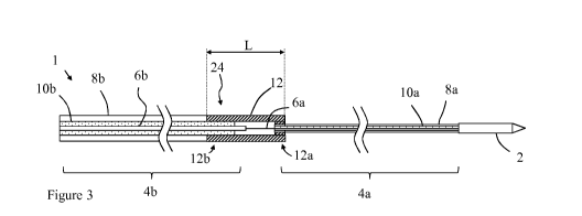
Figure 3 shows an alternative coupling between the joining member 12 and the
proximal
portion 4b of the feeding cable. In this embodiment, the proximal end 12b of
the joining
member 12 is arranged to fit around an exposed portion of the dielectric 10b
of the proximal
portion 4b of the feeding cable 4. The exposed portion of the dielectric 10b
may extend distally
CA 03095592 2020-09-29
WO 2019/185905
PCT/EP2019/058072
14
from a distal end of the outer conductor 8b and may be formed by stripping an
end
portion of the outer conductor away to expose part of the dielectric material
beneath it.
The joining member 12 may fit around the dielectric 10b so that an outer
surface of the joining
member 12 is flush with an outer surface of the outer conductor 8b of the
proximal portion of
the feeding cable. As can be seen in Figure 3, the outer cross sectional size
(e.g. outer
diameter) of the joining member 12 adjacent to the outer conductor 8b may be
equal to the
combined cross sectional size of the inner conductor 8b, dielectric 10b and
outer conductor 8b
of the feeding cable 4b adjacent to the joining member. This means that the
outer surface of the
joining member 12 is flush with the outer conductor 8b. Alternatively, the
thickness of a wall
of the joining member 12 may be equal to the thickness of the outer conductor
8b so that a
flush connection is formed. The flush connection may provide a smooth joint
between the
joining member 12 and the proximal portion of the feeding cable 4a, and may
reduce the
overall cross section of the ablation probe 1. The joining member may be
connected to the
outer conductor 8b by a welded joint (e.g. a laser welded joint) as described
above. Other
connections between the joining member 12 and the outer conductor 8b and/or
the dielectric
10b may be used.
In some embodiments, the outer surface of the proximal end 12b of the joining
member 12 may
have a greater cross sectional size compared to the outer surface of the
distal end 12a of the
joining member 12. In other words, the overall cross sectional size or outer
diameter of the
joining member 12 may reduce along its length between its distal and proximal
ends 12a, 12b.
An example of this is shown in Figures 4 and 5. Figure 4 illustrates an
embodiment in which
the outer surface of the joining member 12 comprises a tapered portion
extending between its
proximal end 12b and its distal end 12a. The tapered outer cross-section may
provide a smooth
transition from the larger proximal portion 4b of the feeding cable to the
smaller distal portion
4a. Figure 4 shows the tapered portion extending along the whole length of the
joining member
12. In other embodiments, only part of the length of the joining member may be
tapered.
Figure 5 illustrates an embodiment in which the outer surface of the joining
member 112
comprises a stepped portion 12c disposed at a point along the length of the
joining member 12
between its proximal end 12b and its distal end 12a. The stepped portion 12c
is arranged to
provide a step down in cross sectional size (e.g. outer diameter) of the
joining member 12
between its distal and proximal ends 12a, 12b. Although Figure 5 shows a
single stepped
portion 12c, one or more stepped portions may be provided at suitable points
along the length
of the joining member 12. In other embodiments, the stepped portion 12c of
Figure 5 and the
tapered portion of Figure 4 may both be provided.
CA 03095592 2020-09-29
WO 2019/185905
PCT/EP2019/058072
As illustrated in Figures 4 and 5, an inner surface of the proximal end 12b of
the joining
member 12 may have a greater cross sectional size compared to an inner surface
of the distal
end 12a of the joining member 12. The inner surface of the joining member may
comprise a
5 tapered portion extending at least partly between its proximal and distal
ends 12a, 12b as
shown in Figure 4. This may provide a reduction of the inner surface cross
sectional size
between its distal and proximal ends 12a, 12b, and may correspond to the
tapered shape of the
outer surface. In the embodiment shown in Figure 5, the inner surface of the
joining member
12 comprises a stepped portion disposed between its proximal end 12b and its
distal end 12b to
10 provide a step down in cross sectional size. The stepped portion of the
inner surface may
correspond to the stepped portion of the outer surface. The tapered or stepped
portions
provided on the inner surface of the joining member 12 may improve impedance
matching (e.g.
may help provide a 50 Ohm impedance match). In the described embodiments, the
inner
surface stepped portion is combined with an outer surface stepped portion, and
the inner
15 surface tapered portion is combined with an outer surface tapered
portion. In other
embodiments, a stepped inner surface may be combined with a tapered outer
surface, or vice
versa. The shape of the tapered portions and stepped potions of the inner and
outer surfaces
shown in Figures 4 and 5 are examples only, with other shapes being possible.
The stepped or
tapered portions may, for example, have curved rather than straight profiles
as shown.
In some embodiments, at least part of the joining member is air filled. An
example of this is
shown in Figure 6. The joining member 12 may define a cavity 13a in which the
inner
conductors 6a, 6b of the feeding cable extend and are joined together. Some,
or all, of the
cavity 13a may be air filled. By filling at least part of the cavity with air
the overall size of the
connector 24 may be reduced and help to provide an overall low profile. This
is because air has
a relatively low dielectric constant, meaning a large volume of other
dielectric constant is not
required. In some embodiments, the cavity 13a is entirely air filled, with no
other dielectric
material (e.g. no solid dielectric or potting agent) provided.
At least part of the joining member may be filled with a potting agent 13b. As
illustrated in
Figure 6, part of the cavity 13a may be filled with the potting agent so that
its surrounds one or
both of the inner conductors 6a, 6b of the feeding cable 4. The potting agent
13b may comprise
any suitable potting agent or compound known in the art. It may, for example,
comprise an
epoxy with a low dielectric value which allows the diameter of the connector
to be minimised,
and may provide improved structural strength. In some embodiments, all of the
cavity 13a may
be filled with potting agent 13b as illustrated in Figure 7. In yet other
embodiments, all of the
CA 03095592 2020-09-29
WO 2019/185905
PCT/EP2019/058072
16
cavity may be air filled, or filled with a combination of air, potting agent
and any
other suitable dielectric material as discussed in connection with Figure 2a.
Referring again to Figure 6, the joining member 12 may further comprises a
bleed hole 12d.
The bleed hole 12d may comprise a through hole extending through the body or
wall of the
joining member 12. The bleed hole 12d may be configured to allow the flow of
potting agent
13b into or out of the cavity 13a within the joining member. This may allow
air to escape
during manufacture to avoid undesired air pockets being formed.
In other embodiments, the joining member 12 comprises one or more weakened
portions. The
weakened portions are arranged to increase the flexibility of the joining
member 12 so that the
ablation probe 1 can be used effectively with an endoscope type delivery
device. An example
of such an embodiment is illustrated in Figure 8. In this embodiment, the
joining member 12
comprises a plurality of slots 30 extending through the body of the joining
member 12 forming
a series of weakened portions. In other embodiments, only a single slot may be
provided. The
slots may be any suitable shape or pattern to provide the desired level of
flexibility. In the
described embodiment, the slots 30 extend through the body of the joining
member 12. In other
embodiments, the weakened portions may be formed by slots or cuts in the
surface of the
joining member 12. In such an embodiment, the slots or cuts do not extend all
the way through
its body. The weakened portions may be formed by laser cutting the joining
member 12.
In the embodiment of Figure 8 the connector comprises a sealing member 18
described
elsewhere herein. The sealing member 18 extends over the joining member in
order to prevent
water ingress through the slots forming the weakened portions.
Figure 9 illustrates an embodiment in which the joining member 12 has a non-
circular outer
profile. In the embodiment shown in Figure 9, the joining member 12 comprises
a heat transfer
structure. The heat transfer structure is formed from a plurality of
protrusions (e.g. fins)
extending from the outer surface of the joining member (four of which are
visible in Figure 9).
The protrusions are adapted to increase the surface area of the joining member
and aid heat
transfer from the joining member 12 to its surroundings. Figure 9 shows only
one example, in
other embodiments other numbers of protrusions, or shapes of protrusions, may
be provided.
The connector 24 described above may be used in any ablation probe having a
feeding cable in
which distal and proximal portions are coupled together, examples of which are
described in
more below. The connector may, for example, be used in an ablation probe
according to those
disclosed in the applicant's previous applications: European application No.
EP17164403.2 and
CA 03095592 2020-09-29
WO 2019/185905
PCT/EP2019/058072
17
International application No. PCT/EP2018/058252 (the contents of
which
are hereby incorporated by reference in their entirety), which disclose an
ablation probe having
a coolant flow path defined partly by a deformable member. The skilled person
will however
understand that the connector 24 is not limited to use with such ablation
probes, and can be
used with ablation probes with and without coolant flow paths, and with or
without deformable
members forming a coolant flow path.
An example of an ablation probe 100 with which the connector may be used in
shown in
Figures 10a and 10b. The ablation probe 100 comprises an applicator 102 and a
feeding cable
104 corresponding to those described in connection with Figure 1.
Corresponding reference
numbers have been used in Figures 10a and 10b for feature common with those
described in
connection with Figures 1 to 9.
The ablation probe 100 further comprises a first coolant path. In the
described embodiment, the
first coolant path is a coolant delivery path 106 via which coolant is able to
flow towards the
applicator 102. For example, the coolant delivery path 106 may deliver a flow
of coolant
towards the distal end of the ablation probe 100 from a coolant supply means
(not shown in the
Figures) coupled to the coolant delivery path 106 at the proximal end of the
ablation probe
100. The flow of coolant may help control the temperature of the ablation
probe 100 during
use. This may allow energy to be delivered to the surrounding tissue for an
extended period of
time without the ablation probe 100 overheating and being damaged, or causing
injury to
healthy tissue. The coolant delivery path may be formed by one or more coolant
channels as
will be described later. The coolant may be a fluid, and may be water, saline
solution, a
cryogenic gas or any other suitable coolant known in the art.
The ablation probe 100 further comprises a second coolant path. In the
described embodiment,
the second coolant path is a coolant return path 108 via which coolant can
return from the
applicator. The coolant return path 108 may therefore return the supply of
coolant from the
distal end of the ablation probe 100 to the proximal end. The ablation probe
100 further
comprises a deformable member 110 which is arranged to move between an
insertion
configuration (shown in Figure 10a) in which insertion of the ablation probe
100 is facilitated
and a deployed configuration (shown in Figure 10b). When in the deployed
configuration, the
coolant return path 108 is provided by the deformable member 110. In some
embodiments, no
coolant return path may be provided when the deformable member is in the
insertion
configuration. This may allow the profile of the ablation probe to be
minimised. In other
embodiments, the return path may not be completely absent when the deformable
member is in
the insertion configuration. The insertion configuration therefore provides a
configuration in
CA 03095592 2020-09-29
WO 2019/185905
PCT/EP2019/058072
18
which the ablation probe 100 may be suitable for delivery to the desired
location within the
body. The insertion configuration may, for example, correspond to a suitable
size and/or shape
adapted to allow insertion with reduced risk of undesired tissue damage. When
in the insertion
configuration, the ablation probe 100 may, for example, have a low profile
(e.g. small cross
sectional size) for ease of insertion through tissue without causing injury or
insertion through
the working channel of an endoscope.
In other embodiments, the first coolant path may act as a coolant return path.
In this
embodiment, the first coolant path is arranged to carry a flow of coolant away
from the
applicator. In this embodiment, the second coolant path may act as a coolant
delivery path
arranged to carry a flow of coolant towards the applicator. A combination of
the first and
second coolant paths may therefore form a coolant circuit arranged to deliver
a flow of coolant
to and away from the applicator, where the coolant can flow in either
direction along each of
the first and second coolant paths. In the embodiment shown in the figures,
the first coolant
path acting as the coolant delivery path may allow a flow of colder coolant
close to the feeding
cable. This may aid cooling of the ablation probe as a significant amount of
heat may be
generated in the feeding cable. In other embodiments, where the second coolant
path acts as
the coolant delivery path, colder coolant may be delivered to the applicator
first to aid cooling
of the applicator.
The ablation probe 100 may therefore be delivered to the desired location
whilst the
deformable member 110 is in the insertion configuration. Once at the desired
location, the
deformable member 110 may be moved to the deployed configuration to allow flow
of the
coolant away from the applicator 102. The coolant can then flow via the
coolant delivery and
return paths to cool the ablation probe 100 during use. The deformable member
110 therefore
is able to provide an insertion configuration suitable for delivery to the
ablation site when the
coolant flow is not required. Once the ablation probe is in position, the
deformable member
110 may be moved to a configuration suitable to provide a flow of coolant as
required during
delivery of energy from the applicator 102. When in the deformable member is
in the insertion
configuration the overall diameter of the ablation probe may be between about
13 to about 25
gauge (approximately 2.5 to 0.5 mm). This may allow easy insertion.
As can be seen in Figures 10a and 10b, the coolant return path 110 may be
provided only by
the deformable member along at least a portion of a length of the ablation
probe 100. For
example, along at least part of the length of the ablation probe 100, no other
channels or
conduits to carry returning coolant may be provided in addition to the coolant
return path 108
formed by the deformable member 110. This may allow the ablation probe 100 to
have a small
CA 03095592 2020-09-29
WO 2019/185905
PCT/EP2019/058072
19
cross sectional size when the deformable member is in the insertion
configuration. Any
additional coolant return paths would require additional space within the body
of the ablation
probe 100 and so would not provide a low profile.
In the embodiment of Figures 10a and 10b, the deformable member 110 is fluidly
connected to
a distal end of the coolant delivery path 106 (e.g. the distal end of the
coolant delivery path
may be joined to the distal end of the fluid return path to form a single path
along which
coolant may flow towards and then away from the applicator (in either
direction)). The coolant
delivery path 106 therefore runs along the inside of the coolant return path
108 when the
deformable member 110 is in the deployed configuration. This arrangement
allows the overall
size of the ablation probe 100 to be reduced when the deformable member 110 is
in the
insertion configuration.
An example embodiment of an ablation probe 200 according to this disclosure is
shown in
more detail in Figures 11 to 17. The embodiment shown in the Figures is only
one such
example.
As can be seen in Figure 11, in this embodiment, the ablation probe 200
generally comprises
two portions: a needle portion 212 and a catheter portion 214. The needle
portion 212 may be
.. arranged at the distal end of the ablation probe 200 and is adapted to be
inserted into tissue
during use to reach the desired ablation location. The catheter portion 214
may be provided at
the proximal end of the ablation probe 200 and is arranged to supply
electromagnetic energy
and a flow of coolant to and from the needle portion 212. In the embodiment
shown in the
Figures, the ablation probe 200 further comprises a handle portion 216 via
which the ablation
probe may be manipulated and positioned during use. The catheter portion may
have an
extended length and flexibility for endoscopic use as shown in Figure 11. In
other non-claimed
embodiments, a shorter, more rigid catheter portion may be provided for
percutaneous use.
In some embodiments, the needle portion may form a small part of the overall
length of the
ablation probe. For example, the needle portion may be 5 mm to 2000 mm in
length, and
preferably may be around 70 mm in length. The length of the needle portion may
be chosen
according to the anatomy to be accessed. For example, the needle portion may
be
approximately between 10 and 100 mm long for delivery of therapy to organs
including the
pancreas, or lung, or longer (for example 100-400mm in length) for delivery of
therapy
percutaneously. A longer length of needle portion may be more suitable for
accessing parts of
the lung, for example. The catheter portion may be around 1000 mm to 2000 mm
in length, and
preferably around 1400 mm in length. The length of the catheter portion may be
chosen
CA 03095592 2020-09-29
WO 2019/185905
PCT/EP2019/058072
according to the position of the ablation site which must be
reached. In other
embodiments, the needle portion of the ablation probe (e.g. that having the
deformable
member) may form a greater proportion of the length of the ablation probe. In
some
embodiments, the entire length of the ablation probe may be formed by the
needle portion. In
5 .. such an embodiment, the deformable member may extend along the majority
or all of the
length of the ablation probe. In such an embodiment, the catheter portion may
not be required.
For example, if the ablation probe is to be used percutaneously the catheter
portion may be
shorter than for endoscopic use, or may not be required.
10 An exploded view of the needle portion is shown in Figure 12. The needle
portion 212 may
comprise a deformable member 210, an applicator 202, a distal portion of the
feeding cable
204a and a distal portion of the coolant delivery path. An exploded view of
the catheter portion
is shown in Figure 13. In this embodiment, the catheter portion 214 may
comprise a proximal
portion of the feeding cable 204b, a proximal portion of the coolant delivery
path, and a
15 coolant return conduit (which may be non-deformable). The proximal
portion of the coolant
delivery path may be formed by a space between a tube 218 housing the proximal
portion of
the feeding cable 204b and a surrounding coolant delivery tube 220. The
coolant return path
may be formed by a space between the coolant delivery tube 220 and a
surrounding coolant
return tube 222. In other embodiments, any other suitable arrangement of
channels or conduits
20 may be provided to form the coolant return and coolant delivery paths
within the catheter
portion 214.
The greatest cross sectional size of the needle portion may be less than the
greatest cross
sectional size of the catheter portion. In other words, the cross section size
(e.g. diameter) of
the needle portion at its largest point may be less that the cross sectional
size (e.g. diameter) of
the catheter portion at its greatest point. This may allow the needle portion
to access an
ablation site whilst reducing any potential for tissue damage. The catheter
portion on the other
hand may be sized to fit through the working channel of the device with which
it is used.
Examples of suitable arrangements of channels forming the coolant return and
coolant delivery
paths are shown in the cross sectional views of Figures 14a to 14c. In Figure
14a, the catheter
portion comprises two lumens each forming one of the coolant return and
coolant delivery
paths. In Figure 14b, the catheter portion comprises four lumens forming the
coolant return
path and coolant delivery path. Two of the lumens may form the coolant return
path, and two
of the lumens may form the coolant delivery path. This embodiment may provide
better kink
resistance and strength. The lumens may not be equally sized as shown in
Figures 14a and 14b.
An example of this is shown in Figure 14c in which three lumens are provided.
A first and
CA 03095592 2020-09-29
WO 2019/185905
PCT/EP2019/058072
21
second lumen may provide the coolant return and delivery paths, with the third
lumen
provided to include other components such as a sensor or the like. The third
lumen may be
small in size compared to the first and second lumens to provide adequate
space for the flow of
coolant.
In other embodiments, the channels forming the coolant return and coolant
delivery paths
within the catheter portion may be formed by one or more channels in the outer
conductor of
the proximal portion of the feeding cable. This may improve flexibility and
provide a compact
arrangement.
In the described embodiment, the feeding cable is formed by two lengths of
cable (the distal
portion 204a and the proximal portion 204b) joined at the boundary between the
needle portion
212 and the catheter portion 214 (shown in more detail in the close up view of
Figure 15a).
The feeding cable may be formed by two lengths of coaxial cable to form an
electrical circuit
to deliver electromagnetic energy to the applicator 202. In other embodiments
a single feeding
cable may be used having regions of different thickness to form the distal and
proximal
portions. In other embodiments, any other suitable conductor may be provided
to deliver a
supply of suitable electromagnetic energy to the applicator 202. The ablation
probe 200 may
further comprise a connector 224 arranged to mechanically and electrically
splice the distal
portion of the feeding cable 204a to the proximal portion of the feeding cable
204b as
described above in connection with the embodiments of Figures 1 to 9. The
connector 224 may
connect the different portions of the feeding cable 204a, 204b while
maintaining an effective
impedance match, minimising electrical losses and ensuring a compact
configuration of the
ablation probe 200.
In the described embodiment, the distal portion of the feeding cable 204a has
a corresponding
distal cross sectional size, and a proximal portion of the feeding cable 204b
has a
corresponding proximal cross sectional size, wherein the distal cross
sectional size is less than
the proximal cross sectional size. The size (e.g. diameter) of the conductor
is therefore
optimised based on its position within the ablation probe 200. The cross
sectional sizes may be
chosen to optimise (e.g. maximise) the feeding cable power handling, while
also reducing
electrical losses and optimising the mechanical strength of the ablation probe
200. In other
words, the length of the smaller cross section portion of the feeding cable is
minimised by
connecting it to a larger cross section feeding cable (e.g. a more efficient
cable) for the portion
of the ablation probe 200 outside of the needle portion 212. This part of the
ablation probe 200
does not need to be inserted into tissue so a small profile is not as
important. The cross section
CA 03095592 2020-09-29
WO 2019/185905
PCT/EP2019/058072
22
of the feeding cable in the catheter portion 212 is therefore increased to
reduce power
loss where a small cross section is less important.
The needle portion of the ablation probe may therefore have a smaller overall
cross sectional
size compared to the catheter portion. The needle portion is therefore
optimised for insertion
into tissue, whilst the catheter portion is optimised for power delivery over
the long length of a
device working channel through which it is inserted. In use, only the needle
portion may
protrude from the working channel through which the ablation probe is
inserted. It is therefore
important for the needle portion to have a relatively small cross sectional
size to reduce tissue
.. damage. For the catheter portion a relatively larger cross sectional size
can be used. Compared
to the needle portion, the catheter portion is instead optimised for power
delivery along the
length of the working channel. In one example, the needle portion, when the
deformable
member is in the insertion configuration, may have an overall diameter of 1 mm
at its largest
point. The catheter portion may have an overall diameter of 3 mm at its
largest point.
In other embodiments, the cross sectional size of the distal and proximal
portions of the
feeding cable may be the same. In this case, a reduction in overall size of
the needle portion
compared to the catheter portion may still be provided by the use of the
deformable member.
The needle portion 212 may further comprise a tube 226 (e.g. a hypotube)
arranged to house
the distal portion of the feeding cable 204a. The tube 226 may be formed from
a metal material
which has sufficient rigidity to allow the needle portion 212 to be inserted
into tissue. In other
embodiments, the tube 226 may be formed from any other suitable material and
may be formed
from a superelastic material, for example Nitinol.
In other embodiments, the tube 226 may be formed form an elastic material (and
not
specifically a superelastic material). By forming the tube from an elastic (or
superelastic)
material it may withstand permanent deformation after being delivered through
the tortuous
path of a working channel. As the ablation probe extends from the working
channel it may
consequently follow a straight path, rather than following a curved path
caused by the material
being deformed by the shape of the working channel. This may help to more
easily guide the
distal tip of the ablation probe to the desired position.
An example of this is shown in Figures 15b and 15c. Figure 15b shows an
example of an
ablation probe 200 having a tube formed from an elastic material extending
from the end of the
working channel 201 through which it has been inserted. The portion of the
ablation probe
extending from the working channel can be seen to follow a straight path.
Figure 15c shows an
CA 03095592 2020-09-29
WO 2019/185905
PCT/EP2019/058072
23
example of an ablation probe with a non- elastic tube. This figure illustrates
how the
portion of such an ablation probe extending from the working channel 201 tends
to follow a
curved path.
In some embodiments, the tube may be formed from an elastic (or superelastic)
electrically
conducting material. This may allow the tube to form part of a choke. The tube
may be formed
from a solid material or a mesh material as appropriate to allow the required
elasticity, for
example a braid or coil reinforced polymer tube.
In one embodiment, the coolant delivery path is provided by a channel formed
between the
feeding cable and inside wall of the tube 226. For example, clearance between
the feeding
cable and the inside wall of the tube 226 may provide space for coolant to
flow. In other
embodiments, slots may be cut into the inside wall of the tube 226 to provide
a space through
which coolant can flow. The amount of clearance may be specified to ensure an
adequate flow
of cooling is achieved while maximising the power carrying capacity of the
feeding cable.
In the embodiment shown in the Figures, the coolant delivery path comprises
one or more
coolant channels formed in the body of the tube 226. The coolant therefore may
partly
surround the feeding cable to aid cooling. The width and number of the
channels may be
chosen to optimise (e.g. maximise) the mechanical strength of the ablation
probe 100 and the
performance of the cooling.
The one or more channels may be cut into the wall of tube 226 to allow cooling
fluid to flow
adjacent to the distal portion of the feeding cable 204a. In the described
embodiment, the one
or more channels may be formed by one or more slots formed in the outer
surface of the tube
226. In this embodiment, the ablation probe 200 may further comprise a
membrane 228
disposed around the tube 226. The membrane 228 may be arranged to separate the
coolant
delivery path from the coolant return path (e.g. it forms a boundary between
them). In some
embodiments, the one or more channels may extend distally past a distal end of
the membrane
228 so that coolant can flow from the one or more channels into the deformable
member 210.
In other embodiments, one or more apertures may be provided in the membrane
228 to fluidly
connect the one or more channels with the deformable member 210. The membrane
228 may be
formed from a thin layer of material (for example a polymer heat shrink)
located over the tube
226 to form an enclosed conduit for the cooling fluid. In other embodiments,
the channels may
be formed within the wall of the tube 226, in which case the membrane 228 may
not be
required.
CA 03095592 2020-09-29
WO 2019/185905
PCT/EP2019/058072
24
In other embodiments (not shown in the Figures), the distal portion of the
feeding
cable 204a may comprise an inner conductor arranged to transmit a signal to
the applicator 202
and an outer conductor arranged to shield the inner conductor (e.g. it may be
a coaxial cable).
The coolant delivery path may comprise one or more coolant channels formed in
the outer
conductor. The coolant channels may, for example, be formed by one or more
slots in an outer
surface of the outer conductor. The coolant and the split outer conductor may
thus form a
mixed media outer conductor arranged to shield the electrically insulating
material. A
membrane may be formed around the outer conductor to form a conduit for the
cooling fluid.
In some embodiments, the feeding cable may be formed by a coaxial cable in
which the outer
conductor is manufactured from a robust material (for example stainless steel)
to form a ridged
body of the needle portion. In this embodiment, the coolant delivery path may
be formed by
channels in the outer conductor, rather than in the tube 228. In such an
embodiment, the tube
may therefore not be required, thus saving space. In other embodiments, the
tube may also be
provided. The cooling channels may also more effectively cool the feeding
cable as well as
deliver coolant to the applicator 202. In some embodiments, the one or more
channels formed
in the outer conductor may be aligned with the central axis of the feeding
cable. The width and
number of the channels may be chosen to optimise the mechanical strength of
the feeding cable
and the performance of the cooling, while minimising electrical losses and
ensuring impedance
matching between portions of the feeding cable having channels in the outer
conductor and
portions of the feeding cable in which the channels are not present (e.g. in
the catheter
portion).
The one or more coolant channels described above forming the coolant delivery
path may be
disposed along a length of the ablation probe as can be seen in the close up
view of the tube
226 shown in Figure 16. In some embodiments, a plurality of channels may be
provided such
that they are spaced equally around a circumference of the outer conductor or
tube 226 housing
the feeding cable. In Figure 16 only one of the channels is visible (labelled
230). In some
embodiments, the plurality of channels may comprise four channels spaced
equally around the
circumference of the outer conductor or tube 226 housing the feeding cable. In
other
embodiments, other numbers and arrangements of channels may be provided
according to the
cooling requirements and mechanical strength requirements of the ablation
probe.
The inner conductor of the distal portion of the feeding cable 204a is coupled
to the applicator
202 as shown in the detailed view of Figure 17. In this embodiment, a distal
end of the distal
portion of the feeding cable is connected to a proximal end of the applicator
202. Where the
feeding cable is formed from an inner and outer conductor, the inner conductor
may be
attached to the applicator 202 to ensure efficient transfer of electromagnetic
energy to the
CA 03095592 2020-09-29
WO 2019/185905
PCT/EP2019/058072
applicator material. The applicator 202 may be formed from a ceramic material
with
suitable dielectric properties (for example zirconia) according to the energy
it is arranged to
apply. An internal bore may be provided in the applicator 202 to receive a
portion of the inner
conductor to ensure a strong mechanical joint that may also be glued in
position. The
5 applicator 202 may further be coupled to the tube 226 housing the feeding
cable where it is
provided. In such an embodiment, the proximal end of the applicator 202 may be
connected to
the tube 226 via a bore to receive the tube or a set of interlocking fingers
to maximise the
mechanical strength of the bond between them. In other embodiments, any other
suitable
connection means between the tube 226 and the applicator 202, or the distal
portion of the
10 feeding cable 204a and the applicator 202 may be provided.
The needle portion further comprises the deformable member 210 as shown in the
exploded
view of Figure 12. In the described embodiment, the deformable member 210 is
formed by an
inflatable member arranged to move between a deflated configuration when the
deformable
15 member 210 is in the insertion configuration and an inflated
configuration when the
deformable member 210 is in the deployed configuration. The inflatable member
may thus
form a balloon which may be inflated by the flow of coolant (e.g. the
inflatable member may
inflate due to the pressure of the coolant). In the described embodiment, the
inflatable member
has an inside diameter that matches the outside diameter of the tube 226 (or
the membrane 228
20 surrounding the tube 228 or the outer conductor or insulating material
respectively). The
inflating member may inflate to a larger diameter when the cooling system is
pressurised. This
may therefore form a conduit for the cooling fluid to return from the
applicator 202. When
moving to the inflated configuration, some, or all, of the inflating member
may change shape
(e.g. expand) to allow space for the coolant to flow. When the inflation
member is deflated, the
25 insertion profile of the ablation probe 200 may be reduced (e.g.
minimised) to aid delivery to
the target ablation site. When the ablation therapy has been delivered, the
inflation member
may be deflated so that it returns to its original diameter to facilitate
removal.
The deformable member 110 may extend along at least part of the length of the
needle portion
212 as shown in the Figures. The deformable member 110 may, for example,
extend from at or
near the boundary between the needle portion 212 and the catheter portion 214
and end at or
near the proximal end of the applicator 202. The coolant may therefore flow
through the
deformable member 210 along the length of the ablation probe (e.g. a flow of
coolant may be
provided between an inlet and an outlet of the deformable member, the inlet
and outlet being
spaced apart along the length of the ablation probe). The deformable member
210 may be
fluidly connected to the non-deformable coolant return conduit at a boundary
between the
needle portion 212 and the catheter portion 214. The coolant may therefore
flow through the
CA 03095592 2020-09-29
WO 2019/185905
PCT/EP2019/058072
26
deformable member 110 (when in the deployed configuration) and then through
the
non-deformable coolant return conduit in the catheter portion to reach the
proximal end of the
ablation probe 200.
Various modifications will be apparent to the skilled person without departing
form the scope
of the claims. Any feature disclosed in connection with one embodiment may be
used in
combination with the features of another embodiment.
Test results
The performance of an ablation probe according to the present application has
been evaluated
by the inventors. An assembly comprising a proximal length of coaxial cable, a
distal length of
relatively thinner coaxial cable, and a connection formed by a joining member
receiving the
end of each cable was manufactured and tested. The embodiment tested was
similar to that
shown in Figure 3, with the joining member having a slightly larger cross-
section than the
proximal portion of the feeding cable, and only air filling the cavity inside
the joining member.
Similar advantageous results would however be expected for the other
embodiments described
herein.
The electrical losses in the connector were evaluated by subtracting the sum
of electrical losses
for each individual cable section (i.e. the assembly without the connector)
and the electrical
losses of the prototyped assembly (i.e. the assembly including the connector)
across a
frequency bandwidth of 2.4 - 2.5 GHz.
The electrical losses for the prototyped assembly are shown in Table 1 below
and Figure 18.
The prototype shows very low electrical losses in the connector across the
frequency
bandwidth 2.4 ¨ 2.5 GHz. At 2.45 GHz, the connector enables a reduction of
electrical losses
of 31% compared to a coaxial cable section of constant diameter (i.e. the same
diameter as the
thinner distal portion) and the same length as the tested assembly.
Frequency Losses [dB]
2.4 0.02
2.405 0.016
2.41 0.022
2.415 0.025
2.42 0.03
2.425 0.037
2.43 0.037
2.435 0.04
2.44 0.04
CA 03095592 2020-09-29
WO 2019/185905
PCT/EP2019/058072
27
2.445 0.055
2.45 0.055
2.455 0.065
2.46 0.065
2.465 0.07
2.47 0.075
2.475 0.075
2.48 0.08
2.485 0.085
2.49 0.085
2.495 0.087
2.5 0.087
Table 1
A tensile force test on the joint between inner conductors of the same
prototype showed that
the strength of the connection between the inner conductors of the two coaxial
cables is higher
than the strength of the inner conductor of the distal cable portion (i.e.
with no joins).
A tensile force test was also carried out on the laser welded joints provided
in the test
prototype between the outer conductors and the joining member forming the
connection. The
laser welded connection between the outer conductors and the joining member
resulted in a
stronger connection compared to the strength of the outer conductor of the
distal portion of the
feeding cable (i.e. with no welded joint). The connector was therefore found
to provide a
suitably strong connection between the separate sections of coaxial feeding
cable.
The prototyped connector assembly was also tested for water-resistance. In the
embodiment
being tested, a laser welded joint between the outer conductors of the feeding
cables and the
joining member of the connector provided the desired sealing. . After
immersion in water at the
pressure of 8 bar for one hour, no water ingress was observed.