Note: Descriptions are shown in the official language in which they were submitted.
CA 03095749 2020-09-30
WO 2019/195344 - 1 - PCT/US2019/025460
COMPLEX HUMAN GUT MICROBIOME CULTURED
IN AN ANAEROBIC HUMAN GUT-ON-A-CHIP
CROSS-REFERENCE TO RELATED APPLICATIONS
[0001] This application claims priority to U.S. Provisional Patent
Application No.
62/722,658, filed August 24, 2018, and U.S. Provisional Patent Application No.
62/651,438,
filed April 2, 2018, each of which is hereby incorporated by reference herein
in its entirety.
FIELD OF THE INVENTION
[0002] The present invention relates generally to supporting dynamic
interactions between
living human intestinal epithelium and a directly opposed complex community of
living human
aerobic and anaerobic commensal gut microbes with a population diversity
similar to that
observed in a living human intestine.
BACKGROUND OF THE INVENTION
[0003] The diverse bacterial populations that comprise the commensal
microbiota of the
human intestine play a central role in health and disease, yet no method is
available to sustain
these complex microbial communities in direct contact with living human
intestinal cells in
vitro. The present disclosure describes a human Gut-on-a-Chip (Gut Chip)
microfluidic
platform that permits control and real-time assessment of physiologically-
relevant oxygen
gradients, and which enables co-culture of living human intestinal epithelium
in direct contact
with stable communities of aerobic and anaerobic microbiota derived from human
stool
specimens. When compared to aerobic co-culture conditions, establishment of a
transluminal
hypoxia gradient sustained higher microbial diversity with over 200 unique
operational
taxonomic units (OTUs) from 11 different genera, and an abundance of obligate
anaerobic
bacteria with ratios of Firmicutes and Bacteroidetes similar to those observed
in human feces,
in addition to increasing intestinal barrier function. The ability to culture
human intestinal
epithelium overlaid by complex human gut microbial communities may enable
investigations
of host-microbiome interactions that were not possible previously, and serve
as a discovery
tool for development of new microbiome-related therapeutics, probiotics, and
nutraceuticals.
[0004] One of the major recent paradigm shifts in medicine relates to the
recognition of the
central role that the microbiome composed of host-specific communities of
commensal
microbes plays in human health and disease. Although human microbiota colonize
mucosal
surfaces of various tissues, the gastrointestinal (GI) tract supports the
greatest mass and
CA 03095749 2020-09-30
WO 2019/195344 - 2 - PCT/US2019/025460
diversity of microorganisms. Aerobic and anaerobic commensal gut microbiota
are essential
for maintenance of normal nutrient absorption, drug metabolism, and immune
responses, as
well as for protection against infectious pathogens. Conversely, changes or
imbalances in the
microbial community within the intestine can contribute to development of a
broad range of
pathological disorders within and beyond the GI system, including inflammatory
bowel
disease, colorectal cancer, radiation enteropathy, diabetes, hepatic
steatosis, obesity, and
rheumatoid arthritis. Thus, the establishment and preservation of balanced
host-intestinal
microbiome interactions are key requirements for maintaining gut homeostasis
and human
health.
[0005] Analysis of gut-microbiome crosstalk has almost exclusively relied
on genomic or
metagenomic analysis of samples collected in vivo because no method exists to
establish stable
complex communities of gut commensal microbes in direct contact with
intestinal epithelium
in vitro. Although animal models have been used to analyze host-microbiome
interactions and
their contributions to pathophysiology, microbiota differ between different
species.
[0006] Existing in vitro models, such as Transwell inserts, have been used
to study human
host-microbe interactions; however, these studies can only be carried out over
a period hours
before bacterial overgrowth leads to cell injury and death. More advanced
models, such as
organoid cultures, have shown great promise for studying host-microbiome
interactions, but
they are limited in providing a vascular interface and oxygen gradients with
below 1% luminal
oxygen levels required for co-culture of certain strict anaerobes. Human
intestinal epithelial
cells have been grown in a microfluidic culture device separated by a
nanoporous membrane
from a single facultative anaerobic bacterium (Lactobacillus rhamnosus GG) and
an obligate
anaerobe (Bacteroides caccae) cultured under anaerobic conditions in a
parallel channel, which
can permit analysis of the effects of soluble mediators, but not the impact of
direct contact
between host cells and a complex community of commensal microbes. A 2-channel,
microfluidic, human Gut Chip device has been previously described as being
lined by human
Caco-2 intestinal epithelial cells culture under dynamic fluid flow and
peristalsis-like
mechanical deformations, which enabled establishment of stable co-cultures of
a human villus
intestinal epithelium in direct contact with up to 8 different strains of
human commensal gut
microbes for weeks in vitro under oxygenated conditions', but the living
intestinal microbiome
contains hundreds of different types of bacteria that are anaerobes as well as
aerobes.
[0007] Thus, there is a great need for experimental models that can sustain
complex
populations of human aerobic and anaerobic microbiota in contact with living
human tissues
to analyze dynamic and physiologically relevant human host-microbiome
interactions.
CA 03095749 2020-09-30
3
WO 2019/195344 - - PCT/US2019/025460
According to another need, an experimental system is required that can support
dynamic
interactions between living human intestinal epithelium and a directly apposed
complex
community of living human aerobic and anaerobic commensal gut microbes with a
population
diversity similar to that observed in living human intestine.
SUMMARY OF THE INVENTION
[0008] Embodiment Al. According to one embodiment of the present
disclosure, a
microfluidic device is directed to sustaining a complex microbial community in
direct and
indirect contact with living human intestinal cells in vitro. The device
includes a first
microchannel having cultured cells of a human intestinal epithelium and
microbiota, the first
microchannel further having a first level of oxygen. The device further
includes a second
microchannel having cultured cells of a vascular endothelium, the second
microchannel further
having a second level of oxygen. The device also includes a membrane located
at an interface
region between the first microchannel and the second microchannel, the
membrane being
composed of an oxygen-permeable material or further having pores via which
oxygen flows
between the first microchannel and the second microchannel to form a
physiologically-relevant
oxygen gradient.
[0009] Embodiment A2. The microfluidic device of embodiment Al, further
comprising
a plurality of microscale oxygen sensors embedded in the first microchannel
and the second
microchannel, the plurality of microscale oxygen sensors providing real-time
oxygen
measurements based on non-invasive monitoring of the physiologically-relevant
oxygen
gradient.
[0010] Embodiment A3. The microfluidic device of embodiment A2, wherein the
plurality of microscale oxygen sensors contain oxygen-quenched fluorescent
particles.
[0011] Embodiment A4. The microfluidic device of embodiment A3, wherein the
oxygen-quenched fluorescent particles are suspended in a polydimethylsiloxane
(PDMS)
polymer or other gas-permeable polymer.
[0012] Embodiment A5. The microfluidic device of embodiment A3, wherein the
oxygen-quenched fluorescent particles are cured in a film having a thickness
of between about
50 and 1,000 micrometers ( m).
[0013] Embodiment A6. The microfluidic device of embodiment A3, wherein the
oxygen-quenched fluorescent particles are in the form of discs having a
diameter of about 0.1-
millimeters (mm).
CA 03095749 2020-09-30
4
WO 2019/195344 - - PCT/US2019/025460
[0014] Embodiment A7. The microfluidic device of embodiment A2, wherein the
plurality of microscale oxygen sensors are placed directly on an interior
surface of at least one
of the first microchannel and the second microchannel.
[0015] Embodiment A8. The microfluidic device of embodiment A2, wherein the
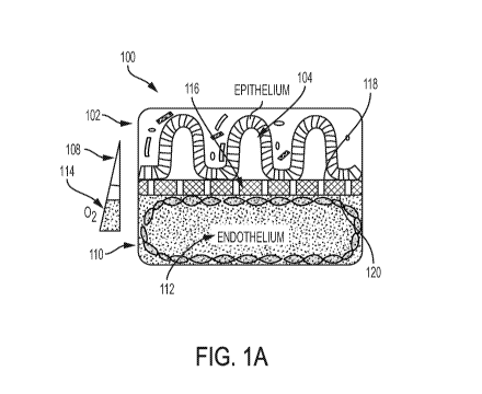
plurality of microscale oxygen sensors of are placed at an inlet region, a
middle region, and an
outlet region of each of the first microchannel and the second microchannel.
[0016] Embodiment A9. The microfluidic device of embodiment A2, wherein
changes in
fluorescent intensities of the plurality of microscale oxygen sensors are
caused by oxygen
tension, the changes being indicative of oxygen concentrations.
[0017] Embodiment A10. The microfluidic device of embodiment Al, wherein
the first
microchannel is a top microchannel and the second microchannel is a bottom
microchannel.
[0018] Embodiment All. The microfluidic device of embodiment Al, wherein
the
cultured cells of the vascular endothelium are human intestinal microvascular
endothelia cells
(HIMEC s).
[0019] Embodiment Al2. The microfluidic device of embodiment Al, wherein
the
physiologically-relevant oxygen gradient is a hypoxia gradient.
[0020] Embodiment Bl. According to another embodiment of the present
disclosure, an
in vitro system is directed to emulating a living human intestine. The system
includes a hypoxic
chamber containing living human commensal gut microbes and cultured cells of a
human
intestinal epithelium in which the microbes are in direct and indirect contact
with the cultured
cells. The hypoxic chamber is configured to establish a physiologically-
relevant oxygen
gradient across the layer of microbes and cultured cells.
[0021] Embodiment B2. The in vitro system of embodiment B 1, further
comprising a
plurality of microscale oxygen sensors providing real-time oxygen measurements
based on
non-invasive monitoring of the oxygen gradient.
[0022] Embodiment B3. The in vitro system of embodiment Bl, wherein the
cultured
cells include one or more of mammalian cells, gut cells of insects, and gut
cells of amphibians.
[0023] Embodiment B4. The in vitro system of embodiment B 1, further
comprising a
mucus layer in contact with the cultured cells, the mucus layer being secreted
by the cultured
cells or separately provided.
[0024] Embodiment B5. The in vitro system of embodiment Bl, wherein the
microbes
are contained in a layer.
[0025] Embodiment B6. The in vitro system of embodiment B5, wherein the
layer of
microbes and the cultured cells are placed within a microchannel.
CA 03095749 2020-09-30
WO 2019/195344 - - PCT/US2019/025460
[0026] Embodiment B7. The in vitro system of embodiment B6, wherein the
microchannel is a top microchannel that is separated from a bottom
microchannel via a
membrane located at an interface region, the membrane being oxygen permeable
or having a
plurality of pores via which oxygen flows between the top microchannel and the
bottom
microchannel to achieve the oxygen gradient.
[0027] Embodiment B8. The in vitro system of embodiment B7, further
comprising a
plurality of microscale oxygen sensors providing real-time oxygen measurements
based on
non-invasive monitoring of the oxygen gradient, the plurality of microscale
oxygen sensors
being embedded in at least one of the top microchannel and the bottom
microchannel.
[0028] Embodiment B9. The in vitro system of embodiment B7, wherein the
oxygen
gradient is based on top oxygen permeability through a device body to an
external environment
maintained at about 0 percent oxygen.
[0029] Embodiment B10. The in vitro system of embodiment Bl, further
comprising a
Charge-Coupled Device (CCD) camera, a photodiode, or other light-sensing
modality via
which fluorescence read-out measurements provide the real-time oxygen
measurements in a
non-invasive manner.
[0030] Embodiment Cl. According to another embodiment of the present
disclosure, a
method is directed to establishing a stable complex community of gut commensal
microbes in
vitro. The method includes providing cultured cells of an intestinal
epithelium and microbiota
in an environment having a first level of oxygen, the microbiota being in
direct and indirect
contact with the intestinal epithelium. The method also includes providing
cultured cells of a
vascular endothelium in an environment having a second level of oxygen, the
second level of
oxygen having a greater oxygen concentration than the first level of oxygen.
The method
further includes facilitating the flux of oxygen between the first level of
oxygen and the second
level of oxygen to form a physiologically-relevant oxygen gradient.
[0031] Embodiment C2. The method of embodiment Cl, further comprising
monitoring
the oxygen gradient in a non-invasive manner, and measuring values of the
oxygen gradient.
[0032] Embodiment C3. The method of embodiment Cl, further comprising
measuring
the values of the oxygen gradient via a non-invasive fluorescence read-out.
[0033] Embodiment C4. The method of embodiment Cl, further comprising
providing
oxygenation of the cultured cells of the intestinal epithelium and the
cultured cells of the
vascular endothelium while simultaneously providing an anaerobic environment
for growth of
obligate anaerobes.
CA 03095749 2020-09-30
WO 2019/195344 - 6 - PCT/US2019/025460
[0034] Embodiment C5. The method of embodiment Cl, further comprising
achieving
an oxygen concentration of less than approximately 0.5-2.0% in the first level
of oxygen.
[0035] Embodiment C6. The method of embodiment Cl, wherein the non-invasive
manner includes positioning a camera directly beneath the cultured cells of
the intestinal
epithelium, the camera providing images of the cultured cells of the
intestinal epithelium and
microbiota.
[0036] Embodiment C7. The method of embodiment Cl, wherein the cultured
cells
include one or more cells of non-gut organs with low oxygen tension.
[0037] Embodiment C8. The method of embodiment C7, wherein the non-gut
organs
include one or more of an oral mucosa, urinary tract, and genital mucosa.
[0038] Embodiment Dl. According to yet another embodiment of the present
disclosure,
a microfluidic device has a first microchannel comprising a plurality of
living parenchyma cells
in direct contact with a plurality of living microbes, wherein the microbes
are derived from a
mammalian fecal sample.
[0039] Embodiment D2. The microfluidic device of embodiment D1, wherein the
parenchyma cells are selected from the group consisting of cells of the small
intestine, ilea,
duodenum, lung, alveolar, and skin.
[0040] Embodiment D3. The microfluidic device of embodiment D1, wherein the
mammal is a human.
[0041] Embodiment D4. The microfluidic device of embodiment D1, further
comprising
a second microchannel.
[0042] Embodiment D5. The microfluidic device of embodiment D4, wherein the
first
and second microchannels comprise media.
[0043] Embodiment D6. The microfluidic device of embodiment D4, wherein the
media
in the second microchannel is oxygenated.
[0044] Embodiment D7. The microfluidic device of embodiment D4, wherein the
device
has a gas gradient.
[0045] Embodiment D8. The microfluidic device of embodiment D7, wherein the
gas in
the first microchannel is at a lower concentration than the gas in the second
microchannel.
[0046] Embodiment D9. The microfluidic device of embodiment D8, wherein the
gas is
selected from the group consisting of oxygen, nitrogen and carbon dioxide.
[0047] Embodiment D10. The microfluidic device of embodiment D4, wherein
the
second microchannel comprises living endothelial cells.
CA 03095749 2020-09-30
WO 2019/195344 - 7 - PCT/US2019/025460
[0048] Embodiment D11. The microfluidic device of embodiment D1, wherein
the
plurality of microbes comprises both anaerobic bacteria and aerobic bacteria.
[0049] Embodiment D12. The microfluidic device of embodiment D1, wherein
the
plurality of microbes comprises both Firmicutes phyla and Bacteroidetes phyla.
[0050] Embodiment D13. The microfluidic device of embodiment D1, wherein
the
Firmicutes species are selected from the group consisting of Akkermansia,
Oscillospira, Blantia
and Sinerell a species
[0051] Embodiment D14. The microfluidic device of embodiment D1, wherein
the
plurality of microbes comprises Coprococcus, Anaerobacillus, Bifidobacterium,
and
Peptoniphilus species
[0052] Embodiment D15. The microfluidic device of embodiment D1, wherein
the
plurality of microbes comprises at least 8 different genera of bacteria found
in human feces.
[0053] Embodiment D16. The microfluidic device of embodiment D15, wherein
the
plurality of microbes comprises at least 11 different genera of bacteria found
in human feces.
[0054] Embodiment El. According to yet another embodiment of the present
disclosure,
a method includes a) providing, i) a mammalian fecal sample comprising living
microbes, and
ii) a solution of fluid; b) suspending at least a portion of the fecal sample
in the solution so as
to create a fecal slurry comprising living microbes; c) filtering the slurry
so as to generate a
microbiome stock derived directly from a fecal sample; d) diluting the
microbiome stock so as
to create a diluted stock; e) introducing the diluted stock into a
microfluidic device; and f)
culturing the diluted stock in the microfluidic device so as to create a
cultured microbiome of
living microbes.
[0055] Embodiment E2. The method of embodiment El, wherein one or more
steps of
the method take place in an anaerobic chamber.
[0056] Embodiment E3. The method of embodiment E2, wherein the suspending
takes
place inside the anaerobic chamber.
[0057] Embodiment E4. The method of embodiment El, wherein the mammalian
fecal
sample is from a human.
[0058] Embodiment E5. The method of embodiment E4, wherein the human is
selected
from the group consisting of a preterm infant, infant, child, teen, and an
adult.
[0059] Embodiment E6. The method of embodiment E4, wherein the fecal sample
is from
a diaper.
[0060] Embodiment E7. The method of embodiment E4, wherein the fecal sample
is a
stool sample.
CA 03095749 2020-09-30
WO 2019/195344 - 8 - PCT/US2019/025460
[0061] Embodiment E8. The method of embodiment E4, wherein the fecal sample
was
obtained during a medical procedure.
[0062] Embodiment E9. The method of embodiment El, wherein the fecal
portion is
suspended at 100 mg.m1-1 for creating the fecal slurry.
[0063] Embodiment E10. The method of embodiment E4, wherein the fecal
sample of
step a) was not passed through another mammal.
[0064] Embodiment E10. The method of embodiment E4, wherein the fecal
sample of
step a) was not cultured in vitro.
[0065] Embodiment Ell. The method of embodiment E4, wherein the fecal
sample
comprises both anaerobic bacteria and aerobic bacteria.
[0066] Embodiment E12. The method of embodiment El, wherein the diluting of
the
microbiome stock generates a concentration of microbes of approximately 1 x 10
CFU m11.
[0067] Embodiment E13. The method of embodiment El, wherein the filtering
of step c)
is done with a filter that has a 40 p.m pore size or less.
[0068] Embodiment E14. The method of embodiment El, wherein the cultured
microbiome comprises organisms from both the Firmicutes phyla and the
Bacteroidetes phyla.
[0069] Embodiment E15. The method of embodiment El, wherein the cultured
microbiome comprises species selected from the group consisting of
Akkermansia,
Oscillospira, Blautia and Suterella species.
[0070] Embodiment E16. The method of embodiment El, wherein the cultured
microbiome comprises Coprococcus, Anaerobacillus, Bifidobacterium, and
Peptoniphilus
species
[0071] Embodiment E17. The method of embodiment El, wherein the cultured
microbiome comprises at least 8 different genera of bacteria found in human
feces.
[0072] Embodiment E18. The method of embodiment El, wherein the cultured
microbiome comprises at least 11 different genera of bacteria found in human
feces.
[0073] Embodiment E19. The method of embodiment E13, further comprising g)
flushing media through the cultured microbiome in the microfluidic device.
[0074] Embodiment E20. The method of embodiment E19, wherein the flushing
provides
a sample of cultured living microbes.
[0075] Embodiment E21. The method of embodiment El, wherein the
microfluidic
device comprises a first microchannel comprising a plurality of living
parenchyma cells.
[0076] Embodiment E22. The method of embodiment E21, wherein the
introducing of
step e) results in the parenchyma cells being in direct contact with a
plurality of living microbes.
CA 03095749 2020-09-30
9
WO 2019/195344 - - PCT/US2019/025460
[0077] Embodiment E23. The method of embodiment E22, wherein the parenchyma
cells
are selected from the group consisting of cells of the small intestine, ilea,
duodenum, lung,
alveolar, and skin.
[0078] Embodiment E24. The method of embodiment E23, wherein the cells are
intestinal epithelial cells.
[0079] Embodiment E25. The method of embodiment E21, wherein the
microfluidic
device further comprises a second microchannel.
[0080] Embodiment E26. The method of embodiment E25, wherein the first and
second
microchannels comprise media.
[0081] Embodiment E27. The method of embodiment E26, wherein the media in
the
second microchannel is oxygenated.
[0082] Embodiment E28. The method of embodiment E26, wherein the
microfluidic
device has a gas gradient.
[0083] Embodiment E29. The method of embodiment E28, wherein the gas in the
first
microchannel is at a lower concentration than the gas in the second
microchannel.
[0084] Embodiment E30. The method of embodiment E29, wherein the gas is
selected
from the group consisting of oxygen, nitrogen and carbon dioxide.
[0085] Embodiment E31. The method of embodiment E25, wherein the second
microchannel comprises living endothelial cells.
[0086] Embodiment E32. The method of embodiment E28, wherein the gas
gradient
provides at least one hypoxic region in the first microchannel.
[0087] Embodiment E33. The method of embodiment E26, wherein the culturing
comprises flowing media at a flow rate.
[0088] Embodiment E34. The method of embodiment E26, wherein the second
microchannel is positioned below the first microchannel and separated from the
first
microchannel by a membrane.
[0089] Embodiment E35. The method of embodiment E34, wherein oxygenated
medium
flows through the second microchannel from external oxygenated medium
reservoirs.
[0090] Embodiment E36. The method of embodiment E35, wherein parenchyma
cells in
the first microchannel get oxygen from the second microchannel.
[0091] Embodiment E37. The method of embodiment El, wherein the culturing
takes
place for at least 2 days.
[0092] Embodiment E38. The method of embodiment El, wherein the culturing
takes
place for at least 3 days.
CA 03095749 2020-09-30
WO 2019/195344 - 10 - PCT/US2019/025460
[0093] Embodiment E39. The method of embodiment El, wherein the culturing
takes
place for at least 5 days.
[0094] Embodiment E40. The method of embodiment E38, wherein cultured
microbiome
comprises both anaerobic bacteria and aerobic bacteria.
[0095] Embodiment E41. The method of embodiment E40, wherein the cultured
microbiome comprises at least 2 anaerobic species found in the fecal sample.
[0096] Embodiment E42. The method of embodiment E40, wherein the cultured
microbiome comprises microbes from at least 2 genera found in the fecal
sample.
[0097] Embodiment Fl. According to yet another embodiment of the present
disclosure,
a method includes a) providing a microfluidic device and a portion of a
mammalian fecal
sample, the portion comprising living microbes; b) introducing the portion
into the microfluidic
device; and c) culturing the living microbes in the microfluidic device so as
to create a cultured
microbiome.
[0098] Embodiment F2. The method of embodiment Fl, wherein, prior to the
introducing
of step b) the portion of the fecal sample is suspended in a sterile solution
so as to create a fecal
slurry.
[0099] Embodiment F3. The method of embodiment F2, wherein the suspending
takes
place inside an anaerobic chamber.
[0100] Embodiment F4. The method of embodiment F2, wherein, after the
suspending,
the slurry is passed through a filter, so as to generate a microbiome stock
derived directly from
a fecal sample.
[0101] Embodiment F5. The method of embodiment F4, wherein the filter has a
40 p.m
pore size or less.
[0102] Embodiment F6. The method of embodiment F4, further comprising
diluting the
microbiome stock so as to create a diluted stock, the diluted stock being
introduced in step b).
[0103] Embodiment F7. The method of embodiment F6, wherein the diluting the
microbiome stock generates a concentration of microbes of approximately 1 x
107 CFU m11.
[0104] Embodiment F8. The method of embodiment Fl, wherein the mammalian
fecal
sample is from a human.
[0105] Embodiment F9. The method of embodiment F8, wherein the human is
selected
from the group consisting of a preterm infant, infant, child, teen, and an
adult.
[0106] Embodiment F10. The method of embodiment F8, wherein the fecal
sample is
from a diaper.
CA 03095749 2020-09-30
WO 2019/195344 - 11 - PCT/US2019/025460
[0107] Embodiment F11. The method of embodiment F8, wherein the fecal
sample is a
stool sample.
[0108] Embodiment F12. The method of embodiment F8, wherein the fecal
sample was
obtained during a medical procedure.
[0109] Embodiment F13. The method of embodiment F2, wherein the fecal
portion is
suspended at 100 mg.m1-1.
[0110] Embodiment F14. The method of embodiment F8, wherein the fecal
sample of
step a) was not passed through another mammal.
[0111] Embodiment F15. The method of embodiment F8, wherein the fecal
sample of
step a) was not cultured in vitro.
[0112] Embodiment F16. The method of embodiment Fl, wherein the fecal
sample
comprises both anaerobic bacteria and aerobic bacteria.
[0113] Embodiment F17. The method of embodiment F16, wherein the cultured
microbiome comprises at least one of the same anaerobic bacteria types and
aerobic bacteria
types of the fecal sample.
[0114] Embodiment F18. The method of embodiment Fl, wherein the cultured
microbiome comprises both Firmicutes phyla and Bacteroidetes phyla.
[0115] Embodiment F19. The method of embodiment Fl, wherein the cultured
microbiome comprises species selected from the group consisting of
Akkermansia,
Oscillospira, Blautia and Suterella species.
[0116] Embodiment F20. The method of embodiment Fl, wherein the cultured
microbiome comprises Coprococcus, Anaerobacillus, Bifidobacterium, and
Peptoniphilus
species.
[0117] Embodiment F21. The method of embodiment Fl, wherein the cultured
microbiome comprises at least 8 different genera of bacteria found in human
feces.
[0118] Embodiment F22. The method of embodiment Fl, wherein the cultured
microbiome comprises at least 11 different genera of bacteria found in human
feces.
[0119] Embodiment F23. The method of embodiment Fl, further comprising d)
flushing
media through the cultured microbiome in the microfluidic device.
[0120] Embodiment F24. The method of embodiment F23, wherein the flushing
provides
a sample of cultured living microbes.
[0121] Embodiment F25. The method of embodiment Fl, wherein the
microfluidic
device comprises a first microchannel comprising a plurality of living
parenchyma cells.
CA 03095749 2020-09-30
WO 2019/195344 - 12 - PCT/US2019/025460
[0122] Embodiment F26. The method of embodiment F25, wherein the
introducing of
step b) results in the parenchyma cells being in direct contact with a
plurality of living
microbes.
[0123] Embodiment F27. The method of embodiment F25, wherein the parenchyma
cells
are selected from the group consisting of cells of the small intestine, ilea,
duodenum, lung,
alveolar, and skin.
[0124] Embodiment F28. The method of embodiment F27, wherein the cells are
intestinal
epithelial cells.
[0125] Embodiment F29. The method of embodiment F25, wherein the
microfluidic
device further comprises a second microchannel.
[0126] Embodiment F30. The method of embodiment F29, wherein the first and
second
microchannels comprise media.
[0127] Embodiment F31. The method of embodiment F30, wherein the media in
the
second microchannel is oxygenated.
[0128] Embodiment F32. The method of embodiment F29, wherein the
microfluidic
device has a gas gradient.
[0129] Embodiment F33. The method of embodiment F32, wherein the gas in the
first
microchannel is at a lower concentration than the gas in the second
microchannel.
[0130] Embodiment F34. The method of embodiment F33, wherein the gas is
selected
from the group consisting of oxygen, nitrogen and carbon dioxide.
[0131] Embodiment F35. The method of embodiment F29, wherein the second
microchannel comprises living endothelial cells.
[0132] Embodiment F36. The method of embodiment F32, wherein the gas
gradient
provides anaerobic conditions in the first microchannel.
[0133] Embodiment F37. The method of embodiment F25, wherein the
introducing of
step b) results in the parenchyma cells being in direct contact with living
obligate anaerobes.
[0134] Embodiment F38. The method of embodiment Fl, wherein the culturing
comprises flowing media at a flow rate.
[0135] Embodiment F39. The method of embodiment F29, wherein the second
microchannel is positioned below the first microchannel and separated from the
first
microchannel by a membrane.
[0136] Embodiment F40. The method of embodiment F39, wherein oxygenated
medium
flows through the second microchannel from external oxygenated medium
reservoirs.
CA 03095749 2020-09-30
WO 2019/195344 - 13 - PCT/US2019/025460
[0137] Embodiment F41. The method of embodiment F40, wherein parenchyma
cells in
the first microchannel get oxygen from the second microchannel.
[0138] Embodiment F42. The method of embodiment Fl, wherein the culturing
takes
place for at least 2 days.
[0139] Embodiment F43. The method of embodiment Fl, wherein the culturing
takes
place for at least 3 days.
[0140] Embodiment F44. The method of embodiment Fl, wherein the culturing
takes
place for at least 5 days.
[0141] Embodiment F45. The method of embodiment F43, wherein cultured
microbiome
comprises both anaerobic bacteria and aerobic bacteria.
[0142] Embodiment F46. The method of embodiment F43, wherein the cultured
microbiome comprises at least 2 anaerobic species found in the fecal sample.
[0143] Embodiment F47. The method of embodiment F43, wherein the cultured
microbiome comprises microbes from at least 2 genera found in the fecal
sample.
[0144] Embodiment G1. According to yet another embodiment of the present
disclosure,
a method includes a) providing a microfluidic device and living microbes from
the surface or
contents of a body, orifice or cavity; b) introducing at least a portion of
the living microbes into
the microfluidic device; and c) culturing the living microbes in the
microfluidic device so as to
create a cultured microbiome.
[0145] Embodiment G2. The method of embodiment Gl, where the surface of a
body is
skin.
[0146] Embodiment G3. The method of embodiment Gl, wherein the content of a
body
is saliva.
[0147] Embodiment G4. The method of embodiment Gl, wherein the body is the
body
of a mammal.
[0148] Embodiment G5. The method of embodiment Gl, wherein the body is the
body
of a non-mammal.
[0149] Embodiment G6. The method of embodiment G5, wherein the non-mammal
is a
bird.
[0150] Embodiment G7. The method of embodiment Gl, wherein the culturing
comprises flowing media at a flow rate.
[0151] Embodiment G8. The method of embodiment G7, wherein the microfluidic
device
comprises a second microchannel positioned below a first microchannel and
separated from
the first microchannel by a membrane.
CA 03095749 2020-09-30
WO 2019/195344 - 14 - PCT/US2019/025460
[0152] Embodiment G9. The method of embodiment G8, wherein oxygenated
medium
flows through the second microchannel from external oxygenated medium
reservoirs.
[0153] Embodiment G10. The method of embodiment G9, wherein living
parenchyma
cells are in the first microchannel.
[0154] Embodiment G11. The method of embodiment G10, wherein the living
parenchyma cells get oxygen from the second microchannel.
[0155] Embodiment 111. According to yet another embodiment of the present
disclosure,
a method includes a) providing a microfluidic device, a source of microbes
comprising living
obligate anaerobes and living parenchyma cells; and b) culturing the obligate
anaerobes and
the parenchyma cells in the microfluidic device such that at least a portion
of the obligate
anaerobes and at least a portion of the parenchyma cells are in direct
contact.
[0156] Embodiment 112. The method of embodiment H1, wherein the living
obligate
anaerobes are from the surface or contents of a body, orifice or cavity.
[0157] Embodiment 113. The method of embodiment H1, wherein the parenchyma
cells
are human intestinal epithelial cells.
[0158] Embodiment 114. The method of embodiment H1, wherein, after the
culturing,
unknown microbes are identified.
[0159] Embodiment 115. The method of embodiment H1, wherein the culturing
comprises flowing media at a flow rate.
[0160] Embodiment 116. The method of embodiment H1, wherein the
microfluidic device
comprises a second microchannel positioned below a first microchannel and
separated from
the first microchannel by a membrane.
[0161] Embodiment 117. The method of embodiment H6, wherein oxygenated
medium
flows through the second microchannel from external oxygenated medium
reservoirs.
[0162] Embodiment 118. The method of embodiment H7, wherein parenchyma
cells in
the first microchannel get oxygen from the second microchannel.
[0163] Embodiment 119. The method of embodiment H1, wherein the culturing
takes
place for at least 2 days.
[0164] Embodiment 1110. The method of embodiment H1, wherein the culturing
takes
place for at least 3 days.
[0165] Embodiment 1111. The method of embodiment H1, wherein the culturing
takes
place for at least 5 days.
[0166] Embodiment 1112. The method of embodiment H10, wherein cultured
microbiome comprises both anaerobic bacteria and aerobic bacteria.
CA 03095749 2020-09-30
WO 2019/195344 - 15 - PCT/US2019/025460
[0167] Embodiment 1113. The method of embodiment H10, wherein the cultured
microbiome comprises at least 2 anaerobic species found in the fecal sample.
[0168] Embodiment 1114. The method of embodiment H10, wherein the cultured
microbiome comprises microbes from at least 2 genera found in the fecal
sample.
[0169] Embodiment Ii. According to yet another embodiment of the present
disclosure,
a method includes a) providing, i) a liquid sample derived from a culture of a
plurality of
microbes of different types, and ii) a first microfluidic device capable of
undergoing fluid flow,
comprising living parenchymal cells in a first microchannel; and b)flowing
said liquid sample
into said first microchannel so that at least a portion of said sample
contacts said living
parenchymal cells.
[0170] Embodiment 12. The method of embodiment Ii, further comprising c)
detecting
an effect of said liquid sample on said living parenchymal cells.
[0171] Embodiment 13. The method embodiment Ii, wherein said parenchymal
cells are
intestinal epithelial cells.
[0172] Embodiment 14. The method of embodiment Ii, wherein said liquid
sample is
derived from a second microfluidic device comprising a microbiome.
[0173] Embodiment 15. The method of embodiment 14, wherein said microbiome
was
created by inoculating said second microfluidic device with a plurality of
microbes derived
from a fecal sample.
[0174] Embodiment 16. The method of embodiment 14, wherein said second
microfluidic
device has an outlet and said liquid sample was collected from said outlet as
an effluent.
[0175] Embodiment 17. The method of embodiment 14, wherein said microbiome
comprises anaerobic and aerobic bacteria.
[0176] Embodiment 18. The method of embodiment 17, wherein said microbiome
was
inoculated with a plurality of enterohemorrhagic Escherichia coli (EHEC).
[0177] Embodiment 19. The method of embodiment Ii, wherein said first
microfluidic
device was inoculated with a plurality of enterohemorrhagic Escherichia coli
(EHEC).
[0178] Embodiment 110. The method of embodiment Ii, wherein said liquid
sample
comprises one or more metabolite compounds generated by said microbes.
[0179] Embodiment Ill. The method of embodiment Ii, wherein said liquid
sample does
not contain a living microbe.
[0180] Embodiment 112. The method of embodiment 13, wherein said intestinal
epithelial
cells are derived from a patent biopsy.
CA 03095749 2020-09-30
WO 2019/195344 - 16 - PCT/US2019/025460
[0181] Embodiment 113. The method of Claim 3, wherein said intestinal
epithelial cells
have a plurality of microvilli.
[0182] Embodiment 114. The method of embodiment Ii, further comprising: i)
providing
a test compound, and ii) flowing said test substance into said first
microchannel.
[0183] Embodiment 115. The method of embodiment Ii, wherein said first
microfluidic
device further comprises a second microfluidic channel, separated by a
membrane from said
first microfluidic channel.
[0184] Embodiment 116. The method of embodiment 115, wherein said second
microfluidic channel comprises endothelial cells.
[0185] Embodiment 117. The method of embodiment 116, further comprising: i)
providing a test compound, and ii) flowing said test substance into said
second microchannel.
[0186] Embodiment 118. The method of embodiment 110, wherein said one or
more
metabolites are selected from the group consisting of 4-methyl benzoic acid,
3,4-
dimethylbenzoic acid, hexanoic acid, and heptanoic acid.
[0187] Embodiment J1. According to yet another embodiment of the present
disclosure,
a method includes a) providing a microfluidic device capable of undergoing
fluid flow,
comprising living parenchymal cells in contact with a plurality of diverse
microbes in a first
microchannel or chamber, wherein said microfluidic device has an outlet at the
end of said first
microchannel or chamber; b) flowing liquid into said first microchannel or
chamber; and c)
collecting effluent at said outlet.
[0188] Embodiment J2. The method of embodiment J1, further comprising d)
testing said
effluent.
[0189] Embodiment J3. The method of embodiment J2, wherein said testing
comprises
flowing at least a portion of said effluent into a second microfluidic device
comprising cells.
[0190] Additional aspects of the disclosure will be apparent to those of
ordinary skill in the
art in view of the detailed description of various embodiments, which is made
with reference
to the drawings, a brief description of which is provided below.
BRIEF DESCRIPTION OF THE DRAWINGS
[0191] FIG. 1A is a schematic representation showing the position of a
human intestinal
epithelium and microbiota on top.
[0192] FIG. 1B is a schematic representation of a Gut Chip with 6 oxygen
quenched
fluorescent particles embed in inlet, middle and outlet of top and bottom
channels (T, top
channel; B, bottom channel).
CA 03095749 2020-09-30
WO 2019/195344 - 17 - PCT/US2019/025460
[0193] FIG. 1C is a graph showing sensitivity analysis of oxygen spots
located in the Gut
Chip in response to defined, standard oxygen concentrations.
[0194] FIG. 1D is a graph showing hypoxic chamber validation at various N2
inflow
pressures.
[0195] FIG. 1E shows microscopic views of villus morphology of the human
Caco-2
intestinal epithelium (bar, 100 p.m) and vascular endothelium (bottom left;
bar, 100 p.m).
[0196] FIG. 1F is a graph showing oxygen concentration profiles within
aerobically- and
anaerobically-cultured Gut Chips.
[0197] FIG. 2A is a graph showing oxygen concentration profiles in aerobic
and anaerobic
Gut Chips co-cultured with Bacteroides fragilis.
[0198] FIG. 2B shows representative vertical cross-sectional, confocal
micrographic views
through the intestinal epithelium-microbiome interface within the Gut Chip.
[0199] FIG. 2C is a graph showing changes in apparent paracellular
permeability (Papp).
[0200] FIG. 2D is a graph showing CFU counts of Bacteroides fragilis co-
cultured in Gut
Chip under aerobic and anaerobic conditions (n=3; *P<0.05, ***P<0.001).
[0201] FIG. 2E is a representative image confirming that Bacteroides
fragilis resides on
top of a mucus layer.
[0202] FIG. 2F shows representative images illustrating a continuous and
dense mucus
blanket after a number of culture days.
[0203] FIG. 3A is a graph showing observed alpha diversity in microbiome
samples.
[0204] FIG. 3B is a graph showing changes in apparent paracellular
permeability (Papp).
[0205] FIG. 3C is a graph showing aerobic, anaerobic, and human stool data.
[0206] FIG. 4A is a graph showing genera growing or maintained in the
anaerobic system
over time.
[0207] FIG. 4B is a graph showing a difference in abundance of bacteria in
aerobic or
anaerobic) when compared to a liquid culture, comparing growth at 3 days.
[0208] FIG. 4C is a graph showing a differential abundance in quantified
genera across 3
days of co-culture.
[0209] FIG. 5A is a representative optical image of an oxygen-sensing Gut
Chip.
[0210] FIG. 5B is an image of the Gut Chip oxygen distribution in aerobic
and anaerobic
culture conditions.
[0211] FIG. 5C is a graph showing an accuracy analysis of oxygen spots
located in the Gut
Chip in response to defined, standard oxygen concentrations.
[0212] FIG. 5D is a graph representative of before and after plasma
treatment.
CA 03095749 2020-09-30
WO 2019/195344 - 18 - PCT/US2019/025460
[0213] FIG. 5E is a graph showing an altered thickness (150 p.m vs. 000 m)
of the spot.
[0214] FIG. 5F shows representative images of the oxygen distribution from
aerobic to
anaerobic conditions.
[0215] FIG. 6A is a schematic representation of a hypoxic chamber.
[0216] FIG. 6B is an image of the hypoxic chamber of FIG. 6A in use.
[0217] FIG. 7A is a graph showing effects on anaerobic culture of
intestinal epithelium and
vascular endothelium.
[0218] FIG. 7B is a graph showing changes in apparent paracellular
permeability (Papp).
[0219] FIG. 8A shows representative images of immunofluorescence staining
of nuclei.
[0220] FIG. 8B is a graph showing the quantification of the percentage of
epithelial and
endothelial cells that expressed HIF 1- a (HIF 1- a+ cells) after exposure to
the conditions shown
in a (n=3; *P<0.05, **P<0.01).
[0221] FIG. 9A shows a fragilis labeled with HADA.
[0222] FIG. 9B shows representative immunofluorescence micrographs of HADA
labeled
Bacteroides fragilis.
[0223] FIG. 10A is a graph showing Caco-2 viability.
[0224] FIG. 10B is a graph showing changes in relative abundance of
quantified microbial
genera.
[0225] FIG. 10C is a graph showing genera abundance in a huma microbiome
stock.
[0226] FIG. 10D is a graph showing a comparison between identified genera
and publicly
available data.
[0227] FIG. 11 is a graph showing Genera growing or maintained in the
anaerobic chip
over time.
[0228] FIG. 12 is a table showing media tested for microbial diversity.
[0229] FIG. 13 is a perspective view of a bioreactor with an oxygen
gradient configuration.
[0230] FIG. 14 is a longitudinal cross-sectional view along cross-sectional
lines "14-14"
of the bioreactor of FIG. 13.
[0231] FIG. 15 is a lateral cross-sectional view along cross-sectional
lines "15-15" of the
bioreactor of FIG. 13.
[0232] FIG. 16A shows a differential interference contract (DIC)
microscopic image of
primary human ileum chips.
[0233] FIG. 16B shows a confocal fluorescence microscopic image of primary
human
ileum chips.
CA 03095749 2020-09-30
WO 2019/195344 - 19 - PCT/US2019/025460
[0234] FIG. 16C is a graph showing a co-culture stably maintained for up to
at least five
days on-chip.
[0235] FIG. 16D is a table showing observed richness of various ileum
samples.
[0236] FIG. 17A shows confocal fluorescence microscopic images with villus
morphology
of a primary ileal epithelium stained for villin, F-actin and DAPI.
[0237] FIG. 17B shows an image of secreted mucus with alcian blue staining.
[0238] FIG. 17C is a graph with quantitation of alcian blue staining in
cultures shown in
FIG. 17B.
[0239] FIG. 18A shows images of differential interference contrast of
colonic epithelium.
[0240] FIG. 18B shows images of an entire colon epithelium.
[0241] FIG. 18C shows graphs representative of quantification of epithelial
lesion areas.
[0242] FIG. 18D shows graphs representative of changes in levels of various
indicated
cytokines released into a vascular channel of colon chips.
[0243] FIG. 19A shows an image of a heat-map of differentially expressed
genes.
[0244] FIG. 19B shows a representative image of a gene enrichment analysis
[0245] FIG. 19C shows an image of a heat-map of chemotaxis and flagellar
assembly
pathways.
[0246] FIG. 19D shows a schematic illustrating key genes critical in
regulating chemotaxis
and flagellar assembly in EHEC.
[0247] FIG. 19E shows plot images illustrating EHEC swimming motility
tracking.
[0248] FIG. 19F shows a graph illustrating quantification of a fraction of
moving EHEC.
[0249] FIG. 19G shows a graph illustrating mean velocity of each tracked
bacterium.
[0250] FIG. 19H shows a graph illustrating a distance traveled by a moving
bacteria.
[0251] FIG. 191 shows a graph illustrating Fli-C ¨luciferase expression
levels.
[0252] FIG. 20A shows a Venn-diagram illustrating metabolomics analysis
workflow.
[0253] FIG. 20B shows a heat-map with 426 compounds produced by commensal
bacteria.
[0254] FIG. 20C shows a plot of relative abundance for 30 microbiome
metabolites that
were tested.
[0255] FIG. 20D shows a plot with results for FliC- luciferase (FliC-lux)
screening for the
30 selected metabolites.
[0256] FIG. 21A shows representative DIC images of a colon epithelium under
various
experimental conditions.
[0257] FIG. 21B shows images of an entire epithelial layer in a colon chip
under the same
conditions.
CA 03095749 2020-09-30
WO 2019/195344 - 20 - PCT/US2019/025460
[0258] FIG. 21C shows a plot representing quantification of an epithelial
area sized under
conditions shown in FIG. 21B.
[0259] While the invention is susceptible to various modifications and
alternative forms,
specific embodiments have been shown by way of example in the drawings and
will be
described in detail herein. It should be understood, however, that the
invention is not intended
to be limited to the particular forms disclosed. Rather, the invention is to
cover all
modifications, equivalents, and alternatives falling within the spirit and
scope of the invention
as defined by the appended claims.
DETAILED DESCRIPTION
[0260] As used herein, the phrases "linked," "connected to," "coupled to,"
"in contact
with" and "in communication with" refer to any form of interaction between two
or more
entities, including mechanical, electrical, magnetic, electromagnetic,
fluidic, and thermal
interaction. For example, in one embodiment, channels in a microfluidic device
are in fluidic
communication with cells and (optionally) a fluid reservoir (or other
components). Two
components may be coupled to each other even though they are not in direct
contact with each
other. For example, two components may be coupled to each other through an
intermediate
component (e.g. tubing or other conduit).
[0261] "Channels" are pathways (whether straight, curved, single, multiple,
in a network,
etc.) through a medium (e.g., silicon, plastic, etc.) that allow for movement
of liquids and
gasses. Channels thus can connect other components, i.e., keep components
"in
communication" and more particularly, "in fluidic communication" and still
more particularly,
"in liquid communication." Such components include, but are not limited to,
liquid-intake
ports and gas vents.
[0262] "Microchannels" are channels with dimensions less than 1 millimeter
and greater
than 1 micron. Additionally, the term "microfluidic" as used herein relates to
components
where moving fluid is constrained in or directed through one or more channels
wherein one or
more dimensions are 1 mm or smaller (microscale). Microfluidic channels may be
larger than
microscale in one or more directions, though the channel(s) will be on the
microscale in at least
one direction. In some instances the geometry of a microfluidic channel may be
configured to
control the fluid flow rate through the channel (e.g. increase channel height
to reduce shear).
Microfluidic channels can be formed of various geometries to facilitate a wide
range of flow
rates through the channels.
CA 03095749 2020-09-30
WO 2019/195344 - 21 - PCT/US2019/025460
[0263] The present invention contemplates a variety of "microfluidic
devices," including
but not limited to microfluidic chips (such as that shown in Figures 1A and
1B). Some
microfluidic devices comprise one or more microchannels with cells and culture
media. For
example, in one embodiment, the present invention contemplates oxygenated
medium flowing
through the lower endothelium-lined vascular channel from external oxygenated
medium
reservoirs. In this embodiment, epithelial cells in the upper channel get
oxygen from the lower
channel (e.g. through a porous membrane, gel, pillars etc. or a combination
thereof).
[0264] A "hypoxic chip" or "hypoxic microfluidic device" comprises a device
with one or
more hypoxic regions. Such regions have low levels of oxygen, i.e. 5% or
lower, more
preferably 4% or lower, 3% or lower, 2% or lower, 1% or lower, 0.5% or lower,
or 0.1% or
lower. That is to say, the entire device need not be hypoxic. Moreover, it is
not intended that
the present invention be limited to how a hypoxic region is generated. Hypoxic
conditions can
be generated with a chamber (as shown in Figure 6) or without a chamber.
Hypoxic conditions
can be generated in a microfluidic device that is not gas permeable, or that
has a region that is
not gas permeable. Hypoxic conditions can be generated using deoxygenated
media. Of
course, these different approaches can be combined, if desired.
[0265] An "aerobic chip" is a microfluidic device where steps have not been
taken to create
hypoxic conditions (e.g. no hypoxic chamber, no deoxygenated media, etc.).
Nonetheless,
system components in an aerobic chip may regulate oxygen to support co-culture
of anaerobes
with mammalian cells. In particular, and without being bound by theory, the
mammalian cells
consume oxygen that is predominantly delivered to them from their basal side;
this reduces the
concentration of oxygen on the anaerobes. In addition, and without being bound
by theory,
other elements of the complex microbiome, for example aerobes present, also
consume
remaining oxygen that may otherwise poison or inhibit growth of the anaerobes.
[0266] While a microbiome is exemplified herein using a fecal sample, the
present
invention contemplates other sources for generating a microbiome in a
microfluidic device,
including but not limited to skin, saliva, lung, armpit, toes, feet, etc.
(e.g. any surface or
contents of a body, orifice or cavity). Moreover, sources from both mammals
and non-
mammals can be used.
[0267] According to the present disclosure, an experimental system has been
developed
that can support dynamic interactions between living human intestinal
epithelium and a directly
apposed complex community of living human aerobic and anaerobic commensal gut
microbes
with a population diversity similar to that observed in living human
intestine. To meet this
challenge, a human Gut Chip was modified by culturing human intestinal
microvascular
CA 03095749 2020-09-30
WO 2019/195344 - 22 - PCT/US2019/025460
endothelial cells (HIMECs) in a lower channel, integrating microscale oxygen
sensors into the
device for in situ oxygen measurements, and placing the Gut Chip within an
engineered
hypoxic chamber to establish a physiologically relevant oxygen gradient across
the Gut Chip
vascular and epithelium channels. To emulate the physiological human
intestinal gut-
microbiota interface on-chip, complex microbiota was derived from healthy
human stool
specimens, which have been maintained stably in gnotobiotic mice for multiple
years. The
disclosure below describes how to establish a hypoxia gradient across
engineered tissue-tissue
(endothelium-epithelium) interface of the Gut Chip, which allows stably co-
culturing of
complex communities of anaerobic and aerobic human commensal gut bacteria in
direct
contact with human villus intestinal epithelium while simultaneously
monitoring oxygen levels
for multiple days in vitro.
[0268] Referring to FIGs. 1A-1F, schematics and data illustrate an oxygen-
sensitive human
Gut chip microfluidic device. FIG. 1A a schematic representation showing the
position of a
human intestinal epithelium and microbiota on top and further shows a vascular
endothelium
on a bottom side of the matrix-coated porous membrane within a 2-channel
microfluidic device
in presence of oxygen gradients. High and low levels of oxygen concentration
are also
illustrated, with high levels being generally towards the bottom and high
levels being generally
towards the top. By way of example,
[0269] Further referring to FIG. 1A, and by way of example, a microfluidic
device 100 is
configured to sustain a complex microbial community in direct and indirect
contact with living
human intestinal cells in vitro. The microfluidic device 100 includes a first
microchannel 102
that has within cultured cells 104 of a human intestinal epithelium and
microbiota. The first
microchannel 102 has a first level of oxygen 108. The microfluidic device 100
further includes
a second microchannel 110 that has within cultured cells 112 of a vascular
endothelium. The
second microchannel 110 has a second level of oxygen 114 that has a greater
oxygen
concentration than the first level of oxygen 108. In this example, the first
microchannel 102 is
a top microchannel and the second microchannel 110 is a bottom microchannel.
[0270] The microfluidic device 100 further includes a membrane 116 that is
located at an
interface region between the first microchannel 102 and the second
microchannel 110. The
membrane 116 has a first surface 118 facing the first microchannel 102 and a
second surface
120 facing the second microchannel 110. The membrane is composed of an oxygen-
permeable
material or has a plurality of pores via which oxygen flows between the first
microchannel 102
and the second microchannel 110 to form a physiologically-relevant oxygen
gradient across
the first microchannel 102 and the second microchannel 110.
CA 03095749 2020-09-30
WO 2019/195344 - 23 - PCT/US2019/025460
[0271] The microfluidic device 100 optionally includes a plurality of
microscale oxygen
sensors 122 that contain oxygen-quenched fluorescent particles. The plurality
of microscale
oxygen sensors 122 are optionally placed directly on an interior surface of at
least one of the
first microchannel 102 and the second microchannel 110. The plurality of
microscale oxygen
sensors 122 are optionally placed at an inlet region 124, a middle region 126,
and an outlet
region 128 of each of the first microchannel 102 and the second microchannel
110. The
oxygen-quenched fluorescent particles are optionally suspended in a
polydimethylsiloxane
(PDMS) polymer or other gas-permeable polymer. Optionally yet, the oxygen-
quenched
fluorescent particles are cured in a film having a thickness of between about
50 and 1,000
micrometers (ull). In another alternative embodiment, the oxygen-quenched
fluorescent
particles are in the form of discs having a diameter of about 0.1-5
millimeters (mm). Optionally
yet, changes in fluorescent intensities of the plurality of microscale oxygen
sensors 122 are
caused by oxygen tension, the changes being indicative of oxygen
concentrations. Other
features or configurations of the microfluidic device 100 are described below
in accordance
with applicable experimental studies and data.
[0272] FIG. 1B shows a Gut Chip with 6 oxygen quenched fluorescent
particles embed in
inlet, middle and outlet of top and bottom channels (T, top channel; B, bottom
channel). FIG.
1C shows sensitivity analysis of oxygen spots located in the Gut Chip in
response to defined,
standard oxygen concentrations. FIG. 1D hypoxic chamber validation at various
N2 inflow
pressures and further shows N2 introduced into the chamber at 81 mL min', 162
mL min', or
243 mL min' for 1 h when gas flow was stopped and chamber was allowed to
recover (n=3,
shaded regions are standard deviation). FIG. 1E shows villus morphology of the
human Caco-
2 intestinal epithelium (bar, 100 p.m) and vascular endothelium (bottom left;
bar, 100 p.m), and
further shows the human Caco-2 intestinal epithelium and vascular endothelium
cultured for 6
days in the Gut Chip under anaerobic condition, when viewed from above by DIC
and phase
contrast imaging, respectively, or by immunofluorescence staining for the
tight junction
protein, ZO-1 (red, top right; bar, 100 p.m) and endothelial cell junction-
associated protein,
VE-cadherin (red, bottom right; bar, 20 p.m). Gray indicates DAPI-stained
nuclei. White
dashed lines indicate borders of oxygen sensor spots). FIG. 1F shows oxygen
concentration
profiles within aerobically- and anaerobically-cultured Gut Chips, and further
shows
representative pseudocolor insets that indicate average oxygen concentration
in aerobic chip
(1), and inlet (2), middle (3) and outlet (4) of anaerobically-cultured
epithelium channel at day
7 of culture.
CA 03095749 2020-09-30
WO 2019/195344 - 24 - PCT/US2019/025460
[0273] Referring to FIGs. 2A-2F, representative images and data show co-
culture of human
intestinal epithelium and obligate anaerobe, Bacteroides fragilis, on-chip.
FIG. 2A shows
oxygen concentration profiles in aerobic and anaerobic Gut Chips co-cultured
with Bacteroides
fragilis. FIG. 2B shows vertical cross-sectional, confocal micrographic views
through the
intestinal epithelium-microbiome interface within the Gut Chip, and further
shows the Gut
Chip cultured under anaerobic condition, when immunostained for villin, ZO-1
nuclei with
DAPI (bar, 50 p.m). B. fragilis is HADA labeled. FIG. 2C shows changes in
apparent
paracellular permeability (Papp), which is measured by quantitating cascade
blue transport
across the tissue-tissue interface within the Gut Chip microdevices co-
cultured with
Bacteroides fragilis under aerobic and anaerobic conditions (n=4; *P<0.05).
FIG. 2D shows
CFU counts of Bacteroides fragilis co-cultured in Gut Chip under aerobic and
anaerobic
conditions (n=3; *P<0.05, ***P<0.001). FIG. 2E shows cross-sectional
fluorescence
microscopic view of the Caco2 epithelium (nuclei stained in blue with DAPI),
overlying mucus
layer stained with Alexa Fluor 488-conjugated WGA (yellow), and B. fragilis
bacteria
(GalCCP labelled, white) when co-cultured in the intestine chip (scale bar, 10
[tm). FIG. 2F
shows SEM views of the apical surface of the Caco2 epithelium in the intestine
chip comparing
the morphology on day 4 of culture before it accumulates a mucus layer and
when the surface
microvilli are visible (top) versus when Bacteroides fragilis have been added
on day 12 after
the mucus layer has accumulated, which can be seen as a dense mat that
separates the bacteria
from the epithelial cell surface (bottom) (scale bar, 2 [tm).
[0274] Referring to FIGs. 3A-3C, representative graphs are generally
directed to the
analysis of the diversity and relative abundance of microbiota co-cultured in
gut Chips under
aerobic and anaerobic conditions. FIG. 3A shows observed alpha diversity in
microbiome
samples in both anaerobic and aerobic conditions, across 3 days of co-
culturing of a
microbiome sample with human intestinal epithelium. FIG. 3B shows changes in
apparent
paracellular permeability (Papp) measured by quantitating cascade blue
transport across the
tissue-tissue interface within the Gut Chip microdevices after diverse
microbiome co-culture,
under aerobic and anaerobic conditions (n=4; *P<0.05, ***P<0.001). FIG. 3C
shows aerobic,
anaerobic, and human stool data.
[0275] Referring to FIGs. 4A-4C, representative graphs are generally
directed to showing
hypoxic Gut Chip-microbiome co-culture that enhances the growth of multiple
genera
compared to conventional liquid culture or aerobic chip system. FIG. 4A shows
genera
growing or maintained in the anaerobic system over time. FIG. 4B shows a
difference in
abundance of bacteria in aerobic or anaerobic) when compared to a liquid
culture, comparing
CA 03095749 2020-09-30
WO 2019/195344 - 25 - PCT/US2019/025460
growth at 3 days. In FIG. 4C, which shows a differential abundance in
quantified genera across
3 days of co-cultuere, the differential abundance was determined using DESeq2
comparing the
anaerobic read counts with the aerobic ones (as disclosed in the methods of
the present
di s cl o sure).
[0276] Referring to FIGs. 5A-5F, images and graphs represent an oxygen-
sensing Gut Chip
200. FIG. 5A shows the oxygen-sensing Gut Chip 200, and FIG. 5B shows the Gut
Chip
oxygen distribution in aerobic and anaerobic culture conditions. FIG. 5C shows
an accuracy
analysis of oxygen spots located in the Gut Chip 200 in response to defined,
standard oxygen
concentrations. FIG. 5D shows before and after plasma treatment of the Gut
Chip 200. FIG.
5E shows an altered thickness (150 p.m vs. 300 m) of the spot. FIG. 5F shows
the oxygen
distribution from aerobic to anaerobic conditions.
[0277] Referring to FIGs. 6A and 6B, representative images show a hypoxic
chamber 300.
In FIG. 6A, which is a schematic representation of a hypoxic chamber 300, a
left image shows
an exploded view of the hypoxic chamber 300, a middle image shows a linear
positiong system
302 for indexed motions of the camera to any sensor spot along the chip or
between the chips,
and a right image shows rendering of a hypoxic farm 304 on imaging stand for
monitoring of
sensors without removing chips from hypoxic chamber 300. In FIG. 6B, which is
an image of
the hypoxic chamber 300 of FIG. 6A in use, chips 306 are placed in a hypoxic
region 308 of
the chamber 300 with media for the epithelium channel 310 (exposed to oxygen).
Media
reservoirs for the vascular channels 312 (inside the anaerobic chamber) are
maintained at
normoxia. The chamber 300 is purged with N2 flow 314 through a bubbler 316.
[0278] Referring to FIGs. 7A and 7B, graphs show effects on anaerobic
culture and
changes in apparent paracellular permeability. In FIG. 7A, effects on
anaerobic culture of
intestinal epithelium and vascular endothelium are assessed by quantifying LDH
release from
cells (data are presented as fold change in LDH levels relative to the aerobic
control chips;
n=4). In FIG. 7B, changes in apparent paracellular permeability (Papp) are
measured by
quantitating cascade blue transport across the tissue-tissue interface within
the Gut Chip
microdevices culture aerobically and anaerobically (n=4).
[0279] Referring to FIGs. 8A and 8B, representative images and a graph
represent
immunofluorescence staining of nuclei and a quantification of the percentage
of epithelial and
endothelial cells. In FIG. 8A, the staining of nuclei is with DAPI and HIF1-a
in human
intestinal epithelial cells and endothelial cells cultured aerobically and
anaerobically (bar, 100
p.m). In FIG. 8B, the graph shows the quantification of the percentage of
epithelial and
CA 03095749 2020-09-30
WO 2019/195344 - 2 6 - PCT/US2019/025460
endothelial cells that expressed HIF 1- a (HIF 1- a+ cells) after exposure to
the conditions shown
in a (n=3; *P<0.05, **P<0.01).
[0280] Referring to FIGs. 9A and 9B, images show a fragilis labeled with
HADA and
representative immunofluorescence micrographs of HADA. In FIG. 9A, a
corresponding
brightfield image (right) represent the fragilis labeled with HADA before
adding to chips. In
FIG. 9B, the immunoflurescence micrographs show HADA labeled Bacteorides
fragilis located
on top of villus structures when viewed from above by phase contrast imaging
(bar, 50 p.m).
[0281] Referring to FIGs. 10A and 10B, graphs show Caco-2 viability and
changes in
relative abundance of quantified microbial genera. In FIG. 10A, Caco-2
viability is represented
in 13 different types of media used for defining optimized microbiota growth.
In FIG. 10B,
the changes are representative of day 3 microbial cultures in cultured in 13
defined media
composition. Relative abundance is determined per sample per day as (genus
read counts) /
(total read counts).
[0282] Referring to FIG. 10C, a graph shows genera abundance in an
originally human
microbiome stock derived from gnotobiotic mice (HMB) at time 0. The general
abundance is
significantly different than what grew out of the human microbiome stock
derived from the
gnotobiotic mice. The graph is further representative of an analysis of the
diversity and relative
abundance of microbiota co-cultured in intestine chips under aerobic and
aerobic conditions.
Relative abundance of genera measured across all samples highlights changes in
the abundance
of the different genera observed over time, with data points representing each
of three replicate
chips cultured under aerobic or anaerobic conditions at 0, 1, 2 or 3 days of
culture (left to right,
respectively) in direct contact with human Caco2 intestinal epithelium. Hmb
indicates genera
abundance in the complex microbiome stock derived from gnotobiotic mice at
time 0.
[0283] Although the observed diversity and Shannon Index are lower than
what is observed
in human stool samples, the graph shows an increase in richness that is
observed compared to
a starting inoculum (human biome cultured in mice) over the course of the
three-day
experiment. More specifically, 11 well-characterized genera are identified,
including
Eubacterium, Oscillospira, Blautia, Sutterella, Biophila, Akkermansia,
Ruminococcus,
Bacteroides, Parabacteroides, Enterococcus and Citrobacter, with an additional
8 OTUs of
unknown genera from Firmicutes (5 OTUs) and Proteobacteria (3 OTUs) phyla,
that are present
in the chips. An observed features indicates that some gut microbial species
may grow better
under conditions that more closely mimic regions of the living intestine than
in stool. A further
beneficial, important feature is that unknown genera were present when the
microbiome
CA 03095749 2020-09-30
WO 2019/195344 - 27 - PCT/US2019/025460
derived from stool was cultured on the microfluidic devices. This beneficial
features indicates
that this platform can permit the growth of species/genera that other culture
systems cannot.
[0284] Referring to FIG. 10D, a graph represents a further assessment of
the physiological
mimicry obtained using the anaerobic intestine chip lined by Caco2 epithelium.
Specifically,
the genera identified in this particular study was compared with publicly
available data from
studies of human stool generated by the Human Microbiome Project 34. It was
not initially
expected that the composition of the microbiome grown on chip would precisely
recapitulate
that of stool because the microbiome of the small intestine is known to show
regional
differences. Nevertheless, the results show that the anaerobic culture system
provides an
environment for complex gut microbiota that sustains a diverse bacterial
community, which
falls into the range of abundances reported in the Human Microbiome Project.
Furthermore,
the relative abundances of the phyla that dominate the human gut,
Bacteroidetes (Bacteroidetes
and Parabacteroides genera) and Firmicutes (Blautia, Enterococcus,
Ruminococcus, and
Oscillospira genera), were higher in the anaerobic chips than in the aerobic
chips with some
genera (Blautia and Oscillospira) missing in the aerobic chips altogether.
[0285] Oxygen sensor readouts in aerobic and anaerobic chips cultured with
a viable
microbiome or sterilely (microbe-free) confirmed that the oxygen concentration
was
maintained below 1% throughout 5-day co-culture period in anaerobic co-
cultures. Moreover,
these results showed a decrease in oxygen concentration in aerobic chips
cultured with
microbiome over time, which is similar to what we observed in the co-culture
with B. fragilis.
This was likely due to the increased vertical growth of villi observed in
these chips relative to
anaerobic chips, as well as to concomitant oxygen utilization by the bacteria,
which increased
in numbers by day 1 in both aerobic and anaerobic chips.
[0286] Although the oxygen concentration in the aerobic chip never reached
the low levels
obtained in anaerobic chips, this decrease in oxygen likely explains the
presence of some
obligate anaerobes, such as Akkermansia, that is observed in the aerobic
chips. This is
surprising because mammalian cells require oxygen while strict anaerobes find
it toxic.
However, it is a unique feature of the disclosed system that the system
components regulate
oxygen to support this co-culture. In particular, and without being bound by
theory, the
mammalian cells consume oxygen that is predominantly delivered to them from
their basal side
to reduce the concentration on the anaerobes. In addition, and without being
bound by theory,
other elements of the complex microbiome, for example aerobes present, also
consume
remaining oxygen that may otherwise poison the anaerobes. This is an exciting
capability of
CA 03095749 2020-09-30
WO 2019/195344 - 28 - PCT/US2019/025460
the disclosed system because it allows the study of the interaction of
anaerobes with the
mammalian tissue.
[0287] Interestingly, the genus Akkermansia, which has been recently
implicated as an
enhancer of gut barrier function, shows a considerably higher number of total
counts in the
anaerobic culture system compared to human stool. Additionally, the genus
Enterococcus is
found to be present at higher levels in both chip culture systems compared to
the stool samples,
suggesting that some gut microbial species may grow better under conditions
that more closely
mimic regions of the living intestine than in stool. Taken together, this data
confirms that this
anaerobic human intestine chip system enables living human intestinal
epithelium to be co-
cultured in the same channel as a complex human gut microbiome containing a
range of
bacterial genera that come much closer to what is observed in healthy human
donors than has
ever been possible before.
[0288] Referring to FIG. 11, a graph shows data representative of genera
growing or
maintained in the anaerobic chip over time.
[0289] Referring to FIG. 12, results show media tested for microbial
diversity.
[0290] Referring to FIGs. 13-15, a bioreactor 400 shows a configuration in
which an
oxygen gradient approach is applied to a non-organ chip design. The bioreactor
includes three
fluidic channels 402 that are wound around a core 404, with gas permeability
properties of each
layer being configured to permit or block oxygen diffusion. The fluidic
channels 402 contain
gas or liquid. According to other example, the bioreactor 400 is any other
reactor configured
with the oxygen gradient design described in the present disclosure.
[0291] Referring to FIGs. 16A-16D, images and data show culture aspects for
primary
human ileum chips. FIG. 16A shows the DIC microscopic image of the primary
human ileum
chips. FIG. 16B shows the confocal fluorescence microscopic image of the
primary human
ileum cips. FIG. 16C shows a graph illustrating the co-culture stably
maintained five days on-
chip. FIG. 16D shows a table with observed richness of various ileum samples.
[0292] Referring to FIGs. 17A-17C, villus morphology is illustrated.
Specifically, FIG.
17A shows confocal fluorescence microscopic views illustrating the villus
morphology of the
primary ileal epithelium stained for villin (cyan), F-actin (magenta), and
DAPI (blue (bar, 50
[tm). FIG. 17B shows phase contrast views of ileum chips stained with alcian
blue. FIG. 17C
shows quantitation of alcian blue staining in cultures shown in FIG. 17B.
[0293] Referring generally to FIGs. 18A-18C, microbiome metabolites
recapitulate
species-specific tolerance in Colon Chips. Human or mouse intestinal
microbiome metabolites
(e.g., isolated from specific strains of bacterial or fecal samples incubated
in a bioreactor) were
CA 03095749 2020-09-30
WO 2019/195344 - 2 9 - PCT/US2019/025460
added to the intestinal channel of optically clear, human colon chips that are
lined by primary
human colon epithelial cells and directly opposed to a second parallel
vascular microchannel
in which HIMVECs are cultured. The two channels are separated by a thin,
porous, ECM-
coated membrane. Human intestinal epithelium was isolated from resections or
endoscopic
tissue biopsies. Endoscopic biopsies were collected from macroscopicallynormal
(grossly
unaffected) areas of the colon undergoing endoscopy for abdominal complaints.
Organoids
were grown from these tissue samples and seeded into the upper chamber of a
two channel
closed top microfluidic device. Human intestinal microvascular endothelial
cells (HIMECs)
were obtained from ScienCell (Cat#2900). The intestinal luminal channel medium
was
switched to 5% (vol vol¨ 1) human (Hmm) or mouse (Mmm) gut microbiome
metabolites
isolated from PolyFermS bioreactors, diluted in phosphate-buffered saline
(PBS) containing
calcium and magnesium (final osmolarity = 300 mOsm kg¨ 1), filtered through a
0.211 m filter
(Corning), and stored at ¨ 80 C.
[0294] Further referring generally to FIGs. 18A-18D, an analysis of EHEC-
induced
epithelial injury on-chip is shown. Referring specifically to FIG. 18A,
representative
differential interference contrast (DIC) images show the colonic epithelium in
the presence of
Hmm or Mmm in the presence or absence of EHEC (bar, 100 11 m). Referring
specifically to
FIG. 18B, pseudo-colored images show the entire colon epithelium within the
upper channel
of the colon chip (yellow or bright region) cultured in the presence of Hmm or
Mmm with or
without EHEC (dark regions indicate lesion areas).
[0295] Referring specifically to FIG. 18C, quantification of epithelial
lesion areas is
represented based on the experimental conditions of FIGs. 18B and 18C.
Epithelial lesion
defined as regions in which cells normally contained within a continuous
intact epithelium have
fully detached from the ECM-coated membrane and their neighboring cells, thus,
leaving
exposed regions of the membrane below. Referring specifically to FIG. 18D,
changes in levels
of various indicated cytokines are released into the vascular channel of the
colon chips by cells
cultured under the conditions described in FIGs. 18B and 18C (* p < 0.05; ** p
<0.01; *** p
<0.001; **** p <0.0001).
[0296] Referring generally to FIGs. 19A-19I, human microbiome metabolites
stimulate
bacterial motility. Referring generally to FIGs. 19A-19D, changes in the EHEC
transcriptome
are induced by exposure to human (Hmm) versus mouse (Mmm) gut microbiome
metabolites.
Referring specifically to FIG. 19A, a heat-map of differentially expressed
genes (red or brighter
area indicates higher levels of expression). Referring specifically to FIG.
19B, a gene
enrichment analysis is presented.
CA 03095749 2020-09-30
WO 2019/195344 - 30 - PCT/US2019/025460
[0297] Referring specifically to FIG. 19C, a heat-map of chemotaxis and
flagellar assembly
pathways shows expression levels for relevant motility-related genes in EHEC
cultured in the
presence of Hmm versus Mmm. Referring specifically to FIG. 19D, a schematic
illustrates key
genes critical in regulating chemotaxis and flagellar assembly in EHEC.
Referring specifically
to FIG. 19E, EHEC swimming motility tracking is illustrated (lines: bacterial
movement tracks;
dots: starting points for all tracked bacteria; bar, 100 lm).
[0298] Referring specifically to FIG. 19F, quantification of the fraction
(%) of moving
EHEC is illustrated. Referring specifically to FIG. 19G, mean velocity of each
tracked
bacterium (red and black: velocity < or > 3 1.tm s¨ 1, respectively) is
illustrated. Referring
specifically to FIG. 19H, a distance traveled (1.tm) by the moving bacteria is
illustrated.
Referring specifically to FIG. 191, Fli-C -luciferase expression levels are
illustrated in medium
supplemented with Hmm or Mmm (determined by quantifying area under the curve
(AUC),
and normalizing for the medium control) (* p < 0.05; **** p <0.0001).
[0299] Referring generally to FIGs. 20A-20D, identification of specific
metabolites that
mediate EHEC motility is illustrated. Referring further generally to FIGs. 20A-
20C, results of
metabolomics analysis of human versus mouse gut microbiome metabolites are
illustrated.
[0300] Referring specifically to FIG. 20A, a Venn-diagram illustrates
metabolomics
analysis workflow and total numbers of compounds identified in the Hmm and Mmm
samples
compared to the pre-fermentation medium (Pre-ferm.; label p25: human pre-
fermentation
medium; label p26 murine pre-fermentation medium). Referring specifically to
FIG. 20B, a
heat-map shows 426 compounds produced by commensal bacteria that were
differentially
abundant in human (Hmm) versus mouse (Mmm) microbiome metabolites.
[0301] Referring specifically to FIG. 20C, relative abundance shows 30
microbiome
metabolites that were tested (blue and red: higher levels in Mmm or Hmm,
respectively).
Referring specifically to FIG. 20D, results show FliC- luciferase (FliC-lux)
screening for the
30 selected metabolites (FliC-lux levels are presented based on quantification
of the AUC;
grape seed oligomeric proanthocyanidins (PAC) was used as a negative control;
the 4 active
metabolites that induced higher FliC levels are highlighted in red/brighter
color; all values were
normalized against the DMSO control).
[0302] Referring generally to FIGs. 21A-21C, identified active metabolites
mediate
increased pathogenicity. Effect of 3,4-dimethylbenzoic acid, 4-methylbenzoic
acid, hexanoic
acid, and heptanoic acid (4 metab.) on epithelial injury in the colon chip is
in the presence or
absence of EHEC, with or without Mmm, compared to the effects of Hmm with
EHEC.
CA 03095749 2020-09-30
WO 2019/195344 - 31 - PCT/US2019/025460
[0303] Referring specifically to FIG. 21A, representative DIC images of the
colon
epithelium are shown under various experimental conditions (bar, 100 pm).
Referring
specifically to FIG. 21B, a pseudo-colored view of the entire epithelial layer
in the colon chip
(yellow or bright area) is shown under the same conditions. Referring
specifically to FIG. 21C,
quantification is shown of an epithelial lesion area sized under conditions
shown in FIG. 21B.
Epithelial lesion is defined as regions in which cells normally contained
within a continuous
intact epithelium have fully detached from the ECM-coated membrane and their
neighboring
cells, thus, leaving exposed regions of the membrane below (* p < 0.05; ** p <
0.01).
[0304] In accordance with the disclosure provided above, the oxygen
gradient is
established across a lumen of the Gut Chip. To recapitulate a physiologically
relevant intestinal
oxygen gradient profile inside Organ Chips (shown in FIG. 1A), an oxygen-
sensing, dual
channel, human Gut Chip is fabricated that is composed of optically clear and
flexible
poly(dimethyl siloxane) (PDMS) polymer (FIGs. 1B and 5A), as well as a hypoxic
chamber
(FIGs. 6A and 6B). For real-time, non-invasive, monitoring of oxygen tension,
six sensor spots
containing oxygen-quenched fluorescent particles are embedded in the top and
bottom portions
of the Gut Chip beneath central microchannels (FIGs. 1B and 5A). Changes in
the fluorescent
intensities of these sensors in response to oxygen tension are captured by a
Visisens camera
(FIGs. 5B and 6A), and translated into oxygen concentrations by comparison
with a standard
Oxy-4 probe system (FIG. 5C). As both the chips and sensors are composed of
highly gas-
permeable PDMS, the sensors respond rapidly (e.g., < 30 seconds) to changes in
oxygen
concentrations (FIG. 1C).
[0305] To simultaneously provide adequate oxygen for maintaining human
cells and an
anaerobic microenvironment suitable for culturing complex human microbiota
while
establishing a functional host-microbiome interface, the custom hypoxic
chamber is flushed
continually with humidified 5% CO2 in nitrogen gas (FIG. 5B). This setup
enables maintaining
low oxygen levels within the lumen of the upper chamber (FIG. 1D), while the
epithelium is
sustained via diffusion of oxygen through the permeable PDMS membrane from the
well-
oxygenated medium flowing through the lower endothelium-lined vascular channel
from
external oxygenated medium reservoirs (FIG. 6B). Using this method, anaerobic
conditions
(<0.5%) are generated within less than 30 minutes at 243 milliliters min' of
nitrogen flow into
the hypoxic chamber (FIG. 1D). The chamber also sustains these low oxygen
levels (e.g.,
<5.0%) for about 15 minutes after it is disconnected from the nitrogen source
(FIG. 1D). This
allows the chamber to be temporarily moved from the incubator for imaging or
into a bacterial
CA 03095749 2020-09-30
WO 2019/195344 - 32 - PCT/US2019/025460
glove box (e.g., to replenish culture medium or add microbiota) without
significantly disturbing
the low oxygen environment.
[0306] When human Caco-2 intestinal epithelial cells are cultured for 5 to
7 days under
aerobic conditions and dynamic flow, they undergo villus differentiation and
express multiple
features of the ileum portion of the human small intestine, including
secretion of a mucus layer
overlying the apical surface of the epithelium and establishment of barrier
function.
Endothelial cells are also co-cultured on the bottom of the central porous
membrane in the
lower channel of the same device, where they form a hollow vascular lumen
lined by cells
joined by VE cadherin-containing cell-cell junctions under aerobic conditions.
The co-culture
of endothelium has been shown to enhance barrier function and mucus production
(e.g.,
expression of MUC2 and MUC5AC), as well as influence villi development and
cytokine
production by intestinal Caco2 epithelium under these conditions. When Gut
Chips are
cultured lined by these same two human intestinal cell types under a hypoxia
gradient using
the chamber, differential interference contrast (DIC) and immunofluorescence
microscopic
analysis confirms the cells again formed a villus intestinal epithelium
containing polarized cells
joined by ZO-1-containing tight junctions (FIG. 1E, top) and a confluent HIMEC
monolayer
with cells linked by VE-cadherin-containing tight junctions even under these
anaerobic culture
conditions (FIG. 1E, bottom). Both cell types also remain viable under these
conditions, as
measured by quantifying release of the intracellular enzyme lactate
dehydrogenase (LDH),
which remained relatively unchanged compared to the aerobic control during one
week of
anaerobic culture (FIG. 7A).
[0307] Measurements of apparent permeability (Papp) of the intestinal
epithelial barrier
similarly reveals no changes in the paracellular barrier function, and these
human Gut Chips
display Papp values of about 1 x 10⁻⁷ centimeters s' after 7 days (FIG. 7B),
which are similar to
those previously reported. Importantly, the present disclosure confirms that
both the human
intestinal epithelium and endothelium experience these oxygen gradients by
demonstrating that
expression of hypoxia-inducible factor la (HIF-1a), a key mediator of oxygen
hemostasis and
intestinal epithelial cell adaptation to oxygen deprivation (which is
stabilized in a graded
fashion in response to decreasing oxygen concentrations), is significantly
higher (-3-fold) in
the anaerobically-cultured epithelium lumen where the sensors indicate a
maintenance of a
hypoxic environment for up to 7 days in culture (FIG. 1F), than in the
adjacent oxygenated
endothelium (FIGs. 8A and 8B).
[0308] The co-culture of human intestinal epithelium is disclosed below
with an obligate
anaerobe on-chip. Specifically, a hypoxic environment is explored to determine
if it can
CA 03095749 2020-09-30
3 3
WO 2019/195344 - - PCT/US2019/025460
support co-culture of the intestinal epithelium with the obligate anaerobe,
Bacteroides fragilis
(B. fragilis; strain NCTC 9343), which is a human commensal symbiotic
bacterium that cannot
grow under aerobic conditions. B. fragilis bacteria (2.5 x 10⁵ CFU;
fluorescently labeled with
HADA25; FIG. 9A) is introduced into the lumen of the intestinal epithelium-
lined upper
channel (FIG. 9B) and subsequently cultured under either aerobic or anaerobic
conditions,
while being flushed daily to carry out CFU counts by plating. Continuous
monitoring of
oxygen concentration from inoculation to day 3 of co-culture reveals that the
anaerobic chip
setup maintains a low oxygen environment that decreases from ¨ 1% oxygen
levels to 0.3% in
the presence of B. fragilis (FIG. 2A). Yet, the intestinal epithelium
maintains its ZO-1-
containing tight junctions and apical brush border polarity when co-cultured
in direct contact
with B. fragilis under these conditions (FIG. 2B). Interestingly, the presence
of this obligate
anaerobe enhances barrier function (reduced Papp by 1.8-fold compared to
aerobic conditions;
FIG. 2C) after 3 days in anaerobic culture and maintains the barrier for up to
8 days in culture.
As expected, the B. fragilis bacteria continues to grow in the anaerobic chips
over 3 days,
whereas they die off and remain at significantly lower levels under aerobic
culture conditions
(FIG. 2D). This data confirms that the hypoxic chips support the growth of an
anaerobic
bacterial species in direct contact with living human intestinal epithelial
cells. This bacteria
would have otherwise died in a conventional aerobic microfluidic system.
[0309] A mucus layers separates the commensal microbes from the epithelium.
One of the
characteristic features of host-microbiome interactions in the living
intestine is that they are
mediated through an intervening mucus layer that is secreted by the epithelium
along its apical
surface. Live staining using Wheat Germ Agglutinin (WGA), which has been
previously used
for mucus visualization in vitro and in vivo, confirmed that B. fragilis
resides on top of the
mucus layer (FIG. 2E), which is secreted by the intestinal Caco2 epithelium.
This was
independently confirmed by scanning electron microscopic (SEM), which clearly
revealed a
continuous and dense mucus blanket that completely covered the surface of the
differentiated
villus epithelium separating it from overlying bacteria after 12 days of
culture (FIG. 2F), much
as is observed in vivo. This was in contrast to SEM analysis of Caco2
intestine chips that were
only cultured for 4 days before full differentiation occurred and mucus had
accumulated where
the microvilli-lined surface of the apical epithelium remained clearly
detectable (FIG. 2F).
Based on these images, the thickness of mucus layer was estimated at ¨ 10 p.m,
which is similar
to that reported with 30-day old mouse ileum.
[0310] A complex human intestinal microbiome is sustained in vitro. The
hypoxic Gut
Chips are inoculated with a sample of complex gut microbiome originally
isolated from human
CA 03095749 2020-09-30
3 4
WO 2019/195344 ¨ ¨ PCT/US2019/025460
feces, which has been stably maintained in gnotobiotic mice (Hmb mice) in
isolators for over
30 generations. To identify a medium composition that would promote the growth
of a
complex set of commensal bacteria, the microbiome stock is first inoculated
into 13 different
types of culture medium in standard culture tubes, then the cultures are laced
in an anaerobic
chamber at 37 C, and then 16s rRNA are carried out sequencing after 3 days of
culture (FIG.
10A). Samples of these 13 types of medium are also added to cultured human
intestinal
epithelial cells to test for toxicity (FIG. 10B). The medium that promotes the
most diverse set
of viable microbes without injuring the epithelium contains DMEM, 20% FBS, 1%
glutamine,
1 mg.m1-1 pectin, 1 mg.m1-1 mucin, 5 [tg.m1-1 Hemin and 0.5 [tg.m1-1 Vitamin K
1 . The
microbiome stock is introduced into this medium (0.1 mg.m1-1) and perfused
through the upper
epithelium-lined channel of the Gut Chip while oxygenated endothelial culture
medium is
flowed through the lower channel. Chips are flushed daily and 16S rRNA
sequencing is carried
out using samples from the effluent of the epithelial channel to assess the
bacterial diversity in
each condition over 3 days of culture.
[0311] After data processing, a total of 938 OTUs are identified among all
samples, which
corresponded to approximately 200 unique OTUs shared between samples of each
chip after
filtering and removing singletons. Analysis of the alpha diversity between the
two conditions
shows that the species diversity in anaerobic chips is statistically different
from aerobic chips
(PERMANOVA, p < 0.001), with the trend being maintained across all 3 days of
co-culture
(FIG. 3A). Interestingly, co-culturing of these diverse microbiota under
hypoxic conditions
for 2 days in direct contact with the human intestinal epithelium does not
compromise intestinal
barrier function, and instead, it increases barrier function by almost 2-fold
(i.e., decreases the
Papp from 3.1 x 10' centimeters s-1 to 1.6 x 10' centimeters s-1 in aerobic
versus anaerobic
chips) (FIG. 3B). In contrast, epithelial barrier function actually decreases
by day 3 of co-
culture under aerobic conditions when co-cultured with complex gut microbiome.
[0312] To further assess the physiological mimicry obtained using the
hypoxic Gut Chip,
the bacterial genera of the present disclosure is compared with publicly
available data from
studies of human stool generated by the Human Microbiome Project (FIG. 3C).
The results
show that the anaerobic culture system provides an environment for complex gut
microbiome
that sustains a diverse bacterial community that is more similar to human
stool than the aerobic
system. The relative abundances of the phyla Bacteroidetes and Firmicutes
(Blautia,
(Nei llospira, and Suterella species) in the anaerobic Gut Chips are similar
to those previously
observed in human stool, and they are all higher than the levels detected
under aerobic
conditions (FIG. 3C). interestingly, Akkermansia muciniphila, which has been
recently
CA 03095749 2020-09-30
WO 2019/195344 - - PCT/US2019/025460
implicated as an enhancer of gut barrier function, is more abundant in the
anaerobic culture
system than in stool while Parabacteroides is lower in both culture systems
indicating some gut
microbial species stabilize at different ratios in the Gut Chip cultures
compared to stool.
Nevertheless, this data confirms that this hypoxic human Gut chip system
enables living human
intestinal epithelium to be co-cultured in direct contact with complex human
gut microbiome
containing a range of bacterial genera that comes much closer to what is
observed in healthy
human volunteers than has ever been possible before.
[0313] To determine if the microbial communities in the anaerobic Gut Chip
system are
stable, growing, or dying during culture on-chip, their relative abundance is
analyzed over the
3 days of co-culture with human intestinal epithelium and underlying
endothelium (FIG. 4A).
It was found that genera composed of obligate anaerobes, such as Akkertnansia,
Oscillospira,
Blautia, and Suterella, actually increased over time, presumably due to
maintenance of low
oxygen concentrations, whereas facultative anaerobic bacterial genera, such as
Enterococcus,
decreased (FI(i. 4A). Bacteroides, which is the highest abundance genus in the
anaerobic Gut
Chips, remain relatively stable over time and are maintained at higher levels
than in aerobic
chips (FIGs. 4B, 4C, and Ii), again confirming that the hypoxia gradient
system provides a
preferential environment for culture of both Bacteroides and various
Firinicutes genera.
[0314] When comparing the microbiorne in the 3-day h.ypoxic Gut Chip co-
cultures with
the microbiota cultured for a similar time in conventional liquid medium
culture in an anaerobic
chamber, some genera are found to grow better in the Gut Chip, whereas other
genera displayed
the opposite behavior (FIG 4B). Notably, Akkemiansia mucinophila grows better
in the
anaerobic Gut Chip, presumably because the intestinal epithelium produces
mein, which can
help fuel its growth. On the other hand, the Gram-negative obligate aerobe,
Citrobacter, is less
abundant on-chip compared with liquid culture. Finally, the differential
abundance of genera
is compared over time in the anaerobic versus aerobic Gut Chips. A.s expected,
an increase is
observed in abundance of obligate anaerobes, such as Sutterella, Bilophila,
Blautia,
Oscillospira and Akkermansia, as well as a concomitant decrease in the
abundance of
Citrobacter, in the anaerobic chips compared to the aerobic chips (FIG. 4C).
[0315] The feasibility of using the present anaerobic co-culture method
with patient
derived specimens was demonstrated by inoculating recently developed primary
human Small
Intestine-on-a-chip (Small Intestine chip) with microbiota from human fecal
samples. The
Small intestine chip utilizes organoids established from intestinal biopsy
specimens or tissue
resections of living human intestine to create 3D intestinal villus-like
structures which exhibit
epithelial barrier function, multi-lineage differentiation, enzymatic activity
of brush border
CA 03095749 2020-09-30
WO 2019/195344 - 36 - PCT/US2019/025460
enzymes and mucus production. For this study, Heal biopsies were initially
used because this
region has the highest bacteria concentration in the small intestine and is of
interest in disease
pathologies such as Chron's and necrotizing, enterocolitis. DIC and confocal
fluorescence
microscopic analyses of primary human Heal chips confirmed the presence of a
continuous,
polarized, epithelial cell monolayer with an apical F.-actin-containing brush
border and basal
nuclei aligned along the boundary of each villin-stained extension into the
lumen of the
epithelial microchannel of the chip. Fecal samples from neonatal intensive
care patients (1
mg.m1-1) were introduced to the apical surface of the ileal chip in
differentiation media
containing microbial supplements while oxygenated expansion media was flowed
through the
basal channel. Chips were co-cultured with microbiota for 5 days during which
time they
maintained epithelial barrier function (up to Papp --1 x 10' cm.s1) while
supporting an average
bacterial richness of 124 OTUs corresponding to 32 unique genera. While there
is limited data
on human neonatal ileum microbiota, it is likely to be less rich than the
adult Heal mucosa
which exhibits a richness varying from 131 OTLIs up to 907 ()TVs. Similar
studies were also
carried out on duodenal chips with a lower density of bacteria (0.01 mg.m1-1)
to reflect the
lower density of bacteria present in this segment of the small intestine in
vivo. The lower
optical density of the bacteria allowed for real time visualization of
bacteria surrounding villi
and penetrating regions above crypts, which is similar to the spatial
organization observed in
vivo.
[0316] An. experimental approach is further directed to culture of fresh
microbiome with
primary intestinal epithelium on-chip. This experimental approach is directed
to co-culture
complex gut microbiome obtained from fresh human stool specimens in direct
contact with
primary human intestinal epithelium (i.e., rather than using the established
Caco2 intestinal cell
line). To do this, human intestine chips are engineered and lined with
intestinal epithelial cells
isolated from organoids derived from normal regions of surgical biopsies of
human ileum,
which exhibit multi-lineage differentiation, villi formation, and mucus
production when grown
on-chip. The epithelial channels of 4 different chips are inoculated with
complex microbiome
isolated from fresh human stool samples collected from four different infants
(one with a
corrected gestational age of 30 weeks and three with an age of 36 weeks). DIC
(FIG. 16A) and
confocal fluorescence microscopic (FIG. 16B and FIG. 17A) imaging of the
primary human
ileum chips confirmed the presence of a villus intestinal epithelium lined by
a continuous
polarized epithelium with F-actin- and villin-containing brush borders along
its apical
membrane, MUC2-producing cells, and basal nuclei. Of note, when production of
secreted
mucus is measured using alcian blue staining (FIG. 17B), blue stained mucus is
observed over
CA 03095749 2020-09-30
3 7
WO 2019/195344 - - PCT/US2019/025460
the apical surface of the epithelium, and up to 600 ug.m1-1 of mucin is
detected in the chip
outflow (FIG. 17C). As expected, the bacterial richness is reduced in the
infant stool stock
(586 OTUs) compared to adult human-derived stool (938 OTUs) at the same
dilution per gram
of materials, and these differences in richness are accurately recapitulated
on-chip. The
primary human intestinal epithelium is co-cultured in direct contact with this
complex gut
microbiome without compromising epithelial barrier function, and this co-
culture is stably
maintained for up to at least 5 days on-chip (FIG. 16C), much as observed with
the Caco2
epithelium. Of further mote, the microbiome cultured in these primary
intestine chips also
maintains a high bacterial richness, ranging from 118 to 135 OTUs (FIG. 16D)
corresponding
to 6 phyla (Actinobacteria, Bacteroidetes, Cyanobacteria, Firmicutes,
Proteobacteria and
Tenericutes) and 32 unique genera. Thus, the hypoxic intestine chip method is
used to sustain
a complex community of human microbes in direct contact with normal, patient-
derived,
human intestinal epithelial cells for many days in culture, which is valuable
for personalized
medicine in the future.
[0317] Based on the importance of commensal gut microbiome for human health
and the
lack of any in vitro model that can faithfully mimic the complex gut-
microbiome interface,
human Organ Chip technology is leveraged to develop a device that enables
human intestinal
epithelium to be co-cultured with the highly diverse community of commensal
microbes that
comprises the human gut microbiome under aerobic and anaerobic conditions. The
results
show that the hypoxic human Gut Chip model offers a robust modular platform
for
recapitulating the human intestinal-microbiome interface in vitro. Using this
method, for the
first time, it is possible to stably co-culture a complex living microbiome
with living
mammalian cells for days in vitro. This model accurately recapitulates in vivo
behaviors,
including the maintenance of an abundance of obligate anaerobic bacteria with
ratios of
Firmicutes and Bacteroidetes similar to those observed in humans feces. These
studies also
reveal that commensal gut microbiota cultured under anaerobic conditions
enhance intestinal
barrier function, which is also consistent with in vivo findings.
[0318] Using a custom-designed hypoxic chamber and chips containing oxygen
sensors
that enable monitoring of local oxygen concentrations on-chip, in vivo-like
oxygen gradients
are recapitulated that demonstrate morphological and functional changes in the
intestinal
epithelium in response to these altered oxygen levels. When the epithelium on-
chip is co-
cultured with either the obligate anaerobe, Bacteroides fragilis, or complex
human microbiome
isolated from human feces under anaerobic conditions, increased bacterial
growth is observed
compared to aerobic conditions. This observation is further accompanied by
enhanced
CA 03095749 2020-09-30
WO 2019/195344 - 38 - PCT/US2019/025460
intestinal barrier function.
Importantly, providing a physiologically-relevant oxygen
microenvironment also sustains a high level microbial diversity (-200 unique
OTUs), increases
abundance of obligate anaerobic microbiota compared to aerobically-cultured
chips, and
maintains a diverse community of commensal microbe that closely resembles that
of the human
gut microbiome in vivo.
[0319]
Oxygen tension is one of the main regulators of intestinal function and
pathogenesis
of GI diseases. By integrating non-toxic oxygen sensors into the devices of
the present
disclosure, oxygen levels are measured throughout the microfluidic Gut Chips
without
interference with microscopy imaging, device fabrication or cell culture. Use
of these sensors,
rather than incorporating multiple external oxygen-detecting probes, enables
this approach to
be more easily scaled to create many miniaturized Organ Chip platforms. The
disclosed
engineered hypoxic chamber also generates radial oxygen gradients across the
endothelium-
epithelium-microbiome interface that allows oxygenation of the human tissues
while providing
an anaerobic environment for growth of the obligate anaerobes. Anaerobic
incubators or glove
boxes are used to maintain hypoxic conditions for bacterial cultures, but they
commonly
provide a single uniform low oxygen concentration, rather than physiologically-
relevant
oxygen gradients directed across tissue-tissue interfaces. In contrast, the
disclosed hypoxic
chamber is portable, highly customizable, compatible with imaging, and most
importantly,
capable of engineering oxygen gradients across the endothelial-epithelial
interface of any
Organ Chip on demand.
[0320]
Oxygen concentrations in the lumen of the human intestine are known to affect
the
spatial distribution and metabolism of gut flora, and most intestinal bacteria
are obligate
anaerobes that fail to grow at oxygen concentrations greater than ¨0.5%. Any
culture systems
that is designed to recapitulate the host gut-microbiome interface must
therefore be able to
achieve and sustain oxygen concentrations at these low levels. A past
microfluidic-based
anaerobic culture system maintained oxygen levels as low as 0.8% using oxygen
scavengers,
but this level is still too high to support obligate anaerobes. Using the
disclosed custom hypoxic
chamber, an oxygen concentration is attained that is less than 0.3% in the
epithelial channel
where the commensal microbes are cultured. This is much closer to that found
in the gut lumen
in vivo. Most importantly, the relevance of these hypoxic culture conditions
is validated by
showing that they support the growth of the obligate anaerobe B. fragilis that
cannot grow in
the presence of greater than ¨0.5% dissolved oxygen, whereas most of these
bacteria died off
after 3 days of in vitro culture under conventional aerobic conditions.
Furthermore, the finding
that co-culture of the human intestinal epithelium with B. fragilis under
anaerobic conditions
CA 03095749 2020-09-30
WO 2019/195344 - 3 9 - PCT/US2019/025460
also increases (rather than decreasing) intestinal barrier function on-chip is
consistent with the
finding that oral delivery of B. fragilis corrects intestinal permeability
defects in a mouse
autism model.
[0321] More importantly, the hypoxic human Gut Chip model supports co-
culture of
complex human microbiota composed of over 200 unique OTUs and at least 11
different genera
of bacteria for at least 3 days in co-culture. Bacterial members of the
Bacteroidetes and
Firmicutes phyla, and to a lesser degree Verrucomicrobia and Proteobacteria,
which dominate
human intestinal microbiome in vivo, also dominate the disclosed Gut Chips. In
addition,
growth of other species is supported, such as Coprococcus, Anaerobacillus,
Bifidobacterium,
and Peptoniphilus, only in the anaerobic chips, whereas Proteobacteria that
accumulate mainly
at more oxygenated regions of the proximal GI tract dominates the aerobic
chips.
[0322] There remains a need to dilute the complex microbiome inoculum to
avoid rapid
unrestrained bacterial overgrowth. This may result in exclusion of some rare
bacteria;
however, this is ameliorated by using larger Gut Chips, optimizing the lumen
perfusion rate,
applying cyclic (peristalsis-like) mechanical deformations, or altering medium
conditions to
limit bacterial overgrowth. Nevertheless, this data shows that the anaerobic
system promote
more bacterial diversity than the aerobic system. Moreover, the anaerobic
human Gut Chip
supports a wide range of bacterial genera similar to those found in human
stool, which is much
more complex than any microbiome community that has been previously cultured
stably for
days directly in contact with mammalian cells in vitro.
[0323] Others have previously maintained complex microbiota in test tube
cultures,
however, the results of the present disclosure indicate that the presence of a
more in vivo-like
intestinal tissue microenvironment significantly influences the composition of
the microbial
community. For example, the mucus requiring, obligate anaerobe Akkermansia
muciniphila
is found in higher abundance in the anaerobic gut chips containing human
intestinal epithelial
cells that secrete mucus than in similarly anaerobic liquid cultures that are
artificially
supplemented with mucin. In contrast to liquid cultures, the hypoxic Gut Chip
also enables
identification of effects of commensal microbes on the host epithelium and
vice versa. For
example, it is interesting that the enhanced growth of Akkermansia muciniphila
in the
anaerobic Gut Chip is accompanied by increased intestinal barrier function
because the high
abundance of this organism has been suggested to enhance gut barrier function
in vivo. HIF-
la is also believed to control barrier integrity by regulating multiple
barrier-protective genes,
and its dysregulation may be involved in GI disorders. Interestingly, although
elevated HIF-
CA 03095749 2020-09-30
WO 2019/195344 - 40 - PCT/US2019/025460
la expression in anaerobic Gut Chip is observed, no changes are detected in
barrier function
unless co-culturing complex microbiota.
[0324] The purpose of this disclosure is to describe an anaerobic method
for co-culturing
human epithelial cells with complex human microbiome in an organ-relevant
microenvironment in vitro. Although this capability is demonstrated for the
human intestine,
the same methodology is applicable to study host-microbiota interactions in
any Organ Chip
(e.g., lung, skin, etc.). Caco2-seeded Gut Chip has been initially chosen
because it not only
exhibits many functions of normal human intestine but also more closely
resembles the ileum
than other parts of the intestine. However, in aerobic condition intestinal
villi grow high
enough to occlude the top channel and thus, interfere with constant medium
flow and extended
co-culture periods. Because villi in the primary intestinal chips grow more
slowly than the
Caco2 cells, the co-cultures of complex human microbiome extend for up to 5
days without
compromising the epithelial viability and integrity. By integrating primary
epithelial cells from
intestinal biopsies or patient-derived induced pluripotent stem (iPS) cells,
as well as patient-
derived microbiomes, it is expected to develop patient-, disease-, and
location-specific, host-
microbiome co-culture models. The Organ Chip technology also allows for the
incorporation
of other cell types, such as immune cells and pathogens, which play crucial
roles in host gut-
microbiome interactions. Thus, this methodology is applicable to unravel
complex functional
links between intestinal epithelial cells, immune cells, and gut microbes to
understand
mechanisms of human disease, discover new therapeutics, and advance
personalized medicine.
[0325] The purpose of this disclosure is further to describe a method for
co-culturing a
complex living human gut microbiome, including obligate anaerobes which
require strict
anaerobic conditions (i.e., < 0.5-1% 02) to survive, in direct contact with
human intestinal
epithelial cells and their overlying mucus layer for extended times in vitro.
Although no
specific region of the gastrointestinal system was modeled using the chips, it
is noted that organ
chips can be lined by cells from different regions of the intestine (e.g.,
duodenum, jejunum,
ileum, colon) and oxygen tensions appropriate for each region (e.g., from 5%
to 0.5% moving
from duodenum to colon) can be used, potentially introducing the microbiome
aspirates from
each of these regions. The primary intestine chip better recapitulates the
morphology,
multicellular composition, and gene expression patterns of the intestinal
segment from which
it was derived than other in vitro intestinal culture systems, such as the
Caco2 chip and 3D
intestinal organoids. Furthermore, by integrating primary epithelial cells
from intestinal
biopsies as disclosed here, or patient-derived induced pluripotent stem (iPS)
cells, in
combination with microbiomes obtained from the same patients, it is possible
to develop
CA 03095749 2020-09-30
WO 2019/195344 - 41 - PCT/US2019/025460
patient-, disease-, and location-specific, host-microbiome co-culture models,
and thus, pursue
a personalized-medicine approach in the future. That said, the Caco2 intestine
chips also
recapitulate many features of human intestinal physiology and pathophysiology,
and these cells
can be obtained commercially (rather than requiring a patient biopsy), which
would enable
their widespread use by academic and industrial laboratories, as well as
regulatory agencies
(e.g., FDA).
[0326] Oxygen sensing Gut Chip manufacturing includes preparation of oxygen
sensor
spots by mixing oxygen sensitive and optical isolating particles (PreSens
GmbH, Germany) at
a weight ratio of 1:1 in methanol (sigma, 50 milligrams m11) for 2 hours under
constant stirring.
PDMS prepolymer (Sylgard 184, Dow Corning) is added to the mixture at 1 gram
m11 and
solvent is subsequently removed by applying -70 kPa vacuum at 55 C for 2
hours. PDMS
prepolymer is then mixed with a curing agent (Sylgard 184, Dow Corning) at a
weight ratio of
10:1 for 4 minutes under vacuum, spin-coated (150 p.m thick) onto a 5
centimeter silanized
silicon wafer at 800 rpm for 2 minutes and cured at 60 C for at least 30
minutes. The wafer is
removed and the 150 p.m thick film is punched into 1-millimeter diameter
sensor discs using a
biopsy punch. The sensor discs are dip-coated in an uncured PDMS (PDMS
prepolymer;
curing agent 10:1) and embedded into the PDMS channels of the Gut Chip by
placing them in
molds at the inlet, middle and outlet of both upper (epithelium) and lower
(endothelium)
channels, and cured in place at 60 C for 30 minutes. Gut Chip fabrication is
then followed as
described previously. Using this two-step molding process, these sensors are
placed directly
on the surface of both the vascular and epithelial channels of the Gut Chips
at their inlet, middle
and outlet regions (FIGs. 1B and 5A). The chip fabrication and sensor
integration steps
involving plasma treatment do not interfere with sensor function or the
functionality of the
microfluidic chips (FIG. 5D), and the thickness of the sensors does not affect
the oxygen
readouts when maintained between 150 to 300 p.m in height (FIGs. 5E and 5F).
[0327] Hypoxic chamber fabrication and validation includes having acrylic
parts cut using
a laser cutter (Epilog) and assembled together with an acrylic solvent
(SciGrip Acrylic
Cement). Gaskets are lasercut from adhesive-backed silicone rubber sheets (20
Shore A
hardness, McMaster-Carr) and magnetic clasps are attached using adhesive
backed magnets.
The hypoxic chamber is tested using a calibrated Oxy-4 optical probe system
(PreSens GmbH,
Germany) to verify the hypoxic conditions. To do so, the chamber is purged
with 5% CO2 in
N2 bubbled through deionized water at 81 mL min', 162 mL min', or 243 mL min'
for 1 h at
which point N2 flow is stopped and the chamber allowed (3 h) to recover to
atmospheric
oxygen.
CA 03095749 2020-09-30
WO 2019/195344 - 42 - PCT/US2019/025460
[0328] Oxygen sensing in the Gut Chip includes visualizing and quantifying
the
concentration of oxygen throughout the chip. Oxygen measurements are performed
through
non-invasive fluorescence read-out using VisiSens-system (PreSens GmbH,
Germany). Using
a CCD-camera and the VisiSens software (V1.1.2.10), oxygen amount is detected
at sensor
spots and displayed using a computer code in pseudo colors. The software is
designed to
calculate oxygen levels on the sensor spots via calibration of fluorescence
reading with defined
oxygen levels at 0 and 100% air saturation (i.e., 20.9% 02 of all dissolved
gas by volume). In
all experiments, oxygen levels are quantified after comparing the readings
with the calibration
values. Air-saturated water and oxygen-free solution (Oakton, WD-00653-00) are
used to
calibrate the sensor spots. Because the field-of-view of the VisiSens camera
is inherently
small, a linear positioning system is designed (FIG. 5A) that positions the
camera directly
beneath the Gut Chips in the hypoxia chamber (FIG. 6A). This allows indexed
motions of the
camera to any sensor spot along the chip or between the chips and thus,
facilitates reproducibly
imaging multiple chips in one run. The sensors do not obscure regular imaging
of the chips as
they only cover a small portion of the culture area (-3 mm2), allowing for
regular monitoring
of cultures throughout the experiment. A black opaque box is designed to cover
the entire chip
culture chamber and VisiSens camera, for blocking extraneous light. To analyze
the accuracy
of sensor spots inside the chips, custom gas mixtures are used with known
oxygen
concentration, i.e., 0, 1, and 12.5% 02. The VisiSens imaging system is
validated using an Oxy-
4 optical probe system (PreSens) with optical fibers (P0E-L2.5, PreSens,
Germany).
[0329] For oxygen sensor analysis, images of oxygen sensors are processed
in MATLAB
(Mathworks). The images are binarized using Otsu's method. Morphological
erosion and
dilation is preformed to eliminate any spurious artifacts created during
binarization. Simulated
annealing is applied to find the correct assignment of sensors in each image
regardless of the
chip alignment. The sum of the distance of each of the sensor's centroids in
the current image
between the nearest sensor's centroid in the original image is minimized.
After aligning the
images, the sensors in the current image are registered consistently with the
sensors in the
former image, and colorimetric analyses are computed. The average intensities
are calculated
for each of the red, blue, and green channels, in each sensor. The
uncalibrated signal from each
sensor is taken to be the average green intensity divided by the average red
intensity. The
uncalibrated signal is then fit to a calibration curve.
[0330] A modified Michaelis-Menten two-point calibration is used as the
most
generalizable model, Coxy= kmtn+ (kmax-kmm) x [xg:r / (krate+Xg4]; kmax=a x
katm, where xg:r denotes
the ratio of average green intensity to average red intensity, Coxy is the
fraction of atmospheric
CA 03095749 2020-09-30
WO 2019/195344 - 4 3 - PCT/US2019/025460
oxygen, kmm is the sensor signal at anaerobic conditions, kmax is the sensor
signal when
saturated with oxygen, and the concentration of oxygen is given as Coxy. krate
explains the effect
that the observed signal, xg:r, has on the concentration of oxygen. The
atmospheric oxygen
concentration does not fully saturate the sensor with oxygen. To overcome
this, actual
maximum possible signal from a sensor, kmax, is estimated by multiplying the
uncalibrated
signal at atmospheric concentration, katm, by a scale factor a. The Michaelis-
Menton curve is
approximately linear between xg:r=karm and xg:r=kmax, scaling by a linear
coefficient does not
hamper the equation's ability to generalize between sensors. The curve is fit
using images
acquired at known oxygen concentrations. The known concentrations are measured
by Oxy-4
optical probe system (PreSens GmbH, Germany). The oxygen concentrations is
also validated
by flowing oxygen at known concentrations over the probe and sensor. Both
krate and a are fit
using the data. The model produces a suitable fit for the data (R2=0.990
training, R2=0.997
and 0.998 for testing) (FIG. 6A). The fitted model generalized well for trials
is repeated in
different chips and on different days (FIG. 6B).
[0331] For
cell culture procedures, prior to cell seeding, microfluidic sensor chips are
activated using oxygen plasma (Diener ATTO) and functionalized with (3-
Aminopropyl)
trimethoxysilane (Sigma, 281778) as reported previously. Chips are then washed
with ethanol,
oven-dried at 80 C and coated with 30 tg m1-1 Collagen (Gibco, A10483-01) and
100 tg m1-1
Matrigel (BD Biosciences, 356237) in the serum-free Dulbecco's Modified Eagle
Medium
(DMEM; Gibco, 10564011) for 1 hour at 37 C. Afterwards, human intestinal
microvascular
endothelial cells (HIMECs; ScienCell) are seeded (1.5x10⁵ cells cm-2) in the
bottom channel
of the chips, on opposite side of the porous membrane. Chips are then placed
in a 37 C
incubator for 1.5 hours. For HIMECs culture, endothelial growth medium (EGM2-
MV)
containing human epidermal growth factor, hydrocortisone, vascular endothelial
growth factor,
human fibroblastic growth factor-B, R3-Insulin-like Growth Factor-1, Ascorbic
Acid and 5%
fetal bovine serum (Lonza Cat. no. CC-3202) is used.
[0332]
Human intestinal epithelial cells (Caco2 BBE human colorectal carcinoma cell,
Harvard Digestive Disease Center) are then seeded into the top microchannel of
the chip
(1.5x10⁵ cells cm-2) and incubated for 1.5 hours. Epithelial cells are fed
with DMEM (Gibco,
10564011) containing Pen/Strep and 20% Fetal Bovine Serum (FBS; Gibco, 10082-
147). After
washing with 200 11.1 of medium, chips are cultured statically overnight to
allow cells to form
monolayers on both sides of the membrane. A day after seeding, top and bottom
channels are
perfused (60 tL 111) with epithelial medium and reduced-FBS endothelial
medium,
respectively.
Chips are kept in this condition until villus-like intestinal epithelium
CA 03095749 2020-09-30
4 4
WO 2019/195344 - - PCT/US2019/025460
spontaneously appears. For anaerobic culture, the same procedure is followed
except that after
1 day of perfusion in aerobic conditions, chips are placed in a hypoxic
chamber and
continuously perfused with 5% CO2 in N2 flowed at 243 mL min1.
[0333] Referring to organoid culture procedure, for human intestinal
organoids, de-
identified endoscopic tissue biopsies were collected from grossly unaffected
(macroscopically
normal) areas of the ileum and duodenum in 10-14-year-old patients undergoing
endoscopy
for gastrointestinal complaints. Informed consent and developmentally-
appropriate assent
were obtained at Boston Children's Hospital from the donors' guardian and the
donor,
respectively. All methods were carried out in accordance with the
Institutional Review Board
of Boston Children's Hospital (Protocol number IRB-P00000529) approval. Tissue
was
digested in 2 mg.m1-1 collagenase I for 40 min at 37 C followed by mechanical
dissociation,
and isolated crypts were re-suspended in growth factor-reduced Matrigel
(Becton Dickinson)
and polymerized at 37 C. Organoids were grown in expansion medium (EM)
consisting of
Advanced DMEM/F12 supplemented with L-WRN conditioned medium (50% v/v, ATCC),
glutamax, HEPES, murine epidermal growth factor (50 ng.m1-1), N2 supplement,
B27
supplement, human [Leu15]-gastrin I (10 nM), n-acetyl cysteine (1 mM),
nicotinamide (10
mM), SB202190 (10 [tM) and A83-01 (500 nM). Differentiation medium (DM) is EM
without
L-WRN conditioned medium, nicotinamide and SB202190, but supplemented with
human
recombinant R-spondin 1 (Peprotech; 1 g.m11), human recombinant Noggin
(Peprotech; 100
ng.m1-1) and y-secretase inhibitor DAPT (10 M). Organoids were passaged
periodically by
incubating in Cell Recovery Solution for 40 min at 4 C, followed by
mechanical dissociation.
Organoids were seeded on chips between passage number 5 and 25.
[0334] Referring to primary small intestine chip culture, microfluidic
chips were obtained
from Emulate Inc. (Boston, MA). Chips were chemically activated using Emulate
ER1 and
ER2 solutions. Type I collagen (200 g.m11) and Matrigel (1% in PBS) were then
introduced
into the channels, and incubated in a humidified 37 C incubator for 2 h
before washing with
PBS. Epithelial organoids were isolated from Matrigel and the cells
dissociated with TrypLE
supplemented with 10 [tM Y-27632. Epithelial cells were then re-suspended in
EM (6 x 10⁶
cells.m11; of which 30 1 is used to fill the apical chamber of each chip
resulting in ¨180,000
cells.chip1), infused into the top channel, and incubated overnight in static
at 37 C. The
following day EM was perfused at 60 .1.111 through the top and bottom
channels and a
peristalsis-like stretch (10% cell strain, 0.15Hz frequency) was applied using
a vacuum pump
controlled by an electronic vacuum regulator (ITV009, SMC Corp.) and an
Arduino
microcontroller. Chips were maintained under these conditions until the visual
development
CA 03095749 2020-09-30
WO 2019/195344 - 4 5 - PCT/US2019/025460
of villus like structures (-14 days). The apical media was then replaced with
antibiotic free
DM containing microbial supplements (1 mg.m1-1 pectin, 1 mg.m1-1 mucin, 5
[tg.m1-1 Hemin
and 0.5 [tg.m1-1 Vitamin K I) and the basal media was replaced with antibiotic
free EM.
[0335] Bacterial and microbiota culture includes B. fragilis (9343 strain)
grown overnight
at 37 C under anaerobic conditions (80% N2, 10% Hz, 10% CO2) in rich media
containing
yeast extract (5 g L-1), proteose peptone (20 g L-1), NaCl (5 g L-1), hemin (5
mg L-1), vitamin
K1 (0.5 mg L-1), K2HPO4 (5 g L-1) and HADA (HCC-amino-D-alanine, kem ¨ 450 nm;
0.8
mM). Hemin, vitamin Kl, K2HPO4, and HADA25 are added through a 0.22 p.m filter
after
autoclaving the other ingredients. B. fragilis is pelleted at 5000 g, washed
once in DMEM, and
re-suspended in Caco2 media (DMEM 20% FBS, 1% glutamine, 1 mg m1-1 pectin, 1
mg m1-1
mucin, 5 [tg m1-1 Hemin, 0.5 [tg m1-1 Vitamin Kl) at 1 x 10⁷ CFU m1-1. For
microbiota co-
culture, colon and cecum content from five mice colonized with healthy human
microbiota 18
is collected and re-suspended in sterile PBS inside an anaerobic chamber (100
mg of content
m1-1). The slurry is then filtered (40 p.m) and aliquoted and stored at -80 C
as the human
microbiome stock, which is diluted 1:100 in epithelial medium when added to
Gut Chips. For
microbiota co-culture with patient-derived specimens, fecal samples were
collected from
infants born at Brigham and Women's Hospital in Boston, MA and cared for in a
single-center
Newborn Intensive Care Unit (NICU). Parental consent was obtained and all
study procedures
followed a protocol that was approved by the Partner's Human Research
Committee for
Brigham and Women's Hospital and Massachusetts General Hospital (Protocol
number 2012-
P-002453). Fecal samples were collected from preterm infants born prior to 32
weeks of
gestation from birth until discharge. Briefly, diapers with fecal samples were
collected daily
by the bedside nurse, placed in a specimen bag, and stored at 4 C for no more
than 24 hours.
Fecal material was extracted from diapers using sterile procedures and
immediately frozen at -
80 C. Selected samples were suspended in Brain Heart Infusion media (100
mg.m1-1) to create
a stock solution.
[0336] Gut-microbiota co-culture in Gut Chips includes washing media
reservoirs with
PBS 24 hours before adding bacteria. Antibiotic-free media is then added to
Gut Chips in a
tissue culture hood (aerobic conditions) or in an anaerobic chamber (anaerobic
conditions).
The next day, 25 11.1 of B. fragilis (1 x 10⁷ CFU m1-1) or microbiota stock
(1:100) is added to
the apical side of differentiated Gut Chips in a tissue culture hood (aerobic
conditions) or in an
anaerobic chamber (anaerobic conditions). Chips are left static for 30 minutes
and then
perfused at 1 11.1 min-1. Every 24 hours, a 2-minute flush at 50 11.1 min-1 is
performed and the
flush outflow is collected and serial dilutions are plated on Brucella plates
incubated at 37 C
CA 03095749 2020-09-30
WO 2019/195344 - 4 6 - PCT/US2019/025460
in an anaerobic chamber (B. fragilis cultures) or sent to Diversigen, Inc.
(complex microbiota
cultures) for 16S rRNA sequencing.
[0337] Morphological analyses include, for each experiment, analysis of 3
independent gut
chip samples at each interval. The intestinal epithelium villus structures are
evaluated using
differential interface contrast (DIC) microscopy (Zeiss Axio Observer Z1 2,
AXI02).
Immunoflorescence microscopy with a laser scanning confocal microscopes (Leica
SP5 X MP
DMI-6000 and Zeiss TIRF/LSM 710) is used to study the villus
microarchitecture. High-
resolution horizontal or vertical cross-sectional images are obtained using
deconvolution
(Huygens) followed by a 2D projection process. IMARIS (MARTS 7.6 Fl
workstation;
Bitplane Scientific Software) and ImageJ ae used for analyzing the obtained
images.
[0338] For immunofluorescence microscopy, epithelial and endothelial cells
are washed
with PBS, fixed with paraformaldehyde (20 min; PFA, 4%; Electron Microscopy
Sciences,
157-4) and subsequently washed with additional PBS. Permeabilization of cells
is done with
0.25% Triton X-100 (20 minutes; 0.25%; Sigma, T8787), followed by incubation
in blocking
buffer containing 1% BSA (Sigma, A4503) and 10% donkey serum (Sigma, D9663)
for 30
minutes at room temperature. Primary antibodies against ZO1 (Life
Technologies, 33-9100,
dilution 1:200), VE-cadherin/CD144 (BD Biosciences, 555661, dilution 1:200),
Villin (Life
Technologies, PA5-29078, dilution 1:100), HIF- 1 a (Abcam, ab16066, dilution
1:100) or
Cleaved Caspase-3 (Cas-3, Cell Signaling, 9661, dilution 1:100) are added and
incubated
overnight at 4 C, followed by 6 PBS washes (5 min each).
[0339] Cells are then incubated with secondary antibodies (Life
Technologies) for 1 hour
at room temperature and washed with PBS afterwards. Cells are co-stained with
DAPI
(Invitrogen, D1306). For terminal deoxynucleotidyl transferase-mediated dUTP-
biotin nick-
end labeling (TUNEL) immunostaining, Click-iT TUNEL Alexa Fluor Assay Kit
(Invitrogen,
C10247) is used according to the manufacturer's protocol. Chips are co-stained
with DAPI
(Invitrogen, D1306) as the nuclear DNA marker. Apoptotic cells are counted
from 20 different
fields (10 fields each from 2 replicates) to get an average number of TUNEL-
and Cas-3-
positive cells per field. To induce apoptosis, chips are treated with 1 unit
of DNase I solution
for 30 min at room temperature. Microscopy is performed with a laser scanning
confocal
microscope (Leica 5P5 X MP DMI-6000 or Zeiss TIRF/LSM 710).
[0340] Referring to mucus detection and visualization, Wheat Germ
Agglutinin (WGA)
Alexa Fluor 488 conjugate (Thermo Fisher Scientific) was used for live cell
imaging. Briefly,
WGA solution (25 g.m1-1 in culture medium) was flowed through the epithelium
channel for
30 min. The top channel was washed subsequently with PBS in the dark and
counter-stained
CA 03095749 2020-09-30
WO 2019/195344 - 4 7 - PCT/US2019/025460
with DAPI to visualize nuclei. To stain acidic mucopolysaccharides within the
intestinal
mucus, intestine chips were stained with 0.1% (w/v) alcian blue solution (pH
2.5; 8GX, Sigma)
in 3% acetic acid (Sigma) by flowing the solution into the microchannels at 50
pL.I1-1 for 12
h, and then washing with PBS.
[0341] Referring to paracellular permeability measurements, 50 m11 of
cascade blue
(5.9 kDa; ThermoFisher, C687) are introduced to the epithelium channel (60 mL
hr-1) and
fluorescence intensity (390 nm/420 nm) of top and bottom channel effluents are
measured
using a multi-mode plate reader (BioTek NEO). Apical-to-basolateral flux of
the paracellular
marker is calculated based on the following equation: Papp=(dQ/dt)/A.dC. Papp
(cm s1) denotes
the apparent permeability coefficient, dQ/dt (g s1) is molecular flux, A (cm2)
is the total area
of diffusion and dC (mg mL-1) is the average gradient.
[0342] Referring to cellular toxicity, CytoTox 96 Non-Radioactive
Cytotoxicity Assay
(LDH; Promega, G1780) is used according to the manufacturer's instructions to
measure
epithelium and endothelium death rate at different intervals in both aerobic
and anaerobic
culture conditions. Effluents are collected from top and bottom channels,
mixed with LDH
substrate reagent and incubated for 30 minutes. The enzymatic reaction is
terminated using
stop solution (containing acetic acid) and the absorbance at 492 nm is
recorded using a multi-
mode plate reader (BioTek NEO). The LDH activity is assessed using
quadruplicate of each
group, calculated after subtracting the background absorbance values and
reported as a fold
change of the total LDH values of control group.
[0343] Referring to rRNA sequencing analysis, raw reads are analyzed using
QIIME 1.0
under standard protocols and resulting joined reads are aligned to the
Greengenes database. A
total of 938 operational taxonomy units (OTUs) are identified. As one of the
steps in the
disclosed analyses of the 16S sequencing data, OTUs that did not meet certain
criteria in terms
of representation across all the samples are removed. The data is loaded into
R and the
phyloseq package is used for further processing. After performing diversity
analyses, all
singletons are removed from the data set and the OTUs are summarized to the
genus level,
resulting in a total 42 unique genera. Differential abundance of these genera
between the two
culture conditions, i.e., aerobic and anaerobic, is done using the DESeq2
package. OTUs
showing a differential abundance with an FDR corrected p-value q < 0.05 are
considered
significant. The PERMANOVA test is run in R using the adonis function in the
vegan package
between aerobic and anaerobic conditions, as well as between the two oxygen
conditions across
the different days.
CA 03095749 2020-09-30
WO 2019/195344 - 48 - PCT/US2019/025460
[0344] Referring to statistical analysis, all experiments are carried out
at n=3-6 (see
captions of respective figures), and results and error bars indicate mean
standard error of the
mean (s.e.m). Data analysis is performed with a one-way analysis of variance
(ANOVA) with
Tukey HSD post hoc tests using Graphpad Prism software. Statistical analysis
between two
conditions is performed by an unpaired student's t-test. P values of less than
0.05 are considered
to be statistically significant (*P<0.05, **P<0.01, ***P<0.001).
[0345] According to other embodiments, a two-channel design is expanded to
bioreactors,
including a parallel plate reactor or a rolled-up plate reactor. One of the
benefits of the oxygen
gradient, which enables co-culture of mammalian and bacterial cells, is the
permeability of the
membrane and the top of the device to oxygen. As such, the oxygen necessary
for human cells
is delivered from the bottom channel but, then, it is consumed by cells or it
quickly diffuses-
out through the top channel Chip body and through the perfuse channel itself.
This aspect is
maintained in large systems as long as there are two or more chambers or
channels that enable
the oxygen flux path.
[0346] According to other aspects of the present disclosure, it is further
disclosed that
endoscopic analysis of human patients infected with EHEC has revealed acute
inflammation
of the colon and ex vivo infection experiments similarly demonstrated
colonization as well as
attaching and effacing (A/E) lesions in human colonic biopsies. Humans are
susceptible to
EHEC infection at a very low dose (102) whereas the dose required to induce
infections in
mice is 100,000-fold higher.
[0347] Surprisingly, the present disclosed studies have discovered that
human microbiome
metabolites increased enterohemorrhagic Escherichia coli (EHEC)'s ability to
induce epithelial
damage. In fact, greater epithelial injury is observed when human metabolites
are present,
while EHEC does not induce lesion formation in the absence of microbiome
metabolites.
Epithelial damage is also associated with an increase in expression of EHEC
genes related to
known virulence pathways related to chemotaxis and motility.
[0348] In contrast, mouse microbiome metabolite product protects against
the damaging
effects of this infectious pathogen. Thus, in some embodiments, metabolites
from samples of
human gut biomes enhance epithelial injury during pathogenic bacteria
infections of the gut.
[0349] Human microbiome metabolites including 4-methyl benzoic acid, 3,4-
dimethylbenzoic acid, hexanoic acid, and heptanoic acid, added individually to
a colon-chip
enhance epithelial injury during a EHEC infection of the chip. Moreover,
addition of these
four identified human microbiome metabolites is sufficient to convert the
tolerant murine
CA 03095749 2020-09-30
WO 2019/195344 - 4 9 - PCT/US2019/025460
microbiome phenotype into an injury response that mimics than produced by
addition of the
human microbiome products.
[0350] For example, on day 8 of a colon chip culture, the luminal culture
medium is
replaced with the same medium supplemented with human or murine microbiome
metabolites
(diluted 1:20 in a PBS-water based solution to 300 mOsm kg¨ 1), while
continuing to flow the
same endothelial culture medium through the vascular channel. Perfusion is
continued for 24
hours, followed by introduction of EHEC (1.7 x 10⁵; serotype 0157:H7) into the
apical lumen
in the same medium for 3 hours under static conditions to allow for bacterial
cell attachment;
medium flow is then re-established and continued for 24 additional hours.
[0351] Although metabolic analysis is used to pursue the mechanism by which
Hmm and
Mmm produce different effects on EHEC-induced epithelial injury, the focus is
on known
metabolites because these compounds could be obtained commercially and tested
experimentally to validate their effects. Other unknown microbiome-derived
metabolites
present in the Hmm sample may have additional modulating activities, which
could be explored
in the future using fractionation of the Hmm sample and in-depth mass
spectrometry analysis.
[0352] A similar experimental approach can identify microbiome-derived
modulators of
other enteropathogens that exhibit species-specific differences in
pathogenicity in the future.
Further, the methods described herein are contemplated to offer new
mechanistic insights into
why certain individuals or species are more tolerant to specific infectious
pathogens than
others.
Table 1
List of exemplary 30 known metabolites enriched in Hmm compared to Mmm that
are
selected for fliC -luciferase screening (CAS n: Chemical Abstracts Service
number; "name":
metabolites with a known name; "similarity": closest MSMS spectrum in the
reference
database to the to the analyte, with a 95% confidence in identification).
Compound Name ID (weight_retention time) CAS n Identification
DiHome 260.198572.219 263399-35-5 similarity
2-Hydroxyhexanoic acid 132.07856 2 024 6064-63-7 name
2-Methylbenzoic acid 272.104983 508 118-90-1 similarity
2-sec-Butyl-3-methoxypyrazin 371.178884.51 24168-70-5 similarity
3,4-Dimethylbenzoic acid 150.06795 2 412 619-04-5 name
4-Dodecylbenzenesulfonic acid 326.220392.728 121-65-3 similarity
CA 03095749 2020-09-30
WO 2019/195344 - 50 - PCT/US2019/025460
4-Methylbenzoic acid 274.189342.471 99-94-5 similarity
Acetylarginine 114.079273.982 155-84-0 similarity
Glutamine 304.17409 7.141 5959-95-5 similarity
Glucosamine 143.0943 9.55 66-84-2 similarity
Deoxycholic acid 392.2928 2.451 83-44-3 name
DL-Arginine 174.11135 8.207 7200-25-1 similarity
DL-Homoserine 100.01609 3.117 1927-25-9 similarity
Docosahexaenoic acid ethyl ester 409.31776 2.465 84494-72-4 similarity
Heptanoic acid 130.09938 2.145 111-14-8 name
Hexanoic acid 116.083682.286 142-62-1 similarity
Isoleucine 187.13167 9.36 73-32-5 similarity
Kanosamine 160.084466.8 57649-10-2 similarity
L-Lysine 146.12996 13.781 56-87-1 similarity
L-Tyrosine methyl ester 195.136684.829 1080-06-4 similarity
Methyl-L-histidinate 169.0847 5.059 332-80-9 similarity
N-Acetyl-L-methionine 175.084354.076 65-82-7 similarity
N-Acetyl-L-phenylalanine 207.0894 5.157 2018-61-3 name
N-Acetylhistamine 153.08987.032 673-49-4 name
Pimelic acid 116.120722.346 111-16-0 similarity
Prolylleucine 228.158086.396 61596-47-2 similarity
Pyridoxine 168.98662 5.97 58-56-0 similarity
Silibinin 178.11017 3.861 22888-70-6 similarity
UDP-N-acetylglucosamine 98.048216.285 91183-98-1 similarity
Uracil 111.992016.581 66-22-8 name
Dimethyl sulfoxide (DMSO) NA 67-68-5 NA
Proanthocyanidin (PAC) NA 222838-60-0 NA
[0353] The following are exemplary materials and methods. Bioreactor
cultures include
soluble metabolites isolated from bioreactor cultures of complex populations
of murine or
human intestinalcommensal microbes. uman microbiome metabolites (Hmm) or mouse
microbiome metabolites (Mmm) are collected from PolyFermS continuous
intestinal
fermentation bioreactors in which complex mouse or human microbiome samples
are cultured
CA 03095749 2020-09-30
WO 2019/195344 - 51 - PCT/US2019/025460
for two weeks under conditions that mimic the internal milieu of the large
intestine; the
commensal bacterial content of the cultures was defined at the phylum and
genus levels using
16S rRNA gene sequencing.
[0354] For
metabolomics, samples are centrifuged at 10,000xg for 5 min followed by
biphasic chloroform-methanol extraction. All
samples are run for untargeted mass
spectrometry on a ThermoFisher Q-exactive mass spectrometer. Compound
Discovery
Software is utilized to assign compound names (95% confidence). If the parent
ion is not
found, the compound with the closest spectrum is used as an identifier, thus
indicating a
potential substructure of the original metabolite. In the case of multiple
metabolites matching
to the same identifier, priority is given to the metabolite identified with
the highest average
area value. In one analysis, 426 metabolites are identified enriched in either
Hmm or Mmm,
and all the metabolites with an assigned compound name are selected. Within
these
metabolites, all 30 commercially available compounds are selected, while known
synthetic
prescription drugs, antimicrobial agents, or potential chemical contaminants
(Table 1,
examples) are excluded and screened them for their effect on EHEC flagellar
motility. Some
readouts include the following: 16S rRNA gene sequencing, using known methods;
bacterial
motility tracking; fliC-luciferase reporter assay; genomic DNA analysis, e.g.
in biomes before,
during and after incubation in a PolyFermS device, before, during (collected
from effluent) and
after incubation on-chip.
[0355]
Referring to colon chip infection, colon chips were cultured in the intestinal
lumen
channel of a chip in 5% (vol vol¨ 1) human or mouse gut microbiome metabolites
isolated
from PolyFermS bioreactors, diluted in phosphate-buffered saline (PBS; final
osmolarity = 300
mOsm kg¨ 1) or 24 hours. The following day, the intestinal channel was
infected with 1.7 x
105. EHEC-GFP or EHEC A fliC (both generated from NR-3 E. coli / EDL931;
serotype
0157:H7), by adding the bacteria into the channel lumen in medium again with
or without
Hmm or Mmm. Chips were maintained under static conditions for 3 hours to
promote EHEC
colonization, and then perfused at 601A1 h¨ 1.
[0356] For
epithelial lesion analysis, one day post-infection, colon chips were washed
with
PBS and fixed with 4% paraformaldehyde in PBS for 2 hours. The chips were
imaged using a
Leica DM IL LED microscope and images were stitched together with Basler
Phylon Software.
The area occupied by cells and the total area of the chip were measured using
Fiji software.
[0357] For
bacteria viability, bacteria were grown 6 hours at 37 C in medium, in some
embodiments, containing Hmm or Mmm, then propidium iodide solution was added
at a final
concentration of 10 mg ml¨ 1 for 5 min at room temperature.
CA 03095749 2020-09-30
WO 2019/195344 - 52 -
PCT/US2019/025460
[0358] For bacteria swimming plate assay, swimming motility was assessed
using 0.25%
agar LB plates. Overnight cultures of EHEC or EHEC-GFP bacteria were
standardized at 1
0D600 and 1.5[4,1 of the culture medium was added to the center of the agar
plate with a sterile
pipette tip. Bacterial swimming was quantified at 12 hours, imaging the plates
using a
FluorChem M imaging system (ProteinSimple). The area occupied by bacteria was
then
measured using Fiji.
Table 2. Exemplary Reagents and resources.
REAGENT OR RESOURCE SOURCE IDENTIFIER
Bacterial Strains
NR-3 Escherichia coli, EDL931, serotype EDL931
(Serotype
bei Resources
0157:H7 0157:H7)
NR-96 Escherichia coli, B2F1 (Serotype B2F1
(Serotype
bei Resources
091:H21) 091:H21)
EHECfliC-luciferase (Serotype 0157:H7) This study
EHECfliC-luciferase (serotype 091:H21) This study
EHEC-GFP This study
EHEC AfliC This study
Experimental Models: Cell Lines
Human Intestinal Microvascular Endothelial
ScienCell Cat#2900
Cells (HIMEC)
Human Colonic Organoids
L-WRN ATCC Cat#CRL-3276
Chemicals, Peptides, recombinant proteins
Pectin (citrus) Sigma-Aldrich Cat#P9135
Chemie Brunschwig
Xylan (beechwood) Cat#APOBI3856
AG
Arabinogalactan (larch) Lonza Cat#189452
Guar gum Sigma-Aldrich Cat#G4129
Inulin Cosucra Cat#FIBRULOSE-F97
Soluble potato starch Sigma-Aldrich Cat#52004-1KG
Soluble corn starch Sigma-Aldrich Cat#59679
CA 03095749 2020-09-30
WO 2019/195344 - 5 3 -
PCT/US2019/025460
Mucine Sigma-Aldrich Cat#M2378
Casein acid hydrolysate Sigma-Aldrich Cat#A2427
Thermo Fisher
Peptone water Cat#CM0009B
Diagnostics AG
Bacto tryptone Becton Dickinson Cat#211705
Yeast extract VWR International Cat#1.11926.1000
L-cysteine HC1 Sigma-Aldrich Cat#W778567
Thermo Fisher
Bile salts Cat#LP0055J
Diagnostics AG
KH2PO4 VWR International Cat#26923.298
NaHCO3 Sigma-Aldrich Cat#13433
NaCl VWR international Cat#1000152
KC1 Sigma-Aldrich Cat#12636
MgSO4 anhydrated Sigma-Aldrich Cat#63140
CaC12*2 H20 Sigma-Aldrich Cat#1000039
MnC12* 4 H20 Sigma-Aldrich Cat#63536
FeSO4* 7H20 Sigma-Aldrich Cat#12354
Hemin Sigma-Aldrich Cat#H9039
Tween 80 Sigma-Aldrich Cat#P8074
Pyridoxine-HC1 (Vit. B6) VWR International Cat#A8093.0025
4-Aminobenzoic acid (PABA) Sigma-Aldrich Cat#A9878
Nicotinic acid (Vit. B3) Sigma-Aldrich Cat#N4126-100G
Biotine Sigma-Aldrich Cat#14400
Folic acid VWR International Cat#A2085.0010
Cyanocobalamin Sigma-Aldrich Cat#V2876
Thiamine Sigma-Aldrich Cat#T4625
Riboflavin Sigma-Aldrich Cat#R4500
Phylloquinone Sigma-Aldrich Cat#95271-1G
Menadione VWR International Cat#ICNA0210225925
Pantothenate Sigma-Aldrich Cat#P2250
Thermo Fisher
Advanced DMEM/F12 Cat#12634-010
Scientific
CA 03095749 2020-09-30
4
WO 2019/195344 - -
PCT/US2019/025460
Thermo Fisher
GlutaMAX Cat#35050-061
Scientific
Thermo Fisher
HEPES Cat#15630-106
Scientific
Thermo Fisher
B27 supplement Cat#17504-044
Scientific
Thermo Fisher
N2 supplement Cat#17502-048
Scientific
Nicotinamide Sigma-Aldrich Cat#N0636
N-acetyl-l-cysteine Sigma-Aldrich Cat#A5099
[Leu15]-gastrin I, human Sigma-Aldrich Cat#G9145
Recombinant murine epidermal growth
Peprotech Cat#315-09
factor
Recombinant murine Noggin Peprotech Cat#250-38
Recombinant murine R-Spondin-1 Peprotech Cat#315-32
Recombinant Murine Wnt-3a Peprotech Cat#315-20
Activin-like kinase (ALK) inhibitor (A83-
Tocris Cat#2939
01)
p38 Mitogen¨activated kinase (MAPK)
Sigma-Aldrich Cat#57067
inhibitor (SB202190)
Rho-associated protein kinase (ROCK)
Sigma-Aldrich Cat#Y0503
inhibitor
Microvascular Endothelial Cell Growth
Lonza Cat#CC3202
Medium-2 BulletKit (EGM-2MV)
Human epidermal growth factor Lonza Cat#CC3202
Vascular endothelial growth factor Lonza Cat#CC3202
Human fibroblastic growth factor-B Lonza Cat#CC3202
R3-Insulin-like Growth Factor-1 Lonza Cat#CC3202
Ascorbic Acid Lonza Cat#CC3202
Primocin InvivoGen Cat#ant-pm-1
Bacto Tryptone BD Biosciences Cat#211699
Bacto Yeast Extract BD Biosciences Cat#212720
CA 03095749 2020-09-30
WO 2019/195344 - 5 5 -
PCT/US2019/025460
Sodium chloride HAWKINS PC Cat#10142-840
RPMI Medium 1640 Life Technologies Cat#72400-120
Grape seeds oligomeric proanthocyanidins
Sigma-Aldrich Cat#1298208
(PAC)
Dimethyl sulfoxide (DMSO) Sigma-Aldrich Cat#D2650
DiHome Cayman Chemicals Cat#10009832
2-Hydroxyhexanoic acid MedChem Express Cat#HY-75954
2-Methylbenzoic acid MedChem Express Cat#HY-41494
2-sec-Butyl-3-methoxypyrazin MedChem Express Cat#HY-W017140
MedChem Express Cat#HY-W017434 and
3,4-Dimethylbenzoic acid
and Sigma-Aldrich D149403
4-Dodecylbenzenesulfonic acid MedChem Express Cat#HY-23059
MedChem Express Cat#HY-76547 and
4-Methylbenzoic acid
and Sigma-Aldrich T36803
Acetylarginine MedChem Express Cat#HY-W014130
Glutamine MedChem Express Cat#HY-100587
Glucosamine MedChem Express Cat#HY-N0733
Deoxycholic acid MedChem Express Cat#HY-N0593
DL-Arginine MedChem Express Cat#HY-N0454
DL-Homoserine MedChem Express Cat#HY-W012870
Docosahexaenoic acid ethyl ester MedChem Express Cat#HY-W011120
MedChem Express Cat#HY-42935 and
Heptanoic acid
and Sigma-Aldrich 75190
MedChem Express Cat#HY-N4078 and
Hexanoic acid
and Sigma-Aldrich 153745
Isoleucine MedChem Express Cat#HY-N0771
Kanosamine MedChem Express Cat#HY-112176
L-Lysine MedChem Express Cat#HY-N0469
L-Tyrosine methyl ester MedChem Express Cat#HY-W007671
Methyl-L-histidinate MedChem Express Cat#HY-W017006
N-Acetyl-L-methionine MedChem Express Cat#HY-W012499
N-Acetyl-L-phenylalanine MedChem Express Cat#HY-Y0068
CA 03095749 2020-09-30
WO 2019/195344 - 5 6 -
PCT/US2019/025460
N-Acetylhistamine MedChem Express Cat#HY-112175
Pimelic acid MedChem Express Cat#HY-Y1139
Prolylleucine MedChem Express Cat#HY-112173
Pyridoxine MedChem Express Cat#HY-N0682
Silibinin MedChem Express Cat#HY-13748
UDP-N-acetylglucosamine MedChem Express Cat#HY-112174
Uracil MedChem Express Cat#HY-I0960
4% Paraformaldehyde Phosphate BufferWako Pure Chemical
Cat#16120141
Solution Corporation
Dulbecco's phosphate-buffered saline, Thermo Fisher
Cat#14040182
calcium, magnesium Scientific
Dulbecco's phosphate-buffered saline, no Thermo Fisher
Cat#14190144
calcium, no magnesium Scientific
Chloroform Sigma-Aldrich Cat#288306
Methanol Sigma-Aldrich Cat#1060351000
Thermo Fisher
Trypsin-EDTA (0.25%) Cat#25200056
Scientific
Thermo Fisher
Collagenase, Type IV Cat#17104019
Scientific
Thermo Fisher
Alexa Fluo 647 Phalloidin Cat#A22287
Scientific
4',6-Diamidino-2-Phenylindole, Thermo Fisher
Cat#D1306
Dihydrochloride (DAPI) Scientific
Anti-green fluorescent protein Alexa Fluor Thermo Fisher
Cat#A21311
488 conjugate Scientific
Type I collagen Corning Cat#354236
TrypLE Express Life Technologies Cat#12605-010
Cell recovery solution BD Cat#354253
Thermo Fisher
Collagenase I Cat#17100-017
Scientific
FBS Gibco Cat#10082-147
Matrigel matrix growth factor reduced Corning Cat#356231
CA 03095749 2020-09-30
WO 2019/195344 - 5 7 -
PCT/US2019/025460
ER-1 activation solution Emulate Inc. Cat#ER-1
ER-2 activation solution Emulate Inc. Cat#ER-2
Critical commercial assays
Meso Scale
https://www.mesoscale.
MSD U-plex Assay
Diagnostoc com
Rneasy Mini Kit Qiagen Cat#74104
Thermo Fisher
PowerUp SYBR Green Master Mix Cat#A25742
Scientific
Thermo Fisher
SuperScript IV VILO Master Mix Cat#11756500
Scientific
Shiga Toxin 1 ELISA Abraxis Cat#542000
Deposited data
Sequence Read
RNA-seq data Archive (accession:
PRJNA497914])
Oligonucleotides
Primer: fliC 1259 Forward:
(Morgan et al., 2014)
GTGATGCTGCGAAGTCTTA
Primer for fliC 1355 Reverse:
(Morgan et al., 2014)
ACAGAGCCGTTATCCTTGT
Primer for rpoA: Forward:
(Yin et al., 2011)
TCAGGTTGAGCAGGATTTCC
Primer for rpoA Reverse:
(Yin et al., 2011)
TGACCCTTGAGCCTTTAGAG
Primer for arcA Forward:
(Jandu et al., 2009)
GAAGACGAGTTGGTAACACG
Primer for arcA Reverse:
(Jandu et al., 2009)
CTTCCAGATCACCGCAGAAGC
Primer for Keio Flic knockout Forward:
This study
GTTCCGTTTGCCAGCCATTT
Primer for Keio Flic knockout Reverse:
This study
TCAGGTTGCTGCCGATGG
CA 03095749 2020-09-30
WO 2019/195344 - 58 -
PCT/US2019/025460
Recombinant DNA
pGEN-GFP(LVA) CbR plasmid (Wiles et al., 2009)
GTTCCGTTTGCCA
F primer FliC 2
GCCATTT
TCAGGTTGCTGCC
R promer FliC 2
GATGG
s28 flagellin gene
fliC-lux promoter fusion Mobley Lab
(AmpR)
Software and algorithms
(Schindelin et al.,
Fiji https://fiji.sc/
2012)
(Thevenaz et al.,
https://imagej.net/Stack
StackReg plugin
1998) Reg
https://imagej.net/Track
TrackMate plugin (Tinevez et al., 2017)
Mate
R language and environment for statistical https://www.r-
computing proj ect.org/
Linear Models for Microarray Data (limma) 10.18129/B9.bioc.limm
(Ritchie et al., 2015)
package a
https://cran.r-
proj ect.org/web/packag
R metabolomics package (Bowne JB, 2014)
es/metabolomics/index.
html
10.18129/B9.bioc.DES
DEseq package (Love et al., 2014)
eq
10.18129/B9.bioc.clust
ClusterProfiler (Yu et al., 2012)
erProfiler
IMAMS Bitplane
[0359] Each of these embodiments and obvious variations thereof is
contemplated as
falling within the spirit and scope of the claimed invention, which is set
forth in the following
CA 03095749 2020-09-30
9
WO 2019/195344 - - PCT/US2019/025460
claims. Moreover, the present concepts expressly include any and all
combinations and sub-
combinations of the preceding elements and aspects.