Note: Descriptions are shown in the official language in which they were submitted.
ANTIBODY SPECIFICALLY BINDING TO FOLR1 AND USES THEREOF
Technical Field
[1] The present invention relates to an antibody that
binds specifically to folate receptor alpha (FOLR1) and
blocks the activity of FOLR1, the antibody being a modified
antibody having significantly increased binding affinity for
an antigen compared to that of the parent antibody. More
particularly, the present invention relates to an antibody
or antigen-binding fragment thereof that binds specifically
to FOLR1, an antibody-drug conjugate comprising the antibody
or antigen-binding fragment thereof, a pharmaceutical
composition for preventing or treating cancer comprising the
same, and a composition for diagnosing disease comprising
the same.
[2]
Background Art
[3] Folate receptor alpha (FOLR1) is a protein which is
expressed at low to moderate levels in normal epithelial
cells and is overexpressed in certain epithelial-derived
cancers such as ovarian cancer, breast cancer, lung cancer,
kidney cancer, colorectal cancer, and endometrial cancer. In
particular, FOLR1 is overexpressed in more than 90% of
ovarian cancer, and thus the antibodies that target FOLR1
are useful for the treatment of cancer (Sudimack and Lee,
1
Date Recue/Date Received 2020-10-13
Adv. Drug Deliv. Rev. 2000, 41, 147-162). As a representative
example of a therapeutic antibody, farletuzumab (MORAb-003)
disclosed in US Patent Application Publication No.
2009/274697 (PCT International Publication No. 2005/080431)
is a humanized monoclonal antibody. Farletuzumab was
developed by Morphotek Inc., and has been reported as a
potential therapeutic agent for ovarian cancer. Farletuzumab
is known to bind to FOLR1 with a binding affinity
corresponding to a KD value of about 2 nM (Grasso et al.,
Cancer Immun. 2007, 7, 6).
[4] Typically, such therapeutic antibodies are
extensively engineered to possess desirable biological and
physicochemical properties, such as low immunogenicity, high
affinity and specificity, optimal effector functions, and
good solubility and stability. In particular, antibody
humanization and affinity maturation are the most frequently
applied engineering processes during the development of
therapeutic antibody candidates. An antibody humanization
method is a method of replacing a complementarity-determining
region (CDR) of a non-human animal antibody with a CDR of a
human antibody. Humanized antibodies resolve problems with
non-human animal antibodies such as mouse antibodies, such
as high immunogenicity, low effector function, and short
blood half-life. By solving these problems, monoclonal
antibodies have been developed as pharmaceuticals, and
2
Date Recue/Date Received 2020-10-13
various humanized antibodies have already been approved for
sale as therapeutic antibodies. Although these humanized
antibodies actually show certain effects in clinical
practice, it is also true that their binding affinities for
antigens are lower than those of the original human
antibodies, and that therapeutic antibodies having higher
effects are required. Since these problems may arise due to
the loss of affinity that results from direct grafting of
murine CDRs onto a human framework acceptor sequence,
mutations in CDRs or framework region (FR) residues
supporting the structure of CDR loops are often necessary.
[5] In this
respect, the application of antibody
engineering techniques to improve antibody efficacy is
required. These techniques include an affinity maturation
technique of increasing the affinity of an antibody for an
antigen. Affinity maturation refers to a technique of
increasing the binding affinity of an antibody for an antigen
by introducing a random mutation into an antibody gene, and
may be very useful for the development of new effective
antibody drugs for therapeutic and diagnostic purposes. For
in vitro affinity maturation, three approaches are typically
used. These approaches include error-prone PCR,
randomization of targeted residues using degenerate
oligonucleotides, and chain shuffling. CDRs that may be
selected as target residues are the logical target for
3
Date Recue/Date Received 2020-10-13
randomization because CDR-H3 and CDR-L3 tend to dominate the
antibody-antigen interaction. The binding affinity of an
antibody is increased by changing the amino acids in the CDR
region of the target antibody gene. It has been reported that,
through this method, the binding affinity of AKA (a humanized
antibody that binds to tumor-associated glycoprotein 72) was
increased 22-fold by changing the amino acids in CDR-H3 of
AKA (Hong et al., J. Biol. Chem. 2006, 281, 6985-6992), and
the binding affinity of a developed antibody for hepatitis B
virus antigen was also increased 6-fold (Hong el al., J.
Microbiol. 2007, 45, 528-533).
[6] A group of sequences having randomly arranged amino
acids in the CDR region may be referred to as a library.
Since antibodies coexist in the library, an operation of
selecting antibodies from the library is required. One of
the most effective technologies of selecting antibodies from
the library is phage display technology. This technology is
based on a direct linkage between phage phenotype and its
encapsulated genotype, which leads to presentation of
molecule libraries on the phage surface. Phage display is
utilized in studying protein-ligand interactions and
receptor binding sites and in improving or modifying the
affinity of proteins for their binding partners.
[7] Phage display involves the expression of selected
proteins on the surface of a filamentous phage through fusion
4
Date Recue/Date Received 2020-10-13
with a phage coat protein containing a genetic sequence that
links a phenotype to genotype selection. When combined with
antibody libraries, phage display allows for rapid in vitro
selection of antigen-specific antibodies and recovery of
coding sequences corresponding thereto. Large non-immune and
synthetic human libraries have been constructed as well as
smaller immune libraries based on capturing a single
individual's immune repertoire. This completely in vitro
process allows for isolation of antibodies against poorly
immunogenic targets as well as those that cannot be obtained
through animal immunization, thus further expanding the
utility of the approach. Phage antibody display represents
the first developed methodology for high-throughput
screening for human therapeutic antibody candidates.
Recently, other methods have been developed for generation
of fully human therapeutic antibodies, such as single B-cell
screening, next-generation genome sequencing and transgenic
mice with human embryonic stem cell with hepatitis B
genes. While each of these methods has particular advantages,
phage display has remained a key methodology for human
antibody discovery in terms of the ease and versatility of
the screening method, because it is a process that is
performed in vitro. In addition, panning, a method of
selecting antibodies using phage display, refers to a process
that comprises immobilizing an antigen on an immunotube and
Date Recue/Date Received 2020-10-13
then adding an antibody library, displayed on the phage
surface, to the immunotube, and selecting only bound
antibodies through washing and elution processes. Phages
carrying Fabs bound or not bound to the antigen are isolated
by repeated washing. The antigen-bound phages are eluted off
either through pH change or protease digestion and re-
infected into E. coli, from which a new library enriched for
antigen-binding clones can be made. After this process is
repeated several times, the library can be sufficiently
enriched so that the individual clones can be isolated
from E. coli stock (expressed as monoclonal phage), tested
and sequenced and the specific antibodies can be expressed.
[8]
[9] Under
this technical background, the present
inventors have recognized that there is an urgent need to
develop an antibody having excellent binding ability for
FOLR1 to improve the efficacy of an antibody against FOLR1,
and have invented an antibody having improved binding
affinity for FOLR1 by introducing a mutation to the
complementarity-determining region. In addition, the present
inventors have constructed antibody libraries having amino
acid mutations induced in the CDRs of the heavy-chain and
light-chain variable regions of a parent antibody by affinity
maturation, and have selected individual antibodies having
increased binding affinity for FOLR1 by phage display
6
Date Recue/Date Received 2020-10-13
technology, thereby completing the present invention.
[10]
[11] The above information disclosed in this Background
section is only for enhancement of understanding of the
background of the present invention. Therefore, it may not
contain information that forms a conventional art that is
already known in the art to which the present invention
pertains.
[12]
[13] Summary of the Invention
[14] An object of the present invention is to provide an
antibody or antigen-binding fragment thereof that binds
specifically to FOLR1 and an antibody having further improved
binding affinity for an antigen compared to the above
antibody.
[15] Another object of the present invention is to provide
an antibody-drug conjugate in which a drug is conjugated to
the antibody or antigen-binding fragment thereof.
[16] Still another object of the present invention is to
provide a pharmaceutical composition for preventing or
treating cancer comprising the antibody or antigen-binding
fragment thereof or the antibody-drug conjugate.
[17] Yet another object of the present invention is to
provide a method for treating cancer comprising administering
7
Date Recue/Date Received 2020-10-13
the antibody or antigen-binding fragment thereof or the
antibody-drug conjugate.
[18] Still yet another object of the present invention is
to provide the use of the antibody or antigen-binding
fragment thereof or the antibody-drug conjugate for treating
cancer and the use of the antibody or antigen-binding
fragment thereof or the antibody-drug conjugate in the
manufacture of a medicament for treating cancer.
[19] A further object of the present invention is to
provide a composition for diagnosing disease comprising the
antibody or antigen-binding fragment thereof or the antibody-
drug conjugate, and a method for diagnosing disease using
the antibody or antigen-binding fragment thereof or the
antibody-drug conjugate.
[20]
[21] To achieve the objects, the present invention
provides an antibody or antigen-binding fragment thereof that
binds specifically to folate receptor-a (FOLR1).
[22] Preferably, the antibody or antigen-binding fragment
thereof may comprise six complementarity-determining regions
(CDRs), and the antibody or antigen-binding fragment thereof
may comprise: a heavy-chain CDR1 of SEQ ID NO: 3, a heavy-
chain CDR2 of SEQ ID NO: 4, and a heavy-chain CDR3 of SEQ ID
NO: 5 or SEQ ID NO: 28; and a light-chain CDR1 of SEQ ID NO:
8
Date Recue/Date Received 2020-10-13
6 or SEQ ID NO: 29, a light-chain CDR2 of SEQ ID NO: 7 or
SEQ ID NO: 30, and a light-chain CDR3 of SEQ ID NO: 8.
[23] The present invention also provides an antibody-drug
conjugate comprising the antibody or antigen-binding
fragment thereof.
[24] The present invention also provides a pharmaceutical
composition for preventing or treating cancer, the
pharmaceutical composition comprising the antibody or
antigen-binding fragment thereof or the antibody-drug
conjugate.
[25] The present invention also provides a method for
treating cancer, the method comprising administering the
antibody or antigen-binding fragment thereof or the antibody-
drug conjugate.
[26] The present invention also provides the use of the
antibody or antigen-binding fragment thereof or the antibody-
drug conjugate for treating cancer and the use of the
antibody or antigen-binding fragment thereof or the antibody-
drug conjugate in the manufacture of a medicament for
treating cancer.
[27] The present invention also provides a composition
for diagnosing disease comprising the antibody or antigen-
binding fragment thereof or the antibody-drug conjugate, and
a method for diagnosing disease using the antibody or
antigen-binding fragment thereof or the antibody-drug
9
Date Recue/Date Received 2020-10-13
conjugate.
[28]
Brief Description of Drawings
[29] FIG. 1 shows a comparison of the heavy-chain variable
region sequence of a parent antibody with that of a modified
antibody. In FIG. 1, matches between the sequences are marked
with asterisks, and one amino acid in the sequence of the
CDR-H3 region is different between the two antibodies.
[30] FIG. 2 shows a comparison of the light-chain variable
region sequence of a parent antibody with that of a modified
antibody. In FIG. 2, matches between the sequences are marked
with asterisks, and the amino acid sequences of the CDR-L1
and CDR-L2 regions are different between the two antibodies.
[31] FIG. 3 shows the results of fractionating a modified
antibody by column purification using affinity resin
chromatography, and shows a chromatogram and the results of
SDS-PAGE analysis of each fraction (M: marker, LS: loading
sample, H: heavy chain, L: light chain, R: reducing
condition).
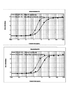
[32] FIG. 4 shows the results of ELISA that indicate the
binding affinity of each of a parent antibody and a modified
antibody for FOLR1 as a function of concentration.
[33] FIG. 5 shows the results of SPR analysis performed
to compare the binding affinity of a parent antibody for
Date Recue/Date Received 2020-10-13
FOLR1 with the binding affinity of a modified antibody for
FOLR1.
[34]
[35] Detailed Description and Preferred Embodiments of
the Invention
[36] Unless otherwise defined, all technical and
scientific terms used in the present specification have the
same meanings as commonly understood by those skilled in the
art to which the present disclosure pertains. In general,
the nomenclature used in the present specification is well
known and commonly used in the art.
[37]
[38] The term "antigen-binding fragment of an antibody"
or "antibody fragment" refers to a fragment having an
antigen-binding function, and includes Fab, F(ab'), F(ab')2
and Fv. Among antibody fragments, Fab is a structure having
light-chain and heavy-chain variable regions, a light-chain
constant region and a first heavy-chain constant region (CH1),
and has one antigen-binding site. Fab' differs from Fab in
that it has a hinge region containing at least one cysteine
residue at the C-terminus of the heavy-chain CH1 region. An
F(ab1)2 antibody has a disulfide bond formed by cysteine
residues in the hinge region of Fab'. Fv is a minimal antibody
fragment having only a heavy-chain variable region and a
light-chain variable region, and recombinant techniques of
11
Date Recue/Date Received 2020-10-13
producing Fv fragments are disclosed in PCT International
Patent Publication Nos. WO 88/10649, WO 88/106630, WO
88/07085, WO 88/07086, and WO 88/09344.
[39] The variable regions of the antibody used in the
present invention include three CDRs (CDR-H1, CDR-H2 and CDR-
H3) in the heavy-chain portion of the antibody and include
three CDRs (CDR-L1, CDR-L2 and CDR-L3) in the light-chain
portion of the antibody. These regions all form a loop and
are regions that bind specifically to an antigen.
[40]
[41] In one aspect, the present invention is directed to
an antibody or antigen-binding fragment thereof that binds
specifically to folate receptor-a (FOLR1).
[42] In the present invention, the antibody or antigen-
binding fragment thereof may inhibit the biological activity
of folate receptor-a. Furthermore, the antibody or antigen-
binding fragment thereof may induce antibody-dependent
cellular cytotoxicity against cells that express folate
receptor-a. In addition, the antibody or antigen-binding
fragment thereof may have a dissociation constant of 1x10⁻⁷
M or less for folate receptor-a.
[43]
[44] As used herein, the term "parent antibody" refers to
an anti-FOLR1 antibody that binds specifically to FOLR1. In
the present invention, the antibody applied in a previous
12
Date Recue/Date Received 2020-10-13
patent application (U52005/0232919 Al) was used as the parent
antibody. The "parent antibody" in the present specification
is one of the antibodies specified in the previous patent
application, and has a heavy-chain sequence corresponding to
SEQ ID NO: 1 below and a light-chain sequence corresponding
to SEQ ID NO: 2 below:
[45]
[46] Heavy chain (SEQ ID NO: 1)
[47] EVQLVESGGGVVQPGRSLRLSCSASGFTFSGYGLSWVRQAPGKGLEWVAM
[48] ISSGGSYTYYADSVKGRFAISRDNAKNTLFLQMDSLRPEDTGVYFCARHG
[49] DDPAWFAYWGQGTPVTVSS
[50]
[51] Light chain (SEQ ID NO: 2)
[52] DIQLTQSPSSLSASVGDRVTITCSVSSSISSNNLHWYQQKPGKAPKPWIY
[53] GTSNLASGVPSRFSGSGSGTDYTFTISSLQPEDIATYYCQQWSSYPYMYT
[54] FGQGTKVEIK
[55]
[56] The scope of the present invention includes not only
a full-length antibody (full-length IgG) that binds
specifically to FOLR1, but also an antigen-binding fragment
(fragmented IgG) of the antibody molecule. The parent
antibody used in the present invention comprises CDRs
represented by sequences of SEQ ID NOs: 3 to 8.
[57] Therefore, in the present invention, the antibody or
antigen-binding fragment thereof may comprise: the heavy-
13
Date Recue/Date Received 2020-10-13
chain CDR1 of SEQ ID NO: 3; the heavy-chain CDR2 of SEQ ID
NO: 4; the heavy-chain CDR3 of SEQ ID NO: 5; the light-chain
CDR1 of SEQ ID NO: 6; the light-chain CDR2 of SEQ ID NO: 7;
and the light-chain CDR3 of SEQ ID NO: 8.
[58] In the present invention, a CDR library based on the
parent antibody was constructed as mentioned in PCT
International Patent Publication No. W02016/114567DP, and a
pComb3X vector having the constructed library gene introduced
therein is a phagemid vector, which is a plasmid DNA having
a phage origin of replication and includes the phage surface
protein pill. The library gene is ligated to the 5' end of
the pIII gene and expressed as a fusion protein in E. coli.
VCSM13 helper phage is a phage that provides necessary
genetic information so that a phagemid is assembled into a
phage particle. The VCSM13 helper phage contains the
kanamycin antibiotic resistance gene so that E. coli infected
with the helper phage may be selected.
[59] In addition, panning refers to a process of
selectively amplifying only clones, which bind to a specific
molecule, from a library of proteins, such as antibodies,
displayed on the phage surface. The procedure comprises:
adding a phage library to a target molecule immobilized on
the surface to induce binding; removing unbound phage clones
by washing; eluting only bound phage clones; re-infecting E.
coli with the eluted phage clones; and amplifying target-
14
Date Recue/Date Received 2020-10-13
bound phage clones using helper phages. By repeating this
procedure, target-bound phage clones having a high binding
affinity for the target molecule immobilized on the surface
are selectively amplified.
[60]
[61] As used herein, the term "modified antibody" refers
to an antibody made by modifying the parent antibody. The
present invention also provides a method for isolating and
purifying the modified antibody. A culture obtained by
culturing under conditions where the antibody protein is
produced may be centrifuged to remove impurities, and the
resulting material may be purified using affinity
chromatography.
[62] In addition, the modified antibody of the present
invention has binding affinity for FOLR1. The binding
affinity for FOLR1 may be measured using ELISA assay, SPR
(surface plasmon resonance) assay, or the like. Specifically,
the binding affinity may be measured by reacting an antibody
composition with FOLR1 immobilized on a plate at various
concentrations, additionally reacting a labeled antibody
that recognizes the antibody, and calculating the
concentration of the antibody composition bound to FOLR1.
Thereby, it was possible to confirm that the binding ability
of the antibody obtained from multiple-CDR libraries was
improved more than that of the antibody obtained from single-
Date Recue/Date Received 2020-10-13
CDR libraries.
[63] In an embodiment of the present invention, the
modified antibody may comprise the heavy-chain CDR3 of SEQ
ID NO: 28, the light-chain CDR1 of SEQ ID NO: 29, or the
light-chain CDR2 of SEQ ID NO: 30, which is a sequence
modified from the parent antibody.
[64] Therefore, in the present invention, the antibody or
antigen-binding fragment thereof may comprise: the heavy-
chain CDR1 of SEQ ID NO: 3; the heavy-chain CDR2 of SEQ ID
NO: 4; the heavy-chain CDR3 of SEQ ID NO: 5 or SEQ ID NO:
28; the light-chain CDR1 of SEQ ID NO: 6 or SEQ ID NO: 29;
the light-chain CDR2 of SEQ ID NO: 7 or SEQ ID NO: 30; and
the light-chain CDR3 of SEQ ID NO: 8.
[65]
[66] In the present invention, the antibody or antigen-
binding fragment thereof may comprise: a heavy-chain variable
region of SEQ ID NO: 1 or SEQ ID NO: 31; and a light-chain
variable region selected from the group consisting of SEQ ID
NO: 2 and SEQ ID NOs: 32 to 34. Preferably, the antibody or
antigen-binding fragment thereof may comprise: the heavy-
chain variable region of SEQ ID NO: 1 and the light-chain
variable region of SEQ ID NO: 32; the heavy-chain variable
region of SEQ ID NO: 1 and the light-chain variable region
of SEQ ID NO: 33; the heavy-chain variable region of SEQ ID
NO: 1 and the light-chain variable region of SEQ ID NO: 34;
16
Date Recue/Date Received 2020-10-13
the heavy-chain variable region of SEQ ID NO: 31 and the
light-chain variable region of SEQ ID NO: 2; or the heavy-
chain variable region of SEQ ID NO: 31 and the light-chain
variable region of SEQ ID NO: 32.
[67]
[68] In another aspect, the present invention is directed
to an antibody-drug conjugate (ADC) in which a drug is
conjugated to the antibody or antigen-binding fragment
thereof.
[69] With regard to the antibody-drug conjugate (ADC),
the anticancer drug should remain stably bound to the
antibody until the anticancer drug is delivered to the target
cancer cell. The drug delivered to the target should be
released from the antibody and induce apoptosis of the target
cell. To this end, the drug should stably bind to the antibody,
and at the same time, should exhibit sufficient cytotoxicity
to induce apoptosis of the target cells when released in the
target cell.
[70] In the present invention, the antibody or antigen-
binding fragment thereof and a cytotoxic substance comprising
a drug such as an anticancer agent are bound to each other
(via, for example, a covalent bond, a peptide bond or the
like), and thus may be used as a conjugate or a fusion protein
(when a cytotoxic substance and/or labeling substance
(marker) is protein). The cytotoxic substance may be any
17
Date Recue/Date Received 2020-10-13
substance which is toxic to cancer cells, particularly solid
cancer cells, and may be at least one selected from the group
consisting of radioisotopes, cytotoxic compounds (small
molecules), cytotoxic proteins, and anticancer drugs, but is
not limited thereto. The cytotoxic protein may be at least
one selected from the group consisting of ricin, saporin,
gelonin, momordin, deBouganin, diphtheria toxin, pseudomonas
toxin, and the like, but is not limited thereto. The
radioisotope may be at least one selected from the group
consisting of 1-31I, 1-88Rh and "Y, but is not limited thereto.
The cytotoxic compound may be at least one selected from the
group consisting of duocarmycin, monomethyl auristatin E
(MMAE), monomethyl auristatin F (MMAF), N2'-diacetyl-N2'-(3-
mercapto-1-oxopropyl)maytansine (DM1), PBD
(pyrrolobenzodiazepine) dimer, and the like, but is not
limited thereto.
[71] In the present invention, the antibody-drug
conjugate may be obtained according to a method well-known
in the art.
[72] In the present invention, the antibody-drug
conjugate may be characterized in that the antibody or
antigen-binding fragment thereof is conjugated to the drug
via a linker.
[73] In the present invention, the linker may be a
cleavable linker or a non-cleavable linker.
18
Date Recue/Date Received 2020-10-13
[74] The linker is a site for linking the antibody to the
drug. For example, the linker allows the drug to be released
in a cleavable form under an intracellular condition, that
is, through cleavage of the linker from the antibody in an
intracellular environment.
[75] The linker may be a peptide linker that can be
cleaved by a cleavage agent present in an intracellular
environment, for example, in the lysosome or endosome, and
can be cleaved by intracellular peptidases or proteases, such
as lysosome or endosome proteases. Generally, a peptide
linker is at least two amino acids in length. The cleavage
agent may include cathepsin B, cathepsin D and plasmin, which
hydrolyze the peptide to release the drug into the target
cell. The peptide linker can be cleaved by a thiol-dependent
protease cathepsin-B, which is highly expressed in cancer
tissue. For example, the peptide linker may be a Phe-Leu or
Gly-Phe-Leu-Gly linker. In addition, the peptide linker may,
for example, be a Val-Cit linker or a Phe-Lys linker, which
can be cleaved by an intracellular protease.
[76] In the present invention, the cleavable linker is
sensitive to pH and may be sensitive to hydrolysis at a
certain pH value. Generally, the pH-sensitive linker is a
linker that can be hydrolyzed under acidic conditions.
Examples of acid-instable linkers that can be hydrolyzed in
lysosomes include hydrazone,
semicarbazone,
19
Date Recue/Date Received 2020-10-13
thiosemicarbazone, cis-aconitic amide, orthoester, acetal,
ketal, and the like.
[77] The linker may also be cleaved under reducing
conditions, and may, for example, be a disulfide linker. A
variety of disulfide bonds can be formed using N-
succinimidyl-S-acetylthioacetate (SATA), N-succinimidy1-3-
(2-pyridyldithio) propionate (SPDP), N-succinimidy1-3-(2-
pyridyldithio)butyrate (SPDB) and N-
succinimidyl-
oxycarbonyl-alpha-methyl-alpha-(2-pyridyl-dithio)toluene
(SMPT).
[78] In the present invention, the drug and/or the drug-
linker may be randomly conjugated through the lysine of the
antibody, or may be conjugated through cysteine, which is
exposed when the disulfide bond chain is reduced. In some
cases, the linker-drug can be conjugated through cysteine
present in a genetically engineered tag, e.g., a peptide or
protein. The genetically engineered tag, e.g., a peptide or
protein, may include an amino acid motif that can be
recognized, for example, by an isoprenoid transferase. The
peptide or protein has a deletion at the carboxyl terminus
of the peptide or protein or an addition at the carboxyl (C)
terminus of the peptide or protein through a covalent bond
of a spacer unit. The peptide or protein may be covalently
bonded directly to an amino acid motif, or may be linked to
the amino acid motif through a covalent bond to the spacer
Date Recue/Date Received 2020-10-13
unit. The amino acid spacer unit consists of 1 to 20 amino
acids, and is particularly preferably a glycine unit.
[79] The linker may comprise a beta-glucuronide linker
that is recognized and hydrolyzed by beta-glucuronidase,
which is present in multiple copies in the lysosome or
overexpressed in some tumor cells. Unlike the peptide linker,
this linker has the advantage of increasing the solubility
of the antibody-drug conjugate when bound to a drug having
high hydrophobicity due to the high hydrophilicity thereof.
[80] In this regard, in the present invention, it is
possible to use a beta-glucuronide linker disclosed in Korean
Patent Application Publication No. 2015-0137015, for
example, a beta-glucuronide linker including a self-
immolative group.
[81] In addition, the linker may be, for example, a non-
cleavable linker, and the drug may be released merely through
a single step of hydrolyzing the antibody, thus producing,
for example, an amino acid/linker/drug complex. This type of
linker can be a thioether group or a maleimidocaproyl group,
and is stable in the blood.
[82] In the present invention, the drug may be a
chemotherapeutic agent, a toxin, microRNA (miRNA), siRNA,
shRNA, or a radioactive isotope. The drug, which is an agent
having a pharmacological effect, may be conjugated to the
antibody.
21
Date Recue/Date Received 2020-10-13
[83] The chemotherapeutic agent may be a cytotoxic agent
or an immunosuppressive agent. Specifically, the
chemotherapeutic agent may comprise a microtubulin
inhibitor, a mitotic inhibitor, a topoisomerase inhibitor,
or a chemotherapeutic agent capable of functioning as a DNA
intercalator. The chemotherapeutic agent may also comprise
an immunomodulatory compound, an anticancer agent, an
antiviral agent, an antibacterial agent, an antifungal agent,
an anthelmintic, or a combination thereof.
[84] For example, the drug may comprise at least one
selected from the group consisting of maytansinoid,
auristatin, aminopterin, actinomycin,
bleomycin,
thalidomide, camptothecin, N8-acetylspermidine, 1-(2
chloroethyl)-1,2-dimethyl sulfonyl hydrazide, esperamycin,
etoposide, 6-mercaptopurine, dolastatin, trichothecene,
calicheamicin, taxol, taxane, paclitaxel, docetaxel,
methotrexate, vincristine, vinblastine,
doxorubicin,
melphalan, chlorambucil, duocarmycin, L-
asparaginase,
mercaptopurine, thioguanine, hydroxyurea, cytarabine,
cyclophosphamide, ifosfamide, nitrosourea, cisplatin,
carboplatin, mitomycins (mitomycin A and mitomycin C),
dacarbazine, procarbazine, topotecan, nitrogen mustard,
cytoxan, etoposide, 5-fluorouracil, CNU (bis-
chloroethylnitrosourea), irinotecan,
camptothecin,
bleomycin, idarubicin, daunorubicin,
dactinomycin,
22
Date Recue/Date Received 2020-10-13
plicamycin, asparaginase,
vinorelbine, chlorambucil,
melphalan, carmustine, lomustine, busulfan, treosulfan,
dacarbazine, etoposide, teniposide, topotecan, 9-
aminocamptothecin, crisnatol, trimetrexate, mycophenolic
acid, tiazofurin, ribavirin, EICAR (5-ethyny1-1-beta-
ribofuranosylimidazole-4-carboxamide),
hydroxyurea,
deferoxamine, floxuridine, doxifluridine, raltitrexed,
cytarabine (ara C), cytosine arabinoside, fludarabine,
tamoxifen, raloxifene, megestrol, goserelin, leuprolide
acetate, flutamide, bicalutamide, EB1089, CB1093, KH1060,
verteporfin, phthalocyanine, photosensitizer Pe4, demethoxy-
hypocrellin A, interferon-a, interferon-y, tumor necrosis
factor, gemcitabine, Velcade, Revlimid, Thalomid,
lovastatin, 1-methyl-4-phenylpyridiniumion, staurosporine,
actinomycin D, dactinomycin, bleomycin A2, bleomycin B2,
peplomycin, epirubicin,
pirarubicin, zorubicin,
mitoxantrone, verapamil and thapsigargin, nucleases, and
toxins derived from bacteria or plants and animals, but the
present invention is not limited thereto.
[85] In the
present invention, the drug may have a
nucleophile group selected from the group consisting of
amine, thiol, hydroxyl, hydrazide, oxime, hydrazine,
thiosemicarbazone, hydrazine carboxylate and aryl hydrazide
groups, which can react with an electrophilic group on the
linker and the linker reagent to form a covalent bond.
23
Date Recue/Date Received 2020-10-13
[86]
[87] In still another aspect, the present invention is
directed to a pharmaceutical composition for preventing
and/or treating cancer, the pharmaceutical composition
comprising the antibody or antigen-binding fragment thereof
or the antibody-drug conjugate.
[88] In yet another aspect, the present invention is
directed to a method for treating cancer, the method
comprising administering the antibody or antigen-binding
fragment thereof or the antibody-drug conjugate to a patient
in need of prevention or treatment.
[89] In still yet another aspect, the present invention
is directed to the use of the antibody or antigen-binding
fragment thereof or the antibody-drug conjugate for treating
cancer.
[90] In further another aspect, the present invention is
directed to the use of the antibody or antigen-binding
fragment thereof or the antibody-drug conjugate in the
manufacture of a medicament for treating cancer.
[91] In the present invention, the cancer may be ovarian
cancer, breast cancer, lung cancer, kidney cancer, colon
cancer, brain cancer, rectal cancer, cervical cancer, or
endometrial cancer, but is not limited thereto.
[92] Although the pharmaceutical composition comprising
the antibody or antigen-binding fragment thereof or the
24
Date Recue/Date Received 2020-10-13
antibody-drug conjugate according to the present invention
may also comprise only the antibody or antigen-binding
fragment thereof or the antibody-drug conjugate as an active
ingredient, it is generally mixed with one or more
pharmacologically acceptable carriers, and is preferably
provided as a pharmaceutical formulation prepared by any
method known in the technical field of pharmaceuticals.
[93] The pharmaceutical composition of the present
invention may be used alone or in combination with at least
one therapeutic drug selected from the above-described
radioisotopes, low-molecular-weight drugs, polymer drugs or
antibody drugs. In addition, the pharmaceutical composition
of the present invention may be used in combination with a
conventional therapeutic agent. That is, the pharmaceutical
composition comprising the antibody or antigen-binding
fragment thereof or the antibody-drug conjugate according to
the present invention may be administered simultaneously or
sequentially with a conventional therapeutic agent such as
an anticancer agent.
[94] As the route of administration, it is preferable to
use the most effective route of administration at the time
of treatment. Examples of the route of administration include
oral administration or parenteral administration such as
intra-mouth, intra-airway, intrarectal,
subcutaneous,
intramuscular or intravenous administration. Intravenous
Date Recue/Date Received 2020-10-13
administration is preferred.
[95] Dosage forms include a spray, a capsule, a tablet, a
powder, a granule, a syrup, an emulsion, a suppository, an
injection, an ointment, or a tape.
[96] The dosage or the frequency of administration varies
according to the desired therapeutic effect, the mode of
administration, the period of treatment, and the patient's
age and body weight, but is usually 10 jig/kg to 10 mg/kg per
day for an adult.
[97]
[98] Since the antibody or antigen-binding fragment
thereof according to the present invention binds specifically
to folate receptor-a (FOLR1), FOLR1 may be detected or
diagnosed using the same. Expression of FOLR1 is related to
several diseases, for example, cancer.
[99] Therefore, in another aspect, the present disclosure
is directed to a composition for diagnosing disease, the
composition comprising the antibody or antigen-binding
fragment thereof.
[100] In the present invention, the disease may be FOLR1-
related disease, for example, cancer, but is not limited
thereto.
[101] In still another aspect, the present invention is
directed to a method for diagnosing disease or a method for
providing information for diagnosing disease, the method
26
Date Recue/Date Received 2020-10-13
comprising a step of treating (administering) a biological
sample isolated from a subject with the antibody or antigen-
binding fragment thereof.
[102] In the present invention, the method for diagnosing
disease may further comprise, after the treatment step, a
step of identifying whether an antigen-antibody reaction
occurs. In the detection method, when the antigen-antibody
reaction is detected, a FOLR1-related disease, for example,
cancer, may be determined to be present in the biological
sample or a patient from which the biological sample has been
obtained. Thus, the method may further comprise, after the
step of identifying, a step of determining that, when the
antigen-antibody reaction is detected, the biological sample
or the patient is a FOLR1-related disease patient, for
example, a cancer patient. The biological sample may be
selected from the group consisting of cells, tissues, body
fluids, cultures thereof and the like, obtained (isolated)
from a mammal such as a human (e.g., a cancer patient).
[103] The step of identifying whether or not the antigen-
antibody reaction occurs may be performed through various
methods known in the art. For example, the step may be
performed through a conventional enzymatic reaction,
fluorescence, luminescence and/or radiation detection.
Specifically, the step may be performed by a method selected
from the group consisting of immunochromatography,
27
Date Recue/Date Received 2020-10-13
immunohistochemistry, enzyme-linked immunosorbent assay
(ELISA), radioimmunoassay (RIA), enzyme immunoassay (EIA),
fluorescence immunoassay (FIA), luminescence immunoassay
(LIA), Western blotting, microarray, and immunoprecipitation
assay, but is not limited thereto.
[104] In this case, the antibody or antigen-binding
fragment thereof may further comprise a marker. The marker
may be at least one selected from the group consisting of
radioactive isotopes, fluorescent substances, chromogen and
dyeing substances. The marker may be bound (linked) to the
antibody or antigen-binding fragment by a conventional method
(for example, a chemical bond such as a covalent bond,
coordination bond or ionic bond). The binding of the antibody
(or antigen-binding fragment) to the marker may be performed
in accordance with techniques known in the art.
[105]
[106] Hereinafter, the present invention will be described
in more detail with reference to examples. It will be obvious
to those skilled in the art that these examples are merely
to illustrate the present invention, and the scope of the
present invention is not limited by these examples.
[107]
[108] Example 1: Selection of Library Clones
28
Date Recue/Date Received 2020-10-13
[109] To obtain optimum sequences having improved binding
affinity for FOLR1, CDR libraries were constructed using Fab
fragments.
[110]
[111] Example 1-1: Preparation of Parent Antibody Fab
Template
[112] Variable regions were synthesized from the light
chain and heavy chain of a parent antibody, respectively,
and constant regions were synthesized from pComb3X-TT. PCR
reaction was performed under the following conditions: pre-
denaturation at 94 C for 2 min, and then 25 cycles, each
consisting of 30 sec at 94 C, 30 sec at 56 C and 30 sec at
72 C, followed by elongation at 72 C for 7 min. In the PCR
reaction, 100 ng of one template was used, or a mixture
obtained by mixing two templates in an amount of 3 pL of each
template was used. 3 pL of each primer was used at a
concentration of 20 pM, 0.05 mM dNTP and 0.5 pL (2.6 units)
Tag polymerase were used, and the reaction volume was 100 pL.
After completion of the reaction, whether amplification
occurred was checked using 1% agarose gel electrophoresis,
and the amplification product was purified using a Qiagen
gel extraction kit. For second and third PCR, overlapping
PCR was performed using the amplified fragment as a template.
The PCR reaction product DNA was purified on an agarose gel
using a Qiagen gel extraction kit, cleaved with a SfiI
29
Date Recue/Date Received 2020-10-13
restriction enzyme, and then subjected to gel extraction.
153 ng of the SfiI-cleaved antibody gene and 136 ng of the
SfiI-cleaved pComb3X vector were mixed with each other, added
to 10X T4 DNA ligation buffer and 10 units of ligase, reacted
at room temperature for 3 hours, and then heat-shocked at
42 C for 45 seconds in E. coli DH5a cells, followed by
incubation at 37 C for 1.5 hours. From the colony obtained
by the above-described transformation method, a template for
antibody library construction composed of a 50-kDa Fab
fragment was obtained.
[113]
[114] Example 1-2: Antibody Library Construction
[115] Libraries were constructed by artificially
introducing diversity into the complementarity-determining
region, and the CDRs and FRs of a given antibody can be
identified according to the content set forth at
(http://www.bioinf.org.uk/abs/).
[116] Libraries were constructed by randomizing the CDR
sequences of the parent antibody based on the template
prepared in Example 1-1. Among the six CDRs of the parent
antibody, CDR-H2 was excluded from the experiment because it
was so long as to be difficult to handle in the experiment.
The CDR sequence of the parent antibody is shown in Table 1
below.
[117]
Date Recue/Date Received 2020-10-13
[118] [Table 1] CDR sequence of parent antibody
SEQ
Residue located Residue located at
CDR Sequence ID
in front back
NO
CDR-H1 Cys-x-x-x GFTFSGYGLS Trp-Val 3
Leu-Gln-Trp-Val-
CDR-H2 MI S S GGSYTYYADSV Lys 4
Ala
CDR-H3 Cy s-Ala-Arg HGDDPAWFAY Trp-Gly-Gln-Gly 5
CDR-L1 Cys SVS SSISSNNLH Trp 6
CDR-L2 Ile-Tyr GTSNLAS Gly 7
CDR-L3 Cys QQWSSYPYMYT
Phe-Gly-Gln-Gly 8
[119]
[120] To randomize a specific position in the region that
binds to an antigen, primers were prepared using mixed base
codes (Table 2). The mixed base code is a degenerated primer
and refers to an oligonucleotide in which two or more bases
exist in one position so that they can bind to similar
nucleotide sequences in consideration of the nucleotide
sequence similarity. Prior to preparation of the primers, in
order to determine the specific position to be randomized,
conserved residues were identified through CDR sequence
analysis of the parent antibody. For codon diversification
of a portion excluding these residues, primers were prepared
using mixed base codes.
[121]
31
Date Recue/Date Received 2020-10-13
[122] [Table 2] Primer sequences
SEQ
CDR Primer Sequence (5' -> 3')
ID NO
GCC TCT GGC TTC ACT TTC AGT RRT TAC 9
Fan-Hi-
CDR- GVT MTG ART TGG GTG AGA CAG GCA CCT
random-f
H1 G
Fan1-H 1-b ACT GAA AGT GAA GCC AGA GGC 10
GGG GTC TAT TTT TGT GCA AGA NNK NNK 11
Far1-H3-
GAC GAT CCA GCA TGG TTT GMT TAC TGG
randoml-f
GGC CAA GGG ACC
GGG GTC TAT TTT TGT GCA AGA CAC NNK 12
Far1-H3-
NNK GAT CCA GCA TGG TTT GMT TAC TGG
random2-f
GGC CAA GGG ACC
GGG GTC TAT TTT TGT GCA AGA CAC GGT 13
Far1-H3-
NNK NNK CCA GCA TGG TTT GMT TAC TGG
CDR- random3-f
GGC CAA GGG ACC
H3
GGG GTC TAT TTT TGT GCA AGA CAC GGT 14
Far1-H3-
GAC NNK NNK GCA TGG TTT GMT TAC TGG
random4-f
GGC CAA GGG ACC
GGG GTC TAT TTT TGT GCA AGA CAC GGT 15
Far1-H3-
GAC GAT NNK NNK TGG TTT GMT TAC TGG
random5-f
GGC CAA GGG ACC
Far1-H3- GGG GTC
TAT TTT TGT GCA AGA CAC GGT 16
random6-f GAC GAT CCA NNK NNK TTT GMT TAC TGG
32
Date Recue/Date Received 2020-10-13
GGC CAA GGG ACC
Farl-H3-b TCT TGC ACA AAA ATA GAC CCC 17
AC AGA GTC ACC ATC ACA TGC AGK GYT 18
Fan-Li-
CDR- TCC TCC RGT VTT AGT TCA ARC WAT CTG
random-f
Li MAC TGG TAT CAG CAG AAG CCC G
Fanl-L 1 -b GCA TGT GAT GGT GAC TCT GT 19
C CCA AAG CCC TGG ATC TAC GVT RCC 20
Farl-L2-
CDR- TCT AVT CKG GMA AST GGG GTG CCT TCA
random-f
L2 AGG TTC A
Farl-L2-b GTA GAT CCA GGG CTT TGG G 21
GCA ACT TAC TAT TGC CAG CAG NNK BMT 22
Farl-L3-
CDR- WAT TWT CCA YMT NNK YMC ACC TTC
random-f
L3 GGT CAG GGC AC
Farl-L3-b CTG CTG GCA ATA GTA AGT TGC 23
All pC3X-f GCA CGA CAG GTT TCC CGAC 24
CDRs pC3X-b AAC CAT CGA TAG CAG CAC CG 25
CDR- Lead-b GGC CAT GGC TGG TTG GGC 26
L3H3 Lead-VH GCC CAA CCA GCC ATG GCC 27
[123]
[124] For codon randomization of the parent antibody CDR-
Li, L2, L3, H1 and H2 regions, each of the regions was
amplified by PCR and attached to PCR-amplified CDR DNA
through overlap extension PCR, thereby constructing single-
CDR libraries having diversity only in one CDR and multiple-
33
Date Recue/Date Received 2020-10-13
CDR libraries having diversity in two CDRs. The PCR reaction
was performed under the following conditions: pre-
denaturation at 94 C for 2 min and then 25 cycles, each
consisting of 30 sec at 94 C, 30 sec at 56 C and 30 sec at
72 C, followed by extension at 72 C for 7 min. However, the
elongation time at 72 C was adjusted to 1 min 30 sec or 2
min depending on the length of predicted DNA fragment
products. Specifically, the elongation time was 1 min 30 sec
for a predicted length of 500 to 1,500 bp and 2 min for a
predicted length of 1,500 to 2,000 bp. The names of the
templates, primers and products used in these processes are
summarized in Table 3 below. The constructed single- or
multiple-CDR library was isolated and purified by 1% agarose
gel electrophoresis, and the purification product was cleaved
by treatment with a SfiI restriction enzyme at 50 C for 12
hours or more. The pComb3X phagemid vector was also cleaved
with a SfiI restriction enzyme in the same manner.
[125]
[126] [Table 3] PCR templates and primers for library
construction
Primer
Single-CDR library PCR Template SEQ ID Product
NO
19, 24 FAR-Li-F
CDR-L1 1' pFAR-FabC 3
i8,25 FAR-Li-R
34
Date Recue/Date Received 2020-10-13
FAR-Li-F, FAR-L1-
2nd 24, 25 FAR-CDR-
L1
R
21, 24 FAR-L2-F
lst pFAR-FabC3
20, 24 FAR-L2-R
CDR-L2
FAR-L2-F, FAR-L2-
2nd 24, 25 FAR-CDR-
L2
R
23, 24 FAR-L3-F
1st pFAR-FabC3
22, 25 FAR-L3-R
CDR-L3
FAR-L3-F, FAR-L3-
2nd 24, 25 FAR-CDR-
L3
R
10, 24 FAR-H 1-F
lst pFAR-FabC3
9, 25 FAR-H 1-R
CDR-H1
FAR-Hi-F, FAR-H1 -
2nd 24, 25 FAR-CDR-
H1
R
17, 24 FAR-H3-F
lst pFAR-FabC3 11 to 16,
FAR-H3-R
CDR-H3 25
FAR-H3-F, FAR-H3 -
2nd 24,25 FAR-CDR-
H3
R
Primer
Multiple-CDR
PCR Template SEQ ID Product
library
NO
pFAR CDR-L3 24,26 FAR-L3
CDR-L3H3 lst
pFAR-CDR-H3 25,27 FAR-H3
Date Recue/Date Received 2020-10-13
2nd FAR-L3, FAR-H3 24,25 FAR-CDR-L3H3
[127]
[128] For ligation, the SfiI-cleaved vector and the SfiI-
cleaved insert were mixed together in equal amounts and
reacted overnight at room temperature. If the volume of the
ligation product is too large for transformation, the volume
of the ligation product can be reduced using Et0H
precipitation, and the method for reducing the volume is as
follows. To 50 pL of the ligation product, 5 pL (1/10) of 3
M sodium acetate (pH 5.2) and 110 pL (2-fold) of 100% Et0H
were added, and DNA was allowed to precipitate at -20 C for
2 hours or more. The precipitated DNA was centrifuged at
12,000 rpm for 15 minutes, and then washed with 1 ml of 70%
Et0H and centrifuged under the same conditions. The pellet
was dried, and then dissolved in 10 pL of deionized water.
Transformation was performed by electroporation.
Specifically, 10 pL of the ligation product and 50 pL of E.
co/i TG1 competent cells were mixed together and then placed
in a 0.2-cm cooled cuvette, which was placed in an
electroporator. Next, the cells were pulsed at 2.5 kV for 4
to 5 msec. 2 mL of recovery medium heated to 37 C was added
thereto, immediately after pulsing and then incubation was
performed at 37 C for 1 hour. Next, 1 pL of the incubated
cells was diluted 1,000-fold with SB medium, and 10 pL and
100 pL of the cell dilution were dispensed on an LB agar
36
Date Recue/Date Received 2020-10-13
plate to prepare samples for measuring the library size, and
the remainder was plated on one plate and incubated overnight
at 37 C. The next day, the library size was measured by
counting the number of colonies on the plate into which the
diluted cells were dispensed. In addition, 5 mL of SB medium
was added to the plate into which the undiluted cells were
dispensed, the cells were collected using a spreader, and
then a 0.5-fold volume of 50% glycerol was added to the cells,
which were then stored in a deep freezer (-75 C)
[129]
[130] [Table 4]
CDR library Library size
CDR-L1 4.56x 10⁶
CDR-L2 /94x107
CDR-L3 6.22x10⁷
CDR-H1 4.78x 10⁶
CDR-H3 1.30x10⁸
CDR-L3H3 8.82x10⁷
[131]
[132] The size of the obtained library indicates
transformation efficiency, specifically the number of
individual clones. As a result, it can be considered that
antigen-binding diversity corresponds to the library size.
That is, the following libraries were prepared: a CDR-L1
37
Date Recue/Date Received 2020-10-13
library having a diversity of 106, a CDR-L2 library having a
diversity of 107, a CDR-L3 library having a diversity of 107,
a CDR-H1 library having a diversity of 106, a CDR-H3 library
having a diversity of 108, and a CDR-L3H3 library having a
diversity of 107 (Table 4).
[133]
[134] Example 1-3: Selection by ELISA after Phage-Display
Panning
[135] Panning was performed to select a library clone that
binds to human FOLR1, and as panning was repeated, a clone
having further increased binding affinity for FOLR1 could be
obtained.
[136] For library amplification and recovery of a Fab-
expressing bacteriophage, 100 pL of a TG1 stock transformed
with the library was seeded into 20 mL of SB/Amp+2% glucose
medium, and the library was expressed in E. coli TG1 cells
at 37 C and 220 rpm for 1.5 to 2 hours. The cell culture was
centrifuged at 3500 rpm for 15 minutes. The supernatant was
removed, and the pellet was re-suspended in 20 ml of SB/Amp
medium. 0.5 mL of helper phage VCSM13 (about 1011 pfu) was
added to the suspension, which was then infected with the
helper phage by culture at 37 C at 120 rpm for 1 hour. Next,
kanamycin (50 mg/mL) was added to the suspension to reach 70
jig/mL, followed by culture at 30 C at 200 rpm for 16 hours.
The culture was centrifuged, and 5 mL of 5X PEG concentrated
38
Date Recue/Date Received 2020-10-13
solution was added to the phage-containing supernatant, which
was then concentrated on ice for 30 minutes. The concentrate
was centrifuged at 12,000 rpm for 15 minutes, and the
supernatant was removed. The phage pellet was re-suspended
in 0.3 mL of PBS to obtain a library phage (for storage, a
0.5-fold volume of 50% glycerol is added to the library phage
which is then stored at -75 C)
[137] To
amplify the obtained library phage, an E. coli
TG1 cell stock was seeded into 10 mL of SB medium, and
cultured in an incubator at 37 C at 220 rpm for about 4 to 5
hours up to the mid-log phase (0D600=0.5 to 1.0), thus
preparing competent cells which were then stored at 4 C until
use. Panning was performed in the following manner. 1 jig/mL
of FOLR1 was coated on an immunotube, and then blocked with
a blocking solution (3% skim milk) at 37 C for 1 hour. 0.5
mL of the library phage was blocked by adding 0.5 mL of
blocking solution thereto at a ratio of 1:1, and then allowed
to react with the immobilized antigen. After the reaction
had proceeded for one hour or more, unreacted or weakly bound
phage was removed through a washing step with PBS-Tween20
buffer, and strongly bound phage was eluted out with 1 mL of
TEA (100 mM) for 10 minutes and then neutralized with 0.5 mL
of Tris-HC1 (1 M, pH 7.4). 1.5 mL of the eluted phage was
added to 8.5 mL of the competent cells and infected into the
cells in an incubator at 37 C at 120 rpm for 1 hour. Next,
39
Date Recue/Date Received 2020-10-13
to measure the library size, 1 pL of the 2 mL reaction
solution was diluted 1,000-fold and 10,000-fold and plated,
and the remaining reaction solution was plated on one plate
and incubated overnight at 37 C.
[138] The
transformed E. coli library was cultured and
infected with VCSM13 helper phage to obtain an antibody phage
library having Fab clones displayed on the surface thereof.
Only phage clones that bound strongly to FOLR1 were selected
by panning the library for 3 to 5 rounds against FOLR1
adsorbed on the immune-tube surface. Through this process,
clones that bound weakly to FOLR1 or did not bind to FOLR1
due to defects in the synthesized CDR sequence were removed,
and as a result, it was possible to select CDR sequences that
had no defects and were better optimized than the existing
sequences. The CDR-L1 and CDR-L2 libraries were panned for 5
rounds, the CDR-L3, CDR-H3 and CDR-L3H3 libraries were panned
for 4 rounds, and the CDR-H1 library was panned for 3 rounds.
For each panning round, the ratio (0/I ratio) of the eluted
phage (output phage) to the phage (input phage) used in
panning was calculated, and the results were expressed as %
bound in Table 5 below. The fact that similar % bound values
appear even when panning is repeated indicates that the
binding affinity for FOLR1 reached saturation (Table 5). When
this result appeared, panning was no longer performed, and
the next step was performed using the eluted phage. To
Date Recue/Date Received 2020-10-13
validate the functionality of the antibody phage libraries
that resulted from panning, 94 clones were screened from each
CDR library by ELISA. The number of ELISA-positive clones
showing a binding signal at least 3 times stronger than the
background signal was identified to be 92 for CDR-L1, 66 for
CDR-L2, 19 for CDR-L3, 94 for CDR-H1, 48 for CDR-H3, and 63
for CDR-L3H3. Eight clones among the clones showing a
stronger binding signal for each library were sequenced
(Table 6) .
[139]
[140] [Table 5] Panning conditions and results
Number Amount
Phage Panning Phage input Phage output
% Bound of of
library round (c.f.u) (c.f.u)
washings antigen
1 3.6 x 10¹⁰
5.1 x 10⁸
1.4 3 1.0 tig
2 -1010
8.5 x 10⁸
8.5 5 0.5 ug
CDR-L1 3 -1010
1.1 x 10⁹
11 10 0.1 ug
4 -1010
5.9 x 10⁸
5.9 10 0.1 Li g
-1010
4.3 x 10⁸
4.3 10 0.1 Li g
1 2.5 x 10¹⁰
6.8 x 10⁸
2.7 3 1.0 tig
2 -1010
7.5 x 10⁸
7.5 5 0.5 tig
CDR-L2 3 -1010
8.0 x 10⁸
8.0 10 0.1 Li g
4 -1010
3.2 x 10⁸
3.2 10 0.1 Li g
5 -1010
3.1 x 10⁸
3.1 10 0.1 ug
41
Date Recue/Date Received 2020-10-13
1 1.4 x 10⁹
4.3 x 10⁶
0.3 3 1.0 Lig
2 3.7 x 10⁸
1.0 X 10⁵ 2.7 x 10⁻²
0.5 Lig
CDR-L3
3 1.7 x 10⁹
7.2 x 10⁷
4.2 5 0.5 Lig
4 5.8 x 10⁹
8.0 x 10⁷
1.4 10 0.1 Lig
1 5.5 x 10¹⁰
4.9 x 10⁸
0.9 3 1.0 Lig
CDR-H1 2 -1010
6.2 x 10⁸
6.2 5 0.5 Lig
3 -1010
6.5 x 10⁸
6.5 10 0.1 tig
1 1.9 x 10⁹
2.2 x 10⁷
1.2 3 1.0 Lig
2 3.5 x 10⁸
1.0 X 10⁵ 2.9 x 10⁻²
5 0.5 Lig
CDR-H3
3 1.8 x 10⁹
8.6 x 10⁷
4.8 5 0.5 Lig
4 4.4 x 10⁹
1.1 X 10⁸
2.5 10 0.1 Lig
1 8.9 x 10⁸
1.0 X 10⁸
11.2 3 1.0 Lig
2 6.0 x 10⁸
1.0 X 10⁵ 1.7 x 10⁻²
5 0.5 Lig
CDR-L3H3
3 1.8 x 10⁹
2.6 x 10⁷
1.4 5 0.5 Lig
4 3.2 x 10⁹
4.0 x 10⁷
1.3 10 0.1 tig
[141]
[142] [Table 61 Sequencing results
CDR Clone Sequencing results
WT SVSSSISSNNLH
C6, E6 SASSGLSSSYLH
C7 SASSSLSSSYLH
CDR-L1
D5 RVSSGISSNNLH
D7 RASSGLSSNNLH
F3 RAS SGVSSNNLH
42
Date Recue/Date Received 2020-10-13
H1 SASSSISSSYLH
H7 SVSSSLSSSNLH
WT GTSNLAS
B1 AT S SRAT
Cl GTSSRAS
C3 ATSNRES
CDR-L2
C10 ATSSLAT
E9, G6 GASSLAT
G3 ATSNLAS
H2 TASSRAS
WT HGDDPAW
CDR-H3 B4, B8, C4, C6, C10, G5 HGDDVAW
C8 HGDDIAW
WT HGDDPAW
CDR-L3H3 A3, A8, A10, F8, H4, H6, H10 HGDDVAW
G8 HGDDISW
[143]
[144] Example 1-4: Fab Production Using Protein-Expressing
Strain TOP1OF' and Purification
[145] To compare the binding affinities of the selected
clones for FOLR1, purification of each clone was performed.
Prior to purification, the host strain was changed from TG1
to TOP1OF' cells to express only a Fab region.
43
Date Recue/Date Received 2020-10-13
[146] A colony corresponding to each of the selected clones
was seeded into 4 mL SB/ampicillin medium and cultured
overnight at 37 C. The next day, 4 ml of the overnight culture
was seeded into 400 mL SB/ampicillin medium and cultured in
an incubator at 37 C for about 3 to 4 hours until the OD600
reached 0.5 to 1Ø Next, for expression of the clone, the
culture was treated with IPTG to a final concentration of 1
mM and then cultured overnight at 30 C. However, when only
expression was to be confirmed, culture was performed in 20
mL of SB/ampicillin medium.
[147] To recover a periplasm from 400 mL of the culture,
the culture was centrifuged to remove the supernatant, and
then the cells were lysed by treatment with 16 mL of 1X TES
solution at 4 C and incubation at 4 C for 1 hour.
Additionally, the cells were treated with 24 mL of 0.2X TES
solution and incubated for 1 hour. Next, the supernatant was
collected by centrifugation and 5 mM MgCl2 was added thereto
in order to remove EDTA. Before loading of the sample, a
column packed with 0.5 mL of Ni-NTA His-Bind resin was washed
with 20 CV of elution buffer (300 mM imidazole in PBS, pH
7.4) and then flushed with 20 CV of PBS. The sample was
loaded onto the column and the flow-through was collected.
After completion of loading, the column was flushed with 20
CV of wash buffer (20 mM imidazole in PBS, pH 7.4) and the
washing-through was collected. Then, the column was flushed
44
Date Recue/Date Received 2020-10-13
with 10 CV of elution buffer and the eluent was collected.
15 pL of the sample collected in each step during the
purification process was loaded in each well in 12% SDS-PAGE
and electrophoresed (at 150 V for 1 hour). The band was
visualized by Coomassie blue staining.
[148]
[149] Example 1-5: Examination of Direct Binding Pattern
of Selected Clone to FOLR1 at Different Concentrations
[150] For the purpose of comparing the binding affinities
of the primarily selected clones with each other, ELISA was
performed at different clone concentrations in the following
manner. 25 pL of FOLR1 was added to each well of a 96-well
plate at a concentration of 1 pg/mL, coated on the plate at
room temperature for 1 hour, and then blocked with 180 pL of
3% skim milk at room temperature for 1 hour. During blocking,
samples to be used as primary antibodies were prepared. As
primary antibodies, samples selected by screening were used.
The samples were diluted to a concentration of 0.1 to 100 nM
(0, 0.1, 0.3, 1, 3, 10, 30, and 100 nM) and cold-stored until
use. After blocking, 3% skim milk was removed, and 25 pL of
the primary antibody was added to each well and allowed to
react at room temperature for 1 hour or more. After
completion of the reaction, each well was washed three times
with PBS-Tween20 (0.1%) buffer. As a secondary antibody, HA-
HRP was diluted 3,000-fold with 3% skim milk, and 25 pL of
Date Recue/Date Received 2020-10-13
the dilution was added to each well and allowed to react at
room temperature for 1 hour or more. After completion of the
reaction, each well was washed three times with PBS-Tween20
(0.1%), and 25 pL of substrate TMB was added to each well to
confirm color development. After about 5 minutes, 25 pL of 1
M H2SO4 was added to each well to stop the reaction, and the
absorbance at a wavelength of 450 nm was measured. From the
measurement results, the EC (Effector concentration) value
was calculated using the GraphPad Prism 7 program.
[151] To measure the binding affinity of the selected clone
for FOLR1, SPR measurement was performed using a Biacore3000.
Before each sample was loaded, a CM5 sensor chip was
activated with 0.1 M NHS/0.4 M EDC, and then FOLR1 (20 pg/mL
in 10 mM acetate, pH 5.0) was immobilized thereon. Then,
unreacted NHS was deactivated with 1 M ethanolamine. 250 pL
of each sample was prepared at concentrations of 1, 2, 5, 10,
20 and 40 nM, and association and dissociation sensorgrams
were obtained while each sample was flushed at 30 pl/min.
The sensor chip was regenerated with 10 mM glycine (pH 2.1).
The obtained sensorgrams were analyzed using the
BIAevaluation software, and the KD values were calculated.
[152]
[153] Example 1-6: Sequencing of Antibody Library
[154] The sequences of the randomized complementarity-
determining regions of the clones selected by ELISA of the
46
Date Recue/Date Received 2020-10-13
phage library were analyzed. Using, as a template, 1 pL of a
culture of each clone selected from the clones cultured in
the 96-well plate, PCR reaction was performed using pC3X-f
and pC3X-b primers under the following conditions: pre-
denaturation at 94 C for 2 minutes, and then 25 cycles, each
consisting of 30 sec at 94 C, 30 sec at 56 C and 2 min at
72 C, followed by elongation at 72 C for 7 min. Whether
amplification occurred was checked by 1% agarose gel
electrophoresis, and the amplified PCR product was purified
with DW using a QIAvac 96 and sequenced. The sequence of the
complementarity-determining region was analyzed using the
leader sequence.
[155]
[156] Example 2: Construction of Modified Antibody (vAb)
[157] The final clone pvAb was constructed in the same
manner as the antibody library construction method. The
templates and primers used are shown in Table 7 below, and
information about the vAb antibody sequence is summarized in
Tables 8 to 10 below.
[158]
[159] [Table 7] PCR templates and primers for final clone
construction
Clone PCR Template Primer Product
pCDR-L l#C6 pC3 X-f, Farl-L2-b CDR-L1
pvAb 1'
pCDR-L2#C 1 Farl-L2-f, Lead-b CDR-L2
47
Date Recue/Date Received 2020-10-13
CDR-L3H3#A3 Lead-VH, pC3X-b CDR-H3
2nd CDR-L2, CDR-H3 Far1-L2-f, pC3X-b CDR-
L2H3
CDR-L1, CDR-
3rd pC3X-f, pC3X-b CDR-L1L2H3
L2H3
[160]
[161] [Table
8] Sequence comparison of complementarity-
determining region between parent antibody and modified
antibody (vAb)
Positi Amino Amino acid
CDR Parent antibody CDR on in acid of
after CDR sequence of
region sequence segue parent
modificatio modified antibody
nce antibody n
CDR- HGDDPAWFAY
HGDDVAWFAY
103 P V
H3 (SEQ ID NO: 5) (SEQ ID
NO: 28)
25 V A
28 S G
CDR- SVS S SI S SNNLH SAS S GL
SSSYLH
29 I L
Li (SEQ ID NO: 6) (SEQ ID
NO: 29)
32 N S
33 N Y
CDR- GTSNLAS (SEQ ID 54 N S GTSSRAS (SEQ ID
L2 NO: 7) 55 L R NO: 30)
[162]
[163] Through
combinations of the CDR sequences identified
as described above, the modified antibodies vAbl, vAb2, vAb3,
48
Date Recue/Date Received 2020-10-13
vAb4 and vAb5 were constructed. vAbl is a modified antibody
that reflects all of CDR-H3, CDR-L1 and CDR-L2, vAb2 is a
modified antibody that reflects CDR-L1 and CDR-L2, vAb3 is a
modified antibody that reflects CDR-L1, vAb4 is a modified
antibody that reflects CDR-L2, and vAb5 is a modified
antibody that reflects CDR-H3.
[164]
[165] [Table 9] Amino acid sequences of parent antibody
and modified antibody (vAb)
SEQID
Type Sequence
NO
EVQLVESGGGVVQPGRSLRLSCSASGFTFSGYGLSWVRQ
APGKGLEWVAMISSGGSYTYYADSVKGRFAISRDNAKNT
HC 1
LFLQMDSLRPEDTGVYFCARHGDDPAWFAYWGQGTPVT
VSS
DIQLTQSPSSLSASVGDRVTITCSVSSSISSNNLHWYQQKP
LC GKAPKPWIYGTSNLASGVPSRFSGSGSGTDYTFTISSLQPE 2
DIATYYCQQWSSYPYMYTFGQGTKVEIK
EVQLVESGGGVVQPGRSLRLSCSASGFTFSGYGLSWVRQ
APGKGLEWVAMISSGGSYTYYADSVKGRFAISRDNAKNT
HC 31
LFLQMDSLRPEDTGVYFCARHGDDVAWFAYWGQGTPVT
VSS
DIQLTQSPSSLSASVGDRVTITCSASSGLSSSYLHWYQQKP
LC GKAPKPWIYGTSSRASGVPSRFSGSGSGTDYTFTISSLQPE 32
DIATYYCQQWSSYPYMYTFGQGTKVEIK
49
Date Recue/Date Received 2020-10-13
DIQLTQSPSSLSASVGDRVTITCSASSGLSSSYLHWYQQKP
LC GKAPKPWIYGTSNLASGVPSRFSGSGSGTDYTFTISSLQPE 33
DIATYYCQQWSSYPYMYTFGQGTKVEIK
DIQLTQSPSSLSASVGDRVTITCSVSSSISSNNLHWYQQKP
LC GKAPKPWIYGTSSRASGVPSRFSGSGSGTDYTFTISSLQPE 34
DIATYYCQQWSSYPYMYTFGQGTKVEIK
[166]
[167] [Table 10] Sequences of modified antibodies (vAb)
Modified antibody HC LC
vAbl SEQ ID NO: 31 SEQ ID NO: 32
vAb2 SEQ ID NO: 1 SEQ ID NO: 32
vAb3 SEQ ID NO: 1 SEQ ID NO: 33
vAb4 SEQ ID NO: 1 SEQ ID NO: 34
vAb5 SEQ ID NO: 31 SEQ ID NO: 2
[168]
[169] Example 3: Expression and Purification of Modified
Antibody (vAb)
[170] To produce the modified antibody (vAb), expiCHO cells
were transfected with a vector (pc 3.4-vAbL, pc 3.4-vAbH)
containing the gene encoding the modified antibody (vAb)
protein and cultured, and the modified antibody was purified
using affinity chromatography. An XK16 column packed with
the affinity resin MabSelect SuReTM (GE Healthcare) was
equilibrated by flushing with buffer A (25 mM Tris, pH 7.0,
25 mM NaCl), and then the culture was flushed and bound to
Date Recue/Date Received 2020-10-13
the affinity resin, and the modified antibody (vAb) protein
was eluted with buffer B (25 mM citric acid, pH 3.5). After
completion of purification, the column was washed with 0.5 M
NaOH, and then packed with 20% ethanol and cold-stored. The
pH of the eluted sample was adjusted to 6.0 by adding a
suitable amount of 1 M Tris (pH 9.0) thereto. The state of
the sample was checked through 10% SDS-PAGE. The obtained
modified antibody (vAb) protein was subjected to buffer
exchange by dialysis against a buffer containing 10 mM sodium
succinate and 30 mM sucrose (pH 6.0).
[171]
[172] Example 4: Analysis of Binding Affinity of Modified
Antibody (vAb) by ELISA Assay
[173] For the purpose of comparing the binding affinity of
the modified antibody (vAb) produced in Example 2 with that
of the parent antibody, ELISA was performed at an antibody
concentration of 0.006 to 6.250 ng/mL in the same manner as
described in Examples 1-5. As a result of the measurement,
it was confirmed that the modified antibodies vAbl to vAb5
bound to FOLR1 with a 4.48- to 1.42-fold higher binding
affinity than the parent antibody (Table 11). The measured
values are obtained in different experiments for comparison
with the parent antibody.
[174]
[175] [Table 11] Results of ELISA assay
51
Date Recue/Date Received 2020-10-13
EC50 Relative
Coating Sample R2
ng/mL Potency
Parent antibody 10.99 1.00
FOLR1-Fc 0.997
vAbl 2.45 4.48
Parent antibody 11.85 1.00
FOLR1-Fc 0.972
vAb2 3.94 3.04
Parent antibody 3.04 1.00
vAb3 0.77 199
FOLR1-Fc 0.996
vAb4 1.59 1.94
vAb5 2.17 1.42
[176]
[177] Example 5: Examination of Binding Affinity of
Modified Antibody (vAb) by SPR Assay
[178] To measure the binding affinity of the modified
antibody vAbl produced in Example 2 for FOLR1, an SPR assay
was performed using a Biacore3000, in the same manner as
described in Examples 1-5. The sensorgram was analyzed using
the BIAevaluation software, and the KD value was calculated.
The modified antibody vAbl showed a KD value of 0.24 nM,
which corresponds to a 4-fold higher binding affinity than
that of the parent antibody (Table 12). This value is similar
to the relative ratio of the values obtained using ELISA.
[179]
52
Date Recue/Date Received 2020-10-13
[180] [Table 12] Dissociation constants of parent antibody
and modified antibody vAbl, measured by SPR
KD Relative
2
Ligand FOLR1 ka (1/Ms) kd KA (1/M) Chi
(nM) Potency
Parent
4.19E+05 4.01E-04 1.05E+09 0.96 1 4.90
antibody
Analyte Modified
antibody 6.12E+05 1.47E-044.17E+09 0.24 4 4.86
vAbl
[181]
Industrial Applicability
[182] The modified antibody according to the present
invention has a sequence in which the antigen-binding site
originally possessed by the parent antibody is substituted
with an amino acid that better binds to the antigen while
preserving the basic structure of the parent antibody. Thus,
the modified antibody has increased binding affinity for the
antigen. In addition, the antibody or antigen-binding
fragment thereof according to the present invention may be
used for the prevention or treatment of cancer, and may also
be used for diagnosis of disease.
[183]
[184] Although the present invention has been described in
detail with reference to specific features, it will be
apparent to those skilled in the art that this description
53
Date Recue/Date Received 2020-10-13
is only of a preferred embodiment thereof, and does not limit
the scope of the present invention. Thus, the substantial
scope of the present invention will be defined by the
appended claims and equivalents thereto.
[185]
Sequence List Free Text
[186] Electronic file is attached.
54
Date Recue/Date Received 2020-10-13