Note: Descriptions are shown in the official language in which they were submitted.
CA 03097782 2020-10-20
WO 2019/218077
PCT/CA2019/050669
TITLE: ISOLATION OF EXTRACELLULAR VESICLES BY PRECIPITATION
OR IMMOBILIZATION USING POLYETHYLENIMINE AND
POLYETHYLENIMINE-COATED SOLID SUPPORTS
RELATED APPLICATION
[0001] The
present application claims the benefit of priority of co-pending
United States provisional patent application no. 62/673,415, filed on May 18,
2018, the contents of which are incorporated herein by reference in their
entirety.
FIELD
[0002] The
present application is related to methods of isolating
extracellular vesicles (EVs). In particular, the application is related to the
use
of polyethylenimine for the enrichment of EVs from biological tissues and
fluids
with diagnostic and prognostic significance to pathological conditions, as
well
as for their inclusion in therapeutic strategies.
BACKGROUND
[0003]
Extracellular vesicles (EVs) are cell-derived vesicles, which
include exosomes, ectosomes (microparticles and microvesicles), and
apoptotic bodies, that are secreted from almost all cell types into the
extracellular environment and which have been found in most body fluids,
including blood, urine, milk, cerebrospinal fluid, semen, malignant effusions,
ascites, etc. These secreted EVs participate in both normal physiological and
pathological processes, in part by transmitting specific information, for
example,
by transmission of DNA, RNA, or protein, from their cell of origin to their
target
cells1. Increased numbers of EVs have been reported in blood and other
biological fluids in response to cancers and other pathological conditions2-5.
[0004] Liquid
biopsy is a term used to describe the analysis of a blood
sample for DNA, RNA, or protein markers derived from diseased cells or EVs
derived from diseased cells that are circulating in blood. Liquid biopsy can
provide valuable genetic and phenotypic information about a patient's disease
which can impact diagnosis and prognosis, as well as highlight actionable
mutations, for example, that can impact patient stratification and therapeutic
strategies. Implementation of routine liquid biopsy analysis has been proposed
1
CA 03097782 2020-10-20
WO 2019/218077
PCT/CA2019/050669
as a means of improving current disease staging protocols in order to improve
patient stratification and outcome6,7
[0005] EVs,
including exosomes (-30-150 nm) and microvesicles (-100-
350 nm), mediate intercellular communication and contain a cell-specific cargo
including, but not limited to, growth factors, enzymes, receptors, cytokines,
lipids, and coding and non-coding DNA and RNA molecules5,9. Since they have
been designed by nature to efficiently deliver cargo to target cells, the
interest
in the use of EVs as therapeutics, for example in anti-tumour immunotherapy,
as anti-infectious agents, and in immune-modulatory and regenerative
therapies, has been growing rapidly10. Although genomic and mitochondria!
DNA has been reported inside EVs, most of the DNA found in human blood
plasma is thought to be outside of the EVs.
[0006] EVs
contain a wide range of RNA types with a reported
prevalence of non-coding RNA, but also ribosomal RNA (rRNA), transfer RNA
(tRNA), long non-coding RNA (IncRNA), piwi-interacting RNA (piRNA), small
nuclear RNA and small nucleolar RNA (snoRNA), microRNA (miRNA) and
messenger RNA (mRNA) have been identified inside EVs12-15. EVs have been
demonstrated to function in the transfer of oncogenic molecules between cells
at metastatic sites16 and transfer of EV-derived mRNA to modulate target cell
behaviour has been identified as an important aspect in tumour development17.
Other studies have demonstrated that EVs contain mRNAs that originate from
a cytoplasmic RNA pool and reflect the profile of the originating cell and
tumour-
derived EVs can carry tumour-specific alterations in mRNA species15. Thus, the
EV mRNA cargo within EVs can be used as a source for cancer-derived
biomarkers that may be important in the development of diagnostic tests. The
use of cell derived vesicles (CDVs), including exosomes, for detecting
biomarkers for diagnostic, therapy-related or prognostic methods is described,
for example, in WO 2010/056337.
[0007] No
single standardized method exists for isolation of EVs for use
in diagnostic or therapeutic applications. The most commonly used methods for
isolating EVs involves ultracentrifugation-based techniques19, used with or
without sucrose density gradients or sucrose cushions, which are time-
2
CA 03097782 2020-10-20
WO 2019/218077
PCT/CA2019/050669
consuming, require specialized equipment not available at point-of-care sites,
and thus can pose significant challenges for their adoption in clinical
diagnostic
labs. Ultracentrifugation (UCF) techniques also suffer from exosome losses due
to the heterogeneity of EVs19 and can result in impairment of the
functionality
of EVsl which may limit its use in isolation of EVs for therapeutic
applications.
[0008] Other
popular methods for extracellular vesicle isolation are size-
based techniques, such as ultrafiltration or size exclusion chromatography.
Although ultrafiltration techniques are faster than ultracentrifugation and
don't
require specialized equipment, the use of force results in shear stress that
can
cause deformation and breaking apart of EVs which may limit their
effectiveness in therapeutic applications as well as be detrimental to
biomarker
analysis19. Losses in EVs can also occur by vesicle binding to filtration
membranes and clogging of filters, which can impact yields. However,
tangential flow filtration has been combined with ultracentrifugation to
isolate
therapeutic exosomes for clinical trials20. Isolation of pure EVs using a new
membrane affinity spin column (exoEasyTM kit, Qiagen) from plasma is limited
by contamination with lipoproteins, albumin, and low levels of EV-associated
proteins suggesting that these methods may be isolating mainly non-EV protein
aggregates21.
[0009] A
popular method for isolation of EVs for research purposes is a
precipitation method which is based on reducing the solubility of EVs by the
use
of water-excluding polymers (e.g. ExoQuickTM, System Biosciences, USA). The
use of water-excluding polymers, such as polyethylene glycol, to precipitate
exosomes using low-speed centrifugation from a biological fluid is described,
for example, in US Patent Number 8,901,284. This method is easy to use,
scalable, and does not require specialized equipment. However, its usefulness
in diagnostics and therapeutics is limited by long processing times and the
low
purity of the resulting EVs fraction due to co-precipitation of non-exosomal
contaminants, including non-exosomal proteins, albumin (plasma or serum
samples), and polymer19,22. Another pitfall with this method is that
ExoquickTM
preparations of EVs have been shown to be contaminated with Argonaut223, an
RNA-binding protein known to form extracellular complexes with miRNAs24,
3
CA 03097782 2020-10-20
WO 2019/218077
PCT/CA2019/050669
thereby indicating that these preparations are contaminated with non-exosomal
RNA which may hamper detection of RNAs of interest that are specifically
contained within EVs23. Extracellular vesicle-derived mRNAs and miRNAs are
potentially useful biomarkers for many pathological conditions28,28.
[0010] Other
methods used for extracellular vesicle isolation include
immunoaffinity-capture based techniques and microfluidic-based methods, but
these methods can only process small samples and result in low yields. In the
case of immunoaffinity-based methods, this may be in part the result of the
dependence on the availability of the necessary epitope. The overall negative
charge of EVs has also been exploited to facilitate their purification using
either
protamine27 or anion exchange chromatography to purify membrane vesicles,
particularly exosomes (see, for example, US Patent Number 6,899,863).
[0011] The
availability of reagents and protocols that are amenable to
the growing field of extracellular vesicle-based diagnostic and translational
therapeutic research are anticipated to be highly useful, especially in any
area
where repetitive and non-injurious collection and analysis of biological fluid
is
desired, such as for liquid biopsy applications.
[0012]
Polyethylenimine (PEI) describes a group of hydrophilic cationic
polymers that encompass polymers that are synthesized as either linear or
branched forms of varying molecular masses and polydispersities and which
contain either secondary amine groups (linear PEI) or primary, secondary, and
tertiary amine groups (branched PEI). A wide range of molecular weights of PEI
exist for both linear and branched forms, ranging from less than 1000 to
750,000
Da and are usually reported as an average molecular weight.
[0013] PEI has
been used in industrial applications, for example, as a
chelator of metal ions28 solution as a binding agent in diffusive gradients in
thin-
films (DGT) technique for measurement of heavy metals in water, it is used
during protein purification to remove contaminating nucleic acids and acidic
pr0tein529-32, as a coating to promote cellular attachment (see, for example,
US
Patent Application Publication Number 2012/003271), and as retention aids in
the manufacturing of paper and paperboard (Polymin0 P). PEI also has
4
CA 03097782 2020-10-20
WO 2019/218077
PCT/CA2019/050669
applications in the biomedical field as a transfection reagent33-36, as well
as for
gene therapy app1icati0n537-39and as an adjuvant for vaccines46. The use of
PEI
conjugates, such as PEI conjugated to glutaraldehyde, for signal amplification
in
biomedical testing applications is described, for example, in US Patent Number
7,964,415.
[0014] PEI is
used as a polymeric transfection reagent due to its ability to
bind and condense nucleic acids into nanoparticles, thereby protecting them
from
degradation and facilitating their uptake into cells, which is facilitated by
interaction with Zwitterionic and anionic lipid membranes41. Several cell-
surface
proteins have been identified as candidates for polyethylenimine polyplex
binding
during the transfection process including heparan sulfate proteoglycans, in
particular syndecan 1 and syndecan 242 and glycosaminoglycans43. Following
uptake, polyethylenimine-supermagnetic iron oxide nanoparticles (SPIONS)
can be internalized into multivesicular bodies and excreted from the cell".
Magnetic beads coated with PEI are used to concentrate viruses45, for anion
exchange46,47, DNA/RNA isolation48, and as retrievable traps for carcinogen
electrophiles49.
[0015] Some
forms of PEI, for example linear PEI MW 25 kDa, are known
to be bi0c0mpatib1e56-53. Biocompatibility of PEI can be further improved by
acylation or glycosylation of primary amine553,54. PEI is also used as a
biocompatible coating55. Furthermore, GMP-grade PEI (invivo-jetPElTM, Polyplus
Transfection) is available and has been used in multiple clinical trials.
SUMMARY
[0016] In the
present application, methods for using polyethylenimine
polymers for the isolation of EVs from a wide variety of biofluids or tissues
quickly and with high efficiency using standard clinical lab equipment are
provided. Also, because polyethylenimine polymers may directly precipitate
EVs, rather than relying on precipitating EVs by means of excluding water
(e.g.
US Patent Number 8,901,284), the resulting isolated EVs are purer due to less
contamination by co-precipitating proteins, such as observed with this other
polymer isolation method. Further, unlike other polymers (e.g. ExoQuickTM)
CA 03097782 2020-10-20
WO 2019/218077
PCT/CA2019/050669
used for EV isolation, polyethylenimines are relatively non-toxic and
biocompatible, and one particular polyethylenimine, (linear 22-kDa
polyethylenimine, invivo-jetPEITm), is available as GMP material, has a drug
file
with the FDA and has been used in multiple clinical trials in human patients.
This opens up possibilities with respect to the use of EVs isolated using a
simple
polyethylenimine-based precipitation method described herein for human
therapeutic solutions since any carry-over of polyethylenimine in the
extracellular vesicle preparation would not prevent their subsequent use
therapeutically, for instance, by injection of the purified EVs into the blood
stream of a patient. The presently available methods used to capture EVs for
human therapeutic clinical trialsw (e.g. ultrafiltration combined with
ultracentrifugation on a sucrose cushion) require expensive equipment, are
time- and labour-intensive, result in high losses of EVs and
ultracentrifugation
has been shown to result in therapeutically inactive fractions of
extracellular
ve5ic1e510. The methods described in the present application are amenable to
processing large sample volumes and provide high yields. Thus the methods,
compositions and kits of the present application represent a simple, fast, and
scalable method for recovering EVs of sufficient functionality and purity for
development of human therapeutics.
[0017] In
addition, the free amine groups of polyethylenimine make it
amenable, for example, to immobilization on solid supports and to adaptation
for use in automated systems.
[0018] The
present application therefore includes a method for the
isolation of EVs from a sample containing EVs, comprising contacting the
sample with one or more polyethylenimines under conditions for the isolation
of
EVs. In some embodiments, the conditions comprise:
(i) contacting the sample with the one or more polyethylenimines to
form an EV-polyethylenimine complex; and
(ii) separating the EV-polyethylenimine complex from the sample.
[0019] The
present application also includes for a kit for the isolation of
EVs from biological samples.
6
CA 03097782 2020-10-20
WO 2019/218077
PCT/CA2019/050669
[0020] Given
the biological relevance of EVs obtained by the methods
described in the application, the application also includes methods for
determining the presence, absence, relapse, remission or progression of one
or more pathological conditions comprising:
(i) isolating EVs from a sample from a subject suspected of having the
one or more pathological conditions using a method of the present
application;
(ii) identifying one or more biomarkers for the one or more pathological
conditions in the EVs, wherein the presence, absence, or change in
amount of the one or more biological markers in the EVs indicates the
presence, absence, relapse, remission, or progression of the one or
more pathological conditions in the subject.
[0021] In some
embodiments, the methods of the present application are
used to determine the effectiveness of a therapy in the treatment of one or
more
pathological conditions. Therefore in some embodiments, the application also
includes a method to determine the effectiveness of a therapy in the treatment
of one or more pathological conditions in a subject diagnosed with having one
or more pathological conditions comprising:
(i) isolating EVs from a sample from the subject using a method of the
present application;
(ii) determining an amount of one or more biomarkers for the one or
more pathological conditions in the EVs,
(iii) subjecting the subject to the therapy;
(iv)isolating EV's from the subject using a method of the present
application one or more times post-therapy and determining an
amount of the one or more biomarkers for the one or more
pathological conditions at each of the one or more times; and
(v) comparing the amount of the one or more biomarkers obtained post-
therapy to the amount of the one or more biomarkers in (ii), wherein
a change in the amount of the one or more biomarkers is indicative
of the effectiveness of the therapy.
7
CA 03097782 2020-10-20
WO 2019/218077
PCT/CA2019/050669
[0022] The present application also includes a method of predicting a
subject's response to one or more therapeutic treatments for one or more
pathological conditions comprising:
(i) isolating EVs from a sample from a subject using a method of the
present application; and
(ii) determining an amount of one or more biomarkers that is/are
predictive of responsiveness to the one or more therapeutic
treatments for the one or more pathological conditions in the EVs,
wherein the presence or absence of the one or more biomarkers is indicative
of the subject's response to the one or more treatments.
[0023] In addition, the present application also includes a method of
determining the presence of one or more pathological conditions comprising:
(i) isolating EVs from a sample from a subject suspected of having
the one or more pathological conditions using a method of the
present application; and
(ii) determining the concentration of the EV's in the sample;
wherein an increase in concentration of EV's in the sample compared to a
concentration of EV's in a sample from healthy subjects is indicative of the
presence of the one or more pathological conditions in the subject.
[0024] In some embodiments, the pathological condition is cancer.
[0025] The present application also includes a method for isolating
EVs
for therapeutic use comprising:
(i) isolating EVs from a sample from a subject using a method of the present
application;
(ii) releasing the EVs from the polyethylenimine, and
(iii) formulating the released EVs in a pharmaceutical composition.
[0026] Other features and advantages of the present application will
become apparent from the following detailed description. However, it should be
understood that the detailed description and the specific examples, while
8
CA 03097782 2020-10-20
WO 2019/218077
PCT/CA2019/050669
indicating embodiments of the application, are given by way of illustration
only
and the scope of the claims should not be limited by these embodiments, but
should be given the broadest interpretation consistent with the description as
a
whole.
DRAWINGS
[0027] The
embodiments of the application will now be described in
greater detail with reference to the attached drawings in which:
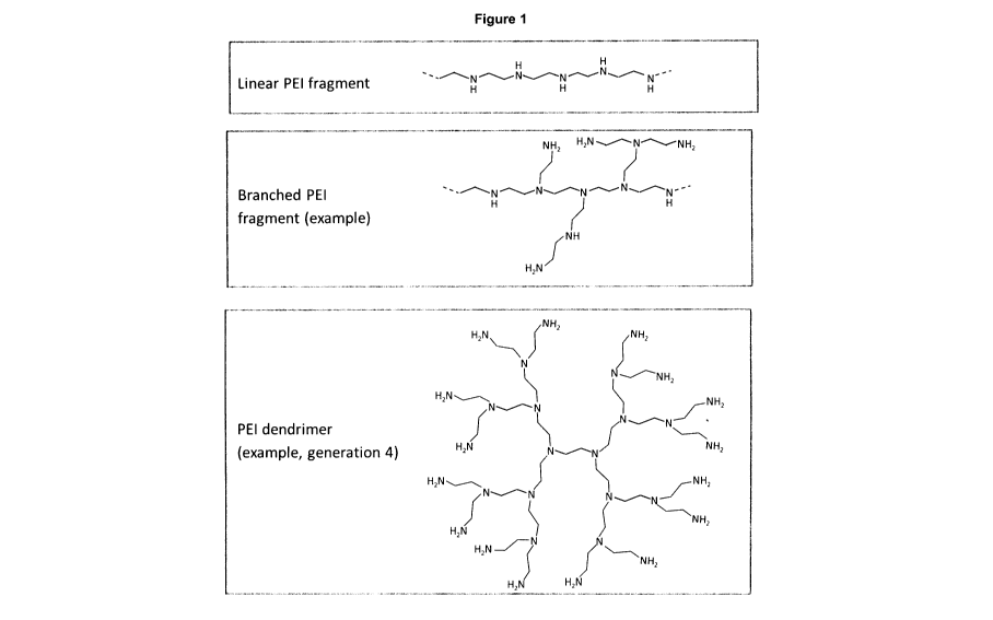
[0028] Figure 1
shows a depiction of the general structure of either, an
exemplary linear PEI fragment, an exemplary branched PEI fragment, or an
exemplary generation 4 PEI dendrimer.
[0029] Figure 2
shows an immunoblot demonstrating the detection of
extracellular vesicle protein markers (0D63, 0D9, HSC70, and flotillin-1)
isolated using an exemplary embodiment of a method of the application, from
media collected from BXPC3 pancreatic cancer cells growing in a bioreactor.
[0030] Figure 3
shows an immunoblot demonstrating the detection of
extracellular vesicle protein markers (0D63, 0D9, HSC70, and Annexin-V)
isolated using an exemplary embodiment of a method of the application from
media collected from T98G glioblastoma multiforme cancer cells growing in a
bioreactor.
[0031] Figure 4
shows an immunoblot demonstrating that calnexin, an
endoplasmic reticulum protein that is not found in EVs, is not detected in EVs
isolated using an exemplary embodiment of a method of the application, from
media collected from T98G glioblastoma multiforme cancer cells growing in a
bioreactor.
[0032] Figure 5
shows a graph demonstrating that the concentration of
particles, as measured by nanoparticle tracking analysis (NTA), is increased
by
using increasing concentrations of PEI and media collected using an exemplary
embodiment of a method of the application from T98G glioblastoma multiforme
cancer cells growing in a bioreactor.
9
CA 03097782 2020-10-20
WO 2019/218077
PCT/CA2019/050669
[0033] Figure 6 shows a graph demonstrating that the concentration of
particles isolated using an exemplary embodiment of a method of the
application, from T98G glioblastoma multiforme cancer cell bioreactor media
does not change after proteinase K digestion, suggesting that the particles
are
EVs and not protein aggregates.
[0034] Figure 7 shows particle size distribution using nanoparticle
tracking analysis of EVs isolated from BXPC3 pancreatic cancer cell bioreactor
media using an exemplary embodiment of a method of the application, after
dissociation from PEI using different dissociation buffers and time for
dissociation to occur
[0035] Figure 8 shows a graph demonstrating analysis of lipid-bound
vesicles isolated from PANC10.05 pancreatic cancer cell bioreactor media
using an exemplary embodiment of a method of the application after labeling
using a cell-penetrating peptide conjugated to Qdot-655 (Qtracker0-655) and
quantification using fluorescent nanoparticle tracking analysis.
[0036] Figure 9 shows a graph demonstrating the relative fluorescent
units using fluorescent spectroscopy of neutrally-charged 100nm DOPC/CHOL
liposomes labeled with Fluorescein DHPE and isolated using an exemplary
embodiment of a method of the application.
[0037] Figure 10 shows an immuno-blot demonstrating the detection of
extracellular vesicle protein markers (0D63, CD9, HSC70, and Flotillin-1)
isolated using an exemplary embodiment of a method of the application, from
conditioned cell media collected from either NCI-H1975 non-small cell lung
cancer cells or MCF-7 breast cancer cells.
[0038] Figure 11 shows an immuno-blot demonstrating the detection of
extracellular vesicle protein markers (0D63, CD9, HSC70, and Flotillin-1)
isolated using an exemplary embodiment of a method of the application, from
human plasma.
[0039] Figure 12 shows the expression of three different miRNAs,
including mir-142-3p, an miRNA known to be enriched in EVs, which were
CA 03097782 2020-10-20
WO 2019/218077
PCT/CA2019/050669
isolated using an exemplary embodiment of a method of the application, from
human plasma.
[0040] Figure
13 shows A) an immuno-blot demonstrating the detection
of extracellular vesicle protein markers (0D63, 0D9, HSC70, and Flotillin-1)
and B) a nanoparticle tracking analysis of EV's isolated using an exemplary
embodiment of a method of the application from media collected from
PANC10.05 pancreatic cancer cells growing in a bioreactor.
[0041] Figure
14 shows an immuno-blot demonstrating the detection of
extracellular vesicle protein markers (0D63, 0D9, HSC70, and Flotillin-1) as
well as absence of a non-EV marker (Calnexin) in EVs isolated using an
exemplary embodiment of a method of the application, from media collected
from PANC10.05 pancreatic cancer cells growing in a bioreactor using.
[0042] Figure
15 shows an immuno-blot demonstrating the detection of
extracellular vesicle protein markers (0D63, CD9, HSC70, and Flotillin-1) as
well as absence of a non-EV marker (Calnexin) in EVs isolated using an
exemplary embodiment of a method of the application, from media collected
from PANC10.05 pancreatic cancer cells growing in a bioreactor.
[0043] Figure
16 shows an immuno-blot demonstrating the detection of
extracellular vesicle protein markers (0D63, CD9, HSC70, and Flotillin-1) as
well as absence of a non-EV marker (Calnexin) from EVs isolated using an
exemplary embodiment of a method of the application from healthy human
plasma.
[0044] Figure
17 shows an immuno-blot demonstrating the detection of
extracellular vesicle protein markers (0D63, CD9, HSC70, and Flotillin-1) as
well as absence of a non-EV marker (Calnexin) from EVs isolated using an
exemplary embodiment of a method of the application, from healthy human
plasma.
[0045] Figure
18 shows a graph demonstrating flow cytometry-based
quantification of 0D63-positive or CD81-positive EVs isolated using an
exemplary embodiment of a method of the application, from healthy human
plasma.
11
CA 03097782 2020-10-20
WO 2019/218077
PCT/CA2019/050669
[0046] Figure
19 shows an immuno-blot demonstrating the detection of
extracellular vesicle protein markers (0D63, 0D9, HSC70) from EVs isolated
using an exemplary embodiment of a method of the application, from media
collected from PANC10.05 pancreatic cancer cells growing in a bioreactor in
comparison to other standard EV isolation protocols.
[0047] Figure
20 shows an immuno-blot demonstrating the detection of
extracellular vesicle protein markers (0D63, 0D9, HSC70, Flotillin) from EVs
isolated from healthy human plasma using an exemplary embodiment of a
method of the application, in comparison to other standard EV isolation
protocols
[0048] Figure
21 shows a graph demonstrating enrichment of EV
proteins from plasma by mass spectrometry using an exemplary embodiment
of a method of the application.
DETAILED DESCRIPTION
I. Definitions
[0049] Unless
otherwise indicated, the definitions and embodiments
described in this and other sections are intended to be applicable to all
embodiments and aspects of the present application herein described for which
they are suitable as would be understood by a person skilled in the art.
[0050] The
present application refers to a number of chemical terms and
abbreviations used by those skilled in the art. Nevertheless, definitions of
selected terms are provided for clarity and consistency.
[0051] As used
herein, the words "comprising" (and any form of
comprising, such as "comprise" and "comprises"), "having" (and any form of
having, such as "have" and "has"), "including" (and any form of including,
such
as "include" and "includes") or "containing" (and any form of containing, such
as "contain" and "contains"), are inclusive or open-ended and do not exclude
additional, unrecited elements or process/method steps.
[0052] As used
herein, the word "consisting" and its derivatives, are
intended to be close ended terms that specify the presence of stated features,
12
CA 03097782 2020-10-20
WO 2019/218077
PCT/CA2019/050669
elements, components, groups, integers, and/or steps, and also exclude the
presence of other unstated features, elements, components, groups, integers
and/or steps.
[0053] The term
"consisting essentially of", as used herein, is intended
to specify the presence of the stated features, elements, components, groups,
integers, and/or steps as well as those that do not materially affect the
basic
and novel characteristic(s) of these features, elements, components, groups,
integers, and/or steps.
[0054] Terms of
degree such as "substantially", "about" and
"approximately" as used herein mean a reasonable amount of deviation of the
modified term such that the end result is not significantly changed. These
terms
of degree should be construed as including a deviation of at least 5% of the
modified term if this deviation would not negate the meaning of the word it
modifies.
[0055] As used
in this application, the singular forms "a", "an" and "the"
include plural references unless the content clearly dictates otherwise. For
example, an embodiment including "a buffer" should be understood to present
certain aspects with one buffer or two or more buffers. In embodiments
comprising an "additional" or "second" component, such as an additional or
second buffer, the second component as used herein is chemically different
from the other components or first component. A "third" component is different
from the other, first, and second components, and further enumerated or
"additional" components are similarly different.
[0056] The term
"and/or" as used herein means that the listed items are
present, or used, individually or in combination. In effect, this term means
that
"at least one of" or "one or more" of the listed items is used or present.
[0057] The term
"pharmaceutical composition" as used herein refers to
a composition for pharmaceutical use.
[0058] The term
"for pharmaceutical use" means compatible with the
treatment of a subject.
13
CA 03097782 2020-10-20
WO 2019/218077
PCT/CA2019/050669
[0059] The term "subject" as used herein refers to the source
organism
from where a sample is obtained and/or the target organism for treatment and
includes all unicellular and multicellular organisms.
[0060] The term a "therapeutically effective amount" refers to a
quantity
of a substance sufficient to, when administered to a subject, effect
beneficial or
desired results, including clinical results, and, as such, a "therapeutically
effective amount" or synonym thereto depends upon the context in which it is
being applied. A "therapeutically effective amount" is intended to mean that
amount of a substance that is sufficient to treat, prevent or inhibit one or
more
pathological conditions. The amount of a given substance that will correspond
to such an amount will vary depending upon various factors, such as the given
substance, the pharmaceutical composition, the route of administration, the
type of pathological condition, the identity of the subject being treated, and
the
like, but can nevertheless be routinely determined by one skilled in the art.
[0061] The term "pathological condition" as used herein refers to any
disease, disorder or condition that is to be treated or receive a treatment.
[0062] The term "treated", "treating" or "treatment", as used herein,
and
as is well understood in the art, means an approach for achieving results,
including clinical results, which are either desired and/or beneficial.
Treatment
that results in clinical benefits can include, but is not limited to,
alleviation or
amelioration of one or more symptoms or conditions associated with the
pathological condition, reducing the extent or spread of the pathological
condition, stabilization of the pathological condition, delay or deceleration
of
progression of the pathological condition, amelioration or palliation of the
pathological condition, reduced incidence of the pathological condition,
reduce
reoccurrence, and either partial or total remission, whether detectable or
undetectable. "Treated", "treating" and "treatment" can also mean prolonging
survival as compared to expected survival if not receiving treatment, or
prophylactic treatment.
[0063] The term "polyethylenimine" or "PEI" refers to a group of
hydrophilic cationic polymers that encompass polymers in a linear form with a
14
CA 03097782 2020-10-20
WO 2019/218077
PCT/CA2019/050669
molecular weight (MW) ranging from approximately 2500 to 250,000 Da,
branched forms with a MW ranging from approximately 600 to 750,000 Da, and
dendrimer forms comprising generations 1 to 6. As used herein,
"polyethylenimine" refers in its ordinary sense to polymers with repeating
units
composed of an amine group and a two-carbon aliphatic (0H20H2) spacer. The
term polyethylenimine includes those having typically 10 or more repeated
units. Linear polyethylenimine refers to a polymer with all secondary amines
(refer to Figure 1), branched polyethylenimine refers to a polymer containing
primary, secondary, and tertiary amino groups (refer to Figure 1). Dendrimer
forms of polyethylenimine refer to repetitively radially branched polymers
containing primary and tertiary amine groups and can be intact or fractured.
[0064] The term
"amine" as used herein refers to a functional group
comprising a nitrogen atom bonded to zero, one or two hydrogen atoms with
the remaining atoms bonded to the nitrogen being 5p3-hybridized carbon atoms.
[0065] The term
"polymer" as used herein refers to a large molecule or
macromolecule composed of many repeated monomeric units wherein the
number of repeated units may be variable and dependent on the particular
polymer in question.
[0066] The term
"extracellular vesicle" or "EV" as used herein refers to a
membrane-bound vesicle wherein at least a portion of the plasma membrane
of the extracellular vesicle is derived directly from a cell, whether it be
from a
uni- or multi-cellular organism. Depending on the manner of generation of the
extracellular vesicle (e.g. membrane inversion, exocytosis, fusion of a
multivesicular body with the plasma membrane, budding from plasma
membrane, etc.), the EVs contemplated herein may exhibit different protein
markers, nucleic acid cargo, metabolites, and surface/lipid characteristics.
EVs
are also referred to as "exosomes", "exosome-like", "microsomes",
"microvesicles", "secretory exosomes", "oncosomes", "membrane vesicles",
"apoptotic bodies", "argosomes", and "microparticles", which are included
within
the definition.
CA 03097782 2020-10-20
WO 2019/218077
PCT/CA2019/050669
[0067] The term
"liposome" as used herein refers to a synthetic lipid-
bound vesicle that can be created from various lipids, where the nature of the
lipids constituting the liposome affects the characteristics of the resulting
liposome, including shape, rigidity, size, and charge. Liposomes may be
created from lipids including, for example, cholesterol, various
phospholipids,
phosphatidylethanolamine, distearoylphosphatidylethanolamine, etc.
Liposomes can be used as a drug carrier by encapsulating biological molecules
or drugs.
[0068] The term
"isolation", "isolating", or "capture" as used herein
means to separate, enrich, precipitate, immobilize, and/or purify EVs from a
particular biological sample.
[0069] By
"isolation of EVs" as used herein, it is meant that the EV's and
EV-associated biological material are isolated. By "associated" it is meant
that
the biological material is either internally or externally located in or on
the EV's
and is isolated along with the EVs.
[0070] The term
"sample" or "biological sample" or "biological fluid" as
used herein refers to a material or mixture of materials containing EVs and
includes biological samples and clinical samples that contain EVs for
isolation.
[0071] The term
"contacting" or "contact" as used herein refers to the
manner in which the one or more polyethylenimines are mixed, blended or
incubated with a sample such that the polyethylenimine and EVs in the sample
form a EV-polyethylenimine complex.
[0072] The term
"extracellular vesicle-polyethylenimine complex" or "EV-
polyethylenimine complex" as used herein refers to the complex of material
that
is formed and then precipitated, pelleted, or captured from a biological
sample
following contact of the sample with the one or more polyethylenimines, or one
or more polyethylenimines immobilized on a solid support. The complex is
expected to contain both EVs (and EV-associated material) and one or more
polyethylenimines.
[0073] The term
"conditions for the isolation of the EVs" as used herein
refers to the conditions that are appropriate for the isolation,
precipitation,
16
CA 03097782 2020-10-20
WO 2019/218077
PCT/CA2019/050669
pelleting or capture of EVs from the sample as described in greater detail
hereinbelow, and may include, but are not limited to, conditions for sample
preparation (e.g. pre-clearing by centrifugation, filtration, dilution, etc.),
concentration of polyethylenimine, duration of contact, temperature, identity
of
buffers for dilution or washing, pH, centrifugation speeds, identity of buffer
or
lysis buffer used to re-suspend EV-polyethylenimine complex or pellet, and
identity of solid support, if used, for isolation and/or separation.
[0074] The term
"suitable" as used herein means that the selection of the
particular conditions would depend on the specific manipulation to be
performed, and the identity of the substances involved, but the selection
would
be well within the skill of a person trained in the art. Unless otherwise
indicated,
all methods and assays described herein are to be conducted under conditions
sufficient to achieve the desired result. A person skilled in the art would
understand that all such conditions, including, for example, pre-treatment or
pre-clearing treatments, pH, concentrations, solvent, time, temperature,
pressure, and/or molar, volume or weight ratios, can be varied to optimize the
desired result and is within their skill to do so.
[0075] The term
"method of the application" as used herein refers to a
method of isolating EVs using one or more polyethylenimines, including all of
the various embodiments thereof, described herein.
[0076] The term
"microfluidics" as used herein refers to an apparatus for
precise manipulation of liquids in very small volumes, typically in the 100
nanoliters to 100 microliters range.
[0077] The term
"marker" or "biomarker" as used herein refers to
biologically-derived molecules or biological and/or cellular events that are
correlated to a specific pathological condition and are intended to help in
understanding, for example, patient stratification, degree of risk for disease
occurrence or progression, type of pathological condition, monitoring
treatment
outcome (e.g. stabilization of disease, remission, relapse, or progression),
and
sensitivity or resistance to a specific drug or therapeutic regimen. A person
skilled in the art would understand that a biomarker is a characteristic that
is
17
CA 03097782 2020-10-20
WO 2019/218077
PCT/CA2019/050669
objectively measured and evaluated as an indicator of normal biological
processes, pathogenic processes, or pharmacological responses to a
therapeutic intervention (Biomarkers Definitions Working Group. Biomarkers
and surrogate endpoints: preferred definitions and conceptual framework. Olin
Pharmacol Ther. 2001; 69: 89-95).
[0078] The term
"conditioned media" as used herein refers to
experimental samples derived from media taken from living cultured cells that
are regularly passaged (e.g. detached from growing surface and transferred to
a new growing vessel before cells get crowded and start to grow on top of each
other) and grown in a monolayer using techniques well known to those skilled
in the art.
[0079] The term
"bioreactor media" as used herein refers to experimental
samples derived from media taken from living cultured cells grown in
bioreactors that utilize membrane technology to separate the cell cultivation
area from the media chamber using techniques well known to those skilled in
the art. This set-up allows cells to be grown for a long period without
passaging
and to produce cell media that is enriched in EVs.
[0080] The term
"fractured" refers to the random disruption of branched
forms of molecules, as used herein in referring to fractured dendrimers, where
heat is typically the method used to fracture the molecule.
[0081] The term
"surface plasmon resonance" or "SPR" as used herein
describes a method to study the interaction between two macromolecules.
Typically, one of the macromolecules to be studied is immobilized on the
surface of a sensor chip while the other is passed through a microfluidics
system to the chip surface. Changes in mass at the sensor surface can be used
to monitor interactions between the two macromolecules.
[0082] The term
"macromolecule" as used herein describes a very large
molecule, typically a molecule containing at least 1000 atoms and more likely
thousands to millions of atoms. Macromolecules are typically the result of
polymerization of molecular subunits. Examples of macromolecules include, for
example, proteins, antibodies, liposomes, nucleic acids, and polymers.
18
CA 03097782 2020-10-20
WO 2019/218077
PCT/CA2019/050669
Exemplary molecular weights include, but are not limited to about 10 kDa to
about 4000 kDa.
[0083] The term
"magnetic bead" as used herein describes any particle
with magnetic properties, whereby the composition, shape and size of the
magnetic core can affect its magnetic properties. The magnetic core may be
composed of various magnetic materials, including iron and cobalt, and may be
coated with various materials and functional groups, and may range in size
from
<50 nm to > 1 pm. By applying a magnetic field, the magnetic beads, and any
associated biological materials, will be attracted towards the magnet,
separating them from the unwanted material in the biological sample.
II. Methods of the Application
Methods for Isolation EVs and Kits Therefore
[0084]
Disclosed herein are methods and compositions using
polyethylenimine (PEI) to isolate EVs from biological samples. The methods to
isolate EVs described in the present application do not depend on methods,
such
as filtration or high-speed centrifugation, which may damage isolated EVs by
centrifugal forces or shear stresses. The methods described herein isolate EVs
from biological samples with greater ease, and equal or superior efficiency in
comparison to other EV precipitation methods, such as ExoQuickTM and
ultracentrifugation.
Furthermore, the methods described in the present
application are scalable and high yields of EVs are possible. The EVs isolated
using the methods described herein are also of higher purity than those
achieved
by other EV precipitation methods, such as ExoQuickTM. Therefore, PEI can be
used to isolate EVs that are suitable for use in therapeutic applications.
Depending on its intended use, EVs used in therapeutics may be isolated from
unmodified cells containing native EVs or genetically-modified cells which
release EVs containing trans-gene products16.
[0085] PEI is
soluble, due to intra-chain repulsion, in 100-150 mM NaCI
and at neutral pH56, which is the typical NaCI concentration and pH range in
cell
media and plasma. It has unexpectedly been found that EVs and liposomes, in
particular 100 nm neutrally-charged liposomes, can be precipitated using
19
CA 03097782 2020-10-20
WO 2019/218077
PCT/CA2019/050669
polyethylenimine using low-speed (benchtop) centrifugation from biofluids.
Although the ability of PEI polyplexes to transfect cells has been
demonstrated
to require heparin sulfate, proteoglycans and glycosaminoglycans42,43, and
these proteins have been found on the cell surface of exosomes, the
unexpected ability of PEI to precipitate neutrally-charged synthetic
liposomes,
which completely lack proteins, negates the binding of PEI to these cell
surface
proteins as the mechanism required for extracellular vesicle precipitation by
PEI
as described by the methods of the present application.
[0086]
Furthermore, in spite of its known ability to bind nucleic acids, PEI
was found to be unable to recover cell-free DNA (cfDNA) from plasma samples
(Table 3), although PEI was able to recover RNA and, in particular, EV-
associated miRNA. This suggests that PEI has an unexpected preference for
associating with EVs compared to nucleic acids in complex biological samples.
The ability of PEI to recover uncharged liposomes and EVs from complex
biological samples, but not DNA, and to recover only a subset of EV-associated
RNA, suggests that PEI has a hitherto unrecognized ability to precipitate
and/or
capture liposomes (including neutrally charged liposomes) and EVs that is not
dependent on simple charge-based interactions.
[0087] The
ability of PEI to recover EV-associated RNA species as
opposed to cell-free RNA (cfRNA) may be of great benefit. Most miRNAs present
in serum are found in aggregation with protein complexes containing Argonaut2
rather than inside EVs24. However, certain specific miRNAs, such as mir-142-
3p, have been found to be enriched in the EV-fraction of plasma24.
Furthermore,
it is the EV-associated miRNA species that retain important inter-cellular
communication functions and which are involved in varied biological processes,
from inflammation and immune system regulation to tumour development and
drug re5i5tance57-61. In light of this, EV-associated RNA species, and miRNAs
in particular, are considered to be better biomarkers than other circulating
miRNAs62-64 and the ability of PEI to specifically recover this population of
miRNAs make it useful for the identification of novel biomarkers for the
development of diagnostic tests and assays.
CA 03097782 2020-10-20
WO 2019/218077
PCT/CA2019/050669
[0088]
Accordingly, in one embodiment the present application includes
a method for the isolation of EVs from a sample containing EVs, comprising
contacting the sample with one or more polyethylenimines under conditions for
the isolation of EVs.
[0089] In some
embodiments, the conditions for the isolation of EVs
comprise:
(i) contacting the sample with the one or more polyethylenimines to
form an EV-polyethylenimine complex; and
(ii) separating the EV-polyethylenimine complex from the sample.
[0090] The use
of polyethylenimine allows for quick isolation of EVs
using, for example, low-speed benchtop centrifugation with quicker processing
times and improved purity over current precipitation methods. The ease of
processing and the ability to coat PEI onto magnetic beads or other solid
matrices lends this method to clinical adaptation for detection of EV-related
biomarkers18,61-63.
[0091] In some
embodiments, EVs are isolated from freshly collected
biological samples, or samples that have been stored, either at room
temperature, refrigerated, or frozen. In some embodiments, the samples have
been preserved or fixed to prolong storage. In some embodiments, the EVs are
membrane-bound vesicles having a diameter (or largest dimension) of between
about 10 nm to about 3000 nm, or between about 40 nm and about 1000 nm,
wherein at least a portion of the plasma membrane of the extracellular vesicle
is derived directly from a cell from a subject.
[0092] In some
embodiments, the sample is obtained from human
subjects, or from human cell lines. In some embodiments, the sample is
obtained from an organism such as, but not limited, to an animal, fish or
bird.
In some embodiments, the animal is a companion animal or livestock. In some
embodiments, the animal is a wild animal. In some embodiments, the sample
is from a subject that is suspected of having, or has been diagnosed with, one
or more pathological conditions.
21
CA 03097782 2020-10-20
WO 2019/218077
PCT/CA2019/050669
[0093] In some
embodiments the sample is a bodily fluid (such as blood,
blood serum, plasma, urine, milk, semen, sweat, cerebral spinal fluid, saliva,
ascites, tears, amniotic fluid, joint fluid and malignant effusions) or tissue
biopsy
or the fluid, media, exudates or discharges from any organism.
[0094] In some
embodiment, the sample is first treated to prepare it for
isolation of the EVs, for example, to remove debris or interfering substances
(e.g. albumin), using methods known to those skilled in the art. In some
embodiments, such treatments are called "pre-clearance treatments". In some
embodiments, the pre-clearance treatments comprise one or more of: filtration,
ultrafiltration, centrifugation, sterilization, treatment with an enzyme and
treatment with a biocide. In some embodiments, the enzyme is selected from
one or more of a protease inhibitor, proteases, DNASE and RNASE.
[0095] In some
embodiments, the sample is pre-cleared to comprise
large (>1 micron) particles by centrifuging at about 17,000g, for about 15
minutes. In some embodiments, the sample is pre-cleared by passing it through
a filter, for example, a 0.45 pm or 0.8 pm filter, to remove unwanted material
from the sample. In some embodiments, for example, after a plasma sample
has been obtained from a patient, the plasma sample is treated, for example
with thrombin, which converts fibrinogen to fibrin, allowing it to be pre-
cleared
by a simple centrifugation step.
[0096] Suitable
polyethylenimines for precipitation of EVs from samples
include linear forms with a molecular weight (MW) ranging from about 2500 to
about 250,000 Da, branched forms with a MW ranging from about 600 Da to
about 750,000 Da, and dendrimer forms comprising generations 1 to 6, whether
intact or fractured. In some embodiments, the polyethylenimine has an average
molecular weight of at least 600 Da, at least 1200 Da, at least 1800 Da, at
least
2000 Da, at least 10,000 Da, at least 25,000 Da, at least 40,000 Da, at least
60,000 Da, at least 70,000 Da, at least 160,000 Da, at least 250,000 Da, or at
least 750,000 Da or above. In some embodiments, the polyethylenimine is a
branched polyethylenimine having a MW of 10,000 Da, 25,000 Da, 70,000 Da
or 750,000 Da. In some embodiments, the polyethylenimine is a branched
polyethylenimine having a MW of 10,000 Da or 25,000 Da. In some
22
CA 03097782 2020-10-20
WO 2019/218077
PCT/CA2019/050669
embodiments the polyethylenimine is branched polyethylenimine having a MW
of 10,000 Da. In some embodiments the polyethylenimine is branched
polyethylenimine having a MW of 25,000 Da. In some embodiments, the
polyethylenimine is a linear form having a MW of 25,000 Da. In some
embodiments, the polyethylenimine is a linear form having a MW of 160,000
Da.
[0097] Using a
non-toxic form of polyethylenimine means that, using
methods of the application, EVs can be isolated while being compatible for use
in therapeutics. In some embodiments the non-toxic form of polyethylenimine is
linear 22,000 Da polyethylenimine. In some embodiments, the non-toxic form of
polyethylenimine is linear 25, 000 Da polyethylenimine.
[0098] The polyethylenimine can be from any source. In some
embodiments the polyethylenimine is synthetic, i.e. is prepared using a
synthetic method rather than isolated from a natural source. In some
embodiments, the polyethylenimine is subjected to one or more processing
steps prior to use in the method of the application. In some embodiments these
processing steps are one or more of purification, chemical-functionalization,
attachment to solid supports, and the like. Such methods are known to those
skilled in the art. Any suitable method known in the art for synthesizing,
preparing, and/or purifying suitable polyethylenimines can be employed.
[0099] In some
embodiments, the polyethylenimine is bonded to a solid
support which aids in the separation of the EVs from the sample, but is not
used
for the isolation step. In some embodiments, the solid support is polystyrene
or glass. In some embodiments, the polyethylenimine is bonded to a solid
support which aids in the separation of the EVs from the sample, and in the
isolation of EVs. In some embodiments, the solid support comprises magnetic
beads coated with one or more polyethylenimines which are easily isolated
using a magnetic field. In some embodiments, the magnetic beads comprise 10
nm to 10 pm in diameter particles with a magnetite core and a shell of
polyethylenimine. In some embodiments, the magnetic beads comprise 150nm
particles with a magnetite core and a shell of polyethylenimine. In some
embodiments, the magnetic beads comprise any magnetic core, from 10 nm to
23
CA 03097782 2020-10-20
WO 2019/218077
PCT/CA2019/050669
pm in diameter, and be coated with one or more linear, or branched
polyethylenimines, or combinations thereof.
[00100] In some
embodiments, the one or more polyethylenimines are
conjugated to a ligand which aids in the separation of the EVs from the
sample.
In some embodiments, the ligand comprises a biotin moiety. When the
polyethylenimine is conjugated a biotin moiety, the EVs in a sample may then
be isolated by contacting the biotin-conjugated polyethylenimine with the
sample, followed by contacting with a biotin-binding protein or a solid
support
comprising a biotin-binding protein. In some embodiments, the biotin-binding
protein is selected from avidin, streptavidin, or other biotin-binding
proteins. In
some embodiments, the solid support is polystyrene, glass or paramagnetic
particles. Methods of conjugating biotin-binding proteins to solid supports
are
well known to those skilled in the art.
[00101] In some
embodiments, one or more polyethylenimines are fixed
to a solid support comprising silicones (for example polydimethylsiloxane) or
other surfaces of a microfluidic apparatus to aid in the isolation of EVs from
the
sample. In some embodiments, the microfluidics apparatus further comprises,
for example, downstream molecular analysis components such as components
that comprise reagents for the polymerase chain reactions (FOR) to identify
nucleic acids (e.g. gene-specific mutations or particular miRNAs) and/or
antibodies to identify proteins. In some embodiments, the microfluidic
apparatus is a surface plasmon resonance (SPR) apparatus.
[00102] In some
embodiments, one or more polyethylenimes are fixed the
surface of, for example, a sensor, in some instances a surface plasmon
resonance biosensor, to aid in the study of biomolecular interactions between
EVs and other molecules, including, but not limited to, proteins, antibodies,
lipids, drugs, polyethylene glycol, and biofluids.
[00103] In some
embodiments, the one or more polyethylenimines are in
a solution. In some embodiments the solution is an aqueous solution. In some
embodiments, the solution is a buffer. In some embodiments, the buffer is
selected from borate, phosphate, acetate, citrate, and TRIS buffers. The pH of
24
CA 03097782 2020-10-20
WO 2019/218077
PCT/CA2019/050669
the buffer may be any pH that is compatible with the polyethylenimine(s)
intended to be put into solution. The concentration of the polyethylenimine(s)
solution may be any concentration compatible with the intended one or more
polyethylenimines to be put into solution. In some embodiments, the one or
more polyethylenimines solution has a concentration ranging from about 1
pg/mL to about 20 mg/mL or from about 1 mg/mL to about 10 mg/mL. In some
embodiments, the one or more polyethylenimines solution is filter sterilized
through a 0.2 pm filter to sterilize the solution and remove any unsolubilized
polyethylenimine.
[00104] A
variety of buffers may be used for incubation with the biological
sample prior to precipitation of EVs, including, but not limited to, borate,
phosphate, acetate, citrate, and TRIS buffers. The pH of the buffer may be any
pH that is compatible with the biological sample. In some embodiments, the
buffer for the sample has a pH ranging from about pH 4 to about pH 11 or about
pH 6 to about pH 8.
[00105] The salt
concentration of the sample may be any concentration
compatible with the sample. In some embodiments, the salt concentration is
about 10 mM to about 1000 mM or about 100 mM to about 200 mM.
[00106] The
amount of the one or more polyethylenimines to be used in
the methods of the application may be any amount that is compatible with the
sample and that will isolate the EVs from the sample. In some embodiments,
the one or more polyethylenimines are used in an amount that is about 1 pg/mL
to about 1000 pg/mL or about 20 pg/mL to about 400 pg/mL in the total mixture
of sample and one or more polyethylenimines.
[00107] The
sample size used to isolate EVs using methods described
herein depends upon the concentration of useful EVs within the sample, the
method of the present application used for their isolation, and the intended
downstream application of isolated material. In some embodiments, the
methods of the application are used to isolate EVs from small sample sizes
using a microfluidic device where the sample size is about 2 microliters to
about
CA 03097782 2020-10-20
WO 2019/218077
PCT/CA2019/050669
100 microliters. In some embodiments, the sample size is from 100 microliters
to more than 10 milliliters or more.
[00108] The
contacting time used to create an association between EVs
and the one or more polyethylenimines can be any time compatible with the
biological sample. In some embodiments, the contacting time is about 1 minute
to about 24 hours. In some embodiments, the contacting time is about 10
minutes to about 10 hours. In some embodiments, the contacting time is about
30 minutes to about 2 hours. In some embodiments, the contacting time is
about 60 minutes. In some embodiments, the contacting is performed with
agitation, rotation and/or stirring. In some embodiments, the contacting is
performed with end-over-end rotation.
[00109] In some
embodiments, the contacting is performed at any
temperature compatible with the biological sample. In some embodiments, the
contacting is performed at a temperature of about 0 C to about 40 C or at
about
ambient or room temperature.
[00110] In some
embodiments, the EV-polyethylenimine complex is
separated using any method that is compatible with the EVs and the complex.
In some embodiments, the EV-polyethylenimine complex is separated by
pelleting, for example using centrifugation.
[00111] In some
embodiments, the centrifugal force used to pellet the EV-
polyethylenimine pellet is any one compatible with the biological sample. In
some embodiments, the centrifugal force is about 20,000g or less. In some
embodiments, the centrifugal force is about 17,000g to initially precipitate
the
EV-polyethylenimine pellet and about 13,000g to precipitate the EV-
polyethylenimine pellet between washes.
[00112] In some
embodiments, the centrifugation time used to pellet the
EV-polyethylenimine pellet is any time compatible with the biological sample
and the complex. In some embodiments, the centrifugation time is about 15
minutes to initially precipitate the EV-polyethylenimine pellet and about 2
minutes to precipitate the EV-polyethylenimine pellet between washes. In some
embodiments, the EV-polyethylenimine pellet is washed by removing the
26
CA 03097782 2020-10-20
WO 2019/218077
PCT/CA2019/050669
sample from the pellet following centrifugation and replacing the buffer with
a
washing liquid. In some embodiments this washing liquid is water or a buffer,
including, but not limited to, borate, phosphate, acetate, citrate, and TRIS
buffers. In some embodiments, this washing buffer is Dulbecco's phosphate
buffered saline. The pH of the buffer may be any pH that is compatible with
the
EV-polyethylenimine complex and downstream applications. In some
embodiments, the pH of the buffer is about pH 4 to about pH 10, or about pH 6
to about pH 8. In some embodiments, the washing buffer includes a detergent
such as, but not limited to, digitonin, Triton X100TM or Tween-20Tm, for
example, in order to reduce non-specific protein interactions. In some
embodiments, the EV-polyethylenimine pellet is pelleted in between washes
using a centrifugal force of about 13,000g. However, in other embodiments, the
centrifugal force may be any that is compatible with the isolation of EVs from
the biological sample. In some embodiments, centrifugation for about 2 minutes
is used to precipitate the EV-polyethylenimine pellet between washes.
However, in other embodiments, the centrifugation time may be any that is
compatible with the isolation of EVs from the biological sample.
[00113] In some
embodiments, when the one or more polyethylenimines
are conjugated to a solid support, the EV-polyethylene complex is separated
using methods to capture the solid support. For example, when the one or
more polyethylenimines are conjugated to a solid support comprising magnetic
properties, the EV-polyethylenimine complex may be isolated using a magnetic
field. As a further example, when the one or more polyethylenimines are
conjugated to a solid support comprising a ligand, the EV-polyethylenimine
complex may be isolated using affinity capture methods.
[00114] In some
embodiments, the isolated EV-polyethylenimine complex
is treated to release the EVs from the polyethylenimine(s). In some
embodiments the EVs are released from the polyethylenimine(s) using
phosphate buffered saline (PBS), NaCI solution (e.g. 2N NaCI) or a polyanion
(e.g. heparin). As above, when the one or more polyethylenimines are
conjugated to a solid support, the EVs and one or more polyethylenimines are
separated using methods to capture the solid support. For example, when the
27
CA 03097782 2020-10-20
WO 2019/218077
PCT/CA2019/050669
one or more polyethylenimines are conjugated to a solid support comprising
magnetic properties, the one or more polyethylenimines may be separated from
the EVs using a magnetic field. As a further example, when the one or more
polyethylenimines are conjugated to a solid support comprising a ligand, the
one or more polyethylenimines may be separated from the EVs using affinity
capture methods.
[00115] In some
embodiments, the EVs are not released from the one or
more polyethylenimines prior to analysis.
[00116] In some
embodiments, the EVs are released from the one or more
polyethylenimines when nanoparticle tracking analysis is to be performed or if
the EVs are to be used for transfection experiments or for therapeutic
delivery.
[00117] In some
embodiments, to analyze multiple different types of
biomarkers in EVs from a single sample from a subject, the isolated EVs are
processed so that multiple types of biomarkers can be analyzed using the same
sample. In some embodiments, for example, the isolated EVs are processed
with a reagent that allows the isolation of RNA and protein separately from a
sample. A non-limiting example of such a reagent is a composition comprising
and effective amount of each of phenol, guanidine isothiocyanate and
ammonium thiocyanate, commercially available as TRIzolTm. In this
embodiment, the reagent is added to an aqueous solution of the isolated EVs
and the aqueous and organic phases collected separately. The aqueous phase
can then be processed for RNA extraction using, for example, a total RNA or
miRNA isolation kit, while the organic phase can be used for protein and lipid
extraction from the EVs using methods known to those knowledgeable in the
art.
[00118] In some
embodiments, the methods of the application further
comprise downstream analyses of the EVs isolated from the sample. In some
embodiments, the downstream analyses include, for example, Western
blotting, flow cytometry, ELISA, mass spectrometry, FOR, RT-qPCR, and/or
RNA-Seq (RNA sequencing).
28
CA 03097782 2020-10-20
WO 2019/218077
PCT/CA2019/050669
[00119] The
present application also includes a kit for the isolation of EVs
from biological samples. In some embodiments, the kit is specific for the
isolation of EVs from a particular sample type. In some embodiments, the kit
is for isolation of EVs from plasma, urine or cell culture media. In some
embodiments, the kit comprises, one or more polyethylenimines and optionally,
one or more buffers (to be used for diluting the sample or the one or more
polyethylenimines, or to be used for washing steps) and a detailed protocol or
instructions for use. In some embodiments the one or more polyethylenimines
are in the form of a powder, a solution, or coated or conjugated onto a solid
support, such as magnetic beads. Depending on the nature of the kit, a
positive
control, for example, a solution of isolated EVs or an EV-rich conditioned
media
that can be used as a positive control in downstream analytical techniques,
may
be included in the kit. In one particular embodiment, a kit for isolation of
extracellular vesicles using PEI might include: (1) a solution of branched MW
25000 PEI; (2) 10X PBS buffer; (3) an EV-rich conditioned media from human
cultured cells to be used as a positive control; and (4) a detailed protocol.
In
another particular embodiment, a kit for the isolation of extracellular
vesicles
using magnetic beads coated with PEI might include: (1) a suspension of PEI-
coated magnetic beads; (2) 10X PBS buffer; and (3) a detailed protocol.
[00120] In some
embodiments, the kit further comprises reagents for
releasing the EVs from the polyethylenimine and/or reagents for performing
analyses on the EVs isolated from the sample. In some embodiments, the
reagents for performing analyses on the EVs are selected from one or more of
reagents for FOR, RNA sequencing, proteomics, and/or lipidomics.
Diagnostic Methods
[00121] Analysis
of EVs that are shed, for example, by cancer cells into
the blood stream of the subject, if captured using a simple and robust method,
would enable clinical analysis of cancer development and progression and
treatment response and monitoring using a non-invasive method, such as
regular blood sampling. Furthermore, analysis of EVs that are shed by cancer
cells using the methods described herein and comparison to control populations
may allow the identification of novel markers of cancer that could be used to
29
CA 03097782 2020-10-20
WO 2019/218077
PCT/CA2019/050669
develop a simple, non-invasive diagnostic test for cancer or a specific type
of
cancer. Analysis of cultured cancer cell types, including pancreatic cancer,
glioblastoma multiforme, and breast cancer, as well as human plasma, using
methods described herein, has demonstrated the capability of polyethylenimine
to facilitate the sedimentation, immobilization, or capture of vesicular
material
from extracellular medium from this material, as determined by the immuno-
detection of prototypical extracellular vesicle protein markers, including
tetraspanins (e.g. 0D63, 0D9), heat shock proteins (e.g. HSP70) and lipid raft
proteins (e.g. flotillin-1).
[00122] The
methods described herein for the use of polyethylenimine to
capture cancer-derived EVs opens the possibility of utilizing this technology
to
capture proteins, lipids, and RNA species, for example, that are protected
within
the EVs, from samples taken from subjects with any pathological condition, and
with the potential for downstream analysis using clinically-amenable methods
such as Western blotting, flow cytometry, ELISA, mass spectrometry, FOR, RT-
qPCR, and RNA-Seq (RNA sequencing), these methods can be used, for
example, for diagnosis and treatment monitoring.
[00123]
Accordingly, the application also includes methods for
determining the presence, absence, relapse, remission or progression of one
or more pathological conditions comprising:
(i) isolating EVs from a sample from a subject suspected of having the
one or more pathological conditions using a method of the present
application;
(ii) identifying one or more biomarkers for the one or more pathological
conditions in the EVs, wherein the presence, absence, or changes in
amount of the one or more biological markers in the EVs indicates the
presence, absence, relapse, remission, or progression of the one or
more pathological conditions in the subject.
[00124] In some
embodiments, any detectable amount of the one or more
biomarkers for the one or more pathological conditions is indicative of the
presence, relapse or progression of the one or more pathological conditions,
CA 03097782 2020-10-20
WO 2019/218077
PCT/CA2019/050669
and conversely, no detectable amount of the one or more biomarkers for the
one or more pathological conditions is indicative of the absence or remission
of
the one or more pathological conditions.
[00125] In some
embodiments the methods of diagnosis further
comprises comparing an amount of the one or more biomarkers for the one or
more pathological conditions in EVs from a subject with one or more references
values. In further embodiments, the comparing is done at multiple time points.
In some embodiments, differences in the amount of the one or more biomarkers
in the sample from the subject compared to the one or more reference values
is indicative of presence, absence, relapse, remission or progression of the
one
or more pathological conditions in the subject. In some embodiments, when
the comparing is done at multiple time points the presence, absence, relapse,
remission or progression of the one or more pathological conditions in the
subject is determined over time.
[00126] In some
embodiments, the one or more reference values are
amounts of the one or more biomarkers in healthy subjects or subjects known
not to have the one or more pathological conditions. In some embodiments,
any detectable increase in the amount of the one or more biomarkers compared
to the one or more reference values, is indicative of the presence,
progression
or relapse of the one or more pathological conditions. In some embodiments,
any detectable amount of the one or more biomarkers that is the same as or
less than the amount of the one or more reference values, is indicative of
remission or absence of the one or more pathological conditions.
[00127] In some
embodiments, the methods of the present application are
used to determine the effectiveness of a therapy in the treatment of one or
more
pathological conditions. Therefore in some embodiments, the application also
includes a method to determine the effectiveness of a therapy in the treatment
of one or more pathological conditions in a subject diagnosed with having the
one or more pathological conditions comprising:
(i) isolating EVs from a sample from the subject using a method of the
present application;
31
CA 03097782 2020-10-20
WO 2019/218077
PCT/CA2019/050669
(ii) determining an amount of one or more biomarkers for the one or
more pathological conditions in the EVs,
(iii) subjecting the subject to the therapy;
(iv)isolating EV's from the subject using a method of the present
application one or more times post-therapy and determining an
amount of the one or more biomarkers for the one or more
pathological conditions at each of the one or more times; and
(v) comparing the amount of the one or more biomarkers obtained post-
therapy to the amount of the one or more biomarkers in (ii), wherein
a change in the amount of the one or more biomarkers is indicative
of the effectiveness of the therapy.
[00128] In some
embodiments any detectable decrease in the amount of
the one or more biomarkers post-therapy compared to the amount of the one
or more biomarkers in (ii) indicates that the therapy is effective. In some
embodiments, any detectable increase or no change in the amount of the one
or more biomarkers post-therapy compared to the amount of the one or more
biomarkers in (ii) indicates that the therapy is not effective.
[00129] The
methods of the present application may also be used to
determine the potential of a subject to respond to a particular treatment of
one
or more pathological conditions, such as cancer, based on the presence or
absence of a biomarker known to be correlated to responsiveness to a given
targeted therapy. By using the methods of the application, the need for a
tissue
biopsy from a subject to determine the mutation status of their given
pathological condition and eligibility for a particular targeted therapy is
avoided.
An example of such a therapy may include, but is not limited to, anti-PD1
therapy in patients diagnosed with a cancer having a PD-L1 mutation.
Accordingly, the present application also includes a method of predicting a
subject's response to one or more therapeutic treatments for one or more
pathological conditions comprising:
(i) isolating EVs from a sample from a subject using a method of the
present application; and
32
CA 03097782 2020-10-20
WO 2019/218077
PCT/CA2019/050669
(ii) determining an amount of one or more biomarkers that is/are
predictive of responsiveness to the one or more therapeutic
treatments for the one or more pathological conditions in the EVs,
wherein the presence or absence of the one or more biomarkers is indicative
of the subject's response to the one or more treatments.
[00130] In some embodiments, the presence of the one or more
biomarkers in the EVs from the subject is predictive of the subject benefiting
from administration of the one or more therapeutic treatments and, conversely,
the absence of the one or more biomarkers in the EVs from the subject is
predictive of the subject not benefiting from administration of the one or
more
therapeutic treatments. In some embodiments, the absence of the one or more
biomarkers in the EVs from the subject is predictive of the subject benefiting
from administration of the one or more therapeutic treatments and, conversely,
the presence of the one or more biomarkers in the EVs from the subject is
predictive of the subject not benefiting from administration of the one or
more
therapeutic treatments. In some embodiments, the method of determining a
subject's response to one or more therapeutic treatments for the one or more
pathological conditions further comprises subsequently determining whether or
not to administer the one or more therapeutic treatments to the subject based
on the presence or absence of the one or more biomarkers.
[00131] The biomarkers are found within, or are associated with, EVs.
Examples of biomarkers include, but are not limited to, lipids, metabolites,
peptides, proteins, and nucleic acids (e.g. RNA, mRNA, miRNA, LincRNA,
misc-RNA, circular-RNA, etc.). In some embodiments the one or more
biomarkers is a cancer biomarker. In some embodiments, the cancer
biomarker is a gene fusion, for example EML4-ALK.
[00132] In addition, the methods of the present application may be
used
to assess the presence of a pathological condition by monitoring the
concentration of EVs in a sample from obtained from a subject. Since certain
pathological conditions, including cancer, are associated with higher
concentrations of EVs65,66 an increased concentration of EVs, for instance in
33
CA 03097782 2020-10-20
WO 2019/218077
PCT/CA2019/050669
plasma, relative to a reference value obtained from samples from healthy
control subjects, may be indicative of a pathological condition in the
subject.
Therefore the present application also includes a method of determining the
presence of one or more pathological conditions comprising:
(i) isolating EVs from a sample from a subject suspected of having
the one or more pathological conditions using a method of the
present application; and
(ii) determining the concentration of the EV's in the sample;
wherein an increase in concentration of EV's in the sample compared to a
concentration of EV's in a sample from healthy subjects is indicative of the
presence of the one or more pathological conditions in the subject.
[00133] In some
embodiments, the concentration of EV's in the sample is
determined using a method known to those knowledgeable in the art67.
[00134] In some
embodiments, the concentration of EV's is determined
before and during treatment for the one or more pathological conditions and
the
concentration is used to monitor the success of the treatment. Therefore, in
some embodiments the concentration of EV's in a sample taken from the
subject prior to treatment is determined and is compared to the concentration
of EVs in samples taken from the subject during and after treatment. Changes
in the concentration of EV's before, during and after treatment are indicative
of
stable disease, progressive disease, or treatment response, depending on
whether or not the concentration of EVs during and after treatment remains the
same, increases, or decreases, respectively.
[00135]
Clinically important samples from subjects with a pathological
condition are often available only in limited quantities and therefore the
ability
to analyze multiple different types of biomarkers from a single sample is
clinically useful. Therefore, in some embodiments, the methods of the present
application are used to analyze multiple different types of biomarkers from a
single sample from a subject.
34
CA 03097782 2020-10-20
WO 2019/218077
PCT/CA2019/050669
[00136] In some
embodiments, to analyze multiple different types of
biomarkers in EVs from a single sample from a subject, the isolated EVs are
processed so that multiple types of biomarkers can be analyzed using the same
sample. In some embodiments, for example, the isolated EVs are processed
with a reagent that allows the isolation of RNA and protein separately from a
sample. A non-limiting example of such a reagent is a composition comprising
and effective amount of each of phenol, guanidine isothiocyanate and
ammonium thiocyanate, commercially available as TRIzolTm. In this
embodiment, the reagent is added to an aqueous solution of the isolated EVs
and the aqueous and organic phases collected separately. The aqueous phase
can then be processed for RNA extraction using, for example, a total RNA or
miRNA isolation kit, while the organic phase can be used for protein and lipid
extraction from the EVs using methods known to those knowledgeable in the
art.
[00137] In some
embodiments, the EVs are analyzed for one or more
markers in the RNA, protein, lipids, etc., extracted from the EVs isolated
using
methods such as FOR, RNA sequencing, proteomics, and/or lipidomics.
[00138] In some
embodiments, the biomarkers include, but are not limited
to, metabolites, lipids, proteins, peptides, mRNA, miRNA and LincRNA.
[00139] In some
embodiments, the one or more pathological conditions
are selected from cancers.
Therapeutic methods
[00140] There
are an increasing number of clinical trials using EVs as
drug delivery systems10.The development of new EV isolation methods that
yield good recovery of functional, therapeutically-active EVs that are
amenable
to scale-up for processing large volumes would be highly valuable additions to
this rapidly expanding field of translational clinical research.
[00141]
Accordingly, is some embodiments, the methods of the present
application are used to isolate EVs for use as therapeutics in the treatment
of
one or more pathological conditions, such as cancer. Therefore in some
CA 03097782 2020-10-20
WO 2019/218077
PCT/CA2019/050669
embodiments, there is included a method for isolating EVs for therapeutic use
comprising:
(i) isolating EVs from a sample from a subject using a method of the present
application;
(ii) releasing the EVs from the polyethylenimine, and
(iii) formulating the released EVs in a pharmaceutical composition.
[00142] In some embodiments, the released EVs are modified to make
them therapeutically active. In some embodiments, the EVs are modified by
incorporation therein of one or more therapeutically active substances, for
example, small molecule drugs (e,g, doxorubicin, paclitaxel, or curcumin), or
nucleic acids (e.g.DNA, siRNA or miRNA), or miRNA mimics or inhibitors68,69.
In some embodiments, the one or more therapeutically active substances are
selected from a drug, peptide, protein, DNA and RNA. The DNA and RNA may
be either non-coding or coding. In some embodiments, when the EVs are
unmodified the EVs have been isolated from stem cells or umbilical cord blood.
[00143] In some embodiments, the EVs are isolated from a sample from
a subject suffering from one or more pathological conditions, such as cancer,
and the subject is responsive to at least one therapy for treatment of the
condition(s). By "responsive" it is meant that the at least one therapy has at
least some beneficial or therapeutic effect on the one or more pathological
conditions.
[00144] In some embodiments, the EVs are isolated from a sample
comprising cultured cell lines or stem cells.
[00145] In some embodiments, the EVs are isolated from a sample
comprising unmodified cells containing native EVs or genetically-modified
cells
which release EVs containing trans-gene products.
[00146] In some embodiments, the sample is from a subject that is not
being treated for one or more pathological conditions i.e. a human donor
(allogeneic), or from a subject's own biofluids (autologuous).
36
CA 03097782 2020-10-20
WO 2019/218077
PCT/CA2019/050669
[00147] In some
embodiments, the sample is from a subject prior to
treatment for the one or more pathological conditions.
[00148] In some
embodiments, the EVs are released from the
polyethylenimine using a polyanion. In some embodiments, the polyanion is
heparin. In some embodiments, the EVs are released using phosphate
buffered saline (PBS), or NaCI solution (e.g. 2N NaCI).
[00149] In some
embodiments, the released EVs are sterilized prior to
formulation in the pharmaceutical composition. In some embodiments, the
released EVs are sterilized by passing through a 0.2 pm filter.
[00150] In some
embodiments, the released EV's are formulated in a
composition for administration by intravenous injection, subcutaneous
injection,
intra-peritoneal injection or intra-nasal administration.
[00151] In some
embodiments, the incorporation of the one or more
therapeutically active substances into the released EVs renders the EVs into
an efficient delivery vehicle for the one or more therapeutically active
substances to a target ce1168,69. For example, in some embodiments, tumour-
derived EVs from either cultured cells or from an autologous or allogeneic
source are isolated using polyethylenimine according to the method of the
application. The EVs are then released from the polyethylenimine, loaded with
one or more chemotherapeutic drugs, for example by exogenous loading, and
then administered to a subject with cancer in order to improve delivery,
availability, the functional half-life and/or safety of the drug(s) compared
to
drug(s) alone (i.e. not incorporated or loaded into EVs).
[00152] The
following non-limiting examples are illustrative of the present
application. As is apparent to those skilled in the art, many of the details
of the
examples may be changed while still practicing the methods, compositions and
kits described herein.
EXAMPLES
Example 1
37
CA 03097782 2020-10-20
WO 2019/218077
PCT/CA2019/050669
[00153] Branched
MW 25,000 Da polyethylenimine was dissolved in
water at 10 mg/mL and filter sterilized with a 0.2 pm filter. Conditioned
media
was obtained from a bioreactor (CELLine 1000) growing BXPC3 pancreatic
cancer cells growing at 37 C with 5% CO2. The conditioned media was pre-
cleared by centrifugation at 800g for 10 minutes at 4 C to remove cells and
cell
debris. The conditioned media was stored at 4 C with 15 pL/mL of protease
inhibitor cocktail III (Cedarlane cat # 539134-1mL) and 0.1% Pro-ClinTM 300
biocide (Sigma 48912-U). The conditioned media was then further pre-cleared
immediately prior to use by centrifugation at 17,000g for 15 minutes at 4 C to
remove large apoptotic bodies and smaller cell debris. PEI was then added at
50 to 400 micrograms per mL into 1 mL of pre-cleared conditioned media from
BXPC3 bioreactor-grown cells. The conditioned media was incubated with PEI
for 1 hour at room temperature with end-over-end rotation. The EV-PEI pellet
was sedimented by centrifugation at 17,000g for 15 minutes. A pellet was
observed in the bottom of the tube and the supernatant containing the non-
sedimented sample was removed from the pellet. Three washes of 1 mL D-
PBS were used to wash the pellet, using a 2 minute 13,000g centrifugation in
between washes. The pellet was resuspended in protein lysis buffer, and a
Western blot to detect EV protein markers (e.g. tetraspanins such as CD63 and
CD9, HSC70, and flotillin-1) was performed. All concentrations of PEI, from 50
to 400 pg/mL were able to pellet extracellular vesicular material from
conditioned media from BXPC3 bioreactor-grown cells as evidenced by the
detection of all four EV protein markers (CD63, CD9, HSC70, and Flotillin-1)
by
Western blotting (Figure 2).
Example 2
[00154] Branched
MW 25,000 Da polyethylenimine was dissolved in
water at 10 mg/mL and filter sterilized with a 0.2 pm filter. Conditioned
media
was obtained from a bioreactor (CELLine 1000) growing T98G glioblastoma
multiforme cancer cells growing at 37 C with 5% CO2. The conditioned media
was pre-cleared by centrifugation at 800g for 10 minutes at 4 C to remove
cells
and cell debris. The conditioned media was stored at 4 C with 15 pL/mL of
protease inhibitor cocktail III (Cedarlane cat # 539134-1mL) and 0.1% Pro-
38
CA 03097782 2020-10-20
WO 2019/218077
PCT/CA2019/050669
Olin TM 300 biocide (Sigma 48912-U). The conditioned media was then further
pre-cleared immediately prior to use by centrifugation at 17,000g for 15
minutes
at 4 C to remove large apoptotic bodies and smaller cell debris. PEI was then
added at 20 to 400 micrograms per mL into 1 mL of pre-cleared conditioned
media from T98G bioreactor-grown cells. The conditioned media was incubated
with PEI for 1 hour at room temperature with end-over-end rotation. The EV-
PEI pellet was sedimented by centrifugation at 17,000g for 15 minutes. A
pellet
was observed in the bottom of the tube and the supernatant containing the non-
sedimented sample was removed from the pellet. Three washes of 1 mL D-
PBS were used to wash the pellet, using a 2 minute 13,000g centrifugation in
between washes. The pellet was resuspended in protein lysis buffer, and a
Western blot to detect EV protein markers (e.g. tetraspanins such as CD63 and
CD9, HSC70, Flotillin-1, and Annexin V) was performed. All concentrations of
PEI, from 20 to 400 pg/mL were able to pellet extracellular vesicular material
from conditioned media from T98G bioreactor-grown cells as evidenced by the
detection of all four EV protein markers (CD63, CD9, HSC70, and Annexin V)
by Western blotting (Figure 3).
Example 3
[00155] Branched
MW 25,000 Da polyethylenimine was dissolved in
water at 10 mg/mL and filter sterilized with a 0.2 pm filter. Conditioned
media
was obtained from a bioreactor (CELLine 1000) growing T98G glioblastoma
multiforme cancer cells growing at 37 C with 5% 002. The conditioned media
was pre-cleared by centrifugation at 800g for 10 minutes at 4 C to remove
cells
and cell debris. The conditioned media was stored at 4 C with 15 pL/mL of
protease inhibitor cocktail III (Cedarlane cat # 539134-1mL) and 0.1% Pro-
Clin TM 300 biocide (Sigma 48912-U). The conditioned media was then further
pre-cleared immediately prior to use by centrifugation at 17,000g for 15
minutes
at 4 C to remove large apoptotic bodies and smaller cell debris. PEI was then
added at 20 to 400 micrograms per mL into 1 mL of pre-cleared conditioned
media from T98G bioreactor-grown cells. The conditioned media was incubated
with PEI for 1 hour at room temperature with end-over-end rotation. The EV-
PEI pellet was sedimented by centrifugation at 17,000g for 15 minutes. A
pellet
39
CA 03097782 2020-10-20
WO 2019/218077
PCT/CA2019/050669
was observed in the bottom of the tube and the supernatant containing the non-
sedimented sample was removed from the pellet. Three washes of 1 mL D-
PBS were used to wash the pellet, using a 2 minute 13,000g centrifugation in
between washes. The pellet was resuspended in protein lysis buffer, and a
Western blot to detect calnexin, an endoplasmic reticulum protein that is not
found in EVs, was performed. Calnexin was not observed in the EV/PEI pellet
using any concentration of PEI, from 20 pg/mL to 400 pg/m L, as evidenced by
Western blotting (Figure 4). This result suggests that cellular proteins that
are
not closely associated with, or packaged inside, or on the surface of, EVs are
not being precipitated by PEI.
Example 4
[00156] Branched
MW 25,000 Da polyethylenimine was dissolved in
water at 10 mg/mL and filter sterilized with a 0.2 pm filter. Conditioned
media
was obtained from a bioreactor (CELLine 1000) growing T98G glioblastoma
multiforme cancer cells growing at 37 C with 5% CO2. The conditioned media
was pre-cleared by centrifugation at 800g for 10 minutes at 4 C to remove
cells
and cell debris. The conditioned media was stored at 4 C with 15 pL/mL of
protease inhibitor cocktail III (Cedarlane cat # 539134-1mL) and 0.1% Pro-
Clin TM 300 biocide (Sigma 48912-U). The conditioned media was then further
pre-cleared immediately prior to use by centrifugation at 17,000g for 15
minutes
at 4 C to remove large apoptotic bodies and smaller cell debris. PEI was then
added at 20 to 400 micrograms per mL into 1 mL of pre-cleared conditioned
media from T98G bioreactor-grown cells. The conditioned media was incubated
with PEI for 1 hour at room temperature with end-over-end rotation. The EV-
PEI pellet was sedimented by centrifugation at 17,000g for 15 minutes. A
pellet
was observed in the bottom of the tube and the supernatant containing the non-
sedimented sample was removed from the pellet. Three washes of 1 mL D-
PBS were used to wash the pellet, using a 2 minute 13,000g centrifugation in
between washes. The pellets were resuspended in 100 pL of 0.5 mg/mL
heparin and incubated overnight at 4 C. The samples were centrifuged at
13,000g for 2 minutes to remove any large particles, and analyzed by
Nanoparticle tracking analysis to determine particle concentration. An
CA 03097782 2020-10-20
WO 2019/218077
PCT/CA2019/050669
increasing number of EV-sized particles (Tables 1 and 2 and Figure 7) were
recovered with increasing concentrations of PEI from T98G bioreactor
supernatant (Figure 5).
Example 5
[00157] Branched
MW 25,000 Da polyethylenimine was dissolved in
water at 10 mg/mL and filter sterilized with a 0.2 pm filter. Conditioned
media
was obtained from a bioreactor (CELLine 1000) growing T98G glioblastoma
multiforme cancer cells growing at 37 C with 5% CO2. The conditioned media
was pre-cleared by centrifugation at 800g for 10 minutes at 4 C to remove
cells
and cell debris. The conditioned media was stored at 4 C with 15 pL/mL of
protease inhibitor cocktail III (Cedarlane cat # 539134-1mL) and 0.1% Pro-
Clin TM 300 biocide (Sigma 48912-U). The conditioned media was then further
pre-cleared immediately prior to use by centrifugation at 17,000g for 15
minutes
at 4 C to remove large apoptotic bodies and smaller cell debris. PEI was then
added at either 100 or 200 micrograms per mL into 1 mL of pre-cleared
conditioned media from T98G bioreactor-grown cells. The conditioned media
was incubated with PEI for 1 hour at room temperature with end-over-end
rotation. The EV-PEI pellet was sedimented by centrifugation at 17,000g for 15
minutes. A pellet was observed in the bottom of the tube and the supernatant
containing the non-sedimented sample was removed from the pellet. Three
washes of 1 mL D-PBS were used to wash the pellet, using a 2 minute 13,000g
centrifugation in between washes. The pellets were resuspended in 100 pL of
0.5 mg/mL heparin and incubated overnight at 4 C. The samples were
centrifuged at 13,000g for 2 minutes to remove any large particles. The
dissociated EVs were then incubated with or without 200 pg/mL of proteinase
K at 60 C for 1 hour in order to remove any particles contributed by protein
aggregates. Following proteinase K-digestion, the EVs were analyzed by
Nanoparticle tracking analysis to determine particle concentration. Proteinase
K-digestion did not alter the concentration of particles recovered by PEI from
T98G bioreactor supernatant (Figure 6), suggesting that the particles
recovered
by PEI were mainly vesicles and not protein aggregates.
Example 6
41
CA 03097782 2020-10-20
WO 2019/218077
PCT/CA2019/050669
[00158] Branched
MW 25,000 Da polyethylenimine was dissolved in
water at 10 mg/mL and filter sterilized with a 0.2 pm filter. Conditioned
media
was obtained from a bioreactor (CELLine 1000) growing BXPC3 pancreatic
cancer cells growing at 37 C with 5% CO2. The conditioned media was pre-
cleared by centrifugation at 800g for 10 minutes at 4 C to remove cells and
cell
debris. The conditioned media was stored at 4 C with 15 pL/mL of protease
inhibitor cocktail III (Cedarlane cat # 539134-1mL) and 0.1% Pro-ClinTM 300
biocide (Sigma 48912-U). The conditioned media was then further pre-cleared
immediately prior to use by centrifugation at 17,000g for 15 minutes at 4 C to
remove large apoptotic bodies and smaller cell debris. PEI was then added at
200 micrograms per mL into 1 mL of pre-cleared conditioned media from
BXPC3 pancreatic bioreactor-grown cells. The conditioned media was
incubated with PEI for 1 hour at room temperature with end-over-end rotation.
The EV-PEI pellet was sedimented by centrifugation at 17,000g for 15 minutes.
A pellet was observed in the bottom of the tube and the supernatant containing
the non-sedimented sample was removed from the pellet. Three washes of 1
mL D-PBS were used to wash the pellet, using a 2 minute 13,000g
centrifugation in between washes. The pellets were resuspended in either D-
PBS, 2N NaCI, or 0.5 mg/mL heparin and incubated for either 15 minutes (while
vortexing at low speed) or overnight at 4 C in 100 pL of 0.5 mg/mL heparin and
incubated overnight at 4 C. The samples were centrifuged at 13,000g for 2
minutes to remove any large particles. The EVs were analyzed by Nanoparticle
tracking analysis to determine particle concentration and mean particle size.
The particles recovered from BXPC3 pancreatic bioreactor supernatant PEI
could be dissociated from PEI using either D-PBS, 2N NaCI, or heparin
(PBS<2N NaCkheparin) and were consistent in size with EVs (Tables 1 and 2
and Figure 7).
Example 7
[00159] Branched
MW 25,000 Da polyethylenimine was dissolved in
water at 10 mg/mL and filter sterilized with a 0.2 pm filter. Conditioned
media
was obtained from a bioreactor (CELLine 1000) growing PANC10.05
pancreatic cancer cells growing at 37 C with 5% CO2. The conditioned media
42
CA 03097782 2020-10-20
WO 2019/218077
PCT/CA2019/050669
was pre-cleared by centrifugation at 800g for 10 minutes at 4 C to remove
cells
and cell debris. The conditioned media was stored at 4 C with 15 pL/mL of
protease inhibitor cocktail III (Cedarlane cat # 539134-1mL) and 0.1% Pro-
Olin TM 300 biocide (Sigma 48912-U). The conditioned media was then further
pre-cleared immediately prior to use by centrifugation at 17,000g for 15
minutes
at 4 C to remove large apoptotic bodies and smaller cell debris. ExoQuickTM
(System Biosciences) was added to 1 mL of pre-cleared conditioned media
from PANC10.05 pancreatic bioreactor-grown cells and EVs were isolated
according to the manufacturers' protocol. PEI was added at 100 micrograms
per mL into 1 mL of pre-cleared conditioned media from PANC10.05 pancreatic
bioreactor-grown cells. The conditioned media was incubated with PEI for 1
hour at room temperature with end-over-end rotation. The EV-PEI pellet was
sedimented by centrifugation at 17,000g for 15 minutes. A pellet was observed
in the bottom of the tube and the supernatant containing the non-sedimented
sample was removed from the pellet. Three washes of 1 mL D-PBS were used
to wash the pellet, using a 2 minute 13,000g centrifugation in between washes.
The pellets were resuspended in either D-PBS (ExoQuickTM) or 0.5 mg/mL
heparin (PEI) and incubated overnight at 4 C. The EVs were labeled with 5 nM
of Qtracker0 655 Cell Labeling Kit (Molecular Probes), a cell-penetrating
peptide conjugated to a fluorescent Qdot that can label membrane-bound
vesicles7 and analyzed by Nanoparticle tracking analysis to determine
particle
concentration of both total particles (total scatter using 405 nm laser/no
filter)
and Qtracker0-655-labeled EVs (405 nm laser/500 nm laser). Higher
concentrations of fluorescently-labeled EVs were obtained using PEI than
ExoQuickTM, although the total number of particles was much higher in the
ExoQuickTM sample (Figure 8). The high concentration of non-fluorescent
particles in the ExoQuickTM sample is likely due to the presence of large
numbers of lipid vesicles (low-density lipoproteins and chylomicrons), which
are
of similar size to EVs, that are being co-isolated from plasma by ExoQuickTM
and which can be detected by light scatter but that do not label with
Qtracker0-
655. This data suggests that PEI isolates EVs with a higher purity than
ExoQuickTM.
43
CA 03097782 2020-10-20
WO 2019/218077
PCT/CA2019/050669
Example 8
[00160] Branched
MW 25,000 Da PEI was dissolved in water at 10 mg/mL
and filter sterilized. PEI was then added at 5 to 400 micrograms per mL into 1
mL of synthetic neutrally-charged liposomes diluted in D-PBS (100nm
DOPC/CHOL Liposomes labeled with Fluorescein DHPE). The liposomes were
incubated with PEI for 1 hour at room temperature and then centrifuged at
17,000g for 15 minutes. A pellet was observed in the bottom of the tube. Three
washes of 1 mL D-PBS were used to wash the pellet, using a 2 minute 13,000g
centrifugation in between washes. The pellet was resuspended in 0.5 mg/mL
heparin to dissociate EVs from the PEI. The mixture was left at 4 C overnight
and then centrifuged at 17,000g for 10 minutes. The supernatant containing the
material dissociated from PEI was then transferred to a black 96-well plate
and
total fluorescence was measured using a fluorescent plate reader.
Fluorescence was observed in the samples indicating that neutrally charged
DOPC/CHOL liposomes were precipitated by incubation with PEI (Figure 9).
Example 9
[00161] Branched
MW 25,000 Da polyethylenimine was dissolved in
water at 10 mg/mL and filter sterilized with a 0.2 pm filter. Conditioned
media
was obtained NCI-H1975 lung cancer cells or MCF-7 breast cancer cells
growing in T-75 plates at 37 C with 5% CO2. The conditioned media was pre-
cleared by centrifugation at 2000g for 10 minutes at 4 C to remove cells and
cell debris. The conditioned media was stored at 4 C with 0.1% Pro-Clin TM 300
biocide (Sigma 48912-U). The conditioned media was then further pre-cleared
immediately prior to use by centrifugation at 2000g for 10 minutes at 4 C to
remove large apoptotic bodies and smaller cell debris. PEI was then added at
50, 200, or 400 micrograms per mL into 1 mL of pre-cleared conditioned media
from NCI-H1975 or MCF-7 cells. The conditioned media was incubated with
PEI for 1 hour at room temperature with end-over-end rotation. The EV-PEI
pellet was sedimented by centrifugation at 17,000g for 15 minutes. A pellet
was
observed in the bottom of the tube and the supernatant containing the non-
sedimented sample was removed from the pellet. Three washes of 1 mL D-
PBS were used to wash the pellet, using a 2 minute 13,000g centrifugation in
44
CA 03097782 2020-10-20
WO 2019/218077
PCT/CA2019/050669
between washes. The pellet was resuspended in protein lysis buffer, and a
Western blot to detect EV protein markers (e.g. tetraspanins such as 0D63 and
0D9, HSC70, and flotillin-1) was performed. All concentrations of PEI, from 50
to 400 pg/mL were able to pellet extracellular vesicular material from
conditioned media from both NCI-H1975 lung cancer and MCF-7 breast cancer
cells, as evidenced by the detection of all four EV protein markers (0D63,
0D9,
HSC70, and Flotillin-1) by Western blotting (Figure 10).
Example 10
[00162] Branched
MW 25,000 Da polyethylenimine was dissolved in
water at 10 mg/mL and filter sterilized with a 0.2 pm filter. Single donor
human
plasma was obtained from Innovative Research. For each PEI concentration
tested, 1 mL of plasma was diluted with 1 mL of D-PBS containing protease
inhibitor cocktail. PEI was then added at 20, 50, 100, 200, or 400 micrograms
per mL of undiluted plasma. The plasma was incubated with PEI for 1 hour at
room temperature with end-over-end rotation. The EV-PEI pellet was
sedimented by centrifugation at 17,000g for 15 minutes. A pellet was observed
in the bottom of the tube and the supernatant containing the non-sedimented
sample was removed from the pellet. Three washes of 1 mL D-PBS were used
to wash the pellet, using a 2 minute 13,000g centrifugation in between washes.
The pellet was resuspended in protein lysis buffer, and a Western blot to
detect
EV protein markers (e.g. tetraspanins such as 0D63 and 0D9, H5070, and
flotillin-1) was performed. All concentrations of PEI, from 20 to 400 pg/mL
were
able to pellet extracellular vesicular material from conditioned media from
human plasma as evidenced by the detection of all four EV protein markers
(0D63, 0D9, H5070, and Flotillin-1) by Western blotting (Figure 11), although
a dose-dependent increase in EV marker expression was observed up to 200
pg/mL.
Example 11
[00163] Branched
MW 25,000 Da polyethylenimine was dissolved in
water at 10 mg/mL and filter sterilized with a 0.2 pm filter. Single donor
human
plasma was obtained from Innovative Research. Plasma was spiked (1x101
CA 03097782 2020-10-20
WO 2019/218077
PCT/CA2019/050669
particles/mL of plasma) with EVs isolated by ultacentrifugation (100,000g)
from
PANC10.05 conditioned media collected from a bioreactor (CELLine 1000)
growing PANC10.05 pancreatic cancer cells growing at 37 C with 5% 002. For
each PEI concentration tested, 1 mL of spiked plasma was diluted with 1 mL of
D-PBS containing protease inhibitor cocktail. PEI was then added at 20 to 400
micrograms per mL of undiluted plasma. The plasma was incubated with PEI
for 1 hour at room temperature with end-over-end rotation. The EV-PEI pellet
was sedimented by centrifugation at 17,000g for 15 minutes. A pellet was
observed in the bottom of the tube and the supernatant containing the non-
sedimented sample was removed from the pellet. Three washes of 1 mL D-
PBS were used to wash the pellet, using a 10 minute 17,000g centrifugation in
between washes. After the final wash, all liquid was removed from the tube,
the
pellet was resuspended in 1 mL of PBS, and any DNA co-precipitated with PEI
was isolated using the Qiamp Circulating Nucleic Acid Kit (Qiagen) using the
manufacturer's protocol, with the exception that the lysis/proteinase k
digestion
step was extended from 30 minutes to 60 minutes at 60 C to ensure complete
lysis of EVs. For comparison, total cfDNA from 1 mL of the same spiked plasma
was isolated in parallel using the same DNA isolation method. The amplifiable
DNA isolated was then quantified by digital drop PCR using a TaqMan Copy
Number Reference Assay, human, RNase P (ThermoFisher Catalog
number: 4403328) (Table 3). Since the spiked EVs from PANC10.05 pancreatic
cells are heterozygous for kRAS-G12D, a digital drop TaqMan PCR assay to
simultaneously quantify kRAS-G12D and wild-type kRAS was used to quantify
kRAS in the DNA samples isolated using PEI according to the method
described. In spite of its known ability to bind DNA, PEI was not able to
efficiently recover cfDNA from the plasma samples, since it recovered < 10%
of the DNA recovered by direct isolation of cfDNA from the sample.
Example 12
[00164] Branched
MW 25,000 Da polyethylenimine was dissolved in
water at 10 mg/mL and filter sterilized with a 0.2 pm filter. Single donor
human
plasma was obtained from Innovative Research. For each PEI concentration
tested, 1 mL of plasma, from 4 different biological donors, was diluted with 1
46
CA 03097782 2020-10-20
WO 2019/218077
PCT/CA2019/050669
mL of D-PBS containing protease inhibitor cocktail. PEI was then added at 200
micrograms per mL of undiluted plasma. The plasma was incubated with PEI
for 1 hour at room temperature with end-over-end rotation. The EV-PEI pellet
was sedimented by centrifugation at 17,000g for 15 minutes. A pellet was
observed in the bottom of the tube and the supernatant containing the non-
sedimented sample was removed from the pellet. Three washes of 1 mL D-
PBS were used to wash the pellet, using a 2 minute 13,000g centrifugation in
between washes. The liquid was removed and the pellet was resuspended in
lysis buffer from the mirVanaTM miRNA isolation kit (Life Technologies) and
0.25% SDS and vortexed for 30 seconds. The lysate was then put onto a
Norgen Biotek DNA column (Plasma/Serum Cell-free Circulating DNA
purification mini kit, Norgen Biotek Corp.) and then centrifuged at 3300g for
30
seconds to selectively capture DNA. The flow-through from the column, which
contains the RNA and protein, was processed using the miRVana TM total RNA
kit to isolate RNA. The RNA isolated from each sample was quantified using
the QubitTM microRNA assay kit and a QubitTM fluorimeter. For comparison, the
total cell free RNA (cfRNA) present in each of the same plasma samples was
isolated using the miRVana miRNA isolation kit using the same procedure as
described above and the cfRNA was quantified using the QubitTM microRNA
assay kit and a Qubit fluorometer (Table 4). PEI was able to recover RNA from
the plasma samples, although significantly less than was recovered from total
cfRNA isolation. This data suggests that PEI is only able to recover a subset
of
RNA that is present in the plasma sample, and specifically, a subset of RNA
that is contained within EVs. The packaging of miRNAs into EVs is a specific
rather than indiscriminate process, and certain specific miRNAs, such as mir-
142-3p, have been found to be enriched in EVs24.The expression of three
specific miRNAs was assessed by converting the miRNA to cDNA using the
TaqMan ADVANCED miRNA cDNA kit (Life Technologies) followed by digital
drop PCR using TaqMan Advanced miRNA assays for miR-99a-5p, miRNA-
16-5p, or miRNA-142-3p (Life Technologies). There was no significant
difference between the expression of miRNAs that are not specifically
associated with EVs, mir-16-5p or mir-99a-5p, in PEI-isolated RNA versus
47
CA 03097782 2020-10-20
WO 2019/218077
PCT/CA2019/050669
cfRNA. However, mir-142-3p was highly enriched in the RNA isolated using PEI
(Figure 12). Packaging of miR-142-3p into EVs secreted from oral cancer cells
has been demonstrated to promote cancer progression by eliminating the tumor
suppressive effect of this miRNA and EV-associated miR-142-3p can also
affect the tumor microenvironment by promoting angiogenesis by its actions on
TGFBR1, a direct target of miR-142-3p68. This data demonstrates that PEI
preferentially isolates the subset of RNA that is present in the plasma sample
that is contained within EVs, and not RNA that exist outside of cells and EVs,
suggesting that the isolation of nucleic acids by PEI is dependent on its
ability
to isolate EVs, rather than non-specific interaction with nucleic acids
encountered in plasma.
Example 13
[00165] PEI-
coated magnetic beads (PEI-M, 25 mg/mL, Micromod
Partikeltechnologie GmbH) or branched 25,000 MW (Da) polyethylenimine
were incubated with conditioned media obtained from a bioreactor (CELLine
1000) growing PANC10.05 pancreatic cancer cells growing at 37 C with 5%
002. The conditioned media was incubated with branched 25,000 Da PEI or
increasing volumes of PEI-coated magnetic beads for 2 hours at room
temperature with end-over-end rotation. Dynabeads0 conjugated to
streptavidin (Dynabeads0 MyOne Streptavidin Cl) were used as a control for
any EVs that may bind directly to magnetic beads. The PEI magnetic beads
were recovered by incubation for 2 minutes on a magnet and washed three
times with PBS using magnetic capture between washes. The magnetic beads
were resuspended in protein lysis buffer, boiled, and protein lysate was
separated from the magnetic beads using a magnet. The branched PEI-EV
pellet was sedimented following incubation of branched PEI in the conditioned
media by centrifugation at 17,000g for 15 minutes. A pellet was observed in
the
bottom of the tube and the supernatant containing the non-sedimented sample
was removed from the pellet. Three washes of 1 mL D-PBS were used to wash
the pellet, using a 2 minute 13,000g centrifugation in between washes. The
pellet was resuspended in a protein lysis buffer and a Western blot (Figure
13A)
to detect EV protein markers (e.g. tetraspanins such as 0D63 and 0D9,
48
CA 03097782 2020-10-20
WO 2019/218077
PCT/CA2019/050669
HSC70, Flotillin-1, and Annexin V) was performed. Alternatively, the PEI-EV
pellets were dissociated in 0.5 mg/mL heparin and analyzed by nanoparticle
tracking analysis to determine particle concentration and size distribution
(Figure 13B). Increasing volumes of PEI magnetic beads were able to recover
increasing amounts of extracellular vesicular material from conditioned media
from PANC10.05 bioreactor-grown cells as evidenced by the detection of all
four EV protein markers (0D63, CD9, HSC70, and Annexin V) by Western
blotting (Figure 13A) and as evidenced by the recovery of a higher
concentration of EV-sized particles using increasing amounts of PEI magnetic
beads (Figure 13B). These data demonstrate that PEI-coated magnetic beads,
but not magnetic beads without PEI, are able to capture EVs from conditioned
cell media.
Example 14
[00166] Branched
polyethylenimines ranging in size from MW 600 Da to
MW 750,000 Da were dissolved in water at 10 mg/mL and filter sterilized with
a 0.2 pm filter. Conditioned media was obtained from a bioreactor (CELLine
1000) growing Panc10.05 pancreatic cancer cells growing at 37 C with 5%
CO2. The conditioned media was pre-cleared by centrifugation at 800g for 10
minutes at 4 C to remove cells and cell debris. The conditioned media was
stored at 4 C with 15 pL/mL of protease inhibitor cocktail III (Cedarlane cat
#
539134-1mL) and 0.1% Pro-ClinTM 300 biocide (Sigma 48912-U). The
conditioned media was then further pre-cleared immediately prior to use by
centrifugation at 17,000g for 10 minutes at 4 C to remove large apoptotic
bodies and smaller cell debris and then diluted 1:1 in Dulbecco's PBS.
Branched polyethylenimines ranging in size from MW 600 Da to MW 750,000
Da were then added at a final concentration of 200 micrograms per mL into 1
mL of pre-cleared conditioned media from Panc10.05 bioreactor-grown cells.
An equivalent volume of water was added to 1 mL of media as a vehicle control.
The conditioned media was incubated with PEI for 2 hours at room temperature
with end-over-end rotation. The EV-PEI pellet was sedimented by
centrifugation at 17,000g for 15 minutes. A pellet was observed in the bottom
of the tube and the supernatant containing the non-sedimented sample was
49
CA 03097782 2020-10-20
WO 2019/218077
PCT/CA2019/050669
removed from the pellet. Three washes of 1 mL D-PBS were used to wash the
pellet, using a 2 minute 13,000g centrifugation in between washes. The pellet
was resuspended in protein lysis buffer, and a Western blot to detect EV
protein
markers (e.g. tetraspanins such as 0D63 and CD9, HSC70, and Flotillin-1) was
performed. The endoplasmic reticulum protein Calnexin was included as a
negative control since it is not localized in EVs.
[00167] All MW
of branched PEI, from 600 Da to 750,000 Da were able to
pellet extracellular vesicular material from conditioned media from Panc10.05
bioreactor-grown cells as evidenced by the detection of all four EV protein
markers (0D63, CD9, HSC70, and Flotillin-1) by Western blotting (Figure 14).
However, PEI with MW of 1200 Da or less were less efficient at precipitating
EVs than PEI with MW of 10,000 Da or greater. Branched PEI with MW of either
10,000 Da, 25,000 Da, 70,000 Da, or 750,000 Da had equivalent efficiency at
precipitating EV markers by Western blotting.
Example 15
[00168] Linear
polyethylenimines ranging in size from MW 2500 Da to MW
160,000 Da were dissolved in water at 1 mg/mL and filter sterilized with a 0.2
pm filter. Branched PEI with MW of either 10,000 or 25,000 Da were dissolved
in water at 10 mg/mL and filter sterilized with a 0.2 pm filter. Conditioned
media
was obtained from a bioreactor (CELLine 1000) growing Panc10.05 pancreatic
cancer cells growing at 37 C with 5% CO2. The conditioned media was pre-
cleared by centrifugation at 800g for 10 minutes at 4 C to remove cells and
cell
debris. The conditioned media was stored at 4 C with 15 pL/mL of protease
inhibitor cocktail Ill (Cedarlane cat # 539134-1mL) and 0.1% Pro-ClinTM 300
biocide (Sigma 48912-U). The conditioned media was then further pre-cleared
immediately prior to use by centrifugation at 17,000g for 10 minutes at 4 C to
remove large apoptotic bodies and smaller cell debris and then diluted 1:1 in
Dulbecco's PBS. Linear or branched polyethylenimines ranging in size from
MW 2500 Da to MW 160,000 Da were then added at a final concentration of
200 micrograms per mL into 1 mL of pre-cleared conditioned media from
Panc10.05 bioreactor-grown cells. An equivalent volume of water was added
to 1 mL of media as a vehicle control. The conditioned media was incubated
CA 03097782 2020-10-20
WO 2019/218077
PCT/CA2019/050669
with PEI for 2 hours at room temperature with end-over-end rotation. The EV-
PEI pellet was sedimented by centrifugation at 17,000g for 15 minutes. A
pellet
was observed in the bottom of the tube and the supernatant containing the non-
sedimented sample was removed from the pellet. Three washes of 1 mL D-
PBS were used to wash the pellet, using a 2 minute 13,000g centrifugation in
between washes. The pellet was resuspended in protein lysis buffer, and a
Western blot to detect EV protein markers (e.g. tetraspanins such as 0D63 and
0D9, HSC70, and Flotillin-1) was performed. The endoplasmic reticulum
protein Calnexin was included as a negative control since it is not localized
in
EVs.
[00169] All MW
of both linear and branched PEI, from 2500 Da to 160,000
Da were able to pellet extracellular vesicular material from conditioned media
from Panc10.05 bioreactor-grown cells as evidenced by the detection of all
four
EV protein markers (0D63, CD9, H5070, and Flotillin-1) by Western blotting
(Figure 15). However, a trend of increasing EV protein marker recovery was
observed with increased MW of linear PEI, with the best EV recovery occurring
with linear PEI with MW of 160,000 Da. Branched PEI with MW of 10,000 Da
or 25,000 Da was more efficient at precipitating EVs than linear PEI with MW
of 160,000 Da as evidenced by EV markers following Western blotting.
Example 16
[00170] Branched
polyethylenimines ranging in size from MW 600 Da to
MW 750,000 Da were dissolved in water at 10 mg/mL and filter sterilized with
a 0.2 pm filter. Human plasma processed from blood collected in EDTA blood
collection tubes was obtained from Cedarlane (Cedarlane cat # IPLA-N-S-
10ML-K2EDTA). The plasma was spiked with 5 pL/mL of protease inhibitor
cocktail III (Cedarlane cat # 539134-1mL) and then diluted 1:1 in Dulbecco's
PBS. Branched polyethylenimines ranging in size from MW 600 Da to MW
750,000 Da were then added to two mL of diluted plasma at a final
concentration of 200 micrograms per mL of undiluted plasma volume. An
equivalent volume of water was added to 2 mL of diluted plasma as a vehicle
control. The plasma was incubated with PEI for 1 hour at room temperature
with end-over-end rotation. The EV-PEI pellet was sedimented by
51
CA 03097782 2020-10-20
WO 2019/218077
PCT/CA2019/050669
centrifugation at 17,000g for 15 minutes. A pellet was observed in the bottom
of the tube and the supernatant containing the non-sedimented sample was
removed from the pellet. Three washes of 2 mL D-PBS were used to wash the
pellet, using a 2 minute 13,000g centrifugation in between washes. The pellet
was resuspended in protein lysis buffer, and a Western blot to detect EV
protein
markers (e.g. tetraspanins such as 0D63 and 0D9, HSC70, and Flotillin-1) was
performed. The endoplasmic reticulum protein Calnexin was included as a
negative control since it is not localized in EVs.
[00171] All MW
of branched PEI, from 600 Da to 750,000 Da were able to
pellet extracellular vesicular material from human plasma as evidenced by the
detection of all four EV protein markers (0D63, CD9, HSC70, and Flotillin-1)
by
Western blotting (Figure 16). However, PEI with MW of 1200 Da or less were
much less efficient at precipitating EVs than PEI with MW of 10,000 Da or
greater. Branched PEI with MW of either 10,000 Da or 25,000 Da, were more
efficient at precipitating EV markers than branched PEI with MW of 70,000 Da
or 750,000 Da as evidenced by Western blotting. Therefore, branched PEI with
MW of either 10,000 Da or 25,000 Da were most efficient at precipitating EVs
as determined by Western blot analysis.
Example 17
Linear polyethylenimines with MW ranging from 2500 Da to MW 160,000 Da
were dissolved in water at 1 mg/mL and filter sterilized with a 0.2 pm filter.
Branched PEls with MW of either 10,000 or 25,000 Da were dissolved in water
at 10 mg/mL and filter sterilized with a 0.2 pm filter. Human plasma processed
from blood collected in EDTA blood collection tubes was obtained from
Cedarlane (Cedarlane cat # IPLA-N-S-10ML-K2EDTA). The plasma was
spiked with 5 pL/mL of protease inhibitor cocktail III (Cedarlane cat # 539134-
1mL) and then diluted 1:1 in Dulbecco's PBS. Linear and branched
polyethylenimines were then added to 2 mL of diluted plasma at a final
concentration of 200 micrograms per mL of undiluted plasma volume. An
equivalent volume of water was added to 2 mL of diluted plasma as a vehicle
control. The plasma was incubated with PEI for 1 hour at room temperature
with end-over-end rotation. The EV-PEI pellet was sedimented by
52
CA 03097782 2020-10-20
WO 2019/218077
PCT/CA2019/050669
centrifugation at 17,000g for 15 minutes. A pellet was observed in the bottom
of the tube and the supernatant containing the non-sedimented sample was
removed from the pellet. Three washes of 2 mL D-PBS were used to wash the
pellet, using a 2 minute 13,000g centrifugation in between washes. The pellet
was resuspended in protein lysis buffer, and a Western blot to detect EV
protein
markers (e.g. tetraspanins such as 0D63 and 0D9, HSC70, and Flotillin-1) was
performed. The endoplasmic reticulum protein Calnexin was included as a
negative control since it is not localized in EVs.
[00172] All MW
of linear PEI, from 2500 Da to 160,000 Da were able to
pellet extracellular vesicular material from human plasma as evidenced by the
detection of all four EV protein markers (0D63, CD9, HSC70, and Flotillin-1)
by
Western blotting (Figure 17). However, a trend of increasing EV protein marker
recovery was observed with increased MW of linear PEI, with the best EV
recovery occurring with linear PEI with MW of 160,000 Da. Branched PEI with
MW of 10,000 Da or 25,000 Da was more efficient at precipitating EVs than
linear PEI with MW of 160,000 Da as evidenced by EV markers following
Western blotting.
Example 18
[00173] Linear
polyethylenimines of MW 25,000 Da and MW 160,000 Da
were dissolved in water at 1 mg/mL and filter sterilized with a 0.2 pm filter.
Branched PEls with MW of either 10,000 or 25,000 Da were dissolved in water
at 10 mg/mL and filter sterilized with a 0.2 pm filter. Human plasma processed
from blood collected in EDTA blood collection tubes was obtained from
Cedarlane (Cedarlane cat # IPLA-N-S-10ML-K2EDTA). The plasma was
spiked with 5 pL/mL of protease inhibitor cocktail III (Cedarlane cat # 539134-
1mL) and then diluted 1:1 in Dulbecco's PBS. Linear and branched
polyethylenimines were then added to one mL of diluted plasma at a final
concentration of 200 micrograms per mL of undiluted plasma volume. An
equivalent volume of water was added to 1 mL of diluted plasma as a vehicle
control. The plasma was incubated with PEI for 1 hour at room temperature
with end-over-end rotation. The EV-PEI pellet was sedimented by
centrifugation at 17,000g for 15 minutes. A pellet was observed in the bottom
53
CA 03097782 2020-10-20
WO 2019/218077
PCT/CA2019/050669
of the tube and the supernatant containing the non-sedimented sample was
removed from the pellet. Three washes of 1 mL D-PBS were used to wash the
pellet, using a 2 minute 13,000g centrifugation in between washes. The pellet
was resuspended in 100 pL of 0.5 mg/mL heparin, vortexed, incubated
overnight at 4 C and then centrifuged at 17,000g for 15 minutes and then the
EVs dissociated from the PEI was transferred to a new microfuge tube and
stored at 4 C. EV isolation using PEI was compared to other well-described EV
isolation methods including ultracentrifugation (UCF) and isolation using size
exclusion chromatography (Izon). EVs were precipitated from 1 mL of diluted
plasma by ultracentrifugation at 100,000g for 90 minutes followed by a PBS
wash and ultracentrifugation at 100,000g for 70 minutes. The resulting EV
pellet
was then resuspended in 100 pL of PBS and stored at 4 C. EVs were isolated
from 1 mL of diluted plasma using a size exclusion column according to the
manufacturer's directions (qEV column, lzon). An equivalent volume of isolated
EVs from each isolation method was incubated for 60 minutes with antibodies
against CD63 (labeled with PE-Cy7 fluorophore) and CD81 (labeled with APC
fluorophore) and EVs labeled with either CD63 or CD81 were quantified (Figure
18).
[00174] Both
linear PEls with MW of 25,000 Da and 160,000 and
branched PEls with MW of 10,000 Da and 25,000 Da recovered CD63+ and
CD81+ EVs from plasma with equal or superior efficiency to standard EV
isolation methods of UCF and size-exclusion chromatography. CD63 and CD81
are canonical EV proteins, thereby indicating that both branched and linear
PEls can isolate CD63+ and CD81+ EVs from plasma with greater ease and
efficiency than other common EV isolation methods.
Example 19
[00175] Linear
polyethylenimines ranging in size from MW 2500 Da to MW
160,000 Da were dissolved in water at 1 mg/mL and filter sterilized with a 0.2
pm filter. Branched PEI with MW of either 10,000 or 25,000 Da were dissolved
in water at 10 mg/mL and filter sterilized with a 0.2 pm filter. Conditioned
media
was obtained from a bioreactor (CELLine 1000) growing Panc10.05 pancreatic
cancer cells growing at 37 C with 5% CO2. The conditioned media was pre-
54
CA 03097782 2020-10-20
WO 2019/218077
PCT/CA2019/050669
cleared by centrifugation at 800g for 10 minutes at 4 C to remove cells and
cell
debris. The conditioned media was stored at 4 C with 15 pL/mL of protease
inhibitor cocktail III (Cedarlane cat # 539134-1mL) and 0.1% Pro-OlinTM 300
biocide (Sigma 48912-U). The conditioned media was then further pre-cleared
immediately prior to use by centrifugation at 17,000g for 10 minutes at 4 C to
remove large apoptotic bodies and smaller cell debris and then diluted 1:1 in
Dulbecco's PBS. Linear or branched polyethylenimines ranging in size from
MW 2500 Da to MW 160,000 Da were then added at a final concentration of
200 micrograms per mL into 1 mL of pre-cleared conditioned media from
Panc10.05 bioreactor-grown cells. Alternatively, 20 pL of PEI-coated magnetic
beads (PEI-Mag, 25 mg/mL, Micromod Partikeltechnologie g) or control beads
were added to 1 mL of diluted media. An equivalent volume of water was added
to 1 mL of media as a vehicle control. The conditioned media was incubated
with PEI, magnetic beads, or vehicle control for 2 hours at room temperature
with end-over-end rotation. The EV-PEI pellet was sedimented by
centrifugation at 17,000g for 15 minutes. A pellet was observed in the bottom
of the tube and the supernatant containing the non-sedimented sample was
removed from the pellet. Three washes of 1 mL D-PBS were used to wash the
pellet, using a 2 minute 13,000g centrifugation in between washes. The pellet
was resuspended in 50 pL of protein lysis buffer. EV isolation using PEI was
compared to other well-described EV isolation methods including Exoquick and
UCF. EVs were isolated using Exoquick-TC from 1 mL of diluted media
according to the manufacturer's instruction and the resulting EV pellet was
resuspended in 50 pL of protein lysis buffer. EVs were precipitated from 1 mL
of diluted media by ultracentrifugation at 100,000g for 90 minutes followed by
a PBS wash and ultracentrifugation at 100,000g for 70 minutes and the
resulting EV pellet was resuspended in 50 pL of protein lysis buffer. Western
blotting to detect EV protein markers (e.g. tetraspanins such as CD63 and CD9,
and HSC70) was performed.
[00176] All MW
of both linear and branched PEI were able to pellet
extracellular vesicular material from conditioned media as evidenced by the
detection of all four EV protein markers (CD63, CD9, HSC70, and Flotillin-1)
by
CA 03097782 2020-10-20
WO 2019/218077
PCT/CA2019/050669
Western blotting (Figure 19), however branched PEI was more efficient than
linear PEI, and was similar to the results obtained for UCF. As expected,
Exoquick recovered much higher levels of EV markers than other methods, but
Exoquick EV preparations are known to suffer from low EV purity due to co-
precipitation of non-specific proteins (see Example 7 and Example 20).
Example 20
[00177] Linear
polyethylenimines with MW 25,000 Da and MW 160,000
Da were dissolved in water at 1 mg/mL and filter sterilized with a 0.2 pm
filter.
Branched PEI with MW of either 10,000 or 25,000 Da were dissolved in water
at 10 mg/mL and filter sterilized with a 0.2 pm filter Human plasma processed
from blood collected in EDTA blood collection tubes was obtained from
Cedarlane (Cedarlane cat # IPLA-N-S-10ML-K2EDTA). The plasma was
spiked with 5 pL/mL of protease inhibitor cocktail III (Cedarlane cat # 539134-
1mL) and then diluted 1:1 in Dulbecco's PBS. Linear or branched
polyethylenimines were then added at a final concentration of 200 micrograms
per mL of undiluted plasma into 1 mL of diluted PBS. An equivalent volume of
water was added to 1 mL of diluted plasma as a vehicle control. The plasma
was incubated with PEI for 1 hour at room temperature with end-over-end
rotation. The EV-PEI pellet was sedimented by centrifugation at 17,000g for 15
minutes. A pellet was observed in the bottom of the tube and the supernatant
containing the non-sedimented sample was removed from the pellet. Three
washes of 1 mL D-PBS were used to wash the pellet, using a 2 minute 13,000g
centrifugation in between washes. The pellet was resuspended in 50 pL of
protein lysis buffer. EV isolation using PEI was compared to other well-
described EV isolation methods including Exoquick and UCF. EVs were
isolated using Exoquick Plasma Prep and exosome isolation kit from 1 mL of
diluted media according to the manufacturer's instruction and the resulting EV
pellet was resuspended in 50 pL of protein lysis buffer. EVs were precipitated
from 1 mL of diluted plasma by ultracentrifugation at 100,000g for 90 minutes
followed by a PBS wash and ultracentrifugation at 100,000g for 70 minutes and
the resulting EV pellet was resuspended in 50 pL of protein lysis buffer.
Western
56
CA 03097782 2020-10-20
WO 2019/218077
PCT/CA2019/050669
blotting to detect EV protein markers (e.g. tetraspanins such as 0D63 and 0D9,
Flotillin-1, and HSC70) was performed.
[00178] Linear
PEI with MW 160,000 and branched PEI MW 25,000 were
able to pellet extracellular vesicular material from plasma as evidenced by
the
detection of all four EV protein markers (0D63, 0D9, HSC70, and Flotillin-1)
by
Western blotting (Figure 20), however branched PEI was more efficient than
linear PEI, and UCF. As expected, Exoquick recovered much higher levels of
Flotillin-1 than other methods but comparatively very little 0D63 and 0D9.
This
unequal recovery of EV proteins is indicative of non-specific protein
contamination due to the nature of Exoquick's precipitation mechanism of
water-exclusion as can be observed in the staining of total protein on the gel
(Figure 20).
Example 21
[00179] Branched
MW 25,000 Da PEI was dissolved in water at 10 mg/mL
and filter sterilized. Human plasma processed from blood collected in EDTA
blood collection tubes was obtained from Cedarlane (Cedarlane cat # IPLA-N-
S-10ML-K2EDTA). The plasma was spiked with 5 pL/mL of protease inhibitor
cocktail III (Cedarlane cat # 539134-1mL) and then diluted 1:1 in Dulbecco's
PBS. Branched polyethylenimine MW 25,000 Da was then added to diluted
plasma at a final concentration of 200 micrograms per mL of undiluted plasma
volume. An equivalent volume of plasma was processed for mass spectrometry
directly without first isolating EVs. The plasma was incubated with PEI for 1
hour at room temperature with end-over-end rotation. The EV-PEI pellet was
sedimented by centrifugation at 17,000g for 15 minutes. A pellet was observed
in the bottom of the tube and the supernatant containing the non-sedimented
sample was removed from the pellet. Three washes of 2 mL D-PBS were used
to wash the pellet, using a 2 minute 13,000g centrifugation in between washes.
Protein was isolated from the EV pellet and processed for mass spectrometry.
Protein (25 pg) samples were processed on 4-12% pre-cast electrophoresis
gels, fixed with 50% methanol containing 5% acetic acid for 1 hour, stained
with
EZ-blue staining reagent and de-stained overnight in water. Each gel lane was
excised into twelve equal sized bands. All bands were processed for Mass
57
CA 03097782 2020-10-20
WO 2019/218077
PCT/CA2019/050669
Spectrometry (nano LC-ms/ms) by tryptic digestion. Extracellular Exosome
Gene ontology (GO:0070062) of Plasma and PEI-EV protein enrichment is
shown in Figure 21. Proteins isolated from plasma were compared to matching
PEI-EV proteins by Mass Spectrometry. Identified proteins were analyzed by
gene ontology enrichment analysis for cellular component. The number of
proteins classified to be of extracellular exosome origin is demonstrated for
both
PEI-isolated EVs and for plasma. PEI enriched extracellular exosome proteins
by double (424/2097 vs 225/2097) compared to plasma, demonstrating the
utility of using PEI to enrich EV proteins from plasma and to characterize
circulating EVs by mass spectrometry.
[00180] All
publications, patents and patent applications are herein
incorporated by reference in their entirety to the same extent as if each
individual
publication, patent or patent application was specifically and individually
indicated to be incorporated by reference in its entirety. Where a term in the
present application is found to be defined differently in a document
incorporated
herein by reference, the definition provided herein is to serve as the
definition for
the term.
58
CA 03097782 2020-10-20
WO 2019/218077
PCT/CA2019/050669
Table 1
Dissociation Time (Particles/mL)
Dissociation Buffer 15 min 24 hours
PEI 200 p.g/mL PBS 2.18E+09 5.37E+09
2N NaCI 1.57E+10 2.62E+10
0.5 mg/mL Heparin 3.19E+10 4.03E+10
Table 2
Dissociation Time (Mean Particle Size, nm)
Dissociation Buffer 15 min 24 hours
PEI 200 g/nn L PBS 132 139
2N NaCI 292 140
0.5 nng/nnL Heparin 123 118
59
CA 03097782 2020-10-20
WO 2019/218077 PCT/CA2019/050669
Table 3
Concentration of PEI ( g/mL) ng of DNA per ml of plasma (RNASE P) wt kFtAS
(copies/mL plasma) kFtAS G12D (copies/mL plasma)
0 0.5 18 11
20 1.4 34 17
50 1.8 22 20
100 0.8 7 5
200 0.6 2 2
400 0.7 3 5
cfDNA 15.5 198 168
Table 4
Test Article (biological plasma replicate) RNA ng/mL plasma; Qubit) Mean RNA
(ng/mL plasma) p value
PEI (n=1) 23.3
,
PEI (n=2) 17.0 22.3 0.041
PEI (n=3) 28.4
PEI (n=4) 20.4
cfRNA (n=1) 30.8
cfRNA (n=2) 30.2 . 33.8
cfRNA (n=3) 29.2
cfRNA (n=4) 44.9
CA 03097782 2020-10-20
WO 2019/218077
PCT/CA2019/050669
FULL CITATIONS FOR DOCUMENTS REFERRED TO IN THE
SPECIFICATION
1. Mittelbrunn & Sanchez-Madrid. Intercellular communication: diverse
structures for exchange of genetic information. Nature Reviews 2012;
13:328-335
2. Soo CY, Song Y, Zheng Y, Campbell EC, Riches AC, Gunn-Moore F,
Powis SJ. Nanoparticle tracking analysis monitors microvesicle and
exosome secretion from immune cells. Immunology. 2012 Jun,136(2):192-
7. doi: 10.1111/j.1365-2567.2012.03569.x.
3. Momen-Heravi F, Saha B, Kodys K, Catalano D, Satishchandran A, Szabo
G. Increased number of circulating exosomes and their microRNA cargos
are potential novel biomarkers in alcoholic hepatitis. J Trans! Med. 2015
Aug 12;13:261. doi: 10.1186/s12967-015-0623-9.
4. Brinton LT, Sloane HS, Kester M, Kelly KA. Formation and role of
exosomes in cancer. Cell Mol Life Sci. 2015 Feb,72(4):659-71. doi:
10.1007/s00018-014-1764-3. Epub 2014 Oct 22. Review.
5. Yu X, Harris SL, Levine AJ. The regulation of exosome secretion: a novel
function of the p53 protein. Cancer Res. 2006 May 1; 66(9):4795-801.
6. Brock G, Castellanos-Rizaldos E, Hu L, Coticchia C, Skog J. Liquid biopsy
for cancer screening, patient stratification and monitoring. Trans! Cancer
Res. 2015; 4(3):280-290.
7. Yang M, Forbes ME, Bitting RL, O'Neill SS, Chou PC, Topaloglu U, Miller
LD, Hawkins GA, Grant SC, DeYoung BR, Petty WJ, Chen K, Pasche BC,
Zhang W. Incorporating blood-based liquid biopsy information into cancer
staging: time for a TNMB system? Ann Oncol. 2018 Feb 1,29(2):311-323.
doi: 10.1093/annonc/mdx766.
8. Yoon YJ, Kim OY, Gho YS. Extracellular vesicles as emerging intercellular
communicasomes. BMB Rep. 2014 Oct,47(10):531-9. Review.
9. EL Andaloussi S, Mager I, Breakefield XO, Wood MJ. Extracellular
vesicles: biology and emerging therapeutic opportunities. Nat Rev Drug
Discov. 2013 May,12(5):347-57. doi: 10.1038/nrd3978. Epub 2013 Apr 15.
Review.
61
CA 03097782 2020-10-20
WO 2019/218077
PCT/CA2019/050669
10. Lener T, Gimona M, Aigner L, Borger V, Buzas E, Camussi G, Chaput N,
Chatterjee D, Court FA, Del Portillo HA, O'Driscoll L, Fais S, Falcon-Perez
JM, Felderhoff-Mueser U, Fraile L, Gho YS, GOrgens A, Gupta RC,
Hendrix A, Hermann DM, Hill AF, Hochberg F, Horn PA, de Kleijn D,
Kordelas L, Kramer BW, Kramer-Albers EM, Laner-Plamberger S, Laitinen
S, Leonardi T, Lorenowicz MJ, Lim SK, LOtvall J, Maguire CA, Marcilla A,
Nazarenko I, Ochiya T, Patel T, Pedersen S, Pocsfalvi G, Pluchino S,
Quesenberry P, Reischl IG, Rivera FJ, Sanzenbacher R, Schallmoser K,
Slaper-Cortenbach I, Strunk D, Tonn T, Vader P, van Balkom BW,
Wauben M, Andaloussi SE, Thery C, Rohde E, Giebel B. Applying
extracellular vesicles based therapeutics in clinical trials - an ISEV
position
paper. J Extracell Vesicles. 2015 Dec 31;4:30087. doi:
10.3402/jev.v4.30087
11. Kawamura Y, Yamamoto Y, Sato TA, Ochiya T. Extracellular vesicles as
trans-genomic agents: Emerging roles in disease and evolution. Cancer
Sci. 2017 May,108(5):824-830. doi: 10.1111/cas.13222. Epub 2017 May
18. Review.
12. Abels ER, Breakefield XO. Introduction to Extracellular Vesicles:
Biogenesis, RNA Cargo Selection, Content, Release, and Uptake. Cell Mol
Neurobiol. 2016 Apr,36(3):301-12. doi: 10.1007/s10571-016-0366-z. Epub
2016 Apr 6. Review.
13. Cheng L, Sun X, Scicluna BJ, et al. Characterization and deep sequencing
analysis of exosomal and non-exosomal miRNA in human urine. Kidney
Int. 2014 Aug; 86(2):433-44 doi: 10.1038/ki.2013.502
14. Huang X, Yuan T, Tschannen M, Sun Z, Jacob H, Du M, Liang M, Dittmar
RL, Liu Y, Liang M, Kohli M, Thibodeau SN, Boardman L, Wang L.
Characterization of human plasma-derived exosomal RNAs by deep
sequencing. BMC Genomics. 2013 May 10; 14:319.
15. Ogawa Y, Taketomi Y, Murakami M, Tsujimoto M, Yanoshita R. Small
RNA transcriptomes of two types of exosomes in human whole saliva
determined by next generation sequencing. Biol Pharm Bull. 2013;
36(1):66-75.
62
CA 03097782 2020-10-20
WO 2019/218077
PCT/CA2019/050669
16. Al-Nedawi K, Meehan B, Micalief J, Lhotak V, May L, Guha A, Rak J.
Intercellular transfer of the oncogenic receptor EGFRvIll by microvesicles
derived from tumour cells. Nat Cell Biol 2008; 10:619-24; PMID:18425114,
http://dx.doi.org/10.1038/ncb1725.
17. Zomer A, Maynard C, Verweij FJ, Kamermans A, Schafer R, Beerling E,
Schiffelers RM, de Wit E, Berenguer J, Ellenbroek SI, et al. In Vivo
imaging reveals extracellular vesicle-mediated phenocopying of metastatic
behavior. Cell 2015; 161:1046-57; PMID:26000481, http://dx.
doi.org/10.1016/j.ce11.2015.04.042)
18. Conley A, Minciacchi VR, Lee DH, Knudsen BS, Karlan BY, Citrigno L,
Viglietto G, Tewari M, Freeman MR, Demichelis F, Di Vizio D. High-
throughput sequencing of two populations of extracellular vesicles
provides an mRNA signature that can be detected in the circulation of
breast cancer patients. RNA Biol. 2017 Mar 4,14(3):305-316. doi:
10.1080/15476286.2016.1259061.
19. Li P, Kaslan M, Lee SH, Yao J, Gao Z. Progress in Exosome Isolation
Techniques. Theranostics. 2017 Jan 26,7(3):789-804. doi:
10.7150/thno.18133.
20. Escudier B, Dorval T, Chaput N, Andre F, Caby MP, Novault S, Flament C,
Leboulaire C, Borg C, Amigorena S, Boccaccio C, Bonnerot C, Dhellin 0,
Movassagh M, Pipemo S, Robert C, Serra V, Valente N, Le Pecq JB,
Spatz A, Lantz 0, Tursz T, Angevin E, Zitvogel L. Vaccination of
metastatic melanoma patients with autologous dendritic cell (DC) derived-
exosomes: results of thefirst phase! clinical trial. J Trans! Med. 2005 Mar
2,3(1):10.
21. Stranska R, Gysbrechts L, Wouters J, Vermeersch P, Bloch K, Dierickx D,
Andrei G, Snoeck R. Comparison of membrane affinity-based method with
size-exclusion chromatography for isolation of exosome-like vesicles from
human plasma. J Trans! Med. 2018 Jan 9,16(1):1. doi: 10.1186/s12967-
017-1374-6.
22. Tang YT, Huang YY, Zheng L, Qin SH, Xu XP, An TX, Xu Y, Wu YS, Hu
XM, Ping BH, Wang Q. Comparison of isolation methods of exosomes and
63
CA 03097782 2020-10-20
WO 2019/218077
PCT/CA2019/050669
exosomal RNA from cell culture medium and serum. Int J Mol Med. 2017
Sep,40(3):834-844. doi: 10.3892/ijmm.2017.3080. Epub 2017 Jul 24.
23. Van Deun J, Mestdagh P, Sormunen R, Cocquyt V, Vermaelen K,
Vandesompele J, Bracke M, De Weyer 0, Hendrix A. The impact of
disparate isolation methods for extracellular vesicles on downstream RNA
profiling. J Extracell Vesicles. 2014 Sep 18;3. doi: 10.3402/jev.v3.24858.
24. Arroyo JD, Chevillet JR, Kroh EM, Ruf IK, Pritchard CC, Gibson DF,
Mitchell PS, Bennett OF, Pogosova-Agadjanyan EL, Stirewalt DL, Tait JF,
Tewari M. Argonaute2 complexes carry a population of circulating
microRNAs independent of vesicles in human plasma. Proc Natl Acad Sci
USA. 2011 Mar 22,108(12):5003-8. doi: 10.1073/pnas.1019055108.
25. Turchinovich A, Weiz L, Langheinz A, Burwinkel B. Characterization of
extracellular circulating microRNA. Nucleic Acids Res. 2011 Sep
1,39(16):7223-33. doi: 10.1093/nar/gkr254WagnerJ, Riwanto M, Besler C,
Knau A, Fichtlscherer S, ROxe T, Zeiher AM, Landmesser U, Dimmeler S.
Characterization of levels and cellular transfer of circulating lipoprotein-
bound microRNAs. Arterioscler Thromb Vasc Biol. 2013 Jun,33(6):1392-
400. doi: 10.1161/ATVBAHA.112.300741.
26. Deregibus MC, Figliolini F, D'Antico S, Manzini PM, Pasquino C, De Lena
M, Tetta C, Brizzi MF, Cam ussi G. Charge-based precipitation of
extracellular vesicles. Int J Mol Med. 2016 Nov,38(5):1359-1366. doi:
10.3892/ijmm.2016.2759.
27. Sui DP, Fan HT, Li J, Li Y, Li Q, Sun T. Application of poly
(ethyleneimine)
solution as a binding agent in DGT technique for measurement of heavy
metals in water. Talanta. 2013 Sep 30;114:276-82. doi:
10.1016/j.talanta.2013.05.027.
28. Simpson RJ. Precipitation of Proteins by Polyethylenimine. Cold Spring
Herb Protoc, 2006(1), doi:10.1101/pdb.prot4312
29. Ma J, Hoang H, Myint T, Peram T, Fahrner R, Chou JH. Using
precipitation by polyamines as an alternative to chromatographic
separation in antibody purification processes. J Chromatogr B Analyt
64
CA 03097782 2020-10-20
WO 2019/218077
PCT/CA2019/050669
Technol Biomed Life Sci. 2010 Mar 15,878(9-10):798-806. doi:
10.1016/j.jchromb.2010.01.044
30. Marenchino M, Armbruster DW, Hennig M. Rapid and efficient purification
of RNA-binding proteins: application to HIV-1 Rev. Protein Expr Purif.
2009 Feb,63(2):112-9. doi: 10.1016/j.pep.2008.09.010.
31. Burgess RR. Protein precipitation techniques. Methods Enzymol.
2009;463:331-42. doi: 10.1016/S0076-6879(09)63020-2. Review.
32. Boussif 0, Lezoualc'h F, Zanta MA, Mergny MD, Scherman D, Demeneix
B, Behr JP. A versatile vector for gene and oligonucleotide transfer into
cells in culture and in vivo: polyethylenimine. Proc Natl Aced Sci U S A.
1995 Aug 1; 92(16):7297-301.
33. Hall A, !AcheIt U, Bartek J, Wagner E, Moghimi SM. Polyplex Evolution:
Understanding Biology, Optimizing Performance. Mol Ther. 2017 Jul
5,25(7):1476-1490. doi: 10.1016/j.ymthe.2017.01.024. Epub 2017 Mar 6.
Review.
34. Wang X, Niu D, Hu C, Li P. Polyethyleneimine-Based Nanocarriers for
Gene Delivery. Curr Pharm Des. 2015,21(42):6140-56. Review.
35. HObel S, Aigner A. Polyethylenimines for siRNA and miRNA delivery in
vivo. Wiley lnterdiscip Rev Nanomed Nanobiotechnol. 2013 Sep-
Oct,5(5):484-501. doi: 10.1002/wnan.1228. Epub 2013 May 29. Review.
36. Buscail L, Boumet B, Vemejoul F, Cambois G, Lulka H, Hanoun N,
Dufresne M, Meulle A, Vignolle-Vidoni A, Ligat L, Saint-Laurent N, Pont F,
Dejean S, Gayral S, Martins F, Torrisani J, Barbey 0, Gross F, Guimbaud
R, Otal P, Lopez F, Tiraby G, Cordelier P. First-in-man Phase 1 Clinical
Trial of Gene Therapy for Advanced Pancreatic Cancer: Safety,
Biodistribution, and Preliminary Clinical Findings. Mol Ther. 2015 Apr;
23(4): 779-789. Prepublished online 2015 Jan 14. Published online 2015
Feb 10. doi: 10.1038/mt.2015.1.
37. Amit D, Hochberg A. Development of targeted therapy for bladder cancer
mediated by a double promoter plasmid expressing diphtheria toxin under
the control of H19 and IGF2-P4 regulatory sequences. J Trans! Med.
CA 03097782 2020-10-20
WO 2019/218077
PCT/CA2019/050669
2010; 8: 134. Published online 2010 Dec 16. doi: 10.1186/1479-5876-8-
134
38. Amit D, Hochberg A. Development of targeted therapy for a broad
spectrum of cancers (pancreatic cancer, ovarian cancer, glioblastoma and
HOC) mediated by a double promoter plasmid expressing diphtheria toxin
under the control of H19 and IGF2-P4 regulatory sequences. Int J Olin Exp
Med. 2012; 5(4): 296-305.
39. Shen C, Li J, Zhang Y, Li Y, Shen G, Zhu J, Tao J. Polyethylenimine-
based micro/nanoparticles as vaccine adjuvants. Int J Nanomedicine.
2017 Jul 31;12:5443-5460. doi: 10.2147/IJN.S137980.
40. Kwolek U, Jamr6z D, Janiczek M, Nowakowska M, Wydro P, Kepczynski
M. Interactions of Polyethylenimines with Zwitterionic and Anionic Lipid
Membranes. Langmuir. 2016 May 17,32(19):5004-18. doi:
10.1021/acs.langmuir.6b00490.
41. Paris S, Burlacu A, Durocher Y. Opposing roles of syndecan-1 and
syndecan-2 in polyethyleneimine-mediated gene delivery. J Biol Chem.
2008 Mar 21,283(12):7697-704. doi: 10.1074/jbc.M705424200
42. Hanzlikova M1, Ruponen M, Galli E, Raasmaja A, Aseyev V, Tenhu H,
Urtti A, Yliperttula M. Mechanisms of polyethylenimine-mediated DNA
delivery: free carrier helps to overcome the barrier of cell-surface
glycosaminoglycans. J Gene Med. 2011 Jul,13(7-8):402-9. doi:
10.1002/jgm.1587.
43. Ferrati S, McConnell KI, Mack AC, Sirisaengtaksin N, Diaz R, Bean AJ,
Ferrari M, Serda RE. Cellular communication via nanoparticle-transporting
biovesicles. Nanomedicine (Lond). 2014 Apr,9(5):581-592. doi:
10.2217/nnm.13.57. Epub 2013 Jun 3.
44. Uchida E, Kogi M, Oshizawa T, Furuta B, Satoh K, lwata A, Murata M,
Hikata M, Yamaguchi T. Optimization of the virus concentration method
using polyethyleneimine-conjugated magnetic beads and its application to
the detection of human hepatitis A, B and C viruses. J Virol Methods. 2007
Jul,143(1):95-103.
66
CA 03097782 2020-10-20
WO 2019/218077
PCT/CA2019/050669
45. Shchukina 01, Zatirakha AV, Uzhel AS, Smolenkov AD, Shpigun OA.
Novel polymer-based anion-exchangers with covalently-bonded functional
layers of guatemized polyethyleneimine for ion chromatography. Anal
Chim Acta. 2017 Apr 29;964:187-194. doi: 10.1016/j.aca.2017.01.062.
46. Jiang L, Jin Y, Marcus RK. Polyethylenimine modified poly(ethylene
terephthalate) capillary channeled-polymer fibers for anion exchange
chromatography of proteins. J Chromatogr A. 2015 Sep 4;1410:200-9. doi:
10.1016/j.chroma.2015.07.102. Epub 2015 Jul 29.
47. Liang J, Niu Q, Xu X, Luo Y, Zhou X, Deng Z, Wang Z. Effective
elimination of nucleic acids from bacterial protein samples for optimized
blue native polyacrylamide gel electrophoresis. Electrophoresis. 2009
Ju1,30(14):2454-9. doi: 10.1002/elps.200900026.
48. Povey AC, Bartsch H, O'Neill 1K. Magnetic polyethyleneimine (PEI)
microcapsules as retrievable traps for carcinogen electrophiles formed in
the gastrointestinal tract. Cancer Lett. 1987 Ju1,36(1):45-53.
49. Bonnet ME, Erbacher P, Bolcato-Bellemin AL. Systemic delivery of DNA or
siRNA mediated by linear polyethylenimine (L-PEI) does not induce an
inflammatory response. Pharm Res. 2008 Dec,25(12):2972-82. doi:
10.1007/s11095-008-9693-1.
50. Wen Y, Pan S, Luo X, Zhang X, Zhang W and Feng M. A biodegradable
low molecular weight polyethylenimine derivative as low toxicity and
efficient gene vector. Bioconjug Chem. 2009; 20(2): 322-332.
51. Forrest ML, KoerberJT and Pack DW. A degradable polyethylenimine
derivative with low toxicity for highly efficient gene delivery. Bioconjug
Chem. 2003; 14(5): 934-940.
52. Leclercq F, Dubertret C, Pitard B, Scherman D and Herscovici J.
Synthesis of glycosylated polyethylenimine with reduced toxicity and high
transfecting efficiency. Bioorg Med Chem Lett. 2000; 10(11):1233-1235.
53. Aravindan L, Bicknell KA, Brooks G, Khutoryanskiy VV, Williams AC.
Effect of acyl chain length on transfection efficiency and toxicity of
polyethylenimine. Int J Pharm. 2009 Aug 13,378(1-2):201-10. doi:
10.1016/j.ijpharm.2009.05.052.
67
CA 03097782 2020-10-20
WO 2019/218077
PCT/CA2019/050669
54. Hamdous Y, Chebbi I, Mandawala C, Le Fevre R, Guyot F, Seksek 0,
Alphandery E. Biocompatible coated magnetosome minerals with various
organization and cellular interaction properties induce cytotoxicity towards
RG-2 and GL-261 glioma cells in the presence of an alternating magnetic
field. J Nanobiotechnology. 2017 Oct 17,15(1):74. doi: 10.1186/s12951-
017-0293-2.
55. Curtis KA, Miller D, Millard P, Basu S, Horkay F, Chandran PL. Unusual
Salt and pH Induced Changes in Polyethylenimine Solutions. PLoS One.
2016 Sep 29,11(9):e0158147. doi: 10.1371/journal.pone.0158147.
56. Fujita Y, Yoshioka Y, Ito S, Araya J, Kuwano K, Ochiya T. Intercellular
communication by extracellular vesicles and their microRNAs in asthma.
Clin Ther. 2014 Jun 1,36(6):873-81. doi: 10.1016/j.clinthera.2014.05.006.
57. Chen J, Hu C, Pan P. Extracellular Vesicle MicroRNA Transfer in Lung
Diseases. Front Physiol. 2017 Dec 12;8:1028. doi:
10.3389/fphys.2017.01028.
58. Samuel P, Fabbri M, Carter DRF. Mechanisms of Drug Resistance in
Cancer: The Role of Extracellular Vesicles. Proteomics. 2017 Dec,17(23-
24). doi: 10.1002/pmic.201600375.
59. Bach DH, Hong JY, Park HJ, Lee SK. The role of exosomes and miRNAs
in drug-resistance of cancer cells. Int J Cancer. 2017 Jul 15,141(2):220-
230. doi: 10.1002/ijc.30669. Epub 2017 Mar 11. Review.
60. Kim KM, Abdelmohsen K, Mustapic M, Kapogiannis D, Gorospe M. RNA in
extracellular vesicles. Wiley lnterdiscip Rev RNA. 2017 Jul,8(4). doi:
10.1002/wma.1413.
61. Kinoshita T, Yip KW, Spence T, Liu FF. MicroRNAs in extracellular
vesicles: potential cancer biomarkers. J Hum Genet. 2017 Jan,62(1):67-
74. doi: 10.1038/jhg.2016.87.
62. Fujita Y, Kuwano K, Ochiya T, Takeshita F. The impact of extracellular
vesicle-encapsulated circulating microRNAs in lung cancer research.
Biomed Res Int. 2014;2014:486413. doi: 10.1155/2014/486413.
68
CA 03097782 2020-10-20
WO 2019/218077
PCT/CA2019/050669
63. Julich H, Willms A, Lukacs-Komek V, Kornek M. Extracellular vesicle
profiling and their use as potential disease specific biomarker. Front
lmmunol. 2014 Sep 1;5:413. doi: 10.3389/fimmu.2014.00413.
64. Matsumoto Y, Kano M, Akutsu Y, Hanari N, Hoshino 1, Murakami K, Usui
A, Suito H, Takahashi M, Otsuka R, Xin H, Komatsu A, lida K, Matsubara
H. Quantification of plasma exosome is a potential prognostic marker for
esophageal squamous cell carcinoma. Oncol Rep. 2016 Nov,36(5):2535-
2543. doi: 10.3892/or.2016.5066.
65. 66. Allenson K, Castillo J, San Lucas FA, Scelo G, Kim DU, Bernard V,
Davis G, Kumar T, Katz M, Overman MJ, Foretova L, Fabianova E,
Holcatova 1, Janout V, Meric-Bernstam F, Gascoyne P, Wistuba 1,
Varadhachary G, Brennan P, Hanash S, Li D, Maitra A, Alvarez H. High
prevalence of mutant KRAS in circulating exosome-derived DNA from
early-stage pancreatic cancer patients. Ann Oncol. 2017 Apr 1,28(4):741-
747. doi: 10.1093/annonc/mdx004.
66. Dragovic RA, Gardiner C, Brooks AS, Tannetta DS, Ferguson DJ, Hole P,
Carr B, Redman OW, Harris AL, Dobson PJ, Harrison P, Sargent IL. Sizing
and phenotyping of cellular vesicles using Nanoparticle Tracking Analysis.
Nanomedicine. 2011 Dec,7(6):780-8. doi: 10.1016/j.nano.2011.04.003.
67. Maas SL, de Vrij J, van der Vlist EJ, Geragousian B, van Bloois L,
Mastrobattista E, Schiffelers RM, Wauben MH, Broekman ML, Nolte-'t Hoen
EN. Possibilities and limitations of current technologies for quantification
of
biological extracellular vesicles and synthetic mimics. J Control Release.
2015 Feb 28;200:87-96. doi: 10.1016/j.jconre1.2014.12.041.
68. Kotmakci M, Bozok cetintas V. Extracellular Vesicles as Natural Nanosized
Delivery Systems for Small-Molecule Drugs and Genetic Material: Steps
towards the Future Nanomedicines. J Pharm Pharm Sci. 2015,18(3):396-
413.
69. Sutaria DS, Badawi M, Phelps MA, Schmittgen TD. Achieving the Promise
of Therapeutic Extracellular Vesicles: The Devil is in Details of Therapeutic
Loading. Pharm Res. 2017 May,34(5):1053-1066. doi: 10.1007/s11095-
017-2123-5.
69
CA 03097782 2020-10-20
WO 2019/218077
PCT/CA2019/050669
70. Dickman CT, Lawson J, Jabalee J, MacLellan SA, LePard NE, Bennewith
KL, Garnis C. Selective extracellular vesicle exclusion of miR-142-3p by
oral cancer cells promotes both internal and extracellular malignant
phenotypes. Oncotarget. 2017 Feb 28,8(9):15252-15266. doi:
10.18632/oncotarget.14862.