Note: Descriptions are shown in the official language in which they were submitted.
CA 03098468 2020-10-27
1
WO 2019/204884 PCT/AU2019/050384
NEURO STIMULATION OF MIXED NERVES
Cross-Reference to Related Applications
[0001] This application claims the benefit of Australian Provisional Patent
Application No.
2018901410 filed 27 April 2018, which is incorporated herein by reference.
Technical Field
[0002] The present invention relates to neuromodulation delivered to mixed
nerve fibres
comprising multiple fibre types, and in particular to a method and device for
assessing
recruitment of a desired subset of the fibre types from electrophysiological
response
measurements.
Background of the Invention
[0003] There are a range of situations in which it is desirable to apply
electrical stimuli to a
nerve in order to give rise to a compound action potential (CAP). A
neuromodulation system
applies an electrical pulse to tissue in order to generate a therapeutic
effect. Such a system
typically comprises an implanted electrical pulse generator, and a power
source such as a battery
that may be rechargeable by transcutaneous inductive transfer. An electrode
array is connected to
the pulse generator, and is positioned adjacent the target neural pathway(s).
An electrical pulse
applied to the neural pathway by an electrode causes the depolarisation of
neurons, and
generation of propagating action potentials.
[0004] Almost all applications of neuromodulation are applied to nerves
containing more than
one type of fibre (referred to herein as a "mixed nerve"). It is often the
case that a large
proportion of the fibres of a mixed nerve, when stimulated, do not produce an
effect that is
directly and immediately perceivable by the subject or an outside observer
(such as a surgeon or
clinician). For example, stimulation of fibres of the autonomic nervous system
is often not
perceptible by the subject.
[0005] A control problem, facing neuromodulation systems of all types, is
achieving neural
recruitment at a sufficient level required for therapeutic effect, but at
minimal expenditure of
energy. The power consumption of the stimulation paradigm employed has a
direct effect on
battery requirements which in turn affects the device's physical size and
lifetime. For
rechargeable systems, increased power consumption results in more frequent
charging and, given
that batteries only permit a limited number of charging cycles, ultimately
this reduces the
implanted lifetime of the device.
CA 03098468 2020-10-27
2
WO 2019/204884 PCT/AU2019/050384
[0006] One example of neuromodulation of a mixed nerve is sacral nerve
stimulation (SNS),
in which stimulation frequencies are typically low (< 20 Hz) and the charge
can be quite high
(for example, each stimulus may comprise a current of up to 7 mA or more, at
pulse widths of
210u5). SNS has been shown to be therapeutically effective for faecal
incontinence (Fl), Urinary
Retention (UR), Urinary Urge Incontinence (UUI, also referred to as overactive
bladder (OAB)),
intractable constipation, and chronic pelvic pain, with further indications
likely.
[0007] The mechanisms of SNS are still poorly understood and various
theories have been
proposed. For existing sacral nerve neuromodulators, following implantation
the process of
adjusting the stimulus amplitude and frequency is a trial and error procedure,
with muscle
contractions in the form of the motor response of the pelvic floor, anal
sphincter, and/or the toe
being used as a proxy for therapeutic efficacy. In this testing method the
stimulus amplitude is
turned up until a muscle response is visually sighted intraoperatively. The
amplitude is then
reduced below sensation threshold and set to that level for ongoing operation,
but how much
reduction is adequate to avoid undesirable motor responses or paraesthesia
while still
maintaining appropriate therapeutic effect is poorly known. This method relies
on a theory that
SNS acts to re-establish sphincter control through stimulation of the efferent
motor fibres, or via
an afferent reflex arc. One theory is that SNS induces a reflex inhibitory
effect on the detrusor
muscle of the urinary bladder through afferent and efferent fibres in the
sacral nerves. Another
proposed mechanism, especially for genitourinary disorders is via inhibition
of bladder
contractions via afferent or central mechanisms.
[0008] To have a SNS device operating continuously at amplitude levels just
below the
muscle or paraesthesia recruitment threshold involves a considerable power
drain on the implant
battery.
[0009] Any discussion of documents, acts, materials, devices, articles or
the like which has
been included in the present specification is solely for the purpose of
providing a context for the
present invention. It is not to be taken as an admission that any or all of
these matters form part
of the prior art base or were common general knowledge in the field relevant
to the present
invention as it existed before the priority date of each claim of this
application.
[0010] Throughout this specification the word "comprise", or variations
such as "comprises"
or "comprising", will be understood to imply the inclusion of a stated
element, integer or step, or
group of elements, integers or steps, but not the exclusion of any other
element, integer or step,
or group of elements, integers or steps.
CA 03098468 2020-10-27
3
WO 2019/204884 PCT/AU2019/050384
[0011] In this specification, a statement that an element may be "at least
one of' a list of
options is to be understood that the element may be any one of the listed
options, or may be any
combination of two or more of the listed options.
Summary of the Invention
[0012] According to a first aspect, the present invention provides a method
of
neurostimulation of a mixed nerve comprising a plurality of nerve fibre types,
the method
comprising:
positioning an implantable electrode array proximal to a mixed nerve
comprising a
plurality of nerve fibre types, the implantable electrode array comprising a
plurality of
electrodes;
delivering an electrical stimulus from at least one nominal stimulus electrode
of the
implantable electrode array, the electrical stimulus being delivered in
accordance with a set of
stimulus parameters;
obtaining from at least one nominal recording electrode of the implantable
electrode array
a recording of the electrophysiological response evoked by the electrical
stimulus;
analysing the recording of the electrophysiological response by assessing one
or more
selected characteristics of the recording, and identifying from the observed
selected
characteristics a level of recruitment of one or more fibre types recruited by
the electrical
stimulus; and
refining the stimulus parameters in a manner to effect selective recruitment
of one or
more fibre types relative to other fibre types of the mixed nerve.
[0013] According to a second aspect, the present invention provides a non-
transitory
computer readable medium for neurostimulation of a mixed nerve comprising a
plurality of
nerve fibre types, comprising instructions which, when executed by one or more
processors,
causes performance of the following:
delivering an electrical stimulus from at least one nominal stimulus electrode
of an
implantable electrode array proximal to a mixed nerve comprising a plurality
of nerve fibre
types, the implantable electrode array comprising a plurality of electrodes,
the electrical stimulus
being delivered in accordance with a set of stimulus parameters;
obtaining from at least one nominal recording electrode of the implantable
electrode array
a recording of the electrophysiological response evoked by the electrical
stimulus;
analysing the recording of the electrophysiological response by assessing one
or more
selected characteristics of the recording, and identifying from the observed
selected
CA 03098468 2020-10-27
4
WO 2019/204884 PCT/AU2019/050384
characteristics a level of recruitment of at least a first fibre type
recruited by the electrical
stimulus; and
refining the stimulus parameters in a manner to effect selective recruitment
of one or
more fibre types relative to other fibre types of the mixed nerve.
[0014] According to a third aspect the present invention provides a
neurostimulation device
comprising:
an implantable electrode array configured to be implanted proximal to a mixed
nerve
comprising a plurality of nerve fibre types, the implantable electrode array
comprising a plurality
of electrodes; and
a control unit configured to deliver an electrical stimulus from at least one
nominal
stimulus electrode of the implantable electrode array, the electrical stimulus
being delivered in
accordance with a set of stimulus parameters; the control unit further
configured to obtain from
at least one nominal recording electrode of the implantable electrode array a
recording of the
electrophysiological response evoked by the electrical stimulus; the control
unit further
configured to analyse the recording of the electrophysiological response by
assessing one or
more selected characteristics of the recording, and identify from the observed
selected
characteristics a level of recruitment of one or more fibre types recruited by
the electrical
stimulus, and the control unit further configured to refine the stimulus
parameters in a manner to
effect selective recruitment of one or more fibre types relative to other
fibre types of the mixed
nerve.
[0015] Importantly, the present invention thus utilises one or more
recordings of electrically
evoked electrophysiological response(s) obtained from a nerve proximal to a
stimulation site as a
means to selectively deliver neurostimulation to a selected fibre type
selected from a plurality of
fibre types existing in the nerve.
[0016] Throughout this specification, the term "electrophysiological
response" is to be
understood as including one or more neural responses (CAPs), myoelectric
responses (such as
motor unit action potentials and compound muscle action potentials (CMAPs)),
and/or
interneuron activity (the firing of neurons that do not possess long axonal
projections such as
sensory fibres). A neural response evoked by an applied stimulus is also
referred to herein as an
evoked compound action potential (ECAP). In addition to a neural response, a
recording of an
electrophysiological response to a stimulus can also include myoelectric
activity, also referred to
in some instances herein as a late response.
CA 03098468 2020-10-27
WO 2019/204884 PCT/AU2019/050384
[0017] Some embodiments of the present invention may additionally or
alternatively be
advantageous over past approaches which target for example activation of a
particular muscle, or
muscle group, such as the anal sphincter, such as by intraoperative
observation of muscle
activation for the purpose of assessing the proximity of the stimulus
electrode to the mixed
nerve. By seeking muscle activation alone, or a proxy thereof, as the goal of
neurostimulation,
such past approaches are blind as to which fibre types are recruited by the
stimuli, so long as
muscle activation is observed. In contrast to such past approaches, the
present invention's
approach of targeting one or more specific fibre types on the basis of
observed
electrophysiological response measurements avoids the obfuscating effect of
the propagation of
the stimulated neural response away from the stimulus site to a muscle, as
such propagation
typically passes a range of neural branches, synapses, terminations, and
further involves muscle
fibre activation by motor neurons, all of which results in muscle observations
being at least
partly blinded to the nature of the neural recruitment occurring at the
stimulus site. Moreover the
present invention's approach of targeting specific fibre types on the basis of
observed
electrophysiological response measurements further opens the possibility of,
in some
embodiments, delivering therapy on the basis of recruitment of fibres
unrelated to muscle
activation and thus wholly undetectable by muscle observations. For example
neuromodulation
of fibres of the autonomic nervous system may not be perceptible by the
patient nor by external
clinical observation. Such embodiments of the present invention thus recognise
that in order to
optimise neuromodulation of a mixed nerve, an objective measure of the
recruited fibres needs to
be used.
[0018] In some embodiments of the invention, the stimulus parameters which
are refined to
effect selective recruitment of one or more fibre types may comprise any one
or more of:
intraoperative electrode placement; intraoperative electrode array type
selection, including lead,
paddle or cuff array selection; stimulus frequency; stimulus amplitude;
stimulus waveform;
stimulus pulse width; stimulus electrode(s) selection, including interposition
of a stimulus site
between electrodes by way of current steering; stimulus shape (biphasic,
triphasic, etc); stimulus
polarity (monopolar, bipolar, tripolar, etc), stimulus electrode size,
stimulus electrode shape, and
the like.
[0019] In some embodiments of the invention, the stimulus parameters are
refined in a
manner to effect selective recruitment of one or more desired fibre types
while further effecting
selective non-recruitment or diminished recruitment of at least one non-
selected fibre type.
CA 03098468 2020-10-27
6
WO 2019/204884 PCT/AU2019/050384
[0020] The one or more selected characteristics of the recording from which
the level of
recruitment of at least the first fibre type recruited by the electrical
stimulus is identified may
comprise any one or more of: one or more ECAP inflexion points; one or more
ECAP peak
positions; one or more ECAP peak amplitudes; ECAP propagation velocity;
propagation or non-
propagation of an electrophysiological response as observed at a recording
site; ECAP duration;
refractory period; strength-duration curve characteristics including chronaxie
or rheobase;
growth curve characteristics including threshold and slope; number of ECAP
peaks with
increasing stimulus current; presence, amplitude and/or latency of a late
response, amplitude and
shape of the electrophysiological response with varying stimulus frequency.
[0021] For example, in embodiments where the selected characteristic is
conduction velocity,
the fibre type recruited may be determined from a priori knowledge of the
linear relationship
between the diameter of a myelinated fibre and the conduction velocity.
[0022] In embodiments where the selected characteristic is conduction
velocity, the
conduction velocity may be measured by determining a propagation time from the
stimulus site
to a single measurement electrode a known distance from the stimulus site.
More preferably, the
conduction velocity may be measured by observing a neural response at a first
measurement
electrode and at a second measurement electrode, and determining a propagation
time between
the first and second measurement electrodes. Determining conduction velocity
from two or
more measurement electrodes allows inspection of particular elements of the
ECAP waveform,
such as a peak arrival time or a zero crossing arrival time of the ECAP,
improving accuracy of
the conduction velocity determination and in turn improving accuracy of
identification of the
recruited fibre type.
[0023] In some embodiments of the present invention, with prior knowledge of
the
morphology of neural responses under different fibre distributions, models can
be used to solve
the reverse problem of retrieving the fibre distribution from one or more
obtained recordings of
electrophysiological responses evoked by electrically stimulating a mixed
nerve. For example
the teachings of the present Applicant's International Patent Publication No.
W02016161484
(PCT/AU2016/050263) relating to fibre distribution modelling may be applied to
this purpose.
[0024] The selected characteristic may be determined by assessment of
recordings obtained
from two or more spaced apart measurement electrodes, the recordings being of
a single
electrophysiological response event. Obtaining spatially distinct recordings
of the same
electrophysiological response for example allows determination of whether,
relative to the
CA 03098468 2020-10-27
7
WO 2019/204884 PCT/AU2019/050384
vicinity of the recording electrodes, the selected characteristics of the
recordings are a
propagating neural response upon the mixed nerve or a non-propagating response
such as
myoelectric activity in the far field of the electrodes, thereby assisting
fibre type determination.
While it is to be noted that recordings obtained in the close vicinity of an
activated muscle will
observe a propagating CMAP, present embodiments of the invention will
typically utilise an
electrode array implanted distally from affected muscles so that myoelectric
activity will in such
embodiments be observed as a non-propagating component of the recordings of
the
electrophysiological response.
[0025] In embodiments where the selected characteristic is a non-
propagating characteristic of
the recording, an existence of such a non-propagating component in the
recording, such as a
component arising 4-10ms after stimulus, may be taken to arise from the
myoelectric activity
produced by the stimulus by, for example, stimulating motor neurons. The
muscle activation
may thus be deduced to arise from activation of Act efferent fibres, which
having a high
conduction velocity may not be directly observable in the recordings, as Act
responses may have
concluded at the recording electrode before the stimulus is complete or before
stimulus artefact
has settled sufficiently to permit direct observation of Act fibre responses.
However, selective Act
recruitment may be observed by reference to the non-propagating component
arising 4-10ms
after stimulus in the recording (for SNS, at least). The amplitude of such a
non-propagating
component may thus be taken as a measure of the number of Act fibres
recruited, permitting
selective recruitment of Act fibres.
[0026] Further embodiments of the invention may provide for differentiation of
a mode of
activation of one or more recruited fibre types. For example, without
intending to be limited by
theory, it is noted that activation of Act fibres may be the result of either
direct activation by the
electrical stimulus, or the result of indirect activation via a reflex arc
such as the H-reflex elicited
when stimulating Ia proprioceptive fibres. The mode of activation can be
difficult to ascertain
given that Ia fibres have substantially the same conduction velocity as Act
fibres and thus cannot
be distinguished by this measure. However, it is further noted that with
increasing stimulation
frequency Act fibre activation by the H-reflex will decline at relatively low
frequencies such as at
around 30 Hz stimulation rate, whereas direct Act fibre activation does not
decline until a higher
stimulation frequency is reached. Accordingly, some embodiments of the
invention may provide
for identifying a mode of activation of one or more fibre types, by applying
varied stimulation
rates, identifying a threshold frequency above which activation declines, and
determining from
the threshold frequency a mode of activation.
CA 03098468 2020-10-27
8
WO 2019/204884 PCT/AU2019/050384
[0027] In some embodiments the selected characteristic may comprise a
propagating response
arising in the recording less than 1 ms after the stimulus and/or having a
conduction velocity in
the range 80-120 m/s, taken to indicate activation of Act fibres.
[0028] In some embodiments the selected characteristic may comprise a
propagating response
arising in the recording less than 6 ms after the stimulus and/or having a
conduction velocity in
the range 3-15 m/s, taken to indicate activation of B fibres.
[0029] In some embodiments the selected characteristic may comprise a
propagating response
arising in the recording less than 6 ms after the stimulus and/or having a
conduction velocity in
the range 0.5-2 m/s, and/or having a duration of over 10 ms, taken to indicate
activation of C
fibres.
[0030] In some embodiments the selected characteristic may comprise a
propagating response
arising in the recording within 3 ms after the stimulus and/or having a
conduction velocity in the
range 30-80 m/s, taken to indicate activation of AP fibres.
[0031] In some embodiments the selected characteristic may correspond to
any fibre type of
interest based on known characteristics of such a fibre type, as defined by
any suitable fibre
classification system.
[0032] In some embodiments, more than one selected characteristic of the
recording of the
electrophysiological response may be assessed in order to determine a level of
recruitment of
each of one or more fibre types recruited by the electrical stimulus. For
example, a level of
recruitment of motor fibres may be determined by reference to both an
amplitude of a late
response in the recording, and also by reference to whether the late response
is non-propagating
between multiple recording electrodes.
[0033] In some embodiments, two or more fibre types may be targeted for
example for the
purpose of more effectively treating a single condition, and/or to
simultaneously treat two or
more co-existing or comorbid conditions, for example where each fibre type is
therapeutic in
relation to a respective condition. For example, one of a sympathetic nerve
fibre type and a
parasympathetic nerve fibre type may be targeted to excite activity of a
selected organ or body
system at a first time, and the other of the sympathetic nerve fibre type and
the parasympathetic
nerve fibre type may be targeted to inhibit activity of the selected organ or
body system at a
second time.
CA 03098468 2020-10-27
9
WO 2019/204884 PCT/AU2019/050384
[0034] A plurality of selected characteristics, or in some embodiments all
of the above
described selected characteristics, may be monitored in the recording(s) of
the
electrophysiological response(s). In some embodiments a machine learning
classifier may be
applied in order to classify observed electrophysiological responses by fibre
type(s) present.
[0035] In some embodiments of the invention, the electrode array comprises
a single
implantable lead. In some embodiments of the invention, the electrode array
comprises a
plurality of electrode leads connected to a single implantable pulse generator
(IPG).
[0036] In some embodiments of the invention, the at least one nominal
recording electrode,
and the at least one nominal stimulus electrode, are positioned adjacent to a
single branch of the
mixed nerve, being a segment of the mixed nerve in which no neural branching
or neural
merging occurs upon the nerve between the nominal recording electrode(s) and
the nominal
stimulus electrode(s).
[0037] In some embodiments of the invention, the nominal recording
electrode(s) and the
nominal stimulus electrode(s) are positioned less than 60 mm apart. In some
embodiments of the
invention, the nominal recording electrode(s) and the nominal stimulus
electrode(s) are
positioned less than 30 mm apart. In some embodiments of the invention, the
nominal recording
electrode(s) and the nominal stimulus electrode(s) are positioned less than 20
mm apart.
Positioning the recording electrodes close to the stimulus site is
advantageous in improving
understanding of the recruitment of the one or more fibre types resulting from
the stimulus,
before the nerve response passes to another vertebral segment or passes a
synapse or ganglion,
for example.
[0038] A mixed nerve is defined herein as including a nerve comprising at
least two fibre
types. Fibre types are defined herein as fibres having distinct diameter,
conduction velocity,
myelination, efference or afference, nervous sub-system (e.g. sympathetic,
parasympathetic) or
other such distinguishable characteristic. For example the fibre types may be
distinct based on
comprising two or more of Aa, AP, Ay, A6, B, and C fibre types, or Ia, Ib, II,
III and IV fibre
types. The mixed nerve may comprise part of the central nervous system, or a
part of the
peripheral nervous system. The mixed nerve may comprise part of the somatic
nervous system,
or part of the autonomic nervous system, or both. The mixed nerve may be
wholly afferent or
wholly efferent, or may comprise both afferent and efferent fibres. The mixed
nerve may
comprise fibres which carry sensory information, motor information, or both.
The mixed nerve
CA 03098468 2020-10-27
WO 2019/204884 PCT/AU2019/050384
may comprise fibres which are part of one or more of the sympathetic nervous
system, the
parasympathetic nervous system and the enteric nervous system.
[0039] It is further to be understood herein that the mixed nerve may comprise
more than one
nerve, such as a plurality of adjacent nerves comprising a plurality of nerve
fibre types. Thus,
some embodiments of the invention may comprise determining which nerve within
a plurality of
adjacent nerves contains the fibres that a given neuromodulation applications
aims to stimulate.
The plurality of adjacent nerves may comprise a nerve plexus, such as the
sacral plexus or the
brachial plexus. Electrode placement and stimulus parameters can in such
embodiments then be
adapted to optimally recruit the desired fibre types while minimising
recruitment of undesired
fibre types. For example, although generally consistent, the human anatomy can
differ from
person to person and some differences in innervation is common. While past
approaches may
operate for example on an anatomical assumption that a given site, such as the
S3 foramen, is a
most appropriate site for neuromodulation, the present invention instead
provides for an
objective determination of which stimulus site is most effectively recruiting
one or more fibres
types of interest, so that a location of stimulation may be refined
accordingly. Such
embodiments of the present invention therefore allow personalised therapies to
be developed that
take into consideration the subject's anatomy.
[0040] The mixed nerve may comprise the vagus nerve. In such embodiments
the first fibre
type preferentially recruited may comprise parasympathetic fibres, such as B
fibres, to provide a
therapy for a brain related condition such as refractory epilepsy or
depression. Additionally or
alternatively vagus nerve stimulation may be configured to preferentially
recruit B fibres in order
to serve a therapeutic effect in the periphery or viscera such as an anti-
inflammatory effect, for
example to influence the spleen to alter the immune response, such as to treat
Crohn's disease or
rheumatoid arthritis, or a condition of the liver.
[0041] Additionally or alternatively, in embodiments targeting the vagus
nerve, the
stimulation may be configured to reduce or avoid recruitment of any one or
more of: Aflfibres to
avoid tingling throat side effects; Act fibres to avoid hoarseness and voice
alteration (difficulty
speaking, dysphonia, etc) side effects; C fibres to avoid pain side effects,
and A6 fibres to avoid
pain side effects. Preferred embodiments selectively recruit each of these
plurality of fibre types
of the vagus nerve only to a degree which is therapeutic while avoiding or
minimising side
effects.
CA 03098468 2020-10-27
11
WO 2019/204884 PCT/AU2019/050384
[0042] In some embodiments one or more fibre types of the vagus nerve may be
targeted in
order to treat one or more of: obesity, epilepsy, paced stomach (gastric
reflux), pancreatitis,
diabetes, inflammatory bowel disease, rheumatoid arthritis, Crohn's disease,
fibromyalgia, other
inflammatory disease, depression, sepsis, or pain (fibromyalgia, migraines).
[0043] In some embodiments one or more proprioceptive or motor fibre types of
the dorsal
roots and/or dorsal columns may be targeted in order to treat one or more of
spasticity,
Parkinson's disease, or other motor control disorders.
[0044] In some embodiments one or more fibre types may be targeted in order to
treat a
disorder of the autonomic nervous system, such as dysregulation of the
bladder, the digestive
system, the heart or the blood vessels.
[0045] In some embodiments one or more fibre types of the phrenic nerve may be
targeted in
order to treat a breathing disorder to induce paced diaphragm contractions.
[0046] In some embodiments one or more fibre types of the tibial nerve may
be targeted in
order to treat bladder control disorders.
[0047] In some embodiments one or more motor fibre types may be targeted
through
functional electrical stimulation in order to treat a motor control
dysfunction or to effect
rehabilitation.
[0048] In some embodiments the relative activation of the postsynaptic
dorsal column
pathway and the primary sensory afferents may be optimised in order to treat
neuropathic pain.
[0049] The mixed nerve may in some embodiments comprise the sacral nerve. In
such
embodiments the first fibre type preferentially recruited may comprise
parasympathetic fibres, or
other fibre types. Sacral nerve stimulation may be provided to selected fibre
types to provide a
therapy for one or more of faecal incontinence (Fl), Urinary Retention (UR),
Urinary Urge
Incontinence (UUI, also referred to as overactive bladder (OAB)), intractable
constipation, and
chronic pelvic pain.
[0050] The mixed nerve may in some embodiments comprise a preganglionic mixed
nerve
such as the vagus nerve, a ventral root, or the sacral nerve. Such embodiments
are advantageous
in permitting stimulation to occur at a site which is relatively easy to
access, and at which
CA 03098468 2020-10-27
12
WO 2019/204884 PCT/AU2019/050384
relatively low stimulation intensity is required, while selectively recruiting
only the fibres of
interest. This is in contrast to past approaches targeting post-ganglionic C
nerves which are
difficult to access and which require high stimulus intensity.
[0051] In some embodiments the mixed nerve may comprise a root of a spinal
nerve such as a
ventral root. The ventral root may comprise motor fibres and parasympathetic
fibres. The
stimulation may in some embodiments be configured in order to preferentially
recruit the motor
fibres of the ventral root in order to directly activate motor neuron fibre(s)
of interest.
[0052] The stimulation may in some embodiments be configured in order to
preferentially
recruit parasympathetic and/or sympathetic fibres of a preganglionic mixed
nerve.
Parasympathetic stimulation may be targeted to any one or more of the heart,
larynx, trachea,
bronchi, oesophagus, stomach, liver, pancreas, small intestine, spleen, large
intestine or kidney,
all originating from the 10th cranial nerve, also referred to as the vagus
nerve. Parasympathetic
stimulation may be targeted to any one or more of the large intestine,
bladder, and genitalia all
originating from the sacral segments of the spinal cord. Sympathetic nerve
stimulation may be
targeted to the heart and/or larynx by stimulating the sympathetic fibres in
one or more of the
ventral roots of thoracic segments T1-T4. Sympathetic nerve stimulation may be
targeted to the
stomach, liver, pancreas, adrenal gland, spleen, and/or small intestine by
stimulating the
sympathetic fibres in one or more of the ventral roots of thoracic segments T5-
T12. Sympathetic
nerve stimulation may be targeted to the kidney, bladder, genitalia, and/or
lower intestine by
stimulating the sympathetic fibres in one or more of the ventral roots of
thoracic segments T11-
T12 and the lumbar segments Li-L3. For example, some embodiments may provide
sympathetic and parasympathetic stimulation of the liver by stimulating the
sympathetic fibres in
the thoracic ventral roots T5-T12 and the parasympathetic fibres originating
in the vagus nerve.
[0053] In some embodiments of the invention refining the stimulus
parameters comprises a
clinical fitting process of stimulus parameters by clinician trial and error
or the like. Refining the
stimulus parameters may in some embodiments comprise intraoperative
repositioning of the
electrodes.
[0054] Refining the stimulus parameters in other embodiments may comprise an
automated
feedback process administered by a processor of the implanted device.
[0055] Some embodiments may spatially target the selected fibre type, by
applying a
supramaximal stimulus from a first electrode to recruit all fibres of the
nerve, and observing the
CA 03098468 2020-10-27
13
WO 2019/204884 PCT/AU2019/050384
recruited responses at selected circumferential positions by using a selected
electrode segment
for recording at the selected circumferential position, analysing the recorded
response to
determine one or more fibre types which are adjacent to that position, and
subsequently applying
stimuli from the selected electrode segment at times when it is desired to
recruit the one or more
fibre types so identified. Other embodiments may spatially target the selected
fibre type by
using a selected electrode segment at a selected circumferential position to
apply stimuli which
are only just above a stimulus threshold to recruit fibres proximal to that
segment, observing
recruited responses at a second electrode, and analysing the recordings to
determine the type of
fibres being recruited by the stimuli from the selected electrode segment.
Such embodiments
may survey multiple electrode segments in this manner to determine the fibre
type(s) adjacent to
all such electrode segments. Other embodiments may apply equivalent spatial
targeting by using
electrodes which spatially differ other than in a circumferential manner, such
as in a grid pattern
or any other form of spatial electrode variations which permit distinct
electrodes to recruit
distinct subgroups of fibres.
[0056] In some embodiments the device is fully implantable and comprises an
implantable
pulse generator configured to deliver the stimuli via the stimulus electrodes,
and to capture and
analyse the recordings of the evoked electrophysiological responses to effect
fibre type targeting.
In alternative embodiments, the electrode array alone may be temporarily
implantable, with an
external control device effecting the fibre type targeting.
[0057] It is to be appreciated that a time of occurrence of
electrophysiological responses in a
recording is generally referred to herein by reference to an amount of time
after the stimulus.
However, this amount of time depends on both the conduction velocity of the
fibre(s) being
observed, and the distance of the respective recording electrode from the
stimulus site. It is to be
understood that time periods presented herein may be particularly applicable
to a single
implanted lead having recording electrodes spaced about 6mm, 12mm and 18mm
from the
stimulus electrode. However, alternative electrode array geometries and
configurations may
provide electrodes at other distances from the stimulus site, and a simple
calculation based on
conduction velocity allows an alternative expected time of arrival of
responses of each given
fibre type to be determined, and such alternatives are within the scope of the
present invention.
Similarly, where a late response arising from far field activation of a muscle
is a selected
characteristic of interest, a time of occurrence of the late response will
depend on a distance from
the stimulus site to the muscle, and variations in a time of occurrence of
such a late response are
CA 03098468 2020-10-27
14
WO 2019/204884 PCT/AU2019/050384
thus also simply determined from the stimulus site and associated anatomy, and
such variations
in the time of occurrence of the late response are within the scope of the
present invention.
[0058] In some embodiments of the device of the third aspect of the
invention the control unit
is further configured to refine the stimulus parameters in a manner to effect
selective recruitment
of one or more fibre types relative to other fibre types of the mixed nerve.
In some embodiments
of the device of the third aspect of the invention the device is an
implantable device, while in
other embodiments the device may be an external device for trial or
intraoperative use.
Brief Description of the Drawings
[0059] An example of the invention will now be described with reference to the
accompanying drawings, in which:
Figure 1 schematically illustrates an implanted sacral nerve stimulator;
Figure 2 is a block diagram of the implanted neurostimulator;
Figure 3 is a schematic illustrating interaction of the implanted stimulator
with a nerve;
Figure 4 illustrates the typical form of an electrically evoked compound
action potential
(ECAP) of a healthy subject;
Figure 5 is a schematic representation of the functional separation of fibres
in the ventral
rootlets of a human S2 nerve in cross-section.
Figure 6, which is a three-dimensional (3D) reconstruction of median nerve
fascicles.
Figure 7 shows the neural responses recorded from the cervical vagus nerve of
a pig from
a recording cuff electrode, at varying stimulus amplitude
Figures 8a and 8b show electrophysiological responses obtained from the S3
sacral nerves
of 2 human patients undergoing SNS therapy, from differing recording
electrodes along the lead
Figure 9 illustrates sympathetic and parasympathetic nerve pathways which form
part of
mixed nerve neural pathways upon which the approach of the present invention
may be applied.
Figures 10a and 10b illustrate changes in amplitude of the AP response, and
changes in
amplitude of the late response, respectively, over time;
Figure 11 a illustrates measurements of the growth curve of the neural AP
response of the
same human SNS patient as Fig 10. Figure 1 lb illustrates measurements of the
late response
growth curve. Fig. 11c illustrates recordings from a human patient, and Figure
lld shows the
growth curve for the late response; Figure 11 e shows the growth curve of a B
fibre response in
one patient with an arrow marking the current threshold for stimulus
perception.
Figure 12 illustrates changes in the amplitude of myoelectric responses
observed in a
human SNS patient over time;
CA 03098468 2020-10-27
WO 2019/204884 PCT/AU2019/050384
Figure 13 illustrates an embodiment of the invention employing multiple
electrode leads
for fibre type targeting;
Figure 14 illustrates an embodiment of the invention employing a single
electrode lead for
fibre type targeting;
Figure 15 illustrates another embodiment of the invention employing multiple
electrode
leads for fibre type targeting;
Figure 16 illustrates an embodiment of the invention employing cuff electrodes
for fibre
type targeting;
Figure 17 and 18 illustrate fibre type targeting flowcharts.
Description of the Preferred Embodiments
[0060] Figure 1 schematically illustrates an implanted sacral nerve
stimulator 100. Stimulator
100 comprises an electronics module 110 implanted at a suitable location in
the patient's lower
abdominal area or posterior superior gluteal region, and an electrode assembly
150 implanted
within the sacrum and connected to the module 110 by a suitable lead. Numerous
aspects of
operation of implanted neural device 100 are reconfigurable by an external
control device 192.
Moreover, implanted neural device 100 serves a data gathering role, with
gathered data being
communicated to external device 192.
[0061] Figure 2 is a block diagram of the implanted neurostimulator 100.
Module 110
contains a battery 112 and a telemetry module 114. In embodiments of the
present invention,
any suitable type of transcutaneous communication 190, such as infrared (IR),
electromagnetic,
capacitive and inductive transfer, may be used by telemetry module 114 to
transfer power and/or
data between an external device 192 and the electronics module 110.
[0062] Module controller 116 has an associated memory 118 storing patient
settings 120,
control programs 122 and the like. Controller 116 controls a pulse generator
124 to generate
stimuli in the form of current pulses in accordance with the patient settings
120 and control
programs 122. Electrode selection module 126 switches the generated pulses to
the appropriate
electrode(s) of electrode array 150, for delivery of the current pulse to the
tissue surrounding the
selected electrode(s). Other electrode arrays may also be provided and may be
similarly
addressed by electrode selection module 126, for example as in the case of
Figures 5 and 6,
discussed further below. Thus, one or more electrodes of array 150 may be
selected to serve as
nominal stimulus electrodes at a given time, while one or more electrodes of
the array 150 may
be selected to serve as nominal sense electrodes at a given time, even though
the electrodes may
CA 03098468 2020-10-27
16
WO 2019/204884 PCT/AU2019/050384
be physically the same and may serve a different role at other times.
Measurement circuitry 128
is configured to capture measurements of neural responses sensed at sense
electrode(s) of the
electrode array as selected by electrode selection module 126. Such
measurements will often
comprise differential measurements between two sense electrodes upon array
150. However the
measurements may additionally or alternatively be obtained from a single sense
electrode of
array 150 electrically referenced to a reference electrode upon a case of the
module 110, or
referenced to a system ground of the controller 116, for example. The sense
electrodes are also
referred to herein as recording electrodes.
[0063] Figure 3 is a schematic illustrating interaction of the implanted
stimulator 100 with a
nerve 180, in this case the sacral nerve however alternative embodiments may
be positioned
adjacent any desired neural tissue including a peripheral nerve, visceral
nerve, spinal nerve or a
brain structure. Electrode selection module 126 selects a stimulation
electrode 2 of electrode
array 150 to deliver an electrical current pulse to surrounding tissue
including nerve 180, and
also selects a return electrode 4 of the array 150 for stimulus current
recovery to maintain a zero
net charge transfer.
[0064] Delivery of an appropriate stimulus to the nerve 180 evokes a neural
response
comprising a compound action potential which will propagate along the nerve
180 as illustrated,
for therapeutic purposes which in the case of a sacral nerve stimulator might
be to stimulate
motor function of desired muscle fibres of the detrusor. To this end the
stimulus electrodes are
used to deliver stimuli at < 20 Hz.
[0065] The device 100 is further configured to sense the existence and
intensity of compound
action potentials (CAPs) propagating along nerve 180, whether such CAPs are
evoked by the
stimulus from electrodes 2 and 4, or otherwise evoked. To this end, any
electrodes of the array
150 may be selected by the electrode selection module 126 to serve as
measurement electrode 6
and measurement reference electrode 8. Signals sensed by the measurement
electrodes 6 and 8
are passed to measurement circuitry 128, which for example may operate in
accordance with the
teachings of International Patent Application Publication No. W02012155183 by
the present
applicant, the content of which is incorporated herein by reference.
[0066] Figure 4 illustrates the typical form of an electrically evoked
compound action
potential (ECAP) when comprised of the contributions from action potentials of
recruited fibres
with similar properties. The shape and duration of the compound action
potential shown in
Figure 4 is predictable because it is a result of the ion currents produced by
the ensemble of
CA 03098468 2020-10-27
17
WO 2019/204884 PCT/AU2019/050384
axons generating action potentials in response to stimulation. The action
potentials generated
among a large number of fibres sum to form a compound action potential (CAP).
The CAP is
the sum of responses from a large number of single fibre action potentials.
The CAP recorded is
the result of a large number of different fibres depolarising. The propagation
velocity of the
action potential on each fibre is determined largely by the diameter of that
fibre. The CAP
generated from the firing of a group of similar fibres is measured as a
positive peak potential P1,
then a negative peak Ni, followed by a second positive peak P2. This is caused
by the region of
activation passing the recording electrode as the action potentials propagate
along the individual
fibres. An observed electrically evoked CAP signal from A13 fibres will
typically have a
maximum amplitude in the range of microvolts and a duration of 2-3 ms.
[0067] The CAP profile takes a typical form and can be characterised by any
suitable
parameter(s) of which some are indicated in Figure 4. Depending on the
polarity of recording, a
normal recorded profile may take an inverse form to that shown in Figure 4,
i.e. having two
negative peaks Ni and N2, and one positive peak P1.
[0068] In almost all neuromodulation applications, a single class of fibre
response is desired,
but the stimuli can recruit action potentials on other classes of fibres which
cause unwanted side
effects. Moreover, the difficulty of recording evoked neural responses has led
to conventional
solutions using proxy indicators such as observations of muscle contractions,
without any
knowledge of actual fibre type recruitment.
[0069] In accordance with the present embodiment, the CAP evoked by a given
stimulus can
be characterised by the parameters of the inflexion points in the curves of
Figure 4, Figure 8a or
Figure 8b, for example. The positions and amplitudes of the peaks can be used
alone or in
combination to generate a correlation between them and the state and severity
of a CNS disorder.
Other electrophysiological data can be used to supplement the ECAP data. For
instance, masker-
probe studies can be used to determine the refractory period and the relative
refractory period.
The measurement of refractory periods allows an estimate of the frequency
response of the fibres
being stimulated. In particular, a shorter refractory period correlates with
higher conduction
velocity, thus allowing a determination of which fibre type(s) was/were
recruited to give rise to
the observed ECAP, and can thus provide a guide for setting stimulation
frequency parameters.
All these neurophysiological properties can be used to identify the stimulated
nerves and can be
used to guide fibre type targeting and stimulus parameter selection.
CA 03098468 2020-10-27
18
WO 2019/204884 PCT/AU2019/050384
[0070] Almost all major nerves in the periphery are of mixed nature,
meaning that the nerve
contains fibres of various types and functions that run together. The
peripheral nerves bundle
together at various stages and form the spinal nerves (such as the S3 nerve,
which is the main
target for SNS). Before joining the spinal cord, the spinal nerves split up
into the ventral and
dorsal roots. In simplified terms, the ventral roots contain mostly a variety
of efferent fibres, and
the dorsal roots contain mostly a variety of afferent fibres. Mixed nerves
therefore can contain
both afferent and efferent axons. Another example of a mixed nerve is the
vagus nerve (VN)
which contains motor fibres, sympathetic fibres, parasympathetic fibres, and
sensory fibres.
[0071] Mixed nerves are heterogenous collections of fascicles, and it has
been shown that the
fascicles bundle nerve fibres that serve similar functions and share common
physiological
properties. This separation of function can be observed from the rootlets
which form the dorsal
and ventral roots of each spinal nerve. Figure 5 is a schematic representation
of the functional
separation of fibres in the ventral rootlets of a human S2 nerve in cross-
section. The root consists
of 2 rootlets. Three different nerve distribution patterns arise: the somatic
type (S) with
predominant large, thickly myelinated fibres and absence of parasympathetic
fibres; the
vegetative type (V) with abundance of parasympathetic fibres; and the mixed
type (M). Note the
topographic aggregation of the fascicles of vegetative and somatic types. The
fascicles with
predominance of parasympathetic fibres are concentrated in the right rootlet
of either root. In
contrast, purely somatic fascicles are found in the left rootlets. It appears
that the nerve fibres do
not simply follow a random distribution, but rather some sort of functional
organization.
[0072] Stimulation of any given subsection of a mixed nerve, for example by
applying stimuli
only from one side of the nerve at an amplitude which only recruits fibres in
fascicles proximal
to the stimulus electrode, will therefore activate a particular portion of
fibres that serve a distinct
function. The present invention recognises that targeting the appropriate
fibres of a mixed nerve
is of great importance for many neuromodulation applications.
[0073] However, such targeting is in essence impossible from a purely
anatomical approach,
because the course of each fascicle in a mixed nerve varies along the nerve.
The fascicles can
cross, merge, and split. An example of this from the median nerve is given in
Figure 6, which is
a three-dimensional (3D) reconstruction of median nerve fascicles. Figure 6a
is a 3D model of a
median nerve at an arbitrary position, showing some changing patterns of
different functional
fascicles. Green represents motor nerve fascicles, yellow represents sensory
nerve fascicles, and
CA 03098468 2020-10-27
19
WO 2019/204884 PCT/AU2019/050384
purple represents mixed (sensory and motor) nerve fascicles. Figure 6b is an
enlarged partial
view of Figure 6a, while Figure 6c presents one transverse cross section
image.
[0074] Figure 6 demonstrates that fibre type targeting is difficult to
address based only on
anatomy because a desired fibre type takes significantly varying positions
within the nerve at
different parts of the nerve. Further, inter-patient variability of the
content and disposition of
fascicles within a nerve exists. These changes occur on scales which are
significantly smaller
than a typical neurostimulation electrode spacing, as typical implanted
electrode arrays utilise
electrodes which are 3mm long, and are 4mm apart from each other, i.e.
positioned on a 7mm
pitch. However, in the space of 7mm along the nerve shown in Figure 6, any
given nerve
fascicle could take any or all positions within the bundle making it
impossible to selectively
target that fascicle based on surrounding anatomical orientations.
Simplistically utilising smaller
electrodes would not resolve these uncertainties.
[0075] Nevertheless, the present invention recognises that nerve fibres can
be classified based
on their physiological properties, such as myelination state and diameter.
These properties result
in differences in electrophysiological properties that allow the
classification of stimulated nerve
fibres through measures of, among others, the conduction velocity of the
generated action
potentials, their refractory period, and their strength-duration curves. For
example, a linear
relationship exists between the diameter of a myelinated fibre and the
conduction velocity. A
range of nerve fibre type classification systems exist, however regardless of
nomenclature or
classification used throughout this document, it is to be understood that it
is the measures of
differences in electrophysiological properties which permit differentiation of
fibre types in
accordance with the present invention, irrespective of nomenclature.
[0076] Figure 7 shows the neural responses recorded from the cervical vagus
nerve of a pig
from a recording cuff electrode which was located about 25 mm away from the
stimulating cuff
electrode around the same nerve, for various currents.
[0077] Figures 8a and 8b show electrophysiological responses obtained from
the S3 sacral
nerves of 2 human patients undergoing SNS therapy. Although the electrodes
were placed using
the same surgical technique for each human patient, the observed responses
were markedly
different between Fig. 8a and Fig. 8b. The sacral responses of Figures 8a and
8b were obtained
by stimulating from standard cylindrical electrodes therefore preferentially
activating the fibres
on the side of the nerve which was in closest proximity with the electrode.
Note that in Figures
8a and 8b the three channels recorded denoted CHn, were obtained from
consecutive electrodes
CA 03098468 2020-10-27
WO 2019/204884 PCT/AU2019/050384
along the lead with increasing distance from the stimulus electrode on the
same lead (6 mm, 12
mm and 18 mm from stimulus, respectively), so that neural responses
propagating as action
potentials along a nerve occur later in time on the channels from more distal
recording
electrodes, whereas non-propagating signals are not spaced apart in time
across the respective
channels, allowing the present invention to distinguish neural responses from
other
electrophysiological activity. Note also that in Figure 8a electrode 4 was
used for stimulation, so
that CH3 was closest to the stimulus site and CH1 was furthest from the
stimulus site, in contrast
to Figure 8b in which electrode 1 was used for stimulation so that CH2 was
closest to the
stimulus site and CH4 was furthest from the stimulus site. The response
amplitude in Figure 8b
is noted to be tens of times larger than that of Figure 8a, despite a slightly
smaller stimulus.
Such variation is common in practice and may well be attributed to differences
in the position of
each respective electrode array relative to the nerve. The large amplitude
responses in Figure 8b
illustrate how much power saving can be achieved by applying the present
invention in order to
reduce the stimulus as much as possible to the minimum level where the desired
therapeutic
effect is achieved, which may also have a further benefit of avoiding
inappropriately high
recruitment levels which may be painful, injurious, or could cause unwanted
side effects.
[0078] In contrast the vagal responses of Figure 7 were obtained by
stimulating from cuff
electrodes surrounding the nerve, so that all fibre types were recruited based
on their thresholds
independently of their location around the circumference of the nerve. This
difference in
recruitment based on nerve fascicle position explains the difference in the
responses obtained
from the vagus and the sacral nerves as seen when comparing Figure 7 on the
one hand to
Figures 8a and 8b on the other hand.
[0079] In particular, in Figure 7, at low currents only larger fibre types
are recruited and the
observed ECAP exhibits few peaks. As the current is increased, smaller fibres
are recruited and
the resulting ECAP starts to display additional peaks, in addition to the
expected increase in peak
amplitude.
[0080] On the other hand in Figure 8a we observe an AP ECAP in the timeframe
around 1-2
ms, and a myoelectric response. The ECAP in the timeframe around 1-2 ms can be
specifically
identified as an Afl ECAP because of the conduction velocity and the distance
of each respective
recording electrode from the stimulus site. Further, the signal component
observed in the
timeframe around 4-10 ms can be specifically associated with an Act response
(either from direct
activation or via a reflex arc by means of for example Ia fibre activation).
While the fast
CA 03098468 2020-10-27
21
WO 2019/204884 PCT/AU2019/050384
conduction velocity of the Act response mean that this neural response itself
is obscured in the
very early part of the recording (e.g. in the period < 1 ms), the existence of
an Act response can
nevertheless be noted because of the existence of the non-propagating signal
component
observed in the timeframe around 4-10 ms, which is an electrical field
resulting from muscle
activation and which therefore must arise due to Act activation as this is the
role of Act fibres. In
Figure 8b we observe B fibres (most likely preganglionic parasympathetic
efferents) dominating
the response, and again this observed component of the recording can be
specifically identified
as a B fibre response because of the observed conduction velocity of the
response past the three
spaced recording electrodes being used. Although therefore likely to be B
fibres, the responses
could also stem from A6 fibres, and it is again to be noted that the
nomenclature used is not
taken to be limiting to the fibre type differentiation enabled by the present
invention.
[0081] The sympathetic and parasympathetic systems serve complementary
roles in the
modulation of visceral function. As a rule of thumb, the parasympathetic
fibres are responsible
for "rest or digest" response whereas the sympathetic fibres are responsible
for "fight or flight"
response. In the case of micturition for example, parasympathetic nerves
excite the bladder and
relax the urethra, whereas the sympathetic fibres inhibit the bladder body and
excite the bladder
base and urethra. In the case of bladder innervation, the preganglionic
sympathetic fibres exit the
spinal cord in the ventral roots at the rostral lumbar segments, the
preganglionic parasympathetic
fibres exit the spinal cord through the ventral roots of the sacral nerves.
Accordingly, in some
embodiments the present invention recognises that selectively targeting these
fibre types may be
of utility in therapy for incontinence, in contrast to prior approaches of
targeting remote muscle
responses as a proxy for therapy. Such embodiments of the invention may thus
provide
techniques for measuring evoked compound action potentials (ECAPs) from the
sacral nerve to
improve SNS both by better targeting the appropriate nerves, and by applying
closed-loop
stimulation in chronic implants based on the ECAP observations.
[0082] More generally, other embodiments of the invention may apply a similar
targeted
approach to neuromodulation upon any mixed nerve of the body in order to
provide therapy for
dysfunction associated with any such nerve. Figure 9 illustrates a
multiplicity of such
sympathetic (red) and parasympathetic (blue) efferent pathways, which form
part of mixed
nerves upon which the approach of the present invention may be applied. The
interrupted red
lines indicate postganglionic rami to the cranial and spinal nerves.
CA 03098468 2020-10-27
22
WO 2019/204884 PCT/AU2019/050384
[0083] Note that parasympathetic innervation of the heart, larynx,
oesophagus, stomach, liver,
pancreas, intestine, and kidney all originate from the 10th cranial nerve,
also referred to as the
vagus nerve. Parasympathetic innervation of the intestine, bladder, and
genitalia originates from
the sacral segments of the spinal cord. Note that the upper and lower parts of
the large intestine
are innervated from different parts, the upper part from the vagus nerve, the
lower part from the
sacral nerves.
[0084] The sacral nerve is a mixed nerve containing the full spectrum of
fibre types and
functions. It contains C fibres as well as myelinated fibres ranging from B
(parasympathetic) to
Aa motor neurons and carries both afferent as well as efferent neural signals.
The sacral nerve is
not homogenous and is made up of rootlets, further subdivided into fascicles,
which preserve
some degree of functional separation of the fibre types.
[0085] Nerve fibres are classified by their function as well as their
physical properties (mostly
myelination state and fibre diameter). It is important to note that this
separation of physical
properties and function is not absolute, and neural signals of a variety of
functions are carried via
fibres of similar properties (there are more functions than there are classes
of fibres). As a rule of
thumb, the diameter of myelinated fibres (in [tm) is correlated to the
conduction velocity (in m/s)
by a factor of about 6. Thanks to this property, it is possible to use ECAPs
measured from SNS
electrodes to determine conduction velocity, and in turn to determine which
fibres are being
recruited by the stimulation paradigm. This can improve the therapy on 2
levels: aetiology-
specific targeting of the appropriate nerves as well as closed-loop feedback
control to maintain a
stable therapy and improve effectiveness.
[0086] As the sacral nerves are non-homogeneous mixed nerves, the location
of the lead with
respect to the nerve fibres plays a large role in determining which fibre
types are activated by
SNS. Without intending to be being limited by theory, the recordings of the
electrophysiological
response made from SNS electrodes can differentiate whether muscle efferents,
sensory
afferents, somatic afferents and efferents, parasympathetic fibres, or C
fibres are being activated.
Both the ECAP properties as well as the late response properties can be used
to determine which
fibres are activated by the stimulation.
[0087] For example, in Figure 8a the presence of a non-propagating late
response indicates
activation of Act efferents (either directly or indirectly), and the presence
of an A13 response
indicates sensory fibres are activated. In Figure 8b, the presence of a B
fibre response can
indicate the activation of parasympathetic fibres. Due to the segmented nature
of the sacral nerve
CA 03098468 2020-10-27
23
WO 2019/204884 PCT/AU2019/050384
(separation into fascicles in which bundles of fibres with similar function
are clustered), the
responses observed from SNS are not homogeneous (see Figure 8). Further, inter-
patient
variability in the sacral plexus likely leads to variations in fibre types
present in each sacral
nerve.
[0088] Independently of the type of response observed with SNS, changes in
the patient's
body posture as well as internal mechanisms that cause a movement of the
electrode with respect
to the nerve are reflected in a change in the amplitude of the neural
response. Figure 10a
illustrates changes in amplitude of the Afl response (example in Fig. 8a
around 1-2m5), denoted
FAST in Fig. 10a. Figure 10b illustrates changes in amplitude of the
myoelectric response
(example in Fig. 8a around 4-10 ms), denoted LR in Fig. 10b. This data was
obtained for a
patient undergoing a SNS trial in the S3 sacral nerve root. The patient was
sitting at the
beginning of the experiment of Fig. 10, stood up at around 300 seconds, and
sat back down at
around 360 seconds. The increase in amplitude of the neural responses, which
approximately
doubled from sitting to standing, was felt as an increase in paraesthesia
sensation by the patient.
A similar increase in amplitude was observed in the myoelectric response.
[0089] Figure 11 a illustrates measurements of the growth curve of the
neural Afl response of
the same human SNS patient as Fig 10. Figure lib illustrates measurements of
the myoelectric
response growth curve. The growth curve herein refers to measures of
electrophysiological
response amplitude in response to increasing stimulus intensity. Where a
recording of an
electrophysiological response comprises multiple components, such as
comprising both an
ECAP and a myoelectric response, separate growth curves of each such component
may be
obtained. Both the Afl response of Fig 11 a and the myoelectric response of
Fig lib display
threshold behaviour, in that for the application of stimuli at current levels
below a certain
threshold, no response arises. The threshold in Figure lla by inspection
appears around 1.5 mA,
while the threshold in Figure 1 lb by inspection appears at around 2mA. Above
the respective
threshold each growth curve exhibits a linear section in which linearly
increasing the stimulus
current leads to an approximately linear increase in both the neural Afl
response and the
myoelectric response. Both curves are expected to plateau at higher stimulus
amplitudes as
maximum recruitment is achieved, however these higher stimulus values were not
applied in this
experiment as it would have been painful to the patient. Figure 11 thus
illustrates that the
myoelectric and Afl responses each have a linear part which can be separately
or jointly
exploited by a feedback loop operating within the respective linear range
shown in Fig. 11.
CA 03098468 2020-10-27
24
WO 2019/204884 PCT/AU2019/050384
[0090] Fig. 11c illustrates recordings from a human patient treated for
urinary incontinence
with S3 sacral nerve stimulation. Clear late responses are visible on all
recording channels CH1,
CH2 and CH4, during the time period around 3-10 ms, at both 0.7 mA stimulation
and 1.3 mA
stimulation. A fast response (ECAP) is not visible in these recordings as the
stimulus pulse width
obscures the ECAP. Figure lid shows the growth curve for the late response
amplitude in
response to increasing stimulus amplitude, as measured at each respective
recording electrode.
As seen in Fig. 11d, this is an example of a plateauing myoelectric response.
Such recordings
enable a useful range of stimulation to be ascertained, as no therapeutic
benefit occurs below the
threshold, and no additional fibres are recruited beyond the plateau in the
growth curve. Figure
lie is a plot of a plateauing growth curve for a slow response thought to be a
B fibre response.
Indicated on the curve is the part of the growth curve at which the patient
started to feel the
stimulation. Notably, this patient threshold was at the plateau of this B
fibre response, meaning
that the slow B fibre response observed in Fig. lie was not linked to
sensation, and that a first
fibre type was recruited without recruiting others.
[0091] Some embodiments may further provide for staged fibre targeting,
whereby in a first
stage of operation a level of recruitment of a desired fibre type is monitored
and maintained
within a desired range of the curve of Figs.11a, lib or lie, and wherein a
second stage of
operation is adopted when recruitment or observation of the desired fibre type
is lost (as can be
common with postural changes or lead migration), in which a recruitment of a
secondary fibre
type is instead monitored and maintained within a desired range so as to
ensure continued neural
recruitment in general even when recruitment of the selected fibre type in
particular becomes
untraceable. An alternative response to a loss of recruitment or observation
of the desired fibre
type may be to trigger any other suitable event, such as a warning signal or
an automatic
reprogramming procedure. Additionally, linearity testing may be undertaken in
response to a
loss of recruitment or observation of the desired fibre type, whereby the
stimulation amplitude is
varied to explore whether the recruitment responds linearly; absence of a
linear response of
recruitment to such stimulus amplitude variations may indicate a loss of
therapeutic efficacy
which may be used to trigger reprogramming or a warning signal or the like.
[0092] As noted in relation to Figure 8b, it is also possible to identify
recruitment of, and
selectively target recruitment of, B fibres of the autonomic nervous system.
The myoelectric
response may similarly be targeted. Figure 12 illustrates changes in the
amplitude of myoelectric
responses observed in a human SNS patient over time (upper trace), together
with the associated
CA 03098468 2020-10-27
WO 2019/204884 PCT/AU2019/050384
control variable (lower trace) over a period of time covering both open and
closed loop modes.
Fig. 12 thus illustrates successful closed loop control based on a myoelectric
response.
[0093] Equipped with these insights, embodiments of the invention may thus
provide for
aetiology specific fibre targeting in a mixed nerve. For example, in the case
of SNS, instead of
relying on past approaches utilising muscle responses without knowledge of
fibre types
recruited, targeting and programming can be performed using the invention.
Aetiology specific
fibre targeting in a mixed nerve recognises that in most cases when
stimulating a mixed nerve,
only a subset of fibres are relevant to the condition being treated by
stimulation. Stimulating all
fibres of the mixed nerve will inevitably lead to unwanted side effects or
inefficient stimulation.
[0094] In the broadest sense, these particular embodiments optimise
neuromodulation therapy
on a mixed nerve by using electrophysiological measurements to selectively
target a subset of
fibre types in a mixed nerve. This will consist in optimising the stimulus
parameters such that the
responses of desired fibre types are obtained whilst minimising the responses
of unwanted fibre
types. In the ideal case, only the desired fibres will be activated by the
device, although in many
embodiments simply achieving preferential targeting of the desired fibre
type(s) and/or
preferentially minimising recruitment of other fibre types may nevertheless
deliver the benefits
of the present invention.
[0095] In one embodiment, this invention proposes a device that can
stimulate and record
electrophysiological signals obtained from electrodes placed near a mixed
nerve. The neural
response can then be analysed to indicate the nature of the stimulated fibres.
In an example, the
electrophysiological response may indicate the presence or absence of specific
nerve fibres.
Based on the desired therapeutic outcome, this information is used to optimise
the therapy. For
example, various parameters for administering the therapy such as, but not
limited to, stimulation
waveform and lead position may be adjusted to recruit the desired fibre types.
In some cases,
several causes can be treated simultaneously, each requiring stimulation of a
different subset of
fibres (such as B fibres for incontinence and sensory fibres for pelvic pain,
both located in the
sacral nerve). In this case, the method and device will use
electrophysiological measurements to
continuously optimise the delivered stimulation regime, in order to elicit
responses of the desired
fibres while minimising the responses from unwanted fibres.
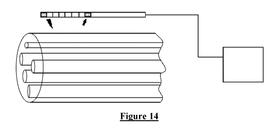
[0096] In one embodiment, shown in Figure 14, we propose a method consisting
of 2 or more
electrodes implanted near a target mixed nerve of which only a subset of
fibres are desired
stimulus targets, and an implantable control unit device capable of recording
the
CA 03098468 2020-10-27
26
WO 2019/204884 PCT/AU2019/050384
electrophysiological response to the stimulation. The stimulated fibre
populations are assessed
via the recorded response and the electrode placement, the electrode
selection, as well as the
stimulus parameters (including, but not limited to, pulse width, stimulus
frequency, stimulus
waveform, stimulus amplitude) are optimised such that the desired fibre types
are preferentially
targeted. Additionally, the recorded responses can be used to optimise
stimulus parameters in
order to minimise power consumption. As long as the desired responses are
observed, the
parameters requiring the least power to achieve this should be used. In one
SNS trial, B fibres
were observed to be fully recruited at a stimulus amplitude which was only
around a quarter of
the amplitude being used as defined by reference to the past approach of the
muscle activation
threshold. This observation indicates that therapeutic benefits provided by B
fibre recruitment
could be achieved at a fraction of the power of current state of the art SNS
techniques which
operate blind to fibre type.
[0097] The characteristics of the neural response (evoked compound action
potential, or
ECAP) are used in order to assess which fibre types are activated by the
stimulus paradigm.
Without being limited by theory, the ECAP characteristics that can be used to
assess the type of
the stimulated fibres includes the conduction velocity (including latency),
strength-duration
characteristics, and the refractory period. Additionally, myoelectric
responses (Late Responses
(LRs)) can be used as a proxy measure for Aa efferent activation (either
directly or indirectly)
based on their presence and latency.
[0098] For example, in the case of sacral nerve stimulation, targeting
parasympathetic fibres
(lightly myelinated B type fibres), the electrodes are placed near the S3
sacral nerve as shown in
figure 14. A first electrode is used for stimulation and neural recordings are
obtained from 1 or
more electrodes of the same array, close to the target nerve. The conduction
velocity of the
stimulated fibres is obtained from the neural recordings and the electrodes
are placed such that
the B fibres response is maximised and all other responses minimised. Further,
the stimulation
parameters are varied such that the B fibre responses are maximised and all
other responses
minimised.
[0099] In other embodiments, such as those of Figures 13 and 15, a device
is provided to
optimise lead positioning and stimulus parameter selection for stimulation of
a mixed nerve
using a plurality of electrodes with different characteristics. This uses more
than 1 lead placed
near the target nerve. The selection of stimulus parameters is then carried
out for more than 1
lead. In the case of a multitude of conditions, such as overactive bladder
(OAB) as well as pelvic
CA 03098468 2020-10-27
27
WO 2019/204884 PCT/AU2019/050384
pain, electrode selection will be done so that either 1 or several electrodes
on either one or
several leads maximises the B fibre activation (for OAB relief) and the
Af3fibre activation (for
pain relief). Once again, it is to be noted that the present invention is not
limited by theory of
mechanism as the fibre type targeting may be performed in respect of any
suitable fibre which
achieves a therapeutic benefit irrespective of the theorised mechanism of
action.
[00100] In other embodiments, the nerves of a plexus can be stimulated in turn
to determine
which branch of the plexus contains the desired fibres using recordings of
evoked
electrophysiological responses. The therapy is then optimised on the best
candidate branch of the
plexus.
[00101] Furthermore, the recording of ECAPs for a given stimulus electrode
does not have to
be done on the same lead. The recording site can be optimised (maximising
signal amplitude)
and any electrode close to the target nerve can be chosen. This is useful as
the fascicles in mixed
nerves do not run parallel inside the nerve (as is shown simply for
illustrative simplicity in
figures 13 to 15, but cross, merge, and split as shown in Figure 6.
[00102] Another embodiment of this device will consist in implanting cuff
electrodes instead
of epidural leads, as shown in figure 16. The cuff electrodes will consist of
several distinct
contacts each (as opposed to some cuff electrodes that have one continuous
electrode running
along the entire cuff), and the 2 or more cuff electrodes can be rotated and
moved up and down
along the mixed nerve to find a position that optimises the discrete
stimulation and recording of
separate fascicles. In one embodiment the arrangement of Fig. 16 can be used
for spatial
targeting of a selected fibre type. This involves the cuff electrode 1610
applying a supramaximal
stimulus around substantially an entire circumference of the nerve, in order
to recruit all fibres of
the nerve. Electrode 1610 may be a non-segmented electrode extending
continuously around the
nerve or may be a segmented electrode with the stimulus being applied
simultaneously by all
segments of the electrode 1610 to ensure all fibres are recruited. The
recruited responses
resulting from the stimulus can then be observed at selected circumferential
positions by using a
selected segment (e.g. segment 1622 of a segmented cuff electrode 1620 (other
segments not
shown for clarity). The response observed at segment 1622 in particular can
then be analysed to
determine a dominant fibre type which is adjacent to that segment 1622. This
knowledge can
then be used to identify which fibre type(s) are adjacent all segments of each
segmented cuff
electrode. In turn, this knowledge can be used to preferentially use a given
segment, such as
segment 1622, to deliver stimulation which will preferentially recruit the
fibre type which is
CA 03098468 2020-10-27
28
WO 2019/204884 PCT/AU2019/050384
known to be adjacent to that segment. Other embodiments may derive the same
knowledge by
instead using segment 1622 to apply stimuli which are only just above a
stimulus threshold and
which therefore only recruit fibres proximal to segment 1622. The responses
may then be
observed at cuff electrode 1610 and analysed to determine the type of fibres
being recruited by
segment 1622 and thus the types of fibres which are adjacent to 1622. A survey
may be carried
out over multiple segments to thereby determine the fibre type(s) adjacent to
all such electrode
segments, and spatial fibre type targeting may then be applied by using such
knowledge as a
lookup table.
[00103] Additionally, the responses can be used to optimise stimulus
parameters in order to
minimise power consumption. As long as the desired responses are observed, the
parameters
requiring the least power to achieve this should be used.
[00104] More generically, different lead shapes can be used, paddle leads,
percutaneous leads,
cuff electrodes, while effecting fibre type targeting in accordance with the
present invention.
[00105] In some embodiments, the device is connected externally to the
electrodes, in other
embodiments, the device is fully implantable. In some embodiments, the
electrodes can be on a
single lead, or multiple leads placed in proximity to the stimulus target,
each lead can contain a
multitude of electrodes. In other embodiments, some electrodes can be located
distally to the
target nerve to serve as reference electrode for the measurement or return
electrode for the
stimulation. In the case where the device is a fully implantable system, the
body of the implant
can be used as one electrode.
[00106] In some embodiments, the leads can be epidural, similar to those used
in sacral nerve
stimulation or spinal cord stimulation. In other embodiments, the leads can be
paddle leads, or
cuff electrodes. In some embodiments, the electrodes can further be segmented.
In one example,
a cuff electrode can in some cases be a ring electrode surrounding the nerve,
and in some cases it
can be a segmented ring whereby electrically distinct portions of the cuff
electrode take distinct
positions around the circumference of the nerve, allowing distinct
circumferential positions
around the nerve to be selectively targeted by stimulating from a selected
segment or to be
selectively observed by recording from a selected segment. In one example, an
epidural lead can
have circular electrode contacts, in other examples the contacts can be
segmented.
[00107] In some embodiments, the stimulation and recording are performed on
the same
electrodes, in other embodiments the stimulation and recording are done on
distinct electrodes
CA 03098468 2020-10-27
29
WO 2019/204884 PCT/AU2019/050384
whether on the same electrode lead or electrode array, or a different
electrode lead or array. In
some embodiments, some electrodes implanted near the target span an area small
enough to
avoid merging or branching of fibre tracts within the nerve. For example, in
some embodiments,
electrodes implanted near the sacral nerve can span a distance of less than 5
centimetres,
preferably less than 2 centimetres.
[00108] In some embodiments, the external device may be used to analyse neural
response
during lead insertion. The external device may be used to deliver stimulation
pulses to a target
nerve in a patient. The external device may include a control unit, a display
module and a
stimulation module. The stimulation unit is configured to deliver stimulation
pulses to a desired
nerve and analyse the neural response generated. Thereafter, the control unit
is adapted to
analyse the neural response and differentiate the recruited nerves based on
one or more
parameters. The control unit is configured to transmit information to display
the neural
recruitment in one or more modes. In a first mode, the neural recruitment is
displayed in the form
of a collection of ECAP waveforms and myoelectric waveforms. In a second mode,
the neural
recruitment is displayed as waveforms which are labelled with the
corresponding fibre types. In a
third mode, the neural recruitment is displayed as waveforms with timing
partitions to indicate
the fibre types which were recruited. In a fourth mode, the neural recruitment
is displayed along
with characteristics of the fibres such as conduction velocity etc.
[00109] In some embodiments, the external device is adapted to operate one or
more
electrodes. The external device is adapted to deliver pulses via paddle leads,
cylindrical leads
and cuff electrodes. Further, the external device is configured to operate in
any combination of
the aforementioned electrodes.
[00110] Implantable device: In some embodiments, the implantable device is
configured to
perform all the functions as the external device except that it is implanted
within the body of the
patient.
[00111] In some embodiments, the electrophysiological responses elicited by
the stimulation
can be displayed in real time. In some embodiments, these responses can
further be processed to
increase the signal to noise ratio. For example, averaging can be used to
decrease the random
noise found on each individual response, and fit-and-remove algorithms can be
used to decrease
the artefact component in the signal. These signals can be used to recognise
the activation of a
certain fibre type or certain fibre types. The stimulus parameters can then be
modified whether
by trial and error, brute force exploration or any suitable search technique
of the stimulus
CA 03098468 2020-10-27
WO 2019/204884 PCT/AU2019/050384
parameter space, so as to optimise fibre activation of the desired subsets of
fibres as indicated by
the displayed responses.
[00112] In some embodiments, the axes of the electrophysiological response
graph can be
labelled to facilitate the interpretation of the responses. For example, based
on the stimulus and
recording electrode configurations, the axes of the response graph can be
labelled to indicate the
times at which the response of a given fibre type should be recorded if
present.
[00113] In some embodiments, the neural responses can be marked and/or
labelled to help
identify the fibre types being recruited by the stimulation. For example, the
peaks of the response
can be labelled with their time and amplitude parameters.
[00114] In some embodiments, a change in morphology of the response with
varying stimulus
parameters can be used to identify fibre type contributions in an ECAP in
which the peaks are
poorly defined. In one example, the current is slowly ramped up and the ECAP
shape observed
or automatically processed. Initially, the largest fibres will respond to
external stimuli first, a
change in ECAP morphology at higher amplitudes could indicate the recruitment
of a second,
slower fibre type. In another example, the frequency of the stimulation can be
increased to an
appropriate level, and the responses observed over time. The small fibres will
fatigue first, the
larger fibres last. The change in morphology over time can be used to
determine which part of
the ECAP is associated with which fibre type. Further, a decrease in amplitude
of the
myoelectric response at increasing stimulus frequency could be due to muscle
fatigue, or
depression of the H-reflex. These properties and others can be used to
determine the fibre types
being stimulated.
[00115] In some embodiments, processing can be done on the recorded neural
responses to
display an estimate of the conduction velocity of the different elements of
the recording of the
electrophysiological response.
[00116] In some embodiments, the recording(s) of the electrophysiological
response can be
processed such that an estimate of the number of fibres of each type being
recruited is displayed.
The estimate could in some instances give a proportion of fibre types or an
absolute estimate of
the number of fibres being activated.
[00117] In some embodiments, stimulation can be performed in a way to approach
a constant
neural recruitment by adapting the stimulus parameters automatically in a
feedback arrangement
CA 03098468 2020-10-27
31
WO 2019/204884 PCT/AU2019/050384
in order to approach a target electrophysiological response. In one example, B
fibres are
preferentially targeted in a mixed nerve. After selecting the stimulus
electrodes and parameters
that optimise B fibre recruitment, the current (or another parameter) could be
automatically
adjusted to maintain a constant response amplitude. In another example, the
late response can be
used as a proxy measure for motor fibre activation, the stimulus can then be
automatically
adjusted to maintain a constant late response amplitude.
[00118] The amplitude of the electrophysiological response or a part of it
could be done by
assessing peak amplitudes, peak-peak amplitudes, the area under the curve, a
convolution with a
filter, or any other method.
[00119] In some embodiments, an implantable device may be configured to
establish closed-
loop stimulation on a targeted fibre type. For instance, the feedback loop may
be established on
the neural response of the B fibres in a mixed nerve. The methods disclosed
herein enable the
implantable device to establish a feedback loop on the neural response of
certain fibre types.
Since the implantable device is configured to determine the fibre types based
on the neural
response, the implantable device can maintain closed loop stimulation based on
the response of
the targeted fibre type. The implantable device may maintain closed-loop
stimulation on at least
one or more of the fibre types which include, but not limited to, Aa, Af3, A6,
B and C fibres. In
some cases, closed-loop stimulation may be maintained on the myoelectric
response elicited by
the stimulation. If the desired response is not observable, another response
can be used as a
proxy measure for establishing and maintaining closed-loop stimulation.
[00120] In some embodiments, the neural recruitment can be automatically
monitored for large
variations that could be indicative of, for example, lead migration. In one
example, a change in
electrode position that would lead to loss of efficacy could be tested by the
device by varying the
current amplitude to a small degree and monitoring the response profile. If no
change is observed
with variations in stimulus amplitude, a warning signal is sent to the user,
or stimulation is
stopped or other suitable action occurs.
[00121] In some further embodiments, the implantable device may be configured
to check the
fibre types which are recruited by the stimulation pulses. This feature may be
used to ensure that
the desired fibre types are recruited for the majority of the stimulus
duration. In some cases,
when the implantable device fails to detect the response of a fibre type for a
duration which
exceeds a threshold, the implantable device may trigger an alert to the
patient that the desired
fibre type is no longer recruited. In such a case, the physician may consider
the possible causes
CA 03098468 2020-10-27
32
WO 2019/204884 PCT/AU2019/050384
and take remedial action. In other embodiments, alternative actions may be
triggered in such an
event, such as automatic reconfiguration of the stimulus paradigm. Such
functions may
alternatively be carried out by an external device such as a clinician's
programmer device.
[00122] In some embodiments, continuous monitoring can be used to both
maintain a near
constant neural recruitment, as well as to warn of a substantial change in
lead placement or nerve
properties. Further embodiments may provide for testing the feedback loop
integrity before
applying the next corrective stim pulse. This could be done by making sure
that if the current is
increased, the response also increases. This would keep the loop from
diverging when the signal
is lost (as can occur when the signal is lost when changing posture, or when
the charger or
another interfering source is applied).
[00123] In some embodiments, the method and device may be applied to vagus
nerve
stimulation, dorsal root stimulation, sacral nerve stimulation, or ventral
root stimulation. In some
embodiments, the method and device can be used in more than one location,
simultaneously or in
tandem. For example, the device could be used in the sacral nerve and the
lower thoracic or
upper lumbar ventral roots which innervate the bladder and bowel. Without
being limited by
theory, it is proposed that such a device can stimulate both the sympathetic
and parasympathetic
nervous system in locations specific for bladder and bowel control.
Stimulating the ventral roots
of the spinal cord and/or the vagus nerve, and/or the sacral nerve can lead to
similar
embodiments in which the target is different from the bladder and bowel, such
as any one or
more of the heart, larynx, trachea, bronchi, oesophagus, stomach, liver,
pancreas, small intestine,
spleen, large intestine, kidney or sexual organs. For example, sympathetic and
parasympathetic
stimulation of the liver could be achieved by stimulating the sympathetic
fibres in the thoracic
sections T5-T12 and the parasympathetic fibres originating in the vagus nerve.
Vagus nerve
stimulation may be improved by some embodiments of the present invention may
selectively
avoiding stimulation of fibre types which in past solutions cause fatigue of
the throat,
swallowing muscles, vocal chords, and the like, which in past solutions are
unnecessarily
recruited. Note also partial lesions of the spinal cord might be best healed
by targeting specific
fibres.
[00124] In other embodiments the programming and display module does not have
to be part
of the implanted device. In such embodiments the implanted stimulator itself
only executes the
program that was set by the control module, and the responses are observed in
real time or later
only on an external programmer, and not on the implanted device.
CA 03098468 2020-10-27
33
WO 2019/204884 PCT/AU2019/050384
[00125] The preceding thus reveals large inter-patient variability in fibre
type recruitment and
significant posture-related effects on the number of fibres recruited.
Further, there was a clear
distinction between fibre type recruited and trial outcome. These first-in-
human results show
that recordings of an electrophysiological response can be used to identify
the type of fibres
recruited by SNS therapy and that the type of fibre may correlate with
therapeutic effectiveness.
These results pave the way for improved targeting as well as closed-loop SNS
that has the
potential to greatly improve the therapy.
[00126] Notably, the fibre targeting of the present invention allowed some
subjects to achieve
effective therapy at stimulation levels which were orders of magnitude less
than a muscle
response threshold or sensory response threshold which would conventionally
have been used to
set a stimulation level. This would lead to a many multiples increase in
battery lifetime.
[00127] Embodiments undertaking fibre type targeting on an ongoing basis to
control a
feedback process have the further advantage of being responsive to lead
movement relative to
the nerve, as occurs with posture changes, coughs, sneezes and the like. In
contrast to
conventional approaches which fix the stimulation at a single high level, the
use of feedback
based on electrophysiological measurements allows stimulation to be constantly
revised to
ensure that the desired fibre type continues to be recruited by suitably
refining the stimulation
parameters on an ongoing basis. Similar benefits can be obtained even in
simpler embodiments
which simply detect a loss of recruitment of the desired fibre type and issue
an alert to the user
to, for example, seek clinical assistance. Alternatively, automatic
modification of the stimulus
paradigm could be performed in some embodiments in the event in which the
determined
stimulus paradigm fails to recruit the desired fibre types.
[00128] It is to be understood that the fibres types named in the examples are
based on current
understanding and should not be considered limiting in the application of the
present invention.
[00129] The described electronic functionality can be implemented by discrete
components
mounted on a printed circuit board, and/or by a combination of integrated
circuits, and/or by an
application-specific integrated circuit (ASIC).
[00130] It will be appreciated by persons skilled in the art that numerous
variations and/or
modifications may be made to the invention as shown in the specific
embodiments without
departing from the spirit or scope of the invention as broadly described. The
present
CA 03098468 2020-10-27
34
WO 2019/204884
PCT/AU2019/050384
embodiments are, therefore, to be considered in all respects as illustrative
and not limiting or
restrictive.