Note: Descriptions are shown in the official language in which they were submitted.
CA 03099042 2020-10-30
WO 2019/217646 PCT/US2019/031467
MESENCHYMAL STROMAL CELL EXOSOME-TREATED MONOCYTES AND USES
THEREOF
RELATED APPLICATION
This application claims the benefit under 35 U.S.C. 119(e) to U.S.
Provisional
Application No. 62/669324, filed May 9, 2018, and entitled "MESENCHYMAL
STROMAL
CELL EXOSOME-TREATED MONOCYTES AND USES THEREOF," the entire contents of
which are incorporated herein by reference.
BACKGROUND
Idiopathic pulmonary fibrosis (IPF) is a chronic progressive respiratory
disease with a
prevalence of 0.5 to 27.9 per 100,000 person years. The lack of complete
understanding of the
underlying mechanism of this disease, may have contributed to the paucity of
successful therapies.
Despite two newly approved drugs, IPF remains fatal with a five-year survival
rate of less than
10%.
SUMMARY
It was shown herein that, a single intravenous (IV) dose of mesenchymal stem
cell (MSC)
exosomes reverts bleomycin-induced pulmonary fibrosis, at least partly through
the modulation of
monocyte phenotypes in the bone marrow and reduction of alveolar epithelial
cell (AEC)
apoptosis. Further, monocytes treated with MSC exosomes, when administered to
a subject
having pulmonary fibrosis, were therapeutically effective against the disease.
Accordingly, provided herein, in some aspects, are methods of regulating a
monocyte
phenotype, the method comprising contacting a monocyte with an isolated
mesenchymal stem cell
(MSC) exosome. In some embodiments, the monocyte is from bone marrow.
In some embodiments, the isolated MSC exosome is isolated from MSC-conditioned
media. In some embodiments, the MSC is from Wharton's Jelly, bone marrow, or
adipose tissue.
In some embodiments, the isolated MSC exosome is substantially free of protein
contaminants. In
some embodiments, the isolated MSC exosome has a diameter of about 50-150 nm.
In some embodiments, the contacting is in vitro. In some embodiments, the
contacting is ex
vivo. In some embodiments, the contacting is in vivo. In some embodiments, the
contacting is for
at least 2 hours.
1
CA 03099042 2020-10-30
WO 2019/217646 PCT/US2019/031467
In some embodiments, the monocyte is pro-inflammatory prior to being contacted
with the
isolated MSC exosome, and is regulatory after being contacted with the
isolated MSC exosome.
Other aspects of the present disclosure provide methods of treating a fibrotic
disease or an
autoimmune disease, the method comprising administering to a subject in need
thereof an effective
amount of a monocyte, wherein the monocyte is treated with an isolated
mesenchymal stem cell
(MSC) exosome prior to being administered.
In some embodiments, the method further comprises isolating the monocyte prior
to
treating the monocyte with the MSC exosome. In some embodiments, the monocyte
is isolated
from the subject. In some embodiments, the monocyte is isolated from the bone
marrow of the
subject.
In some embodiments, the monocyte is treated with the MSC exosome for at least
2 hours
prior to being administered to the subject. In some embodiments, the monocyte
is administered
systemically. In some embodiments, the monocyte is administered via
intravenous infusion. In
some embodiments, the monocyte is administered intratracheally or
intranasally. In some
embodiments, the monocyte is administered once to the subject. In some
embodiments, the
monocyte is administered multiple times to the subject.
In some embodiments, the method further comprises administering to the subject
an
effective amount of a second agent. In some embodiments, the second agent is
an isolated MCS
exosome. In some embodiments, the second agent is nintedanib, Pirfenidone, an
anti-fibrotic
agent, an immunosuppressant, and/or an anti-inflammatory agent.
In some embodiments, the fibrotic disease is selected from the group
consisting of:
systemic sclerosis; liver fibrosis, heart fibrosis, kidney fibrosis, and
myelofibrosis. In some
embodiments, the fibrotic disease is pulmonary fibrosis. In some embodiments,
the pulmonary
fibrosis is idiopathic pulmonary fibrosis (IPF). In some embodiments, the
monocyte reduces
inflammation associated with the fibrotic disease. In some embodiments, the
monocyte reduces
apoptosis associated with the fibrotic disease.
In some embodiments, the subject is a mammal. In some embodiments, the subject
is a
human subject. In some embodiments, the human is a neonate, an infant, or an
adult. In some
embodiments, the human subject is less than four weeks of age. In some
embodiments, the human
subject is four weeks to 3 years of age. In some embodiments, the human
subject is 3-18 years of
age. In some embodiments, the human subject is an adult.
2
CA 03099042 2020-10-30
WO 2019/217646 PCT/US2019/031467
In some embodiments, the human subject is born prematurely. In some
embodiments, the
human subject was born before 37 weeks of gestation. In some embodiments, the
human subject
was born before 26 weeks of gestation.
In some embodiments, the subject is a rodent. In some embodiments, the rodent
is a mouse
or a rat.
In some embodiments, the monocyte is pro-inflammatory prior to being treated
with the
isolated MSC exosome, and is regulatory after being treated with the isolated
MSC exosome.
Other aspects of the present disclosure provide monocytes treated with an
isolated
mesenchymal stem cell (MSC) exosome. In some embodiments, the monocyte is from
bone
marrow. In some embodiments, the isolated MSC exosome is isolated from MSC-
conditioned
media. In some embodiments, the MSC is from Wharton's Jelly, bone marrow, or
adipose tissue.
In some embodiments, the monocyte is pro-inflammatory prior to being treated
with the isolated
MSC exosome, and is regulatory after being treated with the isolated MSC
exosome.
Compositions comprising the monocytes described herein are also provided. In
some
embodiments, the composition further comprises a second agent. In some
embodiments, the
composition is a pharmaceutical composition. In some embodiments, the
composition further
comprises a pharmaceutically acceptable carrier.
Further provided herein are uses of the monocyte or the composition comprising
the
monocytes described herein for treating a fibrotic disease or an autoimmune
disease.
The monocyte or the composition comprising the monocytes described herein may
also be
used use in the manufacturing of a medicament for treating a fibrotic disease
or an autoimmune
disease.
The summary above is meant to illustrate, in a non-limiting manner, some of
the
embodiments, advantages, features, and uses of the technology disclosed
herein. Other
embodiments, advantages, features, and uses of the technology disclosed herein
will be apparent
from the Detailed Description, the Drawings, the Examples, and the Claims.
BRIEF DESCRIPTION OF THE DRAWINGS
The accompanying drawings are not intended to be drawn to scale. In the
drawings, each
identical or nearly identical component that is illustrated in various figures
is represented by a like
numeral. For purposes of clarity, not every component may be labeled in every
drawing. The
patent or application file contains at least one drawing executed in color.
Copies of this patent or
3
CA 03099042 2020-10-30
WO 2019/217646 PCT/US2019/031467
patent application publication with color drawing(s) will be provided by the
Office upon request
and payment of the necessary fee. In the drawings:
FIGS. 1A to 1D show that MEx treatment at the beginning of inflammation
prevents
fibrosis. (FIG. 1A) Ten to fourteen-week old C57BL/6 mice received
endotracheal bleomycin (60
Ilg) or 0.9% normal saline (NS) on day 0 followed by a bolus dose of IV MEx
(Bleo+MEx), NS
(bleo+NS), FEx (Bleo+FEx), or iodixanol (IDX 1:9 dilution, bleo+IDX). Results
were compared
to control group who received either NS (vehicle, control) or NS followed by a
dose of MEx
(control+MEx). Mice were sacrificed on day 14. (FIG. 1B) Lung sections were
stained with
Masson's trichrome. Inserts were taken at 100X magnification. Bleo+NS,
Bleo+FEx, Bleo+IDX
showed architectural destruction, alveolar septal thickening and fibrotic
changes. (FIG. 1C)
Administration of MEx to bleomycin-treated mice substantially reduced fibrosis
and alveolar
distortion. Findings were similar to control or Control+Mex group. Lung
fibrosis was measured at
day 14 by Ashcroft score. (FIG. 1D) Collagen deposition was assessed by Sircol
assay and
represented as mg/ml of left lung homogenate. n=3-4 per group, *p<0.05;
****p<0.0001 vs.
bleomycin-treated group. Scale bar = 100 Ilm.
FIGS. 2A to 2E show that MEx modulates alveolar macrophage phenotypes and
blunt
inflammation. Whole lung RT-qPCR shows an increase in the expression of
macrophage Cc1-2
and Arginase-1 (Arg 1) markers at day 7 (FIG. 2A) and day 14 (FIG. 2B), while
their level was
similar to control with MEx treatment. Interleukin-6 expression showed similar
trend but its
reduction with MEx treatment did not reach statistical significance. Levels of
TGF-remained
unchanged between the three groups. Results are expressed relative to control
expression. Mean
SEM, n=4-8 per group. *p<0.05; ** p<0.01 vs. bleomycin-exposed mice. (FIGS. 2C
and 2D)
Immunofluorescence (IF) analysis of lung sections using antibodies against
markers of M2-like
activation Argl (green) and CD206 (red) shows an increase in mean fluorescent
intensity (MFI) in
bleomycin mice, while the intensity was similar to control levels with MEx
treatment. Nuclei
staining performed with Dapi. Images obtained at x10 magnification. Mean
fluorescent intensity
normalized for cell number (Dapi stain). Analysis performed was by image J
software. N=5 per
group, *p<0.05; ** p<0.01 vs. bleomycin-exposed mice. (FIG. 2E) Cumulative
data and
representative graph depicts the percentage of CD206-Fve alveolar macrophages
(AM)
(CD45-EveCD11b-veCD11c-EveCD 206-Eve cells). Number of CD206-Eve AMs reduced
with MEx
treatment but did not reach statistical significance compared to the bleomycin-
exposed group.
4
CA 03099042 2020-10-30
WO 2019/217646 PCT/US2019/031467
Representative histogram normalized to mode. Mean SEM of n=4-5 per group, **
p<0.01 vs.
bleomycin-exposed mice. Abbreviations: Dapi, 40,6-diamidino-2-phenylindole.
FIGS. 3A to 3F show that MEx modulates monocyte and macrophage phenotype at a
systemic level MEx restore alveolar macrophage and inflammatory monocyte
populations in the
lung. (FIG. 3A) Cytometric analysis in whole lungs 7 days after injury showed
a decrease in the
AM number (represented as CD45-EveCD11b-veCD11c-Eve cells). (FIG. 3B) This was
associated
with an increase in Ly6Chi infiltrating or classical monocytes (Ly6ChiCCR-2-
Eve). (FIG. 3C) On
day 14 AM number increased and (FIG. 3D) classical monocytes number decrease
to
approximately half of the level observed in NS-treated (control) group of mice
(Mean difference:
1.7% 0.44, p<0.01). MEx therapy not only led to the restoration of the AM
population number,
but also modulated the monocyte phenotypes in the lung to levels comparable to
control group
analyzed at day 7 and 14. Mean SEM of n=4-5 per group, *p<0.05; ** p<0.01;
***p<0.001 vs.
bleomycin-exposed mice. Gating strategy was performed according to
fluorescence minus one
controls (See FIG. 8). To investigate the systemic effects of MEx, the myeloid
signature of the
bone marrow was analyzed by flow cytometry. Despite similar numbers of CD45'
cells in the
three groups (data not shown), (FIGS. 3E and 3F) classical monocytes increased
in bleomycin-
exposed group of mice (Mean difference: 17.6% 3.6, p <0.001 vs. bleomycin-
exposed mice),
but regulatory monocytes exhibited a 2-fold decrease (Mean difference: 18%
5.7, p <0.05 vs.
bleomycin-exposed mice) in bleomycin-exposed mice compared to control mice.
Whereas, MEx
therapy led to a decrease in inflammatory monocytes and a shift from
inflammatory to regulatory
(Ly6ClowCCR-2-ve) phenotype, similar to levels observed in control mice (Mean
difference:
10.25% 4.2, p<0.05 and 13.39% 5.76, p<0.05 vs. bleomycin-exposed mice).
n=4-7 per group,
*p<0.05; ** p<0.01; ***p<0.001 vs. bleomycin-exposed mice.
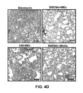
FIGS. 4A to 4F show that adoptive transfer of MEx-pretreated bone marrow
derived
monocytes protects mice from pulmonary fibrosis. The potential therapeutic
effects of ex vivo
treated BMDMo and AMs in the prevention of fibrosis was explored. (FIG. 4A)
BMDMo were
isolated from 6-8-wks-old FVB mice, cultured ex vivo for 3 days and treated
with MEx
(equivalent to EVs produced by 1 x 10⁶ MSCs per 100mm plate) or media alone on
day 1, D1 and
day 2, D2 and stained with Dil on day 3, D3. Cells were adoptively transferred
intravenously at a
one-to-one ratio on days 0 and 3 to C57BL/6 mice following endotracheal
instillation of
bleomycin. Mice were sacrificed at day 14. Data was compared to bleomycin-
exposed mice who
CA 03099042 2020-10-30
WO 2019/217646 PCT/US2019/031467
had received NS only (Bleomycin) (FIG. 4B) Flow cytometric analysis of BMDMo
after 3 days
of culture showed more than 90% CD45+veCD11b+ve cells. (FIG. 4C) Dil-labeled
BMDMo
were detected in the lung 14 days after injection. Images obtained at x20
magnification. (FIGS.
4D to 4F) Fibrosis was ameliorated in mice that received MEx-pretreated
monocytes
(BMDMo+MEx) compared to NS (Bleomycin). Mice who were injected with MEx-
treated AM
(AM+MEx) exhibited substantial fibrotic changes. The administration of
untreated-BMDMo
(BMDM+Media) led to mild amelioration of fibrosis and collagen levels compared
to NS-treated
group of mice. The reduction in collagen deposition did not reach statistical
significance compared
to NS-treated mice. Similar results were noted at collagen level. Arrow marks
the Dil-labeled
monocytes. Between group comparison: *p<0.05, ** p<0.01, ***p<0.001,
****p<0.001. Scale bar
= 100 pm. Abbreviations: Dil, 1,1'-Dioctadecy1-3,3,3',3'-
Tetramethylindocarbocyanine
Perchlorate ('Dil'; Di1C18(3))
FIGS. 5A to 5D shows that MEx therapy decreases apoptosis. (FIGS. 5A and 5B)
Tunel
staining in whole lung sections shows increase in apoptosis (green) in the
bleomycin-exposed
group of mice compared to control (NS) and bleomycin+MEx. Nuclei were stained
with Dapi.
Images obtained at x20 magnification. MFI quantified using image J software
and normalized for
Dapi. *p<0.05, ** p<0.01 vs bleomycin-exposed mice (FIG. 5C) Annexin V/PI
staining in whole
lungs shows an increase in apoptosis (Annexin V+ PI-) in bleomycin-exposed
mice compared to
control and bleomycin+MEx mice. (FIG. 5D) In vitro apoptosis was measured
using Caspase-
Glo 3/7 Assay. More apoptosis is noted in Bleomycin-exposed human alveolar
epithelial cells.
This effect is abrogated with MEx therapy. Relative luminescence unit was used
as a
representative of apoptosis, Y axis represents luminescence relative to
control. n=8 per group, **
p<0.01; ****P<0.0001 vs bleomycin- exposed mice.
FIGS. 6A to 6C show the purification, isolation and characterization of
exosomes.
Conditioned media (CM) from BMSCs or HDFs was differentially centrifuged and
concentrated
through tangential flow filtration. Concentrated (50x) CM was floated on an
iodixanol (OptiPrepTM,
IDX) cushion gradient. Purified EV population in fraction 9 was used for
analysis. (FIG. 6A)
Heterogeneous EV morphology seen on transmission electron microscopy (TEM)
(x30,000g, scale
bar = 100nm). (FIG. 6B) Nanoparticle tracking analysis (NTA) was used to
assess EV
concentration. Representative size distribution of BMSC- EVs and HDF-EVs in
fraction 9 gradient.
(FIG. 6C) Western blot analysis of IDX cushion gradient fractions (7-10),
using antibodies to
exosomal markers flotillin (FLOT-1), CD63 & Alix.
6
CA 03099042 2020-10-30
WO 2019/217646 PCT/US2019/031467
FIGS. 7A to 7D show that MEx treatment at the end of inflammation reverts
fibrosis. (FIG.
7A) MEx were administered 7 days after the administration of bleomycin and
mice were sacrificed
on day 14. (FIGS. 7B and 7C) Lung sections from Control, Bleomycin and
Bleo+MEx mice were
analyzed for histology and (FIG. 7D) collagen deposition. MEx therapy led to
reduction in fibrosis
and collagen deposition on day 7. Data represent mean SEM of n=4 per group,
*p<0.05;
****p<0.0001 vs. bleomycin-exposed mice. Scale bar = 10011m.
FIG. 8 shows the representative in vivo gating strategy of lung macrophage,
monocyte and
bone marrow derived monocytes. Cells were isolated from whole lung after
enzymatic digestion.
Lung aggregates and cell debris were excluded based on forward and side
scatter parameters.
Immune cells were identified by CD45 staining. Alveolar macrophages (AM) were
identified using
a sequential gating strategy to identify CD45-EveCD11b-veCD11c-Eve population.
Subsequent
gating was performed on CD206-Fve AMs. In order to identify monocyte
subpopulation, sequential
gating strategy was performed on non-alveolar macrophage subset of CD45-
Evecells (CD1lbint
CD11C1 w) and further gated for CCR-2-EveLy6Ch1gh and CCR-2-veLy6C1 w
population to
reflect classical or non-classical monocyte phenotype respectively. BMDMo
gating strategy was
similar to above, with the exclusion of CD11c and CD206 (markers of AMs)
staining. Gating
strategy performed according to Fluorescence-minus-one controls.
FIG. 9 shows that labeled-MEx can be detected in the bone marrow. Membrane dye-
labeled EVs were IV injected into mice, and the animals were sacrificed 2
hours after injection.
MEx were detected in the BM cytospins (Labeled-MEx). Injected free dye and dye-
stained EV free
supernatant were used as controls. Counterstaining performed with Dapi. Images
were obtained at
x60 magnification.
DETAILED DESCRIPTION OF CERTAIN EMBODIMENTS
The present disclosure is based, at least in part, on the finding that
mesenchymal stromal
cell (also termed herein interchangeably as "mesenchymal stem cell" or "MSC")
exosomes (also
termed "Mex" herein), when administered to a subject (e.g., systemically), can
modulate monocyte
phenotypes in the bone marrow, resulting in a larger subpopulation of
regulatory monocytes
instead of pro-inflammatory monocytes. Further, monocytes (e.g., bone marrow-
derived
monocytes) treated with MSC exosomes in vitro, when administered to subjects
having pulmonary
fibrosis, have therapeutic effects on fibrotic lungs.
7
CA 03099042 2020-10-30
WO 2019/217646 PCT/US2019/031467
Some aspects of the present disclosure provide monocytes treated with isolated
mesenchymal stem cell (MSC) exosomes. A "monocyte" is a type of leukocyte
(also called "white
blood cell") that can differentiate into macrophages and myeloid lineage
dendritic cells. In
vertebrates, monocytes are part of the innate immune system but can also
influence the process of
adaptive immunity.
Monocytes compose 2% to 10% of all leukocytes in the human body and serve
multiple
roles in immune function, e.g., without limitation, replenishing resident
macrophages under normal
conditions; migration in response to inflammation signals from sites of
infection in the tissues; and
differentiation into macrophages or dendritic cells to effect an immune
response.
Monocytes are heterogeneous populations of cells, and can be divided into
subpopulations
with different phenotypes and functions. In some embodiments, human monocytes
are subdivided
into phenotypically and functionally distinct subpopulations based on the
expression of the
lipopolysaccharide (LPS) receptor (CD14) and the CD16 (Fcgamma receptor III)
(e.g., as
described in Ziegler-Heitbrock et al., Blood, vol. 116, no. 16, pp.
e74¨e80,2010 and Gordon et al.,
Nature Reviews Immunology, vol. 5, no. 12, pp. 953-964,2005, incorporated
herein by reference).
In healthy individuals, approximately 80-90% of monocytes are highly CD14
positive and CD16
negative (CD14 CD16-). The CD14 CD16- monocytes are termed "classical
monocytes" or
"regulatory monocytes" herein. The remaining 10-20% of monocytes are CD16
positive and are
classified as "proinflammatory monocytes." Proinflammatory monocytes can
further subdivided
into CD14"CD16+ and CD14 CD16" cells, which are The CD14"CD16+ monocytes are
also
termed "intermediate monocytes;" and the CD14 CD16 monocytes are also termed
"nonclassical
monocytes." Compared with CD16 negative conventional monocytes, CD16 positive
monocytes
(proinflammatory monocytes), express higher levels of major histocompatibility
complex (MHC)
class II antigens, adhesion molecules, chemokine receptors, and
proinflammatory cytokines such as
TNF-a, but lower levels of the anti-inflammatory cytokine (e.g., IL-10) (e.g.,
as described in
Kawanaka et al., Arthritis & Rheumatism, vol. 46, no. 10, pp. 2578-2586,2002
and Ziegler-
Heitbrock et al., Immunology Today, vol. 17, no. 9, pp. 424-428,1996,
incorporated herein by
reference). Proinflammatory monocytes are elevated in various pathologic
conditions, including
inflammatory and infectious diseases, cancer, and in coronary heart disease.
In mice, monocytes
can also be divided in two subpopulations: proinflammatory monocytes
(Cx3CR11'w, CCR2+,
Ly6C1igh), which are equivalent to human proinflammatory monocytes; and
regulatory monocytes
(Cx3CR lhigh, CCR2-, Ly6C1'), which are equivalent to human CD14"CD16-
monocytes.
8
CA 03099042 2020-10-30
WO 2019/217646 PCT/US2019/031467
Monocytes are produced by the bone marrow from precursors called monoblasts,
bipotent
cells that differentiated from hematopoietic stem cells. Monocytes circulate
in the bloodstream for
about one to three days and then typically move into tissues throughout the
body where they
differentiate into macrophages and dendritic cells. In some embodiments, the
monocytes treated
with MSC exosomes described herein are from bone marrow (e.g., isolated from
bone marrow). In
some embodiments, the monocytes treated with MSC exosomes described herein are
from a
specific tissue (e.g., isolated from a specific tissue such as lungs).
An "exosome" is a membrane (e.g., lipid bilayer) vesicle that is released from
a cell (e.g.,
any eukaryotic cell). Exosomes are present in eukaryotic fluids, including
blood, urine, and
cultured medium of cell cultures. The exosomes of the present disclosure are
released from
mesenchymal stem cells (MSCs) and are interchangeably termed "mesenchymal stem
cell
exosomes" or "MSC exosomes."
A "mesenchymal stem cell (MSC)" is a progenitor cell having the capacity to
differentiate
into neuronal cells, adipocytes, chondrocytes, osteoblasts, myocytes, cardiac
tissue, and other
endothelial or epithelial cells. (See for example Wang et al.õ Stem Cells
2004;22(7);1330-7;
McElreavey;1991 Biochem Soc Trans (1);29s; Takechi, Placenta 1993 March/April;
14 (2); 235-
45; Takechi, 1993; Kobayashi; Early Human Development;1998; July 10; 51(3);
223-33; Yen;
Stem Cells; 2005; 23 (1) 3-9.) These cells may be defined phenotypically by
gene or protein
expression. These cells have been characterized to express (and thus be
positive for) one or more
of CD13, CD29, CD44, CD49a, b, c, e, f, CD51, CD54, CD58, CD71, CD73, CD90,
CD102,
CD105, CD106, CDw119, CD120a, CD120b, CD123, CD124, CD126, CD127, CD140a,
CD166,
P75, TGF-bIR, TGF-bIIR, HLA-A, B, C, SSEA-3, SSEA-4, D7 and PD-Li. These cells
have also
been characterized as not expressing (and thus being negative for) CD3, CD5,
CD6, CD9, CD10,
CD11a, CD14, CD15, CD18, CD21, CD25, CD31, CD34, CD36, CD38, CD45, CD49d,
CD50,
CD62E, L, S, CD80, CD86, CD95, CD117, CD133, SSEA-1, and ABO. Thus, MSCs may
be
characterized phenotypically and/or functionally according to their
differentiation potential.
MSCs may be harvested from a number of sources including but not limited to
bone
marrow, adipose tissue, blood, periosteum, dermis, umbilical cord blood and/or
matrix (e.g.,
Wharton's Jelly), and placenta. For example, MSCs can be isolated from
commercially available
bone marrow aspirates. Enrichment of MSCs within a population of cells can be
achieved using
methods known in the art including but not limited to fluorescence-activated
cell sorting (FACS).
Methods for harvesting MSCs are described in the art, e.g., in US Patent No.
5486359,
9
CA 03099042 2020-10-30
WO 2019/217646 PCT/US2019/031467
incorporated herein by reference.
Commercially available media may be used for the growth, culture and
maintenance of
MSCs. Such media include but are not limited to Dulbecco's modified Eagle's
medium
(DMEM). Components in such media that are useful for the growth, culture and
maintenance of
MSCs, fibroblasts, and macrophages include but are not limited to amino acids,
vitamins, a carbon
source (natural and non-natural), salts, sugars, plant derived hydrolysates,
sodium pyruvate,
surfactants, ammonia, lipids, hormones or growth factors, buffers, non-natural
amino acids, sugar
precursors, indicators, nucleosides and/or nucleotides, butyrate or organics,
DMSO, animal
derived products, gene inducers, non-natural sugars, regulators of
intracellular pH, betaine or
osmoprotectant, trace elements, minerals, non-natural vitamins. Additional
components that can
be used to supplement a commercially available tissue culture medium include,
for example,
animal serum (e.g., fetal bovine serum (FBS), fetal calf serum (FCS), horse
serum (HS)),
antibiotics (e.g., including but not limited to, penicillin, streptomycin,
neomycin sulfate,
amphotericin B, blasticidin, chloramphenicol, amoxicillin, bacitracin,
bleomycin, cephalosporin,
chlortetracycline, zeocin, and puromycin), and glutamine (e.g., L-glutamine).
Mesenchymal stem
cell survival and growth also depends on the maintenance of an appropriate
aerobic environment,
pH, and temperature. MSCs can be maintained using methods known in the art,
e.g., as described
in Pittenger et al., Science, 284:143-147 (1999), incorporated herein by
reference.
In some embodiments, the MSC exosomes used to treat the monocytes are
isolated. As
used herein, an "isolated exosome" is an exosome that is physically separated
from its natural
environment. An isolated exosome may be physically separated, in whole or in
part, from tissue
or cells with which it naturally exists (e.g., MSCs). In some embodiments, the
isolated MSC
exosomes are isolated from the culturing media of MSCs from human bone marrow,
umbilical
cord Wharton's Jelly, or adipose tissue. Such culturing media is termed "MSC-
conditioned
media" herein. In some embodiments, isolated exosomes may be free of cells
such as MSCs, or it
may be free or substantially free of conditioned media, or it may be free of
any biological
contaminants such as proteins. Typically, the isolated exosomes are provided
at a higher
concentration than exosomes present in un-manipulated conditioned media.
In some embodiments, the isolated MSC exosome described herein comprises one
or more
(e.g., 1, 2, 3, 4, 5, or more) known exosome markers. In some embodiments, the
known exosome
markers are selected from the group consisting of: FLOT1 (Flotillin-1, Uniprot
ID: 075955), CD9
(CD9 antigen, Uniprot ID: P21926), and CD63 (CD63 antigen, Uniprot ID:
P08962).
CA 03099042 2020-10-30
WO 2019/217646 PCT/US2019/031467
In some embodiments, the isolated MSC exosome is substantially free of
contaminants
(e.g., protein contaminants). The isolated MSC exosome is "substantially free
of contaminants"
when the preparation of the isolated MSC exosome contains fewer than 20%, 15%,
10%, 5%, 2%,
1%, or less than 1%, of any other substances (e.g., proteins). In some
embodiments, the isolated
MSC is "substantially free of contaminants" when the preparation of the
isolated MSC exosome is
at least 80%, at least 85%, at least 90%, at least 95%, at least 98%, at least
99%, at least 99.9%
pure, with respect to contaminants (e.g., proteins).
"Protein contaminants" refer to proteins that are not associated with the
isolated exosome
and do not contribute to the biological activity of the exosome. The protein
contaminants are also
referred to herein as "non-exosomal protein contaminants."
In some embodiments, the isolated MSC exosome used in accordance with the
present
disclosure has a diameter of about 30-150 nm. For example, the isolated MSC
exosome may have
a diameter of 30-150 nm, 30-140 nm, 30-130 nm, 30-120 nm, 30-110 nm, 30-100
nm, 30-90 nm,
30-80 nm, 30-70 nm, 30-60 nm, 30-50 nm, 30-40 nm, 40-150 nm, 40-140 nm, 40-130
nm, 40-120
nm, 40-110 nm, 40-100 nm, 40-90 nm, 40-80 nm, 40-70 nm, 40-60 nm, 40-50 nm, 50-
150 nm,
50-140 nm, 50-130 nm, 50-120 nm, 50-110 nm, 50-100 nm, 50-90 nm, 50-80 nm, 50-
70 nm, 50-
60 nm, 60-150 nm, 60-140 nm, 60-130 nm, 60-120 nm, 60-110 nm, 60-100 nm, 60-90
nm, 60-80
nm, 60-70 nm, 70-150 nm, 70-140 nm, 70-130 nm, 70-120 nm, 70-110 nm, 70-100
nm, 70-90 nm,
70-80 nm, 80-150 nm, 80-140 nm, 80-130 nm, 80-120 nm, 80-110 nm, 80-100 nm, 80-
90 nm, 90-
150 nm, 90-140 nm, 90-130 nm, 90-120 nm, 90-110 nm, 90-100 nm, 100-150 nm, 100-
140 nm,
100-130 nm, 100-120 nm, 100-110 nm, 110-150 nm, 110-140 nm, 110-130 nm, 110-
120 nm, 120-
150 nm, 120-140 nm, 120-130 nm, 130-150 nm, 130-140 nm, or 140-150 nm. In some
embodiments, the isolated MSC exosome may have a diameter of about 50 nm, 60
nm, 70 nm, 80
nm, 90 nm, 100 nm, 110 nm, 120 nm, 130 nm, 140 nm, or 150 nm. In some
embodiments, the
isolated MSC exosomes exhibit a biconcave morphology.
As described herein, the isolated MSC exosomes can be used to treat the
monocytes to
modulate the monocyte phenotype (e.g., both in vitro and in vivo such as in
the bone marrow).
"Treat a monocyte with an isolated MSC exosome" means contacting the monocyte
with a MSC
exosome (e.g., for a period of time). In some embodiments, the treating (i.e.,
contacting) is
carried out in vitro. For example, monocytes may be cultured in vitro and
isolated MSC
exosomes may be added to the culture such that the monocytes contact the
isolated MSC
exosomes. In some embodiments, the treating (i.e., contacting) is carried out
ex vivo. For
11
CA 03099042 2020-10-30
WO 2019/217646 PCT/US2019/031467
example, monocytes may be isolated from the bone marrow of a subject and
isolated MSC
exosomes may be added to the monocytes such that the monocytes contact the
isolated MSC
exosomes. In some embodiments, the treating (i.e., contacting) is carried out
in vivo. For
example, the isolated MSC exosomes may be administered to a subject (e.g., via
intravenous
injection), reach the one marrow, and contact the monocytes in the bone
marrow.
In some embodiments, the monocyte is treated (i.e., contacted) with the MSC
exosome for
at least 1 hour (e.g., at least 1, at least 2, at least 3, at least 4, at
least 5, at least 6, a least 7, at least
8, at least 9, at least 10, at least 15, at least 20, at least 25, at least
30, at least 35, at least 40, at
least 45, at least 50, at least 55, at least 60, at least 65, at least 70, at
least 75, at least 80, at least
85, at least 90, at least 95, at least 100 hours, or longer). In some
embodiments, the monocyte is
treated (i.e., contacted) with the MSC exosome for 1, 2, 3, 4, 5, 6, 7, 8, 9,
10, 11, 12, 13, 14, 15,
16, 17, 18, 19, 20, 21, 22, 23, 24, 25, 26, 27, 28, 29, 30, 31, 32, 33, 34,
35, 36, 37, 38, 39, 40, 41,
42, 43, 44, 45, 46, 47, 48, 49, 50, 51, 52, 53, 54, 55, 56, 57, 58, 59, 60,
61, 62, 63, 64, 65, 66, 67,
68, 69, 70, 71, 72, 73, 74, 75, 76, 77, 78, 79, 80, 81, 82, 83, 84, 85, 86,
87, 88, 89, 90, 91, 92, 93,
94, 95, 96, 97, 98, 99, 100 hours, or longer.
In some embodiments, the monocyte has been polarized to a pro-inflammatory
state as a
result of environmentally or developmentally-precipitated injury, and its
polarity is modulated to a
regulatory phenotype upon contact with the isolated MSC exosome. In some
embodiments, the
monocyte is a pro-inflammatory monocyte prior to being treated (i.e.,
contacted) with the isolated
MSC exosome, and is a regulatory monocyte after being treated (i.e.,
contacted) with the isolated
MSC exosome. In some embodiments, a mixture of pro-inflammatory monocytes and
regulatory
monocytes are contacted with isolated MSC exosomes and the treating results in
a higher ratio
(e.g., at least 10% higher) of regulatory monocytes in the mixture, being
treated with isolated MSC
exosomes. For example, the ratio of regulatory monocytes may be at least 10%,
at least 20%, at
least 30%, at least 40%, at least 50%, at least 60%, at least 70%, at least
80%, at least 90%, at least
100%, at least 2-fold, at least 5-fold, at least 10-fold, at least 100-fold,
or higher after being treated
with MSC exosomes, compared to before being treated with isolated MSC
exosomes. In some
embodiments, the ratio of regulatory monocytes is 10%, 20%, 30%, 40%, 50%,
60%, 70%, 80%,
90%, 100%, 2-fold, 5-fold, 10-fold, 100-fold, or higher after being treated
with MSC exosomes,
compared to before being treated with isolated MSC exosomes.
Further provided herein are uses of the monocytes treated with isolated MSC
exosomes for
treating a disease (e.g., a fibrotic disease such as pulmonary fibrosis or an
autoimmune disease). In
12
CA 03099042 2020-10-30
WO 2019/217646 PCT/US2019/031467
some embodiments, the monocytes treated with isolated MSC exosomes are used in
the
manufacturing of a medicament for treating a disease (e.g., a fibrotic disease
or an autoimmune
disease). Compositions comprising monocytes treated with isolated MSC exosomes
are also
provided. In some embodiments, the monocytes treated with isolated MSC
exosomes are
formulated in a composition for the treatment of a disease (e.g., a fibrotic
disease or an
autoimmune disease).
In some embodiments, the composition comprising monocytes treated with
isolated MSC
exosomes further comprises a second agent. In some embodiments, the second
agent is a
therapeutic agent effective against the diseases being treated by the
monocytes. For example, the
second agent may be any agent that can be used in the prevention, treatment
and/or management of
a fibrotic disease or an autoimmune disease such as those described herein. In
some embodiments,
the second agent is an isolated MSC exosome.
In some embodiments, the second agent is an agent that is known to have
therapeutic effects
against fibrotic diseases. Exemplary second agents that may be used to treat
fibrotic diseases
include, without limitation: nintedanib (a tyrosine kinase inhibitor),
pirfenidone, an anti-fibrotic
agent, and/or an anti-inflammatory agent. In some embodiments, for pulmonary
fibrosis, other
types of therapies, e.g., oxygen supplement, may be used in conjunction with
the therapeutic
agents described herein.
In some embodiments, the second agent is an agent that is known to have
therapeutic
effects against autoimmune diseases. Such agents include, without limitation,
non-steroidal anti-
inflammatory drugs, glucocorticoids, metrotrexate, leflunomide, anti-TNF
biologicals (e.g.,
antibodies such as infliximab, adalimumab, golinumab, or certolizumab pegol).
Drugs for treating
autoimmune diseases are known in the art, e.g., as described in Li et al.,
Front Pharmacol. 2017;
8: 460, incorporated herein by reference.
In some embodiments, the monocytes treated with isolated MSC exosomes and the
second
agent are formulated in the same composition. In some embodiments, the
monocytes treated with
isolated MSC exosomes and the second agent are formulated in separate
compositions. In some
embodiments, the monocytes treated with isolated MSC exosomes and the second
agent are
administered to the subject simultaneously. In some embodiments, the monocytes
treated with
isolated MSC exosomes and the second agent are administered separately. In
some embodiments,
the monocytes treated with isolated MSC exosomes are administered before the
second agent. In
some embodiments, the monocytes treated with isolated MSC exosomes are
administered after the
13
CA 03099042 2020-10-30
WO 2019/217646 PCT/US2019/031467
second agent.
In some embodiments, the composition comprising the monocytes treated with
isolated
MSC exosomes is a pharmaceutical composition. In some embodiments, the
composition further
comprises pharmaceutically acceptable concentrations of salt, buffering
agents, preservatives, or
compatible carriers.
A pharmaceutically acceptable carrier is a pharmaceutically acceptable
material,
composition or vehicle, such as a liquid or solid filler, diluent, excipient,
solvent or encapsulating
material, involved in carrying or transporting a prophylactically or
therapeutically active agent.
Each carrier must be "acceptable" in the sense of being compatible with the
other ingredients of the
formulation and not injurious to the subject. Some examples of materials which
can serve as
pharmaceutically acceptable carriers include sugars, such as lactose, glucose
and sucrose; glycols,
such as propylene glycol; polyols, such as glycerin, sorbitol, mannitol and
polyethylene glycol;
esters, such as ethyl oleate and ethyl laurate; buffering agents, such as
magnesium hydroxide and
aluminum hydroxide; pyrogen-free water; isotonic saline; Ringer's solution;
ethyl alcohol;
phosphate buffer solutions; and other non-toxic compatible substances employed
in pharmaceutical
formulations.
The compositions may take such forms as water-soluble suspensions, solutions
or
emulsions in oily or aqueous vehicles, and may contain formulatory agents such
as suspending,
stabilizing and/or dispersing agents. Suitable lipophilic solvents or vehicles
include fatty oils such
as sesame oil, or synthetic fatty acid esters, such as ethyl oleate or
triglycerides. Aqueous
injection suspensions may contain substances which increase the viscosity of
the suspension, such
as sodium carboxymethyl cellulose, sorbitol, or dextran. Optionally, the
suspension may also
contain suitable stabilizers or agents which increase solubility.
Alternatively, the exosomes may
be in lyophilized or other powder or solid form for constitution with a
suitable vehicle, e.g., sterile
pyrogen-free water, before use.
Other aspects of the present disclosure provide methods of treating a disease
(e.g., a fibrotic
disease or an autoimmune disease), the method comprising administering to a
subject in need
thereof an effective amount of a monocyte, wherein the monocyte is treated
with an isolated
mesenchymal stem cell (MSC) exosome (e.g., for at least 2 hours) prior to
being administered
using the methods described herein. In some embodiments, the method further
comprises isolating
the monocytes from the subject (e.g., from the bone marrow of the subject)
such that the
monocytes can be treated with isolated MSC exosomes prior to administration to
the subject.
14
CA 03099042 2020-10-30
WO 2019/217646 PCT/US2019/031467
"Treat" or "treatment" of a disease (e.g., a fibrotic disease or an autoimmune
disease)
includes, but is not limited to, preventing, reducing, or halting the
development of a fibrotic disease
or an autoimmune disease, reducing or eliminating the symptoms of a fibrotic
disease or an
autoimmune disease, or preventing a fibrotic disease or an autoimmune disease.
An "effective amount" is the amount of an agent that achieves the desired
outcome. The
absolute amount will depend upon a variety of factors, including the material
selected for
administration, whether the administration is in single or multiple doses, and
individual patient
parameters including age, physical condition, size, weight, and the stage of
the disease. These
factors are well known to those of ordinary skill in the art and can be
addressed with no more than
routine experimentation.
In some embodiments, the effective amount is a dosage of an agent that causes
no toxicity
to the subject. In some embodiments, the effective amount is a dosage of an
agent that causes
reduced toxicity to the subject. Methods for measuring toxicity are well known
in the art (e.g.,
biopsy/histology of the liver, spleen, and/or kidney; alanine transferase,
alkaline phosphatase and
bilirubin assays for liver toxicity; and creatinine levels for kidney
toxicity).
A subject shall mean a human or vertebrate animal or mammal including but not
limited to
a rodent, e.g., a rodent such as a rat or a mouse, dog, cat, horse, cow, pig,
sheep, goat, turkey,
chicken, and primate, e.g., monkey. In some embodiments, the subject is human.
In some
embodiments, the subject is a companion animal. "A companion animal," as used
herein, refers to
pets and other domestic animals. Non-limiting examples of companion animals
include dogs and
cats; livestock such as horses, cattle, pigs, sheep, goats, and chickens; and
other animals such as
mice, rats, guinea pigs, and hamsters. The methods of the present disclosure
are useful for treating
a subject in need thereof. The subjects may be those that have a disease
described herein amenable
to treatment using the monocytes described in this disclosure, or they may be
those that are at risk
of developing such a disease.
In some embodiments, the subject is a human subject. In some embodiments, the
subject is
a human infant. For example, the subject may be a neonate and particularly
neonates born at low
gestational age. As used herein, a human neonate refers to a human from the
time of birth to about
4 weeks of age. As used herein, a human infant refers to a human from about
the age of 4 weeks of
age to about 3 years of age. As used herein, low gestational age refers to
birth (or delivery) that
occurs before a normal gestational term for a given species. In humans, a full
gestational term is
about 40 weeks and may range from 37 weeks to more than 40 weeks. Low
gestational age, in
CA 03099042 2020-10-30
WO 2019/217646 PCT/US2019/031467
humans, akin to a premature birth is defined as birth that occurs before 37
weeks of gestation. The
disclosure therefore contemplates prevention and/or treatment of subjects born
before 37 weeks of
gestation, including those born at even shorter gestational terms (e.g.,
before 36, before 35, before
34, before 33, before 32, before 31, before 30, before 29, before 28, before
27, before 26, or before
25 weeks of gestation).
For infants or neonates, the present disclosure contemplates their treatment
even beyond the
neonate stage and into childhood and/or adulthood. For example, in some
embodiments, the
subject treated using the methods of the present disclosure is 3-18 years of
age. In some
embodiments, the subject treated using the methods of the present disclosure
may be 3-18, 3-17, 3-
16, 3-15, 3-14, 3-13, 3-12, 3-11, 3-10, 3-9, 3-8, 3-7, 3-6, 3-5, 3-4, 4-18, 4-
17, 4-16, 4-15, 4-14, 4-
13, 4-12, 4-11, 4-10, 4-9, 4-8, 4-7, 4-6, 4-5, 5-18, 5-17, 5-16, 5-15, 5-14, 5-
13, 5-12, 5-11, 5-10, 5-
9, 5-8, 5-7, 5-6, 6-18, 6-17, 6-16, 6-15, 6-14, 6-13, 6-12, 6-11, 6-10, 6-9, 6-
8, 6-7, 7-18, 7-17, 7-16,
7-15, 7-14, 7-13, 7-12, 7-11, 7-10, 7-9, 7-8, 8-18, 8-17, 8-16, 8-15, 8-14, 8-
13, 8-12, 8-11, 8-10, 8-
9, 9-18, 9-17, 9-16, 9-15, 9-14, 9-13, 9-12, 9-11, 9-10, 10-18, 10-17, 10-16,
10-15, 10-14, 10-13,
10-12, 10-11, 11-18, 11-17, 11-16, 11-15, 11-14, 11-13, 11-12, 12-18, 12-17,
12-16, 12-15, 12-14,
12-13, 13-18, 13-17, 13-16, 13-15, 13-14, 14-18, 14-17, 14-16, 14-15, 15-18,
15-17, 15-16, 16-18,
16-17, or 17-18 years of age. In some embodiments, the subject is an adult,
e.g., 18 or more than 18
years of age.
Certain subjects may have a genetic predisposition to certain forms of the
diseases (or
conditions) described herein (for example, autoimmune diseases or fibrotic
disease), and those
subjects may also be treated according to the disclosure.
With respect to neonates and particularly low gestation age neonates, the
disclosure
contemplates administration of the monocytes treated with isolated MSC
exosomes or the
composition comprising such within 1 year, 11 months, 10 months, 9 months, 8
months, 7 months,
6 months, 5 months, 4 months, 3 months, 2 months, 1 month, 4 weeks, 3 weeks, 2
weeks, 1 week,
6 days, 5 days, 4 days, 3 days, 2 days, 1 day, 12 hours, 6 hours, 3 hours, or
1 hour of birth. In some
embodiments, the monocytes treated with isolated MSC exosomes or the
composition comprising
such are administered within 1 hour of birth (e.g., within 1 hour, within 55
minutes, within 50
minutes, within 45 minutes, within 40 minutes, within 35 minutes, within 30
minutes, within 25
minutes, within 20 minutes, within 15 minutes, within 10 minutes, within 5
minutes, or within 1
minute). In some embodiments, the monocytes treated with isolated MSC exosomes
or the
composition comprising such monocytes is administered to the subject
immediately after birth.
16
CA 03099042 2020-10-30
WO 2019/217646 PCT/US2019/031467
The present disclosure further contemplates administration of the monocytes
treated with
isolated MSC exosomes or the composition comprising such even in the absence
of symptoms
indicative of a disease or disorder as described herein.
In some embodiments, the monocytes treated with isolated MSC exosomes or the
composition comprising such monocytes are administered to a subject (e.g., a
human subject) once.
In some embodiments, repeated administration of the monocytes treated with
isolated MSC
exosomes or the composition comprising such monocytes, including two, three,
four, five or more
administrations of the monocytes treated with isolated MSC exosomes or the
composition
comprising such monocytes, is contemplated. In some instances, the monocytes
treated with
isolated MSC exosomes or the composition comprising such may be administered
continuously.
Repeated or continuous administration may occur over a period of several hours
(e.g., 1-2, 1-3, 1-6,
1-12, 1-18, or 1-24 hours), several days (e.g., 1-2, 1-3, 1-4, 1-5, 1-6 days,
or 1-7 days) or several
weeks (e.g., 1-2 weeks, 1-3 weeks, or 1-4 weeks) depending on the severity of
the condition being
treated. If administration is repeated but not continuous, the time in between
administrations may
be hours (e.g., 4 hours, 6 hours, or 12 hours), days (e.g., 1 day, 2 days, 3
days, 4 days, 5 days, or 6
days), or weeks (e.g., 1 week, 2 weeks, 3 weeks, or 4 weeks). The time between
administrations
may be the same or they may differ.
In some embodiments, the monocytes treated with isolated MSC exosomes or the
composition comprising such monocytes are administered at least once within 24
hours of birth and
then at least once more within 1 week of birth. In some embodiments, the
monocytes treated with
isolated MSC exosomes or the composition comprising such monocytes are
administered at least
once within 1 hour of birth and then at least once more within 3-4 days of
birth.
The monocytes treated with isolated MSC exosomes or the composition comprising
such
monocytes may be administered by any route that effects delivery to the
fibrotic organ and/or the
bone marrow. Systemic administration routes such as intravenous injection or
continuous infusion
are suitable. Other administration routes that are also suitable include oral
administration,
intranasal administration, intratracheal administration, inhalation,
intravenous administration, etc.
Those of ordinary skill in the art will know the customary routes of
administration.
The monocytes treated with isolated MSC exosomes or the composition comprising
such
monocytes, may be formulated for parenteral administration by injection,
including for example by
bolus injection or continuous infusion. Formulations for injection may be
presented in unit dosage
form, e.g., in ampoules or in multi-dose containers, with or without an added
preservative. The
17
CA 03099042 2020-10-30
WO 2019/217646 PCT/US2019/031467
compositions may take such forms as water-soluble suspensions, solutions or
emulsions in oily or
aqueous vehicles, and may contain formulatory agents such as suspending,
stabilizing and/or
dispersing agents. Suitable lipophilic solvents or vehicles include fatty oils
such as sesame oil, or
synthetic fatty acid esters, such as ethyl oleate or triglycerides. Aqueous
injection suspensions may
contain substances which increase the viscosity of the suspension, such as
sodium carboxymethyl
cellulose, sorbitol, or dextran. Optionally, the suspension may also contain
suitable stabilizers or
agents which increase solubility. Alternatively, the exosomes may be in
lyophilized or other
powder or solid form for constitution with a suitable vehicle, e.g., sterile
pyrogen-free water, before
use.
In some embodiments, if the second agent is not formulated in the same
composition as the
monocytes treated with isolated MSC exosomes, the method described herein
further comprises
administering an effective amount of the second agent (e.g., agents for
treating a fibrotic disease or
an autoimmune disease). The second agent may also be administered by any
suitable route
including systemic administration (e.g., intravenous infusion or injection),
oral administration,
intranasal administration, intratracheal administration, inhalation, etc.
Those of ordinary skill in
the art will know the customary routes of administration for such second
agents.
A "fibrotic disease" or "fibrosis" refers to a condition manifested by the
formation of
excess fibrous connective tissue in an organ or tissue in a reparative or
reactive process. Non-
limiting examples of fibrotic diseases include: systemic sclerosis
(Scleroderma), pulmonary
fibrosis (e.g., cystic fibrosis or idiopathic pulmonary fibrosis), liver
fibrosis (cirrhosis or biliary
atresia, heart fibrosis (e.g., atrial fibrosis, endomyocardial fibrosis, or
old myocardial infarction),
brain fibrosis (e.g., glial scar), kidney fibrosis, and myelofibrosis. Other
types of fibrotic diseases
include, without limitation: arterial stiffness, arthrofibrosis (knee,
shoulder, other joints), crohn's
disease (intestine), dupuytren's contracture (hands, fingers), keloid (skin),
mediastinal fibrosis
(soft tissue of the mediastinum), myelofibrosis (bone marrow), peyronie's
disease (penis),
nephrogenic systemic fibrosis (skin), progressive massive fibrosis (lungs); a
complication of coal
workers' pneumoconiosis, retroperitoneal fibrosis (soft tissue of the
retroperitoneum),
scleroderma/systemic sclerosis (skin, lungs), and some forms of adhesive
capsulitis (shoulder).
In some embodiments, the fibrotic disease is pulmonary fibrosis. "Pulmonary
fibrosis"
refers to a condition where lung tissue becomes damaged and scarred, causing
thickening and
stiffing of the lung tissue and reduced lung function. Pulmonary fibrosis can
have a variety of
cause. Pulmonary fibrosis is typically seen in subjects with bronchopulmonary
dysplasia (BPD).
18
CA 03099042 2020-10-30
WO 2019/217646 PCT/US2019/031467
In some embodiments, the pulmonary fibrosis is idiopathic pulmonary fibrosis
(IPF). Idiopathic
pulmonary fibrosis is characterized by scarring or thickening of the lungs
without a known cause.
It occurs most often in persons 50-70 years of age. Its symptoms include
shortness of breath,
regular cough (typically a dry cough), chest pain, and decreased activity
level. For fibrotic
diseases (e.g., pulmonary fibrosis), administration of the monocytes treated
with isolated MSC
exosomes at the beginning or late stage of inflammation associated with the
fibrosis are shown
herein to both be therapeutically effective against the diseases.
In some embodiments, the monocyte treated with isolated MSC exosomes reduces
inflammation associated with the fibrotic disease. One skilled in the art is
familiar with methods
of assessing the degree of inflammation in a fibrotic organ (e.g., the lung).
In some embodiments,
inflammation may be assessed by measuring the levels of biomarkers of
inflammation in the
fibrotic organ or in the blood. In some embodiments, inflammations in the
fibrotic organ (e.g., the
lung) is reduced by at least 20%, in subjects that have been administered the
monocytes treated
with isolated MSC exosomes, compared to in subjects that have not been
administered the
monocytes treated with isolated MSC exosomes. For example, inflammation may be
reduced by
at least 20%, at least 30%, at least 40%, at least 50%, at least 60%, at least
70%, at least 80%, at
least 90%, at least 95%, at least 99%, or 100%, in subjects that have been
administered the
monocytes treated with isolated MSC exosomes, compared to in subjects that
have not been
administered the monocytes treated with isolated MSC exosomes. In some
embodiments,
inflammation is reduced by 20%, 30%, 40%, 50%, 60%, 70%, 80%, 90%, 95%, 99%,
or 100%, in
subjects that have been administered the monocytes treated with isolated MSC
exosomes,
compared to in subjects that have not been administered the monocytes treated
with isolated MSC
exosomes.
In some embodiments, the monocytes treated with isolated MSC exosomes reduces
apoptosis of epithelial cells in the fibrotic organ (e.g., alveolar epithelial
cells in the lung).
"Apoptosis" refers to the death of cells that occurs as a normal and
controlled part of an
organism's growth or development. In some embodiments, apoptosis of epithelial
cells in the
fibrotic organ (e.g., alveolar epithelial cells in the lung) is considered
"reduced" when the number
of alveolar epithelial cells undergoing apoptosis is reduced by at least 20%,
in subjects that have
been administered the monocytes treated with the isolated MSC exosomes,
compared to in
subjects that have not been administered the monocytes treated with the
isolated MSC exosomes.
For example, apoptosis of epithelial cells in the fibrotic organ (e.g.,
alveolar epithelial cells in the
19
CA 03099042 2020-10-30
WO 2019/217646 PCT/US2019/031467
lung) may be considered "reduced" when the number of alveolar epithelial cells
undergoing
apoptosis is reduced by at least 20%, at least 30%, at least 40%, at least
50%, at least 60%, at least
70%, at least 80%, at least 90%, at least 95%, at least 99%, or 100%, in
subjects that have been
administered the monocytes treated with isolated MSC exosomes, compared to in
subjects that
have not been administered the monocytes treated with isolated MSC exosomes.
In some
embodiments, apoptosis of epithelial cells in the fibrotic organ (e.g.,
alveolar epithelial cells in the
lung) is considered "reduced" when the number of alveolar epithelial cells
undergoing apoptosis is
reduced by 20%, 30%, 40%, 50%, 60%, 70%, 80%, 90%, 95%, 99%, or 100%, in
subjects that
have been administered the monocytes treated with isolated MSC exosomes,
compared to in
subjects that have not been administered the monocytes treated with the MSC
exosomes.
In some embodiments, the monocytes treated with isolated MSC exosomes reduces
pulmonary fibrosis. Pulmonary fibrosis is considered "reduced" when the degree
of pulmonary
fibrosis (e.g., as indicated by collagen deposition on lung tissues) is
reduced by at least 20%, in
subjects that have been administered the monocytes treated with the MSC
exosomes, compared to
in subjects that have not been administered the monocytes treated with the MSC
exosomes. For
example, pulmonary fibrosis may be considered reduced when the degree of
pulmonary fibrosis
(e.g., as indicated by collagen deposition on lung tissues) is reduced by at
least 20%, at least 30%,
at least 40%, at least 50%, at least 60%, at least 70%, at least 80%, at least
90%, at least 95%, at
least 99%, or 100%, in subjects that have been administered the monocytes
treated with the MSC
exosomes, compared to in subjects that have not been administered the
monocytes treated with the
MSC exosomes. In some embodiments, pulmonary fibrosis is considered reduced
when the
degree of pulmonary fibrosis (e.g., as indicated by collagen deposition on
lung tissues) is reduced
by 20%, 30%, 40%, 50%, 60%, 70%, 80%, 90%, 95%, 99%, or 100%, in subjects that
have been
administered the monocytes treated with the MSC exosomes, compared to in
subjects that have
not been administered the monocytes treated with the MSC exosomes.
An "autoimmune disease" is a condition in which your immune system mistakenly
attacks
your body. Normally, the immune system can tell the difference between foreign
cells and your
own cells. In an autoimmune disease, the immune system mistakes part of your
body (e.g., joint or
skin) as foreign. It releases proteins called autoantibodies that attack
healthy cells. Some
autoimmune diseases target only one organ. Type 1 diabetes damages the
pancreas. Other diseases,
like lupus, affect the whole body. Non-limiting examples of autoimmune
diseases include:
Achalasia, Addison's disease, Adult Still's disease, Agammaglobulinemia,
Alopecia areata,
CA 03099042 2020-10-30
WO 2019/217646 PCT/US2019/031467
Amyloidosis, Ankylosing spondylitis, Anti-GBM/Anti-TBM nephritis,
Antiphospholipid
syndrome, Autoimmune angioedema, Autoimmune dysautonomia, Autoimmune
encephalomyelitis, Autoimmune hepatitis, Autoimmune inner ear disease (AIED),
Autoimmune
myocarditis, Autoimmune oophoritis, Autoimmune orchitis, Autoimmune
pancreatitis,
Autoimmune retinopathy, Autoimmune urticaria, Axonal & neuronal neuropathy
(AMAN), Balo
disease, Behcet's disease, Benign mucosal pemphigoid, Bullous pemphigoid,
Castleman disease
(CD), Celiac disease, Chagas disease, Chronic inflammatory demyelinating
polyneuropathy
(CIDP), Chronic recurrent multifocal osteomyelitis (CRMO), Churg-Strauss
Syndrome (CSS) or
Eosinophilic Granulomatosis (EGPA), Cicatricial pemphigoid, Cogan's syndrome,
Cold agglutinin
disease, Congenital heart block, Coxsackie myocarditis, CREST syndrome,
Crohn's disease,
Dermatitis herpetiformis, Dermatomyositis, Devic's disease (neuromyelitis
optica), Discoid lupus,
Dressler's syndrome, Endometriosis, Eosinophilic esophagitis (EoE),
Eosinophilic fasciitis,
Erythema nodosum, Essential mixed cryoglobulinemia, Evans syndrome,
Fibromyalgia, Fibrosing
alveolitis, Giant cell arteritis (temporal arteritis), Giant cell myocarditis,
Glomerulonephritis,
Goodpasture's syndrome, Granulomatosis with Polyangiitis, Graves' disease,
Guillain-Barre
syndrome, Hashimoto's thyroiditis, Hemolytic anemia, Henoch-Schonlein purpura
(HSP), Herpes
gestationis or pemphigoid gestationis (PG), Hidradenitis Suppurativa (HS)
(Acne Inversa),
Hypogammalglobulinemia, IgA Nephropathy, IgG4-related sclerosing disease,
Immune
thrombocytopenic purpura (ITP), Inclusion body myositis (IBM), Interstitial
cystitis (IC), Juvenile
arthritis, Juvenile diabetes (Type 1 diabetes), Juvenile myositis (JM),
Kawasaki disease, Lambert-
Eaton syndrome, Leukocytoclastic vasculitis, Lichen planus, Lichen sclerosus,
Ligneous
conjunctivitis, Linear IgA disease (LAD), Lupus, Lyme disease chronic,
Meniere's disease,
Microscopic polyangiitis (MPA), Mixed connective tissue disease (MCTD),
Mooren's ulcer,
Mucha-Habermann disease, Multifocal Motor Neuropathy (MMN) or MMNCB, Multiple
sclerosis,
Myasthenia gravis, Myositis, Narcolepsy, Neonatal Lupus, Neuromyelitis optica,
Neutropenia,
Ocular cicatricial pemphigoid, Optic neuritis, Palindromic rheumatism (PR),
PANDAS,
Paraneoplastic cerebellar degeneration (PCD), Paroxysmal nocturnal
hemoglobinuria (PNH), Parry
Romberg syndrome, Pars planitis (peripheral uveitis), Parsonnage-Turner
syndrome, Pemphigus,
Peripheral neuropathy, Perivenous encephalomyelitis, Pernicious anemia (PA),
POEMS syndrome,
Polyarteritis nodosa, Polyglandular syndromes type I, II, III, Polymyalgia
rheumatica,
Polymyositis, Postmyocardial infarction syndrome, Postpericardiotomy syndrome,
Primary biliary
cirrhosis, Primary sclerosing cholangitis, Progesterone dermatitis, Psoriasis,
Psoriatic arthritis, Pure
21
CA 03099042 2020-10-30
WO 2019/217646 PCT/US2019/031467
red cell aplasia (PRCA), Pyoderma gangrenosum, Raynaud's phenomenon, Reactive
Arthritis,
Reflex sympathetic dystrophy, Relapsing polychondritis, Restless legs syndrome
(RLS),
Retroperitoneal fibrosis, Rheumatic fever, Rheumatoid arthritis, Sarcoidosis,
Schmidt syndrome,
Scleritis, Scleroderma, Sjogren's syndrome, Sperm & testicular autoimmunity,
Stiff person
syndrome (SPS), Subacute bacterial endocarditis (SBE), Susac's syndrome,
Sympathetic
ophthalmia (SO), Takayasu's arteritis, Temporal arteritis/Giant cell
arteritis, Thrombocytopenic
purpura (TTP), Tolosa-Hunt syndrome (THS), Transverse myelitis, Type 1
diabetes, Ulcerative
colitis (UC), Undifferentiated connective tissue disease (UCTD), Uveitis,
Vasculitis, Vitiligo,
Vogt-Koyanagi-Harada Disease, Wegener's granulomatosis (or Granulomatosis with
Polyangiitis
(GPA)).
In some embodiments, the autoimmune disease is selected from the group
consisting of:
rheumatoid arthritis (RA), systemic lupus erythematosus (SLE), Myasthenia
Gravis (MG), Graves
Disease, Idiopathic Thrombocytopenia Purpura (ITP), Guillain-Barre Syndrome,
autoimmune
myocarditis, Membrane Glomerulonephritis, Type I or Type II diabetes, juvenile
onset diabetes,
multiple sclerosis, Reynaud's syndrome, autoimmune thyroiditis, gastritis,
Celiac Disease, Vitiligo,
Hepatitis, primary biliary cirrhosis, inflammatory bowel disease,
spondyloarthropathies,
experimental autoimmune encephalomyelitis, immune neutropenia, and immune
responses
associated with delayed hypersensitivity mediated by cytokines, T-lymphocytes
typically found in
tuberculosis, sarcoidosis, and polymyositis, polyarteritis, cutaneous
vasculitis, pemphigus (e.g.,
pemphigus vulgaris, pemphigus foliaceus or paraneoplastic pemphigus),
pemphigoid,
Goodpasture's syndrome, Kawasaki's disease, systemic sclerosis, anti-
phospholipid syndrome, and
Sjogren's syndrome.
Some of the embodiments, advantages, features, and uses of the technology
disclosed
herein will be more fully understood from the Examples below. The Examples are
intended to
illustrate some of the benefits of the present disclosure and to describe
particular embodiments, but
are not intended to exemplify the full scope of the disclosure and,
accordingly, do not limit the
scope of the disclosure.
EXAMPLES
Idiopathic pulmonary fibrosis (HT) is a chronic progressive respiratory
disease whose
underlying mechanism is incompletely understood and which currently lacks
effective treatments.
Despite promising results with mesenchymal stromal cell (MSC) treatment in the
prevention of
22
CA 03099042 2020-10-30
WO 2019/217646 PCT/US2019/031467
lung fibrosis, limitations of cell therapies continue to render cell-free
therapies highly desirable.
In pre-clinical models other than IPF, MSC-extracellular vesicles (EVs) or
more specifically
exosomes (MEx) isolated from MSC secretome, have been shown to act as the
therapeutic vector.
The effect of MEx, and their mechanism of action (MOA) in IPF are unknown.
Objectives: The efficacy and MOA of MEx in a bleomycin-IPF model was
investigated.
Methods: Exosomes isolated from human bone marrow MSCs (MEx) were injected
into adult
C57BL/6 mice 0 or 7 days following instillation of endotracheal bleomycin.
Lungs and bone
marrow-derived monocytes (BMDMo) were harvested on day 7 and 14 for
histologic, gene
expression or cytometric analysis.
Measurements and Main Results
MEx treatment concurrent with or 7 days after bleomycin exposure substantially
prevented
lung fibrosis and collagen deposition. MEx treatment blunted inflammation and
reduced classical
(Ly6Chi CCR-2+ve) monocytes in the lung. Exploration of the upstream effects
of MEx revealed
that MEx induced a shift from classical to regulatory monocyte phenotype in
the bone marrow.
Interestingly, the adoptive transfer of MEx-pretreated BMDMo sufficed to
alleviate fibrosis.
Additionally, MEx prevented alveolar epithelial cell apoptosis.
Conclusion: It was shown that systemic therapy with MEx prevented fibrosis if
administered during early or late stages of inflammation. It was further shown
that MEx exert
systemic immunomodulatory effects by regulation of monocyte phenotypes in the
bone marrow
that protected the lung from fibrosis. These results suggest the potential use
of MEx for cell-free
therapy in fibrotic lung diseases.
Introduction
Idiopathic pulmonary fibrosis (IPF) is a chronic progressive respiratory
disease with a
prevalence of 0.5 to 27.9 per 100,000 person years (1, 2). The lack of
complete understanding of
the underlying mechanism of this disease, may have contributed to the paucity
of successful
therapies. Despite two newly approved drugs, IPF remains fatal with a five-
year survival rate of
less than 10% (3-6). In addition to pharmacologic therapy, cell-based
therapies such as
mesenchymal stromal cells (MSCs) have also been explored (7-9). Despite
promising results with
MSC therapy in the prevention of lung fibrosis, limitations such as adverse
immune reactions,
23
CA 03099042 2020-10-30
WO 2019/217646 PCT/US2019/031467
survival challenges, unexpected engraftments, potential for MSC-to-fibroblast
differentiation,
nevertheless, continue to render cell-free therapies highly desirable (8-10).
It was previously demonstrated that the therapeutic capacity of MSCs reside in
their
secretome, which is composed of a heterogeneous pool of bioactive molecules,
often enclosed in
extracellular vesicles (EVs). In pre-clinical models other than IPF, e.g.
bronchopulmonary
dysplasia, pulmonary hypertension and acute lung injury, EVs or more
specifically exosomes
(MEx) isolated from MSC secretome, have been shown to act as the therapeutic
vector (7, 11-19).
The effect of MEx in 1PF is unknown. A growing body of literature supports the
role of
circulating inflammatory monocytes and alveolar M2-like macrophages in the
development and
progression of pulmonary fibrosis (20, 21). Additionally, recent reports in
bleomycin-induced
fibrosis models suggest a detrimental role for monocyte-derived alveolar
macrophages (AM) that
populate the lung after lung injury (21, 22). Whether MEx have any systemic
and
immunomodulatory effects on monocytes remains unknown. Additionally, the
source of action of
MEx is yet to be defined.
In this study, it was shown that systemic therapy with purified MEx prevented
pulmonary
fibrosis if administered during early or late stages of inflammation (day 0
and 7 after the
administration of bleomycin). It was further revealed the systemic and organ-
level effects of MEx
in the modulation of macrophage and monocyte phenotypes. It was demonstrated
that MEx exert
an anti-apoptotic and immunomodulatory effect by altering the monocyte
subpopulation from an
inflammatory to a regulatory phenotype in the bone marrow. The latter findings
led to the
discovery that even the systemic delivery (adoptive transfer) of MEx-treated
bone marrow-derived
monocytes (BMDMo) prevented lung fibrosis. This study provides mechanistic
insights into the
action of MEx, supporting a systemic immunomodulatory potential leading to
secondary
antifibrotic effects in the lung.
Methods:
Animal models, histology and cytometry
All mice were housed and cared for in a pathogen-free facility. All animal
experiments
were approved by the Boston Children's Hospital Animal Core and Use Committee.
Ten to
fourteen-week-old C57BL/6 mice (Charles Laboratories) were anaesthetized with
isoflurane and
endotracheally injected with a dose of 3 U/kg of bleomycin sulfate in 50 pi of
0.9% normal saline
(NS) or NS alone on day 0. Mice received 200 pi of bolus dose of MEx, (EVs
produced by 5 x
24
CA 03099042 2020-10-30
WO 2019/217646 PCT/US2019/031467
106 MSCs, treatment group), human dermal fibroblast- derived exosomes (FEx);
(EVs produced
by 5 x 10⁶ human dermal fibroblasts cells, first control group) or OptiPrepTM
(iodixanol, IDX, 1:9
dilution); (vehicle, second control group) or NS via tail vein injection on
days 0 and 7.
After bleomycin treatment at designed time points, mice were euthanized with
intraperitoneal injection of pentobarbital. The hearts were perfused with
phosphate- buffered
saline (PBS, invitrogen) through the right ventricle.
For histologic analysis, trachea was cannulated and lungs were inflated with
4%
paraformaldehyde. Right lung was embedded in paraffin and sectioned for
hematoxylin and eosin
or Masson's trichrome staining. The left lung was either snap frozen in liquid
nitrogen and used
for RNA and protein isolation or used fresh for collagen quantification or
cytometric analysis.
Randomly selected areas (10-15 fields) from 5 1.tm thick lung sections were
acquired at x100 and
x200 magnification using a Nikon Eclipse 80i microscope (Nikon, Tokyo, Japan).
Large airways
and vessels were not imaged. For histologic quantification, the Ashcroft score
was used in a
blinded fashion. Scores of 0-1 represented no fibrosis, scores of 2-3
represented minimal fibrosis,
scores of 4-5 were considered as moderate fibrosis, and scores of 6-8
indicated severe fibrosis
(23).
BMDMo were isolated as described previously (11). Cell suspension was used for
cytometric analysis and cultured adherent cells after 3 days were used for
adoptive transfer
experiments (further details can be found in online supplementary material).
Exosome isolation and purification
Exosome isolation, purification and characterization were performed as
described
previously using OptiPrepTM (iodixanol; IDX) cushion density flotation (11).
Briefly,
concentrated conditioned media from bone marrow MSCs or human dermal
fibroblasts (HDFs)
was floated on top of IDX cushion and centrifuged for 3.5 hours at 100,000 xg
at 4 C.
Statistics
Data between different groups was compared using ANOVA with Fisher's LSD test
post
hoc analysis on GraphPad Prism (v6.0; GraphPad, CA, US). Flow cytometry data
analyses were
performed using FlowJo software v10.2 (TreeStar, OR, US). The mRNA levels were
assessed by
RT-qPCR and expressed relative to endogenous control. The ACT was used for
statistical
analysis. Data are presented as mean standard error of mean (SEM).
Significance was
CA 03099042 2020-10-30
WO 2019/217646 PCT/US2019/031467
determined with respect to the p < 0.05 threshold unless stated otherwise. A
minimum of 5
animals were used in each group to yield >90% power at the 5% a-level.
Results
MEx administration during early inflammation prevents lung fibrosis
A well-established bleomycin lung injury model was used for pulmonary fibrosis
characterized by an inflammatory (day 0 to 8) followed by a fibrotic stage
(day 9 to 32) (24).
To investigate the role of MEx in the prevention of pulmonary fibrosis, ten to
fourteen-
week old mice received endotracheal bleomycin (3 U/kg) or NS (vehicle,
control) on day 0
followed by a bolus dose of intravenous (IV) MEx via tail vein. Mice were
sacrificed at day 14
and lungs were assessed for fibrosis quantification and collagen content (FIG.
1A). Bleomycin
increased the Ashcroft score more than threefold compared to control mice.
There was a
significant reduction in fibrosis score in the MEx-treated mice (Bleo+MEx)
compared to the
bleomycin group (Bleo+NS, FIGS. 1B and 1C). Similarly, the increase in
collagen deposition
elicited by bleomycin was substantially reduced in Bleo+MEx mice (FIG. 1D). To
ensure that the
therapeutic effect is unique to MEx, bleomycin-exposed mice were injected with
fibroblast
exosomes (Bleo+FEx) and iodixanol (Bleo+IDX) as well. No amelioration in
fibrosis or collagen
deposition was seen in the aforementioned groups. To exclude the potential for
lung architectural
changes with MEx treatment, control mice were injected with MEx. The treatment
was well
tolerated in mice and lung collagen content and histology was similar to the
control mice receiving
NS (FIGS. 1B to 1D).
These results show that a single dose of IV MEx at the beginning of the
inflammatory phase
prevents fibrosis. This effect is unique to MSC exosomes as exosomes derived
from fibroblasts
[and the exosome isolation medium (iodixanol)] did not prevent lung fibrosis.
MEx therapy at the end of inflammation reverts lung fibrosis
In order to assess the effect of MEx at later stages of inflammation, mice
were injected
with MEx 7 days after bleomycin administration (FIG. 7A). Similar to what was
observed in the
preventive therapy experiment (MEx injection on day 0), administration of MEx
during the
inflammatory stage led to an improvement in fibrosis scores and a
statistically significant
reduction in collagen deposition (FIG. 7B, 7C and 7D). Therefore, MEx therapy
ameliorates
fibrosis even if administered at the end of inflammation.
26
CA 03099042 2020-10-30
WO 2019/217646 PCT/US2019/031467
MEx modulate alveolar macrophage phenotypes and blunt inflammation
To investigate the mechanism of action of MEx in the bleomycin lung injury
model,
preventive therapy experiment (MEx injection on day 0) were carried out.
Monocyte-derived macrophages participate in the development and progression of
fibrosis
(20, 21), thus, the role of MEx was assessed in the modulation of inflammation
through regulation
of inflammatory and profibrotic macrophage phenotype. Gene expression analysis
in whole lung
7 or 14 days after the administration of bleomycin, showed an increase in the
expression levels of
the macrophage inflammatory markers, Cc1-2 and Arginase-1 (Argl), while their
mRNA levels
were comparable to those observed in control lungs with MEx treatment.
Interleukin-6 mRNA
levels showed a similar trend to that of Cc1-2 and Argl, though the difference
did not reach
statistical significance between groups. Moreover, TGF-I3 expression was
similar at both time
points in all three experimental groups (FIGS. 2A and 2B). Immunofluorescence
(IF) staining of
lung tissue sections with CD206 and Argl antibodies which are macrophage
markers of M2-like
activation, showed an increase in IF intensity in mice that received bleomycin
but remained
similar to control levels when mice were treated with MEx (FIGS. 2C and 2D).
Flow cytometric
analysis of whole lungs also showed an increase in CD206 expressing alveolar
macrophages (AM)
(CD45+veCD11b-veCD11c+veCD 206+ve cells) in bleomycin mice. Despite lower
number of
CD206 expressing AMs with MEx treatment, the levels did not reach statistical
significance (FIG.
2E). The above results reveal that MEx exert anti-inflammatory effects through
the modulation of
AM phenotype in the lung.
MEx restore alveolar macrophage and regulatory monocyte population in the lung
To investigate the dynamic changes in immune cell populations with bleomycin
injury and
after MEx therapy, cytometric analysis on whole lungs was performed at days 7
or 14 following
the administration of bleomycin. A decrease in AM numbers (CD45+veCD11b-
veCD11c+ve
cells) was noted in bleomycin-treated mice on day 7 (FIG. 3A). This was
associated with an
increase in the number of Ly6Chi classical or inflammatory monocytes
(CD45+veCD11b+veMHC II-veLy6ChiCCR-2+ve cells) (FIG. 3B). On day 14 however,
the
proportion of AMs after bleomycin instillation was increased (FIG. 3C) while
the number of
classical monocytes was reduced (FIG. 3D). MEx therapy led to the restoration
of the AMs and
infiltrating monocyte populations to levels similar to control group both at
day 7 and 14. These
27
CA 03099042 2020-10-30
WO 2019/217646 PCT/US2019/031467
results show that following lung injury, MEx can restore the homeostatic
balance between AM
and recruited monocyte populations to similar to levels and phenotypes found
in control mice.
MEx can modulate monocyte phenotypes in the bone marrow
Following the observation of increased inflammatory monocytes in the lungs of
bleomycin-exposed mice, and the restoration to normal levels after MEx
therapy, and given the
fact that monocyte development occurs in the bone marrow (BM) (25) it was
proposed that MEx
may exert immunomodulatory effects by modifying the monocyte phenotypes in the
BM. To
answer this question, the potential of MEx was first investigated to
infiltrate the BM. Dye
labeled-EVs were IV injected into control mice and the animals were sacrificed
at 2, 4, 8 and 24
hours after injection. Dapi staining of BM cytospins revealed the presence of
EVs in the BM up to
8 hours after injection (FIG. 9, images represent 2 hours after injections,
further time points not
shown).
The systemic effects of MEx was subsequently researched by looking at the
signature of
myeloid cells in the BM. Interestingly, flow cytometric analysis of myeloid
cells isolated from the
BM of control, bleomycin, and MEx-treated mice during the active inflammatory
phase (day 7)
showed similar changes to what was observed in the lung. Despite comparable
numbers of
CD45+ve cells obtained in the three experimental groups (data not shown),
regulatory monocyte
number (Cd45+veCD11bhighMHC II-veLy6ClowCCR-2-ve cells) was less than half in
bleomycin-exposed mice compared to MEx-treated and control mice (14.18% vs
27.57% and
32.3% 5.7 respectively, FIG. 3E). In contrast, the monocyte population in
the bleomycin-
exposed group consisted of ¨70% (67.8% 1.7) classical monocytes compared to
approximately
50-60% in the MEx-treated and control group of mice (57.5% 3.9 and 50.1%
3.2 respectively,
FIG. 3F). These results suggest that in the presence of organ injury, MEx
exert
immunomodulatory effects by the alteration of monocyte populations from a pro-
inflammatory to
a regulatory phenotype in the bone marrow.
The immunomodulatory influence of MEx on BMDMo suffices to prevent pulmonary
fibrosis
Given the increase in BM regulatory monocytes after MEx therapy, it was
hypothesized
that the immunomodulatory effects of MEx on bone marrow monocytes might
suffice to prevent
28
CA 03099042 2020-10-30
WO 2019/217646 PCT/US2019/031467
fibrosis, and that further changes in the lung are the consequence of an
altered BM monocyte
subpopulations.
To test this hypothesis, the effect of ex vivo treated BMDMo was explored in
the
prevention of fibrosis. Adoptive transfer experiments were performed in which
primary Mos were
isolated from wild type FVB mice and cultured for 3 days. Cells were treated
with MEx
(BMDMo+MEx) or media alone (BMDMo+Media) on days 1 and 2 (FIG. 4A). On day 3
it was
confirmed that more than 90% of the bone marrow cells were CD45+veCD11b+ve
(myeloid
subset, FIG. 4B). Monocytes were then labeled with Dil (fluorescent lipophilic
dye) and
adoptively-transferred intravenously to C57BL/6 mice at day 0 and 3 after
instillation of
bleomycin. Mice were sacrificed at day 14 and lungs were assessed for
histology and collagen
content. Results were compared to mice that received bleomycin with NS
injection (bleomycin).
The Dil- labeled monocytes were identified in the lungs 14 days after the
administration of
bleomycin (FIG. 4C). Interestingly, less fibrosis was detected both with
histologic quantification
and collagen assay in mice that received BMDMo+MEx compared to bleomycin and
BMDMo+Media-receiving mice. Surprisingly, minimal amelioration of fibrosis
score on
histology and statistically non-significant collagen deposition in the
BMDMo+Media -treated
group compared to bleomycin-exposed mice (FIGS. 4D, 4E, and 4F) were detected.
To
investigate if the anti-fibrotic effects may be due to resident AMs instead,
MEx-treated AMs
(AM+MEx) were administered endotracheally following bleomycin instillation
(details are
described in supplementary methods). Any amelioration of fibrosis was not
detected in mice who
received pretreated AMs compared to the bleomycin group (FIG. 4E).
These data strongly suggest that treatment of BMDMo with MEx promote a
regulatory
phenotype that by itself ameliorates fibrosis. This further confirms that the
therapeutic influences
of MEx are not confined to the lung and that MEx exert systemic anti-
inflammatory effects by
modulating the bone marrow monocytic phenotype which leads to the dampening of
inflammation
and prevention of fibrosis in the injured lung.
MEx therapy decreases apoptosis
Alveolar epithelial cell apoptosis (AEC) has been described as a trigger for a
pro-fibrotic
signal in damaged lungs (26, 27). To explore further mechanisms by which MEx
protect from
lung injury, the potential role of MEx in the reduction of apoptosis following
bleomycin injury
was investigated. The degree of lung apoptosis was assessed using tunel
staining on lung sections
29
CA 03099042 2020-10-30
WO 2019/217646 PCT/US2019/031467
from control, bleomycin, and MEx-treated mice. There was an increase in
apoptosis noted in the
bleomycin-exposed group, while apoptosis levels were similar in Bleo+MEx and
control mice
(FIG. 5A, 5B). Additionally, Annexin V/PI staining in whole lungs at day 14
was performed.
There was again an increase in apoptosis (Annexin V+/PI-) present in bleomycin
compared to
control and MEx-treated mice (FIG. 5C).
Furthermore, the direct anti-apoptotic effect of MEx on human alveolar
epithelial cells
(A549, AEC) was assessed. An in vitro assay was designed where epithelial cell
apoptosis was
induced by treating A549 cells with bleomycin. A group of bleomycin-exposed
AECs were
treated with MEx for 24 hours and changes in apoptosis were determined by
caspase 3 and 7
activity using Caspase-Glo 3/7 luminescence assay. An increase in apoptosis
in the bleomycin
group was noted which was abrogated in MEx-treated cells (FIG. 5D). The above
findings
support an important anti-fibrotic effect of MEx in vitro and in vivo.
Discussion
This study shows that a single IV dose of human bone marrow-derived MEx either
at the
induction or at the end of the inflammatory phase of bleomycin-induced lung
injury strikingly
prevents fibrosis and restores lung architecture. MEx treatment not only
blunted inflammation in
the lung, but also restored AMs and recruited monocytes numbers to levels
similar to control mice.
The aforementioned observation and the fact that monocyte development stems in
the bone
marrow (BM), led to the investigation of the upstream immunomodulatory effects
of MEx by
researching the BM myeloid signature. In addition to visualization of labeled-
MEx in the BM,
flow cytometric analysis of BM myeloid cells revealed a shift in monocyte
subpopulation from a
pro-inflammatory (Ly6ChiCCR-2+ve) to a regulatory (Ly6ClowCCR-2-ve) phenotype
in MEx-
treated mice.
Interestingly, using novel MEx-pretreated BMDMo adoptive transfer experiments
it was
shown that the immunomodulatory effects of MEx on the BM monocytes at least
partly suffice to
explain their protective effect in the lung. Finally, other potential
mechanisms in the protection
against lung fibrosis were explored and noted a decrease in apoptosis in the
lungs of MEx-treated
mice. Furthermore, the in vitro experiments revealed that MEx exert anti-
apoptotic effects by
targeting the alveolar epithelial cell.
To rationalize the cytometric results at different time points (day 7 and 14)
in bleomycin-
exposed mice, previous findings were considered that recruited inflammatory
(Ly6Chi) monocytes
CA 03099042 2020-10-30
WO 2019/217646 PCT/US2019/031467
and monocyte-derived alternatively-activated macrophages (M2-like) were
associated with the
development and progression of fibrosis (20-22, 28-31). Additionally, these
results revealed that
the increase in inflammatory monocytes following bleomycin lung injury
originates in the BM. It
is plausible that bleomycin-induced loss of resident AMs signals the BM stem
cells to increase
differentiation to pro-inflammatory monocytes, and these cells then populate
the lungs during the
inflammatory phase (as seen on day 7 in the model). These classical monocytes
differentiate into
M2-like AMs at later stages of injury and provide a profibrotic milieu that
further exacerbates the
fibrotic response. This explains the increase in AMs and their inflammatory
markers on day 14.
MSC-EVs can repopulate Sca-1 positive and c-kit low-positive stem cells in the
BM of
irradiated mice (32). They have also been shown to modulate monocytes
trafficking in a model of
myocarditis (33). In the presence of organ injury, MSC-EVs may reprogram
myeloid stem cells to
differentiate into a regulatory phenotype. Accordingly, there was an increase
in regulatory
monocytes in the BM and a reduction in inflammatory monocytes in the lung, and
therefore, less
differentiation to profibrotic macrophages. Prevention of fibrosis with the
adoptive transfer of
MEx-treated BMDMo strongly suggests that the alteration of BM monocyte
phenotype is a
mechanistic explanation for the subsequent anti-fibrotic effect of MEx in the
lung. This effect was
not recapitulated with endotracheal injection of MEx-treated AMs. In a recent
study by Morrison
and colleagues the endotracheal administration of MSC-EV-treated AMs to an LPS-
induced acute
lung injury model, decreased inflammation (17). While these results also agree
with the
immunomodulatory effect of MSC-EV on macrophages, lack of improvement in
fibrosis after AM
transfer in this experiment can be due to the differences in disease models
and therefore different
underlying pathophysiology. Van de Laar and colleagues demonstrated that both
mature AMs and
BMDMo have the capacity to colonize an empty AM niche and develop into
functional tissue-
resident macrophages (34). It is possible that the absence of an empty AM
niche at the beginning
of inflammation (day 0 to 3 in the adoptive transfer experiment) did not allow
sufficient
colonization by the transplanted AMs.
Finally, using different in vivo methods, it was shown that in addition to
immunomodulation, MEx could also potentially prevent fibrosis through the
reduction of
apoptosis. Furthermore, the in vitro assay described herein suggests that this
effect is produced by
targeting the alveolar epithelial cells.
There are limitations to this study. The current therapeutic dose was
estimated based on
the previous experiments. Thus, future studies should be performed to
investigate dose responses.
31
CA 03099042 2020-10-30
WO 2019/217646 PCT/US2019/031467
This study investigated the effects of MSC exosomes in an experimental model
of 113F.
The findings provide new insights into the systemic inflammatory responses
following bleomycin
lung injury and the alterations in monocyte phenotypes in the bone marrow.
Additionally, this
study uncovers new mechanistic explanations for the immunomodulatory effects
of MSC
exosomes and their source of action. MSC exosomes are believed to be a
promising cell-free
therapy for the treatment of fibrotic lung diseases if administered early in
the course of disease.
Supplemental Methods
Cell isolation and culture
Human bone marrow mesenchymal stem cells (BMSCs) were obtained from RoosterBio
(RoosterBio, MD, US). Human foreskin (dermal) fibroblast cells (HDFs) were
established by
tissue explant method (36). BMSCs and HDFs were cultured and expanded and
further
characterized as described previously (37). A549 Alveolar epithelial cells
(ATCC) were cultured
in F-12K medium (Thermo Fisher Scientific, Inc., Waltham, MA).
Transmission electron microscopy (TEM)
An aliquot of 5-10 pi of extracellular vesicle (EV) preparation was adsorbed
for 15
seconds on a formvar/carbon coated grid (Electron Microscopy Sciences, PA,
US). Samples were
stained with 2% uranyl acetate after removal of excess liquid with Whatman
Grade 1 filter paper
(Sigma). EVs were then viewed by a JEOL 1200EX transmission electron
microscope (TEM),
and images were recorded with an AMT 2k CCD camera.
Nanoparticle tracking analysis
Size and concentration distributions of exosomes were determined using
nanoparticle
tracking analysis (NTA, NanoSight LM10 system, Malvern instruments, MA, US) as
described
previously (37).
Western blot analysis
Proteins in exosome preparations were separated on a 4-20% polyacrylamide gel
(Bio-
Rad, Hercules, CA), followed by transfer to 0.45 1.tm PVDF membrane
(Millipore, MA, US).
Rabbit polyclonal anti-flotillin- 1 and anti-CD63 antibodies (Santa Cruz
Biotech, CA, US), and
32
CA 03099042 2020-10-30
WO 2019/217646 PCT/US2019/031467
mouse monoclonal anti-Alix antibody (Santa Cruz Biotech, CA, US) were used
based on
recommended dilutions by the manufacturer.
EV dosing
EV preparations were diluted on PBS to correspond to 5 x 10⁶ cell equivalent.
This dose
was estimated based on previous dose calculation in newborn mice with
corresponding NTA and
protein concentrations (37).
Immunofluorescence staining
Lung tissue sections were de-paraffinized in xylene and rehydrated. Tissue
slides were
treated with 10mM citrate buffer and blocked with serum and BSA for 20 min.
Samples were
then incubated at 40 C overnight with indicated primary antibody, Arginase 1
(Santa Cruz
Biotech, CA, US); CD206 (Santa Cruz Biotech, CA, US), then further incubated
with secondary
antibody (Life technologies, MA, US) for 20 minutes followed by nuclear
staining with DAPI for
minutes.
Arginase 1 and CD206 positive cells were imaged using a Nikon Eclipse 80i
microscope
(Nikon, Tokyo, Japan). 10-15 random images were analyzed using image J
software.
Mean Fluorescence Intensity (MFI) was calculated using the following formula:
MFI =
Integrated Density - (Area of selected cell * Mean fluorescence of background
reading).
Sircol Collagen Assay
The left lung was used for collagen quantification per manufacturer protocol
(Biocolor,
Life Science Assays). Briefly, left lung homogenate were shaken overnight at 4
in 5m1 of 0.5 M
acetic acid with 0.6% pepsin. One ml of dye reagent was added to 100 ill of
transparent
supernatant and the samples were vortexed for 30 minutes. The residual pellet
was washed by
acid-salt wash buffer to eliminate unbound collagen and pH was normalized with
alkalization
buffer. Absorbance was measured at a wavelength of 550 nm in a microplate
reader. Measured
collagen content was compared to a standard curve and represented as mg/ml of
left lung
homogenate.
Cytometric analysis of mouse whole lung and bone marrow
Lung macrophage populations were assessed by flow cytometry as previously
described
(38). Lungs were harvested on days 7 and 14. Left lung was cut into small
pieces and digested in
33
CA 03099042 2020-10-30
WO 2019/217646 PCT/US2019/031467
5m1 of digestion buffer consisting of RPMI-1640 (Invitrogen, CA, US),
Collagenase IV (1.6
mg/ml); and DNAsel (50 unit/ml), both from Worthington Biochemical Corp, NJ,
US. Lung were
shaken at 37 C for 30 minutes and red blood cells (RBC) were lysed using RBC
lysis buffer
(Roche, IN, US). Homogenized lung was passed through a 401.tm cell strainer
(Corning, MA, US)
to obtain a single-cell suspension.
For the assessment of alveolar macrophage and monocyte populations, the cell
suspension
was stained with antibodies; PE/Cy7-conjugated anti-mouse CD45, FITC-
conjugated anti-mouse
CD11b, PerCP Cy 5.5-conjugated anti-mouse CD11c, BV 421-conjugated anti-mouse
CD206, BV
605-conjugated anti-mouse MHC II, BV 510- conjugated anti-mouse Ly6C and Alexa
647-
conjugated anti-mouse CCR-2.
For the evaluation of bone marrow derived monocytes (BMDMo), freshly flushed
cells
from the femur and tibia of adult mice were stained with PE/Cy7-conjugated
anti-mouse CD45,
FITC-conjugated anti-mouse CD11b, BV 605-conjugated anti-mouse MHC II, BV 510-
conjugated
anti-mouse Ly6C and Alexa 647-conjugated anti-mouse CCR-2 (all antibodies were
obtained from
Biolegend, CA, US). Similar staining was performed on harvested BMDMo after 3
hours of in
vitro culture.
Compensation was adjusted accordingly and supported by UltraComp ebeads
(Affymetrix,
CA, US). Fluorescence-minus-one controls were used accordingly. Cell
populations were
identified according to the gating strategy illustrated in FIG. 8 and recorded
as a percentage of
total cell population.
Reverse Transcription-Polymerase Chain Reaction Analysis
Total RNA was extracted from left lung using TRIZOL (Thermo Fisher
Scientific, Inc.,
Waltham, MA) as per manufacturer's instructions. TaqMan primers used in the
PCR reactions
including Cc12, 116, TGF-f3, and Arginase 1 were obtained from Invitrogen.
Nuclear pore protein 133 served as an internal control. Analysis of the fold
change was performed
as previously described compared to control mice (39).
Annexin V/PI apoptosis assay, tunnel staining and caspase 3/7 assay
Annexin V staining kit (Sigma-Aldrich, MO, US) was used to assess apoptosis in
the
whole lung. Single cell suspension was obtained from left lung as described
above. Cells were
34
CA 03099042 2020-10-30
WO 2019/217646 PCT/US2019/031467
then floated in lx binding buffer and stained with FITC conjugated-Annexin V
and PI antibody
for 10 minutes and immediately assessed by flow cytometry.
Apoptosis was assessed in paraffin-embedded lung tissue using TACK) TdT in
situ - Fluorescein
tunnel assay (R&D systems, MN, US) per manufacturer protocol. Briefly,
deparaffinized lung
sections were permeabilized using Cytonin for 1 hour and labeled with a
combination of
Mangenese cation, TdT dNTP Mix, and TdT enzyme followed by incubation with
Strep-Fluor
solution for 20 minutes. Fluorescent imaging and quantification was performed
as described
above.
Caspase 3/7 assays (G8090, Promega) were performed according to the
manufacturer's
instructions. Briefly, 2 x 10⁴ A549 alveolar epithelial cells were plated
overnight in a 96-well
plate. Cells were treated with 0.1 pg/well of bleomycin sulfate or media alone
for 24 hours (8
wells per group). This was followed by treatment of the bleomycin-treated
cells with 10/well of
MEx (equivalent to EVs produced by approximately 2 x 10⁴ MSCs) for 24 hours.
Bleomycin-
treated cells treated with media only were used as control. All the
experiments were performed in
serum free medium. On day 3, cells were washed with PBS and 50p1 of fresh
media was added to
each well. To measure caspase 3/7 activity, 50pt of caspase Glo 3/7 reagent
was added to each
well for 2h at room temperature and the plate was left on a plate shaker.
Luminescence was
measured using VICTOR Multilabel plate reader. The background luminescence
(measured in
cell-free well) was subtracted from each read-out.
Adoptive transfer of MEx treated bone marrow derived monocytes
BMDMo were isolated from 6-8 wk-old FVB by flushing the femur and tibia and
culturing
cells for 3 days in Dulbecco's Modified Eagle Medium (DMEM) supplemented with
10% FBS,
containing 30% v/v L929-conditioned medium (as a source of macrophage colony-
stimulating
factor; M-CSF). Each plate was treated with MEx generated from 1x10⁶ MSCs or
media only on
days 1 and 2. Cells were harvested on day 3 and after two washes with PBS,
stained with Dil as
per the manufacturer protocol (Life technologies). BMDMo were then
administered via tail vein
injection at a 1:1 ratio (BMDMo isolated from one mice were injected into the
experiment mouse)
on day 0 and day 3 after endotracheal instillation of bleomycin.
Adoptive transfer of MEx treated murine derived alveolar macrophages
CA 03099042 2020-10-30
WO 2019/217646 PCT/US2019/031467
Six to eight-weeks FVB mice were euthanized by i.p. pentobarbital injection.
The anterior
wall of the trachea was cannulated with a 21-gauge needle and secured using a
string.
Bronchoalveolar lavage fluid (BALF) was collected with 5 flushes of 0.6 ml of
sterile HBSS
(supplemented with 0.5 mM EDTA and 1mM HEPES) using a 1 ml syringe. BALF was
centrifuged at 400 xg for 5 min and the supernatant was aspirated. Murine AMs
were resuspended
in fresh RPMI media supplemented with 1% penicillin/streptomycin and 10% FBS
and were
seeded in a 35mm plate at a seeding density of 1x10⁶ per plate. Each plate was
treated overnight
with MEx generated from lx106 cells. The cells were harvested after 24 hours,
washed twice with
PBS, stained with Dil and re-suspended in 50 pi of PBS. AMs were administered
endotracheally at
a one-to-one (AMs isolated from one mouse were administered to the experiment
mouse) ratio on
day 0 and 3 following instillation of bleomycin.
Ex vivo EV labelling and bone marrow cytospins
EVs were pelleted for 70 minutes at 100,000 g from concentrated conditioned
media of
bone marrow MSCs. EV protein concentration was determined using micro BCA
protein assay kit
(Thermo Fisher Scientific, Inc., Waltham, MA). EVs were labeled by ExoGlow-
MembraneTm EV
Labeling Kit (System biosciences, CA, USA) per manufacture protocol. Briefly,
50-100m of EVs
were added to the mixture of reaction buffer and labeling dye and incubated at
room temperature
for 30 minutes. Free unlabeled dye was removed following a second
ultracentrifugation at
100,000 g for 70 minutes. The EVs produced by equivalent of 1 x 10⁶ MSCs were
diluted in 200
pi of PBS and injected into C57BL/6 mice using tail vein injection. 200 pi of
stained EV-free SN,
or diluted free dye were used as controls.
Mice were sacrificed at 2, 4, 8 and 24 hours following injections. The femur
bones were
flushed with PBS and cell suspension was cytocentrifuged at 300 g for 5 min
using the Shandon
Cytospin 4 (Thermo Fisher Scientific, Inc., Waltham, MA). Slides were air-
dried, fixed with 4%
paraformaldehyde and counterstained with Dapi. Images were obtained using a
Nikon Eclipse 80i
microscope (Nikon, Tokyo, Japan).
REFERENCES
1. Kaunisto J, Salomaa E-R, Hodgson U, Kaarteenaho R, Myllarniemi M.
Idiopathic
pulmonary fibrosis - a systematic review on methodology for the collection of
epidemiological data. BMC Pulmonary Medicine. 2013;13:1.
36
CA 03099042 2020-10-30
WO 2019/217646 PCT/US2019/031467
2. Ley B, Collard HR. Epidemiology of idiopathic pulmonary fibrosis. Clin
Epidemiol.
2013;5:483-92.
3. Mylla M, Kaarteenaho R. Pharmacological treatment of idiopathic
pulmonary fibrosis A
preclinical and clinical studies of pirfenidone, nintedanib, and N-
acetylcysteine. European
Clinical Respiratory Journal. 2015;2:1-10.
4. Harari S, Caminati A, Madotto F, Conti S, Cesana G. Epidemiology,
survival, incidence
and prevalence of idiopathic pulmonary fibrosis in the USA and Canada. Eur
Respir J.
2017;49(1).
5. Cottin V. The role of pirfenidone in the treatment of idiopathic
pulmonary fibrosis.
Respiratory research. 2013;14 Suppl 1:S5.
6. Noble PW, Albera C, Bradford WZ, Costabel U, Glassberg MK, Kardatzke D,
et al.
Pirfenidone in patients with idiopathic pulmonary fibrosis (CAPACITY): two
randomised
trials. Lancet. 2011;377(9779):1760-9.
7. Matthay MA, Anversa P, Bhattacharya J, Burnett BK, Chapman HA, Hare JM,
et al. Cell
Therapy for Lung Diseases. Report from an NIH¨NHLBI Workshop, November 13-14,
2012. American Journal of Respiratory and Critical Care Medicine2013. p. 370-
5.
8. Ghadiri M, Young PM, Traini D. Cell-based therapies for the treatment of
idiopathic
pulmonary fibrosis (IPF) disease. Expert Opinion on Biological Therapy.
2016;16:375-87.
9. Toonkel RL, Hare JM, Matthay MA, Glassberg MK. Mesenchymal stem cells
and
idiopathic pulmonary fibrosis potential for clinical testing. American Journal
of
Respiratory and Critical Care Medicine. 2013;188:133-40.
10. Srour N, Thebaud B. Mesenchymal Stromal Cells in Animal Bleomycin
Pulmonary
Fibrosis Models: A Systematic Review. Stem Cells Transl Med. 2015;4(12):1500-
10.
11. Willis GR, Fernandez-Gonzalez A, Anastas J, Vitali SH, Liu X, Ericsson
M, et al.
Mesenchymal Stromal Cell Exosomes Ameliorate Experimental Bronchopulmonary
Dysplasia and Restore Lung Function through Macrophage Immunomodulation. Am J
Respir Crit Care Med. 2018;197(1):104-16.
12. Willis GR, Kourembanas S, Mitsialis SA. Toward Exosome-Based
Therapeutics: Isolation,
Heterogeneity, and Fit-for-Purpose Potency. Front Cardiovasc Med. 2017;4:63.
13. Cruz FF, Borg ZD, Goodwin M, Sokocevic D, Wagner DE, Coffey A, et al.
Systemic
Administration of Human Bone Marrow-Derived Mesenchymal Stromal Cell
Extracellular
37
CA 03099042 2020-10-30
WO 2019/217646 PCT/US2019/031467
Vesicles Ameliorates Aspergillus Hyphal Extract-Induced Allergic Airway
Inflammation
in Immunocompetent Mice. Stem cells translational medicine. 2015;4:1302-16.
14. Lee C, Mitsialis SA, Aslam M, Vitali SH, Vergadi E, Konstantinou G, et
al. Exosomes
mediate the cytoprotective action of mesenchymal stromal cells on hypoxia-
induced
pulmonary hypertension. Circulation. 2012;126(22):2601-11.
15. Sdrimas K, Kourembanas S. MSC Microvesicles for the Treatment of Lung
Disease: A
New Paradigm for Cell-Free Therapy. Antioxidants & redox signaling.
2014;21:1905-15.
16. Heldring N, Mager I, Wood M, Le Blanc K, El Andaloussi S. Therapeutic
potential of
multipotent mesenchymal stromal cells and their extracellular vesicles. Human
gene
therapy. 2015;26:506-17.
17. Morrison TJ, Jackson MV, Cunningham EK, Kissenpfennig A, McAuley DF,
O'Kane CM,
et al. Mesenchymal Stromal Cells Modulate Macrophages in Clinically Relevant
Lung
Injury Models by Extracellular Vesicle Mitochondrial Transfer. Am J Respir
Crit Care
Med. 2017;196(10):1275-86.
18. Phinney DG, Di Giuseppe M, Njah J, Sala E, Shiva S, St Croix CM, et al.
Mesenchymal
stem cells use extracellular vesicles to outsource mitophagy and shuttle
microRNAs. Nat
Commun. 2015;6:8472.
19. Burrello J, Monticone S, Gai C, Gomez Y, Kholia S, Camussi G. Stem Cell-
1. Burrello, J.
et al. Stem Cell-Derived Extracellular Vesicles and Immune-Modulation. Front.
Cell Dev.
Biol. 4, 1-10 (2016).Derived Extracellular Vesicles and Immune- Modulation.
Frontiers in
Cell and Developmental Biology. 2016;4:1-10.
20. Gibbons MA, MacKinnon AC, Ramachandran P, Dhaliwal K, Duffin R,
Phythian- Adams
AT, et al. Ly6Chi monocytes direct alternatively activated profibrotic
macrophage
regulation of lung fibrosis. American Journal of Respiratory and Critical Care
Medicine.
2011;184:569-81.
21. Misharin AV, Morales-Nebreda L, Reyfman PA, Cuda CM, Walter JM,
McQuattie-
Pimentel AC, et al. Monocyte-derived alveolar macrophages drive lung fibrosis
and persist
in the lung over the life span. The Journal of experimental medicine.
2017;214:2387-404.
22. McCubbrey AL, Barthel L, Mohning MP, Redente EF, Mould KJ, Thomas SM,
et al.
Deletion of c-FLIP from CD11b(hi) Macrophages Prevents Development of
Bleomycin-
induced Lung Fibrosis. Am J Respir Cell Mol Biol. 2018;58(1):66-78.
38
CA 03099042 2020-10-30
WO 2019/217646 PCT/US2019/031467
23. Hubner RH, Gitter W, El Mokhtari NE, Mathiak M, Both M, Bolte H, et al.
Standardized
quantification of pulmonary fibrosis in histological samples. Biotechniques.
2008;44(4):507-11, 14-7.
24. Moeller A, Ask K, Warburton D, Gauldie J, Kolb M. The bleomycin animal
model: a
useful tool to investigate treatment options for idiopathic pulmonary
fibrosis? Int J
Biochem Cell Biol. 2008;40(3):362-82.
25. Zimmermann HW, Trautwein C, Tacke F. Functional role of monocytes and
macrophages
for the inflammatory response in acute liver injury. Front Physiol. 2012;3:56.
26. Cheresh P, Kim SJ, Tulasiram S, Kamp DW. Oxidative stress and pulmonary
fibrosis.
Biochim Biophys Acta. 2013;1832(7):1028-40.
27. Kim SJ, Cheresh P, Jablonski RP, Williams DB, Kamp DW. The Role of
Mitochondrial
DNA in Mediating Alveolar Epithelial Cell Apoptosis and Pulmonary Fibrosis.
Int J Mol
Sci. 2015;16(9):21486-519.
28. Bellon T, Martinez V, Lucendo B, del Peso G, Castro MJ, Aroeira LS, et
al. Alternative
activation of macrophages in human peritoneum: implications for peritoneal
fibrosis.
Nephrol Dial Transplant. 2011;26(9):2995-3005.
29. Braga TT, Agudelo JSH, Camara NOS. Macrophages during the fibrotic
process: M2 as
friend and foe. Frontiers in Immunology. 2015;6:1-8.
30. Kolahian S, Fernandez IE, Eickelberg 0, Hartl D. Immune Mechanisms in
Pulmonary
Fibrosis. 2016;55:309-22.
31. Mora AL, Torres-Gonzalez E, Rojas M, Corredor C, Ritzenthaler J, Xu J,
et al. Activation
of alveolar macrophages via the alternative pathway in herpesvirus-induced
lung fibrosis.
Am J Respir Cell Mol Biol. 2006;35(4):466-73.
32. Schoefinius JS, Brunswig-Spickenheier B, Speiseder T, Krebs S, Just U,
Lange
33. C. Mesenchymal Stromal Cell-Derived Extracellular Vesicles Provide Long-
Term Survival
After Total Body Irradiation Without Additional Hematopoietic Stem Cell
Support. Stem
Cells. 2017;35(12):2379-89.
34. Miteva K, Pappritz K, El-Shafeey M, Dong F, Ringe J, Tschope C, et al.
Mesenchymal
Stromal Cells Modulate Monocytes Trafficking in Coxsackievirus B3- Induced
Myocarditis. Stem Cells Transl Med. 2017;6(4):1249-61.
39
CA 03099042 2020-10-30
WO 2019/217646 PCT/US2019/031467
35. van de Laar L, Saelens W, De Prijck S, Martens L, Scott CL, Van
Isterdael G, et al. Yolk
Sac Macrophages, Fetal Liver, and Adult Monocytes Can Colonize an Empty Niche
and
Develop into Functional Tissue-Resident Macrophages. Immunity. 2016;44:755-68.
36. Nichols WW, Murphy DG, Cristofalo VJ, Toji LH, Greene AE, Dwight SA.
Characterization of a new human diploid cell strain, IMR-90. Science.
1977;196(4285):60-3.
37. Willis GR, Fernandez-Gonzalez A, Anastas J, Vitali SH, Liu X, Ericsson
M, et al.
Mesenchymal Stromal Cell Exosomes Ameliorate Experimental Bronchopulmonary
Dysplasia and Restore Lung Function through Macrophage Immunomodulation. Am J
Respir Crit Care Med. 2018;197(1):104-16.
38. Misharin AV, Morales-Nebreda L, Mutlu GM, Budinger GR, Perlman H. Flow
cytometric
analysis of macrophages and dendritic cell subsets in the mouse lung. Am J
Respir Cell
Mol Biol. 2013;49(4):503-10.
39. Lee C, Mitsialis SA, Aslam M, Vitali SH, Vergadi E, Konstantinou G, et
al. Exosomes
mediate the cytoprotective action of mesenchymal stromal cells on hypoxia-
induced
pulmonary hypertension. Circulation. 2012;126(22):2601-11.
All publications, patents, patent applications, publication, and database
entries (e.g.,
sequence database entries) mentioned herein, e.g., in the Background, Summary,
Detailed
Description, Examples, and/or References sections, are hereby incorporated by
reference in their
entirety as if each individual publication, patent, patent application,
publication, and database
entry was specifically and individually incorporated herein by reference. In
case of conflict, the
present application, including any definitions herein, will control.
EQUIVALENTS AND SCOPE
Those skilled in the art will recognize, or be able to ascertain using no more
than routine
experimentation, many equivalents of the embodiments described herein. The
scope of the present
disclosure is not intended to be limited to the above description, but rather
is as set forth in the
appended claims.
Articles such as "a," "an," and "the" may mean one or more than one unless
indicated to
the contrary or otherwise evident from the context. Claims or descriptions
that include "or"
between two or more members of a group are considered satisfied if one, more
than one, or all of
CA 03099042 2020-10-30
WO 2019/217646 PCT/US2019/031467
the group members are present, unless indicated to the contrary or otherwise
evident from the
context. The disclosure of a group that includes "or" between two or more
group members
provides embodiments in which exactly one member of the group is present,
embodiments in
which more than one members of the group are present, and embodiments in which
all of the
group members are present. For purposes of brevity those embodiments have not
been
individually spelled out herein, but it will be understood that each of these
embodiments is
provided herein and may be specifically claimed or disclaimed.
It is to be understood that the disclosure encompasses all variations,
combinations, and
permutations in which one or more limitation, element, clause, or descriptive
term, from one or
more of the claims or from one or more relevant portion of the description, is
introduced into
another claim. For example, a claim that is dependent on another claim can be
modified to include
one or more of the limitations found in any other claim that is dependent on
the same base claim.
Furthermore, where the claims recite a composition, it is to be understood
that methods of making
or using the composition according to any of the methods of making or using
disclosed herein or
according to methods known in the art, if any, are included, unless otherwise
indicated or unless it
would be evident to one of ordinary skill in the art that a contradiction or
inconsistency would
arise.
Where elements are presented as lists, e.g., in Markush group format, it is to
be understood
that every possible subgroup of the elements is also disclosed, and that any
element or subgroup of
elements can be removed from the group. It is also noted that the term
"comprising" is intended to
be open and permits the inclusion of additional elements or steps. It should
be understood that, in
general, where an embodiment, product, or method is referred to as comprising
particular
elements, features, or steps, embodiments, products, or methods that consist,
or consist essentially
of, such elements, features, or steps, are provided as well. For purposes of
brevity those
embodiments have not been individually spelled out herein, but it will be
understood that each of
these embodiments is provided herein and may be specifically claimed or
disclaimed.
Where ranges are given, endpoints are included. Furthermore, it is to be
understood that
unless otherwise indicated or otherwise evident from the context and/or the
understanding of one
of ordinary skill in the art, values that are expressed as ranges can assume
any specific value
within the stated ranges in some embodiments, to the tenth of the unit of the
lower limit of the
range, unless the context clearly dictates otherwise. For purposes of brevity,
the values in each
range have not been individually spelled out herein, but it will be understood
that each of these
41
CA 03099042 2020-10-30
WO 2019/217646 PCT/US2019/031467
values is provided herein and may be specifically claimed or disclaimed. It is
also to be
understood that unless otherwise indicated or otherwise evident from the
context and/or the
understanding of one of ordinary skill in the art, values expressed as ranges
can assume any
subrange within the given range, wherein the endpoints of the subrange are
expressed to the same
degree of accuracy as the tenth of the unit of the lower limit of the range.
Where websites are provided, URL addresses are provided as non-browser-
executable
codes, with periods of the respective web address in parentheses. The actual
web addresses do not
contain the parentheses.
In addition, it is to be understood that any particular embodiment of the
present disclosure
may be explicitly excluded from any one or more of the claims. Where ranges
are given, any
value within the range may explicitly be excluded from any one or more of the
claims. Any
embodiment, element, feature, application, or aspect of the compositions
and/or methods of the
disclosure, can be excluded from any one or more claims. For purposes of
brevity, all of the
embodiments in which one or more elements, features, purposes, or aspects is
excluded are not set
forth explicitly herein.
42