Note: Descriptions are shown in the official language in which they were submitted.
CA 03099176 2020-11-02
WO 2020/006374 PCT/US2019/039757
ANTI-SIRP-BETA1 ANTIBODIES AND METHODS OF USE THEREOF
CROSS-REFERENCE TO RELATED APPLICATIONS
[0001] This application claims the benefit of priority of US Provisional
Application No. 62/691,913,
filed June 29, 2018, which is incorporated by reference herein in its entirety
for any purpose.
FIELD OF THE PRESENT DISCLOSURE
[0002] The present disclosure relates to anti-5IRP131 antibodies and
therapeutic uses of such
antibodies.
BACKGROUND OF THE PRESENT DISCLOSURE
[0001] Signal regulatory protein beta (5IRP13) belongs to the SIRP family of
transmembrane
receptors, which are expressed within the myeloid cell lineage (including
monocytes, macrophages,
granulocytes, and dendritic cells) and in neuronal cells. The SIRP family of
proteins is characterized
by an extracellular region containing 2 membrane-proximal IgC domains and a
distal IgV domain.
Unlike SIRPa receptors, SIRPO proteins contain short cytoplasmic domains that
lack cytoplasmic
sequence motifs capable of recruiting protein tyrosine phosphatase SHP-2 and
SHP-1. SIRPf31
isoform 1 associates with the adaptor protein DAP12, which contains a single
cytoplasmic
immunoreceptor tyrosine-based activating (ITAM) motif. See, e.g., Dietrich et
al. 2000, J Immunol
164:9-12; Liu et al. 2005 J Biol Chem 280:36132-36140. SIRPO has been shown to
be a microglial
modulator of phagocytosis in Alzheimer's disease. See, e.g., Gaikwad et at.
2009 Am J Pathol
175:2528-2539.
[0002] Accordingly, there is a need for therapeutic anti-5IRP131 antibodies to
treat disease,
disorders, and conditions associated with 5IRP131 activity.
SUMMARY OF THE PRESENT DISCLOSURE
[0003] In some embodiments, an isolated antibody that binds to human 5IRP131
is provided, wherein
the antibody has at least one, at least two at least three, at least four, or
at least five properties
selected from:
a) binds to human 5IRP131 isoform 1, but does not bind to human SIRPa;
b) binds to human 5IRP131 isoform 1, but does not bind to human SIRPy;
c) binds to human 5IRP131 isoform 1, but does not bind to human 5IRP131
isoform 3;
d) binds to human 5IRP131 isoform 1, but does not bind to mouse 5IRP131;
e) binds to human 5IRP131 isoform 1, but does not bind to cynomolgus monkey
5IRP131
isoform 1;
f) agonizes 5IRP131 activity on CD14-positive monocytes in vitro and/or in
vivo;
g) induces or increases respiratory burst in immune cells, such as neutrophils
and/or monocytes
in vitro and/or in vivo;
h) induces or increases IL-8 expression in monocytes in vitro and/or in vivo;
-1-
CA 03099176 2020-11-02
WO 2020/006374 PCT/US2019/039757
i) induces or increases TNFa expression in macrophages and/or dendritic
cells in vitro and/or
in vivo;
j) induces neutrophil-mediated phagocytosis, for example, of tumor cells in
vitro and/or in
vivo;
k) increases neutrophil-mediated tumor cell clearance in vivo;
1) increases TREM2 expression on macrophages in vitro and/or in vivo;
m) increases viability of macrophages in vitro and/or in vivo, alone and/or in
combination with
an agonist anti-TREM2 antibody; and
n) increases viability of dendritic cells in vitro and/or in vivo.
[0004] In some embodiments, the antibody binds to human SIRP131 isoform 1, but
does not bind to
human SIRPa or human SIRPf31 isoform 3. In some embodiments, the antibody
binds to human
SIRP131 isoform 1, but does not bind to human SIRPa, SIRPy, or human SIRP131
isoform 3. In some
embodiments, the antibody has at least one, at least two, or at least three
properties selected from:
a) agonizes SIRPf31 activity on CD14-positive monocytes in vitro and/or in
vivo;
b) induces or increases respiratory burst in immune cells, such as neutrophils
and/or
monocytes in vitro and/or in vivo;
c) induces or increases IL-8 expression in monocytes in vitro and/or in vivo;
d) induces or increases TNFa expression in macrophages and/or dendritic cells
in vitro
and/or in vivo;
e) induces neutrophil-mediated phagocytosis, for example, of tumor cells in
vitro and/or in
vivo;
o) increases neutrophil-mediated tumor cell clearance in vivo;
p) increases TREM2 expression on macrophages in vitro and/or in vivo;
q) increases viability of macrophages in vitro and/or in vivo, alone and/or in
combination with
an agonist anti-TREM2 antibody; and
f) increases viability of dendritic cells in vitro and/or in vivo.
[0005] In some embodiments, an isolated antibody that binds to human SIRP131
is provided, wherein
the antibody comprises a heavy chain variable region and a light chain
variable region, wherein the
heavy chain variable region comprises (a) HVR-Hl comprising an amino acid
sequence of an HVR-
H1 shown in Table 4 and/or Table 9; (b) HVR-H2 comprising an amino acid
sequence of an HVR-
H2 shown in Table 4 and/or Table 9; (c) HVR-H3 comprising an amino acid
sequence of an HVR-
H3 shown in Table 4 and/or Table 9; (d) HVR-L1 comprising an amino acid
sequence of an HVR-
Ll shown in Table 3 and/or Table 8; (e) HVR-L2 comprising an amino acid
sequence of an HVR-L2
shown in Table 3 and/or Table 8; and (f) HVR-L3 comprising an amino acid
sequence of an HVR-
L3 shown in Table 3 and/or Table 8. In some embodiments, the heavy chain
variable region
comprises one, two, three or four framework regions selected from VH FR1, VH
FR2, VH FR3, and
-2-
CA 03099176 2020-11-02
WO 2020/006374 PCT/US2019/039757
VH FR4 shown in Table 6. In some embodiments, the light chain variable region
comprises one,
two, three or four framework regions selected from VL FR1, VL FR2, VL FR3, and
VL FR4 shown
in Table 5.
[0006] In some embodiments, the antibody comprises HVR-H1, HVR-H2, HVR-H2, HVR-
L1,
HVR-L2, and HVR-L3 of an antibody selected from SB-1, SB-2, SB-3, SB-4, SB-5,
SB-6, SB-7,
SB-8, SB-9, SB-10, SB-11, SB-12, SB-13, SB-14, SB-15, SB-16, SB-17, SB-18, SB-
19, SB-20, SB-
21, SB-22, SB-23, SB-24, SB-25, SB-26, SB-27, SB-28, SB-29, SB-30, SB-31, SB-
32, SB-33, SB-
34, SB-35, SB-36, SB-37, SB-38, SB-39, SB-40, SB-41, SB-42, SB-43, SB-44, SB-
45, SB-46, SB-
47, SB-48, SB-49, SB-50, SB-1-2, SB-1-3, SB-1-4, SB-1-5, SB-2-7, SB-2-8, SB-2-
9, SB-2-10, SB-
2-11, SB-8-13, SB-8-14, SB-8-15, SB-8-16, SB-40-18, SB-40-19, SB-40-20, and SB-
40-21 (shown
in Tables 3, 4, 8, and 9). In some embodiments, the antibody comprises a heavy
chain variable
region comprising an amino acid sequence at least 90%, at least 95%, at least
97%, or at least 99%
identical to a sequence selected from SEQ ID NOs: 268, 270, 272, 274, 276,
279, 281, 283, 285,
287, 289, 291, 293, 295, 297, 299, 301, 303, 305, 307, 309, 311, 313, 315,
317, 319, 321, 323, 325,
327, 329, 331, 333, 335, 337, 339, 341, 343, 345, 347, 349, 351, 353, 355,
357, 359, 361, 363, 365,
366, 367, 368, 369, 371, 372, 373, 374, 375, 376, 377, 378, 379, 380, 381, or
382. In some
embodiments, the antibody comprises a heavy chain variable region selected
from SEQ ID NOs:
268, 270, 272, 274, 276, 279, 281, 283, 285, 287, 289, 291, 293, 295, 297,
299, 301, 303, 305, 307,
309,311,313,315,317,319,321,323,325,327,329,331,333,335,337,339,341,343,345,347
,
349,351,353,355,357,359,361,363,365,366,367,368,369,371,372,373,374,375,376,377
,
378, 379, 380, 381, or 382. In some embodiments, the antibody comprises a
light chain variable
region comprising an amino acid sequence at least 90%, at least 95%, at least
97%, or at least 99%
identical to a sequence selected from SEQ ID NOs: 267, 269, 271, 273, 275,
277, 278, 280, 282,
284, 286, 288, 290, 292, 294, 296, 298, 300, 302, 304, 306, 308, 310, 312,
314, 316, 318, 320, 322,
324, 326, 328, 330, 332, 334, 336, 338, 340, 342, 344, 346, 348, 350, 352,
354, 356, 358, 360, 362,
364, or 370. In some embodiments, the antibody comprises a light chain
variable region selected
from SEQ ID NOs: 267, 269, 271, 273, 275, 277, 278, 280, 282, 284, 286, 288,
290, 292, 294, 296,
298, 300, 302, 304, 306, 308, 310, 312, 314, 316, 318, 320, 322, 324, 326,
328, 330, 332, 334, 336,
338, 340, 342, 344, 346, 348, 350, 352, 354, 356, 358, 360, 362, 364, or 370.
In some embodiments,
the antibody comprises a heavy chain variable region that is at least 90%, at
least 95%, at least 97%,
or at least 99% identical to the heavy chain variable region, and a light
chain variable region 90%, at
least 95%, at least 97%, or at least 99% identical to the light chain variable
region, of an antibody
selected from SB-1, SB-2, SB-3, SB-4, SB-5, SB-6, SB-7, SB-8, SB-9, SB-10, SB-
11, SB-12, SB-
13, SB-14, SB-15, SB-16, SB-17, SB-18, SB-19, SB-20, SB-21, SB-22, SB-23, SB-
24, SB-25, SB-
26, SB-27, SB-28, SB-29, SB-30, SB-31, SB-32, SB-33, SB-34, SB-35, SB-36, SB-
37, SB-38, SB-
39, SB-40, SB-41, SB-42, SB-43, SB-44, SB-45, SB-46, SB-47, SB-48, SB-49, SB-
50, SB-1-2, SB-
-3-
CA 03099176 2020-11-02
WO 2020/006374 PCT/US2019/039757
1-3, SB-1-4, SB-1-5, SB-2-7, SB-2-8, SB-2-9, SB-2-10, SB-2-11, SB-8-13, SB-8-
14, SB-8-15, SB-
8-16, SB-40-18, SB-40-19, SB-40-20, and SB-40-21. In some embodiments, the
antibody comprises
a heavy chain variable region and a light chain variable region of an antibody
selected from SB-1,
SB-2, SB-3, SB-4, SB-5, SB-6, SB-7, SB-8, SB-9, SB-10, SB-11, SB-12, SB-13, SB-
14, SB-15, SB-
16, SB-17, SB-18, SB-19, SB-20, SB-21, SB-22, SB-23, SB-24, SB-25, SB-26, SB-
27, SB-28, SB-
29, SB-30, SB-31, SB-32, SB-33, SB-34, SB-35, SB-36, SB-37, SB-38, SB-39, SB-
40, SB-41, SB-
42, SB-43, SB-44, SB-45, SB-46, SB-47, SB-48, SB-49, SB-50, SB-1-2, SB-1-3, SB-
1-4, SB-1-5,
SB-2-7, SB-2-8, SB-2-9, SB-2-10, SB-2-11, SB-8-13, SB-8-14, SB-8-15, SB-8-16,
SB-40-18, SB-
40-19, SB-40-20, and SB-40-21.
[0007] In some embodiments, the antibody comprises HVR-H1, HVR-H2, HVR-H2, HVR-
L1,
HVR-L2, and HVR-L3 of an antibody selected from SB-1, SB-2, SB-3, SB-4, SB-5,
SB-6, SB-7,
SB-8, SB-9, SB-14, SB-28, SB-39, SB-40, SB-49, SB-1-2, SB-1-3, SB-1-4, SB-1-5,
SB-2-7, SB-2-8,
SB-2-9, SB-2-10, SB-2-11, SB-8-13, SB-8-14, SB-8-15, SB-8-16, SB-40-18, SB-40-
19, SB-40-20,
and SB-40-21 (shown in Tables 3, 4, 8, and 9). In some embodiments, the
antibody comprises a
heavy chain variable region that is at least 90%, at least 95%, at least 97%,
or at least 99% identical
to the heavy chain variable region of an antibody selected from SB-1, SB-2, SB-
3, SB-4, SB-5, SB-
6, SB-7, SB-8, SB-9, SB-14, SB-28, SB-39, SB-40, SB-49, SB-1-2, SB-1-3, SB-1-
4, SB-1-5, SB-2-
7, SB-2-8, SB-2-9, SB-2-10, SB-2-11, SB-8-13, SB-8-14, SB-8-15, SB-8-16, SB-40-
18, SB-40-19,
SB-40-20, and SB-40-21. In some embodiments, the antibody comprises a heavy
chain variable
region of an antibody selected from SB-1, SB-2, SB-3, SB-4, SB-5, SB-6, SB-7,
SB-8, SB-9, SB-14,
SB-28, SB-39, SB-40, SB-49, SB-1-2, SB-1-3, SB-1-4, SB-1-5, SB-2-7, SB-2-8, SB-
2-9, SB-2-10,
SB-2-11, SB-8-13, SB-8-14, SB-8-15, SB-8-16, SB-40-18, SB-40-19, SB-40-20, and
SB-40-21. In
some embodiments, the antibody comprises a light chain variable region that is
at least 90%, at least
95%, at least 97%, or at least 99% identical to the light chain variable
region of an antibody selected
from SB-1, SB-2, SB-3, SB-4, SB-5, SB-6, SB-7, SB-8, SB-9, SB-14, SB-28, SB-
39, SB-40, SB-49,
SB-1-2, SB-1-3, SB-1-4, SB-1-5, SB-2-7, SB-2-8, SB-2-9, SB-2-10, SB-2-11, SB-8-
13, SB-8-14,
SB-8-15, SB-8-16, SB-40-18, SB-40-19, SB-40-20, and SB-40-21. In some
embodiments, the
antibody comprises alight chain variable region of an antibody selected from
SB-1, SB-2, SB-3, SB-
4, SB-5, SB-6, SB-7, SB-8, SB-9, SB-14, SB-28, SB-39, SB-40, SB-49, SB-1-2, SB-
1-3, SB-1-4,
SB-1-5, SB-2-7, SB-2-8, SB-2-9, SB-2-10, SB-2-11, SB-8-13, SB-8-14, SB-8-15,
SB-8-16, SB-40-
18, SB-40-19, SB-40-20, and SB-40-21. In some embodiments, the antibody
comprises a heavy
chain variable region that is at least 90%, at least 95%, at least 97%, or at
least 99% identical to the
heavy chain variable region, and a light chain variable region 90%, at least
95%, at least 97%, or at
least 99% identical to the light chain variable region, of an antibody
selected from SB-1, SB-2, SB-3,
SB-4, SB-5, SB-6, SB-7, SB-8, SB-9, SB-14, SB-28, SB-39, SB-40, SB-49, SB-1-2,
SB-1-3, SB-1-
4, SB-1-5, SB-2-7, SB-2-8, SB-2-9, SB-2-10, SB-2-11, SB-8-13, SB-8-14, SB-8-
15, SB-8-16, SB-
-4-
CA 03099176 2020-11-02
WO 2020/006374 PCT/US2019/039757
40-18, SB-40-19, SB-40-20, and SB-40-21. In some embodiments, the antibody
comprises a heavy
chain variable region and a light chain variable region of an antibody
selected from SB-1, SB-2, SB-
3, SB-4, SB-5, SB-6, SB-7, SB-8, SB-9, SB-14, SB-28, SB-39, SB-40, SB-49, SB-1-
2, SB-1-3, SB-
1-4, SB-1-5, SB-2-7, SB-2-8, SB-2-9, SB-2-10, SB-2-11, SB-8-13, SB-8-14, SB-8-
15, SB-8-16, SB-
40-18, SB-40-19, SB-40-20, and SB-40-21.
[0008] In some embodiments, the antibody comprises HVR-H1, HVR-H2, HVR-H2, HVR-
L1,
HVR-L2, and HVR-L3 of an antibody selected from SB-1-2, SB-1-3, SB-1-4, SB-1-
5, SB-2-7, SB-
2-8, SB-2-9, SB-2-10, SB-2-11, SB-8-13, SB-8-14, SB-8-15, SB-8-16, SB-40-18,
SB-40-19, SB-40-
20, and SB-40-21 (shown in Tables 8 and 9). In some embodiments, the antibody
comprises a heavy
chain variable region that is at least 90%, at least 95%, at least 97%, or at
least 99% identical to the
heavy chain variable region of an antibody selected from SB-1-2, SB-1-3, SB-1-
4, SB-1-5, SB-2-7,
SB-2-8, SB-2-9, SB-2-10, SB-2-11, SB-8-13, SB-8-14, SB-8-15, SB-8-16, SB-40-
18, SB-40-19, SB-
40-20, and SB-40-21. In some embodiments, the antibody comprises a heavy chain
variable region
of an antibody selected from SB-1-2, SB-1-3, SB-1-4, SB-1-5, SB-2-7, SB-2-8,
SB-2-9, SB-2-10,
SB-2-11, SB-8-13, SB-8-14, SB-8-15, SB-8-16, SB-40-18, SB-40-19, SB-40-20, and
SB-40-21. In
some embodiments, the antibody comprises a light chain variable region that is
at least 90%, at least
95%, at least 97%, or at least 99% identical to the light chain variable
region of an antibody selected
from SB-1-2, SB-1-3, SB-1-4, SB-1-5, SB-2-7, SB-2-8, SB-2-9, SB-2-10, SB-2-11,
SB-8-13, SB-8-
14, SB-8-15, SB-8-16, SB-40-18, SB-40-19, SB-40-20, and SB-40-21. In some
embodiments, the
antibody comprises alight chain variable region of an antibody selected from
SB-1-2, SB-1-3, SB-1-
4, SB-1-5, SB-2-7, SB-2-8, SB-2-9, SB-2-10, SB-2-11, SB-8-13, SB-8-14, SB-8-
15, SB-8-16, SB-
40-18, SB-40-19, SB-40-20, and SB-40-21. In some embodiments, the antibody
comprises a heavy
chain variable region that is at least 90%, at least 95%, at least 97%, or at
least 99% identical to the
heavy chain variable region, and a light chain variable region 90%, at least
95%, at least 97%, or at
least 99% identical to the light chain variable region, of an antibody
selected from SB-1-2, SB-1-3,
SB-1-4, SB-1-5, SB-2-7, SB-2-8, SB-2-9, SB-2-10, SB-2-11, SB-8-13, SB-8-14, SB-
8-15, SB-8-16,
SB-40-18, SB-40-19, SB-40-20, and SB-40-21. In some embodiments, the antibody
comprises a
heavy chain variable region and a light chain variable region of an antibody
selected from SB-1-2,
SB-1-3, SB-1-4, SB-1-5, SB-2-7, SB-2-8, SB-2-9, SB-2-10, SB-2-11, SB-8-13, SB-
8-14, SB-8-15,
SB-8-16, SB-40-18, SB-40-19, SB-40-20, and SB-40-21.
[0009] In some embodiments, the antibody comprises (a) HVR-Hl comprising the
amino acid
sequence of SEQ ID NO: 80, 228, or 229; (b) HVR-H2 comprising the amino acid
sequence of SEQ
ID NO: 101, 239, 239, 240, or 241; (c) HVR-H3 comprising the amino acid
sequence of SEQ ID
NO: 125; (d) HVR-Li comprising the amino acid sequence of SEQ ID NO: 383; (e)
HVR-L2
comprising the amino acid sequence of SEQ ID NO: 16; and (f) HVR-L3 comprising
the amino acid
sequence of SEQ ID NO: 31. In some embodiments, the antibody comprises a heavy
chain variable
-5-
CA 03099176 2020-11-02
WO 2020/006374 PCT/US2019/039757
region that is at least 90%, at least 95%, at least 97%, or at least 99%
identical to the amino acid
sequence of SEQ ID NO: 366, 367, 368, or 369. In some embodiments, the
antibody comprises a
light chain variable region that is at least 90%, at least 95%, at least 97%,
or at least 99% identical to
the amino acid sequence of SEQ ID NO: 267. In some embodiments, the antibody
comprises a
heavy chain variable region comprising the amino acid sequence of SEQ ID NO:
366, 367, 368, or
369. In some embodiments, the antibody comprises a light chain variable region
comprising the
amino acid sequence of SEQ ID NO: 267.
[0010] In some embodiments, the antibody comprises (a) HVR-Hl comprising the
amino acid
sequence of SEQ ID NO: 81, 99, 230, 231, or 232; (b) HVR-H2 comprising the
amino acid sequence
of SEQ ID NO: 102, 242, 243, 244, or 245; (c) HVR-H3 comprising the amino acid
sequence of
SEQ ID NO: 126 or 253; (d) HVR-Li comprising the amino acid sequence of SEQ ID
NO: 383; (e)
HVR-L2 comprising the amino acid sequence of SEQ ID NO: 16; and (f) HVR-L3
comprising the
amino acid sequence of SEQ ID NO: 32. In some embodiments, the antibody
comprises a heavy
chain variable region that is at least 90%, at least 95%, at least 97%, or at
least 99% identical to the
amino acid sequence of SEQ ID NO: 371, 372, 373, 374, or 375. In some
embodiments, the
antibody comprises a light chain variable region that is at least 90%, at
least 95%, at least 97%, or at
least 99% identical to the amino acid sequence of SEQ ID NO: 269 or 370. In
some embodiments,
the antibody comprises a heavy chain variable region comprising the amino acid
sequence of SEQ
ID NO: 371, 372, 373, 374, or 375. In some embodiments, the antibody comprises
a light chain
variable region comprising the amino acid sequence of SEQ ID NO: 269 or 370.
In some
embodiments, the antibody comprises a heavy chain variable region comprising
the amino acid
sequence of SEQ ID NO: 371, 372, or 373 and a light chain variable region
comprising the amino
acid sequence of SEQ ID NO: 370; or a heavy chain variable region comprising
the amino acid
sequence of SEQ ID NO: 374 or 375 and a light chain variable region comprising
the amino acid
sequence of SEQ ID NO: 269.
[0011] In some embodiments, the antibody comprises (a) HVR-Hl comprising the
amino acid
sequence of SEQ ID NO: 85, 233, or 234; (b) HVR-H2 comprising the amino acid
sequence of SEQ
ID NO: 107, 246, 247, or 248; (c) HVR-H3 comprising the amino acid sequence of
SEQ ID NO: 131
or 254; (d) HVR-Li comprising the amino acid sequence of SEQ ID NO: 383; (e)
HVR-L2
comprising the amino acid sequence of SEQ ID NO: 19; and (f) HVR-L3 comprising
the amino acid
sequence of SEQ ID NO: 37. In some embodiments, the antibody comprises a heavy
chain variable
region that is at least 90%, at least 95%, at least 97%, or at least 99%
identical to the amino acid
sequence of SEQ ID NO: 376, 377, or 378. In some embodiments, the antibody
comprises a light
chain variable region that is at least 90%, at least 95%, at least 97%, or at
least 99% identical to the
amino acid sequence of SEQ ID NO: 280. In some embodiments, the antibody
comprises a heavy
chain variable region comprising the amino acid sequence of SEQ ID NO: 376,
377, or 378. In
-6-
CA 03099176 2020-11-02
WO 2020/006374 PCT/US2019/039757
some embodiments, the antibody comprises a light chain variable region
comprising the amino acid
sequence of SEQ ID NO: 280.
[0012] In some embodiments, the antibody comprises (a) HVR-Hl comprising the
amino acid
sequence of SEQ ID NO: 97, 235, 236, or 237; (b) HVR-H2 comprising the amino
acid sequence of
SEQ ID NO: 121, 249, 250, 251, or 252; (c) HVR-H3 comprising the amino acid
sequence of SEQ
ID NO: 163; (d) HVR-Li comprising the amino acid sequence of SEQ ID NO: 2; (e)
HVR-L2
comprising the amino acid sequence of SEQ ID NO: 22; and (f) HVR-L3 comprising
the amino acid
sequence of SEQ ID NO: 69. In some embodiments, the antibody comprises a heavy
chain variable
region that is at least 90%, at least 95%, at least 97%, or at least 99%
identical to the amino acid
sequence of SEQ ID NO: 379, 380, 381, or 382. In some embodiments, the
antibody comprises a
light chain variable region that is at least 90%, at least 95%, at least 97%,
or at least 99% identical to
the amino acid sequence of SEQ ID NO: 344. In some embodiments, the antibody
comprises a
heavy chain variable region comprising the amino acid sequence of SEQ ID NO:
379, 380, 381, or
382. In some embodiments, the antibody comprises a light chain variable region
comprising the
amino acid sequence of SEQ ID NO: 344.
[0013] In some embodiments, the antibody comprises (a) HVR-Hl comprising the
amino acid
sequence of SEQ ID NO: 229; (b) HVR-H2 comprising the amino acid sequence of
SEQ ID NO:
239; (c) HVR-H3 comprising the amino acid sequence of SEQ ID NO: 125; (d) HVR-
Li comprising
the amino acid sequence of SEQ ID NO: 383; (e) HVR-L2 comprising the amino
acid sequence of
SEQ ID NO: 16; and (f) HVR-L3 comprising the amino acid sequence of SEQ ID NO:
31. In some
embodiments, the antibody comprises a heavy chain variable region that is at
least 90%, at least
95%, at least 97%, or at least 99% identical to the amino acid sequence of SEQ
ID NO: 367. In
some embodiments, the antibody comprises a light chain variable region that is
at least 90%, at least
95%, at least 97%, or at least 99% identical to the amino acid sequence of SEQ
ID NO: 267. In
some embodiments, the antibody comprises a heavy chain variable region
comprising the amino acid
sequence of SEQ ID NO: 367. In some embodiments, the antibody comprises a
light chain variable
region comprising the amino acid sequence of SEQ ID NO: 267.
[0014] In some embodiments, the antibody comprises (a) HVR-Hl comprising the
amino acid
sequence of SEQ ID NO: 231; (b) HVR-H2 comprising the amino acid sequence of
SEQ ID NO:
243; (c) HVR-H3 comprising the amino acid sequence of SEQ ID NO: 126; (d) HVR-
Li comprising
the amino acid sequence of SEQ ID NO: 383; (e) HVR-L2 comprising the amino
acid sequence of
SEQ ID NO: 16; and (f) HVR-L3 comprising the amino acid sequence of SEQ ID NO:
32. In some
embodiments, the antibody comprises a heavy chain variable region that is at
least 90%, at least
95%, at least 97%, or at least 99% identical to the amino acid sequence of SEQ
ID NO: 372. In
some embodiments, the antibody comprises a light chain variable region that is
at least 90%, at least
95%, at least 97%, or at least 99% identical to the amino acid sequence of SEQ
ID NO: 370. In
-7-
CA 03099176 2020-11-02
WO 2020/006374 PCT/US2019/039757
some embodiments, the antibody comprises a heavy chain variable region
comprising the amino acid
sequence of SEQ ID NO: 372. In some embodiments, the antibody comprises a
light chain variable
region comprising the amino acid sequence of SEQ ID NO: 270.
[0015] In some embodiments, the antibody comprises (a) HVR-Hl comprising the
amino acid
sequence of SEQ ID NO: 233; (b) HVR-H2 comprising the amino acid sequence of
SEQ ID NO:
246; (c) HVR-H3 comprising the amino acid sequence of SEQ ID NO: 131; (d) HVR-
L1 comprising
the amino acid sequence of SEQ ID NO: 383; (e) HVR-L2 comprising the amino
acid sequence of
SEQ ID NO: 19; and (f) HVR-L3 comprising the amino acid sequence of SEQ ID NO:
37. In some
embodiments, the antibody comprises a heavy chain variable region that is at
least 90%, at least
95%, at least 97%, or at least 99% identical to the amino acid sequence of SEQ
ID NO: 376. In
some embodiments, the antibody comprises a light chain variable region that is
at least 90%, at least
95%, at least 97%, or at least 99% identical to the amino acid sequence of SEQ
ID NO: 280. In
some embodiments, the antibody comprises a heavy chain variable region
comprising the amino acid
sequence of SEQ ID NO: 376. In some embodiments, the antibody comprises a
light chain variable
region comprising the amino acid sequence of SEQ ID NO: 280.
[0016] In some embodiments, the antibody is a monoclonal antibody. In some
embodiments, the
antibody is of the IgG class, the IgM class, or the IgA class. In some
embodiments, the antibody is
of the IgG class and has an IgGl, IgG2, or IgG4 isotype. In some embodiments,
the antibody has an
IgG1 isotype. In some embodiments, the antibody comprises a E43 0G
substitution and a P33 1S
substitution according to EU numbering.
[0017] In some embodiments, the antibody is an antibody fragment. In some
embodiments, the
fragment is a Fab, Fab', Fab'-SH, F(ab')2, Fv or scFv fragment.
[0018] In some embodiments, the antibody has an affinity (KD) for human
5IRP131 isoform 1 of 0.1
nM to 50 nM, or 0.5 nM to 10 nM, or 0.5 nM to 5 nM. In some embodiments,
affinity is measured
using a ForteBio Octet system.
[0019] In some embodiments, the antibody recognizes a first and a second
antigen, wherein the first
antigen is 5IRP131 and the second antigen is:
(a) an antigen facilitating transport across the blood-brain-barrier;
(b) an antigen facilitating transport across the blood-brain-barrier
selected from the
group consisting of transferrin receptor (TR), insulin receptor (HIR), insulin-
like
growth factor receptor (IGFR), low-density lipoprotein receptor related
proteins 1
and 2 (LPR-1 and 2), and diphtheria toxin receptor;
(c) a disease-causing agent selected from the group consisting of disease-
causing
peptides or proteins or disease-causing nucleic acids, wherein the disease-
causing
nucleic acids are antisense GGCCCC (G2C4) repeat-expansion RNA, the disease-
causing proteins are selected from the group consisting of amyloid beta,
oligomeric
-8-
CA 03099176 2020-11-02
WO 2020/006374 PCT/US2019/039757
amyloid beta, amyloid beta plaques, amyloid precursor protein or fragments
thereof,
Tau, IAPP, alpha-synuclein, TDP-43, FUS protein, C9orf72 (chromosome 9 open
reading frame 72), ORAN protein, prion protein, PrPSc, huntingtin, calcitonin,
superoxide dismutase, ataxin, ataxin 1, ataxin 2, ataxin 3, ataxin 7, ataxin
8, ataxin
10, Lewy body, atrial natriuretic factor, islet amyloid polypeptide, insulin,
apolipoprotein AT, serum amyloid A, medin, prolactin, transthyretin, lysozyme,
beta
2 microglobulin, gelsolin, keratoepithelin, cystatin, immunoglobulin light
chain AL,
S-IBM protein, Repeat-associated non-ATG (RAN) translation products, DiPeptide
repeat (DPR) peptides, glycine-alanine (GA) repeat peptides, glycine-proline
(GP)
repeat peptides, glycine-arginine (GR) repeat peptides, proline-alanine (PA)
repeat
peptides, ubiquitin, and proline-arginine (PR) repeat peptides;
(d) ligands and/or proteins expressed on immune cells, wherein the ligands
and/or
proteins selected from the group consisting of CD40, 0X40, ICOS, CD28, CD137/4-
1BB, CD27 , GITR, PD-L1, CTLA-4, PD-L2, PD-1, B7-H3, B7-H4, HVEM, BTLA,
KIR, GAL9, TIM3, A2AR, LAG-3, and phosphatidylserine; and
(e) a protein, lipid, polysaccharide, or glycolipid expressed on one or
more tumor cells.
[0020] In some embodiments, an isolated antibody that binds to human 5IRP131
is provided, wherein
the antibody competes with one or more antibodies provided herein for binding
to human SIRPf31
isoform 1. In some embodiments, an isolated antibody that binds to human
SIRPf31 is provided,
wherein the antibody binds essentially the same or overlapping 5IRP131 isoform
1 epitope as one or
more antibodies provided herein.
[0021] In some embodiments, an isolated nucleic acid is provided, comprising a
nucleic acid
sequence encoding an anti-5IRP131 antibody provided herein. In some
embodiments, a vector
comprising the nucleic acid is provided. In some embodiments, an isolated host
cell is provided,
comprising the vector. In some embodiments, an isolated host cell is provided
that expresses an
anti-5IRP131 antibody provided herein. In some embodiments, methods of
producing an antibody
that binds to human 5IRP131 are provided, comprising culturing a host cell
that expresses an anti-
5IRP131 antibody provided herein so that the antibody is produced. In some
embodiments, the
method further comprises recovering the antibody produced by the cell.
[0022] In some embodiments, a pharmaceutical composition is provided,
comprising an anti-
SIRPf31 antibody provided herein and a pharmaceutically acceptable carrier.
[0023] In some embodiments, a method of treating cancer is provided,
comprising administering to
an individual in need thereof a therapeutically effective amount of an anti-
5IRP131 antibody provided
herein. In some embodiments, the cancer is selected from selected from
sarcoma, bladder cancer,
brain cancer, breast cancer, colon cancer, rectal cancer, endometrial cancer,
kidney cancer, renal
pelvis cancer, leukemia, lung cancer, small cell lung cancer, melanoma,
lymphoma, pancreatic
-9-
CA 03099176 2020-11-02
WO 2020/006374 PCT/US2019/039757
cancer, prostate cancer, ovarian cancer, and fibrosarcoma, glioblastoma
multiforme; renal clear
cell carcinoma; adrenocortical carcinoma; bladder urothelial carcinoma,
diffuse large B-cell
lymphoma, lung adenocarcinoma; pancreatic adenocarcinoma, renal cell cancer,
Hodgkin's
lymphoma, non-Hodgkin's lymphoma, indolent B cell lymphoma, aggressive B cell
lymphoma,
T cell lymphoma, acute lymphoblastic leukemia (ALL), acute myeloid leukemia
(AML),
chronic lymphocytic leukemia (CLL), chronic myeloid leukemia (CIVIL), multiple
myeloma,
myelodysplastic syndromes, myeloproliferative neoplasms, breast invasive
carcinoma, cervical
squamous cell carcinoma, endocervical adenocarcinoma, cholangiocarcinoma,
colon
adenocarcinoma, diffuse large B-cell lymphoma, esophageal carcinoma, head and
neck
squamous cell carcinoma, kidney chromophobe, renal papillary cell carcinoma,
lower grade
glioma, hepatocellular carcinoma, lung squamous cell carcinoa, mesothelioma,
ovarian serous
cystadenomcarcinoma, pancreatic adenocarcinoma, pheochromocytoma and
paraganglioma,
prostate adenocarconimo, rectal adenocarcinoma, cutaneous melanoma, stomach
adenocarcinoma, testicular germ cell tumors, thyroid carcinoma, thyumoma,
uterine corpus
endometrial carcinoma, uternine carcinosarcoma, and uveal melanoma.
[0024] In some embodiments, a method of treatment further comprises
administering a therapeutic
agent that inhibits or agonizes PD1, PDL1, CD40, 0X40, ICOS, CD28, CD137/4-
1BB, CD27,
GITR, CTLA4, PD-L2, B7-H3, B7-H4, HVEM, LIGHT, BTLA, CD30, TIGIT, VISTA, KIR,
GAL9, TIM1, TIM3, TIM4, A2AR, LAG3, DR-5, CD2, CD5, CD39, or CD73. In some
embodiments, a method of treatment further comprises administering to the
individual at least one
antibody that specifically binds to an inhibitory checkpoint molecule, and/or
one or more
standard or investigational anti-cancer therapies.
[0025] In some embodiments, a method of treatment further comprises
administering at least one
antibody that specifically binds to an inhibitory checkpoint molecule in
combination with the
anti-SIRPA antibody. In some embodiments, the at least one antibody that
specifically binds to
an inhibitory checkpoint molecule is selected from an anti-PD-Li antibody, an
anti-CTLA4
antibody, an anti-PD-L2 antibody, an anti-PD-1 antibody, an anti-B7-H3
antibody, an anti-B7-
H4 antibody, and anti-HVEM antibody, an anti- B- and T-lymphocyte attenuator
(BTLA)
antibody, an anti-Killer inhibitory receptor (KIR) antibody, an anti-GAL9
antibody, an anti-
TIM-1 antibody, an anti-TIM3 antibody, an anti-TIM-4 antibody, an anti-A2AR
antibody, an
anti-CD39 antibody, an anti-CD73 antibody, an anti-LAG-3 antibody, an anti-
phosphatidylserine antibody, an anti-CD27 antibody, an anti-CD30 antibody, an
anti-TNFa
antibody, an anti-CD33 antibody, an anti-Siglec-5 antibody, an anti-Siglec-7
antibody, an anti-
Siglec-9 antibody, an anti-Siglec-11 antibody, an antagonistic anti-TREM1
antibody, an
-10-
CA 03099176 2020-11-02
WO 2020/006374 PCT/US2019/039757
antagonistic anti-TREM2 antibody, an anti-TIGIT antibody, an anti-VISTA
antibody, an anti-
CD2 antibody, an anti-CD5 antibody, and any combination thereof.
[0026] In some embodiments, a method of treatment further comprises
administering one or more
standard or investigational anti-cancer therapies selected from radiotherapy,
cytotoxic
chemotherapy, targeted therapy, imatinib therapy, trastuzumab therapy,
etanercept therapy,
adoptive cell transfer (ACT) therapy, chimeric antigen receptor T cell
transfer (CAR-T) therapy,
vaccine therapy, and cytokine therapy.
[0027] In some embodiments, a method of treatment further comprises
administering to the
individual at least one antibody that specifically binds to an inhibitory
cytokine. In some
embodiments, the at least one antibody that specifically binds to an
inhibitory cytokine is
selected from an anti-CCL2 antibody, an anti-CSF-1 antibody, an anti-IL-2
antibody, and any
combination thereof.
[0028] In some embodiments, a method of treatment further comprises
administering to the
individual at least one agonistic antibody that specifically binds to a
stimulatory checkpoint
protein. In some embodiments, the at least one agonistic antibody that
specifically binds to a
stimulatory checkpoint protein is selected from an agonist anti-CD40 antibody,
an agonist anti-
0X40 antibody, an agonist anti-ICOS antibody, an agonist anti-CD28 antibody,
an agonistic
anti-TREM1 antibody, an agonistic anti-TREM2 antibody, an agonist anti-CD137/4-
1BB
antibody, an agonist anti-CD27 antibody, an agonist anti-glucocorticoid-
induced TNFR-related
protein GITR antibody, an agonist anti-CD30 antibody, an agonist anti-BTLA
antibody, an
agonist anti-HVEM antibody, an agonist anti-CD2 antibody, an agonist anti-CD5
antibody, and
any combination thereof
[0029] In some embodiments, a method of treatment further comprises
administering to the
individual at least one stimulatory cytokine. In some embodiments, the at
least one stimulatory
cytokine is selected from IFN-a4, IFN-f3, TNF-a, IL-6, IL-8, CRP, IL-20
family
members, LIF, IFN-y, OSM, CNTF, GM-CSF, IL-11, IL-12, IL-15, IL-17, IL-18, IL-
23,
CXCL10, IL-33, MCP-1, MIP-1-beta, and any combination thereof.
[0030] In some embodiments, use of an anti-SIRP131 antibody provided herein
for the preparation
of a medicament is provided. In some embodiments, the medicament is for
treating cancer. In
some embodiments, an anti-SIRP131 antibody provided herein is provided for
treating cancer.
[0031] In some embodiments, a method of treating a neurodegenerative disease
or disorder is
provided, comprising administering to an individual in need thereof a
therapeutically effective
amount of an anti-SIRP131 antibody provided herein. In some embodiments, the
neurodegenerative
disease or disorder is selected from dementia, frontotemporal dementia,
progressive
supranuclear palsy, Alzheimer's disease, vascular dementia, Nasu-Hakola
disease, cognitive
-11-
CA 03099176 2020-11-02
WO 2020/006374 PCT/US2019/039757
deficit, memory loss, seizures, retinal dystrophy, amyotrophic lateral
sclerosis, traumatic brain
injury, a spinal cord injury, stroke, Parkinson's disease, acute disseminated
encephalomyelitis,
retinal degeneration, age related macular degeneration, glaucoma, multiple
sclerosis.
[0032] In some embodiments, use of an anti-SIRP131 antibody provided herein
for the preparation
of a medicament for treating a neurodegenerative disease or disorder is
provided. In some
embodiments, the neurodegenerative disease or disorder is selected from
dementia,
frontotemporal dementia, progressive supranuclear palsy, Alzheimer's disease,
vascular
dementia, Nasu-Hakola disease, cognitive deficit, memory loss, seizures,
retinal dystrophy,
amyotrophic lateral sclerosis, traumatic brain injury, a spinal cord injury,
stroke, Parkinson's
disease, acute disseminated encephalomyelitis, retinal degeneration, age
related macular
degeneration, glaucoma, multiple sclerosis. In some embodiments, an anti-
SIRP131 antibody
provided herein for use in treating a neurodegenerative disease or disorder is
provided. In some
embodiments, the neurodegenerative disease or disorder is selected from
dementia,
frontotemporal dementia, progressive supranuclear palsy, Alzheimer's disease,
vascular
dementia, Nasu-Hakola disease, cognitive deficit, memory loss, seizures,
retinal dystrophy,
amyotrophic lateral sclerosis, traumatic brain injury, a spinal cord injury,
stroke, Parkinson's
disease, acute disseminated encephalomyelitis, retinal degeneration, age
related macular
degeneration, glaucoma, multiple sclerosis.
[0033] It is to be understood that one, some, or all of the properties of the
various embodiments
described herein may be combined to form other embodiments of the present
invention. These and
other aspects of the invention will become apparent to one of skill in the
art. These and other
embodiments of the invention are further described by the detailed description
that follows.
BRIEF DESCRIPTION OF THE DRAWINGS
[0034] FIG. 1A shows an alignment of the amino acid sequences of SIRP131
isoform 1 (SEQ ID
NO: 1; Uniprot Accession No. 000241) and 5IRP131 isoform 3 (SEQ ID NO: 384;
Uniprot
Accession No. Q5TFQ8). FIG. 1B shows an alignment of the amino acid sequences
of 5IRP131
isoform 1 (SEQ ID NO: 1) with SIRPa (SEQ ID NO: 385; Uniprot Accession No.
P78324), which
shows high homology within the extracellular domains.
[0035] FIG. 2 shows an alignment of the amino acid sequences of human 5IRP131
isoform 1 (SEQ
ID NO: 1) and mouse 5IRP131 (SEQ ID NO: 386; Uniprot Accession No. Q8BFX8).
[0036] FIG. 3A-3B shows induction of human SIRPf31-dependent luciferase
expression in a cell-
based reporter assay. Cells were stimulated with plate-bound anti-5IRP131
antibodies (3A) or anti-
SIRPa (SA-9C2) (3B) or human IgG1 isotype control. Results are expressed as
fold over
background. The background level is set to 1 on y-axis.
[0037] FIG. 4A shows the expression pattern of two DAP12-associated receptors,
5IRP131 and
TREM1, on human monocytes. Shaded histograms represent background fluorescence
from isotype
-12-
CA 03099176 2020-11-02
WO 2020/006374 PCT/US2019/039757
stained cells. Black outlined histograms represent receptor expression level
with target-specific
antibody stained cells. FIG. 4B shows the expression of SIRP131 on M1 and M2
polarized
macrophages from 2 healthy donors. Shaded histograms represent background
fluorescence from
isotype stained cells. Black outlined histograms represent 5IRP131 expression
level on M2
macrophages, whereas dashed line histograms represent 5IRP131 expression level
on M1
macrophages. FIG. 4C shows the expression of 5IRP131 and TREM1 on human
monocyte-derived
dendritic cells (DCs) with or without LPS stimulation. Shaded histograms
represent background
fluorescence from isotype stained cells. Black outlined histograms represent
receptor expression
level with target-specific antibody stained cells.
[0038] FIG. 5A profiles the RNA expression pattern of 5IRP131 isoform 1 in
various tissues from
human donors. FIG. 5B profiles the RNA expression pattern of 5IRP131 isoform 3
in various tissues
from human donors. Peak expression for 5IRP131 isoform 3 occurs in the brain
with limited
expression in peripheral tissues.
[0039] FIG. 6A shows SIRPf31-mediated respiratory burst from primary human
neutrophils
obtained from two different donors. FIG. 6B shows SIRPf31-mediated respiratory
burst (top panel)
or IL-8 release (bottom panel) from primary human monocytes. FIG. 6C shows
SIRPf31-mediated
TNFa cytokine release from primary human macrophages or dendritic cells (DCs).
[0040] FIG. 7 shows the anti-tumor activity of primary human neutrophils
stimulated with plate-
bound anti-5IRP131 antibodies. Luminescence values are presented on a relative
scale with the
signal of Raji-Luc co-cultured with neutrophils in the absence of opsonizing
antibody set to 1 on the
y-axis.
[0041] FIG. 8 shows induction of human SIRPf31-dependent luciferase expression
in a cell-based
reporter assay by affinity matured antibodies. Results presented are raw
luminescence values. The
background level is set to 10,000 on y-axis.
[0042] FIG. 9 shows SIRPf31-mediated TNFa cytokine release from primary human
dendritic cells
(DCs).
[0043] FIG. 10A-10B shows the cross-reactivity of anti-5IRP131 antibodies to
multiple 5IRP131
antigens. Soluble, human 5IRP131-Fc isoform 1 (000241) or isoform 3 (Q5TFQ8)
or cynomolgus
5IRP131-Fc (XM005568541) were coated onto plates and incubated with increasing
concentrations
of parental and affinity matured forms of anti-5IRP131 antibodies SB-1, SB-1-
2, SB-2, SB-2-7, SB-8,
SB-8-13, SB-40, and SB-40-21. All anti-5IRP131 antibodies bound only to human
5IRP131-Fc
isoform 1 antigen.
[0044] FIG. 11A shows the upregulation of TREM2 expression on monocyte-derived
human
macrophages from three different healthy donors by plate-bound, full-length
anti-SIRPf31
antibodies SB-1-3, SB-2-8, SB-8-13, SB-8-15, and SB-40-20. Baseline TREM2
expression was
determined from cells incubated on human IgG1 isotype control antibody. TREM2
was
-13-
CA 03099176 2020-11-02
WO 2020/006374 PCT/US2019/039757
detected with an anti-TREM2 antibody (clone ADI-22) conjugated with DyLight650
fluorophore. FIG. 11B shows the viability of monocyte-derived human
macrophages cultured
on plate-bound, full-length anti-SIR1131 antibodies SB-1-3, SB-2-8, SB-8-13,
SB-8-15, and SB-
40-20. Viable cells were quantified by measuring luminescence values following
addition of
Cell Titer Glo substrate. Cells cultured on human IgG1 isotype control or in
the absence of
plate-bound antibody (No Ab) established baseline viability of cells.
[0045] FIG. 12 shows the viability of monocyte-derived macrophages from two
different healthy
donors cultured on increasing concentrations of plate-bound, full-length anti-
SIR1131 antibodies
SB-1-3 and SB-2-8 or human IgG1 isotype control. Viable cells were quantified
by measuring
luminescence values following addition of Cell Titer Glo substrate.
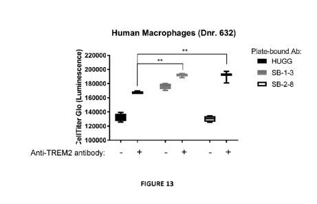
[0046] FIG. 13 shows anti-SIR1131 antibodies enhance the agonistic activity of
anti-TREM2
antibody in a macrophage viability assay. Monocyte-derived macrophages were
cultured on
plate-bound, full-length SB-1-3, SB-2-8, or huIgG1 isotype control in the
presence or absence of
soluble anti-TREM2 antibody. Viable cells were quantified by measuring
luminescence values
following addition of Cell Titer Glo substrate.
[0047] FIG. 14A shows the viability of bone marrow-derived macrophages
obtained from
human SIR1131 BAC transgenic mice cultured on plate-bound, full-length anti-
SIR1131
antibodies SB-1-3, SB-2-8, and SB-8-13 or human IgG1 isotype control. FIG. 14B
shows the
viability of bone marrow-derived dendritic cells obtained from human SIR1131
BAC transgenic
mice cultured on plate-bound, full-length anti-SIRPf31 antibodies SB-1-3, SB-2-
8, and SB-8-13
or human IgG1 isotype control. Viable cells were quantified by measuring
luminescence values
following addition of Cell Titer Glo substrate. Cells cultured on human IgG1
isotype control or
in the absence of plate-bound antibody (No Ab) established baseline viability
of cells.
[0048] FIG. 15A-15C show cross-reactivity of anti-SIR1131 antibodies to
different receptors of the
SIRP family. FIG. 15A and FIG. 15B show titration curves of various anti-SIRP
receptor
antibodies binding BW5147.G.1.4 cells overexpressing recombinant human SIRP131
or recombinant
human SIRPa, respectively. FIG. 15C shows titration curves of antibodies
binding Jurkat cells, an
immortalized human T cell line that endogenously expresses SIRPy. Positive
control antibodies
were anti-SIRPa/13 clone 18D5, anti-SIRPa/f3/y clone KWAR23, and anti-SIRPy
clone LSB2.20.
DETAILED DESCRIPTION
[0049] The present disclosure relates to anti-SIRP131 antibodies (e.g.,
monoclonal antibodies);
methods of making and using such antibodies; pharmaceutical compositions
comprising such
antibodies; nucleic acids encoding such antibodies; and host cells comprising
nucleic acids encoding
such antibodies.
-14-
CA 03099176 2020-11-02
WO 2020/006374 PCT/US2019/039757
[0050] The techniques and procedures described or referenced herein are
generally well understood
and commonly employed using conventional methodology by those skilled in the
art, such as, for
example, the widely utilized methodologies such as those described in Sambrook
et al. Molecular
Cloning: A Laboratory Manual 3d edition (2001) Cold Spring Harbor Laboratory
Press, Cold Spring
Harbor, N.Y.; Current Protocols in Molecular Biology (F.M. Ausubel, et al.
eds., (2003);
Monoclonal Antibodies: A Practical Approach (P. Shepherd and C. Dean, eds.,
Oxford University
Press, 2000).
[0051] All references cited herein, including patent applications and
publications, are hereby
incorporated by reference in their entirety.
I. Definitions
[0052] The terms "Signal-Regulatory Protein (31," "SIRP(31," and "5IRP131
polypeptide" are used
interchangeably herein refer herein to any native SIRP(31 from any vertebrate
source, including
mammals such as primates (e.g., humans and cynos) and rodents (e.g., mice and
rats), unless
otherwise indicated. In some embodiments, the term encompasses both wild-type
sequences and
naturally occurring variant sequences, e.g., splice variants or allelic
variants. In some embodiments,
the term encompasses "full-length," unprocessed 5IRP131 as well as any form of
5IRP131 that results
from processing in the cell. In some embodiments, the 5IRP131 is human
5IRP131. In some
embodiments, the amino acid sequence of an exemplary SIRP(31 isoform 1 is
Uniprot Accession No.
000241 as of February 28, 2018. In some embodiments, the amino acid sequence
of an exemplary
human 5IRP131 is SEQ ID NO: 1. In some embodiments, the amino acid sequence of
an exemplary
5IRP131 isoform 3 is Uniprot Accession No. Q5TFQ8 as of January 31, 2018. In
some embodiments,
the amino acid sequence of an exemplary human 5IRP131 isoform 3 is SEQ ID NO:
384. Unless
specifically indicated otherwise, "SIRP(31" as used herein refers to 5IRP131
isoform 1.
[0053] The terms "anti- 5IRP131 antibody," an "antibody that binds to
SIRP(31," and "antibody that
specifically binds 5IRP131" refer to an antibody that is capable of binding
5IRP131 with sufficient
affinity such that the antibody is useful as a diagnostic and/or therapeutic
agent in targeting 5IRP131.
In one embodiment, the extent of binding of an anti- 5IRP131 antibody to an
unrelated, non- 5IRP131
polypeptide is less than about 10% of the binding of the antibody to 5IRP131
as measured, e.g., by a
radioimmunoassay (RIA). In certain embodiments, an antibody that binds to
5IRP131 has a
dissociation constant (KD) of < 1 pM, < 100 nM, < 10 nM, <1 nM, <0.1 nM, <0.01
nM, or <
0.001 nM (e.g., 10' M or less, e.g. from 10' M to 10-13M, e.g., from 10-9M to
10-13M). In certain
embodiments, an anti-5IRP131 antibody binds to an epitope of 5IRP131 that is
conserved among
5IRP131 polypeptides from different species.
[0054] With regard to the binding of an antibody to a target molecule, the
term "specific binding" or
"specifically binds" or is "specific for" a particular polypeptide or an
epitope on a particular
polypeptide target means binding that is measurably different from a non-
specific interaction.
-15-
CA 03099176 2020-11-02
WO 2020/006374 PCT/US2019/039757
Specific binding can be measured, for example, by determining binding of a
molecule compared to
binding of a control molecule. For example, specific binding can be determined
by competition with
a control molecule that is similar to the target, for example, an excess of
non-labeled target. In this
case, specific binding is indicated if the binding of the labeled target to a
probe is competitively
inhibited by excess unlabeled target. The term "specific binding" or
"specifically binds to" or is
"specific for" a particular polypeptide or an epitope on a particular
polypeptide target as used herein
can be exhibited, for example, by a molecule having a KD for the target of
about any of 10' M or
lower, 10-5 M or lower, 10' M or lower, 10-7 M or lower, 10' M or lower, 10-9
M or lower, 1010 M
or lower, 1011 M or lower, 1012 M or lower or a KD in the range of 10' M to
10' M or 10' M to
1010 M or 10-7 M to 10-9 M. As will be appreciated by the skilled artisan,
affinity and KD values are
inversely related. A high affinity for an antigen is measured by a low KD
value. In one embodiment,
the term "specific binding" refers to binding where a molecule binds to a
particular polypeptide or
epitope on a particular polypeptide without substantially binding to any other
polypeptide or
polypeptide epitope.
[0055] The term "immunoglobulin" (Ig) is used interchangeably with "antibody"
herein. The term
"antibody" herein is used in the broadest sense and specially covers
monoclonal antibodies,
polyclonal antibodies, multispecific antibodies (e.g., bispecific antibodies)
including those formed
from at least two intact antibodies, and antibody fragments so long as they
exhibit the desired
biological activity.
[0056] "Native antibodies" are usually heterotetrameric glycoproteins of about
150,000 Daltons,
composed of two identical Light ("L") chains and two identical heavy ("H")
chains. Each light chain
is linked to a heavy chain by one covalent disulfide bond, while the number of
disulfide linkages
varies among the heavy chains of different immunoglobulin isotypes. Each heavy
and light chain
also has regularly spaced intra-chain disulfide bridges. Each heavy chain has
at one end a variable
domain (VII) followed by a number of constant domains. Each light chain has a
variable domain at
one end (VI) and a constant domain at its other end; the constant domain of
the light chain is aligned
with the first constant domain of the heavy chain, and the light chain
variable domain is aligned with
the variable domain of the heavy chain. Particular amino acid residues are
believed to form an
interface between the light chain and heavy chain variable domains.
[0057] For the structure and properties of the different classes of
antibodies, see, e.g., Basic and
Clinical Immunology, 8th Ed., Daniel P. Stites, Abba I. Terr and Tristram G.
Parslow (eds.),
Appleton & Lange, Norwalk, CT, 1994, page 71 and Chapter 6.
[0058] The light chain from any vertebrate species can be assigned to one of
two clearly distinct
types, called kappa ("K") and lambda ("X"), based on the amino acid sequences
of their constant
domains. Depending on the amino acid sequence of the constant domain of their
heavy chains (CH),
immunoglobulins can be assigned to different classes or isotypes. There are
five classes of
-16-
CA 03099176 2020-11-02
WO 2020/006374 PCT/US2019/039757
immunoglobulins: IgA, IgD, IgE, IgG, and IgM, having heavy chains designated
alpha ("a"), delta
("8"), epsilon ("6"), gamma ("y"), and mu (V), respectively. The y and a
classes are further divided
into subclasses (isotypes) on the basis of relatively minor differences in the
CH sequence and
function, e.g., humans express the following subclasses: IgGl, IgG2, IgG3,
IgG4, IgAl, and IgA2.
The subunit structures and three-dimensional configurations of different
classes of immunoglobulins
are well known and described generally in, for example, Abbas et al., Cellular
and Molecular
Immunology, 4th ed. (W.B. Saunders Co., 2000).
[0059] The "variable region" or "variable domain" of an antibody, such as an
anti- SIR1131 antibody
of the present disclosure, refers to the amino-terminal domains of the heavy
or light chain of the
antibody. The variable domains of the heavy chain and light chain may be
referred to as "VII" and
"VL", respectively. These domains are generally the most variable parts of the
antibody (relative to
other antibodies of the same class) and contain the antigen binding sites.
[0060] The term "variable" refers to the fact that certain segments of the
variable domains differ
extensively in sequence among antibodies, such as anti-5IR1131 antibodies of
the present disclosure.
The variable domain mediates antigen binding and defines the specificity of a
particular antibody for
its particular antigen. However, the variability is not evenly distributed
across the entire span of the
variable domains. Instead, it is concentrated in three segments called
hypervariable regions (HVRs)
both in the light-chain and the heavy chain variable domains. The more highly
conserved portions of
variable domains are called the framework regions (FR). The variable domains
of native heavy and
light chains each comprise four FR regions, largely adopting a beta-sheet
configuration, connected
by three HVRs, which form loops connecting, and in some cases forming part of,
the beta-sheet
structure. The HVRs in each chain are held together in close proximity by the
FR regions and, with
the HVRs from the other chain, contribute to the formation of the antigen-
binding site of antibodies
(see Kabat et al., Sequences of Immunological Interest, Fifth Edition,
National Institute of Health,
Bethesda, MD (1991)). The constant domains are not involved directly in the
binding of antibody to
an antigen, but exhibit various effector functions, such as participation of
the antibody in antibody-
dependent-cellular toxicity.
[0061] The term "monoclonal antibody" as used herein refers to an antibody,
such as a monoclonal
anti-SIR1131 antibody of the present disclosure, obtained from a population of
substantially
homogeneous antibodies, i.e., the individual antibodies comprising the
population are identical
except for possible naturally occurring mutations and/or post-translation
modifications (e.g.,
isomerizations, amidations, etc.) that may be present in minor amounts.
Monoclonal antibodies are
highly specific, being directed against a single antigenic site. In contrast
to polyclonal antibody
preparations which typically include different antibodies directed against
different determinants
(epitopes), each monoclonal antibody is directed against a single determinant
on the antigen. In
addition to their specificity, the monoclonal antibodies are advantageous in
that they are synthesized
-17-
CA 03099176 2020-11-02
WO 2020/006374 PCT/US2019/039757
by the hybridoma culture, uncontaminated by other immunoglobulins. The
modifier "monoclonal"
indicates the character of the antibody as being obtained from a substantially
homogeneous
population of antibodies, and is not to be construed as requiring production
of the antibody by any
particular method. For example, the monoclonal antibodies to be used in
accordance with the present
invention may be made by a variety of techniques, including, for example, the
hybridoma method,
recombinant DNA methods, and technologies for producing human or human-like
antibodies in
animals that have parts or all of the human immunoglobulin loci or genes
encoding human
immunoglobulin sequences.
[0062] The terms "full-length antibody," "intact antibody" or "whole antibody"
are used
interchangeably to refer to an antibody, such as an anti- SIR1131 antibody of
the present disclosure,
in its substantially intact form, as opposed to an antibody fragment.
Specifically, whole antibodies
include those with heavy and light chains including an Fc region. The constant
domains may be
native sequence constant domains (e.g., human native sequence constant
domains) or amino acid
sequence variants thereof. In some cases, the intact antibody may have one or
more effector
functions.
[0063] An "antibody fragment" refers to a molecule other than an intact
antibody that comprises a
portion of an intact antibody that binds the antigen to which the intact
antibody binds. Examples of
antibody fragments include Fab, Fab', F(a131)2 and Fv fragments; diabodies;
linear antibodies (see
U.S. Patent 5641870, Example 2; Zapata et al., Protein Eng. 8(10):1057-1062
(1995)); single-chain
antibody molecules and multispecific antibodies formed from antibody
fragments.
[0064] Papain digestion of antibodies, such as anti-5IRP131 antibodies of the
present disclosure,
produces two identical antigen-binding fragments, called "Fab" fragments, and
a residual "Fc"
fragment, a designation reflecting the ability to crystallize readily. The Fab
fragment consists of an
entire light chain along with the variable region domain of the heavy chain
(VII), and the first
constant domain of one heavy chain (CH1). Each Fab fragment is monovalent with
respect to antigen
binding, i.e., it has a single antigen-binding site. Pepsin treatment of an
antibody yields a single large
F(a131)2 fragment which roughly corresponds to two disulfide linked Fab
fragments having different
antigen-binding activity and is still capable of cross-linking antigen. Fab'
fragments differ from Fab
fragments by having a few additional residues at the carboxy terminus of the
CH1 domain including
one or more cysteines from the antibody hinge region. Fab'-SH is the
designation herein for Fab' in
which the cysteine residue(s) of the constant domains bear a free thiol group.
F(a131)2 antibody
fragments originally were produced as pairs of Fab' fragments which have hinge
cysteines between
them. Other chemical couplings of antibody fragments are also known.
[0065] The Fc fragment comprises the carboxy-terminal portions of both heavy
chains held together
by disulfides. The effector functions of antibodies are determined by
sequences in the Fc region, the
region which is also recognized by Fc receptors (FcR) found on certain types
of cells.
-18-
CA 03099176 2020-11-02
WO 2020/006374 PCT/US2019/039757
[0066] "Functional fragments" of antibodies, such as anti-SIRP131 antibodies
of the present
disclosure, comprise a portion of an intact antibody, generally including the
antigen binding or
variable region of the intact antibody or the Fc region of an antibody which
retains or has modified
FcR binding capability. Examples of antibody fragments include linear
antibody, single-chain
antibody molecules and multispecific antibodies formed from antibody
fragments.
[0067] The term "diabodies" refers to small antibody fragments prepared by
constructing sFy
fragments (see preceding paragraph) with short linkers (about 5-10) residues)
between the VII and
Vt, domains such that inter-chain but not intra-chain pairing of the variable
domains is achieved,
thereby resulting in a bivalent fragment, i.e., a fragment having two antigen-
binding sites. Bispecific
diabodies are heterodimers of two "crossover" sFy fragments in which the VII
and Vt, domains of the
two antibodies are present on different polypeptide chains.
[0068] As used herein, a "chimeric antibody" refers to an antibody
(immunoglobulin), such as a
chimeric anti-SIRP131 antibody of the present disclosure, in which a portion
of the heavy and/or light
chain is identical with or homologous to corresponding sequences in antibodies
derived from a
particular species or belonging to a particular antibody class or subclass,
while the remainder of the
chain(s) is(are) identical with or homologous to corresponding sequences in
antibodies derived from
another species or belonging to another antibody class or subclass, as well as
fragments of such
antibodies, so long as they exhibit the desired biological activity. Chimeric
antibodies of interest
herein include PRIMATIZED antibodies wherein the antigen-binding region of
the antibody is
derived from an antibody produced by, e.g., immunizing macaque monkeys with an
antigen of
interest. As used herein, "humanized antibody" is used a subset of "chimeric
antibodies."
[0069] "Humanized" forms of non-human (e.g., murine) antibodies, such as
humanized forms of
anti- SIRP131 antibodies of the present disclosure, are chimeric antibodies
comprising amino acid
residues from non-human HVRs and amino acid residues from human FRs. In
certain embodiments,
a humanized antibody will comprise substantially all of at least one, and
typically two, variable
domains, in which all or substantially all of the HVRs (e.g., CDRs) correspond
to those of a non-
human antibody, and all or substantially all of the FRs correspond to those of
a human antibody. A
humanized antibody optionally may comprise at least a portion of an antibody
constant region
derived from a human antibody. A "humanized form" of an antibody, e.g., a non-
human antibody,
refers to an antibody that has undergone humanization.
[0070] A "human antibody" is one that possesses an amino-acid sequence
corresponding to that of
an antibody, such as an anti-SIRP131 antibody of the present disclosure,
produced by a human and/or
has been made using any of the techniques for making human antibodies as
disclosed herein. This
definition of a human antibody specifically excludes a humanized antibody
comprising non-human
antigen-binding residues. Human antibodies can be produced using various
techniques known in the
art, including phage-display libraries and yeast-based platform technologies.
Human antibodies can
-19-
CA 03099176 2020-11-02
WO 2020/006374 PCT/US2019/039757
be prepared by administering the antigen to a transgenic animal that has been
modified to produce
such antibodies in response to antigenic challenge, but whose endogenous loci
have been disabled,
e.g., immunized xenomice as well as generated via a human B-cell hybridoma
technology.
[0071] The term "hypervariable region" or "HVR," when used herein refers to
the regions of an
antibody-variable domain, such as that of an anti-SIM:131 antibody of the
present disclosure, that are
hypervariable in sequence and/or form structurally defined loops. Generally,
antibodies comprise six
HVRs; three in the VII (H1, H2, H3), and three in the Vi. (L1, L2, L3). In
native antibodies, H3 and
L3 display the most diversity of the six HVRs, and H3 in particular is
believed to play a unique role
in conferring fine specificity to antibodies. Naturally occurring camelid
antibodies consisting of a
heavy chain only are functional and stable in the absence of light chain.
[0072] A number of HVR delineations are in use and are encompassed herein. In
some
embodiments, the HVRs may be Kabat complementarity-determining regions (CDRs)
based on
sequence variability and are the most commonly used (Kabat et al., supra). In
some embodiments,
the HVRs may be Chothia CDRs. Chothia refers instead to the location of the
structural loops
(Chothia and Lesk I Mot Biol. 196:901-917 (1987)). In some embodiments, the
HVRs may be
AbM HVRs. The AbM HVRs represent a compromise between the Kabat CDRs and
Chothia
structural loops, and are used by Oxford Molecular's AbM antibody-modeling
software. In some
embodiments, the HVRs may be "contact" HVRs. The "contact" HVRs are based on
an analysis of
the available complex crystal structures. The residues from each of these HVRs
are noted below.
Loop Kabat AbM Chothia Contact
Li L24-L34 L24-L34 L26-L32 L30-L36
L2 L50-L56 L50-L56 L50-L52 L46-L55
L3 L89-L97 L89-L97 L91-L96 L89-L96
H1 H31-H35B H26-H35B H26-H32 H30-H35B (Kabat numbering)
H1 H31-H35 H26-H35 H26-H32 H30-H35 (Chothia numbering)
H2 H50-H65 H50-H58 H53-H55 H47-H58
H3 H95-H102 H95-H102 H96-H101 H93-H101
[0073] HVRs may comprise "extended HVRs" as follows: 24-36 or 24-34 (L1), 46-
56 or 50-56
(L2), and 89-97 or 89-96 (L3) in the VL, and 26-35 (H1), 50-65 or 49-65 (a
preferred embodiment)
(H2), and 93-102, 94-102, or 95-102 (H3) in the VH. The variable-domain
residues are numbered
according to Kabat et al., supra, for each of these extended-HVR definitions.
[0074] "Framework" or "FR" residues are those variable-domain residues other
than the HVR
residues as herein defined.
[0075] An "acceptor human framework" as used herein is a framework comprising
the amino acid
sequence of a Vi. or VII framework derived from a human immunoglobulin
framework or a human
consensus framework. An acceptor human framework "derived from" a human
immunoglobulin
-20-
CA 03099176 2020-11-02
WO 2020/006374 PCT/US2019/039757
framework or a human consensus framework may comprise the same amino acid
sequence thereof,
or it may comprise pre-existing amino acid sequence changes. In some
embodiments, the number of
pre-existing amino acid changes are 10 or less, 9 or less, 8 or less, 7 or
less, 6 or less, 5 or less, 4 or
less, 3 or less, or 2 or less. Where pre-existing amino acid changes are
present in a VH, preferable
those changes occur at only three, two, or one of positions 71H, 73H and 78H;
for instance, the
amino acid residues at those positions may by 71A, 73T and/or 78A. In one
embodiment, the VL
acceptor human framework is identical in sequence to the VL human
immunoglobulin framework
sequence or human consensus framework sequence.
[0076] A "human consensus framework" is a framework that represents the most
commonly
occurring amino acid residues in a selection of human immunoglobulin VL or VII
framework
sequences. Generally, the selection of human immunoglobulin VL or VII
sequences is from a
subgroup of variable domain sequences. Generally, the subgroup of sequences is
a subgroup as in
Kabat et al., Sequences of Proteins of Immunological Interest, 5th Ed. Public
Health Service,
National Institutes of Health, Bethesda, MD (1991). Examples include for the
VL, the subgroup may
be subgroup kappa I, kappa II, kappa III or kappa IV as in Kabat et al.,
supra. Additionally, for the
VII, the subgroup may be subgroup I, subgroup II, or subgroup III as in Kabat
et al., supra.
[0077] An "amino-acid modification" at a specified position, e.g., of an anti-
SIRPf31 antibody of the
present disclosure, refers to the substitution or deletion of the specified
residue, or the insertion of at
least one amino acid residue adjacent the specified residue. Insertion
"adjacent" to a specified
residue means insertion within one to two residues thereof. The insertion may
be N-terminal or C-
terminal to the specified residue. The preferred amino acid modification
herein is a substitution.
[0078] An "affinity-matured" antibody, such as an affinity matured anti-
5IRP131 antibody of the
present disclosure, is one with one or more alterations in one or more HVRs
thereof that result in an
improvement in the affinity of the antibody for antigen, compared to a parent
antibody that does not
possess those alteration(s). In one embodiment, an affinity-matured antibody
has nanomolar or even
picomolar affinities for the target antigen. Affinity-matured antibodies are
produced by procedures
known in the art. For example, Marks et al. Bio/Technology 10:779-783 (1992)
describes affinity
maturation by VII- and VL-domain shuffling. Random mutagenesis of HVR and/or
framework
residues is described by, for example: Barbas et al. Proc Nat. Acad. Sci. USA
91:3809-3813 (1994);
Schier et al. Gene 169:147-155 (1995); Yelton et al. J. Immunot 155: 1994-2004
(1995); Jackson et
al. J. Immunol. 154(7):3310-9 (1995); and Hawkins et al, J. Mot Biol. 226:889-
896 (1992).
[0079] "Fv" is the minimum antibody fragment which comprises a complete
antigen-recognition
and -binding site. This fragment consists of a dimer of one heavy- and one
light-chain variable
region domain in tight, non-covalent association. From the folding of these
two domains emanate six
hypervariable loops (3 loops each from the H and L chain) that contribute the
amino acid residues
for antigen binding and confer antigen binding specificity to the antibody.
However, even a single
-21-
CA 03099176 2020-11-02
WO 2020/006374 PCT/US2019/039757
variable domain (or half of an Fv comprising only three HVRs specific for an
antigen) has the ability
to recognize and bind antigen, although at a lower affinity than the entire
binding site.
[0080] "Single-chain Fv" also abbreviated as "sFy" or "seFv" are antibody
fragments that comprise
the VH and VL antibody domains connected into a single polypeptide chain.
Preferably, the sFy
polypeptide further comprises a polypeptide linker between the VII and Vt,
domains, which enables
the sFy to form the desired structure for antigen binding.
[0081] Antibody "effector functions" refer to those biological activities
attributable to the Fc region
(a native sequence Fc region or amino acid sequence variant Fc region) of an
antibody, and vary
with the antibody isotype.
[0082] The term "Fe region" herein is used to define a C-terminal region of an
immunoglobulin
heavy chain, including native-sequence Fc regions and variant Fc regions.
Although the boundaries
of the Fc region of an immunoglobulin heavy chain might vary, the human IgG
heavy-chain Fc
region is usually defined to stretch from an amino acid residue at position
Cys226, or from Pro230,
to the carboxyl-terminus thereof. The C-terminal lysine (residue 447 according
to the EU numbering
system) of the Fc region may be removed, for example, during production or
purification of the
antibody, or by recombinantly engineering the nucleic acid encoding a heavy
chain of the antibody.
Accordingly, a composition of intact antibodies may comprise antibody
populations with all K447
residues removed, antibody populations with no K447 residues removed, and
antibody populations
having a mixture of antibodies with and without the K447 residue. Suitable
native-sequence Fc
regions for use in the antibodies of the present disclosure include human
IgGl, IgG2, IgG3 and
IgG4.
[0083] A "native sequence Fc region" comprises an amino acid sequence
identical to the amino acid
sequence of an Fc region found in nature. Native sequence human Fc regions
include a native
sequence human IgG1 Fc region (non-A and A allotypes); native sequence human
IgG2 Fc region;
native sequence human IgG3 Fc region; and native sequence human IgG4 Fc region
as well as
naturally occurring variants thereof.
[0084] A "variant Fc region" comprises an amino acid sequence which differs
from that of a native
sequence Fc region by virtue of at least one amino acid modification,
preferably one or more amino
acid substitution(s). Preferably, the variant Fc region has at least one amino
acid substitution
compared to a native sequence Fc region or to the Fc region of a parent
polypeptide, e.g. from about
one to about ten amino acid substitutions, and preferably from about one to
about five amino acid
substitutions in a native sequence Fc region or in the Fc region of the parent
polypeptide. The variant
Fc region herein will preferably possess at least about 80% homology with a
native sequence Fc
region and/or with an Fc region of a parent polypeptide, and most preferably
at least about 90%
homology therewith, more preferably at least about 95% homology therewith.
-22-
CA 03099176 2020-11-02
WO 2020/006374 PCT/US2019/039757
[0085] "Fc receptor" or "FcR" describes a receptor that binds to the Fc region
of an antibody. The
preferred FcR is a native sequence human FcR. Moreover, a preferred FcR is one
which binds an
IgG antibody (a gamma receptor) and includes receptors of the FcyRI, FcyRII,
and FcyRIII
subclasses, including allelic variants and alternatively spliced forms of
these receptors, FcyRII
receptors include FcyRIIA (an "activating receptor") and FcyRIIB (an
"inhibiting receptor"), which
have similar amino acid sequences that differ primarily in the cytoplasmic
domains thereof.
Activating receptor FcyRIIA contains an immunoreceptor tyrosine-based
activation motif ("ITAM")
in its cytoplasmic domain. Inhibiting receptor FcyRIIB contains an
immunoreceptor tyrosine-based
inhibition motif ("ITIM") in its cytoplasmic domain. Other FcRs, including
those to be identified in
the future, are encompassed by the term "FcR" herein. FcRs can also increase
the serum half-life of
antibodies.
[0086] As used herein, "percent (%) amino acid sequence identity" and
"homology" with respect to
a peptide, polypeptide or antibody sequence refers to the percentage of amino
acid residues in a
candidate sequence that are identical with the amino acid residues in the
specific peptide or
polypeptide sequence, after aligning the sequences and introducing gaps, if
necessary, to achieve the
maximum percent sequence identity, and not considering any conservative
substitutions as part of
the sequence identity. Alignment for purposes of determining percent amino
acid sequence identity
can be achieved in various ways that are within the skill in the art, for
instance, using publicly
available computer software such as BLAST, BLAST-2, ALIGN or MEGALIGNTm
(DNASTAR)
software. Those skilled in the art can determine appropriate parameters for
measuring alignment,
including any algorithms known in the art needed to achieve maximal alignment
over the full-length
of the sequences being compared.
[0087] The term "compete" when used in the context of antibodies (e.g.,
neutralizing antibodies)
that compete for the same epitope means competition between antibody as
determined by an assay in
which the antibody being tested prevents or inhibits (e.g., reduces) specific
binding of a reference
molecule (e.g., a ligand, or a reference antibody) to a common antigen (e.g.,
5IRP131 or a fragment
thereof). Numerous types of competitive binding assays can be used to
determine if antibody
competes with another, for example: solid phase direct or indirect
radioimmunoassay (RIA), solid
phase direct or indirect enzyme immunoassay (EIA), sandwich competition assay
(see, e.g., Stahli et
al., 1983, Methods in Enzymology 9:242-253); solid phase direct biotin-avidin
ETA (see, e.g.,
Kirkland et al., 1986, J. Immunol. 137:3614-3619) solid phase direct labeled
assay, solid phase
direct labeled sandwich assay (see, e.g., Harlow and Lane, 1988, Antibodies, A
Laboratory Manual,
Cold Spring Harbor Press); solid phase direct label RIA using 1-125 label
(see, e.g., Morel et al.,
1988, Molec. Immunol. 25:7-15); solid phase direct biotin-avidin ETA (see,
e.g., Cheung, et al.,
1990, Virology 176:546-552); and direct labeled RIA (Moldenhauer et al., 1990,
Scand. J. Immunol.
32:77-82). Typically, such an assay involves the use of purified antigen bound
to a solid surface or
-23-
CA 03099176 2020-11-02
WO 2020/006374 PCT/US2019/039757
cells bearing either of these, an unlabelled test antibody and a labeled
reference antibody.
Competitive inhibition is measured by determining the amount of label bound to
the solid surface or
cells in the presence of the test antibody. Usually the test antibody is
present in excess. Antibodies
identified by competition assay (competing antibodies) include antibodies
binding to the same
epitope as the reference antibody and antibodies binding to an adjacent
epitope sufficiently proximal
to the epitope bound by the reference antibody for steric hindrance to occur.
Additional details
regarding methods for determining competitive binding are provided in the
examples herein.
Usually, when a competing antibody is present in excess, it will inhibit
(e.g., reduce) specific
binding of a reference antibody to a common antigen by at least 20%, 30%, 40%,
50%, 60%, 70%,
80%, 85%, 90%, 95%, 97.5%, and/or near 100%.
[0088] As used herein, an "interaction" between a SIR1131 polypeptide and a
second polypeptide
encompasses, without limitation, protein-protein interaction, a physical
interaction, a chemical
interaction, binding, covalent binding, and ionic binding. As used herein, an
antibody "inhibits
interaction" between two polypeptides when the antibody disrupts, reduces, or
completely eliminates
an interaction between the two polypeptides. An antibody of the present
disclosure, thereof, "inhibits
interaction" between two polypeptides when the antibody thereof binds to one
of the two
polypeptides. In some embodiments, the interaction can be inhibited by at
least about any of 20%,
30%, 40%, 50%, 60%, 70%, 80%, 85%, 90%, 95%, 97.5%, and/or near 100%.
[0089] The term "epitope" includes any determinant capable of being bound by
an antibody. An
epitope is a region of an antigen that is bound by an antibody that targets
that antigen, and when the
antigen is a polypeptide, includes specific amino acids that directly contact
the antibody. Most often,
epitopes reside on polypeptides, but in some instances, can reside on other
kinds of molecules, such
as nucleic acids. Epitope determinants can include chemically active surface
groupings of molecules
such as amino acids, sugar side chains, phosphoryl or sulfonyl groups, and can
have specific three-
dimensional structural characteristics, and/or specific charge
characteristics. Generally, antibodies
specific for a particular target antigen will preferentially recognize an
epitope on the target antigen in
a complex mixture of polypeptides and/or macromolecules.
[0090] An "agonist" antibody or an "activating" antibody is an antibody that
induces (e.g.,
increases) one or more activities or functions of the antigen after the
antibody binds the antigen.
[0091] An "antagonist" antibody or a "blocking" antibody or an "inhibitory"
antibody is an antibody
that reduces, inhibits, and/or eliminates (e.g., decreases) antigen binding to
one or more ligand after
the antibody binds the antigen, and/or that reduces, inhibits, and/or
eliminates (e.g., decreases) one
or more activities or functions of the antigen after the antibody binds the
antigen. In some
embodiments, antagonist antibodies, or blocking antibodies, or inhibitory
antibodies substantially or
completely inhibit antigen binding to one or more ligand and/or one or more
activities or functions
of the antigen.
-24-
CA 03099176 2020-11-02
WO 2020/006374 PCT/US2019/039757
[0092] An "isolated" antibody, such as an isolated anti-SIRP131 antibody of
the present disclosure, is
one that has been identified, separated and/or recovered from a component of
its production
environment (e.g., naturally or recombinantly). Preferably, the isolated
antibody is free of
association with all other contaminant components from its production
environment. Contaminant
components from its production environment, such as those resulting from
recombinant transfected
cells, are materials that would typically interfere with research, diagnostic
or therapeutic uses for the
antibody, and may include enzymes, hormones, and other proteinaceous or non-
proteinaceous
solutes. In preferred embodiments, the antibody will be purified: (1) to
greater than 95% by weight
of antibody as determined by, for example, the Lowry method, and in some
embodiments, to greater
than 99% by weight; (2) to a degree sufficient to obtain at least 15 residues
of N-terminal or internal
amino acid sequence by use of a spinning cup sequenator, or (3) to homogeneity
by SDS-PAGE
under non-reducing or reducing conditions using Coomassie blue or, preferably,
silver stain. Isolated
antibody includes the antibody in situ within recombinant T-cells since at
least one component of the
antibody's natural environment will not be present. Ordinarily, however, an
isolated polypeptide or
antibody will be prepared by at least one purification step.
[0093] An "isolated" nucleic acid molecule encoding an antibody, such as an
anti-SIRP131 antibody
of the present disclosure, is a nucleic acid molecule that is identified and
separated from at least one
contaminant nucleic acid molecule with which it is ordinarily associated in
the environment in which
it was produced. Preferably, the isolated nucleic acid is free of association
with all components
associated with the production environment. The isolated nucleic acid
molecules encoding the
polypeptides and antibodies herein is in a form other than in the form or
setting in which it is found
in nature. Isolated nucleic acid molecules therefore are distinguished from
nucleic acid encoding the
polypeptides and antibodies herein existing naturally in cells.
[0094] The term "vector," as used herein, is intended to refer to a nucleic
acid molecule capable of
transporting another nucleic acid to which it has been linked. One type of
vector is a "plasmid,"
which refers to a circular double stranded DNA into which additional DNA
segments may be
ligated. Another type of vector is a phage vector. Another type of vector is a
viral vector, wherein
additional DNA segments may be ligated into the viral genome. Certain vectors
are capable of
autonomous replication in a host cell into which they are introduced (e.g.,
bacterial vectors having a
bacterial origin of replication and episomal mammalian vectors). Other vectors
(e.g., non-episomal
mammalian vectors) can be integrated into the genome of a host cell upon
introduction into the host
cell, and thereby are replicated along with the host genome. Moreover, certain
vectors are capable of
directing the expression of genes to which they are operatively linked. Such
vectors are referred to
herein as "recombinant expression vectors," or simply, "expression vectors."
In general, expression
vectors of utility in recombinant DNA techniques are often in the form of
plasmids. In the present
-25-
CA 03099176 2020-11-02
WO 2020/006374 PCT/US2019/039757
specification, "plasmid" and "vector" may be used interchangeably as the
plasmid is the most
commonly used form of vector.
[0095] "Polynucleotide," or "nucleic acid," as used interchangeably herein,
refer to polymers of
nucleotides of any length, and include DNA and RNA. The nucleotides can be
deoxyribonucleotides,
ribonucleotides, modified nucleotides or bases, and/or their analogs, or any
substrate that can be
incorporated into a polymer by DNA or RNA polymerase or by a synthetic
reaction.
[0096] A "host cell" includes an individual cell or cell culture that can be
or has been a recipient for
vector(s) for incorporation of polynucleotide inserts. Host cells include
progeny of a single host cell,
and the progeny may not necessarily be completely identical (in morphology or
in genomic DNA
complement) to the original parent cell due to natural, accidental, or
deliberate mutation. A host cell
includes cells transfected in vivo with a polynucleotide(s) of this invention.
[0097] "Carriers" as used herein include pharmaceutically acceptable carriers,
excipients, or
stabilizers that are nontoxic to the cell or mammal being exposed thereto at
the dosages and
concentrations employed.
[0098] As used herein, the term "preventing" includes providing prophylaxis
with respect to
occurrence or recurrence of a particular disease, disorder, or condition in an
individual. An
individual may be predisposed to, susceptible to a particular disease,
disorder, or condition, or at risk
of developing such a disease, disorder, or condition, but has not yet been
diagnosed with the disease,
disorder, or condition.
[0099] As used herein, an individual "at risk" of developing a particular
disease, disorder, or
condition may or may not have detectable disease or symptoms of disease, and
may or may not have
displayed detectable disease or symptoms of disease prior to the treatment
methods described herein.
"At risk" denotes that an individual has one or more risk factors, which are
measurable parameters
that correlate with development of a particular disease, disorder, or
condition, as known in the art.
An individual having one or more of these risk factors has a higher
probability of developing a
particular disease, disorder, or condition than an individual without one or
more of these risk factors.
[00100] As used herein, the term "treatment" refers to clinical intervention
designed to alter the
natural course of the individual being treated during the course of clinical
pathology. Desirable
effects of treatment include decreasing the rate of progression, ameliorating
or palliating the
pathological state, and remission or improved prognosis of a particular
disease, disorder, or
condition. An individual is successfully "treated", for example, if one or
more symptoms associated
with a particular disease, disorder, or condition are mitigated or eliminated.
[00101] An "effective amount" refers to at least an amount effective, at
dosages and for periods of
time necessary, to achieve the desired therapeutic or prophylactic result. An
effective amount can be
provided in one or more administrations. An effective amount herein may vary
according to factors
such as the disease state, age, sex, and weight of the individual, and the
ability of the treatment to
-26-
CA 03099176 2020-11-02
WO 2020/006374 PCT/US2019/039757
elicit a desired response in the individual. An effective amount is also one
in which any toxic or
detrimental effects of the treatment are outweighed by the therapeutically
beneficial effects. For
prophylactic use, beneficial or desired results include results such as
eliminating or reducing the risk,
lessening the severity, or delaying the onset of the disease, including
biochemical, histological
and/or behavioral symptoms of the disease, its complications and intermediate
pathological
phenotypes presenting during development of the disease. For therapeutic use,
beneficial or desired
results include clinical results such as decreasing one or more symptoms
resulting from the disease,
increasing the quality of life of those suffering from the disease, decreasing
the dose of other
medications required to treat the disease, enhancing effect of another
medication such as via
targeting, delaying the progression of the disease, and/or prolonging
survival. An effective amount
of drug, compound, or pharmaceutical composition is an amount sufficient to
accomplish
prophylactic or therapeutic treatment either directly or indirectly. As is
understood in the clinical
context, an effective amount of a drug, compound, or pharmaceutical
composition may or may not
be achieved in conjunction with another drug, compound, or pharmaceutical
composition. Thus, an
"effective amount" may be considered in the context of administering one or
more therapeutic
agents, and a single agent may be considered to be given in an effective
amount if, in conjunction
with one or more other agents, a desirable result may be or is achieved.
[00102] An "individual" for purposes of treatment, prevention, or reduction of
risk refers to any
animal classified as a mammal, including humans, domestic and farm animals,
and zoo, sport, or pet
animals, such as dogs, horses, rabbits, cattle, pigs, hamsters, gerbils, mice,
ferrets, rats, cats, and the
like. In some embodiments, the individual is human.
[00103] As used herein, administration "in conjunction" with another compound
or composition
includes simultaneous administration and/or administration at different times.
Administration in
conjunction also encompasses administration as a co-formulation or
administration as separate
compositions, including at different dosing frequencies or intervals, and
using the same route of
administration or different routes of administration. In some embodiments,
administration in
conjunction is administration as a part of the same treatment regimen.
[00104] The term "about" as used herein refers to the usual error range for
the respective value
readily known to the skilled person in this technical field. Reference to
"about" a value or parameter
herein includes (and describes) embodiments that are directed to that value or
parameter per se.
[00105] As used herein and in the appended claims, the singular forms "a,"
"an," and "the" include
plural reference unless the context clearly indicates otherwise. For example,
reference to an
"antibody" is a reference to from one to many antibodies, such as molar
amounts, and includes
equivalents thereof known to those skilled in the art, and so forth.
[00106] It is understood that aspect and embodiments of the present disclosure
described herein
include "comprising," "consisting," and "consisting essentially of' aspects
and embodiments.
-27-
CA 03099176 2020-11-02
WO 2020/006374 PCT/US2019/039757
I. Anti-SIRPfil Antibodies
[00107] Provided herein are anti-SIRP131 antibodies. Antibodies provided are
useful, e.g., for the
diagnosis or treatment of the SIRP131 mediated disorders.
[00108] In some embodiments, an anti-SIRP131 antibody of the present
disclosure is an agonist of
SIRP131 activity. In some embodiments, an anti-SIRP131 antibody is provided
that binds to human
SIRP131 isoform 1 but does not bind to human SIRPa. In some embodiments, an
anti-SIRP131
antibody is provided that binds to human SIRP131 isoform 1 but does not bind
to human SIRPy. In
some embodiments, an anti-SIRP131 antibody is provided that binds to human
SIRP131 isoform 1 but
does not bind to human SIRP131 isoform 3. In some embodiments, an anti-SIRP131
antibody is
provided that binds to human SIRP131 isoform 1 but does not bind to mouse
SIRP131 and/or
cynomolgus monkey SIRP131. In some embodiments, an anti-SIRP131 antibody of
the present
disclosure agonizes SIRP131 activity on CD14-positive monocytes. In some
embodiments, an anti-
SIRP131 antibody of the present disclosure induces respiratory burst in immune
cells, such as
neutrophils, monocytes, and/or macrophages. In some embodiments, an anti-
SIRP131 antibody of the
present disclosure induces or increases IL-8 expression in monocytes. In some
embodiments, an
anti-SIRP131 antibody of the present disclosure induces or increases TNFa
expression in
macrophages and/or dendritic cells. In some embodiments, an anti-SIRP131
antibody of the present
disclosure induces neutrophil-mediated phagocytosis, for example, of tumor
cells. In some
embodiments, an anti-SIRP131 antibody of the present disclosure increases
neutrophil-mediated
tumor cell clearance. In some embodiments, an anti-SIRP131 antibody of the
present disclosure
increases or enhances anti-tumor properties or activity of neutrophils.
[00109] In some embodiments, an anti-SIRP131 antibody of the present
disclosure recruits immune
cells. In some embodiments, an anti-SIRP131 antibody of the present disclosure
induces syk
phosphorylation. In some embodiments, an anti-SIRP131 antibody of the present
disclosure induces
syk phosphorylation when clustered by adjacent cells expressing Fc gamma
receptors.
[00110] In some embodiments, SIRP131 antibody of the present disclosure
downregulates SIRP131
expression on the surface of a cell. In some embodiments, SIRP131 antibody of
the present
disclosure does not downregulate SIRP131 expression on the surface of a cell.
In some embodiments,
SIRPf31 antibody of the present disclosure blocks binding of a ligand to
SIRPf31. In some
embodiments, SIRPf31 antibody of the present disclosure does not block binding
of a ligand to
SIRPf31.
[00111] In some embodiments, an anti-SIRP131 antibody is provided that has one
or more properties
selected from:
a) binds to human SIRP131 isoform 1, but does not bind to human SIRPa;
b) binds to human SIRP131 isoform 1, but does not bind to human SIRPy;
c) binds to human SIRP131 isoform 1, but does not bind to human SIRP131
isoform 3;
-28-
CA 03099176 2020-11-02
WO 2020/006374 PCT/US2019/039757
d) binds to human SIRP131 isoform 1, but does not bind to mouse SIRP131;
e) binds to human SIRP131 isoform 1, but does not bind to cynomolgus monkey
SIRP131;
f) agonizes SIRP131 activity on CD14-positive monocytes in vitro and/or in
vivo;
g) induces or increases respiratory burst in immune cells, such as neutrophils
and/or monocytes
in vitro and/or in vivo;
h) induces or increases IL-8 expression in monocytes in vitro and/or in vivo;
i) induces or increases TNFa expression in macrophages and/or dendritic cells
in vitro and/or
in vivo;
j) induces or increases neutrophil-mediated phagocytosis, for example, of
tumor cells in vitro
and/or in vivo;
k) induces or increases neutrophil-mediated tumor cell clearance in vivo;
1) upregulates TREM2 expression on macrophages in vitro and/or in vivo;
m) increases viability of macrophages in vitro and/or in vivo, alone and/or in
combination with
an agonist anti-TREM2 antibody; and
n) increases viability of dendritic cells in vitro and/or in vivo.
[00112] In some embodiments, an anti-SIRP131 antibody is provided that has the
following
properties:
a) binds to human SIRP131 isoform 1, but does not bind to human SIRPa;
b) binds to human SIRP131 isoform 1, but does not bind to human SIRPy;
c) binds to human SIRP131 isoform 1, but does not bind to human SIRP131
isoform 3;
d) binds to human SIRP131 isoform 1, but does not bind to mouse SIRP131;
e) binds to human SIRP131 isoform 1, but does not bind to cynomolgus monkey
SIRP131;
f) agonizes SIRP131 activity on CD14-positive monocytes in vitro and/or in
vivo;
g) induces or increases respiratory burst in immune cells, such as neutrophils
and/or monocytes
in vitro and/or in vivo;
h) induces or increases IL-8 expression in monocytes in vitro and/or in vivo;
i) induces or increases TNFa expression in macrophages and/or dendritic cells
in vitro and/or
in vivo;
j) induces or increases neutrophil-mediated phagocytosis, for example, of
tumor cells in vitro
and/or in vivo;
o) induces or increases neutrophil-mediated tumor cell clearance in vivo;
p) upregulates TREM2 expression on macrophages in vitro and/or in vivo;
q) increases viability of macrophages in vitro and/or in vivo, alone and/or in
combination with
an agonist anti-TREM2 antibody; and
k) increases viability of dendritic cells in vitro and/or in vivo.
-29-
CA 03099176 2020-11-02
WO 2020/006374 PCT/US2019/039757
A. Exemplary Antibodies and Certain Other Antibody Embodiments
[00113] In some embodiments, provided herein are anti-SIR1131 antibodies
comprising at least one,
two, three, four, five, or six HVRs selected from (a) HVR-Hl comprising an
amino acid sequence of
an HVR-Hl shown in Table 4 and/or Table 9; (b) HVR-H2 comprising an amino acid
sequence of
an HVR-Hl shown in Table 4 and/or Table 9; (c) HVR-H3 comprising an amino acid
sequence of an
HVR-Hl shown in Table 4 and/or Table 9; (d) HVR-Li comprising an amino acid
sequence of an
HVR-Hl shown in Table 3 and/or Table 8; (e) HVR-L2 comprising an amino acid
sequence of an
HVR-Hl shown in Table 3 and/or Table 8; and (f) HVR-L3 comprising an amino
acid sequence of
an HVR-Hl shown in Table 3 and/or Table 8.
[00114] In some embodiments, provided herein are anti-SIR1131 antibodies
comprising HVR-H1,
HVR-H2, HVR-H3, HVR-L1, HVR-L2, and HVR-L3, wherein: (a) HVR-Hl comprising an
amino
acid sequence of an HVR-Hl shown in Table 4 and/or Table 9; (b) HVR-H2
comprising an amino
acid sequence of an HVR-H2 shown in Table 4 and/or Table 9; (c)HVR-H3
comprising an amino
acid sequence of an HVR-H3 shown in Table 4 and/or Table 9; (d) HVR-Li
comprising an amino
acid sequence of an HVR-Li shown in Table 3 and/or Table 8; (e) HVR-L2
comprising an amino
acid sequence of an HVR-L2 shown in Table 3 and/or Table 8; and (f) HVR-L3
comprising an
amino acid sequence of an HVR-L3 shown in Table 3 and/or Table 8.
[00115] In some embodiments, provided herein are anti-SIR1131 antibodies
comprising six HVRs of
an antibody selected from SB-1, SB-2, SB-3, SB-4, SB-5, SB-6, SB-7, SB-8, SB-
9, SB-10, SB-11,
SB-12, SB-13, SB-14, SB-15, SB-16, SB-17, SB-18, SB-19, SB-20, SB-21, SB-22,
SB-23, SB-24,
SB-25, SB-26, SB-27, SB-28, SB-29, SB-30, SB-31, SB-32, SB-33, SB-34, SB-35,
SB-36, SB-37,
SB-38, SB-39, SB-40, SB-41, SB-42, SB-43, SB-44, SB-45, SB-46, SB-47, SB-48,
SB-49, SB-50,
SB-1-2, SB-1-3, SB-1-4, SB-1-5, SB-2-7, SB-2-8, SB-2-9, SB-2-10, SB-2-11, SB-8-
13, SB-8-14,
SB-8-15, SB-8-16, SB-40-18, SB-40-19, SB-40-20, and SB-40-21 (shown in Tables
3, 4, 8, and 9).
[00116] In some embodiments, provided herein are anti-SIR1131 antibodies
comprising six HVRs of
an antibody selected from SB-1, SB-2, SB-3, SB-4, SB-5, SB-6, SB-7, SB-8, SB-
9, SB-14, SB-28,
SB-39, SB-40, SB-49, SB-1-2, SB-1-3, SB-1-4, SB-1-5, SB-2-7, SB-2-8, SB-2-9,
SB-2-10, SB-2-11,
SB-8-13, SB-8-14, SB-8-15, SB-8-16, SB-40-18, SB-40-19, SB-40-20, and SB-40-21
(shown in
Tables 3, 4, 8, and 9).
[00117] In some embodiments, provided herein are anti-SIR1131 antibodies
comprising six HVRs of
an antibody selected from SB-1-2, SB-1-3, SB-1-4, SB-1-5, SB-2-7, SB-2-8, SB-2-
9, SB-2-10, SB-
2-11, SB-8-13, SB-8-14, SB-8-15, SB-8-16, SB-40-18, SB-40-19, SB-40-20, and SB-
40-21 (shown
in Tables 8 and 9).
[00118] In some embodiments, provided herein are anti-SIR1131 antibodies
comprising six HVRs of
an antibody selected from SB-1-3, SB-2-8, and SB-8-13 (shown in Tables 8 and
9).
-30-
CA 03099176 2020-11-02
WO 2020/006374 PCT/US2019/039757
[00119] In some embodiments, provided herein are anti-SIR1131 antibodies
comprising (a) HVR-Hl
comprising the amino acid sequence of SEQ ID NO: 228; (b) HVR-H2 comprising
the amino acid
sequence of SEQ ID NO: 238; (c) HVR-H3 comprising the amino acid sequence of
SEQ ID NO:
125; (d) HVR-Li comprising the amino acid sequence of SEQ ID NO: 383; (e) HVR-
L2 comprising
the amino acid sequence of SEQ ID NO: 16; and (f) HVR-L3 comprising the amino
acid sequence of
SEQ ID NO: 31.
[00120] In some embodiments, provided herein are anti-5IR1131 antibodies
comprising (a) HVR-Hl
comprising the amino acid sequence of SEQ ID NO: 229; (b) HVR-H2 comprising
the amino acid
sequence of SEQ ID NO: 239; (c) HVR-H3 comprising the amino acid sequence of
SEQ ID NO:
125; (d) HVR-Li comprising the amino acid sequence of SEQ ID NO: 383; (e) HVR-
L2 comprising
the amino acid sequence of SEQ ID NO: 16; and (f) HVR-L3 comprising the amino
acid sequence of
SEQ ID NO: 31.
[00121] In some embodiments, provided herein are anti-5IR1131 antibodies
comprising (a) HVR-Hl
comprising the amino acid sequence of SEQ ID NO: 229; (b) HVR-H2 comprising
the amino acid
sequence of SEQ ID NO: 240; (c) HVR-H3 comprising the amino acid sequence of
SEQ ID NO:
125; (d) HVR-Li comprising the amino acid sequence of SEQ ID NO: 383; (e) HVR-
L2 comprising
the amino acid sequence of SEQ ID NO: 16; and (f) HVR-L3 comprising the amino
acid sequence of
SEQ ID NO: 31.
[00122] In some embodiments, provided herein are anti-5IR1131 antibodies
comprising (a) HVR-Hl
comprising the amino acid sequence of SEQ ID NO: 229; (b) HVR-H2 comprising
the amino acid
sequence of SEQ ID NO: 241; (c) HVR-H3 comprising the amino acid sequence of
SEQ ID NO:
125; (d) HVR-Li comprising the amino acid sequence of SEQ ID NO: 383; (e) HVR-
L2 comprising
the amino acid sequence of SEQ ID NO: 16; and (f) HVR-L3 comprising the amino
acid sequence of
SEQ ID NO: 31.
[00123] In some embodiments, provided herein are anti-5IR1131 antibodies
comprising (a) HVR-Hl
comprising the amino acid sequence of SEQ ID NO: 230; (b) HVR-H2 comprising
the amino acid
sequence of SEQ ID NO: 242; (c) HVR-H3 comprising the amino acid sequence of
SEQ ID NO:
126; (d) HVR-Li comprising the amino acid sequence of SEQ ID NO: 383; (e) HVR-
L2 comprising
the amino acid sequence of SEQ ID NO: 16; and (f) HVR-L3 comprising the amino
acid sequence of
SEQ ID NO: 32.
[00124] In some embodiments, provided herein are anti-5IR1131 antibodies
comprising (a) HVR-H1
comprising the amino acid sequence of SEQ ID NO: 231; (b) HVR-H2 comprising
the amino acid
sequence of SEQ ID NO: 243; (c) HVR-H3 comprising the amino acid sequence of
SEQ ID NO:
126; (d) HVR-Li comprising the amino acid sequence of SEQ ID NO: 383; (e) HVR-
L2 comprising
the amino acid sequence of SEQ ID NO: 16; and (f) HVR-L3 comprising the amino
acid sequence of
SEQ ID NO: 32.
-31-
CA 03099176 2020-11-02
WO 2020/006374 PCT/US2019/039757
[00125] In some embodiments, provided herein are anti-SIR1131 antibodies
comprising (a) HVR-Hl
comprising the amino acid sequence of SEQ ID NO: 232; (b) HVR-H2 comprising
the amino acid
sequence of SEQ ID NO: 244; (c) HVR-H3 comprising the amino acid sequence of
SEQ ID NO:
126; (d) HVR-Li comprising the amino acid sequence of SEQ ID NO: 383; (e) HVR-
L2 comprising
the amino acid sequence of SEQ ID NO: 16; and (f) HVR-L3 comprising the amino
acid sequence of
SEQ ID NO: 32.
[00126] In some embodiments, provided herein are anti-5IR1131 antibodies
comprising (a) HVR-Hl
comprising the amino acid sequence of SEQ ID NO: 99; (b) HVR-H2 comprising the
amino acid
sequence of SEQ ID NO: 245; (c) HVR-H3 comprising the amino acid sequence of
SEQ ID NO:
126; (d) HVR-Li comprising the amino acid sequence of SEQ ID NO: 383; (e) HVR-
L2 comprising
the amino acid sequence of SEQ ID NO: 16; and (f) HVR-L3 comprising the amino
acid sequence of
SEQ ID NO: 32.
[00127] In some embodiments, provided herein are anti-5IR1131 antibodies
comprising (a) HVR-Hl
comprising the amino acid sequence of SEQ ID NO: 230; (b) HVR-H2 comprising
the amino acid
sequence of SEQ ID NO: 242; (c) HVR-H3 comprising the amino acid sequence of
SEQ ID NO:
253; (d) HVR-Li comprising the amino acid sequence of SEQ ID NO: 383; (e) HVR-
L2 comprising
the amino acid sequence of SEQ ID NO: 16; and (f) HVR-L3 comprising the amino
acid sequence of
SEQ ID NO: 32.
[00128] In some embodiments, provided herein are anti-5IR1131 antibodies
comprising (a) HVR-Hl
comprising the amino acid sequence of SEQ ID NO: 233; (b) HVR-H2 comprising
the amino acid
sequence of SEQ ID NO: 246; (c) HVR-H3 comprising the amino acid sequence of
SEQ ID NO:
131; (d) HVR-L1 comprising the amino acid sequence of SEQ ID NO: 383; (e) HVR-
L2 comprising
the amino acid sequence of SEQ ID NO: 19; and (f) HVR-L3 comprising the amino
acid sequence of
SEQ ID NO: 37.
[00129] In some embodiments, provided herein are anti-5IR1131 antibodies
comprising (a) HVR-Hl
comprising the amino acid sequence of SEQ ID NO: 234; (b) HVR-H2 comprising
the amino acid
sequence of SEQ ID NO: 247; (c) HVR-H3 comprising the amino acid sequence of
SEQ ID NO:
131; (d) HVR-L1 comprising the amino acid sequence of SEQ ID NO: 383; (e) HVR-
L2 comprising
the amino acid sequence of SEQ ID NO: 19; and (f) HVR-L3 comprising the amino
acid sequence of
SEQ ID NO: 37.
[00130] In some embodiments, provided herein are anti-5IR1131 antibodies
comprising (a) HVR-H1
comprising the amino acid sequence of SEQ ID NO: 233; (b) HVR-H2 comprising
the amino acid
sequence of SEQ ID NO: 248; (c) HVR-H3 comprising the amino acid sequence of
SEQ ID NO:
131; (d) HVR-L1 comprising the amino acid sequence of SEQ ID NO: 383; (e) HVR-
L2 comprising
the amino acid sequence of SEQ ID NO: 19; and (f) HVR-L3 comprising the amino
acid sequence of
SEQ ID NO: 37.
-32-
CA 03099176 2020-11-02
WO 2020/006374 PCT/US2019/039757
[00131] In some embodiments, provided herein are anti-SIM:131 antibodies
comprising (a) HVR-Hl
comprising the amino acid sequence of SEQ ID NO: 233; (b) HVR-H2 comprising
the amino acid
sequence of SEQ ID NO: 246; (c) HVR-H3 comprising the amino acid sequence of
SEQ ID NO:
254; (d) HVR-L1 comprising the amino acid sequence of SEQ ID NO: 383; (e) HVR-
L2 comprising
the amino acid sequence of SEQ ID NO: 19; and (f) HVR-L3 comprising the amino
acid sequence of
SEQ ID NO: 37.
[00132] In some embodiments, provided herein are anti-5IM:131 antibodies
comprising (a) HVR-Hl
comprising the amino acid sequence of SEQ ID NO: 235; (b) HVR-H2 comprising
the amino acid
sequence of SEQ ID NO: 249; (c) HVR-H3 comprising the amino acid sequence of
SEQ ID NO:
163; (d) HVR-Li comprising the amino acid sequence of SEQ ID NO: 2; (e) HVR-L2
comprising
the amino acid sequence of SEQ ID NO: 22; and (f) HVR-L3 comprising the amino
acid sequence of
SEQ ID NO: 69.
[00133] In some embodiments, provided herein are anti-5IM:131 antibodies
comprising (a) HVR-Hl
comprising the amino acid sequence of SEQ ID NO: 236; (b) HVR-H2 comprising
the amino acid
sequence of SEQ ID NO: 250; (c) HVR-H3 comprising the amino acid sequence of
SEQ ID NO:
163; (d) HVR-Li comprising the amino acid sequence of SEQ ID NO: 2; (e) HVR-L2
comprising
the amino acid sequence of SEQ ID NO: 22; and (f) HVR-L3 comprising the amino
acid sequence of
SEQ ID NO: 69.
[00134] In some embodiments, provided herein are anti-5IM:131 antibodies
comprising (a) HVR-Hl
comprising the amino acid sequence of SEQ ID NO: 237; (b) HVR-H2 comprising
the amino acid
sequence of SEQ ID NO: 251; (c) HVR-H3 comprising the amino acid sequence of
SEQ ID NO:
163; (d) HVR-Li comprising the amino acid sequence of SEQ ID NO: 2; (e) HVR-L2
comprising
the amino acid sequence of SEQ ID NO: 22; and (f) HVR-L3 comprising the amino
acid sequence of
SEQ ID NO: 69.
[00135] In some embodiments, provided herein are anti-5IM:131 antibodies
comprising (a) HVR-Hl
comprising the amino acid sequence of SEQ ID NO: 236; (b) HVR-H2 comprising
the amino acid
sequence of SEQ ID NO: 252; (c) HVR-H3 comprising the amino acid sequence of
SEQ ID NO:
163; (d) HVR-Li comprising the amino acid sequence of SEQ ID NO: 2; (e) HVR-L2
comprising
the amino acid sequence of SEQ ID NO: 22; and (f) HVR-L3 comprising the amino
acid sequence of
SEQ ID NO: 69.
[00136] In some embodiments, provided herein are anti-SITU:131 antibodies
comprising at least one,
at least two, or all three VII HVR sequences selected from (a) HVR-H1
comprising an amino acid
sequence of an HVR-H1 shown in Table 4 and/or Table 9; (b) HVR-H2 comprising
an amino acid
sequence of an HVR-H2 shown in Table 4 and/or Table 9; (c) HVR-H3 comprising
an amino acid
sequence of an HVR-H3 shown in Table 4 and/or Table 9. In some embodiments, an
anti-5IM:131
antibody comprises HVR-H1, HVR, H2, and HVR-H3 of an antibody selected from SB-
1, SB-2,
-33-
CA 03099176 2020-11-02
WO 2020/006374 PCT/US2019/039757
SB-3, SB-4, SB-5, SB-6, SB-7, SB-8, SB-9, SB-10, SB-11, SB-12, SB-13, SB-14,
SB-15, SB-16,
SB-17, SB-18, SB-19, SB-20, SB-21, SB-22, SB-23, SB-24, SB-25, SB-26, SB-27,
SB-28, SB-29,
SB-30, SB-31, SB-32, SB-33, SB-34, SB-35, SB-36, SB-37, SB-38, SB-39, SB-40,
SB-41, SB-42,
SB-43, SB-44, SB-45, SB-46, SB-47, SB-48, SB-49, SB-50, SB-1-2, SB-1-3, SB-1-
4, SB-1-5, SB-2-
7, SB-2-8, SB-2-9, SB-2-10, SB-2-11, SB-8-13, SB-8-14, SB-8-15, SB-8-16, SB-40-
18, SB-40-19,
SB-40-20, and SB-40-21 (shown in Tables 4 and 9). In some embodiments, an anti-
SIM:131
antibody comprises HVR-H1, HVR, H2, and HVR-H3 of an antibody selected from SB-
1, SB-2,
SB-3, SB-4, SB-5, SB-6, SB-7, SB-8, SB-9, SB-14, SB-28, SB-39, SB-40, SB-49,
SB-1-2, SB-1-3,
SB-1-4, SB-1-5, SB-2-7, SB-2-8, SB-2-9, SB-2-10, SB-2-11, SB-8-13, SB-8-14, SB-
8-15, SB-8-16,
SB-40-18, SB-40-19, SB-40-20, and SB-40-21 (shown in Tables 4 and 9). In some
embodiments, an
anti-SIM:131 antibody comprises HVR-H1, HVR, H2, and HVR-H3 of an antibody
selected from
SB-1-2, SB-1-3, SB-1-4, SB-1-5, SB-2-7, SB-2-8, SB-2-9, SB-2-10, SB-2-11, SB-8-
13, SB-8-14,
SB-8-15, SB-8-16, SB-40-18, SB-40-19, SB-40-20, and SB-40-21 (shown in Table
9). In some
embodiments, an anti-5IM:131 antibody comprises HVR-H1, HVR, H2, and HVR-H3 of
an antibody
selected from SB-1-3, SB-2-8, and SB-8-13 (shown in Table 9). In some
embodiments, an anti-
5IRI131 antibody comprises (a) HVR-Hl comprising the amino acid sequence of
SEQ ID NO: 229;
(b) HVR-H2 comprising the amino acid sequence of SEQ ID NO: 239; and (c) HVR-
H3 comprising
the amino acid sequence of SEQ ID NO: 125. In some embodiments, an anti-
5IM:131 antibody
comprises (a) HVR-Hl comprising the amino acid sequence of SEQ ID NO: 231; (b)
HVR-H2
comprising the amino acid sequence of SEQ ID NO: 243; and (c) HVR-H3
comprising the amino
acid sequence of SEQ ID NO: 126. In some embodiments, an anti-5IM:131 antibody
comprises (a)
HVR-Hl comprising the amino acid sequence of SEQ ID NO: 233; (b) HVR-H2
comprising the
amino acid sequence of SEQ ID NO: 246; and (c) HVR-H3 comprising the amino
acid sequence of
SEQ ID NO: 131.
[00137] In some embodiments, provided herein are anti-SIM:131 antibodies
comprising at least one,
at least two, or all three Vi. HVR sequences selected from (a) HVR-Li
comprising an amino acid
sequence of an HVR-Li shown in Table 3 and/or Table 8; (b) HVR-L2 comprising
an amino acid
sequence of an HVR-L2 shown in Table 3 and/or Table 8; (c) HVR-L3 comprising
an amino acid
sequence of an HVR-L3 shown in Table 3 and/or Table 8. In some embodiments, an
anti-5IM:131
antibody comprises HVR-L1, HVR, L2, and HVR-L3 of an antibody selected from SB-
1, SB-2, SB-
3, SB-4, SB-5, SB-6, SB-7, SB-8, SB-9, SB-10, SB-11, SB-12, SB-13, SB-14, SB-
15, SB-16, SB-
17, SB-18, SB-19, SB-20, SB-21, SB-22, SB-23, SB-24, SB-25, SB-26, SB-27, SB-
28, SB-29, SB-
30, SB-31, SB-32, SB-33, SB-34, SB-35, SB-36, SB-37, SB-38, SB-39, SB-40, SB-
41, SB-42, SB-
43, SB-44, SB-45, SB-46, SB-47, SB-48, SB-49, SB-50, SB-1-2, SB-1-3, SB-1-4,
SB-1-5, SB-2-7,
SB-2-8, SB-2-9, SB-2-10, SB-2-11, SB-8-13, SB-8-14, SB-8-15, SB-8-16, SB-40-
18, SB-40-19, SB-
40-20, and SB-40-21 (shown in Tables 3 and 8). In some embodiments, an anti-
SIM:131 antibody
-34-
CA 03099176 2020-11-02
WO 2020/006374 PCT/US2019/039757
comprises HVR-L1, HVR, L2, and HVR-L3 of an antibody selected from SB-1, SB-2,
SB-3, SB-4,
SB-5, SB-6, SB-7, SB-8, SB-9, SB-14, SB-28, SB-39, SB-40, SB-49, SB-1-2, SB-1-
3, SB-1-4, SB-
1-5, SB-2-7, SB-2-8, SB-2-9, SB-2-10, SB-2-11, SB-8-13, SB-8-14, SB-8-15, SB-8-
16, SB-40-18,
SB-40-19, SB-40-20, and SB-40-21 (shown in Tables 3 and 8). In some
embodiments, an anti-
SIRI131 antibody comprises HVR-L1, HVR, L2, and HVR-L3 of an antibody selected
from SB-1-2,
SB-1-3, SB-1-4, SB-1-5, SB-2-7, SB-2-8, SB-2-9, SB-2-10, SB-2-11, SB-8-13, SB-
8-14, SB-8-15,
SB-8-16, SB-40-18, SB-40-19, SB-40-20, and SB-40-21 (shown in Table 8). In
some embodiments,
an anti-SIM:131 antibody comprises HVR-L1, HVR, L2, and HVR-L3 of an antibody
selected from
SB-1-3, SB-2-8, and SB-8-13 (shown in Table 8). In some embodiments, an anti-
SIM:131 antibody
comprises (a) HVR-Li comprising the amino acid sequence of SEQ ID NO: 383; (b)
HVR-L2
comprising the amino acid sequence of SEQ ID NO: 16; and (c) HVR-L3 comprising
the amino acid
sequence of SEQ ID NO: 31. In some embodiments, an anti-5IM:131 antibody
comprises (a) HVR-
Ll comprising the amino acid sequence of SEQ ID NO: 383; (b) HVR-L2 comprising
the amino
acid sequence of SEQ ID NO: 16; and (c) HVR-L3 comprising the amino acid
sequence of SEQ ID
NO: 32. In some embodiments, an anti-SIM:131 antibody comprises (a) HVR-Li
comprising the
amino acid sequence of SEQ ID NO: 383; (b) HVR-L2 comprising the amino acid
sequence of SEQ
ID NO: 19; and (c) HVR-L3 comprising the amino acid sequence of SEQ ID NO: 37.
[00138] In some embodiments, provided herein are anti-5IM:131 antibodies
comprising (a) a VII
domain comprising at least one, at least two, or all three VH HVR sequences
selected from (i) HVR-
H1 comprising an amino acid sequence of an HVR-Hl shown in Table 4 and/or
Table 9, (ii) HVR-
H2 comprising an amino acid sequence of an HVR-Hl shown in Table 4 and/or
Table 9, and (iii)
HVR-H3 comprising an amino acid sequence of an HVR-Hl shown in Table 4 and/or
Table 9; and
(b) a Vi. domain comprising at least one, at least two, or all three Vi. HVR
sequences selected from
(i) HVR-Li comprising an amino acid sequence of an HVR-Hl shown in Table 3
and/or Table 8, (ii)
HVR-L2 comprising an amino acid sequence of an HVR-Hl shown in Table 3 and/or
Table 8, and
(iii) HVR-L3 comprising an amino acid sequence of an HVR-Hl shown in Table 3
and/or Table 8.
[00139] In some embodiments, provided herein are anti-SIM:131 antibodies
comprising (a) a VII
domain comprising (i) HVR-Hl comprising an amino acid sequence of an HVR-Hl
shown in Table
4 and/or Table 9, (ii) HVR-H2 comprising an amino acid sequence of an HVR-Hl
shown in Table 4
and/or Table 9, and (iii) HVR-H3 comprising an amino acid sequence of an HVR-
Hl shown in
Table 4 and/or Table 9; and (b) a Vi. domain comprising (i) HVR-Li comprising
an amino acid
sequence of an HVR-Hl shown in Table 3 and/or Table 8, (ii) HVR-L2 comprising
an amino acid
sequence of an HVR-Hl shown in Table 3 and/or Table 8, and (iii) HVR-L3
comprising an amino
acid sequence of an HVR-Hl shown in Table 3 and/or Table 8.
[00140] In some embodiments, provided herein are anti-SIM:131 antibodies
comprising a VII domain
comprising HVR-H1, HVR-H2, and HVR-H2, and a Vi. domain comprising HVR-L1, HVR-
L2, and
-35-
CA 03099176 2020-11-02
WO 2020/006374 PCT/US2019/039757
HVR-L3, wherein HVR-H1, HVR-H2, HVR-H2, HVR-L1, HVR-L2, and HVR-L3 are the
HVRs of
an antibody selected from SB-1, SB-2, SB-3, SB-4, SB-5, SB-6, SB-7, SB-8, SB-
9, SB-10, SB-11,
SB-12, SB-13, SB-14, SB-15, SB-16, SB-17, SB-18, SB-19, SB-20, SB-21, SB-22,
SB-23, SB-24,
SB-25, SB-26, SB-27, SB-28, SB-29, SB-30, SB-31, SB-32, SB-33, SB-34, SB-35,
SB-36, SB-37,
SB-38, SB-39, SB-40, SB-41, SB-42, SB-43, SB-44, SB-45, SB-46, SB-47, SB-48,
SB-49, SB-50,
SB-1-2, SB-1-3, SB-1-4, SB-1-5, SB-2-7, SB-2-8, SB-2-9, SB-2-10, SB-2-11, SB-8-
13, SB-8-14,
SB-8-15, SB-8-16, SB-40-18, SB-40-19, SB-40-20, and SB-40-21 (shown in Tables
3, 4, 8, and 9).
[00141] In some embodiments, provided herein are anti-SIR1131 antibodies
comprising a VII domain
comprising HVR-H1, HVR-H2, and HVR-H2, and a Vi. domain comprising HVR-L1, HVR-
L2, and
HVR-L3, wherein HVR-H1, HVR-H2, HVR-H2, HVR-L1, HVR-L2, and HVR-L3 are the
HVRs of
an antibody selected from SB-1, SB-2, SB-3, SB-4, SB-5, SB-6, SB-7, SB-8, SB-
9, SB-14, SB-28,
SB-39, SB-40, SB-49, SB-1-2, SB-1-3, SB-1-4, SB-1-5, SB-2-7, SB-2-8, SB-2-9,
SB-2-10, SB-2-11,
SB-8-13, SB-8-14, SB-8-15, SB-8-16, SB-40-18, SB-40-19, SB-40-20, and SB-40-21
(shown in
Tables 3, 4, 8, and 9).
[00142] In some embodiments, provided herein are anti-SIR1131 antibodies
comprising a VII domain
comprising HVR-H1, HVR-H2, and HVR-H2, and a Vi. domain comprising HVR-L1, HVR-
L2, and
HVR-L3, wherein HVR-H1, HVR-H2, HVR-H2, HVR-L1, HVR-L2, and HVR-L3 are the
HVRs of
an antibody selected from SB-1-2, SB-1-3, SB-1-4, SB-1-5, SB-2-7, SB-2-8, SB-2-
9, SB-2-10, SB-
2-11, SB-8-13, SB-8-14, SB-8-15, SB-8-16, SB-40-18, SB-40-19, SB-40-20, and SB-
40-21 (shown
in Tables 8 and 9).
[00143] In some embodiments, provided herein are anti-SIR1131 antibodies
comprising a VII domain
comprising HVR-H1, HVR-H2, and HVR-H2, and a Vi. domain comprising HVR-L1, HVR-
L2, and
HVR-L3, wherein HVR-H1, HVR-H2, HVR-H2, HVR-L1, HVR-L2, and HVR-L3 are the
HVRs of
an antibody selected from SB-1-3, SB-2-8, and SB-8-13 (shown in Tables 8 and
9).
[00144] In some embodiments, provided herein are anti-SIR1131 antibodies
comprising (a) a VII
domain comprising (i) HVR-Hl comprising the amino acid sequence of SEQ ID NO:
229; (ii) HVR-
H2 comprising the amino acid sequence of SEQ ID NO: 239; (iii) HVR-H3
comprising the amino
acid sequence of SEQ ID NO: 125; and (b) a Vi. domain comprising (iv) HVR-Li
comprising the
amino acid sequence of SEQ ID NO: 383; (v) HVR-L2 comprising the amino acid
sequence of SEQ
ID NO: 16; and (vi) HVR-L3 comprising the amino acid sequence of SEQ ID NO:
31.
[00145] In some embodiments, provided herein are anti-SIR1131 antibodies
comprising (a) a VII
domain comprising (i) HVR-Hl comprising the amino acid sequence of SEQ ID NO:
231; (ii) HVR-
H2 comprising the amino acid sequence of SEQ ID NO: 243; (iii) HVR-H3
comprising the amino
acid sequence of SEQ ID NO: 126; and (b) a Vi. domain comprising (iv) HVR-Li
comprising the
amino acid sequence of SEQ ID NO: 383; (v) HVR-L2 comprising the amino acid
sequence of SEQ
ID NO: 16; and (vi) HVR-L3 comprising the amino acid sequence of SEQ ID NO:
32.
-36-
CA 03099176 2020-11-02
WO 2020/006374 PCT/US2019/039757
[00146] In some embodiments, provided herein are anti-SIM:131 antibodies
comprising (a) a VII
domain comprising (i) HVR-H1 comprising the amino acid sequence of SEQ ID NO:
233; (ii) HVR-
H2 comprising the amino acid sequence of SEQ ID NO: 246; (iii) HVR-H3
comprising the amino
acid sequence of SEQ ID NO: 131; and (b) a Vi. domain comprising (iv) HVR-L1
comprising the
amino acid sequence of SEQ ID NO: 383; (v) HVR-L2 comprising the amino acid
sequence of SEQ
ID NO: 19; and (vi) HVR-L3 comprising the amino acid sequence of SEQ ID NO:
37.
[00147] In another aspect, an anti-5IM:131 antibody comprises a heavy chain
variable domain (VII)
sequence having at least 90%, 91%, 92%, 93%, 94%, 95%, 96%, 97%, 98%, 99%, or
100%
sequence identity to the amino acid sequence of SEQ ID NO: 268, 270, 272, 274,
276, 279, 281,
283, 285, 287, 289, 291, 293, 295, 297, 299, 301, 303, 305, 307, 309, 311,
313, 315, 317, 319, 321,
323, 325, 327, 329, 331, 333, 335, 337, 339, 341, 343, 345, 347, 349, 351,
353, 355, 357, 359, 361,
363, 365, 366, 367, 368, 369, 371, 372, 373, 374, 375, 376, 377, 378, 379,
380, 381, or 382. In
certain embodiments, a VH sequence having at least 90%, 91%, 92%, 93%, 94%,
95%, 96%, 97%,
98%, or 99% identity to the amino acid sequence of SEQ ID NO: 268, 270, 272,
274, 276, 279, 281,
283,285,287,289,291,293,295,297,299,301,303,305,307,309,311,313,315,317,319,321
,
323,325,327,329,331,333,335,337,339,341,343,345,347,349,351,353,355,357,359,361
,
363, or 365 contains substitutions (e.g., conservative substitutions),
insertions, or deletions relative
to the reference sequence, but an anti-5IM:131 antibody comprising that
sequence retains the ability
to bind to SIRF131. In certain embodiments, a total of 1 to 10 amino acids
have been substituted,
inserted, and/or deleted in SEQ ID NO: 268, 270, 272, 274, 276, 279, 281, 283,
285, 287, 289, 291,
293, 295, 297, 299, 301, 303, 305, 307, 309, 311, 313, 315, 317, 319, 321,
323, 325, 327, 329, 331,
333,335,337,339,341,343,345,347,349,351,353,355,357,359,361,363,365,366,367,368
,
369, 371, 372, 373, 374, 375, 376, 377, 378, 379, 380, 381, or 382. In certain
embodiments, a total
of 1 to 5 amino acids have been substituted, inserted and/or deleted in SEQ ID
NO: 268, 270, 272,
274, 276, 279, 281, 283, 285, 287, 289, 291, 293, 295, 297, 299, 301, 303,
305, 307, 309, 311, 313,
315,317,319,321,323,325,327,329,331,333,335,337,339,341,343,345,347,349,351,353
,
355,357,359,361,363,365,366,367,368,369,371,372,373,374,375,376,377,378,379,380
,
381, or 382. In certain embodiments, substitutions, insertions, or deletions
occur in regions outside
the HVRs (i.e., in the FRs). Optionally, the anti-SIM:131 antibody comprises
the VII sequence of
SEQ ID NO: 268, 270, 272, 274, 276, 279, 281, 283, 285, 287, 289, 291, 293,
295, 297, 299, 301,
303,305,307,309,311,313,315,317,319,321,323,325,327,329,331,333,335,337,339,341
,
343,345,347,349,351,353,355,357,359,361,363,365,366,367,368,369,371,372,373,374
,
375, 376, 377, 378, 379, 380, 381, or 382, including post- translational
modifications of that
sequence. In a particular embodiment, the VII comprises one, two or three HVRs
selected from: (a)
HVR-Hl comprising an amino acid sequence of an HVR-Hl shown in Table 4 and/or
Table 9; (b)
-37-
CA 03099176 2020-11-02
WO 2020/006374 PCT/US2019/039757
HVR-H2 comprising an amino acid sequence of an HVR-Hl shown in Table 4 and/or
Table 9; (c)
HVR-H3 comprising an amino acid sequence of an HVR-Hl shown in Table 4 and/or
Table 9.
[00148] In another aspect, an anti-SIM:131 antibody is provided, wherein the
antibody comprises a
light chain variable domain (VL) having at least 90%, 91%, 92%, 93%, 94%, 95%,
96%, 97%, 98%,
99%, or 100% sequence identity to the amino acid sequence of SEQ ID NO: 267,
269, 271, 273,
275, 277, 278, 280, 282, 284, 286, 288, 290, 292, 294, 296, 298, 300, 302,
304, 306, 308, 310, 312,
314, 316, 318, 320, 322, 324, 326, 328, 330, 332, 334, 336, 338, 340, 342,
344, 346, 348, 350, 352,
354, 356, 358, 360, 362, 364, or 370. In certain embodiments, a VL sequence
having at least 90%,
91%, 92%, 93%, 94%, 95%, 96%, 97%, 98%, or 99% identity to the amino acid
sequence of SEQ ID
NO: 267, 269, 271, 273, 275, 277, 278, 280, 282, 284, 286, 288, 290, 292, 294,
296, 298, 300, 302,
304, 306, 308, 310, 312, 314, 316, 318, 320, 322, 324, 326, 328, 330, 332,
334, 336, 338, 340, 342,
344, 346, 348, 350, 352, 354, 356, 358, 360, 362, 364, or 370 contains
substitutions (e.g.,
conservative substitutions), insertions, or deletions relative to the
reference sequence, but an anti-
5IRF131 antibody comprising that sequence retains the ability to bind to
5IRF131. In some
embodiments, a total of 1 to 10 amino acids have been substituted, inserted
and/or deleted in SEQ ID
NO: 267, 269, 271, 273, 275, 277, 278, 280, 282, 284, 286, 288, 290, 292, 294,
296, 298, 300, 302,
304, 306, 308, 310, 312, 314, 316, 318, 320, 322, 324, 326, 328, 330, 332,
334, 336, 338, 340, 342,
344, 346, 348, 350, 352, 354, 356, 358, 360, 362, 364, or 370. In certain
embodiments, a total of 1 to
amino acids have been substituted, inserted and/or deleted in SEQ ID NO: 267,
269, 271, 273, 275,
277, 278, 280, 282, 284, 286, 288, 290, 292, 294, 296, 298, 300, 302, 304,
306, 308, 310, 312, 314,
316, 318, 320, 322, 324, 326, 328, 330, 332, 334, 336, 338, 340, 342, 344,
346, 348, 350, 352, 354,
356, 358, 360, 362, 364, or 370. In certain embodiments, the substitutions,
insertions, or deletions
occur in regions outside the HVRs (i.e., in the FRs). Optionally, the anti-
5IRF131 antibody
comprises the VL sequence of SEQ ID NO: 267, 269, 271, 273, 275, 277, 278,
280, 282, 284, 286,
288, 290, 292, 294, 296, 298, 300, 302, 304, 306, 308, 310, 312, 314, 316,
318, 320, 322, 324, 326,
328, 330, 332, 334, 336, 338, 340, 342, 344, 346, 348, 350, 352, 354, 356,
358, 360, 362, 364, or
370, including post-translational modifications of that sequence. In a
particular embodiment, the VL
comprises one, two or three HVRs selected from (a) HVR-Li comprising an amino
acid sequence of
an HVR-Hl shown in Table 3 and/or Table 8; (b) HVR-L2 comprising an amino acid
sequence of an
HVR-Hl shown in Table 3 and/or Table 8; and (c) HVR-L3 comprising an amino
acid sequence of
an HVR-Hl shown in Table 3 and/or Table 8.
1001491ln some embodiments, the anti-5IM:131 antibody is provided, wherein the
antibody
comprises a VII as in any of the embodiments provided above, and a VL as in
any of the
embodiments provided above. In some embodiments, provided herein are anti-
5IM:131 antibodies,
wherein the antibody comprises a VII as in any of the embodiments provided
above, and a VL as in
any of the embodiments provided above. In one embodiment, the antibody
comprises a VII sequence
-38-
CA 03099176 2020-11-02
WO 2020/006374 PCT/US2019/039757
selected from SEQ ID NO: 268, 270, 272, 274, 276, 279, 281, 283, 285, 287,
289, 291, 293, 295,
297,299,301,303,305,307,309,311,313,315,317,319,321,323,325,327,329,331,333,335
,
337,339,341,343,345,347,349,351,353,355,357,359,361,363,365,366,367,368,369,371
,
372, 373, 374, 375, 376, 377, 378, 379, 380, 381, and 382; and Vt, sequence
selected from SEQ ID
NO: 267, 269, 271, 273, 275, 277, 278, 280, 282, 284, 286, 288, 290, 292, 294,
296, 298, 300, 302,
304, 306, 308, 310, 312, 314, 316, 318, 320, 322, 324, 326, 328, 330, 332,
334, 336, 338, 340, 342,
344, 346, 348, 350, 352, 354, 356, 358, 360, 362, 364, and 370, including post-
translational
modifications of those sequences. In some embodiments, the antibody comprises
the VII sequence
and Vt, sequence of an antibody selected from SB-1, SB-2, SB-3, SB-4, SB-5, SB-
6, SB-7, SB-8,
SB-9, SB-10, SB-11, SB-12, SB-13, SB-14, SB-15, SB-16, SB-17, SB-18, SB-19, SB-
20, SB-21,
SB-22, SB-23, SB-24, SB-25, SB-26, SB-27, SB-28, SB-29, SB-30, SB-31, SB-32,
SB-33, SB-34,
SB-35, SB-36, SB-37, SB-38, SB-39, SB-40, SB-41, SB-42, SB-43, SB-44, SB-45,
SB-46, SB-47,
SB-48, SB-49, SB-50, SB-1-2, SB-1-3, SB-1-4, SB-1-5, SB-2-7, SB-2-8, SB-2-9,
SB-2-10, SB-2-11,
SB-8-13, SB-8-14, SB-8-15, SB-8-16, SB-40-18, SB-40-19, SB-40-20, and SB-40-
21. In some
embodiments, the antibody comprises the VII sequence and Vt, sequence of an
antibody selected
from SB-1, SB-2, SB-3, SB-4, SB-5, SB-6, SB-7, SB-8, SB-9, SB-14, SB-28, SB-
39, SB-40, SB-49,
SB-1-2, SB-1-3, SB-1-4, SB-1-5, SB-2-7, SB-2-8, SB-2-9, SB-2-10, SB-2-11, SB-8-
13, SB-8-14,
SB-8-15, SB-8-16, SB-40-18, SB-40-19, SB-40-20, and SB-40-21. In some
embodiments, the
antibody comprises the VII sequence and Vt, sequence of an antibody selected
from SB-1-2, SB-1-3,
SB-1-4, SB-1-5, SB-2-7, SB-2-8, SB-2-9, SB-2-10, SB-2-11, SB-8-13, SB-8-14, SB-
8-15, SB-8-16,
SB-40-18, SB-40-19, SB-40-20, and SB-40-21. In some embodiments, the antibody
comprises the
VH sequence and Vt, sequence of an antibody selected from SB-1-3, SB-2-8, and
SB-8-13. In some
embodiments, the antibody comprises the VII sequence of SEQ ID NO: 367 and Vt,
sequence of SEQ
ID NO: 267. In some embodiments, the antibody comprises the VII sequence of
SEQ ID NO: 372
and Vt, sequence of SEQ ID NO: 370. In some embodiments, the antibody
comprises the VII
sequence of SEQ ID NO: 376 and Vt, sequence of SEQ ID NO: 280.
[00150] In some embodiments, an anti-SIM:131 antibody competes for binding
with an antibody
comprising a VII sequence selected from SEQ ID NO: 268, 270, 272, 274, 276,
279, 281, 283, 285,
287, 289, 291, 293, 295, 297, 299, 301, 303, 305, 307, 309, 311, 313, 315,
317, 319, 321, 323, 325,
327, 329, 331, 333, 335, 337, 339, 341, 343, 345, 347, 349, 351, 353, 355,
357, 359, 361, 363, 365,
366, 367, 368, 369, 371, 372, 373, 374, 375, 376, 377, 378, 379, 380, 381, and
382, and a VL
sequence selected from SEQ ID NO: 267, 269, 271, 273, 275, 277, 278, 280, 282,
284, 286, 288,
290, 292, 294, 296, 298, 300, 302, 304, 306, 308, 310, 312, 314, 316, 318,
320, 322, 324, 326, 328,
330, 332, 334, 336, 338, 340, 342, 344, 346, 348, 350, 352, 354, 356, 358,
360, 362, 364, and 370.
In some embodiments, an anti-SIM:131 antibody competes for binding with an
antibody selected
from SB-1, SB-2, SB-3, SB-4, SB-5, SB-6, SB-7, SB-8, SB-9, SB-10, SB-11, SB-
12, SB-13, SB-14,
-39-
CA 03099176 2020-11-02
WO 2020/006374 PCT/US2019/039757
SB-15, SB-16, SB-17, SB-18, SB-19, SB-20, SB-21, SB-22, SB-23, SB-24, SB-25,
SB-26, SB-27,
SB-28, SB-29, SB-30, SB-31, SB-32, SB-33, SB-34, SB-35, SB-36, SB-37, SB-38,
SB-39, SB-40,
SB-41, SB-42, SB-43, SB-44, SB-45, SB-46, SB-47, SB-48, SB-49, SB-50, SB-1-2,
SB-1-3, SB-1-4,
SB-1-5, SB-2-7, SB-2-8, SB-2-9, SB-2-10, SB-2-11, SB-8-13, SB-8-14, SB-8-15,
SB-8-16, SB-40-
18, SB-40-19, SB-40-20, and SB-40-21. In some embodiments, an anti-SIM:131
antibody competes
for binding with an antibody selected from SB-1, SB-2, SB-3, SB-4, SB-5, SB-6,
SB-7, SB-8, SB-9,
SB-14, SB-28, SB-39, SB-40, SB-49, SB-1-2, SB-1-3, SB-1-4, SB-1-5, SB-2-7, SB-
2-8, SB-2-9,
SB-2-10, SB-2-11, SB-8-13, SB-8-14, SB-8-15, SB-8-16, SB-40-18, SB-40-19, SB-
40-20, and SB-
40-21. In some embodiments, an anti-SIM:131 antibody competes for binding with
an antibody
selected from SB-1-2, SB-1-3, SB-1-4, SB-1-5, SB-2-7, SB-2-8, SB-2-9, SB-2-10,
SB-2-11, SB-8-
13, SB-8-14, SB-8-15, SB-8-16, SB-40-18, SB-40-19, SB-40-20, and SB-40-21. In
some
embodiments, an anti-SIM:131 antibody competes for binding with an antibody
selected from SB-1-
3, SB-2-8, and SB-8-13. In some embodiments, an anti-SIM:131 antibody competes
for binding with
an antibody comprising the VII sequence of SEQ ID NO: 367 and Vi. sequence of
SEQ ID NO: 267.
In some embodiments, an anti-SIM:131 antibody competes for binding with an
antibody comprising
the VII sequence of SEQ ID NO: 372 and Vi. sequence of SEQ ID NO: 370. In some
embodiments,
an anti-SIM:131 antibody competes for binding with an antibody comprising the
VII sequence of SEQ
ID NO: 376 and Vi. sequence of SEQ ID NO: 280.
[00151] In some embodiments, the antibody binds to an epitope of human SIRI131
that is the same as
or overlaps with the epitope bound by an anti-SIM:131 antibody comprising a
VII sequence selected
from SEQ ID NO: 268, 270, 272, 274, 276, 279, 281, 283, 285, 287, 289, 291,
293, 295, 297, 299,
301,303,305,307,309,311,313,315,317,319,321,323,325,327,329,331,333,335,337,339
,
341,343,345,347,349,351,353,355,357,359,361,363,365,366,367,368,369,371,372,373
,
374, 375, 376, 377, 378, 379, 380, 381, and 382, and a Vi. sequence selected
from SEQ ID NO: 267,
269, 271, 273, 275, 277, 278, 280, 282, 284, 286, 288, 290, 292, 294, 296,
298, 300, 302, 304, 306,
308, 310, 312, 314, 316, 318, 320, 322, 324, 326, 328, 330, 332, 334, 336,
338, 340, 342, 344, 346,
348, 350, 352, 354, 356, 358, 360, 362, 364, and 370. In some embodiments, the
antibody binds to
an epitope of human SIRI131 that is the same as or overlaps with the epitope
bound by an anti-
SIRI131 antibody selected from SB-1, SB-2, SB-3, SB-4, SB-5, SB-6, SB-7, SB-8,
SB-9, SB-10, SB-
11, SB-12, SB-13, SB-14, SB-15, SB-16, SB-17, SB-18, SB-19, SB-20, SB-21, SB-
22, SB-23, SB-
24, SB-25, SB-26, SB-27, SB-28, SB-29, SB-30, SB-31, SB-32, SB-33, SB-34, SB-
35, SB-36, SB-
37, SB-38, SB-39, SB-40, SB-41, SB-42, SB-43, SB-44, SB-45, SB-46, SB-47, SB-
48, SB-49, SB-
50, SB-1-2, SB-1-3, SB-1-4, SB-1-5, SB-2-7, SB-2-8, SB-2-9, SB-2-10, SB-2-11,
SB-8-13, SB-8-
14, SB-8-15, SB-8-16, SB-40-18, SB-40-19, SB-40-20, and SB-40-21.
[00152] In some embodiments, the antibody binds to an epitope of human SIRI131
that is the same as
or overlaps with the epitope bound by an anti-SIM:131 antibody selected from
SB-1, SB-2, SB-3, SB-
-40-
CA 03099176 2020-11-02
WO 2020/006374 PCT/US2019/039757
4, SB-5, SB-6, SB-7, SB-8, SB-9, SB-14, SB-28, SB-39, SB-40, SB-49, SB-1-2, SB-
1-3, SB-1-4,
SB-1-5, SB-2-7, SB-2-8, SB-2-9, SB-2-10, SB-2-11, SB-8-13, SB-8-14, SB-8-15,
SB-8-16, SB-40-
18, SB-40-19, SB-40-20, and SB-40-21. In some embodiments, the antibody binds
to an epitope of
human SIRT131 that is the same as or overlaps with the epitope bound by an
anti-SIM:131 antibody
selected from SB-1-2, SB-1-3, SB-1-4, SB-1-5, SB-2-7, SB-2-8, SB-2-9, SB-2-10,
SB-2-11, SB-8-
13, SB-8-14, SB-8-15, SB-8-16, SB-40-18, SB-40-19, SB-40-20, and SB-40-21. In
some
embodiments, the antibody binds to an epitope of human SIRT131 that is the
same as or overlaps with
the epitope bound by an anti-SIM:131 antibody selected from SB-1-3, SB-2-8,
and SB-8-13. In some
embodiments, an anti-SIM:131 antibody binds to an epitope of human SIRT131
that is the same as or
overlaps with the epitope bound by an anti-SIM:131 antibody comprising the VII
sequence of SEQ ID
NO: 367 and Vt, sequence of SEQ ID NO: 267. In some embodiments, an anti-
5IM:131 antibody
binds to an epitope of human 5IRT131 that is the same as or overlaps with the
epitope bound by an
anti-5IM:131 antibody comprising the VH sequence of SEQ ID NO: 372 and Vt,
sequence of SEQ ID
NO: 370. In some embodiments, an anti-5IM:131 antibody binds to an epitope of
human 5IRT131
that is the same as or overlaps with the epitope bound by an anti-5IM:131
antibody comprising the
VII sequence of SEQ ID NO: 376 and Vt, sequence of SEQ ID NO: 280. In some
embodiments, the
epitope of human 5IRT131 is the same epitope as bound by an anti-5IM:131
antibody.
[00153] In some embodiments, an anti-5IM:131 antibody binds to an epitope
within amino acids 30 to
148 of SEQ ID NO: 1. In some embodiments, an anti-5IM:131 antibody binds to an
epitope within
amino acids 30 to 136 of SEQ ID NO: 1. In some embodiments, an anti-SIM:131
antibody binds to
an epitope within amino acids 30 to 80 of SEQ ID NO: 1. In some embodiments,
an anti-5IM:131
antibody binds to an epitope within amino acids 40 to 90 of SEQ ID NO: 1. In
some embodiments,
an anti-SIM:131 antibody binds to an epitope within amino acids 50 to 100 of
SEQ ID NO: 1. In
some embodiments, an anti-5IM:131 antibody binds to an epitope within amino
acids 60 to 110 of
SEQ ID NO: 1. In some embodiments, an anti-5IM:131 antibody binds to an
epitope within amino
acids 70 to 120 of SEQ ID NO: 1. In some embodiments, an anti-5IM:131 antibody
binds to an
epitope within amino acids 80 to 130 of SEQ ID NO: 1. In some embodiments, an
anti-5IM:131
antibody binds to an epitope within amino acids 90 to 140 of SEQ ID NO: 1.
[00154] In some embodiments, the anti-5IM:131 antibody according to any of the
above embodiments
is a monoclonal antibody, including a humanized and/or human antibody. In some
embodiments, the
anti- 5IRT131 antibody is an antibody fragment, e.g., a Fv, Fab, Fab', scFv,
diabody, or F(ab')2
fragment. In some embodiments, the anti-5IM:131 antibody is a substantially
full-length antibody,
e.g., an IgG1 antibody, IgG2a antibody or other antibody class or isotype as
defined herein.
[00155] In some embodiments, an anti-SIM:131 antibody according to any of the
above embodiments
may incorporate any of the features, singly or in combination, as described in
Sections 1-7 below:
-41-
CA 03099176 2020-11-02
WO 2020/006374 PCT/US2019/039757
Anti-SIRP )6 1 antibody binding affinity
[00156] In some embodiments of any of the antibodies provided herein, the
antibody has a
dissociation constant (KD) of < 1 [.1.M, < 100 nM, <10 nM, < 1 nM, <0.1 nM,
<0.01 nM, or < 0.001
nM (e.g., 10' M or less, e.g., from 10-8M to 10-13M, e.g., from 10-9M to 10-
13M). Dissociation
constants may be determined through any analytical technique, including any
biochemical or
biophysical technique such as ELISA, surface plasmon resonance (SPR), bio-
layer interferometry
(see, e.g., Octet System by ForteBio), isothermal titration calorimetry (ITC),
differential scanning
calorimetry (DSC), circular dichroism (CD), stopped-flow analysis, and
colorimetric or fluorescent
protein melting analyses. In one embodiment, KD is measured by a radiolabeled
antigen binding
assay (RIA). In some embodiments, an RIA is performed with the Fab version of
an antibody of
interest and its antigen, for example as described in Chen et al. I Mot Biol.
293:865-881(1999)). In
some embodiments, KD is measured using a BIACORE surface plasmon resonance
assay, for
example, an assay using a BIACORE -2000 or a BIACORE -3000 (BIAcore, Inc.,
Piscataway, NJ) is
performed at 25 C with immobilized antigen CM5 chips at ¨10 response units
(RU). In some
embodiments, KD is measured using a ForteBio Octet Red384 system (ForteBio,
Menlo Park, CA),
for example, as discussed in the examples herein.
[00157] In some embodiments, the KD is determined using a monovalent antibody
(e.g., a Fab) or a
full-length antibody. In some embodiments, the KD is determined using a full-
length antibody in a
monovalent form.
(2) Antibody fragments
1001581ln some embodiments of any of the antibodies provided herein, the
antibody antibodies is an
antibody fragment. Antibody fragments include, but are not limited to, Fab,
Fab', Fab'-SH, F(a131)2,
Fv, and scFv fragments, and other fragments described below. For a review of
certain antibody
fragments, see Hudson et al. Nat. Med. 9:129-134 (2003). For a review of scFv
fragments, see, e.g.,
WO 93/16185; and U.S. Patent Nos. 5571894 and 5587458. For discussion of Fab
and F(a131)2
fragments comprising salvage receptor binding epitope residues and having
increased in vivo half-
life, see U.S. Patent No. 5869046.
[00159] Diabodies are antibody fragments with two antigen-binding sites that
may be bivalent or
bispecific. See, for example, EP404097; WO 1993/01161; Hudson et al. Nat. Med.
9:129-134
(2003). Triabodies and tetrabodies are also described in Hudson et al. Nat.
Med. 9:129-134 (2003).
Single-domain antibodies are antibody fragments comprising all or a portion of
the heavy chain
variable domain or all or a portion of the light chain variable domain of an
antibody. In certain
embodiments, a single-domain antibody is a human single-domain antibody (see,
e.g., U.S. Patent
No. 6248516).
-42-
CA 03099176 2020-11-02
WO 2020/006374 PCT/US2019/039757
[00160] Antibody fragments can be made by various techniques, including but
not limited to
proteolytic digestion of an intact antibody as well as production by
recombinant host cells (e.g., E.
coli or phage), as described herein.
(3) Chimeric and Humanized antibodies
[00161] In some embodiments of any of the antibodies provided herein, the
antibody is a chimeric
antibody. Certain chimeric antibodies are described, e.g., in U.S. Patent No.
4816567. In one
example, a chimeric antibody comprises a non-human variable region (e.g., a
variable region derived
from a mouse, rat, hamster, rabbit, or non-human primate, such as a monkey)
and a human constant
region. In a further example, a chimeric antibody is a "class switched"
antibody in which the class or
subclass has been changed from that of the parent antibody. Chimeric
antibodies include antigen-
binding fragments thereof.
[00162] In some embodiments of any of the antibodies provided herein, the
antibody is a humanized
antibody. Typically, a non-human antibody is humanized to reduce
immunogenicity to humans,
while retaining the specificity and affinity of the parental non-human
antibody. In certain
embodiments, a humanized antibody is substantially non-immunogenic in humans.
In certain
embodiments, a humanized antibody has substantially the same affinity for a
target as an antibody
from another species from which the humanized antibody is derived. See, e.g.,
U.S. Pat. No.
5530101, 5693761; 5693762; and 5585089. In certain embodiments, amino acids of
an antibody
variable domain that can be modified without diminishing the native affinity
of the antigen binding
domain while reducing its immunogenicity are identified. See, e.g., U.S. Pat.
Nos. 5766886 and
5869619. Generally, a humanized antibody comprises one or more variable
domains in which HVRs
(or portions thereof) are derived from a non-human antibody, and FRs (or
portions thereof) are
derived from human antibody sequences. A humanized antibody optionally will
also comprise at
least a portion of a human constant region. In some embodiments, some FR
residues in a humanized
antibody are substituted with corresponding residues from a non-human antibody
(e.g., the antibody
from which the HVR residues are derived), for example, to restore or improve
antibody specificity
or affinity.
[00163] Humanized antibodies and methods of making them are reviewed, for
example, in Almagro
et al. Front. Biosci. 13:161 9-1633 (2008), and are further described, e.g.,
in US Patent Nos.
5821337, 7527791, 6982321, and 7087409. Human framework regions that may be
used for
humanization include but are not limited to: framework regions selected using
the "best- fit" method
(see, e.g., Sims et al. I Immunol. 151:2296 (1993)); framework regions derived
from the consensus
sequence of human antibodies of a particular subgroup of light or heavy chain
variable regions (see,
e.g., Carter et al. Proc. Natl. Acad. Sci. USA 89:4285 (1992); and Presta et
al., I Immunol.
151 :2623 (1993)); human mature (somatically mutated) framework regions or
human germline
framework regions (see, e.g., Almagro and Fransson Front. Biosci. 13:1619-1633
(2008)); and
-43-
CA 03099176 2020-11-02
WO 2020/006374 PCT/US2019/039757
framework regions derived from screening FR libraries (see, e.g., Baca et al.
I Biol. Chem.
272:10678-10684 (1997) and Rosok et al. I Biol. Chem. 271:22611-22618 (1996)).
(4) Human Antibodies
[00164] In some embodiments of any of the antibodies provided herein, the
antibody is a human
antibody. Human antibodies can be produced using various techniques known in
the art. Human
antibodies are described generally in van Dijk et al. Curr. Opin. Pharmacol.
5:368-74 (2001) and
Lonberg Curr. Opin. Immunol. 20:450-459 (2008).
[00165] Human antibodies may be prepared by administering an immunogen to a
transgenic animal
that has been modified to produce intact human antibodies or intact antibodies
with human variable
regions in response to antigenic challenge. One can engineer mouse strains
deficient in mouse
antibody production with large fragments of the human Ig loci in anticipation
that such mice would
produce human antibodies in the absence of mouse antibodies. Large human Ig
fragments can
preserve the large variable gene diversity as well as the proper regulation of
antibody production and
expression. By exploiting the mouse machinery for antibody diversification and
selection and the
lack of immunological tolerance to human proteins, the reproduced human
antibody repertoire in
these mouse strains can yield high affinity fully human antibodies against any
antigen of interest,
including human antigens. Using the hybridoma technology, antigen-specific
human MAbs with the
desired specificity can be produced and selected. Certain exemplary methods
are described in U.S.
Pat. No. 5545807, EP 546073, and EP 546073. See also, for example, U.S. Patent
Nos. 6075181 and
6150584 describing XENOMOUSETm technology; U.S. Patent No. 5770429 describing
HUMABO
technology; U.S. Patent No. 7041870 describing K-M MOUSE technology, and U.S.
Patent
Application Publication No. US 2007/0061900, describing VELOCIMOUSEO
technology. Human
variable regions from intact antibodies generated by such animals may be
further modified, e.g., by
combining with a different human constant region.
[00166] Human antibodies can also be made by hybridoma-based methods. Human
myeloma and
mouse-human heteromyeloma cell lines for the production of human monoclonal
antibodies have
been described. (See, e.g., Kozbor I Immunol. 133:3001 (1984) and Boerner et
al. I Immunol.
147:86 (1991)). Human antibodies generated via human B-cell hybridoma
technology are also
described in Li et al. Proc. Natl. Acad. Sci. USA, 1 03:3557-3562 (2006).
Additional methods
include those described, for example, in U.S. Patent No. 7189826 (describing
production of
monoclonal human IgM antibodies from hybridoma cell lines). Human hybridoma
technology
(Trioma technology) is also described in Vollmers et al. Histology and
Histopathology 20(3) :927-
937 (2005) and Vollmers et al. Methods and Findings in Experimental and
Clinical Pharmacology
27(3):185-91 (2005). Human antibodies may also be generated by isolating Fv
clone variable
domain sequences selected from human-derived phage display libraries. Such
variable domain
-44-
CA 03099176 2020-11-02
WO 2020/006374 PCT/US2019/039757
sequences may then be combined with a desired human constant domain.
Techniques for selecting
human antibodies from antibody libraries are described below.
[0100] In some embodiments of any of the antibodies provided herein, the
antibody is a human
antibody isolated by in vitro methods and/or screening combinatorial libraries
for antibodies with the
desired activity or activities. Suitable examples include but are not limited
to phage display (CAT,
Morphosys, Dyax, Biosite/Medarex, Xoma, Symphogen, Alexion (formerly
Proliferon), Affimed)
ribosome display (CAT), yeast-based platform technology (Adimab), and the
like. In certain phage
display methods, repertoires of VH and VL genes are separately cloned by
polymerase chain
reaction (PCR) and recombined randomly in phage libraries, which can then be
screened for antigen-
binding phage as described in Winter et al. Ann. Rev. Immunol. 12: 433-455
(1994). For example, a
variety of methods are known in the art for generating phage display libraries
and screening such
libraries for antibodies possessing the desired binding characteristics. See
also Sidhu et al. J. Mol.
Biol. 338(2): 299-310, 2004; Lee et al. J. Mot Biol. 340(5): 1073-1093, 2004;
Fellouse Proc. Natl.
Acad. Sci. USA 101(34):12467-12472 (2004); and Lee et al. J. Immunol. Methods
284( -2):1 19-132
(2004). Phage typically display antibody fragments, either as single-chain Fv
(scFv) fragments or as
Fab fragments. Libraries from immunized sources provide high-affinity
antibodies to the
immunogen without the requirement of constructing hybridomas. Alternatively,
the naive repertoire
can be cloned (e.g., from human) to provide a single source of antibodies to a
wide range of non-self
and also self-antigens without any immunization as described by Griffiths et
al. EMBO J. 12: 725-
734 (1993). Finally, naive libraries can also be made synthetically by cloning
unrearranged V-gene
segments from stem cells, and using PCR primers comprising random sequence to
encode the highly
variable HVR3 regions and to accomplish rearrangement in vitro, as described
by Hoogenboom et
al. J. Mol. Biol., 227: 381-388, 1992. Patent publications describing human
antibody phage libraries
include, for example: US Patent No. 5750373, and US Patent Publication Nos.
2007/0292936 and
2009/0002360. Antibodies isolated from human antibody libraries are considered
human antibodies
or human antibody fragments herein.
(5) Constant Regions including Fc regions
[0101] In some embodiments of any of the antibodies provided herein, the
antibody comprises an
Fc. In some embodiments, the Fc is a human IgGl, IgG2, IgG3, and/or IgG4
isotype. In some
embodiments, the antibody is of the IgG class, the IgM class, or the IgA
class.
[0102] In certain embodiments of any of the antibodies provided herein, the
antibody has an IgG2
isotype. In some embodiments, the antibody contains a human IgG2 constant
region. In some
embodiments, the human IgG2 constant region includes an Fc region. In some
embodiments, the
antibody induces the one or more 5IRP131 activities or independently of
binding to an Fc receptor. In
some embodiments, the antibody binds an inhibitory Fc receptor. In certain
embodiments, the
inhibitory Fc receptor is inhibitory Fc-gamma receptor JIB (Fcy1113).
-45-
CA 03099176 2020-11-02
WO 2020/006374 PCT/US2019/039757
[0103] In certain embodiments of any of the antibodies provided herein, the
antibody has an IgG1
isotype. In some embodiments, the antibody contains a mouse IgG1 constant
region. In some
embodiments, the antibody contains a human IgG1 constant region. In some
embodiments, the
human IgG1 constant region includes an Fc region. In some embodiments, the
antibody binds an
inhibitory Fc receptor. In certain embodiments, the inhibitory Fc receptor is
inhibitory Fc-gamma
receptor JIB (Fcyn13).
[0104] In certain embodiments of any of the antibodies provided herein, the
antibody has an IgG4
isotype. In some embodiments, the antibody contains a human IgG4 constant
region. In some
embodiments, the human IgG4 constant region includes an Fc region. In some
embodiments, the
antibody binds an inhibitory Fc receptor. In certain embodiments, the
inhibitory Fc receptor is
inhibitory Fc-gamma receptor IIB (FcyllB).
[0105] In certain embodiments of any of the antibodies provided herein, the
antibody has a hybrid
IgG2/4 isotype. In some embodiments, the antibody includes an amino acid
sequence comprising
amino acids 118 to 260 according to EU numbering of human IgG2 and amino acids
261-447
according to EU numbering of human IgG4 (WO 1997/11971; WO 2007/106585).
[0106] In some embodiments, the Fc region increases clustering without
activating complement as
compared to a corresponding antibody comprising an Fc region that does not
comprise the amino
acid substitutions. In some embodiments, the antibody induces one or more
activities of a target
specifically bound by the antibody. In some embodiments, the antibody binds to
SIRP131.
[0107] It may also be desirable to modify an anti-SIRP131 antibody of the
present disclosure to
modify effector function and/or to increase serum half-life of the antibody.
For example, the Fc
receptor binding site on the constant region may be modified or mutated to
remove or reduce binding
affinity to certain Fc receptors, such as FcyRI, FcyRII, and/or FcyRIII to
reduce Antibody-dependent
cell-mediated cytotoxicity. In some embodiments, the effector function is
impaired by removing N-
glycosylation of the Fc region (e.g., in the CH2 domain of IgG) of the
antibody. In some
embodiments, the effector function is impaired by modifying regions such as
233-236, 297, and/or
327-331 of human IgG as described in WO 99/58572 and Armour et al. Molecular
Immunology 40:
585-593 (2003); Reddy et al. I Immunology 164:1925-1933 (2000). In other
embodiments, it may
also be desirable to modify an anti-SIRP131 antibody of the present disclosure
to modify effector
function to increase finding selectivity toward the ITIM-containing FcyRIIb
(CD32b) to increase
clustering of SIRP131 antibodies on adjacent cells without activating humoral
responses including
Antibody-dependent cell-mediated cytotoxicity and antibody-dependent cellular
phagocytosis.
[0108] To increase the serum half-life of the antibody, one may incorporate a
salvage receptor
binding epitope into the antibody (especially an antibody fragment) as
described in U.S. Patent
5739277, for example. As used herein, the term "salvage receptor binding
epitope" refers to an
-46-
CA 03099176 2020-11-02
WO 2020/006374 PCT/US2019/039757
epitope of the Fc region of an IgG molecule (e.g., IgGi, IgG2, IgG3, or IgG4)
that is responsible for
increasing the in vivo serum half-life of the IgG molecule. Other amino acid
sequence modifications.
(6) Multispecific Antibodies
[0109] Multispecific are antibodies that have binding specificities for at
least two different epitopes,
including those on the same or another polypeptide (e.g., one or more SIRP131
polypeptides of the
present disclosure). In some embodiments, the multispecific antibody is a
bispecific antibody. In
some embodiments, the multispecific antibody is a trispecific antibody. In
some embodiments, the
multispecific antibody is a tetraspecific antibody. Such antibodies can be
derived from full-length
antibodies or antibody fragments (e.g., F(ab')2bispecific antibodies). In some
embodiments, the
multispecific antibody comprises a first antigen binding region which binds to
first site on 5IRP131
and comprises a second antigen binding region which binds to a second site on
SIRPf31. In some
embodiment, the multispecific antibodies comprises a first antigen binding
region which binds to
5IRP131 and a second antigen binding region that binds to a second
polypeptide.
[0110] Provided herein are multispecific antibodies comprises a first antigen
binding region,
wherein the first antigen binding region comprises the six HVRs of an antibody
described herein,
which binds to 5IRP131 and a second antigen binding region that binds to a
second polypeptide. In
some embodiments, the first antigen binding region comprises the VII or VL of
an antibody described
herein.
[0111] In some embodiments of any of the multispecific antibodies, the second
polypeptide is an
antigen facilitating transport across the blood-brain-barrier. In some
embodiments, and antibody
herein is conjugated to a peptide that facilitates transport across the blood-
brain barrier. Numerous
antigens and peptides are known in the art that facilitate transport across
the blood-brain barrier (see,
e.g., Gabathuler R. Neurobiol. Dis. 37:48-57 (2010)). Such second antigens and
peptides include,
without limitation, transferrin receptor (TR), insulin receptor (HIR), insulin-
like growth factor
receptor (IGFR), low-density lipoprotein receptor related proteins 1 and 2
(LPR-1 and 2), diphtheria
toxin receptor, including CRM197 (a non-toxic mutant of diphtheria toxin),
TMEM 30(A)
(Flippase), protein transduction domains such as TAT, Syn-B, or penetratin,
poly-arginine or
generally positively charged peptides, Angiopep peptides such as ANG1005 (see,
e.g., Gabathuler,
2010), and other cell surface proteins that are enriched on blood-brain
barrier endothelial cells (see,
e.g., Daneman et al. PLoS One 5(10):e13741 (2010)). In some embodiments, the
second
polypeptide is transferrin.
[0112] In some embodiments of any of the multispecific antibodies, the second
polypeptide is a
disease-causing protein selected from amyloid beta, oligomeric amyloid beta,
amyloid beta plaques,
amyloid precursor protein or fragments thereof, Tau, IAPP, alpha-synuclein,
TDP-43, FUS protein,
C9orf72 (chromosome 9 open reading frame 72), c9RAN protein, prion protein,
PrPSc, huntingtin,
calcitonin, superoxide dismutase, ataxin, ataxin 1, ataxin 2, ataxin 3, ataxin
7, ataxin 8, ataxin 10,
-47-
CA 03099176 2020-11-02
WO 2020/006374 PCT/US2019/039757
Lewy body, atrial natriuretic factor, islet amyloid polypeptide, insulin,
apolipoprotein AT, serum
amyloid A, medin, prolactin, transthyretin, lysozyme, beta 2 microglobulin,
gelsolin, keratoepithelin,
cystatin, immunoglobulin light chain AL, S-IBM protein, Repeat-associated non-
ATG (RAN)
translation products, DiPeptide repeat (DPR) peptides, glycine-alanine (GA)
repeat peptides,
glycine-proline (GP) repeat peptides, glycine-arginine (GR) repeat peptides,
proline-alanine (PA)
repeat peptides, ubiquitin, and proline-arginine (PR) repeat peptides. In some
embodiments, the
second polypeptide is Tau. In some embodiments, the second polypeptide is AP.
In some
embodiments, the second polypeptide is TREM2. In some embodiments, the second
polypeptide is
a-synuclein.
[0113] In some embodiments of any of the multispecific antibodies, the second
polypeptide is a
ligand and/or protein expressed on immune cells, wherein the ligand and/or
protein is selected from
CD40, 0X40, ICOS, CD28, CD137/4-1BB, CD27, GITR, PD-L1, CTLA-4, PD-L2, PD-1,
B7-H3,
B7-H4, HVEM, BTLA, KIR, GAL9, TIM3, A2AR, LAG-3, and phosphatidylserine.
[0114] In some embodiments of any of the multispecific antibodies, the second
polypeptide is a
protein, lipid, polysaccharide, or glycolipid expressed on one or more tumor
cells and any
combination thereof.
[0115] In some embodiments of any of the multispecific antibodies, the second
polypeptide is
an immunoglobulin-like receptor, such as TREM2. In some embodiments of any of
the
multispecific antibodies, the second polypeptide is an immunoglobulin-like
receptor expressed
on myeloid lineage cells.
[0116] The multivalent antibody contains at least one polypeptide chain (and
preferably two
polypeptide chains), wherein the polypeptide chain or chains comprise two or
more variable
domains. For instance, the polypeptide chain or chains may comprise VD1-(X1),-
VD2-(X2),-Fc,
wherein VD1 is a first variable domain, VD2 is a second variable domain, Fc is
one polypeptide
chain of an Fc region, X1 and X2 represent an amino acid or polypeptide, and n
is 0 or 1. Similarly,
the polypeptide chain or chains may comprise VH-CH1-flexible linker-VH-CH1-Fc
region chain; or
VH-CH1-VH-CH1-Fc region chain. The multivalent antibody herein preferably
further comprises at
least two (and preferably four) light chain variable domain polypeptides. The
multivalent antibody
herein may, for instance, comprise from about two to about eight light chain
variable domain
polypeptides. The light chain variable domain polypeptides contemplated here
comprise a light chain
variable domain and, optionally, further comprise a CL domain.
[0117] Techniques for making multispecific antibodies include, but are not
limited to, recombinant
co-expression of two immunoglobulin heavy chain- light chain pairs having
different specificities
(see Milstein and Cuello Nature 305: 537 (1983), WO 93/08829, and Traunecker
et al. EMBO J.
10:3655 (1991)), and "knob-in-hole" engineering (see, e.g., U.S. Patent No.
5731168). See also WO
2013/026833 (CrossMab). Multi-specific antibodies may also be made by
engineering electrostatic
-48-
CA 03099176 2020-11-02
WO 2020/006374 PCT/US2019/039757
steering effects for making antibody Fc- heterodimeric molecules (WO
2009/089004A1); cross-
linking two or more antibodies (see, e.g., US Patent No. 4676980); using
leucine; using "diabody"
technology for making bispecific antibody fragments (see, e.g., Hollinger et
al. Proc. Natl. Acad.
Sci. USA 90:6444-6448 (1993)); and using single-chain Fv (scFv) dimers (see,
e.g., Gruber et al. J.
Immunol. 152:5368 (1994)); and preparing trispecific antibodies as described,
e.g., in Tuft et al. J.
Immunol. 147: 60 (1991).
[0118] Engineered antibodies with three or more functional antigen binding
sites, including
"Octopus antibodies," are also included herein (see, e.g., US 2006/0025576).
The antibody herein
also includes a "Dual Acting FAb" or "DAF" comprising an antigen binding site
that binds to
multiple 5IR1131 (see, US 2008/0069820, for example).
(7) Antibody Variants
[0119] In some embodiments of any of the antibodies provided herein, amino
acid sequence
variants of the antibodies are contemplated. For example, it may be desirable
to improve the binding
affinity and/or other biological properties of the antibody.
Substitution, Insertion, and Deletion Variants
[0120] In some embodiments of any of the antibodies provided herein, antibody
variants having one
or more amino acid substitutions are provided. Amino acid sequence variants of
an antibody may be
prepared by introducing appropriate modifications into the nucleotide sequence
encoding the
antibody, or by peptide synthesis. Such modifications include, for example,
deletions from, and/or
insertions into and/or substitutions of residues within the amino acid
sequences of the antibody.
TABLE 1: Amino Acid Substitutions
Original Residue Exemplary ns Substitutio preferred
Substitutions
Ala (A) Val; Leu; Ile Val
Arg (R) Lys; Gln; Asn Lys
Asn (N) Gln; His; Asp, Lys; Arg Gln
Asp (D) Glu; Asn Glu
Cys (C) Ser; Ala Ser
Gln (Q) Asn; Glu Asn
Glu (E) Asp; Gln Asp
Gly (G) Ala Ala
His (H) Asn; Gln; Lys; Arg Arg
Ile (I) Leu; Val; Met; Ala; Phe; Norleucine Leu
Leu (L) Norleucine; Ile; Val; Met; Ala; Phe Ile
Lys (K) Arg; Gln; Asn Arg
Met (M) Leu; Phe; Ile Leu
-49-
CA 03099176 2020-11-02
WO 2020/006374 PCT/US2019/039757
Original Residue Exemplary Substitutions Preferred Substitutions
Phe (F) Leu; Val; Ile; Ala; Tyr Tyr
Pro (P) Ala Ala
Ser (S) Thr Thr
Thr (T) Ser Ser
Trp (W) Tyr; Phe Tyr
Tyr (Y) Trp; Phe; Thr; Ser Phe
Val (V) Ile; Leu; Met; Phe; Ala; Norleucine Leu
[0121] Substantial modifications in the biological properties of the antibody
are accomplished by
selecting substitutions that differ significantly in their effect on
maintaining (a) the structure of the
polypeptide backbone in the area of the substitution, for example, as a sheet
or helical conformation,
(b) the charge or hydrophobicity of the molecule at the target site, or (c)
the bulk of the side chain.
Naturally occurring residues are divided into groups based on common side-
chain properties:
(1) hydrophobic: Norleucine, Met, Ala, Val, Leu, Ile;
(2) neutral hydrophilic: Cys, Ser, Thr, Asn, Gln;
(3) acidic: Asp, Glu;
(4) basic: His, Lys, Arg;
(5) residues that influence chain orientation: Gly, Pro; and
(6) aromatic: Trp, Tyr, Phe.
[0122] For example, non-conservative substitutions can involve the exchange of
a member of one of
these classes for a member from another class. Such substituted residues can
be introduced, for
example, into regions of a human antibody that are homologous with non-human
antibodies, or into
the non-homologous regions of the molecule.
[0123] In making changes to the polypeptide or antibody described herein,
according to certain
embodiments, the hydropathic index of amino acids can be considered. Each
amino acid has been
assigned a hydropathic index on the basis of its hydrophobicity and charge
characteristics. They are:
isoleucine (+4.5); valine (+4.2); leucine (+3.8); phenylalanine (+2.8);
cysteine/cystine (+2.5);
methionine (+1.9); alanine (+1.8); glycine (-0.4); threonine (-0.7); serine (-
0.8); tryptophan (-0.9);
tyrosine (-1.3); proline (-1.6); histidine (-3.2); glutamate (-3.5); glutamine
(-3.5); aspartate (-3.5);
asparagine (-3.5); lysine (-3.9); and arginine (-4.5).
[0124] The importance of the hydropathic amino acid index in conferring
interactive biological
function on a protein is understood in the art. Kyte et al. J. Mol. Biol.,
157:105-131 (1982). It is
known that certain amino acids can be substituted for other amino acids having
a similar hydropathic
index or score and still retain a similar biological activity. In making
changes based upon the
hydropathic index, in certain embodiments, the substitution of amino acids
whose hydropathic
-50-
CA 03099176 2020-11-02
WO 2020/006374 PCT/US2019/039757
indices are within 2 is included. In certain embodiments, those which are
within 1 are included,
and in certain embodiments, those within 0.5 are included.
[0125] It is also understood in the art that the substitution of like amino
acids can be made
effectively on the basis of hydrophilicity, particularly where the
biologically functional protein or
peptide thereby created is intended for use in immunological embodiments, as
in the present case. In
certain embodiments, the greatest local average hydrophilicity of a protein,
as governed by the
hydrophilicity of its adjacent amino acids, correlates with its immunogenicity
and antigenicity, i.e.,
with a biological property of the protein.
[0126] The following hydrophilicity values have been assigned to these amino
acid residues:
arginine (+3.0); lysine (+3.0 1); aspartate (+3.0 1); glutamate (+3.0 1);
serine (+0.3); asparagine
(+0.2); glutamine (+0.2); glycine (0); threonine (-0.4); proline (-0.5 1);
alanine (-0.5); histidine
(-0.5); cysteine (-1.0); methionine (-1.3); valine (-1.5); leucine (-1.8);
isoleucine (-1.8); tyrosine
(-2.3); phenylalanine (-2.5) and tryptophan (-3.4). In making changes based
upon similar
hydrophilicity values, in certain embodiments, the substitution of amino acids
whose hydrophilicity
values are within 2 is included, in certain embodiments, those which are
within 1 are included,
and in certain embodiments, those within 0.5 are included. One can also
identify epitopes from
primary amino acid sequences on the basis of hydrophilicity. These regions are
also referred to as
"epitopic core regions".
[0127] In certain embodiments, substitutions, insertions, or deletions may
occur within one or more
HVRs so long as such alterations do not substantially reduce the ability of
the antibody to bind
antigen. For example, conservative alterations (e.g., conservative
substitutions as provided herein)
that do not substantially reduce binding affinity may be made in HVRs. Such
alterations may, for
example, be outside of antigen contacting residues in the HVRs. In certain
embodiments of the
variant VH and VL sequences provided above, each HVR either is unaltered, or
contains no more
than one, two or three amino acid substitutions.
[0128] Amino acid sequence insertions include amino- and/or carboxyl-terminal
fusions ranging in
length from one residue to polypeptides comprising a hundred or more residues,
as well as
intrasequence insertions of single or multiple amino acid residues. Examples
of terminal insertions
include an antibody with an N-terminal methionyl residue. Other insertional
variants of the antibody
molecule include the fusion to the N- or C-terminus of the antibody to an
enzyme (e.g., for ADEPT)
or a polypeptide which increases the serum half-life of the antibody.
[0129] Any cysteine residue not involved in maintaining the proper
conformation of the antibody
also may be substituted, generally with serine, to improve the oxidative
stability of the molecule and
prevent aberrant crosslinking. Conversely, cysteine bond(s) may be added to
the antibody to improve
its stability (particularly where the antibody is an antibody fragment, such
as an Fv fragment).
Glycosylation variants
-51-
CA 03099176 2020-11-02
WO 2020/006374 PCT/US2019/039757
[0130] In some embodiments of any of the antibodies provided herein, the
antibody is altered to
increase or decrease the extent to which the antibody is glycosylated.
Addition or deletion of
glycosylation sites to an antibody may be conveniently accomplished by
altering the amino acid
sequence such that one or more glycosylation sites is created or removed.
101311 Glycosylation of antibodies is typically either N-linked or 0-linked. N-
linked refers to the
attachment of the carbohydrate moiety to the side chain of an asparagine
residue. The tripeptide
sequences asparagine-X-serine and asparagine-X-threonine, where X is any amino
acid except
proline, are the recognition sequences for enzymatic attachment of the
carbohydrate moiety to the
asparagine side chain. Thus, the presence of either of these tripeptide
sequences in a polypeptide
creates a potential glycosylation site. 0-linked glycosylation refers to the
attachment of one of the
sugars N-aceylgalactosamine, galactose, or xylose to a hydroxyamino acid, most
commonly serine
or threonine, although 5-hydroxyproline or 5-hydroxylysine may also be used.
101321 Addition of glycosylation sites to the antibody is conveniently
accomplished by altering the
amino acid sequence such that it contains one or more of the above-described
tripeptide sequences
(for N-linked glycosylation sites). The alteration may also be made by the
addition of, or substitution
by, one or more serine or threonine residues to the sequence of the original
antibody (for 0-linked
glycosylation sites).
[0133] Where the antibody comprises an Fc region, the carbohydrate attached
thereto may be
altered. Native antibodies produced by mammalian cells typically comprise a
branched, biantennary
oligosaccharide that is generally attached by an N-linkage to Asn297 according
to Kabat numbering
of the CH2 domain of the Fc region. The oligosaccharide may include various
carbohydrates, for
example, mannose, N-acetyl glucosamine (G1cNAc), galactose, and sialic acid,
as well as a fucose
attached to a GlcNAc in the "stem" of the biantennary oligosaccharide
structure. In some
embodiments, modifications of the oligosaccharide in an antibody of the
invention may be made in
order to create antibody variants with certain improved properties.
[0134] In one embodiment, antibody variants are provided having a carbohydrate
structure that
lacks fucose attached (directly or indirectly) to an Fc region. See, e.g., US
Patent Publication Nos.
2003/0157108 and 2004/0093621. Examples of publications related to
"defucosylated" or "fucose-
deficient" antibody variants include: US 2003/0157108; US 2003/0115614; US
2002/0164328; US
2004/0093621; US 2004/0132140; US 2004/0110704; US 2004/0110282; US
2004/0109865;
Okazaki et al. I Mot Biol. 336:1239-1249 (2004); Yamane-Ohnuki et al. Biotech.
Bioeng. 87:614
(2004). Examples of cell lines capable of producing defucosylated antibodies
include Led 3 CHO
cells deficient in protein fucosylation (Ripka et al. Arch. Biochem. Biophys.
249:533-545 (1986); US
2003/0157108), and knockout cell lines, such as alpha-1,6-fucosyltransferase
gene, FUT8, knockout
CHO cells (see, e.g., Yamane-Ohnuki et al. Biotech. Bioeng. 87: 614 (2004) and
Kanda et al.
Biotechnol. Bioeng. 94(4):680-688 (2006)).
-52-
CA 03099176 2020-11-02
WO 2020/006374 PCT/US2019/039757
(iii) Modified Constant regions
101351 In some embodiments of any of the antibodies provided herein, the
antibody Fc is an
antibody Fc isotype and/or modification. In some embodiments, the antibody Fc
isotype and/or
modification is capable of binding to Fc gamma receptor.
[0136] In some embodiments of any of the antibodies provided herein, the
modified antibody Fc is
an IgG1 modified Fc. In some embodiments, the IgG1 modified Fc comprises one
or more
modifications. For example, in some embodiments, the IgG1 modified Fc
comprises one or more
amino acid substitutions (e.g., relative to a wild-type Fc region of the same
isotype). In some
embodiments, the one or more amino acid substitutions are selected from N297A
(Bolt S et al.
(1993) Eur Immunol 23:403-411), D265A (Shields et al. (2001) R. I Biol. Chem.
276, 6591-
6604), L234A, L235A (Hutchins et al. (1995) Proc Nail Acad Sci USA, 92:11980-
11984; Alegre et
al., (1994) Transplantation 57:1537-1543. 31; Xu et al., (2000) Cell Immunol,
200:16-26), G237A
(Alegre et al. (1994) Transplantation 57:1537-1543. 31; Xu et al. (2000) Cell
Immunol, 200:16-26),
C2265, C2295, E233P, L234V, L234F, L235E (McEarchern et al., (2007) Blood,
109:1185-1192),
P33 1S (Sazinsky et al., (2008) Proc Natl Acad Sci USA 2008, 105:20167-20172),
5267E, L328F,
A330L, M252Y, 5254T, and/or T256E, where the amino acid position is according
to the EU
numbering convention.
[0137] In some embodiments of any of the IgG1 modified Fc, the Fc comprises
N297A mutation
according to EU numbering. In some embodiments of any of the IgG1 modified Fc,
the Fc comprises
D265A and N297A mutations according to EU numbering. In some embodiments of
any of the IgG1
modified Fc, the Fc comprises D270A mutations according to EU numbering. In
some embodiments,
the IgG1 modified Fc comprises L234A and L235A mutations according to EU
numbering. In some
embodiments of any of the IgG1 modified Fc, the Fc comprises L234A and G237A
mutations
according to EU numbering. In some embodiments of any of the IgG1 modified Fc,
the Fc comprises
L234A, L235A and G237A mutations according to EU numbering. In some
embodiments of any of
the IgG1 modified Fc, the Fc comprises one or more (including all) of P238D,
L328E, E233,
G237D, H268D, P271G and A330R mutations according to EU numbering. In some
embodiments of
any of the IgG1 modified Fc, the Fc comprises one or more of 5267E/L328F
mutations according to
EU numbering. In some embodiments of any of the IgG1 modified Fc, the Fc
comprises P238D,
L328E, E233D, G237D, H268D, P271G and A330R mutations according to EU
numbering. In some
embodiments of any of the IgG1 modified Fc, the Fc comprises P238D, L328E,
G237D, H268D,
P271G and A330R mutations according to EU numbering. In some embodiments of
any of the IgG1
modified Fc, the Fc comprises P238D, 5267E, L328E, E233D, G237D, H268D, P271G
and A330R
mutations according to EU numbering. In some embodiments of any of the IgG1
modified Fc, the Fc
comprises P238D, 5267E, L328E, G237D, H268D, P271G and A330R mutations
according to EU
numbering. In some embodiments of any of the IgG1 modified Fc, the Fc
comprises C2265, C2295,
-53-
CA 03099176 2020-11-02
WO 2020/006374 PCT/US2019/039757
E233P, L234V, and L235A mutations according to EU numbering. In some
embodiments of any of
the IgG1 modified Fc, the Fc comprises L234F, L235E, and P33 1S mutations
according to EU
numbering. In some embodiments of any of the IgG1 modified Fc, the Fc
comprises 5267E and
L328F mutations according to EU numbering. In some embodiments of any of the
IgG1 modified
Fc, the Fc comprises 5267E mutations according to EU numbering. In some
embodiments of any of
the IgG1 modified Fc, the Fc comprises a substitute of the constant heavy 1
(CH1) and hinge region
of IgG1 with CH1 and hinge region of IgG2 (amino acids 118-230 of IgG2
according to EU
numbering) with a Kappa light chain.
[0138] In some embodiments of any of the IgG1 modified Fc, the Fc includes two
or more amino
acid substitutions that increase antibody clustering without activating
complement as compared to a
corresponding antibody having an Fc region that does not include the two or
more amino acid
substitutions. Accordingly, in some embodiments of any of the IgG1 modified
Fc, the IgG1
modified Fc is an antibody comprising an Fc region, where the antibody
comprises an amino acid
substitution at position E430G and one or more amino acid substitutions in the
Fc region at a residue
position selected from: L234F, L235A, L235E, 5267E, K322A, L328F, A3305, P33
is, and any
combination thereof according to EU numbering. In some embodiments, the IgG1
modified Fc
comprises an amino acid substitution at positions E430G, L243A, L235A, and P33
1S according to
EU numbering. In some embodiments, the IgG1 modified Fc comprises an amino
acid substitution
at positions E430G and P33 1S according to EU numbering. In some embodiments,
the IgG1
modified Fc comprises an amino acid substitution at positions E430G and K322A
according to EU
numbering. In some embodiments, the IgG1 modified Fc comprises an amino acid
substitution at
positions E430G, A3305, and P33 1S according to EU numbering. In some
embodiments, the IgG1
modified Fc comprises an amino acid substitution at positions E430G, K322A,
A3305, and P33 i5
according to EU numbering. In some embodiments, the IgG1 modified Fc comprises
an amino acid
substitution at positions E430G, K322A, and A3305 according to EU numbering.
In some
embodiments, the IgG1 modified Fc comprises an amino acid substitution at
positions E430G,
K322A, and P33 i5 according to EU numbering.
[0139] In some embodiments of any of the IgG1 modified Fc, the IgG1 modified
Fc may further
comprise herein may be combined with an A330L mutation (Lazar et al. Proc Natl
Acad Sci USA,
103:4005-4010 (2006)), or one or more of L234F, L235E, and/or P33 i5 mutations
(Sazinsky et al.
Proc Natl Acad Sci USA, 105:20167-20172 (2008)), according to the EU numbering
convention, to
eliminate complement activation. In some embodiments of any of the IgG1
modified Fc, the IgG1
modified Fc may further comprise one or more of A330L, A3305, L234F, L235E,
and/or P3315
according to EU numbering. In some embodiments of any of the IgG1 modified Fc,
the IgG1
modified Fc may further comprise one or more mutations to enhance the antibody
half-life in human
serum (e.g., one or more (including all) of M252Y, 5254T, and T256E mutations
according to the
-54-
CA 03099176 2020-11-02
WO 2020/006374 PCT/US2019/039757
EU numbering convention). In some embodiments of any of the IgG1 modified Fc,
the IgG1
modified Fc may further comprise one or more of E430G, E430S, E430F, E430T,
E345K, E345Q,
E345R, E345Y, S440Y, and/or S440W according to EU numbering.
[0140] Other aspects of the present disclosure relate to antibodies having
modified constant regions
(i.e., Fc regions). An antibody dependent on binding to FcyR receptor to
activate targeted receptors
may lose its agonist activity if engineered to eliminate FcyR binding (see,
e.g., Wilson et al. Cancer
Cell 19:101-113 (2011); Armour et al. Immunology 40:585-593 (2003); and White
et al. Cancer Cell
27:138-148 (2015)). As such, it is thought that an anti- SIRP131 antibody of
the present disclosure
with the correct epitope specificity can activate the target antigen, with
minimal adverse effects,
when the antibody has an Fc domain from a human IgG2 isotype (CH1 and hinge
region) or another
type of Fc domain that is capable of preferentially binding the inhibitory
FcyRIIB receptors, or a
variation thereof.
[0141] In some embodiments of any of the antibodies provided herein, the
modified antibody Fc is
an IgG2 modified Fc. In some embodiments, the IgG2 modified Fc comprises one
or more
modifications. For example, in some embodiments, the IgG2 modified Fc
comprises one or more
amino acid substitutions (e.g., relative to a wild-type Fc region of the same
isotype). In some
embodiments of any of the IgG2 modified Fc, the one or more amino acid
substitutions are selected
from V234A (Alegre et al. Transplantation 57:1537-1543 (1994); Xu et al. Cell
Immunol, 200:16-26
(2000)); G237A (Cole et al. Transplantation, 68:563-571 (1999)); H268Q, V309L,
A330S, P33 1S
(US 2007/0148167; Armour et al. Eur J Immunol 29: 2613-2624 (1999); Armour et
al. The
Haematology Journal l(Supp1.1):27 (2000); Armour et al. The Haematology
Journal l(Supp1.1):27
(2000)), C2195, and/or C2205 (White et al. Cancer Cell 27, 138-148 (2015));
5267E, L328F (Chu
et al. Mol Immunol, 45:3926-3933 (2008)); and M252Y, 5254T, and/or T256E
according to the EU
numbering convention. In some embodiments of any of the IgG2 modified Fc, the
Fc comprises an
amino acid substitution at positions V234A and G237A according to EU
numbering. In some
embodiments of any of the IgG2 modified Fc, the Fc comprises an amino acid
substitution at
positions C2195 or C2205 according to EU numbering. In some embodiments of any
of the IgG2
modified Fc, the Fc comprises an amino acid substitution at positions A3305
and P33 1S according
to EU numbering. In some embodiments of any of the IgG2 modified Fc, the Fc
comprises an amino
acid substitution at positions 5267E and L328F according to EU numbering.
[0142] In some embodiments of any of the IgG2 modified Fc, the Fc comprises a
C127S amino acid
substitution according to the EU numbering convention (White et al., (2015)
Cancer Cell 27, 138-
148; Lightle et al. Protein Sci. 19:753-762 (2010); and WO 2008/079246). In
some embodiments of
any of the IgG2 modified Fc, the antibody has an IgG2 isotype with a Kappa
light chain constant
domain that comprises a C2145 amino acid substitution according to the EU
numbering convention
-55-
CA 03099176 2020-11-02
WO 2020/006374 PCT/US2019/039757
(White et al. Cancer Cell 27:138-148 (2015); Lightle et al. Protein Sci.
19:753-762 (2010); and WO
2008/079246).
[0143] In some embodiments of any of the IgG2 modified Fc, the Fc comprises a
C220S amino acid
substitution according to the EU numbering convention. In some embodiments of
any of the IgG2
modified Fc, the antibody has an IgG2 isotype with a Kappa light chain
constant domain that
comprises a C214S amino acid substitution according to the EU numbering
convention.
[0144] In some embodiments of any of the IgG2 modified Fc, the Fc comprises a
C219S amino acid
substitution according to the EU numbering convention. In some embodiments of
any of the IgG2
modified Fc, the antibody has an IgG2 isotype with a Kappa light chain
constant domain that
comprises a C214S amino acid substitution according to the EU numbering
convention.
[0145] In some embodiments of any of the IgG2 modified Fc, the Fc includes an
IgG2 isotype
heavy chain constant domain 1(CH1) and hinge region (White et al. Cancer Cell
27:138-148
(2015)). In certain embodiments of any of the IgG2 modified Fc, the IgG2
isotype CH1 and hinge
region comprise the amino acid sequence of 118-230 according to EU numbering.
In some
embodiments of any of the IgG2 modified Fc, the antibody Fc region comprises a
S267E amino acid
substitution, a L328F amino acid substitution, or both, and/or a N297A or
N297Q amino acid
substitution according to the EU numbering convention.
[0146] In some embodiments of any of the IgG2 modified Fc, the Fc further
comprises one or more
amino acid substitution at positions E430G, E430S, E430F, E430T, E345K, E345Q,
E345R, E345Y,
S440Y, and S440W according to EU numbering. In some embodiments of any of the
IgG2 modified
Fc, the Fc may further comprise one or more mutations to enhance the antibody
half-life in human
serum (e.g., one or more (including all) of M252Y, S254T, and T256E mutations
according to the
EU numbering convention). In some embodiments of any of the IgG2 modified Fc,
the Fc may
further comprise A330S and P33 1S.
[0147] In some embodiments of any of the IgG2 modified Fc, the Fc is an IgG2/4
hybrid Fc. In
some embodiments, the IgG2/4 hybrid Fc comprises IgG2 aa 118 to 260 and IgG4
aa 261 to 447. In
some embodiments of any IgG2 modified Fc, the Fc comprises one or more amino
acid substitutions
at positions H268Q, V309L, A330S, and P33 1S according to EU numbering.
[0148] In some embodiments of any of the IgG1 and/or IgG2 modified Fc, the Fc
comprises one or
more additional amino acid substitutions selected from A330L, L234F; L23 SE,
or P3315 according
to EU numbering; and any combination thereof.
[0149] In certain embodiments of any of the IgG1 and/or IgG2 modified Fc, the
Fc comprises one or
more amino acid substitutions at a residue position selected from C127S,
L234A, L234F, L235A,
L235E, 5267E, K322A, L328F, A3305, P331S, E345R, E430G, 5440Y, and any
combination
thereof according to EU numbering. In some embodiments of any of the IgG1
and/or IgG2 modified
Fc, the Fc comprises an amino acid substitution at positions E430G, L243 A,
L23 SA, and P33 1S
-56-
CA 03099176 2020-11-02
WO 2020/006374 PCT/US2019/039757
according to EU numbering. In some embodiments of any of the IgG1 and/or IgG2
modified Fc, the
Fc comprises an amino acid substitution at positions E430G and P33 1S
according to EU numbering.
In some embodiments of any of the IgG1 and/or IgG2 modified Fc, the Fc
comprises an amino acid
substitution at positions E430G and K322A according to EU numbering. In some
embodiments of
any of the IgG1 and/or IgG2 modified Fc, the Fc comprises an amino acid
substitution at positions
E430G, A3 30S, and P33 i5 according to EU numbering. In some embodiments of
any of the IgG1
and/or IgG2 modified Fc, the Fc comprises an amino acid substitution at
positions E430G, K322A,
A3 30S, and P33 1S according to EU numbering. In some embodiments of any of
the IgG1 and/or
IgG2 modified Fc, the Fc comprises an amino acid substitution at positions
E430G, K322A, and
A3305 according to EU numbering. In some embodiments of any of the IgG1 and/or
IgG2 modified
Fc, the Fc comprises an amino acid substitution at positions E430G, K322A, and
P33 i5 according to
EU numbering. In some embodiments of any of the IgG1 and/or IgG2 modified Fc,
the Fc
comprises an amino acid substitution at positions 5267E and L328F according to
EU numbering. In
some embodiments of any of the IgG1 and/or IgG2 modified Fc, the Fc comprises
an amino acid
substitution at position C1 27S according to EU numbering. In some embodiments
of any of the
IgG1 and/or IgG2 modified Fc, the Fc comprises an amino acid substitution at
positions E345R,
E430G and 5440Y according to EU numbering.
[0150] In some embodiments of any of the antibodies provided herein, the
modified antibody Fc is
an IgG4 modified Fc. In some embodiments, the IgG4 modified Fc comprises one
or more
modifications. For example, in some embodiments, the IgG4 modified Fc
comprises one or more
amino acid substitutions (e.g., relative to a wild-type Fc region of the same
isotype). In some
embodiments of any of the IgG4 modified Fc, the one or more amino acid
substitutions are selected
from L235A, G237A, 5229P, L236E (Reddy et al. J Immunol 164:1925-1933(2000)),
5267E,
E318A, L328F, M252Y, 5254T, and/or T256E according to the EU numbering
convention. In some
embodiments of any of the IgG4 modified Fc, the Fc may further comprise L235A,
G237A, and
E318A according to the EU numbering convention. In some embodiments of any of
the IgG4
modified Fc, the Fc may further comprise 5228P and L235E according to the EU
numbering
convention. In some embodiments of any of the IgG4 modified Fc, the IgG4
modified Fc may
further comprise 5267E and L328F according to the EU numbering convention.
[0151] In some embodiments of any of the IgG4 modified Fc, the IgG4 modified
Fc comprises may
be combined with an 5228P mutation according to the EU numbering convention
(Angal et al. Mol
Immunol. 30:105-108 (1993)) and/or with one or more mutations described in
(Peters et al. J Biol
Chem. 287(29):24525-33 (2012)) to enhance antibody stabilization.
[0152] In some embodiments of any of the IgG4 modified Fc, the IgG4 modified
Fc may further
comprise one or more mutations to enhance the antibody half-life in human
serum (e.g., one or more
-57-
CA 03099176 2020-11-02
WO 2020/006374 PCT/US2019/039757
(including all) of M252Y, S254T, and T256E mutations according to the EU
numbering
convention).
[0153] In some embodiments of any of the IgG4 modified Fc, the Fc comprises
L23 5E according to
EU numbering. In certain embodiments of any of the IgG4 modified Fc, the Fc
comprises one or
more amino acid substitutions at a residue position selected from C127S,
F234A, L235A, L235E,
S267E, K322A, L328F, E345R, E430G, S440Y, and any combination thereof,
according to EU
numbering. In some embodiments of any of the IgG4 modified Fc, the Fc
comprises an amino acid
substitution at positions E430G, L243 A, L23 5A, and P33 1S according to EU
numbering. In some
embodiments of any of the IgG4 modified Fc, the Fc comprises an amino acid
substitution at
positions E430G and P33 1S according to EU numbering. In some embodiments of
any of the IgG4
modified Fc, the Fc comprises an amino acid substitution at positions E430G
and K322A according
to EU numbering. In some embodiments of any of the IgG4 modified Fc, the Fc
comprises an amino
acid substitution at position E430 according to EU numbering. In some
embodiments of any of the
IgG4 modified Fc, the Fc region comprises an amino acid substitution at
positions E430G and
K322A according to EU numbering. In some embodiments of any of the IgG4
modified Fc, the Fc
comprises an amino acid substitution at positions 5267E and L328F according to
EU numbering. In
some embodiments of any of the IgG4 modified Fc, the Fc comprises an amino
acid substitution at
position C1275 according to EU numbering. In some embodiments of any of the
IgG4 modified Fc,
the Fc comprises an amino acid substitution at positions E345R, E430G and
5440Y according to EU
numbering.
(8) Other antibody modifications
[0154] In some embodiments of any of the antibodies, the antibody is a
derivative. The term
"derivative" refers to a molecule that includes a chemical modification other
than an insertion,
deletion, or substitution of amino acids (or nucleic acids). In certain
embodiments, derivatives
comprise covalent modifications, including, but not limited to, chemical
bonding with polymers,
lipids, or other organic or inorganic moieties. In certain embodiments, a
chemically modified antigen
binding protein can have a greater circulating half-life than an antigen
binding protein that is not
chemically modified. In certain embodiments, a chemically modified antigen
binding protein can
have improved targeting capacity for desired cells, tissues, and/or organs. In
some embodiments, a
derivative antigen binding protein is covalently modified to include one or
more water soluble
polymer attachments, including, but not limited to, polyethylene glycol,
polyoxyethylene glycol, or
polypropylene glycol. See, e.g., U.S. Pat. Nos. 4640835, 4496689, 4301144,
4670417, 4791192 and
4179337. In certain embodiments, a derivative antigen binding protein
comprises one or more
polymer, including, but not limited to, monomethoxy-polyethylene glycol,
dextran, celluloseõ
copolymers of ethylene glycol/propylene glycol, carboxymethylcellulose,
polyvinyl pyrrolidone,
poly-1, 3-dioxolane, poly-1,3,6-trioxane, ethylene/maleic anhydride copolymer,
polyaminoacids
-58-
CA 03099176 2020-11-02
WO 2020/006374 PCT/US2019/039757
(either homopolymers or random copolymers), poly-(N-vinyl pyrrolidone)-
polyethylene glycol,
propylene glycol homopolymers, a polypropylene oxide/ethylene oxide co-
polymer,
polyoxyethylated polyols (e.g., glycerol) and polyvinyl alcohol, as well as
mixtures of such
polymers.
[0155] In certain embodiments, a derivative is covalently modified with
polyethylene glycol (PEG)
subunits. In certain embodiments, one or more water-soluble polymer is bonded
at one or more
specific position, for example at the amino terminus, of a derivative. In
certain embodiments, one or
more water-soluble polymer is randomly attached to one or more side chains of
a derivative. In
certain embodiments, PEG is used to improve the therapeutic capacity for an
antigen binding
protein. In certain embodiments, PEG is used to improve the therapeutic
capacity for a humanized
antibody. Certain such methods are discussed, for example, in U.S. Pat. No.
6133426, which is
hereby incorporated by reference for any purpose.
[0156] Peptide analogs are commonly used in the pharmaceutical industry as non-
peptide drugs
with properties analogous to those of the template peptide. These types of non-
peptide compound are
termed "peptide mimetics" or "peptidomimetics." Fauchere, I Adv. Drug Res.,
15:29 (1986); and
Evans et al. I Med. Chem., 30:1229 (1987), which are incorporated herein by
reference for any
purpose. Such compounds are often developed with the aid of computerized
molecular modeling.
Peptide mimetics that are structurally similar to therapeutically useful
peptides can be used to
produce a similar therapeutic or prophylactic effect. Generally,
peptidomimetics are structurally
similar to a paradigm polypeptide (i.e., a polypeptide that has a biochemical
property or
pharmacological activity), such as human antibody, but have one or more
peptide linkages optionally
replaced by a linkage selected from: -CH2NH-, -CH2S-, -CH2-CH2-, -CH=CH-(cis
and trans), -
COCH2-, -CH(OH)CH2-, and -CH2S0-, by methods well known in the art. Systematic
substitution of
one or more amino acids of a consensus sequence with a D-amino acid of the
same type (e.g., D-
lysine in place of L-lysine) can be used in certain embodiments to generate
more stable peptides. In
addition, constrained peptides comprising a consensus sequence or a
substantially identical
consensus sequence variation can be generated by methods known in the art
(Rizo and Gierasch Ann.
Rev. Biochem., 61:387 (1992), incorporated herein by reference for any
purpose); for example, by
adding internal cysteine residues capable of forming intramolecular disulfide
bridges which cyclize
the peptide.
[0157] Drug conjugation involves coupling of a biological active cytotoxic
(anticancer) payload or
drug to an antibody that specifically targets a certain tumor marker (e.g. a
polypeptide that, ideally,
is only to be found in or on tumor cells). Anti bodies track these proteins
down in the body and attach
themselves to the surface of cancer cells. The biochemical reaction between
the antibody and the
target protein (antigen) triggers a signal in the tumor cell, which then
absorbs or internalizes the
antibody together with the cytotoxin. After the ADC is internalized, the
cytotoxic drug is released
-59-
CA 03099176 2020-11-02
WO 2020/006374 PCT/US2019/039757
and kills the cancer. Due to this targeting, ideally the drug has lower side
effects and gives a wider
therapeutic window than other chemotherapeutic agents. Technics to conjugate
antibodies are
disclosed are known in the art (see, e.g., Jane de Lartigue OncLive July 5,
2012; ADC Review on
antibody-drug conjugates; and Ducry et al. Bioconjugate Chemistry 21 (1):5-13
(2010).
Antibody activities
[0158] In some embodiments, an anti-SIRP131 antibody is provided that binds to
human SIRP131
isoform 1, but does not bind to human SIRPa. In some embodiments, an anti-
SIRP131 antibody is
provided that binds to human SIRP131 isoform 1, but does not bind to human
SIRPy. In some
embodiments, an anti-SIRP131 antibody is provided that binds to human SIRP131
isoform 1, but does
not bind to human SIRP131 isoform 3. In some embodiments, an anti-SIRP131
antibody is provided
that binds to human SIRP131 isoform 1, but does not bind to mouse SIRP131. In
some embodiments,
an anti-SIRP131 antibody is provided that binds to human SIRP131 isoform 1,
but does not bind to
cynomolgus monkey SIRP131. In some embodiments, an anti-SIRP131 antibody is
provided that
binds to human SIRPf31 isoform 1, but does not bind to cynomolgus monkey
SIRPf31 isoform 1.
[0159] In some embodiments, binding of an anti-SIRP131 antibody to an antigen
may be determined
using a ForteBio Octet Red384 system (ForteBio, Menlo Park, CA), for example,
at described in
Example 1. Antibody binding may be determined using, for example, a Fab
fragment of the
antibody (for monovalent binding) or a full-length antibody such as an IgG
(for bivalent binding, or
avidity). An exemplary binding assay for full-length antibodies using a
ForteBio Octet Red384
system is as follows. Antibodies are loaded onto AHQ sensors. The loaded
antibodies are then
exposed to antigen, and the off-rate measured in assay buffer at various time
intervals (such as 3
minutes). Kinetics data may then be fit using a binding model in the data
analysis software provided
with the system. For antibody Fab fragment affinity measurements, in some
embodiments, an
antigen-Fc may be loaded onto AHQ sensors and then exposed to the antibody Fab
fragment. The
off-rate is measured as above, and the data analyzed using the software
provided with the system.
[0160] In some embodiments, and anti-SIRP131 antibody is provided that
agonizes SIRP131 activity
on CD14-positive monocytes in vitro and/or in vivo. In some embodiments, and
anti-SIRP131
antibody is provided that agonizes SIRP131 activity on CD14-positive monocytes
in vitro. In some
embodiments, and anti-SIRP131 antibody is provided that agonizes SIRP131
activity on CD14-
positive monocytes in vivo. In some embodiments, the anti-SIRP131 antibody
agonizes SIRP131
activity on CD14-positive monocytes in vitro in an assay in which the anti-
SIRP131 antibody is
immobilized on a solid support, such as a culture plate, or bound by a
secondary anti-IgG antibody,
or bound by Fc gamma receptor on an accessory cell. In some embodiments, and
anti-SIRP131
antibody is provided that agonizes SIRP131 activity on CD14-positive monocytes
in vivo. A
nonlimiting exemplary assay for determining whether an anti-SIR1131 antibody
agonizes SIRPf31
activity on CD14-positive monocytes in vitro is described in Examples 3 and 4.
For example,
-60-
CA 03099176 2020-11-02
WO 2020/006374 PCT/US2019/039757
isolated monocytes are starved for a period of time, such as 4 hours, and then
incubated with anti-
SIRP131 antibody, e.g., in the presence of anti-IgG antibody. Following
incubation, cells are lysed
and phosphorylation of one or more of Syk, ERK, AKT, SIRP131, and/or DAP12 is
measured, for
example, using an anti-phosphotyrosine and/or anti-phosphoserine antibody. An
increase in
phosphorylation of one or more of Syk, ERK, AKT, SIRPf31, and/or DAP12 is
indicative of an
increase in SIRP131 activity (i.e., agonist of SIRP131 activity). As another
example, monocytes may
be incubated with anti-SRP131 antibody in the presence of accessory cells that
express Fc gamma
receptors, such as B cells. Following incubation, cells are lysed and
phosphorylation of one or more
of Syk, ERK, AKT, SIRP131, and/or DAP12 is determined as above. An increase in
phosphorylation
of one or more of Syk, ERK, AKT, SIRPf31, and/or DAP12 is indicative of an
increase in SIRPf31
activity (i.e., that the anti-SIRP131 antibody is an agonist of SIRP131
activity). A nonlimiting
exemplary assay to determine whether an anti-SIRP131 antibody agonizes SIRP131
activity on CD14-
positive monocytes in vivo comprises administering the anti-SIRP131 antibody
to a human SIRP131
BAC transgenic C57BL/6 mouse, and isolating CD14-positive monocytes, e.g., by
FACS. The
monocytes are then lysed and phosphorylation of one or more of Syk, ERK, AKT,
SIRPf31, and/or
DAP12 is measured as described above. An increase in phosphorylation of one or
more of Syk,
ERK, AKT, SIRP131, and/or DAP12 is indicative of an increase in SIRP131
activity (i.e., agonist of
SIRP131 activity).
[0161] In some embodiments, an anti-SIRP131 antibody is provided that induces
or increases
respiratory burst in immune cells, such as neutrophils and/or monocytes in
vitro and/or in vivo. In
some embodiments, an anti-SIRP131 antibody is provided that induces or
increases respiratory burst
in immune cells, such as neutrophils and/or monocytes in vitro. In some
embodiments, an anti-
SIRPf31 antibody is provided that induces or increases respiratory burst in
immune cells, such as
neutrophils and/or monocytes in vivo. Nonlimiting exemplary in vitro assays
for determining
whether an anti-SIRP131 antibody induces or increases respiratory burst in
immune cells, such as
neutrophils and/or monocytes, are described in Examples 5 and 6. For example,
primary neutrophils
are contacted with anti-SIRPf31 antibody and the production of reactive oxygen
species (ROS) is
detected using a general oxidative stress indicator, such as fluorescent dye
CM-H2DCFDA.
Alternatively, the assay may be carried out using anti-SIRP131 antibody
immobilized on a solid
support, such as a culture plate, or bound by a secondary anti-IgG antibody,
or bound by Fc gamma
receptor on an accessory cell.
[0162] In some embodiments, an anti-SIRP131 antibody is provided that induces
or increases IL-8
expression in monocytes in vitro and/or in vivo. In some embodiments, an anti-
SIR1131 antibody is
provided that induces or increases IL-8 expression in monocytes in vitro. In
some embodiments, an
anti-SIRP131 antibody is provided that induces or increases IL-8 expression in
monocytes in vivo. A
nonlimiting exemplary in vitro assay for determining whether an anti-SIRP131
antibody induces or
-61-
CA 03099176 2020-11-02
WO 2020/006374 PCT/US2019/039757
increases IL-8 expression in monocytes is described in Example 5. For example,
primary monocytes
may be stimulated with anti-SIRP131 antibody immobilized on a solid support,
such as a culture
plate, or bound by a secondary anti-IgG antibody, or bound by Fc gamma
receptor on an accessory
cell, for example, overnight. The supernatant may be collected to assay for IL-
8 release. A
nonlimiting exemplary assay to determine whether an anti-SIRP131 antibody
induces or increases IL-
8 expression in monocytes in vivo comprises administering the anti-SIRP131
antibody to human
SIRP131 BAC transgenic C57BL/6 mice and obtaining one or more blood samples.
Serum
concentration of IL-8 may be determined using a commercial assay, such as the
Duoset ELISA kit
(R&D Systems).
[0163] In some embodiments, an anti-5IRP131 antibody is provided that induces
or increases TNFa
expression in macrophages and/or dendritic cells in vitro and/or in vivo. In
some embodiments, an
anti-5IRP131 antibody is provided that induces or increases TNFa expression in
macrophages and/or
dendritic cells in vitro. In some embodiments, an anti-5IRP131 antibody is
provided that induces or
increases TNFa expression in macrophages and/or dendritic cells in vivo. A
nonlimiting exemplary
in vitro assay for determining whether an anti-5IRP131 antibody induces or
increases TNFa
expression is described in Example 6. For example, monocyte-derived
macrophages or dendritic
cells may be stimulated with LPS in the presence of anti-SIRPf31 antibody
immobilized on a solid
support, such as a culture plate, or bound by a secondary anti-IgG antibody,
or bound by Fc gamma
receptor on an accessory cell, for example, overnight. The supernatant may be
collected to assay for
TNFa release. A nonlimiting exemplary assay to determine whether an anti-
5IRP131 antibody
induces or increases TNFa expression in macrophages and/or dendritic cells in
vivo comprises
administering the anti-5IRP131 antibody to human 5IRP131 BAC transgenic
C57BL/6 mice along
with LPS (such as 5 Kg of LPS/mouse). Mice are then sacrificed, e.g., by CO2
asphyxiation and
peritoneal fluid is recovered and clarified by centrifugation. TNF-a
concentration may be
determined using a commercial assay, such as the Duoset ELISA kit (R&D
Systems).
[0164] In some embodiments, an anti-5IRP131 antibody is provided that induces
or increases
neutrophil-mediated phagocytosis, for example, of tumor cells in vitro and/or
in vivo. In some
embodiments, an anti-5IRP131 antibody is provided that induces or increases
neutrophil-mediated
phagocytosis, for example, of tumor cells in vitro. In some embodiments, an
anti-5IRP131 antibody
is provided that induces or increases neutrophil-mediated phagocytosis, for
example, of tumor cells
in vivo. In some embodiments, an anti-5IRP131 antibody is provided that
induces or increases
neutrophil-mediated tumor cell clearance in vivo. A nonlimiting exemplary in
vitro assay for
determining whether an anti-5IRP131 antibody induces or increases neutrophil-
mediated
phagocytosis is described in Example 7. For example, primary neutrophils are
contacted with anti-
5IRP131 antibody immobilized on a solid support, such as a culture plate, or
bound by a secondary
anti-IgG antibody, or bound by Fc gamma receptor on an accessory cell. Cancer
cells, such as Raji
-62-
CA 03099176 2020-11-02
WO 2020/006374 PCT/US2019/039757
B cell lymphoma cells engineered to express luciferase, are co-cultured with
the neutrophils and
immobilized anti-SIRP131 antibody in the presence of opsonizing antibody, such
as anti-CD20
antibody. Viable Raji cells are quantified by measuring luciferase activity. A
reduction in viable
Raji cells in the presence of anti-SIRP131 antibody compared to an IgG control
antibody indicates
that the anti-SIRP131 antibody induces or increases neutrophil-mediated
phagocytosis. A nonlimiting
exemplary assay to determine whether an anti-SIRP131 antibody induces or
increases neutrophil-
mediated tumor cell clearance in vivo comprises injecting intravenously GFP-
expressing B16F10
melanoma cells into human SIRP131 BAC transgenic mice. Mice are then treated
with the anti-
SIRP131 antibody with an opsonizing anti-gp75 antibody. Peripheral blood is
collected, red blood
cells are lysed, and white blood cells resuspended in FACS buffer. Neutrophils
are stained with anti-
CD1lb and anti-Ly6G antibodies and analyzed by flow cytometry for the
acquisition of green
fluorescence signal from ingested tumor cells. An increase in green
fluorescence signal in the
neutrophils compared to the same experiment with an isotype-matched control
antibody indicates
that anti-SIRP131 antibody induces or increases neutrophil-mediated
phagocytosis.
[0165] A further nonlimiting exemplary assay to determine whether an anti-
SIRP131 antibody
induces or increases neutrophil-mediated tumor cell clearance in vivo uses a
lung metastasis model,
e.g., as follows. Fc receptor y-chain deficient C57BL/6 mice, which lack
expression of activating
FcyRs, are injected intravenously with B16F10 melanoma cells. Bone marrow
neutrophils isolated
from human SIRP131 BAC transgenic mice are then injected intravenously with
the anti-SIRP131
antibody in combination with an opsonizing anti-gp75 antibody. After a period
of time, mice are
euthanized and lungs are harvested and fixed. The number of metastatic tumor
nodules with dark
pigmentation are visually counted. A reduction in the number of metastatic
tumor nodules compared
to the same experiment with an isotype-matched control antibody indicates that
the anti-SIRPB1
antibody induces or increases neutrophil-mediated tumor cell clearance in
vivo.
[0166] In some embodiments, an anti-SIRP131 antibody is provided that
increases TREM2
expression on macrophages in vitro and/or in vivo. In some embodiments, an
anti-SIRP131 antibody
is provided that increases TREM2 expression on macrophages in vitro. In some
embodiments, an
anti-SIRP131 antibody is provided that increases TREM2 expression on
macrophages in vivo. A
nonlimiting exemplary in vitro assay for determining whether an anti-SIRP131
antibody increases
TREM2 expression on macrophages is described in Example 16. For example,
monocyte-derived
macrophages differentiated in culture with M-CSF are incubated with anti-
SIRP131 antibody
immobilized on a solid support, such as a culture plate, or bound by a
secondary anti-IgG antibody,
or bound by Fc gamma receptor on an accessory cell. TREM2 expression on the
mcrophages is
analyzed, e.g., by FACS analysis. A nonlimiting exemplary assay to determine
whether an anti-
SIRP131 antibody increases TREM2 expression on macrophages in vivo is as
follows. Human
SIRP131 BAC transgenic C57BL/6 mice are challenged with aged sterile
thioglycolate broth injected
-63-
CA 03099176 2020-11-02
WO 2020/006374 PCT/US2019/039757
into the peritoneum. Following challenge, mice are treated with the anti-
SIRP131 antibody. Mice are
then sacrificed, and peritoneal macrophages are harvested after euthanasia,
e.g., by rinsing the
peritoneal cavity with PBS. Macrophages are defined as CD11b+ Ly6C- F4/80+
cells, and may be
analyzed by FACS to determine TREM2 expression levels.
[0167] In some embodiments, an anti-5IRP131 antibody is provided that
increases viability of
macrophages in vitro and/or in vivo, alone and/or in combination with an
agonist anti-TREM2
antibody. In some embodiments, an anti-5IRP131 antibody is provided that
increases viability of
macrophages in vitro, alone and/or in combination with an agonist anti-TREM2
antibody. In some
embodiments, an anti-SIRPf31 antibody is provided that increases viability of
macrophages in vivo,
alone and/or in combination with an agonist anti-TREM2 antibody. In some
embodiments, an anti-
SIRPf31 antibody is provided that increases viability of dendritic cells in
vitro and/or in vivo. In
some embodiments, an anti-5IRP131 antibody is provided that increases
viability of dendritic cells in
vitro. In some embodiments, an anti-5IRP131 antibody is provided that
increases viability of
dendritic cells in vivo. Nonlimiting exemplary in vitro assays for determining
whether an anti-
SIRPf31 antibody increases viability of macrophages in vitro, alone or in
combination with an
agonist anti-TREM2 antibody, are described in Examples 17, 18, and 19. For
example, monocyte-
derived macrophages differentiated in culture with M-CSF are incubated with
anti-5IRP131 antibody
immobilized on a solid support, such as a culture plate, or bound by a
secondary anti-IgG antibody,
or bound by Fc gamma receptor on an accessory cell, and with or without anti-
TREM2 antibody.
Cell viability is measured, for example, using Cell Titer Glo kit (Promega), a
reagent that produces a
luminescence signal relative to ATP concentration in the sample. A similar
assay may be used to
determine whether an anti-5IRP131 antibody increases the viability of bone
marrow-derived
macrophages or dendritic cells in vitro. For this assay, bone marrow cells are
cultured with M-CSF
to differentiate macrophages, or with GM-CSF to differentiate dendritic cells.
The macrophages or
dendritic cells are incubated with anti-5IRP131 antibody immobilized on a
solid support, such as a
culture plate, or bound by a secondary anti-IgG antibody, or bound by Fc gamma
receptor on an
accessory cell, and with or without anti-TREM2 antibody. As above, cell
viability is measured, for
example, using Cell Titer Glo kit (Promega), a reagent that produces a
luminescence signal relative
to ATP concentration in the sample. A nonlimiting exemplary assay to determine
whether an anti-
SIRPf31 antibody increases viability of macrophages in vivo is as follows.
Prolonged treatment with
a blocking antibody to CSF1R depletes peritoneal and tissue resident F4/80+
macrophages due to the
abrogation of M-CSF-mediated survival signal. For this assay, human 5IRP131
BAC transgenic
C57BL/6 mice are administered anti-CSF1R antibody along with the anti-5IRP131
antibody.
Following treatment, mice are sacrificed, e.g., by CO2 asphyxiation, and
peripheral blood is
collected by cardiac puncture. Peritoneal macrophages are collected by lavage
with PBS.
-64-
CA 03099176 2020-11-02
WO 2020/006374 PCT/US2019/039757
Macrophage populations are counted by FACS by gating on CD11b+ Ly6C- F4/80+
cells in blood
and peritoneum.
III. Nucleic acids, vectors, and host cells
[0168] Anti-SIRP131 antibodies of the present disclosure may be produced using
recombinant
methods and compositions, e.g., as described in U.S. Patent No. 4816567. In
some embodiments,
isolated nucleic acids having a nucleotide sequence encoding any of the anti-
SIRP131 antibodies of
the present disclosure are provided. Such nucleic acids may encode an amino
acid sequence
comprising the Vi. and/or an amino acid sequence comprising the VII of the
anti-SIRPf31 antibody
(e.g., the light and/or heavy chains of the antibody). In some embodiments,
one or more vectors
(e.g., expression vectors) comprising such nucleic acids are provided. In some
embodiments, a host
cell comprising such nucleic acid is also provided. In some embodiments, the
host cell comprises
(e.g., has been transduced with): (1) a vector comprising a nucleic acid that
encodes an amino acid
sequence comprising the Vi. of the antibody and an amino acid sequence
comprising the VII of the
antibody, or (2) a first vector comprising a nucleic acid that encodes an
amino acid sequence
comprising the Vi. of the antibody and a second vector comprising a nucleic
acid that encodes an
amino acid sequence comprising the \Tx of the antibody. In some embodiments,
the host cell is
eukaryotic, e.g., a Chinese Hamster Ovary (CHO) cell or lymphoid cell (e.g.,
YO, NSO, Sp20 cell).
Host cells of the present disclosure also include, without limitation,
isolated cells, in vitro cultured
cells, and ex vivo cultured cells.
[0169] Methods of making an anti-5IRP131 antibody of the present disclosure
are provided. In some
embodiments, the method includes culturing a host cell of the present
disclosure comprising a
nucleic acid encoding the anti-5IRP131 antibody, under conditions suitable for
expression of the
antibody. In some embodiments, the antibody is subsequently recovered from the
host cell (or host
cell culture medium).
[0170] For recombinant production of an anti-5IRP131 antibody of the present
disclosure, a nucleic
acid encoding the anti-5IRP131 antibody is isolated and inserted into one or
more vectors for further
cloning and/or expression in a host cell. Such nucleic acid may be readily
isolated and sequenced
using conventional procedures (e.g., by using oligonucleotide probes that are
capable of binding
specifically to genes encoding the heavy and light chains of the antibody).
[0171] Suitable vectors comprising a nucleic acid sequence encoding any of the
anti-5IRP131
antibodies of the present disclosure, or cell-surface expressed fragments or
polypeptides thereof
polypeptides (including antibodies) described herein include, without
limitation, cloning vectors and
expression vectors. Suitable cloning vectors can be constructed according to
standard techniques, or
may be selected from a large number of cloning vectors available in the art.
While the cloning vector
selected may vary according to the host cell intended to be used, useful
cloning vectors generally
have the ability to self-replicate, may possess a single target for a
particular restriction endonuclease,
-65-
CA 03099176 2020-11-02
WO 2020/006374 PCT/US2019/039757
and/or may carry genes for a marker that can be used in selecting clones
comprising the vector.
Suitable examples include plasmids and bacterial viruses, e.g., pUC18, pUC19,
Bluescript (e.g., pBS
SK+) and its derivatives, mp18, mp19, pBR322, pMB9, ColE1, pCR1, RP4, phage
DNAs, and shuttle
vectors such as pSA3 and pAT28. These and many other cloning vectors are
available from
commercial vendors such as BioRad, Strategene, and Invitrogen.
[0172] Suitable host cells for cloning or expression of antibody-encoding
vectors include
prokaryotic or eukaryotic cells. For example, anti-5IRP131 antibodies of the
present disclosure may
be produced in bacteria, in particular when glycosylation and Fc effector
function are not needed.
For expression of antibody fragments and polypeptides in bacteria (e.g., U.S.
Patent Nos. 5648237,
5789199, and 5840523. After expression, the antibody may be isolated from the
bacterial cell paste
in a soluble fraction and can be further purified.
[0173] In addition to prokaryotes, eukaryotic microorganisms, such as
filamentous fungi or yeast,
are also suitable cloning or expression hosts for antibody-encoding vectors,
including fungi and
yeast strains whose glycosylation pathways have been "humanized," resulting in
the production of
an antibody with a partially or fully human glycosylation pattern (e.g.,
Gerngross Nat. Biotech.
22:1409-1414 (2004); and Li et al. Nat. Biotech. 24:210-215 (2006)).
101741 Suitable host cells for the expression of glycosylated antibody can
also be derived from
multicellular organisms (invertebrates and vertebrates). Examples of
invertebrate cells include plant
and insect cells. Numerous baculoviral strains have been identified which may
be used in
conjunction with insect cells, particularly for transfection of Spodoptera
frugiperda cells. Plant cell
cultures can also be utilized as hosts (e.g., U.S. Patent Nos. 5959177,
6040498, 6420548, 7125978,
and 6417429, describing PLANTIBODIES' technology for producing antibodies in
transgenic
plants).
[0175] Vertebrate cells may also be used as hosts. For example, mammalian cell
lines that are
adapted to grow in suspension may be useful. Other examples of useful
mammalian host cell lines
are monkey kidney CV1 line transformed by 5V40 (COS-7); human embryonic kidney
line (293 or
293 cells as described, e.g., in Graham et al. J. Gen Virol. 36:59 (1977));
baby hamster kidney cells
(BEIK); mouse sertoli cells (TM4 cells as described, e.g., in Mather, Biol.
Reprod 23:243-251
(1980)); monkey kidney cells (CV1); African green monkey kidney cells (VERO-
76); human
cervical carcinoma cells (BELA); canine kidney cells (MDCK; buffalo rat liver
cells (BRL 3A);
human lung cells (W138); human liver cells (Hep G2); mouse mammary tumor
(MN/IT 060562); TRI
cells, as described, e.g., in Mather et al. Annals NY. Acad. Sci. 383:44-68
(1982); MRC 5 cells; and
F54 cells. Other useful mammalian host cell lines include Chinese hamster
ovary (CHO) cells,
including DHFR- CHO cells (Urlaub et al. Proc. Natl. Acad. Sci. USA 77:4216
(1980)); and
myeloma cell lines such as YO, NSO and Sp2/0. For a review of certain
mammalian host cell lines
-66-
CA 03099176 2020-11-02
WO 2020/006374 PCT/US2019/039757
suitable for antibody production, see, e.g., Yazaki and Wu, Methods in
Molecular Biology, Vol. 248
(B.K.C. Lo, ed., Humana Press, Totowa, NJ), pp. 255-268 (2003).
IV. Pharmaceutical compositions/formulations
[0176] Provided herein are pharmaceutical compositions and/or pharmaceutical
formulations
comprising the anti-SIRP131 antibodies of the present disclosure and a
pharmaceutically acceptable
carrier.
[0177] In some embodiments, pharmaceutically acceptable carrier preferably are
nontoxic to
recipients at the dosages and concentrations employed. The antibodies
described herein may be
formulated into preparations in solid, semi-solid, liquid or gaseous forms.
Examples of such
formulations include, without limitation, tablets, capsules, powders,
granules, ointments, solutions,
suppositories, injections, inhalants, gels, microspheres, and aerosols.
Pharmaceutically acceptable
carriers can include, depending on the formulation desired, pharmaceutically-
acceptable, non-toxic
carriers of diluents, which are vehicles commonly used to formulate
pharmaceutical compositions
for animal or human administration. In certain embodiments, the pharmaceutical
composition can
comprise formulation materials for modifying, maintaining or preserving, for
example, the pH,
osmolarity, viscosity, clarity, color, isotonicity, odor, sterility,
stability, rate of dissolution or release,
adsorption or penetration of the composition.
[0178] In certain embodiments, pharmaceutically acceptable carriers include,
but are not limited to,
amino acids (such as glycine, glutamine, asparagine, arginine or lysine);
antimicrobials; antioxidants
(such as ascorbic acid, sodium sulfite or sodium hydrogen-sulfite); buffers
(such as borate,
bicarbonate, Tris-HC1, citrates, phosphates or other organic acids); bulking
agents (such as mannitol
or glycine); chelating agents (such as ethylenediamine tetraacetic acid
(EDTA)); complexing agents
(such as caffeine, polyvinylpyrrolidone, beta-cyclodextrin or hydroxypropyl-
beta-cyclodextrin);
fillers; monosaccharides; disaccharides; and other carbohydrates (such as
glucose, mannose or
dextrins); proteins (such as serum albumin, gelatin or immunoglobulins);
coloring, flavoring and
diluting agents; emulsifying agents; hydrophilic polymers (such as
polyvinylpyrrolidone); low
molecular weight polypeptides; salt-forming counterions (such as sodium);
preservatives (such as
benzalkonium chloride, benzoic acid, salicylic acid, thimerosal, phenethyl
alcohol, methylparaben,
propylparaben, chlorhexidine, sorbic acid or hydrogen peroxide); solvents
(such as glycerin,
propylene glycol or polyethylene glycol); sugar alcohols (such as mannitol or
sorbitol); suspending
agents; surfactants or wetting agents (such as pluronics, PEG, sorbitan
esters, polysorbates such as
polysorbate 20, polysorbate 80, triton, tromethamine, lecithin, cholesterol,
tyloxapal); stability
enhancing agents (such as sucrose or sorbitol); tonicity enhancing agents
(such as alkali metal
halides, preferably sodium or potassium chloride, mannitol sorbitol); delivery
vehicles; diluents;
excipients and/or pharmaceutical adjuvants. Further examples of formulations
that are suitable for
various types of administration can be found in Remington: The Science and
Practice of Pharmacy,
-67-
CA 03099176 2020-11-02
WO 2020/006374 PCT/US2019/039757
Pharmaceutical Press 22nd ed. (2013). For a brief review of methods for drug
delivery, see, Langer,
Science 249:1527-1533 (1990).
[0179] Formulations suitable for parenteral administration include aqueous and
non-aqueous,
isotonic sterile injection solutions, which can comprise antioxidants,
buffers, bacteriostats, and
solutes that render the formulation isotonic with the blood of the intended
recipient, and aqueous and
non-aqueous sterile suspensions that can include suspending agents,
solubilizers, thickening agents,
stabilizers, and preservatives.
[0180] Formulations may be optimized for retention and stabilization in the
brain or central nervous
system. When the agent is administered into the cranial compartment, it is
desirable for the agent to
be retained in the compartment, and not to diffuse or otherwise cross the
blood brain barrier.
Stabilization techniques include cross-linking, multimerizing, or linking to
groups such as
polyethylene glycol, polyacrylamide, neutral protein carriers, etc. in order
to achieve an increase in
molecular weight.
[0181] Other strategies for increasing retention include the entrapment of the
antibody, such as an
anti-5IRP131 antibody of the present disclosure, in a biodegradable or
bioerodible implant. The rate
of release of the therapeutically active agent is controlled by the rate of
transport through the
polymeric matrix, and the biodegradation of the implant. Implants may be
particles, sheets, patches,
plaques, fibers, microcapsules and the like and may be of any size or shape
compatible with the
selected site of insertion. Biodegradable polymeric compositions which may be
employed may be
organic esters or ethers, which when degraded result in physiologically
acceptable degradation
products, including the monomers. Anhydrides, amides, orthoesters or the like,
by themselves or in
combination with other monomers, may find use. The polymers will be
condensation polymers. The
polymers may be cross-linked or non-cross-linked. Of particular interest are
polymers of
hydroxyaliphatic carboxylic acids, either homo- or copolymers, and
polysaccharides. Included
among the polyesters of interest are polymers of D-lactic acid, L-lactic acid,
racemic lactic acid,
glycolic acid, polycaprolactone, and combinations thereof. Among the
polysaccharides of interest
are calcium alginate, and functionalized celluloses, particularly
carboxymethylcellulose esters
characterized by being water insoluble, a molecular weight of about 5 kD to
500 kD, etc.
Biodegradable hydrogels may also be employed in the implants of the subject
invention. Hydrogels
are typically a copolymer material, characterized by the ability to imbibe a
liquid.
V. Therapeutic uses
[0182] As disclosed herein, anti-5IRP131 antibodies of the present disclosure
may be used for
preventing, reducing risk, or treating diseases and disorders. In various
embodiments, the anti-
5IRP131 antibodies provided herein have one or more activities selected from:
agonizing 5IRP131
activity on CD14-positive monocytes in vitro and/or in vivo; inducing
respiratory burst in immune
cells, such as neutrophils and/or monocytes in vitro and/or in vivo; inducing
IL-8 expression in
-68-
CA 03099176 2020-11-02
WO 2020/006374 PCT/US2019/039757
monocytes in vitro and/or in vivo; inducing TNFa expression in macrophages
and/or dendritic cells
in vitro and/or in vivo; inducing neutrophil-mediated phagocytosis, for
example, of tumor cells in
vitro and/or in vivo; increasing neutrophil-mediated tumor cell clearance in
vivo; upregulating
TREM2 expression on macrophages in vitro and/or in vivo; increasing viability
of macrophages in
vitro and/or in vivo, alone and/or in combination with an agonist anti-TREM2
antibody; and
increasing viability of dendritic cells in vitro and/or in vivo.
[0183] In various embodiments, the anti-SIRP131 antibodies provided herein may
be used for
preventing, reducing risk, or treating dementia, frontotemporal dementia,
Alzheimer's disease,
vascular dementia, mixed dementia, Creutzfeldt-Jakob disease, normal pressure
hydrocephalus,
amyotrophic lateral sclerosis, Huntington's disease, Taupathy disease, Nasu-
Hakola disease, stroke,
acute trauma, chronic trauma, lupus, acute and chronic colitis, wound healing,
Crohn's disease,
inflammatory bowel disease, ulcerative colitis, obesity, Malaria, essential
tremor, central nervous
system lupus, Behcet's disease, Parkinson's disease, dementia with Lewy
bodies, multiple system
atrophy, Shy-Drager syndrome, progressive supranuclear palsy, cortical basal
ganglionic
degeneration, acute disseminated encephalomyelitis, granulomartous disorders,
Sarcoidosis, diseases
of aging, seizures, spinal cord injury, traumatic brain injury, age related
macular degeneration,
glaucoma, retinitis pigmentosa, retinal degeneration, respiratory tract
infection, sepsis, eye infection,
systemic infection, lupus, arthritis, multiple sclerosis, low bone density,
osteoporosis, osteogenesis,
osteopetrotic disease, Paget's disease of bone, and/or cancer. In some such
embodiments, the anti-
SIRP131 antibody is an agonist antibody.
[0184] In some embodiments, provided herein are methods of preventing,
reducing risk, or treating
an individual having dementia, frontotemporal dementia, Alzheimer's disease,
vascular dementia,
mixed dementia, Creutzfeldt-Jakob disease, normal pressure hydrocephalus,
amyotrophic lateral
sclerosis, Huntington's disease, Taupathy disease, Nasu-Hakola disease,
stroke, acute trauma,
chronic trauma, lupus, acute and chronic colitis, wound healing, Crohn's
disease, inflammatory
bowel disease, ulcerative colitis, obesity, Malaria, essential tremor, central
nervous system lupus,
Behcet's disease, Parkinson's disease, dementia with Lewy bodies, multiple
system atrophy, Shy-
Drager syndrome, progressive supranuclear palsy, cortical basal ganglionic
degeneration, acute
disseminated encephalomyelitis, granulomartous disorders, Sarcoidosis,
diseases of aging, seizures,
spinal cord injury, traumatic brain injury, age related macular degeneration,
glaucoma, retinitis
pigmentosa, retinal degeneration, respiratory tract infection, sepsis, eye
infection, systemic infection,
lupus, arthritis, multiple sclerosis, low bone density, osteoporosis,
osteogenesis, osteopetrotic
disease, Paget's disease of bone, and/or cancer, by administering to the
individual a therapeutically
effective amount of an anti-SIRP131 antibody of the present disclosure. In
some such embodiments,
the anti-SIRP131 antibody is an agonist of (e.g., induces or increases)
SIRP131 activity.
-69-
CA 03099176 2020-11-02
WO 2020/006374 PCT/US2019/039757
[0185] As disclosed herein, anti-SIRP131 antibodies of the present disclosure
may also be used for
inducing and/or promoting innate immune cell survival. In some embodiments,
the present
disclosure provides methods of inducing or promoting innate immune cell
survival in an individual
in need thereof, by administering to the individual a therapeutically
effective amount of an agonist
anti-SIRP131 antibody of the present disclosure.
[0186] As disclosed herein, anti-SIRP131 antibodies of the present disclosure
may also be used for
inducing and/or promoting wound healing, such as after injury. In some
embodiments, the wound
healing may be colonic wound repair following injury. In some embodiments, the
present disclosure
provides methods of inducing or promoting wound healing in an individual in
need thereof, by
administering to the individual a therapeutically effective amount of an
agonist anti-SIRPO I
antibody of the present disclosure.
[0187] In some embodiments, a subject or individual is a mammal. Mammals
include, without
limitation, domesticated animals (e.g., cows, sheep, cats, dogs, and horses),
primates (e.g., humans
and non-human primates such as monkeys), rabbits, and rodents (e.g., mice and
rats). In some
embodiments, the subject or individual is a human.
[0188] An antibody provided herein (and any additional therapeutic agent) can
be administered by
any suitable means, including parenteral, intrapulmonary, intranasal,
intralesional administration,
intracerobrospinal, intracranial, intraspinal, intrasynovial, intrathecal,
oral, topical, or inhalation
routes. Parenteral infusions include intramuscular, intravenous administration
as a bolus or by
continuous infusion over a period of time, intraarterial, intra-articular,
intraperitoneal, or
subcutaneous administration. In some embodiments, the administration is
intravenous
administration. In some embodiments, the administration is subcutaneous.
Dosing can be by any
suitable route, e.g. by injections, such as intravenous or subcutaneous
injections, depending in part
on whether the administration is brief or chronic. Various dosing schedules
including but not limited
to single or multiple administrations over various time-points, bolus
administration, and pulse
infusion are contemplated herein.
[0189] Antibodies provided herein would be formulated, dosed, and administered
in a fashion
consistent with good medical practice. Factors for consideration in this
context include the particular
disorder being treated, the particular mammal being treated, the clinical
condition of the individual
patient, the cause of the disorder, the site of delivery of the agent, the
method of administration, the
scheduling of administration, and other factors known to medical
practitioners. The antibody need
not be, but is optionally formulated with one or more agents currently used to
prevent or treat the
disorder in question. The effective amount of such other agents depends on the
amount of antibody
present in the formulation, the type of disorder or treatment, and other
factors discussed above.
These are generally used in the same dosages and with administration routes as
described herein, or
-70-
CA 03099176 2020-11-02
WO 2020/006374 PCT/US2019/039757
about from 1 to 99% of the dosages described herein, or in any dosage and by
any route that is
empirically/clinically determined to be appropriate.
[0190] For the prevention or treatment of disease, the appropriate dosage of
an antibody of the
invention (when used alone or in combination with one or more other additional
therapeutic agents)
will depend on the type of disease to be treated, the type of antibody, the
severity and course of the
disease, whether the antibody is administered for preventive or therapeutic
purposes, previous
therapy, the patient's clinical history and response to the antibody, and the
discretion of the attending
physician. The antibody is suitably administered to the patient at one time or
over a series of
treatments.
[0191] Depending on the type and severity of the disease, about 1 jig/kg to 15
mg/kg (e.g.,
0.1 mg/kg-10 mg/kg) of antibody can be an initial candidate dosage for
administration to the patient,
whether, for example, by one or more separate administrations, or by
continuous infusion. One
typical daily dosage might range from about 1 jig/kg to 100 mg/kg or more,
depending on the factors
mentioned above. For repeated administrations over several days or longer,
depending on the
condition, the treatment would generally be sustained until a desired
suppression of disease
symptoms occurs. One exemplary dosage of the antibody would be in the range
from about
0.05 mg/kg to about 10 mg/kg. Thus, one or more doses of about 0.5 mg/kg, 2.0
mg/kg, 4.0 mg/kg,
or 10 mg/kg (or any combination thereof) may be administered to the patient.
Such doses may be
administered intermittently, e.g., every week or every three weeks (e.g., such
that the patient
receives from about two to about twenty, or e.g., about six doses of the
antibody). In certain
embodiments, dosing frequency is three times per day, twice per day, once per
day, once every other
day, once weekly, once every two weeks, once every four weeks, once every five
weeks, once every
six weeks, once every seven weeks, once every eight weeks, once every nine
weeks, once every ten
weeks, or once monthly, once every two months, once every three months, or
longer. An initial
higher loading dose, followed by one or more lower doses may be administered.
However, other
dosage regimens may be useful. The progress of this therapy is easily
monitored by conventional
techniques and assays.
Dementia
[0192] Dementia is a non-specific syndrome (i.e., a set of signs and
symptoms) that presents as
a serious loss of global cognitive ability in a previously unimpaired person,
beyond what might be
expected from normal ageing. Dementia may be static as the result of a unique
global brain injury.
Alternatively, dementia may be progressive, resulting in long-term decline due
to damage or disease
in the body. While dementia is much more common in the geriatric population,
it can also occur
before the age of 65. Cognitive areas affected by dementia include, without
limitation, memory,
attention span, language, and problem solving. Generally, symptoms must be
present for at least six
months to before an individual is diagnosed with dementia.
-71-
CA 03099176 2020-11-02
WO 2020/006374 PCT/US2019/039757
[0193] Exemplary forms of dementia include, without limitation,
frontotemporal dementia,
Alzheimer's disease, vascular dementia, semantic dementia, and dementia with
Lewy bodies.
[0194] In some embodiments, administering an anti-SIRP131 antibody of the
present disclosure
can prevent, reduce the risk, and/or treat dementia. In some embodiments,
administering an anti-
SIRP131 antibody may induce one or more SIRP131 activities in an individual
having dementia.
Frontotemporal dementia
[0195] Frontotemporal dementia (FTD) is a condition resulting from the
progressive
deterioration of the frontal lobe of the brain. Over time, the degeneration
may advance to the
temporal lobe. Second only to Alzheimer's disease (AD) in prevalence, FTD
accounts for 20% of
pre-senile dementia cases. The clinical features of FTD include memory
deficits, behavioral
abnormalities, personality changes, and language impairments (Cruts, M. & Van
Broeckhoven, C.,
Trends Genet. 24:186-194 (2008); Neary, D., et al., Neurology 51:1546-1554
(1998); Ratnavalli, E.,
Brayne, C., Dawson, K. & Hodges, J. R., Neurology 58:1615-1621 (2002)).
[0196] A substantial portion of FTD cases are inherited in an autosomal
dominant fashion, but
even in one family, symptoms can span a spectrum from FTD with behavioral
disturbances, to
Primary Progressive Aphasia, to Cortico-Basal Ganglionic Degeneration. FTD,
like most
neurodegenerative diseases, can be characterized by the pathological presence
of specific protein
aggregates in the diseased brain. Historically, the first descriptions of FTD
recognized the presence
of intraneuronal accumulations of hyperphosphorylated Tau protein in
neurofibrillary tangles or Pick
bodies. A causal role for the microtubule associated protein Tau was supported
by the identification
of mutations in the gene encoding the Tau protein in several families (Hutton,
M., et al., Nature
393:702-705 (1998). However, the majority of FTD brains show no accumulation
of
hyperphosphorylated Tau but do exhibit immunoreactivity to ubiquitin (Ub) and
TAR DNA binding
protein (TDP43) (Neumann, M., et al., Arch. Neurol. 64:1388-1394 (2007)). A
majority of those
FTD cases with Ub inclusions (FTD-U) were shown to carry mutations in the
progranulin gene.
[0197] In some embodiments, administering an anti-5IRP131 antibody of the
present disclosure
can prevent, reduce the risk, and/or treat FTD. In some embodiments,
administering an anti-5IRP131
antibody may induce one or more 5IRP131 activities in an individual having
FTD.
Alzheimer's disease
[0198] Alzheimer's disease (AD) is the most common form of dementia. There
is no cure for
the disease, which worsens as it progresses, and eventually leads to death.
Most often, AD is
diagnosed in people over 65 years of age. However, the less-prevalent early-
onset a/zheimer's can
occur much earlier.
[0199] Common symptoms of Alzheimer's disease include, behavioral symptoms,
such as
difficulty in remembering recent events; cognitive symptoms, confusion,
irritability and aggression,
mood swings, trouble with language, and long-term memory loss. As the disease
progresses bodily
-72-
CA 03099176 2020-11-02
WO 2020/006374 PCT/US2019/039757
functions are lost, ultimately leading to death. Alzheimer's disease develops
for an unknown and
variable amount of time before becoming fully apparent, and it can progress
undiagnosed for years.
[0200] In some embodiments, administering an anti-SIRP131 antibody of the
present disclosure
can prevent, reduce the risk, and/or treat Alzheimer's disease. In some
embodiments, administering
an anti-SIRP131 antibody may induce one or more SIRP131 activities in an
individual having
Alzheimer's disease.
Nasu-Hakola disease
[0201] Nasu-Hakola disease (NHD), which may alternatively be referred to as
polycystic
lipomembranous osteodysplasia with sclerosing leukoencephalopathy (PLOSL), is
a rare inherited
leukodystrophy characterized by progressive presenile dementia associated with
recurrent bone
fractures due to polycystic osseous lesions of the lower and upper
extremities. NEID disease course
is generally divided into four stages: latent, osseous, early neurologic, and
late neurologic. After a
normal development during childhood (latent stage), NHD starts manifesting
during adolescence or
young adulthood (typical age of onset 20-30 years) with pain in the hands,
wrists, ankles, and feet.
Patients then start suffering from recurrent bone fractures due to polycystic
osseous and
osteroporotic lesions in the limb bones (osseous stage). During the third or
fourth decade of life
(early neurologic stage), patients present with pronounced personality changes
(e.g., euphoria, lack
of concentration, loss of judgment, and social inhibitions) characteristic of
a frontal lobe syndrome.
Patients also typically suffer from progressive memory disturbances. Epileptic
seizures are also
frequently observed. Finally (late neurologic stage), patients progress to a
profound dementia, are
unable to speak and move, and usually die by the age of 50.
[0202] In some embodiments, administering an anti-SIRP131 antibody of the
present disclosure
can prevent, reduce the risk, and/or treat Nasu-Hakola disease (NEID). In some
embodiments,
administering an anti-SIRP131 antibody may induce one or more SIRP131
activities in an individual
having NEID.
Parkinson's disease
[0203] Parkinson's disease, which may be referred to as idiopathic or
primary parkinsonism,
hypokinetic rigid syndrome (FIRS), or paralysis agitans, is a
neurodegenerative brain disorder that
affects motor system control. The progressive death of dopamine-producing
cells in the brain leads
to the major symptoms of Parkinson's. Most often, Parkinson's disease is
diagnosed in people over
50 years of age. Parkinson's disease is idiopathic (having no known cause) in
most people.
However, genetic factors also play a role in the disease.
[0204] Symptoms of Parkinson's disease include, without limitation, tremors
of the hands, arms,
legs, jaw, and face, muscle rigidity in the limbs and trunk, slowness of
movement (bradykinesia),
postural instability, difficulty walking, neuropsychiatric problems, changes
in speech or behavior,
depression, anxiety, pain, psychosis, dementia, hallucinations, and sleep
problems.
-73-
CA 03099176 2020-11-02
WO 2020/006374 PCT/US2019/039757
[0205] In some embodiments, administering an anti-SIRP131 antibody of the
present disclosure
can prevent, reduce the risk, and/or treat Parkinson's disease. In some
embodiments, administering
an anti-SIRP131 antibody may induce one or more SIRP131 activities in an
individual having
Parkinson's disease.
Amyotrophic lateral sclerosis
[0206] As used herein, amyotrophic lateral sclerosis (ALS) or motor neuron
disease or Lou
Gehrig's disease are used interchangeably and refer to a debilitating disease
with varied etiology
characterized by rapidly progressive weakness, muscle atrophy and
fasciculations, muscle spasticity,
difficulty speaking (dysarthria), difficulty swallowing (dysphagia), and
difficulty breathing
(dyspnea).
[0207] It has been shown that progranulin play a role in ALS (Schymick, JC
et al., (2007) J
Neurol Neurosurg Psychiatry.;78:754-6) and protects again the damage caused by
ALS causing
proteins such as TDP-43 (Laird, AS et al., (2010). PLoS ONE 5: e13368). It was
also demonstrated
that pro-NGF induces p75 mediated death of oligodendrocytes and corticospinal
neurons following
spinal cord injury (Beatty et al., Neuron (2002),36, pp. 375-386; Giehl et al,
Proc. Natl. Acad. Sci
USA (2004), 101, pp 6226-30).
[0208] In some embodiments, administering an anti-5IRP131 antibody of the
present disclosure
can prevent, reduce the risk, and/or treat ALS. In some embodiments,
administering an anti-5IRP131
antibody may induce one or more 5IRP131 activities in an individual having
ALS.
Huntington's disease
[0209] Huntington's disease (HD) is an inherited neurodegenerative disease
caused by an
autosomal dominant mutation in the Huntingtin gene (HTT). Expansion of a
cytokine-adenine-
guanine (CAG) triplet repeat within the Huntingtin gene results in production
of a mutant form of
the Huntingtin protein (Htt) encoded by the gene. This mutant Huntingtin
protein (mHtt) is toxic
and contributes to neuronal death. Symptoms of Huntington's disease most
commonly appear
between the ages of 35 and 44, although they can appear at any age.
[0210] Symptoms of Huntington's disease, include, without limitation, motor
control problems,
jerky, random movements (chorea), abnormal eye movements, impaired balance,
seizures, difficulty
chewing, difficulty swallowing, cognitive problems, altered speech, memory
deficits, thinking
difficulties, insomnia, fatigue, dementia, changes in personality, depression,
anxiety, and compulsive
behavior.
[0211] In some embodiments, administering an anti-5IRP131 antibody of the
present disclosure
can prevent, reduce the risk, and/or treat Huntington's disease (HD). In some
embodiments,
administering an anti-5IRP131 antibody may induce one or more 5IRP131
activities in an individual
having HD.
-74-
CA 03099176 2020-11-02
WO 2020/006374
PCT/US2019/039757
Taupathy disease
[0212]
Taupathy diseases, or Tauopathies, are a class of neurodegenerative disease
caused by
aggregation of the microtubule-associated protein tau within the brain.
Alzheimer's disease (AD) is
the most well-known Taupathy disease, and involves an accumulation of tau
protein within neurons
in the form of insoluble neurofibrillary tangles (NFTs). Other Taupathy
diseases and disorders
include progressive supranuclear palsy, dementia pugilistica (chromic
traumatic encephalopathy),
Frontotemporal dementia and parkinsonism linked to chromosome 17, Lytico-Bodig
disease
(Parkinson-dementia complex of Guam), Tangle-predominant dementia,
Ganglioglioma and
gangliocytoma, Meningioangiomatosis, Subacute sclerosing panencephalitis, lead
encephalopathy,
tuberous sclerosis, Hallervorden-Spatz disease, lipofuscinosis, Pick's
disease, corticobasal
degeneration, Argyrophilic grain disease (AGD), Huntington's disease,
frontotemporal dementia,
and frontotemporal lobar degeneration.
[0213] In some embodiments, administering an anti-5IRP131 antibody of the
present disclosure
can prevent, reduce the risk, and/or treat Taupathy disease. In some
embodiments, administering an
anti-5IRP131 antibody may induce one or more 5IRP131 activities in an
individual having Taupathy
disease.
Multiple sclerosis
[0214] Multiple sclerosis (MS) can also be referred to as disseminated
sclerosis or
encephalomyelitis disseminata. MS is an inflammatory disease in which the
fatty myelin sheaths
around the axons of the brain and spinal cord are damaged, leading to
demyelination and scarring as
well as a broad spectrum of signs and symptoms. MS affects the ability of
nerve cells in the brain
and spinal cord to communicate with each other effectively. Nerve cells
communicate by sending
electrical signals called action potentials down long fibers called axons,
which are contained within
an insulating substance called myelin. In MS, the body's own immune system
attacks and damages
the myelin. When myelin is lost, the axons can no longer effectively conduct
signals. MS onset
usually occurs in young adults, and is more common in women.
[0215] Symptoms of MS include, without limitation, changes in sensation,
such as loss of
sensitivity or tingling; pricking or numbness, such as hypoesthesia and
paresthesia; muscle
weakness; clonus; muscle spasms; difficulty in moving; difficulties with
coordination and balance,
such as ataxia; problems in speech, such as dysarthria, or in swallowing, such
as dysphagia; visual
problems, such as nystagmus, optic neuritis including phosphenes, and
diplopia; fatigue; acute or
chronic pain; and bladder and bowel difficulties; cognitive impairment of
varying degrees; emotional
symptoms of depression or unstable mood; Uhthoffs phenomenon, which is an
exacerbation of
extant symptoms due to an exposure to higher than usual ambient temperatures;
and Lhermitte's
sign, which is an electrical sensation that runs down the back when bending
the neck.
-75-
CA 03099176 2020-11-02
WO 2020/006374 PCT/US2019/039757
[0216] In some embodiments, administering an anti-SIM:131 antibody of the
present disclosure
can prevent, reduce the risk, and/or treat multiple sclerosis. In some
embodiments, administering an
anti-SITU:131 antibody may induce one or more SIRT131 activities in an
individual having multiple
sclerosis.
Cancer
[0217] Yet further aspects of the present disclosure provide methods for
preventing, reducing
risk, or treating an individual having cancer, comprising administering to the
individual a
therapeutically effective amount of an isolated anti-SIM:131 antibody of the
present disclosure. Any
of the isolated antibodies of the present disclosure may be used in these
methods. In some
embodiments, the isolated antibody is an agonist antibody of the present
disclosure.
[0218] The tumor microenvironment is known to contain a heterogeneous
immune infiltrate,
which includes T lymphocytes, macrophages and cells of myeloid/granulocytic
lineage. In
particular, the presence of M2-macrophages in tumors is associated with poor
prognosis. Therapies
that reduce the number of these cells in the tumor, such as CSF1R blocking
agents, are showing
beneficial effects in preclinical models and early stage clinical studies. A
seminal preclinical study
has also shown synergies between drugs that target tumor-associated
macrophages (e.g.,
CSF1/CSF1R blocking antibodies) and checkpoint blocking antibodies that target
T cells, indicating
that manipulating both cell types shows efficacy in tumor models where
individual therapies are
poorly effective (Zhu Y; Cancer Res. 2014 Sep 15; 74(18):5057-69). Without
wishing to be bound
by any particular theory, it is thought that inducing 5IRT131 signaling in
tumor associated
macrophages and/or neutrophils may inhibit suppression of the immune response
in the tumor
microenvironment, resulting in a therapeutic anti-tumor immune response.
[0219] In certain embodiments, a cancer to be prevented or treated by the
methods of the present
disclosure includes, without limitation, squamous cell carcinoma (e.g.,
epithelial squamous cell
carcinoma), lung cancer, small-cell lung cancer, non-small cell lung cancer,
squamous non-small cell
lung cancer (NSCLC), adenocarcinoma of the lung, squamous carcinoma of the
lung, non-squamous
NSCLC, glioma, cancer of the peritoneum, hepatocellular cancer,
gastrointestinal cancer, gastric or
stomach cancer including gastrointestinal cancer and gastrointestinal stromal
cancer, renal cancer
(e.g. clear cell carcinoma), ovarian cancer, liver cancer, colorectal cancer,
endometrial cancer,
kidney cancer (e.g., renal cell carcinoma (RCC)), prostate cancer (e.g.
hormone refractory prostate
adenocarcinoma), thyroid cancer, neuroblastoma, pancreatic cancer,
glioblastoma (glioblastoma
multiforme), cervical cancer, stomach cancer, bladder cancer, hepatoma, breast
cancer, colon
carcinoma, and head and neck cancer (or carcinoma), gastric cancer, germ cell
tumor, pediatric
sarcoma, sinonasal natural killer, melanoma (e.g., metastatic malignant
melanoma, such as
cutaneous or intraocular malignant melanoma), bone cancer, skin cancer,
uterine cancer, cancer of
the anal region, testicular cancer, carcinoma of the fallopian tubes,
carcinoma of the endometrium,
-76-
CA 03099176 2020-11-02
WO 2020/006374 PCT/US2019/039757
carcinoma of the cervix, carcinoma of the vagina, carcinoma of the vulva,
cancer of the esophagus,
cancer of the small intestine, cancer of the endocrine system, cancer of the
parathyroid gland, cancer
of the adrenal gland, sarcoma of soft tissue, cancer of the urethra, cancer of
the penis, solid tumors of
childhood, cancer of the ureter, carcinoma of the renal pelvis, neoplasm of
the central nervous
system (CNS), primary CNS lymphoma, myelodysplastic syndromes, colorectal
neoplasms, solid
tumors, tumor angiogenesis, spinal axis tumor, brain stem glioma, pituitary
adenoma, Kaposi's
sarcoma, epidermoid cancer, squamous cell cancer, T-cell lymphoma,
environmentally-induced
cancers including those induced by asbestos, virus-related cancers (e.g.,
human papilloma virus
(HPV)-related tumor), and hematologic malignancies derived from either of the
two major blood cell
lineages, i.e., the myeloid cell line (which produces granulocytes,
erythrocytes, thrombocytes,
macrophages and mast cells) or lymphoid cell line (which produces B, T, NK and
plasma cells),
such as all types of leukemias, lymphomas, and myelomas, e.g., acute, chronic,
lymphocytic and/or
myelogenous leukemias, such as acute leukemia (ALL), acute myelogenous
leukemia (AML),
chronic lymphocytic leukemia (CLL), and chronic myelogenous leukemia (CML),
undifferentiated
AML (MO), myeloblastic leukemia (M1), myeloblastic leukemia (M2; with cell
maturation),
promyelocytic leukemia (M3 or M3 variant [M3V]), myelomonocytic leukemia (M4
or M4 variant
with eosinophilia [M4E]), monocytic leukemia (M5), erythroleukemia (M6),
megakaryoblastic
leukemia (M7), isolated granulocytic sarcoma, and chloroma; lymphomas, such as
Hodgkin's
lymphoma (HL), non-Hodgkin's lymphoma (NHL), indolent lymphoma, large B cell
diffuse
lymphoma, B cell hematologic malignancy, e.g., B-cell lymphomas, T-cell
lymphomas,
lymphoplasmacytoid lymphoma, monocytoid B-cell lymphoma, mucosa-associated
lymphoid tissue
(MALT) lymphoma, anaplastic (e.g., Ki 1+) large-cell lymphoma, adult T-cell
lymphoma/leukemia,
mantle cell lymphoma, angio immunoblastic T-cell lymphoma, angiocentric
lymphoma, intestinal T-
cell lymphoma, primary mediastinal B-cell lymphoma, precursor T-lymphoblastic
lymphoma, T-
lymphoblastic; and lymphoma/leukemia (T-Lbly/T-ALL), peripheral T-cell
lymphoma,
lymphoblastic lymphoma, post-transplantation lymphoproliferative disorder,
true histiocytic
lymphoma, primary central nervous system lymphoma, primary effusion lymphoma,
lymphoblastic
lymphoma (LBL), hematopoietic tumors of lymphoid lineage, acute lymphoblastic
leukemia, diffuse
large B-cell lymphoma, Burkitt's lymphoma, follicular lymphoma, diffuse
histiocytic lymphoma
(DHL), immunoblastic large cell lymphoma, precursor B-lymphoblastic lymphoma,
cutaneous T-cell
lymphoma (CTLC) (also called mycosis fungoides or Sezary syndrome), and
lymphoplasmacytic
lymphoma (LPL) with Waldenstrom's macroglobulinemia; myelomas, such as IgG
myeloma, light
chain myeloma, nonsecretory myeloma, smoldering myeloma (also called indolent
myeloma),
solitary plasmacytoma, and multiple myelomas, chronic lymphocytic leukemia
(CLL), hairy cell
lymphoma; hematopoietic tumors of myeloid lineage, tumors of mesenchymal
origin, including
fibrosarcoma and rhabdomyosarcoma; seminoma, teratocarcinoma, tumors of the
central and
-77-
CA 03099176 2020-11-02
WO 2020/006374 PCT/US2019/039757
peripheral nervous, including astrocytoma, schwannomas; tumors of mesenchymal
origin, including
fibrosarcoma, rhabdomyosarcoma, and osteosarcoma; and other tumors, including
melanoma,
xeroderma pigmentosum, keratoacanthoma, seminoma, thyroid follicular cancer
and
teratocarcinoma, hematopoietic tumors of lymphoid lineage, for example T-cell
and B-cell tumors,
including but not limited to T-cell disorders such as T-prolymphocytic
leukemia (T-PLL), including
of the small cell and cerebriform cell type; large granular lymphocyte
leukemia (LGL) preferably of
the T-cell type; a/d T-NEIL heptasyllabic lymphoma; peripheral/post-thymic T
cell lymphoma
(pleomorphic and immunoblastic subtypes); angiocentric (nasal) T-cell
lymphoma; cancer of the
head or neck, renal cancer, rectal cancer, cancer of the thyroid gland; acute
myeloid lymphoma, as
well as any combinations of said cancers. An anti-SIRP131 antibody of the
present disclosure may
also be used to treat metastatic cancer.
[0220] In some embodiments, the cancer is selected from sarcoma, bladder
cancer, brain cancer,
breast cancer, colon cancer, rectal cancer, endometrial cancer, kidney cancer,
renal pelvis cancer,
leukemia, lung cancer, small cell lung cancer, melanoma, lymphoma, pancreatic
cancer, prostate
cancer, ovarian cancer, and fibrosarcoma. In some embodiments, the cancer is
triple-negative breast
carcinoma. In some embodiments, the cancer may be an early stage cancer or a
late stage cancer. In
some embodiments, the cancer may be a primary tumor. In some embodiments, the
cancer may be a
metastatic tumor at a second site derived from any of the above types of
cancer.
[0221] In some embodiments, the cancer is selected from glioblastoma
multiforme; renal clear
cell carcinoma; adrenocortical carcinoma; bladder urothelial carcinoma;
diffuse large B-cell
lymphoma; lung adenocarcinoma; pancreatic adenocarcinoma, renal cell cancer,
Hodgkin's
lymphoma, non-Hodgkin's lymphoma, indolent B cell lymphoma, aggressive B cell
lymphoma, T
cell lymphoma, acute lymphoblastic leukemia (ALL), acute myeloid leukemia
(AML), chronic
lymphocytic leukemia (CLL), chronic myeloid leukemia (CML), multiple myeloma,
myelodysplastic syndromes, myeloproliferative neoplasms, breast invasive
carcinoma, cervical
squamous cell carcinoma, endocervical adenocarcinoma, cholangiocarcinoma,
colon
adenocarcinoma, diffuse large B-cell lymphoma, esophageal carcinoma, head and
neck squamous
cell carcinoma, kidney chromophobe, renal papillary cell carcinoma, lower
grade glioma,
hepatocellular carcinoma, lung squamous cell carcinoma, mesothelioma, ovarian
serous
cystadenocarcinoma, pancreatic adenocarcinoma, pheochromocytoma and
paraganglioma, prostate
adenocarcinoma, rectal adenocarcinoma, cutaneous melanoma, stomach
adenocarcinoma, testicular
germ cell tumors, thyroid carcinoma, thymoma, uterine corpus endometrial
carcinoma, uterine
carcinosarcoma, and uveal melanoma.
[0222] In some embodiments, the present disclosure provides methods of
preventing, reducing
risk, or treating an individual having cancer, by administering to the
individual a therapeutically
effective amount of an anti-SIRP131 antibody of the present disclosure.
-78-
CA 03099176 2020-11-02
WO 2020/006374 PCT/US2019/039757
[0223] In some embodiments, the present disclosure provides methods of
preventing, reducing
risk, or treating an individual having cancer, wherein the individual is
refractory to checkpoint
inhibitor therapy, by administering to the individual a therapeutically
effective amount of an anti-
SIRPf31 antibody of the present disclosure.
[0224] In some embodiments, an anti-SIRP131 antibody of the present disclosure
may be
administered in conjunction with a therapeutic agent that acts as a checkpoint
inhibitor. In some
embodiments, the method further includes administering to the individual at
least one antibody that
specifically binds to an inhibitory immune checkpoint molecule, and/or another
standard or
investigational anti-cancer therapy. In some embodiments, the inhibitory
checkpoint molecule is
selected from PD1, PDL1, CD40, 0X40, ICOS, CD28, CD137/4-1BB, CD27, GITR,
CTLA4, PD-
L2, B7-H3, B7-H4, HVEM, LIGHT, BTLA, CD30, TIGIT, VISTA, KIR, GAL9, TIM1,
TIM3,
TIM4, A2AR, LAG3, DR-5, CD2, CD5, CD39, and CD73. In typical embodiments, the
therapeutic
agent is an antibody to a checkpoint inhibitor selected from D1, PDL1, CD40,
0X40, ICOS, CD28,
CD137/4-1BB, CD27, GITR, CTLA4, PD-L2, B7-H3, B7-H4, HVEM, LIGHT, BTLA, CD30,
TIGIT, VISTA, KIR, GAL9, TIM1, TIM3, TIM4, A2AR, LAG3, DR-5, CD2, CD5, CD39,
or
CD73. In some embodiments, the at least one antibody that specifically binds
to an inhibitory
checkpoint molecule is administered in combination with the anti-5IRP131
antibody of the present
disclosure. In some embodiments, a combination of antibodies to directed
against checkpoint
inhibitors is administered in conjunction with an anti-5IRP131 antibody of the
present invention.
[0225] In some embodiments, an anti-5IRP131 antibody of the present disclosure
may be
administered in conjunction with at least one agonistic antibody that
specifically binds to a
stimulatory checkpoint protein, e.g., an agonist anti-CD40 antibody, an
agonist anti-0X40 antibody,
an agonist anti-ICOS antibody, an agonist anti-CD28 antibody, an agonistic
anti-TREM1 antibody,
an agonistic anti-TREM2 antibody, an agonist anti-CD137/4-1BB antibody, an
agonist anti-CD27
antibody, an agonist anti-glucocorticoid-induced TNFR-related protein GITR
antibody, an agonist
anti-CD30 antibody, an agonist anti-BTLA antibody, an agonist anti-HVEM
antibody, an agonist
anti-CD2 antibody, an agonist anti-CD5 antibody, and any combination thereof.
[0226] In some embodiments, the method further includes administering to the
individual at least
one antibody that specifically binds to an inhibitory immune checkpoint
molecule, and/or another
standard or investigational anti-cancer therapy. In some embodiments, the at
least one antibody that
specifically binds to an inhibitory checkpoint molecule is administered in
combination with the anti-
5IRP131 antibody of the present disclosure. In some embodiments, the at least
one antibody that
specifically binds to an inhibitory checkpoint molecule is selected from an
anti-PD-Li antibody, an
anti-CTLA4 antibody, an anti-PD-L2 antibody, an anti-PD-1 antibody, an anti-B7-
H3 antibody, an
anti-B7-H4 antibody, and anti-HVEM antibody, an anti- B- and T-lymphocyte
attenuator (BTLA)
antibody, an anti- Killer inhibitory receptor (KIR) antibody, an anti-GAL9
antibody, an anti-TIM3
-79-
CA 03099176 2020-11-02
WO 2020/006374 PCT/US2019/039757
antibody, an anti-A2AR antibody, an anti-LAG-3 antibody, an anti-
phosphatidylserine antibody, an
anti-CD27 antibody, and any combination thereof. In some embodiments, the
standard or
investigational anti-cancer therapy is one or more therapies selected from
radiotherapy, cytotoxic
chemotherapy, targeted therapy, imatinib (Gleevece), trastuzumab (Herceptine),
adoptive cell
transfer (ACT), chimeric antigen receptor T cell transfer (CAR-T), vaccine
therapy, and cytokine
therapy. In some embodiments, the method further includes administering to the
individual at least
one antibody that specifically binds to an inhibitory cytokine. In some
embodiments, the at least one
antibody that specifically binds to an inhibitory cytokine is administered in
combination with the
anti-SIRP131 antibody of the present disclosure. In some embodiments, the at
least one antibody that
specifically binds to an inhibitory cytokine is selected from an anti-CCL2
antibody, an anti-CSF-1
antibody, an anti-IL-2 antibody, and any combination thereof.
[0227] In some embodiments, the method further includes administering to the
individual at least
one agonistic antibody that specifically binds to a stimulatory immune
checkpoint protein. In some
embodiments, the at least one agonistic antibody that specifically binds to a
stimulatory checkpoint
protein is administered in combination with the anti-SIRP131 antibody of the
present disclosure. In
some embodiments, the at least one agonistic antibody that specifically binds
to a stimulatory
checkpoint protein is selected from an agonist anti-CD40 antibody, an agonist
anti-0X40 antibody,
an agonist anti-ICOS antibody, an agonist anti-CD28 antibody, an agonist anti-
CD137/4-1BB
antibody, an agonist anti-CD27 antibody, an agonist anti-glucocorticoid-
induced TNFR-related
protein GITR antibody, and any combination thereof.
[0228] In some embodiments, an anti-SIRP131 antibody of the present invention
is administered in
combination with radiation therapy and/or a chemotherapeutic agent.
Chemotherapeutic agents
include, for example, the following groups: anti-metabolites/anti-cancer
agents, such as pyrimidine
analogs (5-fluorouracil, floxuridine, capecitabine, gemcitabine and
cytarabine) and purine analogs,
folate antagonists and related inhibitors (methotrexate, pemetrexed,
mercaptopurine, thioguanine,
pentostatin and 2-chlorodeoxyadenosine (cladribine));
antiproliferative/antimitotic agents including
natural products such as vinca alkaloids (vinblastine, vincristine, and
vinorelbine), microtubule
disruptors such as taxane (paclitaxel, docetaxel), vincristin, vinblastin,
nocodazole, epothilones,
eribulin and navelbine; epidipodophyllotoxins (etoposide, teniposide); DNA
damaging agents
(actinomycin, amsacrine, anthracyclines, bleomycin, busulfan, camptothecin,
carboplatin,
chlorambucil, cisplatin, cyclophosphamide, Cytoxan, dactinomycin,
daunorubicin, doxorubicin,
epirubicin, hexamethylmelamineoxaliplatin, iphosphamide, melphalan,
merchlorehtamine,
mitomycin, mitoxantrone, nitrosourea, plicamycin, procarbazine, taxol,
taxotere, temozolamide,
teniposide, triethylenethiophosphoramide and etoposide (VP 16)); DNA
methyltransferase inhibitors
(azacytidine); antibiotics such as dactinomycin (actinomycin D), daunorubicin,
doxorubicin
(adriamycin), idarubicin, anthracyclines, mitoxantrone, bleomycins, plicamycin
(mithramycin) and
-80-
CA 03099176 2020-11-02
WO 2020/006374 PCT/US2019/039757
mitomycin; enzymes (L-asparaginase which systemically metabolizes L-asparagine
and deprives
cells which do not have the capacity to synthesize their own asparagine);
antiplatelet agents;
antiproliferative/antimitotic alkylating agents such as nitrogen mustards
(mechlorethamine,
cyclophosphamide and analogs, melphalan, chlorambucil), ethylenimines and
methylmelamines
(hexamethylmelamine and thiotepa), alkylsulfonates (busulfan), nitrosoureas
(carmustine (BCNU)
and analogs, streptozocin), triazenes (dacarbazine (DTIC));
antiproliferative/antimitotic
antimetabolites such as folic acid analogs (methotrexate); platinum
coordination complexes
(cisplatin, carboplatin), procarbazine, hydroxyurea, mitotane,
aminoglutethimide; hormones,
hormone analogs (estrogen, tamoxifen, goserelin, bicalutamide, nilutamide) and
aromatase inhibitors
(letrozole, anastrozole); anticoagulants (heparin, synthetic heparin salts and
other inhibitors of
thrombin); fibrinolytic agents (such as tissue plasminogen activator,
streptokinase and urokinase),
aspirin, dipyridamole, ticlopidine, clopidogrel, abciximab; antimigratory
agents; antisecretory agents
(breveldin); immunosuppressives (cyclosporine, tacrolimus (FK-506), sirolimus
(rapamycin),
azathioprine, mycophenolate mofetil); anti-angiogenic compounds (TNP470,
genistein,
pomalidomide) and growth factor inhibitors (vascular endothelial growth factor
(VEGF) inhibitors,
such as ziv-aflibercept; fibroblast growth factor (FGF) inhibitors);
inhibitors of apoptosis protein
(TAP) antagonists (birinapant); histone deacetylase (HDAC) inhibitors
(vorinostat, romidepsin,
chidamide, panobinostat, mocetinostat, abexinostat, belinostat, entinostat,
resminostat, givinostat,
quisinostat, SB939); proteasome inhibitors (ixazomib); angiotensin receptor
blocker; nitric oxide
donors; anti-sense oligonucleotides; antibodies (trastuzumab, panitumumab,
pertuzumab, cetuximab,
adalimumab, golimumab, infliximab, rituximab, ocrelizumab, ofatumumab,
obinutuzumab,
alemtuzumab, abciximab, atlizumab, daclizumab, denosumab, efalizumab,
elotuzumab,
rovelizumab, ruplizumab, ustekinumab, visilizumab, gemtuzumab ozogamicin,
brentuximb vedotin);
chimeric antigen receptors; cell cycle inhibitors (flavopiridol, roscovitine,
bryostatin-1) and
differentiation inducers (tretinoin); mTOR inhibitors, topoisomerase
inhibitors (doxorubicin
(adriamycin), amsacrine, camptothecin, daunorubicin, dactinomycin, eniposide,
epirubicin,
etoposide, idarubicin, irinotecan (CPT-11) and mitoxantrone, topotecan,
irinotecan), corticosteroids
(cortisone, dexamethasone, hydrocortisone, methylpednisolone, prednisone, and
prenisolone); PARP
inhibitors (niraparib, olaparib); focal adhesion kinase (FAK) inhibitors
(defactinib (VS-6063), VS-
4718, VS-6062, GSK2256098); growth factor signal transduction kinase
inhibitors (cediranib,
galunisertib, rociletinib, vandetanib, afatinib, EGF816, AZD4547); c-Met
inhibitors (capmatinib,
INC280); ALK inhibitors (ceritinib, crizotinib); mitochondrial dysfunction
inducers, toxins such as
Cholera toxin, ricin, Pseudomonas exotoxin, Bordetella pertussis adenylate
cyclase toxin, or
diphtheria toxin, and caspase activators; and chromatin disruptors. In some
embodiments, a
chemotherapeutic agent is a B-Raf inhibitor, a MEK inhibitor, a VEGF
inhibitor, a VEGFR
inhibitor, a tyrosine kinase inhibitor, an anti-mitotic agent, or any
combination thereof.
-81-
CA 03099176 2020-11-02
WO 2020/006374 PCT/US2019/039757
[0229] In some embodiments, an anti-SIRP131 antibody of the present disclosure
is administered
in combination with adoptive cell transfer (ACT) therapy, chimeric antigen
receptor T cell transfer
(CAR-T) therapy, vaccine therapy, and/or cytokine therapy.
[0230] In some embodiments, an anti-SIRP131 antibody of the present disclosure
is administered
in combination with at least one antibody that specifically binds to an
inhibitory cytokine, e.g., an
inhibitory cytokine such as an anti-CCL2 antibody, an anti-CSF-1 antibody, or
an anti-IL-2
antibody.
[0231] In some embodiments, an anti-SIRP131 antibody of the present disclosure
is administered
in combination with at least one stimulatory cytokine. In some embodiments
that may be combined
with any of the preceding embodiments, the at least one stimulatory cytokine
is selected from IFN-
a4, IFN-13, IL-113, TNF-a, IL-6, IL-8, CRP, IL-20 family members, LIF, IFN-y,
OSM, CNTF, GM-
CSF, IL-11, IL-12, IL-15, IL-17, IL-18, IL-23, CXCL10, IL-33, MCP-1, MIP-1-
beta, and any
combination thereof.
VI. Diagnostic uses
[0232] In some embodiments of any of the antibodies, any of the anti-SIRP131
antibodies provided
herein is useful for detecting the presence of SIRP131 in a sample or an
individual. The term
"detecting" as used herein encompasses quantitative or qualitative detection.
Provided herein are
methods of using the antibodies of this disclosure for diagnostic purposes,
such as the detection of
SIRP131 in an individual or in tissue samples derived from an individual. In
some embodiments, the
individual is a human.
[0233] The detection method may involve quantification of the antigen-bound
antibody. Antibody
detection in biological samples may occur with any method known in the art,
including
immunofluorescence microscopy, immunocytochemistry, immunohistochemistry,
ELISA, FACS
analysis, immunoprecipitation, or micro-positron emission tomography. In
certain embodiments, the
antibody is radiolabeled, for example with 18F and subsequently detected
utilizing micro-positron
emission tomography analysis. Antibody-binding may also be quantified in a
patient by non-invasive
techniques such as positron emission tomography (PET), X-ray computed
tomography, single-
photon emission computed tomography (SPECT), computed tomography (CT), and
computed axial
tomography (CAT).
VII. Articles of Manufacture
[0234] Provided herein are articles of manufacture (e.g., kit) comprising an
anti-SIRP131 antibody
described herein. Article of manufacture may include one or more containers
comprising an antibody
described herein. Containers may be any suitable packaging including, but is
not limited to, vials,
bottles, jars, flexible packaging (e.g., sealed Mylar or plastic bags), and
the like. The containers may
be unit doses, bulk packages (e.g., multi-dose packages) or sub-unit doses.
-82-
CA 03099176 2020-11-02
WO 2020/006374 PCT/US2019/039757
[0235] In some embodiments, the kits may further include a second agent. In
some embodiments,
the second agent is a pharmaceutically-acceptable buffer or diluting agent
including, but not limited
to, such as bacteriostatic water for injection (BWFI), phosphate- buffered
saline, Ringer's solution
and dextrose solution. In some embodiments, the second agent is a
pharmaceutically active agent. In
some embodiments, the second agent is a pharmaceutically active agent
described herein.
[0236] In some embodiments of any of the articles of manufacture, the article
of manufactures
further include instructions for use in accordance with the methods of this
disclosure. The
instructions generally include information as to dosage, dosing schedule, and
route of administration
for the intended treatment. In some embodiments, these instructions comprise a
description of
administration of the isolated antibody of the present disclosure (e.g., an
anti-SIRP131 antibody
described herein) to prevent, reduce risk, or treat an individual having a
disease, disorder, or injury
selected from dementia, frontotemporal dementia, Alzheimer's disease, vascular
dementia, mixed
dementia, Creutzfeldt-Jakob disease, normal pressure hydrocephalus,
amyotrophic lateral sclerosis,
Huntington's disease, taupathy disease, Nasu-Hakola disease, stroke, acute
trauma, chronic trauma,
lupus, acute and chronic colitis, rheumatoid arthritis, wound healing, Crohn's
disease, inflammatory
bowel disease, ulcerative colitis, obesity, malaria, essential tremor, central
nervous system lupus,
Behcet's disease, Parkinson's disease, dementia with Lewy bodies, multiple
system atrophy, Shy-
Drager syndrome, progressive supranuclear palsy, cortical basal ganglionic
degeneration, acute
disseminated encephalomyelitis, granulomartous disorders, sarcoidosis,
diseases of aging, seizures,
spinal cord injury, traumatic brain injury, age related macular degeneration,
glaucoma, retinitis
pigmentosa, retinal degeneration, respiratory tract infection, sepsis, eye
infection, systemic infection,
lupus, arthritis, multiple sclerosis, low bone density, osteoporosis,
osteogenesis, osteopetrotic
disease, Paget's disease of bone, and cancer, according to any methods of this
disclosure. In some
embodiments, the instructions include instructions for use of the anti-SIRP131
antibody and the
second agent (e.g., second pharmaceutically active agent).
[0237] The present disclosure will be more fully understood by reference to
the following
Examples. They should not, however, be construed as limiting the scope of the
present disclosure.
All citations throughout the disclosure are hereby expressly incorporated by
reference.
EXAMPLES
[0238] The following examples are provided by way of illustration only and not
by way of
limitation. Those of skill in the art will readily recognize a variety of non-
critical parameters that
could be changed or modified to yield essentially similar results.
Example 1: Production, identification, and characterization of agonist anti-
SIR1131 antibodies
[0239] The amino acid sequence of the human SIRP131 isoform 1 protein
(SIRP131) is set forth
below in SEQ ID NO: 1. Human 5IRP131 contains a signal peptide located at
amino residues 1-29 of
SEQ ID NO: 1; an extracellular immunoglobulin-like variable-type (IgV) domain
located at amino
-83-
CA 03099176 2020-11-02
WO 2020/006374 PCT/US2019/039757
residues 30-136 of SEQ ID NO: 1; two immunoglobulin-like constant (IgC)
domains located at
amino acids 147-246 and 253-347 SEQ ID NO: 1; a transmembrane domain located
at amino
residues 372-392 of SEQ ID NO: 1; and an intracellular domain located at amino
residues 393-398
of SEQ ID NO: 1.
Human SIR1131 amino acid sequence (SEQ ID NO: 1):
20 30 40 50
MPVPASWPHL PSPFLLMTLL LGRLTGVAGE DELQVIQPEK SVSVAAGESA
60 70 80 90 100
TLRCAMTSLI PVGPIMWFRG AGAGRELIYN QKEGHFPRVT TVSELTKRNN
110 120 130 140 150
LDFSISISNI TPADAGTYYC VKFRKGSPDD VEFKSGAGTE LSVRAKPSAP
160 170 180 190 200
VVSGPAVRAT PEHTVSFTCE SHGFSPRDIT LKWFKNGNEL SDFQTNVDPA
210 220 230 240 250
GDSVSYSIHS TARVVLTRGD VHSQVICEIA HITLQGDPLR GTANLSEAIR
260 270 280 290 300
VPPTLEVTQQ PMRAENQANV TCQVSNFYPR GLQLTWLENG NVSRTETAST
310 320 330 340 350
LIENKDGTYN WMSWLLVNTC AHRDDVVLTC QVEHDGQQAV SKSYALEISA
360 370 380 390
HQKEHGSDIT HEAALAPTAP LLVALLLGPK LLLVVGVSAI YICWKQKA
[0240] The human 5IRP131 amino acid sequence comprises a lysine residue
(aa380) within the
transmembrane domain that interacts with an aspartic acid in DAP12, a key
adaptor protein that
transduces signaling from 5IRP131, as well as TREM1, TREM2, and other related
IgV family
members. A BLAST analysis of human 5IRP131 identified multiple isoforms
possibly derived from
alternative splicing. Among these transcript variants, 5IRP131 isoform 3
shares the most sequence
identity to 5IRP131 isoform 1 (FIG. 1A). Human 5IRP131 is also related to
human SIRPa. An
alignment of the amino acid sequences of human 5IRP131 and human SIRPa was
generated by 2-way
blast (FIG. 1B). The extracellular region of both receptors share 85% sequence
identity. In
contrast, human 5IRP131 displays poorer homology to mouse 5IRP131 (FIG. 2)
suggesting that the
SIRP gene loci in both species experience divergent selection pressure.
[0241] Antibodies that bind the extracellular domain of human 5IRP131,
particularly the
extracellular IgV domain (amino acid residues 30-136 of SEQ ID NO: 1) can be
generated using
mouse hybridoma technology, phage display technology, and yeast-based platform
technology.
Antibodies are screened for their ability to bind cells that express SIRPf31
and for their ability to
activate SIRPf31 signaling, activity, and function in cells and in a whole
animal in vivo as described
-84-
CA 03099176 2020-11-02
WO 2020/006374 PCT/US2019/039757
in Examples 2-20 below. For example, agonistic anti-SIRP131 antibodies can be
produced that target
the IgV domain (amino acid residues 30-136). By analogy to SIRPa, the IgV
domain of SIRP131 is
predicted bind to an unknown ligand(s), and through multimerization of
receptor, lead to activation.
Production of His-tagged and Fc-conjugated human SIRPa and human SIR1131 IgV
domains
[0242] For mammalian expression of human SIRPa and human SIRP131 IgV domain
(also referred
to as "domain 1") antigens (SEQ ID NOs: 387 and 388, respectively), synthetic
genes based on
cDNA were cloned into mammalian expression vectors, followed by transient
transfection and
expression in HEK293/T cells. Constructs included a heterologous signal
peptide and C-terminal
human IgG1 Fc and/or or His tag. Briefly, expression vectors containing the
antigen of interest were
transfected by complexing with a transfection reagent followed by exposure to
HEK293/T cells for
one hour followed by dilution of culture media to a final density of 4 million
cells per mL. The cells
were then cultured for 7 days with fresh feed media every 48 hours. After 7
days, the supernatant
was collected following centrifugation and purification was performed using Ni-
sepharose and, in
some cases, an SEC column purification to reach >95% non-aggregated monomer
content. SIRPa
and 5IRP131 monomer antigens were prepared by fragmenting a SIRPa/131 Fc
fusion antigen with
modified hinge region (Lynaugh et al., MAbs. 2013 Oct;5(5):641-45) with
FabRICATOR (IdeS)
protease (Genovis, Cat # A2-FR2-1000), followed by Protein A affinity
purification to remove
undigested Fc fusion protein and SEC to remove aggregated monomer.
Library screening for anti-SIR1131 antibodies
[0243] Eight naive (pre-immune) human synthetic yeast libraries each of ¨109
diversity were
designed, generated, and propagated as described previously (see, e.g.: Xu et
al, 2013;
W02009036379; W02010105256; W02012009568; Xu et al., Protein Eng Des Sel. 2013
Oct;26(10):663-70). Ten parallel selections were performed, using the eight
naive libraries for
human 5IRP131-Fc fusion antigen selections and two pools of the eight
libraries for human 5IRP131
monomer selections. For the first two rounds of selection, a magnetic bead
sorting technique
utilizing the Miltenyi MACs system was performed, essentially as described
(Siegel et al., J
Immunol Methods. 2004 Mar;286(1-2): 141-53). Briefly, yeast cells (-109
cells/library, total density
¨1010) were incubated with 3 ml of 10 nM biotinylated 5IRP131-Fc fusion
antigen or 100 nM
biotinylated 5IRP131 monomer antigen for 15 min at room temperature in FACS
wash buffer PBS
with 0.1% BSA. After washing once with 50 ml ice-cold wash buffer, the cell
pellet was
resuspended in 40 mL wash buffer, and 500 pt Streptavidin MicroBeads (Miltenyi
Biotec, Bergisch
Gladbach, Germany. Cat # 130-048-101) were added to the yeast and incubated
for 15 min at 4 C.
Next, yeast cells were pelleted, resuspended in 5 mL wash buffer, and loaded
onto a MACS LS
column (Miltenyi Biotec, Bergisch Gladbach, Germany. Cat.#130-042-401). After
the 5 mL was
loaded, the column was washed 3 times with 3 ml FACS wash buffer. The column
was then
-85-
CA 03099176 2020-11-02
WO 2020/006374 PCT/US2019/039757
removed from the magnetic field, and the yeast were eluted with 5 mL of growth
media and then
grown overnight. The following four rounds of sorting were performed using
flow cytometry.
[0244] For the first FACS selection round, approximately 1x10⁸ yeast were
pelleted, washed three
times with wash buffer, and incubated with 10 nM biotinylated SIRP131-Fc
fusion antigen or 100 nM
biotinylated SIRP131 monomer antigen for 10 min at room temperature. Yeast
were then washed
twice and stained with goat anti-human F(a131)2 kappa-FITC diluted 1:100
(Southern Biotech,
Birmingham, Alabama, Cat# 2062-02) and either streptavidin-Alexa Fluor 633
(Life Technologies,
Grand Island, NY, Cat # S21375) diluted 1:500, or Extravidin-phycoerythrin
(Sigma-Aldrich, St
Louis, Cat # E4011) diluted 1:50, secondary reagents for 15 min at 4 C. After
washing twice with
ice-cold wash buffer, the cell pellets were resuspended in 0.4 mL wash buffer
and transferred to
strainer-capped sort tubes. Sorting was performed using a FACS ARIA sorter (BD
Biosciences) and
sort gates were determined to select only SIRPf31 binding clones. In the
following selection round
approximately 2 x 10⁷ yeast were prepared as above but incubation was with a
polyspecific
reactivity reagent to conduct a negative sort to decrease polyspecific binders
(Xu et al., PEDS. 2013
Oct;26(10):663-70), and binders to control protein, HIS-tagged human SIRPa
monomer. The next
round utilized labeling with 10 nM human 5IRP131-Fc fusion antigens and 100 nM
human 5IRP131
monomer antigen. After the final round of sorting, yeast cells were plated and
individual colonies
were picked for characterization. The final round utilized labeling with 100
nM and 10 nM human
SIRPf31 monomer antigen.
[0245] Heavy chains from the second and fourth FACS sorting selection round
outputs were used to
prepare light chain diversification libraries used for additional selections.
For these selections, the
first selection round utilized Miltenyi MACs beads and labeling with 10 nM
human 5IRP131-Fc
fusion antigen. Four rounds of FACS sorting followed. The first round used 100
nM human 5IRP131
monomer antigen. The second FACS round was a negative sort to decrease binding
to reagent
binders, polyspecific binders, and binders to control protein human SIRPa HIS
tagged monomer.
The last two rounds utilized human 5IRP131 monomer titration (100 nM, 10 nM,
and 1 nM) to select
highest affinity binders, 100 nM human SIRPa monomer, and competition with
control AM4-5
antibody, which binds to 5IRP131 IgV domain, to assess competitor
representation in the enriched
population. (See United Stated Patent Application Publication No.
U52014/0242095.) After the
final round of sorting, yeast cells were plated and individual colonies were
picked for
characterization.
Antibody IgG and Fab production and purification
[0246] Yeast clones were grown to saturation and then induced for 48 h at 30 C
with shaking. After
induction, yeast cells were pelleted and the supernatants were harvested for
purification. IgGs were
purified using a Protein A column and eluted with acetic acid, pH 2Ø Fab
fragments were generated
by papain digestion and purified over CaptureSelect IgG-CH1 affinity matrix
-86-
CA 03099176 2020-11-02
WO 2020/006374 PCT/US2019/039757
(LifeTechnologies, Cat # 1943200250).
Octet binding experiments
[0247] The affinities of the anti-SIRP131 antibodies were determined by
measuring their dissociation
constants (K6) using a ForteBio Octet Red384 system (ForteBio, Menlo Park,
CA), performed
generally as previously described (Estep et al., MAbs. 2013 Mar-Apr;5(2):270-
8). Briefly, Octet
affinity measurements were performed by loading IgGs on-line onto AHQ sensors.
Sensors were
equilibrated off-line in assay buffer for 30 min and then monitored on-line
for 60 seconds for
baseline establishment. For avid binding measurement, sensors with loaded IgGs
were exposed to
100 nM antigen (human SIRPa domain 1 (D1) Fc fusion or 5IRP131 domain 1 (D1)
Fc fusion) for 3
min, and then transferred to assay buffer for 3 min for off-rate measurement.
Additional avid binding
was determined by loading biotinylated SIRPf31 monomer on SA sensors and
exposing to 100 nM
IgG in solution. Monovalent binding measurements were obtained by loading
human SIRPa- or
5IRP131-Fc fusion antigens onto AHQ sensor, followed by exposure to 100 nM
anti-5IRP131
antibody Fab. Additional monovalent measurements were made by loading
biotinylated human
SIRPa or 5IRP131 monomer to SA sensor followed by exposure to 100 nM Fab in
solution. Kinetics
data were fit using a 1:1 binding model in the data analysis software provided
by ForteBio (ForteBio
Data Analysis Software 7.0).
Epitope binning
[0248] Epitope binning of the anti-5IRP131 antibodies was performed on a
ForteBio Octet Red384
system (ForteBio, Menlo Park, CA) using a standard sandwich format binning
assay. Control anti-
target IgG was loaded onto AHQ sensors and unoccupied Fc-binding sites on the
sensor were
blocked with a non-relevant human IgG1 antibody. The sensors were then exposed
to 100 nM target
antigen followed by a second anti-target antibody. Data was processed using
ForteBio Data Analysis
Software 7Ø Additional binding by the second antibody after antigen
association indicates an
unoccupied epitope (non-competitor), while no binding indicates epitope
blocking (competitor). This
process was iterated for two reference antibodies that bind the IgV domain of
5IRP131: (i) AM4-5,
which defines bin 1; and (ii) SB-17, which defines bin 2.
[0249] The final set of anti-5IRP131 antibodies were selected based on antigen
binding affinities.
Antibodies that were positive for binding to human 5IRP131 were tested for
cross-reactivity with
human SIRPa. The bin category of each of the antibodies are listed below in
Table 2. In Table 2,
"ND" refers to antibodies for which the Bin category was not determined; "NB"
refers to antibodies
for which there was no binding to indicated antigen; "PF" refers to antibodies
for which antigen
binding kinetics showed poor fit to 1:1 binding model. No detectable binding
to mouse 5IRP131 D1-
Fc, human SIRPa Dl-Fc, or mouse SIRPa Dl-Fc was observed for any of the anti-
5IRP131
antibodies.
-87-
CA 03099176 2020-11-02
WO 2020/006374
PCT/US2019/039757
Table 2: Biochemical Characterization of anti-SIRP131 antibodies
Fab KD
Clone Bin Code Human IgG KD Human
SIRPI31 Dl-Fc
Index (Human SIRPI31-Fc
(SB-#) SIRPI31) (M) 01)
Avid
Monovalent
1* 2 9.79E-08 9.66E-10
2* 2 2.66E-07 1.97E-09
3 2 P.F. P.F.
4 2 P.F. P.F.
2 P.F. P.F.
6 2 1.31E-07 1.30E-09
7 2 P.F. P.F.
8* 2 1.26E-07 2.72E-09
9 2 P.F. 3.66E-09
2 P.F. P.F.
11 2 6.78E-07 3.69E-09
12 1 N.B. 4.94E-09
13 1 P.F. P.F.
14 2 P.F. P.F.
2 4.07E-07 3.45E-09
16 2 N.B. P.F.
17 2 2.46E-07 3.32E-09
18 2 P.F. P.F.
19 2 N.B. 7.41E-09
2 P.F. P.F.
21 1 N.B. 5.86E-09
22 2 N.B. P.F.
23 2 P.F. P.F.
24 1 N.B. P.F.
2 N.B. P.F.
26 2 N.B. 6.17E-09
27 2 P.F. P.F.
28 2 P.F. 5.59E-09
29 2 N.B. 1.07E-08
2 N.B. 7.87E-09
31 2 P.F. 3.75E-09
32 1 N.B. 7.13E-09
33 2 N.B. 9.23E-09
34 1 N.B. P.F.
2 N.B. 7.47E-09
36 2 N.B. P.F.
37 2 N.B. 7.50E-09
38 2 N.B. 7.33E-09
39 1 N.B. 2.14E-08
-88-
CA 03099176 2020-11-02
WO 2020/006374 PCT/US2019/039757
40* 1,2 4.27E-07 9.64E-09
41 1,2 N.B. 1.03E-08
42 1 N.B. 1.23E-08
43 1 N.B. 1.23E-08
44 1,2 N.B. 1.89E-08
45 1 N.B. 1.39E-08
46 2 N.B. 2.00E-08
47 1,2 N.B. 1.44E-08
48 1,2 N.B. 2.77E-08
49 2 1.56E-07 4.39E-09
50 2 P.F. P.F.
Antibody heavy chain and light chain variable domain sequences
102501 Using standard techniques, the amino acid sequences encoding the light
chain variable
domains and the heavy chain variable domains of the antibodies were
determined. The EU or Kabat
sequences of the antibodies are set forth in Tables 3-6, as follows. The EU or
Kabat light chain HVR
sequences of the antibodies are set forth in Table 3. The EU or Kabat heavy
chain HVR sequences
of the antibodies are set forth in Table 4. The EU or Kabat light chain
framework (FR) sequences of
the antibodies are set forth in Table 5. The EU or Kabat heavy chain framework
(FR) sequences of
the antibodies are set forth in Table 6.
Table 3: EU or Kabat light chain HVR sequences of anti-SIRPIH antibodies
Ab ID HVR Li SEQ HVR L2 SEQ HVR L3
SEQ
ID NO ID NO ID NO
SB-1 RASQSVSSSYLA 383 GASSRAT 16
QLLGSSPRT 31
SB-2 RASQSVSSSYLA 383 GASSRAT 16 QQSSSHPFT 32
SB-3 RASQSVSSYLA 2
DASNRAT 17 QQRLFHPPT 33
SB-4 RASQSVSSSYLA 383 GASSRAT 16 QQYADAPIT 34
SB-5 RASQSVSSYLA 2
DASNRAT 17 QQRLFHPPT 33
SB-6 RASQSVSSSYLA 383 GASSRAT 16 QQSGHLPIT 35
SB-7 KSSQSVLFSSNNKNYLA 3 WASTRES 18 QQHYIAPFT
36
SB-8 RASQSVSSSYLA 383 GASNRAT 19 QQVYSSPYT 37
SB-9 KSSQSVLFSSNNKNYLA 3 WASTRES 18
QQYHSVPPIT 38
SB-10 RSSQSLLHSNGYNYLD 4 LGSNRAS 20 MQAIESPLT 39
SB-11 RSSQSLLYSNGYNYLD 5 LGSNRAS 20 VQALQTPLT 40
SB-12 RASQSVSSNLA 6
SASTRAT 21 QQLDNLPYT 41
SB-13 RSSQSLLYSNGYNYLD 5 LGSNRAS 20 MQALRSPIT 42
SB-14 RASQSVSSYLA 2
DSSNRAT 22 QQFSYYPIT 43
SB-15 RASQSISSYLN 7
AASSLQS 23 QQAYSHPFT 44
SB-16 KSSQSVLFSSNNKNYLA 3 WASTRES 18 QQLFSTPFT
45
-89-
CA 03099176 2020-11-02
WO 2020/006374 PCT/US2019/039757
Ab ID HVR Li SEQ HVR L2 SEQ HVR L3 SEQ
ID NO ID NO ID NO
SB -17 KS SQSVLFSSNI\IKNYLA 3 WASTRES 18 QQYYDDPYT 46
SB -18 RSSQSLLHSNGYNYLD 4 LGSNRAS 20 LQALQTPIT 47
SB -19 RSSQSLLHSNGYNYLD 4 LGSNRAS 20 MQAIGVPPT 48
SB -20 KS SQSVLYSSNI\IKNYLA 8 WASTRES 18 QQYYLSPFT 49
SB -21 RSSQSLLHSNGYNYLD 4 LGSNRAS 20 MQTLRIPPT 50
SB-22 RASQGISSWLA 9
AASNLQS 24 QQGNSYPIT 51
SB-23 RASQSISSYLN 7
AASSLQS 23 QQAYPYPLT 52
SB-24 RASQSVSSNLA 6
GASTRAT 25 QQLNIHPWT 53
SB-25 RASQGISSWLA 9
AASSLQS 23 QQVNSFPWT 54
SB -26 RSSQSLLHSNGYNYLD 4 LGSNRAS 20 MQARGLPT 55
SB -27 KS SQSVLFSSNI\IKNYLA 3 WASTRES 18 QQAVSDPPT 56
SB-28 RASQSVSSSYLA 383 GAS SRAT 16 QQDGNFPLT 57
SB -29 RSSQSLLHSNGYNYLD 4 LGSNRAS 20 MQARGSPIT 58
SB-30 RASQSVSSSFLA 10 GA S SRAT 16 QQFLSSPWT 59
SB-31 RASQGISSWLA 9
AASSLQS 23 QQAVSHPFT 60
SB -32 KS SQSVLYSSNI\IKNYLA 8 WASTRES 18 QQDFLTPIT 61
SB -33 QASQDISNYLN 11 DASNLET 26 QQFAFLPLT 62
SB-34 RASQSVSSNLA 6
GASTRAT 25 QQDNTFPYT 63
SB -35 RSSQSLLHSNGYNYLD 4 LGSNRAS 20 MQTLQVPLT 64
SB-36 RASQGISSWLA 9
AASSLQS 23 QQAFSHRT 65
SB-37 RASQSVSSYLA 2
DASNRAT 17 QQRHTYPLT 66
SB-38 RASQGISSWLA 9
AASSLQS 23 QQAVSYPIT 67
SB-39 RASQSISSYLN 7
GASSLQS 27 QQSYDFPLT 68
SB-40 RASQSVSSYLA 2 DSSNRAT
22 QQRDEHPPWT 69
SB -41 QASQDITNYLN 12 DASNLET 26 QQADNFPYT 70
SB-42 RASQSISSYLN 7
SASSLQS 28 QQGDSFPIT 71
SB-43 RASQSVSSYLA 2
DASKRAT 29 QQRFDFPIT 72
SB-44 RASQSIGSWLA 13
KASSLES 30 QEYGSYRT 73
SB-45 RASQSVSSSFLA 10 GAS SRAT 16 QQVVSVPT 74
SB -46 RASQSVRSSYLA 14 GAS SRAT 16 QQLYSSPYT 75
SB -47 QASQDISNYLN 11 DASNLET 26 QQADYFPIT 76
SB-48 RASQGIDSWLA 15
AASSLQS 23 QQASNFPIT 77
SB -49 QASQDITNYLN 12 DASNLET 26 QQYFHPPLT 78
SB -50 KS SQSVLFSSNI\IKNYLA 3 WASTRES 18 QQFLHTPRT 79
-90-
CA 03099176 2020-11-02
WO 2020/006374 PCT/US2019/039757
Table 4: EU or Kabat heavy chain HVR sequences of anti-SIRPIH antibodies
Ab ID HVR H1 SEQ HVR H2 SEQ HVR H3 SEQ
ID ID ID
NO NO NO
SB -1 SYAMS 80 TISGSGGSTYYADSVKG 101 DF1EVVGWLGMDV 125
SB -2 SYGMN 81 VIWYDGSNKYYADSVKG 102 DQTAAAAIWGMD V 126
SB -3 GYYMH 82 WINPSSGGTNYAQKFQG 103 EGIAATDAYFDL 127
SB -4 SYGIS 83 WISAYNGNTNYAQKLQG 104 SGTHFGTYSYSNWFDP 128
SB -5 GYYMH 82 WINPNSGGTNYAQKFQG 105 EGDEDWFDP 129
SB -6 SYAMS 80 TISGSGGSTYYADSVKG 101 DF1EVVGWLGMDV 125
SB -7 SYAIS 84 GIIPIFGTASYAQKFQG 106 ETRQDSAHYYGMDV 130
SB -8 SGYYWG 85 SIYHSGSTYYNPSLKS 107 GGAMTPAGMD V 131
SB -9 SYGIH 86 WISAYNGNTNYAQKLQG 104 DGLHYGDYIVYYGMDV 132
SB -10 SYAIS 84 GIIPIFGTANYAQKFQG 108 GVPRGDLGMDV 133
SB -11 NYAIS 87 GIIPIFGTANYAQKFQG 108 PVDSSSYSLGYYYGMDV 134
SB -12 GYYMH 82 WINPNSGGTSYAQKFQG 109 DTYAYSYGMDV 135
SB -13 SYYWS 88 SIYYSGSTNYNPSLKS 110 GDTSGGAYFDL 136
SB -14 SYAIS 84 GIIPIFGTASYAQKFQG 106 DRGGVGFDY 137
SB -15 SNSYYWG 89 SIYYSGSTYYNPSLKS 111 EVGAPPSYPFDI 138
SB -16 SYAIS 84 SIIPIFGTANYAQKFQG 112 ANYYDSSGYSGLDL 139
SB -17 SYGIS 83 WISAYNGNTNYAQKLQG 104 GPLLYGDYHVRYGMDV 140
SB -18 SYAIS 84 GIIPIFGTANYAQKFQG 108 AKPRGDYGMDV 141
SB -19 SYAIS 84 GIIPIFGTANYAQKFQG 108 DGGGGYAYEYFQH 142
SB -20 SYAIS 84 SIIPIFGTANYAQKFQG 112 DGREYGGHYYGMDV 143
SB -21 SNGIS 90 WISAYNGNTNYAQKLQG 104 VGNMDQEYFDL 144
SB -22 SNYMS 91 VIY SD GSTYYAD SVKG 113 PTRYGYDRLGMDV 145
SB -23 SYAIS 84 GIAPIFGTANYAQKFQG 114 TTYRDYYMDV 146
SB -24 SGYYWA 92 SIYHSGSTYYNPSLKS 107 DRSRGYPVYGMDV 147
SB -25 SLATS 93 GIIPIFGTANYAQKFQG 108 SGGDYSGYDYASGMDV 148
SB -26 SYAIS 84 GIIPIFGTANYAQKFQG 108 D GS AGRQEHGMDV 149
SB -27 SYAIS 84 GIIPIFGTANYAQKFQG 108 QDLGSSHWHFDL 150
SB -28 SSSYYWG 94 SISYSGSTYYNPSLKS 115 DPRDYSSGSSGGGWGYF 151
DL
SB -29 SYAIS 84 SIIPIFGTANYAQKFQG 112 APYGSSSGYGYFDL 152
SB -30 SGGYYWS 95 YIYYSGSTVYNPSLKS 116 EGP GYP SYFDP 153
SB -31 SYYMH 96 IINPGGGSTSYAQKFQG 117 EGLYSSGWYIDV 154
SB -32 SYYWS 88 YIYSSGSTNYNPSLKS 118 GDSSSGGLDL 155
SB -33 SNYMS 91 VIYSGGSTYYADSVKG 119 GQYTGSLDV 156
SB -34 GYYMH 82 WINPNSGGTKYAQKFQG 120 DTYYTPYGMDV 157
SB -35 SYAIS 84 GIIPIFGTANYAQKFQG 108 GRPQSESYLLDY 158
SB -36 SYYMH 96 IINPSGGSTSYAQKFQG 121 EGPEQLWYLDY 159
SB -37 NYAIS 87 GIIPIFGTANYAQKFQG 108 SRWGASGYYYYMDV 160
SB -38 SYYMH 96 IINPSGGSTSYAQKFQG 121 ES GTDFGTISY 161
SB -39 SGYYWA 92 SIYHSGSTYYNPSLKS 107 GGSNYGDYGRFDY 162
SB -40 SYYMS 97 IINPSGGSTSYAQKFQG 121 DTGEYSYSPHGMDV 163
SB -41 SSSYYWG 94 SIYYSGSTYYNPSLKS 111 VGQYPIYGMDV 164
SB -42 SYGIS 83 WISAYNGNTNYAQKLQG 104 GPGHYYVAGMDV 165
SB -43 SGYYWA 92 SIYHSGSTYYNPSLKS 107 DAP GYPMLGMD V 166
SB -44 SNYMS 91 VIYSGGDTYYADSVKG 122 EGSSFWSGSAVSYYGM 167
DV
SB -45 SGYYWA 92 SIYHSGSTYYNPSLKS 107 DLSRGYAVSGMDV 168
SB -46 SYAMS 80 AISGSGGSTYYADSVKG 123 ASPWELDV 169
SB -47 SSSYAWG 98 SIYYSGSTYYNPSLKS 111 DLGHYDYWSGSRDYYY 170
GMDV
SB -48 SYGMH 99 VISYDGSNKYYADSVKG 124 DGTIAAAGWPPEYFQH 171
SB -49 SSDYYWG 100 SIYYSGSTYYNPSLKS 111 GPTGYKDKWRYYYGM 172
DV
SB -50 SYAIS 84 GIIPIFGTANYAQKFQG 108 EGGGHASYHYYGMDV 173
-91-
CA 03099176 2020-11-02
WO 2020/006374 PCT/US2019/039757
Table 5: EU or Kabat light chain Framework sequences of anti-SIRPIll
antibodies
Ab ID VL FR1 SEQ VL FR2 SEQ VL F3 SEQ VL FR4 SEQ
ID ID ID ID
NO NO NO NO
SB-1 EIVMTQSP 174 WYQQKPGQ 186 GIPDRFSGSGSG 192 FGGGTKVEIK 200
GTLSLSPG APRLLIY TDFTLTISRLEP
ERATLSC EDFAVYYC
SB-2 EIVLTQSPG 175 WYQQKPGQ 186 GIPDRFSGSGSG 192 FGGGTKVEIK 200
TLSLSPGER APRLLIY TDFTLTISRLEP
ATLSC EDFAVYYC
SB-3 EIVMTQSP 176 WYQQKPGQ 186 GIPARFSGSGSG 193 FGGGTKVEIK 200
ATLSLSPG APRLLIY TDFTLTISSLEP
ERATLSC EDFAVYYC
SB-4 EIVLTQSPG 175 WYQQKPGQ 186 GIPDRFSGSGSG 192 FGGGTKVEIK 200
TLSLSPGER APRLLIY TDFTLTISRLEP
ATLSC EDFAVYYC
SB-5 EIVLTQSPA 177 WYQQKPGQ 186 GIPARFSGSGSG 193 FGGGTKVEIK 200
TLSLSPGER APRLLIY TDFTLTISSLEP
ATITC EDFAVYYC
SB-6 EIVLTQSPG 175 WYQQKPGQ 186 GIPDRFSGSGSG 192 FGGGTKVEIK 200
TLSLSPGER APRLLIY TDFTLTISRLEP
ATLSC EDFAVYYC
SB-7 DIVMTQSP 178 WYQQKPGQ 187 GVPDRFSGSGS 194 FGGGTKVEIK 200
DSLAVSLG PPKLLIY GTDFTLTISSLQ
ERATINC AEDVAVYYC
SB-8 EIVMTQSP 174 WYQQKPGQ 186 GIPDRFSGSGSG 192 FGGGTKVEIK 200
GTLSLSPG APRLLIY TDFTLTISRLEP
ERATLSC EDFAVYYC
SB-9 DIVMTQSP 178 WYQQKPGQ 187 GVPDRFSGSGS 194 FGGGTKVEIK 200
DSLAVSLG PPKLLIY GTDFTLTISSLQ
ERATINC AEDVAVYYC
SB-10 DIVMTQSP 179 WYLQKPGQ 188 GVPDRFSGSGS 195 FGGGTKVEIK 200
LSLPVTPG SPQLLIY GTDFTLKISRVE
EPASISC AEDVGVYYC
SB-11 DIVMTQSP 179 WYLQKPGQ 188 GVPDRFSGSGS 195 FGGGTKVEIK 200
LSLPVTPG SPQLLIY GTDFTLKISRVE
EPASISC AEDVGVYYC
SB-12 EIVMTQSP 180 WYQQKPGQ 186 GIPARFSGSGSG 196 FGGGTKVEIK 200
ATLSVSPG APRLLIY 1EFTLTISSLQS
ERATLSC EDFAVYYC
SB-13 DIVMTQSP 179 WYLQKPGQ 189 GVPDRFSGSGS 195 FGGGTKVEIK 200
LSLPVTPG SPQVLIY GTDFTLKISRVE
EPASISC AEDVGVYYC
SB-14 EIVMTQSP 176 WYQQKPGQ 186 GIPARFSGSGSG 193 FGGGTKVEIK 200
ATLSLSPG APRLLIY TDFTLTISSLEP
ERATLSC EDFAVYYC
SB-15 DIQMTQSP 181 WYQQKPGK 190 GVPSRFSGSGS 197 FGGGTKVEIK 200
SSLSASVG APKLLIY GTDFTLTISSLQ
DRVTITC PEDFATYYC
SB-16 DIVMTQSP 178 WYQQKPGQ 187 GVPDRFSGSGS 194 FGGGTKVEIK 200
DSLAVSLG PPKLLIY GTDFTLTISSLQ
ERATINC AEDVAVYYC
SB-17 DIVMTQSP 178 WYQQKPGQ 187 GVPDRFSGSGS 194 FGGGTKVEIK 200
DSLAVSLG PPKLLIY GTDFTLTISSLQ
ERATINC AEDVAVYYC
SB-18 EIVLTQSPA 182 WYLQKPGQ 188 GVPDRFSGSGS 195 FGGGTKVEIK 200
TLSLSPGER SPQLLIY GTDFTLKISRVE
ATLSC AEDVGVYYC
SB-19 DIVMTQSP 179 WYLQKPGQ 188 GVPDRFSGSGS 195 FGGGTKVEIK 200
LSLPVTPG SPQLLIY GTDFTLKISRVE
EPASISC AEDVGVYYC
-92-
CA 03099176 2020-11-02
WO 2020/006374 PCT/US2019/039757
Ab ID VL FR1 SEQ VL FR2 SEQ VL F3 SEQ VL FR4 SEQ
ID ID ID ID
NO NO NO NO
SB -20 DIVMTQ SP 178 WYQQKPGQ 187 GVPDRF S GS G S 194 FGGGTKVEIK 200
D SLAVSLG PPKLLIY GTDFTLTIS SLQ
ERATINC AEDVAVYYC
SB -21 DIVMTQ SP 179 WYLQKPGQ 188 GVPDRF S GS G S 195 FGGGTKVEIK 200
LSLPVTPG SPQLLIY GTDFTLKISRVE
EP ASIS C AEDVGVYYC
SB -22 DIQMTQ SP 183 WYQQKPGK 190 GVPSRF S GS GS 197 FGGGTKVEIK 200
SSVSASVG APKLLIY GTDFTLTIS SLQ
DRVTITC PEDFATYYC
SB -23 DIQMTQ SP 181 WYQQKPGK 190 GVPSRF S GS GS 197 FGGGTKVEIK 200
SSLSASVG APKLLIY GTDFTLTIS SLQ
DRVTITC PEDFATYYC
SB -24 EIVMTQ SP 180 WYQQKPGQ 186 GIPARF S GS GS G 196 FGGGTKVEIK 200
ATL SVSPG APRLLIY 1EFTLTIS SLQS
ERATLSC EDFAVYYC
SB -25 DIQMTQ SP 183 WYQQKPGK 190 GVPSRF S GS GS 197 FGGGTKVEIK 200
SSVSASVG APKLLIY GTDFTLTIS SLQ
DRVTITC PEDFATYYC
SB -26 DIVMTQ SP 179 WYLQKPGQ 188 GVPDRF S GS G S 195 FGGGTKVEIK 200
LSLPVTPG SPQLLIY GTDFTLKISRVE
EP ASIS C AEDVGVYYC
SB -27 DIVMTQ SP 178 WYQQKPGQ 187 GVPDRF S GS G S 194 FGGGTKVEIK 200
D SLAVSLG PPKLLIY GTDFTLTIS SLQ
ERATINC AEDVAVYYC
SB -28 EIVLTQSPG 175 WYQQKPGQ 186 GIPDRF S GS GS G 192 FGGGTKVEIK 200
TL SL SP GER APRLLIY TDFTLTISRLEP
ATL SC EDFAVYYC
SB -29 DIVMTQ SP 179 WYLQKPGQ 191 GVPDRF S GS G S 195 FGGGTKVEIK 200
LSLPVTPG SPQLLIF GTDFTLKISRVE
EP ASIS C AEDVGVYYC
SB -30 EIVLTQSPG 175 WYQQKPGQ 186 GIPDRF S GS GS G 192 FGGGTKVEIK 200
TL SL SP GER APRLLIY TDFTLTISRLEP
ATL SC EDFAVYYC
SB -31 DIQMTQ SP 183 WYQQKPGK 190 GVPSRF S GS GS 197 FGGGTKVEIK 200
SSVSASVG APKLLIY GTDFTLTIS SLQ
DRVTITC PEDFATYYC
SB -32 DIVMTQ SP 178 WYQQKPGQ 187 GVPDRF S GS G S 194 FGGGTKVEIK 200
D SLAVSLG PPKLLIY GTDFTLTIS SLQ
ERATINC AEDVAVYYC
SB -33 DIQMTQ SP 181 WYQQKPGK 190 GVPSRF S GS GS 198 FGGGTKVEIK 200
SSLSASVG APKLLIY GTDFTFTIS SLQ
DRVTITC PEDIATYYC
SB -34 EIVMTQ SP 180 WYQQKPGQ 186 GIPARF S GS GS G 196 FGGGTKVEIK 200
ATL SVSPG APRLLIY 1EFTLTIS SLQS
ERATLSC EDFAVYYC
SB -35 DIVMTQ SP 179 WYLQKPGQ 188 GVPDRF S GS G S 195 FGGGTKVEIK 200
LSLPVTPG SPQLLIY GTDFTLKISRVE
EP ASIS C AEDVGVYYC
SB -36 DIQMTQ SP 183 WYQQKPGK 190 GVPSRF S GS GS 197 FGGGTKVEIK 200
SSVSASVG APKLLIY GTDFTLTIS SLQ
DRVTITC PEDFATYYC
SB -37 EIVL TQ SPA 182 WYQQKPGQ 186 GIPARF S GS GS G 193 FGGGTKVEIK 200
TL SL SP GER APRLLIY TDFTLTIS SLEP
ATL SC EDFAVYYC
SB -38 DIQMTQ SP 183 WYQQKPGK 190 GVPSRF S GS GS 197 FGGGTKVEIK 200
SSVSASVG APKLLIY GTDFTLTIS SLQ
DRVTITC PEDFATYYC
-93-
CA 03099176 2020-11-02
WO 2020/006374 PCT/US2019/039757
Ab ID VL FR1 SEQ VL FR2 SEQ VL F3 SEQ VL FR4 SEQ
ID ID ID ID
NO NO NO NO
SB -39 DIQMTQSP 181 WYQQKPGK 190 GVPSRFSGSGS 197 FGGGTKVEIK 200
SSL SASVG APKLLIY GTDFTLTISSLQ
DRVTITC PEDFATYYC
SB -40 EIVL TQ SPA 182 WYQQKPGQ 186 GIPARFSGSGSG 193 FGGGTKVEIK 200
TL SL SPGER APRLLIY TDFTLTISSLEP
ATL SC EDFAVYYC
SB -41 DIQMTQSP 181 WYQQKPGK 190 GVPSRFSGSGS 198 FGGGTKVEIK 200
SSL SASVG APKLLIY GTDFTFTISSLQ
DRVTITC PEDIATYYC
SB -42 EIVMTQ SP 184 WYQQKPGK 190 GVPSRFSGSGS 197 FGGGTKVEIK 200
ATL SVSPG APKLLIY GTDFTLTISSLQ
ERATITC PEDFATYYC
SB -43 EIVL TQ SPA 182 WYQQKPGQ 186 GIPARFSGSGSG 193 FGGGTKVEIK 200
TL SL SPGER APRLLIY TDFTLTISSLEP
ATL SC EDFAVYYC
SB -44 DIQMTQSP 185 WYQQKPGK 190 GVPSRFSGSGS 199 FGGGTKVEIK 200
STLSASVG APKLLIY GI EFTLTIS SLQ
DRVTITC PDDFATYYC
SB -45 EIVLTQSPG 175 WYQQKPGQ 186 GIPDRFSGSGSG 192 FGGGTKVEIK 200
TL SL SPGER APRLLIY TDFTLTISRLEP
ATL SC EDFAVYYC
SB -46 EIVLTQSPG 175 WYQQKPGQ 186 GIPDRFSGSGSG 192 FGGGTKVEIK 200
TL SL SPGER APRLLIY TDFTLTISRLEP
ATL SC EDFAVYYC
SB -47 DIQMTQSP 181 WYQQKPGK 190 GVPSRFSGSGS 198 FGGGTKVEIK 200
SSL SASVG APKLLIY GTDFTFTISSLQ
DRVTITC PEDIATYYC
SB -48 DIQMTQSP 183 WYQQKPGK 190 GVPSRFSGSGS 197 FGGGTKVEIK 200
SSVSASVG APKLLIY GTDFTLTISSLQ
DRVTITC PEDFATYYC
SB -49 DIQMTQSP 181 WYQQKPGK 190 GVPSRFSGSGS 198 FGGGTKVEIK 200
SSL SASVG APKLLIY GTDFTFTISSLQ
DRVTITC PEDIATYYC
SB -50 DIVMTQSP 178 WYQQKPGQ 187 GVPDRFSGSGS 194 FGGGTKVEIK 200
DSLAVSLG PPKLLIY GTDFTLTISSLQ
ERATINC AEDVAVYYC
Table 6: EU or Kabat heavy chain Framework sequences of anti-SIRPIll
antibodies
Ab ID VH FR1 SEQ VH FR2 SEQ VH F3 SEQ VH FR4 SEQ
ID ID ID ID
NO NO NO NO
SB -1 EVQLLESGGGL 201 WVRQAPGKG 210 RFTISRDNSKNT 215 WGQGTTV 222
VQPGGSLRL SC LEWVS LYLQMNSLRAE TVS S
AASGFTFS DTAVYYCAK
SB -2 QVQLVESGGG 202 WVRQAPGKG 211 RFTISRDNSKNT 216 WGQGTTV 222
VVQPGRSLRLS LEWVA LYLQMNSLRAE TVS S
CAASGFTFS DTAVYYCAR
SB -3 QVQLVQSGAE 203 WVRQAPGQG 212 RVTMTRDTSIST 217 WGRGTLV 223
VKKPGASVKV LEWMG AYMEL SRLRSD TVS S
SCKASGYTFT DTAVYYCAR
SB -4 QVQLVQSGAE 203 WVRQAPGQG 212 RVTMTTDTSTST 218 WGQGTLV 224
VKKPGASVKV LEWMG AYMELRSLRSD TVS S
SCKASGYTFT DTAVYYCAR
SB -5 QVQLVQSGAE 203 WVRQAPGQG 212 RVTMTRDTSIST 217 WGQGTLV 224
VKKPGASVKV LEWMG AYMEL SRLRSD TVS S
SCKASGYTFT DTAVYYCAR
-94-
CA 03099176 2020-11-02
WO 2020/006374 PCT/US2019/039757
Ab ID VH FR1 SEQ VH FR2 SEQ VH F3 SEQ VH FR4 SEQ
ID ID ID ID
NO NO NO NO
SB -6 EVQLLESGGGL 201 WVRQAPGKG 210 RFTISRDNSKNT 215 WGQGTTV 222
VQPGGSLRLSC LEWVS LYLQMNSLRAE TVS S
AASGFTFS DTAVYYCAK
SB -7 QVQLVQSGAE 204 WVRQAPGQG 212 RVTITADEST ST 219 WGQGTTV 222
VKKPGS SVKVS LEWMG AYMEL S SLRSED TVS S
CKASGGTFS TAVYYCAR
SB -8 QVQL QE S GP GL 205 WIRQPPGKGL 213 RVTI S VD T SKNQ 220 WGQGTTV 222
VKPSETL SLTC EWIG FSLKLS SVTAAD TVS S
AVSGYSIS TAVYYCAR
SB -9 QVQLVQSGAE 203 WVRQAPGQG 212 RVTWITTDTST ST 218 WGQGTTV 222
VKKPGASVKV LEWMG AYMIELRSLRSD TVS S
SCKASGYTFT DTAVYYCAR
SB -10 QVQLVQSGAE 204 WVRQAPGQG 212 RVTITADEST ST 219 WGQGTTV 222
VKKPGS SVKVS LEWMG AYMEL S SLRSED TVS S
CKASGGTFS TAVYYCAR
SB -11 QVQLVQSGAE 204 WVRQAPGQG 212 RVTITADEST ST 219 WGKGTTV 225
VKKPGS SVKVS LEWMG AYMEL S SLRSED TVS S
CKASGGTFS TAVYYCAR
SB -12 QVQLVQSGAE 203 WVRQAPGQG 212 RVTWITRDTSIST 217 WGQGTTV 222
VKKPGASVKV LEWMG AYMEL SRLRSD TVS S
SCKASGYTFT DTAVYYCAR
SB -13 QVQLQE S GP GL 206 WIRQPPGKGL 213 RVTI S VD T SKNQ 220 WGRGTLV 223
VKPSETL SLTC EWIG FSLKLS SVTAAD TVS S
TVS GGSIS TAVYYCAR
SB -14 QVQLVQSGAE 204 WVRQAPGQG 212 RVTITADEST ST 219 WGQGTLV 224
VKKPGS SVKVS LEWMG AYMEL S SLRSED TVS S
CKASGGTFS TAVYYCAR
SB -15 QLQL QE S GP GL 207 WIRQPPGKGL 213 RVTI S VD T SKNQ 220 WGQGTM 226
VKPSETL SLTC EWIG FSLKLS SVTAAD VTVS S
TVS GGSIS TAVYYCAR
SB -16 QVQLVQSGAE 204 WVRQAPGQG 212 RVTITADEST ST 219 WGRGTLV 223
VKKPGS SVKVS LEWMG AYMEL S SLRSED TVS S
CKASGGTFS TAVYYCAR
SB -17 QVQLVQSGAE 203 WVRQAPGQG 212 RVTWITTDTST ST 218 WGQGTTV 222
VKKPGASVKV LEWMG AYMIELRSLRSD TVS S
SCKASGYTFT DTAVYYCAR
SB -18 QVQLVQSGAE 204 WVRQAPGQG 212 RVTITADEST ST 219 WGQGTTV 222
VKKPGS SVKVS LEWMG AYMEL S SLRSED TVS S
CKASGGTFS TAVYYCAR
SB -19 QVQLVQSGAE 204 WVRQAPGQG 212 RVTITADEST ST 219 WGQGTLV 224
VKKPGS SVKVS LEWMG AYMEL S SLRSED TVS S
CKASGGTFS TAVYYCAR
SB -20 QVQLVQSGAE 204 WVRQAPGQG 212 RVTITADEST ST 219 WGQGTTV 222
VKKPGS SVKVS LEWMG AYMEL S SLRSED TVS S
CKASGGTFS TAVYYCAR
SB -21 QVQLVQSGAE 203 WVRQAPGQG 212 RVTWITTDTST ST 218 WGRGTLV 223
VKKPGASVKV LEWMG AYMIELRSLRSD TVS S
SCKASGYTFT DTAVYYCAR
SB -22 EVQLVES GGGL 208 WVRQAPGKG 210 RFTISRDNSKNT 216 WGQGTTV 222
VQPGGSLRLSC LEWVS LYLQMNSLRAE TVS S
AASGFTVS DTAVYYCAR
SB -23 QVQLVQSGAE 204 WVRQAPGQG 212 RVTITADEST ST 219 WGKGTTV 225
VKKPGS SVKVS LEWMG AYMEL S SLRSED TVS S
CKASGGTFS TAVYYCAR
SB -24 QVQLQE S GP GL 205 WIRQPPGKGL 213 RVTI S VD T SKNQ 220 WGQGTTV 222
VKPSETL SLTC EWIG FSLKLS SVTAAD TVS S
AVSGYSIS TAVYYCAR
-95-
-96-
ITIVJAAAVI SISAD SAY
S SAS CWVIAS S INIS A DIM DrISIIRScINA
LZZ
AIIDODM OZZ ONNSICEASTIAIT I Z IDNOcIcTOITIM SOZ ID cID SROIOAO .17- EFS
livoxikAvia IlLADSVNDS
S SAI asilsigarnav DIAIMal ANA SVDcDDIA
ZZZ
AIIDODM 8 I Z ISISICLIITALLAIT Z I Z DOD cIVOITAM 0Z RYDSOAIOAO Z.17- EFS
ITIVJAAAVI SISDOSAI
S SAI CWVIAS S INIS A DIM DrISIIRScINA
ZZZ
AIIDODM OZZ ONNSICEASTIAIT I Z IDNOcIcTOITIM LOZ ID cID SRO1010 I 17- EFS
ITIVJAAAVI IlLADSVNDS
S SAI cosigsslarnun DIAIMal ANA SVDcDDIA
ZZZ
AIIDODM I ZZ ISISICPTITALLAIT Z I Z DOD cIVOITAM 0Z RYDSOAIOAO Of- as
ITIVJAAAVI SISAD SAY
S SAI CWVIAS S INIS A DIM DrISIIRScINA
17ZZ AlIDODM OZZ ONNSICEASTIAIT I Z IDNOcIcTOITIM SOZ ID cID SROIOAO 6-S
ITIVJAAAVI IlLADSVNDS
S SAI cosigsslarnun DIAIMal ANA SVDcDDIA
17ZZ AlIDODM I ZZ ISISICPTITALLAIT Z I Z DOD cIVOITAM 0Z RYDSOAIOAO
ITIVJAAAVI SAIDDSYND
S SAIA cosigsslarnav DIAIMal SANASSOcDDIA
9ZZ
TATID ODM 6 I Z ISISRCIVITIAIT Z I Z DOD cIVOITAM 170Z RYDSOAIOAO L-EES
ITIVJAAAVI IlLADSVNDS
S SAI cosigsslarnun DIAIMal ANA SVDcDDIA
17ZZ AlIDODM I ZZ ISISICPTITALLAIT Z I Z DOD cIVOITAM 0Z RYDSOAIOAO 9 -EES
ITIVJAAAVI SAIDDSYND
S SAI cosigsslarnav DIAIMal SANASSOcDDIA
17ZZ AlIDODM 6 I Z ISISRCIVITIAIT Z I Z DOD cIVOITAM 170Z RYDSOAIOAO g - EFS
livoxikAvia IlLADSVNDS
S SAI asi-nislarnav DIAIMal ANA SVDcDDIA
ZZZ
AIIDODM LIZ ISISICPTITALLAIT Z I Z DOD cIVOITAM 0Z RYDSOAIOAO 17 - as
livoxikAvia SAIADSVY
S SAIA AVUIST\ITATOIKI SAMal OSIIIISDOcIOA
9ZZ
TATID ODM 9T Z INNS NCEITS II All 0 I Z DNOcIVOITAM 80Z 'TODD SRA IOAR -EES
ITIVJAAAVI SISDOSAI
S SAI CWVIAS S INIS A DIM DrISIIRScINA
ZZ AlIDITOM OZZ ONNSICEASTIAIT I Z IDNOcIcTOITIM 90Z ID cID SROIOAO Z -
EES
ITIVJAAAVI IlLADSVNDS
S SAI cosigsslarnun DIAIMal ANA SVDcDDIA
17ZZ AlIDODM I ZZ ISISICPTITALLAIT Z I Z DOD cIVOITAM 0Z RYDSOAIOAO I -EES
ITIVJAAAVI SISDOSAI
S SAI CWVIAS S INIS A DIMal DrISTLOSaxn
17ZZ AlIDODM OZZ ONNSICEASTIAIT 17 I Z ONDc1HOITIM 60Z ID cID SROIOAO 0-S
ITIVJAAAVI SAIDDSYND
S SAI cosigsslarnav DIAIMal SANASSOcDDIA
ZZ AlIDITOM 6 I Z ISISRCWITIAIT Z I Z DOD cIVOITAM 170Z RYDSOAIOAO 6 Z- EES
ITIVJAAAVI SISDOSAI
S SAI CWVIAS S INIS A DIM DrISIIRScINA
ZZ AlIDITOM OZZ ONNSICEASTIAIT I Z IDNOcIcTOITIM LOZ ID cID SRO1010 8 Z-
EES
ITIVJAAAVI SAIDDSYND
S SAI cosigsslarnav DIAIMal SANASSOcDDIA
ZZ AlIDITOM 6 I Z ISISRCWITIAIT Z I Z DOD cIVOITAM 170Z RYDSOAIOAO LZ-EES
ITIVJAAAVI SAIDDSYND
S SAI cosigsslarnav DIAIMal SANASSOcDDIA
ZZZ
AIIDODM 6 I Z ISISRCWITIAIT Z I Z DOD cIVOITAM 170Z RYDSOAIOAO 9 Z- EES
ITIVJAAAVI SAIDDSYND
S SAI cosigsslarnav DIAIMal SANASSOcDDIA
ZZZ
AIIDODM 6 I Z ISISRCWITIAIT Z I Z DOD cIVOITAM 170Z RYDSOAIOAO SZ-EES
ON ON ON ON
HI at at at
OS 1114 HA bas 1 HA bas MI HA bas PH
HA at qy
LiL60/610ZSI1/13.1
tL900/0Z0Z OM
ZO-TT-OZOZ 9LT6600 VD
CA 03099176 2020-11-02
WO 2020/006374 PCT/US2019/039757
Ab ID VH FR1 SEQ VH FR2 SEQ VH F3 SEQ VH FR4 SEQ
ID ID ID ID
NO NO NO NO
SB-44 EVQLVESGGGL 208 WVRQAPGKG 210 RFTISRDNSKNT 216 WGQGTTV 222
VQPGGSLRLSC LEWVS LYLQMNSLRAE TVS S
AASGFTVS DTAVYYCAR
SB-45 QVQLQESGPGL 205 WIRQPPGKGL 213 RVTISVDTSKNQ 220 WGQGTTV 222
VKPSETLSLTC EWIG FSLKLSSVTAAD TVS S
AVSGYSIS TAVYYCAR
SB-46 EVQLLESGGGL 201 WVRQAPGKG 210 RFTISRDNSKNT 216 WGQGTM 226
VQPGGSLRLSC LEWVS LYLQMNSLRAE VTVSS
AASGFTFS DTAVYYCAR
SB-47 QLQLQESGPGL 207 WIRQPPGKGL 213 RVTISVDTSKNQ 220 WGQGTTV 222
VKPSETLSLTC EWIG FSLKLSSVTAAD TVS S
TVSGGSIS TAVYYCAR
SB-48 QVQLVESGGG 202 WVRQAPGKG 211 RFTISRDNSKNT 216 WGQGTLV 224
VVQPGRSLRLS LEWVA LYLQMNSLRAE TVS S
CAASGFTFS DTAVYYCAR
SB-49 QLQLQESGPGL 207 WIRQPPGKGL 213 RVTISVDTSKNQ 220 WGQGTTV 222
VKPSETLSLTC EWIG FSLKLSSVTAAD TVS S
TVSGGSIS TAVYYCAR
SB-50 QVQLVQSGAE 204 WVRQAPGQG 212 RVTITADESTST 219 WGQGTTV 222
VKKPGSSVKVS LEWMG AYMELSSLRSED TVS S
CKASGGTFS TAVYYCAR
Characterization of SIR1131 antibody binding
[0251] Initial characterization of anti-SIRPf31 antibodies involved
determining their ability to bind
cell lines expressing human or mouse SIRPf31. Cells were harvested, plated at
106/m1 in a 96-well
plate, washed, and incubated in 100 pl FACS buffer containing 1 pg/ml anti-
SIRP131 antibody for
0.5 hour on ice. Cells were then washed twice and incubated in 100 pl FACS
buffer containing
0.5 pg/ml PE-conjugated secondary antibody for 30 minutes on ice. Cells were
washed twice in cold
FACS buffer and acquired on a BD FACS Canto. Data analysis and calculation of
mean
fluorescence intensity (MFI) values or % positive cells was performed with
FlowJo (TreeStar)
software version 10Ø7.
[0252] Table 7 shows the mean fluorescence intensity (MFI) values of anti-
SIRP131 antibodies
binding to a Chinese hamster ovary (CHO) cell line expressing low levels of
recombinant human
SIRPf31. The human IgG1 isotype control established the background
fluorescence signal set to 1.
Of the 50 anti-SIRP131 antibody clones tested, 32 clones bound to cells with
an MFI? 2-fold over
background. As a negative control, the anti-SIRP131 antibodies were also
screened for surface
binding to CHO cells overexpressing recombinant mouse SIRPf31. As expected,
none of the test
antibodies bound to mouse SIRPf31. Given the high sequence similarity between
receptors of the
SIRP family, anti-SIRP131 antibodies were also screened for cross-reactivity
to human and mouse
SIRPa. In cell binding assays, none of the anti-SIRP131 antibodies bound cells
overexpressing
human or mouse SIRPa.
-97-
CA 03099176 2020-11-02
WO 2020/006374
PCT/US2019/039757
Table 7: Cell Binding Characterization of anti-SIRP131 antibodies
Cell Binding
Clone Cell Binding Human Cell Binding Mouse Cell Binding Mouse
Human SIRPP
Index SIRPoc FOB (Fold SIRPP FOB (Fold SIRPot FOB (Fold
FOB (Fold Over
(SB-#) Over Background) Over Background) Over Background)
Background)
1* 5.7 2.3 1.1 1.4
2* 8.7 3.0 1.3 1.1
3 2.5 1.8 1.3 1.3
4 1.7 2.0 1.2 0.9
1.6 2.0 1.3 1.5
6 4.3 1.2 1.2 1.0
7 3.7 2.0 1.5 1.0
8* 4.1 2.1 1.7 1.2
9 1.8 2.1 1.0 0.8
3.8 2.4 1.4 1.0
11 6.3 2.3 1.2 0.9
12 1.6 2.0 1.3 1.2
13 1.9 1.9 1.2 0.9
14 4.5 2.5 1.3 1.2
8.0 2.2 1.3 1.4
16 2.0 2.0 1.5 1.4
17 2.8 2.5 1.3 1.6
18 2.8 2.4 1.2 0.9
19 1.8 2.2 1.3 0.9
2.6 2.2 1.3 1.2
21 1.9 2.0 1.3 1.2
22 1.4 1.9 1.4 0.9
23 2.4 1.9 1.5 1.4
24 3.7 2.5 1.3 1.1
2.1 2.5 1.2 1.3
26 2.0 2.1 1.3 1.2
27 3.9 2.4 1.3 1.1
28 4.1 2.1 1.2 1.0
29 1.8 1.9 1.2 0.9
1.7 2.1 1.4 1.5
31 3.1 1.8 1.4 1.0
32 1.5 2.2 1.2 1.2
33 1.4 2.2 1.3 1.4
34 1.8 2.0 1.2 1.3
2.1 2.6 1.1 1.0
36 1.7 2.1 1.1 1.2
37 3.8 2.2 1.2 1.0
38 1.5 2.1 1.3 1.1
39 5.0 2.0 1.4 1.1
40* 7.0 2.5 1.1 1.2
-98-
CA 03099176 2020-11-02
WO 2020/006374 PCT/US2019/039757
41 1.7 2.2 1.3 1.7
42 3.3 2.7 1.2 1.2
43 2.8 2.3 1.1 1.6
44 2.8 2.9 1.5 1.2
45 8.5 2.5 1.4 1.2
46 5.3 2.1 1.3 1.4
47 1.6 2.1 1.3 1.7
48 1.7 2.1 1.1 2.4
49 3.4 2.7 1.2 1.7
50 2.8 2.5 1.2 1.7
[0253] Additionally, anti-SIRP131 antibodies were also screened for antigen
specificity by using a
reporter cell line expressing the luciferase gene under the control of an NFAT
(nuclear factor of
activated T-cells) promoter. The cell line BW5147.G.1.4 (ATCCe TIB48Tm),
derived from mouse
thymus lymphoma T lymphocytes, was infected with Cignal Lenti NFAT-luciferase
virus (Qiagen),
resulting inBWZ/NFAT-luciferase reporter cells. Subsequently, cells were
transduced with either a
lentivirus expressing human SIRPa-DAP12 chimera, in which the intracellular
ITIM motif of SIRPa
was substituted with the intracellular ITAM motif of DAP12, or with two
lentiviruses expressing
human 5IRP131 and human DAP12. Test antibodies, as well as the human IgG1
isotype control,
were adsorbed onto a 96-well plate at lOug/mL. After washing, 105 NFAT-
luciferase reporter cells
expressing the huSIRPa/DAP12 chimera (BWZ-huSIRPa) or co-expressing huSIRPf31
and DAP12
(BWZ-huSIRPf31) were seeded onto plates and incubated overnight at 37C.
Luciferase activity was
measured by adding OneGlo Reagent (Promega) to each well and incubating the
samples for 3 min
at room temperature on a plate shaker. The luminescence signal was quantified
using a BioTek
SynergyTM Microplate Reader using GEN5TM 2.04 software. As shown in FIG. 3A,
48 out of 50
anti-5IRP131 clones induced luciferase expression >5-fold over background in
BWZ-huSIRPf31
reporter cells. In contrast, anti-5IRP131 antibodies failed to induce
luciferase expression when BWZ-
huSIRPa reporter cells were added onto antibody-coated wells (FIG. 3B). In
summary, only
reporter cells expressing human 5IRP131 induced luciferase expression in the
presence of
immobilized anti-5IRP131 antibodies, as measured by luminescence signal. These
results establish
that most anti-5IRP131 antibodies capable of binding membrane-bound antigen
demonstrate
specificity towards the target antigen without cross-reacting to other SIRP
receptors.
Example 2: SIR1131 expression profile on myeloid cells
[0254] The SIRP family comprises several transmembrane glycoproteins primarily
expressed within
the myeloid cell compartment. The expression pattern of 5IRP131 was verified
on primary human
cells isolated from healthy human donors. Human primary monocytes were
isolated from
-99-
CA 03099176 2020-11-02
WO 2020/006374 PCT/US2019/039757
heparinized human peripheral blood obtained from two healthy donors (Blood
Centers of the
Pacific) using RosetteSep Human Monocyte Enrichment Cocktail (STEMCELL
Technologies),
according to the manufacturer's protocol. Both SIRP131 and TREM1 are
preferentially expressed on
CD14-high monocytes. Monocytes were seeded in RPMI (Invitrogen) containing 10%
Fetal Calf
Serum (Hyclone) and 50 ng/ml M-CSF or GM-CSF (Peprotech) to induce
differentiation of M2-like
or Ml-like macrophages, respectively. After 5-6 days, macrophages were
harvested by scraping cells
attached to plastic. Alternatively, monocytes were seeded in RPMI medium
containing 10% Fetal
Calf Serum (Hyclone) and 20 ng/ml IL-4 and GM-CSF (Peprotech) to induce
differentiation of
immature dendritic cells. After 6-7 days, dendritic cells were harvested by
scraping cells attached to
plastic.
[0255] As shown in FIG. 4A, CD14 expression defines a subset of primary human
monocytes.
Classical and intermediate monocytes express high levels of CD14, whereas, non-
classical
monocytes lack CD14 expression. Like TREM1, 5IRP131 is abundantly expressed in
CD14+
monocytes. Monocytes isolated from peripheral blood as described above were
cultured for 5 days
with M-CSF or GM-CSF to generate M2 or M1 macrophages, respectively. SIRPf31
expression
decreases upon differentiation of primary monocytes into macrophages with Ml-
like macrophages
expressing higher levels of the receptor relative to M2-like macrophages (FIG.
4B). Similarly,
SIRPf31 expression decreases upon differentiation of primary monocytes into
immature dendritic
cells. LPS-induced maturation of dendritic cells further downregulates SIRPf31
expression, in
contrast to the increased expression observed for TREM1 (FIG. 4C). Unlike
TREM1, LPS
treatment downregulates SIRPf31 expression on DCs. Peak expression for SIRPf31
occurs in blood,
spleen, and lung. Surveying SIRPf31 expression pattern from RNA-seq data sets
from multiple
tissue samples gathered from healthy donors confirms that SIRPf31 transcript
levels predominate in
the blood, consistent with high expression of the receptor in circulating
monocytes and neutrophils
(FIG. 5A). However, RNA-seq data reveals that the expression pattern of
SIRPf31 isoform 3
transcript differs from that of SIRPf31 isoform 1; SIRPf31 isoform 3 is mostly
expressed in the brain
and not peripheral tissues (FIG. 5B). (Data were obtained from Genotype-Tissue
Expression (GTEx)
Consortium database.)
Example 3: SIR1131 antibodies induce Syk phosphorylation
[0256] Spleen tyrosine kinase (Syk) is an intracellular signaling molecule
that functions downstream
of SIRPf31 by phosphorylating several substrates, thereby facilitating the
formation of a signaling
complex leading to cellular activation and inflammatory processes. The ability
of agonist SIRPf31
antibodies to induce Syk activation is determined by culturing mouse monocytes
and measuring the
phosphorylation state of Syk protein in cell extracts. In these experiments, a
secondary antibody is
used to cross-link anti-SIRPf31 antibodies on cells to induce intracellular
signaling.
-100-
CA 03099176 2020-11-02
WO 2020/006374 PCT/US2019/039757
[0257] Bone marrow-derived monocytes (BMDM) from wild-type (WT) mice, from
human SIRP131
BAC transgenic mice, and from mice that lack expression of functional Fc
receptor common gamma
chain gene (FcgR KO; REF: Takai T 1994. Cell 76(3):519-29) are starved for 4
hours in 1% serum
RPMI and then removed from tissue culture dishes with PBS-EDTA, washed with
PBS, and
counted. The cells are coated with full-length 5IRP131 antibodies or with
huIgG1 isotype control for
15 minutes on ice. After washing with cold PBS, cells are incubated at 37 C
for the indicated period
of time in the presence of goat anti-human IgG. After stimulation, cells are
lysed with lysis buffer
(1% v/v NP-40%, 50 Mm Tris-HC1 (pH 8.0), 150 mM NaCl, 1 mM EDTA, 1.5 mM MgCl2,
10%
glycerol, plus protease and phosphatase inhibitors) followed by centrifugation
at 16,000 g for 10 min
at 4 C to remove insoluble materials. Lysates are then immunoprecipitated with
anti-Syk antibody
(N-19 for BMDM or 4D10 for human DCs, Santa Cruz Biotechnology). Precipitated
proteins are
fractionated by SDS-PAGE, transferred to PVDF membranes and probed with anti-
phosphotyrosine
antibody (4G10, Millipore). To confirm that all substrates are adequately
immunoprecipitated,
immunoblots are reprobed with anti-Syk antibody (Abcam, for BMDM) or anti-Syk
(Novus
Biological, for human DCs). Visualization is performed with the enhanced
chemiluminescence
(ECL) system (GE healthcare), as described (e.g., Peng et al., (2010) Sci
Signal., 3(122): ra38).
These studies provide support that anti-5IRP131 antibodies of the present
invention induce syk
phosphorylation.
Example 4: SIR1131 antibodies induce Syk phosphorylation when clustered by
adjacent cells that
expresses Fc gamma receptors.
[0258] Activation of spleen tyrosine kinase (Syk) is facilitated by
crosslinking two or more 5IRP131
receptors with antibodies, thereby facilitating the formation of a signaling
complex leading to
cellular activation and inflammatory processes. In vivo cross-linking is
mediated by adjacent cells
that express Fc receptors (FcR), such as B cells and other leukocytes (White
AL Cancer Immunol
Immunother (2013) 62:941-948; Wilson NS 2011, Cancer Cell 19, 101-113;
Bartholomaeus P J
Immunol 2014; 192:2091-2098). In these experiments, accessory cells expressing
Fc gamma
receptors (i.e., B cells) are used to cross-link anti-5IRP131 antibodies to
induce intracellular
signaling.
[0259] The ability of Fc receptors to induce activation of Syk through
antibody clustering is
determined by culturing mouse monocytes in the presence of cells expressing Fc
receptors and
measuring the phosphorylation state of Syk protein in cell extracts. Bone
marrow-derived monocytes
(BMDM) from wild-type (WT) mice and human 5IRP131 BAC transgenic mice are
starved for
4 hours in 1% serum RPMI and then removed from tissue culture dishes with PBS-
EDTA, washed
with PBS, and counted. The cells are coated with full-length 5IRP131
antibodies, or huIgG1 isotype
control for 15 minutes on ice. After washing with cold PBS, cells are
incubated for 5 minutes at
-10t-
CA 03099176 2020-11-02
WO 2020/006374 PCT/US2019/039757
37 C with glutaraldehyde-fixed cells that express Fc receptors and that are
previously prepared as
follows. Briefly, Fc receptor expressing cells are either B cells isolated
from mouse spleens using
MACS microbeads (CD19+ B-cell isolation kit Miltenyi Biotec) according to the
manufacturer's
protocol, or alternatively, the P815 cell line that overexpresses FcR2b and
FcR3. 2x10⁶ cells/ml cells
are fixed with 0.05% glutaraldehyde for 1 minute at room temperature, the
reaction is stopped with
1 M Glycine and cells are then washed extensively with PBS. After stimulation,
cells are lysed
with lysis buffer (1% v/v NP-40%, 50 Mm Tris-HC1 (pH 8.0), 150 mM NaCl, 1 mM
EDTA, 1.5 mM
MgCl2, 10% glycerol, plus protease and phosphatase inhibitors) followed by
centrifugation at 16,000
g for 10 min at 4 C to remove insoluble materials. Lysates are then
immunoprecipitated with anti-
Syk antibody (N-19 for BMDM or 4D10 for human DCs, Santa Cruz Biotechnology).
Precipitated
proteins are fractionated by SDS-PAGE, transferred to PVDF membranes and
probed with anti-
phosphotyrosine ntibody (4G10, Millipore). To confirm that all substrates are
adequately
immunoprecipitated, immunoblots are reprobed with anti-Syk antibody (Abcam,
for BMDM) or
anti-Syk (Novus Biological, for human DCs). Visualization is performed with
the enhanced
chemiluminescence (ECL) system (GE healthcare), as described (e.g., Peng et
al., (2010) Sci
Signal., 3(122): ra38). These studies provide support that anti-5IRP131
antibodies of the present
invention induce syk phosphorylation when clustered by adjacent cells
expressing Fc gamma
receptors.
Example 5: SIR1131 antibodies induce respiratory burst in immune cells
[0260] The agonistic function of 5IRP131 antibodies of the present disclosure
was evaluated in
primary human innate immune cells (e.g., monocytes and neutrophils).
[0261] Anti-5IRP131 and isotype control antibodies were diluted in serum-free
RPMI media and
mixed with 100,000 neutrophils on 96-well plates at a concentration of 10
pg/ml. Primary
neutrophils were isolated with EasySepTM Direct Human Neutrophil Isolation Kit
(STEMCELL)
according to the manufacturer's instructions from peripheral blood obtained
the same day. To detect
the production of reactive oxygen species (ROS), cells were labeled with 2 pM
of the fluorescent
dye, CM-H2DCFDA. Cells were stimulated with soluble, full-length human IgG1
isotype control or
the anti-5IRP131 antibodies SB-1, -2, -3, -4, -5, -6, -7, -8, -9, -11, -14, -
15, -17, -27, -28, -31, -39, -
40, -41, -45, -46, and -49. Following 1 hour of antibody-mediated stimulation
in the presence of
CM-H2DCFDA at 37 C, the relative fluorescence units in cells were measured at
excitation
wavelength 495 nm and emission wavelength 530 nm. Specific fluorescence index
of stimulated
cells was obtained by subtraction of background fluorescence of labeled cells
incubated in medium
alone and/or with isotype control antibody (huIgG1). Plates were read with a
BioTek SynergyTM
Microplate Reader using GENSTM 2.04 software.
-102-
CA 03099176 2020-11-02
WO 2020/006374 PCT/US2019/039757
[0262] In FIG. 6A, primary human neutrophils from 2 healthy donors were
stimulated with
pg/mL of soluble full-length anti-SIRPf31 antibodies or human IgG1 isotype
control and labeled
with 2 pM CM-H2DCFDA for 1 hour at 37 C to monitor SIRPf31-mediated ROS
production. The
SIRP131 antibodies SB-1, -2, -3, and -5 proved highly agonistic in solution.
In contrast, the SIRPf31
antibodies SB-15, -17, -27, -28, -31, -39, -40, -41, -45, -46, and -49 weakly
activated SIRPf31-
mediated respiratory burst in solution.
[0263] Cells were left untreated or stimulated with soluble, full-length human
IgG1 isotype control
or the anti-SIRPf31 antibodies SB-1, -2, -3, -4, -5, -6, -7, -8, -9, -11, -12,
-13, -14, -15, -17, -23, -26,-
27, -28, -31, -33, -34, -35, -36, -37, -39, -40, -41, -45, -46, and -49. In
FIG. 6B, primary human
monocytes were stimulated with 20 pg/mL of soluble full-length anti-SIRPf31
antibodies or human
IgG1 isotype control and labeled with 2 pM CM-H2DCFDA for 1 hour at 37 C to
monitor SIRPf31-
mediated ROS production and IL-8 production. The SIRPf31 antibodies SB-2, -3, -
4, -5, -6, -7, -28,
and -49 proved highly agonistic in solution. In contrast, the SIRPf31
antibodies SB-14, -15, -17, -27,
-41, -45, and -46 weakly activated 5IRP131-mediated respiratory burst while in
solution.
Additionally, primary monocytes were stimulated overnight at 37 C with anti-
SIRPf31 antibodies
adsorbed onto 96-well plates at 10 pg/mL. The supernatant fraction was
subsequently collected and
assayed for IL-8 release. The SIRPf31 antibodies SB-1, -2, -3, -7, -8, -9, -
14, 28, and -49 proved
highly agonistic as plate-bound antibodies. In contrast, the SIRPf31
antibodies SB-4, -12, -23, -26, -
33, -34, -35, -36, and -37 weakly activated SIRPf31-mediated cytokine release.
Example 6: SIR1131 increases secretion of inflammatory cytokines from
macrophages.
[0264] Published literature describe bone marrow-derived macrophages (BMDM) or
primary
peritoneal macrophage possessing altered TLR responses when deficient in
DAP12. To determine
whether SIRPf31 antibodies of the present disclosure induce changes in
inflammatory cytokine
production, primary human monocyte-derived macrophages and dendritic cells are
cultured with
plate-bound test antibodies in combination with non-saturating levels of TLR
stimulators and the
level of cytokines are measured after 24h. To generate monocyte-derived
macrophages and
dendritic cells, human primary monocytes were isolated from heparinized human
blood (Blood
Centers of the Pacific) using RosetteSep Human Monocyte Enrichment Cocktail
(STEMCELL
Technologies), according to the manufacturer's protocol. Monocytes were seeded
in RPMI
(Invitrogen) containing 10% Fetal Calf Serum (Hyclone) and 50 ng/ml M-CSF to
induce
macrophage differentiation. After 5-6 days, macrophages were harvested by
scraping cells attached
to plastic. Alternatively, monocytes were seeded in RPMI medium containing 10%
Fetal Calf
Serum (Hyclone) and 20 ng/ml IL-4 and GM-CSF (Peprotech) to induce
differentiation of immature
dendritic cells. After 6-7 days, dendritic cells were harvested by scraping
cells attached to plastic.
Macrophages or dendritic cells are plated on 96-well plates coated with
indicated antibody at
-103-
CA 03099176 2020-11-02
WO 2020/006374 PCT/US2019/039757
105ce11s/well and incubated for 24h at 37 C. Cells are co-stimulated with TLR4
agonist, LPS
(Salmonella abortus equi).
[0265] As shown in FIG. 6C, monocyte-derived macrophages and dendritic cells
were stimulated
overnight at 37 C with 0.5 ng/mL LPS in the presence of anti-SIRP131
antibodies (SB-1, -2, -3, -4, -
5, -6, -7, -8, -9, -11, -12, -14, -15, -16, -17, -18, -19, -20, -21, -28, -32,
-40, and -49) adsorbed onto
96-well plates at 10 p.g/mL. The supernatant fraction was subsequently
collected and assayed for
TNFa release. As a negative control, DCs were stimulated with LPS in the
presence of plate-bound
anti-SIRPa antibody, SA-56-90, which suppresses TNFa release. In all
experiments measuring
respiratory burst, production of reactive oxygen species (ROS) was monitored
by labeling cells with
2 pM of the fluorescent indicator, CM-H2DCFDA. The SIRPf31 antibodies SB-1, -
2, -6, -8, and -49
proved highly agonistic as plate-bound antibodies. In contrast, most remaining
SIRPf31 antibodies
weakly activated SIRPf31-mediated cytokine release. Similarly, plate-bound SB-
1 and SB-40, but
not SB-32, enhanced LPS-induced TNFa release from monocyte-derived dendritic
cells. In contrast,
LPS-induced TNFa release was decreased in dendritic cells stimulated with
plate-bound anti-SIRPa
antibody, SA-90, confirming that agonistic anti-SIRPf31 antibodies activate
cells, whereas, agonistic
SIRPa antibodies inhibit cellular activity.
Example 7: Anti-tumor activity of SIR1131-stimulated neutrophils
[0266] Though neutrophils are efficient phagocytes for antibody- or complement-
opsonized target
cells, in the context of the tumor microenvironment, neutrophils generally
contribute towards tumor
progression, invasion, and angiogenesis. However, recent publications suggest
that tumor-associated
neutrophils, like other myeloid cells, retain the potential to polarize
towards an anti-tumor
phenotype. Since neutrophils express high levels of SIRPf31, anti-SIRPf31
antibodies were evaluated
for their ability to induce neutrophil-mediated tumor cell clearance in vitro.
Primary neutrophils
were isolated with EasySepTM Direct Human Neutrophil Isolation Kit (STEMCELL)
according to
the manufacturer's instructions from peripheral blood of healthy donors
obtained the same day.
Isolated human neutrophils were then added onto 96-well plates previously
coated with 10 p.g/mL
anti-SIRPf31 antibodies or isotype control. Subsequently, Raji B cell lymphoma
cells engineered to
stably express luciferase were mixed with neutrophils in a 1:1 ratio with or
without opsonizing
antibody (anti-CD20 human IgG1). Co-cultured cells were incubated overnight at
37 C, and viable
Raji cells were quantified by measuring luciferase activity following the
addition of OneGlo reagent
(Promega) and incubating samples at room temperature for 3 min on a plate
shaker. The
luminescence signal was detected with a BioTek SynergyTM Microplate Reader
using GENSTM 2.04
software.
[0267] As shown in FIG. 7, human neutrophils failed to eliminate Raji cells
when co-cultured in the
presence of immobilized isotype control antibody. The addition of anti-CD20
huIgG1 did not
-104-
CA 03099176 2020-11-02
WO 2020/006374 PCT/US2019/039757
restore tumor cell clearance with unstimulated neutrophils. Similarly,
neutrophils stimulated with
immobilized anti-5IRP131 antibodies failed to significantly eliminate Raji
cells. However, 5IRP131-
stimulated neutrophils cleared anti-CD20 opsonized Raji cells. The agonistic
anti-5IRP131
antibodies SB-1, SB-2, SB-3, and SB-4 reduced luminescence signal from viable
Raji cells ¨50%
compared to isotype control-treated neutrophils. The anti-SIRPf31 antibodies
SB-28 and SB-49
demonstrated weak activity against opsonized Raji cells. These results suggest
that agonistic anti-
SIRPf31 antibodies enhance the anti-tumor properties of neutrophils.
Example 8: Affinity maturation of anti-SIR1131 antibodies
[0268] Five anti-SIRPf31 antibodies, SB-1, SB-2, SB-8, and SB-40 (termed
"parent" antibodies),
with various physical and functional attributes were affinity-matured.
Briefly, diversified antibody
libraries were created in yeast for each of the starting parent antibodies.
The diversity was created by
utilizing standard molecular cloning techniques to combine the parental heavy
chain CDR-H3 and
light chain (LC) with pre-existing genetic diversity in the CDR-H1 and CDR-H2
regions of the
heavy chain (HC) (termed "Hl/H2" optimization). This resulted in six libraries
of roughly 105 clones
in size; 1:755-768 for selection of antibodies with improved affinity.
Selection pressures used for
screening the libraries included human SIRPa and SIR1131 antigen equilibrium
titration, parental
antibody Fab competition kinetics, and the use of polyspecificity reagent
deselection (as described,
for example, in WO 2014/179363; Xu et al., Protein Eng Des Sel, 26(10): 663-
670). FACS flow
cytometry was then employed to visualize and select antibodies, using standard
techniques (see, e.g.,
Chao et al. Nature Protocols, 2006;1:755-768). The desired population was then
carried forward into
additional selection rounds. After 6 rounds of enrichment, yeast cells were
plated out in order to
obtain single antibody isolates, which were then produced and characterized as
described in Example
1. Seventeen affinity-improved antibodies from four of the five starting
parental antibodies were thus
obtained.
Antibody IgG and Fab production and purification
[0269] Yeast clones were grown to saturation and then induced for 48 h at 30 C
with shaking. After
induction, yeast cells were pelleted and the supernatants were harvested for
purification.
[0270] Immunoglobulins were purified using a Protein A column and eluted with
acetic acid, pH
2Ø Fab fragments were generated by papain digestion and purified over
CaptureSelect IgG-CH1
affinity matrix (LifeTechnologies).
Affinity determination
[0271] The affinities of the anti-SIRPf31 antibodies were determined by
measuring KD values by
ForteBio Octet and Meso Scale Discovery (MSD) instrument. Octet affinity
measurements were
performed at room temperature, generally as previously described (Estep et al,
MAbs. 2013 Mar-
Apr;5(2):270-8). Briefly, Octet affinity measurements were performed by
loading IgGs on-line
-105-
CA 03099176 2020-11-02
WO 2020/006374 PCT/US2019/039757
onto AHQ sensors. Sensors were equilibrated off-line in assay buffer for 30
min and then monitored
on-line for 60 seconds for baseline establishment. For avid binding
measurement, sensors with
loaded IgGs were exposed to 100 nM antigen (human SIRPa or SIRPf31 Fc fusion)
for 3 min, and
then transferred to assay buffer for 3 min for off-rate measurement.
Additional avid binding was
determined by loading biotinylated 5IRP131 monomer on SA sensors and
exposuring to 100 nM IgG
in solution. Monovalent binding measurements were obtained by loading human
5IRP131 Fc fusion
antigens to AHQ sensor, followed by exposure to 100 nM anti-5IRP131 antibody
Fab. Additional
monovalent measurements were made by loading biotinylated human SIRPf31
monomer to SA
sensor followed by exposure to 100 nM Fab in solution. Kinetics data were fit
using a 1:1 binding
model in the data analysis software provided by ForteBio.
[0272] For MSD-SET KD measurements, solution equilibrium titrations (SET) were
performed in
PBS + 0.1% IgG-Free BSA (PBSF) with recombinant human 5IRP131, held constant
at 100 pM and
incubated with 3-to 5-fold serial dilutions of antibody starting at around 50
nM. Antibodies (20 nM
in PBS) were coated onto standard bind MSD-ECL plates overnight at 4 C or at
room temperature
for 30 min. Plates were then blocked with 1% BSA for 30 min with shaking at
700 rpm, followed by
three washes with wash buffer (PBSF + 0.05% Tween 20). SET samples were
applied and incubated
on the plates for 150s with shaking at 700 rpm followed by one wash. Antigen
captured on a plate
was detected with 250 ng/ml sulfotag-labeled streptavidin in PBSF by
incubation on the plate for 3
min. The plates were washed three times with wash buffer and then read on the
MSD Sector Imager
2400 instrument using lx Read Buffer T with surfactant. The percent free
antigen was plotted as a
function of titrated antibody in Prism and fit to a quadratic equation to
extract the KD. To improve
throughput, liquid handling robots were used throughout MSD-SET experiments,
including SET
sample preparation.
[0273] Cell binding affinity measurements were performed at 4 C using BWZ
reporter cells either
expressing either human SIRPa or 5IRP131. Briefly, cells were harvested,
washed in PBS and
incubated with increasing concentration of anti-5IRP131 antibodies or isotype
control. Antibodies
were diluted in FACS buffer (PBS + 2% FBS). After incubation on ice for 30
min, cells were
washed two times in FACS buffer and incubated with anti-human PE conjugated
secondary antibody
(BD Biosciences) for 30 min on ice. Cells were then washed twice in 200u1 FACS
buffer, and
subsequently analyzed on a FACS Canto screening instrument (BD). Apparent KD
values were
determined by non-linear curve fitting (modified OneSiteTotal, Graph Pad
Prism).
Anti-SIR1131 antibody selection
[0274] Affinity-matured anti-SIRPf31 antibody clones, which showed improved
affinity compared to
the respective parental antibody, were characterized further. After initial
screening of all affinity-
matured antibody clones, clones for each parental antibody were selected for
further analysis.
-106-
CA 03099176 2020-11-02
WO 2020/006374 PCT/US2019/039757
Antibody heavy chain and light chain variable domain sequences
[0275] Using standard techniques, the amino acid sequences encoding the light
chain variable and
the heavy chain variable domains of the affinity matured antibodies were
determined. The Kabat
light chain HVR sequences of the affinity matured antibodies are set forth in
Table 8. The Kabat
heavy chain HVR sequences of the antibodies are set forth in Table 9. The
Kabat heavy chain
framework (FR) sequences of the antibodies are set forth in Table 10. The
Kabat light chain
framework (FR) sequences of the antibodies are set forth in Table 11.
Table 8: Kabat light chain HVR sequences of affinity matured anti-SIRP131
antibodies
Ab ID HVR L1 SEQ HVR L2 SEQ HVR L3 SEQ
ID NO ID NO ID NO
SB-1 (p) RASQSVSSSYLA 383 GAS SRAT 16 QLLGSSPRT 31
SB-1-2 RASQSVSSSYLA 383 GAS SRAT 16 QLLGSSPRT 31
SB-1-3 RASQSVSSSYLA 383 GAS SRAT 16 QLLGSSPRT 31
SB-1-4 RASQSVSSSYLA 383 GAS SRAT 16 QLLGSSPRT 31
SB-1-5 RASQSVSSSYLA 383 GAS SRAT 16 QLLGSSPRT 31
SB-2 (p) RASQSVSSSYLA 383 GAS SRAT 16 QQSSSHPFT 32
SB-2-7 RASQSVSSSYLA 383 GAS SRAT 16 QQSSSHPFT 32
SB-2-8 RASQSVSSSYLA 383 GAS SRAT 16 QQSSSHPFT 32
SB-2-9 RASQSVSSSYLA 383 GAS SRAT 16 QQSSSHPFT 32
SB-2-10 RASQSVSSSYLA 383 GAS SRAT 16 QQSSSHPFT 32
SB-2-11 RASQSVSSSYLA 383 GAS SRAT 16 QQSSSHPFT 32
SB-8 (p) RASQSVSSSYLA 383 GASNRAT 19 QQVYSSPYT 37
SB-8-13 RASQSVSSSYLA 383 GASNRAT 19 QQVYSSPYT 37
SB-8-14 RASQSVSSSYLA 383 GASNRAT 19 QQVYSSPYT 37
SB-8-15 RASQSVSSSYLA 383 GASNRAT 19 QQVYSSPYT 37
SB-8-16 RASQSVSSSYLA 383 GASNRAT 19 QQVYSSPYT 37
SB-40 (p) RASQSVSSYLA 2 DSSNRAT 22 QQRDEHPPWT 69
SB-40-18 RASQSVSSYLA 2 DSSNRAT 22 QQRDEHPPWT 69
SB-40-19 RASQSVSSYLA 2 DSSNRAT 22 QQRDEHPPWT 69
SB-40-20 RASQSVSSYLA 2 DSSNRAT 22 QQRDEHPPWT 69
SB-40-21 RASQSVSSYLA 2 DSSNRAT 22 QQRDEHPPWT 69
(p) denotes parental antibody sequence
-107-
CA 03099176 2020-11-02
WO 2020/006374 PCT/US2019/039757
Table 9: Kabat heavy chain HVR sequences of affinity matured anti-SIRPIll
antibodies
Ab ID HVR H1 SEQ HVR H2 SEQ
HVR H3 SEQ
ID ID ID
NO NO NO
SB-1 (p) SYAMS 80 TISGSGGSTYYADSVKG 101 DF IEVVGWLGMDV
125
SB-1-2 SFGMN 228 AITASGGSTFYADSVKG 238 DF IEVVGWLGMDV 125
SB-1-3 AYGMN 229 AITSSGRSTYYADSVKG 239 DF IEVVGWLGMDV 125
SB-1-4 AYGMN 229 AIRASGGATYYADSVKG 240 DFTEVVGWLGMDV 125
SB-1-5 AYGMN 229 AISASGRSTFYADSVKG 241 DF IEVVGWLGMDV 125
SB-2 (p) SYGMN 81 VIWYDGSNKYYADSVKG 102 DQTAAAAIWGMDV 126
SB-2-7 RYGMH 230 AISGLAGPT-YADSVKG 242 DQTAAAAIWGMDV 126
SB-2-8 DYGMH 231 AISAFAGST-YADSVKG 243 DQTAAAAIWGMDV 126
SB-2-9 TYGMH 232 HIWYEGSNKVYADSVKG 244 DQTAAAAIWGMDV 126
SB-2-10 SYGMH 99 AISGLAGQT-YADSVKG 245 DQTAAAAIWGMDV 126
SB-2-11 RYGMH 230 AISGLAGPT-YADSVKG 242 DQTAAWGIWGMDV 253
SB-8 (p) SGYYWG 85 SIYHSGSTYYNPSLKS 107 GGAMTPAGMDV 131
SB-8-13 AHYYWG 233 SIFHSGHTYYNPSLKS 246 GGAMTPAGMDV 131
SB-8-14 PHYYWG 234 SIYHSGHTYYNPSLKS 247 GGAMTPAGMDV 131
SB-8-15 AHYYWG 233 SIYQSGHTYYNPSLKS 248 GGAMTPAGMDV 131
SB-8-16 AHYYWG 233 SIFHSGHTYYNPSLKS 246 AGAMTPAGMDV 254
SB-40 (p) SYYMS 97 IINPSGGSTSYAQKFQG 121 DTGEYSYSPHGMDV
163
SB-40-18 SYYMA 235 WINPAVGATIYSQKFQG 249 DTGEYSYSPHGMDV 163
SB-40-19 SYYMV 236 IINPSSGATNYAQKFQG 250 DTGEYSYSPHGMDV 163
SB-40-20 SFYIS 237 IINPSSGHTNYAQKLQG 251 DTGEYSYSPHGMDV 163
SB-40-21 SYYMV 236 IINPSSGDTNYAQKFQG 252 DTGEYSYSPHGMDV 163
(p) denotes parental antibody sequence
Table 10: Kabat light chain FR sequences of affinity matured anti-SIRPI31
antibodies
Ab ID VL FR1 SEQ VL FR2 SEQ VL F3 SEQ VL
FR4 SEQ
ID ID ID ID
NO NO NO NO
SB-1 (p) EIVMT 174 WYQQKPGQA 186 GIPDRFSGSGSG 192 FGGGTKV 200
QSPGTL PRLLIY TDFTLTISRLEP EIK
SLSPGE EDFAVYYC
RATL SC
SB-1-2 EIVMT 174 WYQQKPGQA 186 GIPDRFSGSGSG 192 FGGGTKV 200
QSPGTL PRLLIY TDFTLTISRLEP EIK
SLSPGE EDFAVYYC
RATL SC
SB-1-3 EIVMT 174 WYQQKPGQA 186 GIPDRFSGSGSG 192 FGGGTKV 200
QSPGTL PRLLIY TDFTLTISRLEP EIK
EDFAVYYC
-108-
CA 03099176 2020-11-02
WO 2020/006374 PCT/US2019/039757
Ab ID VL FR1 SEQ VL FR2 SEQ VL F3 SEQ VL FR4 SEQ
ID ID ID ID
NO NO NO NO
SLSPGE
RATLSC
SB -1-4 EIVIVIT 174 WYQQKPGQA 186 GIPDRF S GS GS G 192 FGGGTKV 200
QSPGTL PRLLIY TDFTLTISRLEP EIK
SLSPGE EDFAVYYC
RATLSC
SB -1-5 EIVIVIT 174 WYQQKPGQA 186 GIPDRF S GS GS G 192 FGGGTKV 200
QSPGTL PRLLIY TDFTLTISRLEP EIK
SLSPGE EDFAVYYC
RATLSC
SB -2 (p) EIVLTQ 175 WYQQKPGQA 186 GIPDRF S GS GS G 192 FGGGTKV 200
SPGTL S PRLLIY TDFTLTISRLEP EIK
L SPGER EDFAVYYC
AILS C
SB -2-7 EVVLT 255 WYQQKPGQA 186 GIPDRF S GS GS G 192 FGGGTKV 200
QSPGTL PRLLIY TDFTLTISRLEP EIK
SLSPGE EDFAVYYC
RATLSC
SB -2-8 EVVLT 255 WYQQKPGQA 186 GIPDRF S GS GS G 192 FGGGTKV 200
QSPGTL PRLLIY TDFTLTISRLEP EIK
SLSPGE EDFAVYYC
RATLSC
SB -2-9 EVVLT 255 WYQQKPGQA 186 GIPDRF S GS GS G 192 FGGGTKV 200
QSPGTL PRLLIY TDFTLTISRLEP EIK
SLSPGE EDFAVYYC
RATLSC
SB -2-10 EIVLTQ 175 WYQQKPGQA 186 GIPDRF S GS GS G 192 FGGGTKV 200
SPGTL S PRLLIY TDFTLTISRLEP EIK
L SPGER EDFAVYYC
AILS C
SB -2-11 EIVLTQ 175 WYQQKPGQA 186 GIPDRF S GS GS G 192 FGGGTKV 200
SPGTL S PRLLIY TDFTLTISRLEP EIK
L SPGER EDFAVYYC
AILS C
SB -8 (p) EIVIVIT 174 WYQQKPGQA 186 GIPDRF S GS GS G 192 FGGGTKV 200
QSPGTL PRLLIY TDFTLTISRLEP EIK
SLSPGE EDFAVYYC
RATLSC
SB -8-13 EIVIVIT 174 WYQQKPGQA 186 GIPDRF S GS GS G 192 FGGGTKV 200
QSPGTL PRLLIY TDFTLTISRLEP EIK
SLSPGE EDFAVYYC
RATLSC
SB -8-14 EIVIVIT 174 WYQQKPGQA 186 GIPDRF S GS GS G 192 FGGGTKV 200
QSPGTL PRLLIY TDFTLTISRLEP EIK
SLSPGE EDFAVYYC
RATLSC
SB -8-15 EIVIVIT 174 WYQQKPGQA 186 GIPDRF S GS GS G 192 FGGGTKV 200
QSPGTL PRLLIY TDFTLTISRLEP EIK
SLSPGE EDFAVYYC
RATLSC
SB -8-16 EIVIVIT 174 WYQQKPGQA 186 GIPDRF S GS GS G 192 FGGGTKV 200
QSPGTL PRLLIY TDFTLTISRLEP EIK
SLSPGE EDFAVYYC
RATLSC
-109-
CA 03099176 2020-11-02
WO 2020/006374 PCT/US2019/039757
Ab ID VL FR1 SEQ VL FR2 SEQ VL F3 SEQ VL FR4
SEQ
ID ID ID ID
NO NO NO NO
SB-40 (p) EIVLTQ 182 WYQQKPGQA 186 GIPARFSGSGSG 193 FGGGTKV 200
SPATLS PRLLIY TDFTLTISSLEP EIK
LSPGER EDFAVYYC
ATLSC
SB-40-18 EIVLTQ 182 WYQQKPGQA 186 GIPARFSGSGSG 193 FGGGTKV 200
SPATLS PRLLIY TDFTLTISSLEP EIK
LSPGER EDFAVYYC
ATLSC
SB-40-19 EIVLTQ 182 WYQQKPGQA 186 GIPARFSGSGSG 193 FGGGTKV 200
SPATLS PRLLIY TDFTLTISSLEP EIK
LSPGER EDFAVYYC
ATLSC
SB-40-20 EIVLTQ 182 WYQQKPGQA 186 GIPARFSGSGSG 193 FGGGTKV 200
SPATLS PRLLIY TDFTLTISSLEP EIK
LSPGER EDFAVYYC
ATLSC
SB-40-21 EIVLTQ 182 WYQQKPGQA 186 GIPARFSGSGSG 193 FGGGTKV 200
SPATLS PRLLIY TDFTLTISSLEP EIK
LSPGER EDFAVYYC
ATLSC
(p) denotes parental antibody sequence
Table 11: Kabat heavy chain FR sequences of affinity matured anti-SIRPI31
antibodies
Ab ID VH FR1 SEQ VH FR2 SEQ VH F3 SEQ VH FR4
SEQ
ID ID ID ID
NO NO NO NO
SB-1 (p) EVQLLE 201 WVRQAPGKG 210 RFTISRDNSKN 215 WGQGTT 222
SGGGLV LEWVS TLYLQMNSLR VTVSS
QPGGSL AEDTAVYYCA
RLSCAA K
SGFTFS
SB-1-2 QVQLVE 256 WVRQAPGKG 210 RFTISRDNSKN 215 WGQGTT 222
SGGGVV LEWVS TLYLQMNSLR VTVSS
QPGRSL AEDTAVYYCA
RLSCAA K
SGFTFG
SB-1-3 EVQLVE 257 WVRQAPGKG 210 RFTISRDNSKN 215 WGQGTT 222
SGGGLV LEWVS TLYLQMNSLR VTVSS
KPGGSL AEDTAVYYCA
RLSCAA K
SGFTFS
SB-1-4 QVQLVE 258 WVRQAPGKG 210 RFTISRDNSKN 215 WGQGTT 222
SGGGVV LEWVS TLYLQMNSLR VTVSS
QPGGSL AEDTAVYYCA
RLSCAA K
SGFTFS
SB-1-5 QVQLVE 202 WVRQAPGKG 210 RFTISRDNSKN 215 WGQGTT 222
SGGGVV LEWVS TLYLQMNSLR VTVSS
QPGRSL AEDTAVYYCA
RLSCAA K
SGFTFS
SB-2 (p) QVQLVE 202 WVRQAPGKG 211 RFTISRDNSKN 216 WGQGTT 222
SGGGVV LEWVA TLYLQMNSLR VTVSS
QPGRSL AEDTAVYYCA
RLSCAA R
SGFTFS
-110-
- I T T -
SSAIA :TISSIATAIAVI DIAIMT1 NARVDS
ZZZ IIDODM g
9Z SYSIMTILLAIT 179Z ITODcIVOITAM 0Z ONIOAO 8 T -017-RS
LEADS
:119r VND SAN
oxikAviaasi ASVDcDI
SSAIA IS S IAINAAI S DIAIMT1 NARVDS
ZZZ IIDODM T
ZZ ISICEITITALLA/1 Z T Z DODcIVO/TAM 0Z ONIOAO (d) of-as
sisxo
SAVOI1
ivoxikAvisav SIIRS cDI
SSAIA VIAS S IN-IS AO DIM R AlOcIDS
ZZZ IIDODM OZZ
NNSICEASTIAIT T Z IDNOcIcTOITIM SOZ ROIOAO 91 -8- EES
SISAD
SAVOI1
ivoxikAvisav SIIRS cDI
SSAIA VIAS S IN-IS AO DIM R AlOcIDS
ZZZ IIDODM OZZ
NNSICEASTIAIT T Z IDNOcIcTOITIM SOZ ROIOAO g T -8- EFS
SISAD
SAVOI1
ivoxikAvisav SIIRS cDI
SSAIA VIAS S IN-IS AO DIM R AlOcIDS
ZZZ IIDODM OZZ
NNSICEASTIAIT T Z IDNOcIcTOITIM SOZ ROIOAO 17 T -8- EFS
SISAD
SAVOI1
ivoxikAvisav SIIRS cDI
SSAIA VIAS S IN-IS AO DIM R AlOcIDS
ZZZ IIDODM OZZ
NNSICEASTIAIT T Z IDNOcIcTOITIM SOZ ROIOAO T -8-EFS
SISAD
SAVOI1
ivoxikAvisav SIIRS cDI
SSAIA VIAS S IN-IS AO DIM R AlOcIDS
ZZZ IIDODM OZZ NNSICEASTIA/1 T Z IDNOcIcIZATIM SOZ ROIOAO (d)
VAIADS
/I VIO S III
voxikAviaav IsoodO
SSAIA UISNIAIOIAII SAMT1 AIDDDS
ZZZ IIDODM 9 T Z
NNSNCEITSTIAll 0 T Z DNOcIVOITAM 9Z RAIOAO T T -Z-EES
DIIADS
/I VIO S III
voxikAviaav IsoodO
SSAIA UISNIAIOIAII SAMT1 AIDDDS
ZZZ IIDODM 9 T
Z NNSNCEITSTIAll 0 T Z DNOcIVOITAM Z9Z RTIOAR 0 T -Z-EES
NAIADS
/I VIO S III
voxikAviaav IsITocTO
SSAIA UISNIAIOIAII VAMT1 AADDDS
ZZZ IIDODM 9 T Z
NNSNCEITSIL411 T T Z DNOcIVOITAM T 9Z RAIOAO 6- Z- EES
'LEADS
/I VIO S III
voxikAviaav IsITocTO
SSAIA UISNIAIOIAII SAMT1 AADDDS
ZZZ IIDODM 9 T Z
NNSNCEITSTIAll 0 T Z DNOcIVOITAM 09Z RAIOAO 8 - Z- EES
VAIADS
/I VIO S III
voxikAviaav IsoodO
SSAIA UISNIAIOIAII SAMT1 AIDDDS
ZZZ IIDODM 9 T Z
NNSNCEITSTIAll 0 T Z DNOcIVOITAM 6 g Z RTIOAR t-z-as
ON ON ON ON
HI at at at
OS 1114 HA bas 1 HA bas MI HA bas PH HA
at qy
LiL60/610ZSI1/13.1
tL900/0Z0Z OM
ZO-TT-OZOZ 9LT6600 VD
CA 03099176 2020-11-02
WO 2020/006374
PCT/US2019/039757
Ab ID VH FR1 SEQ VH FR2 SEQ VH F3 SEQ VH
FR4 SEQ
ID ID ID ID
NO NO NO NO
KPGASV SEDTAVYYCA
KVSCKA R
SGYTFT
SB-40-19 QVQLVQ 203 WVRQAPGQG 212 RVTMTTDTST 218 WGQGTT 222
SGAEVK LEWMG STAYMELRSL VTVSS
KPGASV RSDDTAVYYC
KVSCKA AR
SGYTFT
SB-40-20 QVQLVQ 203 WVRQAPGQG 212 RVTMTTDTST 266 WGQGTT 222
SGAEVK LEWMG STAYMELSSL VTVSS
KPGASV RSEDTAVYYC
KVSCKA AR
SGYTFT
SB-40-21 QVQLVQ 203 WVRQAPGQG 212 RVTMTRDTSIS 217 WGQGTT 222
SGAEVK LEWMG TAYMELSRLR VTVSS
KPGASV SDDTAVYYCA
KVSCKA R
SGYTFT
(p) denotes parental antibody sequence
Characterization of affinity-matured anti-SIR1131 antibody binding
[0276] A final set of affinity matured anti-SIRP131 antibodies were selected
based on antigen
binding affinities. Antibodies that were positive for binding to human SIRP131
were tested for cross-
reactivity to human SIRPa. The biochemical characteristics of each antibody
are listed below in
Table 12. In Table 12, "N.B." refers to antibodies for which there is no
binding to the indicated
antigen; "P.F." refers to antibodies for which antigen binding kinetics show
poor fit to 1:1 binding
model; "N.M." refers to not measurable.
Table 12: Biochemical Characterization of affinity matured anti-SIRP131
antibodies
IgG KD
MSD Fab KD
. Fab KD Human IgG KD Mouse
Clone Optimization Human
Human SIRPI3
SIRPI3 Fc (M) SIRPI3 Fc (M)
Index Method SIRPa Fc HIS
(M)
Monovalent Avid
(M) Avid
Monovalent
SB-1 Parent 1.72E-07 N.B. N.B. N.M.
SB-1-2 H1H2 3.78E-08 N.B. N.B. N.M.
SB-1-3 H1H2 4.54E-08 N.B. N.B. N.M.
SB-1-4 H1H2 4.33E-08 N.B. N.B. N.M.
SB-1-5 H1H2 8.24E-08 N.B. N.B. N.M.
SB-2 Parent 9.77E-07 N.B. N.B. N.M.
SB-2-7 H1H2 6.21E-09 N.B. N.B. N.M.
SB-2-8 H1H2 5.02E-09 N.B. N.B. N.M.
SB-2-9 H1H2 1.92E-08 N.B. N.B. N.M.
SB-2-10 H1H2 1.23E-08 N.B. N.B. N.M.
SB-2-11 H3 4.40E-09 N.B. N.B. N.M.
SB-8 Parent 1.51E-07 N.B. N.B. N.M.
SB-8-13 H1H2 2.85E-09 N.B. N.B.
6.3E-11
-112-
CA 03099176 2020-11-02
WO 2020/006374 PCT/US2019/039757
SB-8-14 H1H2 3.25E-09 N.B. 1.83E-08 5.3E-11
SB-8-15 H1H2 3.73E-09 N.B. N.B. 4.8E-11
SB-8-16 H3 2.32E-09 N.B. N.B. 5.5E-11
SB-40 Parent 9.18E-07 N.B. N.B. N.M.
SB-40-18 H1H2 1.81E-08 N.B. N.B. N.M.
SB-40-19 H1H2 4.48E-08 N.B. N.B. N.M.
SB-40-20 H1H2 9.41E-09 N.B. N.B. N.M.
SB-40-21 H1H2 4.04E-08 N.B. N.B. N.M.
[0277] Subsequent characterization of affinity matured anti-SIRPf31 antibodies
involved
determining their ability to bind cell lines expressing human or mouse
SIRPf31. Cells were
harvested, plated at 106/m1 in a 96-well plate, washed, and incubated in 1000
FACS buffer
containing 1 pg/ml anti-5IRP131 antibody for 0.5 hour on ice. Cells were then
washed twice and
incubated in 100u1 FACS buffer containing 0.5pg/m1 PE-conjugated secondary
antibody for 30
minutes on ice. Cells were washed twice in cold FACS buffer and acquired on a
BD FACS Canto.
Data analysis and calculation of mean fluorescence intensity (MFI) values or %
positive cells was
performed with FlowJo (TreeStar) software version 10Ø7.
[0278] Table 13 shows the mean fluorescence intensity (MFI) values of affinity
matured anti-
5IRP131 antibodies binding to the Chinese hamster ovary (CHO) cell line
expressing low levels of
human 5IRP131. The human IgG1 isotype control established the background
fluorescence signal set
to 1. Of the 22 anti-5IRP131 antibody clones tested, 17 clones bound to cells
with an MFI? 3-fold
over background. As a negative control, the anti-5IRP131 antibodies were also
screened for surface
binding to CHO cells overexpressing mouse 5IRP131. As expected, none of the
test antibodies
showed significant binding to mouse 5IRP131, as clones were originally
selected for binding the
human antigen. Given the high sequence similarity between receptors of the
SIRP family, anti-
5IRP131 antibodies were also screened for cross-reactivity to human SIRPa. In
cell binding assays,
none of the anti-5IRP131 antibodies bound cells overexpressing human SIRPa.
Table 13: Cell Binding Characterization of affinity matured anti-SIRP131
antibodies
Cell Binding
Cell Binding Cell Binding
Mouse SIRPI3
Clone Human SIRPI3 Human SIRPa
EC50
FOB ld
Index FOB (Fold Over FOB (Fold Over (Fo (nM)
Over
Background) Background)
Background)
SB-1 2 1 1 0.072
SB-1-2 4 1 1 0.059
SB-1-3 4 0 2 0.062
SB-1-4 3 1 1 0.073
SB-1-5 3 1 2 0.038
SB-2 2 1 1 0.235
SB-2-7 5 1 1 0.132
SB-2-8 5 1 1 0.174
-113-
CA 03099176 2020-11-02
WO 2020/006374 PCT/US2019/039757
SB-2-9 4 1 2 0.469
SB-2-10 4 1 2 0.234
SB-2-11 6 0 2 0.220
SB-8 2 1 2 0.441
SB-8-13 7 1 2 0.253
SB-8-14 7 1 1 0.304
SB-8-15 7 1 1 0.737
SB-8-16 9 1 2 0.147
SB-40 2 1 1 0.707
SB-40-18 4 1 2 0.320
SB-40-19 3 1 2 0.713
SB-40-20 3 1 2 0.405
SB-40-21 5 1 2 0.715
[0279] Though the original four parental anti-SIRP131 antibodies did not
demonstrate SIRPa cross-
reactivity, Octet analysis revealed that one of the progeny antibodies (SB-8-
14) acquired avid
binding to SIRPa despite the application of negative selection pressure during
library screening. See
Table 12 above. To determine if avid binding triggers SIRPa-dependent
signaling, anti-5IRP131
antibodies were assessed for the ability to induce gene expression in human
SIRPa and 5IRP131
reporter cells. As described previously, test antibodies or two anti-SIRPa
antibodies (clones SA-90,
SA-94) were adsorbed onto 96-well plates at 10 ug/mL. After washing, 105 BWZ-
huSIRPa or
BWZ-huSIRPf31 NFAT-luciferase reporter cells were seeded onto wells and
incubated overnight at
37 C. Luciferase activity was quantified by adding OneGlo reagent (Promega) to
each well and
incubating samples at room temperature for 3 min on a plate shaker. The
luminescence signal was
quantified using a BioTek SynergyTM Microplate Reader using GEN5TM 2.04
software. As shown in
FIG. 8, both the parental anti-5IRP131 antibodies and their affinity improved
progeny retained the
ability to induce luciferase expression in the BWZ-huSIRPf31 reporter cells.
All affinity matured
clones, including SB-8-14, failed to significantly induce luciferase
expression when BWZ-huSIRPa
reporter cells were added onto antibody-coated wells (FIG. 8). In contrast,
both anti-SIRPa
antibody clones induced luciferase expression in BWZ-huSIRPa and not in BWZ-
huSIRPf31 reporter
cells (FIG. 8). These results establish that despite avid binding to soluble
SIRPa of one of the
affinity matured progeny antibodies disclosed herein, all anti-5IRP131
antibodies tested failed to
functionally engage membrane-bound SIRPa. Instead, the anti-5IRP131 antibodies
demonstrated
functional specificity towards membrane-bound human SIRPf31.
Example 9: Affinity matured anti-SIR1131 antibodies increase secretion of
inflammatory cytokines
from dendritic cells.
[0280] To determine whether affinity matured anti-5IRP131 antibodies retain
the ability to induce
changes in inflammatory cytokine production, primary human monocyte-derived
dendritic cells are
-114-
CA 03099176 2020-11-02
WO 2020/006374 PCT/US2019/039757
cultured with plate-bound test antibodies in combination with non-saturating
levels of TLR
stimulators and the level of cytokines are measured after 24 hours. To
generate monocyte-derived
dendritic cells, human primary monocytes were isolated from heparinized human
blood (Blood
Centers of the Pacific) using RosetteSep Human Monocyte Enrichment Cocktail
(STEMCELL
Technologies), according to the manufacturer's protocol. Monocytes were seeded
in RPMI
(Invitrogen) containing 10% Fetal Calf Serum (Hyclone) and 20 ng/ml IL-4 and
GM-CSF
(Peprotech) to induce differentiation of immature dendritic cells. After 6-7
days, dendritic cells were
harvested by scraping cells attached to plastic. Dendritic cells are plated on
96-well plates coated
with indicated antibody at 105 cells/well and incubated for 24h at 37 C. Cells
are co-stimulated with
TLR4 agonist, LPS (Salmonella abortus equi).
[0281] In FIG. 9, monocyte-derived dendritic cells from 3 healthy donors were
stimulated overnight
at 37 C with 0.5 ng/mL LPS in the presence of indicated affinity matured anti-
5IRP131 antibodies
adsorbed onto 96-well plates at 2 pg/mL. The supernatant fraction was
subsequently collected and
assayed for TNFa release. The 5IRP131 antibodies SB-1-3, SB-2-8, and SB-8-13
proved highly
agonistic as plate-bound antibodies. In contrast, remaining 5IRP131
antibodies, SB-2-8, SB-8-15,
and SB-40-20, weakly activated 5IRP131-mediated cytokine release.
Example 10: Anti-SIR1131 antibodies are specific to human SIR1131 isoform 1
[0282] Among the multiple 5IRP131 isoforms catalogued, isoform 1 is the full-
length version of the
receptor primarily expressed in the periphery. Recombinant antigen based on
the sequence of
5IRP131 isoform 1 was used for selecting anti-5IRP131 antibodies from yeast
library pools. To
ascertain if the selected anti-5IRP131 antibodies cross-reacted with other
isoforms of human 5IRP131
or with cynomolgus 5IRP131, recombinant antigens based on human 5IRP131
isoform 3 sequence and
cynomolgus 5IRP131 isoform 1 sequence were produced by transient transfection
in HEK293 cells.
Purified, Fc-tagged recombinant proteins were adsorbed onto 96-well plates at
1 pg/mL overnight at
4 C. Plates were subsequently washed with PBS + 0.05% Tween-20 and blocked
with 1% BSA +
PBS for 2 hours at room temperature. Antibodies diluted in blocking buffer at
1 p.g/mL were added
to wells, serially diluted, and allowed to bind antigen overnight at 4 C. The
following day, the
primary antibodies were removed and anti-human kappa light chain antibody
conjugated to
horseradish peroxidase diluted 1:10,000 in blocking buffer were added to wells
and incubated for 1
hour at room temperature. Plates were washed with PBS + 0.05% Tween-20 and 100
pL of TMB
substrate solution was added to wells to generate a colorimetric signal
proportional to amount of
anti-5IRP131 antibody bound to antigen. Once the reaction reached an
appropriate color intensity,
100 pt of Stop solution was added to quench the enzyme. Plates were read with
a BioTek
SynergyTM Microplate Reader using GENSTM 2.04 software.
-115-
CA 03099176 2020-11-02
WO 2020/006374 PCT/US2019/039757
[0283] FIG. 10A-10B shows binding curves of anti-SIRP131 antibodies bound to
indicated SIRP131
antigens based on ELISA data. Eight anti-SIRP131 antibodies (four parental
antibodies and four
corresponding affinity matured antibodies) were selected for analysis. EC50
values calculated from
these binding curves demonstrated that all eight anti-SIRP131 antibodies
recognized human SIRP131
isoform 1 with high affinity. For example, the EC50 values for SB-1 and SB-1-2
are 0.034 nM and
0.045 nM, respectively. The EC50 values for SB-2 and SB-2-7 are 0.032 nM and
0.029 nM,
respectively. The EC50 values for SB-8 and SB-8-13 are 0.022 nM and 0.019 nM,
respectively. The
EC50 values for SB-40 and SB-40-21 are 0.040 nM and 0.025 nM, respectively.
However, the anti-
5IRP131 antibodies tested did not cross-react to either human 5IRP131 isoform
3 or to cynomolgus
SIRPf31. These results demonstrate that the screening and selection platform
yielded highly specific
antibodies to human 5IRP131 isoform 1 with no evidence of cross-reactivity
towards human SIRPa,
human SIRPf31 isoform 3, murine SIRPf31, or cynomolgus SIRPf31.
Example 11: Analysis of the effect of anti-5IR1131 antibodies in increasing
recruitment of immune
cells in vivo
[0284] The ability of anti-5IRP131 antibodies to modulate the recruitment of
inflammatory cells
(neutrophil granulocytes, monocytes, and macrophages) in the peritoneal cavity
(PEC) of human
SIRPf31 BAC-transgenic mice after intraperitoneal (IP) administration of
either antibody alone or in
combination with LPS is evaluated as follows. Briefly, mice receive first an
IP injection of
40 mg/kg anti-5IRP131 antibody or isotype control antibody mIgG1 (clone MOPC-
21, Bioxcell).
14 hours later, mice receive an IP injection of 4 mg/kg LPS, or PBS as a
control. 6 hours after LPS
or PBS injection, cells are harvested from the PEC as described (see, e.g.,
Gawish R et al, 2014
FASEB J) and analyzed by FACS. For FACS analysis, PEC cells are incubated with
anti-CD11b-
Pacific Blue, anti-CD11 c PeCy7, anti-MCH-II- APCCy7, anti-Grl-FITC, anti-Ly6G-
PE and a
viability die (Life Technologies, Cat# L34957) for 1 hour on ice, then washed
twice with cold FACS
buffer. 4% PFA-fixed samples are then acquired. Data are acquired on a BD FACS
CANTO II
cytometer (Becton Dickinson) and analyzed with FlowJo software. These studies
provide support
that anti-5IRP131 antibodies of the present disclosure increase recruitment of
immune cells in vivo.
Example 12: 5IR1131 expression in the tumor microenvironment
[0285] Groups of 3 human SIRPf31 BAC-transgenic mice (females, 8 weeks old)
are challenged
subcutaneously with 1x10⁶ MC38 or CT26 colon carcinoma cells, or EMT-6 murine
mammary
carcinoma cells, suspended in 100p1 PBS. Animals are anesthetized with
isoflurane prior to implant.
When the tumors reach a size of 700-1000 mm3, tumors are explanted to analyze
5IRP131 expression
in the tumor microenvironment by FACS. As a comparison, the spleen of the
tumor bearing mice or
control spleen of naive mice is also analyzed. For expression analysis by
FACS, tumor and spleens
-116-
CA 03099176 2020-11-02
WO 2020/006374 PCT/US2019/039757
are incubated in PBS containing 1mg/m1 collagenase and then processed through
a cell strained to
obtain a single cell suspension. Cells are then incubated with anti-CD45-PerCp-
Cy7, anti-CD11b-
PerCP-Cy5.5, anti- CD3-PC, anti-Grl-FITC, anti-NK1.1-PE, anti-5IRP131-APC
antibodies and a
viability die (Life Technologies, Cat# L34957) for 30min on ice, then washed
twice with cold FACS
buffer. 4% PFA-fixed samples are then acquired. Data are acquired on a BD FACS
CANTO II
cytometer (Becton Dickinson) and analyzed with FlowJo software. These studies
provide support
that myeloid cells or cells of myeloid lineage express 5IRP131 in the tumor
environment.
Example 13: Analysis of tumor growth in human SIR1131 BAC transgenic mice
[0286] Groups of wild-type (WT, n=11) and human 5IRP131 BAC transgenic mice
(SIRPf31tg,
n=14) mice (sex and age-matched littermates, 10 weeks old (+/- 2 weeks)) are
challenged
subcutaneously with 1x10⁶ MC38 colon carcinoma tumor cells suspended in 100 ul
PBS. Mice are
then anesthetized with isoflurane prior to tumor implant. Following tumor
implant, the mice are
administered various concentrations of an anti-SIRPf31 antibody of the present
invention. Tumor
growth is monitored with a caliper biweekly starting at day 5. The endpoint of
the experiment is a
tumor volume of 2000 mm3 or 60 days. The effect of anti-SIRPf31 antibody
administration on
reducing tumor growth or tumor size (expressed as volume, mm3), as compared to
that observed in
control animals, is determined. These studies provide support that anti-
5IRP131 antibodies of the
present invention reduce tumor growth in vivo.
Example 14: Anti-SIR1131 antibodies induce the expression of CD83 and CD86 on
human dendritic
cells (DCs)
[0287] To evaluate the ability of anti-5IRP131 antibodies to modify expression
of CD83 and CD86,
both plate-bound and soluble antibodies are incubated with dendritic cells
(DCs), and the expression
of CD83, CD86, CCR7, and phosphorylated ERK are measured. Antibodies are
plated overnight at
4 C in 12 well plates at 2 or 10 pg/ml in PBS. Wells are washed 3X with PBS
the next day. Primary
human monocytes isolated from peripheral blood of healthy donors are added to
antibody coated
wells in RPMI media supplemented with 10% FBS and 20 ng/mL IL-4 and GM-CSF and
incubated
at 37 C, 5% CO2 for 5 days. On day 5, immature human DCs are harvested and
analyzed by FACS
for CD86, CD83, CD1a, and HLA-DR, on a BD FACS Canto. Data analysis is
performed with
FlowJo (TreeStar) software version 10Ø7. Levels of CD83 and CD86 are
evaluated for
CD1a+/EILA-/DR+ cell populations. For intracellular ERK phosphorylation, cells
are fixed with 1%
formaldehyde, permeabilized with cytofix/cytoperm kit (BD), and intracellular
Erk phopshorylation
is determined with flow cytometry after staining with PE-ERK antibody (BD).
These studies
provide support that anti-5IRP131 antibodies of the present invention increase
expression of CD83,
CD86 and CCR7 in human dendritic cells. Additionally, these studies provide
support that anti-
-117-
CA 03099176 2020-11-02
WO 2020/006374
PCT/US2019/039757
SIRP131 antibodies of the present invention increase or induce ERK
phosphorylation in human
dendritic cells.
Example 15: Screening for anti-SIR1131 and/or anti-SIR1131 bispecific
antibodies that induce
phosphorylation of SIR1131, DAP12, SYK, ERK, and AKT, which indicate
activation of the PI3K
pathway
[0288] Primary human myeloid cells or murine myeloid cell lines engineered to
express human
5IRP131 (J774, RAW 264.7, BMIM cells, or osteoclasts) are removed from tissue
culture dishes with
PBS-EDTA, washed with PBS, and counted. J774 (40 x 106) or RAW 264.7 cells (10
x 106 BMM or
osteoclasts) are incubated with an anti-5IRP131 and/or an anti-5IRP131
bispecific antibody (such as an
anti-5IRP131/TREM2 bispecific antibody) or with an isotype-matched control
antibody at 1 pg/106
cells for 20 min on ice or under other conditions. Cells are lysed in ice-cold
radioimmunoprecipitation assay (RIPA) buffer for 20 min followed by
centrifugation at 16,000 g for
min at 4 C to remove insoluble materials. The resulting supernatant is
subjected to
immunoprecipitation reactions with the indicated antibodies (DAP12, ERK, or
AKT) and protein A-
or protein G-agarose (Sigma). The beads are extensively washed with RIPA
buffer and the proteins
are separated by SDS-polyacrylamide gel electrophoresis (SDS-PAGE). The
proteins are then
transferred to nitrocellulose membranes by Western blotting, incubated with
the appropriate
antibodies (antibodies that specifically recognize the phosphorylated form of
DAP12, ERK, or
AKT), and visualized with the enhanced chemiluminescence (ECL) system
(Pierce), as described
(e.g., Peng et al., (2010) Sci Signal., 3(122): ra38). These studies provide
support that anti-5IRP131
antibodies of the present invention induce phosphorylation of 5IRP131, DAP12,
SYK, ERK, and
AKT. Additionally, these studies provide support that anti-5IRP131 antibodies
of the present
invention are effective at activating the PI3K pathway.
Example 16: Anti-SIR1131 antibodies upregulate TREM2 expression on human
macrophages
[0289] 5IRP131 and TREM2 are DAP12-associated receptors expressed on myeloid
cells. To
evaluate if SIRPf31 modifies the expression of TREM2, macrophages were
cultured on plate-bound
antibodies and analyzed for cell surface levels of TREM2 by flow cytometry.
[0290]
Antibodies (SB-1-3, SB-2-8, SB-8-13, SB-8-15, SB-40-20, and huIgG1 control)
were
adsorbed onto 96-well plates at 37 C for 4 hours at 2 pg/mL in PBS. Wells were
washed twice with
PBS. Monocyte-derived human macrophages, differentiating in culture with M-CSF
for 5-6 days,
were harvested and plated at 100,000 cells per well. Macrophages were
incubated overnight at 37 C,
5% CO2. FACS analysis of TREM2 expression was performed on a BD FACS Canto II
and data
analysis was performed with FlowJo (TreeStar) software.
-118-
CA 03099176 2020-11-02
WO 2020/006374 PCT/US2019/039757
[0291] As shown in FIG. 11A, Plate-bound anti-SIRP131 antibodies SB-2-8, SB-8-
15, and SB-40-
20 increased TREM2 expression approximately 2-fold relative to huIgG1 isotype
control treated
macrophages obtained from 3 healthy donors. In 2 out of 3 healthy donors (624
and 626), however,
plate-bound anti-SIRP131 antibodies SB-1-3 and SB-8-13 only partially
increased TREM2
expression relative to huIgG1 isotype control treated macrophages. In
macrophages from a third
healthy donor (625), plate-bound SB-1-3 and SB-8-13 increased TREM2 expression
approximately
2-fold relative to huIgG1 isotype control. Based on these results, anti-
SIRP131 antibodies SB-2-8,
SB-8-15, and SB-40-20 function as agonists to induce cell surface TREM2
expression.
Example 17: Anti-SIR1131 antibodies increase viability of human macrophages
[0292] Evidence in the literature suggests that TREM2 promotes
macrophage/microglia viability.
Since 5IRP131 associates with DAP12 adaptor molecule and may influence TREM2
expression, anti-
5IRP131 antibodies were evaluated for the ability to increase human macrophage
viability.
[0293] Antibodies (SB-1-3, SB-2-8, SB-8-13, SB-8-15, SB-40-20, and huIgG1
control) were
adsorbed onto 96-well plates at 37 C for 4 hours at 2 pg/mL in PBS. Wells were
washed twice with
PBS. Monocyte-derived human macrophages, differentiating in culture with M-CSF
for 5-6 days,
were harvested and washed to remove residual M-CSF. Macrophages were
resuspended in RPMI
growth media supplemented with 10% FBS and 1% Penn/Strep at 500,000 cell per
mL. Cells were
diluted with equal volume of PBS and 100 pt containing 25,000 cells were added
to each well.
Macrophages were incubated at 37 C for 2 days. Analysis of viability was
performed using Cell
Titer Glo kit (Promega), a reagent that produces a luminescence signal
relative to ATP concentration
in the sample. Plates were read with a Biotek Synergy Microplate Reader using
GEN5 2.04
software.
[0294] As shown in FIG. 11B, baseline luminescence was established with
macrophages cultured
on plate-bound, full-length huIgG1 isotype control. In comparison to
macrophages seeded in the
absence of antibody (No Ab), there is no significant change in macrophage
viability with the isotype
control. However, macrophages cultured on plate-bound, full-length anti-
5IRP131 antibodies SB-1-3,
SB-8-13, and SB-8-15 demonstrated a significant increase in viability relative
to isotype control
treated cells. Plate-bound, full-length anti-5IRP131 antibodies SB-2-8 and SB-
40-20 failed to
increase macrophage viability relative to the isotype control treated cells.
[0295] Given the differences in activity initially observed between the
anti-5IRP131 antibodies,
subsequent assays determined if antibody activity depended on concentration of
adsorbed protein.
Anti-5IRP131 antibodies SB-1-3 and SB-2-8 or the human IgG1 isotype control
were coated onto 96-
well plates at 37 C for 4 hours starting at 10 pg/mL in PBS and serially
diluted 3-fold. As described
previously, macrophages were diluted with equal volume of PBS and 100 pt
containing 25,000 cells
were added to each well. Macrophages were incubated at 37 C for 2 days.
Analysis of viability was
-119-
CA 03099176 2020-11-02
WO 2020/006374 PCT/US2019/039757
performed using Cell Titer Glo kit (Promega) and plates were read with a
Biotek Synergy
Microplate Reader using GEN5 2.04 software.
[0296] As shown in FIG. 12, plate-bound, full-length anti-SIRP131 antibody
SB-1-3 increased
macrophage viability in a dose-dependent manner relative to isotype control
treated cells. In
contrast, plate-bound, full-length anti-5IRP131 antibody SB-2-8 failed to
increase macrophage
viability even at high concentrations of coated protein. Thus, anti-5IRP131
antibodies SB-1-3, SB-8-
13, and SB-8-15 demonstrated agonistic activity in this assay.
Example 18: Additive and synergistic effect of combination treatment of
macrophages with anti-
SIR1131 and anti-TREM2 antibodies
[0297] Agonistic anti-TREM2 antibodies have been shown to increase macrophage
viability when
added to cells in a soluble format. Viability assays were performed to
determine if co-stimulating
macrophages with anti-5IRP131 antibodies further enhances the agonist activity
anti-TREM2
antibody.
[0298] Anti-5IRP131 antibodies SB-1-3 and SB-2-8 or the human IgG1 isotype
control were
coated onto 96-well plates at 37 C for 4 hours at 10 p.g/mL in PBS. As
described previously,
macrophages were diluted with equal volume of PBS and 100 pL containing 25,000
cells were
added to each well. Where indicated, macrophages were also treated with 50
pg/mL of anti-TREM2
antibody. Macrophages were incubated at 37 C for 2 days. Analysis of viability
was performed
using Cell Titer Glo kit (Promega) and plates were read with a Biotek Synergy
Microplate Reader
using GEN5 2.04 software.
[0299] As shown in FIG. 13, adding agonist anti-TREM2 antibody to cells
cultured on human
IgG1 isotype control significantly increased macrophage viability relative to
cells cultured in the
absence of agonist anti-TREM2 antibody, establishing the reproducibility of
this assay.
Furthermore, culturing macrophages on plate-bound, full-length anti-5IRP131
antibody SB-1-3
significantly increased viability relative to isotype treated cells, whereas
anti-5IRP131 antibody SB-2-
8 did not show that activity (FIG. 12). Co-stimulating macrophages with plate-
bound anti-5IRP131
antibody SB-1-3 and soluble agonist anti-TREM2 antibody shows additivity by
further enhancing
macrophage viability. Interestingly, co-stimulating macrophages with plate-
bound anti-5IRP131
antibody SB-2-8 and soluble anti-TREM2 antibody shows synergistic activity by
increasing
macrophage viability. Thus, anti-5IRP131 antibodies enhance the activity of
agonistic anti-TREM2
antibodies.
Example 19: Anti-SIR1131 antibodies increase the viability of bone marrow-
derived macrophages
[0300] Human 5IRP131 BAC transgenic mice that recapitulate the expression of
the human antigen
in mouse myeloid cells were generated. Murine bone marrow cells from human
5IRP131 transgenic
-120-
CA 03099176 2020-11-02
WO 2020/006374 PCT/US2019/039757
mice were obtained by flushing tibia and femurs with PBS. Bone marrow cells
were cultured in
RPMI media supplemented with 50 ng/mL M-CSF to differentiate macrophages or 10
ng/mL GM-
CSF to differentiate dendritic cells. Anti-5IRP131 antibodies were evaluated
for their ability to
increase murine myeloid cell viability.
[0301] Anti-5IRP131 antibodies SB-1-3, SB-2-8, and SB-8-13 or the human
IgG1 isotype control
were coated onto 96-well plates at 37 C for 4 hours at 10 p.g/mL in PBS.
Macrophages and dendritic
cells were diluted with equal volume of PBS and 100 pt containing 25,000 cells
were added to each
well. Cells were incubated at 37 C for 2 days. Analysis of viability was
performed using Cell Titer
Glo kit (Promega) and plates were read with a Biotek Synergy Microplate Reader
using GEN5 2.04
software. As shown in FIG. 14A, consistent with human macrophages, culturing
bone marrow-
derived macrophages on plate-bound, full-length anti-5IRP131 antibodies SB-1-3
and SB-8-13
enhanced macrophage viability relative to isotype control treated cells. In
contrast, anti-5IRP131
antibody SB-2-8 failed to increase macrophage viability. As shown in FIG. 14B,
when dendritic
cells (DC) were cultured on plate-bound antibodies, all anti-5IRP131
antibodies increased DC
viability relative to isotype control treated cells. However, only anti-
5IRP131 antibody SB-8-13
treated DC showed increased viability relative to untreated dendritic cells
(No Ab.). Thus, anti-
5IRP131 antibodies show agonistic activity with murine myeloid cells
expressing human 5IRP131.
Example 20: Cross-reactivity of affinity matured anti-SIR1131 antibodies to
related SIRP receptors
[0302] A characteristic feature of proteins of the SIRP family is their
extensive amino acid
sequence conservation in the extracellular domain. For example, the
extracellular region of 5IRP131
shares ¨90% sequence identity with SIRPa and ¨77% sequence identity with
SIRPy. Antibodies
developed against one SIRP protein family member often cross-react with
multiple SIRP receptor
family members.
[0303] Despite the homology in sequence/structure, the SIRP receptors exhibit
different
functional properties and expression patterns. For example, SIRPa contains a
cytoplasmic ITIM
motif to mediate immune suppression upon binding to CD47. In contrast,
5IRP131, which lacks
intracellular signaling motifs and does not bind CD47, associates with the
ITAM-containing DAP12
adaptor protein to propagate activation signals. SIRPy, unlike other SIRP
receptors that are
primarily expressed on myeloid cells, is expressed on lymphocytes and NK cells
and serves to
stabilize cell-cell adhesion during antigen presentation to T cells.
[0304] To determine antigen specificity of anti-5IRP131 antibodies of the
present disclosure, cell-
based affinity measurements were performed to ascertain the apparent
affinities of affinity-matured
anti-5IRP131 antibodies to cell surface-expressed antigen. BW5147.G.1.4 cells,
an immortalized
mouse T-cell line, were engineered to overexpress human 5IRP131 (BWZ-
huSIRPf31) or human
-121-
CA 03099176 2020-11-02
WO 2020/006374 PCT/US2019/039757
SIRPa (BWZ-huSIRPa). Jurkat cells, an immortalized human T cell line,
endogenously express
SIRPy.
[0305] In these experiments, two antibodies previously described were used
as positive controls:
antibody 18D5 (an anti-SIRP antibody that cross-reacts with SIRPa and SIRP13)
and antibody
KWAR23 (an anti-SIRP antibody that cross-reacts with SIRPa, SIRPO, and SIRPy).
These
antibodies were recombinantly produced on a human IgG4 backbone. A
commercially available
anti-SIRPy antibody (clone LSB2.20; Biolegend, San Diego) conjugated with
phycoerythrin (PE)
fluorophore was used to verify SIRPy expression on Jurkat cells. Serial
dilutions of each of the anti-
SIRPf31 monoclonal antibodies and control antibodies were added to 105 cells
and allowed to
achieve binding equilibrium at 4 C. After addition of fluorescently-labeled
secondary antibody and
brief washing steps, MFI values as a function of titrated antibody
concentration were recorded via
FACS analysis.
[0306] In Table 14 below, EC50 values assign relative affinity by adding
increasing
concentrations of antibody variants to cells overexpressing 5IRP131. Receptor
bound antibodies
were detected by staining cells with anti-human IgG PE secondary antibody
(Southern Biotech).
Curves were fit using nonlinear regression analysis with Graphpad Prism 6
software. As shown in
Fig. 15A, anti-5IRP131 antibodies bound BWZ-huSIRPf31 cells with distinct EC50
profiles. Anti-
5IRP131 antibody SB1-3 showed the best apparent affinity relative to other
anti-5IRP131 antibodies,
as well as compared to that of the positive control antibody, 18D5. Anti-
5IRP131 antibodies failed to
bind cells expressing SIRPa (Fig. 15B) or SIRPy (Fig. 15C) demonstrating that
these antibodies are
antigen-specific.
Table 14: EC50 values of antibodies binding BWZ-human SIRP131 cells
SB1-3 SB2-8 SB8-13 SB8-15 SB40-20 18D5
EC50 (nM) 0.02837 0.2025 0.1993 0.5962 8.704 0.4769
-122-
CA 03099176 2020-11-02
WO 2020/006374 PCT/US2019/039757
CERTAIN SEQUENCES
SB-1: Light Chain Variable Region
EIVMTQSPGILSLSPGERATLSCRASQSVSSSYLAWYQQKPGQAPRLLIYGASSRATGIPDRFSGSG
SGTDFTLTISRLEPEDFAVYYCQLLGSSPRTFGGGTKVEIK (SEQ ID NO: 267)
SB-1: Heavy Chain Variable Region
EVQLLESGGGLVQPGGSLRLSCAASGFTFSSYAMSWVRQAPGKGLEWVSTISGSGGSTYYADSVKGR
FTISRDNSKNTLYLQMNSLRAEDTAVYYCAKDFTEVVGWLGMDVWGQGTIVIVSS (SEQ ID NO:
268)
SB-2: Light Chain Variable Region
EIVLIQSPGILSLSPGERATLSCRASQSVSSSYLAWYQQKPGQAPRLLIYGASSRATGIPDRFSGSG
SGTDFTLTISRLEPEDFAVYYCQQSSSHPFTFGGGTKVEIK (SEQ ID NO: 269)
SB-2: Heavy Chain Variable Region
QVQLVES GGGVVQPGRS LRL S CAAS GFT FS S YGMNWVRQAPGKGLEWVAVIWYDGSNKYYADSVKGR
FTISRDNSKNTLYLQMNSLRAEDTAVYYCARDQTAAAAIWGMDVWGQGTIVIVSS (SEQ ID NO:
270)
SB-3: Light Chain Variable Region
EIVMTQSPATLSLSPGERATLSCRASQSVSSYLAWYQQKPGQAPRLLIYDASNRATGIPARFSGSGS
GTDFTLTISSLEPEDFAVYYCQQRLFHPPTFGGGTKVEIK (SEQ ID NO: 271)
SB-3: Heavy Chain Variable Region
QVQLVQSGAEVKKPGASVKVSCKASGYTFTGYYMHWVRQAPGQGLEWMGWINPSSGGINYAQKFQGR
VTMTRDTSISTAYMELSRLRSDDTAVYYCAREGIAATDAYFDLWGRGTLVTVSS (SEQ ID NO:
272)
SB-4: Light Chain Variable Region
EIVLIQSPGILSLSPGERATLSCRASQSVSSSYLAWYQQKPGQAPRLLIYGASSRATGIPDRFSGSG
SGTDFTLTISRLEPEDFAVYYCQQYADAPITFGGGTKVEIK (SEQ ID NO: 273)
SB-4: Heavy Chain Variable Region
QVQLVQSGAEVKKPGASVKVSCKASGYTFTSYGISWVRQAPGQGLEWMGWISAYNGNTNYAQKLQGR
VTMTTDTSTSTAYMELRSLRSDDTAVYYCARSGTHFGTYSYSNWFDPWGQGTLVTVSS (SEQ ID
NO: 274)
SB-5: Light Chain Variable Region
EIVLTQSPATLSLSPGERATITCRASQSVSSYLAWYQQKPGQAPRLLIYDASNRATGIPARFSGSGS
GTDFTLTISSLEPEDFAVYYCQQRLFHPPTFGGGTKVEIK (SEQ ID NO: 275)
SB-5: Heavy Chain Variable Region
-123-
CA 03099176 2020-11-02
WO 2020/006374 PCT/US2019/039757
QVQLVQS GAEVKKPGASVKVS CKAS GYT FTGYYMHWVRQAPGQGLEWMGWI NPNS GGTNYAQKFQGR
VTMTRDTS I STAYMELSRLRSDDTAVYYCAREGDEDWFDPWGQGTLVTVSS ( SEQ ID NO: 276)
SB-6: Light Chain Variable Region
E IVLTQS PGTLS LS PGERATLSCRASQSVS S S YLAWYQQKPGQAPRLL I YGAS SRATGI PDRFS GS
G
SGTDFTLT I SRLE PEDFAVYYCQQS GHLP I T FGGGTKVE IK ( SEQ ID NO: 277)
SB-6: Heavy Chain Variable Region
EVQLLES GGGLVQPGGS LRLS CAAS GFT FS S YAMSWVRQAPGKGLEWVS T I SGSGGSTYYADSVKGR
FT I SRDNSKNTLYLQMNSLRAEDTAVYYCAKDFTEVVGWLGMDVWGQGTIVIVSS ( SEQ ID NO:
268)
SB-7: Light Chain Variable Region
DIVMTQSPDSLAVSLGERATINCKSSQSVLFSSNNKNYLAWYQQKPGQPPKLLIYWASTRESGVPDR
FSGSGSGTDFTLTISSLQAEDVAVYYCQQHYIAPFTFGGGTKVEIK (SEQ ID NO: 278)
SB-7: Heavy Chain Variable Region
QVQLVQS GAEVKKPGS SVKVS CKAS GGT FS S YAI SWVRQAPGQGLEWMGGI I P1 FGTAS
YAQKFQGR
VT I TADE S T STAYMELS SLRSEDTAVYYCARETRQDSAHYYGMDVWGQGTIVIVS S ( SEQ ID NO:
279)
SB-8: Light Chain Variable Region
E IVMTQS PGTLS LS PGERATLSCRASQSVS S S YLAWYQQKPGQAPRLL I YGASNRATGI PDRFS GS
G
SGTDFTLT I SRLEPEDFAVYYCQQVYSSPYTFGGGTKVEIK ( SEQ ID NO: 280)
SB-8: Heavy Chain Variable Region
QVQLQESGPGLVKPSETLSLTCAVSGYS I S S GYYWGWIRQPPGKGLEWI GS I YHS GS TYYNPS LKSR
VT I SVDTSKNQFSLKLSSVTAADTAVYYCARGGAMTPAGMDVWGQGTTVTVSS ( SEQ ID NO:
281)
SB-9: Light Chain Variable Region
DIVMTQSPDSLAVSLGERATINCKSSQSVLFSSNNKNYLAWYQQKPGQPPKLLIYWASTRESGVPDR
FSGSGSGTDFTLTISSLQAEDVAVYYCQQYHSVPPITFGGGTKVEIK (SEQ ID NO: 282)
SB-9: Heavy Chain Variable Region
QVQLVQS GAEVKKPGASVKVS CKAS GYT FT S YG I HWVRQAPGQGLEWMGWI SAYNGNTNYAQKLQGR
VTMTTDTSTSTAYMELRSLRSDDTAVYYCARDGLHYGDYIVYYGMDVWGQGTIVIVSS ( SEQ ID
NO: 283)
SB-10: Light Chain Variable Region
-124-
CA 03099176 2020-11-02
WO 2020/006374 PCT/US2019/039757
DIVMTQS PLSLPVTPGE PAS I SCRS SQS LLHSNGYNYLDWYLQKPGQS PQLL I YLGSNRAS GVPDRF
S GS GS GTDFTLKI SRVEAEDVGVYYCMQAIESPLTFGGGTKVEIK ( SEQ ID NO: 284)
SB-10: Heavy Chain Variable Region
QVQLVQS GAEVKKPGS SVKVS CKAS GGT FS S YAI SWVRQAPGQGLEWMGGI I P1 FGTANYAQKFQGR
VT I TADE S T STAYMELS SLRSEDTAVYYCARGVPRGDLGMDVWGQGTIVIVS S ( SEQ ID NO:
285)
SB-11: Light Chain Variable Region
DIVMTQS PLSLPVTPGE PAS I SCRS SQS LLYSNGYNYLDWYLQKPGQS PQLL I YLGSNRAS GVPDRF
S GS GS GTDFTLKI SRVEAEDVGVYYCVQALQTPLTFGGGTKVEIK ( SEQ ID NO: 286)
SB-11: Heavy Chain Variable Region
QVQLVQSGAEVKKPGSSVKVSCKASGGTFSNYAISWVRQAPGQGLEWMGGI I P1 FGTANYAQKFQGR
VT I TADE S T STAYMELS SLRSEDTAVYYCARPVDS S S YS LGYYYGMDVWGKGTIVIVS S ( SEQ
ID
NO: 287)
SB-12: Light Chain Variable Region
EIVMTQSPATLSVSPGERATLSCRASQSVSSNLAWYQQKPGQAPRLLIYSASTRATGIPARFSGSGS
GTEFTLTISSLQSEDFAVYYCQQLDNLPYTFGGGTKVEIK (SEQ ID NO: 288)
SB-12: Heavy Chain Variable Region
QVQLVQSGAEVKKPGASVKVSCKASGYTFTGYYMHWVRQAPGQGLEWMGWINPNSGGTSYAQKFQGR
VTMTRDTS I STAYMELSRLRSDDTAVYYCARDTYAYSYGMDVWGQGTIVIVSS ( SEQ ID NO:
289)
SB-13: Light Chain Variable Region
DIVMTQS PLSLPVTPGE PAS I SCRS SQS LLYSNGYNYLDWYLQKPGQS PQVL I YLGSNRAS GVPDRF
S GS GS GTDFTLKI SRVEAEDVGVYYCMQALRS P I T FGGGTKVE IK ( SEQ ID NO: 290)
SB-13: Heavy Chain Variable Region
QVQLQES GPGLVKPSETLS =TVS GGS I S S YYWSWIRQPPGKGLEWI GS I YYSGS TNYNPSLKSRV
T I SVDT S KNQFS LKL S SVTAADTAVYYCARGDT S GGAYFDLWGRGTLVTVS S ( SEQ ID NO:
291)
SB-14: Light Chain Variable Region
EIVMTQSPATLSLSPGERATLSCRASQSVSSYLAWYQQKPGQAPRLLIYDSSNRATGIPARFSGSGS
GTDFTLTISSLEPEDFAVYYCQQFSYYPITFGGGTKVEIK (SEQ ID NO: 292)
SB-14: Heavy Chain Variable Region
QVQLVQS GAEVKKPGS SVKVS CKAS GGT FS S YAI SWVRQAPGQGLEWMGGI I P1 FGTAS
YAQKFQGR
VT I TADE S T STAYMELS SLRSEDTAVYYCARDRGGVGFDYWGQGTLVTVS S ( SEQ ID NO: 2 9 3
)
-125-
CA 03099176 2020-11-02
WO 2020/006374 PCT/US2019/039757
SB-15: Light Chain Variable Region
DIQMTQSPSSLSASVGDRVTITCRASQSISSYLNWYQQKPGKAPKLLIYAASSLQSGVPSRFSGSGS
GTDFTLTISSLQPEDFATYYCQQAYSHPFTFGGGTKVEIK (SEQ ID NO: 294)
SB-15: Heavy Chain Variable Region
QLQLQESGPGLVKPSETLSLTCTVSGGSISSNSYYWGWIRQPPGKGLEWIGSIYYSGSTYYNPSLKS
RVTISVDTSKNQFSLKLSSVTAADTAVYYCAREVGAPPSYPFDIWGQGTMVIVSS (SEQ ID NO:
295)
SB-16: Light Chain Variable Region
DIVMTQSPDSLAVSLGERATINCKSSQSVLFSSNNKNYLAWYQQKPGQPPKLLIYWASTRESGVPDR
FSGSGSGTDFTLTISSLQAEDVAVYYCQQLFSTPFTFGGGTKVEIK (SEQ ID NO: 296)
SB-16: Heavy Chain Variable Region
QVQLVQSGAEVKKPGSSVKVSCKASGGTFSSYAISWVRQAPGQGLEWMGSIIPIFGTANYAQKFQGR
VTITADESTSTAYMELSSLRSEDTAVYYCARANYYDSSGYSGLDLWGRGTLVTVSS (SEQ ID NO:
297)
SB-17: Light Chain Variable Region
DIVMTQSPDSLAVSLGERATINCKSSQSVLFSSNNKNYLAWYQQKPGQPPKLLIYWASTRESGVPDR
FSGSGSGTDFTLTISSLQAEDVAVYYCQQYYDDPYTFGGGTKVEIK (SEQ ID NO: 298)
SB-17: Heavy Chain Variable Region
QVQLVQSGAEVKKPGASVKVSCKASGYTFTSYGISWVRQAPGQGLEWMGWISAYNGNTNYAQKLQGR
VTMTTDTSTSTAYMELRSLRSDDTAVYYCARGPLLYGDYHVRYGMDVWGQGTTVTVSS (SEQ ID
NO: 299)
SB-18: Light Chain Variable Region
EIVLTQSPATLSLSPGERATLSCRSSQSLLHSNGYNYLDWYLQKPGQSPQLLIYLGSNRASGVPDRF
SGSGSGTDFTLKISRVEAEDVGVYYCLQALQTPITFGGGTKVEIK (SEQ ID NO: 300)
SB-18: Heavy Chain Variable Region
QVQLVQSGAEVKKPGSSVKVSCKASGGTFSSYAISWVRQAPGQGLEWMGGIIPIFGTANYAQKFQGR
VTITADESTSTAYMELSSLRSEDTAVYYCARAKPRGDYGMDVWGQGTTVTVSS (SEQ ID NO:
301)
SB-19: Light Chain Variable Region
DIVMTQSPLSLPVTPGEPASISCRSSQSLLHSNGYNYLDWYLQKPGQSPQLLIYLGSNRASGVPDRF
SGSGSGTDFTLKISRVEAEDVGVYYCMQAIGVPPTFGGGTKVEIK (SEQ ID NO: 302)
SB-19: Heavy Chain Variable Region
-126-
CA 03099176 2020-11-02
WO 2020/006374 PCT/US2019/039757
QVQLVQS GAEVKKPGS SVKVS CKAS GGT FS S YAI SWVRQAPGQGLEWMGGI I P1 FGTANYAQKFQGR
VT I TADE S T STAYMEL S SLRSEDTAVYYCARDGGGGYAYEYFQHWGQGTLVTVS S ( SEQ ID NO:
303)
SB-20: Light Chain Variable Region
DIVMTQSPDSLAVSLGERATINCKSSQSVLYSSNNKNYLAWYQQKPGQPPKLLIYWASTRESGVPDR
FSGSGSGTDFTLTISSLQAEDVAVYYCQQYYLSPFTFGGGTKVEIK (SEQ ID NO: 304)
SB-20: Heavy Chain Variable Region
QVQLVQS GAEVKKPGS SVKVS CKAS GGT FS S YAI SWVRQAPGQGLEWMGS I IPI FGTANYAQKFQGR
VT I TADE S T STAYMEL S SLRSEDTAVYYCARDGREYGGHYYGMDVWGQGTIVIVS S ( SEQ ID NO:
305)
SB-21: Light Chain Variable Region
DIVMTQS PL SLPVTPGE PAS I SCRS SQS LLHSNGYNYLDWYLQKPGQS PQLL I YLGSNRAS GVPDRF
S GS GS GTDFTLKI SRVEAEDVGVYYCMQTLRI PPTFGGGTKVEIK ( SEQ ID NO: 306)
SB-21: Heavy Chain Variable Region
QVQLVQS GAEVKKPGASVKVS CKAS GYT FT SNG I SWVRQAPGQGLEWMGWI SAYNGNTNYAQKLQGR
VTMTTDTSTSTAYMELRSLRSDDTAVYYCARVGNMDQEYFDLWGRGTLVTVSS ( SEQ ID NO:
307)
SB-22: Light Chain Variable Region
DIQMTQSPSSVSASVGDRVTITCRASQGISSWLAWYQQKPGKAPKLLIYAASNLQSGVPSRFSGSGS
GTDFTLTISSLQPEDFATYYCQQGNSYPITFGGGTKVEIK (SEQ ID NO: 308)
SB-22: Heavy Chain Variable Region
EVQLVESGGGLVQPGGSLRLSCAASGFTVSSNYMSWVRQAPGKGLEWVSVIYSDGSTYYADSVKGRF
T I SRDNS KNTLYLQMNS LRAE DTAVYYCARPTRYGYDRLGMDVWGQGT TVIVS S ( SEQ ID NO:
3 0 9 )
SB-23: Light Chain Variable Region
DIQMTQSPSSLSASVGDRVTITCRASQSISSYLNWYQQKPGKAPKLLIYAASSLQSGVPSRFSGSGS
GTDFTLTISSLQPEDFATYYCQQAYPYPLTFGGGTKVEIK (SEQ ID NO: 310)
SB-23: Heavy Chain Variable Region
QVQLVQS GAEVKKPGS SVKVS CKAS GGT FS S YAI SWVRQAPGQGLEWMGGIAP I FGTANYAQKFQGR
VT I TADE S T STAYMEL S SLRSEDTAVYYCART T YRDYYMDVWGKGTIVIVS S ( SEQ ID NO:
311)
SB-24: Light Chain Variable Region
-127-
CA 03099176 2020-11-02
WO 2020/006374 PCT/US2019/039757
EIVMTQSPATLSVSPGERATLSCRASQSVSSNLAWYQQKPGQAPRLLIYGASTRATGIPARFSGSGS
GTEFTLTISSLQSEDFAVYYCQQLNIHPWTFGGGTKVEIK (SEQ ID NO: 312)
SB-24: Heavy Chain Variable Region
QVQLQESGPGLVKPSETLSLTCAVSGYSISSGYYWAWIRQPPGKGLEWIGSIYHSGSTYYNPSLKSR
VTISVDTSKNQFSLKLSSVTAADTAVYYCARDRSRGYPVYGMDVWGQGTTVTVSS (SEQ ID NO:
313)
SB-25: Light Chain Variable Region
DIQMTQSPSSVSASVGDRVTITCRASQGISSWLAWYQQKPGKAPKLLIYAASSLQSGVPSRFSGSGS
GTDFTLTISSLQPEDFATYYCQQVNSFPWTFGGGTKVEIK (SEQ ID NO: 314)
SB-25: Heavy Chain Variable Region
QVQLVQSGAEVKKPGSSVKVSCKASGGTFSSLAISWVRQAPGQGLEWMGGIIPIFGTANYAQKFQGR
VTITADESTSTAYMELSSLRSEDTAVYYCARSGGDYSGYDYASGMDVWGQGTTVTVSS (SEQ ID
NO: 315)
SB-26: Light Chain Variable Region
DIVMTQSPLSLPVTPGEPASISCRSSQSLLHSNGYNYLDWYLQKPGQSPQLLIYLGSNRASGVPDRF
SGSGSGTDFTLKISRVEAEDVGVYYCMQARGLPTFGGGTKVEIK (SEQ ID NO: 316)
SB-26: Heavy Chain Variable Region
QVQLVQSGAEVKKPGSSVKVSCKASGGTFSSYAISWVRQAPGQGLEWMGGIIPIFGTANYAQKFQGR
VTITADESTSTAYMELSSLRSEDTAVYYCARDGSAGRQEHGMDVWGQGTTVTVSS (SEQ ID NO:
317)
SB-27: Light Chain Variable Region
DIVMTQSPDSLAVSLGERATINCKSSQSVLFSSNNKNYLAWYQQKPGQPPKLLIYWASTRESGVPDR
FSGSGSGTDFTLTISSLQAEDVAVYYCQQAVSDPPTFGGGTKVEIK (SEQ ID NO: 318)
SB-27: Heavy Chain Variable Region
QVQLVQSGAEVKKPGSSVKVSCKASGGTFSSYAISWVRQAPGQGLEWMGGIIPIFGTANYAQKFQGR
VTITADESTSTAYMELSSLRSEDTAVYYCARQDLGSSHWHFDLWGRGTLVTVSS (SEQ ID NO:
319)
SB-28: Light Chain Variable Region
EIVLTQSPGTLSLSPGERATLSCRASQSVSSSYLAWYQQKPGQAPRLLIYGASSRATGIPDRFSGSG
SGTDFTLTISRLEPEDFAVYYCQQDGNFPLTFGGGTKVEIK (SEQ ID NO: 320)
SB-28: Heavy Chain Variable Region
-128-
CA 03099176 2020-11-02
WO 2020/006374 PCT/US2019/039757
QLQLQESGPGLVKPSETLSLICTVSGGS ISSSSYYWGWIRQPPGKGLEWIGS ISYSGSTYYNPSLKS
RVTISVDTSKNQFSLKLSSVTAADTAVYYCARDPRDYSSGSSGGGWGYFDLWGRGTLVTVSS (SEQ
ID NO: 321)
SB-29: Light Chain Variable Region
DIVMTQSPLSLPVTPGEPAS ISCRSSQSLLHSNGYNYLDWYLQKPGQSPQLLIFLGSNRASGVPDRF
SGSGSGTDFTLKISRVEAEDVGVYYCMQARGSPITFGGGTKVEIK (SEQ ID NO: 322)
SB-29: Heavy Chain Variable Region
QVQLVQSGAEVKKPGSSVKVSCKASGGTFSSYAISWVRQAPGQGLEWMGSIIPIFGTANYAQKFQGR
VTITADESTSTAYMELSSLRSEDTAVYYCARAPYGSSSGYGYFDLWGRGTLVTVSS (SEQ ID NO:
323)
SB-30: Light Chain Variable Region
EIVLIQSPGILSLSPGERATLSCRASQSVSSSFLAWYQQKPGQAPRLLIYGASSRATGIPDRFSGSG
SGTDFTLTISRLEPEDFAVYYCQQFLSSPWTFGGGTKVENQ (SEQ ID NO: 324)
SB-30: Heavy Chain Variable Region
QVQLQESGPGLVKPSQTLSLICTVSGGS ISSGGYYWSWIRQHPGKGLEWIGYIYYSGSTVYNPSLKS
RVTISVDTSKNQFSLKLSSVTAADTAVYYCAREGPGYPSYFDPWGQGTLVTVSS (SEQ ID NO:
325)
SB-31: Light Chain Variable Region
DIQMTQSPSSVSASVGDRVTITCRASQGISSWLAWYQQKPGKAPKLLIYAASSLQSGVPSRFSGSGS
GTDFTLTISSLQPEDFATYYCQQAVSHPFTFGGGTKVEIK (SEQ ID NO: 326)
SB-31: Heavy Chain Variable Region
QVQLVQS GAEVKKPGASVKVS CKAS GYT FTS YYMHWVRQAPGQGLEWMGI INPGGGS TS YAQKFQGR
VTMTRDTSTSTVYMELSSLRSEDTAVYYCAREGLYSSGWYIDVWGQGTLVTVSS (SEQ ID NO:
327)
SB-32: Light Chain Variable Region
DIVMTQSPDSLAVSLGERAT INCKSSQSVLYSSNNKNYLAWYQQKPGQPPKLLIYWASTRESGVPDR
FSGSGSGTDFILTISSLQAEDVAVYYCQQDFLTPITFGGGTKVEIK (SEQ ID NO: 328)
SB-32: Heavy Chain Variable Region
QVQLQESGPGLVKPSETLSLICTVSGGS ISSYYWSWIRQPPGKGLEWIGYIYSSGSTNYNPSLKSRV
TISVDTSKNQFSLKLSSVTAADTAVYYCARGDSSSGGLDLWGRGTLVTVSS (SEQ ID NO: 3 2 9 )
SB-33: Light Chain Variable Region
-129-
CA 03099176 2020-11-02
WO 2020/006374 PCT/US2019/039757
DIQMTQSPSSLSASVGDRVTITCQASQDISNYLNWYQQKPGKAPKLLIYDASNLETGVPSRFSGSGS
GTDFTFTISSLQPEDIATYYCQQFAFLPLTFGGGTKVENQ (SEQ ID NO: 330)
SB-33: Heavy Chain Variable Region
EVQLVESGGGLVQPGGSLRLSCAASGFTVSSNYMSWVRQAPGKGLEWVSVIYSGGSTYYADSVKGRF
TISRDNSKNTLYLQMNSLRAEDTAVYYCARGQYTGSLDVWGQGTMVTVSS (SEQ ID NO: 331)
SB-34: Light Chain Variable Region
EIVMTQSPATLSVSPGERATLSCRASQSVSSNLAWYQQKPGQAPRLLIYGASTRATGIPARFSGSGS
GTEFTLTISSLQSEDFAVYYCQQDNTFPYTFGGGTKVEIK (SEQ ID NO: 332)
SB-34: Heavy Chain Variable Region
QVQLVQSGAEVKKPGASVKVSCKASGYTFTGYYMHWVRQAPGQGLEWMGWINPNSGGTKYAQKFQGR
VTMTRDTSISTAYMELSRLRSDDTAVYYCARDTYYTPYGMDVWGQGTTVTVSS (SEQ ID NO:
333)
SB-35: Light Chain Variable Region
DIVMTQSPLSLPVTPGEPASISCRSSQSLLHSNGYNYLDWYLQKPGQSPQLLIYLGSNRASGVPDRF
SGSGSGTDFTLKISRVEAEDVGVYYCMQTLQVPLTFGGGTKVEIK (SEQ ID NO: 334)
SB-35: Heavy Chain Variable Region
QVQLVQSGAEVKKPGSSVKVSCKASGGTFSSYAISWVRQAPGQGLEWMGGIIPIFGTANYAQKFQGR
VTITADESTSTAYMELSSLRSEDTAVYYCARGRPQSESYLLDYWGQGTLVTVSS (SEQ ID NO:
335)
SB-36: Light Chain Variable Region
DIQMTQSPSSVSASVGDRVTITCRASQGISSWLAWYQQKPGKAPKLLIYAASSLQSGVPSRFSGSGS
GTDFTLTISSLQPEDFATYYCQQAFSHRTFGGGTKVEIK (SEQ ID NO: 336)
SB-36: Heavy Chain Variable Region
QVQLVQSGAEVKKPGASVKVSCKASGYTFTSYYMHWVRQAPGQGLEWMGIINPSGGSTSYAQKFQGR
VTMTRDTSTSTVYMELSSLRSEDTAVYYCAREGPEQLWYLDYWGQGTLVTVSS (SEQ ID NO:
337)
SB-37: Light Chain Variable Region
EIVLTQSPATLSLSPGERATLSCRASQSVSSYLAWYQQKPGQAPRLLIYDASNRATGIPARFSGSGS
GTDFTLTISSLEPEDFAVYYCQQRHTYPLTFGGGTKVEIK (SEQ ID NO: 338)
SB-37: Heavy Chain Variable Region
QVQLVQSGAEVKKPGSSVKVSCKASGGTFSNYAISWVRQAPGQGLEWMGGIIPIFGTANYAQKFQGR
VTITADESTSTAYMELSSLRSEDTAVYYCARSRWGASGYYYYMDVWGQGTMVTVSS (SEQ ID NO:
339)
-BO-
CA 03099176 2020-11-02
WO 2020/006374 PCT/US2019/039757
SB-38: Light Chain Variable Region
DIQMTQSPSSVSASVGDRVTITCRASQGISSWLAWYQQKPGKAPKLLIYAASSLQSGVPSRFSGSGS
GTDFTLTISSLQPEDFATYYCQQAVSYPITFGGGTKVEIK (SEQ ID NO: 340)
SB-38: Heavy Chain Variable Region
QVQLVQSGAEVKKPGASVKVSCKASGYTFTSYYMHWVRQAPGQGLEWMGIINPSGGSTSYAQKFQGR
VTMTRDTSTSTVYMELSSLRSEDTAVYYCARESGTDFGTISYWGQGTLVTVSS (SEQ ID NO:
341)
SB-39: Light Chain Variable Region
DIQMTQSPSSLSASVGDRVTITCRASQSISSYLNWYQQKPGKAPKLLIYGASSLQSGVPSRFSGSGS
GTDFTLTISSLQPEDFATYYCQQSYDFPLTFGGGTKVEIK (SEQ ID NO: 342)
SB-39: Heavy Chain Variable Region
QVQLQESGPGLVKPSETLSLTCAVSGYSISSGYYWAWIRQPPGKGLEWIGSIYHSGSTYYNPSLKSR
VTISVDTSKNQFSLKLSSVTAADTAVYYCARGGSNYGDYGRFDYWGQGTLVTVSS (SEQ ID NO:
343)
SB-40: Light Chain Variable Region
EIVLTQSPATLSLSPGERATLSCRASQSVSSYLAWYQQKPGQAPRLLIYDSSNRATGIPARFSGSGS
GTDFTLTISSLEPEDFAVYYCQQRDEHPPWTFGGGTKVEIK (SEQ ID NO: 344)
SB-40: Heavy Chain Variable Region
QVQLVQSGAEVKKPGASVKVSCKASGYTFTSYYMSWVRQAPGQGLEWMGIINPSGGSTSYAQKFQGR
VTMTRDTSTSTVYMELSSLRSEDTAVYYCARDTGEYSYSPHGMDVWGQGTTVTVSS (SEQ ID NO:
345)
SB-41: Light Chain Variable Region
DIQMTQSPSSLSASVGDRVTITCQASQDITNYLNWYQQKPGKAPKLLIYDASNLETGVPSRFSGSGS
GTDFTFTISSLQPEDIATYYCQQADNFPYTFGGGTKVEIK (SEQ ID NO: 346)
SB-41: Heavy Chain Variable Region
QLQLQESGPGLVKPSETLSLTCTVSGGSISSSSYYWGWIRQPPGKGLEWIGSIYYSGSTYYNPSLKS
RVTISVDTSKNQFSLKLSSVTAADTAVYYCARVGQYPIYGMDVWGQGTTVTVSS (SEQ ID NO:
347)
SB-42: Light Chain Variable Region
EIVMTQSPATLSVSPGERATITCRASQSISSYLNWYQQKPGKAPKLLIYSASSLQSGVPSRFSGSGS
GTDFTLTISSLQPEDFATYYCQQGDSFPITFGGGTKVEIK (SEQ ID NO: 348)
SB-42: Heavy Chain Variable Region
-131-
CA 03099176 2020-11-02
WO 2020/006374 PCT/US2019/039757
QVQLVQS GAEVKKPGASVKVS CKAS GYT FT S YGI SWVRQAPGQGLEWMGWI SAYNGNTNYAQKLQGR
VTMTTDTSTSTAYMELRSLRSDDTAVYYCARGPGHYYVAGMDVWGQGT TVTVSS (SEQ ID NO:
349)
SB-43: Light Chain Variable Region
EIVLTQSPATLSLSPGERATLSCRASQSVSSYLAWYQQKPGQAPRLLIYDASKRATGIPARFSGSGS
GTDFTLTISSLEPEDFAVYYCQQRFDFPITFGGGTKVEIK (SEQ ID NO: 350)
SB-43: Heavy Chain Variable Region
QVQLQESGPGLVKPSETLSLTCAVSGYSISSGYYWAWIRQPPGKGLEWIGSIYHSGSTYYNPSLKSR
VTISVDTSKNQFSLKLSSVTAADTAVYYCARDAPGYPMLGMDVWGQGTTVSVSS (SEQ ID NO:
351)
SB-44: Light Chain Variable Region
DIQMTQSPSTLSASVGDRVTITCRASQSIGSWLAWYQQKPGKAPKLLIYKASSLESGVPSRFSGSGS
GTEFTLTISSLQPDDFATYYCQEYGSYRTFGGGTKVEIK (SEQ ID NO: 352)
SB-44: Heavy Chain Variable Region
EVQLVESGGGLVQPGGSLRLSCAASGFTVSSNYMSWVRQAPGKGLEWVSVIYSGGDTYYADSVKGRF
TISRDNSKNTLYLQMNSLRAEDTAVYYCAREGSSFWSGSAVSYYGMDVWGQGTTVTVSS (SEQ ID
NO: 353)
SB-45: Light Chain Variable Region
VLTQSPGTLSLSPGERATLSCRASQSVSSSFLAWYQQKPGQAPRLLIYGASSRATGIPDRFSGSGSG
TDFTLTISRLEPEDFAVYYCQQVVSVPTFGGGTKVEIK (SEQ ID NO: 354)
SB-45: Heavy Chain Variable Region
QVQLQESGPGLVKPSETLSLTCAVSGYSISSGYYWAWIRQPPGKGLEWIGSIYHSGSTYYNPSLKSR
VTISVDTSKNQFSLKLSSVTAADTAVYYCARDLSRGYAVSGMDVWGQGTTVIVSS (SEQ ID NO:
355)
SB-46: Light Chain Variable Region
EIVLTQSPGTLSLSPGERATLSCRASQSVRSSYLAWYQQKPGQAPRLLIYGASSRATGIPDRFSGSG
SGTDFTLTISRLEPEDFAVYYCQQLYSSPYTFGGGTKVEIK (SEQ ID NO: 356)
SB-46: Heavy Chain Variable Region
EVQLLESGGGLVQPGGSLRLSCAASGFTFSSYAMSWVRQAPGKGLEWVSAISGSGGSTYYADSVKGR
FTISRDNSKNTLYLQMNSLRAEDTAVYYCARASPWELDVWGQGTMVTVSS (SEQ ID NO: 357)
SB-47: Light Chain Variable Region
-132-
CA 03099176 2020-11-02
WO 2020/006374 PCT/US2019/039757
DIQMTQSPSSLSASVGDRVTITCQASQDISNYLNWYQQKPGKAPKLLIYDASNLETGVPSRFSGSGS
GTDFTFTISSLQPEDIATYYCQQADYFPITFGGGTKVEIK (SEQ ID NO: 358)
SB-47: Heavy Chain Variable Region
QLQLQESGPGLVKPSETLSLTCTVSGGSISSSSYAWGWIRQPPGKGLEWIGSIYYSGSTYYNPSLKS
RVTISVDTSKNQFSLKLSSVTAADTAVYYCARDLGHYDYWSGSRDYYYGMDVWGQGTTVTVSS
(SEQ ID NO: 359)
SB-48: Light Chain Variable Region
DIQMTQSPSSVSASVGDRVTITCRASQGIDSWLAWYQQKPGKAPKLLIYAASSLQSGVPSRFSGSGS
GTDFTLTISSLQPEDFATYYCQQASNFPITFGGGTKVEIK (SEQ ID NO: 360)
SB-48: Heavy Chain Variable Region
QVQLVESGGGVVQPGRSLRLSCAASGFTFSSYGMHWVRQAPGKGLEWVAVISYDGSNKYYADSVKGR
FTISRDNSKNTLYLQMNSLRAEDTAVYYCARDGTIAAAGWPPEYFQHWGQGTLVTVSS (SEQ ID
NO: 361)
SB-49: Light Chain Variable Region
DIQMTQSPSSLSASVGDRVTITCQASQDITNYLNWYQQKPGKAPKLLIYDASNLETGVPSRFSGSGS
GTDFTFTISSLQPEDIATYYCQQYFHPPLTFGGGTKVEIK (SEQ ID NO: 362)
SB-49: Heavy Chain Variable Region
QLQLQESGPGLVKPSETLSLTCTVSGGSISSSDYYWGWIRQPPGKGLEWIGSIYYSGSTYYNPSLKS
RVTISVDTSKNQFSLKLSSVTAADTAVYYCARGPTGYKDKWRYYYGMDVWGQGTTVTVSS
(SEQ
ID NO: 363)
SB-50: Light Chain Variable Region
DIVMTQSPDSLAVSLGERATINCKSSQSVLFSSNNKNYLAWYQQKPGQPPKLLIYWASTRESGVPDR
FSGSGSGTDFTLTISSLQAEDVAVYYCQQFLHTPRTFGGGTKVEIK (SEQ ID NO: 364)
SB-50: Heavy Chain Variable Region
QVQLVQSGAEVKKPGSSVKVSCKASGGTFSSYAISWVRQAPGQGLEWMGGIIPIFGTANYAQKFQGR
VTITADESTSTAYMELSSLRSEDTAVYYCAREGGGHASYHYYGMDVWGQGTTVTVSS (SEQ ID
NO: 365)
SB-1-2: Light Chain Variable Region
EIVMTQSPGTLSLSPGERATLSCRASQSVSSSYLAWYQQKPGQAPRLLIYGASSRATGIPDRFSGSG
SGTDFTLTISRLEPEDFAVYYCQLLGSSPRTFGGGTKVEIK (SEQ ID NO: 267)
SB-1-2: Heavy Chain Variable Region
-133-
CA 03099176 2020-11-02
WO 2020/006374 PCT/US2019/039757
QVQLVESGGGVVQPGRSLRLSCAASGFTFGSFGMNWVRQAPGKGLEWVSAITASGGSTFYADSVKGR
FTISRDNSKNTLYLQMNSLRAEDTAVYYCAKDFTEVVGWLGMDVWGQGTIVIVSS (SEQ ID NO:
366)
SB-1-3: Light Chain Variable Region
EIVMTQSPGTLSLSPGERATLSCRASQSVSS SYLAWYQQKPGQAPRLLI YGAS SRATGI PDRFSGSG
SGTDFTLTISRLEPEDFAVYYCQLLGSSPRTFGGGTKVEIK (SEQ ID NO: 267)
SB-1-3: Heavy Chain Variable Region
EVQLVESGGGLVKPGGSLRLSCAASGFTFSAYGMNWVRQAPGKGLEWVSAI TS SGRS TYYADSVKGR
FTISRDNSKNTLYLQMNSLRAEDTAVYYCAKDFTEVVGWLGMDVWGQGTIVIVSS (SEQ ID NO:
367)
SB-1-4: Light Chain Variable Region
EIVMTQSPGTLSLSPGERATLSCRASQSVSS SYLAWYQQKPGQAPRLLI YGAS SRATGI PDRFSGSG
SGTDFTLTISRLEPEDFAVYYCQLLGSSPRTFGGGTKVEIK (SEQ ID NO: 267)
SB-1-4: Heavy Chain Variable Region
QVQLVESGGGVVQPGGSLRLSCAASGFTFSAYGMNWVRQAPGKGLEWVSAIRASGGATYYADSVKGR
FTISRDNSKNTLYLQMNSLRAEDTAVYYCAKDFTEVVGWLGMDVWGQGTIVIVSS (SEQ ID NO:
368)
SB-1-5: Light Chain Variable Region
EIVMTQSPGTLSLSPGERATLSCRASQSVSS SYLAWYQQKPGQAPRLLI YGAS SRATGI PDRFSGSG
SGTDFTLTISRLEPEDFAVYYCQLLGSSPRTFGGGTKVEIK (SEQ ID NO: 267)
SB-1-5: Heavy Chain Variable Region
QVQLVESGGGVVQPGRSLRLSCAASGFTFSAYGMNWVRQAPGKGLEWVSAISASGRSTFYADSVKGR
FTISRDNSKNTLYLQMNSLRAEDTAVYYCAKDFTEVVGWLGMDVWGQGTIVIVSS (SEQ ID NO:
369)
SB-2-7: Light Chain Variable Region
EVVLIQSPGILSLSPGERATLSCRASQSVSSSYLAWYQQKPGQAPRLLIYGASSRATGIPDRFSGSG
SGTDFTLTISRLEPEDFAVYYCQQSSSHPFTFGGGTKVEIK (SEQ ID NO: 370)
SB-2-7: Heavy Chain Variable Region
EVQLLESGGGLVQPGGSLRLSCAASGFTFARYGMHWVRQAPGKGLEWVSAISGLAGPTYADSVKGRF
TISRDNSKNTLYLQMNSLRAEDTAVYYCARDQTAAAAIWGMDVWGQGTIVIVSS (SEQ ID NO:
371)
SB-2-8: Light Chain Variable Region
-134-
CA 03099176 2020-11-02
WO 2020/006374 PCT/US2019/039757
EVVLIQSPGILSLSPGERATLSCRASQSVSSSYLAWYQQKPGQAPRLLIYGASSRATGIPDRFSGSG
SGTDFTLTISRLEPEDFAVYYCQQSSSHPFTFGGGTKVEIK (SEQ ID NO: 370)
SB-2-8: Heavy Chain Variable Region
QVQLVESGGGVVQPGRSLRLSCAASGFTFLDYGMHWVRQAPGKGLEWVSAISAFAGSTYADSVKGRF
TISRDNSKNTLYLQMNSLRAEDTAVYYCARDQTAAAAIWGMDVWGQGTIVIVSS (SEQ ID NO:
372)
SB-2-9: Light Chain Variable Region
EVVLIQSPGILSLSPGERATLSCRASQSVSSSYLAWYQQKPGQAPRLLIYGASSRATGIPDRFSGSG
SGTDFTLTISRLEPEDFAVYYCQQSSSHPFTFGGGTKVEIK (SEQ ID NO: 370)
SB-2-9: Heavy Chain Variable Region
QVQLVESGGGVVQPGRSLRLSCAASGFTFKTYGMHWVRQAPGKGLEWVAHIWYEGSNKVYADSVKGR
FTISRDNSKNTLYLQMNSLRAEDTAVYYCARDQTAAAAIWGMDVWGQGTIVIVSS (SEQ ID NO:
373)
SB-2-10: Light Chain Variable Region
EIVLIQSPGILSLSPGERATLSCRASQSVSSSYLAWYQQKPGQAPRLLIYGASSRATGIPDRFSGSG
SGTDFTLTISRLEPEDFAVYYCQQSSSHPFTFGGGTKVEIK (SEQ ID NO: 269)
SB-2-10: Heavy Chain Variable Region
EVQLLESGGGLVQPGGSLRLSCAASGFTFGSYGMHWVRQAPGKGLEWVSAISGLAGQTYADSVKGRF
TISRDNSKNTLYLQMNSLRAEDTAVYYCARDQTAAAAIWGMDVWGQGTIVIVSS (SEQ ID NO:
374)
SB-2-11: Light Chain Variable Region
EIVLIQSPGILSLSPGERATLSCRASQSVSSSYLAWYQQKPGQAPRLLIYGASSRATGIPDRFSGSG
SGTDFTLTISRLEPEDFAVYYCQQSSSHPFTFGGGTKVEIK (SEQ ID NO: 269)
SB-2-11: Heavy Chain Variable Region
QVQLVESGGGLVQPGGSLRLSCAASGFTFARYGMHWVRQAPGKGLEWVSAISGLAGPTYADSVKGRF
TISRDNSKNTLYLQMNSLRAEDTAVYYCARDQTAAWGIWGMDVWGQGTIVIVSS (SEQ ID NO:
375)
SB-8-13: Light Chain Variable Region
EIVMTQSPGILSLSPGERATLSCRASQSVSSSYLAWYQQKPGQAPRLLIYGASNRATGIPDRFSGSG
SGTDFTLTISRLEPEDFAVYYCQQVYSSPYTFGGGTKVEIK (SEQ ID NO: 280)
SB-8-13: Heavy Chain Variable Region
-135-
CA 03099176 2020-11-02
WO 2020/006374 PCT/US2019/039757
QVQLQESGPGLVKPSETLSLTCAVSGYS I SAHYYWGWIRQPPGKGLEWIGS IFHSGHTYYNPSLKSR
VTISVDTSKNQFSLKLSSVTAADTAVYYCARGGAMTPAGMDVWGQGTTVTVSS (SEQ ID NO:
376)
SB-8-14: Light Chain Variable Region
EIVMTQSPGILSLSPGERATLSCRASQSVSSSYLAWYQQKPGQAPRLLIYGASNRATGIPDRFSGSG
SGTDFTLTISRLEPEDFAVYYCQQVYSSPYTFGGGTKVEIK (SEQ ID NO: 280)
SB-8-14: Heavy Chain Variable Region
QVQLQESGPGLVKPSETLSLTCAVSGYS I SPHYYWGWIRQPPGKGLEWIGS IYHSGHTYYNPSLKSR
VTISVDTSKNQFSLKLSSVTAADTAVYYCARGGAMTPAGMDVWGQGTTVTVSS (SEQ ID NO:
377)
SB-8-15: Light Chain Variable Region
EIVMTQSPGILSLSPGERATLSCRASQSVSSSYLAWYQQKPGQAPRLLIYGASNRATGIPDRFSGSG
SGTDFTLTISRLEPEDFAVYYCQQVYSSPYTFGGGTKVEIK (SEQ ID NO: 280)
SB-8-15: Heavy Chain Variable Region
QVQLQESGPGLVKPSETLSLTCAVSGYS I SPHYYWGWIRQPPGKGLEWIGS IYHSGHTYYNPSLKSR
VTISVDTSKNQFSLKLSSVTAADTAVYYCARGGAMTPAGMDVWGQGTTVTVSS (SEQ ID NO:
377)
SB-8-16: Light Chain Variable Region
EIVMTQSPGILSLSPGERATLSCRASQSVSSSYLAWYQQKPGQAPRLLIYGASNRATGIPDRFSGSG
SGTDFTLTISRLEPEDFAVYYCQQVYSSPYTFGGGTKVEIK (SEQ ID NO: 280)
SB-8-16: Heavy Chain Variable Region
QVQLQESGPGLVKPSETLSLTCAVSGYS I SAHYYWGWIRQPPGKGLEWIGS IFHSGHTYYNPSLKSR
VTISVDTSKNQFSLKLSSVTAADTAVYYCARAGAMTPAGMDVWGQGTTVTVSS (SEQ ID NO:
378)
SB-40-18: Light Chain Variable Region
EIVLIQSPATLSLSPGERATLSCRASQSVSSYLAWYQQKPGQAPRLLIYDSSNRATGIPARFSGSGS
GTDFTLTISSLEPEDFAVYYCQQRDEHPPWTFGGGTKVEIK (SEQ ID NO: 344)
SB-40-18: Heavy Chain Variable Region
QVQLVQS GAEVKKPGASVKVS CKAS GYT FTS YYMAWVRQAPGQRLEWMGWINPAVGAT I YS QKFQGR
VTITRDTSASTAYMELSSLRSEDTAVYYCARDTGEYSYSPHGMDVWGQGTIVIVSS (SEQ ID NO:
379)
SB-40-19: Light Chain Variable Region
-136-
CA 03099176 2020-11-02
WO 2020/006374 PCT/US2019/039757
EIVLTQSPATLSLSPGERATLSCRASQSVSSYLAWYQQKPGQAPRLLIYDSSNRATGIPARFSGSGS
GTDFTLTISSLEPEDFAVYYCQQRDEHPPWTFGGGTKVEIK (SEQ ID NO: 344)
SB-40-19: Heavy Chain Variable Region
QVQLVQSGAEVKKPGASVKVSCKASGYTFTSYYMVWVRQAPGQGLEWMGIINPSSGATNYAQKFQGR
VTMTTDTSTSTAYMELRSLRSDDTAVYYCARDTGEYSYSPHGMDVWGQGTTVTVSS (SEQ ID NO:
380)
SB-40-20: Light Chain Variable Region
EIVLTQSPATLSLSPGERATLSCRASQSVSSYLAWYQQKPGQAPRLLIYDSSNRATGIPARFSGSGS
GTDFTLTISSLEPEDFAVYYCQQRDEHPPWTFGGGTKVEIK (SEQ ID NO: 344)
SB-40-20: Heavy Chain Variable Region
QVQLVQSGAEVKKPGASVKVSCKASGYTFTSFYISWVRQAPGQGLEWMGIINPSSGHTNYAQKLQGR
VTMTTDTSTSTAYMELSSLRSEDTAVYYCARDTGEYSYSPHGMDVWGQGTTVTVSS (SEQ ID NO:
381)
SB-40-21: Light Chain Variable Region
EIVLTQSPATLSLSPGERATLSCRASQSVSSYLAWYQQKPGQAPRLLIYDSSNRATGIPARFSGSGS
GTDFTLTISSLEPEDFAVYYCQQRDEHPPWTFGGGTKVEIK (SEQ ID NO: 344)
SB-40-21: Heavy Chain Variable Region
QVQLVQSGAEVKKPGASVKVSCKASGYTFTSYYMVWVRQAPGQGLEWMGIINPSSGDTNYAQKFQGR
VTMTRDTSISTAYMELSRLRSDDTAVYYCARDTGEYSYSPHGMDVWGQGTTVTVSS (SEQ ID NO:
382)
SIRPa domain 1 (IgV domain)
EELQVIQPDKSVLVAAGETATLRCTATSLIPVGPIQWFRGAGPGRELIYNQKEGHFPRVTTVSDLTK
RNNMDFSIRIGNITPADAGTYYCVKFRKGSPDDVEFKSGAGTELSVRAKPS (SEQ ID NO: 387)
SIRPI31 domain 1 (IgV domain)
EDELQVIQPEKSVSVAAGESATLRCAMTSLIPVGPIMWFRGAGAGRELIYNQKEGHFPRVTIVSELT
KRNNLDFSISISNITPADAGTYYCVKFRKGSPDDVEFKSGAGTELSVRAKPS (SEQ ID NO:
388)
-137-