Note: Descriptions are shown in the official language in which they were submitted.
CA 03099968 2020-11-11
[DESCRIPTION]
[Title of Invention]
USE FOR PREVENTING AND TREATING MYELOID-DERIVED
SUPPRESSOR CELL-RELATED DISEASES
[Technical field]
The present invention relates to an immune-enhancing agent comprising an
antibody or an antigen-binding fragment thereof specifically binding to CD66c
which is
expressed in myeloid-derived suppressor cell (MD SC), and a use of prevention,
improvement or treatment for MD SC-related diseases using the immune-enhancing
agent. Specifically, the present invention provides a use of prevention,
improvement or
treatment, or a use of diagnosis for MDSC-related diseases, by regulating
production,
death, or activity with a monoclonal antibody, so as to reduce an
immunosuppressive
activity of MDSC.
[Related Art]
The studies on immunotherapy using antibodies or immune cell vaccines have
been actively conducted in the treatment of cancer recently. However, the
immune
evasion and suppression action of cancer cells inhibit the therapeutic
effects. Cancer
cells reduce the activity of various immune cells for the purpose of
preventing an
immune response to themselves, and induce cells with immune suppression
functions
such as inactive dendritic cells, regulatory T cells (Treg), and Tumor-
associated
macrophages (TAM). As one of the immunosuppressive cells, the role of myeloid-
derived suppressor cells (MDSCs) has been recently gained great attention.
MDSC is defined as a collection of bone marrow-derived immature bone
marrow cells with immunosuppressive function. It is reported that they are
accumulated
in peripheral blood, lymphatic organs, spleen, and cancer tissues in
pathological
conditions such as chronic/acute infections and cancer, .although the number
of MDSC
is limited in healthy individuals.
MDSC can also promote the growth of cancer cells, and induce remote
metastasis of cancer cells, by inhibiting the immune response of T cells and
NK cells
and inducing the generation of Treg cells which are immunosuppressive cells.
The immunosuppression mechanisms of MDSC known so far can be divided
into four major types. The first is to be deficient in nutrients required by
lymphocytes.
The second is to generate oxidative stress, which inhibits various steps such
as of
1
Date Recue/Date Received 2020-11-11
CA 03099968 2020-11-11
proliferation to function of T cell by making active oxygen or active
nitrogen. The third
is to affect the trafficking and survival of lymphocytes. Specifically, the
mechanisms
such as inhibiting the recirculation process of T cells to lymph nodes,
preventing the
movement of T cells to the center of a tumor, and inducing T cell death are
known.
Fourth, it is known to proliferate antigen-specific natural Treg cells and
promote the
process of converting naive CD4+ T cells to Tregs.
One of the greatest features of MDSC is its diversity in form, phenotype, and
function. As the markers for MDSC, Lineage(-), HLA-DRLOW/(-), CD 11b(+), and
CD33(+) are known. Since these markers are commonly expressed in several
different
types of myeloid cells such as dendritic cells, macrophages, and precursor
cells of
granular leukocytes, MDSC has been defined as a group of myeloid-derived cells
with
immune suppression functions. This diversity of MDSC has led to different
analyzes in
studying the origins and characteristics of MDSCs, thereby causing great
confusion of
the study. Accordingly, a study has been conducted to clarify the subgroups of
MDSC,
to currently find that MDSC consists of 80% of granulocytic MDSCs and 20% of
monocytic MDSCs. These two cell types differ not only in shape and phenotype,
but
also in the mechanism of suppressing immunity. The granulocytic MDSC induces
antigen-specific immunosuppress ion through contact between T cells via active
oxygen.
The monocytic MDSC exhibits immunosuppressive function mainly by using high
expression of arginase and various immunosuppressive cytokines.
Recent studies have reported that the accumulation of MDSC is involved in the
immunosuppressive environment occurred in cancer patients, which is common in
almost all cancer types. It is supported by many studies that the degree of
increase in
MDSC becomes higher as the stage of cancer progresses. Accordingly, studies to
use
the increase degree of MDSC as a prognostic marker for the low survival rate
and
treatment response rate of cancer patients are actively underway. It seems
clear that
MDSC plays an important role in the pathophysiology of cancer.
[Disclosure]
[Technical Problem]
An embodiment of the present invention is an immune-enhancing agent, and
immune-activating agent, or a composition for reducing or eliminating an
immunosuppressive activity of MDSC, comprising an antibody or an antigen-
binding
fragment thereof specifically binding to CD66c which is expressed in myeloid-
derived
2
Date Recue/Date Received 2020-11-11
CA 03099968 2020-11-11
suppressor cell (MDSC).
An embodiment of the present invention is a pharmaceutical composition or a
use for prevention, improvement or treatment of MDSC-related diseases
comprising an
antibody or an antigen-binding fragment thereof specifically binding to CD66c
which is
expressed in MDSC.
An embodiment of the present invention is a method of enhancing or activating
an immune response of a subject, comprising administering an antibody or an
antigen-
binding fragment thereof specifically binding to CD66c which is expressed in
MDSC,
to the subject in need of.
An additional embodiment of the present invention is a method of inhibiting an
activity of MDSC, comprising contacting MDSC with an antibody or an antigen-
binding fragment thereof specifically binding to CD66c which is expressed in
MDSC.
In addition, an embodiment of the present invention is a method of prevention,
improvement or treatment of MDSC-related diseases, comprising administering an
immune-enhancing agent, or immune-activating agent, to a subject with MDSC-
related
disease. In addition, an embodiment of the present invention is a method of
prevention,
improvement or treatment of MDSC-related diseases, comprising administering an
immune-enhancing agent, or immune-activating agent comprising an antibody or
an
antigen-binding fragment thereof specifically binding to CD66c which is
expressed in
myeloid-derived suppressor cell, to a subject with MDSC-related diseases.
The antibody or antigen-binding fragment thereof specifically binding to
CD66c which is expressed in myeloid-derived suppressor cell in accordance with
the
present invention, eliminates or reduces an immunosuppressive activity of
MDSC,
decreases the number of MDSC, regulates an activity, production or cell death
of
MDSC, or induces the cell death.
[Technical Solution]
The present invention relates a use of immune-enhancement, immune-
activation, or reduction or elimination of an immunosuppressive activity of
MDSC,
comprising an antibody or an antigen-binding fragment thereof specifically
binding to
CD66c which is expressed in MDSC.
A further embodiment of the present invention relates to a use of prevention,
improvement or treatment of MDSC-related diseases, for examples cancers,
infective
diseases, and the like, comprising an antibody or an antigen-binding fragment
thereof
3
Date Recue/Date Received 2020-11-11
CA 03099968 2020-11-11
specifically binding to CD66c which is expressed in MDSC.
Specifically, the present invention relates a use of prevention, treatment or
diagnosis of MDSC-related diseases, by inducing the reduction of
immunosuppressive
activity of MDSC. The antibody may be a polyclonal antibody or a monoclonal
antibody, and may be a mouse antibody, chimeric antibody, or humanized
antibody.
Another embodiment provides a nucleic acid molecule encoding the anti-
CD66c antibody or antigen-binding fragment thereof.
Another embodiment provides a recombinant vector comprising the nucleic
acid molecule. The recombinant vector may be used as an expression vector for
expressing the nucleic acid molecule in a host celL
Further embodiment provides a recombinant cell comprising the nucleic acid
molecule or the recombinant vector. The recombinant cell may be obtained by
transforming the nucleic acid molecule or the recombinant vector into a host
celL
Another embodiment provides a method of preparing the anti-CD66c antibody
or antigen-binding fragment thereof. The preparing method may include a step
of
expressing the nucleic acid molecule in a host cell. The step of expressing
may include
culturing the recombinant cells, and optionally, may further include
separating and/or
purifying the antibody from the obtained cell culture. The method may include
the
following steps:
(a) preparing a recombinant cell transformed with the nucleic acid molecule or
the recombinant vector;
(b) culturing the recombinant cell under conditions and/or a period for
sufficient expression of the nucleic acid molecule; and
(c) separating and/or purifying the anti-CD 66c antibody or antigen-binding
fragment thereof from the culture obtained in step (c).
Hereinafter, the present invention will be described in more detail.
In one embodiment, the preparing method relates to a composition for reducing
or eliminating an immune-suppressing ability of MDSC, an immune-enhancing
agent,
or an immune activating agent, including an antibody or antigen-binding
fragment
thereof that binds to CD66c expressed in MDSC.
MDSC is defined as a collection of bone marrow-derived immature bone
marrow cells having immunosuppressive function, and is reported to be
accumulated in
peripheral blood, lymphatic organs, spleen, cancer tissues, etc in
pathological conditions
such as chronic/acute infections and cancer.
MDSC promotes the growth of cancer cells, and can also induce remote
4
Date Recue/Date Received 2020-11-11
CA 03099968 2020-11-11
metastasis of cancer cells by inhibiting the immune response of T cells and NK
cells,
and inducing the generation of Treg cells, which are immunosuppressive cells.
The
immunosuppression mechanisms of MDSC known so far are to deplete nutrients
required by lymphocytes, to affect the trafficking and survival of
lymphocytes, to
generate oxidative stress, which inhibits various steps, for examples from
proliferation
to function of T cell by making active oxygen or active nitrogen, to inhibit
recirculation
of T cell to lymph node, or to block the movement of T cell to the center of
cancer
tissue, and to induce the cell death of T cells. In addition, MDSC has been
known to
proliferate antigen-specific natural Treg cells and promote the process of
converting
naive CD4+ T cells to Tregs.
MDSC is defined as a collection of bone marrow-derived immature bone
marrow cells with immunosuppressive function. Although the number is limited
in
healthy individuals, it is accumulated in peripheral blood, lymphatic organs,
spleen, and
cancer tissues in pathological conditions such as chronic/acute infections and
cancer.
MDSC accumulation and immunosuppressive function in carcinoma have been
reported
in colon cancer, fibrosarcoma, thymoma, lung cancer, mesothelioma, lymphoma,
prostate cancer, head and neck cancer, melanoma and the like (Gabrilovich DI,
et aL,
Coordinated regulation of myeloid cells by tumors, Nat Rev Immunol. 12(4):253-
68
(2012)). Besides the cancers, MDSC accumulation has been known to induce
immunosuppression in infections such as Trypanosoma cruzi, Listeria
monocytogenes,
Le ishman ia major, he lminths , C and ida a lb ic ans , P orphyromonas ging
iva lis , and the like,
or diseases of toxoplasmosis and polymicrobic sepsis (Garbrilovich DI, et aL
aL,
Myeloid-derived suppressor cells as regulators of the immune systems. Nat Rev
ImmunoL 9(3):162-74 (2009)).
In the present disclosure, MDSC which is a phenotype of a non-lymphatic
HLA-DRLow/(-), CD11b+, and CD33+, and expresses CD66c, can be a target of the
anti-CD66c antibody or antigen-binding fragment thereof according to the
present
invention. Particularly, the present invention can target for the accumulation
of CD66c
positive MDSCs among MDSCs which is a phenotype of a non-lymphatic HLA-
DRLow/(-), CD 11 b+ and CD33+, and thus present a plan for improvement or
treatment
of immunity deficiency, immunity decrease, immunity damage caused by MDSC. For
example, MDSC can be designated by designating monocytic region and
granulocytic
regions in reference to the cell size in dot plot, except lymphocyte,
selecting groups of
no or lower expression level of HLA-DR, and selecting groups of CD 11b and
CD33
positive.
Date Recue/Date Received 2020-11-11
CA 03099968 2020-11-11
The present invention can provide a pharmaceutical composition or a use
thereof for prevention, improvement or treatment of MDSC-related diseases, by
using
an antibody or an antigen-binding fragment thereof specifically binding to
CD66c
expressed in MDSC in accordance with the present invention.
The lysis effect of MDSC by the anti-CD66c antibody according to the present
invention can induce a decreased number of MDSC cells or apoptosis in CEACAM6-
positive cells in both whole blood and PMBC. Preferably, the anti-CD66c
antibody can
induce a decreased number or cell death in a ADCC manner. In whole blood,
neutrophils positive for CEACAM6 target antigen and MDSC are mixed, and thus
it is
difficult to say that only MDSCs are selectively lysed. However, it is
possible.to
perform selective lysis of MDSC by using anti-CD66c antibody, in peripheral
blood
mononuclear cells (PBMC) obtained after removing the neutrophiL
The MDSC-related diseases is a disease that exhibits immunosuppressive
activity by MDSC, and is a disease in which the level of CD66c-positive MDSCs
are
increased compared to those of normal cells, which is a criterion used for
determining
the disease. For example, the number or the activity of CD66c-positive MDSCs
in a
subject with a specific disease is about 200% or more, about 300% or more,
about 500%
or more, about 700% or more, about 1,000% or more, or about 1,500% or more,
for
example, about 200 to 5,000%, or 200% to 3,000%, 200 to 1,500%, and the like,
based
on 100% of the number or activity of CD66c-positive MDSCs per unit volume of
the
corresponding normal subject sample. For example, the increase in the number
of
MDSCs can be determined by taking samples, such as bloods from a subject
suspected
of having MDSC-related diseases and a normal subject, analyzing the number of
MSDCs in sample with a flow cytometer, and comparing the number of MDSCs of
the
subject suspected of having MDSC-related diseases, with that of normal
subject.
Specifically, in the subject having MDSC-related diseases, the number of MSDCs
per
unit volume of a sample (e.g. blood) may be increased compared to that of a
normal
subject, and for example, it may be about 200% or more, about 300% or more,
about
500% or more, about 700% or more, about 1,000% or more, about 1,500% or more,
for
example, about 200 to 5,000%, or 200% to 3,000%, 200 to 1,500%, and the like,
based
on 100% of the number or activity of MDSCs per unit volume of the sample of
corresponding normal subject.
Specifically, the MDSC-related diseases are for example, diseases in which
MDSCs expressing CD66c among MDSCs showing the phenotype of non-lymphatic
HLA-DRLow/(-), CD1 lb+, and CD33+ are increased or accumulated or diseases in
6
Date Recue/Date Received 2020-11-11
CA 03099968 2020-11-11
which the number of MDSCs are increased compared to that of normal cells. The
examples of MDSC-related diseases include chronic/acute infections, cancers
and the
like, specifically chronic/acute infections, cancers and the like which shows
the
immunosuppressive activity of MDSC. For example, the diseases may be
chronic/acute
infections, cancers and the like in which CD66c-positive MDSC among the MDSCs
showing the phenotype of non-lymphatic HLA-DRLow/(-), CD1 lb+, and CD33+ are
accumulated.
The MDSC-related infective diseases may be infections such as Trypanosoma
cruzi, Listeria monocytogenes, Leishmania major, helminths, Candida albicans,
or
Porphyromonas gingivalis, or diseases of toxoplasmosis or polymicrobic sepsis.
For example, the MDSC-related cancer may be a cancer with increased CD66c-
positive MDSC, and includes solid cancer and hematologic cancer. The examples
of the
solid cancer include colon cancer, fibrosarcoma, thymoma, lung cancer,
mesothelioma,
lymphoma, prostate cancer, head and neck cancer, melanoma, stomach cancer,
liver
cancer, or breast cancer, or preferably colon cancer, stomach cancer, or liver
cancer.
The use of the prevention, inhibition, or treatment of cancer and cancer
metastasis can,
for example, inhibit cancer cell growth. Example of the hematopoietic
malignancy
includes acute myeloid leukemia, acute lymphoblastic leukemia, acute monocytic
leukemia, Hodgkin's lymphoma, and non-Hodgkin's lymphoma.
The present invention relates to an antibody or antigen-binding fragment
thereof binding to CD66c expressed in MD SC. CD66c (Cluster of Differentiation
66c)
is also known as CEACAM 6 (carcinoembryonic antigen-related cell adhesion
molecule
6) or NCA (non-specific cross-reacting glycoprotein antigen). It is known as
an
important protein associated with cell adhesion. CD66c may preferably be
represented
by the amino acid sequence of SEQ ID NO: 1 (Genbank Protein No. AAH05008), but
is
not limited thereto.
As used herein, the term, -antibody" means a substance produced by
stimulation of an antigen in the immune system, and the kind thereof is not
particularly
limited. The antibody may be generated in a non-natural manner, for example,
recombinantly or synthetically generated. The antibody may be an animal
antibody (e.
g., mouse antibody, etc.), a chimeric antibody, a humanized antibody or a
human
antibody. The antibody may be a monoclonal antibody or a polyclonal antibody.
The anti-CD66c antibody or antigen-binding fragment specifically binds to a
specific epitope of CD66c described above, and can be selected from the group
consisting of animal antibodies (e.g., mouse antibodies), chimeric antibodies,
7
Date Recue/Date Received 2020-11-11
CA 03099968 2020-11-11
humanized antibodies, and antigen-binding fragments thereof. The animal
antibody may
be derived from an animal species other than human, for example, rat, mouse,
goat,
guinea pig, donkey, rabbit, horse, llama, camel, bird (e.g., chicken, duck,
etc.), but not
limited thereto. Techniques for producing chimeric antibodies and/or humanized
antibodies from such animal antibodies are well known in the art. The
humanized
antibody may be any suitable isotype such as IgG (IgGl, IgG2, IgG3, IgG4),
IgM, IgA,
IgD, IgE or any subclass, preferably IgG1 or IgG2 isotype, or more preferably
de-
fucosylated IgG1 or IgG2 isotype.
In addition, herein, an antibody can be understood to include an antigen-
binding
fragment of an antibody having antigen-binding ability, unless otherwise
specified. In
the present specification, the term, -complementarity determining regions
(CDR)"
refers to a region of antibody that imparts the binding specificity of
antibody to an
antigen among variable regions of the antibody. The antigen-binding fragment
of the
antibody described above may be an antibody fragment comprising at least one
of the
complementarity determining regions. The tem', "CDR (complementarity
determining
region)" means an amino acid sequence of the hypervariable region of the heavy
chain
sand light chain of an immunoglobulin. Each of the heavy chain and light chain
may
comprise three CDRs (CDRH1, CDRH2, CDRH3 and CDRL1, CDRL2, CDRL3). The
CDRs can provide key contact residues for the antibody to bind to an antigen
or epitope.
On the other hand, in the present disclosure, the terms "specifically bind" or
"specifically recognize" means the same as those commonly known to those
skilled in
the art.
The term "antigen-binding fragment" refers to a fragment thereof for the
entire
structure of an immunoglobulin, and refers to a portion of a polypeptide
including a
portion to which an antigen can bind. For examples, the fragments may be scFv,
(scFv)2,
scFv-Fc, Fab, Fab' or F(ab')2, but not limited thereto.
The anti-CD66c antibody according to the present invention specifically
recognizes and/or binds to CD66c, and the antibody includes a mouse antibody,
chimeric antibody or humanized antibody. The chimeric antibody in the present
invention is an antibody that the sequence of the variable region is derived
from one
species and the sequence of the constant region is derived from other species,
for
example, that the variable region is derived from mouse and the constant
region is
derived from human. The humanized antibody in the present invention is an
antibody
which has a low immunogenic ity in human and an activity of non-human
antibody. For
example, it can be prepared by keeping non-human CDR region and substituting
the rest
8
Date Recue/Date Received 2020-11-11
CA 03099968 2020-11-11
of the region with human counterparts. For example, the literature is
referenced:
Morrison et al, Proc. Natl. Acad. ScL USA, 81:6851-6855(1984); Morrison and
0i, Adv.
ImmunoL, 44:65-92 (1988); Verhoeyen et al, Science, 239:1534-1536 (1988);
Padlan,
Molec. Immun., 28:489-498 (1991); Padlan, Molec. Immun., 31(3):169-217 (1994).
The antibody fragment in the present invention is not limited, as long as it
recognizes specifically CD66c epitope and includes variable region of a light
chain (VI)
and variable region of a heavy chain (VH). It can be selected from a group
consisting of
Fab, Fab', F(ab')2, scFv, dsFy and CDR. Especially, scFy is an antibody
fragment
prepared as a single chain by connecting the variable region of a heavy chain
(VH) and
variable region of a light chain (VI) with a linker polypeptide.
The term "hinge region" is a region included in the heavy chain of an
antibody,
exists between the CH1 and CH2 regions, and refers to a region to provide
flexibility of
the antigen binding site in the antibody. For example, the hinge may be
derived from a
human antibody, and specifically, may be derived from IgA, IgE, or IgG, such
as IgGl,
IgG2, IgG3, or IgG4.
The anti-CD66c antibody may be a monoclonal antibody or a polyclonal
antibody, such as a monoclonal antibody. Monoclonal antibodies can be prepared
according to methods well known in the art. For example, it can be
manufactured using
a phage display technique.
Unlace mouse antibodies or chimeric antibodies, the humanized antibodies
showed 10 times higher stability than chimeric 8F5 antibodies in terms of
stability in
addition to the different characteristic that significantly reduces the cause
of
immunogenicity when administered to humans. Specifically, at a high
temperature, for
example, 62 C, the antibody has a high stability because the fluorescence
variability
against the ANS reagent was less than 200%.
Under specific severe conditions, due to variations in antibody properties,
chimeric 8F5 antibodies showed 1,406% of ANS reactivity change, whereas
humanized
antibodies showed relatively insignificant changes of about 114% and 133%,
indicating
that they were significantly stabilized protein.
The chimeric 8F5 and the humanized antibody increase the activation of T
cells,
which is also shown in increased activity of T cell caused by T cell activator
and T cell
activity conditions due to mixing of allogeneic dendritic cells and T cells of
different
people. This induction of T cell activation induces the death of cancer cells
when co-
cultured with cancer cells, and T cell activation under co-culture conditions
with various
cancer cells.
9
Date Recue/Date Received 2020-11-11
CA 03099968 2020-11-11
The antibody or fragment thereof according to the present invention has a
tumor
regression activity and a direct inhibitory effect on tumor cell lines. In the
present
disclosure, the tumor regression includes inducing or promoting a decrease in
the size of
a tumor and/or inhibiting, stopping or reducing the growth of tumor cells. For
example,
the reduction in tumor size means that the tumor size obtained by
administering the
composition comprising the antibody or fragment thereof is 97% or less, 95% or
less,
90% or less, 85% or less, 80% or less, and 75% or less, based on 100% before
treatment
of the composition comprising the antibody or fragment thereof of the present
invention.
The antibody according to the present invention has antibody-dependent cell-
mediated cytotoxicity (ADCC) and complement-dependent cytotoxicity (CDC), and
preferably ADCC characteristics.
The antibody or antigen-binding fragment thereof according to the present
invention can improve or treat MDSC-related diseases by using a combination of
natural killer cell or NK cell-derived cell therapy.
Specifically, the anti-CD 66c antibody according to the present invention
increases cancer cell killing ability by combination with natural killer
cells, and thus,
has an excellent effect of NK cells or NK cell therapeutic agents for
effective removal
of not only CEACAM6-positive cancer cells, but also CEACAM6-positive MDSCs.
In a specific experiment, as a result of measuring cell viability using the EZ-
cytox enhanced cell viability kit (Daeil Lab), it was confirmed that the
apoptosis effect
by the combination with natural killer cells was higher in two types of cancer
cell lines
compared to the case of single treatment (Figs. 12a and 12b). According to the
selective
lysis result of MDSC caused by the anti-CD 66c antibody of the present
invention, and
the combination therapy effect of anti-CD66c antibody and NK cells or NK cell
therapeutics, the antibody could remove targets including CEACAM6-positive
cancer
cell and CEACAM6-positive MDSC. The anti-CD 66c antibody according to the
present
invention shows ADCC against different target cells, such as MDSC and cancer
cells,
respectively. In the case of cancer patients in which two types of cells are
actually
increased together, the anti-CD 66c antibody of the present invention can
remove the
two types of targets together, and shows increased efficacy of simultaneous
removal of
cancer cells and MDSC targets, in combination with NK cell therapeutics.
The antibody according to the present invention may remove partially or
completely fucose as a sugar residue bound to the antibody. The fucose-
removing
antibody of the present invention has an apoptosis activity of MDSC, and in
one
Date Recue/Date Received 2020-11-11
CA 03099968 2020-11-11
embodiment, the antibody of the present invention has an apoptosis activity of
MDSC
as a low fucose form or afucose form compared to a fucose form of antibody, so
as to
have high immunity enhancement. As used herein, -normal fucose" or -normal
fucose
content" refers to an antibody having a fucose content of at least 90%
typically. The low
fucose or afucose form of the antibody according to the present invention may
be an
antibody having a fucose content of about 10% or less, about 7% or less, or
about 5% or
less, for example, 0 to about 10%, 0 To about 7%, or 0 to about 5%.
Specifically, the antibody of the present invention can comprises the
following
complementarity determining regions (CDRs):
CDR-H1 comprising an amino acid sequence of SEQ ID NO: 1 or SEQ ID NO:
9,
CDR-H2 comprising an amino acid sequence of SEQ ID NO: 2 or SEQ ID NO:
10,
CDR-H3 comprising an amino acid sequence of SEQ ID NO: 3,
CDR-L1 comprising an amino acid sequence of SEQ ID NO: 4, SEQ ID NO:
11 or SEQ ID NO: 12,
CDR-L2 comprising an amino acid sequence of SEQ ID NO: 5 and
CDR-L3 comprising an amino acid sequence of SEQ ID NO: 6 or SEQ ID NO:
13.
The heavy chain variable region of the antibody comprises at least one
selected
from the group consisting of framework sequence (V-FR1) including the amino
acid
sequence of SEQ ID NOs: 22, 23, 24, 25, 26 or 27, framework sequence (V-FR2)
including the amino acid sequence of SEQ ID NOs: 32, 33, 34, 35, 36 or 37,
framework
sequence (V-FR3) including the amino acid sequence of SEQ ID NOs: 42, 43, 44,
45,
46 or 47, and framework sequence (V-FR4) including the amino acid sequence of
SEQ
ID NOs: 52, 53, 54, 55, 56 or 57.
The light chain variable region of the antibody comprises at least one
selected
from the group consisting of framework sequence (L-FR1) including the amino
acid
sequence of SEQ ID NOs: 28, 29, 30 or 31, framework sequence (L-FR2) including
the
amino acid sequence of SEQ ID NOs: 38, 39, 40 or 41, framework sequence (L-
FR3)
including the amino acid sequence of SEQ ID NOs: 48, 49, 50, or 51, and
framework
sequence (L-FR1) including the amino acid sequence of SEQ ID NOs: 58, 59, 60
or 61.
Te antibody comprises a heavy chain variable region including the amino acid
sequence of SEQ ID NOs: 7, 14, 15, 16, 17 or 18, and a light chain variable
region
including the amino acid sequence of SEQ ID NOs: 8, 19, 20, or 21.
11
Date Recue/Date Received 2020-11-11
CA 03099968 2020-11-11
An example of a mouse antibody or a chimeric antibody according to the
present invention can be an antibody or antigen-binding fragment thereof
including at
least one selected from the group consisting of an amino acid sequence of VH
CDR
comprising the amino acid sequences of SEQ ID NOS: 1 to 3 and an amino acid
sequence of VL CDR comprising amino acid sequences of SEQ ID NOs: 4 to 6. The
CDRs and the variable regions of an example of the mouse antibody or chimeric
antibody are summarized in Table 1 below.
Specifically, an example of the antibody of the present invention may include
SEQ ID NO: 1 (CDR1), SEQ ID NO: 2 (CDR2) and SEQ ID NO: 3 (CDR3) as VH
CDR and/or SEQ ID NO: 4 (CDR1), SEQ ID NO: 5 (CDR2), and SEQ ID NO: 6
(CDR3) as VL CDR.
The mouse antibody or chimeric antibody may comprise a VH region including
the amino acid sequence of SEQ ID NO: 7 and a VL region including the amino
acid
sequence of SEQ ID NO: 8.
The present invention relates to a pharmaceutical composition, a kit or a
method of prevention or treatment of a MDSC-related disease and a symptom
thereof,
comprising a mouse antibody or chimeric antibody as an active ingredient.
The present invention also relates to a pharmaceutical composition for
prevention or treatment of a MDSC-related disease and a symptom thereof,
comprising
a mouse antibody or chimeric antibody as an active ingredient, for example an
anti-
CD66c antibody or antigen-binding fragment thereof including CDR-H1, CDR-H2,
CDR-H3, CDR-L1, CDR-L2, and CDR-L3 of the antibody produced by a hybridoma
cell deposited as an accession number of KCLRF-BP-00230. The hybridoma cell
was
deposited with the Korean Cell Line Research Foundation (KCLRF) as '8F5' on
Feb. 22,
2010 and received an accession number of KCLRF-BP-00230, which has been
described in detail in KR 10-1214177.
The present invention can prepare a humanized antibody by using the amino
acid sequence of anti-CD 66c antibody 8F5 in the mouse antibody or chimeric
antibody
and the framework sequences of human. On the basis of expression degree,
aggregation,
and cell binding degree, the humanized antibodies which are expressed
normally, are
less aggregated due to the instability of protein itself, and have ability to
bind to target
antigen-positive cells similar to the chimeric antibody. Specifically, the
cell binding
profile is similar to that of the chimeric antibody and is obtained by
multiplying positive
rate of antibody positivity (% gated) with the average fluorescence (mean),
and then
comparing with the chimeric antibody to select the candidate antibodies within
the
12
Date Recue/Date Received 2020-11-11
CA 03099968 2020-11-11
range of 20% (Example 2). Therefore, when CDR region sequences of the mouse
antibody are inserted into the framework region of human antibody at the time
of
preparing the humanized antibody, the binding ability of prepared antibody is
rapidly
decreased due to the change of the original protein structure. In
consideration of the
decreased the binding ability of prepared antibody, the selected humanized
antibodies of
the present invention are very excellent antibody.
Preferably, five kinds of recombinant humanized antibodies exhibiting a high
binding affinity based on the cell binding ability when compared to the
chimeric
antibody are selected and subjected to binding assay for CD66c antigen and
similar
antigen to CD66 antigen by ELISA.
In addition, the humanized antibody according to the present invention
exhibits
excellent stability compared to the chimeric antibody, for example, an
antibody having
stability which is reflected as ANS reactivity variation of less than 200%.
The ANS
reactivity variation of less than 200% is regarded as a very small variation
and the
higher variation value than 200% can be interpreted as observing ANS
reactivity due to
the significant structural change of protein. Accordingly, the humanized
antibody
according to the present invention has similar antigen binding activity and
cell binding
ability to the chimeric antibody, and the increased physical stability of the
antibody
protein itself, which can be very excellent in terms of druggability of the
therapeutic
antibody.
The fluorescence variation of the antibody against the ANS reagent can be
measured by dividing the difference between the fluorescence value measured at
low
temperature condition (e.g, 4 C.) and the fluorescence value measured at high
temperature condition (e.g, 62 C.), with the fluorescence value measured at
low
temperature conditions.
[Mathematic Equation]
Fluorescence variation = (fluorescence value measured at high temperature
condition - fluorescence value measured at low temperature condition) /
(fluorescence
value measured at low temperature condition)
As a method for obtaining a specific fluorescence variation of antibody, the
reactivity of ANS reagent was measured by a fluorescent reader after being
left for 4
hours at a refrigeration condition (4 C) and a temperature of 62 C, and
expressed as a
fluorescence value, and the fluorescence variation can be obtained using the
equation.
Examples of the humanized antibody according to the present invention may
include one or more amino acid sequences selected from the group consisting of
amino
13
Date Recue/Date Received 2020-11-11
CA 03099968 2020-11-11
acid sequences that determine the CDRs of the heavy chain variable region or
light
chain variable region comprising the amino acid sequences of SEQ ID NOs: 9 to
13.
The examples of mouse antibody and the chimeric antibody ma include one or
more
amino acid sequences selected from the group consisting of amino acid
sequences that
determine the CDRs of the heavy chain variable region comprising the amino
acid
sequences of SEQ ID NOs: 1 to 3 or light chain variable region comprising the
amino
acid sequences of SEQ ID NOs: 4 to 6.
Specifically, an example of a humanized antibody includes an amino acid
sequence determining the CDR1 of the VH region comprising the amino acid
sequence
of SEQ ID NO: 1 or 9, an amino acid sequence determining the CDR2 of the VH
region
comprising the amino acid sequence of SEQ ID NO: 2 or 10, and the CDR3 of the
VH
region comprising the amino acid sequence of SEQ ID NO: 3.
Examples of the humanized antibodies include an amino acid sequence
determining the CDR1 of the VL region including the amino acid sequence of SEQ
ID
NO: 4, 11 or 12, an amino acid sequence determining the CDR2 of the VL region
including the amino acid sequence of SEQ ID NO: 5, and an amino acid sequence
determining the CDR3 of the VL region comprising the amino acid sequence of
SEQ ID
NO: 6 or 13.
Examples of the humanized antibody include a heavy chain variable region
selected from the group consisting of the amino acid sequence of SEQ ID NO: 7
and
SEQ ID NOs: 14 to 18 and a light chain variable region selected from the group
consisting of the amino acid sequence of SEQ ID NO: 8 and SEQ ID Nos: 19-21,
but
does not include the antibody comprising SEQ ID NO: 7 and SEQ ID NO: 8.
The CDR sequences and variable region sequences according to an example of
the humanized antibody are summarized in Table 1 below.
[Table 1]
Name SEQUENCE SEQ ID
NO
8F5-chimeric VH-CDR1 ASGYSFTDYTMN 1
8F5-chimeric V11-CDR2 LINPFHGGTVSNQRFKV 2
8F5-chimeric VH-CDR3 VRGDPVRHYYALAY 3
8F5-chimeric VL-CDR1 GASENVYGTLN 4
8F5-chimeric VL-CDR2 GATNLAD 5
8F5-chimeric VL-CDR3 VATYYCQNVLSAPYT 6
8F5-chimeric VH EVQLQQSGPELVKPGASMKISCKAS GYSF 7
TDYTMNWVKQSHGKNLEWI GLINPFHGG
TVSNQRFKVKATLTVDVSSNTAYMELLS
14
Date Recue/Date Received 2020-11-11
CA 03099968 2020-11-11
LTSDDSAVYYCVRGDPVRHYYALAYWG
QGTSVTVSS
8F5-chimeric VL DIQMTQ SPASL SA SVGE TVTITC GA SENV 8
YGTLNWYQRKQGKSPQLLIYGATNLAD G
MSSRF S GS GS GRQYSLKIS SLHPDDVATY
YCQNVLSAPYTFGGGTKLEII
8F5-human VH-CDR1 AS GYSFTDYTMN 1
8F5-human VH-CDR1 AS GYSFTDYTMH 9
8F5-human V11-CDR2 INPFHGGTVSNQRFKV 2
8F5-human V11-CDR2 LINPFGGSTSYAQKFKG 10
8F5-human VH-CDR3 VRGDPVRHYYALAY 3
8F5-human VL-CDR1 GA SENVYGTLN 4
8F5-human VL-CDR1 GA SENVYGTLA 11
8F5-human VL-CDR1 RASENVYGTLN 12
8F5-human VL-CDR2 GATNLAD 5
8F5-human VL-CDR3 VATYYC QNVL SAP YT 6
8F5-human VL-CDR3 FATYYCQNVLSAPYT 13
8F5-human-VH 5 QV QLVQ S GAEVKKP GA SVKI SCKA SGY S 14
FTDYTMNWVRQAHGQNLEWI GLINPFHG
GTVSNQRFKVKATLTVDVSTNTAYMEL S
RLRSDDTAVYYCVRGDPVRHYYALAYW
GQGTLVTVSS
8F5-human-VH 6 QV QLVQ S GAEVKKP GA SMKI SCKA S GYS 15
FTDYTMNWVKQAPGQNLEWIGLINPFHG
GTVSNQRFKVKATLTVDVSTNTAYMEL S
RLRSDDTAVYYCVRGDPVRHYYALAYW
GQGTLVTVSS
8F5-human-VH 7 QV QLVQ S GAEVKKP GA SMKI SCKA S GYS 16
FTDYTMNWVRQAP GQGLEWI GLINPFH G
GTVSNQRFKVKATLTVDVSTNTAYMEL S
RLRSDDTAVYYCVRGDPVRHYYALAYW
GQGTLVTVSS
8F5-human-VH 10 QV QLVQ S GAEVKKP GA SVKVSCKA SGY S 17
FTDYTMNWVKQAPGQNLEWIGLINPFHG
GTVSNQRFKVKATMTVDVSTNTAYMEL
SRLRSDDTAVYYCVRGDPVRHYYALAY
WGQGTLVTVSS
8F5-human-VH 11 QV QLVQ S GAEVKKP GA SVKI SCKA SGY S 18
FTDYTMHWVKQAPGQNLEWIGLINPF GG
STSYAQKFKGRVTMTRDTSTNTAYMELS
RLRSDDTAVYYCVRGDPVRHYYALAYW
GQGTLVTVSS
8F5-human-VK5 DIQMTQ SP STLSASVGDRVTITCGA SENV 19
YGTLAWYQRKP GKAPKLLIYGATNLADG
VP SRFSGS GSGREYTLTIS SLQPDDFATYY
C QNVL SAP YTFGGGTKLEIK
8F5-human-VK7 DIQMTQ SP STL SA SV GDRVTITC GA SENV 20
Date Recue/Date Received 2020-11-11
CA 03099968 2020-11-11
YGTLNWYQRKP GKAPKLLIYGATNLADG
VP SRFSGS GSGTEYTLTIS SLQPDDFATYY
CQNVLSAPYTFGGGTKLEIK
8F5-human-VK8 DI QMTQ SP STLSASV GDRVTITCRASENV 21
YGTLNWYQRKP GKAPKLLIYGATNLADG
MP SRFS GS GS GTEYTL TISSLQPDDFATYY
CQNVLSAPYTFGGGTKLEIK
The framework sequences of one example of a humanized antibody according
to the present invention are shown in Tables 2 and 3 below, wherein said
antibody may
include at least one selected from the group consisting of frameworks 1 to 4
of the
heavy chain variable region and frameworks 1 to 4 of the light chain variable
region
And may be an antibody comprising one or more frameworks.
Specifically, the amino acid sequence of framework 1 of in the heavy chain
variable region may comprise SEQ ID NOS: 23 to 27, the amino acid sequence of
framework 2 may comprise SEQ ID NOS: 32 to 37, and the amino acid sequence of
framework 3 43 to 47, and the amino acid sequence of Framework 4 may include
SEQ
ID NOS: 53 to 57.
In the light chain variable region, the amino acid sequence of Framework 1
may comprise SEQ ID Nos: 29 to 31, the amino acid sequence of Framework 2 may
comprise SEQ ID NOs: 39 to 41, and the amino acid sequence of Framework 3 may
correspond to the amino acid sequence of SEQ ID NOs: 49-51, and the amino acid
sequence of Framework 4 may comprise SEQ ID NOs: 59 to 61. The framework
sequences according to examples of the humanized antibody are shown in the
following
table.
[Table 2]
Name FRI SEQ ID FR2 SEQ ID
NO
NO
VH- Chimeric EVQLQQSGPELVKP GAS 22 WVKQSHGKNLE 32
MKISCK WIG
VHS QVQLVQSGAEVKKP GA 23 WVRQAHGQNLE 33
SVKISCK WIG
VH6 QVQLVQSGAEVKKP GA 24 WVKQAPGQNLE 34
SMKISCK WIG
VH7 QVQLVQSGAEVKKP GA 25 WVRQAPGQGLE 35
SMKISCK WIG
VH10 QVQLVQSGAEVKKP GA 26 WVKQAPGQNLE 36
16
Date Recue/Date Received 2020-11-11
CA 03099968 2020-11-11
SVKVSCK WIG
VH11 QVQLVQSGAEVKKP GA 27 WVKQAPGQNLE 37
SVKISCK WIG
VL-Chimeric DI QMTQ SPA SL SA SVGE T 28 WYQRKQGKSPQL 38
VTITC LIY
VK5 DI QMTQ SP STL SA SVGD 29
WYQRKPGKAPKL 39
RVTITC LIY
VK7 DI QMTQ SP STL SA SVGD 30
WYQRKPGKAPKL 40
RVTITC LIY
VK8 DI QMTQ SP STL SA SVGD 31
WYQRKPGKAPKL 41
RVTITC LIY
[Table 3]
Name FRI SEQ ID FR2 SEQ ID
NO
NO
VH- Chimeric NQRFKVKATLTVDVSSN 42 WGQGTSVTVSS 52
TAYMELL SLT SDDSAVY
YCVR
VH5 NQRFKVKATLTVDVSTN 43 WGQGTLVTVSS 23
TAYMELSRLRSDDTAV
YYCVR
VH6 NQRFKVKATLTVDVSTN 44 WGQGTLVTVSS 24
TAYMELSRLRSDDTAV
YYCVR
VH7 NQRFKVKATLTVDVSTN 45 WGQGTLVTVSS 55
TAYMELSRLRSDDTAV
YYCVR
VH10 NQRFKVKATMTVDVST 46 WGQGTLVTVSS 56
NTAYMELSRLRSDDTA
VYYCVR
VH11 AQKFKGRVIMTRDTST 47 WGQGTLVTVSS 57
NTAYMELSRLRSDDTA
VYYCVR
VL-Chimeric GM SSRF SG S GS GRQYSL 48 FGGGTKLEII 58
KISSLHPDD
VK5 GVPSRFS GS GS GRQYSL 49 FGGGTKLEIK 59
KISSLHPDD
VK7 GVPSRFS GS GS GTEYTL 50 FGGGTKLEIK 60
TISSIPDD
VK8 GMP SRFS GSGS GTEYTL 51 FGGGTKLEIK 61
TISSLQPDD
The humanized antibody may comprise a VH region selected from the group
consisting of the amino acid sequences of SEQ ID NOs: 14 to 18 and a VL region
17
Date Recue/Date Received 2020-11-11
CA 03099968 2020-11-11
selected from the group consisting of the amino acid sequences of SEQ ID NOs:
19 to
21. Specifically, the examples of the humanized antibody include an antibody
(V1(8
+VH6) comprising a VH region including the amino acid sequence of SEQ ID NO:
15
and a VL region comprising the amino acid sequence of SEQ ID NO: 21, an
antibody
(V1(8 + VH11) comprising a VH region including an amino acid sequence of SEQ
ID
NO: 18 and a VL region comprising the amino acid sequence of SEQ ID NO: 21, an
antibody (Vk5+VH7) comprising a VH region including the amino acid sequence of
SEQ ID NO: 16, and a VL region comprising the amino acid sequence of SEQ ID
NO:
19, an antibody (Vk7 + VH6) comprising a VH region including the amino acid
sequence of SEQ ID NO: 17 and a VL region comprising the amino acid sequence
of
SEQ ID NO: 20, an antibody (Vk7 + VH10) comprising a VH region including the
amino acid sequence of SEQ ID NO: 15, and a VL region comprising the amino
acid
sequence of SEQ ID NO: 20, an antibody (Vk7 + VH7) comprising a VH region
comprising the amino acid sequence of SEQ ID NO: 16 and a VL region comprising
the
amino acid sequence of SEQ ID NO: 20, an antibody (Vk7 + VHS) comprising a VH
region comprising the amino acid sequence of SEQ ID NO: 14 and a VL region
comprising the amino acid sequence of SEQ ID NO: 20, and an antibody (V1(8 +
VH7)
comprising a VH region comprising the amino acid sequence of SEQ ID NO: 16 and
a
VL region comprising the amino acid sequence of SEQ ID NO: 21. Specific
combinations and amino acid sequences of the antibodies are shown in Table 6
below.
The preferred examples of antibody include an antibody (V1(8 +VH6) comprising
a VH
region including the amino acid sequence of SEQ ID NO: 15 and a VL region
comprising the amino acid sequence of SEQ ID NO: 21, an antibody (Vk8 + VH11)
comprising a VH region including an amino acid sequence of SEQ ID NO: 18 and a
VL
region comprising the amino acid sequence of SEQ ID NO: 21, an antibody
(V16+VH7)
comprising a VH region including the amino acid sequence of SEQ ID NO: 16, and
a
VL region comprising the amino acid sequence of SEQ ID NO: 19, an antibody
(V1(7 +
VH6) comprising a VH region including the amino acid sequence of SEQ ID NO: 17
and a VL region comprising the amino acid sequence of SEQ ID NO: 20, and an
antibody (Vk7 + VH10) comprising a VH region including the amino acid sequence
of
SEQ ID NO: 15, and a VL region comprising the amino acid sequence of SEQ ID
NO:
20.
The anti-CD 66c antibody or fragment thereof may be coupled to various
labeling agents, toxins, or anti-tumor drugs. It will be apparent to those
skilled in the
18
Date Recue/Date Received 2020-11-11
CA 03099968 2020-11-11
art that the antibody of the invention can be coupled to a labeling agent, a
toxin, or an
anti-tumor drug by a method well known in the art. Such coupling may be
chemically
conducted on the site of attachment after expression of the antibody or
antigen.
Alternatively, the coupling product may be engineered into the antibody or
antigen of
the invention at the DNA level. Subsequently, the product may be expressed in
a
suitable host system as described herein below, and the expressed proteins are
collected
and, if necessary, renatured. The coupling may be performed via a linker that
has been
known in the art. In particular, various linkers that release a toxin or an
anti-tumor drug
under acidic or reductive conditions or upon exposure to specific proteases
may be used
with this technology. In some embodiments, it may be desirable that the linker
is
attached to the labeling agent, toxin, or anti-tumor drug via spacer arms in
various
lengths to reduce potential steric hindrance.
An antibody to an antigen-determining region of CD66c or a fragment thereof,
may be produced using a typical method with a CD66c protein, an antigen-
determining
region of CD66c, a portion of CD66c containing an antigen-determining region
of
CD66c, or a cell expressing an antigen-determining region of CD66c serving as
an
antigen. For example, a method for producing an anti-CD66c antibody can be
achieved through a method for producing a cell line producing an anti-CD66c
antibody,
comprising (a) injecting and immunizing an animal with a CD66c protein, an
antigen-
determining region of CD66c, a portion of CD66c containing an antigen-
determining
region of CD66c, or a cell expressing an antigen-determining region of CD66c,
(b)
obtaining splenocytes producing an antibody specific for CD66c, and (c) fusing
the
splenocytes with myeloma cells to give hybridoma cells and selecting a
hybridoma cell
producing an antibody to CD66c. The antibody can be isolated by culturing the
cell
line in vitro or by introducing the cell line in vivo. For example, the cell
line may be
intraperitoneally injected into mice, followed by isolating and purifying the
antibody
from the ascites. Isolation and purification of monoclonal antibodies may be
achieved
by subjecting the culture supernatant and ascites to ion exchange
chromatography
(DEAE or DE52) or affinity chromatography using an anti- immunoglobulin column
or
protein A column.
The antigen-determining region to which the antibody of the present invention
binds exhibits MDSC-specific expression. Hence, the anti-CD66c antibody can
not
only be effectively used to detect MDSC, but can also exert cytotoxicity only
on tumor
cells when it carries a toxic substance.
19
Date Recue/Date Received 2020-11-11
CA 03099968 2020-11-11
Another embodiment provides a use of the anti- CD66c antibody according to
the present invention as a marker for detection of MDSC, or specifically a use
of
detecting MDSC, diagnosing MDSC-related diseases, or providing information on
diagnosis of MDSC-related diseases, using the antibody or antigen-binding
fragment
thereof against CD66c.
For example, it provides a composition for detection of MDSC containing a
substance that interacts with the antigen-determining region of the antibody
by using the
antibody or antigen-binding fragment thereof against CD66c. The interacting
substance
includes all substances being capable of interact with the antigen-determining
region
CD66c, and can be at least one selected from small molecular chemicals,
antibodies,
antigen-binding fragments of antibodies, aptamers, and the like.
The diagnostic composition of the present invention is useful in the detection
of
undesired expression or over-expression of CD66c in various cells, tissues or
another
suitable sample, by contacting a sample with an antibody of the present
invention and
determining the presence of a CD66c in the sample. Accordingly, the diagnostic
composition of the invention may be available for assessing the onset or
status of
disease, as defined herein below. In particular, MDSC being capable of
expressing
CD66c can be targeted with the antibody of the present invention, or a
fragment or
derivative thereof. The cells which have bound the antibody of the present
invention
might be attacked by immune system functions such as the complement system or
by
cell-mediated cytotoxicity, and thus reduces the number of or completely
eradicating the
cells showing undesired expression or over-expression of CD66c.
As a specific example, a method or a composition for diagnosis MDSC-related
diseases using the antibody or antigen-binding fragment for CD66c according to
the
present invention is provided.
In the case of diagnosing MDSC-related diseases, for example cancer, the
antibody against CD66c or antigen-binding fragment thereof according to the
present
invention can be used for diagnosis and treatment by targeting MD SC
infiltrated around
cancer tissues regardless of the expression of CEACAM 6 antigen in cancer
tissues or
cancer cells. The antibody against CD66c according to the present invention
not only
binds to CD66c expressed in solid cancer cells, but also binds to CD66c
expressed in
MDSC, and thus, can detect the cancer by targeting the increased state of MDSC
caused
by cancer, even in cancers that do not express CD66c in solid cancer cells.
Specifically,
in cancer tissues of lung adenocarcinoma which is CEACAM6 positive in cancer
cells,
and lung squamous cell carcinoma, urinary bladder cancer, and melanoma
malignancy
Date Recue/Date Received 2020-11-11
CA 03099968 2020-11-11
which are CEACAM6 negative in cancer cells, the result of staining the cancer
tissue
confitined that CEACAM 6-positive MDSC were in the non-tumor site of the
cancer
tissue (FIG. 13).
Accordingly, the patients with cancer show the increased level of MDSCs
regardless of the CEACAM 6 positivity in the surface of cancer cells, and
thus, as
shown in the result of Example 8, MDSC infiltrated into the cancer
microenvironment
can be detected and confirmed. When considered together with the results of
Example
5.2 showing that MDSC can be selectively dissolved, this indicates that MDSC
can be
used as a target for diagnosis and treatment purposes regardless of the
CEACAM6
positivity on cancer cells. Although the presence or absence of CEACAM6
expression
on the surface of cancer cells may vary depending on the cancer type, MDSCs
are
increased in most cancer types regardless of CEACAM6 expression. Thus, the
anti-
CD66c antibody according to the present invention can target MDSC and can be
used
for diagnostic and therapeutic purpose in various applications.
In another embodiment, the antibody of the present invention, or a fragment or
derivative thereof is coupled to a labeling agent. Such antibodies are
particularly
suitable for diagnostic applications.
The composition of the invention can be administered as an active agent alone
or in combination with other agents.
A still further embodiment of the present invention relates to a method for
detecting MDSC, which comprises (a) reacting the anti- CD66c antibody with a
sample
including MDSC, and (b) determining that the sample is MDSC if the sample is
positive
to the antibody. The sample may include, but is not limited to, lymphoid
fluid, bone
marrow, blood, and blood corpuscles. When used for screening MDSC, the anti-
CD66c antibody may be conjugated with a label capable of indicating antigen-
antibody
reactivity. The label useful for this purpose may include a radioisotope, a
fluorescent,
a luminescent, a chromogen, and a dye.
Also, the anti- CD66c antibody of the present invention may be provided for a
kit for diagnosing MDSC-related diseases. The diagnostic kit may comprise a
means for
detecting an antigen-antibody reaction in addition to the anti-CD66c antibody.
The
detecting means may be an agent useful for performing a technique selected
from the
group consisting of flow cytometry, immunohistochemical staining, enzyme-
linked
immunosorbent assay (ELISA), radioimmunoassay (RIA), enzyme immunoassay (EIA),
fluorescence immunoassay (FIA), and luminescence immunoassay (LIA).
The therapeutic effect of the solid cancer are the effects of suppressing the
21
Date Recue/Date Received 2020-11-11
CA 03099968 2020-11-11
cancer exacerbation including not only the growth inhibition (quantitative
reduction)
and apoptosis effect of cancer cells (especially cancer stem cells) or cancer
tissues
including the same, as well as migration, invasion, metastasis, etc. In order
to maximize
the effect of the antibody according to the present invention, the antibody
can be treated
in combination with STING agonist or 5-Fu, which can be expected to obtain a
higher
effect in combination treatment.
As used herein the term ``subject" or ``patient" refers to a mammal, including
a
primate such as a human, a monkey, etc., and a rodent such as a mouse, a rat,
etc., that
is afflicted with, or has the potential to be afflicted with MDSC-related
diseases or
symptom and thus which is in need of alleviation, prevention, and/or treatment
of the
MDSC.
The administration of the antibody or its fragment according to the present
invention may be conducted in any acceptable manner. For example, a
therapeutic
agent including the anti-CD66c antibody as an active ingredient is
administered orally
or parenterally, and preferably parenterally, to a subject, e.g., a human or
an animal that
has MDSC-related diseases. The therapeutic agent may include a
pharmaceutically
acceptable excipient, and the dose of the therapeutic agent may vary depending
on the
condition of the patient, and may range from, for example, 3 mg to 6,000 mg
per day.
The therapeutic agent may take such forms as liquids, powders, emulsions,
suspensions
or injections, but is not limited thereto.
Further, the present invention provides a method for treating MDSC-related
diseases, using at least one selected from among an antibody to an antigen-
determining
region of CD66c, a fragment of the antibody (F(ab')2, Fab, Fv, etc.), and a
ligand to an
antigen-determining region of CD66c. An antibody or a fragment thereof may be
monoclonal or polyclonal, and may be derived from humans or animals. The anti-
CD66c antibody or its fragment may further comprise the toxin described above.
The
toxin may be fused, coupled, conjugated or linked to the antibody using a well-
known
technique.
The pharmaceutical composition of the present invention may be administered
as a single active agent or in combination with any other agents that are
preferable for
the treatment of the disease of interest. In addition, the antibody of the
present
invention may be used in conjunction with other anticancer therapies, such as
chemotherapy, radiotherapy, cytotherapy, etc. The well-known various
anticancer
agents may be used in chemotherapy or cytotherapy.
22
Date Recue/Date Received 2020-11-11
CA 03099968 2020-11-11
[Effect of Invention]
The present invention provides to an immune-enhancing agent comprising an
antibody specifically binding to CD66c which is expressed in myeloid-derived
suppressor cell (MD SC) or an antigen-binding fragment thereof, and a use of
the
immune-enhancing agent for prevention, improvement or treatment of MDSC-
related
diseases.
[Brief Description of Drawings]
Fig. 1 shows the result of cloning a gene of antibody from mouse 8F5 antibody
and expressing it as a chimeric recombinant antibody and binding to the
surface of
CD66c antigen-positive A549 cells.
Figs. 2A to 2C show the results of HPLC analysis of eight kinds of recombinant
humanized antibodies selected first among 96 kinds of recombinant humanized
antibodies. The results are shown as left measured at OD 220 nm and right
measured at
OD 280 nm for each antibody. The result represent whether the aggregation of
antibodies and the impurities derived from antibody such as fragments are or
not.
Figs. 3a to 3e show similar degree of cell surface binding of recombinant
humanized antibodies as compared to chimeric antibodies, as a result of
confirming
CD66c antigen positive cell surface binding of eight recombinant humanized
antibodies
firstly selected among 96 recombinant humanized antibodies
Figs. 4a and 4b show results of ELISA analysis for the binding ability of the
five recombinant humanized antibodies selected from 96 recombinant humanized
antibodies to the CD66c antigen. Fig. 4a shows the results for the CECACAM6
(CD66c)
as an antigen and Fig. 4b shows the result for the CEACAM1 (CD66a) antigen.
Figs. 5a and 5b show results of evaluating the antibody stability at a severe
temperature condition for five recombinant humanized antibodies selected from
among
96 recombinant humanized antibodies.
Figs. 6a to 6d show the cell surface binding of CD66c antigen positive cell
A549 of recombinant humanized antibodies expressed in CHO cell.
FIG. 7 is a schematic diagram for helping understanding of the MDSC analysis
method and specifically a schematic diagram of the specific dot plot obtaining
by
designating only the monocyte and granulocyte regions excluding lymphocytes
according to the size of the cells in dot-plot, selecting the groups with no
or low
expression of HLA-DR, and determining the group that is positive for CD lib
and
23
Date Recue/Date Received 2020-11-11
CA 03099968 2020-11-11
CD33 among the groups as MDSCs in the upper part, and the result of the
positivity rate
of DNP002 among the determined MDSC group in the lower part.
Fig. 8 is a representative result of analyzing the MDSC killing effect after
treatment with DNP002, showing that MDSCs was significantly reduced by
afucoslyated DNP002. CD66b is expressed in granulocytic MDSC, but not
monocytic
MDSC, and is used for MDSC subtype classification. Most of MD SCs designated
in Fig.
8 were CD66b-positive and could be classified as granulocytic MDSCs. Thus, the
granulocytic MDSCs were significantly reduced by treatment with DNP002.
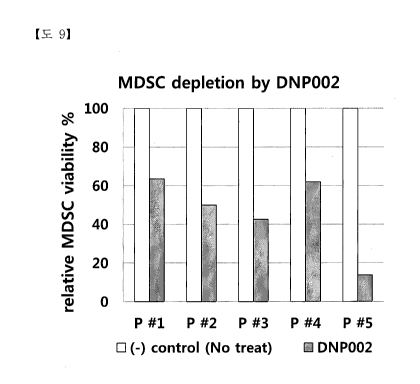
Fig. 9 is a result of analyzing the MDSC killing effect after treatment with
DNP002 by showing a result of comparing the percentage of the decreased number
of
MDSCs due to the DNP002 treatment in all five patients compared to the control
group,
where P#1 on the horizontal axis in the graph means patient's whole blood #1,
and the
vertical axis represents a change in the relative MDSC viability % change.
Fig.10 shows a result of analyzing the MDSC killing effect using a flow
cytometer after treating PBMCs isolated from blood of stomach cancer patient
with
DNP002 antibody.
Fig. ha is a result of comparing the MDSC killing effect according to the
is otype of DNP 002, confirming that DNP 002 in afucosylated I gG1 type
induces MDSC
killing in the blood most effectively.
Fig. 11b is a result of comparing the MDSC killing effect in five stomach
patients according to the isotype of DNP002, by showing that DNP002 in
afucosylated
IgG1 type provides highest MDSC killing effect in the blood of five stomach
patients
from the result of comparing the percentage of the MD SC killing effect where
P#1 on
the horizontal axis in the graph means patient's whole blood #1, and the
vertical axis
represents a change in the relative MDSC viability % change.
Fig. 12a and Fig.12b are the results of the apoptosis effect analyzed under
the
conditions of DNP002 antibody alone, NK cells alone, and combination of DNP002
antibody and NK cells on stomach cancer cell line A549and pancreatic cancer
cell line
AsPC-1 which are positive for CEACAM6 as a target antigen.
Fig. 13 is a picture showing the presence of CEACAM6-positive MDSCs in
non-tumor site of the cancer tissue by performing CEACAM6 immunostaining of
cancer tissues of lung adenocarcinoma in which CEACAM6 is positive in cancer
cells,
and lung squamous cell carcinoma, urinary bladder cancer and skin cancer
(Melanoma
malignancy) in which CEACAM6 is negative in cancer cells themselves.
24
Date Recue/Date Received 2020-11-11
CA 03099968 2020-11-11
[Mode for the Invention]
A better understanding of the present invention may be obtained through the
following examples which are set forth to illustrate, but are not to be
construed as
limiting the present invention.
Example 1: Preparation of anti-CD66c chimeric antibody
1.1. Gene sequence cloning ofanti-CD66c antibody
The 8F5 antibody gene was cloned using Mouse Ig-Primer Set (Millipore, Cat.
#: 69831). The RNA isolated from the 8F5 hybridoma was PCR using the mouse Ig-
primer set, inserted into a pGem-T vector (Promega, Cat. #: A3600), sequenced
to
confinn the DNA sequence, and the mouse antibody gene was identified through
the
IMGT site (www.inigt.org). The heavy chain variable region sequences and light
chain
variable region sequences of the analyzed 8F5 antibody are as follows.
[Table 4]
Name sequence SEQ
ID NO
8F5-chimeric VH-CDR1 ASGYSFTDYTMN 1
8F5-chimeric VH-CDR2 LINPFHGGTVSNQRFKV 2
8F5-chimeric VH-CDR3 VRGDPVRHYYALAY 3
8F5-chimeric VL-CDR1 GASENVYGTLN 4
8F5-chimeric VL-CDR2 GATNLAD 5
8F5-chimeric VL-CDR3 VATYYCQNVLSAPYT 6
8F5-chimeric VH
EVQLQQSGF'ELVKPGASMKISCKASGYSFTDYTMNWVKQS 7
HGKNLEWIGLINPFHGGTVSNQRFKVKATLTVDVSSNTAY
MELLSLTSDDSAVYYCVRGDPVRHYYALAYWGQGTSVTVS
S
8F5-chimeric V. DIQMTQSPASLSASVGETVTITCGASENVYGTLNWYQRKQG 8
KSPQLLIYGATNLADGMSSRFSGSGSGRQYSLKISSLITPDDV
ATYYCQNVLSAPYTFGGGTKLEII
8F5-chimeric VH
Gaggtccagctgcaacagtctggacctgaactggtgaagcctggagcttcaatgaagatatcc 62
tgcaaggcttctggttactcattcactgactacaccatgaactgggtgaagcagagccatggaa
agaaccttgagtggattggacttattaatcctttccatggtggtactgtctccaaccagaggttcaa
ggtcaaggccacattaactgtagacaagtcatccaacacagcctacatggagctcctcagtctg
acatctgacgactctgcggtctattactgtgtaagaggtgacccggtccgccattactatgctttg
gcctactggggtcagggaacctcagtcaccgtctcctca
8F5-chimeric VI,
gacatccagatgactcagtctccagcttcactgtctgcatctgtgggagaaactgtcaccatcac 63
atgtggagcaagtgagaatgtttacggtactttaaattggtatcagcggaaacagggaaaatctc
Date Recue/Date Received 2020-11-11
CA 03099968 2020-11-11
ctcagctcctgatctatggtgcaaccaacttggcagatggcatgtcatcgaggttcagtggcagt
ggttctggtagacagtattctc
1-2. Production of Chimeric antibody
Based on the amino acid sequence of the constructed anti-CD66c mouse
antibody 8F5, an anti-CD66c chimeric antibody was prepared.
1-2-1. Plasmid production
For expressing the anti-CD66c chimeric antibody, a plasmid for heavy chain
and a light chain expression plasmid were respectively prepared. POptiVEC
(Invitrogen)
vector was used as the light chain expression plasmid, and pcDNA3.3
(Invitrogen)
vector was used as the heavy chain expression plasmid.
In order to express the variable region coding cDNA and the constant region
coding cDNA of each antibody as a continuous amino acid sequence without
additional
amino acid insertion, the coding sequence of the cloned variable region and
the known
human IgG1 constant region (heavy chain) and the kappa constant region (light
chain)
coding sequences were synthesized (Bioneer). The synthesized heavy gene and
light
chain gene were cut with restriction enzymes Xho I and Sal I and the light
chain gene
fragment was ligated to the pOptiVec vector and the heavy chain gene fragment
was
ligated to the pcDNA3.3 vector, respectively, to construct a complete antibody
expression plasmid (pcDNA3.3-anti-CD66c heavy chain expression plasmid and
pOptiVEC-anti-CD66c light chain expression plasmid).
1-2-2. Transfection
The prepared pcDNA3.3-anti-CD66c heavy chain expression plasmid and
pOptiVEC-anti-CD66c light chain expression plasmid were transfected into CHO
cell-
derived DG44 cells (Invitrogen).
Three days prior to transfection, DG44 cells in suspension were adapted to
MEMS medium containing 5% FBS to convert them into adherent cells and to
improve
transfection efficiency. Transfection was performed on a 6-well plate using
the ViaFect
transfection regent (Promega, Cat. #: E4981). On the day before the
transfection, DG44
cells adapted to the adhered state were prepared by subculturing at a
concentration of 1
X 105 cells/well. The amount of DNA used for transfection was determined by
using
pcDNA3.3-anti-CD66c heavy chain expression plasmid and pOptiVEC-anti- CD66c
light chain expression plasmids were used at an amount of 2ug and 1.5ug
respectively at
26
Date Recue/Date Received 2020-11-11
CA 03099968 2020-11-11
a ratio of 1.5: 1. Transfection was carried out for 48 hours. Flow cytometry
was used to
analyze the transfected cell population. As shown in Fig. 1, the expression of
chimeric
antibody was confiinied by A549 non-small cell lung cancer cell line. Fig. 1
shows the
result of cloning an antibody gene from mouse 8F5 antibody and expressing it
as a
recombinant chimeric antibody and binding to the surface of CD66c antigen-
positive
A549 cells.
Example 2: Preparation of Humanized Anti-CD66c Monoclonal Antibody
2.1 Selection of recombinant antibody sequence by in silico humanization
If the antigen binding affinity is equal or superior by maintaining the
sequence
of CDRs (CDRH1: ASGYSFTDYTMN) SEQ ID NO: 1, CDRH2: SEQ ID NO: 2
(LINPFHGGTVSNQRFKV); CDRH3: SEQ ID NO: 3 (VRGDPVRHYYALAY);
CDRL1: SEQ ID NO: 4 (GASENVYGTL); CDRL2: SEQ ID NO: 5 (GATNLAD);
CDR3: SEQ ID NO: 6 (VATYYCQNVLSAPYT) of the heavy chain and light chain of
the mouse anti-CD66c antibody, 8F5 (heavy chain amino acid sequence: SEQ ID
NO: 7,
heavy chain encoding DNA: SEQ ID NO: 62; light chain amino acid sequence: SEQ
ID
NO: 8; light chain encoding DNA: SEQ ID NO: 63) as similar as possible the
humanized antibody sequences recombined the framework region sequences with
germline sequence encoding the human antibody gene were selected in silico
method.
The germline gene of human antibody used as a backbone of the recombinant
humanized antibody sequence is most similar to the heavy chain and light chain
of the
mouse CD66c antibody 8F5, respectively, as shown in Table 5. The amino acid
sequence and the nucleic acid sequence of the heavy chain variable region and
the light
chain variable region of the mouse CD66c antibody and the CDR sequences of the
heavy chain variable region and the light chain variable region are shown in
Table 6.
[Table 5]
Human Ab Germline
Heavy chain Light chain
IGHV1-69-2*01 IGKV1-27*01
IGHV1-2*02 IGKV1-5*01 Homo sapiens
IGHV1-46*01 IGKV1-39*01 Homo sapiens
Twelve (12) heavy chain variable regions and eight (8) light chain variable
regions were selected as the humanized 8F5 antibody sequence selected using
the
human antibody germline gene sequence, as shown in Table 3. The amino acid
27
Date Recue/Date Received 2020-11-11
CA 03099968 2020-11-11
sequences of the heavy chain variable region and the light chain variable
region, CDR
sequences, and the framework sequences of the selected humanized antibody are
shown
in Tables 6 to 8. The heavy chain variable region and the light chain variable
region of
the chimeric antibody and the humanized antibody are shown in Table 1. It is
preferable
that the mouse antibody and the humanized antibody have the same amino acid
sequences of heavy chain CDR3 and light chain CDR2. The bold and underlined
parts
in Table 6 are the CDR sequences of antibody. The bold and underlined parts in
Table 7
indicate the modified amino acid.
[Table 6]
Antibo combin name Amino acid sequence
dy ation
number
3043 Vk8+ V116 QVQLVQS GAEVKKP GA SMKISCKAS GYS F1DYTMNW VKQAPGQNLE
V116 WIGLINPFHGGI'VSNORFKVKATLTVDVSTNTAYMELSRLRSDDTAV
YYCVRGDPVRHYYALAYWGQGTLVTVSS
Vk8 DIQMTQSPSTLSASVGDRVTITCRAS ENVYGTLNWYQRKPGKAPKLLI
YGATNLAD GM PSRF SGS GS GTEYTLTISSLQPDDFATYYCONVLSAPY
TFGGGTKLEIK
3058 Vk8+ VH11 QVQLVQS GAEVKKP GA SVKISCKAS GYS F1DYTMHW VKQAPGQNLE
VH11 W IGLINPFGGS TSYAOKFKGRVTM TRDTSTNTA YMELSRLRSDDTAV
YYCVRGDPVRHYYALAYW GQGTL VT VSS
Vk8 DIQMTQSPSTLSASVGDRVTITCRAS ENVYGTLNWYQRKPGKAPKLLI
YGATNLAD GM PSRF SGS GS GTEYTLTISSLQPDDFATYYCONVLSAPY
TFGGGTKLEIK
2938 Vla+ VH7 QVQLVQS GAEVKKP GA SMKISCKAS GYS F1DYTMNW VRQAPGQGLE
VH7 WIGLINPFHGGI'VSNORFKVKATLTVDVSTNTAYMELSRLRSDDTAV
YYCVRGDPVRHYYALAYWGQGTLVTVSS
Vla DIQMTQSPSTLSASVGDRVTITC GASENVYGTLAWYQRKPGKAPKLLI
YGATNLADGVPSRFS GS GS GREYTLTISSLQPDDFATYYCQNVLSAPY
TFGGGTKLEIK
3007 Vk7+ VH6 QVQLVQS GAEVKKP GA SMKISCKAS GYS F1DYTMNW VKQAPGQNLE
VH6 WIGLINPFHGGI'VSNORFKVKATLTVDVSTNTAYMELSRLRSDDTAV
YYCVRGDPVRHYYALAYWGQGTLVTVSS
Vk7 DIQMTQSPSTLSASVGDRVTITC GASENVYGTLNWYQRKPGKAPKLLI
YGATNLADGVPSRFS GS GS GTEYTLTISSLQPDDFATYYCONVLSAPY
TFGGGTKLEIK
3019 Vk7+ VH10 QVQLVQS GAEVKKP GA SVK VSCKAS GYSF1DYTMNW VKQA PGQNL E
VH10 W IGLINPFHGGI'VSNORFKVKA TMTVDVSTNTAYMELSRLRSDDTA V
YYCVRGDPVRHYYALAYWGQGTLVTVSS
Vk7 DIQMTQSPSTLSASVGDRVTITC GASENVYGTLNWYQRKPGKAPKLLI
YGATNLADGVPSRFS GS GS GTEYTLTISSLQPDDFATYYCONVLSAPY
TFGGGTKLEIK
3010 Vk7+ VH7 QVQLVQS GAEVKKP GA SMKISCKAS GYS F1DYTMNW VRQAPGQGLE
VH7 WIGLINPFHGGI'VSNORFKVKATLTVDVSTNTAYMELSRLRSDDTAV
YYCVRGDPVRHYYALAYWGQGTLVTVSS
Vk7 DIQMTQSPSTLSASVGDRVTITC GASENVYGTLNWYQRKPGKAPKLLI
YGATNLADGVPSRFS GS GS GTEYTLTISSLQPDDFATYYCQNVLSAPY
TFGGGTKLEIK
3004 Vk7+ VH5 QVQLVQS GAEVKKP GA SVKISCKAS GYS F1DYTMNW VRQAHGQNLE
VH5 W IGLINPFHGGTVSNORFKVKA TLTVDVSTNTA YMELSRLRSDDTAV
YYCVRGDPVRHYYALAYWGQGTLVTVSS
28
Date Recue/Date Received 2020-11-11
CA 03099968 2020-11-11
Vk7
DIQMTQSPSTLSASVGDRVTITC GASKWYGTLNVVYQRKPGKAPKLLI
YGATNLAD GVPSRF S GS GS GTEYTLTIS S LQPDDFATYYCONVLS APY
TFGGGTKLEIK
3046 Vk8+ V117 QVQL VQS GAEVKKP GA SMKISCKAS GYSFIDYTMNVVVRQAPGQGLE
V117 W
IGLINPFHGGTVSNORFKVKA TLTVDVSTNTAYMELSRLRSDDTAV
YYCVRC;DPVRHYYALAYVVGQGTLVTVSS
Vk8 DIQMTQSPSTLSASVGDRVTITCRAS KWYGTLNVVYQRKPGKAPKLLI
YGATNLAD GM PSRF SGS GS GTEYTLTI S S LQPDDFATYYCQNVLS APY
TFGGGTKLEIK
[Table 7]
Name CDR1 SEQ CDR2 SEQ CDR3 SEQ
ID NO ID ID NO
NO
VH- Chimeric A SGYSFTDYT 1
LINPFHGGTVSNQRF 2 GDPVRHYYALA 3
MN KV Y
V115, 6,7, 10 A SGYSFTDYT 1
LINPFHGGTVSNQRF 2 GDPVRHYYALA 3
MN KV Y
VH11 A SGYSFTDYT 9
LINPFGGSTSYAQKF 10 GDPVRHYYALA 3
MH KG Y
VL-Chimeric GA SENVYGTL 4 GATNLAD 5 VATYYCQNVLS
6
N APYT
VK5 GA SENVYGTL 11 GATNLAD 5 FATYYCQNVLS
13
A APYT
VK7 GA SENVYGTL 4 GATNLAD 5
FATYYCQNVLS 13
N APYT
VK8 RA SENVYGTL 12 GATNLAD 5 FATYYCQNVLS
13
N APYT
[Table 8]
Name FR1 SEQ FR2 SEQ FR3 SEQ FR4 SEQ
ID ID ID ID
NO NO NO NO
VH- EVQLQQSGPEL 22 WVKQSHG 32 NQRFKVKATLT 42 WGQGTSV 52
Chimeric VKPGA SMKISC KNLEWIG VDVSSNTAYM TVSS
K ELLSLTSDDSA
VYYCVR
VHS QVQLVQSGAE 23 WVRQAH 33 NQRFKVKATLT 43 WGQGTLV 53
VKKPGA SVKIS GQNLEW I VDVSTNTAYM TVSS
CK G ELSRLRSDDTA
VYYCVR
V116 QVQLVQSGAE 24 WVKQAPG 34 NQRFKVKATLT 44 WGQGTLV 54
VKKPGA SMKIS QNLEWIG VDVSTNTAYM TVSS
CK ELSRLRSDDTA
VYYCVR
V117 QVQLVQSGAE 25 WVRQAPG 35 NQRFKVKATLT 45 WGQGTLV 55
VKKPGA SMKIS OGLEWIG VDVSTNTAYM TVSS
CK ELSRLRSDDTA
VYYCVR
VH10 QVQLVQSGAE 26 WVKQAPG 36 NQRFKVKATM 46 WGQGTLV 56
VKKPGA SVKV QNLEWIG TVDVSTNTAY TVSS
29
Date Recue/Date Received 2020-11-11
CA 03099968 2020-11-11
SCK M ELS RLRSDDT
AVYYCVR
VT-Ill QVQLVQSGAE 27 W VKQAPG 37 AQEFKGRVTM 47 W GQGTL V 57
VKKPGA SVKIS QNLEW IG TRDTSTNTAY TVSS
CK M ELS RLRSDDT
AVYYCVR
VL- DIQMTQSPASL 28 W YQRKQG 38 GM SSRFSGS GS 48 FGGGTKL 58
Chimeric SA SVGETVTIT KSPQLLIY GRQYSLKISSL EH
C IIPDD
VK5 DIQMTQSPS TL 29 W YQRKPG 39 GVPSRFS GS GS 49 FGGGTKL 59
SA SVGDRVTIT KAPKLLIY GRQYSLKISSL EIK
C IIPDD
VK7 DIQMTQSPS TL 30 W YQRKPG 40 GVPSRFS GS GS 50 FGGGTKL 60
SA SVGDRVTIT KAPKLLIY GT EYT LTISSL ELK
C QPDD
VK8 DIQMTQSPS TL 31 W YQRKPG 41 GMPSRFSGS GS 51 FGGGTKL 61
SA SVGDRVTIT KAPKLLIY GT EYT LTISSL ELK
C QPDD
2.2 Expression and Analysis of Recombinant humanized antibodies
The sequences of selected antibody were expressed in 293 cells in the form of
human IgG1 by connecting the human IgG1 heavy chain constant region and the
kappa
light chain constant region, respectively. Seven days after the transfection,
the
recombinant humanized antibody was purified using KanCap A resin (Kaneca).
The purified antibody was quantitated by measuring at OD 280 nm and SDS-
PAGE was performed. The purity and the aggregation of the antibody were
analyzed by
analyzing with 280 nm and 220 nm by HPLC using Sepax Zenix-C SEC-300 size
exclusion column (Sepax Technologies) (Figs. 2a to 2c)
2.3 Cell binding and antigen binding analysis of recombinant humanized
antibodies
2-3-1 cell binding assay
Each expressed 96 recombinant humanized antibody was poured and reacted
in a test tube containing the same amount (1 ug) of CD66c-positive A549 non-
small-cell
lung cancer cell line at 4 C for 30 minutes, washed with PBS, and treated with
FITC-
conjugated goat anti-Huma IgG (DNona Inc, Korea) was added and incubated at 4
C
for 15 minutes. After washing with PBS, the cells were analyzed with a flow
cytometer
(Stratedigm, S1000EXi) and the results are shown below.
Among the 96 recombinant humanized antibody candidates, eight were firstly
selected based on the degree of expression, the presence of aggregation, and
the degree
of cell binding (Table 9 and Table 10, Figs. 3a to 3e). Tables 9 and 10 are
the results of
Date Recue/Date Received 2020-11-11
CA 03099968 2020-11-11
analysis of primary selected anti-CD66c humanized antibody and chimeric 8F5,
and
Table 10 shows the results of flow cytometer analysis.
[Table 9]
No. #71 #86 #93 #43 #51 #45 #41 #74
Protein ID 3043 3058 2938 3007 3019 3010 3004 3046
H & Vk8+V Vk8+V
Vk5+V Vk7+V Vk7+V Vk7+V Vk7+V Vk8+V
L_ -It 116 1111 117 116 1110 117 115 117
Well Fll 112 119 D7 E3 D9 D5 G2
OD
3.54 3.86 3.32 4.01 4.03 3.5 3.56 3.54
280nm
Volume
0.25 0.25 0.25 0.25 0.25 0.25 0.25 0.25
(ml)
Conc.
2.52 2.71 2.36 2.85 2.86 2.49 2.53 2.52
(mg/nil)
Yield(mg) 0.66 0.71 0.61 0.74 0.74 0.65 0.66 0.66
extinction
102190 103680 102190 102190 102190 102190 102190 102190
coefficient
MW (Da) 72750 72691 72616 72633 72521 72604 72669
7272
[Table 10]
No. H & L % gated Mean % (%gatedx
Protein ID
combination mean)
Chimeric ** ** 73 619 100.0
antibody
#71 3043 Vk8+VH6 69 686 104.8
#86 3058 Vk8+VH11 72 645 102.8
#93 2938 Vk5+VH7 71 611 96.0
#43 3007 Vk7+VH6 71 565 88.8
#51 3019 Vk7+VH10 70 561 86.9
#45 3010 Vk7+VH7 70 554 85.8
#41 3004 Vk7+VH5 71 541 85.0
#74 3046 Vk8+VH7 71 523 82.2
Table 9 shows the degree of expression, the presence or absence of
aggregation,
and the degree of cell binding of 8 selected antibodies. Specifically, the
expression
levels and the molecular weights of the 8 selected antibodies were summarized.
In
addition, according to the results of flow cytometry in Table 10, it was
confirmed that
31
Date Recue/Date Received 2020-11-11
CA 03099968 2020-11-11
the eight recombinant humanized antibodies exhibited cell binding strengths of
20%
which showed very similar cell binding strength to chimeric antibodies. As a
result, the
8 antibody being normally expressed and having few aggregations formed due to
the
instability of the protein itself, and the similar binding affinity to the
target antigen-
positive cells to the chimeric antibody were firstly selected from 96
humanized
candidate antibodies
Some contents may be missed. In particular, the binding ability of cell lines
seemed to be different in numerical values. However, as shown in Fig. 3, the
actual cell
binding profile was similar to that of chimeric antibody, and 8 kinds of
humanized
antibodies were determined by obtaining the numerical value with multiplying
the
antibody positivity (% gated) with the mean fluorescence (mean) and comparing
it with
the chimeric antibody, so as to select the humanized antibodies within 20%.
In
general, when the mouse antibody CDR region sequence is inserted into the
framework
region of the humanized antibody at the time of the production of the
humanized
antibody, the antibody binding affinity is sharply decreased due to the change
of the
original protein structure. In considering the general property of humanized
antibody,
very good humanized antibodies were be selected in the present invention.
2-3-2 Antigen binding assay
Among the eight selected recombinant humanized antibodies, five kinds of
recombinant humanized antibodies exhibiting a high binding affinity as
compared to
that of the chimeric antibody were selected and analyzed for their binding
affinity to
CD66c antigen and similar CD66 antigens by ELISA, respectively.
[Table 11]
Protein ED HC & LC combination
3043 Vk8+VH6
3058 Vk8+VH11
2938 V1(5+VH7
3007 Vk7+VH6
3019 Vk7+VH10
Antigen CD66c (CEACAM6; Sino Biological, Inc.) and CEACAM1 antigen
(Sino Biological, Inc.) were coated on a 96-well plate at a rate of 100 ng per
well and
then blocked. The primary antibody was diluted 3-fold from 10 ug / ml and
bound at 37
C for 1 hour. The primary antibody was diluted three times from lOug/m1 at
initial
32
Date Recue/Date Received 2020-11-11
CA 03099968 2020-11-11
concentration and was bound at 37 C for 1 hour, and goat anti-Human Ig-HRP
conjugate (Jackson ImmunoResearch) as a secondary antibody was diluted 1:
10,000
and incubated at 37 C for 30 minutes. The washing was carried out at three
times
between each step, and the TMB reaction was performed, stopped with 1N H2SO4
solution at the same amount of TMB solution (100u1) and then OD value was
measured
at 450nm.
As a result of the experiment, the binding affinities to CD66c antigen of the
five
kinds of recombinant humanized antibodies selected from among 96 kinds of
recombinant humanized antibodies are shown in Fig. 4a, Table 12, Fig. 4b and
Table 13.
Fig. 4a and Table 12 show the binding capacity of the antibodies to CECACAM6
CD66c) antigen, and Fig. 4b and Table 13 are the results for the CEACAM1
(CD66a)
antigen.
From the binding affinity of antibodies to the antigen in Fig. 4a, Table 12,
Fig.
4b and Table 13, all antibodies to CECACAM6 showed a similar binding profile
to the
chimeric antibody to CEACAM6 and were divided into the groups that did not
bind or
bound weakly CEACAM1.
[Table 12]
Antibody
concentration chi 8F5 hu 3043 hu 3058 hu 2938 hu 3007 hu
3019
(ng/ml)
10000.00 3.17 2.73 2.87 3.43 3.37 3.13
3333.33 3.41 2.95 3.18 3.27 3.24 3.26
1111.11 3.30 2.74 3.19 3.16 3.23 3.34
370.37 3.30 2.92 2.80 3.08 3.16 3.23
123.46 2.83 2.29 2.22 2.56 2.90 3.04
41.15 2.02 1.73 1.38 1.72 2.17 2.45
13.72 1.15 1.37 1.06 1.15 1.64 1.77
4.57 0.80 0.91 0.74 0.65 1.02 1.32
1.52 0.53 0.70 0.53 0.48 0.79 0.95
0.51 0.41 0.49 0.38 0.36 0.64 0.80
0.17 0.40 0.41 0.37 0.30 0.53 0.68
[Table 13]
Antibody
concentration chi 8F5 hu 3043 hu 3058 hu 2938 hu 3007 hu
3019
(ng/ml)
10000.00 1.21 0.91 0.42 0.32 1.01 0.99
33
Date Recue/Date Received 2020-11-11
CA 03099968 2020-11-11
3333.33 1.24 1.22 0.31 0.27 1.15 1.07
1111.11 1.21 1.17 0.19 0.21 1.06 1.02
370.37 1.13 0.80 0.12 0.14 0.92 0.89
123.46 0.95 0.70 0.07 0.10 0.82 0.79
41.15 0.82 0.61 0.07 0.07 0.66 0.58
13.72 0.51 0.43 0.05 0.06 0.46 0.41
4.57 0.28 0.27 0.05 0.05 0.27 0.24
1.52 0.16 0.16 0.05 0.05 0.18 0.16
0.51 0.10 0.10 0.04 0.05 0.11 0.11
0.17 0.08 0.08 0.04 0.04 0.09 0.08
2.4 Stability analysis of recombinant humanized antibodies
The experiments were conducted to determine the stability of the antibodies by
leaving the five recombinant humanized antibodies of Example 3.3 selected by
the
binding profile to antigen and cell under high temperature conditions.
The stability was determined by performing the binding experiments using 8-
anilino-1-naphthalenesulfonic acid (ANS, Sigma). ANS is a compound that can
detect
the denaturation of proteins by measuring the change in fluorescence
wavelength
between the binding to and not binding to hydrophobic sites exposed when
protein is
denatured.
The recombinant humanized antibody was adjusted to a concentration of 0.2 mg
/ml using PBS (phosphate buffered saline), and left at 50 C for 4 hours as
severe
conditions. 0.2 jig/ml of ANS solution was mixed at 20 p.1 per 500 p.1 of the
diluted
solution of antibody to be analyzed, and analyzed after 5 minutes later with a
fluorescent reader at 360 nm excitation and 460 nm emission conditions. In
addition, the
ANS reagent reaction was also measured at a temperature of 70 C for
additional 30
minutes.
Figs. 5a and 5b show the results of confirming the antibody stability of the
five
recombinant humanized antibodies shown in Table 11 under severe temperature
conditions. That is, the reactivity of the ANS reagent was measured by
fluorescence
after leaving the antibody at 50 C for 4 hours under the sever condition,
and further
left at 70 C for 30 minutes. As shown in the results of the experiment of
Fig. 5a, most 5
kinds antibodies showed little ANS response when the antibodies were left at
the
temperature of 50 C for 4 hours, but the greatly increased fluorescence
value of the
antibodies when the antibodies were additional left at 70 C for 30 minutes.
Among
34
Date Recue/Date Received 2020-11-11
CA 03099968 2020-11-11
them, the recombinant antibody having protein ID: 3058 exhibited the smallest
increased fluorescence value, and thus showed the most stable property in the
temperature change among the five recombinant humanized antibodies.
In order to measure the change rate of ANS reagent reactivity, ANS reagent
reactivity was analyzed with a fluorescent reader in the same manner as above
after
leaving the antibodies for 4 hours at a refrigeration condition (4 2 C.)
and a
temperature of 62 C. The fluorescence value variability of the antibody
against the
ANS reagent can be determined by obtaining the difference between the
fluorescence
value measured at low temperature conditions (e.g., 4 C.) and the
fluorescence value
measured at high temperature conditions (e.g., 62 C.) and dividing with the
fluorescence value measured at low temperature conditions.
[Mathematical Equation]
Fluorescence value variability = (fluorescence value measured at high
temperature condition - fluorescence value measured at low temperature
condition) /
(fluorescence value measured at low temperature condition)
As shown in Fig. 5b, the reaction was allowed for 4 hours at a temperature of
62 C which was somewhat increased from temperature (50 C), and then the ANS
reagent reactivity was confirmed. Five recombinant humanized antibodies and
the
chimeric antibodies showed little ANS response under refrigerated conditions,
but
increased with increasing temperature. However, Chimeric 8F5 antibody showed
the
reactivity of ANS reagent increased to 1,406% by keeping the temperature
condition at
62 C but the precipitates occurred to be unstable. However, the five
humanized
antibodies showed significantly lower variability of ANS reagent reactivity
than
chimeric antibodies, and no precipitates were produced. In particular, the
humanized
antibodies Protein ID 3019 and Protein ID 3058 had variability of ANS reagent
reactivity of 114% and 133%, respectively, and thus were considered as most
stable
antibodies. The meaning of increased ANS reagent reactivity refers to the
increased
exposure of the hydrophobic amino acid placed inside the protein structure,
which is
responsible for the denaturation of the protein structure and the resulting
protein
aggregate, i.e., precipitate formation. The humanized antibody according to
the present
invention is considered to be stable antibody having the ANS reactivity
variation of less
than 200%. The ANS reactivity variation of less a change of less than 200% is
considered to be very low, and over the value, a more significant change in
protein
structure is considered to be the observation of ANS reactivity. Accordingly,
the
Date Recue/Date Received 2020-11-11
CA 03099968 2020-11-11
humanized antibody according to the present invention has similar antigen
binding and
cell binding ability to chimeric antibody, and the increased physical
stability of the
antibody protein itself, such facts are a very excellent feature in the
drugability for the
therapeutic antibody.
2.5 CHO cell expression and analysis of recombinant humanized antibody
The five recombinant humanized antibodies selected in Example 2.3 were
expressed in CHO cells used for expressing most therapeutic antibodies and
analyzed.
The light chain variable region DNA sequence and heavy chain variable region
DNA
sequence to construct the selected five recombinant humanized antibodies were
performed by the codon optimization, synthesized, and ligated with the human
IgG1
constant region gene by overlay PCR method. The product was cut with XhoI and
EcoRI and ligated into the pcDNA3.4 vector (Life Technology). Table 11 shows
the
light chain and heavy chain combinations of humanized antibodies selected for
CHO
cell expression.
The DNA primer sequences used for PCR on the variable region and constant
region gene are shown in Table 14 below.
[Table 14]
Primer use Target gene sequences SEQ
name ID NO
8F01 1" frag forward VH6, VH7, VH10, ATTACTCGAGGCCACC 64
VH11 ATGAA
8F02 Pt frag reverse VH6, VH10, VH11 AGTTGAAGCGCTGCTC 65
ACAGTCA
8F03 211d frag forward VH6, VH10, VH11 GT GAG CAGC GCT T CAA 66
CTAAGGG
3E04 2nd frag reverse VH6, VH7, VH10, AGT CGAAT T CT CAT T T 67
VH11 CCCAGGAGAG
8F04 1" frag reverse VH7 AGTTGAAGCAGAAGAC 68
ACT GT CA
8F05 2nd frag forward VH7 GT GTCT TCTGCTTCAAC 69
TAAGGG
3E01 1" frag forward Vk5, Vk7, Vk8 ATTACTCGAGGCCACC 70
ATGAAGTGGG
8F06 1" frag reverse Vk5, Vk7, Vk8 AACAGT CCGCT T GAT C 71
TCCAGCT
3EL02(2) 2nd frag forward Vk5, Vk7, Vk8 GAGATCAAGCGGACTG 72
TTGCTGC
3E08 2nd frag reverse Vk5, Vk7, Vk8 AT T AGAAT T CT CAGCA 73
CTCGCCGCGG
36
Date Recue/Date Received 2020-11-11
CA 03099968 2020-11-11
The five (5) recombinant humanized antibodies were transiently transfected
using the ExpiCHO (trademark) Expression System Kit (ThermoFisher, Cat. No.
A29133), and the expressed antibodies were transfected with CD66c-positive
A549
non-small lung cancer cell line and analyzed by flow cytometer, as shown in
Fig. 6a to
6c. All five recombinant humanized antibodies showed similar binding
affinities to
chimeric antibodies. The measured fluorescence value of the flow cytometer was
divided by the antibody expression amount of the CHO culture medium to obtain
the
relative the binding affinity of antibody to the antibody expression amount,
as shown in
Fig. 6d. Therefore, it was confirmed that the recombinant humanized antibody
was
properly expressed in CHO cells. The binding affinity of the antibody to the
cell surface
was determined as 100%, and the relative change was shown in Fig. 6d.
<Example 3> CHO cell expression and analysis of DNP002
DNP002, a humanized antibody against anti-CD66c was expressed and
analyzed in CHO cells used to express most of the therapeutic. In order to
test the
difference in function according to the subtype of the DNP002 antibody, IgG1
type and
IgG2 type antibodies were prepared.
After performing the codon optimization of DNA sequences of the light and
heavy chain variable region for constructing humanized recombinant antibodies,
they
were synthesized and linked with constant region o human IgG1 or IgG2 by
overlay
PCR method, and XhoI and EcoRI gene fragment was cloned into pcDNA3.4 vector
(Life Technology).
[Table 15]
clas sifica Amino acid sequence cDNA sequence
t ion
Constant A STKGF'SVFPLA PSSK GCTTCAACTAAGGGACCAAGCGTATTCCCACTTGCTCCAT
region of STSGGTAALGCLVKD CTAGCAAGAGCACTAGCGGAGGAACAGCTGCTTTGGGGT
IgG1 YFP EP VINSWNSGAL GTTTGGTAAAGGATTACT 11 CCCGAACCTGTTACCGTGAG
heavy TSGVHTFPAVLQSSGL CTGGAACAGCGGGGCTTTGACAAGTGGCGTTCATACAT 11
chain YSLSS VVTVPSSSL GT CCTGCCGTTTTGCAAAGCAGCGGCTTGTATAGCTTGAGCT
QTYICNVNHKPSNIK CTGTTGTTACCGTTCCAAGCTCATCTCTGGGCACACAAAC
VDKKVEPKSCDKTHT ATACATCTGCAACGTGAACCACAAGCCCTCAAACACCAA
CPPCPA PELL GGF'S VF GUT GGA CAA GAA GGT GGA GC CAAA GTCTTGCGA CA A GA C
LF PPKPKDTLM I SRTP CCA CA CCTGTCCA CCTTGTCCA GC CCCT GA A CTCCTGGGG
EVTCVVVDVSHEDPE GGCCCTTCAGTTTTTCTC 11 TCCTCCTAAACCTAAAGATAC
VKFNWYVDGVEVHN ACTCATGATCAGTCGGACCCCT GAAGTTACCTGTGTGGT G
37
Date Recue/Date Received 2020-11-11
CA 03099968 2020-11-11
AKTKPREEQYNST YR GTC GA TGTGTCTCATGAAGA TCCTGAAGTCAAGTTTAACT
VVSVLTVLHQDWLN GGTA TGTGGACGGC GT GGAGGTGCATAATGCCAAGACCA
GKEYKCKVSNKALPA AGCCTC GGGA GGAGCAATATAATTCTACCTATCGC GTC GT
PIEKTISKAKGQPREP CTCTGTCCTCACCGTCCT GCATCAGGACTGGCTGAATGGC
QVYTLPPSRDELTKN AAA GAGTATAAGTGCAAAGTCA GTAACAAAGCCCTCCC C
QVSLTCLVKGFYPSDI GC CCC CATAGA GAAAA CCATTAGTAAAGCCAAAGGGCAG
AVEWFSNGQPENNY CCCC GC GAGCC CCAGGTCTA TACACTGCCCC CCAGTAGA
KTTPPVLDSDGSFFLY GA CGA GCTGA CAAA GAA TCA GGT GTCTCTGA CA TGCCT G
SKLTVDKSRWQQGN GT GA AA GGCTT TTATCCCTCTGA CA TTGCCGTC GA GT GGG
VESCSVMHEALEINHY AGTCTAATGGGCAGC CC GAGAA TAATTATAA GACAA CAC
TQKSLSISPCK CCCCCGTGCTGGACAGTGACGGCTCATTT 11 CCTGTATTC
(SEQ ID NO: 74) AAAACTGACAGTGGACAAAAGTCGGTGGCAGCAGGGGA
ATGTGTTTTCATGCAGTGTCATGCACGAGGCCCTCCACAA
TCACTATACCCAGAAATCTCTGAGTCTCTCTCCTGGGAAA
TGA (SEQ ID NO: 75)
Constant A STKGF'SVFPLAPCSR GCTTCCACCAAGGGCCCATCCGTGTTCCCTCT GGCCCCAT
region of STSFSTAALGCLVKD GTTCTA GGTCTACA TCTGAGA GCACCGCC GC CCTC GGCT G
IgG2 YFPEPVTVSWNSGAL TCTGGTGAAGGA TTATTTCCCCGA GC CC GT GACCGTGTCT
heavy TSGVHTFPAVLQSSGL TGGA A CA GC GGA GCC CTGA CTA GC GGA GTGCA CA CCTTC
chain YS LS S VVTVPS SNF GT CCAGCT GTGCTGCA GA GCTCC GGCCT GTA CA GCCTCTCTT
QTYTCNVDHKPSNTK CTGT GGTGACC GT GCC CTCTA GCAAC 11 CGGAACA CAGA
VDKTVERKCCVECPP CCTACACATGTAACGTGGATCACAAGCCTTCCAACACCA
CPAPPVA GF'SVF TIPP A GGT GGATAA GA CCGT GGA GA GA AA GT GCT GT GT GGA GT
KPKDTLM I SRTPEVTC GC CCTCCA TGTCCT GC CCCA CCT GTGGCTGGACCTTCTGT
VVVDVSHEDPEVQFN GTTTCTGTTCCCTCCAAAGCCAAAGGATACCCTGATGATC
WYVDGVEVHNAKTK A GCA GAA CTCCTGA GGT GA CCTGT GT GGT GGT GGACGT G
PREEQFNSTERVVSVL A GCCA CGA GGA TCCTGA GGT GCAGTTTAACTGGTACGT G
TVVHQDWLNGKEYK GA TGGC GT GGAGGT GCA TAA CGCTAA GA CAAA GCCTA GG
CKVSNKGLPAPIEKTI GA GGA GCA GTTTAA CA GCA CCTTCA GA GTGGT GA GC GT G
SKTKGQPREPQVYTL CTGA CC GTGGT GCACCAGGATTGGCT GAACGGCAAGGA G
PPSREEMTKNQVS LT TATAAGTGTAAGGTGTCTAACAAGGGCCT GC CA GCC CCT
CLVKGFYPSDIAVEW A 11 GA GAAGA CCATCAGTAAGA CCAAGGGACAGCCTAGG
FSNGQPENNYKTTPP GAGCCTCAGGTGTACACCCTGCCTCCTTCCAGAGAGGAG
MLDSDGSFFLYSKLT A TGA CAAA GAA CCA GGT GA GCCT GA CCTGTCT GGT GA A G
VDKSRW QQGNVF S C S GGCTTCTA CCCTA GC GA TA TCGCC GT GGA GT GGGA GA GC
VMHEALEINHYTQKS AA CGGCCAGCCT GAGAACAACTACAAGACCACCCCACCT
LSLSPGK ATGCTGGACA GC GATGGCTCTTTCTTCCTGTA CTCTAAGC
(SEQ ID NO: 76) TGACCGT GGACAAGAGCAGATGGCAGCAGGGCAACGTGT
TTTCTTGTTCTGTGATGCACGAGGCCCT GCA CAA CCA CTA
CACCCAGAAGTCTCTGTCTCTGTCTCCAGGCAAGTGA
(SEQ ID NO: 77)
38
Date Recue/Date Received 2020-11-11
CA 03099968 2020-11-11
Constant RTVAAPSVF1FPPSDE CGGA CTGTT GCT GCTCCATCTGTTTTTA TA 11 TCCTCCCA G
region of QLKS GTA SWCL INN CGAC GA GCA GCTGAAAA GC GGCA CTGC CTCTGT GGT GTG
light FYPREAKVQWKVDN TCTGCT GAATAA T 11 TTACCCCCGGGAAGCCAAA GTCCAG
chain ALQSGNSQFSVTEQD TGGAAGGTGGATAATGCCCTCCAGTCT GGGAACAGTCAG
SKDSTYSISSTLTLSK GAAAGTGTGACAGAACAGGATAGTAAGGACTCTAC flAT
ADYEKHKVYACEVT AGCCTCTCTTCTACACTGACTCTGTCAAAGGCCGACTATG
HQGLSSPVTKSFNRGE AGAAGCA TAAAGTGTA TGCCT GC GAGGT GACACA TCAGG
GC CTGA GTTCACCC GT GACAAAATC fl TTAACCGC GGC GA
(SEQ ID NO: 78) GTGCTGA
(SEQ ID NO: 79)
To prepare afucosylated DNP002 humanized antibody, when the DNP002 IgG1
type antibody was expressed, 2F-PF (2F-Peracetyl-Fucose; Merck, Cat#: 344827)
was
added to the culture medium at 50 uM and cultured, and then purified using
Mabselect
sure Protein A column (GE Healthcare Lifescience, Cat# :11003494). The
purified
antibody was dialyzed with phosphate buffered saline, and the absorbance at
280 nm
was divided by the absorbance coefficient of 1.4 and converted into a
concentration unit
of "mg/mL", and then, it was used for the subsequent experiment.
Afucosylation was evaluated by relatively comparing the reactivity of
Biotinylated Lens culinaris agglutinin (Vector laboratories, Cat#:B-1045)
having a
binding property to fucose. Fucose-conjugated IgG1 DNP002 reacted with
Biotinylated
Lens culinaris agglutinin and were used for TMB color development by SA-HRP
(Jackson immunoresearch, Cat# :016-030-084). However, afucosylated DNP002 had
relatively little color development (Table 16).
[Table 16]
Antibody concentration DNP002 humanized afucosylated
(ng/ml) antibody DNP002 humanized antibody
10000 1.452 0.229
3333 1.254 0.210
1111 0.993 0.164
370 0.283 0.115
123 0.116 0.076
41 0.080 0.083
14 0.095 0.059
0.094 0.070
<Example 4> Investigation of reactivity of DNP002 antibody to MD SC
The reactivity of DNP002 antibody against MDSC was evaluated by flow
39
Date Recue/Date Received 2020-11-11
CA 03099968 2020-11-11
cytometric analysis.
Specifically, after preparing blood from stomach cancer patients, DNP002 with
bound with APC and the antibodies (anti-HLA-DR-FITC, CD11b-PE, CD33-PE
antibodies) against the labeled antigens of MDSC with different fluorescence
together
were added to 100 uL of whole blood and reacted at 4 C for 20 minutes. The
product
was added with 5 ml of red blood cell (RBC) lysis buffer of 1X RBC Lysis
Buffer
(ThermoFisher, Cat#:00-4333-57), reacted at room temperature for 30 minutes,
centrifuged to remove the decomposed RBC, washed again with PBS, and performed
by
flow cytometry. The staining intensity was measured as a log of fluorescence
intensity
and expressed in units of tens.
In the analysis of the results, after only monocytes and granulocyte regions
except lymphocyte were designated according to the cell size in the dot plot,
the groups
with no or low HLA-DR expression were selected, and the groups being positive
for
CD11b and CD33 were selected from that groups and designated as MDSC. The
positive rate of DNP002 in the designated MDSC group was confirmed (Fig. 7).
Specifically, from left to right direction in the upper graphs in FIG. 7, 1)
gated
only monocytes and granulocytes in FSC and SSC dot plots with excluding
lymphocytes. (FSC: forward scatter, a variable indicating the size of cells to
be analyzed,
SSC: side scatter, a variable indicating the granularity of the cells to be
analyzed and the
degree of granule existing in the cells), 2) gated HLA-DR Low or (-) groups,
and 3)
MDSC being positive to CD11b and CD33among the 1st and 2nd groups. DNP002
positive MDSCs are 90.9% (upper right site in the right dot plot of Fig. 7),
and DNP002
negative MDSCs are 4.4% (upper left site in the right dot plot of Fig. 7).
MDSC is
divided into subtypes of a monocytic MDCS and a granulocytic MDSC. The
granulocytic MDSC expresses CD66b, but the monocytic MDCS does not express
CD66b, so whether or not CD66b is expressed can be usefully used for
discriminating
MDSC subtypes.
Fig. 7 is a result of proving the method of defining MDSC (excluding
lymphocytes, HLA-DR low/(-), CD 11b+, CD33+) and the reactivity of DNP002
(anti-
CD66c) specific to MDSC. Because DNP002 binds to MDSC, DNP002 can be used as
a method for specifying MDSC (Fig. 7), and DNP002 can cause ADCC effects to
remove MDSC (Fig. 8). In the following test results, the MDSC bound with
DNP002
was 90.9%.
Fig. 8 shows a phenomenon in which CD66b-positive granulocytic MDSCs
were killed by DNP002 treatment and descried in their ratio. In Fig. 8, the
upper dot
Date Recue/Date Received 2020-11-11
CA 03099968 2020-11-11
plot (control group) represents a sample before treatment with DNP002, and the
bottom
dot plot (DNP002) is the result of decreased MDSC after treatment with DNP002.
CD66b is a marker that can differentiate between monocytic MDSC and
granulocytic
MDSC. In the following test results, most of the MDSCs were CD66b-positive
granulocytic MDSCs, and granulocytic MDSCs were effectively killed by DNP002.
As a result of analyzing the blood of 19 stomach cancer patients, the positive
rate of DNP002 on MDSC in all PBMCs was 34.3 ¨ 76.7%, and the average positive
rate was 55.1%. Table 17 below is an analysis result of the reactivity of
DNP002
antibody to MDSC with samples of 19 stomach cancer patients.
[Table 17]
Blood # DNPO 0 2 MDS C of total PBMC
(%
GC patient #1 49.8 55.1
GC patient #2 50.5
GC patient #3 68.6
GC patient #4 61.8
GC patient #5 48.8
GC patient #6 70.5
GC patient #7 45.6
GC patient #8 36.0
GC patient #9 51.4
GC patient #10 34.3
GC patient #11 49.8
GC patient #12 53.5
GC patient #13 63.7
GC patient #14 72.0
GC patient #15 43.5
GC patient #16 67.4
GC patient #17 76.7
GC patient #18 42.5
GC patient #19 60.4
<Example 5> lysis effect ofDNP002 on MDSC
5.1. Lys is effect ofDNP002 on MDSC in whole blood
In order to check the MDSC killing effect by DNP002, erythrocyte lysis buffer
of 1X RBC Lysis Buffer (ThermoFisher, Cat# :00-4333-57) was added to the blood
of 5
stomach cancer patients to dissolve red blood cells, and then the product was
poured 1
41
Date Recue/Date Received 2020-11-11
CA 03099968 2020-11-11
X 105 per a well of 12-well plate. DNP002 antibody was added to each well at a
concentration of 10 ug/mL and incubated in an incubator at 37 C for one day.
After
incubation, the cells were washed with PBS, and reacted with antibodies (anti-
HLA-DR,
CD11b, CD33 antibodies) against the MDSC-labeled antigens with different
fluorescence at 4 C for 20 minutes. After washing with PBS, the flow cytometry
was
performed. The staining intensity was measured as a log of fluorescence
intensity and
expressed in units of tens.
As a result of the above experiment, FIG. 8 shows a representative result in
which the DNP002 antibody effectively induces apoptosis of MDSC in the blood,
and
the MDSC ratio is significantly reduced compared to pre-treatment with the
DNP002
antibody. Fig. 9 is a diagram illustrating the same test as in FIG. 8 for the
blood samples
of five (5) stomach cancer patients. The open bar means the ratio of MDSC
before
DNP002 treatment, and the closed bar means the relative ratio of MDSC after
DNP002
treatment. After DNP002 treatment, it can be seen that the MDSC ratio has been
significantly reduced in all 5 patient samples.
5.2. Lys is effect ofDNP002 on MDSC in PBMC
In order to more clearly clarify the MDSC-targeted killing ability of the
DNP002 antibody, MDSC was only obtained with excluding mature neutrophils, and
the MDSC killing effect of the DNP002 antibody was tested without the effect
of
neutrophils.
Specifically, only the PBMC layer containing MDSC was separated from the
blood of two stomach cancer patients using Ficoll-Paque PLUS (Ge healthcare,
Cat#:17-1440-02) solution. The density gradient separation of blood cells
through the
Ficoll solution effectively excludes mature neutrophils, so that MDSC killing
effects
can be analyzed more accurately. The prepared PBMC were dispensed at lx105 per
well
into a 12 well plate, and DNP002 antibody was added to each well at a
concentration of
g/mL, followed by incubation at 37 C for 48 hours. At this time, the MDSC
killing
ability was compared using Nivolumab (Bristol-Myers Squibb) of an antibody
against
PD-1, as a control After culture, the cells were washed with PBS, and the
increase or
decrease of the MDSC group was analyzed through flow cytometry (FIG. 10).
As a result of the flow cytometry, the group treated with DNP002 showed an
average of about 49% apoptosis compared to the group with no treatment of
DNP002.
On the other hand, Nivolumab used as a control showed only about 24% MDSC
killing
effect. The MDSC killing effect was the same in both MDSCs isolated from two
42
Date Recue/Date Received 2020-11-11
CA 03099968 2020-11-11
patients. Therefore, this experiment confiims that the effect of killing MDSC
by the
DNP002 antibody is significant.
The lysis effect of MDSC by DNP002 was confirmed for whole blood
(Example 5.1) and PBMC (peripheral blood mononuclear cells; Example 5.2),
respectively. In whole blood and PBMC,NK cells of the patient who can induce
ADCC
are included, and can lyse CEACAM6-positive cells with ADCC via DNP002.
However,
in whole blood, neutrophils positive for the CEACAM6 target antigen and MDSC
are
mixed, and it is difficult to say that only MDSs are selectively lysed. In
order to clarify
the selective lysis of MDSC by DNP002, an additional experiment was performed
on
PBMCs in which neutrophils are removed by layer separation with centrifugation
(Example 5.2). Accordingly, it was confirmed that the MDSC lysis by DNP002 was
evident.
<Example 6> Lysis effect of different antibody isotypes on MDSC
In order to test the ability of the DNP002 antibody to kill the MDSC target,
three types of DNP002 antibodies were prepared. The antibodies differ in the
affinity to
FcrRIII (CD16) expressed on NK cells depending on the isotypes of antibodies,
and
antibody-dependent cell-mediated cytotoxicity (ADCC) increases in proportional
to the
affinity. The IgG2 isotype had a very low affinity to FcrRIII and did not have
ADCC
efficacy, whereas the IgG1 isotype had high affinity for FcrRIII and had
excellent
ADCC efficacy. It has been reported that the ADCC efficacy of an antibody
depends
not only on the isotype but also on the sugar chain structure linked to the
297th
asparagine of the antibody. In particular, when there is no fucose in the
sugar chain,
ADCC efficacy increases (Shitara K., et al, J Immunol Methods. 2005 Nov
30;306(1-2)
IgG subclass-independent improvement of antibody-dependent cellular
cytotoxicity by
fucose removal from Asn297-linked oligosaccharides).
In vitro tests were performed to test the ability to kill MD SC targets
depending
on the isotype and the afucosylation of the DNP002 antibody. RBC lysis buffer
of 1X
RBC Lysis Buffer (ThermoFisher, Cat#:00-4333-57) was added to the blood of
five
stomach cancer patients to lyse RBC, and then the product was poured at 1 X 10
per
well into a 12-well plate. Three kinds of antibodies such as DNP002 IgG1 type,
DNP002 IgG2 type, and aflucosylated IgG1 type were added to each well at a
concentration of 10 ug/mL and incubated in an incubator at 37 C for one day.
After
incubation, the cells were washed with PBS, and reacted with antibodies (anti-
HLA-DR,
CD11b, CD33 antibodies) against the MDSC-labeled antigens with different
43
Date Recue/Date Received 2020-11-11
CA 03099968 2020-11-11
fluorescence at 4 C for 20 minutes. After washing with PBS, the flow cytometry
was
performed. The staining intensity was measured as a log of fluorescence
intensity and
expressed in units of tens.
It was observed that the MDSC killing effect was increased in the order of
IgG2,
IgGl, and afucosylated IgG1 type in all five stomach cancer patients as test
subjects
(FIGS. ha and 11b). The difference in MDSC killing effect according to the
isotype
and the afucosylation degree (the fucose content is less than 10%) is
considered by the
difference in the affinity of the antibodies to FcrRIII, and the killing
effect of MDSC
can be understood as the ADCC effect by NK cells.
As shown in FIG. 11a, it is a representative result confiinting the DNP002
formulation type capable of effectively removing MDSC. Compared to the upper
part
(control), a large portion of MDSC remained in DNP002 IgG2, but the MDSC ratio
was
significantly reduced in DNP002 IgGl. This test confirmed that the killing
effect of
MDSC by DNP002 IgG1 was significant high, but that by DNP002 IgG2 was not
significant. Since the ADCC effect of the IgG2 isotype was absent or very low
compared to the IgG1 isotype, the MDSC killing effect by DNP002 IgG1 was
inferred
by ADCC.
As shown in FIG. 11b, the same test as in FIG. 9 is tested and plotted in
blood
samples of 5 stomach cancer patients, and the horizontal axis of the graph
represents
each of the 5 stomach cancer patients (P#1, P#2, P#3. , P#4, P#5). The MDSC
killing
effects were observed in the order of Control, IgG2, IgGl, and IgGl-
afucosylated. The
IgG2 isotype has no or insignificant MDSC killing effect, because it does not
have
ADCC function, but the MDSC killing effect is excellent in IgG1 having ADCC
function and IgGl-afucosylated having enhanced ADCC function by
defucosylation.
< Example 7> Cancer cell killing effect by combined use of DNP002 and
natural killer cells
In vitro tests were performed to test the combined effect of the DNP002
antibody and natural killer (NK) cells. After separating PBMC from three
normal blood
using Ficoll-Paque PLUS (Ge healthcare, Cat#:17-1440-02) solution, only CD56-
positive natural killer cells were isolated by using CD56 micro bead (Miltenyi
Biotec,
Cat#:130-050-401). In FIGs. 12a and 12b, the horizontal axis represents three
blood
donors, which are the origins of the natural killer cells, respectively.
The stomach cancer cell line A549 and the pancreatic cancer cell line AsPC-1
which were positive for CEACAM6 of a target antigen of DNP002, were dispensed
in a
44
Date Recue/Date Received 2020-11-11
CA 03099968 2020-11-11
96-well plate at 1x10⁴ per well, dispensed with the previously isolated
natural killer
cells at 2x10⁵ per well, treated withDNP002 antibody at 10Kg/mL, and then was
incubated at 37 C for 6 hours.
As a result of measuring cell viability using the EZ-cytox enhanced cell
viability kit (Daeil Lab), it was confirmed that the apoptosis effect by the
combination
of DNP002 antibody and natural killer cells in both cancer cell lines
increased
compared to single treatment (Fig. 12a and Fig. 12b).
This indicates that the cancer cell killing ability of DNP002 was
significantly
amplified by the combination with natural killing cells. Through this, it is
indicated that
combination treatment with NK cells or NK cell therapeutic agents can be
excellent for
effective removal of CEACAM6-positive MDSCs as well as CEACAM6-positive
cancer cells.
The selective lysis of MDSC by DNP002 in Example 5.2 and the combined
effect with NK cells or NK cell therapeutic agents in Example 7 confirm that
both
CEACAM6-positive cancer cells and CEACAM6-positive MDSCs can be eliminated as
the targets. Although Example 5.2 and Example 7 showed ADCC for different
target
cells as MDSC and cancer cells, respectively, in the case of cancer patients
in which
two types of cells are actually increased together, DNP002 can simultaneously
remove
both types of targets, and can be used in combination with NK cell therapeutic
agent, in
order to double the efficacy of simultaneous removal of cancer cells and MDSCs
targets.
<Example 8> Detection of MDSC in cancer micro e nvironme nt of
CEACAM6-negative patie nt
Since CEACAM6 antigen is expressed not only in cancer cells but also in
MDSC, it is possible to detect not only cancer cells but also MDSC using the
DNP002
antibody. To test this, MDSC but not cancer cells were detected in cancer
patient tissues
with positive or negative CEACAM6 antigens, by immunohistoc hemistry
(Immunohistochemistry).
Immunohistochemical staining was performed in the following manner. The
tissue was deparaffmized in xylene for 10 minutes at 3 times, 100% alcohol for
10
minutes at 2 times, 80% alcohol for 5 minutes, and 70% alcohol for 3 minutes,
and then
washed with 3rd distilled water. Then, Peroxidase blocking was carried out by
immersing in 0.03% H202 for 10 minutes at room temperature, and washed with
3rd
distilled water. Immediately, the slide was put in 1X citrate buffer (Citrate
buffer, pH
6.0), heated in a boiling tap water for 20 minutes, cool it slowly, wash it
with 3rd
Date Recue/Date Received 2020-11-11
CA 03099968 2020-11-11
distilled water, and washed it once again with 1X PBS. The monoclonal antibody
of
DNP002, 8F5 antibody was reacted on the region of tissue at room temperature
for 90
minutes at 150 ul (10 ug/ml) per slide. After the reaction, the slide was
washed with
1X PBS for 5 minutes each at 4 times. The secondary antibody was reacted with
100 ul
per slide for 20 minutes at room temperature, and after the reaction, washed 4
times
with 1X PBST for 5 minutes each. DAB Chromogen developed 100 ul per slide at
room
temperature for 3 minutes and the slide was washed with tap water for 10
minutes.
Mayer's Hematoxylin was counter-stained at room temperature for 3 minutes at
100 ul
per slide and washed under running tap water for 10 minutes. After
dehydration, the
slide was mounted.
As results of CEACAM6 immunostaining on lung adenocarcinoma which was
CEACAM6 positive in cancer cells, and lung squamous cell carcinoma, urinary
bladder
cancer, and melanoma malignancy which were CEACAM6 negative in cancer cells
themselves, it was confirmed that there was CEACAM6-positive MD SC in the non-
tumor site of the cancer tissue (Fig. 13). This indicates that, regardless of
the expression
of CEACAM6 antigen in cancer tissues or cancer cells themselves, MDSCs
infiltrated
around cancer tissues can be used as a target for diagnosis and treatment.
Regardless of the degree of CEACAM6 on the cell surface of cancer cells,
MDSC tended to increase in cancer patients, which could detect and confirm
MDSCs
infiltrating the tumor microenvironment as in Example 8. This indicates that
MDSC can
be used as a target for diagnosis and treatment purposes regardless of the
positivity of
CEACAM6 on cancer cells, when considered together with the result of Example
5.2
showing the selective lysis of MDSC. The presence or absence of CEACAM6
expression on the cell surface of cancer cells may differ depending on the
cancer type,
but regardless of this, MDSC is increased in most cancer types. Accordingly,
it
indicates that DNP002 can be used for diagnostic and therapeutic purposes in a
most
cancers by targeting MDSC.
46
Date Recue/Date Received 2020-11-11