Note: Descriptions are shown in the official language in which they were submitted.
CA 03100200 2020-11-12
WO 2019/226514 PCT/US2019/033052
MOLECULAR GENE SIGNATURES AND METHODS OF USING SAME
CROSS-REFERENCE TO RELATED APPLICATIONS
[0001] This application claims priority to, and the benefit of, U.S.
Provisional Application No.
62/674,285, filed May 21, 2018 and U.S. Provisional Application No.
62/747,853, filed October
19, 2018. The contents of each of the aforementioned patent applications are
incorporated herein
by reference in their entireties.
BACKGROUND OF THE INVENTION
[0002] The balance between effective anti-tumor immunity and immune evasion
depends on
diverse factors, including the abundance of various immune cell populations in
the tumor
microenvironment, the activities of those immune cells, tumor cell
receptiveness to immune
signaling, and microenvironment factors like nutrient availability and stroma.
Many of these
processes are onerous to measure, and no assay measures more than a small
subset of them,
slowing development of new immunotherapies and predictive biomarkers.
[0003] As gene expression in tumor specimens reflects activities within both
tumor and immune
cells, it promises a detailed readout of the tumor-immune interaction.
However, gene expression
results resist straightforward interpretation: even when we know the pathways
a gene participates
in, we often have little basis for linking its transcript's abundance to
activity levels of a biological
process. Thus a gene expression result, for example, "cytotoxicity genes are
up-regulated in
responders", seldom establishes a more useful claim about biology, for
example, "cytotoxic
activity is higher in responders".
[0004] Although, the project of linking gene expression to biological
interpretation has been
advanced by a growing literature using gene expression to measure the
abundance of immune cell
populations, cell type abundance provides an incomplete picture of the tumor
microenvironment.
[0005] Hence, there is a current need to build a steady bridge from gene
expression to biological
interpretation in immune oncology, identifying genes whose expression appears
to track a specific
biological process and incorporating these genes into signatures measuring the
key biology of
immune oncology. In addition, more than the presence of immune cells, there is
a need to measure
the activities of those cells, as well the diverse interactions between tumor
cells and the immune
system. For example, immune processes like cytotoxicity, antigen presentation
and interferon
1
CA 03100200 2020-11-12
WO 2019/226514 PCT/US2019/033052
gamma signaling may be more important to measure than the cell types capable
of performing
them, and cell type measurements are blind to the non-immune-intrinsic
processes that shape the
tumor-immune interaction, such as nutrient availability, angiogenesis, and
antigen presentation
and JAK-STAT signaling within tumor cells.
[0006] The present invention addresses the above-mentioned needs and expands
the window gene
expression provides into the tumor-immune interaction, by providing signatures
of the various
tumor- and immune-intrinsic processes driving immune response and escape.
SUMMARY OF THE INVENTION
[0007] In one aspect, the present disclosure relates to a method of selecting
treatment for a cancer
patient in need thereof, comprising determining the expression level of any
combination of any
gene, or groups of genes, or combination of genes or of groups of genes,
recited in any gene
signature herein in any form.
[0008] In one aspect, the invention relates to a method of selecting a
treatment for a cancer patient
in need thereof comprising determining the expression level of one or more
genes in at least one
of the signatures (a)-(q) in a biological sample obtained from the patient:
(a) MKI67, CEP55, KIF2C, MELK, CENPF, EX01, ANLN, RRM2,
UBE2C, CCNB1 and CDC20;
(b) FAP, COL6A3, ADAM12, OLFML2B, PDGFRB and LRRC32;
(c) CXCL10, CXCR3, CX3CL1, PRF1, GZMK, GZMB, CD27,
IL2RG, KLRK1, CTLA4, GZMH, CD3D, KLRB1, KLRD1, LCK,
CD5, IRF4, CD8A, CD38, EOMES, GZMM, GNLY, IFITM1,
IDOL MS4A1, GZMA, CD2, CD3E, CD3G, CD4OLG, CD6, CD7,
CD79A, CD8B, CXCL11, CXCL13, CXCL9, HLA-DOB, IFNG,
LAG3, LY9, PDCD1, TBX21, TIGIT, ZAP70, SLAMF7, CD96,
PVR, STAT1, JAK1, JAK2, STAT2, IRF9, IGF2R, CD48 and
ICOS;
(d) ITGAM, TLR4, IL1B, CSF1R, CSF3R, TLR2, TLR1, ITGAX,
HCK, TLR8, SLC11A1, CD47, CD14, CLEC4E, CLEC7A, FCAR,
FCN1, LILRA5, LILRB2, LYZ, NFAM1, P2RY13, S100A8,
S100A9, SERPINAL SIRPA, SIRPB2, TREM1, CLEC5A, CSF1,
2
CA 03100200 2020-11-12
WO 2019/226514 PCT/US2019/033052
CYBB, FCGR1A, MARCO, NLRP3, FPR1, FPR3, CCL3, DAB2,
OLR1, C5AR1, TREM2, MRC1 and CEBPB;
(e) BCL6B, CDH5, CLEC14A, CXorf36, EMCN, FAM124B, KDR,
MMRN2, MYCT1, PALMD, ROB04, SHE, TEK and TIE1;
(f) B2M, TAP1, TAP2, TAPBP, HLA-A, HLA-B and HLA-C;
(g) HLA-DRB5, HLA-DPA1, HLA-DPB1, HLA-DQB1, HLA-DRA,
HLA-DRB1, HLA-DMA and HLA-DOA;
(h) STAT1, CXCL9, CXCL10 and CXCL11;
(i) GZMA, GZMB, GZMH, PRF1 and GNLY;
(j) PSMB8, PSMB9 and PSMB10;
(k) AXIN1, BAD, BAX, BBC3 and BCL2L1;
(1) CCL2, CCL3, CCL4, CCL7 and CCL8;
(m)BNIP3, SLC2A1, PGK1, BNIP3L, P4HA1, ADM, PDK1, ALDOC,
PLOD2, P4HA2 and MXI1;
(n) MAGEA3, MAGEA6, MAGEA1, MAGEA12, MAGEA4,
MAGEB2, MAGEC2 and MAGEC1;
(o) AKT1, HIF1A, SLC2A1, HK2, TPI1, EN01, LDHA, PFKFB3,
PFKM, GOT1, GOT2, GLUD1 and HK1;
(p) 1E116, 1E127, 1E135, IFIH1, IFIT1, IFIT2, IFITM1, IFITM2, IRF1,
APOL6, TMEM140, PARP9, TRIM21, GBP1, DTX3L, PSMB9,
OAS1, 0A52, ISG15, MX1, IFI6, IFIT3, IRF9 and STAT2;
(q) CXCL1, CXCL3, CXCL2, CCL20, AREG, FOSL1, CSF3, PTGS2,
IER3 and IL6;
wherein a change in the level of expression of one or more of the genes in the
at least one gene
signature identifies a patient for treatment. In another aspect, the method
comprises of selecting a
treatment for a cancer patient in need thereof comprising determining the
expression level of one
or more genes, or groups of genes, or combination of genes or of groups of
genes, recited in
signatures (a)-(q) in a biological sample obtained from the patient, wherein a
change in the level
of expression of one or more genes, or groups of genes, or combination of
genes or of groups of
genes, in the gene signatures (a)-(q) identifies a patient for treatment.
3
CA 03100200 2020-11-12
WO 2019/226514 PCT/US2019/033052
[0009] In a related aspect, the invention relates to a method of selecting a
subject having cancer
for treatment with a therapeutic comprising determining the expression level
of one or more genes
in at least one of the signatures (a)-(q) in a biological sample obtained from
the subject:
(a) MKI67, CEP55, KIF2C, MELK, CENPF, EX01, ANLN, RRM2,
UBE2C, CCNB1 and CDC20;
(b) FAP, COL6A3, ADAM12, OLFML2B, PDGFRB and LRRC32;
(c) CXCL10, CXCR3, CX3CL1, PRF1, GZMK, GZMB, CD27,
IL2RG, KLRK1, CTLA4, GZMH, CD3D, KLRB1, KLRD1, LCK,
CD5, IRF4, CD8A, CD38, EOMES, GZMM, GNLY, IFITM1,
IDOL MS4A1, GZMA, CD2, CD3E, CD3G, CD4OLG, CD6, CD7,
CD79A, CD8B, CXCL11, CXCL13, CXCL9, HLA-DOB, IFNG,
LAG3, LY9, PDCD1, TBX21, TIGIT, ZAP70, SLAMF7, CD96,
PVR, STAT1, JAK1, JAK2, STAT2, IRF9, IGF2R, CD48 and
IC OS;
(d) ITGAM, TLR4, IL1B, CSF1R, CSF3R, TLR2, TLR1, ITGAX,
HCK, TLR8, SLC11A1, CD47, CD14, CLEC4E, CLEC7A, FCAR,
FCN1, LILRA5, LILRB2, LYZ, NFAM1, P2RY13, 5100A8,
5100A9, SERPINAL SIRPA, SIRPB2, TREM1, CLEC5A, CSF1,
CYBB, FCGR1A, MARCO, NLRP3, FPR1, FPR3, CCL3, DAB2,
OLR1, C5AR1, TREM2, MRC1 and CEBPB;
(e) BCL6B, CDH5, CLEC14A, CXorf36, EMCN, FAM124B, KDR,
MMRN2, MYCT1, PALM]), ROB04, SHE, TEK and TIE 1;
(f) B2M, TAP1, TAP2, TAPBP, HLA-A, HLA-B and HLA-C;
(g) HLA-DRB5, HLA-DPA1, HLA-DPB 1, HLA-DQB1, HLA-DRA,
HLA-DRB1, HLA-DMA and HLA-DOA;
(h) STAT1, CXCL9, CXCL10 and CXCL11;
(i) GZMA, GZMB, GZMH, PRF1 and GNLY;
(j) PSMB8, PSMB9 and PSMB10;
(k) AXIN1, BAD, BAX, BBC3 and BCL2L1;
(1) CCL2, CCL3, CCL4, CCL7 and CCL8;
4
CA 03100200 2020-11-12
WO 2019/226514 PCT/US2019/033052
(m)BNIP3, SLC2A1, PGK1, BNIP3L, P4HA1, ADM, PDK1, ALDOC,
PLOD2, P4HA2 and MXI1;
(n) MAGEA3, MAGEA6, MAGEA1, MAGEA12, MAGEA4,
MAGEB2, MAGEC2 and MAGEC1;
(o) AKT1, HIF 1A, SLC2A1, HK2, TPI1, EN01, LDHA, PFKFB3,
PFKM, GOT1, GOT2, GLUD1 and HK1;
(p) IFI16, IF127, IF135, IFIH1, IFIT1, IFIT2, IFITM1, IFITM2, IRF1,
APOL6, TMEM140, PARP9, TRIM21, GBP1, DTX3L, PSMB9,
OAS1, OAS2, ISG15, MX1, IFI6, IFIT3, IRF9 and STAT2;
(q) CXCL1, CXCL3, CXCL2, CCL20, AREG, FOSL1, CSF3, PTGS2,
IER3 and IL6;
wherein a change in the level of expression of one or more of the genes in the
at least one of the
gene signatures (a)-(q) identifies a subject for treatment with a therapeutic.
In another aspect, the
method comprises of selecting a subject having cancer for treatment with a
therapeutic comprising
determining the expression level of one or more genes, or groups of genes, or
combination of genes
or of groups of genes, recited in signatures (a)-(q) in a biological sample
obtained from the patient,
wherein a change in the level of expression of one or more of the genes, or
groups of genes, or
combination of genes or of groups of genes, in the gene signatures (a)-(q)
identifies a subject for
treatment with a therapeutic.
[0010] In a related aspect, the invention relates to a method of identifying a
subject having cancer
as likely to respond to treatment with a therapeutic comprising determining
the expression level of
one or more genes in at least one of the signatures (a)-(q) in a biological
sample obtained from the
subj ect:
(a) MKI67, CEP55, KIF2C, MELK, CENPF, EX01, ANLN, RRM2,
UBE2C, CCNB1 and CDC20;
(b) FAP, COL6A3, ADAM12, OLFML2B, PDGFRB and LRRC32;
(c) CXCL10, CXCR3, CX3CL1, PRF1, GZMK, GZMB, CD27,
IL2RG, KLRK1, CTLA4, GZMH, CD3D, KLRB1, KLRD1, LCK,
CD5, IRF4, CD8A, CD38, EOMES, GZMM, GNLY, IFITM1,
IDOL MS4A1, GZMA, CD2, CD3E, CD3G, CD4OLG, CD6, CD7,
CD79A, CD8B, CXCL11, CXCL13, CXCL9, HLA-DOB, IFNG,
CA 03100200 2020-11-12
WO 2019/226514
PCT/US2019/033052
LAG3, LY9, PDCD1, TBX21, TIGIT, ZAP70, SLAMF7, CD96,
PVR, STAT1, JAK1, JAK2, STAT2, IRF9, IGF2R, CD48 and
IC Os;
(d) ITGAM, TLR4, IL1B, C5F1R, CSF3R, TLR2, TLR1, ITGAX,
HCK, TLR8, 5LC11A1, CD47, CD14, CLEC4E, CLEC7A, FCAR,
FCN1, LILRA5, LILRB2, LYZ, NFAM1, P2RY13, 5100A8,
5100A9, 5ERPINA1, SIRPA, 5IRPB2, TREM1, CLEC5A, C5F1,
CYBB, FCGR1A, MARCO, NLRP3, FPR1, FPR3, CCL3, DAB2,
OLR1, C5AR1, TREM2, MRC1 and CEBPB;
(e) BCL6B, CDH5, CLEC14A, CXorf36, EMCN, FAM124B, KDR,
MMRN2, MYCT1, PALM]), ROB04, SHE, TEK and TIE 1;
(f) B2M, TAP1, TAP2, TAPBP, HLA-A, HLA-B and HLA-C;
(g) HLA-DRB5, HLA-DPA1, HLA-DPB 1, HLA-DQB1, HLA-DRA,
HLA-DRB1, HLA-DMA and HLA-DOA;
(h) STAT1, CXCL9, CXCL10 and CXCL11;
(i) GZMA, GZMB, GZMH, PRF1 and GNLY;
(j) PSMB8, PSMB9 and PSMB10;
(k) AXIN1, BAD, BAX, BBC3 and BCL2L1;
(1) CCL2, CCL3, CCL4, CCL7 and CCL8;
(m)BNIP3, SLC2A1, PGK1, BNIP3L, P4HA1, ADM, PDK1, ALDOC,
PLOD2, P4HA2 and MXI1;
(n) MAGEA3, MAGEA6, MAGEA1, MAGEA12, MAGEA4,
MAGEB2, MAGEC2 and MAGEC1;
(o) AKT1, HIF1A, SLC2A1, HK2, TPI1, EN01, LDHA, PFKFB3,
PFKM, GOT1, GOT2, GLUD1 and HK1;
(p) IFI16, IFI27, IFI35, IFIH1, IFIT1, IFIT2, IFITM1, IFITM2, IRF1,
APOL6, TMEM140, PARP9, TRIM21, GBP1, DTX3L, PSMB9,
OAS1, 0A52, ISG15, MX1, IFI6, IFIT3, IRF9 and STAT2;
(q) CXCL1, CXCL3, CXCL2, CCL20, AREG, FOSL1, CSF3, PTGS2,
IER3 and IL6;
6
CA 03100200 2020-11-12
WO 2019/226514 PCT/US2019/033052
wherein a change in the level of expression of one or more of the genes in the
at least one of the
gene signatures (a)-(q) identifies a patient likely to respond to treatment
with a therapeutic. In
another aspect, the method comprises identifying a subject having cancer as
likely to respond to
treatment with a therapeutic comprising determining the expression level of
one or more genes, or
groups of genes, or combination of genes or of groups of genes, recited in
signatures (a)-(q) in a
biological sample obtained from the patient, wherein a change in the level of
expression of one or
more genes, or groups of genes, or combination of genes or of groups of genes,
in the gene
signatures (a)-(q) identifies a patient likely to respond to treatment with a
therapeutic.
[0011] In a related aspect, the invention relates to a method for monitoring
pharmacodynamic
activity of a cancer treatment in a subject, comprising:
(i) measuring the expression level of one or more of the genes in at least one
of the signatures
(a)-(q) in a biological sample obtained from the subject, wherein the subject
has been treated
with a therapeutic
(a) MKI67, CEP55, KIF2C, MELK, CENPF, EX01, ANLN, RRM2,
UBE2C, CCNB1 and CDC20;
(b) FAP, COL6A3, ADAM12, OLFML2B, PDGFRB and LRRC32;
(c) CXCL10, CXCR3, CX3CL1, PRF1, GZMK, GZMB, CD27,
IL2RG, KLRK1, CTLA4, GZMH, CD3D, KLRB1, KLRD1, LCK,
CD5, IRF4, CD8A, CD38, EOMES, GZMM, GNLY, IFITM1,
IDOL MS4A1, GZMA, CD2, CD3E, CD3G, CD4OLG, CD6, CD7,
CD79A, CD8B, CXCL11, CXCL13, CXCL9, HLA-DOB, IFNG,
LAG3, LY9, PDCD1, TBX21, TIGIT, ZAP70, SLAMF7, CD96,
PVR, STAT1, JAK1, JAK2, STAT2, IRF9, IGF2R, CD48 and
ICOS;
(d) ITGAM, TLR4, IL1B, CSF1R, CSF3R, TLR2, TLR1, ITGAX,
HCK, TLR8, SLC11A1, CD47, CD14, CLEC4E, CLEC7A, FCAR,
FCN1, LILRA5, LILRB2, LYZ, NFAM1, P2RY13, S100A8,
S100A9, SERPINAL SIRPA, SIRPB2, TREM1, CLEC5A, CSF1,
CYBB, FCGR1A, MARCO, NLRP3, FPR1, FPR3, CCL3, DAB2,
OLR1, C5AR1, TREM2, MRC1 and CEBPB;
7
CA 03100200 2020-11-12
WO 2019/226514 PCT/US2019/033052
(e) BCL6B, CDH5, CLEC14A, CXorf36, EMCN, FAM124B, KDR,
MMRN2, MYCT1, PALMD, ROB04, SHE, TEK and T1E1;
(f) B2M, TAP1, TAP2, TAPBP, HLA-A, HLA-B and HLA-C;
(g) HLA-DRB5, HLA-DPA1, HLA-DPB1, HLA-DQB1, HLA-DRA,
HLA-DRB1, HLA-DMA and HLA-DOA;
(h) STAT1, CXCL9, CXCL10 and CXCL11;
(i) GZMA, GZMB, GZMH, PRF1 and GNLY;
(j) PSMB8, PSMB9 and PSMB10;
(k) AXIN1, BAD, BAX, BBC3 and BCL2L1;
(1) CCL2, CCL3, CCL4, CCL7 and CCL8;
(m)BNIP3, SLC2A1, PGK1, BNIP3L, P4HA1, ADM, PDK1, ALDOC,
PLOD2, P4HA2 and MXI1;
(n) MAGEA3, MAGEA6, MAGEA1, MAGEA12, MAGEA4,
MAGEB2, MAGEC2 and MAGEC1;
(o) AKT1, HIF1A, SLC2A1, HK2, TPI1, EN01, LDHA, PFKFB3,
PFKM, GOT1, GOT2, GLUD1 and HK1;
(p) 1E116, 1E127, 1E135, IFIH1, IFIT1, 1FIT2, IFITM1, IFITM2, IRF1,
APOL6, TMEM140, PARP9, TRIM21, GBP1, DTX3L, PSMB9,
OAS1, 0A52, ISG15, MX1, 1F16, IFIT3, IRF9 and STAT2;
(q) CXCL1, CXCL3, CXCL2, CCL20, AREG, FOSL1, CSF3, PTGS2,
1ER3 and IL6; and
[0012] (ii) determining the treatment as demonstrating pharmacodynamic
activity based on the
expression level of the one or more genes in the sample obtained from the
subject, wherein an
increased or decreased expression level of the one or more genes in the sample
obtained from the
subject indicates pharmacodynamic activity of the therapeutic. In another
aspect, the invention
relates to a method for monitoring pharmacodynamic activity of a cancer
treatment in a subject,
comprising:
(i) measuring the expression level of one or more genes, or groups of genes,
or combination of
genes or of groups of genes,in the signatures (a)-(q) in a biological sample
obtained from the
subject, wherein the subject has been treated with a therapeutic, and
(ii) determining the treatment as demonstrating pharmacodynamic activity based
on the
8
CA 03100200 2020-11-12
WO 2019/226514 PCT/US2019/033052
expression level of the of one or more genes, or groups of genes, or
combination of genes or of
groups of genes, in the sample obtained from the subject, wherein an increased
or decreased
expression level of the one or more genes, or groups of genes, or combination
of genes or of
groups of genes, in the sample obtained from the subject indicates
pharmacodynamic activity of
the therapeutic.
[0013] In another related aspect, the invention features a method of selecting
a patient having
cancer for treatment with a therapeutic, the method comprising determining the
expression level
of a cell gene signature in a biological sample obtained from the patient, the
cell gene signature
comprising one or more of the following genes (e.g., 1, 2, 3, 4, 5, 6, 7, 8,
9, 10, 11, 12, 13, 14, 15,
16, 17, or more of the genes selected from the gene signatures in Table 1).
[0014] In one embodiment, a method provided herein is carried out using any
combination of
genes or any combination of gene signatures set forth in Table 1. In another
embodiment, a method
provided herein is carried out using any combination or permutation (in any
order) of any one or
more of the 17 gene signatures set forth in Table 1. In some embodiments, the
invention features
a method of selecting a patient having cancer for treatment with a
therapeutic, the method
comprising determining the expression level of a cell gene signature in a
biological sample
obtained from the patient, the cell gene signature comprising one or more of
the genes in at least
one of the signatures recited in Table 1 herein, wherein a change in the level
of expression of the
one or more genes in the cell gene signature relative to a median level
identifies a patient for
treatment with a therapeutic.
[0015] In some embodiments, the invention features a method of selecting a
patient having cancer
for treatment with an immunotherapy, the method comprising determining the
expression level of
an cell gene signature in a biological sample obtained from the patient, the
cell gene signature
comprising one or more of the genes in at least one of the signatures recited
in Table 1 herein,
wherein a change in the level of expression of the one or more genes in the
cell gene signature
relative to a median level identifies a patient for treatment with an
immunotherapy.
[0016] In one embodiment, the method of the present invention further
comprises the step of
informing the patient that they have an increased likelihood of being
responsive to the therapeutic.
In another embodiment, the method further comprises the step of providing a
recommendation to
the patient for a particular therapeutic. In some embodiments, the method
further comprises the
9
CA 03100200 2020-11-12
WO 2019/226514 PCT/US2019/033052
step of administering a targeted therapy to the patient if it is determined
that the patient may benefit
from the therapeutic.
[0017] In some embodiments, the method further comprises the step of informing
the patient that
they have an increased likelihood of being responsive to an immunotherapy. In
other embodiments,
the method further comprises the step of providing a recommendation to the
patient for a particular
immunotherapy. In some embodiments, the method further comprises the step of
administering an
immunotherapy to the patient if it is determined that the patient may benefit
from the
immunotherapy. In other embodiments, the immunotherapy is an activating
immunotherapy or a
suppressing immunotherapy.
[0018] In one embodiment, an increase in expression level of one or more of
the genes recited in
Table 1 indicates that the patient is likely to benefit from an activating
immunotherapy. In some
embodiments, the activating immunotherapy comprises an agonist of at least one
or more genes
from one or more gene signature recited in Table 1. In some embodiments, where
the patient is
likely to benefit from a suppressing immunotherapy, the suppressing
immunotherapy comprises
an antagonist of at least one or more genes from at least one or more gene
signature recited in
Table 1. In one embodiment, the activating immunotherapy or suppressing
immunotherapy
comprises an agonist or antagonist of at least at one or more genes selected
from the proliferation,
lymphoid, cytotoxi city, myeloid, myeloid inflammation, interferon-gamma,
interferon-
downstream, WW2 or a combination thereof gene signatures from Table 1.
[0019] In one embodiment, the expression level of one or more genes recited in
Table 1 is linked
to a biological process described herein, such as a cancer, or a condition or
disease. In some
embodiments, the expression level of one or more genes listed in at least the
lymphoid cell gene
signature recited in Table 1 is correlated with the presence of lymphoid cells
in the tumor or in the
tumor microenvironment. In some embodiments, the expression level of one or
more genes listed
in at least the myeloid cell gene signature recited in Table 1 is correlated
with the presence of
myeloid cells in the tumor or in the tumor microenvironment. In some
embodiments, the
expression level of one or more genes listed in at least the cell
proliferation gene signature recited
in Table 1 is correlated with cellular proliferation. In some embodiments, the
expression level of
one or more genes listed in at least the lymphoid cell gene signature recited
in Table 1 is correlated
with the presence of B cells in the tumor microenvironment. In some
embodiments, the expression
level of one or more genes listed in at least the lymphoid cell gene signature
recited in Table 1 is
CA 03100200 2020-11-12
WO 2019/226514 PCT/US2019/033052
correlated with the presence of Natural Killer cells in the tumor
microenvironment. In some
embodiments, the expression level of one or more of genes listed in at least
the lymphoid cell gene
signature recited in Table 1 is correlated with the presence of costimulatory
ligands in the tumor
microenvironment. In some embodiments, the expression level of one or more of
genes listed in
at least the lymphoid cell gene signature recited in Table 1 is correlated
with the presence of
costimulatory receptors in the tumor microenvironment. In some embodiments,
the expression
level of one or more of genes listed in at least the lymphoid cell gene
signature recited in Table 1
is correlated with the presence of T cells in the tumor microenvironment. In
some embodiments,
the expression level of one or more genes listed in at least the myeloid cell
gene signature listed in
Table 1 is correlated with the presence of macrophage cells in the tumor
microenvironment.
[0020] In some embodiments, the expression level of one or more genes listed
in at least the
myeloid cell gene signature recited in Table 1 is correlated with the presence
of M2 macrophage
cells in the tumor microenvironment. In some embodiments, the expression level
of one or more
of genes listed in at least the myeloid cell gene signature, the myeloid
inflammation gene signature
or the inflammatory chemokines gene signature recited in Table 1 is correlated
with the presence
of inflammatory cells in the tumor microenvironment. In some embodiments, the
expression level
of one or more of genes listed in at least the myeloid cell gene signature or
the lymphoid cell gene
signature recited in Table 1 is correlated with the presence of T cell immune
blockers in the tumor
microenvironment. In some embodiments, the expression level of one or more of
genes listed in
at least the myeloid cell gene signature or the lymphoid cell gene signature
recited in Table 1 is
correlated to the presence of antigen presenting cell (APC) immune blockers in
the tumor
microenvironment. In some embodiments, the expression level of one or more of
genes listed in
at least the interferon gamma gene signature or the lymphoid cell gene
signature recited in Table
1 is correlated with T cell chemotaxis. In some embodiments, the expression
level of one or more
of genes listed in at least the antigen processing machinery (APM) cell or the
immunoproteosome
gene signature recited in Table 1 is correlated with the presence of antigen
processing in the tumor
microenvironment. In some embodiments, the expression level of one or more of
genes listed in
at least the cytotoxicity cell gene signature recited in Table 1 is correlated
with cytolytic activity
and/or the presence of cytolytic cells in the tumor microenvironment. In some
embodiments, the
expression level of one or more of genes listed in at least the stroma cell
gene signature recited in
Table 1 is correlated with the presence of active fibroblasts in the tumor
microenvironment. In
11
CA 03100200 2020-11-12
WO 2019/226514 PCT/US2019/033052
some embodiments, the expression level of one or more of genes listed in at
least the MAGE gene
signature recited in Table 1 is correlated with the presence of MAGE-class
antigens on the tumor
surface. In some embodiments, the expression level of one or more of genes
listed in at least the
interferon gamma gene signature is correlated with T cell chemotaxis.
[0021] In some embodiments, the expression level of one or more of genes
listed in at least the
apoptosis gene signature recited in Table 1 is correlated with the presence of
cells undergoing
apoptosis in the tumor or tumor microenvironment In some embodiments, the
expression level of
one or more of genes listed in at least the hypoxia gene signature recited in
Table 1 is correlated
with the abundance of cells initiating angiogenesis and regulating cellular
metabolism to overcome
hypoxia. In some embodiments, the expression level of one or more of genes
listed in the glycolytic
activity gene signature recited in Table 1 is correlated with the amount of
glycolysis in a tumor.In
some embodiments, the expression level of one or more of genes listed in at
least the interferon-
downstream gene signature recited in Table 1 is correlated with the amount of
the tumor's
signaling pathway activity induced by exposure to interferons.
[0022] In other embodiments of any of the above methods, the expression level
is one or more of
a gene listed in a gene signature recited in Table 1 is determined.
[0023] In some embodiments of any of the above methods, the method further
comprises
determining the ratio of expression level of one or more genes listed in at
least one gene signature
recited in Table 1 relative to a medial level.
[0024] In some embodiments of any of the above methods, the method is carried
out prior to
administering the targeted therapy in order to provide a patient with a pre-
administration prognosis
for response. In some embodiments of any of the above methods, the method is
carried out prior
to administering the therapeutic in order to provide a patient with a pre-
administration prognosis
for response.
[0025] In some embodiments of any of the above methods, the cancer is a cancer
is adrenocortical
carcinoma, bladder urothelial carcinoma, breast invasive carcinoma, cervical
squamous cell
carcinoma, endocervical adenocarcinoma, cholangiocarcinoma, colon
adenocarcinoma, lymphoid
neoplasm diffuse large B-cell lymphoma, esophageal carcinoma, glioblastoma
multiforme, head
and neck squamous cell carcinoma, kidney chromophobe, kidney renal clear cell
carcinoma,
kidney renal papillary cell carcinoma, acute myeloid leukemia, brain lower
grade glioma, liver
hepatocellular carcinoma, lung adenocarcinoma, lung squamous cell carcinoma,
mesothelioma,
12
CA 03100200 2020-11-12
WO 2019/226514 PCT/US2019/033052
ovarian serous cystadenocarcinoma, pancreatic adenocarcinoma,
pheochromocytoma,
paraganglioma, prostate adenocarcinoma, rectum adenocarcinoma, sarcoma, skin
cutaneous
melanoma, stomach adenocarcinoma, testicular germ cell tumors, thyroid
carcinoma, thymoma,
uterine carcinosarcoma, uveal melanoma, breast cancer, lung cancer, lymphoma,
melanoma, liver
cancer, colorectal cancer, ovarian cancer, bladder cancer, renal cancer or
gastric cancer,
neuroendocrine cancer, non-small cell lung cancer (NSCLC), small cell lung
cancer, thyroid
cancer, endometrial cancer, biliary cancer, esophageal cancer, anal cancer,
salivary, cancer, vulvar
cancer or a cervical cancer.
[0026] In some embodiments of any of the above methods, expression of the cell
gene signature
in the biological sample obtained from the patient is detected by measuring
mRNA.
[0027] In some embodiments of any of the above methods, expression of the cell
gene signature
in the biological sample obtained from the patient is detected by measuring
protein levels.
[0028] The methods of the present disclosure can further comprise
administering to the subject
at least one therapeutically effective amount of at least one treatment. The
at least one treatment
can comprise anti-cancer therapy. The at least one treatment can comprise
immunotherapy.
Immunotherapy can comprise activating immunotherapy, suppressing
immunotherapy, or a
combination of an activating and a suppressing immunotherapy. Immunotherapy
can comprise
the administration of at least one therapeutically effective amount of at
least one checkpoint
inhibitor, at least one therapeutically effective amount of at least one
chimeric antigen receptor
T-cell therapy, at least one therapeutically effective amount of at least one
oncolytic vaccine, at
least one therapeutically effective amount of at least one cytokine agonist,
at least one
therapeutically effective amount of at least one cytokine antagonist, or any
combination thereof.
[0029] Any of the above aspects can be combined with any other aspect.
[0030] Unless otherwise defined, all technical and scientific terms used
herein have the same
meaning as commonly understood by one of ordinary skill in the art to which
this disclosure
belongs. In the Specification, the singular forms also include the plural
unless the context clearly
dictates otherwise; as examples, the terms "a," "an," and "the" are understood
to be singular or
plural and the term "or" is understood to be inclusive. By way of example, "an
element" means
one or more element. Throughout the specification the word "comprising," or
variations such as
"comprises" or "comprising," will be understood to imply the inclusion of a
stated element, integer
or step, or group of elements, integers or steps, but not the exclusion of any
other element, integer
13
CA 03100200 2020-11-12
WO 2019/226514 PCT/US2019/033052
or step, or group of elements, integers or steps. About can be understood as
within 10%, 9%, 8%,
7%, 6%, 5%, 4%, 3%, 2%, 1%, 0.5%, 0.1%, 0.05%, or 0.01% of the stated value.
Unless otherwise
clear from the context, all numerical values provided herein are modified by
the term "about."
[0031] Other features and advantages of the present invention will become
apparent from the
following detailed description examples and figures. It should be understood,
however, that the
detailed description and the specific examples while indicating embodiments of
the invention are
given by way of illustration only, since various changes and modifications
within the spirit and
scope of the invention will become apparent to those skilled in the art from
this detailed
description.
BRIEF DESCRIPTION OF THE DRAWINGS
[0032] Any of the above aspects and embodiments can be combined with any other
aspect or
embodiment as disclosed here in the Summary and/or Detailed Description
sections.
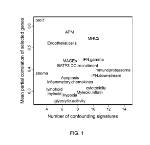
[0033] FIG. 1 illustrates the strength of co-expression in each signature's
gene set.
[0034] FIG. 2 illustrates the effectiveness of predictor training using single
genes vs. our
signatures in an immunotherapy dataset with 8 responders and 34 non-
responders.
[0035] FIG. 3 illustrates the association between immune signatures and
response to anti-PD1
immunotherapy. Boxes show average 10g2 fold-changes between responders and non-
responders;
bars show 95% confidence intervals.
[0036] FIG. 4 illustrates results of models predicting response from pairs of
signatures. Color
denotes -logio p-values. Signature pairs with p-values above 0.05 are white.
DETAILED DESCRIPTION OF THE INVENTION
[0037] In many cases, a gene signature that merely averages a collection of
biologically plausible
genes will successfully measure the intended biological process. However, many
biological
processes are governed not by modulating mRNA abundance but rather protein
abundance,
binding or location and hence, attempts to measure these processes with gene
expression will
produce misleading results. Therefore, biological knowledge alone is an
unsuitable basis for gene
signatures. The present invention provides a bridge from gene expression to
biological
interpretation in immune oncology, identifying genes whose expression track a
specific biological
14
CA 03100200 2020-11-12
WO 2019/226514 PCT/US2019/033052
process and incorporating these genes into signatures measuring the key
biology of immune
oncology.
[0038] Accordingly, the invention provides methods for selecting a patient
having cancer (e.g.,
bladder cancer, breast cancer, colorectal cancer, gastric cancer, liver
cancer, melanoma, lung
cancer (e.g., non- small cell lung carcinoma), ovarian cancer, or renal cell
carcinoma) for treatment
with an immunotherapy by determining the expression level of one or more cell
gene signatures,
and comparing this level of expression to the median level of expression of
the one or more cell
gene signatures. Detection of increased expression of the one or more cell
gene signatures relative
to a median level (i.e., higher expression of the one or more cell gene
signatures relative to the
median level in the cancer type) identifies the patient for treatment with an
immunotherapy. The
invention also provides methods for treating a patient having cancer (e.g.,
bladder cancer, breast
cancer, colorectal cancer, gastric cancer, liver cancer, melanoma, lung cancer
(e.g., non-small cell
lung carcinoma), ovarian cancer, or renal cell carcinoma) who may benefit from
a therapeutic
described herein. An example of a therapeutic described herein can be
administering an activating
immunotherapy or a suppressing immunotherapy alone or in combination with a
chemotherapy
regimen and/or other anti-cancer therapy regimen by determining the expression
level of one or
more cell gene signatures in the patient.
[0039] Definitions
[0040] Unless defined otherwise, technical and scientific terms used herein
have the same meaning
as commonly understood by one of ordinary skill in the art to which this
invention belongs.
Singleton et al., Dictionary of Microbiology and Molecular Biology 2nd ed., J.
Wiley & Sons
(New York, N.Y. 1994), and March, Advanced Organic Chemistry Reactions,
Mechanisms and
Structure 4th ed., John Wiley & Sons (New York, N.Y. 1992), provide one
skilled in the art with
a general guide to many of the terms used in the present application.
[0041] For purposes of interpreting this specification, the following
definitions will apply and
whenever appropriate, terms used in the singular will also include the plural
and vice versa. In the
event that any definition set forth below conflicts with any document
incorporated herein by
reference, the definition set forth below shall control.
[0042] The term "antagonist" is used in the broadest sense, and includes any
molecule that partially
or fully blocks, inhibits, interferes, or neutralizes a normal biological
activity of a native
polypeptide disclosed herein (e.g., an immune cell receptor or ligand, such as
CTLA-4, PD-1 ,
CA 03100200 2020-11-12
WO 2019/226514 PCT/US2019/033052
TIM-3, BTLA, VISTA, LAG -3, B7H4, CD96, TIGIT, or CD226), either by decreasing
transcription or translation of the nucleic acid encoding the native
polypeptide, or by inhibiting or
blocking the native polypeptide activity, or both. It will be understood by
one of ordinary skill in
the art that, in some instances, an antagonist may antagonize one activity of
the native polypeptide
without affecting another activity of the native polypeptide. It will also be
understood by one of
ordinary skill in the art that, in some instances, an antagonist may be a
therapeutic agent that is
considered an activating or suppressing immunotherapy depending on the native
polypeptide that
it binds, interacts, or associates with. Examples of antagonists include, but
are not limited to,
antisense polynucleotides, interfering RNAs, catalytic RNAs, RNA-DNA chimeras,
native
polypeptide-specific aptamers, antibodies, antigen-binding fragments of
antibodies, native
polypeptide- binding small molecules, native polypeptide-binding peptides, and
other peptides that
specifically bind the native polypeptide (including, but not limited to native
polypeptide-binding
fragments of one or more native polypeptide ligands, optionally fused to one
or more additional
domains), such that the interaction between the antagonist and the native
polypeptide results in a
reduction or cessation of native polypeptide activity or expression.
[0043] In a similar manner, the term "agonist" is used in the broadest sense
and includes any
molecule that mimics, promotes, stimulates, or enhances a normal biological
activity of a native
polypeptide disclosed herein (e.g., an immune cell receptor or ligand, such as
GITR, 0X40, TIM3,
LAG3, KIR, CD28, CD137, CD27, CD40, CD70, CD276, ICOS, HVEM, NKG2D, NKG2A,
MICA, 2B4 or 41BB agonist, or combination thereof), by increasing
transcription or translation
of the nucleic acid encoding the native polypeptide, and/or by inhibiting or
blocking activity of a
molecule that inhibits the expression or activity of the native polypeptide,
and/or by enhancing
normal native polypeptide activity (including, but not limited to, enhancing
the stability of the
native polypeptide, or enhancing binding of the native polypeptide to one or
more target ligands).
It will be understood by one of ordinary skill in the art that, in some
instances, an agonist may
agonize one activity of the native polypeptide without affecting another
activity of the native
polypeptide. It will also be understood by one of ordinary skill in the art
that, in some instances,
an agonist may be a therapeutic agent that is considered an activating or
suppressing
immunotherapy depending on the native polypeptide that it binds, interacts, or
associates with.
The agonist can be selected from an antibody, an antigen-binding fragment, an
aptamer, an
interfering RNA, a small molecule, a peptide, an antisense molecule, and
another binding
16
CA 03100200 2020-11-12
WO 2019/226514 PCT/US2019/033052
polypeptide. In another example, the agonist can be a polynucleotide selected
from an aptamer,
interfering RNA, or antisense molecule that interferes with the transcription
and/or translation of
a native polypeptide-inhibitory molecule.
[0044] Methods for identifying agonists or antagonists of a polypeptide may
comprise contacting
a polypeptide with a candidate agonist or antagonist molecule and measuring a
detectable change
in one or more biological activities normally associated with the polypeptide.
[0045] The term "activating immunotherapy" refers to the use of a therapeutic
agent that induces,
enhances, or promotes an immune response, including, e.g., a T cell response.
The term
"suppressing immunotherapy" refers to the use of a therapeutic agent that
interferes with,
suppresses, or inhibits an immune response, including, e.g., a T cell
response.
[0046] "Human effector cells" refer to leukocytes that express one or more
FcRs and perform
effector functions. In certain embodiments, the cells express at least FcyRIII
and perform ADCC
effector function(s). Examples of human leukocytes which mediate ADCC include
peripheral
blood mononuclear cells (PBMC), natural killer (NK) cells, monocytes,
cytotoxic T cells, and
neutrophils. The effector cells may be isolated from a native source, e.g.,
from blood.
[0047] "Regulatory T cells (Treg)" refer to a subset of helper T cells that
play a role in inhibition
of self- reactive immune responses and are often found in sites of chronic
inflammation such as in
tumor tissue, in certain embodiments, Tregs are defined phenotypically by high
cell surface
expression of CD25, CLTA4, GITR, and neuropilin-1 and are under the control of
transcription
factor FOXP3. In other embodiments, Tregs perform their suppressive function
on activated T cells
through contact-dependent mechanisms and cytokine production. In some
embodiments, Tregs also
modulate immune responses by direct interaction with ligands on dendritic
cells (DC), such as,
e.g., CTLA4 interaction with B7 molecules on DC that elicits the induction of
indoieamine 2, 3-
dioxygenase (IDO).
[0048] The term "antibody" herein is used in the broadest sense and
encompasses various antibody
structures, including but not limited to monoclonal antibodies, polyclonal
antibodies, multispecific
antibodies (e.g., bispecific antibodies), and antibody fragments so long as
they exhibit the desired
antigen-binding activity. An antibody that binds to a target refers to an
antibody that is capable of
binding the target with sufficient affinity such that the antibody is useful
as a diagnostic and/or
therapeutic agent in targeting the target. In one embodiment, the extent of
binding of an anti-target
antibody to an unrelated, non-target protein is less than about 10% of the
binding of the antibody
17
CA 03100200 2020-11-12
WO 2019/226514 PCT/US2019/033052
to target as measured, e.g., by a radioimmunoassay (MA) or biacore assay. In
certain
embodiments, an antibody that binds to a target has a dissociation constant
(Kd) of < 1 [tM, < 100
nM, < 10 nM, < 1 nM, <0.1 nM, <0.01 nM, or < 0.001 nM (e.g. 108 M or less,
e.g. from 108 M
to 10'3 M, e.g., from 109 M to 1013 M). In certain embodiments, an anti-target
antibody binds to
an epitope of a target that is conserved among different species.
[0049] A "blocking antibody" or an "antagonist antibody" is one that partially
or fully blocks,
inhibits, interferes, or neutralizes a normal biological activity of the
antigen it binds. For example,
an antagonist antibody may block signaling through an immune cell receptor
(e.g., a T cell
receptor) so as to restore a functional response by T cells (e.g.,
proliferation, cytokine production,
target cell killing) from a dysfunctional state to antigen stimulation.
[0050] An "agonist antibody" or "activating antibody" is one that mimics,
promotes, stimulates,
or enhances a normal biological activity of the antigen it binds. Agonist
antibodies can also
enhance or initiate signaling by the antigen to which it binds. In some
embodiments, agonist
antibodies cause or activate signaling without the presence of the natural
ligand. For example, an
agonist antibody may increase memory T cell proliferation, increase cytokine
production by
memory T cells, inhibit regulatory T cell function, and/or inhibit regulatory
T cell suppression of
effector T cell function, such as effector T cell proliferation and/or
cytokine production.
[0051] An "antibody fragment" refers to a molecule other than an intact
antibody that comprises
a portion of an intact antibody that binds the antigen to which the intact
antibody binds. Examples
of antibody fragments include but are not limited to Fv, Fab, Fab', Fab'-SH,
F(ab')2; diabodies;
linear antibodies; single-chain antibody molecules (e.g. scFv); and
multispecific antibodies formed
from antibody fragments.
[0052] The term "benefit" is used in the broadest sense and refers to any
desirable effect and
specifically includes clinical benefit as defined herein. Clinical benefit can
be measured by
assessing various endpoints, e.g., inhibition, to some extent, of disease
progression, including
slowing down and complete arrest; reduction in the number of disease episodes
and/or symptoms;
reduction in lesion size; inhibition (i.e., reduction, slowing down or
complete stopping) of disease
cell infiltration into adjacent peripheral organs and/or tissues; inhibition
(i.e. reduction, slowing
down or complete stopping) of disease spread; decrease of auto-immune
response, which may, but
does not have to, result in the regression or ablation of the disease lesion;
relief, to some extent, of
one or more symptoms associated with the disorder; increase in the length of
disease-free
18
CA 03100200 2020-11-12
WO 2019/226514 PCT/US2019/033052
presentation following treatment, e.g., progression-free survival; increased
overall survival; higher
response rate; and/or decreased mortality at a given point of time following
treatment.
[0053] As used herein, the term "binds," "specifically binds to," or is
"specific for" refers to
measurable and reproducible interactions such as binding between a target and
an antibody, which
is determinative of the presence of the target in the presence of a
heterogeneous population of
molecules including biological molecules. For example, an antibody that
specifically binds to a
target (which can be an epitope) is an antibody that binds this target with
greater affinity, avidity,
more readily, and/or with greater duration than it binds to other targets. In
one embodiment, the
extent of binding of an antibody to an unrelated target is less than about 10%
of the binding of the
antibody to the target as measured, for example, by a radioimmunoassay (RIA).
In certain
embodiments, an antibody that specifically binds to a target has a
dissociation constant (Kd) of <
1 [tM, < 100 nM, <1 0 nM, <1 nM, or < 0.1 nM. In certain embodiments, an
antibody specifically
binds to an epitope on a protein that is conserved among the protein from
different species. In
another embodiment, specific binding can include, but does not require
exclusive binding.
[0054] The term "biological sample" or "sample" as used herein includes, but
is not limited to,
blood, serum, plasma, sputum, tissue biopsies, tumor tissue, and nasal samples
including nasal
swabs or nasal polyps. In one embodiment, the biological sample is obtained
from the subject
before a therapy or therapeutic described herein is administered to the
subject. In another
embodiment, the biological sample is obtained from the subject after the
therapy or therapeutic
described herein is administered to the subject. In one particular embodiment,
the biological
sample is tumor tissue. In another particular embodiment, the biological
sample is blood. In other
embodiment, the sample is plasma, cerebrospinal fluid (CSF), saliva, or any
bodily fluid.
[0055] The terms "cancer" and "cancerous" refer to or describe the
physiological condition in
mammals that is typically characterized by unregulated cell growth. Included
in this definition are
benign and malignant cancers. Examples of cancer include but are not limited
to, carcinoma,
lymphoma, blastoma, sarcoma, and leukemia. More particular examples of such
cancers include
adrenocortical carcinoma, bladder urothelial carcinoma, breast invasive
carcinoma, cervical
squamous cell carcinoma, endocervical adenocarcinoma, cholangiocarcinoma,
colon
adenocarcinoma, lymphoid neoplasm diffuse large B-cell lymphoma, esophageal
carcinoma,
glioblastoma multiforme, head and neck squamous cell carcinoma, kidney
chromophobe, kidney
renal clear cell carcinoma, kidney renal papillary cell carcinoma, acute
myeloid leukemia, brain
19
CA 03100200 2020-11-12
WO 2019/226514 PCT/US2019/033052
lower grade glioma, liver hepatocellular carcinoma, lung adenocarcinoma, lung
squamous cell
carcinoma, mesothelioma, ovarian serous cystadenocarcinoma, pancreatic
adenocarcinoma,
pheochromocytoma, paraganglioma, prostate adenocarcinoma, rectum
adenocarcinoma, sarcoma,
skin cutaneous melanoma, stomach adenocarcinoma, testicular germ cell tumors,
thyroid
carcinoma, thymoma, uterine carcinosarcoma, uveal melanoma. Other examples
include breast
cancer, lung cancer, lymphoma, melanoma, liver cancer, colorectal cancer,
ovarian cancer, bladder
cancer, renal cancer or gastric cancer. Further examples of cancer include
neuroendocrine cancer,
non-small cell lung cancer (NSCLC), small cell lung cancer, thyroid cancer,
endometrial cancer,
biliary cancer, esophageal cancer, anal cancer, salivary, cancer, vulvar
cancer or cervical cancer.
[0056] An "advanced" cancer is one which has spread outside the site or organ
of origin, either by
local invasion or metastasis.
[0057] A "refractory" cancer is one which progresses even though an anti-tumor
agent, such as a
chemotherapeutic agent, is being administered to the cancer patient. An
example of a refractory
cancer is one which is platinum refractory.
[0058] A "recurrent" cancer is one which has regrown, either at the initial
site or at a distant site,
after a response to initial therapy.
[0059] By "platinum-resistant" cancer is meant cancer in a patient that has
progressed while the
patient was receiving platinum-based chemotherapy or cancer in a patient that
has progressed
within, e.g., 12 months (for instance, within 6 months) after the completion
of platinum-based
chemotherapy. Such a cancer can be said to have or exhibit "platinum-
resistance."
[0060] By "chemotherapy-resistant" cancer is meant cancer in a patient that
has progressed while
the patient is receiving a chemotherapy regimen or cancer in a patient that
has progressed within,
e.g., 12 months (for instance, within 6 months) after the completion of a
chemotherapy regimen.
Such a cancer can be said to have or exhibit "chemotherapy-resistance."
[0061] The term "tumor" refers to all neoplastic cell growth and
proliferation, whether malignant
or benign, and all pre-cancerous and cancerous cells and tissues. The terms
"cancer," "cancerous,"
"cell proliferative disorder," "proliferative disorder" and "tumor" are not
mutually exclusive as
referred to herein.
[0062] As used herein, "metastasis" is meant the spread of cancer from its
primary site to other
places in the body. Cancer cells can break away from a primary tumor,
penetrate into lymphatic
and blood vessels, circulate through the bloodstream, and grow in a distant
focus (metastasize) in
CA 03100200 2020-11-12
WO 2019/226514 PCT/US2019/033052
normal tissues elsewhere in the body. Metastasis can be local or distant.
Metastasis is a sequential
process, contingent on tumor cells breaking off from the primary tumor,
traveling through the
bloodstream, and stopping at a distant site. At the new site, the cells
establish a blood supply and
can grow to form a life-threatening mass. Both stimulatory and inhibitory
molecular pathways
within the tumor cell regulate this behavior, and interactions between the
tumor cell and host cells
in the distant site are also significant. The term "chimeric" antibody refers
to an antibody in which
a portion of the heavy and/or light chain is derived from a particular source
or species, while the
remainder of the heavy and/or light chain is derived from a different source
or species.
[0063] The "class" of an antibody refers to the type of constant domain or
constant region
possessed by its heavy chain. There are five major classes of antibodies: IgA,
IgD, IgE, IgG, and
IgM, and several of these may be further divided into subclasses (isotypes),
e.g., IgGI, lgG2, lgG3,
lgG4, IgAI, and lgA2. The heavy chain constant domains that correspond to the
different classes
of immunoglobulins are called a, 6, , y, and 11, respectively.
[0064] A "chemotherapeutic agent" includes chemical compounds useful in the
treatment of
cancer. Examples of chemotherapeutic agents include erlotinib (TARCEVA ,
Genentech/OSI
Pharm.), bortezomib (VELCADE , Millennium Pharm.), disulfiram,
epigallocatechin gallate,
salinosporamide A, carfilzomib, 17-AAG (geldanamycin), radicicol, lactate
dehydrogenase A
(LDH-A), fulvestrant (FASLODEX , AstraZeneca), sunitib (SUTENT ,
Pfizer/Sugen), letrozole
(FEMARA , Novartis), imatinib mesylate (GLEEVEC , Novartis), finasunate
(VATALANIB ,
Novartis), oxaliplatin (ELOXATIN , Sanofi), 5-FU (5-fluorouracil), leucovorin,
Rapamycin
(Sirolimus, RAPAMUNE , Wyeth), Lapatinib (TYKERB , GSK572016, Glaxo Smith
Kline),
Lonafamib (SCH 66336), sorafenib (NEXAVAR , Bayer Labs), gefitinib (IRESSA ,
AstraZeneca), AG1478, alkylating agents such as thiotepa and CYTOXAN
cyclosphosphamide;
alkyl sulfonates such as busulfan, improsulfan and piposulfan; aziridines such
as benzodopa,
carboquone, meturedopa, and uredopa; ethylenimines and methylamelamines
including
altretamine, triethylenemelamine, triethylenephosphoramide,
triethylenethiophosphoramide and
trimethylomelamine; acetogenins (especially bullatacin and bullatacinone); a
camptothecin
(including topotecan and irinotecan); bryostatin; callystatin; CC-1065
(including its adozelesin,
carzelesin and bizelesin synthetic analogs); cryptophycins (particularly
cryptophycin 1 and
cryptophycin 8); adrenocorticosteroids (including prednisone and
prednisolone); cyproterone
acetate; 5a-reductases including finasteride and dutasteride); vorinostat,
romidepsin, panobinostat,
21
CA 03100200 2020-11-12
WO 2019/226514 PCT/US2019/033052
valproic acid, mocetinostat dolastatin; aldesleukin, talc duocarmycin
(including the synthetic
analogs, KW-2189 and CB1 -TM1 ); eleutherobin; pancratistatin; a sarcodictyin;
spongistatin;
nitrogen mustards such as chlorambucil, chlomaphazine, chlorophosphamide,
estramustine,
ifosfamide, mechlorethamine, mechlorethamine oxide hydrochloride, melphalan,
novembichin,
phenesterine, prednimustine, trofosfamide, uracil mustdnitrosoureas such as
carmustine,
chlorozotocin, fotemustine, lomustine, nimustine, and ranimnustine;
antibiotics such as the
enediyne antibiotics (e.g., calichmicin, especially calicheamicin y 1 1 and
calicheamicin col 1
(Angew Chem. Intl. Ed. Engl. 1994 33:183-186); dynemicin, including dynemicin
A;
bisphosphonates, such as clodronate; an esperamicin ; as well as
neocarzinostatin chromophore
and related chromoprotein enediyne antibiotic chromophores), aclacinomysins,
actinomycin,
authramycin, azaserine, bleomycins, cactinomycin, carabicin, caminomycin,
carzinophilin,
chromomycinis, dactinomycin, daunorubicin, detorubicin, 6-diazo-5-oxo-L-
norleucine,
ADRIAMYCIN (doxorubicin), morpholino-doxorubicin, cyanomorpholino-
doxorubicin, 2-
pyrrolino-doxorubicin and deoxydoxorubicin), epirubicin, esorubicin,
idarubicin, marcellomycin,
mitomycins such as mitomycin C, mycophenolic acid, nogalamycin, olivomycins,
peplomycin,
porfiromycin, puromycin, quelamycin, rodorubicin, streptonigrin, streptozocin,
tubercidin,
ubenimex, zinostatin, zorubicin; anti-metabolites such as methotrexate and 5-
fluorouracil (5-FU);
folic acid analogs such as denopterin, methotrexate, pteropterin,
trimetrexate; purine analogs such
as fludarabine, 6-mercaptopurine, thiamiprine, thioguanine; pyrimidine analogs
such as
ancitabine, azacitidine, 6-azauridine, carmofur, cytarabine, dideoxyuridine,
doxifluridine,
enocitabine, floxuridine; androgens such as calusterone, dromostanolone
propionate, epitiostanol,
mepitiostane, testolactone; anti-adrenals such as aminoglutethimide, mitotane,
trilostane; folic acid
replenisher such as frolinic acid; aceglatone; aldophosphamide glycoside;
aminolevulinic acid;
eniluracil; amsacrine; bestrabucil; bisantrene; edatraxate; defofamine;
demecolcine; diaziquone;
elfomithine; elliptinium acetate; an epothilone; etoglucid; gallium nitrate;
hydroxyurea; lentinan ;
lonidainine; maytansinoids such as maytansine and ansamitocins; mitoguazone;
mitoxantrone;
mopidamnol; nitraerine; pentostatin ; phenamet; pirarubicin; losoxantrone;
podophyllinic acid; 2-
ethylhydrazide; procarbazine; PSK polysaccharide complex (JHS Natural
Products, Eugene,
Oreg.); razoxane; rhizoxin; sizofuran; spirogermanium ; tenuazonic acid;
triaziquone; 2,2',2"-
trichlorotriethylamine; trichothecenes (especially T- 2 toxin, verracurin A,
roridin A and
angui dine); urethan; vinde sine; dacarb azine; mannomustine; mitobronitol;
mitolactol;
22
CA 03100200 2020-11-12
WO 2019/226514 PCT/US2019/033052
pipobroman; gacytosine; arabinoside ("Ara-C"); cyclophosphamide; thiotepa;
taxoids, e.g.,
TAXOL (paclitaxel; Bristol-Myers Squibb Oncology, Princeton, N.J.), ABRAXANE
(Cremophor-free), albumin-engineered nanoparticle formulations of paclitaxel
(American
Pharmaceutical Partners, Schaumberg, III.), and TAXOTERE (docetaxel,
doxetaxel; Sanofi-
Aventi s); chloranmbucil; GEMZ AR (gemcitabine); 6-thioguanine;
mercaptopurine;
methotrexate; platinum analogs such as cisplatin and carboplatin; vinblastine;
etoposide (VP-16);
ifosfamide; mitoxantrone; vincristine; NAVELBINE (vinorelbine); novantrone;
teniposide;
edatrexate; daunomycin; aminopterin; capecitabine (XELODA ); ibandronate; CPT-
1 1 ;
topoisomerase inhibitor RFS 2000; difluoromethylornithine (DMF0); retinoids
such as retinoic
acid; and pharmaceutically acceptable salts, acids and derivatives of any of
the above.
[0065] A chemotherapeutic agent also includes (i) anti-hormonal agents that
act to regulate or
inhibit hormone action on tumors such as anti-estrogens and selective estrogen
receptor
modulators (SERMs), including, for example, tamoxifen (including NOLVADEX ,
tamoxifen
citrate), raloxifene, droloxifene, iodoxyfene, 4-hydroxytamoxifen, trioxifene,
keoxifene, LY1
17018, onapristone, and FARESTON (toremifine citrate); (ii) aromatase
inhibitors that inhibit
the enzyme aromatase, which regulates estrogen production in the adrenal
glands, such as, for
example, 4(5)-imidazoles, aminoglutethimide, MEGASE (megestrol acetate),
AROMASIN
(exemestane; Pfizer), formestanie, fadrozole, RIVISOR (vorozole), FEMARA
(letrozole;
Novartis), and ARIMIDEX (anastrozole; AstraZeneca); (iii) anti-androgens such
as flutamide,
nilutamide, bicalutamide, leuprolide and goserelin; buserelin, tripterelin,
medroxyprogesterone
acetate, diethylstilbestrol, premarin, fluoxymesterone, all transretionic
acid, fenretinide, as well as
troxacitabine (a 1 ,3-dioxolane nucleoside cytosine analog); (iv) protein
kinase inhibitors; (v) lipid
kinase inhibitors; (vi) antisense oligonucleotides, particularly those which
inhibit expression of
genes in signaling pathways implicated in aberrant cell proliferation, such
as, for example, PKC-
alpha, Ralf and H-Ras; (vii) ribozymes such as VEGF expression inhibitors
(e.g.,
ANGIOZYME ) and HER2 expression inhibitors; (viii) vaccines such as gene
therapy vaccines,
for example, ALLOVECTIN , LEUVECTIN , and VAXID , PROLEUKIN , r1L-2; a
topoisomerase 1 inhibitor such as LURTOTECANg; ABARELIX rmRH; and (ix)
pharmaceutically acceptable salts, acids and derivatives of any of the above.
[0066] A chemotherapeutic agent also includes antibodies such as alemtuzumab
(Campath),
bevacizumab (AVASTIN , Genentech); cetuximab (ERBITUX , Imclone); panitumumab
23
CA 03100200 2020-11-12
WO 2019/226514 PCT/US2019/033052
(VECTIBIX , Amgen), rituximab (RITUXAN , Genentech/Biogen Idee), pertuzumab
(OMNITARG , 2C4, Genentech), trastuzumab (HERCEPTIN , Genentech), tositumomab
(Bexxar, Corixia), and the antibody drug conjugate, gemtuzumab ozogamicin
(MYLOTARG ,
Wyeth). Additional humanized monoclonal antibodies with therapeutic potential
as agents in
combination with the compounds of the invention include: apolizumab,
aselizumab, atlizumab,
bapineuzumab, bivatuzumab mertansine, cantuzumab mertansine, cedelizumab,
certolizumab
pegol, cidfusituzumab, cidtuzumab, daclizumab, eculizumab, efalizumab,
epratuzumab,
erlizumab, felvizumab, fontolizumab, gemtuzumab ozogamicin, inotuzumab
ozogamicin,
ipilimumab, labetuzumab, lintuzumab, matuzumab, mepolizumab, motavizumab,
motovizumab,
natalizumab, nimotuzumab, nolovizumab, numavizumab, ocrelizumab, omalizumab,
palivizumab,
pascolizumab, pecfusituzumab, pectuzumab, pexelizumab, ralivizumab,
ranibizumab,
reslivizumab, reslizumab, resyvizumab, rovelizumab, ruplizumab, sibrotuzumab,
siplizumab,
sontuzumab, tacatuzumab tetraxetan, tadocizumab, talizumab, tefibazumab,
tocilizumab,
toralizumab, tucotuzumab celmoleukin, tucusituzumab, umavizumab, urtoxazumab,
ustekinumab,
visilizumab, and the anti¨ interleukin-12 (ABT-874/J695, Wyeth Research and
Abbott
Laboratories) which is a recombinant exclusively human-sequence, full-length
lgG1 X. antibody
genetically modified to recognize interleukin-12 p40 protein.
[0067] A chemotherapeutic agent also includes "EGFR inhibitors," which refers
to compounds
that bind to or otherwise interact directly with EGFR and prevent or reduce
its signaling activity,
and is alternatively referred to as an "EGFR antagonist." Examples of such
agents include
antibodies and small molecules that bind to EGFR. Examples of antibodies which
bind to EGFR
include MAb 579 (ATCC CRL HB 8506), MAb 455 (ATCC CRL HB8507), MAb 225 (ATCC
CRL 8508), MAb 528 (ATCC CRL 8509) (see, US Patent No. 4,943, 533, Mendelsohn
et al.) and
variants thereof, such as chimerized 225 (C225 or Cetuximab; ERBUTIX ) and
reshaped human
225 (H225) (see, WO 96/40210, Imclone Systems Inc.); IMC-1 1 F8, a fully
human, EGFR-
targeted antibody (Imclone); antibodies that bind type II mutant EGFR (US
Patent No. 5,212,290);
humanized and chimeric antibodies that bind EGFR as described in US Patent No.
5,891 ,996; and
human antibodies that bind EGFR, such as ABX-EGF or Panitumumab (see
W098/50433,
Abgenix/Amgen); EMD 55900 (Stragliotto et al. Eur. J. Cancer 32A:636-640 (1
996)); EMD7200
(matuzumab) a humanized EGFR antibody directed against EGFR that competes with
both EGF
and TGF-alpha for EGFR binding (EMD/Merck); human EGFR antibody, HuMax-EGFR
24
CA 03100200 2020-11-12
WO 2019/226514 PCT/US2019/033052
(GenMab); fully human antibodies known as El .1 , E2.4, E2.5, E6.2, E6.4, E2.1
1 , E6. 3 and
E7.6. 3 and described in US 6,235,883; MDX-447 (Medarex Inc); and mAb 806 or
humanized
mAb 806 (Johns et al., J. Biol. Chem. 279(29):30375-30384 (2004)). The anti-
EGFR antibody
may be conjugated with a cytotoxic agent, thus generating an immunoconjugate
(see, e.g.,
EP659,439A2, Merck Patent GmbH). EGFR antagonists include small molecules such
as
compounds described in US Patent Nos: 5,616,582; 5,457,105; 5,475,001;
5,654,307; 5,679,683;
6,084,095; 6,265,410; 6,455,534; 6,521,620; 6,596,726; 6,713,484; 5,770,599;
6,140,332;
5,866,572; 6,399,602; 6,344,459; 6,602,863; 6,391,874; 6,344,455; 5,760,041;
6,002,008; and
5,747,498, as well as the following PCT publications: W098/14451, W098/50038,
W099/09016,
and W099/24037. Particular small molecule EGFR antagonists include OSI-774 (CP-
358774,
erlotinib, TARCEVA Genentech/OSI Pharmaceuticals); PD 183805 (CI 1033, 2-
propenamide,
N44- [(3 -chloro-4-fluorophenyl)amino]-743 -(4-morpholinyl)propoxy]-6-
quinazoliny1]-,
dihydrochloride, Pfizer Inc.); ZD1839, gefitinib (IRESSAg) 4-(3'-Chloro-4'-
fluoroanilino)-7-
methoxy-6-(3- morpholinopropoxy)quinazoline, AstraZeneca); ZM 105180 ((6-amino-
4-(3-
methylphenyl-amino)- quinazoline, Zeneca); BIBX-1382 (N8-(3-chloro-4-fluoro-
pheny1)-N2-(1 -
methyl-piperidin-4-y1)-pyrimido[5,4- d]pyrimidine-2,8-diamine, Boehringer
Ingelheim); PKI-166
((R)-4-[4-[(1 -phenylethyl)amino]-1 H-pyrrolo[2,3- d]pyrimidin-6-y1]-phenol);
(R)-6-(4-
hydroxypheny1)-4-[(1 -phenylethyl)amino]-7H-pyrrolo[2,3-d]pyrimidine); CL-
387785 (N-[4-[(3-
bromophenyl)amino]-6-quinazoliny1]-2-butynamide);
EKB -569 (N-[4-[(3-chloro-4-
fluorophenyl)amino]-3-cyano-7-ethoxy-6-quinoliny1]-4-(dimethylamino)-2-
butenamide)
(Wyeth); AG1478 (Pfizer); AG1571 (SU 5271 ; Pfizer); dual EGFR/HER2 tyrosine
kinase
inhibitors such as lapatinib (TYKERB , G5K572016 or N-[3-chloro-4-[(3
fluorophenyl)methoxy]pheny1]-6 [5 [ [[2methyl sulfonyl)ethyl] amino]methy1]-2-
furany1]-4-
quinazolinamine).
[0068] Chemotherapeutic agents also include "tyrosine kinase inhibitors"
including the EGFR-
targeted drugs noted in the preceding paragraph; small molecule HER2 tyrosine
kinase inhibitor
such as TAK165 available from Takeda; CP-724,714, an oral selective inhibitor
of the ErbB2
receptor tyrosine kinase (Pfizer and OSI); dual-HER inhibitors such as EKB-569
(available from
Wyeth) which preferentially binds EGFR but inhibits both HER2 and EGFR-
overexpressing cells;
lapatinib (G5K572016; available from Glaxo-SmithKline), an oral HER2 and EGFR
tyrosine
kinase inhibitor; PKI-166 (available from Novartis); pan-HER inhibitors such
as canertinib (CI-
CA 03100200 2020-11-12
WO 2019/226514 PCT/US2019/033052
1033; Pharmacia); Raf-1 inhibitors such as antisense agent ISIS-5132 available
from ISIS
Pharmaceuticals which inhibit Raf-1 signaling; non-HER targeted TK inhibitors
such as imatinib
mesylate (GLEEVEC , available from Glaxo SmithKline); multi-targeted tyrosine
kinase
inhibitors such as sunitinib (SUTENT , available from Pfizer); VEGF receptor
tyrosine kinase
inhibitors such as vatalanib (PTK787/ZK222584, available from
Novartis/Schering AG); MAPK
extracellular regulated kinase I inhibitor CI-1040 (available from Pharmacia);
quinazolines, such
as PD 153035, 4-(3-chloroanilino) quinazoline; pyridopyrimidines;
pyrimidopyrimidines;
pyrrolopyrimidines, such as CGP 59326, CGP 60261 and CGP 62706;
pyrazolopyrimidines, 4-
(phenylamino)-7H-pyrrolo[2,3-d] pyrimidines; curcumin (diferuloyl methane, 4,5-
bis (4-
fluoroanilino)phthalimide); tyrphostines containing nitrothiophene moieties;
PD-0183805
(Warner-Lamber) ; antisense molecules (e.g. those that bind to HER-encoding
nucleic acid);
quinoxalines (US Patent No. 5,804,396); tryphostins (US Patent No. 5,804,396);
ZD6474 (Astra
Zeneca); PTK-787 (Novartis/Schering AG); pan-HER inhibitors such as Cl- 1033
(Pfizer);
Affinitac (ISIS 3521 ; Isis/Lilly); imatinib mesylate (GLEEVEC ); PKI 166
(Novartis); GW2016
(Glaxo SmithKline); CI-1033 (Pfizer); EKB-569 (Wyeth); Semaxinib (Pfizer);
ZD6474
(AstraZeneca); PTK-787 (Novartis/Schering AG); INC-1 Cl 1 (Imclone), rapamycin
(sirolimus,
RAPAMUNE ); or as described in any of the following patent publications: US
Patent No.
5,804,396; WO 1999/09016 (American Cyanamid); WO 1998/43960 (American
Cyanamid); WO
1997/38983 (Warner Lambert) ; WO 1 999/06378 (Warner Lambert) ; WO 1 999/06396
(Warner
Lambert) ; WO 1 996/30347 (Pfizer, Inc) ; WO 1 996/33978 (Zeneca) ; WO 1
996/3397 (Zeneca)
and WO 1 996/33980 (Zeneca).
[0069] Chemotherapeutic agents also include dexamethasone, interferons,
colchicine, metoprine,
cyclosporine, amphotericin, metronidazole, alemtuzumab, alitretinoin,
allopurinol, amifostine,
arsenic trioxide, asparaginase, BCG live, bevacuzimab, bexarotene, cladribine,
clofarabine,
darbepoetin alfa, denileukin, dexrazoxane, epoetin alfa, elotinib, filgrastim,
histrelin acetate,
ibritumomab, interferon alfa- 2a, interferon alfa-2b, lenalidomide,
levamisole, mesna,
methoxsalen, nandrolone, nelarabine, nofetumomab, oprelvekin, palifermin,
pamidronate,
pegademase, pegaspargase, pegfilgrastim, pemetrexed disodium, plicamycin,
porfimer sodium ,
quinacrine, rasburicase, sargramostim, temozolomide, VM-26, 6-TG, toremifene,
tretinoin,
ATRA, valrubicin, zoledronate, and zoledronic acid, and pharmaceutically
acceptable salts
thereof.
26
CA 03100200 2020-11-12
WO 2019/226514 PCT/US2019/033052
[0070] By "platinum-based chemotherapeutic agent" or "platin" is meant an
antineoplastic drug
that is a coordination complex of platinum. Examples of platinum-based
chemotherapeutic agents
include carboplatin, cisplatin, satraplatin, picoplatin, nedaplatin,
triplatin, lipoplatin, and
oxaliplatinum.
[0071] By "platinum-based chemotherapy" is meant therapy with one or more
platinum-based
chemotherapeutic agent, optionally in combination with one or more other
chemotherapeutic
agents.
[0072] By "correlate" or "correlation" or grammatical equivalents is meant
comparing, in any way,
the performance and/or results of a first analysis or protocol with the
performance and/or results
of a second analysis or protocol. For example, one may use the results of a
first analysis or protocol
to determine the outcome or result of a second analysis or protocol. Or one
may use the results of
a first analysis or protocol to determine whether a second analysis or
protocol should be performed.
For example, with respect to the embodiment of gene expression analysis or
protocol, one may use
the results of the gene expression analysis or protocol to determine whether a
specific immune cell
type or subset is present.
[0073] "Effector functions" refer to those biological activities attributable
to the Fc region of an
antibody, which vary with the antibody isotype. Examples of antibody effector
functions include:
Clq binding and complement dependent cytotoxicity (CDC); Fc receptor binding;
antibody-
dependent cell-mediated cytotoxicity (ADCC); phagocytosis; down regulation of
cell surface
receptors (e.g. B cell receptor); and B cell activation.
[0074] "Enhancing T cell function" means to induce, cause or stimulate an
effector or memory T
cell to have a renewed, sustained or amplified biological function. Examples
of enhancing T cell
function include: increased secretion of y-interferon from CD8 effector T
cells, increased secretion
of y-interferon from CD4+ memory and/or effector T cells, increased
proliferation of CD4+
effector and/or memory T cells, increased proliferation of CD8 effector T
cells, increased antigen
responsiveness (e.g., clearance), relative to such levels before the
intervention. In one embodiment,
the level of enhancement is at least 50%, alternatively 60%, 70%, 80%, 90%,
100%, 120%, 1 50%,
200%. The manner of measuring this enhancement is known to one of ordinary
skill in the art.
[0075] A sample, cell, tumor, or cancer which "expresses" one or more cell
gene signatures at an
increased expression level relative to a median level of expression (e.g., the
median level of
expression of the one or more cell gene signatures in the type of cancer (or
in a cancer type, wherein
27
CA 03100200 2020-11-12
WO 2019/226514 PCT/US2019/033052
the "cancer type" is meant to include cancerous cells (e.g., tumor cells,
tumor tissues) as well as
non-cancerous cells (e.g. , stromal cells, stromal tissues) that surround the
cancerous/tumor
environment) is one in which the expression level of one or more cell gene
signatures is considered
to be a "high cell gene signature expression level" to a skilled person for
that type of cancer.
Generally, such a level will be in the range from about 50% up to about 100%
or more (e.g., 50%,
55%, 60%, 65%, 70%, 75%, 80%, 85%, 90%, 95%, 100%, or more) relative to cell
gene signature
levels in a population of samples, cells, tumors, or cancers of the same
cancer type. For instance,
the population that is used to arrive at the median expression level may be
particular cancer
samples (e.g., adrenocortical carcinoma, bladder urothelial carcinoma, breast
invasive carcinoma,
cervical squamous cell carcinoma, endocervical adenocarcinoma,
cholangiocarcinoma, colon
adenocarcinoma, lymphoid neoplasm diffuse large B-cell lymphoma, esophageal
carcinoma,
glioblastoma multiforme, head and neck squamous cell carcinoma, kidney
chromophobe, kidney
renal clear cell carcinoma, kidney renal papillary cell carcinoma, acute
myeloid leukemia, brain
lower grade glioma, liver hepatocellular carcinoma, lung adenocarcinoma, lung
squamous cell
carcinoma, mesothelioma, ovarian serous cystadenocarcinoma, pancreatic
adenocarcinoma,
pheochromocytoma, paraganglioma, prostate adenocarcinoma, rectum
adenocarcinoma, sarcoma,
skin cutaneous melanoma, stomach adenocarcinoma, testicular germ cell tumors,
thyroid
carcinoma, thymoma, uterine carcinosarcoma, uveal melanoma. Other examples
include breast
cancer, lung cancer, lymphoma, melanoma, liver cancer, colorectal cancer,
ovarian cancer, bladder
cancer, renal cancer or gastric cancer. Further examples of cancer include
neuroendocrine cancer,
non-small cell lung cancer (NSCLC), small cell lung cancer, thyroid cancer,
endometrial cancer,
biliary cancer, esophageal cancer, anal cancer, salivary, cancer, vulvar
cancer or cervical cancer)
generally, or subgroupings thereof, such as chemotherapy-resistant cancer,
platinum-resistant
cancer, as well as advanced, refractory, or recurrent cancer samples.
[0076] By "determining the expression level" used in reference to a particular
biomarker (e.g., one
or more genes from the cell gene signatures), means expression of the
biomarker(s) (e.g., one or
more genes from the cell gene signatures) in a cancer-associated biological
environment (e.g.,
expression of the biomarker(s) in the tumor cells), tumor-associated cells
(e.g., tumor-associated
stromal cells), as determined using a diagnostic test, any of the detection
methods described herein,
or the similar. In one embodiment, expression of the one or more genes in the
biological sample
form the patient is determined by measuring mRNA. In other embodiments,
expression of the one
28
CA 03100200 2020-11-12
WO 2019/226514 PCT/US2019/033052
or more genes in the biological sample form the patient is determined by
measuring mRNA in
plasma, by measuring mRNA in tissue, by measuring mRNA in FFPE tissue, by
measuring protein
levels, by measuring protein levels in plasma, by measuring protein levels in
tissue, by measuring
protein levels in FFPE tissue or a combination thereof
[0077] The term "Fc region" herein is used to define a C-terminal region of an
immunoglobulin
heavy chain that contains at least a portion of the constant region. The term
includes native
sequence Fc regions and variant Fc regions. In one embodiment, a human IgG
heavy chain Fc
region extends from Cys226, or from Pro230, to the carboxyl-terminus of the
heavy chain.
However, the C-terminal lysine (Lys447) of the Fc region may or may not be
present. Unless
otherwise specified herein, numbering of amino acid residues in the Fc region
or constant region
is according to the EU numbering system, also called the EU index, as
described in Kabat et al,
Sequences of Proteins of Immunological Interest, 5th Ed. Public Health
Service, National Institutes
of Health, Bethesda, MD, 1991 .
[0078] "Framework" or "FR" refers to variable domain residues other than
hypervariable region
(HVR) residues. The FR of a variable domain generally consists of four FR
domains: FR1 , FR2,
FR3, and FR4. Accordingly, the HVR and FR sequences generally appear in the
following
sequence in VH (or VL): FR1 - H1 (L1 )-FR2-H2(L2)-FR3-H3(L3)-FR4. In some
embodiments,
an antibody used herein comprises a human consensus framework.
[0079] The terms "full length antibody," "intact antibody," and "whole
antibody" are used herein
interchangeably to refer to an antibody having a structure substantially
similar to a native antibody
structure or having heavy chains that contain an Fc region as defined herein.
[0080] A "human antibody" is one which possesses an amino acid sequence which
corresponds to
that of an antibody produced by a human or a human cell or derived from a non-
human source that
utilizes human antibody repertoires or other human antibody-encoding
sequences. This definition
of a human antibody specifically excludes a humanized antibody comprising non-
human antigen-
binding residues.
[0081] A "human consensus framework" is a framework which represents the most
commonly
occurring amino acid residues in a selection of human immunoglobulin VL or VH
framework
sequences. Generally, the selection of human immunoglobulin VL or VH sequences
is from a
subgroup of variable domain sequences. Generally, the subgroup of sequences is
a subgroup as in
Kabat et al, Sequences of Proteins of Immunological Interest, Fifth Edition,
NIH Publication 91 -
29
CA 03100200 2020-11-12
WO 2019/226514 PCT/US2019/033052
3242, Bethesda MD (1991 ), vols. 1 -3. In one embodiment, for the VL, the
subgroup is subgroup
kappa I as in Kabat et al, supra. In one embodiment, for the VH, the subgroup
is subgroup III as
in Kabat et al, supra. A "humanized" antibody refers to a chimeric antibody
comprising amino acid
residues from non- human HVRs and amino acid residues from human FRs. In
certain
embodiments, a humanized antibody will comprise substantially all of at least
one, and typically
two, variable domains, in which all or substantially all of the HVRs (e.g.,
CDRs) correspond to
those of a non-human antibody, and all or substantially all of the FRs
correspond to those of a
human antibody. A humanized antibody optionally may comprise at least a
portion of an antibody
constant region derived from a human antibody. A "humanized form" of an
antibody, e.g., a non-
human antibody, refers to an antibody that has undergone humanization.
[0082] The term "hypervariable region" or "HVR," as used herein, refers to
each of the regions of
an antibody variable domain which are hypervariable in sequence and/or form
structurally defined
loops ("hypervariable loops"). Generally, native four-chain antibodies
comprise six HVRs; three
in the VH (HI, H2, H3), and three in the VL (LI, L2, L3). HVRs generally
comprise amino acid
residues from the hypervariable loops and/or from the "complementarity
determining regions"
(CDRs), the latter typically being of highest sequence variability and/or
involved in antigen
recognition. An HVR region as used herein comprise any number of residues
located within
positions 24-36 (for HVRL1 ), 46-56 (for HVRL2), 89-97 (for HVRL3), 26-35B
(for HVRH1),
47-65 (for HVRH2), and 93-102 (for HVRH3).
[0083] "Tumor immunity" refers to the process in which tumors evade immune
recognition and
clearance. Thus, as a therapeutic concept, tumor immunity is "treated" when
such evasion is
attenuated, and the tumors are recognized and attacked by the immune system.
Examples of tumor
recognition include tumor binding, tumor shrinkage, and tumor clearance.
"Immunogenicity"
refers to the ability of a particular substance to provoke an immune response.
Tumors are
immunogenic and enhancing tumor immunogenicity aids in the clearance of the
tumor cells by the
immune response. Examples of enhancing tumor immunogenicity include but are
not limited to
treatment with a CD28, 0X40, GITR, CD137, CD27, ICOS, HVEM, NKG2D, MICA, or
2B4
agonist or treatment with a CTLA-4, PD-1 axis, TIM-3, BTLA, VISTA, LAG-3,
B7H4, CD96,
TIGIT, or CD226 antagonist.
[0084] An "immunoconjugate" is an antibody conjugated to one or more
heterologous
molecule(s), including but not limited to a cytotoxic agent.
CA 03100200 2020-11-12
WO 2019/226514 PCT/US2019/033052
[0085] An "individual" or "subject" is a mammal. Mammals include, but are not
limited to,
domesticated animals (e.g., cows, sheep, cats, dogs, and horses), primates
(e.g., humans and non-
human primates such as monkeys), rabbits, and rodents (e.g., mice and rats).
In certain
embodiments, the individual or subject is a human.
[0086] An "isolated" antibody is one which has been separated from a component
of its natural
environment. In some embodiments, an antibody is purified to greater than 95%
or 99% purity as
determined by, for example, electrophoretic (e.g., SDS-PAGE, isoelectric
focusing (IEF), capillary
electrophoresis) or chromatographic (e.g., ion exchange or reverse phase
HPLC). F or review of
methods for assessment of antibody purity, see, e.g., Flatman et al, J.
Chromatogr. B 848:79-87
(2007).
[0087] An "isolated" nucleic acid refers to a nucleic acid molecule that has
been separated from a
component of its natural environment. An isolated nucleic acid includes a
nucleic acid molecule
contained in cells that ordinarily contain the nucleic acid molecule, but the
nucleic acid molecule
is present extrachromosomally or at a chromosomal location that is different
from its natural
chromosomal location. "Isolated nucleic acid encoding an anti-target antibody"
refers to one or
more nucleic acid molecules encoding antibody heavy and light chains (or
fragments thereof),
including such nucleic acid molecule(s) in a single vector or separate
vectors, and such nucleic
acid molecule(s) present at one or more locations in a host cell.
[0088] A "loading" dose herein generally comprises an initial dose of a
therapeutic agent
administered to a patient, and is followed by one or more maintenance dose(s)
thereof. Generally,
a single loading dose is administered, but multiple loading doses are
contemplated herein. Usually,
the amount of loading dose(s) administered exceeds the amount of the
maintenance dose(s)
administered and/or the loading dose(s) are administered more frequently than
the maintenance
dose(s), so as to achieve the desired steady-state concentration of the
therapeutic agent earlier than
can be achieved with the maintenance dose(s).
[0089] The term "monoclonal antibody" as used herein refers to an antibody
obtained from a
population of substantially homogeneous antibodies, i.e., the individual
antibodies comprising the
population are identical and/or bind the same epitope, except for possible
variant antibodies, e.g.,
containing naturally occurring mutations or arising during production of a
monoclonal antibody
preparation, such variants generally being present in minor amounts. In
contrast to polyclonal
antibody preparations, which typically include different antibodies directed
against different
31
CA 03100200 2020-11-12
WO 2019/226514 PCT/US2019/033052
determinants (epitopes), each monoclonal antibody of a monoclonal antibody
preparation is
directed against a single determinant on an antigen. Thus, the modifier
"monoclonal" indicates the
character of the antibody as being obtained from a substantially homogeneous
population of
antibodies, and is not to be construed as requiring production of the antibody
by any particular
method. For example, the monoclonal antibodies to be used according to the
methods provided
herein may be made by a variety of techniques, including but not limited to
the hybridoma method,
recombinant DNA methods, phage-display methods, and methods utilizing
transgenic animals
containing all or part of the human immunoglobulin loci, such methods and
other exemplary
methods for making monoclonal antibodies being described herein.
[0090] A "naked antibody" refers to an antibody that is not conjugated to a
heterologous moiety
(e.g., a cytotoxic moiety) or radiolabel. The naked antibody may be present in
a pharmaceutical
formulation.
[0091] "Native antibodies" refer to naturally occurring immunoglobulin
molecules with varying
structures. For example, native IgG antibodies are heterotetrameric
glycoproteins of about 150,000
daltons, composed of two identical light chains and two identical heavy chains
that are disulfide-
bonded. From N- to C-terminus, each heavy chain has a variable region (VH),
also called a variable
heavy domain or a heavy chain variable domain, followed by three constant
domains (CHL CH2,
and CH3). Similarly, from N- to C-terminus, each light chain has a variable
region (VL), also
called a variable light domain or a light chain variable domain, followed by a
constant light (CL)
domain. The light chain of an antibody may be assigned to one of two types,
called kappa (x) and
lambda (k), based on the amino acid sequence of its constant domain.
[0092] "Patient response" or "response" (and grammatical variations thereof)
can be assessed
using any endpoint indicating a benefit to the patient, including, without
limitation, (1) inhibition,
to some extent, of disease progression, including slowing down and complete
arrest; (2) reduction
in the number of disease episodes and/or symptoms; (3) reduction in lesional
size; (4) inhibition
(i.e., reduction, slowing down or complete stopping) of disease cell
infiltration into adjacent
peripheral organs and/or tissues; (5) inhibition (i.e. reduction, slowing down
or complete stopping)
of disease spread; (6) decrease of auto-immune response, which may, but does
not have to, result
in the regression or ablation of the disease lesion; (7) relief, to some
extent, of one or more
symptoms associated with the disorder; (8) increase in the length of disease-
free presentation
following treatment; and/or (9) decreased mortality at a given point of time
following treatment.
32
CA 03100200 2020-11-12
WO 2019/226514 PCT/US2019/033052
[0093] By "radiation therapy" or "radiation" is meant the use of directed
gamma rays or beta rays
to induce sufficient damage to a cell so as to limit its ability to function
normally or to destroy the
cell altogether. It will be appreciated that there will be many ways known in
the art to determine
the dosage and duration of treatment. Typical treatments are given as a one-
time administration
and typical dosages range from 10 to 200 units (Grays) per day.
[0094] The term "small molecule" refers to an organic molecule having a
molecular weight
between 50 Daltons to 2500 Daltons.
[0095] The terms "cell gene signature" refers to any one or a combination or
sub-combination of
the genes set forth in Table 1. Such sub-combinations of these genes are
sometimes referred to as
"gene sets," and exemplary "gene sets" are set forth in Tables 2-17. The term
"immune cell
signature" refers to the gene expression pattern of a cell gene signature in a
patient that correlates
with the presence of an immune cell subtype (e.g., T effector cells, T
regulatory cells, B cells, NK
cells, myeloid cells, Th17 cells, inflammatory cells, T cell immune blockers,
and antigen
presenting cell (APC) immune blockers). Each individual gene or member of a
cell gene signature
is a "cell signature gene." Further, each individual gene or member of an
immune cell gene
signature is an "immune cell signature gene." These genes include, without
limitation the genes
from the lymphoid gene signature set in Table 1: CXCL10, CXCR3, CX3CL1, PRF1,
GZMK,
GZMB, CD27, IL2RG, KLRK1, CTLA4, GZMH, CD3D, KLRB1, KLRD1, LCK, CD5, IRF4,
CD8A, CD38, EOMES, GZMM, GNLY, IFITM1, ID01, MS4A1, GZMA, CD2, CD3E, CD3G,
CD4OLG, CD6, CD7, CD79A, CD8B, CXCL11, CXCL13, CXCL9, HLA-DOB, IFNG, LAG3,
LY9, PDCD1, TBX21, TIGIT, ZAP70, SLAMF7, CD96, PVR, STAT1, JAK1, JAK2, STAT2,
IRF9, IGF2R, CD48, ICOS or for example, the genes from the myeloid gene
signature set in Table
1: ITGAM, TLR4, IL1B, CSF1R, CSF3R, TLR2, TLR1, ITGAX, HCK, TLR8, SLC11A1,
CD47,
CD14, CLEC4E, CLEC7A, FCAR, FCN1, LILRA5, LILRB2, LYZ, NFAM1, P2RY13, 5100A8,
5100A9, SERPINA1, SIRPA, SIRPB2, TREM1, CLEC5A, CSF1, CYBB, FCGR1A, MARCO,
NLRP3, FPR1, FPR3, CCL3, DAB2, OLR1, C5AR1, TREM2, MRC1, CEBPB.
[0096] The term "PD1-axis antagonist" refers to a molecule that inhibits the
interaction of a PD-1
axis binding partner with either one or more of its binding partner, so as to
remove T cell
dysfunction resulting from signaling on the PD-1 signaling axis-with a result
being to restore or
enhance T cell function (e.g., proliferation, cytokine production, target cell
killing). As used
33
CA 03100200 2020-11-12
WO 2019/226514 PCT/US2019/033052
herein, a PD-1 axis antagonist includes a PD-1 binding antagonist, a PD-Li
binding antagonist,
and a PD-L2 binding antagonist.
[0097] "Survival" refers to the patient remaining alive, and includes overall
survival as well as
progression free survival.
[0098] "Overall survival" refers to the patient remaining alive for a defined
period of time, such
as 1 year, 5 years, etc. from the time of diagnosis or treatment.
[0099] The phrase "progression-free survival" in the context of the present
invention refers to the
length of time during and after treatment during which, according to the
assessment of the treating
physician or investigator, a patient's disease does not become worse, i.e.,
does not progress. As the
skilled person will appreciate, a patient's progression-free survival is
improved or enhanced if the
patient experiences a longer length of time during which the disease does not
progress as compared
to the average or mean progression free survival time of a control group of
similarly situated
patients.
[00100] By "standard of care" herein is intended the anti-tumor/anti-cancer,
anti-condition or anti-
disease agent or agents that are routinely used to treat a particular form of
cancer, condition or
disease.
[00101] The terms "therapeutically effective amount" or "effective amount"
refer to an amount of
a drug effective to treat a cancer, condition or disease in the patient. For
example, with respect to
cancer, the effective amount of the drug may reduce the number of cancer
cells; reduce the tumor
size; inhibit (i.e., slow to some extent and preferably stop) cancer cell
infiltration into peripheral
organs; inhibit (i.e., slow to some extent and preferably stop) tumor
metastasis; inhibit, to some
extent, tumor growth; and/or relieve to some extent one or more of the
symptoms associated with
the cancer. To the extent the drug may prevent growth and/or kill existing
cancer cells, it may be
cytostatic and/or cytotoxic. The effective amount may extend progression free
survival (e.g. as
measured by Response Evaluation Criteria for Solid Tumors, RECIST, or CA-125
changes), result
in an objective response (including a partial response, PR, or complete
response, CR), improve
survival (including overall survival and progression free survival) and/or
improve one or more
symptoms of cancer (e.g. as assessed by FOSI). Most preferably, the
therapeutically effective
amount of the drug is effective to improve progression free survival (PFS)
and/or overall survival
(OS).
34
CA 03100200 2020-11-12
WO 2019/226514 PCT/US2019/033052
[00102] As used herein, "treatment" refers to clinical intervention in an
attempt to alter the natural
course of the individual or cell being treated, and can be performed either
for prophylaxis or during
the course of clinical pathology. Desirable effects of treatment include
preventing occurrence or
recurrence of disease, alleviation of symptoms, diminishment of any direct or
indirect pathological
consequences of the disease, decreasing the rate of disease progression,
amelioration or palliation
of the disease state, and remission or improved prognosis. In some
embodiments, methods and
compositions of the invention are useful in attempts to delay development of a
disease or disorder.
[00103] The term "variable region" or "variable domain" refers to the domain
of an antibody
heavy or light chain that is involved in binding the antibody to antigen. The
variable domains of
the heavy chain and light chain (VH and VL, respectively) of a native antibody
generally have
similar structures, with each domain comprising four conserved framework
regions (FRs) and
three hypervariable regions (HVRs). (See, e.g., Kindt et al. Kuby Immunology,
6th ed., W.H.
Freeman and Co., page 91 (2007).) A single VH or VL domain may be sufficient
to confer antigen-
binding specificity. Furthermore, antibodies that bind a particular antigen
may be isolated using a
VH or VL domain from an antibody that binds the antigen to screen a library of
complementary
VL or VH domains, respectively. See, e.g., Portolano et al, J. Immunol. 1
50:880-887 (1993);
Clarkson et al, Nature 352:624-628 (1991).
[00104] Methods of Prognosis and Detection
[00105] The present invention relates to the identification, selection, and
use of biomarkers of
cancer (e.g., adrenocortical carcinoma, bladder urothelial carcinoma, breast
invasive carcinoma,
cervical squamous cell carcinoma, endocervical adenocarcinoma,
cholangiocarcinoma, colon
adenocarcinoma, lymphoid neoplasm diffuse large B-cell lymphoma, esophageal
carcinoma,
glioblastoma multiforme, head and neck squamous cell carcinoma, kidney
chromophobe, kidney
renal clear cell carcinoma, kidney renal papillary cell carcinoma, acute
myeloid leukemia, brain
lower grade glioma, liver hepatocellular carcinoma, lung adenocarcinoma, lung
squamous cell
carcinoma, mesothelioma, ovarian serous cystadenocarcinoma, pancreatic
adenocarcinoma,
pheochromocytoma, paraganglioma, prostate adenocarcinoma, rectum
adenocarcinoma, sarcoma,
skin cutaneous melanoma, stomach adenocarcinoma, testicular germ cell tumors,
thyroid
carcinoma, thymoma, uterine carcinosarcoma, uveal melanoma, breast cancer,
lung cancer,
lymphoma, melanoma, liver cancer, colorectal cancer, ovarian cancer, bladder
cancer, renal cancer
or gastric cancer, neuroendocrine cancer, non-small cell lung cancer (NSCLC),
small cell lung
CA 03100200 2020-11-12
WO 2019/226514 PCT/US2019/033052
cancer, thyroid cancer, endometrial cancer, biliary cancer, esophageal cancer,
anal cancer,
salivary, cancer, vulvar cancer or cervical cancer) that are correlated with
an immune cell subtype
(e.g., T effector cells, T regulatory cells, B cells, NK cells, myeloid cells,
inflammatory cells, T
cell immune blockers, antigen presenting cell (APC) immune blockers). In this
respect, the
invention relates to analysis of expression profile(s) in samples from
patients with cancer involved
in tumor immunity and the use of these biomarkers in selecting patients for
treatment with
immunotherapy. The biomarkers of the invention are listed herein, e.g., in
Table 1. Gene signature
sets.
[00106] Table 1. Gene Signature Sets
Gene Signature Gene Signature Gene Members
Proliferation MKI67, CEP55, KIF2C, MELK, CENPF, EX01,
ANLN, RRM2, UBE2C, CCNB1, CDC20
Stroma FAP, COL6A3, ADAM12, OLFML2B, PDGFRB,
LRRC32
Lymphoid CXCL10, CXCR3, CX3CL1, PRF1, GZMK,
GZMB, CD27, IL2RG, KLRK1, CTLA4, GZMH,
CD3D, KLRB1, KLRD1, LCK, CD5, IRF4, CD8A,
CD38, EOMES, GZMM, GNLY, IFITM1, ID01,
MS4A1, GZMA, CD2, CD3E, CD3G, CD4OLG,
CD6, CD7, CD79A, CD8B, CXCL11, CXCL13,
CXCL9, HLA-DOB, IFNG, LAG3, LY9, PDCD1,
TBX21, TIGIT, ZAP70, SLAMF7, CD96, PVR,
STAT1, JAK1, JAK2, STAT2, IRF9, IGF2R, CD48,
ICOS
Myeloid ITGAM, TLR4, IL1B, CSF1R, CSF3R, TLR2,
TLR1, ITGAX, HCK, TLR8, SLC11A1, CD47,
CD14, CLEC4E, CLEC7A, FCAR, FCN1, LILRA5,
LILRB2, LYZ, NFAM1, P2RY13, 5100A8,
5100A9, SERPINA1, SIRPA, SIRPB2, TREM1,
CLEC5A, CSF1, CYBB, FCGR1A, MARCO,
NLRP3, FPR1, FPR3, CCL3, DAB2, OLR1,
C5AR1, TREM2, MRC1, CEBPB
Endothelial Cell BCL6B, CDH5, CLEC14A, CXorf36, EMCN,
FAM124B, KDR, MMRN2, MYCT1, PALMD,
ROB04, SHE, TEK, TIE1
Antigen Presenting Machinery (APM) B2M, TAP1, TAP2, TAPBP, HLA-A, HLA-B,
HLA-C
MHC2 HLA-DRB5, HLA-DPA1, HLA-DPB1, HLA-
DQB1, HLA-DRA, HLA-DRB1, HLA-DMA, HLA-
DOA
Interferon-gamma STAT1, CXCL9, CXCL10, CXCL11
36
CA 03100200 2020-11-12
WO 2019/226514 PCT/US2019/033052
Cytotoxicity GZMA, GZMB, GZMH, PRF1, GNLY
Immunoproteosome PSMB8, PSMB9, PSMB10
Apoptosis AXIN1, BAD, BAX, BBC3, BCL2L1
Inflammatory Chemokines CCL2, CCL3, CCL4, CCL7, CCL8
Hypoxia BNIP3, SLC2A1, PGK1, BNIP3L, P4HA1, ADM,
PDK1, ALDOC, PLOD2, P4HA2, MXI1
MAGEs MAGEA3, MAGEA6, MAGEA1, MAGEA12,
MAGEA4, MAGEB2, MAGEC2, MAGEC1
Glycolytic Activity AKT1, HIF 1A, SLC2A1, HK2, TPI1, EN01,
LDHA, PFKFB3, PFKM, GOT1, GOT2, GLUD1,
HK1
Interferon-downstream IFI16, IFI27, IFI35, IFIH1, IFIT1, IFIT2,
IFITM1,
IFITM2, IRF1, APOL6, TMEM140, PARP9,
TRIM21, GBP1, DTX3L, PSMB9, OAS1, OAS2,
ISG15, MX1, IFI6, IFIT3, IRF9, STAT2
Myeloid Inflammation CXCL1, CXCL3, CXCL2, CCL20, AREG, FOSL1,
CSF3, PTGS2, IER3, IL6
[00107] The invention provides methods for selecting patients with for
treatment with
immunotherapy by determining the expression level of one or more cell gene
signatures (e.g., one
or more of the genes listed in Table 1 or combinations thereof, e.g., as
listed in Tables 2-17), and
comparing the expression level of the cell gene signature to a median level
for expression of the
cell gene signature (e.g. , the median level for expression of the cell gene
signature in the cancer
type), where a change in the level of expression of the cell gene signature
identifies patients for
treatment with therapeutic. In some embodiments, the cell gene signature is an
immune cell gene
signature and in another embodiment, the therapeutic is an immunotherapy.
Optionally, the
methods include the step of informing the patient that they have an increased
likelihood of being
responsive to an therapeutic and/or proving a recommendation to the patient
for a particular
therapeutic based on the expression level of one or more cell gene signatures
(e.g., one or more of
the genes listed in Table 1 or combinations thereof, e.g., as listed in Tables
2-17).
[00108] In one particular embodiment of the invention, provided is a
method of selecting a
treatment for a cancer patient in need thereof comprising determining the
expression level of one
or more genes in at least one of the signatures (a)-(q) in a biological sample
obtained from the
patient:
(a) MKI67, CEP55, KIF2C, MELK, CENPF, EX01, ANLN, RRM2,
UBE2C, CCNB1 and CDC20;
(b) FAP, COL6A3, ADAM12, OLFML2B, PDGFRB and LRRC32;
37
CA 03100200 2020-11-12
WO 2019/226514
PCT/US2019/033052
(c) CXCL10, CXCR3, CX3CL1, PRF1, GZMK, GZMB, CD27,
IL2RG, KLRK1, CTLA4, GZMH, CD3D, KLRB1, KLRD1, LCK,
CD5, IRF4, CD8A, CD38, EOMES, GZMM, GNLY, IFITM1,
ID01, MS4A1, GZMA, CD2, CD3E, CD3G, CD4OLG, CD6, CD7,
CD79A, CD8B, CXCL11, CXCL13, CXCL9, HLA-DOB, IFNG,
LAG3, LY9, PDCD1, TBX21, TIGIT, ZAP70, SLAMF7, CD96,
PVR, STAT1, JAK1, JAK2, STAT2, IRF9, IGF2R, CD48 and
IC Os;
(d) ITGAM, TLR4, IL1B, C5F1R, CSF3R, TLR2, TLR1, ITGAX,
HCK, TLR8, 5LC11A1, CD47, CD14, CLEC4E, CLEC7A, FCAR,
FCN1, LILRA5, LILRB2, LYZ, NFAM1, P2RY13, 5100A8,
5100A9, 5ERPINA1, SIRPA, 5IRPB2, TREM1, CLEC5A, C5F1,
CYBB, FCGR1A, MARCO, NLRP3, FPR1, FPR3, CCL3, DAB2,
OLR1, C5AR1, TREM2, MRC1 and CEBPB;
(e) BCL6B, CDH5, CLEC14A, CXorf36, EMCN, FAM124B, KDR,
MMRN2, MYCT1, PALM]), ROB04, SHE, TEK and TIE 1;
(f) B2M, TAP1, TAP2, TAPBP, HLA-A, HLA-B and HLA-C;
(g) HLA-DRB5, HLA-DPA1, HLA-DPB 1, HLA-DQB1, HLA-DRA,
HLA-DRB1, HLA-DMA and HLA-DOA;
(h) STAT1, CXCL9, CXCL10 and CXCL11;
(i) GZMA, GZMB, GZMH, PRF1 and GNLY;
(j) PSMB8, PSMB9 and PSMB10;
(k) AXIN1, BAD, BAX, BBC3 and BCL2L1;
(1) CCL2, CCL3, CCL4, CCL7 and CCL8;
(m)BNIP3, 5LC2A1, PGK1, BNIP3L, P4HA1, ADM, PDK1, ALDOC,
PLOD2, P4HA2 and MXI1;
(n) MAGEA3, MAGEA6, MAGEA1, MAGEA12, MAGEA4,
MAGEB2, MAGEC2 and MAGEC1;
(o) AKT1, HIF1A, 5LC2A1, HK2, TPI1, EN01, LDHA, PFKFB3,
PFKM, GOT1, GOT2, GLUD1 and HK1;
38
CA 03100200 2020-11-12
WO 2019/226514 PCT/US2019/033052
(p) IFI16, 1F127, 1F135, IFIH1, IFIT1, 1FIT2, IFITM1, IFITM2, IRF1,
APOL6, TMEM140, PARP9, TRIM21, GBP1, DTX3L, PSMB9,
OAS1, OAS2, ISG15, MX1, 1F16, IFIT3, IRF9 and STAT2;
(q) CXCL1, CXCL3, CXCL2, CCL20, AREG, FOSL1, CSF3, PTGS2,
1ER3 and IL6;
wherein a change in the level of expression of one or more of the genes in the
at least one gene
signature identifies a patient for treatment.
[00109] In another particular embodiment of the invention, provided is a
method of
selecting a subject having cancer for treatment with a therapeutic comprising
determining the
expression level of one or more genes in at least one of the signatures (a)-
(q) in a biological sample
obtained from the subject:
(a) MKI67, CEP55, KIF2C, MELK, CENPF, EX01, ANLN, RRM2,
UBE2C, CCNB1 and CDC20;
(b) FAP, COL6A3, ADAM12, OLFML2B, PDGFRB and LRRC32;
(c) CXCL10, CXCR3, CX3CL1, PRF1, GZMK, GZMB, CD27,
1L2RG, KLRK1, CTLA4, GZMH, CD3D, KLRB1, KLRD1, LCK,
CD5, IRF4, CD8A, CD38, EOMES, GZMM, GNLY, IFITM1,
IDOL MS4A1, GZMA, CD2, CD3E, CD3G, CD4OLG, CD6, CD7,
CD79A, CD8B, CXCL11, CXCL13, CXCL9, HLA-DOB, IFNG,
LAG3, LY9, PDCD1, TBX21, TIGIT, ZAP70, SLAMF7, CD96,
PVR, STAT1, JAK1, JAK2, STAT2, 1RF9, IGF2R, CD48 and
ICOS;
(d) ITGAM, TLR4, 1L1B, CSF1R, CSF3R, TLR2, TLR1, ITGAX,
HCK, TLR8, SLC11A1, CD47, CD14, CLEC4E, CLEC7A, FCAR,
FCN1, LILRA5, LILRB2, LYZ, NFAM1, P2RY13, S100A8,
S100A9, SERPINAL SIRPA, SIRPB2, TREM1, CLEC5A, CSF1,
CYBB, FCGR1A, MARCO, NLRP3, FPR1, FPR3, CCL3, DAB2,
OLR1, C5AR1, TREM2, MRC1 and CEBPB;
(e) BCL6B, CDH5, CLEC14A, CXorf36, EMCN, FAM124B, KDR,
MMRN2, MYCT1, PALM]), ROB04, SHE, TEK and T1E1;
(f) B2M, TAP1, TAP2, TAPBP, HLA-A, HLA-B and HLA-C;
39
CA 03100200 2020-11-12
WO 2019/226514 PCT/US2019/033052
(g) HLA-DRB5, HLA-DPA1, HLA-DPB1, HLA-DQB1, HLA-DRA,
HLA-DRB1, HLA-DMA and HLA-DOA;
(h) STAT1, CXCL9, CXCL10 and CXCL11;
(i) GZMA, GZMB, GZMH, PRF1 and GNLY;
(j) PSMB8, PSMB9 and PSMB10;
(k) AXIN1, BAD, BAX, BBC3 and BCL2L1;
(1) CCL2, CCL3, CCL4, CCL7 and CCL8;
(m)BNIP3, SLC2A1, PGK1, BNIP3L, P4HA1, ADM, PDK1, ALDOC,
PLOD2, P4HA2 and MXI1;
(n) MAGEA3, MAGEA6, MAGEA1, MAGEA12, MAGEA4,
MAGEB2, MAGEC2 and MAGEC1;
(o) AKT1, HIF1A, SLC2A1, HK2, TPI1, EN01, LDHA, PFKFB3,
PFKM, GOT1, GOT2, GLUD1 and HK1;
(p) IFI16, IFI27, IFI35, IFIH1, IFIT1, IFIT2, IFITM1, IFITM2, IRF1,
APOL6, TMEM140, PARP9, TRIM21, GBP1, DTX3L, PSMB9,
OAS1, OAS2, ISG15, MX1, IFI6, IFIT3, IRF9 and STAT2;
(q) CXCL1, CXCL3, CXCL2, CCL20, AREG, FOSL1, CSF3, PTGS2,
IER3 and IL6;
wherein a change in the level of expression of one or more of the genes in the
at least one of the
gene signatures (a)-(q) identifies a subject for treatment with a therapeutic.
[00110] In another particular embodiment of the invention, provided is a
method of
identifying a subject having cancer as likely to respond to treatment with a
therapeutic comprising
determining the expression level of one or more genes in at least one of the
signatures (a)-(q) in a
biological sample obtained from the subject:
(a) MKI67, CEP55, KIF2C, MELK, CENPF, EX01, ANLN, RRM2,
UBE2C, CCNB1 and CDC20;
(b) FAP, COL6A3, ADAM12, OLFML2B, PDGFRB and LRRC32;
(c) CXCL10, CXCR3, CX3CL1, PRF1, GZMK, GZMB, CD27,
IL2RG, KLRK1, CTLA4, GZMH, CD3D, KLRB1, KLRD1, LCK,
CD5, IRF4, CD8A, CD38, EOMES, GZMM, GNLY, IFITM1,
ID01, MS4A1, GZMA, CD2, CD3E, CD3G, CD4OLG, CD6, CD7,
CA 03100200 2020-11-12
WO 2019/226514
PCT/US2019/033052
CD79A, CD8B, CXCL11, CXCL13, CXCL9, HLA-DOB, IFNG,
LAG3, LY9, PDCD1, TBX21, TIGIT, ZAP70, SLAMF7, CD96,
PVR, STAT1, JAK1, JAK2, STAT2, IRF9, IGF2R, CD48 and
IC Os;
(d) ITGAM, TLR4, IL1B, C5F1R, CSF3R, TLR2, TLR1, ITGAX,
HCK, TLR8, 5LC11A1, CD47, CD14, CLEC4E, CLEC7A, FCAR,
FCN1, LILRA5, LILRB2, LYZ, NFAM1, P2RY13, 5100A8,
5100A9, 5ERPINA1, SIRPA, 5IRPB2, TREM1, CLEC5A, C5F1,
CYBB, FCGR1A, MARCO, NLRP3, FPR1, FPR3, CCL3, DAB2,
OLR1, C5AR1, TREM2, MRC1 and CEBPB;
(e) BCL6B, CDH5, CLEC14A, CXorf36, EMCN, FAM124B, KDR,
MMRN2, MYCT1, PALM]), ROB04, SHE, TEK and TIE 1;
(f) B2M, TAP1, TAP2, TAPBP, HLA-A, HLA-B and HLA-C;
(g) HLA-DRB5, HLA-DPA1, HLA-DPB 1, HLA-DQB1, HLA-DRA,
HLA-DRB1, HLA-DMA and HLA-DOA;
(h) STAT1, CXCL9, CXCL10 and CXCL11;
(i) GZMA, GZMB, GZMH, PRF1 and GNLY;
(j) PSMB8, PSMB9 and PSMB10;
(k) AXIN1, BAD, BAX, BBC3 and BCL2L1;
(1) CCL2, CCL3, CCL4, CCL7 and CCL8;
(m)BNIP3, SLC2A1, PGK1, BNIP3L, P4HA1, ADM, PDK1, ALDOC,
PLOD2, P4HA2 and MXI1;
(n) MAGEA3, MAGEA6, MAGEA1, MAGEA12, MAGEA4,
MAGEB2, MAGEC2 and MAGEC1;
(o) AKT1, HIF1A, SLC2A1, HK2, TPI1, EN01, LDHA, PFKFB3,
PFKM, GOT1, GOT2, GLUD1 and HK1;
(p) IFI16, IFI27, IFI35, IFIH1, IFIT1, IFIT2, IFITM1, IFITM2, IRF1,
APOL6, TMEM140, PARP9, TRIM21, GBP1, DTX3L, PSMB9,
OAS1, 0A52, ISG15, MX1, IFI6, IFIT3, IRF9 and STAT2;
(q) CXCL1, CXCL3, CXCL2, CCL20, AREG, FOSL1, CSF3, PTGS2,
IER3 and IL6;
41
CA 03100200 2020-11-12
WO 2019/226514 PCT/US2019/033052
wherein a change in the level of expression of one or more of the genes in the
at least one of the
gene signatures (a)-(q) identifies a patient likely to respond to treatment
with a therapeutic.
[00111] In some embodiments, the patient is identified for treatment with
a therapeutic, such
as an activating immunotherapy or selected as having the likelihood of
benefiting from an
activating immunotherapy regimen if there is an increase in expression level
of one or more cell
gene signatures in the proliferation gene signature set (i.e., one or more of
MKI67, CEP55, KIF2C,
MELK, CENPF, EX01, ANLN, RRM2, UBE2C, CCNB1 or CDC20). In other embodiments,
the
patient is identified for treatment with a suppressing immunotherapy or
selected as having the
likelihood of benefiting from a suppressing immunotherapy if there is a
decrease in expression
level of one or more cell gene signatures in the cytotoxic activity gene
signature set (i.e., one or
more of GZMA, GZMB, GZMH, PRF1 or GNLY). In other embodiments, in addition to
determining the expression levels of one or more cell gene signatures in the
proliferation and
cytotoxic activity gene sets, expression levels of one or more cell gene
signatures in combinations
of any one of the gene sets as set forth in Tables 2-17 can be determined in
order to identify a
patient for a particular immunotherapy regimen (e.g., an activating
immunotherapy regimen or a
suppressing immunotherapy regimen). Optionally, these methods are carried out
prior to
administering an immunotherapy regimen in order to provide the patient with a
pre-administration
prognosis for response to immunotherapy.
[00112] In another embodiment of the invention, provided is a method for
monitoring
pharmacodynamic activity of a cancer treatment in a subject, comprising:
(i) measuring the expression level of one or more of the genes in at least one
of the signatures
(a)-(q) in a biological sample obtained from the subject, wherein the subject
has been treated
with a therapeutic,
(a) MKI67, CEP55, KIF2C, MELK, CENPF, EX01, ANLN, RRM2,
UBE2C, CCNB1 and CDC20;
(b) FAP, COL6A3, ADAM12, OLFML2B, PDGFRB and LRRC32;
(c) CXCL10, CXCR3, CX3CL1, PRF1, GZMK, GZMB, CD27,
IL2RG, KLRK1, CTLA4, GZMH, CD3D, KLRB1, KLRD1, LCK,
CD5, IRF4, CD8A, CD38, EOMES, GZMM, GNLY, IFITM1,
ID01, MS4A1, GZMA, CD2, CD3E, CD3G, CD4OLG, CD6, CD7,
CD79A, CD8B, CXCL11, CXCL13, CXCL9, HLA-DOB, IFNG,
42
CA 03100200 2020-11-12
WO 2019/226514 PCT/US2019/033052
LAG3, LY9, PDCD1, TBX21, TIGIT, ZAP70, SLAMF7, CD96,
PVR, STAT1, JAK1, JAK2, STAT2, IRF9, IGF2R, CD48 and
IC Os;
(d) ITGAM, TLR4, IL1B, C5F1R, CSF3R, TLR2, TLR1, ITGAX,
HCK, TLR8, 5LC11A1, CD47, CD14, CLEC4E, CLEC7A, FCAR,
FCN1, LILRA5, LILRB2, LYZ, NFAM1, P2RY13, 5100A8,
5100A9, 5ERPINA1, SIRPA, 5IRPB2, TREM1, CLEC5A, C5F1,
CYBB, FCGR1A, MARCO, NLRP3, FPR1, FPR3, CCL3, DAB2,
OLR1, C5AR1, TREM2, MRC1 and CEBPB;
(e) BCL6B, CDH5, CLEC14A, CXorf36, EMCN, FAM124B, KDR,
MMRN2, MYCT1, PALMD, ROB04, SHE, TEK and TIE 1;
(f) B2M, TAP1, TAP2, TAPBP, HLA-A, HLA-B and HLA-C;
(g) HLA-DRB5, HLA-DPA1, HLA-DPB 1, HLA-DQB1, HLA-DRA,
HLA-DRB1, HLA-DMA and HLA-DOA;
(h) STAT1, CXCL9, CXCL10 and CXCL11;
(i) GZMA, GZMB, GZMH, PRF1 and GNLY;
(j) PSMB8, PSMB9 and PSMB10;
(k) AXIN1, BAD, BAX, BBC3 and BCL2L1;
(1) CCL2, CCL3, CCL4, CCL7 and CCL8;
(m)BNIP3, SLC2A1, PGK1, BNIP3L, P4HA1, ADM, PDK1, ALDOC,
PLOD2, P4HA2 and MXI1;
(n) MAGEA3, MAGEA6, MAGEA1, MAGEA12, MAGEA4,
MAGEB2, MAGEC2 and MAGEC1;
(o) AKT1, HIF1A, SLC2A1, HK2, TPI1, EN01, LDHA, PFKFB3,
PFKM, GOT1, GOT2, GLUD1 and HK1;
(p) IFI16, IFI27, IFI35, IFIH1, IFIT1, IFIT2, IFITM1, IFITM2, IRF1,
APOL6, TMEM140, PARP9, TRIM21, GBP1, DTX3L, PSMB9,
OAS1, 0A52, ISG15, MX1, IFI6, IFIT3, IRF9 and STAT2;
(q) CXCL1, CXCL3, CXCL2, CCL20, AREG, FOSL1, CSF3, PTGS2,
IER3 and IL6; and
(ii) determining the treatment as demonstrating pharmacodynamic activity based
on the
43
CA 03100200 2020-11-12
WO 2019/226514 PCT/US2019/033052
expression level of the one or more genes in the sample obtained from the
subject, wherein an
increased or decreased expression level of the one or more genes in the sample
obtained from the
subject indicates pharmacodynamic activity of the therapeutic.
[00113] In some embodiment, the patient is monitored for a pre-determined
period as
established by a clinician or technician performing the monitoring. In other
embodiments, the
patient is monitored for a pre-determined period according to standard of
care.
[00114] In certain embodiments, the expression level of one or more of the
genes in a cell
gene signature in any one particular gene signature set from Table 1 is
determined. In another
embodiment, the expression levels of one or more genes in a cell gene
signature in two particular
gene signature sets from table 1 are determined. In some embodiments, a
combination of two
particular gene signature sets includes, or consists of, a combination
including one or more genes
of any two gene signature sets listed in Table 1. In some embodiments, a
combination of two
particular gene signature sets includes, or consists of, a combination
including all of the genes of
any two gene signature sets listed in Table 1.
[00115] In another embodiment, the expression levels of one or more of the
genes in a cell
gene signature in three particular gene signature sets are determined. In some
embodiments, a
combination of three particular gene signature sets includes, or consists of,
a combination
including one or more genes of any three gene signature sets listed in Table
1. In some
embodiments, a combination of three particular gene signature sets includes,
or consists of, a
combination including all of the genes of any three gene signature sets listed
in Table 1.
[00116] In another embodiment, the expression levels of one or more of the
genes in a cell
gene signature in four particular gene signature sets are determined. In some
embodiments, a
combination of four particular gene signature sets includes, or consists of, a
combination including
one or more genes of any four gene signature sets listed in Table 1. In some
embodiments, a
combination of four particular gene signature sets includes, or consists of, a
combination including
all of the genes of any four gene signature sets listed in Table 1.
[00117] In another embodiment, the expression levels of one or more of the
genes in a cell
gene signature in five particular gene signature sets are determined. In some
embodiments, a
combination of five particular gene signature sets includes, or consists of, a
combination including
one or more genes of five gene signature sets listed in Table 1. In some
embodiments, a
44
CA 03100200 2020-11-12
WO 2019/226514 PCT/US2019/033052
combination of five particular gene signature sets includes, or consists of, a
combination including
all of the genes of any five gene signature sets listed in Table 1.
[00118] In another embodiment, the expression levels of one or more of the
genes in a cell
gene signature in six particular gene signature sets are determined. In some
embodiments, a
combination of six particular gene signature sets includes, or consists of, a
combination including
one or more genes of any six gene signature sets listed in Table 1. In some
embodiments, a
combination of six particular gene signature sets includes, or consists of, a
combination including
all of the genes of any six gene signature sets listed in Table 1.
[00119] In another embodiment, the expression levels of one or more of the
genes in a cell
gene signature in seven particular gene signature sets are determined. In some
embodiments, a
combination of seven particular gene signature sets includes, or consists of,
a combination
including one or more genes of any seven gene signature sets listed in Table
1. In some
embodiments, a combination of seven particular gene signature sets includes,
or consists of, a
combination including all of the genes of any seven gene signature sets listed
in Table 1.
[00120] In another embodiment, the expression levels of one or more of the
genes in a cell
gene signature in eight particular gene signature sets are determined.. In
some embodiments, a
combination of eight particular gene signature sets includes, or consists of,
a combination
including one or more genes of any eight gene signature sets listed in Table
1. In some
embodiments, a combination of eight particular gene signature sets includes,
or consists of, a
combination including all of the genes of any eight gene signature sets listed
in Table 1.
[00121] In another embodiment, the expression levels of one or more of the
genes in a cell
gene signature in nine particular gene signature sets are determined. In some
embodiments, a
combination of nine particular gene signature sets includes, or consists of, a
combination including
one or more genes of any nine gene signature sets listed in Table 1. In some
embodiments, a
combination of nine particular gene signature sets includes, or consists of, a
combination including
all of the genes of any nine gene signature sets listed in Table 1.
[00122] In another embodiment, the expression levels of one or more of the
genes in a cell
gene signature in ten particular gene signature sets are determined. In some
embodiments, a
combination of ten particular gene signature sets includes, or consists of, a
combination including
one or more genes of any ten gene signature sets listed in Table 1. In some
embodiments, a
CA 03100200 2020-11-12
WO 2019/226514 PCT/US2019/033052
combination of ten particular gene signature sets includes, or consists of, a
combination including
all of the genes of any ten gene signature sets listed in Table 1.
[00123] In another embodiment, the expression levels of one or more of the
genes in a cell
gene signature in eleven particular gene signature sets are determined. In
some embodiments, a
combination of eleven particular gene signature sets includes, or consists of,
a combination
including one or more genes of any eleven gene signature sets listed in Table
1. In some
embodiments, a combination of eleven particular gene signature sets includes,
or consists of, a
combination including all of the genes of any eleven gene signature sets
listed in Table 1.
[00124] In another embodiment, the expression levels of one or more of the
genes in a cell
gene signature in twelve particular gene signature sets are determined. In
some embodiments, a
combination of twelve particular gene signature sets includes, or consists of,
a combination
including one or more genes of any twelve gene signature sets listed in Table
1. In some
embodiments, a combination of twelve particular gene signature sets includes,
or consists of, a
combination including all of the genes of any twelve gene signature sets
listed in Table 1.
[00125] In another embodiment, the expression levels of one or more of the
genes in a cell
gene signature in thirteen particular gene signature sets are determined. In
some embodiments, a
combination of thirteen particular gene signature sets includes, or consists
of, a combination
including one or more genes of any thirteen gene signature sets listed in
Table 1. In some
embodiments, a combination of thirteen particular gene signature sets
includes, or consists of, a
combination including all of the genes of any thirteen gene signature sets
listed in Table 1.
[00126] In another embodiment, the expression levels of one or more of the
genes in a cell
gene signature in fourteen particular gene signature sets are determined. In
some embodiments, a
combination of fourteen particular gene signature sets includes, or consists
of, a combination
including one or more genes of any fourteen gene signature sets listed in
Table 1. In some
embodiments, a combination of fourteen particular gene signature sets
includes, or consists of, a
combination including all of the genes of any fourteen gene signature sets
listed in Table 1.
[00127] In another embodiment, the expression levels of one or more of the
genes in a cell
gene signature in fifteen particular gene signature sets are determined. In
some embodiments, a
combination of fifteen particular gene signature sets includes, or consists
of, a combination
including one or more genes of any fifteen gene signature sets listed in Table
1. In some
46
CA 03100200 2020-11-12
WO 2019/226514 PCT/US2019/033052
embodiments, a combination of fifteen particular gene signature sets includes,
or consists of, a
combination including all of the genes of any fifteen gene signature sets
listed in Table 1.
[00128] In another embodiment, the expression levels of one or more of the
genes in a cell
gene signature in sixteen particular gene signature sets are determined. In
some embodiments, a
combination of sixteen particular gene signature sets includes, or consists
of, a combination
including one or more genes of any sixteen gene signature sets listed in Table
1. In some
embodiments, a combination of sixteen particular gene signature sets includes,
or consists of, a
combination including all of the genes of any sixteen gene signature sets
listed in Table 1.
[00129] In another embodiment, the expression levels of one or more of the
genes in a cell
gene signature in seventeen particular gene signature sets are determined. In
some embodiments,
a combination of seventeen particular gene signature sets includes, or
consists of, a combination
including one or more genes of any seventeen gene signature sets listed in
Table 1. In some
embodiments, a combination of seventeen particular gene signature sets
includes, or consists of, a
combination including all of the genes of any seventeen gene signature sets
listed in Table 1.
[00130] In one embodiment, a method provided herein is carried out using
any combination
of genes or any combination of gene signatures set forth in Table 1. In
another embodiment, a
method provided herein is carried out using any combination or permutation (in
any order) of any
one or more of the seventeen gene signatures set forth in Table 1. In another
embodiment, a method
provided herein is carried out using any combination or permutation (in any
order) of the seventeen
gene signatures set forth in Table 1. In another embodiment, a method provided
herein is carried
out using any combination or permutation (in any order) of any one or more
genes of the seventeen
gene signatures set forth in Table 1. In another embodiment, a method provided
herein is carried
out using any combination or permutation (in any order) of any one or more
genes of any one or
more of the seventeen gene signatures set forth in Table 1. In another
embodiment, a method
provided herein is carried out using any combination or permutation (in any
order) of all of the
genes in any one or more of the seventeen gene signatures set forth in Table
1. In another
embodiment, a method provided herein is carried out using any combination or
permutation (in
any order) of all of the genes in all of the seventeen gene signatures set
forth in Table 1.
[00131] In one particular embodiment, the expression levels of at least
one gene in at least
two, at least three, at least four, at least five, at least six, at least 7,
at least 8 at least 9 at least 10,
at least 11, at least 12, at least 13, at least 14, at least 15, at least 16
or at least 17 of the signatures
47
CA 03100200 2020-11-12
WO 2019/226514 PCT/US2019/033052
(a)-(q) disclosed herein are determined in a biological sample obtained from
the patient. In typical
embodiments, the expression levels of at least two genes in at least one of
the signatures (a)-(q)
disclosed herein are determined in a biological sample obtained from the
patient. In another
embodiment, the expression levels of at least three genes in at least one of
the signatures (a)-(q)
disclosed herein are determined in a biological sample obtained from the
patient. In another
embodiment, the expression levels of each gene in at least one of the
signatures (a)-(q) disclosed
herein is determined in a biological sample obtained from the patient. In
another embodiment, the
expression levels of at least one gene in at least 2, at least 3, at least 4,
at least 5, at least 6, at least
7, at least 8 at least 9 at least 10, at least 11, at least 12, at least 13,
at least 14, at least 15, at least
16 or at least 17 of the signatures (a)-(q) disclosed herein are determined in
a biological sample
obtained from the patient. In another embodiment, the expression levels of at
least one gene in
each of the signatures (a)-(q) disclosed herein are determined in a biological
sample obtained from
the patient.
[00132] In one embodiment, the expression levels of each gene in each of
the signatures (a)-
(q) disclosed herein is determined in a biological sample obtained from the
patient. In one
embodiment, the expression levels of at least one gene in each of the
signatures (a)-(q) disclosed
herein are determined in a biological sample obtained from the patient. In
other embodiments, the
expression level of one or more of MKI67, CEP55, KIF2C, MELK, CENPF, EX01,
ANLN,
RRM2, UBE2C, CCNB1 or CDC20 is determined in a biological sample obtained from
the patient.
In some embodiments, the expression level of one or more of FAP, COL6A3,
ADAM12,
OLFML2B, PDGFRB or LRRC32 is determined in a biological sample obtained from
the patient.
In some embodiments, the expression level of one or more of CXCL10, CXCR3,
CX3CL1, PRF1,
GZMK, GZMB, CD27, IL2RG, KLRK1, CTLA4, GZMH, CD3D, KLRB1, KLRD1, LCK, CD5,
IRF4, CD8A, CD38, EOMES, GZMM, GNLY, IFITM1, ID01, MS4A1, GZMA, CD2, CD3E,
CD3G, CD4OLG, CD6, CD7, CD79A, CD8B, CXCL11, CXCL13, CXCL9, HLA-DOB, IFNG,
LAG3, LY9, PDCD1, TBX21, TIGIT, ZAP70, SLAMF7, CD96, PVR, STAT1, JAK1, JAK2,
STAT2, IRF9, IGF2R, CD48 or ICOS is determined in a biological sample obtained
from the
patient. In some embodiments, the expression level of one or more of ITGAM,
TLR4, IL1B,
CSF1R, CSF3R, TLR2, TLR1, ITGAX, HCK, TLR8, SLC11A1, CD47, CD14, CLEC4E,
CLEC7A, FCAR, FCN1, LILRA5, LILRB2, LYZ, NFAM1, P2RY13, S100A8, S100A9,
SERPINA1, SIRPA, SIRPB2, TREM1, CLEC5A, C SF1, CYBB, FCGR1A, MARCO, NLRP3,
48
CA 03100200 2020-11-12
WO 2019/226514 PCT/US2019/033052
FPR1, FPR3, CCL3, DAB2, OLR1, C5AR1, TREM2, MRC1 or CEBPB is determined in a
biological sample obtained from the patient. In some embodiments, the
expression level of one or
more of BCL6B, CDH5, CLEC14A, CXorf36, EMCN, FAM124B, KDR, MMRN2, MYCT1,
PALMD, ROB04, SHE, TEK or TIE1 is determined in a biological sample obtained
from the
patient. In some embodiments, the expression level of one or more of B2M,
TAP1, TAP2, TAPBP,
HLA-A, HLA-B or HLA-C is determined in a biological sample obtained from the
patient. In some
embodiments, the expression level of one or more of HLA-DRB5, HLA-DPA1, HLA-
DPB1,
HLA-DQB1, HLA-DRA, HLA-DRB1, HLA-DMA or HLA-DOA is determined in a biological
sample obtained from the patient. In some embodiments, the expression level of
one or more of
STAT1, CXCL9, CXCL10 or CXCL11 is determined in a biological sample obtained
from the
patient. In some embodiments, the expression level of one or more of GZMA,
GZMB, GZMH,
PRF1 or GNLY is determined in a biological sample obtained from the patient.
In some
embodiments, the expression level of one or more of PSMB8, PSMB9 or PSMB10 is
determined
in a biological sample obtained from the patient. In some embodiments, the
expression level of
one or more of AXIN1, BAD, BAX, BBC3 of BCL2L1 is determined in a biological
sample
obtained from the patient. In some embodiments, the expression level of one or
more of CCL2,
CCL3, CCL4, CCL7 or CCL8 is determined in a biological sample obtained from
the patient. In
some embodiments, the expression level of one or more of BNIP3, SLC2A1, PGK1,
BNIP3L,
P4HA1, ADM, PDK1, ALDOC, PLOD2, P4HA2 or MXI1 is determined in a biological
sample
obtained from the patient. In some embodiments, the expression level of one or
more of MAGEA3,
MAGEA6, MAGEA1, MAGEA12, MAGEA4, MAGEB2, MAGEC2 or MAGEC1 is determined
in a biological sample obtained from the patient. In some embodiments, the
expression level of
one or more of AKT1, HIF1A, SLC2A1, HK2, TPI1, EN01, LDHA, PFKFB3, PFKM, GOT1,
GOT2, GLUD1 or HK1 is determined in a biological sample obtained from the
patient. In some
embodiments, the expression level of one or more of 1E116, 1E127, 1E135,
IFIH1, IFIT1, IFIT2,
IFITM1, IFITM2, IRF1, APOL6, TMEM140, PARP9, TRIM21, GBP1, DTX3L, PSMB9, OAS1,
0A52, ISG15, MX1, IFI6, IFIT3, IRF9 or STAT2 is determined in a biological
sample obtained
from the patient. In some embodiments, the expression level of one or more of
CXCL1, CXCL3,
CXCL2, CCL20, AREG, FOSL1, CSF3, PTGS2, IER3 or IL6 is determined in a
biological sample
obtained from the patient.
49
CA 03100200 2020-11-12
WO 2019/226514 PCT/US2019/033052
[00133] In one embodiment, the expression level of one or more genes
recited in Table 1 is
linked to a biological process described herein, such as a cancer, or a
condition or disease. In
another embodiment, the expression level of one or more genes in at least one
of the cell gene
signatures recited in Table 1 is correlated to a biological process in a
patient from which a
biological sample has been obtained. In some embodiments, the expression level
of one or more
genes listed in at least the lymphoid cell gene signature recited in Table 1
is correlated with the
presence or abundance of lymphoid cells in the biological sample. In some
embodiments, the
expression level of one or more genes listed in at least the myeloid cell gene
signature recited in
Table 1 is correlated with the presence or abundance of myeloid cells in the
biological sample. In
some embodiments, the expression level of one or more genes listed in at least
the cell proliferation
gene signature recited in Table 1 is correlated with cellular proliferation.
In some embodiments,
the expression level of one or more genes listed in at least the lymphoid cell
gene signature recited
in Table 1 is correlated with the presence or abundance of B cells in the
biological sample. In some
embodiments, the expression level of one or more genes listed in at least the
lymphoid cell gene
signature recited in Table 1 is correlated with the presence or abundance of
Natural Killer cells in
the biological sample. In some embodiments, the expression level of one or
more of genes listed
in at least the lymphoid cell gene signature recited in Table 1 is correlated
with the presence or
abundance of costimulatory ligands in the biological sample. In some
embodiments, the expression
level of one or more of genes listed in at least the lymphoid cell gene
signature recited in Table 1
is correlated with the presence or abundance of costimulatory receptors in the
biological sample.
In some embodiments, the expression level of one or more of genes listed in at
least the lymphoid
cell gene signature recited in Table 1 is correlated with the presence or
abundance of T cells in the
biological sample. In some embodiments, the expression level of one or more
genes listed in at
least the myeloid cell gene signature listed in Table 1 is correlated with the
presence or abundance
of macrophage cells in the biological sample.
[00134] In some embodiments, the expression level of one or more genes
listed in at least
the myeloid cell gene signature recited in Table 1 is correlated with the
presence or abundance of
M2 macrophage cells in the biological sample. In some embodiments, the
expression level of one
or more of genes listed in at least the myeloid cell gene signature, the
myeloid inflammation gene
signature or the inflammatory chemokines gene signature recited in Table 1 is
correlated with the
presence or abundance of inflammatory cells in the biological sample. In some
embodiments, the
CA 03100200 2020-11-12
WO 2019/226514 PCT/US2019/033052
expression level of one or more of genes listed in at least the myeloid cell
gene signature or the
lymphoid cell gene signature recited in Table 1 is correlated with the
presence of T cell immune
blockers in the biological sample. In some embodiments, the expression level
of one or more of
genes listed in at least the myeloid cell gene signature or the lymphoid cell
gene signature recited
in Table 1 is correlated with the presence of antigen presenting cell (APC)
immune blockers in the
biological sample. In some embodiments, the expression level of one or more of
genes listed in at
least the interferon gamma gene signature or the lymphoid cell gene signature
recited in Table 1
is correlated with T cell chemotaxis. In some embodiments, the expression
level of one or more of
genes listed in at least the antigen processing machinery (APM) cell or the
immunoproteosome
gene signature recited in Table 1 is correlated with the presence of antigen
processing in the
biological sample. In some embodiments, the expression level of one or more of
genes listed in at
least the cytotoxicity cell gene signature recited in Table 1 is correlated
with cytolytic activity
and/or the presence or abundance of cytolytic cells in the biological sample.
In some embodiments,
the expression level of one or more of genes listed in at least the stroma
cell gene signature recited
in Table 1 is correlated with the presence or abundance of active fibroblasts
in the biological
sample. In some embodiments, the expression level of one or more of genes
listed in at least the
MAGE gene signature recited in Table 1 is correlated with the presence or
abundance of tumor
progression in the biological sample. In some embodiments, the expression
level of one or more
of genes listed in at least the interferon gamma gene signature is correlated
with T cell chemotaxis.
In some embodiments, the expression level of one or more of genes listed in at
least the apoptosis
gene signature recited in Table 1 is correlated with the presence or abundance
of cells undergoing
apoptosis in a biological sample. In some embodiments, the expression level of
one or more of
genes listed in at least the hypoxia or glycolytic activity gene signature
recited in Table 1 is
correlated with the presence or abundance of cells initiating angiogenesis and
regulating cellular
metabolism to overcome hypoxia in the biological sample. In some embodiments,
the expression
level of one or more of genes listed in at least the interferon-downstream
gene signature recited in
Table 1 is correlated with the presence or abundance of cells that secrete
interferon in the biological
sample.
[00135] It is to be understood that a measured correlation in a biological
sample to a cancer,
condition or disease, according to the methods disclosed herein, is directly
applicable the source
from which the biological sample was obtained in the patient. For example, if
the expression of
51
CA 03100200 2020-11-12
WO 2019/226514 PCT/US2019/033052
one or more of the genes or biomarkers from the at least one or more gene
signatures (from Table
1) are positively identified in a biological sample obtained from a tumor or
tumor
microenvironment, the same correlation can be made with respect to the
expression of the one or
more genes or biomarkers from the at least one or more gene signatures in the
tumor or tumor
microenvironment from which the biological sample was obtained.
[00136] In one embodiment, expression level of one or more of MKI67,
CEP55, KIF2C,
MELK, CENPF, EX01, ANLN, RRM2, UBE2C, CCNB1 or CDC20 is correlated with tumor
proliferation. In another embodiment, the expression level of one or more of
FAP, COL6A3,
ADAM12, OLFML2B, PDGFRB or LRRC32 is correlated with stromal components in a
biological sample. In another embodiment, the expression level of one or more
of CXCL10,
CXCR3, CX3CL1, PRF1, GZMK, GZMB, CD27, IL2RG, KLRK1, CTLA4, GZMH, CD3D,
KLRB1, KLRD1, LCK, CD5, IRF4, CD8A, CD38, EOMES, GZMM, GNLY, IFITM1, ID01,
MS4A1, GZMA, CD2, CD3E, CD3G, CD4OLG, CD6, CD7, CD79A, CD8B, CXCL11, CXCL13,
CXCL9, HLA-DOB, IFNG, LAG3, LY9, PDCD1, TBX21, TIGIT, ZAP70, SLAMF7, CD96,
PVR, STAT1, JAK1, JAK2, STAT2, IRF9, IGF2R, CD48 or ICOS is correlated with
the lymphoid
abundance and activity within a biological sample. In another embodiment, the
expression level
of one or more of ITGAM, TLR4, IL1B, CSF1R, CSF3R, TLR2, TLR1, ITGAX, HCK,
TLR8,
SLC11A1, CD47, CD14, CLEC4E, CLEC7A, FCAR, FCN1, LILRA5, LILRB2, LYZ, NFAM1,
P2RY13, 5100A8, 5100A9, SERPINA1, SIRPA, SIRPB2, TREM1, CLEC5A, CSF1, CYBB,
FCGR1A, MARCO, NLRP3, FPR1, FPR3, CCL3, DAB2, OLR1, C5AR1, TREM2, MRC1 or
CEBPB is correlated with the myeloid abundance and activity in a biological
sample. In another
embodiment, the expression level of one or more of BCL6B, CDH5, CLEC14A,
CXorf36, EMCN,
FAM124B, KDR, MMRN2, MYCT1, PALMD, ROB04, SHE, TEK or TIE1 is correlated with
the abundance of endothelial cells in a biological sample. In another
embodiment, the expression
level of one or more of B2M, TAP1, TAP2, TAPBP, HLA-A, HLA-B or HLA-C is
correlated with
antigen presentation and/or processing in a tumor. In another embodiment, the
expression level of
one or more of HLA-DRB5, HLA-DPA1, HLA-DPB1, HLA-DQB1, HLA-DRA, HLA-DRB1,
HLA-DMA or HLA-DOA is correlated with the amount of class II antigen
presentation in a
biological sample. In another embodiment, the expression level of one or more
of STAT1, CXCL9,
CXCL10 or CXCL11 is correlated with interferon-gamma signaling in a biological
sample. In
another embodiment, the expression level of one or more of GZMA, GZMB, GZMH,
PRF1 or
52
CA 03100200 2020-11-12
WO 2019/226514 PCT/US2019/033052
GNLY is correlated with the amount of cytotoxic activity in a biological
sample. In another
embodiment, the expression level of one or more of PSMB8, PSMB9 or PSMB10 is
correlated
with proteasome activity in a biological sample. In another embodiment, the
expression level of
one or more of AXIN1, BAD, BAX, BBC3 of BCL2L1 is correlated with apoptosis in
a biological
sample. In another embodiment, the expression level of one or more of CCL2,
CCL3, CCL4, CCL7
or CCL8 is correlated with signaling that recruits myeloid and lymphoid cells
to a biological
sample. In another embodiment, the expression level of one or more of BNIP3,
SLC2A1, PGK1,
BNIP3L, P4HA1, ADM, PDK1, ALDOC, PLOD2, P4HA2 or MXI1 is correlated with
hypoxia in
a biological sample. In another embodiment, the expression level of one or
more of MAGEA3,
MAGEA6, MAGEA1, MAGEA12, MAGEA4, MAGEB2, MAGEC2 or MAGEC1 is correlated
with the presence of melanoma-associated antigens in a biological sample. In
another embodiment,
the expression level of one or more of AKT1, HIF1A, SLC2A1, HK2, TPI1, EN01,
LDHA,
PFKFB3, PFKM, GOT1, GOT2, GLUD1 or HK1 is correlated with glycolysis in a
biological
sample. In another embodiment, the expression level of one or more of IFI16,
IFI27, IFI35, IFIH1,
IFIT1, IFIT2, IFITM1, IFITM2, IRF1, APOL6, TMEM140, PARP9, TRIM21, GBP1,
DTX3L,
PSMB9, OAS1, OAS2, ISG15, MX1, IFI6, IFIT3, IRF9 or STAT2 is correlated with
response to
interferons in a biological sample. In another embodiment, the expression
level of one or more of
CXCL1, CXCL3, CXCL2, CCL20, AREG, FOSL1, CSF3, PTGS2, IER3 or IL6 is
correlated with
the presence of myeloid derived cytokines and chemokines in a biological
sample.
[00137] Optionally, the methods include determining the ratio of
expression levels of one
or more cell gene signatures between gene sets to further identify a cancer
patient for treatment
with an immunotherapy or who may have the likelihood of benefiting from a
particular
immunotherapy. For example, the ratio of expression levels of one or more cell
gene signatures in
the cytotoxic activity gene set (e.g., one or more of GZMA, GZMB, GZMH, PRF1
or GNLY) may
be compared to the expression levels of one or more cell gene signatures in
any of the tumor
proliferation set (e.g., one or more of MKI67, CEP55, KIF2C, MELK, CENPF,
EX01, ANLN,
RRM2, UBE2C, CCNB1 or CDC20), to determine whether the patient should be
treated with an
immunotherapy or would have a likelihood of benefitting from particular
immunotherapy. In other
embodiments, the methods include determining the ratio of the presence of the
immune cell
subtype (e.g., Teff to Treg, Teff to B cells, Teff to NK cells, Teff to IB T
cell, Teff to IMMUSIO Blocking
APC, Tar to inflammatory cells) in a sample from a patient with cancer (e.g.,
adrenocortical
53
CA 03100200 2020-11-12
WO 2019/226514 PCT/US2019/033052
carcinoma, bladder urothelial carcinoma, breast invasive carcinoma, cervical
squamous cell
carcinoma, endocervical adenocarcinoma, cholangiocarcinoma, colon
adenocarcinoma, lymphoid
neoplasm diffuse large B-cell lymphoma, esophageal carcinoma, glioblastoma
multiforme, head
and neck squamous cell carcinoma, kidney chromophobe, kidney renal clear cell
carcinoma,
kidney renal papillary cell carcinoma, acute myeloid leukemia, brain lower
grade glioma, liver
hepatocellular carcinoma, lung adenocarcinoma, lung squamous cell carcinoma,
mesothelioma,
ovarian serous cystadenocarcinoma, pancreatic adenocarcinoma,
pheochromocytoma,
paraganglioma, prostate adenocarcinoma, rectum adenocarcinoma, sarcoma, skin
cutaneous
melanoma, stomach adenocarcinoma, testicular germ cell tumors, thyroid
carcinoma, thymoma,
uterine carcinosarcoma, uveal melanoma, breast cancer, lung cancer, lymphoma,
melanoma, liver
cancer, colorectal cancer, ovarian cancer, bladder cancer, renal cancer or
gastric cancer,
neuroendocrine cancer, non-small cell lung cancer (NSCLC), small cell lung
cancer, thyroid
cancer, endometrial cancer, biliary cancer, esophageal cancer, anal cancer,
salivary, cancer, vulvar
cancer or cervical cancer).
[00138] The expression level of a cell gene signature may be assessed by
any method known
in the art suitable for determination of specific protein levels in a patient
sample, including by an
immunohistochemical ("IHC") method employing antibodies specific for an immune
cell gene
signature (e.g. the lymphoid, cytotoxicity, MHC2, or interferon-gamma gene
signatures in Table
1). Such methods are well known and routinely implemented in the art, and
corresponding
commercial antibodies and/or kits are readily available. In one embodiment,
the expression levels
of the marker/indicator proteins of the invention are assessed using the
reagents and/or protocol
recommendations of the antibody or kit manufacturer. The skilled person will
also be aware of
further means for determining the expression level of a cell gene signature
disclosed herein by IHC
methods.
[00139] In one embodiment, the expression level of an cell gene signature
may be assessed
by using nCounter systems and methods from NanoString Technologies , as
described in
U52003/0013091, U52007/0166708, U52010/0015607, U52010/0261026,
U52010/0262374,
U52010/01 12710, U52010/0047924, U52014/0371088, U5201 1/0086774 and
W02017/015099), as a preferred means for identifying target proteins and/or
target nucleic acids.
nCounter systems, and methods from NanoString Technologies allow
simultaneous
54
CA 03100200 2020-11-12
WO 2019/226514 PCT/US2019/033052
multiplexed identification a plurality (800 or more) distinct target proteins
and/or target nucleic
acids.
[00140] Together, a comparison of the identity and abundance of the target
proteins and/or
target nucleic acids present in first region of interest (e.g., tissue type, a
cell type (including normal
and abnormal cells), and a subcellular structure within a cell) and the
identity and abundance of
the target proteins and/or target nucleic acids present in second region of
interest or more regions
of interest can be made.
[00141] The nCounter Digital Multiplexed Immunohistochemistry (IHC) assay
(see
W02017/015099) relies upon antibodies coupled to photo-cleavable
oligonucleotide tags which
are released from discrete regions of a tissue using focused through-objective
UV (e.g., ¨ 365nm)
exposure. Cleaved tags are quantitated in an nCounter assay and counts mapped
back to tissue
location, yielding a spatially-resolved digital profile of protein abundance.
The protein-detection
may be performed along with or separate from a nucleic acid-detection assay
which uses nucleic
acid probes comprising photo-cleavable oligonucleotide tags. Thus, this assay
can provide
spatially-resolved digital profile of protein abundance, spatially-resolved
digital profile of protein
and nucleic acid abundance, or spatially-resolved digital profile of nucleic
acid abundance.
[00142] Advantages of the assay include, but are not limited to: high
sensitivity (e.g., ¨ 1 to
4 cells), all digital counting, with large dynamic range (> 105), highly
multiplexed (e.g., 30 targets
and scalable, with no change in instrumentation, to 800 targets), simple
workflow, compatibility
with FFPE, no secondary antibodies (for protein detection) or amplification
reagents, and potential
for clinical assays.
[00143] Therefore, the expression level of one or more of the
biomarkers/indicators of the
invention can be routinely and reproducibly determined by a person skilled in
the art without undue
burden. However, to ensure accurate and reproducible results, the invention
also encompasses the
testing of patient samples in a specialized laboratory that can ensure the
validation of testing
procedures.
[00144] Furthermore, the expression level of one or more of the
biomarkers/indicators of
the invention can be normalized using any sensible method. For example,
expression levels of the
genes in any of the gene signatures in Table 1 may be normalized against
housekeeping
genes. Useful housekeeping genes include ABCF1, NRDE2, G6PD, OAZ1, POLR2A,
SDHA,
STK11IP, TBC1D10B, TBP, UBB and ZBTB34 subset combinations thereof. A useful
subset of
CA 03100200 2020-11-12
WO 2019/226514 PCT/US2019/033052
housekeeping genes which the expression levels of the genes in any of the gene
signatures in Table
1 may be normalized against is ABCF1, NRDE2, G6PD, OAZ1, POLR2A, SDHA,
STK11IP,
TBC 1D 1 OB, TBP and UBB.
[00145] Preferably, the expression level of a cell gene signature is
assessed in a biological
sample that contains or is suspected to contain cancer cells. The sample may
be, for example, a
tissue resection, a tissue biopsy, or a metastatic lesion obtained from a
patient suffering from,
suspected to suffer from, or diagnosed with cancer (e.g., bladder cancer,
breast cancer, colorectal
cancer, gastric cancer, liver cancer, melanoma, lung cancer (e.g., non-small
cell lung carcinoma),
ovarian cancer, or renal cell carcinoma). Preferably, the sample is a sample
of a tissue, a resection
or biopsy of a tumor, a known or suspected metastatic cancer lesion or
section, or a blood sample,
e.g., a peripheral blood sample, known or suspected to comprise circulating
cancer cells. The
sample may comprise both cancer cells, i.e., tumor cells, and non-cancerous
cells, and, in certain
embodiments, comprises both cancerous and non-cancerous cells. In embodiments
of the invention
comprising the determination of gene expression in stroma components, the
sample comprises
both cancer/tumor cells and non-cancerous cells that are, e.g., associated
with the cancer/tumor
cells (e.g., tumor associated fibroblasts, endothelial cells, pericytes, the
extra-cellular matrix,
and/or various classes of leukocytes). In other embodiments, the skilled
artisan, e.g., a pathologist,
can readily discern cancer cells from non-cancerous (e.g., stromal cells,
endothelial cells, etc.).
Methods of obtaining biological samples including tissue resections, biopsies,
and body fluids,
e.g., blood samples comprising cancer/tumor cells, are well known in the art.
In some
embodiments, the sample obtained from the patient is collected prior to
beginning any
immunotherapy or other treatment regimen or therapy, e.g., chemotherapy or
radiation therapy for
the treatment of cancer or the management or amelioration of a symptom
thereof. Therefore, in
some embodiments, the sample is collected before the administration of
immunotherapeutic agents
or other agents, or the start of immunotherapy or other treatment regimen.
[00146] Immunohistochemical methods for assessing the expression level of
one or more
cell gene signatures, such as by Western blotting and ELISA-based detection
may also be used in
the methods of the present invention. As is understood in the art, the
expression level of the
biomarker/indicator proteins of the invention may also be assessed at the mRNA
level by any
suitable method known in the art, such as Northern blotting, real time PCR,
and RT PCR.
Immunohistochemical- and mRNA-based detection methods and systems are well
known in the
56
CA 03100200 2020-11-12
WO 2019/226514 PCT/US2019/033052
art and can be deduced from standard textbooks, such as Lottspeich
(Bioanalytik, Spektrum
Akademisher Verlag, 1998) or Sambrook and Russell (Molecular Cloning: A
Laboratory Manual,
CSH Press, Cold Spring Harbor, N.Y., U.S.A., 2001 ). The described methods are
of particular use
for determining the expression levels of a cell gene signature in a patient or
group of patients
relative to control levels established in a population diagnosed with advanced
stages of a cancer.
For use in the detection methods described herein, the skilled person has the
ability to label the
polypeptides or oligonucleotides encompassed by the present invention. As
routinely practiced in
the art, hybridization probes for use in detecting mRNA levels and/or
antibodies or antibody
fragments for use in IHC methods can be labeled and visualized according to
standard methods
known in the art. Non-limiting examples of commonly used systems include the
use of radiolabels,
enzyme labels, fluorescent tags, biotin-avidin complexes, chemiluminescence,
and the like.
[00147] The expression level of one or more of a cell gene signature
listed in Table 1 can
also be determined on the protein level by taking advantage of
immunoagglutination,
immunoprecipitation (e.g., immunodiffusion, immunelectrophoresis, immune
fixation), western
blotting techniques (e.g., in situ immuno histochemistry, in situ immuno
cytochemistry, affinity
chromatography, enzyme immunoassays), and the like. Amounts of purified
polypeptide may also
be determined by physical methods, e.g., photometry. Methods of quantifying a
particular
polypeptide in a mixture usually rely on specific binding, e.g., of
antibodies.
[00148] As mentioned above, the expression level of the
biomarker/indicator proteins
according to the present invention may also be reflected in increased or
decreased expression of
the corresponding gene(s) encoding the cell gene signature. Therefore, a
quantitative assessment
of the gene product prior to translation (e.g. spliced, unspliced or partially
spliced mRNA) can be
performed in order to evaluate the expression of the corresponding gene(s).
The person skilled in
the art is aware of standard methods to be used in this context or may deduce
these methods from
standard textbooks (e.g. Sambrook, 2001). For example, quantitative data on
the respective
concentration/amounts of mRNA encoding one or more of a cell gene signature as
described herein
can be obtained by Northern Blot, Real Time PCR, and the like.
[00149] Methods of Treatment
[00150] The invention provides methods for administering a targeted
therapy to a patient
having a cancer, condition or disease, where the targeted therapy may be an
immunotherapy,
57
CA 03100200 2020-11-12
WO 2019/226514 PCT/US2019/033052
chemotherapy, cell-based therapy (e.g. CAR-T cell), radiation, or other type
of therapy or
combination thereof available in the art.
[00151] The invention further provides methods for administering an
activating or
suppressing immunotherapy to patients with a cancer (e.g., adrenocortical
carcinoma, bladder
urothelial carcinoma, breast invasive carcinoma, cervical squamous cell
carcinoma, endocervical
adenocarcinoma, cholangiocarcinoma, colon adenocarcinoma, lymphoid neoplasm
diffuse large
B-cell lymphoma, esophageal carcinoma, glioblastoma multiforme, head and neck
squamous cell
carcinoma, kidney chromophobe, kidney renal clear cell carcinoma, kidney renal
papillary cell
carcinoma, acute myeloid leukemia, brain lower grade glioma, liver
hepatocellular carcinoma,
lung adenocarcinoma, lung squamous cell carcinoma, mesothelioma, ovarian
serous
cystadenocarcinoma, pancreatic adenocarcinoma, pheochromocytoma,
paraganglioma, prostate
adenocarcinoma, rectum adenocarcinoma, sarcoma, skin cutaneous melanoma,
stomach
adenocarcinoma, testicular germ cell tumors, thyroid carcinoma, thymoma,
uterine
carcinosarcoma, uveal melanoma, breast cancer, lung cancer, lymphoma,
melanoma, liver cancer,
colorectal cancer, ovarian cancer, bladder cancer, renal cancer or gastric
cancer, neuroendocrine
cancer, non-small cell lung cancer (NSCLC), small cell lung cancer, thyroid
cancer, endometrial
cancer, biliary cancer, esophageal cancer, anal cancer, salivary, cancer,
vulvar cancer or cervical
cancer), if the patient is determined to have a change in the level of
expression of one or more cell
gene signatures in any of the gene sets disclosed herein. In one embodiment,
the method of the
present invention comprises the step of informing the patient that they have
an increased likelihood
of being responsive to therapy. In another embodiment, the method of the
present invention
comprises the step of recommending a particular therapeutic treatment to the
patient. In other
embodiments, the method of the present invention further comprises the step of
administering a
therapy to the patient if it is determined that the patient may benefit from
the therapy.
[00152] In one embodiment, the patient is administered an activating
immunotherapy if
there is an increase in expression level of one or more cell gene signatures
in the cytotoxicity gene
set (i.e., one or more of GZMA, GZMB, GZMH, PRF1, GNLY). In other embodiments,
the patient
is administered a suppressing immunotherapy if there is a decrease in
expression level of one or
more cell gene signatures in the cytotoxicity gene set (i.e., one or more of
GZMA, GZMB, GZMH,
PRF 1, GNLY). In other embodiments, in addition to determining the expression
levels of one or
more cell gene signatures in the lymphoid and/or cytotoxicity gene sets,
expression levels of one
58
CA 03100200 2020-11-12
WO 2019/226514 PCT/US2019/033052
or more cell gene signatures in combinations of any one of the gene sets as
set forth in Tables 2-
17 can be determined prior to administering a particular immunotherapy regimen
to the patient
(e.g., an activating immunotherapy regimen or a suppressing immunotherapy
regimen).
[00153] In some embodiments, the activating immunotherapy includes a GITR,
0X40,
TIM3, LAG3, KIR, CD28, CD137, CD27, CD40, CD70, CD276, ICOS, HVEM, NKG2D,
NKG2A, MICA, 2B4 or 41BB agonist, or a combination thereof In particular
embodiments, the
agonist increases, enhances, or stimulates an immune response or function in a
patient having
cancer. In some embodiments, the agonist modulates the expression and/or
activity of a ligand
(e.g., a T cell receptor ligand), and/or increases or stimulates the
interaction of the ligand with its
immune receptor, and/or increases or stimulates the intracellular signaling
mediated by ligand
binding to the immune receptor. In other embodiments, the suppressing
immunotherapy includes
a CTLA4, PD-1 axis, TIM3, BTLA, VISTA, LAG3, B7H4, CD96, TIGIT or a CD226
antagonist,
or a combination thereof In particular embodiments, the antagonist is an agent
that inhibits and/or
blocks the interaction of a ligand (e.g., a T cell receptor ligand) with its
immune receptor or is an
antagonist of ligand and/or receptor expression and/or activity, or is an
agent that blocks the
intracellular signaling mediated by a ligand (e.g., a T cell receptor ligand)
with its immune
receptor.
[00154] In some embodiments, the methods of the invention may further
comprise
administering the activating immunotherapy (e.g., a GITR, 0X40, TIM3, LAG3,
KIR, CD28,
CD137, CD27, CD40, CD70, CD276, ICOS, HVEM, NKG2D, NKG2A, MICA, 2B4 or 41BB
agonist, or combination thereof) or the suppressing immunotherapy (e.g., a
CTLA-4, PD-1 axis,
TIM-3, BTLA, VISTA, LAG-3, B7H4, CD96, TIGIT, or CD226 antagonist or a
combination
thereof) with an additional therapy. The additional therapy may be radiation
therapy, surgery,
chemotherapy, gene therapy, DNA therapy, viral therapy, RNA therapy, bone
marrow
transplantation, nanotherapy, monoclonal antibody therapy, or a combination of
the foregoing. The
additional therapy may be in the form of an adjuvant or neoadjuvant therapy.
In some
embodiments, the additional therapy is the administration of side-effect
limiting agents (e.g.,
agents intended to lessen the occurrence and/or severity of side effects of
treatment, such as anti-
nausea agents, etc.). In some embodiments, the additional therapy is radiation
therapy. In some
embodiments, the additional therapy is surgery. In some embodiments, the
additional therapy may
be one or more of the chemotherapeutic agents described hereinabove. For
example, these methods
59
CA 03100200 2020-11-12
WO 2019/226514 PCT/US2019/033052
involve the co-administration of the activating immunotherapy (e.g., a GITR,
0X40, TIM3, LAG3,
KIR, CD28, CD137, CD27, CD40, CD70, CD276, ICOS, HVEM, NKG2D, NKG2A, MICA, 2B4
or 41BB agonist, or combination thereof) or the suppressing immunotherapy
(e.g., a CTLA-4, PD-
1 axis, TIM-3, BTLA, VISTA, LAG-3, B7H4, CD96, TIGIT, or CD226 antagonist or a
combination thereof) with one or more additional chemotherapeutic agents
(e.g., carboplatin
and/or paclitaxel), as described further below. Immunotherapy optionally in
combination with one
or more chemotherapeutic agents (e.g., carboplatin and/or paclitaxel)
preferably extends and/or
improves survival, including progression free survival (PFS) and/or overall
survival (OS). In one
embodiment, immunotherapy extends survival at least about 20% more than
survival achieved by
administering an approved anti-tumor agent, or standard of care, for the
cancer being treated.
[00155]
In one additional embodiment, the immunotherapy comprises a checkpoint
inhibitor, a chimeric antigen receptor T-cell therapy, an oncolytic vaccine, a
cytokine agonist or a
cytokine antagonist, or a combination thereof, or any other immunotherapy
available in the art.
[00156]
Oncolytic virotherapy concerns the use of lytic viruses which selectively
infect and
kill cancer cells. The oncolytic virus may be any oncolytic virus. Preferably
it is a replication-
competent virus, being replication-competent at least in the target tumor
cells. In some
embodiments the oncolytic virus is selected from one of an oncolytic herpes
simplex virus,
an oncolytic reovirus, an oncolytic vaccinia
virus, an oncolytic adenovirus, an o
oncolytic Newcastle Disease Virus, an oncolytic Coxsackie virus, an oncolytic
measles virus.
An oncolytic virus is a virus that will lyse cancer cells (oncolysis),
preferably in a selective
manner. Viruses that selectively replicate in dividing cells over non-dividing
cells are
often oncolytic. Oncolytic viruses are well known in the art and are reviewed
in Molecular
Therapy Vol.18 No.2 Feb 2010 pg. 233-234 and are also described in
W02014/053852.
[00157]
The activating immunotherapy may further comprise the use of checkpoint
inhibitors. Checkpoint inhibitors are readily available in the art and
include, but are not limited to,
a PD-1 inhibitor, PD-Li inhibitor, PD-L2 inhibitor, or a combination thereof.
[00158]
Additionally, the immunotherapy that is provided to a patient in need thereof
according to the methods of the present invention comprises providing a
cytokine agonist or
cytokine antagonist, that is an agonist or antagonist of interferon, IL-2, GMC
SF, IL-17E, IL-6, IL-
la, IL-12, TFGB2, IL-15, IL-3, IL-13, IL-2R, IL-21, IL-4R, IL-7, M-CSF, MIF,
myostatin, I1-10,
11-24, CEA, IL-11, IL-9, IL-15, IL-2Ra, TNF or a combination thereof
CA 03100200 2020-11-12
WO 2019/226514 PCT/US2019/033052
[00159] For the prevention or treatment of a cancer (e.g., a cancer
disclosed herein), the
dose of the agonist (e.g., a GITR, 0X40, TIM3, LAG3, KIR, CD28, CD137, CD27,
CD40, CD70,
CD276, ICOS, HVEM, NKG2D, NKG2A, MICA, 2B4 or 41BB agonist, or combination
thereof)
or antagonist (e.g., a CTLA-4, PD-1 axis, TIM-3, BTLA, VISTA, LAG-3, B7H4,
CD96, TIGIT,
or CD226 antagonist or a combination thereof) disclosed herein will depend on
the type of cancer
to be treated, as defined above, the severity and course of the cancer,
whether the antibody is
administered for preventive or therapeutic purposes, previous therapy, the
patient's clinical history
and response to the drug, and the discretion of the attending physician.
[00160] In one embodiment, a fixed dose of the agonist (e.g., a GITR,
0X40, TIM3, LAG3,
KIR, CD28, CD137, CD27, CD40, CD70, CD276, ICOS, HVEM, NKG2D, NKG2A, MICA, 2B4
or 41BB agonist, or combination thereof) or antagonist (e.g., a CTLA-4, PD-1
axis, TIM-3, BTLA,
VISTA, LAG-3, B7H4, CD96, TIGIT, or CD226 antagonist or a combination thereof)
is
administered. The fixed dose may suitably be administered to the patient at
one time or over a
series of treatments. Where a fixed dose is administered, preferably it is in
the range from about
20 mg to about 2000 mg. For example, the fixed dose may be approximately 420
mg,
approximately 525 mg, approximately 840 mg, or approximately 1,050 mg of the
agonist (e.g., a
CD28, 0X40, GITR, CD137, CD27, ICOS, HVEM, NKG2D, MICA, or 2B4 agonist, or
combination thereof) or antagonist (e.g., a CTLA-4, PD-1 axis, TIM-3, BTLA,
VISTA, LAG-3,
B7H4, CD96, TIGIT, or CD226 antagonist, or combination thereof). Where a
series of doses are
administered, these may, for example, be administered approximately every
week, approximately
every 2 weeks, approximately every 3 weeks, or approximately every 4 weeks,
but preferably
approximately every 3 weeks. The fixed doses may, for example, continue to be
administered until
disease progression, adverse event, or other time as determined by the
physician. For example,
from about two, three, or four, up to about 17 or more fixed doses may be
administered.
[00161] In one embodiment, one or more loading dose(s) of the agonist
(e.g., a GITR,
0X40, TIM3, LAG3, KIR, CD28, CD137, CD27, CD40, CD70, CD276, ICOS, HVEM,
NKG2D,
NKG2A, MICA, 2B4 or 41BB agonist or combination thereof) or antagonist (e.g.,
a CTLA-4, PD-
1 axis, TIM-3, BTLA, VISTA, LAG-3, B7H4, CD96, TIGIT, or CD226 antagonist, or
combination
thereof) are administered, followed by one or more maintenance dose(s). In
another embodiment,
a plurality of the same dose is administered to the patient.
61
CA 03100200 2020-11-12
WO 2019/226514 PCT/US2019/033052
[00162] While the agonist (e.g., a GITR, 0X40, TIM3, LAG3, KIR, CD28,
CD137, CD27,
CD40, CD70, CD276, ICOS, HVEM, NKG2D, NKG2A, MICA, 2B4 or 41BB agonist, or
combination thereof) or antagonist (e.g., a CTLA-4, PD-1 axis, TIM-3, BTLA,
VISTA, LAG-3,
B7H4, CD96, TIGIT, or CD226 antagonist, or combination thereof) may be
administered as a
single anti-tumor agent, the patient is optionally treated with a combination
of agonist (e.g., a
GITR, 0X40, TIM3, LAG3, KIR, CD28, CD137, CD27, CD40, CD70, CD276, ICOS, HVEM,
NKG2D, NKG2A, MICA, 2B4 or 41BB agonist, or combination thereof) or antagonist
(e.g., a
CTLA-4, PD-1 axis, TIM-3, BTLA, VISTA, LAG-3, B7H4, CD96, TIGIT, or CD226
antagonist
or combination thereof) and one or more (additional) chemotherapeutic
agent(s). Exemplary
chemotherapeutic agents herein include: gemcitabine, carboplatin, oxaliplatin,
irinotecan,
fluoropyrimidine (e.g., 5-FU), paclitaxel (e.g., nab-paclitaxel), docetaxel,
topotecan, capecitabine,
temozolomide, interferon-alpha, and/or liposomal doxorubicin (e.g., pegylated
liposomal
doxorubicin). The combined administration includes co-administration or
concurrent
administration, using separate formulations or a single pharmaceutical
formulation, and
consecutive administration in either order, wherein preferably there is a time
period while both (or
all) active agents simultaneously exert their biological activities. Thus, the
chemotherapeutic agent
may be administered prior to, or following, administration of the agonist
(e.g., a GITR, 0X40,
TIM3, LAG3, KIR, CD28, CD137, CD27, CD40, CD70, CD276, ICOS, HVEM, NKG2D,
NKG2A, MICA, 2B4 or 41BB agonist, or combination thereof) or antagonist (e.g.,
a CTLA-4,
PD-1 axis, TIM-3, BTLA, VISTA, LAG-3, B7H4, CD96, TIGIT, or CD226 antagonist,
or
combination thereof). In this embodiment, the timing between at least one
administration of the
chemotherapeutic agent and at least one administration of the (e.g., a GITR,
0X40, TIM3, LAG3,
KIR, CD28, CD137, CD27, CD40, CD70, CD276, ICOS, HVEM, NKG2D, NKG2A, MICA, 2B4
or 41BB agonist, or combination thereof) or antagonist (e.g., a CTLA-4, PD-1
axis, TIM-3, BTLA,
VISTA, LAG-3, B7H4, CD96, TIGIT, or CD226 antagonist, or combination thereof)
is preferably
approximately 1 month or less (3 weeks, 2, weeks, 1 week, 6 days, 5, days, 4
days, 3 days, 2 days,
1 day). Alternatively, the chemotherapeutic agent and the agonist (e.g., a
GITR, 0X40, TIM3,
LAG3, KIR, CD28, CD137, CD27, CD40, CD70, CD276, ICOS, HVEM, NKG2D, NKG2A,
MICA, 2B4 or 41BB agonist or combination thereof) or antagonist (e.g., a CTLA-
4, PD-1 axis,
TIM-3, BTLA, VISTA, LAG-3, B7H4, CD96, TIGIT, or CD226 antagonist, or
combination
thereof) are administered concurrently to the patient, in a single formulation
or separate
62
CA 03100200 2020-11-12
WO 2019/226514 PCT/US2019/033052
formulations. Treatment with the combination of the chemotherapeutic agent
(e.g., carboplatin
and/or paclitaxel) and the agonist (e.g., a GITR, 0X40, TIM3, LAG3, KIR, CD28,
CD137, CD27,
CD40, CD70, CD276, ICOS, HVEM, NKG2D, NKG2A, MICA, 2B4 or 41BB agonist, or
combination thereof) or antagonist (e.g., a CTLA-4, PD-1 axis, TIM-3, BTLA,
VISTA, LAG-3,
B7H4, CD96, TIGIT, or CD226 antagonist, or combination thereof) may result in
a synergistic, or
greater than additive, therapeutic benefit to the patient.
[00163] Particularly desired chemotherapeutic agents for combining with
the agonist (e.g.,
a GITR, 0X40, TIM3, LAG3, KIR, CD28, CD137, CD27, CD40, CD70, CD276, ICOS,
HVEM,
NKG2D, NKG2A, MICA, 2B4 or 41BB agonist, or combination thereof) or antagonist
(e.g., a
CTLA-4, PD-1 axis, TIM-3, BTLA, VISTA, LAG-3, B7H4, CD96, TIGIT, or CD226
antagonist,
or combination thereof) , e.g. for therapy of ovarian cancer, include: a
chemotherapeutic agent
such as a platinum compound (e.g., carboplatin), a taxol such as paclitaxel or
docetaxel, topotecan,
or liposomal doxorubicin.
[00164] Particularly desired chemotherapeutic agents for combining with
the agonist (e.g.,
a GITR, 0X40, TIM3, LAG3, KIR, CD28, CD137, CD27, CD40, CD70, CD276, ICOS,
HVEM,
NKG2D, NKG2A, MICA, 2B4 or 41BB agonist or combination thereof) or antagonist
(e.g., a
CTLA-4, PD-1 axis, TIM-3, BTLA, VISTA, LAG-3, B7H4, CD96, TIGIT, or CD226
antagonist,
or combination thereof), e.g., for therapy of breast cancer, include:
chemotherapeutic agents such
as capecitabine, and a taxol such as paclitaxel (e.g., nab- paclitaxel) or
docetaxel.
[00165] Particularly desired chemotherapeutic agents for combining with
the agonist (e.g.,
a GITR, 0X40, TIM3, LAG3, KIR, CD28, CD137, CD27, CD40, CD70, CD276, ICOS,
HVEM,
NKG2D, NKG2A, MICA, 2B4 or 41BB agonist, or combination thereof) or antagonist
(e.g., a
CTLA-4, PD-1 axis, TIM-3, BTLA, VISTA, LAG-3, B7H4, CD96, TIGIT, or CD226
antagonist,
or combination thereof), e.g., for therapy of colorectal cancer, include:
chemotherapeutic agents
such as a fluoropyrimidine (e.g., 5-FU), paclitaxel, cisplatin, topotecan,
irinotecan,
fluoropyrimidine-oxaliplatin, fluoropyrimidine-irinotecan, FOLFOX4 (5-FU,
lecovorin,
oxaliplatin), and IFL (ironotecan, 5-FU, leucovorin).
[00166] Particularly desired chemotherapeutic agents for combining with
the agonist (e.g.,
a GITR, 0X40, TIM3, LAG3, KIR, CD28, CD137, CD27, CD40, CD70, CD276, ICOS,
HVEM,
NKG2D, NKG2A, MICA, 2B4 or 41BB agonist, or combination thereof) or antagonist
(e.g., a
CTLA-4, PD-1 axis, TIM-3, BTLA, VISTA, LAG-3, B7H4, CD96, TIGIT, or CD226
antagonist,
63
CA 03100200 2020-11-12
WO 2019/226514 PCT/US2019/033052
or combination thereof), e.g., for therapy of renal cell carcinoma, include:
chemotherapeutic agents
such as interferon-a1pha2a.
[00167] A chemotherapeutic agent, if administered, is usually administered
at dosages
known therefore, or optionally lowered due to combined action of the drugs or
negative side effects
attributable to administration of the chemotherapeutic agent. Preparation and
dosing schedules for
such chemotherapeutic agents may be used according to manufacturers'
instructions or as
determined empirically by the skilled practitioner. Where the chemotherapeutic
agent is paclitaxel,
preferably, it is administered at a dose between about 130 mg/m2 to 200 mg/m2
(for example
approximately 175 mg/m2), for instance, over 3 hours, once every 3 weeks.
Where the
chemotherapeutic agent is carboplatin, preferably it is administered by
calculating the dose of
carboplatin using the Calvert formula which is based on a patient's
preexisting renal function or
renal function and desired platelet nadir. Renal excretion is the major route
of elimination for
carboplatin. The use of this dosing formula, as compared to empirical dose
calculation based on
body surface area, allows compensation for patient variations in pretreatment
renal function that
might otherwise result in either underdosing (in patients with above average
renal function) or
overdosing (in patients with impaired renal function). The target AUC of 4-6
mg/mL/min using
single agent carboplatin appears to provide the most appropriate dose range in
previously treated
patients. Aside from the agonist (e.g., a GITR, 0X40, TIM3, LAG3, KIR, CD28,
CD137, CD27,
CD40, CD70, CD276, ICOS, HVEM, NKG2D, NKG2A, MICA, 2B4 or 41BB agonist, or
combination thereof) or antagonist (e.g., a CTLA-4, PD-1 axis, TIM-3, BTLA,
VISTA, LAG-3,
B7H4, CD96, TIGIT, or CD226 antagonist, or combination thereof) and
chemotherapeutic agent,
other therapeutic regimens may be combined therewith. For example, a second
(third, fourth, etc.)
chemotherapeutic agent(s) may be administered, wherein the second
chemotherapeutic agent is an
antimetabolite chemotherapeutic agent, or a chemotherapeutic agent that is not
an antimetabolite.
For example, the second chemotherapeutic agent may be a taxane (such as
paclitaxel or docetaxel),
capecitabine, or platinum-based chemotherapeutic agent (such as carboplatin,
cisplatin, or
oxaliplatin), anthracycline (such as doxorubicin, including, liposomal
doxorubicin), topotecan,
pemetrexed, vinca alkaloid (such as vinorelbine), and TLK 286.
[00168] "Cocktails" of different chemotherapeutic agents may be
administered.
[00169] Other therapeutic agents that may be combined with the agonist
(e.g., a GITR,
0X40, TIM3, LAG3, KIR, CD28, CD137, CD27, CD40, CD70, CD276, ICOS, HVEM,
NKG2D,
64
CA 03100200 2020-11-12
WO 2019/226514 PCT/US2019/033052
NKG2A, MICA, 2B4 or 41BB agonist, or combination thereof) or antagonist (e.g.,
a CTLA-4,
PD-1 axis, TIM-3, BTLA, VISTA, LAG-3, B7H4, CD96, TIGIT, or CD226 antagonist
or
combination thereof), and/or chemotherapeutic agent include any one or more
of: a HER inhibitor,
HER dimerization inhibitor (for example, a growth inhibitory HER2 antibody
such as trastuzumab,
or a HER2 antibody which induces apoptosis of a HER2- overexpressing cell,
such as 7C2, 7F3
or humanized variants thereof); an antibody directed against a different tumor
associated antigen,
such as EGFR, HER3, RE R4; anti-hormonal compound, e.g., an anti- estrogen
compound such
as tamoxifen, or an aromatase inhibitor; a cardioprotectant (to prevent or
reduce any myocardial
dysfunction associated with the therapy); a cytokine; an EGFR-targeted drug
(such as
TARCEVA IRESSA or cetuximab); a tyrosine kinase inhibitor; a COX inhibitor
(for instance
a COX-1 or COX-2 inhibitor); non-steroidal anti-inflammatory drug, celecoxib
(CELEBREX );
farnesyl transferase inhibitor (for example, Tipifarnib/ZARNESTRA R1 15777
available from
Johnson and Johnson or Lonafarnib 5CH66336 available from Schering-Plough);
antibody that
binds oncofetal protein CA 125 such as Oregovomab (MoAb B43.13); HER2 vaccine
(such as
HER2AutoVac vaccine from Pharmexia, or APC8024 protein vaccine from Dendreon,
or HER2
peptide vaccine from GSK/Corixa); another HER targeting therapy (e.g.
trastuzumab, cetuximab,
ABX-EGF, EMD7200, gefitinib, erlotinib, CP724714, CM 033, GW572016, IMC-1 1
F8,
TAK165, etc); Raf and/or ras inhibitor (see, for example, WO 2003/86467);
doxorubicin HCI
liposome injection (DOXIL ); topoisomerase 1 inhibitor such as topotecan;
taxane; HER2 and
EGFR dual tyrosine kinase inhibitor such as lapatinib/GW572016; TLK286
(TELCYTA );
EMD-7200; a medicament that treats nausea such as a serotonin antagonist,
steroid, or
benzodiazepine; a medicament that prevents or treats skin rash or standard
acne therapies,
including topical or oral antibiotic; a medicament that treats or prevents
diarrhea; a body
temperature-reducing medicament such as acetaminophen, diphenhydramine, or
meperidine;
hematopoietic growth factor, etc.
[00170] Suitable dosages for any of the above-noted co-administered agents
are those
presently used and may be lowered due to the combined action (synergy) of the
agent and the
agonist (e.g., a GITR, 0X40, TIM3, LAG3, KIR, CD28, CD137, CD27, CD40, CD70,
CD276,
ICOS, HVEM, NKG2D, NKG2A, MICA, 2B4 or 41BB agonist, or combination thereof)
or
antagonist (e.g., a CTLA-4, PD-1 axis, TIM-3, BTLA, VISTA, LAG-3, B7H4, CD96,
TIGIT, or
CD226 antagonist, or combination thereof). In addition to the above
therapeutic regimes, the
CA 03100200 2020-11-12
WO 2019/226514 PCT/US2019/033052
patient may be subjected to surgical removal of tumors and/or cancer cells,
and/or radiation
therapy.
[00171] Where the agonist (e.g., a GITR, 0X40, TIM3, LAG3, KIR, CD28,
CD137, CD27,
CD40, CD70, CD276, ICOS, HVEM, NKG2D, NKG2A, MICA, 2B4 or 41BB agonist, or
combination thereof) or antagonist (e.g., a CTLA-4, PD-1 axis, TIM-3, BTLA,
VISTA, LAG-3,
B7H4, CD96, TIGIT, or CD226 antagonist, or combination thereof) is an
antibody, preferably the
administered antibody is a naked antibody. The agonist (e.g., a GITR, 0X40,
TIM3, LAG3, KIR,
CD28, CD137, CD27, CD40, CD70, CD276, ICOS, HVEM, NKG2D, NKG2A, MICA, 2B4 or
41BB agonist, or combination thereof) or antagonist (e.g., a CTLA-4, PD-1
axis, TIM-3, BTLA,
VISTA, LAG-3, B7H4, CD96, TIGIT, or CD226 antagonist, or combination thereof)
administered
may be conjugated with a cytotoxic agent. Preferably, the conjugate and/or
antigen to which it is
bound is/are internalized by the cell, resulting in increased therapeutic
efficacy of the conjugate in
killing the cancer cell to which it binds. In a preferred embodiment, the
cytotoxic agent targets or
interferes with nucleic acid in the cancer cell. Examples of such cytotoxic
agents include
maytansinoids, calicheamicins, ribonucleases, and DNA endonucleases.
[00172] The agonist (e.g., a GITR, 0X40, TIM3, LAG3, KIR, CD28, CD137,
CD27, CD40,
CD70, CD276, ICOS, HVEM, NKG2D, NKG2A, MICA, 2B4 or 41BB agonist, or
combination
thereof) or antagonist (e.g., a CTLA-4, PD-1 axis, TIM-3, BTLA, VISTA, LAG-3,
B7H4, CD96,
TIGIT, or CD226 antagonist, or a combination thereof) can be administered by
gene therapy. See,
for example, WO 96/07321 published Mar. 14, 1996 concerning the use of gene
therapy to generate
intracellular antibodies. There are two major approaches to getting the
nucleic acid (optionally
contained in a vector) into the patient's cells; in vivo and ex vivo. For in
vivo delivery the nucleic
acid is injected directly into the patient, usually at the site where the
antibody is required. For ex
vivo treatment, the patient's cells are removed, the nucleic acid is
introduced into these isolated
cells and the modified cells are administered to the patient either directly
or, for example,
encapsulated within porous membranes which are implanted into the patient
(see, e.g. U.S. Pat.
Nos. 4,892,538 and 5,283,187). There are a variety of techniques available for
introducing nucleic
acids into viable cells. The techniques vary depending upon whether the
nucleic acid is transferred
into cultured cells in vitro or in vivo in the cells of the intended host.
Techniques suitable for the
transfer of nucleic acid into mammalian cells in vitro include the use
ofliposomes, electroporation,
microinjection, cell fusion, DEAE-dextran, the calcium phosphate precipitation
method, etc. A
66
CA 03100200 2020-11-12
WO 2019/226514 PCT/US2019/033052
commonly used vector for ex vivo delivery of the gene is a retrovirus. The
currently preferred in
vivo nucleic acid transfer techniques include transfection with viral vectors
(such as adenovirus,
Herpes simplex I virus, or adeno-associated virus) and lipid-based systems
(useful lipids for lipid-
mediated transfer of the gene are DOTMA, DOPE and DC-Choi, for example). In
some situations
it is desirable to provide the nucleic acid source with an agent that targets
the target cells, such as
an antibody specific for a cell surface membrane protein or the target cell, a
ligand for a receptor
on the target cell, etc. Where liposomes are employed, proteins which bind to
a cell surface
membrane protein associated with endocytosis may be used for targeting and/or
to facilitate
uptake, e.g. capsid proteins or fragments thereof tropic for a particular cell
type, antibodies for
proteins which undergo internalization in cycling, and proteins that target
intracellular localization
and enhance intracellular half-life. The technique of receptor-mediated
endocytosis is described,
for example, by Wu et al., J. Biol. Chem. 262:44294432 (1 987); and Wagner et
al., Proc. Natl.
Acad. Sci. USA 87:3410-3414 (1990). For review of the currently known gene
marking and gene
therapy protocols see Anderson et al., Science 256:808-813 (1992). See also WO
93/25673 and
the references cited therein.
[00173] A targeted therapeutic disclosed herein such as an agonist or
antagonist, in which
the targeted therapeutic is administered to a subject in need thereof, the
targeted therapeutic
includes a pharmaceutically acceptable carrier or diluent. The targeted
therapeutic can be
administered orally or parenterally, for example, transdermally (e.g., patch)
intravenously
(injection), intraperitoneally (injection), subcutaneously, and locally
(injection).
[00174] Kits
[00175] This disclosure encompasses kits, which include, but are not
limited to, assays,
probes and directions (written instructions for their use) for determining
expression levels of genes
or protein levels resulting from each cell gene signature set. The components
listed above can be
tailored to the particular study to be undertaken. The kit can further include
appropriate buffers
and reagents known in the art for carrying out the necessary assays.
[00176] Any of the above aspects and embodiments can be combined with any
other aspect
or embodiment as disclosed here in the Summary and/or Detailed Description
sections.
[00177] The following examples are presented in order to more fully
illustrate the preferred
embodiments of the invention. They should in no way be construed, however, as
limiting the
broad scope of the invention.
67
CA 03100200 2020-11-12
WO 2019/226514 PCT/US2019/033052
[00178] EXAMPLES
[00179] EXAMPLE 1: TRAINING IF A SINGLE SIGNATURE OF INTRINSIC
BIOLOGY OF IMMUNE ONCOLOGY
[00180] To derive a signature measuring a given biological process, domain
knowledge
and literature searches is used to identify candidate genes whose expression
is likely to track the
process. To ensure that each signature retains strong biological plausibility,
genes known to
actively participate in the biological process are sought, not just genes
previously reported to be
correlated with it. For example, these included cytotoxicity candidate genes
coding the proteins
delivered by cytotoxic granules, and antigen processing candidate genes which
code for the
molecules used to transport antigens within the tumor and display them on the
cell surface.
[00181] To screen for genes that fail to measure their intended biological
process, candidate
genes are tested for the co-expression patterns that would be expected from
genes whose
expression is linked to the biological process in question. Thus, if a
collection of genes measures
a process, those genes will all rise and fall as the process does and they'll
be correlated.
Specifically, it is required not only that candidate genes be correlated, but
also that their correlation
cannot be explained by another biological variable. For example, for
cytotoxicity genes that are
expressed in CD8 and NK cells, suggesting variable CD8 and NK cell abundance
could potentially
induce correlation among these genes even in the absence of any cytotoxic
activity. Therefore, to
believe that candidate cytotoxicity genes are measuring cytotoxicity and not
merely CD8 and NK
cell abundance, it is necessary for cytotoxicity signature genes to display co-
expression beyond
what could be explained by CD8 and NK cell abundance.
[00182] For a given set of candidate genes, the procedure for removing
poorly performing
genes is as follows:
1. Use biological knowledge to identify potential confounding signatures: any
signatures that
could plausibly explain co-expression of the candidate genes.
2. Within each of The Cancer Genome Atlas (TCGA) dataset, regress each
candidate gene on
the confounding signatures, and save the residuals.
3. Within each TCGA dataset, compute the correlation matrix of the signature
genes'
residuals, define the genes' similarity matrix as the average of these dataset-
specific
correlation matrices.
4. Initially define the "active" gene set as all the candidate genes in the
set.
68
CA 03100200 2020-11-12
WO 2019/226514 PCT/US2019/033052
5. Over successive iterations, identify the gene with the lowest average
similarity with the
other genes in the active gene set, and remove it from the active gene set.
Save the average
similarity between the active genes at each iteration.
6. Permutation test: for 1000 random gene sets, repeat steps 2-5. Each
iteration's p-value is
the proportion of permutated gene sets for which the active genes at that
iteration achieve
a higher average similarity.
7. Choose the first iteration where the permutation p-value <0.01 and the
minimum active
gene's similarity with the other active genes is > 0.2.
[00183] Weight optimization
[00184] Given a set ofp signature genes, the process for training
optimized weights from
a single dataset is as follows:
[00185] Call ypX1 the random vector of 10g2 expression values of the p
selected genes in a
random patient.
[00186] Call xioci the random vector of 10g2 activity levels for the
process in question and
the k-1 confounding processes. Let the first element of this vector represent
the activity level of
the process in question, and denote it xi.
[00187] Call Ey, the covariance of x.
[00188] Call 13p2u, the matrix of linear associations between each process
and each gene,
such that (31,2 is the rate of increase of 10g2 expression in gene 1
associated with a unit increase in
the second process in x.
[00189] The signature genes' expression are modeled as follows:
Y = I3x +
[00190] where cpx/ is the vector of errors, where var(e) = Of. And write
the covariance
matrix of i as E, = diag(o72
o_p2).
[00191] Finally, call the signature weights wpxi, where the signature
score is calculated as
wTy. The w that minimizes var(wTy ¨ xi), the variance of the difference
between the signature score
and the true activity level of the process in question, is what is being
sought. (The mean difference
is of no concern, as the unit of measurement of xi is undefinable.) It is
further required that each
element of w is positive, making each signature a simple weighted average of
its expression genes.
It is also required that w sums to 1, placing each signature on the 10g2 scale
such that a unit increase
corresponds to roughly a doubling of signature gene expression.
69
CA 03100200 2020-11-12
WO 2019/226514 PCT/US2019/033052
[00192] Formally, then, following is calculated: 1-4) = argminw{var(wTy ¨
x1)} subject
tow > 0 and Ei wi = 1. Now wT y ¨ x1 = wT (f3x + E) ¨ x1 = (wT f3 + hT)x + wT
E, where
h = (1,0, )T such that /A = xi. Then var(wT y ¨ x1) = var((wT f3 + hT)x + wT
E) =
(wrp hr)Ex(wr hr)T wrzEw = wr Exp."' ErE)w wrupErEhy hrExh
[00193] As the last term is
constant, iv' is calculated as follows, 1-4) =
argminwfwT(f3 Exf3T + ZE)w + wT (2f3E'Eh)T} subject to w > 0 and Ei wi = 1.
This is a
standard quadratic optimization problem, which is solved using the R library
quadprog.
[00194] Before optimization, the constants in the optimization function
must be estimated:
Ex, 13, and ai2,
ap2. Estimates for all of these quantities depend on knowing the scores of the
signature in question and its confounding signatures in the training dataset.
As a stand-in for the
unknown true level of the biological process in question, the average of the
selected genes is
determined, and the previously calculated scores are relied upon for the
confounding signatures.
Then Ey, can be calculated as the empirical covariance matrix of these
signatures scores.
[00195] Each row of 13 corresponds to the associations between a single
gene and the
biological processes under consideration. To estimate a row of 13
corresponding to a given gene,
then, the gene's 10g2 expression is regressed against signature scores for the
process in question
and for the confounding signatures. To avoid bias in this model, the score is
re-calculated for the
process in question as the average of the 10g2 expression of the remaining
genes, not as the average
of all genes.
[00196] Finally, to obtain a gene's residual variance aj2, the variance of
the residuals is
determined from this regression model. Once these constants are defined, the
quadratic
optimization problem is computed and an optimal weights vector is calculated.
[00197] The above section detailed the process for estimating an optimal
weights vector
from a single dataset. To derive our final weights vector, the above process
is applied separately
to each TCGA dataset, and the average of the resulting weights vectors is
determined.
[00198] Table 2 below sets forth exemplary sets of weighting coefficients
generated via the
process described above for use in calculating signature scores for gene
signatures of the invention.
[00199] Table 2 ¨ Exemplary Gene Weights
Gene Signature Gene Weight
Proliferation MKI67 0.091114
CA 03100200 2020-11-12
WO 2019/226514
PCT/US2019/033052
Proliferation CEP55 0.116275
Proliferation KIF2C 0.118987
Proliferation MELK 0.085436
Proliferation CENPF 0.095276
Proliferation EX01 0.082624
Proliferation ANLN 0.080802
Proliferation RRM2 0.081381
Proliferation UBE2C 0.067309
Proliferation CCNB1 0.096929
Proliferation CDC20 0.083867
Stroma FAP 0.134653
Stroma COL6A3 0.211119
Stroma ADAM12 0.112668
Stroma OLFML2B 0.179006
Stroma PDGFRB 0.242222
Stroma LRRC32 0.120331
Lymphoid CXCL10 0.010413
Lymphoid CXCR3 0.022631
Lymphoid CX3CL1 0.008287
Lymphoid PRF 1 0.021885
Lymphoid GZMK 0.015327
Lymphoid GZMB 0.016324
Lymphoid CD27 0.023481
Lymphoid IL2RG 0.023319
Lymphoid KLRK1 0.022768
Lymphoid CTLA4 0.014502
Lymphoid GZMI-1 0.017586
Lymphoid CD3D 0.028817
Lymphoid KLRB1 0.009325
Lymphoid KLRD1 0.013017
Lymphoid LCK 0.024795
Lymphoid CD5 0.017805
Lymphoid IRF4 0.01149
Lymphoid CD8A 0.026744
Lymphoid CD38 0.009396
Lymphoid EOMES 0.012484
Lymphoid GZ MM 0.012494
Lymphoid GNLY 0.006649
Lymphoid IFITM1 0.0083
Lymphoid IDO1 0.00774
Lymphoid MS4A1 0.004497
71
CA 03100200 2020-11-12
WO 2019/226514
PCT/US2019/033052
Lymphoid GZMA 0.020973
Lymphoid CD2 0.041952
Lymphoid CD3E 0.046196
Lymphoid CD3G 0.018133
Lymphoid CD4OLG 0.010665
Lymphoid CD6 0.020622
Lymphoid CD7 0.015825
Lymphoid CD79A 0.005826
Lymphoid CD8B 0.011294
Lymphoid CXCL11 0.008773
Lymphoid CXCL13 0.006097
Lymphoid CXCL9 0.012208
Lymphoid HLA-DOB 0.008473
Lymphoid IFNG 0.018151
Lymphoid LAG3 0.014957
Lymphoid LY9 0.015996
Lymphoid PDCD1 0.018796
Lymphoid TBX21 0.029064
Lymphoid TIGIT 0.030909
Lymphoid ZAP70 0.018452
Lymphoid SLAMF 7 0.012334
Lymphoid CD96 0.030636
Lymphoid PVR 0.024396
Lymphoid STAT1 0.020179
Lymphoid JAK1 0.025708
Lymphoid JAK2 0.015418
Lymphoid STAT2 0.031651
Lymphoid IRF9 0.019892
Lymphoid IGF2R 0.015111
Lymphoid CD48 0.021603
Lymphoid ICOS 0.019632
Myeloid ITGAM 0.034733
Myeloid TLR4 0.018114
Myeloid IL1B 0.013049
Myeloid C SF1R 0.031755
Myeloid CSF3R 0.031024
Myeloid TLR2 0.02849
Myeloid TLR1 0.014478
Myeloid ITGAX 0.029154
Myeloid HCK 0.048681
Myeloid TLR8 0.022877
72
CA 03100200 2020-11-12
WO 2019/226514
PCT/US2019/033052
Myeloid SLC11A1 0.032729
Myeloid CD47 0.029953
Myeloid CD14 0.038081
Myeloid CLEC4E 0.013908
Myeloid CLEC7A 0.032998
Myeloid FCAR 0.024558
Myeloid FCN1 0.012618
Myeloid LILRA5 0.022702
Myeloid LILRB2 0.046666
Myeloid LYZ 0.010314
Myeloid NF AM1 0.03044
Myeloid P2RY13 0.01101
Myeloid S100A8 0.013836
Myeloid S100A9 0.015231
Myeloid SERPINA1 0.01047
Myeloid SIRPA 0.022067
Myeloid SIRPB2 0.025276
Myeloid TREM1 0.018972
Myeloid CLEC5A 0.025164
Myeloid CSF1 0.014595
Myeloid CYBB 0.036902
Myeloid FCGR1A 0.021665
Myeloid MARCO 0.009061
Myeloid NLRP3 0.026562
Myeloid FPR1 0.026696
Myeloid FPR3 0.025551
Myeloid CCL3 0.014343
Myeloid DAB2 0.015733
Myeloid OLR1 0.012732
Myeloid C5AR1 0.033396
Myeloid TREM2 0.016772
Myeloid MRC1 0.013418
Myeloid CEBPB 0.023226
Endothelial Cell BCL6B 0.04523
Endothelial Cell CDH5 0.123398
Endothelial Cell CLEC14A 0.098468
Endothelial Cell CXorf36 0.106952
Endothelial Cell EMCN 0.053754
Endothelial Cell FAM124B 0.032154
Endothelial Cell KDR 0.043769
Endothelial Cell MMRN2 0.102035
73
CA 03100200 2020-11-12
WO 2019/226514
PCT/US2019/033052
Endothelial Cell MYCT1 0.102441
Endothelial Cell PALMD 0.031286
Endothelial Cell ROB04 0.067891
Endothelial Cell SHE 0.048303
Endothelial Cell TEK 0.054209
Endothelial Cell TIE1 0.090109
Antigen Presenting Machinery (APM) B2M 0.113864
Antigen Presenting Machinery (APM) TAP1 0.180766
Antigen Presenting Machinery (APM) TAP2 0.118815
Antigen Presenting Machinery (APM) TAPBP 0.129885
Antigen Presenting Machinery (APM) HLA-A 0.138324
Antigen Presenting Machinery (APM) HLA-B 0.167481
Antigen Presenting Machinery (APM) HLA-C 0.150865
WW2 HLA-DRB5 0.071544
WW2 HLA-DPA1 0.157085
WW2 HLA-DPB1 0.166988
WW2 HLA-DQB1 0.073489
WW2 HLA-DRA 0.166587
WW2 HLA-DRB1 0.18042
WW2 HLA-DMA 0.103877
WW2 HLA-DOA 0.080009
Interferon-gamma STAT1 0.261104
Interferon-gamma CXCL9 0.188978
Interferon-gamma CXCL10 0.308838
Interferon-gamma CXCL11 0.24108
Cytotoxicity GZMA 0.226344
Cytotoxicity GZMB 0.198289
Cytotoxicity GZMI-1 0.180784
Cytotoxicity PRF1 0.237575
Cytotoxicity GNLY 0.157007
Immunoproteosome PSMB8 0.397488
Immunoproteosome PSMB9 0.318256
Immunoproteosome PSMB10 0.284256
Apoptosis AXIN1 0.203918
Apoptosis BAD 0.18699
Apoptosis BAX 0.249206
Apoptosis BBC3 0.192091
Apoptosis BCL2L1 0.167796
Inflammatory Chemokines CCL2 0.197584
Inflammatory Chemokines CCL3 0.205297
Inflammatory Chemokines CCL4 0.23028
74
CA 03100200 2020-11-12
WO 2019/226514
PCT/US2019/033052
Inflammatory Chemokines CCL7 0.155351
Inflammatory Chemokines CCL8 0.211488
Hypoxia BNIP3 0.099679
Hypoxia SLC2A1 0.072022
Hypoxia PGK1 0.130471
Hypoxia BNIP3L 0.119342
Hypoxia P4HA1 0.154173
Hypoxia ADM 0.054241
Hypoxia PDK1 0.109277
Hypoxia ALDOC 0.051235
Hypoxia PLOD2 0.068027
Hypoxia P4HA2 0.07164
Hypoxia MXI1 0.069893
MAGEs MAGEA3 0.154693
MAGEs MAGEA6 0.15147
MAGEs MAGEA1 0.112482
MAGEs MAGEA12 0.13496
MAGEs MAGEA4 0.077596
MAGEs MAGEB2 0.118492
MAGEs MAGEC2 0.121232
MAGEs MAGEC1 0.129074
Glycolytic Activity AKT1 0.076033
Glycolytic Activity HIF1A 0.071693
Glycolytic Activity SLC2A1 0.054196
Glycolytic Activity HK2 0.062052
Glycolytic Activity TPI1 0.100451
Glycolytic Activity EN01 0.101153
Glycolytic Activity LDHA 0.106651
Glycolytic Activity PFKFB3 0.066591
Glycolytic Activity PFKM 0.057343
Glycolytic Activity GOT1 0.061029
Glycolytic Activity GOT2 0.092339
Glycolytic Activity GLUD1 0.058242
Glycolytic Activity HK1 0.092228
Interferon-downstream IFI16 0.025849
Interferon-downstream IFI27 0.026465
Interferon-downstream IFI35 0.052622
Interferon-downstream IFIH1 0.040208
Interferon-downstream IFIT1 0.037882
Interferon-downstream IFIT2 0.032315
Interferon-downstream IFITM1 0.033252
CA 03100200 2020-11-12
WO 2019/226514 PCT/US2019/033052
Interferon-downstream IFITM2 0.025157
Interferon-downstream IRF1 0.038673
Interferon-downstream APOL6 0.032011
Interferon-downstream TMEM140 0.036513
Interferon-downstream PARP9 0.053613
Interferon-downstream TRIM21 0.054735
Interferon-downstream GBP1 0.028901
Interferon-downstream DTX3L 0.046913
Interferon-downstream PSMB9 0.038147
Interferon-downstream OAS1 0.044569
Interferon-downstream OAS2 0.055781
Interferon-downstream ISG15 0.03628
Interferon-downstream MX1 0.044668
Interferon-downstream IFI6 0.032674
Interferon-downstream IFIT3 0.064899
Interferon-downstream IRF9 0.067692
Interferon-downstream STAT2 0.050182
Myeloid Inflammation CXCL1 0.092222
Myeloid Inflammation CXCL3 0.152267
Myeloid Inflammation CXCL2 0.151529
Myeloid Inflammation CCL20 0.060025
Myeloid Inflammation AREG 0.064212
Myeloid Inflammation FOSL1 0.089301
Myeloid Inflammation CSF3 0.090233
Myeloid Inflammation PTGS2 0.070274
Myeloid Inflammation IER3 0.132017
Myeloid Inflammation IL6 0.097919
Training of all signatures
[00200] The first step was to train signatures of the high-level biology
likely to influence
large numbers of genes but unlikely to be driven by other signatures under
consideration: stroma
abundance and tumor proliferation. To avoid spurious co-expression induced by
batch effects or
strong biological effects like subtypes, these signature genes conditional on
the first three principal
components of all our initial candidate genes in principal components of
immune-related genes
each TCGA dataset, are evaluated. The choice to perform Principal Component
Analysis (PCA)
on just the 1699 candidate genes and not the whole transcriptome was arbitrary
but likely to be
conservative, as principal components of genes relevant to immune oncology are
more likely to
explain variance of immune oncology gene clusters than principal components
fit to more distal
76
CA 03100200 2020-11-12
WO 2019/226514 PCT/US2019/033052
biology. All other signatures are trained including stroma, proliferation, and
the data's first 3
principal components among their confounding variables.
[00201] The next step was to train the broadest-scope immune signatures:
those of
lymphoid and myeloid cell activity. This pair of signatures forms the only
cycle in our hierarchy
of signature dependencies: each is included as a confounding signature for the
other. To reconcile
these two signatures' mutual dependency, initial versions of the lymphoid and
myeloid signatures
are calculated as the simple mean of all their candidate genes' 10g2
expression, those initial
signatures are included as confounders when training the final myeloid and
lymphoid signatures.
All the remaining signatures include the lymphoid and myeloid signatures among
their
confounders. The remaining signatures have diverse additional dependencies: on
signatures of
immune cell type abundance and on each other. Table 3 graphs the full
conditioning relationships
among the signatures.
[00202] Table 3 ¨ Conditioning relationships among signatures.
Signature Conditioned Signature Conditioned Signature Conditioned
Signature Conditioned
on on on on
cytotoxicit CD 8 . BATF3 .DC.
prolif PC1 Y NK cells exhaustion CD8 T cells recruitment
prolif
cytotoxicit NK CD56dim immunoprote BATF3 .DC.
prolif PC2 Y cells asome PC1 recruitment stroma
cytotoxicit immunoprote BATF3 .DC.
prolif PC3 Y prolif asome PC2 recruitment DC
cytotoxicit immunoprote Inflammatory.
stroma PC1 Y stroma asome PC3 chemokines PC1
immunoprote Inflammatory.
stroma PC2 Type 1.IFN PC1 asome lymphoid chemokines PC2
immunoprote Inflammatory.
stroma PC3 Type 1.IFN PC2 asome myeloid chemokines PC3
immunoprote Inflammatory.
lymphoid PC1 Type 1.IFN PC3 asome prolif chemokines lymphoid
immunoprote Inflammatory.
lymphoid PC2 Type 1.IFN lymphoid asome stroma chemokines
myeloid
immunoprote Inflammatory.
lymphoid PC3 Type 1.IFN myeloid asome monocytic.up chemokines
prolif
immunoprote Inflammatory.
lymphoid stroma Type 1.IFN prolif asome Macrophages chemokines stroma
immunoprote
lymphoid myl. temp Type 1.IFN stroma asome Neutrophils
Hypoxia PC1
co stim. immunoprote
myeloid PC1 coinhib PC1 asome DC Hypoxia PC2
co stim. immunoprote
myeloid PC2 coinhib PC2 asome APM Hypoxia PC3
myeloid PC3 co stim. PC3 immunoprote MHC2 Hypoxia lymphoid
77
CA 03100200 2020-11-12
WO 2019/226514
PCT/US2019/033052
coinhib asome
co stim.
myeloid stroma coinhib lymphoid Apoptosis PC1 Hypoxia myeloid
co stim.
myeloid lym.temp coinhib myeloid Apoptosis PC2 Hypoxia prolif
Endothelial. co stim.
cells PC1 coinhib prolif Apoptosis PC3 Hypoxia stroma
Endothelial. co stim.
cells PC2 coinhib stroma Apoptosis lymphoid MAGEs PC1
Endothelial. co stim.
cells PC3 coinhib T-cells Apoptosis myeloid MAGEs PC2
Endothelial. co stim.
cells stroma coinhib CD8 T cells Apoptosis prolif MAGEs
PC3
Endothelial.
cells lymphoid co stim PC1 Apoptosis stroma MAGEs
lymphoid
Endothelial. Tumeh.
cells myeloid co stim PC2 eosinophil PC1 MAGEs myeloid
Tumeh.
APM PC1 co stim PC3 eosinophil PC2 MAGEs prolif
Tumeh.
APM PC2 co stim lymphoid eosinophil PC3
MAGEs stroma
Tumeh. glycolytic.
APM PC3 co stim myeloid eosinophil lymphoid activity
PC1
Tumeh. glycolytic.
APM lymphoid co stim prolif eosinophil myeloid
activity PC2
Tumeh. glycolytic.
APM myeloid co stim stroma eosinophil prolif activity
PC3
Tumeh. glycolytic.
APM prolif co stim T-cells eosinophil stroma activity
lymphoid
gluconeogen glycolytic.
APM stroma co stim CD8 T cells esis PC1 activity myeloid
gluconeogen glycolytic.
MHC2 PC1 coinhib PC1 esis PC2 activity prolif
gluconeogen glycolytic.
MHC2 PC2 coinhib PC2 esis PC3 activity stroma
gluconeogen IFN.
MHC2 PC3 coinhib PC3 esis lymphoid downstream PC1
gluconeogen IFN.
MHC2 lymphoid coinhib lymphoid esis myeloid downstream PC2
gluconeogen IFN.
MHC2 myeloid coinhib myeloid esis prolif downstream PC3
gluconeogen IFN.
MHC2 DC coinhib prolif esis stroma downstream lymphoid
Monocyte.
MD S C.
migration. to . IFN.
MHC2 Macrophages coinhib stroma tumors PC1 downstream
myeloid
Monocyte.
MD S C.
migration. to . IFN.
MHC2 B -cells coinhib T-cells tumors PC2 downstream
prolif
Monocyte.
MD S C. IFN.
MHC2 prolif coinhib CD8 T cells migration.to. PC3 downstream
stroma
78
CA 03100200 2020-11-12
WO 2019/226514 PCT/US2019/033052
tumors
Monocyte.
MD S C.
monocytic. migration. to . IFN.
MHC2 stroma up PC1 tumors lymphoid downstream IFN.
gamma
Monocyte.
MD S C.
IFN. monocytic. migration. to . IFN.
gamma PC1 up PC2 tumors myeloid downstream Macrophages
Monocyte.
MD S C.
monocytic. migration. to . IFN.
IFN. gamma PC2 up PC3 tumors prolif downstream
Neutrophils
Monocyte.
MD S C.
monocytic. migration. to . IFN.
IFN. gamma PC3 up lymphoid tumors stroma downstream CD8 T
cells
Monocyte.
MD S C.
monocytic. migration.to. Monocytic IFN.
IFN. gamma lymphoid up myeloid tumors .up downstream Thl cells
Monocyte.
MD S C.
monocytic. migration. to . Myeloid.
IFN. gamma myeloid up prolif tumors Macrophages inflam PC1
Monocyte.
MD S C.
monocytic. migration. to . Myeloid.
IFN. gamma NK cells up stroma tumors Neutrophils inflam PC2
Monocyte.
MD S C.
NK CD56dim monocytic. migration. to . Myeloid.
IFN. gamma cells up Macrophages tumors DC inflam PC3
Augophagy.
monocytic. PTEN. Myeloid.
IFN. gamma Th 1 cells up Neutrophils resistance PC1
inflam lymphoid
Augophagy.
PTEN. Myeloid.
IFN. gamma prolif MD SC PC1 resistance PC2 inflam myeloid
Augophagy.
PTEN. Myeloid.
IFN. gamma stroma MD SC PC2 resistance PC3 inflam prolif
Augophagy.
STAT 1 . PTEN. Myeloid.
regulated PC1 MD S C PC3 resistance lymphoid inflam
stroma
Augophagy.
STAT 1 . PTEN. Myeloid
regulated PC2 MD S C lymphoid resistance myeloid .inflam
Macrophages
Augophagy.
STAT 1 . PTEN. Myeloid.
regulated PC3 MD S C myeloid resistance prolif inflam
Neutrophils
Augophagy.
STAT 1 . PTEN.
regulated lymphoid MD SC prolif resistance stroma
angiogenesis PC1
79
CA 03100200 2020-11-12
WO 2019/226514 PCT/US2019/033052
STAT1.
regulated myeloid MDSC stroma B eta. catenin PC1
angiogenesis PC2
STAT1.
regulated NK cells MDSC Macrophages Beta.catenin PC2 angiogenesis PC3
STAT1. NK CD56dim
regulated cells MDSC monocytic.up Beta.catenin PC3 angiogenesis
lymphoid
STAT1.
regulated Thl cells MDSC Neutrophils
Beta.catenin lymphoid angiogenesis myeloid
STAT1. CD8.exhau
regulated prolif stion PC1 Beta.catenin myeloid angiogenesis
prolif
STAT1. CD8.exhau
regulated stroma stion PC2 Beta.catenin prolif angiogenesis
stroma
CD8.exhau
cytotoxicity PC1 stion PC3 Beta.catenin stroma
CD8.exhau BATF3.DC.
cytotoxicity PC2 stion lymphoid recruitment PC1
CD8.exhau BATF3.DC.
cytotoxicity PC3 stion myeloid recruitment PC2
CD8.exhau BATF3.DC.
cytotoxicity lymphoid stion prolif recruitment PC3
CD8.exhau BATF3.DC.
cytotoxicity myeloid stion stroma recruitment lymphoid
CD8.exhau BATF3.DC.
cytotoxicity CD8 T cells stion T-cells recruitment myeloid
[00203] Results
[00204] Signature training and improved training of predictive algorithms
for
immunotherapy
[00205] The designed method failed 12 of 31 candidate gene lists entirely;
in the average
passing signature, it failed 24% of the candidate genes. Table 1 displays the
signatures trained and
the strength of co-expression in each signature's gene set is shown in FIG. 1.
Notable candidate
gene lists whose co-expression was inconsistent with their measuring the
target biology include
CD8 exhaustion, co-stimulatory and co-inhibitory signaling, MDSC activity, and
beta catenin
signaling.
[00206] The small sample size typical of early phase clinical trials limits
is insufficient to
power a predictor training exercise using a large gene set, delaying
incorporation of predictive
biomarkers into trial protocols. Basing algorithm training on a small set of
well-chosen signatures
can improve statistical power by controlling dimensionality, focusing the
training effort on the
realm of biology most plausibly associated with drug response and reducing the
measurement error
seen in single genes.
[00207] Table 1 ¨ Gene Signatures
CA 03100200 2020-11-12
WO 2019/226514 PCT/US2019/033052
Gene Signature Gene Signature Gene Members
Proliferation MKI67, CEP55, KIF2C, MELK, CENPF, EX01,
ANLN, RRM2, UBE2C, CCNB1, CDC20
Stroma FAP, COL6A3, ADAM12, OLFML2B, PDGFRB,
LRRC32
Lymphoid CXCL10, CXCR3, CX3CL1, PRF1, GZMK,
GZMB, CD27, IL2RG, KLRK1, CTLA4, GZMH,
CD3D, KLRB1, KLRD1, LCK, CD5, IRF4, CD8A,
CD38, EOMES, GZMM, GNLY, IFITM1, ID01,
MS4A1, GZMA, CD2, CD3E, CD3G, CD4OLG,
CD6, CD7, CD79A, CD8B, CXCL11, CXCL13,
CXCL9, HLA-DOB, IFNG, LAG3, LY9, PDCD1,
TBX21, TIGIT, ZAP70, SLAMF7, CD96, PVR,
STAT1, JAK1, JAK2, STAT2, IRF9, IGF2R, CD48,
ICOS
Myeloid ITGAM, TLR4, IL1B, CSF1R, CSF3R, TLR2,
TLR1, ITGAX, HCK, TLR8, SLC11A1, CD47,
CD14, CLEC4E, CLEC7A, FCAR, FCN1, LILRA5,
LILRB2, LYZ, NFAM1, P2RY13, S100A8,
S100A9, SERPINA1, SIRPA, SIRPB2, TREM1,
CLEC5A, CSF1, CYBB, FCGR1A, MARCO,
NLRP3, FPR1, FPR3, CCL3, DAB2, OLR1,
C5AR1, TREM2, MRC1, CEBPB
Endothelial Cell BCL6B, CDH5, CLEC14A, CXorf36, EMCN,
FAM124B, KDR, MMRN2, MYCT1, PALMD,
ROB04, SHE, TEK, TIE1
Antigen Presenting Machinery (APM) B2M, TAP1, TAP2, TAPBP, HLA-A, HLA-B,
HLA-C
MHC2 HLA-DRB5, HLA-DPA1, HLA-DPB1, HLA-
DQB1, HLA-DRA, HLA-DRB1, HLA-DMA, HLA-
DOA
Interferon-gamma STAT1, CXCL9, CXCL10, CXCL11
Cytotoxicity GZMA, GZMB, GZMH, PRF1, GNLY
Immunoproteosome PSMB8, PSMB9, PSMB10
Apoptosis AXIN1, BAD, BAX, BBC3, BCL2L1
Inflammatory Chemokines CCL2, CCL3, CCL4, CCL7, CCL8
Hypoxia BNIP3, SLC2A1, PGK1, BNIP3L, P4HA1, ADM,
PDK1, ALDOC, PLOD2, P4HA2, MXI1
MAGEs MAGEA3, MAGEA6, MAGEA1, MAGEA12,
MAGEA4, MAGEB2, MAGEC2, MAGEC1
Glycolytic Activity AKT1, HIF1A, SLC2A1, HK2, TPI1, EN01,
LDHA, PFKFB3, PFKM, GOT1, GOT2, GLUD1,
HK1
Interferon-downstream IFI16, IFI27, IFI35, IFIH1, IFIT1, IFIT2,
IFITM1,
IFITM2, IRF1, APOL6, TMEM140, PARP9,
81
CA 03100200 2020-11-12
WO 2019/226514 PCT/US2019/033052
TRIM21, GBP1, DTX3L, PSMB9, OAS1, OAS2,
ISG15, MX1, IFI6, IFIT3, IRF9, STAT2
Myeloid Inflammation CXCL1, CXCL3, CXCL2, CCL20, AREG, FOSL1,
CSF3, PTGS2, IER3, IL6
[00208] The effectiveness of predictor training was evaluated using single
genes vs. our
signatures in an immunotherapy dataset with 8 responders and 34 non-responders
The
effectiveness of predictor training was evaluated using single genes vs. our
signatures in a dataset
of melanomas biopsied prior to treatment with Ipilimumab, with 8 responders
and 34 non-
responders. Samples were profiled using the 770-gene NanoString PanCancer
Immune panel with
an additional 30 genes spiked in. The data is partitioned into 1000 train-test
splits, and in each
training set the elastic net is used to train predictors of response from
genes only, from signatures
only, and from both genes and signatures. In all models, cross-validation is
used to select tuning
parameters. In models with both genes and signatures, cross-validation is used
to select an
additional tuning parameter: a constant factor between 0.1 and 1 by which the
penalties applied to
the signatures are reduced, thereby increasing their weight in the resulting
models. Each
algorithm's performance is measured with the area under the ROC curve (AUC) in
its matching
test set.
[00209] EXAMPLE 2: PREDICTING RESPONSE TO AN IMMUNOTHERAPY AGENT
[00210] Here we demonstrate the use of these signatures to predict
response to an
immunotherapy agent. Pratt et al (2017) collected gene expression profiles
from a variety of tumors
treated with anti-PD1 immunotherapy. Using the publicly available supplemental
data from this
paper, we calculated the immune signatures referenced in this patent filing
and compared them to
responder/non-responder status.
[00211] Methods
[00212] Signatures scores were calculated using the genes available in the
data and the
weight derivation method described in Example 1. Table 4 provides the gene
list. The response
between progressive disease vs. stable disease was dichotomized, partial
response and complete
response. A t-test was used to compare each signature's mean value in
responders vs. non-
responders. To evaluate whether pairs of signatures were predictive, logistic
regression predicting
response from pairs of signatures was carried out along with a likelihood
ratio test to determine
whether a model with both signatures predicted response better than the null,
intercept-only mode.
[00213] Table 4 ¨ Gene list.
82
CA 03100200 2020-11-12
WO 2019/226514 PCT/US2019/033052
TNFRSF1
A2M CCL3L 1 CFB FADD IL11 ITGB1 MME S100Al2 4
TNFRSF1
AB CB 1 CCL4 CFD FAS IL11RA ITGB 2 MNX1 S100A7 7
FCER1 TNFRSF1
ABL 1 CCL5 CFI A IL 12A ITGB3 MPPED1 S100A8 8
FCER1 TNFRSF1
ADA CCL7 CFP G IL 12B ITGB4 MR1 SlOOB A
ADORA IL 12RB TNFRSF1
2A CCL8 CHIT1 FCER2 1 ITK MRC1 SAA1 B
FCGR1 IL 12RB
AICD A CCND3 CHUK A 2 JAK1 MS4A1 SBNO2 TNFRSF4
FCGR2
AIRE CCR1 CKLF A IL 13 JAK2 MS4A2 SELE TNFRSF 8
CLEC4 FCGR2 IL 13RA
AKT3 CCR2 A B 1 JAK3 MSR1 SELL TNFRSF9
CLEC4 FCGR3 IL 13RA
ALCAM CCR3 C A 2 JAM3 MST1R SELPLG TNFSF 10
CLEC5
AMBP CCR4 A FEZ1 IL15 KIR3DL1 MUC1 SEMG1 TNF SF11
AMICA CLEC6 SERPINB
1 CCR5 A FLT3 IL 15RA KIR3DL2 MX1 2 TNFSF 12
ANP32 CLEC7 FLT3L SERPIN
B CCR6 A G IL 16 KIR3DL3 MYD88 G1 TNFSF 13
KIR_
Activating_ TNFSF 13
ANXA1 CCR7 CLU FN1 IL 17A Subgroup_l NCAM1 5H2B 2 B
KIR_
Activating_
APOE CCR9 CMA1 FOS IL17B Subgroup_2 NCF4 SH2D1A TNF SF 14
KIR_
CMKL Inhibiting_
APP CCRL2 R1 FOXJ1 IL 17F Subgroup_l NCR1 SH2D1B TNF SF 15
KIR_
Inhibi
ting_
COL3A Subgr
ARG1 CD14 1 FOXP3 IL 17RA oup_2 NEFL SIGIRR TNF SF18
COLEC
ARG2 CD160 12 FPR2 IL 17RB KIT NFATC1 SIGLEC1 TNFSF4
ATF1 CD163 CR1 FUT5 IL 18 KLRB1 NFATC2 SLAMF1 TNF SF 8
ATF2 CD164 CR2 FUT7 IL 18R1 KLRC1 NFATC3 SLAMF6 TOLLIP
IL 18RA
ATG10 CD180 CREB 1 FYN P KLRC2 NFATC4 SLAMF7 TP53
GAGE
ATG12 CD19 CREB 5 1 IL 19 KLRD1 NFKB1 SLC11A1 TPSAB1
ATG16 CREBB GATA
Li CD1A P 3 IL lA KLRF1 NFKB 2 SMAD2 TP IE
ATG5 CD1B CRP GNLY IL 1B KLRG1 NFKBIA SMAD3 TRAF2
ATG7 CD1C CSF1 GPI IL 1R1 KLRK1 NLRC5 SMPD3 TRAF3
GTF3C
ATM CD1D CSF 1R 1 IL 1R2 LAG3 NLRP3 SOC S1 TRAF6
AXL CD1E CSF2 GZMA IL 1RAP LAIR2 NOD1 SPA17 TREM1
BAGE CD2 CSF2R GZMB IL1RAP LAMP1 NOD2 SPACA3 TREM2
83
CA 03100200 2020-11-12
WO 2019/226514 PCT/US2019/033052
B L2
SPANXB
BATF CD200 CSF3 GZMH IL1RL1 LAMP2 NOS2A 1 TTK
NOTCH
BAX CD207 CSF3R GZM K IL 1RL2 LAMP3 1 SPINK5 TX K
CT45A
BCL10 CD209 1 GZMM IL 1RN LBP NRP1 SPN TXNIP
CTAG1
BCL2 CD22 B HAMP IL2 LCK NT5E SP011 TYK2
CTAG HAVC
BCL2L 1 CD24 El R2 IL21 LCN2 NUP107 SPP1 UBC
BCL6 CD244 CTCFL HCK IL21R LCP1 OAS3 SSX1 ULBP2
BID CD247 CTLA4 HLA-A IL22 L GAL S3 0 SM SSX4 USP9Y
IL22RA ST6GAL
BIRC5 CD27 CTSG HLA-B 1 LIF PASD1 1 VCAM1
IL22RA
BLK CD274 CTSH HLA-C 2 LILRA1 PAX5 STAT1 VEGFA
HLA-
BLNK CD276 CT SL DMA IL23A LILRA4 PBK STAT2 VEGFC
HLA-
BMI1 CD28 CTS S DMB IL23R LILRA5 PD CD1 STAT3 XCL2
HLA- PDCD1L
B ST1 CD33 CTSW DOB IL24 LILRB 1 G2 STAT4 XCR1
CX3CL HLA-
B ST2 CD34 1 DPA1 IL25 LILRB2 PDGFC STAT5B YTHDF2
CX3 CR HLA- PDGFR
BTK CD36 1 DPB 1 IL26 LILRB3 B STAT6 ZAP70
HLA- PECAM
BTLA CD37 CXCL1 DQA1 IL27 LRP1 1 SYCP1 ZNF205
CXCL1 HLA-
C1QA CD38 0 DQB 1 IL2RA LRRN3 PIK3 CD SYK AB CF1
CXCL1 HLA-
C1QB CD3D 1 DRA IL2RB LTA PIK3 CG SYT17 AGK
CXCL1 HLA-
C1QBP CD3E 2 DRB3 IL2RG LTB PIN1 TAB 1 ALAS1
CD3EA CXCL1 HLA- PLA2G1 AMMEC
C1R P 3 DRB4 IL3 LTBR B TAL 1 R1L
CXCL1
C 1 S CD3G 4 HLA-E IL32 LTF PLA2G6 TANK CC2D1B
CXCL1
C2 CD4 6 HLA-G IL34 LTK PLAU TAP1 CN0T10
HMGB
C3 CD40 CXCL2 1 IL3RA LY86 PLAUR TAP2 CNOT4
CD4OL
C3AR1 G CXCL3 HRAS IL4 LY9 PMCH TAPBP COG?
HSD11
C4B CD44 CXCL5 B1 IL4R LY96 PNMA1 TARP DDX50
POU2AF
C4BPA CD46 CXCL6 ICAM1 IL5 LYN 1 TBK1 DHX16
C5 CD47 CXCL9 ICAM2 IL5RA MAF POU2F2 TBX21 DNAJC14
C6 CD48 CXCR1 ICAM3 IL6 MAGEA1 PPARG TCF7 EDC3
C7 CD5 CXCR2 ICAM4 IL6R MAGEA12 PPBP TFE3 ElF2B4
C8A CD53 CXCR3 ICOS IL6 ST MAGEA3 PRAME TFEB ERCC3
C8B CD55 CXCR4 ICOSL IL? MAGEA4 PRF1 TFRC FCF1
84
CA 03100200 2020-11-12
WO 2019/226514 PCT/US2019/033052
G
C8G CD58 CXCR5 IDO1 IL7R MA GEB 2 PRG2 TGFB1 G6PD
GPATCH
C9 CD59 CXCR6 IF116 IL 8 MAGEC1 PRKCD TGFB2 3
CAMP CD6 CYBB IF127 IL 9 MAGEC2 PRKCE THBD GU SB
CARD1 CYFIP
1 CD63 2 IF135 ILF3 MAP2K1 PRM1 THB S1 HD AC3
CARD9 CD68 CYLD IFIH1 INPP5D MAP2K2 P SEN1 THY1 HPRT1
CASP1 CD? DD X43 IFIT1 IRAK1 MAP2K4 PSEN2 TICAM1 M RP S5
CASP10 CD70 DD X58 IFIT2 IRAK2 MAP3K1 PSMB10 TICAM2 MTM R14
IFITM
CASP3 CD74 DEFB 1 1 IRAK4 MAP3K5 P SMB 7 TIGIT NOL7
DMBT IFITM
CASP8 CD79A 1 2 IRF1 MAP3K7 P SMB 8 TIRAP NUBP1
DOCK
CCL1 CD79B 9 IFNA1 IRF2 MAP4K2 PSMB9 TLR1 POLR2A
IFNA1
CCL 11 CD80 DPP4 7 IRF3 MAPK1 PSM D7 TLR10 PP IA
CCL13 CD81 DUSP4 IFNA2 IRF4 MAPK11 PTGDR2 TLR2 PRPF38A
CCL14 CD83 DU SP6 IFNA7 IRF5 MAPK14 PTGS2 TLR3 SAP130
CCL15 CD84 EBI3 IFNA8 IRF7 MAPK3 PTPRC TLR4 SDHA
IFNAR
CCL16 CD86 ECSIT 1 IRF8 MAPK8 PVR TLR5 SF3A3
IFNAR PYCAR
CCL 17 CD8A EGR1 2 IRGM MAPKAPK2 D TLR6 TBP
CCL18 CD8B EGR2 IFNB1 ISG15 MARCO RAG1 TLR7 TLK2
CCL 19 CD9 ELANE IFNG I S G20 MASP1 REL TLR8 TMUB 2
IFNGR
CCL2 CD96 ELK1 1 ITCH MASP2 RELA TLR9 TRIM39
CCL20 CD97 ENG IFNL 1 IT GA1 MAVS RELB TMEFF2 TUBB
ENTPD
CCL21 CD99 1 IFNL2 ITGA2 MBL2 REP S1 TI\IF USP39
EOME ITGA2
CCL22 CDH1 S IGF1R B MCAM RIPK2 TNFAIP3 ZC3H14
TNFRSF ZKS CAN
CCL23 CDH5 EP300 IGF2R ITGA4 MEF2 C ROPN1 10B 5
EPCA TNFRSF
CCL24 CDK1 M IGLL1 ITGA5 MEFV RORA 10C ZNF143
CDKN1 TNFRSF
CCL25 A ETS1 IKBKB ITGA6 MERTK RORC 11A ZNF346
CEACA TNFRSF
CCL26 M1 EWSR1 IKBKE ITGAE Alf GE8 RP S6 11B
CEACA TNFRSF
CCL27 M6 F12 IKBKG ITGAL MICA RRAD 12A
CEACA TNFRSF
CCL28 M8 F 13A1 IL10 ITGAM MICB RUNX1 13B
ILlOR TNFRSF
CCL3 CEBPB F2RL1 A ITGAX MIF RUNX3 13C
[00214] Results
CA 03100200 2020-11-12
WO 2019/226514 PCT/US2019/033052
[00215] Many of the immune gene signatures are associated with response
(FIG. 3),
showing the ability of these signatures to predict immunotherapy response
before it is clinically
apparent.
[00216] Many pairs of immune signatures were also associated with anti-PD1
response in
this data (FIG. 4).
[00217] Conclusions
[00218] The immune signatures described here can be used individually or
in combination
to predict immunotherapy response.
[00219] Having described preferred embodiments of the invention with
reference to the
accompanying drawings, it is to be understood that the invention is not
limited to the precise
embodiments, and that various changes and modifications may be effected
therein by those skilled
in the art without departing from the scope or spirit of the invention as
defined in the appended
claims.
86