Note: Descriptions are shown in the official language in which they were submitted.
CA 03100382 2020-11-13
WO 2019/222487
PCT/US2019/032643
TITLE OF THE INVENTION
METHODS AND COMPOSITIONS FOR GENERATING CELLS OF
ENDODERMAL LINEAGE AND BETA CELLS AND USES THEREOF
CROSS-REFERENCE TO RELATED APPLICATIONS
This application claims priority from U.S. Provisional Application Serial
No. 62/672,300 filed on 16 May 2018; U.S. Provisional Application Serial No.
62/672,695 filed on 17 May 2018; U.S. Provisional Application Serial No.
62/799,252 filed on 31 January 2019; and U.S. Provisional Application Serial
No.
62/789,724 filed on 08 January 2019, which are incorporated herein by
reference
in their entireties.
STATEMENT REGARDING FEDERALLY SPONSORED RESEARCH OR
DEVELOPMENT
This invention was made with government support under grant number
DK114233 awarded by National Institutes of Health. The government has certain
rights in the invention.
MATERIAL INCORPORATED-BY-REFERENCE
The Sequence Listing, which is a part of the present disclosure, includes
a computer readable form comprising nucleotide and/or amino acid sequences
of the present invention. The subject matter of the Sequence Listing is
incorporated herein by reference in its entirety.
FIELD OF THE INVENTION
The present disclosure generally relates to cellular therapies and methods
of making beta-like cells.
SUMMARY OF THE INVENTION
Among the various aspects of the present disclosure is the provision of
methods and compositions to generate cells of endodermal lineage and uses
thereof.
An aspect of the present disclosure provides for a method of generating
insulin-producing beta cells in a suspension comprising: providing a stem
cell;
1
CA 03100382 2020-11-13
WO 2019/222487
PCT/US2019/032643
providing serum-free media; contacting the stem cell with a TGF6/Activin
agonist
or a glycogen synthase kinase 3 (GSK) inhibitor or WNT agonist for an amount
of time sufficient to form a definitive endoderm cell; contacting the
definitive
endoderm cell with a FGFR2b agonist for an amount of time sufficient to form a
primitive gut tube cell; contacting the primitive gut tube cell with an RAR
agonist,
and optionally a rho kinase inhibitor, a smoothened antagonist, a FGFR2b
agonist, a protein kinase C activator, or a BMP type 1 receptor inhibitor for
an
amount of time sufficient to form an early pancreas progenitor cell;
incubating the
early pancreas progenitor cell for at least about 3 days and optionally
contacting
the early pancreas progenitor cell with a rho kinase inhibitor, a TGF-
6/Activin
agonist, a smoothened antagonist, an FGFR2b agonist, or a RAR agonist for an
amount of time sufficient to form a pancreatic progenitor cell; contacting the
pancreatic progenitor cell with an Alk5 inhibitor, a gamma secretase
inhibitor,
SANT1, Erbb1 (EGFR) or Erbb4 agonist, or a RAR agonist for an amount of time
sufficient to form an endoderm cell; or resizing cell clusters within about 24
hours
and allowing the endoderm cell to mature for an amount of time in serum-free
media sufficient to form a beta cell.
In some embodiments, the TGF6/Activin agonist is Activin A; the glycogen
synthase kinase 3 (GSK) inhibitor or the WNT agonist is CHIR, the FGFR2b
agonist is KGF, the smoothened antagonist is SANT-1; the RAR agonist is
retinoic acid (RA); the protein kinase C activator is PdBU, the BMP type 1
receptor inhibitor is LDN, the rho kinase inhibitor is Y27632; the Alk5
inhibitor is
Alk5i, or the Erbb4 agonist is betacellulin.
In some embodiments, the serum-free media comprises one or more
selected from the group consisting of: MCDB131, glucose, NaHCO3, BSA, ITS-
X, Glutamax, vitamin C, penicillin-streptomycin, CMRL 10666, FBS, Heparin,
NEAA, trace elements A, trace elements B, or ZnSO4.
In some embodiments, the method comprises reducing cluster size of the
endoderm, wherein resizing cell clusters comprise breaking apart clusters and
reaggregating prior to maturation into beta cells.
In some embodiments, the pancreatic progenitor cell is not incubated with
any one or more of serum, T3, N-acetyl cysteine, Trolox, and R428.
2
CA 03100382 2020-11-13
WO 2019/222487
PCT/US2019/032643
In some embodiments, the amount of time sufficient to form a definitive
endoderm cell, a primitive gut tube cell, an early pancreas progenitor cell, a
pancreatic progenitor cell, an endoderm cell, or a beta cell is between about
1
day and about 8 days.
In some embodiments, the method does not comprise the use of a
TGF6R1 inhibitor (e.g., Alk5 inhibitor II) in the maturation of endoderm cells
to
beta cells.
In some embodiments, the absence of a TGF6R1 inhibitor allows for
TGF6 signaling and promotes functional maturation of beta cells from endoderm
cells.
In some embodiments, the absence of TGF6R1 inhibitor allows for an
increase in insulin secretion from the cells in response to an increased
glucose
level or an increased secretogouge level.
In some embodiments, the method does not comprise T3, N-acetyl
cysteine, Trolox, or R428 in the maturation of endoderm cells to beta cells.
In some embodiments, the beta cell is an SC-6 cell expressing at least
one 13 cell marker and undergoes glucose-stimulated insulin secretion (GSIS)
comprising first and second phase dynamic insulin secretion; the beta cell
secretes insulin in substantially similar amounts compared to cadaveric human
islets; or the beta cell retains functionality for 1 or more days.
In some embodiments, the stem cell is an HUES8 embryonic cell, SEVA
1016, or SEVA 1019.
Another aspect of the present disclosure provides for a method of treating
a subject in need thereof comprising: administering a therapeutically
effective
amount of insulin-producing beta cells to a subject, wherein the beta cells
are
generated according to the above.
Another aspect of the present disclosure provides for a method of
differentiating a stem cell into a cell of endodermal lineage comprising:
providing
a stem cell; providing serum-free media; contacting the stem cell with a
TGF6/Activin agonist and a glycogen synthase kinase 3 (GSK) inhibitor or WNT
agonist for an amount of time sufficient to form a definitive endoderm cell;
3
CA 03100382 2020-11-13
WO 2019/222487
PCT/US2019/032643
contacting the definitive endoderm cell with a FGFR2b agonist for an amount of
time sufficient to form a primitive gut tube cell; contacting the primitive
gut tube
cell with an RAR agonist and, optionally, a smoothened antagonist/sonic
hedgehog inhibitor, a FGF family member/FGFR2b agonist, a protein kinase 3
activator, a BMP inhibitor, or a rho kinase inhibitor, optionally, for an
amount of
time sufficient to form an early pancreas progenitor cell; incubating the
early
pancreas progenitor cell for at least about 3 days and optionally comprising
contacting the early pancreas progenitor cell with a smoothened antagonist, an
FGFR2b agonist, a RAR agonist, a rho kinase inhibitor, or a TGF-6/Activin
agonist, for an amount of time sufficient to form a pancreatic progenitor
cell;
contacting the pancreatic progenitor cell with an Alk5 inhibitor/TGF-6
receptor
inhibitor, thyroid hormone, and a gamma secretase inhibitor and optionally
SANT1, a Erbb1 (EGFR) or Erbb4 agonist/EGF family member, or a RAR
agonist for an amount of time sufficient to form an endodermal cell or
endocrine
cell; optionally contacting the endodermal cell or the endocrine cell with an
Alk5
inhibitor/TGF-6 receptor inhibitor or a thyroid hormone for an amount of time
sufficient to form a cell of endodermal lineage (e.g., pancreatic cell, liver
cell, or
beta cell/SC-6 cell); or plating cells on a stiff or soft substrate or
introducing a
cytoskeletal-modulating agent to cells, optionally the cytoskeletal-modulating
agent comprises latrunculin A, latrunculin B, nocodazole, cytochalasin D,
jasplakinolide, blebbistatin, y-27632, y-15, gdc-0994, or an integrin
modulating
agent, at a time and for an amount of time sufficient to increase
differentiation
efficiency.
Another aspect of the present disclosure provides for a method of
differentiating a stem cell into a cell of endodermal lineage comprising:
incubating a stem cell in media comprising a TGF6/Activin agonist, Activin A,
a
WNT agonist, and CHIR for about 24 hours, followed by about 3 days of
incubating cells in media comprising the Activin A absent CHIR, resulting in
stage 1, definitive endoderm cells; generating exocrine pancreas cells
comprising incubating the stage 1, definitive endoderm cells for about two
days
in media comprising a FGFR2b agonist, KGF, resulting in stage 2 cells;
incubating the stage 2 cells for 2 days in media comprising the FGFR2b
agonist,
KGF, a BMP inhibitor, LDN193189, TPPB, a RAR agonist, retinoic acid (RA);
4
CA 03100382 2020-11-13
WO 2019/222487
PCT/US2019/032643
and a smoothened antagonist, SANT1, resulting in stage 3 cells; incubating
stage 3 cells for about four days in media comprising the FGFR2b agonist, KGF,
the BMP inhibitor, LDN193189, TPP13, the RAR agonist, retinoic acid; and the
smoothened antagonist, SANT1, resulting in stage 4 cells, wherein latrunculin
A
is added for about the first 24 hours of incubation or nocodazole is added for
an
entirety of about four days of incubation; and incubating stage 4 cells in
media
comprising bFGF for about six days, wherein nicotinamide is added during the
last two days of the six days; generating intestine cells comprising
incubating the
stage 1, definitive endoderm cells for about four days in media comprising the
WNT agonist, CHIR and FGF4, wherein latrunculin A is added for about the first
24 hours of incubation or nocodazole is added for the entirety of about four
days
of incubation, resulting in stage 2 cells; incubating stage 2 cells for about
7 days
in media comprising R-spondin1 and the BMP inhibitor, LDN193189, or
generating liver cells comprising incubating the stage 1, definitive endoderm
cells for about two days in media comprising the FGFR2b agonist, KGF,
resulting in stage 3 cells; incubating stage 3 cells for about four days in
media
comprising BMP4, wherein the RAR agonist, retinoic acid and either latrunculin
A or nocodazole were added for about the first 24 hours of incubation,
resulting
in stage 4 cells; and incubating the stage 4 cells in media comprising OSM,
HGF, and dexamethasone for about 5 days.
In some embodiments, the methods comprise resizing clusters prior to
forming a cell of endodermal lineage.
In some embodiments, the TGF[3/Activin agonist is Activin A; the glycogen
synthase kinase 3 (GSK) inhibitor or the WNT agonist is CHIR, the FGFR2b
agonist is KGF, the smoothened antagonist or sonic hedgehog inhibitor is SANT-
1; the FGF family member/FGFR2b agonist is KGF, the RAR agonist is RA; the
protein kinase 3 activator is PDBU, the BMP inhibitor is LDN, the rho kinase
inhibitor is Y27632; the Alk5 inhibitor/TGF-13 receptor inhibitor is Alk5i,
the thyroid
hormone is T3, the gamma secretase inhibitor is XXI; the Erbb1 (EGFR) or
Erbb4 agonist/EGF family member is betacellulin, or RAR agonist is RA.
In some embodiments, the serum-free media comprises one or more
selected from the group consisting of: MCDB131, glucose, NaHCO3, BSA, ITS-
5
CA 03100382 2020-11-13
WO 2019/222487
PCT/US2019/032643
X, Glutamax, vitamin C, penicillin-streptomycin, CMRL 10666, FBS, Heparin,
NEAA, trace elements A, trace elements B, or ZnSO4.
In some embodiments, the amount of time sufficient to form a definitive
endoderm cell, a primitive gut tube cell, an early pancreas progenitor cell, a
pancreatic progenitor cell, an endoderm cell, or a beta cell is between about
1
day and about 15 days.
In some embodiments, the early pancreatic progenitor cells are plated or
YAP activated with sip (sphingosine-1-phosphate) (e.g., during about stage 4),
to increase SC-6 cell induction, prevent undesirable premature endocrine
commitment, or allowing for correct timing of transcription factor expression.
In some embodiments, Latrunculin A, Latrunculin B, or nocodazole is
introduced (e.g., throughout stage 4, at stage 5 or about day 7) to the
pancreatic
progenitor cell, resulting in enhanced endocrine induction of plated cells and
enhanced glucose-stimulated insulin secretion of subsequently generated 13
cells.
In some embodiments, Latrunculin A or Latrunculin B is introduced to the
pancreatic progenitor cell, generating a cell of endodermal lineages, such as
liver cells, or the Latrunculin A or Latrunculin B disrupts cytoskeleton actin
(e.g.,
introduction of Latrunculin A or Latrunculin B prior to stage 5 results in
liver cells
or introduction of Latrunculin A or Latrunculin B throughout stage 5 results
in
increased number of 13 cells).
In some embodiments, a YAP inhibitor (e.g., Verteporfin) is introduced to
the pancreatic progenitor cell.
In some embodiments, Latrunculin A or Latrunculin B is introduced to the
pancreatic progenitor cell, increasing glucose-mediated insulin secretion or
insulin gene expression.
In some embodiments, the cell of endodermal lineage is selected from a
beta cell, a liver cell, or a pancreas cell.
In some embodiments, the method enhances induction and function of
beta cells.
In some embodiments, the method is comprises culturing in a planar
6
CA 03100382 2020-11-13
WO 2019/222487
PCT/US2019/032643
(attached) culture.
In some embodiments, the method comprises plating cells on a stiff
substrate, wherein NKX6.1 expression increases on a stiff substrate compared
to NKX6.1 expression on a soft substrate or in a suspension culture.
In some embodiments, planar (attached) cells are dispersed and
reaggregated or combined with surfaces that change hydrophobicity with an
external cue (e.g., temperature), allowing detachment of cells and retaining
cell
arrangement, extracellular matrix proteins, and insulin secretion.
In some embodiments, the beta cells are SC-6 cells.
In some embodiments, the stem cells are selected from HUES8 and
1016SeVA.
Another aspect of the present disclosure provides for a method of
screening comprising: providing a cell generated from any one of the above
aspects or embodiments; or introducing a compound or composition to the cell.
Another aspect of the present disclosure provides for a method of treating
a subject in need thereof comprising: administering a therapeutically
effective
amount of cells of endodermal lineage to a subject, wherein the cells are
generated according to any one of the above aspects or embodiments.
In some embodiments, the subject has diabetes or the cells are
transplanted into the subject.
Another aspect of the present disclosure provides for a cell generated by
the method of any one of the above aspects or embodiments.
Another aspect of the present disclosure provides for methods for
generating or a cell generated by the method of any one of the above aspects
or
embodiments, wherein the cell of endodermal lineage, beta cell, or
intermediate
cell expresses CDX2, CHGA, FOXA2, SOX17, PDX1, NKX6-1, NGN3,
NEUROG3, NEUROD1, NXK2-2, ISL1, KRT7, KRT19, PRSS1, PRSS2, or INS.
Other objects and features will be in part apparent and in part pointed out
hereinafter.
7
CA 03100382 2020-11-13
WO 2019/222487
PCT/US2019/032643
DESCRIPTION OF THE DRAWINGS
Those of skill in the art will understand that the drawings, described
below, are for illustrative purposes only. The drawings are not intended to
limit
the scope of the present teachings in any way.
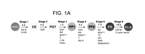
FIG. 1A-FIG. 1F show SC-6 cell clusters undergo glucose-stimulated
insulin secretion (GSIS). (A) Overview of differentiation procedure used. (B)
Images of unstained whole Stage 6 clusters under phase contrast (top) or
stained with dithizone (DTZ) imaged under bright field (bottom). (C)
lmmunostaining of sectioned paraffin-embedded Stage 6 clusters stained for
Glucagon (GCG), NKX6-1, or PDX1 in red, C-peptide (CP) in green, and stained
with the nuclei marker 4,6-diamidino-2-phenylindole (DAP!). (D) Human insulin
secretion of Stage 6 cells generated with the protocol from this study (n=16),
Stage 6 cells generated with the Pagliuca protocol (n=12), and cadaveric human
islets (n=12) in a static glucose-stimulated insulin secretion (GSIS) assay.
**P <
0.01, ****P <0.001 by one-sided paired t-test. #P <0.05, P < 0.0001 by
one-way ANOVA Dunnett multiple comparison test comparing to this study. (E)
Static GSIS assay of Stage 6 cells from this study subjected to either 2, 5.6,
11.1, or 20 mM glucose (n=4). *P <o05, ***P <0.001, not significant (ns) by
one-way ANOVA Dunnett multiple comparison test comparing to 2 mM glucose.
(F) Dynamic human insulin secretion of Stage 6 cells generated with the
protocol
from this study (n=12), Stage 6 cells generated with the Pagliuca protocol
(n=4),
and cadaveric human islets (n=12) in a perfusion GSIS assay. Cells are
perfused with low glucose (2 mM) except where high glucose (20 mM) is
indicated. Act A, activin A; CHIR, CHIR9901, KGF, keratinocyte growth factor;
RA, retinoic acid; Y, Y27632; LDN, LDN193189, PdbU, phorbol 12,13-dibutyrate,
T3, triiodothyronine, Alk5i, Alk5 inhibitor type ESFM, Enriched Serum-Free
Medium. All Stage 6 data shown is with HUES8.
FIG. 2A-FIG. 2D show SC-6 cells express 13 cell and islet markers. (A)
lmmunostaining of Stage 6 clusters single-cell dispersed, plated overnight,
and
stained for Chromogranin A (CHGA), GCG, Somatostatin (SST), NEUROD1,
NKX6-1, PDX1, or PAX6 in red, C-peptide (CP) in green, and stained with DAPI.
(B) Representative flow cytometric dot plots of Stage 6 clusters single-cell
8
CA 03100382 2020-11-13
WO 2019/222487
PCT/US2019/032643
dispersed and immunostained for the indicated markers. (C) Box-and-whiskers
plots quantifying fraction of cells expressing the indicated markers. Each
point is
an independent experiment. (D) Real-time FOR analysis of Stage 6 cells
generated with the protocol from this study (n=8), Stage 6 cells generated
with
.. the Pagliuca protocol (n=5), and cadaveric human islets (n=7). ns, *P <
0.05, **P
<0.01, ***P < 0.001, ****P < 0.0001 by one-way ANOVA Dunnett multiple
comparison test comparing to this study. All Stage 6 data shown is with HUES8.
FIG. 3A-FIG. 3H show 50-6 cells greatly improve glucose tolerance and
have persistent function for months after transplantation. (A) Serum human
insulin of a non-STZ-treated mouse cohort (n=3) 6 months after transplantation
fasted overnight 0 and 60 min after an injection of 2 g/kg glucose. **P < 0.01
by
one-sided paired t-test. (B) lmmunostaining of sectioned paraffin-embedded
explanted kidneys of non-STZ-treated mice 6 months after transplantation for 0-
peptide with DAPI (left) or 0-peptide and PDX1 with DAPI (right). White dashed
line is manually drawn to show border between kidney and graft (*). (C)
Glucose
tolerance test (GTT) 10 d after surgery for STZ-treated mice cohort without a
transplant (STZ, No Txp, n=6), untreated mice without a transplant (No STZ, No
Txp, n=5), and STZ-treated mice with a transplant (STZ, Txp, n=6). *P <0.05,
**P < 0.01, ***P < 0.001, ****P < 0.0001 by two-way ANOVA Tukey multiple
comparison. (D) Area under the curve (AUC) calculations for data shown in (C).
**P < 0.01 by one-way ANOVA Tukey multiple comparison test. (E) Serum
human insulin of STZ, Txp mice (n=5) fasted overnight 0 and 60 min after an
injection of 2 g/kg glucose. **P < 0.01 by one-sided paired t-test. (F) GTT 10
wk
after surgery for STZ, No Txp mice (n=6), No STZ, No Txp mice (n=4), and STZ,
.. Txp mice (n=5). **P < 0.01, ***P < 0.001, ****P < 0.0001 by two-way ANOVA
Tukey multiple comparison test. (G) AUC calculations for data shown in (d). **
P
<0.01 by one-way ANOVA Tukey multiple comparison test. (H) Serum human
insulin of STZ, Txp mice (n=5) fasted overnight 0 and 60 min after an
injection of
2 g/kg glucose. **P < 0.01 by one-sided paired t-test. All data shown is with
HUES8. Panels (A-B) are SCID/Beige and panels (C-H) are NOD/SCID mice.
FIG. 4A-FIG. 40 show SC-6 cells have transient dynamic function in vitro,
respond to multiple stimuli, and sustain second phase insulin secretion at
high
glucose. (A) Dynamic human insulin secretion cells in Stage 6 for 5, 9, 15,
22,
9
CA 03100382 2020-11-13
WO 2019/222487
PCT/US2019/032643
26, and 35 d in a perfusion GSIS assay. Data for each individual time point is
shown as mean SEM and the final graph shows only the means of each graph.
Cells are perfused with low glucose (2 mM) except where high glucose (20 mM)
is indicated (n=3 for each Stage 6 time point). (B) Dynamic human insulin
secretion of Stage 6 cells in a perfusion GSIS assay treated with multiple
secretagogues. Cells are perfused with low glucose (2 mM) except where high
(20 mM) glucose is indicated (Glu), then perfused with a second challenge of
high glucose alone or with additional compounds (Tolbutamide, IBMX, and
Extendin-4 on the left KCL and L-Arginine on the right) where indicated
(Glu+Factor). (C) Dynamic human insulin secretion of Stage 6 cells in a
perfusion GSIS assay with an extended high glucose treatment. Cells are
perfused with low glucose (2 mM) except where high glucose (20 mM) is
indicated (n=3). All data shown is with HUES8.
FIG. 5A-FIG. 5F shows Alk5 inhibitor type ll reduces SC-6 cell GSIS. (A)
Box-and-whiskers plot of human insulin secretion of Stage 6 cells in static
GSIS
assay treated with DMSO or Alk5i (n=9). ***P < 0.001, ****P < 0.0001 by two-
way paired t-test, P < 0.0001 by two-way unpaired t-test. (B) Cellular
insulin
content of Stage 6 cells treated with DMSO or Alk5i (n=18). ****P < 0.0001 by
two-way unpaired t-test. (C) Cellular proinsulin/insulin content ratio of
Stage 6
.. cells treated with DMSO or Alk5i (n=17). ns by two-way unpaired t-test. (D-
E)
Representative flow cytometric dot plots of Stage 6 clusters single-cell
dispersed
and immunostained for Chromogranin A and PDX1 (D) or C-peptide and NKX6-1
(E). (F) Dynamic human insulin secretion of Stage 6 cells treated with DMSO or
Alk5i in a perfusion GSIS assay. Cells are perfused with low glucose (2 mM)
except where high glucose (20 mM) is indicated (n=12) . All data shown is with
HUES8.
FIG. 6A-FIG. 6E shows blocking TGF6 signaling during Stage 6 hampers
GSIS. (A) Western blot of Stage 6 cells cultured with DMSO or Alk5i stained
for
phosphorylated SMAD 2/3 (pSMAD2/3), total SMAD 2/3 (tSMAD2/3), and Actin.
.. Data shown is from HUES8. (B) Real-time PCR of Stage 6 cells transduced
with
lentiviruses containing shRNA against GFP (control) or one of two sequences
against TGFBR1 (TGFBR1 #1 and #2) (n=3). ****P < 0.0001 by one-way
ANOVA Dunnett multiple comparison test comparing to GFP. (C) Western blot of
CA 03100382 2020-11-13
WO 2019/222487
PCT/US2019/032643
Stage 6 cells transduced with lentiviruses containing GFP or TGFBR1 #1
shRNA. Data shown is from 1013-4FA. (D) Human insulin secretion of Stage 6
cells in static GSIS assay transduced with lentiviruses containing GFP, TGFBR1
#1, or TGFBR1 #2 shRNA (n=3). **P <0.01 by paired two-way t-test. ##P <0.01
by one-way ANOVA Dunnett multiple comparison test comparing to GFP. Data
shown is from HUES8. (E) Dynamic human insulin secretion of Stage 6 cells
transduced with lentiviruses containing GFP or TGFBR1 #1 shRNA in a
perfusion GSIS assay. Cells are perfused with low glucose (2 mM) except where
high glucose (20 mM) is indicated (n=4). Data shown is from HUES8.
FIG. 7A-FIG. 7G shows Alk5 inhibitor type II treatment during Stage 5 is
important for generation of insulin-producing cells. (A-B) Representative flow
cytometric dot plots of Stage 5 clusters single-cell dispersed and
immunostained
for Chromogranin A and NKX6-1 (A) or C-peptide and NKX6-1 (B). (C) Fraction
of cells expressing the indicated markers (n=4 except CHGA, which was n=3).
*P <0.05, **P <0.01, or ns by unpaired two-way t-test. (D-F) Real-time FOR
measuring relative gene expression of Stage 5 cells cultured with DMSO or
Alk5i
for pancreatic hormones (D), 13 cell markers (E), or endocrine markers (F)
(n=6).
*P <o05, **P < 0.01, ****P < 0.0001, or ns by unpaired two-way t-test. (G)
Human insulin secretion at 20 mM glucose of cells cultured in Stage 5 in
either
DMSO or Alk5i plus an additional 7 d in Stage 6 without Alk5i and without
cluster
resizing (n=3). **P < 0.01 by unpaired two-way t-test. All data shown is from
HUES8.
FIG. 8A-FIG. 8D shows data leading to new differentiation strategy and
hiPSC reproduction. (A) Human insulin secretion of Stage 6 cells generated in
CMRLS or ESFM, with or without resizing, and with or without factors (Alk5i
and
T3) in a static GSIS assay. The combinations investigated were (1) CMRLS, no
resize, no factors (n=3), (2) CMRLS, yes resize, no factors (n=6), (3) ESFM,
no
resize, no factors (n=3), (4) ESFM, yes resize, no factors (n=3), (5) ESFM,
yes
resize, yes factors (n=3). HUES8 cell line used. (B) Flow cytometric dot plots
of
Stage 6 cells generated in CMRLS or ESFM, with or without resizing, and with
or
without factors (Alk5i and T3) immunostained for C-peptide and NKX6-1. HUES8
cell line used. (C) Human insulin secretion in a static GSIS assay of three
hiPSC
lines (n=3 each). *P < 0.05, **P <0.01, and ***P <0.0001 by one-sided paired t-
11
CA 03100382 2020-11-13
WO 2019/222487
PCT/US2019/032643
test. (D) Dynamic human insulin secretion of Stage 6 cells generated with two
hiPSC lines in a perfusion GSIS assay. Cells are perfused with low glucose (2
mM) except where high glucose (20 mM) is indicated (n=3 for 1013-4FA and n=4
for 1016SeVA).
FIG. 9A-FIG. 9C shows additional immunostaining data for Stage 6 cells.
(A) lmmunostaining of Stage 6 clusters single-cell dispersed, plated
overnight,
and stained for the indicated markers. Stage 6 cells were generated from two
hiPSC lines with the protocol from this paper and the HUES8 cell line with the
Pagliuca protocol. Scale bar=50 pm for 1016SeVA and 1013-4FA and 25 pm for
Pagliuca protocol. (B-C) Flow cytometric dot plots of Stage 6 cells generated
from two hiPSC lines with the protocol from this paper and the HUES8 cell line
with the Pagliuca protocol stained with the indicated markers.
FIG. 10 shows additional gene expression data for Stage 6 cells. Gene
expression data for Stage 6 cells generated with the new differentiation
protocol
from the HUES8 (n=8) and 1013-4FA (n=10) lines and human islets (n=7)
measured with real-time PCR. The HUES8 and human islet plotted here is the
same as from FIG. 2.
FIG. 11A-FIG. 11D shows additional immunostaining, serum human
insulin measurements, and mouse C-peptide measurements. (A)
lmmunostaining of sectioned paraffin-embedded explanted kidneys of non-STZ-
treated mice 6 months after transplantation for C-peptide (CP; 13 cell
marker),
PDX1 (13 cell marker), glucagon (GCG, a cell marker), somatostatin (SST; El
cell
marker), KRT19 (ductal marker), and trypsin (acinar marker). Scale bar=25 pm.
(B) Serum human insulin of STZ, No Txp mice (n=6) and No Stz, No Txp (n=5)
fasted overnight 0 and 60 min after an injection of 2 g/kg glucose. (B) Serum
mouse C-peptide of STZ, No Txp (n=6), No STZ, No Txp (n=4), and STZ, TXP
(n=5). ****P < 0.0001 and ns by one-way ANOVA Tukey multiple comparison
test. (C) lmmunostaining of sectioned paraffin-embedded explanted kidneys of
STZ-treated mice 11 wk after transplantation for the indicated markers. Scale
bar=25 pm. HUES8 cell line used.
FIG. 12A-FIG. 12B shows temporal flow cytometry during Stage 6 and
KCI challenge of human islets. (A) Flow cytometric dot plots of Stage 6 cells
at
12
CA 03100382 2020-11-13
WO 2019/222487
PCT/US2019/032643
early (9 d) and late (26 d) time points stained for 0-peptide and NKX6-1.
HUES8
cell line used. (B) Dynamic human insulin secretion of human islets in a
perfusion GSIS assay perfused with low glucose (2 mM) except where high (20
mM) glucose is indicated (Glu), then perfused with a second challenge of high
glucose with KCI where indicated (Glu+Factor) (n=4).
FIG. 13A-FIG. 130 shows stage 6 cells generated from hiPSC undergo
GSIS that is inhibited by Alk5i, flow cytometry controls, and gene expression
data. (A) Human insulin secretion of Stage 6 cells generated from three hiPSC
lines (1013-4FA, n=4; 1016SeVA, n=3; 1019SeVF, n=3) in static GSIS assay
treated with DMSO or Alk5i. *P < 0.05, **P < 0.01 , ****P <0.0001 by two-way
paired t-test, ##P <0.01, ###P <0.001, P <0.0001 by two-way unpaired t-
test. The control data here is the same data in FIG. 21. (B) Flow cytometry
controls for FIG. 19. The 0-peptide/NKX6-1 control is the same as shown in
FIG.
16. (C) Real-time FOR analysis of Stage 6 cells with or without resizing
treated
with Alk5i or DMSO (n=3). Data generated with the 1013-4FA cell line.
FIG. 14A-FIG. 14B shows resized and unresized Stage 6 clusters have
SMAD2/3 phosphorylation and reduced GSIS with Alk5i treatment. (A) Western
blot of Stage 6 cells with and without resizing stained for phosphorylated
SMAD
2/3 (pSMAD2/3), total SMAD 2/3 (tSMAD2/3), and Actin. (B) Human insulin
secretion of Stage 6 cells in static GSIS assay resized or unresized with
treatment of DMSO or Alk5i. All data shown is from 1013-4FA.
FIG. 15A-FIG. 151 is a series of illustrations, images, and graphs depicting
the state of the cytoskeleton controls expression of the transcription
factors.
NEUROG3 and NKX6-1 in pancreatic progenitors. (a) Schematic of the
differentiation protocol5 used for suspension differentiation and plate down
studies. (b) Images of clusters at the beginning of stage 4 dispersed and
plated
onto ECM-coated TOP for culture for the remainder of the protocol. Scale bar =
100 pm. (c) qRT-PCR of pancreatic genes at the end of stage 4 of cells plated
on collagen! at the beginning of stage 4 compared to regular suspension
cluster
or clusters reaggregated after dispersion (Tukey's HSD test, n = 4). (d) qRT-
PCR
of pancreatic genes at the end of stage 4 of cells plated on varying heights
of
collagen 1 gels at the beginning of stage 4. Increasing the height of
collagen!
13
CA 03100382 2020-11-13
WO 2019/222487
PCT/US2019/032643
gels fixed to TOP correlates with decreasing the effective stiffness
experienced
by cells (ANOVA, n=4). (e) qRT-PCR of plated stage 4 cells treated with a
screen of cytoskeletal modifying compounds to identify latrunculin A as potent
endocrine inducer. XXi, a y-secretase inhibitor, was used as a positive
control
(Dunnett's multiple comparisons test, n=4). (f) lmmunostaining of plated cells
at
the end of stage 4 demonstrating that a 1 pM latrunculin A treatment increases
NEUROG3+ and decreases NKX6-1+ cells. Scale bar = 50 pm. (g) Latrunculin A
dose response of pancreatic gene expression added during stage 4 measured
with qRT-PCR (ANOVA, n = 4). (h) lmmunostaining of plated stage 4 cells
treated for 24 hours with 1 pM latrunculin, demonstrating depolymerization of
F-
actin but maintenance of PDX1 expression. (i) Western blot quantification of
the
G/F actin ratio within cells under different culture formats and treated with
latrunculin A (n=3). All data was generated with HUES8. All data error bars
represent SEM. ns = not significant, * = p < 0.05, ** = p < 0.01, ***= p <
0.001.
FIG. 16A-FIG. 160 is a series of projections, plots, and graphs depicting
single-cell RNA sequencing demonstrating that cytoskeletal state directs
pancreatic progenitor fate. (a) tSNE projection of single-cell RNA sequencing
performed on plated stage 4 cells and treated with either 0.5 pM latrunculin A
or
5 pM nocodazole. Unsupervised clustering of the combined cell population from
all three conditions revealed four separate clusters. (b) Violin plots
indicating
important upregulated genes in each cluster. (c) The percentage of cells
within
each cluster for each condition. All data was generated with HUES8.
FIG. 17A-FIG. 171 is a series of plots and images depicting Latrunculin A
treatment during stage 5 drastically increased 50-6 cell specification of
plated
pancreatic progenitors. (a) Flow cytometry two weeks into stage 6 for NKX6-1,
CHGA, and 0-peptide of plated cells as per FIG. 15(a), untreated or treated
with
0.5 pM latrunculin A throughout stage 4, 5, or 6 (Dunnett's multiple
comparisons
test, n = 4). (b) Static GSIS two weeks into stage 6 of plated cells,
untreated or
treated with 0.5 pM latrunculin A throughout stage 4, 5, or 6 (paired t-test
compares between low and high glucose for a particular sample, Dunnett's test
compares insulin secretion at high glucose to the control, n = 4). (c)
Optimization
of latrunculin A concentration and timing during stage 5 for plated cells.
Static
GSIS was performed after 2 weeks of stage 6 (t-tests, n = 4). (d) Insulin
content
14
CA 03100382 2020-11-13
WO 2019/222487
PCT/US2019/032643
of plated cells two weeks into stage 6, untreated or treated 24 hour with 1 pM
latrunculin A (unpaired t-tests, n= 4). (e) Proinsulin/insulin ratio of plated
cells
two weeks into stage 6, untreated or treated 24 hour with 1 pM latrunculin A
(unpaired t-tests, n = 4). (f) qRT-PCR measuring pancreatic (left) and non-
pancreatic (right) gene expression of plated cells two weeks into stage 6,
untreated or treated 24 hour with 1 pM latrunculin A (unpaired t-tests, n =
4). (g)
lmmunostaining for AFP and 0-peptide of plated cells two weeks into stage 6,
untreated or treated 24 hour with 1 pM latrunculin A. Scale bar= 100 pm. (h)
Images of aggregation of plated cells after one week in stage 6. (i) Dynamic
glucose-stimulated insulin secretion of stage 6 cells exhibiting first and
second
phase insulin release. All data was generated with HUES8. All data error bars
represent SEM. ns = not significant, * = p < 0.05, ** = p <0.01, *** = p <
0.001.
FIG. 18A-FIG. 18J is a series of illustrations, graphs, and images
depicting 50-6 cells differentiated with the new planar protocol expressing 13
cell
markers and function in vitro. (a) Schematic of the new planar protocol for
making 50-6 cells incorporating a 1 pM latrunculin A treatment for the first
24
hour of stage 5. (b) Flow cytometry after one week in stage 6 of cells from
HUES8 with and without stage 5 latrunculin A treatment measuring endocrine
induction (CHGA+) and 50-6 cell specification (C-peptide+/NKX6-1+) (unpaired
t- tests, n = 4). (c) Flow cytometry of islet and 50-6 cells markers for stage
6
cells differentiated from HUES8, 1013-4FA, and 1016SeVA hPSC lines (n = 4).
(d) qRT-PCR of islet and disallowed genes for stage 6 cells and human islets
(Dunnett's multiple comparisons test, n = 4 for 50-6 cells, n = 3 for human
islets). (e) lmmunostaining of aggregated planar stage 6 cells from HUES8. (f)
Insulin content of stage 6 cells (n = 4). (g) Proinsulin/insulin content ratio
for
stage 6 cells (n = 4). (h) Static GSIS for stage 6 cells (paired t-tests, n =
4). (i)
Dynamic GSIS for planar stage 6 cells generated from HUES8 (n = 7), 1013-4FA
(n = 3), and 1016SeVA (n = 4). Suspension stage 6 data is replotted from
Velazco-Cruz et al.5 (HUES8, n = 12; 1013-4FA, n = 3; 1016SeVA, n = 4). (j)
Planar static GSIS data from (i) plotted together compared to human islet data
replotted from Velazco-Cruz et al.5 (n = 12). All data shown in this figure is
of
cells generated with the planar differentiation protocol unless otherwise
noted.
All data error bars represent SEM. ns = not significant, * = p < 0.05, ** = p
<
CA 03100382 2020-11-13
WO 2019/222487
PCT/US2019/032643
0.01, *** = p <0.001.
FIG. 19A-FIG. 190 is a series of graphs and images depicting SC-6 cells
generated with the new planar protocol can rapidly cure pre-existing diabetes
in
mice. (a) Diabetes was induced with STZ in a total of 19 mice. 4 weeks after
injection, SC-6 cells generated with the planar protocol were transplanted
into 12
of these mice. 5 non-diabetic mice served as controls. Glucose tolerance tests
were performed 3, 10, and 13 weeks after transplantation. A nephrectomy was
performed 12 weeks after transplantation (Tukey's HSD test, f= different than
no
transplant, = different than transplant, # = different than untreated
control). (b)
In vivo GSIS of mice receiving the SC-6 cell transplant 2 and 10 weeks after
transplantation measuring human insulin. ns = not significant, * = p < 0.05,
** = p
<0.01, *** = p < 0.001. (c) lmmunostaining of sectioned kidneys transplanted
with SC-6 cells 3 weeks after transplantation showing C-peptide+ cells. All
data
was generated with HUES8 using the planar protocol outlined in FIG. 19A. All
data error bars represent SEM.
FIG. 20A-FIG. 20G is a series of heat maps, plots, and images showing
the state of the cytoskeleton influences endodermal cell fate. (a) Suspension
and
plated pancreatic progenitors differentiated to stage 6 as per FIG. 15(a)
either
untreated, treated with 0.5 pM latrunculin A throughout stage 4, or treated
with 1
pM latrunculin A for the first 24 hours of stage 5. Bulk RNA sequencing at two
weeks into stage 6 was used to generate a heat map of the 1000 most
differentially expressed genes between the stage 5 latrunculin A treatment and
plated control. (b) Heat map from bulk RNA sequencing of select genes from
multiple endodermal lineages. (c) Volcano plot from bulk RNA sequencing data
showing expression differences of select genes between untreated plated cells
and stage 5 latrunculin treated cells. (d) Gene enrichment analysis from bulk
RNA sequencing of select gene sets from multiple endodermal lineages. (e)
lmmunostaining (left) and qRT-PCR (right) of cells differentiated with an
exocrine
differentiation protocol treated with latrunculin A or nocodazole (Dunnett's
multiple comparisons test, n = 4). (f) lmmunostaining (left) and qRT-PCR
(right)
of cells differentiated with an intestinal differentiation protocol treated
with
latrunculin A or nocodazole (Dunnett's multiple comparisons test, n = 4). (g)
lmmunostaining (left) and qRT-PCR (right) of cells differentiated with a
hepatic
16
CA 03100382 2020-11-13
WO 2019/222487
PCT/US2019/032643
differentiation protocol treated with latrunculin A or nocodazole (Dunnett's
multiple comparisons test, n = 4). Scale bars = 50 pm. All data was generated
with HUES8. All data error bars represent SEM. ns = not significant, * = p <
0.05,
** = p <0.01, ***= p <0.001.
FIG. 21A-FIG. 21D is a series of images and bar graphs. (a) Images of
pancreatic progenitors plated at beginning of stage 4 onto ECM-coated TOP as
per FIG. 15(a). Scale bar = 200 pm. (b) qRT-PCR of plated cells at the end of
stage 4 (n = 4). (c) A colorimetric antibody-based integrin adhesion assay at
the
beginning and end of stage 4 confirmed high expression of integrin subunits
that
bind to collagens I and IV (al, a2, [31), fibronectin (aV, 131, a561),
vitronectin (aV,
131, aV[35) and some but not all laminin isoforms (a3, [31). Data is
normalized to
an isotype control. All data was generated with HUES8.
FIG. 22A-FIG. 22H is a series of plots and heat maps. (a) Latrunculin A
dose response of pancreatic gene expression added during stage 4 from 1013-
4FA and 1016SeVA measured with qRT-PCR (n = 4). (b) qRT-PCR of pancreatic
gene expression at the end of stage 4 in response to latrunculin B dosing on
plated HUES8 (ANOVA, n = 4). (c) qRT-PCR of untreated HUES8 plated stage 4
cells, untreated reaggregated clusters, and reaggregated clusters treated with
the actin polymerizer jasplakinolide (unpaired t-tests, n = 4). (d) tSNE plot
heat
map generated from single-cell RNA sequencing data of plated HUES8
pancreatic progenitors showing expression of pancreatic genes. All data
generated as per FIG. 15(a). All data error bars represent SEM. ns = not
significant, * = p < 0.05, **= p < 0.01, *** = p < 0.001.
FIG. 23A-FIG. 23H (a) qRT-PCR of HUES8 cells differentiated with the
.. new planar protocol to the end of stage 4, untreated or treated throughout
stage
4 with 0.5 pM latrunculin A (unpaired t-tests, n = 4). (b-d) qRT-PCR of HUES8
cells differentiated with the planar protocol to stage 6 with or without a 24
hour 1
pM latrunculin A treatment at the beginning of stage 5, (b,c) showing
expression
of islet and 13 cell genes and (d) non-pancreatic genes (unpaired t-tests, n=
4).
(e, f) lmmunostaining of aggregates generated from the planar protocol with
(e)
1013-4FA and (f) 1016SeVA iPSC lines. Scale bars = 50 pm. (g) Quantification
of mouse 0-peptide with ELISA of serum from mice. (h) Quantification of human
17
CA 03100382 2020-11-13
WO 2019/222487
PCT/US2019/032643
insulin in the serum of mice without a transplant. All data was generated with
HUES8 with the new planar protocol was per FIG. 18(a). All data error bars
represent SEM. ns = not significant, * = p < 0.05, ** = p < 0.01, *** = p
<0.001.
DETAILED DESCRIPTION OF THE INVENTION
The present disclosure is based, at least in part, on the discovery that a
modified process can produce cells that can respond to glucose appropriately
to
near islet-like levels, demonstrating both a first phase and second phase
response. As described herein is a protocol to generate beta-like cells from
human pluripotent stem cells with dynamic insulin secretion. Furthermore, the
present disclosure is based, at least in part, on the discovery that
modulation of
the actin cytoskeleton can enhance pancreatic differentiation of human
pluripotent stem cells.
GENERATING BETA-LIKE CELLS FROM HUMAN PLURIPOTENT STEM CELLS WITH
DYNAMIC INSULIN SECRETION
It was discovered that the currently described method generated stem
cell-derived beta (SC-6) cells function better (undergoing glucose-stimulated
insulin secretion) than cells in the published literature (Pagliuca et al.
Cell 2014)
and express beta cell markers. This includes increased insulin secretion with
a
static assay and having first and second phase insulin response in a dynamic
assay.
As described herein, stem cell-derived beta (SC-6) cells can be useful as
a cellular therapy for diabetes or for drug screening. The presently disclosed
process enhances differentiation of human pluripotent stem cells to insulin-
producing beta cells. This process is modified from a previously described 6-
step
differentiation protocol published by Pagliuca et al. Cell 2014. With this new
process, cells that can respond to glucose appropriately to near islet-like
levels
have been generated, demonstrating both a first phase and second phase
response.
In order to achieve the above modulation, the following was performed:
(1) shorten stage 3 to 1 day; (2) allow for TGFb signaling in stage 6 by
removal
of Alk5 inhibitor II (current literature includes this inhibitor); (3) remove
T3 from
18
CA 03100382 2020-11-13
WO 2019/222487
PCT/US2019/032643
stage 6 (current literature includes this inhibitor); (4) perform stage 6 in a
serum-
free basal media (formulation included); and (5) break apart and reaggregate
clusters at the beginning of stage 6.
Using the above modulations, enhanced stem cell-derived beta cells that
better perform glucose-stimulated insulin secretion were generated. The field
currently includes Alk5 inhibitor II and T3 during the last stage of culture
to
mature stem cell-derived beta cells. The field has been unable to generate
functional stem cell-derived beta cells that have both first phase and second
phase insulin secretion (see Rezania et al. Nature Biotechnology 2014 for the
poor dynamic function stem cell-derived beta cells have in the field).
For example, Example 1 describes methods for generating stem cell
derived beta-like (SC-6) cells. It was discovered that a differentiation
strategy
focusing on modulating TGFp signaling, controlling cellular cluster size, and
using an enriched serum-free media (ESFM) to generate SC-6 cells that express
13 cell markers and undergo GSIS with first and second phase dynamic insulin
secretion.
MODULATION OF THE ACTIN CYTOSKELETON ENHANCES PANCREATIC
DIFFERENTIATION OF HUMAN PLURIPOTENT STEM CELLS
As described herein, this work has identified the actin cytoskeleton as a
crucial regulator of human pancreatic cell fate. By controlling the state of
the
cytoskeleton with either cell arrangement (two- vs three-dimensional),
substrate
stiffness, or directly with chemical treatment, it is shown herein that a
polymerized cytoskeleton prevents premature induction of NEUROG3
expression in pancreatic progenitors, but also inhibits subsequent
differentiation
to SC-6 cells.
As shown herein, it was discovered that modulation of the actin
cytoskeleton and its downstream effector Yes-Associated Protein (YAP) at
specific time points during differentiation can enhance differentiation of
human
pluripotent stem cells to cells of endodermal lineage, pancreatic progenitors,
and
-- insulin-producing beta cells. Using a 6-step differentiation protocol
modified from
Pagliuca et al. Cell 2014, the following specific features were observed: (1)
actin
polymerization and YAP activity during Stage 4 enhances generation of
19
CA 03100382 2020-11-13
WO 2019/222487
PCT/US2019/032643
pancreatic progenitors (PDX1+/NKX6-1+/SOX9+), (2) actin depolymerization
and loss of YAP activity during Stage 5, preferentially during the first 24-48
hr of
Stage 5, enhances generation of endocrine cells, specifically beta cells that
demonstrate enhanced glucose-stimulated insulin secretion.
In order to achieve the above modulation, the following can be performed:
(1) promoting actin polymerization by plating onto stiff surfaces, such as
tissue
culture plastic with a thin layer of ECM protein to promote attachment; (2)
promoting actin depolymerization by plating onto soft surfaces, such as
hydrogels, or by treating cells with latrunculin A and/or latrunculin 13; (3)
promoting YAP transcriptional activity using the same methods to promote actin
polymerization; and/or (4) inhibiting YAP transcriptional activity using the
same
methods to promote actin depolymerization or by treatment with Verteporfin.
Using the above modulations, enhanced stem cell-derived beta cells were
generated to better perform glucose-stimulated insulin secretion than previous
methods and can be generated on attachment culture. Currently in the field,
stem cell-derived beta cells can be generated but do not function as well as
with
the presently disclosed approach. The field does not utilize actin
cytoskeleton
and YAP signaling in their protocols. The field is also unable to generate
functional stem cell-derived beta cells with the cells in attachment culture ¨
it
must either be done in suspension aggregates (the control for many experiments
in the attached data set, first reported in Pagliuca et al. Cell 2014) or in
aggregates on an air-liquid-interface (first reported in Rezania et al. Nature
Biotechnology 2014).
Described herein is the generation of stem cell-derived beta cells that
function better (undergoing glucose-stimulated insulin secretion) than cells
in the
published literature (Pagliuca et al. Cell 2014) and express beta cell
markers.
Also described herein are methods for the generation of stem cell-derived
beta cells in a planar protocol that can undergo glucose-stimulated insulin
secretion (GSIS).
Also described herein is the demonstration that cells can be detached
from a plate, either using UpCell technology that does not require cell
dispersion
or by dispersing and reaggregating the cells, and maintain insulin secretion
CA 03100382 2020-11-13
WO 2019/222487
PCT/US2019/032643
capacity, better enabling transplantation.
Also described herein is the generation of pancreatic progenitor cells that
have reduced endocrine expression (such as expression of NGN3, NEUROD1)
and increased pancreatic progenitor expression (such as expression of NKX6-1,
SOX9).
Pancreatic progenitors and stem cell-derived beta cells can be useful as a
cellular therapy for diabetes. Stem cell-derived beta cells are also useful
for drug
screening. The presently disclosed attachment culture approach yields a
convenient platform for drug screening studies.
The presently disclosed culture approach can also facilitate enhanced
quality and reproducibility of the differentiations and is conducive to
automation
of the differentiation process for commercialization.
An an example, differentiation protocols, as described in example 2, by
cytoskeletal modulation can generate cells of several lineages (e.g., 50-13,
beta-
like cells). It was discovered that the state of the actin cytoskeleton is
critical to
endodermal cell fate choice. By utilizing a combination of cell-biomaterial
interactions as well as small molecule regulators of the actin cytoskeleton
(e.g., a
cytoskeletal-modulating agent), the timing of endocrine transcription factor
expression can be controlled to modulate differentiation fate and develop a
two-
dimensional protocol for differentiating cells. Importantly, this new planar
protocol
greatly enhances the function of 50-13 cells differentiated from induced
pluripotent stem cell (iPSC) lines and forgoes the requirement for three-
dimensional cellular arrangements.
Different degrees of actin polymerization at specific points of
differentiation biased cells toward different endodermal lineages, and thus
non-
optimal cytoskeletal states led to large inefficiencies in cell specification.
Furthermore, the methods described herein can control actin
polymerization to direct differentiations of these other endodermal cell fates
to
modulate lineage specification.
Other lineages that can be generated according to the provided methods
can be liver, esophageal, exocrine, pancreas, intestine, or stomach.
21
CA 03100382 2020-11-13
WO 2019/222487
PCT/US2019/032643
A cytoskeletal-modulating agent can be any agent that promotes or
inhibits actin polymerization or microtubule polymerization. For example, the
cytoskeletal-modulating agent can be an actin depolymerization or
polymerization agent, a microtubule modulating agent, or an integrin
modulating
agent (e.g., compounds, such as antibodies and small molecules). For example,
the cytoskeletal-modulating agent can be latrunculin A, latrunculin B,
nocodazole, cytochalasin D, jasplakinolide, blebbistatin, y-27632, y-15, gdc-
0994, or an integrin modulating agent. The cytoskeletal-modulating agent can
be
any cytoskeletal-modulating agent known in the art (see e.g., Ley et al. Nat
Rev
Drug Discov. 2016 Mar; 15(3): 173-183).
CELL CLUSTER RESIZING
Resizing of cell clusters can be performed by any methods known in the
art. For example, cell resizing can comprise breaking apart cell clusters and
reaggregating. As another example, the cell clusters can be resized by
incubating in a cell-dissociating reagent and passed through a cell strainer
(e.g.,
a 100 pm nylon cell strainer). As another example, cells can be resized by
single
cell dispersing with TrypLE and reaggregating.
FORMULATION
The agents and compositions described herein can be formulated by any
conventional manner using one or more pharmaceutically acceptable carriers or
excipients as described in, for example, Remington's Pharmaceutical Sciences
(A.R. Gennaro, Ed.), 21st edition, ISBN: 0781746736 (2005), incorporated
herein by reference in its entirety. Such formulations will contain a
therapeutically
effective amount of cells as described herein, which can be in purified form,
together with a suitable amount of carrier so as to provide the form for
proper
administration to the subject.
The term "formulation" refers to preparing a drug in a form suitable for
administration to a subject, such as a human. Thus, a "formulation" can
include
pharmaceutically acceptable excipients, including diluents or carriers.
The term "pharmaceutically acceptable" as used herein can describe
substances or components that do not cause unacceptable losses of
22
CA 03100382 2020-11-13
WO 2019/222487
PCT/US2019/032643
pharmacological activity or unacceptable adverse side effects. Examples of
pharmaceutically acceptable ingredients can be those having monographs in
United States Pharmacopeia (USP 29) and National Formulary (NF 24), United
States Pharmacopeia! Convention, Inc, Rockville, Maryland, 2005 ("USP/NF"), or
a more recent edition, and the components listed in the continuously updated
Inactive Ingredient Search online database of the FDA. Other useful components
that are not described in the USP/NF, etc. may also be used.
The term "pharmaceutically acceptable excipient," as used herein, can
include any and all solvents, dispersion media, coatings, antibacterial and
antifungal agents, isotonic, or absorption delaying agents. The use of such
media and agents for pharmaceutical active substances is well known in the art
(see generally Remington's Pharmaceutical Sciences (A.R. Gennaro, Ed.), 21st
edition, ISBN: 0781746736 (2005)). Except insofar as any conventional media or
agent is incompatible with an active ingredient, its use in the therapeutic
compositions is contemplated. Supplementary active ingredients can also be
incorporated into the compositions.
A "stable" formulation or composition can refer to a composition having
sufficient stability to allow storage at a convenient temperature, such as
between
about 0 C and about 60 C, for a commercially reasonable period of time, such
as at least about one day, at least about one week, at least about one month,
at
least about three months, at least about six months, at least about one year,
or
at least about two years.
The formulation should suit the mode of administration. The agents of use
with the current disclosure can be formulated by known methods for
administration to a subject using several routes which include, but are not
limited
to, parenteral, pulmonary, oral, topical, intradermal, intramuscular,
intraperitoneal, intravenous, subcutaneous, intranasal, epidural, ophthalmic,
buccal, and rectal. The individual agents may also be administered in
combination with one or more additional agents or together with other
biologically active or biologically inert agents. Such biologically active or
inert
agents may be in fluid or mechanical communication with the agent(s) or
attached to the agent(s) by ionic, covalent, Van der Waals, hydrophobic,
23
CA 03100382 2020-11-13
WO 2019/222487
PCT/US2019/032643
hydrophilic or other physical forces.
Controlled-release (or sustained-release) preparations may be formulated
to extend the activity of the agent(s) and reduce dosage frequency. Controlled-
release preparations can also be used to effect the time of onset of action or
other characteristics, such as blood levels of the agent, and consequently
affect
the occurrence of side effects. Controlled-release preparations may be
designed
to initially release an amount of an agent(s) that produces the desired
therapeutic effect, and gradually and continually release other amounts of the
agent to maintain the level of therapeutic effect over an extended period of
time.
In order to maintain a near-constant level of an agent in the body, the agent
can
be released from the dosage form at a rate that will replace the amount of
agent
being metabolized or excreted from the body. The controlled-release of an
agent
may be stimulated by various inducers, e.g., change in pH, change in
temperature, enzymes, water, or other physiological conditions or molecules.
Agents or compositions described herein can also be used in combination
with other therapeutic modalities, as described further below. Thus, in
addition to
the therapies described herein, one may also provide to the subject other
therapies known to be efficacious for treatment of the disease, disorder, or
condition.
THERAPEUTIC METHODS
Also provided is a process of using generated cells for cell replacement
therapies or stem cell transplant. For example, the disclosed compositions and
methods can be used to treat diabetes or other disease associated with
dysfunctional endodermal cells in a subject in need administration of a
therapeutically effective amount of cells of endodermal lineage or beta cells,
so
as to induce insulin secretion.
Methods described herein are generally performed on a subject in need
thereof. A subject in need of the therapeutic methods described herein can be
a
subject having, diagnosed with, suspected of having, or at risk for developing
a
diabetes or other disease associated with dysfunctional endodermal cells. A
determination of the need for treatment will typically be assessed by a
history
and physical exam consistent with the disease or condition at issue. Diagnosis
of
24
CA 03100382 2020-11-13
WO 2019/222487
PCT/US2019/032643
the various conditions treatable by the methods described herein is within the
skill of the art. The subject can be an animal subject, including a mammal,
such
as horses, cows, dogs, cats, sheep, pigs, mice, rats, monkeys, hamsters,
guinea
pigs, and chickens, and humans. For example, the subject can be a human
subject.
Generally, a safe and effective amount of cells of endodermal lineage
(e.g., hepatocytes, insulin-expressing cells (e.g., p cells, SC-13 cells),
intestinal
cells) is, for example, that amount that would cause the desired therapeutic
effect in a subject while minimizing undesired side effects.
In various embodiments, an effective amount of endodermal lineage or
beta cells described herein can respond to glucose by secretion of insulin. In
various embodiments, an effective amount of cells described herein can treat
diabetes or other disease associated with dysfunctional endodermal cells,
substantially inhibit diabetes or other disease associated with dysfunctional
endodermal cells, slow the progress of diabetes or other disease associated
with
dysfunctional endodermal cells, or limit the development of diabetes or other
disease associated with dysfunctional endodermal cells.
According to the methods described herein, administration can be a cell
transplantation, cell implantation, parenteral, pulmonary, oral, topical,
intradermal, intramuscular, intraperitoneal, intravenous, subcutaneous,
intranasal, epidural, ophthalmic, buccal, or rectal administration.
When used in the treatments described herein, a therapeutically effective
amount of beta cells or cells of endodermal lineage can be employed in pure
form or, where such forms exist, in pharmaceutically acceptable salt form and
with or without a pharmaceutically acceptable excipient. For example, the
compounds of the present disclosure can be administered, at a reasonable
benefit/risk ratio applicable to any medical treatment, in a sufficient amount
to
induce insulin secretion.
The amount of a composition described herein that can be combined with
a pharmaceutically acceptable carrier to produce a single dosage form will
vary
depending upon the host treated and the particular mode of administration. It
will
be appreciated by those skilled in the art that the unit content of agent
contained
CA 03100382 2020-11-13
WO 2019/222487
PCT/US2019/032643
in an individual dose of each dosage form need not in itself constitute a
therapeutically effective amount, as the necessary therapeutically effective
amount could be reached by administration of a number of individual doses.
Toxicity and therapeutic efficacy of compositions described herein can be
.. determined by standard pharmaceutical procedures in cell cultures or
experimental animals for determining the LD50 (the dose lethal to 50% of the
population) and the ED50, (the dose therapeutically effective in 50% of the
population). The dose ratio between toxic and therapeutic effects is the
therapeutic index that can be expressed as the ratio LD50/ED50, where larger
therapeutic indices are generally understood in the art to be optimal.
The specific therapeutically effective dose level for any particular subject
will depend upon a variety of factors including the disorder being treated and
the
severity of the disorder; activity of the specific compound employed; the
specific
composition employed; the age, body weight, general health, sex and diet of
the
-- subject; the time of administration; the route of administration; the rate
of
excretion of the composition employed; the duration of the treatment; drugs
used
in combination or coincidental with the specific compound employed; and like
factors well known in the medical arts (see e.g., Koda-Kimble et al. (2004)
Applied Therapeutics: The Clinical Use of Drugs, Lippincott Williams &
Wilkins,
ISBN 0781748453; Winter (2003) Basic Clinical Pharmacokinetics, 4th ed.,
Lippincott Williams & Wilkins, ISBN 0781741475; Shamel (2004) Applied
Biopharmaceutics & Pharmacokinetics, McGraw-Hill/Appleton & Lange, ISBN
0071375503). For example, it is well within the skill of the art to start
doses of the
composition at levels lower than those required to achieve the desired
therapeutic effect and to gradually increase the dosage until the desired
effect is
achieved. If desired, the effective daily dose may be divided into multiple
doses
for purposes of administration. Consequently, single dose compositions may
contain such amounts or submultiples thereof to make up the daily dose. It
will
be understood, however, that the total daily usage of the compounds and
compositions of the present disclosure will be decided by an attending
physician
within the scope of sound medical judgment.
Again, each of the states, diseases, disorders, and conditions, described
26
CA 03100382 2020-11-13
WO 2019/222487
PCT/US2019/032643
herein, as well as others, can benefit from compositions and methods described
herein. Generally, treating a state, disease, disorder, or condition includes
preventing or delaying the appearance of clinical symptoms in a mammal that
may be afflicted with or predisposed to the state, disease, disorder, or
condition
but does not yet experience or display clinical or subclinical symptoms
thereof.
Treating can also include inhibiting the state, disease, disorder, or
condition,
e.g., arresting or reducing the development of the disease or at least one
clinical
or subclinical symptom thereof. Furthermore, treating can include relieving
the
disease, e.g., causing regression of the state, disease, disorder, or
condition or
at least one of its clinical or subclinical symptoms. A benefit to a subject
to be
treated can be either statistically significant or at least perceptible to the
subject
or to a physician.
Administration of cells of endodermal lineage or beta cells can occur as a
single event or over a time course of treatment. For example, cells of
endodermal lineage or beta cells can be administered daily, weekly, bi-weekly,
or monthly. For treatment of acute conditions, the time course of treatment
will
usually be at least several days. Certain conditions could extend treatment
from
several days to several weeks. For example, treatment could extend over one
week, two weeks, or three weeks. For more chronic conditions, treatment could
extend from several weeks to several months or even a year or more.
Treatment in accord with the methods described herein can be performed
prior to, concurrent with, or after conventional treatment modalities for
diabetes
or other disease associated with dysfunctional endodermal cells.
ADMINISTRATION
Agents and compositions described herein can be administered according
to methods described herein in a variety of means known to the art. The agents
and composition can be used therapeutically either as exogenous materials or
as endogenous materials. Exogenous agents are those produced or
manufactured outside of the body and administered to the body. Endogenous
agents are those produced or manufactured inside the body by some type of
device (biologic or other) for delivery within or to other organs in the body.
As discussed above, administration can be implantation, transplantation,
27
CA 03100382 2020-11-13
WO 2019/222487
PCT/US2019/032643
parenteral, pulmonary, oral, topical, intradermal, intramuscular,
intraperitoneal,
intravenous, subcutaneous, intranasal, epidural, ophthalmic, buccal, or rectal
administration.
Agents and compositions described herein can be administered in a
variety of methods well known in the arts. Administration can include, for
example, methods involving direct injection (e.g., systemic or stereotactic),
transplantation, or implantation of generated cells, oral ingestion, cell-
releasing
biomaterials, polymer matrices, gels, permeable membranes, osmotic systems,
multilayer coatings, microparticles, implantable matrix devices, mini-osmotic
pumps, implantable pumps, injectable gels and hydrogels, liposomes, micelles
(e.g., up to 30 p.m), nanospheres (e.g., less than 1 p.m), microspheres (e.g.,
1-
100 p.m), reservoir devices, a combination of any of the above, or other
suitable
delivery vehicles to provide the desired release profile in varying
proportions.
Other methods of controlled-release delivery of agents or compositions will be
known to the skilled artisan and are within the scope of the present
disclosure.
Delivery systems may include, for example, an infusion pump which may
be used to administer the cells in a manner similar to that used for
delivering
insulin or chemotherapy to specific organs or tumors. Typically, using such a
system, cells can be administered in combination with a biodegradable,
biocompatible polymeric implant that contains or releases the cells over a
controlled period of time at a selected site. Examples of polymeric materials
include polyanhydrides, polyorthoesters, polyglycolic acid, polylactic acid,
polyethylene vinyl acetate, and copolymers and combinations thereof. In
addition, a controlled release system can be placed in proximity of a
therapeutic
target, thus requiring only a fraction of a systemic dosage.
Agents can be encapsulated and administered in a variety of carrier
delivery systems. Examples of carrier delivery systems include microspheres,
hydrogels, polymeric implants, smart polymeric carriers, and liposomes (see
generally, Uchegbu and Schatzlein, eds. (2006) Polymers in Drug Delivery,
CRC, ISBN-10: 0849325331). Carrier-based systems for molecular or
biomolecular agent delivery can: improve the transport of the therapeutic
cells to
its site of action; allow colocalized deposition with other agents or
excipients,
28
CA 03100382 2020-11-13
WO 2019/222487
PCT/US2019/032643
improve the stability of the cells in vivo; prolong the residence time of the
cells at
the site of action by reducing clearance; decrease the nonspecific delivery of
the
cells to nontarget tissues; alter the immunogenicity of the agent; decrease
dosage frequency; or improve shelf life of the product.
SCREENING
Also provided are methods for screening. The screening method can
comprise providing a generated cell by any of the methods described herein and
introducing a compound or composition
(e.g., a secretagogue) to the cell. For example, the screening method can be
used for drug screening or toxicity screening on any cell of endodermal
lineage
or beta cell provided herein.
The subject methods find use in the screening of a variety of different
candidate molecules (e.g., potentially therapeutic candidate molecules).
Candidate substances for screening according to the methods described herein
include, but are not limited to, fractions of tissues or cells, nucleic acids,
polypeptides, siRNAs, antisense molecules, aptamers, ribozymes, triple helix
compounds, antibodies, and small (e.g., less than about 2000 mw, or less than
about 1000 mw, or less than about 800 mw) organic molecules or inorganic
molecules including but not limited to salts or metals.
Candidate molecules encompass numerous chemical classes, for
example, organic molecules, such as small organic compounds having a
molecular weight of more than 50 and less than about 2,500 Da!tons. Candidate
molecules can comprise functional groups necessary for structural interaction
with proteins, particularly hydrogen bonding, and typically include at least
an
amine, carbonyl, hydroxyl or carboxyl group, and usually at least two of the
functional chemical groups. The candidate molecules can comprise cyclical
carbon or heterocyclic structures and/or aromatic or polyaromatic structures
substituted with one or more of the above functional groups.
A candidate molecule can be a compound in a library database of
compounds. One of skill in the art will be generally familiar with, for
example,
numerous databases for commercially available compounds for screening (see
e.g., ZINC database, UCSF, with 2.7 million compounds over 12 distinct subsets
29
CA 03100382 2020-11-13
WO 2019/222487
PCT/US2019/032643
of molecules; Irwin and Shoichet (2005) J Chem Inf Model 45, 177-182). One of
skill in the art will also be familiar with a variety of search engines to
identify
commercial sources or desirable compounds and classes of compounds for
further testing (see e.g., ZINC database; eMolecules.com, and electronic
libraries of commercial compounds provided by vendors, for example:
Chem Bridge, Princeton BioMolecular, Ambinter SARL, Enamine, ASDI, Life
Chemicals etc.).
Candidate molecules for screening according to the methods described
herein include both lead-like compounds and drug-like compounds. A lead-like
compound is generally understood to have a relatively smaller scaffold-like
structure (e.g., molecular weight of about 150 to about 350 kD) with
relatively
fewer features (e.g., less than about 3 hydrogen donors and/or less than about
6
hydrogen acceptors; hydrophobicity character xlogP of about -2 to about 4)
(see
e.g., Angewante (1999) Chemie Int. ed. Engl. 24, 3943-3948). In contrast, a
drug-like compound is generally understood to have a relatively larger
scaffold
(e.g., molecular weight of about 150 to about 500 kD) with relatively more
numerous features (e.g., less than about 10 hydrogen acceptors and/or less
than
about 8 rotatable bonds; hydrophobicity character xlogP of less than about 5)
(see e.g., Lipinski (2000) J. Pharm. Tox. Methods 44, 235-249). Initial
screening
can be performed with lead-like compounds.
When designing a lead from spatial orientation data, it can be useful to
understand that certain molecular structures are characterized as being "drug-
like". Such characterization can be based on a set of empirically recognized
qualities derived by comparing similarities across the breadth of known drugs
within the pharmacopoeia. While it is not required for drugs to meet all, or
even
any, of these characterizations, it is far more likely for a drug candidate to
meet
with clinical successful if it is drug-like.
Several of these "drug-like" characteristics have been summarized into
the four rules of Lipinski (generally known as the "rules of fives" because of
the
prevalence of the number 5 among them). While these rules generally relate to
oral absorption and are used to predict bioavailability of compound during
lead
optimization, they can serve as effective guidelines for constructing a lead
CA 03100382 2020-11-13
WO 2019/222487
PCT/US2019/032643
molecule during rational drug design efforts such as may be accomplished by
using the methods of the present disclosure.
The four "rules of five" state that a candidate drug-like compound should
have at least three of the following characteristics: (i) a weight less than
500
Daltons, (ii) a log of P less than 5; (iii) no more than 5 hydrogen bond
donors
(expressed as the sum of OH and NH groups); and (iv) no more than 10
hydrogen bond acceptors (the sum of N and 0 atoms). Also, drug-like molecules
typically have a span (breadth) of between about 8A to about 15A.
KITS
Also provided are kits. Such kits can include an agent or composition
described herein and, in certain embodiments, instructions for administration.
Such kits can facilitate performance of the methods described herein. When
supplied as a kit, the different components of the composition can be packaged
in separate containers and admixed immediately before use. Components
include, but are not limited to stem cells, media, and factors as described
herein.
Such packaging of the components separately can, if desired, be presented in a
package, pack, or dispenser device which may contain one or more unit dosage
forms containing the composition. The pack may, for example, comprise metal or
plastic foil such as a blister pack. Such packaging of the components
separately
can also, in certain instances, permit long-term storage without losing
activity of
the components.
Kits may also include reagents in separate containers such as, for
example, sterile water or saline to be added to a lyophilized active component
packaged separately. For example, sealed glass ampules may contain a
lyophilized component and in a separate ampule, sterile water, sterile saline
or
sterile each of which has been packaged under a neutral non-reacting gas, such
as nitrogen. Ampules may consist of any suitable material, such as glass,
organic polymers, such as polycarbonate, polystyrene, ceramic, metal or any
other material typically employed to hold reagents. Other examples of suitable
containers include bottles that may be fabricated from similar substances as
ampules, and envelopes that may consist of foil-lined interiors, such as
aluminum or an alloy. Other containers include test tubes, vials, flasks,
bottles,
31
CA 03100382 2020-11-13
WO 2019/222487
PCT/US2019/032643
syringes, and the like. Containers may have a sterile access port, such as a
bottle having a stopper that can be pierced by a hypodermic injection needle.
Other containers may have two compartments that are separated by a readily
removable membrane that upon removal permits the components to mix.
Removable membranes may be glass, plastic, rubber, and the like.
In certain embodiments, kits can be supplied with instructional materials.
Instructions may be printed on paper or other substrate, and/or may be
supplied
as an electronic-readable medium, such as a floppy disc, mini-CD-ROM, CD-
ROM, DVD-ROM, Zip disc, videotape, audio tape, and the like. Detailed
instructions may not be physically associated with the kit; instead, a user
may be
directed to an Internet web site specified by the manufacturer or distributor
of the
kit.
Compositions and methods described herein utilizing molecular biology
protocols can be according to a variety of standard techniques known to the
art
(see, e.g., Sambrook and Russel (2006) Condensed Protocols from Molecular
Cloning: A Laboratory Manual, Cold Spring Harbor Laboratory Press, ISBN-10:
0879697717; Ausubel et al. (2002) Short Protocols in Molecular Biology, 5th
ed.,
Current Protocols, ISBN-10: 0471250929; Sambrook and Russel (2001)
Molecular Cloning: A Laboratory Manual, 3d ed., Cold Spring Harbor Laboratory
.. Press, ISBN-10: 0879695773; Elhai, J. and Wolk, C. P. 1988. Methods in
Enzymology 167, 747-754; Studier (2005) Protein Expr Purif. 41(1), 207-234;
Gellissen, ed. (2005) Production of Recombinant Proteins: Novel Microbial and
Eukaryotic Expression Systems, Wiley-VCH, ISBN-10: 3527310363; Baneyx
(2004) Protein Expression Technologies, Taylor & Francis, ISBN-10:
0954523253).
Definitions and methods described herein are provided to better define
the present disclosure and to guide those of ordinary skill in the art in the
practice of the present disclosure. Unless otherwise noted, terms are to be
understood according to conventional usage by those of ordinary skill in the
relevant art.
In some embodiments, numbers expressing quantities of ingredients,
properties such as molecular weight, reaction conditions, and so forth, used
to
32
CA 03100382 2020-11-13
WO 2019/222487
PCT/US2019/032643
describe and claim certain embodiments of the present disclosure are to be
understood as being modified in some instances by the term "about." In some
embodiments, the term "about" is used to indicate that a value includes the
standard deviation of the mean for the device or method being employed to
determine the value. In some embodiments, the numerical parameters set forth
in the written description and attached claims are approximations that can
vary
depending upon the desired properties sought to be obtained by a particular
embodiment. In some embodiments, the numerical parameters should be
construed in light of the number of reported significant digits and by
applying
ordinary rounding techniques. Notwithstanding that the numerical ranges and
parameters setting forth the broad scope of some embodiments of the present
disclosure are approximations, the numerical values set forth in the specific
examples are reported as precisely as practicable. The numerical values
presented in some embodiments of the present disclosure may contain certain
errors necessarily resulting from the standard deviation found in their
respective
testing measurements. The recitation of ranges of values herein is merely
intended to serve as a shorthand method of referring individually to each
separate value falling within the range. Unless otherwise indicated herein,
each
individual value is incorporated into the specification as if it were
individually
recited herein.
In some embodiments, the terms "a" and "an" and "the" and similar
references used in the context of describing a particular embodiment
(especially
in the context of certain of the following claims) can be construed to cover
both
the singular and the plural, unless specifically noted otherwise. In some
.. embodiments, the term "or" as used herein, including the claims, is used to
mean
"and/or" unless explicitly indicated to refer to alternatives only or the
alternatives
are mutually exclusive.
The terms "comprise," "have" and "include" are open-ended linking verbs.
Any forms or tenses of one or more of these verbs, such as "comprises,"
"comprising," "has," "having," "includes" and "including," are also open-
ended.
For example, any method that "comprises," "has" or "includes" one or more
steps
is not limited to possessing only those one or more steps and can also cover
other unlisted steps. Similarly, any composition or device that "comprises,"
"has"
33
CA 03100382 2020-11-13
WO 2019/222487
PCT/US2019/032643
or "includes" one or more features is not limited to possessing only those one
or
more features and can cover other unlisted features.
All methods described herein can be performed in any suitable order
unless otherwise indicated herein or otherwise clearly contradicted by
context.
The use of any and all examples, or exemplary language (e.g. "such as")
provided with respect to certain embodiments herein is intended merely to
better
illuminate the present disclosure and does not pose a limitation on the scope
of
the present disclosure otherwise claimed. No language in the specification
should be construed as indicating any non-claimed element essential to the
practice of the present disclosure.
Groupings of alternative elements or embodiments of the present
disclosure disclosed herein are not to be construed as limitations. Each group
member can be referred to and claimed individually or in any combination with
other members of the group or other elements found herein. One or more
members of a group can be included in, or deleted from, a group for reasons of
convenience or patentability. When any such inclusion or deletion occurs, the
specification is herein deemed to contain the group as modified thus
fulfilling the
written description of all Markush groups used in the appended claims.
All publications, patents, patent applications, and other references cited in
this application are incorporated herein by reference in their entirety for
all
purposes to the same extent as if each individual publication, patent, patent
application or other reference was specifically and individually indicated to
be
incorporated by reference in its entirety for all purposes. Citation of a
reference
herein shall not be construed as an admission that such is prior art to the
present
.. disclosure.
Having described the present disclosure in detail, it will be apparent that
modifications, variations, and equivalent embodiments are possible without
departing the scope of the present disclosure defined in the appended claims.
Furthermore, it should be appreciated that all examples in the present
disclosure
are provided as non-limiting examples.
34
CA 03100382 2020-11-13
WO 2019/222487
PCT/US2019/032643
EXAMPLES
The following non-limiting examples are provided to further illustrate the
present disclosure. It should be appreciated by those of skill in the art that
the
techniques disclosed in the examples that follow represent approaches the
inventors have found function well in the practice of the present disclosure,
and
thus can be considered to constitute examples of modes for its practice.
However, those of skill in the art should, in light of the present disclosure,
appreciate that many changes can be made in the specific embodiments that are
disclosed and still obtain a like or similar result without departing from the
spirit
and scope of the present disclosure.
EXAMPLE 1: ACQUISITION OF DYNAMIC FUNCTION IN HUMAN STEM CELL-
DERIVED BETA CELLS
The following example describes a new six-stage differentiation strategy
to improve functional maturation of stem cell-derived 13 (50-13) cells, which
secrete large amounts of insulin and are glucose-responsive, displaying both
first
and second phase insulin release. Also described herein is the dynamic
function
in stem cell-derived 13 cells.
Recent advances in human pluripotent stem cell (hPSC) differentiation
protocols have generated insulin-producing cells resembling pancreatic 13
cells.
While these stem cell-derived 13 (50-13) cells are capable of undergoing
glucose-
stimulated insulin secretion (GSIS), insulin secretion per cell remains low
compared to islets and lack clear first and second phase dynamic insulin
release. Herein, this work reports a differentiation strategy focused on
modulating TGF[3 signaling, controlling cellular cluster size, and using an
enriched serum-free media (ES FM) to generate 50-13 cells that express 13 cell
markers and undergo GSIS with first and second phase dynamic insulin
secretion. Transplantation of these cells into mice greatly improves glucose
tolerance. These results reveal that specific time frames (or periods of time)
for
inhibiting and permitting TGF13 signaling are required during SC-13 cell
differentiation to achieve dynamic function. The capacity of these cells to
undergo GSIS with dynamic insulin release makes them a promising cell source
for diabetes cellular therapy.
CA 03100382 2020-11-13
WO 2019/222487
PCT/US2019/032643
Introduction
Diabetes mellitus is a global health problem affecting over 400 million
people worldwide and is increasing in prevalence. Diabetes is principally
caused
by the death or dysfunction of insulin-producing 13 cells found within islets
of
Langerhans in the pancreas, resulting in improper insulin secretion and
failure of
patients to maintain normal glycemia, which in severe cases can cause
ketoacidosis and death. Patients are often reliant on insulin injections but
can
still suffer from long-term complications, including retinopathy, neuropathy,
nephropathy, and cardiovascular disease. An alternative treatment is
replacement of the endogenous 13 cells by transplantation of pancreatic
islets.
While this therapy has had clinical success, limited availability of cadaveric
donor
islets largely hampers its widespread application.
Differentiation of hPSCs into stem cell-derived 13 cells (SC-6 cells) is a
promising alternative cell source for diabetes cell replacement therapy as
well as
other applications, such as modeling disease and studying pancreatic
development. Through modulation of pathways identified from embryonic
development, studies with hPSCs have detailed protocols for generating cells
that resemble early endoderm and pancreatic progenitors, the latter of which
can
be transplanted into rodents and spontaneously differentiated into 6-like
cells
after several months.
Approaches for generating SC-6 cells in vitro have been published that in
part use the compound Alk5 inhibitor type II (Alk5i) to inhibit TGF6 signaling
during the last stages of differentiation30. These approaches produced SC-6
cells
for the first time capable of undergoing GSIS in static incubations, express
13 cell
markers, and control blood sugar in diabetic mice after several weeks.
However,
these cells had inferior function compared to human islets, including lower
insulin
secretion and little to no first and second phase insulin release in response
to a
high glucose challenge, demonstrating that these SC-6 cells were less mature
than 13 cells from islets. Several follow-up studies have been performed
introducing new differentiation factors or optimizing the process but have
failed
to bring SC-6 cell function equivalent to human islets14,26,36,55.
Here this work demonstrates a new six-stage differentiation strategy that
generates almost pure populations of endocrine-containing 13-like cells that
36
CA 03100382 2020-11-13
WO 2019/222487
PCT/US2019/032643
secrete high levels of insulin and express 13 cell markers by modulating Alk5i
exposure to inhibit and permit TGF6 signaling during key stages in combination
with cellular cluster resizing and ESFM culture. These cells are glucose-
responsive, exhibiting first and second phase insulin release, and respond to
multiple secretagogues. Transplanted cells greatly improve glucose tolerance
in
mice. This work demonstrates that inhibiting TGF6 signaling during Stage 6
greatly reduces the function of these differentiated cells while treatment
with
Alk5i during Stage 5 is necessary for a robust 6-like cell phenotype.
Results
Differentiation to glucose-responsive SC-f3 cells in vitro
An improved differentiation protocol was developed using the HUES8 cell
line. Y27632 was included during Stages 3-4 and activin A during Stage 4 to
help maintain cluster integrity and shortened Stage 3 from 2 to only 1 day to
enhance progenitors. An ESFM was also developed for Stage 6 to replace the
serum-containing media used previously to have a serum-free protocol. During
protocol pilot studies, both resizing clusters and removal Alk5i and T3 was
observed to increase insulin secretion while maintaining the C-peptide+
population (see e.g., FIG. 8A-FIG. 8B).
Combining these modifications resulted in the new six-stage
differentiation protocol outlined in FIG. 1A. Stage 6 cells are grown as
clusters in
suspension culture (see e.g., FIG. 1B) that averaged 172 34 pm
(mean standard deviation; n=353 individual clusters) in diameter, less than
half
the diameter of the clusters before resizing, which was 364 55 pm (n=155
individual clusters). Stage 6 clusters stained red for the zinc-chelating dye
dithizone (DTZ), which stains 6 cells. lmmunostaining of sectioned clusters
revealed most cells to be C-peptide+, a protein also produced by the INS gene,
in addition to PDX1+ and NKX6-1+, 6 cell markers (see e.g., FIG. 1C). A subset
of cells stained positive for glucagon (GCG+) or were polyhormonal, staining
positive for both C-peptide and GCG. These polyhormonal cells are known to not
to resemble adult 6 cells and are not functional.
Function was tested for Stage 6 cells generated with the new
differentiation protocol using both static (see e.g., FIG. 1D-FIG. 1E; FIG.
8C) and
dynamic GSIS assays (see e.g., FIG. 1F, FIG. 8D) and found that not only do
the
37
CA 03100382 2020-11-13
WO 2019/222487
PCT/US2019/032643
cells secrete insulin but also increase insulin release when moved from low to
high glucose. With static GSIS, while there was some variability, Stage 6
cells
increased insulin secretion on average by a factor of 3.0 0.1 when moved from
2
to 20 mM glucose, an improvement compared to cells generated from a
previously published protocol (1.4 0.1), referred to here as the Pagliuca
protoco130, but less than human islets (3.2 0.1) on average (see e.g., FIG.
1D).
Stage 6 cells from this study did not increase insulin secretion in response
to 5.6
mM glucose but did increase secretion in response to higher concentrations
(11.1 and 20 mM), indicating that the cells are not stimulated by a low
glucose
threshold (see e.g., FIG. 1 E). In terms of insulin secretion per cell, Stage
6 cells
secreted on average 5.3 0.5 plU/103 cells at 20 mM glucose, 9.2 1.1 times
more than cells generated with the Pagliuca protocol and 2.3 0.3 times less
than
human islets, on average (see e.g., FIG. 1D).
With dynamic GSIS, Stage 6 cells displayed a rapid first phase insulin
release within 3-5 min of high glucose exposure, increasing insulin secretion
by
a factor of 7.6 1.3 to 159 21 plU/pg DNA, higher than Stage 6 cells generated
from the Pagliuca protocol (1.7 0.2x increase to 11 1 plU/pg DNA) but lower
than human islets (15.0 2.4x increase to 245 26 plU/pg DNA) (see e.g., FIG.
1F). Second phase insulin secretion was observed with continued high glucose
exposure, with cells maintaining 2.1 0.3 higher insulin secretion than the
initial
low glucose, a higher increase than with the Pagliuca protocol (0.9 0.1) but
lower than human islets (6.7 0.8) (see e.g., FIG. 1F). When the cells were
returned to low glucose, insulin secretion from Stage 6 cells returned to a
reduced rate. Elevating insulin secretion and displaying first and second
phase
insulin release to a high glucose challenge are key features of 13 cell
behavior.
Overall, Stage 6 cells generated with this differentiation strategy produced
cells
with clear first and second phase insulin secretion, which was not
demonstrated
by Pagliuca3 and not seen with Stage 6 cells produced with the Pagliuca
protocol. However, when compared to human islets containing 13 cells, these
Stage 6 cells still have lower insulin secretion per cell at high glucose,
lower
glucose stimulation on average, and slightly slower first phase insulin
release.
To further characterize Stage 6 cells generated with the new
differentiation protocol, cells were immunostained with a panel of pancreatic
islet
38
CA 03100382 2020-11-13
WO 2019/222487
PCT/US2019/032643
markers (see e.g., FIG. 2A-20, FIG. 9). The vast majority of cells expressed
chromogranin A (96 1%), a pan-endocrine marker, and most cells expressed C-
peptide (73 3%) (see e.g., FIG. 2). These fractions are higher than in Stage 6
cells generated with the Pagliuca protocol (see e.g., FIG. 9) and those
previously
reported30. Many C-peptide+ cells from both protocols expressed other markers
found in 13 cells and expression of the other pancreatic hormones was observed
(see e.g., FIG. 2, FIG. 9). The majority of C-peptide+ cells expressed NKX6-1
(see e.g., FIG. 2) and were monohormonal, which was presumed to be the 50-13
cell population. The fraction of C-peptide+ cells not expressing another
hormone
was increased compared to Stage 6 cells generated with the Pagliuca protocol
and that previously reported3 while the fraction of these cells expressing
another
hormone was comparable (see e.g., FIG. 2, FIG. 9). This data shows that Stage
6 cells generated with this new strategy are predominantly pancreatic
endocrine
with the majority expressing 0-peptide.
Expression of several genes was measured comparing Stage 6 cells
generated with the Pagliuca protocol, Stage 6 cells generated with the
protocol
from this work, and human islets (see e.g., FIG. 2D and FIG. 10). Many islet
and
13 cell genes were increased compared to the Pagliuca protocol, including INS,
CHGA, NKX2-2, PDX1, NKX6-1, MAFB, GCK and GLUT1. Interesting, LDHA
and SLC16A1, disallowed 13 cell genes, had reduced expression in the Stage 6
cells compared to both the Pagliuca protocol and human islets (LDHA) and the
Pagliuca protocol (SLC16A1). The Stage 6 cells generated from the protocol in
this work had increased expression of CHGA, NKX6-1, MAFB, GCK, and GLUT1
compared to human islets. However, INS, GCG, SST, and particularly MAFA
and UCN3 had reduced expression compared to Stage 6 cells. However, several
recent reports have provided evidence that question the utility of MAFA and
UCN3 in evaluating human 50-13 cell maturation. MAFA expression is low in
juvenile human 13 cells. MAFB is expressed in human but not mouse 13 cells.
UCN3 expression is much higher in mouse than human 13 cells and is also
expressed by human a cells. This data shows that the Stage 6 cells generated
in
this work have improved gene expression for many markers compared to the
Pagliuca protocol and, while the expression of several 13 cell markers are
equal
to or great than human islets, other markers remain low.
39
CA 03100382 2020-11-13
WO 2019/222487
PCT/US2019/032643
Transplantation of SC-f3 cells into glucose-intolerant mice
To evaluate the functional potential of Stage 6 cells in vivo, cells were
first
transplanted under the renal capsule of non-diabetic mice and the ability of
the
graft to respond to a glucose challenge was evaluated (see e.g., FIG. 3A).
Even
after extended time post-transplantation (6 months), the grafts responded to a
glucose injection by increasing human insulin by a factor of 1.9 0.5. Excision
and immunostaining of the transplanted kidneys revealed C-peptide+ cells that
tended to be clustered together in addition to other pancreatic endocrine and
exocrine markers (see e.g., FIG. 313, FIG. 11A). To more rigorously evaluate
Stage 6 cells in vivo, a separate mouse cohort that had been chemically
induced
to be diabetic with streptozotocin (STZ) was transplanted and function was
evaluated at early (10 and 16 d) and late (10 wk) time points. After only 10 d
post-transplantation, STZ-treated mice receiving Stage 6 cells had greatly
improved glucose tolerance compared to STZ-treated sham mice and had
similar glucose clearance as the no STZ-treated mice (see e.g., FIG. 30-FIG.
3D). Measurements of human insulin 16 d after transplantation revealed high
insulin concentration that increased by a factor of 2.3 0.6 with a glucose
injection to 16.6 3.1 plU/mL (see e.g., FIG. 3E). These values are greater
than
what was reported previously30 under similar conditions, which had an insulin
increase of 1.4 0.3 and concentration of 3.8 0.8 plU/mL. Observing the cohort
10 wk after transplantation revealed similar results as the 10 d and 16 d
data,
with transplanted mice having greatly improved glucose tolerance (see e.g.,
FIG.
3F-FIG. 3G) and glucose-responsive insulin secretion (see e.g., FIG. 3H). Mice
not receiving STZ had similar glucose tolerance as mice receiving a
therapeutic
dose of human islets. Mice that did not receive Stage 6 cells had undetectable
human insulin and mice that received STZ had drastically reduced mouse 0-
peptide compared to non-STZ treated mice (see e.g., FIG. 11B-FIG. 110). Grafts
from these STZ-treated mice contained cells that expressed 13 cell markers in
addition to other endocrine and exocrine markers (see e.g., FIG. 11D). Overall
this data demonstrates that Stage 6 cells generated with the new protocol are
functional both at early and late time points in vivo, greatly improving
glucose
tolerance to equal that of non-STZ-treated mice.
CA 03100382 2020-11-13
WO 2019/222487
PCT/US2019/032643
Characterization of SC-f3 cell dynamic function
Since the differentiation protocol produces cells that are capable of
dynamic insulin secretion, this phenotype was studied in more detail. A
dynamic
GSIS was performed on cells as they progressed through Stage 6 (see e.g., FIG.
4A). Robust dynamic function was transient, with cells at 5 d secreting low
amounts of insulin and exhibiting weak first and second phases while later
time
points (9 - 26 d) secreting higher amounts of insulin with a clear first and
second
phase response. During this time, the fraction of C-peptide+ cells decreased
slightly (see e.g., FIG. 12A). By 35 d, insulin secretion at low glucose had
risen
such that first and second phase were difficult to clearly identify. This data
shows
that 50-13 cells require 9 d in Stage 6 to acquire dynamic function, this
function
persists for weeks, but after extended in vitro culture glucose-responsiveness
is
lost. Similarly, cadaveric human islets are known to have a limited functional
lifetime in vitro, and the cause of this is not clear. This data further
suggests an
optimal time frame for these cells to be used in transplantation and drug
screening studies. To further characterize dynamic insulin secretion,
perifusion
experiments were performed to assay whether 50-13 cells could respond to
sequential challenges with several known secretagogues (see e.g., FIG. 4B).
After an initial high glucose challenge, 50-13 cells were able to respond to a
second high glucose-only challenge, albeit less strongly than the first
challenge,
and extending the first glucose challenge to 1 hr in a separate experiment did
not
reduce insulin secretion (see e.g., FIG. 40). Addition of other secretagogues
during the second challenge further increased insulin secretion (see e.g.,
FIG.
4B). Membrane depolarizers KCI and L-Arginine had the largest increases.
Tolbutamide (blocks potassium channel), 3-isobuty1-1-methylxanthine (IBMX;
raises cytosolic cAMP), and exendin-4 (agonist of GLP-1 receptor) also
increased insulin secretion over high glucose alone. Not only was insulin
secretion increased but it rose faster than with high glucose alone. However,
the
response of Stage 6 cells to KCI challenge was stronger than in human islets
(see e.g., FIG. 12B), an observation made by others comparing 13-like cells to
human islets, possibly indicative of continued immature or juvenile 13 cell
phenotype. Taken together, these data show that SC-6 cells can respond to
several secretagogues that have diverse modes of action and have potential
41
CA 03100382 2020-11-13
WO 2019/222487
PCT/US2019/032643
application in drug screening.
Role of TGFf3 signaling in SC-f3 cell differentiation and maturation
After having evaluated SC-6 cells generated with the new protocol, the
protocol changes that were made were investigated in order to gain insights
into
sc-p cell differentiation and maturation. While inclusion of Alk5i during
Stage 6
resulted in relatively weak but statistically significant GSIS in a static
assay,
similar to data from the Pagliuca protocol (see e.g., FIG. 1D), omission of
Alk5i
drastically increased insulin secretion and glucose stimulation (see e.g.,
FIG. 5A
and FIG. 13A). Insulin content also increased with removal of Alk5i during
Stage
6 (see e.g., FIG. 5B), but the proinsulin/insulin ratio remained similar (see
e.g.,
FIG. 50), suggesting the increased insulin content is not due to hormone
processing. Furthermore, the fraction of cells expressing pancreatic endocrine
markers, including 0-peptide, remained similar between DMS0- and Alk5i-
treated cells (see e.g., FIG. 5D-FIG. 5E, FIG. 13B). Gene expression was
similar
overall with and without Alk5i treatment, with cluster resizing typically
having a
larger effect (see e.g., FIG. 130). Cells treated with Alk5i during Stage 6
also
had dramatically reduced insulin secretion with the dynamic GSIS assay,
displaying weak to no first and second phase response (see e.g., FIG. 5F)
similar to cells generated with the Pagliuca protocol (see e.g., FIG. 1F).
This
data shows that Alk5i treatment during Stage 6 inhibits functional maturation
of
50-13 cells.
The studies with Alk5i during Stage 6 suggested that permitting TGF6
signaling was necessary for robust functional maturation of 50-6 cells, as
inhibition of TGFBR1 is the canonical function of Alk5i. To test this
hypothesis,
western blot analysis was used to validate that TGF6 signaling was occurring
in
the Stage 6 cells via SMAD phosphorylation (see e.g., FIG. 6A). Alk5i
treatment
diminished phosphorylated SMAD, confirming that TGF6 signaling was indeed
occurring and inhibited by Alk5i. SMAD phosphorylation was observed in Stage
6 clusters regardless of whether they were resized, consistent with
observations
that Alk5i treatment reduced GSIS regardless of resizing (see e.g., FIG. 14).
Next, two lentiviruses were generated carrying shRNA designed to knockdown
TGFBR1 (TGFBR1 #1 and #2). These viruses were capable of reducing
TGFBR1 transcript compared to control virus targeting GFP in Stage 6 cells
(see
42
CA 03100382 2020-11-13
WO 2019/222487
PCT/US2019/032643
e.g., FIG. 6B) and reduced SMAD phosphorylation (see e.g., FIG. 60, FIG. 14),
albeit to much lesser extent than Alk5i treatment (see e.g., FIG. 6A). Similar
to
Alk5i treatment (see e.g., FIG. 5A, FIG. 5F), Stage 6 cells transduced with
shRNA against TGFBR1 had reduced insulin secretion and reduced positive
glucose responsiveness in the static GSIS assay (see e.g., FIG. 60) and
blunted
glucose-response in the dynamic GSIS assay (see e.g., FIG. 6D). This data
shows permitting TGF6 signaling during Stage 6 is important for 50-6 cell
functional maturation, which is inhibited by treatment with Alk5i.
Finally, the role of Alk5i was studied during Stage 5 of differentiation to
evaluate its effects on differentiation toward pancreatic endocrine cells.
These
experiments were performed as outlined in FIG. 1A in the presence or absence
of Alk5i. The fraction of cells differentiated to endocrine cells (CHGA+) was
unchanged but the fraction of cells differentiated to a C-peptide+ phenotype
was
decreased by omitting Alk5i (see e.g., FIG. 7A-FIG. 70). Similarly, the
fraction of
cells co-expressing 0-peptide and NKX6-1, an important transcription factor
for
specifying 13 cells, was decreased by omitting Alk5i. INS and GCG gene
expression decreased with Alk5i omission, but surprisingly SST expression was
slightly increased (see e.g., FIG. 7D). Expression of NKX6-1 and PDX1 were
reduced without Alk5i (see e.g., FIG. 7E) while expression of several
pancreatic
endocrine markers were either unchanged or only slightly changed (see e.g.,
FIG. 7F). To further test the importance of Alk5i during Stage 5, cells
treated with
or without Alk5i during Stage 5 were further cultured for 7 d in Stage 6
without
Alk5i nor cluster resizing, and insulin secretion was substantially higher in
cells
treated with Alk5i during Stage 5 (see e.g., FIG. 7G). Taken together, these
data
show that Alk5i treatment during Stage 5 positively influences specification
to 13-
like cell fate, not necessary to specify endocrine cells, and is necessary for
high
insulin secretion of resulting 50-6 cells. In addition, these observations
illustrate
the importance of stage-specific treatment of the TGF6 signaling-inhibitor
Alk5i
to both generate and functionally mature SC-6 cells.
Discussion
This work demonstrates that enhanced functional maturation of SC-6 cells
is achieved with a new six-stage differentiation strategy. These cells secrete
a
43
CA 03100382 2020-11-13
WO 2019/222487
PCT/US2019/032643
large amount of insulin and are glucose-responsive, displaying both first and
second phase insulin release. This differentiation procedure generates almost
pure endocrine cell populations without selection or sorting, and most cells
express C-peptide and other 13 cell markers. Upon transplantation into STZ-
treated mice, glucose tolerance is rapidly restored and function persists for
months. These SC-6 cells respond to multiple secretagogues in a perifusion
assay. Modulating TGF6 signaling was crucial for success, with inhibition
during
Stage 5 increasing 50-13 cell differentiation but inhibition during Stage 6
reducing
function and insulin content. Permitting TGF6 signaling during Stage 6 was
necessary for robust dynamic function.
50-13 cells generated by previously reported protocols39,32 do not produce
robust first and second phase insulin release in response to glucose
stimulation.
Both protocols inhibited TGF6 signaling during the final stage of
differentiation,
and many subsequent reports also include inhibitors of TGF6 signaling without
demonstrating proper dynamic function. However, a major observation of the
current study is that correct modulation of TGF6 signaling during key cell
transition and maturation steps is critical for successful differentiation to
functional 50-13 cells, with permitting TGF6 signaling being required for
improved
functional maturation during Stage 6.
50-13 cells in this report were able to control glucose in STZ-treated mice
rapidly within 10 d. Currently, a key limitation in diabetes cell replacement
therapy is the need for sustainable source of functional 13 cells and
improving the
quality of 50-13 cells to be transplanted helps overcome this challenge. The
process of making 50-13 cells demonstrated by this work is scalable, with the
cells grown and differentiated as clusters in suspension culture. The use of
cellular clusters in suspension culture allows flexibility for many
applications,
such as large animal transplantation studies or therapy (order 109 cells).
This strategy enhances the utility of in vitro-differentiated 50-13 cells for
drug screening due to their improved kinetics. Proper dynamic insulin release
is
an important feature of 13 cell metabolism that is commonly lost in diabetes.
This
work has established a renewable resource of SC-6 cells with dynamic insulin
release that can be used to better study the mechanism of 13 cell failure in
diabetes and demonstrated their response to several secretagogues.
44
CA 03100382 2020-11-13
WO 2019/222487
PCT/US2019/032643
The culmination of numerous modifications to the protocol produced SC-6
cells exhibiting dynamic glucose response. In addition to modulating TGF6
signaling, other notable changes included the removal of serum, reducing
cluster
size, and the lack of several additional factors (T3, N-acetyl cysteine,
Trolox, and
R428) used in other reports during the last stage. While this work
demonstrated
reproducibility of the protocol across multiple cell lines, marker expression
and
function were greatest in the HUES8 cell line.
Methods
Culture of undifferentiated cells
Undifferentiated hPSC lines were cultured using mTeSR1 in 30-mL
spinner flasks on a rotator stir plate spinning at 60 RPM in a humidified 5%
CO2
37 C incubator. Cells were passaged every 3-4 d by single cell dispersion.
The
HUES8 hESC line, 1013-4FA (a non-diabetic hiPSC line), 1016SeVA (a non-
diabetic hiPSC line), and 1019SeVF (a type 1-diabetic hiPSC line) have been
previously published26,30. Undifferentiated cells were cultured using mTeSR1
(StemCell Technologies; 05850) in 30-mL spinner flasks (REPROCELL,
ABBVVVS03A) on a rotator stir plate (Chemglass) spinning at 60 RPM in a
humidified 5% CO2 37 C incubator. Cells were passaged every 3-4 days by
single cell dispersion using Accutase (StemCell Technologies; 07920), viable
cells counted with Vi-Cell XR (Beckman Coulter) and seeded at 6 x 10⁵ cells/mL
in mTeSR1+ 10 pM Y27632 (Abcam, ab120129).
Cell line differentiation
To initiate differentiation, undifferentiated cells were single-cell dispersed
using Accutase and seeded at 6 x 10⁵ cells/mL in mTeSR1+ 10 pM Y27632 in a
30-ml spinner flask. Cells were then cultured for 72 hr in mTeSR1 and then
cultured in the following differentiation media. Stage 1 (3 days): 51 media +
100
ng/ml Activin A (R&D Systems; 338-AC) + 3 pM Chir99021 (Stemgent, 04-0004-
10) for 1 day. 51 media + 100 ng/ml Activin A for 2 days. Stage 2(3 days): S2
media + 50 ng/ml KGF (Peprotech, AF-100-19). Stage 3 (1 day): S3 media + 50
.. ng/ml KGF + 200 nM LDN193189 (Reprocell, 040074) + 500 nM PdBU
(MilliporeSigma, 524390) + 2 pM Retinoic Acid (MilliporeSigma, R2625) + 0.25
pM Sant1 (MilliporeSigma, S4572) + 10 pM Y27632. Stage 4 (5 days): S4 media
CA 03100382 2020-11-13
WO 2019/222487
PCT/US2019/032643
+ 5 ng/mL Activin A + 50 ng/mL KGF + 0.1 pM Retinoic Acid + 0.25 pM SANT1 +
pM Y27632. Stage 5 (7 days): S5 media + 10 pM ALK5i II (Enzo Life
Sciences; ALX-270-445-M005) + 20 ng/mL Betacellulin (R&D Systems; 261-CE-
050) + 0.1 pM Retinoic Acid + 0.25 pM SANT1 + 1 pM T3 (Biosciences, 64245)
5 .. + 1 pM XXI (MilliporeSigma; 595790). Stage 6 (7-35 days): ESFM.
Differentiation media formulations used were the following. 51 media: 500
mL MCDB 131 (Cellgro, 15-100-CV) supplemented with 0.22 g glucose
(MilliporeSigma; G7528), 1.23 g sodium bicarbonate (MilliporeSigma; S3817), 10
g bovine serum albumin (BSA) (Proliant, 68700), 10 pL ITS-X (Invitrogen,
10 51500056), 5 mL GlutaMAX (Invitrogen, 35050079), 22 mg vitamin C
(MilliporeSigma; A4544), and 5 mL penicillin/streptomycin (P/S) solution
(Cellgro,
30-002-CI). S2 media: 500 mL MCDB 131 supplemented with 0.22 g glucose,
0.615 g sodium bicarbonate, 10 g BSA, 10 pL ITS-X, 5 mL GlutaMAX, 22 mg
vitamin C, and 5 mL P/S. S3 media: 500mL MCDB 131 supplemented with 0.22
.. g glucose, 0.615 g sodium bicarbonate, 10 g BSA, 2.5 mL ITS-X, 5 mL
GlutaMAX, 22 mg vitamin C, and 5 mL P/S. S5 media: 500mL MCDB 131
supplemented with 1.8 g glucose, 0.877 g sodium bicarbonate, 10 g BSA, 2.5
mL ITS-X, 5 mL GlutaMAX, 22 mg vitamin C, 5 mL P/S, and 5 mg heparin
(MilliporeSigma; A4544). ESFM: 500mL MCDB 131 supplemented with 0.23 g
glucose, 10.5 g BSA, 5.2 mL GlutaMAX, 5.2 mL P/S, 5 mg heparin, 5.2 mL MEM
nonessential amino acids (Corning; 20-025-CI), 84 pg Zn504 (MilliporeSigma;
10883), 523 pL Trace Elements A (Corning; 25-021-CI), and 523 pL Trace
Elements B (Corning; 25-022-CI). Cells were sometimes cultured with 0.01%
DMSO. Cells were resized the first day of Stage 6 by incubating in Gentle Cell
Dissociation Reagent (StemCell Technologies; 07174) for 8 min, washed with
ESFM, passed through a 100 pm nylon cell strainer (Corning; 431752), and
cultured in ESFM in 6-well plates on an Orbi-Shaker (Benchmark) set at 100
RPM. Assessment assays were performed between 10-16 days of stage 6
unless otherwise stated. Human islets were acquired from Prodo Labs for
comparison. A subset of Stage 6 experiments were performed without cluster
resizing, with Alk5i and T3, with Alk5i, and/or CMRL 1066 Supplemented
(CMRLS) (Mediatech, 99-603-CV) + 10% fetal bovine serum (FBS) (HyClone;
16777) + 1% P/S rather than ESFM, as indicated. To perform the Pagliuca
46
CA 03100382 2020-11-13
WO 2019/222487
PCT/US2019/032643
protocol, the protocol outlined in Pagliuca, Millman, alter et al. 201430 was
followed in 30-mL spinner flasks.
Light microscopy
Light Microscopy images were taken of unstained or stained with 2.5
pg/mL DTZ (MilliporeSigma, 194832) cell clusters using an inverted light
microscope (Leica DMil).
Immunostaining
To immunostain in vitro cell clusters or ex vivo transplanted grafts within
mouse kidneys, samples were fixed with 4% paraformaldehyde (Electron
Microscopy Science; 15714) overnight at 4 C. After fixation, cell clusters
were
embedded in Histogel (Thermo Scientific; hg-4000-012). Embedded cell clusters
and grafts were placed in 70% ethanol and submitted for paraffin-embedding
and sectioning. Paraffin was removed using Histoclear (Thermo Scientific; 078-
2-G), samples rehydrated, and antigens retrieved with 0.05 M EDTA (Ambion,
AM9261) in a pressure cooker (Proteogenix, 2100 Retriever). Samples were
blocked and permeabilized for 30-min with staining buffer (5% donkey serum
(Jackson Immunoresearch, 017-000-121) and 0.1% Triton-X 100 (Acros
Organics; 327371000) in PBS), stained overnight with primary antibodies at 4
C, stained for 2 hr with secondary antibodies at 4 C, and treated with
mounting
solution DAPI Fluoromount-G (SouthernBiotech, 0100-20). To immunostain
plated cells, clusters were single-cell dispersed using TrypIE Express
(Fisher,
12604039), plated down onto Matrigel (Fisher, 356230)-coated plates, cultured
in ESFM for 16 hr, and fixed for 30 min with 4% paraformaldehyde at RT. Fixed
cells were blocked and permeabilized with staining buffer for 45 min at RT,
stained overnight with primary antibodies at 4 C, stained for 2 hr with
secondary
antibodies at RT, and stained with DAPI for 5 min. Imaging was performed on a
Nikon Al Rsi confocal microscope or Leica DMI4000 fluorescence microscope.
Primary antibody solutions were made in staining buffer with the following
antibodies at 1:300 dilution unless otherwise noted: rat-anti-0-peptide
(DSH13,
GN-ID4-S), 1:100 mouse-anti-nkx6.1 (DSHB, F55Al2-S), mouse-anti-glucagon
(ABCAM, ab82270), goat-anti-pdx1 (R&D Systems; AF2419), rabbit-anti-
somatostatin (ABCAM, ab64053), mouse-anti-pax6 (BDBiosciences,561462),
rabbit-anti-chromogranin a (abl 5160), goat-anti-neurodl (R&D Systems;
47
CA 03100382 2020-11-13
WO 2019/222487
PCT/US2019/032643
AF2746), mouse-anti -Islet1 (DSHB, 40.2d6-s), 1:100 mouse-anti-cytokeratin 19
(Delco; M0888), undiluted rabbit-anti-glucagon (Cell Marque; 259A-18), 1:100
sheep-anti-trypsin (R&D Systems; AF3586). Secondary antibody solutions were
made in staining buffer with the following antibodies at 1:300 dilution: anti-
rat-
alexa fluor 488 (Invitrogen, a21208), anti-mouse-alexa fluor 594 (Invitrogen,
a21203), anti-rabbit-alexa fluor 594 (Invitrogen, a21207), anti-goat-alexa
fluor
594 (Invitrogen, a11058).
Static GSIS
Assays were performed by collecting -20-30 stage 6 clusters or cadaveric
human islets, washed twice with KRB buffer (128 mM NaCI, 5 mM KCI, 2.7 mM
CaCl2 1.2 mM MgSO4, 1 mM Na2HPO4, 1.2 mM KH2PO4, 5 mM NaHCO3, 10 mM
HEPES (Gibco, 15630-080), and 0.1% BSA), resuspended in 2 mM glucose
KRB, and placed into transwells (Corning; 431752) in 24-well plates. Clusters
were incubated at 2 mM glucose KRB for a 1 hr equilibration. The transwell was
then drained and transferred into a new 2 mM glucose KRB well, discarding the
old KRB solution. Clusters were again incubated for 1 hr at low glucose and
then
the transwell is drained and transferred into a new 2, 5.6, 11.1, or 20 mM
glucose KRB well, retaining the old 2 mM glucose KRB. Clusters were then
incubated for 1 hr at high glucose and then the transwell was drained and the
old
glucose KRB was retained. The retained KRB was run with the Human Insulin
Elise (ALPCO, 80-INSHU-E10.1) to quantify insulin secretion. The cells were
single-cell dispersed by TrypLE treatment, counted on a Vi-Cell XR, and viable
cell counts used to normalize insulin secretion.
Dynamic glucose-stimulated insulin secretion
A perifusion system was assembled, as has been previously reported5.
The system used a high precision 8-channel dispenser pump (ISMATEC,
ISM931C) in conjunction with 0.015" inlet and outlet two-stop tubing (ISMATEC,
070602-04i-ND) connected to 275-pl cell chamber (BioRep, Pen-Chamber) and
dispensing nozzle (BioRep, PERI-NOZZLE) using 0.04" connection tubbing
(BioRep, Peri-TUB-040). Solutions, tubing, and cells were maintained at 37 C
in
a water bath. Stage 6 clusters and cadaveric human islets were washed with
KRB twice and resuspended in 2 mM glucose KRB. Cells were then loaded onto
a Biorep perifusion chamber sandwiched between two layers of Bio-Gel P-4
48
CA 03100382 2020-11-13
WO 2019/222487
PCT/US2019/032643
polyacrylamide beads (Bio-Rad, 150-4124). Cells were perfused with 2 mM
glucose KRB for 90 min prior to sample collection for equilibration. For
single
high glucose challenges, sample collection was started with cells exposed to 2
mM glucose KRB for 12 min, followed by 24 min of 20 mM glucose KRB, and
back to 2 mM glucose KRB for an additional 12 min. For multiple secretagogue
challenges, sample collection was started with cells exposed to 2 mM glucose
KRB for 6 min, followed by 12 min of 20 mM glucose KRB, 6 min 2 mM glucose
KRB, 12 min of 20 mM glucose KRB plus treatment, and finally 6 min of 2 mM
glucose KRB. Treatments with multiple secretagogues were as follows: 20 mM
glucose only, 10 nM Extendin-4 (MilliporeSigma, E7144), 100 pM IBMX
(MilliporeSigma, 15879), 300 pM Tolbutamide (MilliporeSigma, T0891), 20 mM L-
Arginine (MilliporeSigma, A5006), and 30 mM KCL (Thermo Fisher; BP366500).
Effluent was collected at a 100 pl/min flow rate with 2-4 min collection
points.
After sample collection, clusters were collected and lysed in 10 mM Tris
(MilliporeSigma, T6066), 1 mM EDTA, and 0.2% Triton-X 100 solution and DNA
was quantified using Quant-iT Picogreen dsDNA assay kit (Invitrogen, P7589).
Insulin secretion was quantified using the Human Insulin Elise kit.
Flow cytometry
Clusters were single-cell dispersed with TrypLE, fixed with 4%
paraformaldehyde for 30 min at 4 C, blocked and permeabilized with staining
buffer for 30 min at 4 C, incubated with primary antibodies in staining
buffer
overnight at 4 C, incubated with secondary antibodies in staining buffer for
2 hr
at 4 C, resuspended in staining buffer, and analyzed on an LSRII (BD
Biosciences) or X-20 (BD Biosciences). Dot plots and percentages were
generated using FlowJo. All antibodies were used at 1:300 dilution except
where
noted. The antibodies used were: rat-anti-C-peptide, mouse-anti-nio(6.1
(1:100),
mouse-anti-glucagon, rabbit-anti-somatostatin, rabbit-anti-chromogranin A
(1:1000), goat-anti-pdx1, anti-rat-alexa fluor 488, anti-mouse-alexa fluor 647
(Invitrogen, a31571), anti-rabbit-alexa fluor 647 (Invitrogen, a31573), anti-
goat-
alexa fluor 647 (Invitrogen, a21447), anti-rabbit-alexa fluor 488 (Invitrogen,
a21206).
Real-time PCR
RNA was extracted using the RNeasy Mini Kit (Qiagen, 74016) with
49
CA 03100382 2020-11-13
WO 2019/222487
PCT/US2019/032643
DNase treatment (Qiagen, 79254), and cDNA was synthesized using High
Capacity cDNA Reverse Transcriptase Kit (Applied Biosystems, 4368814). Real-
time PCR reactions were performed in PowerUp SYBR Green Master Mix
(Applied Biosystems, A25741) on a StepOnePlus (Applied Biosystems) and
analyzed using AACt methodology. TBP was used as a normalization gene.
TABLE 1. Primer sequences used (gene, forward primer, reverse primer).
Gene SEQ Forward primer SEQ Reverse primer
name ID sequence ID sequence
NO. NO.
INS 1 CAATGCCACGCTTC 2 TTCTACACACCCAAGACC
TGC CG
PDX1 3 CGTCCGCTTGTTCT 4 CCTTTCCCATGGATGAAG
CCTC TC
GCG 5 AGCTGCCTTGTACC 6 TGCTCTCTCTTCACCTGC
AGCATT TCT
SST 7 TGGGTTCAGACAGC 8 CCCAGACTCCGTCAGTTT
AGCTC CT
TBP 9 GCCATAAGGCATCA 10 AACAACAGCCTGCCACCT
TTGGAC TA
NKX6-1 11 CCGAGTCCTGCTTC 12 ATTCGTTGGGGATGACAG
TTCTTG AG
CHGA 13 TGACCTCAACGATG 14 CTGTCCTGGCTCTTCTGC
CATTTC TC
NEUROD 15 ATGCCCGGAACTTT 16 CATAGAGAACGTGGCAGC
1 TTCTTT AA
NGN3 17 CTTCGTCTTCCGAG 18 CTATTCTTTTGCGCCGGT
GCTCT AG
NKX2-2 19 GGAGCTTGAGTCCT 20 TCTACGACAGCAGCGACA
GAGGG AC
TGFBR1 21 CGACGGCGTTACAG 22 CCCATCTGTCACACAAGT
TGTTTCT AAA
GUSB 23 CGTCCCACCTAGAA 24 TTGCTCACAAAGGTCACA
TCTGCT GG
UCN3 25 GGAGGGAAGTCCAC 26 TGTAGAACTTGTGGGGGA
TCTCG GG
CA 03100382 2020-11-13
WO 2019/222487 PCT/US2019/032643
MAFA 27 GAGAGCGAGAAGTG 28 TTCTCCTTGTACAGGTCC
CCAACT CG
GCK 29 ATGCTGGACGACAG 30 CCTTCTTCAGGTCCTCCT
AGCC CC
MAFB 31 CATAGAGAACGTGG 32 ATGCCCGGAACTTTTTCT
CAGCAA TT
LDHA 33 GGCCTGTGCCATCA 34 GGAGATCCATCATCTCTC
GTATCT CC
GLUT1 35 ATGGAGCCCAGCAG 36 GGCATTGATGACTCCAGT
CAA GTT
SLC16A1 37 CACTTAAAATGCCA 38 AGAGAAGCCGATGGAAAT
CCAGCA GA
Transplantation studies
All animal work was performed in accordance to Washington University
International Animal Care and Use Committee regulations. Mice were randomly
assigned to transplantation or no transplantation groups, mouse number was
chosen to be sufficient to allow for statistical significance based on prior
studies.
All procedures were performed by unblinded individuals. Two mouse cohorts
were used in this study. The first consisted of non-STZ treated SCID/Beige
male
mice 50-56 days of age purchased from Charles River. The second consisted of
STZ-treated and control-treated NOD/SCID male mice 6 weeks of age
purchased from Jackson Laboratories. Mice were anaesthetized with isoflurane
and injected with -5x10⁶ Stage 6 cells or saline (no transplant control) under
the
kidney capsule, similar to as previously reported. Mice were monitored up to 6
months after transplantation by performing glucose-tolerance tests and in vivo
GSIS. Mice were fasted 16 hr and then injected with 2 g/kg of glucose. Blood
.. was collected via tail bleed. Blood glucose levels were measured with a
handheld glucometer (Contour Blood Glucose Monitoring System Model 9545C,
Bayer). Human insulin was determined by collecting blood and separating serum
in microvettes (Sarstedt, 16.443.100) and quantifying using the Human
Ultrasensitive Insulin ELISA (ALPCO Diagnostics; 80-ENSHUU-E01.1). Serum
mouse C-peptide concentration was determined by collecting blood from fed
mice, separating serum in microvettes, and quantifying using a Mouse C-peptide
ELISA (ALPCO Diagnostics; 80-CPTMS-E01).
51
CA 03100382 2020-11-13
WO 2019/222487
PCT/US2019/032643
Insulin and proinsulin content
Stage 6 clusters were washed thoroughly with PBS, immersed in a
solution of 1.5% HCI and 70% ethanol, kept at -20 C for 24 hr, retrieved and
vortexed vigorously, returned and kept at -20 C for an additional 24 hr,
retrieved
and vortexed vigorously, and centrifuged at 2100 RCF for 15 min. The
supernatant was collected and neutralized with an equal volume of 1 M TRIS
(pH 7.5). Human insulin and pro-insulin content were quantified using Human
Insulin Elisa and Proinsulin Elisa (Mercodia, 10-1118-01) respectively.
Samples
were normalized to viable cell counts made using the Vi-Cell XR.
Western blot
Protein was extracted from cell clusters after washing with PBS by placing
in western blot lysis buffer consisting 50 mM HEPES, 140 mM NaCI
(MilliporeSigma, 7647-14-5), 1 mM EDTA (MilliporeSigma, 1233508), 1% Triton
X-100, 0.1% Na-deoxycholate (MilliporeSigma: D6750), 0.1% SDS
(ThermoScientific, 24730020), 1 mM Na3VO4 (MilliporeSigma, 450243), 10 mM
NaF (MilliporeSigma, S7920), and 1% Protease Inhibitor Cocktail
(MilliporeSigma, p8340), incubating on a shaker for 15 min at 4 C, and
centrifuging at 10000 RCF for 10 min at 4 C. Protein amount was quantified
with
the Pierce BOA Protein Assay (Thermo Scientific; 23228). Protein (30 pg) was
loaded onto a 4-20% gradient polyacrylamide gel (Invitrogen, SP04200BOX),
resolved by electrophoresis, and transferred onto a 0.45 pm nitrocellulose
membrane (BioRad, 1620115). The nitrocellulose membrane was blocked with
Blotting Grade Blocker (BioRad, 170-6404) and incubated with rabbit-anti-
phospho-SMAD2/3 1:1000 (Cell Signaling Technologies; 8828) and rabbit-anti-
Actin 1:1000 (Santa Cruz Biotechnology; S01616) antibodies in blocker
overnight at 4 C. Membrane was washed and stained with rabbit secondary
antibody 1:2500 (Jackson lmmuno Research Laboratories; 211-032-171) in
blocker for 2 hr at 4 C and developed using SuperSignal West Femto (Thermo
Scientific; 34096). Images were taken on an Odyssey FO (Li-COR). After
imaging, the nitrocellulose membrane was stripped using Restore Western Blot
Stripping Buffer (Thermo Scientific; 21059), incubated with rabbit-anti-
SMAD2/3
(Cell Signaling Technologies; 8685) antibody overnight at 4 C, washed and
stained with rabbit secondary antibody 1:2500 in blocker for 2 hr 4 C,
developed
52
CA 03100382 2020-11-13
WO 2019/222487
PCT/US2019/032643
using SuperSignal West Femto, and imaged using the Odyssey FO.
Len tivirus
pLK0.1 TRC plasmids containing shRNA sequences contained the
following sequences: shRNA GFP, GCGCGATCACATGGTCCTGCT (SEQ ID
NO: 89); shRNA TGFBR1 #1, GATCATGATTACTGTCGATAA (SEQ ID NO: 90);
shRNA TGFBR1 #2, GCAGGATTCTTTAGGCTTTAT (SEQ ID NO: 91).
Lentivirus particles were generated and titered using pMD-Lgp/RRE and pCMV-
G, and RSV-REV packaging plasmids to contain shRNA. Stage 6 Day 1 cells
were single cell dispersed using TrypLE, and 3 million cells were seeded in 4
mL
ESFM lentivirus particles at MOI 3-5 on the shaker. Transduced cells were
washed with fresh ESFM 16 hr post transduction. RNA extraction and static
GSIS was performed on stage 6 day 13.
Statistical analysis
Statistical significance was calculated using GraphPad Prism using the
indicated statistical test. Slope and error in slope was calculated with the
LINEST
function in Excel. Data shown as mean SEM unless otherwise noted or box-
and-whiskers showing minimum to maximum point range, as indicated. n
indicates the total number of independent experiments.
References
1. Amin, S., Cook, B., Zhou, T., Ghazizadeh, Z., Lis, R., Zhang, T.,
Khalaj,
M., Crespo, M., Perera, M., Xiang, J.Z., et al. (2018). Discovery of a drug
candidate for GLI53-associated diabetes. Nat Commun 9, 2681.
2. Arda, H.E., Li, L., Tsai, J., Torre, E.A., Rosli, Y., Peiris, H.,
Spitale, R.C.,
Dai, C., Gu, X., Qu, K., et al. (2016). Age-Dependent Pancreatic Gene
Regulation Reveals Mechanisms Governing Human beta Cell Function.
Cell metabolism 23, 909-920.
3. Baron, M., Veres, A., Wolock, S.L., Faust, A.L., Gaujoux, R., Vetere,
A.,
Ryu, J.H., Wagner, B.K., Shen-Orr, S.S., Klein, A.M., et al. (2016). A
Single-Cell Transcriptomic Map of the Human and Mouse Pancreas
Reveals Inter- and Intra-cell Population Structure. Cell Syst.
4. Bellin, M.D., Barton, F.B., Heitman, A., Harmon, J.V., Kandaswamy, R.,
53
CA 03100382 2020-11-13
WO 2019/222487
PCT/US2019/032643
Balamurugan, A.N., Sutherland, D.E., Alejandro, R., and Hering, B.J.
(2012). Potent induction immunotherapy promotes long-term insulin
independence after islet transplantation in type 1 diabetes. Am J
Transplant 12, 1576-1583.
5. Bentsi-Barnes, K., Doyle, M.E., Abad, D., Kandeel, F., and Al-Abdullah,
I.
(2011). Detailed protocol for evaluation of dynamic perifusion of human
islets to assess beta-cell function. Islets 3, 284-290.
6. Blum, B., Roose, A.N., Barrandon, 0., Maehr, R., Arvanites, A.C.,
Davidow, L.S., Davis, J.C., Peterson, Q.P., Rubin, L.L., and Melton, D.A.
(2014). Reversal of beta cell de-differentiation by a small molecule
inhibitor of the TGFbeta pathway. eLife 3, e02809.
7. Bonner-Weir, S., and Weir, G.C. (2005). New sources of pancreatic beta-
cells. Nature biotechnology 23, 857-861.
8. Bruin, J.E., Asadi, A., Fox, J.K., Erener, S., Rezania, A., and Kieffer,
T.J.
(2015). Accelerated Maturation of Human Stem Cell-Derived Pancreatic
Progenitor Cells into Insulin-Secreting Cells in lmmunodeficient Rats
Relative to Mice. Stem cell reports 5, 1081-1096.
9. Caumo, A., and Luzi, L. (2004). First-phase insulin secretion: does it
exist
in real life? Considerations on shape and function. Am J Physiol
Endocrinol Metab 287, E371-385.
10. D'Amour, K., Bang, A., Eliazer, S., Kelly, 0., Agulnick, A., Smart, N.,
Moorman, M., Kroon, E., Carpenter, M., and Baetge, E. (2006).
Production of pancreatic hormone-expressing endocrine cells from human
embryonic stem cells. Nature biotechnology 24, 1392-1793.
11. D'Amour, K.A., Agulnick, A.D., Eliazer, S., Kelly, 0.G., Kroon, E., and
Baetge, E.E. (2005). Efficient differentiation of human embryonic stem
cells to definitive endoderm. Nature biotechnology 23, 1534-1541.
54
CA 03100382 2020-11-13
WO 2019/222487
PCT/US2019/032643
12. Del Prato, S., and Tiengo, A. (2001). The importance of first-phase
insulin
secretion: implications for the therapy of type 2 diabetes mellitus.
Diabetes/metabolism research and reviews 17, 164-174.
13. Dhawan, S., Dirice, E., Kulkarni, R.N., and Bhushan, A. (2016).
Inhibition
of TGF-beta Signaling Promotes Human Pancreatic beta-Cell Replication.
Diabetes 65, 1208-1218.
14. Ghazizadeh, Z., Kao, Dl., Amin, S., Cook, B., Rao, S., Zhou, T., Zhang,
T., Xiang, Z., Kenyon, R., Kaymakcalan, 0., etal. (2017). ROCK!!
inhibition promotes the maturation of human pancreatic beta-like cells.
Nat Commun 8,298.
15. Hering, B.J., Clarke, W.R., Bridges, N.D., Eggerman, T.L., Alejandro,
R.,
Bellin, M.D., Chaloner, K., Czamiecki, C.W., Goldstein, J.S., Hunsicker,
L.G., etal. (2016). Phase 3 Trial of Transplantation of Human Islets in
Type 1 Diabetes Complicated by Severe Hypoglycemia. Diabetes Care
39, 1230-1240.
16. Hrvatin, S., O'Donnell, C.W., Deng, F., Millman, JR., Pagliuca, F.W.,
Dilorio, P., Rezania, A., Gifford, D.K., and Melton, D.A. (2014).
Differentiated human stem cells resemble fetal, not adult, beta cells.
Proceedings of the National Academy of Sciences of the United States of
America 111, 3038-3043.
17. Kayton, N.S., Poffenberger, G., Henske, J., Dai, C., Thompson, C.,
Aramandla, R., Shostak, A., Nicholson, W., Brissova, M., Bush, W.S., et
al. (2015). Human islet preparations distributed for research exhibit a
variety of insulin-secretory profiles. Am J Physiol Endocrinol Metab 308,
E592-602.
18. Kroon, E., Martinson, L., Kadoya, K., Bang, A., Kelly, 0., Eliazer, S.,
Young, H., Richardson, M., Smart, N., Cunningham, J., etal. (2008).
Pancreatic endoderm derived from human embryonic stem cells
CA 03100382 2020-11-13
WO 2019/222487
PCT/US2019/032643
generates glucose-responsive insulin-secreting cells in vivo. Nature
biotechnology 26, 443-495.
19. Kudva, Y.C., Ohmine, S., Greder, L.V., Dutton, J.R., Armstrong, A., De
Lamo, J.G., Khan, Y.K., Thatava, T., Hasegawa, M., Fusaki, N., etal.
(2012). Transgene-Free Disease-Specific Induced Pluripotent Stem Cells
from Patients with Type 1 and Type 2 Diabetes. Stem Cell Trans! Med 1,
451-461.
20. Lacy, P.E., and Kostianovsky, M. (1967). Method for the isolation of
intact
islets of Langerhans from the rat pancreas. Diabetes 16, 35-39.
21. Lin, H.M., Lee, J.H., Yadav, H., Kamaraju, AK., Liu, E., Zhigang, D.,
Vieira, A., Kim, S.J., Collins, H., Matschinsky, F., etal. (2009).
Transforming growth factor-beta/5mad3 signaling regulates insulin gene
transcription and pancreatic islet beta-cell function. The Journal of
biological chemistry 284, 12246-12257.
22. Lyon, J., Manning Fox, J.E., Spigelman, A.F., Kim, R., Smith, N.,
O'Gorman, D., Kin, T., Shapiro, A.M., Rajotte, R.V., and MacDonald, P.E.
(2016). Research-Focused Isolation of Human Islets From Donors With
and Without Diabetes at the Alberta Diabetes Institute IsletCore.
Endocrinology 157, 560-569.
23. Maehr, R., Chen, S., Snitow, M., Ludwig, T., Yagasaki, L., Goland, R.,
Leibel, R.L., and Melton, D.A. (2009). Generation of pluripotent stem cells
from patients with type 1 diabetes. Proceedings of the National Academy
of Sciences of the United States of America 106, 15768-15773.
24. Mathers, C.D., and Loncar, D. (2006). Projections of global mortality
and
burden of disease from 2002 to 2030. PLoS Med 3, e442.
25. McCall, M., and Shapiro, A.M. (2012). Update on islet transplantation.
Cold Spring Herb Perspect Med 2, a007823.
56
CA 03100382 2020-11-13
WO 2019/222487
PCT/US2019/032643
26. Millman, J.R., and Pagliuca, F.W. (2017). Autologous Pluripotent Stem
Cell-Derived beta-Like Cells for Diabetes Cellular Therapy. Diabetes 66,
1111-1120.
27. Millman, J.R., Xie, C., Van Dervort, A., Gurtler, M., Pagliuca, F.W.,
and
Melton, D.A. (2016). Generation of stem cell-derived beta-cells from
patients with type 1 diabetes. Nat Commun 7, 11463.
28. Nathan, D.M. (1993). Long-term complications of diabetes mellitus. N
Engl J Med 328, 1676-1685.
29. Nostro, M.C., Sarangi, F., Yang, C., Holland, A., Elefanty, A.G.,
Stanley,
E.G., Greiner, D.L., and Keller, G. (2015). Efficient Generation of NKX6-
1(+) Pancreatic Progenitors from Multiple Human Pluripotent Stem Cell
Lines. Stem cell reports 4, 591-604.
30. Pagliuca, F.W., Millman, J.R., Gurtler, M., Segel, M., Van Dervort, A.,
Ryu, J.H., Peterson, Q.P., Greiner, D., and Melton, D.A. (2014).
Generation of functional human pancreatic beta cells in vitro. Cell 159,
428-439.
31. Petersen, M.B.K., Azad, A., Ingvorsen, C., Hess, K., Hansson, M.,
Grapin-
Botton, A., and Honore, C. (2017). Single-Cell Gene Expression Analysis
of a Human ESC Model of Pancreatic Endocrine Development Reveals
Different Paths to beta-Cell Differentiation. Stem cell reports 9, 1246-
1261.
32. Rezania, A., Bruin, J.E., Arora, P., Rubin, A., Batushansky, I., Asadi,
A.,
O'Dwyer, S., Quiskamp, N., Mojibian, M., Albrecht, T., etal. (2014).
Reversal of diabetes with insulin-producing cells derived in vitro from
human pluripotent stem cells. Nature biotechnology 32, 1121-1133.
33. Rezania, A., Bruin, J.E., Riedel, M.J., Mojibian, M., Asadi, A., Xu,
J.,
Gauvin, R., Narayan, K., Karanu, F., O'Neil, J.J., etal. (2012). Maturation
57
CA 03100382 2020-11-13
WO 2019/222487
PCT/US2019/032643
of human embryonic stem cell-derived pancreatic progenitors into
functional islets capable of treating pre-existing diabetes in mice. Diabetes
61, 2016-2029.
34. Rezania, A., Bruin, J.E., Xu, J., Narayan, K., Fox, J.K., O'Neil, J.J.,
and
Kieffer, T.J. (2013). Enrichment of human embryonic stem cell-derived
NKX6.1-expressing pancreatic progenitor cells accelerates the maturation
of insulin-secreting cells in vivo. Stem Cells 31, 2432-2442.
35. Rieck, S., Bankaitis, E.D., and Wright, C.V. (2012). Lineage
determinants
in early endocrine development. Seminars in cell & developmental biology
23, 673-684.
36. Russ, HA., Parent, A.V., Ringler, J.J., Hennings, T.G., Nair, G.G.,
Shveygert, M., Guo, T., Puri, S., Haataja, L., Cirulli, V., etal. (2015).
Controlled induction of human pancreatic progenitors produces functional
beta-like cells in vitro. The EMBO journal 34, 1759-1772.
37. Scharp, D.W., Lacy, P.E., Santiago, J.V., McCullough, CS., Weide, L.G.,
Falqui, L., Marchetti, P., Gingerich, R.L., Jaffe, AS., Cryer, P.E., etal.
(1990). Insulin independence after islet transplantation into type I diabetic
patient. Diabetes 39, 515-518.
38. Seino, S., Shibasaki, T., and Minami, K. (2011). Dynamics of insulin
secretion and the clinical implications for obesity and diabetes. The
Journal of clinical investigation 121, 2118-2125.
39. Shang, L., Hue, H., Foo, K., Martinez, H., Watanabe, K., Zimmer, M.,
Kehler, D.J., Freeby, M., Chung, W., LeDuc, C., etal. (2014). beta-cell
dysfunction due to increased ER stress in a stem cell model of Wolfram
syndrome. Diabetes 63, 923-933.
40. Shapiro, A.M., Lakey, JR., Ryan, E.A., Korbutt, G.S., Toth, E.,
Warnock,
G.L., Kneteman, N.M., and Rajotte, R.V. (2000). Islet transplantation in
58
CA 03100382 2020-11-13
WO 2019/222487
PCT/US2019/032643
seven patients with type 1 diabetes mellitus using a glucocorticoid-free
immunosuppressive regimen. N Engl J Med 343, 230-238.
41. Shapiro, A.M., Ricordi, C., Hering, B.J., Auchincloss, H., Lindblad,
R.,
Robertson, R.P., Secchi, A., Brendel, M.D., Berney, T., Brennan, D.C., et
al. (2006). International trial of the Edmonton protocol for islet
transplantation. N Engl J Med 355, 1318-1330.
42. Simsek, S., Zhou, T., Robinson, CL., Tsai, S.Y., Crespo, M., Amin, S.,
Lin, X., Hon, J., Evans, T., and Chen, S. (2016). Modeling Cystic Fibrosis
Using Pluripotent Stem Cell-Derived Human Pancreatic Ductal Epithelial
Cells. Stem Cells Trans! Med 5,572-579.
43. Song, J., and Millman, J.R. (2016). Economic 3D-printing approach for
transplantation of human stem cell-derived beta-like cells. Biofabrication
9,015002.
44. Stokes, A., and Preston, S.H. (2017). Deaths Attributable to Diabetes
in
the United States: Comparison of Data Sources and Estimation
Approaches. PLoS One 12, e0170219.
45. Sui, L., Danzl, N., Campbell, SR., Viola, R., Williams, D., Xing, Y.,
Wang,
Y., Phillips, N., Poffenberger, G., Johannesson, B., etal. (2018). beta-Cell
Replacement in Mice Using Human Type 1 Diabetes Nuclear Transfer
Embryonic Stem Cells. Diabetes 67, 26-35.
46. Teo, AK., Windmueller, R., Johansson, B.B., Dirice, E., Njolstad, P.R.,
Tjora, E., Raeder, H., and Kulkarni, R.N. (2013). Derivation of human
induced pluripotent stem cells from patients with maturity onset diabetes
of the young. The Journal of biological chemistry 288, 5353-5356.
47. Tomei, A.A., Villa, C., and Ricordi, C. (2015). Development of an
encapsulated stem cell-based therapy for diabetes. Expert Opin Biol Ther
15, 1321-1336.
59
CA 03100382 2020-11-13
WO 2019/222487
PCT/US2019/032643
48. Totsuka, Y., Tabuchi, M., Kojima, I., Eto, Y., Shibai, H., and Ogata,
E.
(1989). Stimulation of insulin secretion by transforming growth factor-beta.
Biochem Biophys Res Commun 158, 1060-1065.
49. Tritschler, S., Theis, F.J., Lickert, H., and Bottcher, A. (2017).
Systematic
single-cell analysis provides new insights into heterogeneity and plasticity
of the pancreas. Molecular metabolism 6, 974-990.
50. Vegas, A.J., Veiseh, 0., Gurtler, M., Millman, J.R., Pagliuca, F.W.,
Bader,
A.R., Doloff, J.C., Li, J., Chen, M., Olejnik, K., etal. (2016). Long-term
glycemic control using polymer-encapsulated human stem cell-derived
beta cells in immune-competent mice. Nat Med 22, 306-311.
51. Weir, GO., Cavelti-Weder, C., and Bonner-Weir, S. (2011). Stem cell
approaches for diabetes: towards beta cell replacement. Genome Med 3,
61.
52. Xin, Y., Kim, J., Okamoto, H., Ni, M., Wei, Y., Adler, C., Murphy,
A.J.,
Yancopoulos, G.D., Lin, C., and Gromada, J. (2016). RNA Sequencing of
Single Human Islet Cells Reveals Type 2 Diabetes Genes. Cell
metabolism 24, 608-615.
53. Zeng, H., Guo, M., Zhou, T., Tan, L., Chong, ON., Zhang, T., Dong, X.,
Xiang, J.Z., Yu, AS., Yue, L., etal. (2016). An lsogenic Human ESC
Platform for Functional Evaluation of Genome-wide-Association-Study-
Identified Diabetes Genes and Drug Discovery. Cell stem cell 19, 326-
340.
54. Zhang, C.Y., Baffy, G., Perret, P., Krauss, S., Peroni, 0., Grujic, D.,
Hagen, T., Vidal-Puig, A.J., Boss, 0., Kim, Y.B., etal. (2001). Uncoupling
protein-2 negatively regulates insulin secretion and is a major link
between obesity, beta cell dysfunction, and type 2 diabetes. Cell 105,
745-755.
CA 03100382 2020-11-13
WO 2019/222487
PCT/US2019/032643
55. Zhu, S., Russ, H.A., Wang, X., Zhang, M., Ma, T., Xu, T., Tang, S.,
Hebrok, M., and Ding, S. (2016). Human pancreatic beta-like cells
converted from fibroblasts. Nat Commun 7, 10080.
EXAMPLE 2: CYTOSKELETAL REGULATION OF HUMAN PANCREATIC CELL FATE
The following example describes cytoskeletal modulation to enhance
pancreatic differentiation. The method of cytoskeletal modulation can be used
to
generate cells of several lineages, not just pancreatic cells. Furthermore,
this
example describes the methodology for making insulin-producing beta-like cells
from human pluripotent stem cells (hPSC) for Type 1 diabetic (Ti D) cell
replacement therapy and disease modeling for drug screening.
Recent progress has been made in the differentiation of human
pluripotent stem cells (hPSCs) to insulin-producing 13 cells, with the
ultimate goal
of a cell replacement therapy for insulin-dependent diabetes. These approaches
utilize the addition of soluble factors to activate developmental signal
.. transduction pathways to drive a pancreatic fate. Interestingly, all
successful
protocols currently must include three-dimensional cell aggregation, but the
reasons for this requirement are unknown. This work establishes a link between
the microenvironment and the state of the actin cytoskeleton with the
expression
of crucial pancreatic transcription factors that drive pancreatic lineage
specification. The results demonstrate that temporal control of the actin
cytoskeleton strongly influences cell fate choice to endodermal lineages. A
combination of cell-biomaterial interactions and the actin depolymerizer
latrunculin A was used to develop a new two-dimensional differentiation
protocol
for generating stem cell-derived 13 (SC-6) cells with a high degree of
reproducibility across several hPSC lines that are capable of robust dynamic
glucose-stimulated insulin secretion. Furthermore, this work demonstrates that
these SC-6 cells are capable of rapidly reversing severe pre-existing diabetes
in
mice.
Introduction
The recent development of protocols for the production of SC-6 cells has
61
CA 03100382 2020-11-13
WO 2019/222487
PCT/US2019/032643
offered the promise of a cell-based therapy for the treatment of diabetes.
These
differentiation strategies rely on the precise activation and repression of
specific
developmental pathways with soluble growth factors and small molecules to
achieve a functional SC-6 cell fate. Interestingly, all successful SC-6 cell
protocols currently must utilize a three-dimensional arrangement of cells
either
as suspension clusters or as aggregates on an air-liquid interface for the
differentiation of pancreatic progenitors to SC-6 cells. The reason for this
requirement has been unknown, particularly in understanding the effects of the
insoluble microenvironment on pancreatic fate choice.
Current methodologies to generate SC-6 cells differentiate hPSCs
through intermediate endodermal and pancreatic progenitor stages. Given
appropriate signals, these progenitors are capable of producing non-pancreatic
lineages, such as intestine or hepatocytes (liver cells). Within the
pancreatic
lineage, premature induction of endocrine genes, such as NEUROG3, before the
induction of NKX6-1+ pancreatic progenitors results in the generation of non-6
cell polyhormonal cells. While full differentiation to a SC-6 cell fate has
only been
achieved with three-dimensional cell arrangements, induction of this NKX6-1+
phenotype has been demonstrated both in two-dimensional and three-
dimensional cell culture.
Cells can sense their surrounding microenvironment via transmembrane
proteins called integrins, and the different combinations of the a and 13
integrin
subunits dictate the extracellular matrix (ECM) proteins to which a particular
cell
can adhere. Integrins bound to ECM proteins cluster together and recruit other
adhesion proteins that act as an anchor for the assembly of the actin
cytoskeleton, providing a means for cells to generate mechanical forces. Not
only do these forces allow cells to migrate and change shape, but they can
also
be transduced into biochemical signaling within the cell. Specific material
properties of the ECM substrate can drastically influence this response by
altering the degree of actin polymerization. For example, matrix stiffness,
geometry, and adhesion density have all been shown to guide stem cell
differentiation. This concept of manipulating the cytoskeleton, however, has
not
been widely applied to the differentiation of endodermal lineages.
Herein, this work identifies that the state of the actin cytoskeleton is
62
CA 03100382 2020-11-13
WO 2019/222487
PCT/US2019/032643
critical to endodermal cell fate choice. In the context of SC-6 cells,
cytoskeletal
state drastically influences NEUROG3-induced endocrine induction and
subsequent SC-6 cell specification. By utilizing a combination of cell-
biomaterial
interactions as well as small molecule regulators of the actin cytoskeleton,
the
timing of endocrine transcription factor expression was controlled to modulate
differentiation fate and develop a two-dimensional protocol for making SC-6
cells. Importantly, this new planar protocol greatly enhances the function of
SC-6
cells differentiated from induced pluripotent stem cell (iPSC) lines and
forgoes
the requirement for three-dimensional cellular arrangements. Different degrees
of actin polymerization at specific points of differentiation biased cells
toward
different endodermal lineages, and thus non-optimal cytoskeletal states led to
large inefficiencies in SC-6 cell specification. Furthermore, this work
demonstrates that this concept of controlling actin polymerization can be
applied
to directed differentiations of these other endodermal cell fates to modulate
lineage specification.
Results
The actin cytoskeleton regulates maintenance of PDX1-expressing
progenitors
To better understand the role of the microenvironment on SC-6 cell
differentiation, stage 3 PDX1+ pancreatic progenitor cells were generated with
a
suspension-based differentiation protocol, a single-cell dispersion was
created
from these clusters, and cells were seeded onto tissue-culture polystyrene
(TOP)
plates coated with a wide variety of ECM proteins (see e.g., FIG. 15a-FIG.
15b,
FIG. 21a). This stage of the protocol is designed to generate NKX6-1+
pancreatic progenitors, while the subsequent stage 5 initiates endocrine
induction of these progenitors by inducing NEUROG3. The most striking
observation from these experiments was that plating the cells down for the
duration of stage 4 on most ECM proteins prevented the premature expression
of NEUROG3 relative to the normal suspension clusters, while reaggregating the
cells back into clusters after single-cell dispersion greatly increased
expression
(see e.g., FIG. 15c, FIG. 21b). Downstream NEUROG3 targets NKX2.2 and
NEUROD1 followed the same decreasing trend, while SOX9 expression
increased (see e.g., FIG. 15c, FIG. 21b). Interestingly, the ECM protein
inducing
63
CA 03100382 2020-11-13
WO 2019/222487
PCT/US2019/032643
the highest NEUROG3 expression was laminin 211, which corresponded to poor
cell adhesion (see e.g., FIG. 21b). A colorimetric antibody-based integrin
adhesion assay at the beginning and end of stage 4 confirmed high expression
of integrin subunits that bind to collagens I and IV (al, a2, [31),
fibronectin (aV,
[31, a5[31), vitronectin (aV, [31, aV[35) and some but not all laminin
isoforms (a3,
[31) (see e.g., FIG. 21c). Thus, strong attachment to the culture surface
rather
than the composition of a particular ECM protein coating prevented premature
endocrine induction during stage 4.
One major difference between culturing cells in suspension as clusters
compared to plating them onto TOP plates is the large difference in substrate
stiffness experienced by each cell. To test the influence of substrate
stiffness on
endocrine induction, PDX1-expressing pancreatic progenitors were plated onto
type 1 collagen gels of various heights attached to TOP plates, as decreasing
gel
height increases the effective stiffness experienced by the cell. Increasing
gel
height led to increases in NEUROG3, NKX2.2, and NEUROD1 and decreases in
SOX9, consistent with endocrine induction (see e.g., FIG. 15d). NKX6-1
expression followed the reverse trend as NEUROG3, illustrating that premature
NEUROG3 expression induced by a soft substrate is detrimental to NKX6-1
induction in pancreatic progenitors.
To further probe how cell adhesion affects endocrine induction, a
compound screen was performed with factors that influenced different aspects
of
cellular adhesion. This screen revealed that latrunculin A, which binds and
sequesters the monomer form of cytoskeletal actin, greatly increased
expression
of NEUROG3 as well as its downstream targets NKX2.2 and NEUROD1 (see
e.g., FIG. 15e-FIG. 15f). This increase was even larger than that induced by
the
y-secretase inhibitor XXi, which inhibits NOTCH signaling and has been used to
generate endocrine cells. NEUROG3 expression in response to latrunculin A
treatment was highly dose-dependent for both HUES8 (see e.g., FIG. 15g) and
two iPSC lines (see e.g., FIG. 22a). Latrunculin B, which is a less potent
form of
the compound, increased NEUROG3 expression in a dose-dependent manner
as well but required -10x higher concentration to achieve a similar effect
(see
e.g., FIG. 22b). NKX6-1 expression followed the reverse trend as NEUROG3
(see e.g., FIG. 15f-FIG. 15g), again illustrating the need to prevent
premature
64
CA 03100382 2020-11-13
WO 2019/222487
PCT/US2019/032643
NEUROG3 expression in order for NKX6-1 to turn on during stage 4.
Treatment with 1 i_siM latrunculin A for 24 hours of plated stage 4 cells
resulted in almost complete depolymerization of F-actin (see e.g., FIG. 15h)
and
an increased G/F-actin ratio (see e.g., FIG. 15i), corresponding to high
NEUROG3 expression. Furthermore, the G/F-actin ratio for all conditions
matched the trend observed for NEUROG3 expression (see e.g., FIG. 15c), with
the plated cells having the lowest levels, followed by the normal suspension
culture, reaggregated clusters, and finally the plated cells receiving the
latrunculin A treatment. In contrast, adding the actin polymerizer
jasplakinolide to
pancreatic progenitors during reaggregation after dispersion attenuated
premature NEUROG3 expression (see e.g., FIG. 22c). Collectively, these data
indicate that the polymerization state of the actin cytoskeleton is crucial to
the
expression of the important pancreatic transcription factors NEUROG3 and
NKX6-1.
Cytoskeletal state guides the pancreatic progenitor program
To further investigate how the state of the cytoskeleton influences the
pancreatic progenitor program, single-cell RNA sequencing was performed on
plated pancreatic progenitors treated with the cytoskeletal-modulating
compounds latrunculin A or nocodazole throughout stage 4. While latrunculin A
depolymerizes F-actin of these plated progenitors, treatment with nocodazole
depolymerizes microtubules, leading to hyper-contraction of F-actin. By the
end
stage 4, four populations were identified with unsupervised clustering (see
e.g.,
FIG. 16a-FIG. 16b, FIG. 22d). Two populations of pancreatic progenitors were
identified by their expression of SOX9 and PDX1 but distinguished based on
differential NKX6-1 expression. In contrast, cells experiencing premature
endocrine induction had high expression of markers such as CHGA, NEUROG3,
NKX2-2, NEUROD1, and ISL1. Importantly, however, they lacked NKX6-1
expression. Exocrine progenitors were characterized by high expression of
ductal markers KRT7 and KRT19 and the acinar marker PRSS1 (trypsin).
The state of the cytoskeleton during stage 4 had drastic effects on the
distribution of cells into these four groups (see e.g., FIG. 16c). The largest
population of cells in the plated control (39.0%) were pancreatic progenitor 2
cells that expressed NKX6-1, which is the progenitor population desired at
this
CA 03100382 2020-11-13
WO 2019/222487
PCT/US2019/032643
stage of the protocol. Very few of these plated cells expressed endocrine
genes
(4.9%). Conversely, latrunculin A treatment decreased the NKX6-1+ population
(2.5%) while simultaneously drastically increasing endocrine induction
(44.7%).
These results correspond to the preceding qRT-PCR data illustrating that
plating
pancreatic progenitors prevents NEUROG3 from turning on but promotes NKX6-
1 expression, while latrunculin A is a potent endocrine inducer. In contrast,
treatment with nocodazole promoted exocrine-like progenitors (67.0%). These
data suggest that an optimal cytoskeletal state is needed for NKX6-1
expression
during stage 4. Specifically, a depolymerized cytoskeleton during stage 4
leads
to endocrine induction before NKX6-1 can turn on, while a hyper-activated
cytoskeleton also prevents NXK6-1 expression and instead promotes an
exocrine progenitor-like fate. Taken together, these data demonstrate that the
polymerization state of the actin cytoskeleton in pancreatic progenitors is a
crucial regulator of pancreatic cell fate.
Differentiation to SC-f3 cells is temporally regulated by the actin
cytoskeleton
The timing of pancreatic transcription factor expression, notably NKX6-1
and NEUROG3, is critical to proper 50-13 cell differentiation. Specifically,
non-
functional polyhormonal cells or glucagon-positive cells arise if NEUROG3 is
expressed before NKX6-1, while NEUROG3 expression after NKX6-1 induction
leads to a 50-13 cell fate. Because the state of the cytoskeleton was crucial
to the
expression of these genes, latrunculin A was added throughout different stages
of the 50-13 cell differentiation protocol after pancreatic progenitors were
plated
on type 1 collagen-coated TOP. Without the addition of latrunculin A, plated
pancreatic progenitors had poor differentiation efficiency (see e.g., FIG.
17a),
and the resulting cells secreted little insulin (see e.g., FIG. 17b). Adding
0.5 pM
latrunculin A throughout either stage 4 (pancreatic progenitors) or stage 6
(50-13
cell maturation) increased both general endocrine induction (CHGA+) and 13-
cell
specification (NKX6-1+/c-peptide+). However, latrunculin A added during stage
5, which is designed to induce endocrine, led to the greatest increase in
endocrine induction, SC-6 cell specification, and glucose-stimulated insulin
secretion (GSIS) (see e.g., FIG. 17a-b). These data demonstrate that
attachment
of pancreatic progenitors onto TCP inhibits SC-6 cell differentiation, which
is
66
CA 03100382 2020-11-13
WO 2019/222487
PCT/US2019/032643
overcome by stage-dependent depolymerization of the actin cytoskeleton with
latrunculin A.
To optimize the benefits of latrunculin A on SC-6 cell induction, a range of
durations and concentrations were tested during stage 5 (see e.g., FIG. 17c).
Both duration and concentration influenced GSIS, with a 1 pM treatment during
the first 24 hours of stage 5 having the most benefit at the shortest and
lowest
dose. This 24 hour treatment seemed to be sufficient to rescue SC-6 cell
specification, while extended culture with latrunculin A in stage 5 hampered
this
effect. Subsequent characterization illustrated that this 24 hour 1 pM
latrunculin
A treatment increased total insulin content (see e.g., FIG. 17d), improved pro-
insulin/insulin ratio (see e.g., FIG. 17e), and increased expression of
endocrine
genes (see e.g., FIG. 17f). Expression of markers associated with other
endodermal lineages was reduced (see e.g., FIG. 17f), as were regions of off-
target cell types that were easily distinguished visually by differences in
cell
morphology and that stained with other non-pancreatic markers, such as AFP
(see e.g., FIG. 17g). While plated 50-6 cells generated with latrunculin A
treatment were functional on TOP in stage 6 (see e.g., FIG. 17c), they could
also
be aggregated into clusters within 6-well plates on an orbital shaker (see
e.g.,
FIG. 17h). The resulting clusters could be assessed by a dynamic GSIS assay in
a perifusion system, exhibiting both first and second phase insulin secretion
(see
e.g., FIG. 17i).
Collectively, these data demonstrate that the state of the cytoskeleton is
critical for maintaining pancreatic progenitors and specifying pancreatic cell
fate,
particularly to 50-6 cells. Specifically, adequate cytoskeletal polymerization
is
important for the pancreatic progenitor program during stage 4, but
differentiation
towards 50-6 cells requires actin depolymerization during stage 5 endocrine
induction. While the high stiffness of TOP induces actin polymerization that
prevents premature NEUROG3 expression and promotes NKX6-1 expression
during stage 4, it also inhibits NEUROG3 expression during stage 5 and
subsequently blocks SC-6 cell specification. Treatment with latrunculin A
depolymerizes the cytoskeleton during stage 5, enabling robust generation of
functional SC-6 cells on TOP without the requirement for a three-dimensional
cell
arrangement.
67
CA 03100382 2020-11-13
WO 2019/222487
PCT/US2019/032643
Latrunculin A treatment enables a planar protocol for making SC-f3 cells
The previous ECM and cytoskeletal experiments initially differentiated
cells using the suspension-based differentiation protocol for the first 3
stages to
produce pancreatic progenitors followed by attachment onto TOP for continued
differentiation and experimentation (see e.g., FIG. 15a). Using the new
understanding of the role of the cytoskeleton in pancreatic differentiation,
this
work developed a new completely planar SC-6 cell differentiation protocol to
overcome the current requirement in the field of three-dimensional cell
arrangements (see e.g., FIG. 18a). Similar to earlier experiments, adding
latrunculin A during stage 4 dramatically increased premature expression of
NEUROG3 and its downstream targets while simultaneously decreasing NKX6-1
expression (see e.g., FIG. 23a), confirming that pancreatic progenitors
generated with both protocols have similar responses to latrunculin A. Without
the use of latrunculin A in planar culture, almost no 50-13 cells could be
generated (see e.g., FIG. 18b), consistent with the requirement of three-
dimensional culture in prior reports. However, addition of 1 pM latrunculin A
for
the first 24 hours of stage 5 during planar differentiation greatly increased
endocrine induction and 50-13 cell specification while decreasing off-target
lineages (see e.g., FIG. 18b, FIG. 22b-FIG. 22d).
To further characterize this new planar differentiation protocol, three
hPSC lines from a previous work (HUES8, 1013-4FA, and 1016SeVA) were
differentiated with this planar protocol. After one week in stage 6, cells
could be
aggregated into clusters on an orbital shaker to be used for the same in vitro
and
in vivo assessment methods as suspension-based differentiations. This yielded
aggregated clusters with up to approximately 40% 50-13 cells (NKX6-1+/c-
peptide+) and low percentages of polyhormonal cells (C-peptide+/GCG+ or C-
peptide+/SST+) (see e.g., FIG. 18c). Expression of many 13 cell and islet
genes
were similar to the expression in human islets, but MAFA and UCN3 expression
remained low (see e.g., FIG. 18d), similar to reports with the suspension
protocol. Most cells within these clusters immunostained with c-peptide and
were
co-positive with several important 13-cell markers (see e.g., FIG. 18e, FIG.
23e-
FIG. 23f). All three lines had similar insulin content (see e.g., FIG. 18f),
pro-
insulin/insulin ratio (see e.g., FIG. 18g), static GSIS (see e.g., FIG. 18h),
and
68
CA 03100382 2020-11-13
WO 2019/222487
PCT/US2019/032643
dynamic GSIS (see e.g., FIG. 18i). Much weaker dynamic function with SC-6
cells generated from 1013-4FA and 1016SeVA has been previously reported
compared to HUES8 using a suspension-based protoco1.5 Differentiation with
this new planar protocol, however, greatly enhanced both first and second
phase
dynamic insulin release of these iPSC lines, with dynamic function of all
three
lines now approaching that of human islets (see e.g., FIG. 18i-FIG. 18j). This
planar protocol thus enables greater translatability of SC-8 cells generated
from
different genetic backgrounds.
To evaluate in vivo function of these cells, stage 6 clusters generated
from HUES8 with the planar protocol were transplanted underneath the kidney
capsule of streptozotocin (STZ)-induced diabetic mice (see e.g., FIG. 23g).
Fasting glucose levels began approaching those of the untreated controls
within
two weeks after transplantation, staying below 200 mg/dL afterwards (see e.g.,
FIG. 19a). Glucose tolerance tests performed at 3 and 10 weeks demonstrated
that STZ-treated mice receiving the SC-6 cell transplants had similar glucose
tolerance as untreated control mice (see e.g., FIG. 19a). Furthermore, high
levels of human insulin were detected in the serum of the transplanted mice
and
were regulated by glucose levels (see e.g., FIG. 19b, FIG. 23h). During week
12
after transplantation, a nephrectomy was performed on 4 transplanted mice to
remove the human grafts, resulting in rapid loss of glycemic control and
confirming that the restoration of glucose homeostasis arose from the
transplanted cells (see e.g., FIG. 19a). lmmunostaining of excised kidneys
revealed large regions of C-peptide+ cells, and no overgrowths were observed
(see e.g., FIG. 19d). Collectively, these data demonstrate that this new
planar
differentiation protocol generates functional SC-6 cells capable of rapidly
reversing pre-existing diabetes in mice.
Cytoskeletal modulation influences endodermal fate choice
To further investigate the effects of the cytoskeleton on endodermal cell
fate choice, a bulk RNA sequencing was performed at stage 6 of the SC-6 cell
protocol on cells that had been plated during stage 4 and which were treated
with latrunculin A during either the pancreatic progenitor stage (stage 4) or
during endocrine induction (stage 5). These cells were also compared with
untreated plated and suspension differentiations. A heat map of the 1000 most
69
CA 03100382 2020-11-13
WO 2019/222487
PCT/US2019/032643
differentially expressed genes illustrates that the timing of latrunculin A
treatment
had a drastic effect on the expression profile of the resulting cells (see
e.g., FIG.
20a). Specifically, the optimal stage 5 latrunculin A treatment shifted the
gene
expression profile of plated cells toward that of the suspension-based 50-13
cell
differentiation, increasing expression of 13 cell and islet genes.
Interestingly,
many other differentially expressed genes were associated with non-endocrine
lineages (see e.g., FIG. 20b-FIG. 20d), with stage 4 latrunculin A treatment
increasing intestine and stomach gene expression and the plated control
increasing expression of genes associated with the liver and esophagus. Thus,
the timing of cytoskeletal modulation is crucial to endodermal cell fate, as
having
an intact or depolymerized cytoskeleton at specific time points alters
endodermal
lineage specification.
Collectively, these data indicate that the state of the cytoskeleton is
important not only to 13 cell specification but broadly to endodermal cell
fate.
Because cytoskeletal modulation influenced fate choice to several endodermal
lineages within the 50-13 cell protocol, incorporating latrunculin A and
nocodazole into other established differentiation protocols was tested for
generating exocrine pancreas, intestine, and liver. With the exocrine
differentiation, nocodazole greatly increased trypsin gene expression (PRSS1,
PRSS2) and immunostaining but inhibited endocrine induction (see e.g., FIG.
20e), corresponding to our earlier single cell RNA sequencing results which
indicated nocodazole was driving an exocrine progenitor program. Nocodazole in
the intestinal differentiation, on other hand, greatly increased CDX2 gene
expression and immunostaining (see e.g., FIG. 20f). Latrunculin A treatment,
in
contrast, greatly increased markers intestinal stem cells as well as Paneth
cells,
which are known to be important for LGR5+ intestinal stem cell viability. With
the
liver differentiation, interestingly, both nocodazole and latrunculin A
increased
hepatocyte gene expression (see e.g., FIG. 20g). However, immunostaining for
albumin was more abundant with nocodazole treatment while AFP was more
prevalent with latrunculin A treatment, suggesting differences in hepatic
phenotype. As a whole, these data provide a proof-of-principle that the
cytoskeleton is a critical component of endodermal cell fate decisions during
directed differentiation. While these protocols could certainly benefit from
further
CA 03100382 2020-11-13
WO 2019/222487
PCT/US2019/032643
optimization as this work has demonstrated with the SC-13 cell
differentiation,
these data indicate that the use of specific cytoskeletal-modulating compounds
may help increase differentiation efficiency of other endodermal
differentiation
protocols when used at the appropriate time and dosage. Furthermore, due to
the influence that a substrate can have on cytoskeletal dynamics, this data
further suggests that culture format is most likely critical to the success of
these
directed differentiations.
Discussion
Herein, this work has identified the actin cytoskeleton as a crucial
regulator of human pancreatic cell fate. By controlling the state of the
cytoskeleton with either cell arrangement (two- vs three-dimensional),
substrate
stiffness, or directly with chemical treatment, this work has shown that a
polymerized cytoskeleton prevents premature induction of NEUROG3
expression in pancreatic progenitors but also inhibits subsequent
differentiation
to SC-13 cells. Appropriately timed cytoskeletal depolymerization with
latrunculin
A overcomes this inhibition to enable robust generation of SC-13 cells. This
work
has translated these findings to develop a new planar differentiation protocol
capable of generating highly functional SC-13 cells that undergo first and
second
phase dynamic insulin secretion and rapidly reverse pre-existing diabetes upon
transplantation into mice. Single-cell and bulk RNA sequencing revealed that
multiple endodermal lineages, not just 50-13 cells, were influenced by the
state of
the cytoskeleton, and the methods allowed for enhance differentiation to
exocrine, intestine, and liver cell fates by cytoskeletal modulation.
There are several distinct advantages for a planar protocol for making SC-
13 cells, including better control over important transcription factors like
NEUROG3 as well as improved cell line reproducibility due to the more
controlled, homogenous microenvironment of a tissue culture plate compared to
a large cluster of cells. Perhaps the most important benefit of this new
protocol,
however, is the large improvements in dynamic function of SC-13 cells from the
two iPSC lines. It has been previously published that with a suspension-based
protocol, SC-13 cells produced with these two lines have considerably weaker
dynamic function. Translatability of differentiation strategies is a
longstanding
71
CA 03100382 2020-11-13
WO 2019/222487
PCT/US2019/032643
challenge faced by the field and has been particularly problematic when
studying
patient-derived iPSCs that often have weak in vitro and in vivo SC-6 cell
phenotypes. Furthermore, it has been observed that certain iPSC lines can
often
be difficult to even adapt to suspension culture. The use of this planar
approach
with human patient iPSCs better facilitates rigorous study of diabetes for
drug
screening and autologous cell replacement therapy for diabetes.
This study also solves a longstanding mystery in the field of why three-
dimensional cell arrangements were required for generation of SC-6 cells. This
study highlights the importance of cell culture format in the study of stem
cell
differentiation and provides other practical benefits to the SC-6 cell field,
namely
elimination of complicated, laborious, and expensive three-dimensional cell
culture requirements. Modulating the cytoskeleton via planar culture and
subsequent latrunculin A treatment may also better facilitate the correct
timing of
NKX6-1 and NEUROG3 expression that promotes functional, monohormonal
SC-6 cells. The seemingly short time requirement for cytoskeletal
depolymerization at the start of endocrine induction is likely due to a
positive
feedback loop that maintains NEUROG3 expression once it has been turned on.
These findings also appear to parallel actin dynamics in vivo, whereby the
cytoskeleton is reorganized within cells of the developing pancreatic ducts to
induce delamination and subsequent islet formation.
Another important observation from this work is that cytoskeletal state not
only regulates SC-6 cell differentiation but more broadly influences
endodermal
lineage specification. Depending on the timing of latrunculin A treatment
during
SC-6 cell differentiations, gene signatures of exocrine, liver, esophagus,
stomach, and intestine were detected in stage 6. These findings were applied
by
adding cytoskeletal-modulating compounds during directed differentiation
protocols for some of these other lineages, often improving differentiation
outcomes. Thus, the effects of cytoskeletal state are dependent upon the
desired
endodermal lineage as well as the type and timing of cytoskeletal modulation
within these directed differentiation protocols. While these modulations
within
these other protocols could be further optimized, this work as a whole
emphasizes that cytoskeletal dynamics are crucial to endodermal cell fate,
with
cytoskeletal signaling working synergistically with soluble biochemical
factors to
72
CA 03100382 2020-11-13
WO 2019/222487
PCT/US2019/032643
regulate cell fate decisions. Consequently, combinations of cell-biomaterial
interactions and cytoskeletal-modulating compounds can be leveraged to
improve differentiation outcomes to endodermal lineages.
Methods
Stem cell culture
Three stem cell lines previously used in SC-6 cell differentiation protocols
were utilized in this study, including the HUES8 hESC line and two non-
diabetic
human iPSC lines (1013-4FA and 1016SeVA). Experiments were performed with
the HUES8 line unless indicated otherwise. Undifferentiated cells were
propagated with mTeSR1 (StemCell Technologies, 05850) in a humidified
incubator at 5% CO2 at 37 C. For suspension culture, cells were passaged
every 3 days with Accutase (StemCell Technologies, 07920) and seeded at 0.6 x
106 cells/mL in 30 mL spinner flasks (REPROCELL, ABBVVVS03A) at 60 RPM
on a magnetic stir plate (Chemglass). For planar culture, cells were passaged
every 4 days with TrypLE (Life Technologies, 12-604-039) and seeded onto
Matrigel (Corning, 356230) coated 6-well plates at 3-5 million cells/well,
with
density dependent on cell line. All cells were seeded in mTeSR1 supplemented
with 10 pM Y-27632.
SC-f3 cell differentiation
Suspension protocol: 72 hours after passaging, cells in 30 mL spinner
flasks were differentiated in a 6 stage protocol, using the following
formulations.
Stage 1 (3 days): 51 media + 100 ng/ml Activin A (R&D Systems, 338-AC) + 3
pM CHIR99021(Stemgent, 04-0004-10) for 1 day. 51 media + 100 ng/ml Activin
A for the next 2 days. Stage 2 (3 days): S2 media + 50 ng/ml KGF (Peprotech,
AF-100-19). Stage 3 (1 day): S3 media + 50 ng/ml KGF + 200 nM LDN193189
(Reprocell, 040074) + 500 nM PdBU (MilliporeSigma, 524390) + 2 pM retinoic
acid (MilliporeSigma, R2625) + 0.25 pM SANT1 (MilliporeSigma, S4572) + 10
pM Y27632. Stage 4 (5 days): S3 media + 5 ng/mL Activin A + 50 ng/mL KGF +
0.1 pM retinoic acid + 0.25 pM SANT1 + 10 pM Y27632. Stage 5(7 days): S5
media + 10 pM ALK5i II (Enzo Life Sciences, ALX-270-445-M005) + 20 ng/mL
Betacellulin (R&D Systems, 261-CE-050) + 0.1 pM retinoic acid + 0.25 pM
SANT1 + 1 pM T3 (Biosciences, 64245) + 1 pM XXI (MilliporeSigma, 595790).
73
CA 03100382 2020-11-13
WO 2019/222487
PCT/US2019/032643
Stage 6 (7-25 days): Enriched serum-free media (ESFM). On the first day of
stage 6, clusters were resized by single-cell dispersing with TrypLE and
reaggregating in a 6-well plate on an orbital shaker (Benchmark Scientific,
OrbiShaker) at 100 RPM in ESFM.
The base differentiation media formulations used in each stage were as
follows. 51 media: 500mL MCDB 131 (Cellgro, 15-100-CV) supplemented with
0.22 g glucose (MilliporeSigma, G7528), 1.23 g sodium bicarbonate
(MilliporeSigma, S3817), 10 g bovine serum albumin (BSA) (Proliant, 68700), 10
pL ITS-X (Invitrogen, 51500056), 5 mL GlutaMAX (Invitrogen, 35050079), 22 mg
vitamin C (MilliporeSigma, A4544), and 5 mL penicillin/streptomycin (P/S)
solution (Cellgro, 30-002-CI). S2 media: 500mL MCDB 131 supplemented with
0.22 g glucose, 0.615 g sodium bicarbonate, 10 g BSA, 10 pL ITS-X, 5 mL
GlutaMAX, 22 mg vitamin C, and 5 mL P/S. S3 media: 500mL MCDB 131
supplemented with 0.22 g glucose, 0.615 g sodium bicarbonate, 10 g BSA, 2.5
mL ITS-X, 5 mL GlutaMAX, 22 mg vitamin C, and 5 mL P/S. S5 media: 500mL
MCDB 131 supplemented with 1.8 g glucose, 0.877 g sodium bicarbonate, 10 g
BSA, 2.5 mL ITS-X, 5 mL GlutaMAX, 22 mg vitamin C, 5 mL P/S, and 5 mg
heparin (MilliporeSigma, A4544). ESFM: 500mL MCDB 131 supplemented with
0.23 g glucose, 10.5 g BSA, 5.2 mL GlutaMAX, 5.2 mL P/S, 5 mg heparin, 5.2
mL MEM nonessential amino acids (Corning, 20-025-CI), 84 pg Zn504
(MilliporeSigma, 10883), 523 pL Trace Elements A (Coming, 25-021-CI), and
523 pL Trace Elements B (Corning, 25-022-CI).
For experiments investigating the effects of plating pancreatic progenitors,
cells were differentiated with the suspension protocol for stages 1-3. At the
end
of stage 3, clusters were single cell dispersed with TrypLE and plated onto
tissue
culture plates coated with various ECM proteins at 0.625 x10⁶ cells/cm2.
Differentiation media for the remainder of this hybrid protocol were the same
as
for the suspension protocol with the exception that Y-27632 and Activin A were
omitted on days 2-5 of stage 4. Additional compounds were added as indicated
in each experiment: 1 pM latrunculin A (Cayman Chemical, 10010630), 1 pM
latrunculin B (Cayman Chemical, 10010631), 1 pM cytochalasin D
(MilliporeSigma,C2618), 1 pM jasplakinolide (Cayman Chemical, 11705), 10 pM
blebbistatin (MilliporeSigma, 203389), 1 pM nocodazole (Cayman Chemical,
74
CA 03100382 2020-11-13
WO 2019/222487
PCT/US2019/032643
13857), 1 pM Y-15 (Cayman Chemical, 14485), 10 pM Y-27632, and 10 pM
GDC-0994 (Selleckchem, S7554). A variety of ECM coatings were initially tested
with this plating methodology, including collagen I (Corning, 354249),
collagen IV
(Corning, 354245), fibronectin (Gibco, 33016-015), vitronectin (Gibco,
A14700),
matrigel (Corning, 356230), gelatin (Fisher, G7-500), and laminins 111, 121,
211, 221, 411, 421, 511, and 521 (Biolamina, LNKT-0201). All subsequent
experiments with this hybrid protocol were performed on collagen I.
Planar protocol: 24 hours after passaging, cells seeded onto 6 or 24-well
plates at 0.313-0.521 x 10⁶ cells/cm2 were differentiated with a new 6 stage
protocol using the following formulations, with media changes every day. Stage
1
(4 days): BEI media + 100 ng/mL Activin A + 3 pM CHIR99021 for the first 24
hours, followed with 3 days of BD containing 100 ng/mL Activin A only. Stage 2
(2 days): BE2 media + 50 ng/mL KGF. Stage 3 (2 days): BE3 + 50 ng/mL KGF,
200 nM LDN193189, 500 nM TPPB (Tocris, 53431), 2 pM retinoic acid, and 0.25
pM SANT1. Stage 4(4 days): BE3 + 50 ng/mL KGF, 200 nM LDN193189, 500
nM TPPB, 0.1 pM retinoic acid, and 0.25 pM SANT1. Stage 5 (7 days): S5
media + 10 pM ALK5i II + 20 ng/mL Betacellulin + 0.1 pM retinoic acid + 0.25
pM
SANT1 + 1 pM T3 + 1 pM XXI. 1 pM Latrunculin A was added to this media for
the first 24 hours only. Stage 6 (7-25 days): Cultures were kept on the plate
with
ESFM for the first 7 days. To move to suspension culture, cells could be
single
cell dispersed with TrypLE and placed in 6 mL ESFM within a 6-well plate at a
concentration of 4-5 million cells/well on an orbital shaker at 100 RPM.
Assessments were performed 5-8 days after cluster aggregation.
The base differentiation media formulations that differed from the
suspension protocol were as follows. BD media: 500mL MCDB 131
supplemented with 0.8 g glucose, 0.587 g sodium bicarbonate, 0.5 g BSA, and 5
mL GlutaMAX. BE2 media: 500mL MCDB 131 supplemented with 0.4 g glucose,
0.587 g sodium bicarbonate, 0.5 g BSA, 5 mL GlutaMAX, and 22 mg vitamin C.
BE3 media: 500mL MCDB 131 supplemented with 0.22 g glucose, 0.877 g
sodium bicarbonate, 10 g BSA, 2.5 mL ITS-X, 5 mL GlutaMAX, and 22 mg
vitamin C.
CA 03100382 2020-11-13
WO 2019/222487
PCT/US2019/032643
Microscopy and immunocytochemistry
Brightfield images were taken with a Leica DMil inverted light
microscope, and fluorescence images were captured with a Nikon Al Rsi
confocal microscope. For immunostaining, cells were fixed with 4%
paraformaldehyde (PFA) at room temperature for 30 minutes. They were then
blocked and permeabilized for 45 minutes at room temperature with an
immunocytochemistry (ICC) solution consisting of 0.1% triton X (Acros
Organics,
327371000) and 5% donkey serum (Jackson lmmunoresearch, 017000-121) in
PBS (Corning, 21-040-CV). Samples were then incubated with primary
antibodies diluted in ICC solution overnight at 4 C, washed with ICC,
incubated
with secondary antibodies diluted in ICC for 2 hours at room temperature, and
stained with DAPI for 15 minutes at room temperature. For histological
sectioning, whole SC-6 cell clusters generated with the planar protocol and
mouse kidneys containing transplanted cells were fixed overnight with 4% PFA
at 4 C. The in vitro clusters were also embedded in Histogel (Thermo
Scientific,
hg-4000-012). These samples were then paraffin-embedded and sectioned by
the Division of Comparative Medicine (DCM) Research Animal Diagnostic
Laboratory Core at Washington University in St. Louis. Paraffin was removed
from sectioned samples with Histoclear (Thermo Scientific, C78-2-G), and
antigen retrieval was carried out in a pressure cooker (Proteogenix, 2100
Retriever) with 0.05 M EDTA (Ambion, AM9261). Slides were blocked and
permeabilized with ICC solution for 45 minutes, incubated with primary
antibodies in ICC solution overnight at 4 C, and incubated with secondary
antibodies for 2 hours at room temperature. Slides were then sealed with DAPI
Fluoromount-G (SouthernBiotech, 0100-20).
Primary antibodies were diluted in ICC solution at 1:300 unless indicated
otherwise: rat anti-C-peptide (DSHB, GN-1D4-S), 1:100 mouse anti-NKX6-1
(DSHB, F55Al2-S), goat anti-PDX1(R&D Systems, AF2419), sheep anti-
NEUROG3 (R&D Systems, AF2746), 1:200 TRITC-conjugated phalloidin
(MilliporeSigma, FAK 100), rabbit anti-somatostatin (ABCAM, ab64053), mouse
anti-glucagon (ABCAM, ab82270), mouse anti-NKX2-2 (DSHB, 74.5A5-S), goat
anti- NEUROD1 (R&D Systems, AF2746), mouse anti-ISL1 (DSHB, 40.2d6-s),
rabbit anti-CHGA (ABCAM, ab15160), 1:100 sheep anti-PRSS1/2/3 (R&D
76
CA 03100382 2020-11-13
WO 2019/222487
PCT/US2019/032643
Systems, AF3586), 1:100 mouse-anti-KRT19 (Dako, M0888), goat anti-KLF5
(R&D Systems, AF3758), rabbit anti-CDX2 (Abcam, ab76541), mouse anti-AFP
(Abcam, ab3980), rabbit anti-albumin (Abcam, ab207327).
Secondary antibodies were diluted in ICC solution at 1:300. All secondary
antibodies were raised in donkey: anti-goat alexa fluor 594 (Invitrogen,
A11058),
anti-goat alexa fluor 647 (Invitrogen, A31571, anti-mouse alexa fluor 488
(Invitrogen, A21202), anti-mouse alexa fluor 594 (Invitrogen, A21203), anti-
mouse alexa fluor 647 (Invitrogen, A31571), anti-rabbit alexa fluor 488
(Invitrogen, A21206), anti-rabbit alexa fluor 594 (Invitrogen, A21207), anti-
rabbit
alexa fluor 647 (Invitrogen, A31573), anti-rat alexa fluor 488 (Invitrogen,
A21208), anti-sheep alexa fluor 594 (Invitrogen, A11016).
qRT-PCR
RNA was extracted from either whole clusters or cells directly on the plate
with the RNeasy Mini Kit (Qiagen, 74016). Samples were treated with a DNAse
kit (Qiagen, 79254) during extraction. The High Capacity cDNA Reverse
Transcriptase Kit (Applied Biosystems, 4368814) was used to synthesize cDNA
on a thermocycler (Applied Biosystems, A37028). The PowerUp SYBR Green
Master Mix (Applied Biosystems, A25741) was used on a StepOnePlus (Applied
Biosystems), and real time PCR results were analyzed using a AACt
methodology. TBP and GUSB were both used as housekeeping genes. Primer
sequences were as follows.
TABLE 2. Primer sequences for qRT-PCR.
Gene name SEQ Forward primer SEQ Reverse primer
ID sequence ID sequence
NO. NO.
TBP 9 GCCATAAGGCATCATT 10 AACAACAGCCTGCCAC
GGAC CTTA
GUSB 23 CGTCCCACCTAGAATC 24 TTGCTCACAAAGGTCA
TGCT CAGG
INS 1 CAATGCCACGCTTCTG 2 TTCTACACACCCAAGA
CCCG
CHGA 13 TGACCTCAACGATGCA 14 CTGTCCTGGCTCTTCT
TTTC GCTC
NEUROD1 15 ATGCCCGGAACTTTTT 16 CATAGAGAACGTGGCA
CTTT GCAA
SST 7 TGGGTTCAGACAGCAG 8 CCCAGACTCCGTCAGT
CTC TTCT
77
CA 03100382 2020-11-13
WO 2019/222487
PCT/US2019/032643
GCG 5 AGCTGCCTTGTACCAG 6 TGCTCTCTCTTCACCT
CATT GCTCT
PDX1 3 CGTCCGCTTGTTCTCC 4 CCTTTCCCATGGATGA
TC AGTC
NKX2-2 19 GGAGCTTGAGTCCTGA 20 TCTACGACAGCAGCGA
GGG CAAC
NKX6-1 11 CCGAGTCCTGCTTCTT 12 ATTCGTTGGGGATGAC
CTTG AGAG
ISL1 39 TCACGAAGTCGTTCTT 40 CATGCTTTGTTAGGGA
GCTG TGGG
GCK 29 ATGCTGGACGACAGAG 30 CCTTCTTCAGGTCCTC
CC CTCC
MAFB 31 CATAGAGAACGTGGCA 32 ATGCCCGGAACTTTTT
GCAA CTTT
AFP 41 TGTACTGCAGAGATAA 42 CCTTGTAAGTGGCTTC
GTTTAGCTGAC TTGAACA
PRSS1 43 TATCAGCAGGCCACTG 44 CCTCCAGGACTTCGAT
CTAC GTTG
CDX2 45 GAACCTGTGCGAGTGG 46 TAAGCCTGGGGCTCAA
ATG ACT
SOX2 47 TTGCTGCCTCTTTAAG 48 GGTCAGTAACCTCGGA
ACTAGGA CCTG
KRT19 49 AGGATGCTGAAGCCTG 50 GGTCAGTAACCTCGGA
GTT CCTG
SERP I NA1 51 CCCTGTTTGCTCCTCC 52 GATGCCCCACGAGACA
GATAA GAAG
FAH 53 GCCAGTGTGCTGGAAA 54 CTGGCAGGGAGGCTTT
AGTG ACAC
HNF4A 55 GGACATGGCCGACTAC 56 CTCGAGGCACCGTAGT
AGTG GTTT
CEBPA 57 TATAGGCTGGGCTTCC 58 AGCTTTCTGGTGTGAC
CCTT TCGG
CYP3A4 59 CACCCCCAGTTAGCAC 60 CCACGCCAACAGTGAT
CATT TACA
FABP 1 61 TCTCCGGCAAGTACCA 62 GATTTCCGACACCCCC
ACTG TTGA
LG R5 63 CTTGGTGCCCAAAGCT 64 TCTTTTCCAGGTATGT
CA TCATTGC
ASCL2 65 CACTGGGGATCTGTGG 66 TTCTGTAAGGCCCAAA
ACTG GCGT
FABP2 67 GCCCAAGGACAGACCT 68 CAAGTGCTGTCAAACG
GAAT CCAT
MUC2 69 CAGCTCATCTCGTCCG 70 GTGTAGGTGTGTGTCA
TCTC GCGA
M M P7 71 CATGATTGGCTTTGCG 72 CTACCATCCGTCCAGC
CGAG GTTC
LYZ 73 TCAGCCTAGCACTCTG 74 GCCCTGGACCGTAACA
ACCT GALA
PRSS2 75 GCTACAAGTCGGCAAT 76 CGATGTTGTGCTCTCC
TAACTCA CAGT
AMY2B 77 GGAGCCTCTGTGTTTC 78 GCACTTGAAGGACACG
TTTGTT GGA
NR5A2 79 CCGACAAGTGGTACAT 80 TCCGGCTTGTGATGCT
GGAA ATTA
78
CA 03100382 2020-11-13
WO 2019/222487
PCT/US2019/032643
ALDH 1 81 ATCAAAGAAGCTGCCG 82
GCATTGTCCAAGTCGG
GGAA CATC
TAT 83
CAGTCCCCGAGGTGAT 84 CTGAGTGTGGGTGTGG
GATG TTGT
TBX3 85
AAACTCTGCGCGGAGA 86 CCCCCAGTAGCTCAAT
AAGA GCAA
HNF6 87
ATGTCCAGCGTCGAAC 88 TGCTTTGGTACAAGTG
TCTAC CTTGAT
LDHA 33
GGAGATCCATCATCTC 34 GGCCTGTGCCATCAGT
TCCC ATCT
SLC16A1 37
CACTTAAAATGCCACC 38 AGAGAAGCCGATGGAA
AGCA ATGA
MAFA 27
GAGAGCGAGAAGTGCC 28 TTCTCCTTGTACAGGT
AACT CCCG
UCN3 25
GGAGGGAAGTCCACTC 26 TGTAGAACTTGTGGGG
TCG GAGG
Collagen gels
Type 1 collagen (Corning, 354249) gels were created at a concentration
of 5 mg/mL using 10x PBS, sterile deionized water, and 1 M NaOH according to
the manufacturer's instructions. Various volumes of this collagen solution
were
pipetted into the center of wells of a 24-well plate and briefly centrifuged
to
obtain a uniform coating. Collagen gel heights were calculated based on the
volume of collagen gel solution, the radius of the 24-well plate, and the
equation
for the height of a cylinder.
G/F actin ratio
G/F actin ratio was determined by western blot following the instructions
of the G-actin/F-actin In Vivo Assay Kit (Cytoskeleton, Inc, BK037). Western
blot
was visualized using SuperSignal West Pico PLUS Chemiluminescent substrate
(ThermoScientific, 34577) and the Odyssey FC (LI-COR) imager.
Inte grin assay
To quantify which integrins were expressed on the surface of pancreatic
progenitors, cells generated in suspension culture were dispersed with TrypLE
at
either the end of stage 3 or stage 4 and plated onto wells coated with
monoclonal antibodies for different a and 13 integrin subunits using the
Alpha/Beta lntegrin-Mediated Cell Adhesion Array Combo Kit (MilliporeSigma,
ECM532). Integrin expression was quantified according to the manufacturer's
instructions.
79
CA 03100382 2020-11-13
WO 2019/222487
PCT/US2019/032643
Single-cell RNA sequencing
Cells generated with the suspension protocol were single-cell dispersed
with TrypLE from clusters at the end of stage 3 and seeded onto collagen 1
coated 24-well plates at 0.625 x10⁶ cells/ cm2. Either 0.5 pM latrunculin A or
5
pM nocodazole were added throughout the entirety of stage 4. At the end of
stage 4, cells were single-cell dispersed, suspended in DMEM, and submitted to
the Washington University Genome Technology Access Center. Library
preparation was done using the Chromium Single Cell 3' Library and Gel Bead
Kit v2 (10x Genomics, 120237). Briefly, single cells were isolated in
emulsions
.. using a microfluidic platform, and each single cell emulsion was barcoded
with a
unique set of oligonucleotides. The GemCode Platform was used to carry out
reverse transcription within each single cell emulsion, which was amplified to
construct a library. The libraries were sequenced with paired-end reads of
26x98
primerbp using the Illumine HiSeq2500.
Seurat v2.0 was used to perform single cell RNA analyses. Duplicate cells
and cells with high mitochondrial gene expression were filtered out using
FilterCells (>9000 total genes and >5% mitochondrial genes for Untreated
Control, >6000 genes and >6% mitochondrial genes for latrunculin A, >12000
genes and >4% mitochondrial genes for nocodazole). Each data set was
normalized using global-scaling normalization. FindVariableGenes identified
and
removed outlier genes using scaled z-score dispersion. The datasets were then
combined and a canonical correlation analysis (CCA) was performed with
RunMultiCCA. AlignSubspace was used to align the CCA subspaces and
generated a new dimension reduction for integrated analysis. Unsupervised
TSNE plots were generated using RunTSNE, and the resulting clusters were
defined and labeled using FindMarkers. VInPlot (Violin plots) and FeaturePlot
(tsne plots) were used to visualize differences in gene expressions across
each
cluster and conditions.
Flow cytometry
Cells were single-cell dispersed with TrypLE and fixed with 4% PFA for 30
minutes. Cells were then washed with PBS and incubated with ICC solution for
45 minutes at room temperature, incubated with primary antibodies overnight at
4 C, and incubated with secondary antibodies for 2 hours at room temperature.
CA 03100382 2020-11-13
WO 2019/222487
PCT/US2019/032643
Cells were then washed twice with ICC solution and filtered before running on
the LSRII flow cytometer (BD Biosciences). Analysis was completed with
FlowJo.
Glucose stimulated insulin secretion
Static GSIS: To assess the function of cells produced by the hybrid
protocol, static GSIS was performed with cells still attached to 96 or 24-well
tissue culture plates. To assess function of clusters generated with the
planar
protocol, approximately 30 clusters were collected and placed in tissue
culture
transwell inserts (MilliporeSigma, PIXP01250) in a 24-well plate. All were
first
washed twice with KRB buffer (128 mM NaCI, 5 mM KCI, 2.7 mM CaCl2 1.2 mM
MgSO4, 1 mM Na2HPO4, 1.2 mM KH2PO4, 5 mM NaHCO3, 10 mM HEPES
(Gibco, 15630-080), and 0.1% BSA). Cells were first incubated in a 2 mM
glucose KRB solution at 37 C for one hour, after which this solution was
discarded and replaced with fresh 2 mM glucose KRB. After an additional hour,
the supernatant was collected. 20 mM glucose KRB was added for the next
hour, after which the supernatant was again collected. Cells were washed with
fresh KRB during each solution change. Cells were then single-cell dispersed
with TrypLE and counted with the Vi-Cell XR (Beckman Coulter). Supernatants
from the low and high glucose challenges were quantified with a human insulin
ELISA (ALPCO, 80-INSHU-E10.1), and cell counts were used to normalize
insulin secretion.
Dynamic GSIS: Dynamic function of SC-6 cells was assessed with a
perifusion setup as we have reported.5 0.015 inch inlet and outlet tubing
(ISMATEC, 070602-04i-ND) was connected with 0.04" connection tubing
(BioRep, Peri-TUB-040) to 275-pl cell chambers (BioRep, Pen-Chamber) and
dispensing nozzles (BioRep, PERI-NOZZLE). Approximately 30 SC-6 cell
clusters were washed twice with KRB buffer and loaded into the chambers,
sandwiched between two layers of hydrated Bio-Gel P-4 polyacrylamide beads
(Bio-Rad, 150-4124). These chambers were connected to a high precision 8-
channel dispenser pump (ISMATEC, I5M931C) and immersed in a 37 C water
bath for the remainder of the assay. A 2 mM glucose KRB solution was perfused
through the chambers for the first 90 minutes at a flow rate of 100 pL/min.
After
this equilibration period, effluent was collected in 2 minute time intervals,
81
CA 03100382 2020-11-13
WO 2019/222487
PCT/US2019/032643
switching glucose solutions as follows: 2 mM glucose KRB for 12 minutes, 20
mM glucose KRB for 24 minutes, and 2 mM glucose KRB for 16 minutes. The
SC-6 cell clusters were then lysed with a solution of 10 mM Tris
(MilliporeSigma,
T6066), 1 mM EDTA (Ambion, AM9261), and 0.2% Triton-X (Acros Organics,
327371000). DNA was quantified using the Quant-iTPicogreen dsDNA assay kit
(Invitrogen, P7589) and was used to normalize insulin values quantified with a
human insulin ELISA.
Insulin and proinsulin content
Whole SC-6 cell clusters or cells attached to culture plates were washed
twice thoroughly with PBS. Half of the clusters or an equivalent well of
plated
cells were immersed in TrypLE for cell counts on the Vi-Cell XR. For the other
half of the samples, a solution of 1.5% HCI and 70% ethanol was added to
either
the clusters in eppendorf tubes or directly onto plated cells. After 15
minutes, the
plated cells were pipetted vigorously and transferred to eppendorf tubes. The
eppendorf tubes from both clusters and plated cells were kept at -20 C for 72
hours, vortexing vigorously every 24 hours. Samples were then centrifuged at
2100 RCF for 15 minutes. The supernatant of each sample was collected,
neutralized with an equal volume of 1 M TRIS (pH 7.5), and quantified using
proinsulin ELISA (Mercodia, 10-1118-01) and human insulin ELISA kits.
Proinsulin and insulin secretion were normalized to the viable cell counts.
Transplantation studies
In vivo studies were carried out in accordance to the Washington
University International Care and Use Committee regulations .7-week-old male
immunodeficient mice (NOD.Cg-Prkdcscid112rgtm1Wjl/SzJ) were purchased
from Jackson Laboratories. Randomly selected mice were induced with diabetes
by administering 45 mg/kg STZ (R&D Systems, 1621500) in PBS for 5
consecutive days via intraperitoneal injection. Mice became diabetic
approximately one week after STZ treatment. After 2 more weeks, transplant
surgeries were performed by injecting -5 million SC-6 cells generated with the
planar protocol under the kidney capsule of diabetic mice anaesthetized with
isoflurane. All mice were monitored weekly after transplant surgeries. Removal
of the kidneys containing SC-6 cells of randomly selected transplanted mice
were performed during week 12 after transplant.
82
CA 03100382 2020-11-13
WO 2019/222487
PCT/US2019/032643
Fasting blood glucose measurements, glucose tolerance tests, and in vivo
GSIS were performed for in vivo assessments. Mice were fasted 4-6 hours for
all
studies. For fasting measurements, blood glucose levels were obtained from a
tail bleed using a handheld glucometer (Bayer, 95450). For glucose tolerance
tests, 2 g/kg glucose in 0.9% saline (Moltox, 51-405022.052) were injected and
measured blood glucose every 30 minutes for 150 minutes. For in vivo GSIS,
approximately 30 pL of blood via tail bleed was collected using microvettes
(Sarstedt, 16.443.100) before and 60 minutes post glucose injection. The blood
samples were centrifuged at 2500 rpm for 15 minutes at 4 C and the serum was
collected to be quantified with the Human Ultrasensitive Insulin ELISA kit
(ALPCO Diagnostics, 80-ENSHUU-E01.1) and Mouse 0-peptide ELISA kit
(ALPCO Diagnostics, 80-CPTMS-E01).
Bulk RNA sequencing
Cells generated with the suspension protocol were single-cell dispersed
from clusters with TrypLE at the end of stage 3 and seeded onto collagen 1
coated 24-well plates at 0.625 x10⁶cells/cm2. Either 0.5 pM latrunculin A was
added throughout the entirety of stage 4 or 1 pM latrunculin A was added for
the
first 24 hours of stage 5. After two weeks in stage 6, RNA was extracted with
the
RNeasy Mini Kit (Qiagen, 74016), including a DNase treatment (Qiagen, 79254)
during extraction. Samples were delivered to Washington University in St.
Louis
Genome Technology Access Center for library preparation and sequencing.
Samples were prepared by RNA depletion using Ribo-Zero according to library
kit manufacturer's protocol, indexed, pooled, and sequenced on an Illumine
HiSeq.
Differential gene expression analysis was performed using EdgeR.
DGEList was used to create the count object and normalized the data using the
trimmed mean M-values (TMM) method with calcNormFactors. Pairwise
comparisons were performed using exactTest and used topTags to obtain
differentially expressed genes and their respective log fold change (logFC)
and
adjusted p-value (FDR). These values were used to generate volcano plots
using ggp10t2. Hierarchical clustering and heatmaps were performed and
generated with heatmap.2 (gplots) using logCPM calculated expression levels.
Gene set analyses were performed with gene set enrichment analysis (GSEA).
83
CA 03100382 2020-11-13
WO 2019/222487
PCT/US2019/032643
Lineage specific gene sets including Exocrine (GO: 0035272,M13401), Pancreas
Beta cells (Hallmark, M5957) and Intestinal epithelial (GO: 0060576, M12973)
were obtained from the Molecular Signatures Database (MdigDB). Gene sets for
liver, esophagus and stomach were customized using the Human Protein Atlas
and literature.
Differentiation to other endodermal lineages
For differentiation to other endodermal lineages, HUES8 stem cells were
cultured and passaged normally. Differentiations were initiated 24 hours after
seeding 24-well plates at 0.521 x 10⁶ cells/cm2. Protocols for exocrine
pancreas,
intestine, and liver were adapted, from literature. Either latrunculin A or
nocodazole were added as indicated in each protocol. All three differentiation
protocols used the same stage 1 to induce endoderm. Stage 1 (4 days): BD
media + 100 ng/mL Activin A + 3 pM 0HIR99021 for the first 24 hours, followed
with 3 days of BD containing 100 ng/mL Activin A only.
Exocrine Pancreas: Stage 2 (2 days): BE2 media + 50 ng/mL KGF. Stage
3(2 days): BE3 + 50 ng/mL KGF, 200 nM LDN193189, 500 nM TPPB, 2 pM
retinoic acid, and 0.25 pM SANT1. Stage 4 (4 days): BE3 + 50 ng/mL KGF, 200
nM LDN193189, 500 nM TPPB, 0.1 pM retinoic acid, and 0.25 pM SANT1.
Either 1 pM latrunculin A was added for the first 24 hours of this stage, or 1
pM
nocodazole was added for the entirety of stage 4. Stage 5 (6 days): S5 media +
10 ng/mL bFGF. 10 mM nicotinamide (MilliporeSigma, 72340) was added for the
last two days.
Intestine Differentiation: Stage 2 (4 days): BE2 media + 3 pM 0HIR99021
+ 500 ng/mL FGF4 (R&D Systems, 235-F4). Either 1 pM latrunculin A was
added for the first 24 hours of this stage, or 1 pM nocodazole was added for
the
entirety of stage 2. Stage 3 (7 days): BE3 media + 500 ng/mL R-spondin1 (R&D
Systems, 4645-RS) + 100 ng/mL EGF (R&D Systems, 236-EG) + 200 nM
LDN193189.
Liver Differentiation: Stage 2 (2 days): BE2 media + 50 ng/mL KGF. Stage
3 (4 days): BE3 media + 10 ng/mL bFGF + 30 ng/mL BMP4 (R&D Systems, 314-
BP). For the first 24 hours only, 2 pM retinoic acid and either 1 pM
latrunculin A
or 1 pM nocodazole were added. Stage 4 (5 days): BE3 media + 20 ng/mL OSM
(R&D Systems, 295-0M) + 20 ng/mL HGF (R&D Systems, 294-HG) + 100 nM
84
CA 03100382 2020-11-13
WO 2019/222487
PCT/US2019/032643
dexamethasone (MilliporeSigma, D4902).
Statistical analysis
Data analysis was performed in Graph Pad Prism, version 7. Analyzed
data was evaluated by either two-sided t-tests or AN OVA followed by either
Dunnett's multiple comparison test or Tukey's HSD test. The following
convention is used for indicating p-values: ns = not significant, * = p <
0.05, ** =
p < 0.01, *** = p < 0.001. All data error bars represent SEM. The sample size
(n)
indicates the total number of biological replicates.
References
1. Millman, J. R. & Pagliuca, F. W. Autologous pluripotent stem cell-
derived
6-like cells for diabetes cellular therapy. Diabetes 66, 1111-1120 (2017).
2. Tomei, A. A., Villa, C. & Ricordi, C. Development of an encapsulated
stem
cell-based therapy for diabetes. Expert Opin. Biol. Ther. 15, 1321-1336
(2015).
3. Jennings, R. E., Berry, A. A., Strutt, J. P., Gerrard, D. T. & Hanley,
N. A.
Human pancreas development. Development 142, 3126-3137 (2015).
4. Pagliuca, F. W. et al. Generation of functional human pancreatic 13
cells in
vitro. Cell 159, 428-439 (2014).
5. Velazco-Cruz, L. et al. Acquisition of Dynamic Function in Human Stem
Cell-Derived 13 Cells. Stem Cell Reports 12, 351-365 (2019).
6. Rezania, A. et al. Reversal of diabetes with insulin-producing cells
derived
in vitro from human pluripotent stem cells. Nat. Biotechnol. 32, 1121-1133
(2014).
7. Russ, H. A. et al. Controlled induction of human pancreatic progenitors
produces functional beta-like cells in vitro. EMBO J. 34, 1759-1772
(2015).
8. Nair, G. G. et al. Recapitulating endocrine cell clustering in culture
promotes maturation of human stem-cell-derived 13 cells. Nat. Cell Biol. 21,
263-274 (2019).
CA 03100382 2020-11-13
WO 2019/222487
PCT/US2019/032643
9. Sui, L. etal. 6-Cell replacement in mice using human type 1 diabetes
nuclear transfer embryonic stem cells. Diabetes 67, 26-35 (2018).
10. Ghazizadeh, Z. etal. ROCK!! inhibition promotes the maturation of human
pancreatic beta-like cells. Nat. Commun. 8, (2017).
11. D'Amour, K. A. et al. Efficient differentiation of human embryonic stem
cells to definitive endoderm. Nat. Biotechnol. 23, 1534-1541 (2005).
12. Amour, K. A. D. etal. Production of pancreatic hormone ¨ expressing
endocrine cells from human embryonic stem cells. 24, 1392-1401 (2006).
13. Kroon, E. et al. Pancreatic endoderm derived from human embryonic stem
cells generates glucose-responsive insulin-secreting cells in vivo. Nat.
Biotechnol. 26, 443-452 (2008).
14. Nostro, M. C. etal. Efficient generation of NKX6-1+ pancreatic
progenitors
from multiple human pluripotent stem cell lines. Stem Cell Reports 4, 591-
604 (2015).
.. 15. Rezania, A. et al. Maturation of human embryonic stem cell-derived
pancreatic progenitors into functional islets capable of treating pre-existing
diabetes in mice. Diabetes 61, 2016-2029 (2012).
16. Spence, J. R. etal. Directed differentiation of human pluripotent
stem cells
into intestinal tissue in vitro. Nature 470, 105-110 (2011).
17. Cheng, X. et al. Self-renewing endodermal progenitor lines generated from
human pluripotent stem cells. Cell Stem Cell 10, 371-384 (2012).
18. Gu, G., Dubauskaite, J. & Melton, D. A. Direct evidence for the
pancreatic
lineage: NGN3+ cells are islet progenitors and are distinct from duct
progenitors. Development 129, 2447-57 (2002).
19. Johansson, K. A. et al. Temporal Control of Neurogenin3 Activity in
Pancreas Progenitors Reveals Competence Windows for the Generation
of Different Endocrine Cell Types. Dev. Cell 12, 457-465 (2007).
20. Nelson, S. B., Schaffer, A. E. & Sander, M. The transcription factors
Nkx6.1 and Nio(6.2 possess equivalent activities in promoting beta-cell fate
86
CA 03100382 2020-11-13
WO 2019/222487
PCT/US2019/032643
specification in Pdx1+ pancreatic progenitor cells. Development 134,
2491-2500 (2007).
21. Schulz, T. C. et al. A scalable system for production of functional
pancreatic progenitors from human embryonic stem cells. PLoS One 7,
(2012).
22. Barczyk, M., Carracedo, S. & Gullberg, D. lntegrins. Cell Tissue Res.
339,
269-280 (2010).
23. Vicente-Manzanares, M., Ma, X., Adelstein, R. S. & Horwitz, A. R. Non-
muscle myosin II takes centre stage in cell adhesion and migration. Nat.
Rev. Mol. Cell Biol. 10, 778-90 (2009).
24. Engler, A. J., Sen, S., Sweeney, H. L. & Discher, D. E. Matrix
elasticity
directs stem cell lineage specification. Cell 126, 677-89 (2006).
25. Hogrebe, N. J. & Gooch, K. J. Direct influence of culture
dimensionality on
hMSC differentiation at various matrix stiffnesses using a fibrous self-
assembling peptide hydrogel. J. Biomed. Mater. Res. Part A 104, 2356-68
(2016).
26. Kilian, K. a, Bugarija, B., Lahn, B. T. & Mrksich, M. Geometric cues
for
directing the differentiation of mesenchymal stem cells. Proc. Natl. Acad.
Sci. U. S. A. 107, 4872-7 (2010).
27. McBeath, R., Pirone, D. M., Nelson, C. M., Bhadriraju, K. & Chen, C. S.
Cell shape, cytoskeletal tension, and RhoA regulate stem cell lineage
commitment. Dev. Cell 6, 483-95 (2004).
28. Ahn, E. H. et al. Spatial control of adult stem cell fate using
nanotopographic cues. Biomaterials 35, 2401-10 (2014).
29. Frith, J. E., Mills, R. J. & Cooper-White, J. J. Lateral spacing of
adhesion
peptides influences human mesenchymal stem cell behaviour. J. Cell Sci.
125, 317-27 (2012).
87
CA 03100382 2020-11-13
WO 2019/222487
PCT/US2019/032643
30. Hogrebe, N. J. et al. Independent control of matrix adhesiveness and
stiffness within a 3D self-assembling peptide hydrogel. Acta Biomater. 70,
110-119 (2018).
31. Leong, W. S. et al. Thickness sensing of hMSCs on collagen gel directs
stem cell fate. Biochem. Biophys. Res. Commun. 401, 287-92 (2010).
32. Brenner, S. L. & Korn, D. inhibition of actin polymerization by
latrunculin A.
FEBS Lett. 2, 316-318 (1987).
33. Peng, G. E., Wilson, S. R. & Weiner, 0. D. A pharmacological cocktail
for
arresting actin dynamics in living cells. Mo/. Biol. Cell 22, 3986-3994
(2011).
34. GEF-H1 Couples Nocodazole-induced Microtubule Disassembly to Cell
Contractility via RhoA. Mo/. Biol. Cell 19, 2147-2153 (2008).
35. Hohwieler, M. etal. Human pluripotent stem cell-derived acinar/ductal
organoids generate human pancreas upon orthotopic transplantation and
allow disease modelling. Gut 66, 473-486 (2017).
36. McCracken, K. W., Howell, J. C., Wells, J. M. & Spence, J. R. Generating
human intestinal tissue from pluripotent stem cells in vitro. Nat. Protoc. 6,
1920-1928 (2011).
37. Ang, L. T. et al. A Roadmap for Human Liver Differentiation from
Pluripotent Stem Cells Resource A Roadmap for Human Liver
Differentiation from Pluripotent Stem Cells. Cell Rep. 22, 2190-2205
(2018).
38. Yin, X. et al. Niche-independent high-purity cultures of Lgr5 +
intestinal
stem cells and their progeny. Nat. Methods 11, 106-112 (2014).
39. Wang, X. et al. Point mutations in the PDX1 transactivation domain impair
human 6-cell development and function. Mo/. Metab. 1-18 (2019).
doi:10.1016/J.MOLMET.2019.03.006
40. Balboa, D. et al. Insulin mutations impair beta-cell development in a
patient-derived iPSC model of neonatal diabetes. Elife 7, 1-35 (2018).
88
CA 03100382 2020-11-13
WO 2019/222487
PCT/US2019/032643
41. Ma, S. et al. 13 Cell Replacement after Gene Editing of a Neonatal
Diabetes-Causing Mutation at the Insulin Locus. Stem Cell Reports 11,
1407-1415 (2018).
42. Maxwell, K. et al. Rapid reversal of pre-existing diabetes in mice with
transplantation of CRISPR/Cas9-corrected human stem cell-derived
diabetic patient 13 cells. Submitted (2019).
43. Seymour, P. A. 50x9: A master regulator of the pancreatic program. Rev.
Diabet. Stud. 11, 51-83 (2014).
44. Kesavan, G. et al. Cdc42/N-WASP signaling links actin dynamics to
pancreatic cell delamination and differentiation. J. Cell Sci. 127, e1¨e1
(2014).
45. Butler, A., Hoffman, P., Smibert, P., Papalexi, E. & Satija, R.
Integrating
single-cell transcriptomic data across different conditions, technologies,
and species. Nat. Biotechnol. 36, 411-420 (2018).
46. McCarthy, D. J., Chen, Y. & Smyth, G. K. Differential expression analysis
of multifactor RNA-Seq experiments with respect to biological variation.
Nucleic Acids Res. 40, 4288-4297 (2012).
47. Robinson, M. D., McCarthy, D. J. & Smyth, G. K. edgeR: A Bioconductor
package for differential expression analysis of digital gene expression
data. Bioinformatics 26, 139-140 (2009).
48. Subramanian, A. et al. Gene set enrichment analysis: a knowledge-based
approach for interpreting genome-wide expression profiles. Proc. Natl.
Acad. Sci. U. S. A. 102, 15545-50 (2005).
49. Mootha, V. K. etal. PGC-la-responsive genes involved in oxidative
phosphorylation are coordinately downregulated in human diabetes. 34, 1-
7 (2003).
50. Liberzon, A. et al. Molecular signatures database (MSigDB) 3Ø
Bioinformatics 27, 1739-1740 (2011).
89
CA 03100382 2020-11-13
WO 2019/222487
PCT/US2019/032643
51. Liberzon, A. et al. The Molecular Signatures Database Hallmark Gene Set
Collection. Cell Syst. 1, 417-425 (2015).
52. Uhlen, M. et al. Tissue-based map of the human proteome. Science. 347,
1-9 (2015).
.. 53. Takebe, T. et al. Vascularized and functional human liver from an iPSC-
derived organ bud transplant. Nature 499, 481-484 (2013).
54. Finkbeiner, S. R. et al. Transcriptome-wide Analysis Reveals
Hallmarks of
Human Intestine Development and Maturation In Vitro and In Vivo. Stem
Cell Reports 4, 1140-1155 (2015).
.. 55. McCracken, K. W. et al. Modelling human development and disease in
pluripotent stem-cell-derived gastric organoids. Nature 516, 400-404
(2014).
56. Rosekrans, S. L., Baan, B., Muncan, V. & van den Brink, G. R.
Esophageal development and epithelial homeostasis. Am. J. Physiol. Liver
Physiol. 309, G216¨G228 (2015).
57. Trisno, S. L. et al. Esophageal Organoids from Human Pluripotent Stem
Cells Delineate 5ox2 Functions during Esophageal Specification. Cell
Stem Cell 23, 501-515.e7 (2018).
58. Willet, S. G. & Mills, J. C. Stomach Organ and Cell Lineage
Differentiation:
From Embryogenesis to Adult Homeostasis. Cmgh 2, 546-559 (2016).