Note: Descriptions are shown in the official language in which they were submitted.
PCT/US19/32707 17 March 2020 (17.03.2020) PCT/US2019/032707 17.08.2020
1801240.121-US3
DEVICE, SYSTEM AND METHOD FOR DIRECT ELECTRICAL MEASUREMENT OF ENZYME
ACTIVITY
FEDERALLY SPONSORED RESEARCH OR DEVELOPMENT
This invention was made with government support under HG009180 awarded by the
National Institutes of
Health. The government has certain rights in the invention.
BACKGROUND
Electrical readout of motions that underlie functions of a native protein
might enable many new types of
analytical measurement without labeling. For example, monitoring functional
fluctuations of an enzyme
would provide a rapid and simple way of screening candidate drug molecules.
Monitoring the
fluctuations of proteins that process biopolymers would reveal information
about their composition and
conformation.
Electrical readout of enzyme function was demonstrated by Choi et al. (Choi,
Moody et al. 2012) who
showed that telegraph noise, induced in a carbon nanotube field effect
transistor, reflected the functional
motion of the enzyme lysozyme when acting on its substrate, peptidoglycan. It
was realized that
monitoring the fluctuations of precessive enzymes, such as DNA polymerase
might thus give a method
for sequencing DNA. One example was given In a controversial paper, in which
the Huang group (Chen,
Lee et al. 2013) claimed to measure electrical fluctuations in a polymerase as
nucleotides were
incorporated into an extending chain, the signals reporting the sequence of
the template being extended
with high accuracy. The paper was subsequently retracted (Nature Mmotechnology
8, 452--458 (2013);
published online 5 May 2013; corrected after print 11 July 2013 and 28 August
2013; retracted after
print 3 June 2015) but illustrates what might be possible if the structural
fluctuations of a protein could
be monitored by an electrical readout. More significantly, a working
realization of this proposal was
demonstrated around the same time by the Collins group who used a carbon
nanotube field effect
transistor to which a polymerase was tethered (Olsen, Choi et al. 2013). The
signals consisted of
telegraph noise that were shown to be associated with the opening and closing
of the polymerase as
nucleotides were incorporated. Importantly, the characteristics of the
1
REPLACEMENT SHEET
AMENDED SHEET - IPEA/US
CA 03100693 2020-11-17
CA 03100693 2020-11-17
WO 2019/222527 PCT/US2019/032707
noise reflected the specific nucleotide that was being incorporated, opening
the way to
electrical single-molecule readout of DNA sequences.
In Olsen, Choi et al. 2013, fluctuations of the protein were detected
indirectly via the
electric field fluctuations they generate, the field fluctuations being sensed
by a field effect
transistor channel in close proximity to the polymerase or lysozyme. FIG. 1
illustrates the
proposal of Chen et al. A polymerase 1 bound with a primed DNA template 2 in
the
presence of nucleotide triphosphates in solution 3 is bound by means of
antibodies 4 to
gold beads 5 that span the gap between the source 6 and drain 7 of a field
effect transistor,
the channel of which 8 is formed over a gate electrode 9. In view of the
retraction of the
original report, it is not clear that this invention actually worked, but it
contains many of
the elements of the successful invention of the Collins group. This is
illustrated in FIG.
2A where the source 21 and drain 22 of a field effect transistor are joined by
a carbon
nanotube 23 that forms the channel of the transistor. An enzyme (lysozyme in
this case)
24 is attached to the carbon nanotube. A semiconductor back-gate 25 is used to
set the
transistor to its most sensitive operating point, midway between turn-on and
turn off, and
enzyme activity is detected via fluctuations in the FET current. In a later
paper, the same
group showed (FIG. 2B) how a polymerase 26 attached to the carbon nanotube
channel 23
and bound by a primed DNA template 27 generated noise spikes, each one of
which was
associated with the incorporation of a nucleotide by the polymerase. Two
examples of the
train of signals obtained are given in FIG. 2C for a poly(dA) 28 and a
poly(dC) 29. Clear
differences in the signals show that sequencing is possible though the noise
background 30
from the CNT FET is significant compared to the signal level 31. A rather
similar method
has been proposed by Merriman and Mola (Merriman and Mola, 2016). This is
illustrated
in FIG. 2C. A polymerase 436 is chemically linked via a linker 437 to a
molecular wire
433 that is connected to the source 438 and drain 439 of a field effect
transistor. A gate
electrode 440 is placed below the molecular wire. The data presented in their
patent
application seems to indicate even lower signal levels than those obtained in
the device of
Olsen et al.
Clearly, it would be desirable to make a more direct electrical connection to
the enzyme
under test. We have developed a technology called recognition tunneling and
have used
recognition molecules to bind a protein to at least one of a pair of closely
spaced
electrodes (Zhang, Song et al. 2017). This approach is illustrated in FIG. 3.
The device
consists of a first electrode 41 and a second electrode 42 separated by a thin
dielectric
layer 43. Recognition molecules 44 are strongly attached to each of the
electrodes by, for
2
CA 03100693 2020-11-17
WO 2019/222527 PCT/US2019/032707
example, thiol-metal bonds. The molecules are chosen to be specifically
recognized and
bound by a target protein 45. These recognition events are generally
reversible, and so
unsuitable for holding a protein of interest in the gap for studies of its
function. Thus, our
previous technology of recognition tunneling is inapplicable to the present
problem and
irrelevant to it, as here we need to keep a known protein connected to the
measuring
device, rather than use the measuring device to detect the arrival of an
unknown protein.
More limiting still, is the requirement that these devices be operated at a
high enough bias
(Vt, 46) such that the bias itself drives the protein into a mode where
telegraph noise
fluctuations in current (47) are generated. In the case of this published
work, where the
protein was integrin and the recognition molecule was an RGD peptide, protein
binding is
detected by operating the device at a relatively high bias (>100mV) and
observing the
fluctuations induced by the applied bias. The use of voltage-induced
fluctuations as a
detector of protein binding entirely precludes the use of this device to
measure fluctuations
in a protein's structure and conformation that enable the protein's critical
biomolecular
function, because these voltage-induced fluctuations occur in all proteins
exposed to a
high enough potential difference, regardless of functional motions. Therefore,
in the art
previously discussed, signals from these critical functional motions cannot be
distinguished from voltage-induced fluctuations. Accordingly, it is desired to
find a system
and method for detecting the binding of a protein molecule across a pair of
electrodes in
conditions that eliminate voltage-induced conductance fluctuations and keep
the protein in
place while it is exposed to chemical stimuli that generate key and measurable
functional
fluctuations and to measure the response of those fluctuations to other
chemicals, such as
candidate drugs or biopolymers.
Citation of any reference in this section is not to be construed as an
admission that such
reference is prior art to the present disclosure.
SUMMARY
The present disclosure relates to devices, systems and methods for direct
electrical
measurement of protein activity. In some embodiments, a device is provided,
the device
comprising: a first and a second electrode, the first and second electrode
being separated
by a gap; a protein attached to one or both electrodes; wherein the first
electrode and the
second electrode are configured for contact with a sample to be analyzed.
In some embodiments, the protein is attached to one electrode. In other
embodiments, the
protein is attached to two electrodes.
3
CA 03100693 2020-11-17
WO 2019/222527 PCT/US2019/032707
In some embodiments, the device further comprises an insulating dielectric
layer disposed
within the gap.
In some embodiments, the protein is selected from the group consisting of a
polymerase, a
nuclease, a proteasome, a glycopeptidase, a glycosidase, a kinase, and an
endonuclease.
In some embodiments, the protein is attached to one electrode. In other
embodiments, the
protein is attached to both electrodes.
In some embodiments, the protein is attached to the electrode via a linker.
In some embodiments, the device comprises: a first and a second electrode, the
first and
second electrode being separated by a gap; a protein attached to one or both
electrodes;
wherein a current fluctuation is produced when the protein interacts with a
chemical entity.
In some embodiments, a device is provided, the device comprising:
(a) a dielectric substrate;
(b) a first electrode disposed on the dielectric substrate;
(c) an insulating dielectric layer disposed on the first electrode;
(d) a second electrode disposed on the insulating dielectric layer;
(e) a passivation layer disposed on the second electrode;
(f) a protein attached to one or both the electrodes;
wherein the first electrode, the insulating dielectric layer, the second
electrode and
passivation layer have an opening formed therethrough.
In some embodiments, the device comprises: a first and a second electrode, the
first and
second electrode being separated by a gap; a protein attached to one or both
electrodes;
wherein the first electrode and the second electrode have an opening formed
therethrough.
In some embodiments, the device comprises: a first and a second electrode, the
first and
second electrode being co-planar and separated by a gap; a protein attached to
one or both
electrodes; wherein the first electrode and the second electrodes are
configured for contact
with a sample to be analyzed.
4
CA 03100693 2020-11-17
WO 2019/222527 PCT/US2019/032707
In some embodiments, the device further comprises an insulating dielectric
layer disposed
within the gap.
In some embodiments, the protein is attached to one electrode. In some aspects
of this
embodiment, the protein is a polymerase. In some aspects of this embodiment,
the
polymerase is attached to the electrode via a linker. In some aspects, the
polymerase is a
biotinylated polymerase. In some aspects, the polymerase is a biotinylated
polymerase
and is attached to the electrode via streptavidin.
In some embodiments of the device, the first and/or second electrode comprise
a metal
selected from the group consisting of gold, platinum, palladium, and
ruthenium. In some
embodiments, the metal is palladium.
In some embodiments, the gap has a width of about 1.0 nm to about 20.0 nm. In
some
embodiments, the gap has a width of about 1.0 nm to about 10.0 nm. In some
embodiments, the gap has a width of about 2.0 nm to about 10.0 nm. In some
embodiments, the gap has a width of about 1.0 nm to about 7.5 nm. In some
embodiments, the gap has a width of about 1.0 nm to about 5.0 nm. In some
embodiments, the gap has a width of about 4.0 nm to about 5.0 nm. In some
embodiments,
the gap has a width of about 5.0 nm to about 6.0 nm.
In some embodiments, the device can be used to detect a single molecule.
In some embodiments, a system is provided, the system comprising a device as
described
herein; a means for introducing a chemical entity that is capable of
interacting with the
protein; a means for applying a bias between the first and second electrode of
value; and a
means for monitoring fluctuations that occur as a chemical entity interacts
with the
protein.
In some embodiments, the bias is between 1 mV and 50mV.
In some embodiments, the bias is between 1 mV and 100mV.
In some embodiments, a method is provided, the method comprising (a) providing
a
system as described herein; (b) contacting the protein with a chemical entity;
(c) applying
a bias between the first and second electrode of value such that spontaneous
fluctuations of
the current between the electrodes do not occur; (d) detecting fluctuations
that occur as the
chemical entity interacts with the protein.
CA 03100693 2020-11-17
WO 2019/222527 PCT/US2019/032707
The methods of the disclosure can be used to detect the activity of a single
protein
molecule. The methods can also be used to sequence a biopolymer. The methods
can also
be used in drug screening assays. Advantageously, the methods require no
labels or
special chemistries.
The methods of sequencing a biopolymer provide for long reads (>10kB), and
polymerase
runs at the speed of native polymerase (100nt/s).
The devices of the disclosure have simple device geometries, which allows for
easy scale
up.
The present disclosure relates to an array, system and method for sequencing a
biopolymer
by direct electrical measurements on single processive protein.
In one embodiment, the present disclosure provides an array for sequencing a
biopolymer
comprising: an arrangement of a plurality of devices, as described herein. In
one aspect
of this embodiment, the array is for sequencing DNA.
The present disclosure provides a system for direct measurement of protein
activity. The
system comprises: (a) an array as described herein; (b) optionally a means for
introducing
and removing a solution to the array; (c) a means for applying a bias between
the first and
second electrode; and (d) a means for monitoring the current generated between
the first
and second electrodes. In one aspect of this embodiment, the system is for
direct
measurement of polymerase activity.
The present disclosure also provides a method for sequencing a biopolymer. In
one
embodiment, the method is for sequencing DNA, the method comprises: (a)
introducing a
solution comprising a DNA template to a system as described herein; (b)
measuring a first
current generated when a bias is applied to a system as described herein; (b)
introducing a
solution comprising a dNTP to the system under conditions that allow for
incorporation of
the dNTP complementary to the DNA template; (c) measuring a second current
generated
in step (b); (d) removing the solution comprising unincorporated dNTP; (e)
repeating steps
(b) through (d) with each of the remaining three types of dNTPs not used in
step (b); (f)
repeating steps (b) through (e); wherein the DNA is sequenced from the
generated current
signals.
In another embodiment, the method comprises: (a) introducing a solution
comprising a
DNA template to a system as described herein; (b) measuring a first current
generated
6
CA 03100693 2020-11-17
WO 2019/222527
PCT/US2019/032707
when a bias is applied to a system as described herein; (b) introducing a
solution
comprising at least two types of dNTPs to the system under conditions that
allow for
incorporation of the dNTP complementary to the DNA template, wherein the types
of
dNTPs are present in the solution at different concentrations; (c) measuring a
second
current generated in step (b); (d) removing the solution comprising the
unincorporated
dNTPs; (e) repeating steps (b) through (d) with the remaining types of dNTPs
not used in
step (b); (f) repeating steps (b) through (e); wherein the DNA is sequenced
from the
generated current signals.
BRIEF DESCRIPTION OF FIGURES
FIG. 1 shows a known detection system for polymerase fluctuations according to
Chen et
al.
FIG. 2A shows a known detection system for Lysozyme fluctuations according to
Choi et
al. FIG. 2B shows a known detection system for polymerase fluctuations
according to
Olsen et al. FIG. 2C shows nucleotide sequence dependent data according to
Olson et al.
FIG 2D shows a known detection system for polymerase fluctuations according to
Merriman and Mola.
FIG. 3 shows a known device for protein detection.
FIG. 4 shows a schematic diagram of an embodiment of the disclosure.
FIG. 5A shows the linear current voltage characteristic obtained when a
protein is bound
at two points as shown in FIG. 4. FIG. 5B shows the distribution of slopes of
the linear
region for a large number of single molecule measurements.
FIG. 6 shows signals representative of protein fluctuations.
FIG. 7 shows a schematic diagram of an embodiment of the disclosure in which
the
protein is a polymerase.
FIG. 8A shows the principle of the fluorescence activity assay used to measure
polymerase activity. FAM (F) fluorescence is quenched unless polymerase
activity is
present. FIG. 8B shows the fluorescent intensity of FAM on the substrate after
60-minute
incubation with wild type, exonuclease-free (D12A, E14A) or exonuclease-free
enzyme
attached to streptavidin.
7
CA 03100693 2020-11-17
WO 2019/222527 PCT/US2019/032707
FIG. 9 shows distribution of conductances measured for (left) thiolated
streptavidin and
(right) after attachment of biotinylated phi29 polymerase. The streptavidin
measurements
were made at a gap of 2.5 nm and the streptavidin plus phi29 measurements were
made at
a gap of 3.5 nm.
FIG. 10 shows conductance changes with conformation. Left panel is for thio-
streptavidin
and the right panel is data taken from the same film of molecules after biotin
was added.
The conductance changes showing that it is sensitive to the conformational
changes
induced by biotin binding.
FIG. 11 shows high contrast, high time-resolution recordings of protein
fluctuations in
(left) and STM gap and (right) a solid-state chip. The sample is anti-DNP IgE
with DNP
on the electrodes. The devices are operated at 200 mV with a gap of 4.6 nm.
FIG. 12 shows current collected at constant gap (as marked lower left on each
panel) and
50mV bias on phi29-streptavidin complex showing just contact fluctuations in
the absence
of template DNA and dNTPs (A,B,C) and the additional telegraph noise that
appears when
template DNA and dNTPs are added (D,E,F). Insets to right show full runs over
20 to 40s
duration. The red circle denotes the high current point expanded in the traces
on the left.
FIG. 13 shows distributions of measured conductances (log scale) for three
antibodies and
integrin bound to peptide ligands (as listed in Table 1) on Pd electrodes for
a gap of about
4.5 nm, with the exception of the data for streptavidin coupled by thiol bonds
to the
electrodes where data was taken at a gap of about 2.5 nm. The distributions
are arbitrarily
displaced vertically for clarity. The insets illustrate how the antibodies,
with two binding
sites for peptide attached to the electrodes can bridge the gap, resulting the
second, higher
peak in conductance (labeled "2"). Single connections can also occur ("1").
These data
show how high conductance (about 2 nS) can be obtained over long distances
(the 13 nm
between the binding sites of the antibodies) if two chemical connections are
made between
the protein and the electrodes, one to each electrode.
FIG. 14 shows gap distance-dependence of conductance distributions for (left)
streptavidin and right, a streptavidin-polymerase complex taken at different
gap sizes as
marked. The conductance distributions change little with gap size, showing
that
conduction in these proteins is by a delocalized transport mechanism. Inset
shows
estimates of the protein heights - the streptavidin is about 4 nm high, and
complex of
phi29 bound to streptavidin is about 9nm high. Signals were obtained from gaps
as big as
8
CA 03100693 2020-11-17
WO 2019/222527 PCT/US2019/032707
5.5 nm showing that the conduction path must be, in part, through the phi29,
although the
probe is almost certainly in contact with the polymerase interior in these
data sets.
FIG. 15 shows attachment of a polymerase via two biotinylated sites on the
polymerase,
separated by 5 nm, to two streptavidin molecules on the electrodes. The
streptavidin
molecules are coupled to the electrodes via thiol moieties.
FIG. 16 shows an array of polymerase molecules bound with DNA templates, each
polymerase being wired into an individually addressed pair of electrodes.
FIG. 17 shows a sequence of exposure to nucleotide triphosphates and rinses
for
determining the sequence of each template molecule at each site.
FIG. 18 shows two telegraph noise bursts characteristic of the sequential
incorporation of
two identical nucleotides at sites containing identical bases
DETAILED DESCRIPTION
The disclosure includes at least the following:
(1.) A device substantially as shown and described.
(2.) A system for direct electrical measurement of protein activity as
shown
and described.
(3.) A method for detecting protein activity as shown and described.
(4.) A method of sequencing a biopolymer as shown and described.
(5.) A device for direct measurement of protein activity, the device
comprising a first and a second electrode, the first and second electrode
being separated by
a gap; and a protein attached to one or both electrodes; wherein the first
electrode and the
second electrode have an opening formed therethrough.
(6.) The device of the above (5.), wherein the gap has a width of about 1.0
nm
to about 20.0 nm.
(7.) A device for direct measurement of protein activity, the device
comprising:
(a) a dielectric substrate;
9
CA 03100693 2020-11-17
WO 2019/222527
PCT/US2019/032707
(b) a first electrode disposed on the dielectric substrate;
(c) an insulating dielectric layer disposed on the first electrode;
(d) a second electrode disposed on the insulating dielectric layer;
(e) a passivation layer disposed on the second electrode;
(0 a protein attached to one or both the electrodes;
wherein the first electrode, the insulating dielectric layer, the second
electrode and
passivation layer have an opening formed therethrough.
(8.) A device for direct measurement of protein activity, the
device
comprising:
(a) a first and a second electrode, the first and second electrode being
co-planar and separated by a gap;
(b) a protein attached to at least one electrode;
wherein the first electrode and the second electrodes are configured for
contact
with a sample to be analyzed.
(9.) The device of any of the above (5.) to (8.), wherein the
protein is
selected from the group consisting of a polymerase, a nuclease, a proteasome,
a
glycopeptidase, a glycosidase, a kinase and an endonuclease.
(10.) The device of the above (9.), wherein the protein is a
polymerase.
(11.) The device of the above (10.), wherein the polymerase is
attached to one
electrode.
(12.) The device of the above (10.) or (11.), wherein the polymerase is
attached
to the electrode via a linker.
(13.) The device of the any of the above (10.) to (12.), wherein
the protein is a
biotinylated polymerase.
(14.) The device of the above (13.), wherein the biotinylated polymerase is
attached to the electrode via a thio-streptavidin linker.
CA 03100693 2020-11-17
WO 2019/222527
PCT/US2019/032707
(15.) The device of the any of the above (5.) to (14.), wherein the first
and/or
second electrode comprise a metal selected from the group consisting of gold,
platinum,
palladium, and ruthenium.
(16.) The device of the above (15.), wherein the first and/or second electrode
comprise palladium.
(17.) A device for direct measurement of protein activity, the device
comprising:
(a) a dielectric substrate;
(b) a first electrode disposed on the dielectric substrate;
(c) a second electrode disposed on the dielectric substrate, wherein
the first and second electrode being separated by a gap between 1 and 10 nm;
(d) a passivation layer disposed on top of the electrodes; and
(e) a protein attached to one or both the electrodes;
wherein the passivation layer has an opening formed therethrough positioned to
allow a sample to pass to the gap between the first and second electrode.
(18.) A system for direct electrical measurement of protein activity
comprising
(a) a device of any of the above (5.) to (17.);
(b) a means for introducing a chemical entity that is capable of
interacting with the protein;
(c) a means for applying a bias between the first and second
electrode; and
(d) a means for monitoring fluctuations that occur as the chemical
entity interacts with the protein.
(19.) The system of the above (18.), wherein the protein is a polymerase.
(20.) The system of the above (18.), wherein the protein is an exonuclease,
proteasome, or glycan.
11
CA 03100693 2020-11-17
WO 2019/222527 PCT/US2019/032707
(21.) The system of the above (18.), wherein the protein is a kinase.
(22.) A method of detecting the activity of a single protein molecule, the
method comprising
(a) introducing a chemical entity that is capable of interacting with
the protein molecule to the system of the above (18.);
(b) applying a bias between the two electrodes chosen so that a steady
DC current is observed; and
(c) observing fluctuations in current between the two electrodes that
arise when the chemical entity interacts with the protein.
(23.) The method of the above (22.), wherein the protein is selected from the
group consisting of a polymerase, a nuclease, a proteasome, a glycopeptidase,
a
glycosidase, a kinase and an endonuclease.
(24.) The method of the above (22.) or (23.), wherein the chemical entity is
selected from the group consisting of a nucleotide triphosphate, a nucleic
acid, a peptide, a
glycan and a kinase.
(25.) A method of sequencing DNA, said method comprising
(a) introducing a primed DNA template to the system of the above
(18.);
(b) introducing a solution comprising the four dNTPs;
(c) applying a bias between the two electrodes chosen so that a steady
DC current is observed;
(d) detecting fluctuations in current between the two electrodes that
arise when each new nucleotide is incorporated to the primer; and
(e) determining the identity of each of nucleotides being
incorporated.
(26.) The method of the above (25.), wherein the solution comprises the four
dNTPs at about the same concentration relative to each other.
12
CA 03100693 2020-11-17
WO 2019/222527 PCT/US2019/032707
(27.) The method of the above (25.) or (26.), wherein the concentrations of
the
dNTPs are about equal to or above the saturation concentration of the template-
bound
polymerase.
(28.) The method of any of the above (25.) through (27.), wherein step (d)
comprises detecting the presence of one or more current spike(s).
(29.) The method of any of the above (25.) through (27.), wherein step (e)
comprises using the characteristics of each spike.
(30.) A method of sequencing a biopolymer, said method comprising
(a) introducing a biopolymer to the system of the above (18.);
(b) applying a bias between the two electrodes chosen so that a steady
DC current is observed;
(c) detecting fluctuations in current between the two electrodes that
arise when a monomer is removed from the end of the biopolymer; and
(d) determining the identity of each monomer removed from the
biopolymer.
(31.) The method of the above (30.), wherein the biopolymer is DNA, a
peptide, or a glycan.
(32.) A method of detecting the activity of kinase, the method comprising
(a) introducing a candidate kinase inhibitor molecule to the system of
the above (20.);
(b) applying a bias between the two electrodes chosen so that a steady
DC current is observed;
(c) detecting fluctuations in current between the two electrodes that
arise when the kinase interacts with the candidate kinase inhibitor molecule;
and
(d) determining whether the kinase has activity in the presence of the
candidate kinase inhibitor molecule.
(33.) An array for sequencing a biopolymer as herein described.
13
CA 03100693 2020-11-17
WO 2019/222527 PCT/US2019/032707
(34.) An array for sequencing DNA as herein described.
(35.) An array for sequencing DNA comprising:
an arrangement of a plurality of devices, wherein each device comprises:
(a) a dielectric substrate;
(b) a first electrode disposed on the dielectric substrate;
(c) an insulating dielectric layer disposed on the first electrode;
(d) a second electrode disposed on the insulating dielectric layer;
(e) a passivation layer disposed on the second electrode; and
(f) a polymerase molecule attached to the first and second electrode,
wherein the first electrode, the insulating dielectric layer, the second
electrode and passivation layer have an opening formed therethrough.
(36.) An array for sequencing DNA comprising:
an arrangement of a plurality of devices, wherein the device comprises:
(a) a dielectric substrate;
(b) a first electrode disposed on the dielectric substrate;
(c) a second electrode disposed on the dielectric substrate;
(d) a passivation layer disposed on top of the electrodes; and
(e) a polymerase molecule attached to one or both the electrodes;
wherein the passivation layer has an opening formed therethrough.
(37.) An array for sequencing DNA comprising:
an arrangement of a plurality of devices, wherein the device comprises:
(a) a first and a second electrode, the first and second
electrode being
separated by a gap and lying in a plane together with the second electrode;
14
CA 03100693 2020-11-17
WO 2019/222527 PCT/US2019/032707
(b) a polymerase attached to at least one electrode;
wherein the first electrode and the second electrodes are configured for
contact
with a sample to be analyzed.
(38.) The array of any of the above (33.) to (37.), wherein the arrangement is
a grid.
(39.) A system for direct measurement of polymerase activity comprising:
(a) an array as described herein;
(b) optionally a means for introducing and removing a solution to the
array;
(c) a means for applying a bias between the first and second
electrode; and
(d) a means for monitoring the current generated between the first
and second electrodes.
(40.) A method for sequencing DNA, the method comprising:
(a) introducing a solution comprising a DNA template to a system as
described herein;
(b) measuring a first current generated when a bias is applied to a
system as described herein;
(c) introducing a solution comprising a dNTP to the system under
conditions that allow for incorporation of the dNTP complementary to the DNA
template;
(d) measuring a second current generated in step (c);
(e) removing the solution comprising unincorporated dNTP;
(0 repeating steps (c) through (e) with each of the
remaining three
types of dNTPs not used in step (c); and
(g) repeating steps (c) through (0;
wherein the DNA is sequenced from the generated current signals.
CA 03100693 2020-11-17
WO 2019/222527 PCT/US2019/032707
(41.) A method for sequencing DNA, the method comprising:
(a) introducing a solution comprising a DNA template to a system as
described herein;
(b) measuring a first current generated when a bias is applied to a
system as described herein;
(c) introducing a solution comprising at least two types of dNTPs to
the system under conditions that allow for incorporation of the dNTP
complementary to
the DNA template, wherein the types of dNTPs are present in the solution at
different
concentrations;
(d) measuring a second current generated in step (b);
(e) removing the solution comprising the unincorporated dNTPs;
(0 repeating steps (c) through (e) with the remaining
types of dNTPs
not used in step (c); and
(g) repeating steps (c) through (0;
wherein the DNA is sequenced from the generated current signals.
(42.) The method of the above (41.), wherein the solution in step (c)
comprises
four types of dNTPs.
(43.) The method of the above (41.), wherein the solution in step (c)
comprises
at least two types of dNTPs.
DEFINITIONS
Unless defined otherwise, all technical and scientific terms used herein have
the same
meaning as those commonly understood by one of ordinary skill in the art to
which this
invention belongs. Although methods and materials similar or equivalent to
those
described herein can be used in the practice or testing of the present
disclosure, suitable
methods and materials are described below. The materials, methods and examples
are
illustrative only, and are not intended to be limiting. All publications,
patents and other
documents mentioned herein are incorporated by reference in their entirety.
16
CA 03100693 2020-11-17
WO 2019/222527
PCT/US2019/032707
Throughout this specification, the word "comprise" or variations such as
"comprises" or
µ`comprising" will be understood to imply the inclusion of a stated integer or
groups of
integers but not the exclusion of any other integer or group of integers.
The term "a" or "an" may mean more than one of an item.
The terms "and" and "or" may refer to either the conjunctive or disjunctive
and mean
"and/or".
The term "about" means within plus or minus 10% of a stated value. For
example, "about
100" would refer to any number between 90 and 110.
The term "nucleotide" refers to a base-sugar-phosphate combination and
includes
ribonucleoside triphosphates ATP, UTP, CTG, GTP and deoxyribonucleoside
triphosphates such as dATP, dCTP, dITP, dUTP, dGTP, dTTP, or derivatives
thereof.
DEVICE AND SYSTEM FOR DIRECT MEASUREMENT OF PROTEIN
ACTIVITY
The present disclosure provides a device for direct measurement of protein
activity. In
one embodiment, the device comprises a first and a second electrode, the first
and second
electrode being separated by a gap; and a protein attached to one or both
electrodes;
wherein the first electrode and the second electrode have an opening formed
therethrough.
In another embodiment, the device comprises a first and a second electrode,
the first and
second electrode being separated by a gap; and a protein attached to one or
both
electrodes.
In some embodiments, the device comprises:
(a) a dielectric substrate;
(b) a first electrode disposed on the dielectric substrate;
(c) an insulating dielectric layer disposed on the first electrode;
(d) a second electrode disposed on the insulating dielectric layer;
(e) a passivation layer disposed on the second electrode;
(f) a protein attached to one or both the electrodes;
17
CA 03100693 2020-11-17
WO 2019/222527 PCT/US2019/032707
wherein the first electrode, the insulating dielectric layer, the second
electrode and
passivation layer have an opening formed therethrough.
In some embodiments, the device comprises:
(a) a dielectric substrate;
(b) a first electrode disposed on the dielectric substrate;
(c) a second electrode disposed on the insulating dielectric layer;
(d) a passivation layer disposed on top of the electrodes; and
(e) a protein attached to one or both the electrodes;
wherein the passivation layer has an opening formed therethrough.
In some embodiments, the device comprises:
(a) a first and a second electrode, the first and second electrode being co-
planar and
separated by a gap and lying in a plane together with the second electrode;
(b) a protein attached to at least one electrode;
wherein the first electrode and the second electrodes are configured for
contact with a
sample to be analyzed.
In embodiments in which the electrodes are planar, the device advantageously
does not
require a dielectric layer. Devices requiring dielectric layers can suffer
from drawbacks.
Dielectric layers require adhesion layers to adhere to the electrodes. These
adhesion layers
can oxidize upon exposure to air, which, in effect, increases the size of the
gap between
the electrodes. To compensate for this effect, the dielectric layer can be
made thinner.
However, a thin dielectric layer is susceptible to pinholes, which can be
difficult to
eliminate.
In each of the device embodiments described herein, the protein is selected
from the group
consisting of a polymerase, a nuclease, a proteasome, a glycopeptidase, a
glycosidase, a
kinase and an endonuclease.
The protein can be attached to one electrode directly or indirectly. In some
embodiments,
the protein is attached to the electrode via a linker. In some embodiments,
the protein is
18
CA 03100693 2020-11-17
WO 2019/222527 PCT/US2019/032707
attached to the electrode indirectly via interactions with a ligand attached
to the electrode.
In some embodiments, the protein is modified to incorporate a ligand-binding
site.
In one embodiment, the device comprises: a first and a second electrode, the
first and
second electrode being separated by a gap; a polymerase attached to one or
both
electrodes; wherein the first electrode and the second electrode have an
opening formed
therethrough.
In some embodiments, the polymerase is attached to one electrode. In some
aspects of this
embodiment, the polymerase is attached to the electrode via a linker. In some
aspects, the
polymerase is a biotinylated polymerase. In some aspects, the polymerase is a
biotinylated
polymerase and is attached to the electrode via streptavidin.
In each of the device embodiments described herein, the first and/or second
electrode
comprise a metal selected from the group consisting of gold, platinum,
palladium, and
ruthenium. In some embodiments, the metal is palladium.
In some embodiments, the gap has a width of about 1.0 nm to about 20.0 nm. In
some
embodiments, the gap has a width of about 1.0 nm to about 10.0 nm. In some
embodiments, the gap has a width of about 1.0 nm to about 7.5 nm. In some
embodiments, the gap has a width of about 1.0 nm to about 5.0 nm. In some
embodiments, the gap has a width of about 4.0 nm to about 5.0 nm.
In some embodiments, the device can be used to detect a single molecule.
The present disclosure also provides a system for direct measurement of
protein activity.
The system comprises a device as described herein; a means for introducing a
chemical
entity that is capable of interacting with the protein; a means for applying a
bias between
the first and second electrode; and a means for monitoring the current
generated between
the first and second electrodes as the chemical entity interacts with the
protein.
In each of the system embodiments described herein, the protein is selected
from the group
consisting of a polymerase, a nuclease, a proteasome, a glycopeptidase, a
glycosidase, a
kinase and an endonuclease. In one embodiment, the protein is a polymerase.
When the protein is a polymerase, the polymerase is attached to one electrode,
or
preferably both electrodes. In some aspects of this embodiment, the polymerase
is
attached to the electrodes via one or more linkers. In some aspects, the
polymerase is a
19
CA 03100693 2020-11-17
WO 2019/222527 PCT/US2019/032707
biotinylated polymerase. In some aspects, the polymerase is a biotinylated
polymerase
and is attached to the electrode via streptavidin.
In each of the system embodiments described herein, the first and/or second
electrode
comprise a metal selected from the group consisting of gold, platinum,
palladium, and
ruthenium. In some embodiments, the metal is palladium.
In some embodiments, the gap has a width of about 1.0 nm to about 20.0 nm. In
some
embodiments, the gap has a width of about 1.0 nm to about 10.0 nm. In some
embodiments, the gap has a width of about 1.0 nm to about 7.5 nm. In some
embodiments, the gap has a width of about 1.0 nm to about 5.0 nm. In some
embodiments, the gap has a width of about 4.0 nm to about 5.0 nm.
FIG. 4 shows a schematic diagram of a system according to an embodiment of the
disclosure. A protein molecule 51 is covalently modified at certain sites 53
and 54. Such
sites can be surface cysteine residues modified by reaction with a maleimide,
lysine
residues modified by means of an NHS ester or the insertion of a histidine tag
at the N or
C terminus of the protein to bind nitrilotriacetic acid, or by biotinylation
of the protein and
attachment via thiolated streptavidin molecules attached to the electrodes.
Other means of
attachment such as Myc tags or GST tags may be used as is well known in the
art. The
critical and unique design aspects of this embodiment are that the protein
itself is utilized
as the detector, to which end strong and permanent chemical tethers are used
to attach the
protein to the electrodes. The modified sites are coupled to flexible linkers,
which may be
short (1 to 10 repeats) alkane oligomers or polyethylene glycol oligomers or
short peptide
chains incorporated into the protein recombinantly. These are in turn
terminated by
reactive groups 56 that tether the linker molecules to the metal electrodes 57
and 58.
While a variety of linkages are possible, thiol linkages are preferred, and
amines may also
be used, as can biotin-streptavidin linkages. A bias 59 is applied between the
two
electrodes and the current passing between the electrodes is monitored 60. The
system is
immersed in a buffer solution containing ions necessary for enzyme function
and the
effects of introducing chemical entity 61 (such as a substrate for the enzyme,
and/or a
drug) is recorded.
The basis of the present disclosure lies in a remarkably unexpected and very
recent
observation about the behavior of a protein in a large (approximately 4.5 nm)
gap when
the protein is strongly tethered to two electrodes as described above. We find
that below
the critical bias voltage previously reported for the onset of telegraph noise
signals, a
CA 03100693 2020-11-17
WO 2019/222527 PCT/US2019/032707
simple linear (Ohmic) response is found. This is completely unexpected because
proteins
are believed to be molecular solids in which the mode of electron transport
should only be
tunneling. However, tunneling cannot account for the large currents with
linear current-
voltages observed when proteins are tethered in the manner described in FIG.
4. An
example of a typical current-voltage curve measured on a single protein
molecule is shown
in FIG. 5. Large noise fluctuations are observed above about 100mV as
previously
reported, but below 100mV (boxed area labeled 71 in FIG. 5A) there is a
linear region
which implies a remarkably high DC conductance for the protein, even over this
large (4.5
nm) distance. When this linear region is fitted and a distribution of fitted
conductances
obtained, the conductance distribution can be fitted by an exponential
distribution as
shown in FIG. 5B (solid line is the fit). Note that in this case the mean
conductance, K,
has a value of 1.5 nS.
A larger collection of measurements reveals a more complex distribution of
conductances
as shown in FIG 13. This figure plots histograms of the frequency of a given
conductance
versus conductance for a series of proteins coupled to the electrodes by
various means.
The conductance scale is logarithmic (base 10) and the peaks that are fitted
as shown by
the lines are Gaussian, so these conductances are distributed according to a
log-normal
distribution. (The distributions have been arbitrarily displaced vertically
for clarity.) The
proteins are tethered to the electrodes either by binding to a specific ligand
for a particular
protein, or, in the case of streptavidin via thiol linkages. The ligands were
chemically
attached to the electrodes vis thiol (cysteine) linkages. The various
proteins, their ligands,
the dissociation constant for the protein-ligand complex, controls used to
verify specific
binding and the peaks values of conductance obtained by fitting the
distributions are listed
in Table 1.
Protein Ligand KD Control Peak
Conductance
(nS)
IgE Anti-DNP Thiolated- 65 nM IgE isotype 0.266, 1.95
dinitrophenol
IgG Anti-HIV CHNTPVYKLDISEATQV 240 nM IgG isotype 0.334, 2.21
IgG Anti-Ebola CALDRWEKIRLR 1400 nM IgG isotype 0.260, 2.09
avI33 lntegrin Cyclic RGD-C ¨10 nM (1,4131 lntegrin 0.341
Thio-Streptavidin NA NA Add Biotin 0.336
Table 1: Proteins used in the conductance study
21
CA 03100693 2020-11-17
WO 2019/222527 PCT/US2019/032707
No conductance was observed when electrodes were exposed to the control
molecules
listed, showing that specific chemical tethering of the protein to the
electrodes is required
for electronic conductance to be observed.
In the case of the three antibodies, two binding sites are available, one at
each of the two
binding domains, separated by 13 nm. As a consequence, a second, higher
conductance
peak is observed in the distributions for these three molecules. This yields
the second
peak conductance listed for these molecules in Table 1. Consequently, high
conductance
can be obtained over long distances (13 nm) if proteins are chemically
tethered to both
electrodes.
This threshold for the onset of voltage-driven fluctuations of about 100mV has
been found
for a number of proteins studied to date. Thus, by operating the junction
below this
threshold for the onset of spontaneous fluctuations (i.e., V<VC in FIG. 4,
where VC is
about 100mV) a DC current serves to indicate that a protein is trapped in its
quiescent
state. This signal is quite large: on average 75 pA at 50 mV bias for the
example just
given, and substantially more if the protein is chemically tethered so as to
bridge the
electrode gap.
Protein fluctuations open up additional channels for electron transport. Thus,
when a
protein is biased below VC but stimulated by introducing a substrate molecule,
large
current fluctuations can occur. An example of the current signals induced by
protein
fluctuations is shown in FIG. 6. Note the greatly improved signal 82 to noise
81
compared to that shown in FIG. 2C. A further example of induced protein
fluctuations is
given in FIG. 12. This shows data collected by an STM probe held at a constant
3.5 or 4.5
nm height above a monolayer of phi29 polymerase coupled to a palladium
electrode via a
biotinylated N-terminus binding thiolated streptavidin molecules bound on the
electrode
surface. Panels A, B and C show the fluctuations in current that occur as a
result of
fluctuations in the contact point with the polymerase. Over time (insets to
right) this can
lead to large changes in current but ms-timescale telegraph noise is not
observed if the bias
is below VC. The expanded current-time traces on the left are taken from the
peak current
regions (circled in red on the long-time scale plots inset on the right). When
primed,
single-stranded DNA template and dNTPs are added, ms-timescale telegraph noise
is
induced, as shown in panels D, E and F.
The signals shown in FIG. 12 were taken with a single chemical attachment
point for the
polymerase. In consequence, the connection to the second electrode is highly
variable, as
22
CA 03100693 2020-11-17
WO 2019/222527 PCT/US2019/032707
shown by the current fluctuations in FIGS. 12 A, B and C. Another important
drawback of
a single chemical contact is the poor performance of the physical contact
between the
protein and the second electrode. This is illustrated in FIG. 14. This shows
conductance
distributions measured over a streptavidin monolayer (left side) and over the
same
monolayer after binding biotinylated phi29 polymerase (right side) as the
electrode gap is
increased in 1 nm increments for gap values as marked (the distribution curves
are
displace vertically for clarity). For the case of streptavidin, very few
curves are recorded at
a gap distance of 3.5 nm. For the case where phi29 is bound to the
streptavidin, curves are
recorded out to 4.5 nm (with a few recordings at 5.5 nm - not shown). However,
this is
substantially smaller than the overall height of the polymerase-streptavidin
complex of
about 9 nm (shown in the inset). This stands in sharp contrast to the antibody
data shown
in FIG. 13 where high conductances were obtained over the 13 nm path that
separates the
two binding domains. This demonstrates the desirability of forming two
chemically well-
defined contacts, one to each electrode in the pair.
Methods of Making a Device of the Disclosure
A device of the disclosure can be readily fabricated by depositing a layer of
a noble metal
such as Au, Pt or Pd onto a silicon, glass or sapphire wafer (or other
dielectric substrate),
then depositing a thin (typically 1 nm) layer of a reactive metal for adhesion
(such a s
chrome or titanium), and then a layer of 1 to 10, or 1 to 20 or 1 to 50 nm of
the noble
metal. This bottom electrode is than covered with an insulating dielectric
layer, preferably
alumina, though other oxides such as SiO2 or hafnium oxide can be used. The
layer
should be between 2 and 10 nm in thickness. A 2 nm layer can be deposited by
coating
the bottom noble metal electrode with 1 to 1.5 nm of aluminum, and allowing it
to oxidize
in air, thereby producing a 2 to 3 nm thick layer of A1203. If a greater
thickness of
dielectric is required, further A1203 can be added by atomic layer deposition
with
water/trimethylaluminum cycles as is well known in the art.
A second noble metal electrode is then deposited, again using a thin adhesion
layer
(chrome or titanium) but of a maximum of mm so as not to alter significantly
the gap
presented at the edge of the device where this adhesion layer will oxidize.
Finally, a passivation layer is placed on top of the top electrode. This can
be alumina,
SiO2, hafnium oxide or a resist material such as PMMA or SU8.
23
CA 03100693 2020-11-17
WO 2019/222527 PCT/US2019/032707
In order to make a cavity small enough to ensure that the exposed electrode
area is such
that only one polymerase is attached, a small opening is then made using
Reactive Ion
Etching (RIE) as is well known in the art. This opening may be between 10 and
500 nm in
diameter with about 50 nm preferred. The depth of the opening should be large
enough so
that it cuts through the passivation layer, the top electrode, the dielectric
layer separating
the electrodes, and into the bottom electrode.
A second way to limit the amount of exposed electrode area is to make one of
the
electrodes (top or bottom, with top preferred) a thin wire of 50 to 100 nm in
width. The
RIE opening can then be much larger (e.g., micron sized permitting
conventional
lithography) because the exposed electrode will be limited by the small width
of the
electrode.
A third way is to control the functionalization chemistry by controlling the
amount of time
that the junction is exposed to polymerase molecules and/or the concentration
of
polymerase. The loading of each junction can be tested by monitoring the
telegraph noise
that is induced by contact fluctuations when the applied bias is above 100mV.
The
presence of 2 signal levels indicates that just a single molecule is trapped.
The presence of
three levels indicates that two molecules are trapped and so on. In this way
the
concentration and exposure time can be adjusted an ideal Poisson loading
wherein about
30% of the sites are singly occupied.
After cleaning with an oxygen plasma, the exposed area of the electrodes in
the opening
can be functionalized with protein. This may be either directly, using the
native thiols on
the surface of the protein, or via chemical modifications that attach
sulfhydryl groups to
the protein, or indirectly, by attaching a thiolated streptavidin and then
capturing a
biotinylated protein.
A second approach to making the device is to form the two electrodes in the
same plane
with a small gap between them. This can be done by opening a trench across a
single wire
using e-beam lithography and lift-off or reactive ion etching (RIE) as is well
known in the
art. Other approaches are to use helium ion milling, or angled deposition of
metal over a
step edge so that a gap is naturally formed. The electrode pair are then
covered with a
passivation layer and an opening formed by RIE such that the electrode gap is
exposed.
The electrodes can then be functionalized as described above.
24
CA 03100693 2020-11-17
WO 2019/222527 PCT/US2019/032707
The width of the exposed electrodes is important, because the devices
described here
generally rely on connecting to just one molecule. In the case of extracting
sequence
information, this requirement of a single molecule signal is particularly
important. If the
electrodes are not much wider than a single molecule (5 to 15 nm) then
attachment of
multiple molecules across the gap is not possible. However, reliable
functionalization and
fabrication of such small electrodes is very difficult. In practice, we have
found that
electrodes of up to 100 nm width are unlikely to capture more than one protein
molecule.
In particular, when the probability of binding in the desired (bridging)
configuration is
small, it may even be desirable to have even wider electrodes, of 200, 300,
400, 500 or
even 1000 nm width. Protein molecules that are bound to just one electrode,
rather than
bridging the pair of electrodes, will contribute relatively small amounts of
current.
Methods of Attaching a Polymerase to the Electrodes
When the protein is a polymerase, it should be a polymerase with high
processivity, such
as the phi29 polymerase, and its exonuclease function should be disabled as
described
below.
The wild-type (WT) polymerase requires modification to (a) remove its
exonuclease
activity and (b) add a chemical attachment point if so desired. This
modification is
achieved by recombinant DNA using an E. Coli expression system to produce the
modified polymerase.
Exonuclease activity of phi29 requires the following acidic amino acids: D12,
E14, D66
and D169. Mutating any one of these will eliminate the exonuclease activity,
and we
have mutated D12 and E 14 to alanine.
The clone we used has both the his-tag and the avitag (for biotinylation) at
the N-terminus,
ie His-Avitag-Phi29. As a result, the following sequence was added to the N-
terminus of
the enzyme:
MGSSHHEIHHHSSGLVPRGSGLNDIFEAQKIEWHEGASS.
The six histidine residues are the his tag (used for purification of the
desired enzyme
product) and the GLNDIFEAQKIEWHE is the Avitag. The biotin is attached to the
K in
the avitag by the biotin ligase BirA (Avidity, Lansing, MI). Activity assays
show that the
biotinylated enzyme attached to streptavidin is still active (FIG. 8B).
CA 03100693 2020-11-17
WO 2019/222527 PCT/US2019/032707
Another useful and unexpected feature of the present disclosure is that both
streptavidin
and polymerase are conductive proteins, so, as we show below, the polymerase
can be
attached to the electrode indirectly. First a streptavidin, modified with
thiols, is attached
to the electrode, and then biotinylated polymerase introduced. This binds to
the
streptavidin, providing a conductive path to the electrode.
The polymerase can also be modified at the C-terminus by the same recombinant
methods.
In addition to the avitag, other peptide-based binding tags can be used such
as GST tags,
Myc tags and His tags. These tags can all be incorporated at either the N- or
C-terminus
of the polymerase. Incorporation at the C terminus places the tag site close
to the site at
which a primed template is captured, so oligoalanine or glycine-glycine-serine
spacer
sequences can be incorporated between the C terminus and the tag to reduce
interference
with the template capture activity of the polymerase.
The same technology can also be used to attach other proteins whose activity
is to be
monitored using the methods of the present disclosure (such as kinases,
proteases or
molecules that process glycans).
In addition to modification at the N- or C- termini, there are seven cysteines
in phi29.
None are disulfide bonded, so all have the potential for forming disulfide
bonds, offering
additional sites for attachment to electrodes. Based on the structure of phi29
with template
and primer C448, C106 and C22 are most surface exposed and look like good
candidates
for either biotinylation through maleimide or direct attachment to heavy
metals that bind
sulfhydryl groups. The problem is how to control specificity. We tried to
mutate out all but
one cysteine once, but the result is insoluble protein. However, up to four
may be removed
without affecting solubility, leaving C448, C106 and C22 as targets for
attachment points.
Although the present disclosure works with just one chemical attachment site
to one
electrode, the second contact being made by physical contact between the
protein and the
metal, it is desirable to make two chemically well-defined contacts in a
manner that spans
the gap between the two electrodes. The C terminus is separated from the N
terminus by a
distance of 5nm, and if biotinylation via an avitag is used, attachment to the
same
streptavidin molecule by both the N- and C- termini of the same polymerase is
improbable. Thus, with both electrodes functionalized with thio-streptavidin,
there is an
opportunity for bridging structures to form in which the N terminus is
connected to one
electrode and the C terminus to the second electrode.
26
CA 03100693 2020-11-17
WO 2019/222527 PCT/US2019/032707
Another approach is to use two attachment points that are widely spaced, but
in an inactive
region of the protein. In the case of phi29 polymerase, where the exonuclease
domain has
been disabled by mutations of D12 and E14, two points in the exonuclease
domain spaced
by > 5nm are found between G111 and K112, and between E279 and D280.
Accordingly,
with the Avitag sequence GLNDIFEAQKIEWHE inserted at these two points, and the
Avitag lysine biotinylated by BirA, the polymerase can be bound across a pair
of
electrodes as shown in FIG. 15. This shows a phi29 polymerase 1501 that is
biotinylated
using Avitags place between G111 and K112, and between E279 and D280 (1502 and
1503). These biotins bind thiolated streptavidin molecules 1504 and 1505 that
are attached
via sulfur linkages 1506 and 1507 to the electrodes 1508 and 1509. Since gap
between
these attachment points (double headed arrow on FIG. 15) is a little over 5nm,
this
attachment geometry requires a gap 1510 of a little under 5 nm.
Making deterministic contacts between polymerases and electrodes by adding
'conducting whiskers' to the polymerase.
At the N terminus add recombinantly:
CGSSHHHHHHSSFTLIELLIVVAIIGILAAIAIPQFSAYRVKAYNSAASSDRLNLKTA
LESAFADDQTYPPESGLVPRGSGASS-f29
The terminal C is the cysteine for attachment to the first electrode.
The his tag is for protein extraction and purification.
The 61-amino acid sequence following SS is the sequence of the pilus protein
from
geobacter sulferreductans, which acts as a metallic wire.
At the C terminus add recombinantly:
f29-
AAFTLIELLIVVAIIGILAAIAIPQFSAYRVKAYNSAASSDRLNLKTALESAFADDQT
YPPESC
The 61-amino acid sequence following AA is the sequence of the pilus protein
from
geobacter sulferreductans.
The terminal C is the cysteine for attachment to the first electrode.
27
CA 03100693 2020-11-17
WO 2019/222527 PCT/US2019/032707
Conductivity of the complex
Key to the present disclosure is that good electronic conductance can be
obtained through
the polymerase. FIG. 9 shows (left panel) a distribution of conductances
through
streptavidin connected to the substrate via thiol bonds. These data were taken
with an
electrode gap of 2.5 nm. If the gap is increased to 3.5 nm, very few reads
occur. If the
gap is further increased to 4.5 nm, no reads at all are obtained. This is
consistent with the
fact that a streptavidin molecule, lying flat on the surface, is about 4 nm
high. The right-
hand side of FIG. 9 shows data for a complex of biotinylated phi29 polymerase
with
streptavidin on a Pd electrode. These data were obtained at a gap size of 3.5
nm. Similar
data were recorded at a gap size of 4.5 nm for the complex (a gap where no
conductance
was recorded for the streptavidin). Since these distances are larger than the
gaps at which
the streptavidin alone gave robust signals, they show that conduction is
occurring through
the polymerase wired in series with the streptavidin. Note how the
distribution of
conductances is altered in the complex compared to streptavidin alone.
Conductivity changes with conformation
FIG. 10 shows a measured distribution of conductances for streptavidin alone
(left panel)
and streptavidin after exposure to biotin. The distribution of measured
conductances
changes after the streptavidin binds biotin, showing that the conductance of
the protein is
sensitive to changes in protein conformation.
Dynamic monitoring of protein conformation
FIG. 11 shows a recording of voltage-induced conformational fluctuations
recorded (left
panel) with a scanning tunneling microscope and (right panel) with a solid-
state chip using
a gap of 4.5 nm. The bias was 200mV, 100mV above VC. The protein is an anti-
dinitrophenol molecule binding dinitrophenol (DNP) attached to the electrodes.
Very
large changes in conductance on the millisecond timescale are readily recorded
with
excellent signal to noise ratio. FIGS. 12D, E and F show the fluctuations in
current that
occur when the bias is reduced below VC (to 50mV in this case) and the protein
activated
by addition of substrate. Telegraph noise is generated on ms timescales with
very high
signal-to-noise ratio.
28
CA 03100693 2020-11-17
WO 2019/222527 PCT/US2019/032707
METHODS OF USE OF THE DEVICES AND SYSTEMS
The present disclosure provides methods for sequencing a biopolymer. We
illustrate this
for the specific case of a nucleic acid chain being extended by a polymerase
in FIG. 7.
Here, a polymerase 96, is modified at two points 53, 52. A specific example
would be the
two surface-exposed cysteines available in phi29 polymerase. Another example
is the
biotin functionalization shown in FIG. 15. Maleimide modified alkanes or PEG
linkers 54,
55, are used to attach the polymerase to the electrodes 57, 58 via strong
thiol bonds (for
example) 56. The binding of the polymerase in the electrode gap is verified by
means of a
steady DC current 60 when V<VC 59. When the polymerase 96 is complexed with a
primed template 91 and exposed to a solution comprising each of the four
nucleotide
triphosphates 92 93 94 95, characteristic current fluctuations will signal the
incorporation
of a given nucleotide. In an alternative embodiment, surface cysteines could
be used to
make one of the attachments directly.
In use, once the device is prepared, it should be rinsed and then exposed to
the primed
template DNA to be sequenced. This DNA is prepared from the sample to be
sequenced
by ligating hairpin primers as well known in the art. A buffer solution
comprising the four
dNTPs and Mg' should be introduced to the device. The dNTPs are present in the
buffer
solution in about equal concentration. In one embodiment, the concentrations
of dNTPs
are about equal to the saturation concentration of template-bound polymerase,
at the
saturation of concentration of template-bound polymerase in a second
embodiment, and
above the saturation concentration in third embodiment. When the
concentrations of
dNTPs are at or above the saturation concentration, the polymerase runs fast
(i.e., 100
nucleotide incorporations per second).
When the buffer solution comprising the dNTPs is introduced to the device, a
polymerization reaction will be initiated and the captured template will be
copied to the
primer, producing a series of current spikes. Each spike (or cluster of
spikes) occurs as
each new nucleotide is incorporated to the primer, and the characteristics of
each spike
(duration, amplitude, shape) used to decode the identity of the nucleotide
being
incorporated. A typical sequencing speed at saturation concentration (>30 [LM)
of
nucleotides is about 100 nucleotides per second. The saturation concentration
of template
is about 10 nM with a new template incorporated almost immediately after
completion of
the previous template. Each molecule will continue to turn over template so
long as there
29
CA 03100693 2020-11-17
WO 2019/222527 PCT/US2019/032707
are templates available in solution. Therefore, one molecule can sequence
continuously
for as long as the device is operated.
The device geometry is extremely simple with no need to separate fluidic
compartments
for each junction, so one junction would only occupy about a micron2. Allowing
for
interconnects, isolation and on-chip processing electronics, a single reading
device could
readily be fitted into an area of 100 microns by 100 microns, so an active
chip area of 1
cm2 would accommodate 10,000 devices. A chip with 10,000 junctions on a chip
operated
for 1 hour would sequence an entire human genome (10000 x 3600 x 100=3.6x10⁹).
A
denser device geometry or a larger chip would accommodate even a significant
fraction of
inactive devices and still permit genome-scale sequencing on one small device
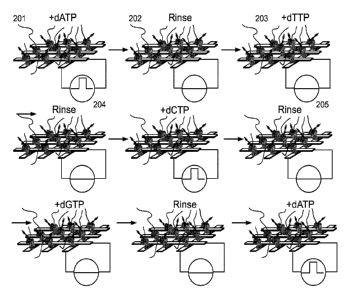
in times of
an hour or less.
The preceding example illustrates the sequencing of nucleic acid polymers, but
it also can
be applied such that other enzymes that process polymers could be used. For
example,
current fluctuations in an exonuclease will reflect the composition of the
nucleic acid they
are degrading. The same would be true of proteasomes that digest peptides. An
example
is the proteasome 20S CP, and proteasomes like this could likely be used for
single
molecule peptide sequencing by incorporating them into the system of FIG. 4
and
monitoring the electrical signals generated when they are fed peptide
molecules and biased
be low VC.
Similar enzymes, called glycosidases, exist for digesting glycans. The
incorporation of a
glycosidase into the device of FIG. 4 would allow electronic sequencing of
glycans. The
variation in bonding of glycans might preclude a direct linear read out of
sequence, but the
organization of cutting events in time may allow for identification of the
glycans.
In yet another embodiment, the present disclosure provides a method for
detecting kinase
activity. In this embodiment, a kinase is incorporated into the device of FIG.
4, exposed
to its substrate, and kinase activity signaled by the generation of large
current fluctuations
when biased below VC. The system could then be exposed to candidate kinase
inhibitor
drugs as the fluctuations are monitored, to discover which drugs "kill" the
activity of the
kinase. In the present art, the use of fluorescence labeling methods may
interfere with a
protein enzyme interacting with its substrate, and the present disclosure
removes the
requirement for labeling of proteins.
CA 03100693 2020-11-17
WO 2019/222527
PCT/US2019/032707
In all of these methods, the use of a simple junction (as opposed to a FET
structure)
greatly simplifies both manufacture and enables scale up to large parallel
arrays of
devices. The device of the disclosure may be prepared in massively parallel
fabrication
using methods for scalable fabrication of junction devices, as described
below.
ARRAYS AND SYSTEMS FOR SEQUENCING DNA OR OTHER POLYMERS
The present disclosure provides an array for sequencing biopolymers using any
of the
enzymes that interact processively with molecular templates such as a
nuclease, a
proteasome, a glycopeptidase, a glycosidase, a kinase and an endonuclease. The
embodiments below illustrate an array for sequencing DNA using a polymerase.
It should
be understood that any processive enzyme can be substituted for the polymerase
in the
arrays.
The array comprises an arrangement of a plurality of devices. The device used
in the
arrays of the present disclosure include the following.
In one embodiment, the device comprises a first and a second electrode, the
first and
second electrode being separated by a gap; and a polymerase attached to one or
both
electrodes; wherein the first electrode and the second electrode have an
opening formed
therethrough.
In another embodiment the device comprises a first and a second electrode, the
first and
second electrode being separated by a gap; and a polymerase attached to both
the first and
second electrode.
In some embodiments, the device comprises:
(a) a dielectric substrate;
(b) a first electrode disposed on the dielectric substrate;
(c) an insulating dielectric layer disposed on the first electrode;
(d) a second electrode disposed on the insulating dielectric layer;
(e) a passivation layer disposed on the second electrode;
(f) a polymerase molecule attached to the first and second electrodes;
31
CA 03100693 2020-11-17
WO 2019/222527
PCT/US2019/032707
wherein the first electrode, the insulating dielectric layer, the second
electrode and
passivation layer have an opening formed therethrough.
In some embodiments, the device comprises:
(a) a dielectric substrate;
(b) a first electrode disposed on the dielectric substrate;
(c) a second electrode disposed on the dielectric substrate;
(d) a passivation layer disposed on top of the electrodes; and
(e) a polymerase molecule attached to one or both the electrodes;
wherein the passivation layer has an opening formed therethrough.
In some embodiments, the device comprises:
(a) a first and a second electrode, the first and second electrode being co-
planar and
separated by a gap;
(b) a protein attached to at least one electrode;
wherein the first electrode and the second electrodes are configured for
contact with a
sample to be analyzed.
In each of the device embodiments described herein, the first and/or second
electrode
comprise a metal selected from the group consisting of gold, platinum,
palladium, and
ruthenium. In some embodiments, the metal is palladium.
In some embodiments, the gap has a width of about 1.0 nm to about 20.0 nm. In
some
embodiments, the gap has a width of about 1.0 nm to about 10.0 nm. In some
embodiments, the gap has a width of about 1.0 nm to about 7.5 nm. In some
embodiments, the gap has a width of about 1.0 nm to about 5.0 nm. In some
embodiments, the gap has a width of about 4.0 nm to about 5.0 nm.
The array of devices can be arranged in any suitable manner, e.g., in a grid.
In some embodiments, the array comprises polymerase molecules bound with
template
DNA. Such templates can be made by ligating genomic DNA fragments (generated
by
32
CA 03100693 2020-11-17
WO 2019/222527 PCT/US2019/032707
sonication, for example) with primer sequences containing a nick for binding
by
polymerase, as is well known in the art. The result is a library of templates
spanning an
entire genome, if needed. Each template will then randomly bind one polymerase
in the
array.
Referring now to FIG. 16, a grid of contacts 101, 102 is formed from two
layers of contact
metals separated by a dielectric as is well known in the art, and then covered
by a
passivation layer. Each intersection is then exposed by selectively removing
the
passivation, and polymerase molecules 103 bound at each junction. These
biomolecular
junctions can be prepared by methods discussed in the previous section. By
making the
electrodes narrow enough (about one micron in width) usually only one
polymerase (or
none) will bind. In the case where two or a few more bind, the signals can
still be
deconvolved because they consist of two current levels for one molecule, three
for two,
and so on.
The present disclosure also provides a system of arrays for direct measurement
of
polymerase activity. The system comprises an array as described herein;
optionally a
means for introducing and removing a solution to the array; a means for
applying a bias
between the first and second electrode; and a means for monitoring the current
generated
between the first and second electrodes.
Referring back to FIG. 16, means 106 are provided for applying a small bias
(typically
50mV) and reading the current from each junction so that it may be recorded by
a
computer storage system.
METHODS FOR DNA SEQUENCING USING A SYSTEM OF ARRAYS
The present disclosure also provides a method for DNA sequencing using a
system of
arrays as described herein.
The sequencing proceeds by introducing a solution comprising one nucleotide
monophosphate (e.g., one from among dATP, dGTP, dCTP, dTTP) together with
magnesium to the array. Each polymerase bound with a complementary nucleotide
will
generate a signal, which is read by the unique pair of electrodes to which
each polymerase
is bound. For example, if the added nucleotide is dCTP, then every template
bound at a G
base will incorporate a C into the extending chain, generating a signal,
whereas the other
sequences will generate distinctly different signals. For example, a
polymerase presented
with a non-matching base will generate a brief burst of signal as the mis-
matched base is
33
CA 03100693 2020-11-17
WO 2019/222527 PCT/US2019/032707
captured, but the signal train will terminate prematurely as the mismatch is
rejected and
the process of polymerization and translocation is aborted. In contrast,
incorporation of a
matching base results in a much longer train of pulses as the process of
incorporation and
translocation is completed. As the second step, the array is rinsed to remove
excess
nucleotide and a solution comprising the next nucleotide is introduced (for
example dATP,
so that all site with a T now generate a signal). The cycle is continued until
all four dNTPs
have been cycled through the device, after which the cycle is repeated. This
cycling can
be repeated until all the template DNA is exhausted, thus generating sequence
data for the
entire library of fragments.
FIG. 17 shows a method for sequencing DNA. On adding dATP to the array 201,
the
molecule shown will generate a signal 204 as the A is incorporated into the
extending
chain. The array is then rinsed to remove dATP 202, and the next nucleotide
(shown as
dTTP here) added 203. The cycle is continued as shown. The addition of dATP,
then
dCTP and then dATP all give signals of base incorporation 204 whereas the
presence of
the other nucleotides do not 205. Accordingly, the sequence at that particular
location will
be recorded as TGT.
It will be recognized that this approach has two major advantages over current
sequencing
strategies that use cycling of dNTPs. One is that, by utilizing a single
molecule read-out at
a time-scale faster than the base-incorporation rate of a polymerase, it now
becomes
straightforward to count repeats of the same base. So the sequence AAAAA would
give 5
distinct bursts of signal in the presence of dTTP, and so on. The second is
that, in contrast
to known optical readout schemes, the length of template that is read is not
constrained by
distance from the mounting substrate, so that the potential read length is as
high as the
processivity of the polymerase (10kB for phi29).
FIG. 18 shows the ability to read individual sequential incorporations, and
hence read
homopolymeric runs of sequence. A current vs. time recording 300 shows the
incorporation of two sequential bases (here, electrical data are shown for
dTTP being
incorporated at two successive A sites). Each signal consists of a burst 301
of telegraph
noise of duration about 20 ms, separated by gaps between bursts, also, on
average of about
to 20 ms at micromolar concentrations of nucleotide triphosphate. Thus, on any
given
step of the chemical cycling illustrated in FIG. 17, where just one nucleotide
triphosphate
is present, the number of repeated bases of its complement in the template
strand may be
counted simply by recording the number of such bursts of telegraph noise.
34
CA 03100693 2020-11-17
WO 2019/222527 PCT/US2019/032707
In another embodiment, the solution comprises more than one dNTP, with the
dNTPs
present in different concentrations. For example, the solution comprises 1 mM
dATP, 100
[IM dGTP 10 [IM dCTP and 0.1 [IM dTTP. In this embodiment, the polymerases in
the
array would generate signals continuously as each template is extended. At
points where
T is present in the template DNA, the signal of incorporation would follow the
previous
burst of telegraph noise rapidly (generally within 10 ms). The template DNAs
in which the
next base was a C would show a more delayed burst of telegraph noise because
of the
slower arrival of dGTP owing to its lower concentration. Similarly, templates
containing a
G would be preceded by a longer delay because of even lower concentration of
dCTP.
The longest delays would precede A bases because the concentration of dTTP is
lowest.
In yet another embodiment, the two approaches can be combined, using 2 cycles
of
rinsing, using one pair of nucleotides in unequal concentration in the first
cycle, and then
the other two, also in unequal concentration in the second cycle.
While the preceding section describes methods of sequencing DNA using a system
of
arrays comprising a polymerase, the system of arrays can be easily modified to
sequence
other polymers as well.
Although the invention has been described and illustrated in the foregoing
illustrative
embodiments, it is understood that the present disclosure has been made only
by way of
example, and that numerous changes in the details of implementation of the
invention can
be made without departing from the spirit and scope of the invention. Features
of the
disclosed embodiments can be combined and rearranged in various ways. All
publications, patents and other documents mentioned herein are incorporated by
reference
in their entirety.
REFERENCES
The following references are hereby incorporated by reference in their
entireties:
Chen, Y.-S., C.-H. Lee, M.-Y. Hung, H.-A. Pan, J.-C. Chiou and G. S. Huang
(2013).
"DNA sequencing using electrical conductance measurements of a DNA
polymerase."
Nature Nano Technology 8: 452-458.
Choi, Y., I. S. Moody, P. C. Sims, S. R. Hunt, B. L. Corso, I. Perez, G. A.
Weiss and P. G.
Collins (2012). "Single-molecule lysozyme dynamics monitored by an electronic
circuit."
Science 335(6066): 319-324.
CA 03100693 2020-11-17
WO 2019/222527
PCT/US2019/032707
Merriman, B. and P. Mola, Biomolecular Sensors and Methods. US patent
application
number: WO 2016210386A1, 2016
Olsen, T. J., Y. Choi, P. C. Sims, 0. T. Gul, B. L. Corso, C. Dong, W. A.
Brown, P. G.
Collins and G. A. Weiss (2013). "Electronic measurements of single-molecule
processing
by DNA polymerase I (Klenow fragment)." J Am Chem Soc 135(21): 7855-7860.
Zhang, B., W. Song, P. Pang, Y. Zhao, P. Zhang, I. Csabai, G. Vattay and S.
Lindsay
(2017). "Observation of Giant Conductance Fluctuations in a Protein." Nano
Futures 1(3).
36