Note: Descriptions are shown in the official language in which they were submitted.
CA 03100902 2020-11-18
WO 2019/226643 PCT/US2019/033303
METHODS FOR TREATING SPINAL CORD INJURY
CROSS REFERENCE TO RELATED APPLICATION
[0001] This application is an International Application which claims the
benefit under 35 U.S.C.
119(e) of U.S. Provisional Applications No. 62/676,464, filed on May 25, 2018,
the contents of which is
incorporated herein by reference in its entirety.
FIELD OF THE INVENTION
[0002] The field of the invention relates to the treatment of spinal cord
injuries.
BACKGROUND
[0003] Many human spinal cord injuries are anatomically incomplete, but
exhibit complete
paralysis. It is unknown why spared axons fail to mediate functional recovery
in these cases.
Current therapeutics for such injury are limited, and often do not regenerate
functional recovery
of a spinal cord injury. Thus, a better understanding of axon regeneration is
required for
developing effective treatments.
SUMMARY
[0004] The invention described herein is related, in part, to the discovery
that an agent, e.g.,
CLP290, that upmodulates neuron-specific K+-C1- co-transporter (KCC2) activity
and/or levels
was capable of restoring stepping function in mice with staggered bilateral
hemisections, e.g., a
severe spinal cord injury model. Further, overexpression of KCC2 recapitulated
this restoration
of stepping. It is further shown herein that the inhibition of Na+/2C1-/K+ co-
transporter (NKCC)
additionally restores stepping ability.
[0005] Further, work described herein show that agents that reduce
excitability in interneurons
in combination with clozapine N-oxide additionally restore the stepping
ability in mice that have
previously lost this ability following a staggered bilateral hemisection. Such
agents include an
agen that upmodulates a Gi-DREADD which has been optimized for expression in
inhibitory
interneurons, and Kir2.1.
[0006] Additionally, described herein are compositions comprising agent for
modulating KCC2,
NKCC, Gi-DREADD, and Kir2.1 to be used, e.g., in the treatment of a spinal
cord injury.
[0007] Accordingly, one aspect of the invention described herein provides a
method for treating
a spinal injury, comprising administering to a subject having a spinal injury
an effective amount
of an agent that upmodulates KCC2.
1
CA 03100902 2020-11-18
WO 2019/226643 PCT/US2019/033303
[0008] In one embodiment of any aspect, the agent that upmodulates KCC2 is
selected from the
group consisting of a small molecule, a peptide, a gene editing system, and an
expression vector
encoding KCC2.
[0009] In one embodiment of any aspect, the small molecule is CLP290.
[0010] In one embodiment of any aspect, the vector is non-integrative or
integrative. In another
embodiment of any aspect, the vector is a viral vector or non-viral vector.
[0011] Exemplary non-integrative vectors include, but are not limited to, an
episomal vector, an
EBNA1 vector, a minicircle vector, a non-integrative adenovirus, a non-
integrative RNA, and a
Sendai virus.
[0012] Exemplary viral vectors include, but are not limited to, retrovirus,
lentivirus, adenovirus,
herpesvirus, poxvirus, alpha virus, vaccinia virus, and adeno-associated
viruses.
[0013] Exemplary non-viral vectors include, but are not limited to, a
nanoparticle, a cationic
lipid, a cationic polymer, a metallic nanoparticle, a nanorod, a liposome,
microbubbles, a cell
penetrating peptide and a liposphere.
[0014] In one embodiment of any aspect, the vector crosses the blood brain
barrier.
[0015] In one embodiment of any aspect, KCC2 is upmodulated by at least 2-
fold, 5-fold, 6-
fold, 7-fold, 8-fold, 9-fold, 10-fold as compared to an appropriate control.
[0016] In one embodiment of any aspect, the spinal injury is a severe spinal
cord injury.
[0017] In one embodiment of any aspect, the subject is human. In one
embodiment of any
aspect, the subject has been diagnosed with a spinal injury. In one embodiment
of any aspect,
the subject has been previously diagnosed with a spinal injury. In one
embodiment of any
aspect, the subject has been previously treated for a spinal injury.
[0018] In one embodiment of any aspect, prior to administering, the subject is
diagnosed with
having a spinal cord injury.
[0019] In one embodiment of any aspect, the subject is further administered at
least a second
spinal injury treatment. In one embodiment of any aspect, the subject is
further administered at
least a second therapeutic compound. Exemplary second therapeutic compound
include, but are
not limited to osteopontin, growth factors, or 4-aminopuridine.
[0020] Another aspect of the invention described herein provides a method for
treating a spinal
injury, comprising administering to a subject having a spinal injury an
effective amount of an
agent that inhibits Na+/2C1-/K+ co-transporter (NKCC).
[0021] In one embodiment of any aspect, the agent that inhibits Na+/2C1-/K+ co-
transporter
(NKCC) is selected from the group consisting of a small molecule, an antibody,
a peptide, an
antisense oligonucleotide, and an RNAi. In one embodiment of any aspect, the
RNAi is a
2
CA 03100902 2020-11-18
WO 2019/226643 PCT/US2019/033303
microRNA, an siRNA, or an shRNA. In one embodiment of any aspect, the small
molecule is
bumetanide.
[0022] In one embodiment of any aspect, the agent is comprised in a vector.
[0023] Yet another aspect of the invention described herein provides a method
for treating a
spinal injury, comprising administering to a subject having a spinal injury an
effective amount of
an agent that reduces excitability of inhibitory interneurons.
[0024] In one embodiment of any aspect, the agent upmodulates the inhibitory
Gi-coupled
receptor Gi-DREADD.
[0025] In one embodiment of any aspect, the agent is an expression vector
encoding Gi-
DREADD. In one embodiment of any aspect, the agent is an expression vector
encoding Kir2.1.
[0026] In one embodiment of any aspect, the method further comprises
administering clozapine
N-oxide at substantially the same time as the agent.
[0027] In one embodiment of any aspect, the excitability of inhibitory
interneurons is reduced
by at least 10%, at least 20%, at least 30%, at least 40%, at least 50%, at
least 60%, at least 70%,
at least 80%, at least 90, at least 99%, or more as compared to an appropriate
control.
[0028] Another aspect of the invention described herein provides a method for
treating a spinal
injury, comprising administering to a subject having a spinal injury an
effective amount
electrical stimulation that reduces excitability of inhibitory interneurons.
In one embodiment of
any aspect, the method further comprises administering clozapine N-oxide.
[0029] In one embodiment of any aspect, the electrical stimulation is applied
directly to the
spinal cord. In one embodiment of any aspect, the electrical stimulation is
applied directly to the
spinal cord at the site of injury.
[0030] In one embodiment of any aspect, the excitability of inhibitory
interneurons is reduced
by at least 10%, at least 20%, at least 30%, at least 40%, at least 50%, at
least 60%, at least 70%,
at least 80%, at least 90, at least 99%, or more as compared to an appropriate
control.
[0031] Another aspect of the invention described herein provides a
pharmaceutical composition
comprising an effective amount of KCC2 polypeptide or a vector comprising a
nucleic acid
sequence encoding the KCC2 polypeptide and a pharmaceutically acceptable
carrier, for use in
treating spinal cord injury.
[0032] In one embodiment of any aspect, the KCC2 polypeptide has, comprises,
consists of, or
consists essentially of at least 85%, at least 86%, at least 87%, at least
88%, at least 89%, at least
90%, at least 91%, at least 92%, at least 93%, at least 94%, at least 95%, at
least 96%, at least
97%, at least 98%, at least 99%, or more amino acid sequence identity to SEQ
ID NO: 1 and
retains at least 80% of the biological activity of KCC2 of SEQ ID NO: 1.
3
CA 03100902 2020-11-18
WO 2019/226643 PCT/US2019/033303
[0033] In one embodiment of any aspect, the composition further comprises at
least a second
therapeutic compound.
[0034] Another aspect of the invention described herein provides a
pharmaceutical composition
comprising an effective amount of Gi-DREADD polypeptide or a vector comprising
a nucleic
acid sequence the Gi-DREADD polypeptide and a pharmaceutically acceptable
carrier, for use
in treating spinal cord injury.
[0035] In one embodiment of any aspect, the Gi-DREADD polypeptide is an
optimized Gi-
DREADD polypeptide. In one embodiment of any aspect, the Gi-DREADD polypeptide
comprises the sequence of SEQ ID NO: 2.
[0036] In one embodiment of any aspect, the Gi-DREADD polypeptide has,
comprises, consists
of, or consists essentially of at least 85%, at least 86%, at least 87%, at
least 88%, at least 89%,
at least 90%, at least 91%, at least 92%, at least 93%, at least 94%, at least
95%, at least 96%, at
least 97%, at least 98%, at least 99%, or more amino acid sequence identity to
SEQ ID NO: 2
and retains at least 80% of the biological activity of Gi-DREADD of SEQ ID NO:
2.
[0037] In one embodiment of any aspect, the composition further comprises at
least a second
therapeutic compound. In one embodiment of any aspect, the composition further
comprises
clozapine N-oxide.
[0038] Another aspect of the invention described herein provides a
pharmaceutical composition
comprising an effective amount of Kir2.1 polypeptide or a vector comprising a
nucleic acid
sequence the Kir2.1 polypeptide and a pharmaceutically acceptable carrier, for
use in treating
spinal cord injury.
[0039] In one embodiment of any aspect, the Kir2.1 polypeptide comprises the
sequence of SEQ
ID NO: 3.
[0040] In one embodiment of any aspect, the Kir2.1 polypeptide has, comprises,
consists of, or
consists essentially of at least 85%, at least 86%, at least 87%, at least
88%, at least 89%, at least
90%, at least 91%, at least 92%, at least 93%, at least 94%, at least 95%, at
least 96%, at least
97%, at least 98%, at least 99%, or more amino acid sequence identity to SEQ
ID NO: 3 and
retains at least 80% of the biological activity of Kir2.1 of SEQ ID NO: 3.
[0041] In one embodiment of any aspect, the composition further comprises
clozapine N-oxide.
In one embodiment of any aspect, the composition further comprises at least a
second
therapeutic compound.
[0042] Another aspect of the invention described herein provides a
pharmaceutical composition
comprising an effective amount of any of the agents that inhibit NKCC as
described herein and a
pharmaceutically acceptable carrier, for use in treating spinal cord injury.
In one embodiment of
any aspect, the composition further comprises at least a second therapeutic
compound.
4
CA 03100902 2020-11-18
WO 2019/226643 PCT/US2019/033303
[0043] Another aspect of the invention described herein provides a method for
treating a spinal
injury, comprising administering to a subject having a spinal injury an
effective amount of
CLP290.
[0044] In one embodiment of any aspect, CLP290 crosses the blood brain
barrier. For example,
CLP290 is formulated in a way that allows it to cross the blood brain barrier.
[0045] In one embodiment of any aspect, the subject is further administered at
least a second
spinal injury treatment. In one embodiment of any aspect, the subject is
further administered at
least a second therapeutic compound. In one embodiment of any aspect, the
second therapeutic
compound is selected from the group consisting of osteopontin, a growth
factor, or 4-
aminopuridine.
Definitions
[0046] For convenience, the meaning of some terms and phrases used in the
specification,
examples, and appended claims, are provided below. Unless stated otherwise, or
implicit from
context, the following terms and phrases include the meanings provided below.
The definitions
are provided to aid in describing particular embodiments, and are not intended
to limit the
claimed technology, because the scope of the technology is limited only by the
claims. Unless
otherwise defined, all technical and scientific terms used herein have the
same meaning as
commonly understood by one of ordinary skill in the art to which this
technology belongs. If
there is an apparent discrepancy between the usage of a term in the art and
its definition
provided herein, the definition provided within the specification shall
prevail.
[0047] As used herein, the terms "treat," "treatment," "treating," or
"amelioration" refer to
therapeutic treatments, wherein the object is to reverse, alleviate,
ameliorate, inhibit, slow down
or stop the progression or severity of a condition associated with a spinal
cord injury. The term
"treating" includes reducing or alleviating at least one adverse effect or
symptom of a spinal
cord injury, e.g., partial or complete paralysis. Treatment is generally
"effective" if one or more
symptoms or clinical markers are reduced. Alternatively, treatment is
"effective" if the
progression of a disease is reduced or halted. That is, "treatment" includes
not just the
improvement of symptoms or markers, but also a cessation of, or at least
slowing of, progress or
worsening of symptoms compared to what would be expected in the absence of
treatment.
Beneficial or desired clinical results include, but are not limited to,
alleviation of one or more
symptom(s), diminishment of extent of disease, stabilized (i.e., not
worsening) state of a spinal
cord injury, delay or slowing of a spinal cord injury progression,
amelioration or palliation of the
injury state, remission (whether partial or total), and/or decreased
mortality, whether detectable
CA 03100902 2020-11-18
WO 2019/226643 PCT/US2019/033303
or undetectable. The term "treatment" of a spinal cord injury also includes
providing relief from
the symptoms or side-effects of the disease (including palliative treatment).
[0048] As used herein, the term "administering," refers to the placement of a
therapeutic (e.g.,
an agent that upmodulates KCC2 or reduces excitability of inhibitory
interneurons) or
pharmaceutical composition as disclosed herein into a subject by a method or
route which
results in at least partial delivery of the agent to the subject.
Pharmaceutical compositions
comprising agents as disclosed herein can be administered by any appropriate
route which
results in an effective treatment in the subject.
[0049] As used herein, a "subject" means a human or animal. Usually the animal
is a vertebrate
such as a primate, rodent, domestic animal or game animal. Primates include,
for example,
chimpanzees, cynomologous monkeys, spider monkeys, and macaques, e.g., Rhesus.
Rodents
include, for example, mice, rats, woodchucks, ferrets, rabbits and hamsters.
Domestic and game
animals include, for example, cows, horses, pigs, deer, bison, buffalo, feline
species, e.g.,
domestic cat, canine species, e.g., dog, fox, wolf, avian species, e.g.,
chicken, emu, ostrich, and
fish, e.g., trout, catfish and salmon. In some embodiments, the subject is a
mammal, e.g., a
primate, e.g., a human. The terms, "individual," "patient" and "subject" are
used
interchangeably herein.
[0050] Preferably, the subject is a mammal. The mammal can be a human, non-
human primate,
mouse, rat, dog, cat, horse, or cow, but is not limited to these examples.
Mammals other than
humans can be advantageously used as subjects that represent animal models of
spinal cord
injury. A subject can be male or female.
[0051] A subject can be one who has been previously diagnosed with or
identified as suffering
from or having a spinal cord injury or one or more complications related to
such an injury, and
optionally, have already undergone treatment for a spinal cord injury or the
one or more
complications related to the injury. Alternatively, a subject can also be one
who has not been
previously diagnosed as having such spinal cord injury or related
complications. For example, a
subject can be one who exhibits one or more risk factors for a spinal cord
injury, e.g.,
participates in an activity that is likely to result in a spinal cord injury,
for example, a full
contact sport, e.g., American football, or one or more complications related
to spinal cord injury
or a subject who does not exhibit risk factors.
[0052] Methods and compositions described herein are used for the treatment of
a spinal cord
injury. As used herein, a "spinal cord injury" refers to any insult to any
region of the spinal cord,
e.g., the cervical vertebrae, the thoracic vertebrae, the lumbar vertebrae,
the sacral vertebrae, the
sacrum, or the coccyx. A "spinal cord injury" can result in various levels of
severity, ranging
from no effect on mobility, e.g., retain walking ability, to paraplegia (e.g.,
paralysis of legs and
6
CA 03100902 2020-11-18
WO 2019/226643 PCT/US2019/033303
lower region of body), and tretraplegia (e.g., loss of muscle strength in all
four extremities). A
"spinal cord injury" can be a complete spinal cord injury, e.g., an injury
that produces total loss
of all motor and sensory function below the site of injury. A "spinal cord
injury" can be an
incomplete spinal cord injury, e.g., in which some motor function remains
below the primary
site of the injury. Non-limiting examples of incomplete spinal cord injuries
include, but are not
limited to, anterior cord syndrome, center cord syndrome, and Brown-Sequard
syndrome. A
"spinal cord injury" can be a spinal concussion or spinal contusion, e.g., an
injury that resolves
itself in, e.g., one or two days. A spinal concussion or contusion can be
complete or incomplete.
[0053] As used herein, an "agent" refers to e.g., a molecule, protein,
peptide, antibody, or
nucleic acid, that inhibits expression of a polypeptide or polynucleotide, or
binds to, partially or
totally blocks stimulation, decreases, prevents, delays activation,
inactivates, desensitizes, or
down regulates the activity of the polypeptide or the polynucleotide. Agents
that inhibit NKCC,
e.g., inhibit expression, e.g., translation, post-translational processing,
stability, degradation, or
nuclear or cytoplasmic localization of a polypeptide, or transcription, post
transcriptional
processing, stability or degradation of a polynucleotide or bind to, partially
or totally block
stimulation, DNA binding, transcription factor activity or enzymatic activity,
decrease, prevent,
delay activation, inactivate, desensitize, or down regulate the activity of a
polypeptide or
polynucleotide. An agent can act directly or indirectly.
[0054] The term "agent" as used herein means any compound or substance such
as, but not
limited to, a small molecule, nucleic acid, polypeptide, peptide, drug, ion,
etc. An "agent" can
be any chemical, entity or moiety, including without limitation synthetic and
naturally-occurring
proteinaceous and non-proteinaceous entities. In some embodiments, an agent is
nucleic acid,
nucleic acid analogues, proteins, antibodies, peptides, aptamers, oligomer of
nucleic acids,
amino acids, or carbohydrates including without limitation proteins,
oligonucleotides,
ribozymes, DNAzymes, glycoproteins, RNAis (e.g., microRNAs, siRNAs, and
shRNAs)
lipoproteins, aptamers, and modifications and combinations thereof etc. In
certain embodiments,
agents are small molecule having a chemical moiety. For example, chemical
moieties included
unsubstituted or substituted alkyl, aromatic, or heterocyclyl moieties
including macrolides,
leptomycins and related natural products or analogues thereof. Compounds can
be known to
have a desired activity and/or property, or can be selected from a library of
diverse compounds.
[0055] The agent can be a molecule from one or more chemical classes, e.g.,
organic molecules,
which may include organometallic molecules, inorganic molecules, genetic
sequences, etc.
Agents may also be fusion proteins from one or more proteins, chimeric
proteins (for example
domain switching or homologous recombination of functionally significant
regions of related or
7
CA 03100902 2020-11-18
WO 2019/226643 PCT/US2019/033303
different molecules), synthetic proteins or other protein variations including
substitutions,
deletions, insertion and other variants.
[0056] As used herein, the term "small molecule" refers to a chemical agent
which can include,
but is not limited to, a peptide, a peptidomimetic, an amino acid, an amino
acid analog, a
polynucleotide, a polynucleotide analog, an aptamer, a nucleotide, a
nucleotide analog, an
organic or inorganic compound (e.g., including heterorganic and organometallic
compounds)
having a molecular weight less than about 10,000 grams per mole, organic or
inorganic
compounds having a molecular weight less than about 5,000 grams per mole,
organic or
inorganic compounds having a molecular weight less than about 1,000 grams per
mole, organic
or inorganic compounds having a molecular weight less than about 500 grams per
mole, and
salts, esters, and other pharmaceutically acceptable forms of such compounds.
[0057] Methods and compositions described herein require that the level of
KCC2 is
upmodulated. As used herein, "K+-C1- co-transporter (KCC2)" refers to a
protein with lower
intracellular chloride concentrations below the electrochemical equilibrium
potential. KCC2 can
function in either a net efflux or influx pathway, depending on the chemical
concentration
gradients of potassium and chloride. Sequences for KCC2, also known as Solute
carrier family
12 member 5, are known for a number of species, e.g., human KCC2 (NCBI Gene
ID: 57468)
polypeptide (e.g., NCBI Ref Seq NP 001128243.1) and mRNA (e.g., NCBI Ref Seq
NM 001134771.1). KCC2 can refer to human KCC2, including naturally occurring
variants,
molecules, and alleles thereof KCC2 refers to the mammalian KCC2 of, e.g.,
mouse, rat, rabbit,
dog, cat, cow, horse, pig, and the like. The nucleic sequence of SEQ ID NO:1
comprises the
nucleic sequence which encodes rat KCC2.
[0058] Methods and compositions described herein require that the levels
and/or activity of
NKCC are inhibited. As used herein, "Na+/2C1-/K+ co-transporter (NKCC)" refers
to a protein
required to maintain proper ionic balance and cell volume by, e.g., mediating
sodium and
chloride transport and reabsorption. Sequences for NKCC, also known as Solute
carrier family
12 member 2 and NKCC1, are known for a number of species, e.g., human NKCC
(NCBI Gene
ID: 6558) polypeptide (e.g., NCBI Ref Seq NP 001037.1) and mRNA (e.g., NCBI
Ref Seq
NM 001046.2). NKCC can refer to human NKCC, including naturally occurring
variants,
molecules, and alleles thereof NKCC refers to the mammalian NKCC of, e.g.,
mouse, rat,
rabbit, dog, cat, cow, horse, pig, and the like. The nucleic sequence of SEQ
ID NO: 4 comprises
the nucleic sequence which encodes NKCC.
[0059] Methods and compositions described herein require that the levels
and/or activity of
Kir2.1. are increased. As used herein, "Kir2.1" refers to potassium voltage-
gated channel
subfamily J member 2, characterized by having a greater tendency to allow
potassium to flow
8
CA 03100902 2020-11-18
WO 2019/226643 PCT/US2019/033303
into, rather than out of, a cell. Kir2.1 may participate in establishing
action potential waveform
and excitability of neuronal and muscle tissues. Kir2.1sequences are known for
a number of
species, e.g., human Kir2.1 (NCBI Gene ID: 3759) polypeptide (e.g., NCBI Ref
Seq
NP 000882.1) and mRNA (e.g., NCBI Ref Seq NM 000891.2). Kir2.1 can refer to
human
Kir2.1, including naturally occurring variants, molecules, and alleles thereof
Kir2.1 refers to
the mammalian Kir2.1 of, e.g., mouse, rat, rabbit, dog, cat, cow, horse, pig,
and the like. The
nucleic sequence of SEQ ID NO: 3 comprises an amino acid sequence which
encodes human
Kir2.1. The nucleic sequence of SEQ ID NO: 5 comprises an amino acid sequence
which
encodes mouse Kir2.1.
[0060] The term "upmodulation" and "upmodulate" as used herein refer to a
change or an
alteration that results in an increase in a biological activity (e.g., of
KCC2, Gi-DREADD, or
Kir2.1). Upmodulation includes, but is not limited to, stimulating or
promoting an activity.
Upmodulation may be a change in activity and/or levels, a change in binding
characteristics, or
any other change in the biological, functional, or immunological properties
associated with the
activity of a protein, a pathway, a system, or other biological targets of
interest that results in its
increased activity and/ or levels. In some embodiments, the term "upmodulate"
can mean an
increase of at least 10% as compared to a reference level, for example an
increase of at least
about 20%, or at least about 30%, or at least about 40%, or at least about
50%, or at least about
60%, or at least about 70%, or at least about 80%, or at least about 90% or up
to and including a
100% increase or any increase between 10-100% as compared to a reference
level, or at least
about a 2-fold, or at least about a 3-fold, or at least about a 4-fold, or at
least about a 5-fold or at
least about a 10-fold increase, a 20-fold increase, a 30-fold increase, a 40-
fold increase, a 50-
fold increase, a 60-fold increase, a 75-fold increase, a 100-fold increase,
etc., or any increase
between 2-fold and 10-fold or greater as compared to an appropriate control.
[0061] The term "decrease", "reduced", "reduction", or "inhibit" are all used
herein to mean a
decrease by a statistically significant amount. In some embodiments,
"decrease", "reduced",
"reduction", or "inhibit" typically means a decrease by at least 10% as
compared to an
appropriate control (e.g. the absence of a given treatment) and can include,
for example, a
decrease by at least about 10%, at least about 20%, at least about 25%, at
least about 30%, at
least about 35%, at least about 40%, at least about 45%, at least about 50%,
at least about 55%,
at least about 60%, at least about 65%, at least about 70%, at least about
75%, at least about
80%, at least about 85%, at least about 90%, at least about 95%, at least
about 98%, at least
about 99%, or more. As used herein, "reduction" or "inhibition" does not
encompass a complete
inhibition or reduction as compared to a reference level. "Complete
inhibition" is a 100%
inhibition as compared to an appropriate control.
9
CA 03100902 2020-11-18
WO 2019/226643 PCT/US2019/033303
[0062] The terms "increase", "enhance", or "activate" are all used herein to
mean an increase by
a reproducible statistically significant amount. In some embodiments, the
terms "increase",
"enhance", or "activate" can mean an increase of at least 10% as compared to a
reference level,
for example an increase of at least about 20%, or at least about 30%, or at
least about 40%, or at
least about 50%, or at least about 60%, or at least about 70%, or at least
about 80%, or at least
about 90% or up to and including a 100% increase or any increase between 10-
100% as
compared to a reference level, or at least about a 2-fold, or at least about a
3-fold, or at least
about a 4-fold, or at least about a 5-fold or at least about a 10-fold
increase, a 20 fold increase, a
30 fold increase, a 40 fold increase, a 50 fold increase, a 6 fold increase, a
75 fold increase, a
100 fold increase, etc. or any increase between 2-fold and 10-fold or greater
as compared to an
appropriate control. In the context of a marker, an "increase" is a
reproducible statistically
significant increase in such level.
[0063] As used herein, an "appropriate control" refers to an untreated,
otherwise identical cell
or population (e.g., a patient who was not administered an agent described
herein, or was
administered by only a subset of agents described herein, as compared to a non-
control patient).
[0064] The term "pharmaceutically acceptable carrier" as used herein means a
pharmaceutically
acceptable material, composition or vehicle, such as a liquid or solid filler,
diluent, excipient,
solvent or encapsulating material, involved in carrying or transporting the
active ingredient (e.g.,
cells) to the targeting place in the body of a subject. Each carrier must be
"acceptable" in the
sense of being compatible with the other ingredients of the formulation and is
compatible with
administration to a subject, for example a human.
[0065] The term "statistically significant" or "significantly" refers to
statistical significance and
generally means a two standard deviation (2SD) or greater difference.
[0066] As used herein the term "comprising" or "comprises" is used in
reference to
compositions, methods, and respective component(s) thereof, that are essential
to the method or
composition, yet open to the inclusion of unspecified elements, whether
essential or not.
[0067] As used herein the term "consisting essentially of' refers to those
elements required for a
given embodiment. The term permits the presence of elements that do not
materially affect the
basic and novel or functional characteristic(s) of that embodiment. The term
"consisting of'
refers to compositions, methods, and respective components thereof as
described herein, which
are exclusive of any element not recited in that description of the
embodiment.
[0068] The singular terms "a," "an," and "the" include plural referents unless
context clearly
indicates otherwise. Similarly, the word "or" is intended to include "and"
unless the context
clearly indicates otherwise. Although methods and materials similar or
equivalent to those
described herein can be used in the practice or testing of this disclosure,
suitable methods and
CA 03100902 2020-11-18
WO 2019/226643 PCT/US2019/033303
materials are described below. The abbreviation, "e.g." is derived from the
Latin exempli gratia,
and is used herein to indicate a non-limiting example. Thus, the abbreviation
"e.g." is
synonymous with the term "for example."
BRIEF DESCRIPTION OF THE DRAWINGS
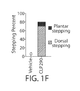
[0069] Figure 1A-1K present data that show identification of CLP290 as a
compound
leading to functional recovery in mice with staggered lesions. (FIG. 1A)
Schematic of
staggered lateral hemisections at T7 and T10. Arrowheads indicate lesions, L =
left, R = right.
(FIG. 1B) Representative image of an anti-GFAP stained spinal cord section 10
weeks after
over-stagger lesion. Dashed line indicates midline. Scale bar: 5001.tm. (FIG.
1C) Representative
image stacks of anti-5HT-stained transverse sections from T5 (rostral to
lesions), T8 (between
lesions), and L2 (caudal to lesions) of mice at 2 weeks after staggered
lesions. Scale bar: 100
1.tm. (FIG. 1D) Experimental scheme. Each BMS test was performed 24 hr prior
to daily
compound treatment. (FIG. 1E) BMS scores in injured mice with continuous
treatment of
CLP290 (35mg/kg) and vehicle solution. Two-way repeated-measures ANOVA
followed by
post hoc Bonferroni correction. Both groups started as n = 10, and at week 9
(the termination
time point) n = 8, and 10 for vehicle and CLP290 respectively. *P<0.05;
****P<0.0001. Error
bars: SEM. (FIG. 1F) Percentage of mice that reached stepping. CLP290 versus
vehicle at 9
weeks post staggered injury (n = 8 and 10 for vehicle and CLP290 group
respectively). (FIG.
1G). Sustained behavioral improvements after CLP290 withdrawal in mice with 10-
week
treatment. BMS was tested on Day 1, 2, 3, 7 and 14 after compound withdrawal
(n = 7). Two-
way repeated-measure ANOVA followed by post hoc Bonferroni correction. **p <
0.01. Error
bars: SEM. (FIG. 1H) Color-coded stick view decomposition of mouse right
hindlimb
movements during swing, stance (Intact group), dragging (Vehicle group) and
stepping (CLP290
group). (FIG. 11 and FIG. 1J). Quantification of bodyweight support (FIG. 11)
and stride length
(FIG. 1J) of mice at 9 weeks post staggered injury (n = 8 and 10 for vehicle
and CLP290 group
respectively). Student's t-test (two-tailed, unpaired). *p <0.05; **p <0.01.
Error bars: SEM.
(FIG. 1K) Representative right hindlimb knee and ankle angle oscillation trace
and simultaneous
EMG recording from tibias anterior (TA) and gastrocnemius medialis (GS)
muscle.
[0070] Figure 2A-2H present data that show widespread KCC2 expression mimics
the
effects of CLP290 to promote functional recovery. (FIG. 2A) Experimental
scheme. (FIG.
2B) Representative image stacks of longitudinal (upper) and transverse (lower)
spinal cord
sections, taken from the mice at 8 weeks after staggered injury, stained with
anti-HA (to detect
the HA-KCC2 protein). Scale bar: 50011m (upper) and 1001.tm (lower). (FIG. 2C)
BMS
performance in experimental (AAV-PHP.B-HA-KCC2) and control (AAV-PHP.B-H2B-
GFP)
11
CA 03100902 2020-11-18
WO 2019/226643 PCT/US2019/033303
groups. Two-way repeated-measures ANOVA followed by post hoc Bonferroni
correction. *p <
0.05. (FIG. 2D) Percentage of mice that reached stepping at 8 weeks after
injury. (FIG. 2E and
FIG. 2F) Quantification of bodyweight support (FIG. 2E) and stride length
(FIG. 2F) at 8 weeks
(n = 10 per group). Student's t-test (two-tailed, unpaired) was applied. *p <
0.05; **p < 0.01.
Error bars: SEM. (FIG. 2G) Color-coded stick view decomposition of mouse right
hindlimb
movement during dragging (AAV-PHP.B-H2B-GFP group) and stepping (AAV-PHP.B-HA-
KCC2 group). (FIG. 2H) Representative right hindlimb knee and ankle angle
oscillation trace
and simultaneous EMG recording of mice at 8 weeks after injury.
[0071] Figure 3A-3E present data that show KCC2 expression in inhibitory
neurons leads
to functional recovery. (FIG. 3A, 3B) Representative image stacks showing
expression of GFP
(FIG. 3A) or HA-KCC2 (FIG. 3B) in T8 spinal cord of indicated transgenic mice
with tail-vein
injection of AAV-PHP.B-CAG-Flex-H2B-GFP (FIG. 3A) or AAV-PHP.B-Syn-Flex-HA-
KCC2
(FIG. 3B). Scale bar: 10011m. (FIG. 3C) BMS performance in indicated groups.
Two-way
repeated-measure ANOVA followed by post hoc Bonferroni correction. *p < 0.05;
****p <
0.0001. Error bars: SEM. (FIG. 3D) Breakdown of BMS scores for indicated
treatment groups at
8 weeks after injury. (FIG. 3E) Percentage of mice that reached plantar or
dorsal stepping at 8
weeks after injury.
[0072] Figure 4A-41I present data that show KCC2 acts on inhibitory neurons in
the spinal
cord segments between and around the lesions. (FIG. 4A) Experimental scheme
for FIG. 4B-
FIG. 4D. (FIG. 4B) Representative images of anti-HA-stained transverse
sections of the thoracic
and lumbar spinal cord at 8 weeks. Scale bar: 10011m. (FIG. 4C and FIG. 4D)
Left, BMS
performance in different treatment groups in wild type mice (FIG. 4C), and
Vgat-Cre mice (FIG.
4D). Right, percentage of mice that reached stepping in WT mice (FIG. 4C) and
Vgat-Cre mice
(FIG. 4D). ANOVA followed by post hoc Bonferroni correction. Error bars: SEM.
(FIG. 4E)
Experimental scheme for FIG. 4F- FIG. 4H. (FIG. 4F) Representative images of
anti-HA-stained
transverse sections of the thoracic and lumbar spinal cord at 8 weeks after
injury. Scale bar: 100
1.tm. (FIG. 4G and FIG. 4H) Left, BMS performance in experimental and control
groups in WT
mice (FIG. 4G), and Vgat-Cre mice (FIG. 4H). Right, percentage of mice that
reached stepping
in WT mice (FIG. 4G) and Vgat-Cre mice (FIG. 4H). ANOVA followed by post hoc
Bonferroni
correction. *p < 0.05. Error bars: SEM.
[0073] Figure 5A-5F present data that show altered neuronal activation
patterns and relay
formation facilitated by CLP290/KCC2. (FIG. 5A) Schematics of transverse
spinal cord
sections showing c-Fos expression patterns in T8/9 segments after 1 hour of
continuous
locomotion in intact mice and injured mice with treatment of vehicle, CLP290,
AAV-PHP.B-
syn-HA-KCC2 or L838,417. Each spot represents a cell positively stained with
both c-Fos and
12
CA 03100902 2020-11-18
WO 2019/226643 PCT/US2019/033303
NeuN. Representative raw images are shown in Figure 11A. (FIG. 5B) Average
number of c-
Fos+ neurons per section in the dorsal zone or the intermediate and ventral
zones in all groups.
One-way ANOVA followed by Bonferroni post hoc test (c-Fos+ NeuN+ numbers of
the dorsal
or intermediate/ventral zones in the Vehicle, CLP290, AAV-PHP.B-syn-HA-KCC2 or
L838,417
treated groups were compared to that of the intact group, respectively). n = 3
sections per mouse,
n = 3 mice per group. *p <0.05; ***P<0.001; ****P < 0.0001; n.s. not
significant. Error bars:
SEM. (FIG. 5C) Average percentage of c-Fos+ neurons per section in Laminae 1-5
(Dorsal) or
in Laminae 6-10 (Inter-ventral) in all groups One-way ANOVA followed by
Bonferroni post
hoc test (c-Fos+ NeuN+ percentages of the dorsal or intermediate/ventral zones
in the Vehicle,
CLP290, AAV-PHP.B-syn-HA-KCC2 or L838,417 treated groups were compared to that
of the
intact group, respectively). n = 3 sections per mouse, n = 3 mice per group,
*p < 0.05; **P<
0.01; ***P<0.001; n.s. not significant. Error bars: SEM. (FIG. 5D) Left,
schematic of cortical
stimulation and TA muscle EMG experiments. Right, representative responses in
the right TA
muscle evoked by a train of epidural motor cortex stimulations in STA control,
AAV-PHP.B-
syn-HA-KCC2, CLP290 treated, full transection, and intact groups. (FIG. 5E)
Right TA muscle
EMG response amplitude from indicated groups. One-way ANOVA followed by
Bonferroni
post hoc test. n = 3 attempts per mouse, n = 3 mice per group, ***p < 0.001;
n.s. not significant;
error bars, SEM. (FIG. 5F) Right TA muscle EMG response latency from indicated
groups.
One-way ANOVA followed by Bonferroni post hoc test. n = 3 attempts per mouse,
n = 3 mice
per group, ***p < 0.001; n.s. not significant. Error bars: SEM.
[0074] Figure 6A-6F present data that show Gi-DREADD expression in inhibitory
interneurons between and around the lesion mimics the effects of KCC2/CLP290.
(FIG.
6A) Experimental scheme. (FIG. 6B) Representative images of transverse
sections of the
thoracic and lumbar spinal cord at 8 weeks post-SCI immunostained with anti-
RFP to indicate
hM4Di DREADD expression. Scale bar: 100 [tm. (FIG. 6C) BMS performance over
time after
SCI and virus injections in Gi-DREADD and GFP groups in Vgat-Cre mice. ANOVA
followed
by post hoc Bonferroni correction. **p < 0.001, ****p < 0.0001, error bars,
SEM. (FIG. 6D).
Schematic of transverse spinal cord sections showing c-Fos positive neurons in
T8/9 segments
after 1 hour of continuous locomotion in AAV-9-Syn-Gi-DREADD treated mice
(dorsal/plantar
stepping) and AAV-9-Syn-GFP mice group (dragging). (FIG. 6E) Average numbers
of c-Fos+
neurons (all laminae) per section in indicated groups. Student's t-test (two-
tailed, unpaired). n =
3 sections per mouse, n = 3 mice per group. n.s. not significant. Error bars:
SEM. (FIG. 6F)
Percentage of c-Fos+ neurons in Laminae 1-5 or Laminae 6-10 in indicated
groups. Sudent's t-
test (two-tailed, unpaired). n = 9 sample slides per group, n = 3 mice per
group. **P < 0.01; n.s.
not significant. Error bars: SEM.
13
CA 03100902 2020-11-18
WO 2019/226643 PCT/US2019/033303
[0075] Figure 7A-7F present data that show effects of small molecule compounds
in mice
with staggered or complete spinal cord injury. (FIG. 7A) BMS scores measured
at 24 hr after
compound administration in stagger-lesioned mice with continuous treatment of
indicated
compounds. Repeated measures ANOVA followed by post hoc Bonferroni correction.
All
groups started as n = 10, and at week 9 (the termination time point) n = 8,
10, 3, 8, 4, 7 and 7 for
saline, CP101606 (10mg/Kg), bumetanide (0.3mg/Kg), baclofen (lmg/Kg), L838,417
(lmg/Kg),
8-0H-DPAT (0.1mg/Kg) and quipazine (0.2mg/Kg) respectively. Error bars, SEM.
(FIG. 7B)
BMS scores measured acutely after compound treatments (10, 30, 30, 60 and 120
min after
compound administration) in stagger-lesioned mice at 8 weeks after SCI. Two
way repeated
measures ANOVA followed by post hoc Bonferroni correction. All groups n = 5,
****P <
0.0001; error bars, SEM. (FIG. 7C) Representative confocal images of
transverse sections,
stained with anti-5HT antibody, from L2 spinal level of injured mice with
CLP290 treatment at
weeks post staggered injury. Scale bar: 100p.m. (FIG. 7D) Left, Schematic of
full transection
(FT) at T8. Arrowhead indicates lesion. Right top: Representative confocal
image stack of a
longitudinal spinal cord section (from T5 to T12) at 10 weeks post FT lesion
immunostained
with anti-GFAP. Dashed line indicates midline. Scale bar: 500 jim. Right
bottom:
Representative confocal image stacks of transverse sections from the thoracic
and lumbar spinal
cord (T5, rostral to lesions, T9 and L2, caudal to lesion) at 8 weeks post
over-stagger lesion
immunostained with anti-5HT (serotonergic axons). Scale bar: 100p.m. (FIG. 7E)
BMS scores
measured at 24 hr after vehicle or CLP290 administration in mice with full
transection. Repeated
measures ANOVA followed by post hoc Bonferroni correction. Both groups started
as n = 10,
and at week 9 (the termination time point) n = 8, and 10 for vehicle and
CLP290 respectively.
Error bars, SEM. (FIG. 7F) BMS scores measured acutely after compound
treatments (10, 30,
30, 60 and 120 min after compound administration) at 8 weeks in mice after
full transection
without chronic treatments. Repeated measures ANOVA followed by post hoc
Bonferroni
correction. All groups n = 5, ****P <0.0001; error bars, SEM.
[0076] Figure 8A-8F present data that show no significant effects of CLP290 on
axon
growth (Retrograde labeling). (FIG. 8A) Left: Schematic of HiRet-mCherry
injection to
retrogradely labeled propriospinal and brain neurons with descending
projections to right side
lumbar spinal cord (L2-4). Mice received HiRet-mCherry injection at either 1
day (acute) or 8
weeks (chronic) after injury. The mice were terminated at 2 weeks after viral
injection for
histological analysis. Middle: Longitudinal representations of propriospinal
neurons labeled at
acute and chronic stages. Each dot represents 5 neurons. Right: Representative
confocal image
stacks of transverse sections of T8 (between the lesions) and T13 (below the
lesions) at 10
weeks post staggered injury stained with anti-RFP. Scale bar: 100 jim. Bottom:
Ipsi-tracing PNs:
14
CA 03100902 2020-11-18
WO 2019/226643 PCT/US2019/033303
ipsilateral tracing propriospinal neurons, Midline-crossing PNs: middle line
crossing
propriospinal neurons (relative to injection site). (FIG. 8B and FIG. 8C)
Quantification of
labeled neurons in the brain and spinal cord from A. Numbers of retrogradely
labeled neurons in
different brain regions and spinal segments in mice with vehicle treatment at
acute and chronic
stages (FIG. 8B) or in mice with vehicle or CLP290 treatment at chronic stage
(FIG. 8C) were
normalized to those retrogradely labeled neurons in intact mice Rostral: above
T7; inter, T8-
T10; caudal: T1O-Li. L: left, R: right. Student's t test; n = 3 each for
intact, acute and chronic
SCI mice. *P <0.05, n.s. not significant. Error bars: SEM. (FIG. 8D) Left:
Schematic of HiRet-
mCherry injection to retrogradely label propriospinal and brain neurons with
descending
projections to left side lumbar spinal cord (L2-4). Animals received HiRet-
mCherry injection at
either 1 day (acute) or 8 weeks (chronic) after staggered injury. The mice
were terminated at 2
weeks after viral injection for histological analysis. Middle: Longitudinal
representations of
propriospinal neurons labeled at acute and chronic stages. Each dot represents
5 neurons. Right:
Representative confocal image stacks of transverse sections of T8 (between the
lesions) and T13
(below the lesions) at 10 weeks post staggered injury stained with anti-RFP.
Scale bar: 100
Bottom: Ipsi-tracing PNs: ipsilateral tracing propriospinal neurons, Midline-
crossing PNs:
middle line crossing propriospinal neurons (relative to injection site). (FIG.
8E and FIG. 8F)
Quantification of labeled neurons in the brain and spinal cord from D. Numbers
of mCherry-
marked of brain and propriospinal neurons in different spinal segments in mice
with vehicle
treatment at acute and chronic stages (FIG. 8E) or in mice with vehicle or
CLP290 treatment at
chronic stage (FIG. 8F) were normalized to those retrogradely labeled neurons
in intact mice.
Rostral: above T7; inter, T8-T10; caudal: T1O-Li. L: left, R: right. Student's
t test; n = 3 each
for intact, acute and chronic SCI mice. * P < 0.05, n.s. not significant.
Error bars: SEM.
[0077] Figure 9A-91 present data that show no effects of CLP290 on axon growth
of
descending axons. (FIG. 9A) Left: Schematic of AAV injection strategy for
anterograde
labeling of neurons from brainstem reticular formation. Animals received an
injection of AAV-
ChR2-mCherry (left) and AAV-ChR2-GFP (right side) at either 1 day (acute) or 8
weeks
(chronic) after injury. The mice were terminated at 2 weeks after viral
injection for histological
analysis. Black line: axons descending from left side reticular formation;
gray line: axons
descending from right side reticular formation. Right: Representative confocal
image stacks of
transverse sections of the thoracic and lumbar spinal cord at 2 weeks and 10
weeks post injury
stained with anti-RFP and anti-GFP. Scale bar: 100 jim. (FIG. 9B) The
fluorescence intensity of
mCherry and GFP immunostaining at 2 weeks and 10 weeks post staggered injury
in vehicle
treated groups. All images were acquired using identical imaging parameters
and scan settings.
In each case, the intensities were normalized to 2 weeks post staggered injury
in the rostral level.
CA 03100902 2020-11-18
WO 2019/226643 PCT/US2019/033303
Student's t test; n = 3 sections per mouse and n = 3 mice per group. *p < 0.05
and ns, not
significant. Error bar: SEM. (FIG. 9C) The fluorescence intensity of mCherry
and GFP
immunostaining at 10 weeks post staggered injury in the vehicle treated and
CLP290 treated
groups. All images were acquired using identical imaging parameters and scan
settings. In each
case, the intensities were normalized to 2 weeks post staggered injury in
rostral levels. Student's
t test; n = 3 sections per mouse and n = 3 mice per group. *p < 0.05 and ns,
not significant. Error
bar: SEM. (FIG. 9D) Schematic and images to show serotonergic axons in
different levels of the
spinal cord taken from 2 or 10 weeks after injury with or without CLP290
treatment. (FIG. 9E,
FIG. 9F). The fluorescence intensity of 5-HT immunostaining was compared at
acute and
chronic stages for vehicle treated groups (FIG. 9E), and also compared at
chronic stages
between vehicle and CLP290 treated groups (FIG. 9F). Student's t test; n = 3
sections per mouse
and n = 3 mice per group. *p <0.05 and ns, not significant. Error bar: SEM.
(FIG. 9G- FIG. 91).
(FIG. 9G) AAV-ChR2-GFP injected to the right cortex to trace CST axon
terminations in
different spinal cord levels in 2 or 10 week after injury with or without
CLP290 treatment. The
fluorescence intensity of anti-GFP immunostaining was compared between acute
and chronic
stages in vehicle treated mice (FIG. 9H), and between vehicle or CLP290
treated groups at 10
weeks after injury (FIG. 91). Scale bar: 100 jim. Student's t test; n = 3
sections per mouse and n
= 3 mice per group. ns, not significant. Error bar: SEM.
[0078] Figure 10A-10E present data that show AAV-mediated KCC2 expression in
spinal
neurons and its behavioral outcomes. (FIG. 10A, FIG. 10B) Representative
Western blotting
images and quantification showing KCC2 protein levels in the inter-lesion
region (T8/9) (FIG.
10A) and in the lumbar spinal cord (L2-4) (FIG. 10B) of intact or stagger
lesioned mice treated
with either AAV-PHP.B-FLEX-GFP or AAV-PHP.B-HA-KCC2, at 10 weeks after injury.
Actin
as a loading control. n =6, 5 and 5 mice for intact, AAV-PHP.B-GFP and AAV-
PHP.B-HA-
KCC2 group respectively. Student's t test; *P<0.05; **P < 0.01; Error bars:
SEM. (FIG. 10C)
Left, Schematic of experimental design. AAV virus was intraspinally injected
into lumbar
segments (L2-4) of experimental (AAV-1-Syn-HA-KCC2) and control mice (AAV-1-
Syn-GFP-
H2B). Right, representative confocal image stack of a longitudinal spinal cord
section (from T5
to 51) at 10 weeks post staggered injury immunostained with anti-HA to label
virally expressed
KCC2. (FIG. 10D) Left, Schematic of experimental design. AAV virus was
injected into the tail
vein of experimental (AAV-9-Syn-HA-KCC2) and control (AAV-9-Syn-GFP-H2B) mice.
Right, representative confocal image stack of a longitudinal spinal cord
section (from T5 to L3)
at 10 weeks post staggered injury immunostained with anti-HA to label virally
expressed KCC2.
Scale bar: 500 jim. (FIG. 10E) BMS scores measured at 24 hr in Vgat-Cre mice
with tail vein
injection of AAV-9-Syn-HA-KCC2 and treatment of vehicle or CLP290. Both groups
started as
16
CA 03100902 2020-11-18
WO 2019/226643 PCT/US2019/033303
n = 8, and at week 9 (the termination time point) n = 6 for both vehicle and
CLP290
respectively. Repeated measures ANOVA followed by post hoc Bonferroni
correction. **P <
0.01; error bars, SEM.
[0079] Figure 11A-11D present data that show altered c-Fos expression patterns
in T8/9 of
stagger-lesioned mice with different treatments. (FIG. 11A) Representative
confocal image
stacks of transverse sections from T8/9 spinal cord at 8 weeks after injury
stained with antibody
against c-Fos, NeuN or both c-Fos and NeuN. Scale bar: 100[tm. (FIG. 11B)
Percentages of
NeuN+ cells among c-fos+ cells in intact mice or injured mice with individual
treatments
(vehicle control, CLP290, AAV-PHP.B-HA-KCC2 and L838,417). One-way ANOVA
followed
by Bonferroni post hoc test. n = 3 sections per mouse, n = 3 mice per group.
n.s. not significant.
Error bars: SEM. (FIG. 11C) Average number of c-Fos+ neurons per section in
dorsal zone or in
intermediate and ventral zones of staggered-lesioned mice with the treatment
of vehicle (STA),
continuous CLP290 treatment (CLP290), and 2 weeks after CLP290 withdrawal
(CLP290
withdrawal). One-way ANOVA followed by Bonferroni post hoc test (c-Fos+ NeuN+
numbers
of the dorsal or intermediate/ventral zones in the CLP290, or CLP290
withdrawal groups were
compared to that of the vehicle group, respectively). n = 3 sections per
mouse, n = 3 mice per
group. *p <0.05; **P< 0.01; n.s. not significant. Error bars: SEM. (FIG. 11D)
Average
percentage of c-Fos+ neurons per section in Laminae 1-5 or in Laminae 6-10 in
staggered-
lesioned mice with the treatment of vehicle (STA), continuous CLP290 treatment
(CLP290), and
2 weeks after CLP290 withdrawal (CLP290 withdrawal). One-way ANOVA followed by
Bonferroni post hoc test (c-Fos+ NeuN+ percentages of the dorsal or
intermediate/ventral zones
in the CLP290, or CLP290 withdrawal groups were compared to that of the
vehicle group,
respectively). n = 3 sections per mouse, n = 3 mice per group, **P< 0.01; n.s.
not significant.
Error bars: SEM.
[0080] Figure 12A-12C present data that show Gq-DREADD expression. (FIG. 12A)
Representative confocal images of transverse sections of the thoracic and
lumbar spinal cord at 8
weeks post staggered injury stained with anti-RFP to indicate hM3D DREADD
expression.
Scale bar: 100 [tm. (FIG. 12B) BMS scores of staggered injured Vglut2-Cre mice
with viral
injection of AAV9-Syn-FLEX-GFP or AAV9-FLEX-hM3Dq-mCherry. Repeated measures
ANOVA followed by post hoc Bonferroni correction. n = 5 for each group. Error
bars: SEM.
(FIG. 12C) BMS scores measured acutely after compound treatments (10, 30,
60,120 and 180
min after CNO administration) in stagger-lesioned vGlut2-Cre mice at 8 weeks
after SCI.
Repeated measures ANOVA followed by post hoc Bonferroni correction. n = 5,
*P<0.05; ***P
<0.001; error bars, SEM.
17
CA 03100902 2020-11-18
WO 2019/226643 PCT/US2019/033303
[0081] Figure 13A-13C present data that show efficacy of treatment with AAV-
PHP.B-HA-
KCC2 in spinal cord injury model. (FIG. 13A) BMS scores in T10 contusion
injured mice
with KCC2 treatment (AAV-PHP.B-HA-KCC2) and control. Two-way repeated-measures
ANOVA followed by post hoc Bonferroni correction. * P<0.05, **P<0.01. Error
bars, SEM.
(n=11 in control group, n=10 in KCC2 group). (FIG. 13B) Quantification of
bodyweight support
(top) and step height (bottom) 8 weeks after contusion injury (n=11 in control
group, n=10 in
KCC2 group). Student's t test (two-tailed, unpaired) was applied. *p < 0.05;
**p < 0.01. Error
bars, SEM. (FIG. 13C) Percentage of mice that reached stepping at 8 weeks
after injury (top).
Percentage of mice that had spasticity at 8 weeks after injury (bottom).
Injured mice were
classified as "spasticity-strong" if they showed spasm over 50% BMS scoring
time (n=11 in
control group, n=10 in KCC2 group).
DETAILED DESCRIPTION
[0082] The invention described herein is based, in part, on the discovery
that a KCC2
agonist restored stepping ability in mice with staggered bilateral
hemisections, e.g., an injury in
which the lumbar spinal cord is deprived of all direct brain-derived
innervation but dormant
relay circuits remain. It was further found that this restoration of stepping
ability can additionally
be mimicked by selective expression of KCC2, or hyperpolarizing DREADDs (e.g.,
optimized
Gi-DREADD) in the inhibitory interneurons between and around the staggered
spinal lesions.
[0083] Additionally, provided herein is evidence that shows the inhibition or
NKCC, or the
expression of Kir2.1 results in the increased stepping ability in mice who
have previously lost
this ability due to, e.g., a staggered bilateral hemisection. Mechanistically,
these treatments
transformed this injury-induced dysfunctional spinal circuit to a functional
state, facilitating the
relay of brain-derived commands towards the lumbar spinal cord.
[0084] Thus, provided herein are methods for increasing expression of KCC2, Gi-
DREADD, or
Kir2.1, or inhibiting NKCC, in patients having a spinal cord injury.
Additionally, described
herein are compositions comprising agents useful for increasing expression of
KCC2, Gi-
DREADD, or Kir2.1, or inhibiting NKCC. Further provided herein are
compositions comprising
agents that modulate KCC2, NKCC, Gi-DREAD, or Kir2.1 for the use of treatment
of a spinal
cord injury
[0085] Treating a spinal cord injury
[0086] Methods provided herein are directed at treating a spinal cord injury.
In one embodiment,
the spinal injury is a severe spinal injury. A spinal cord injury refers to
any insult to the any
region of the spinal cord, e.g., the cervical vertebrae, the thoracic
vertebrae, the lumbar
18
CA 03100902 2020-11-18
WO 2019/226643 PCT/US2019/033303
vertebrae, the sacral vertebrae, the sacrum, or the coccyx, that causes a
negative effect on the
function of the spinal cord, e.g., reduce mobility of feeling in limbs. A
severity of a spinal cord
injury is measured in levels of the injury's outcome, e.g., ranging from no
effect on mobility,
e.g., retained walking capacity, to paraplegia (e.g., paralysis of legs and
lower region of body),
and tretraplegia (e.g., loss of muscle strength in all four extremities). In
one embodiment, the
methods and compositions described herein are used to treat a severe spinal
cord injury. As used
herein, "severe spinal cord injury" refers to the complete or incomplete
spinal cord injury that
produces total loss of all motor and sensory function below the level of
injury.
[0087] One aspect of the invention provides a method for treating a spinal
injury, comprising
administering to a subject having a spinal injury an effective amount of an
agent that
upmodulates neuron-specific K+-C1- co-transporter (KCC2).
[0088] A second aspect of the invention provides a method for treating a
spinal injury,
comprising administering to a subject having a spinal injury an effective
amount of an agent that
inhibits Na+/2C1-/K+ co-transporter (NKCC).
[0089] A third aspect of the invention provides a method for treating a spinal
injury, comprising
administering to a subject having a spinal injury an effective amount of an
agent that reduces
excitability of inhibitory interneurons. In one embodiment, the agent
upmodulates the inhibitory
Gi-coupled receptor Gi-DREADD. Gi-coupled DREADD refers to a designer receptor
exclusively activated by designer drugs (DREADD). Gi-DREADD can be expressed
in a
specific localization, e.g., expressed on inhibitory interneurons, and can be
controlled, e.g., via
its agonist or antagonist. DREADDs are further described in, e.g., Saloman,
it, et at. Journal of
neuroscience. 19 Oct 2016: 36 (42); 10769-10781, which is incorporated herein
by reference in
its entirety.
[0090] Used herein is a Gi-DREADD optimized for expression in the inhibitory
interneurons. In
one embodiment, Gi-DREADD is expressed in the spinal cord. In one embodiment,
Gi-
DREADD is expressed at the site of injury. In one embodiment, Gi-DREADD is
expressed on
inhibitory interneurons. In yet another embodiment, the agent is administered
at substantially the
same time as an agonist of Gi-DREADD, e.g., clozapine N-oxide. In another
embodiment, the
agent upmodulates Kir2.1.
[0091] A fourth aspect of the invention provides a method for treating a
spinal injury,
comprising administering to a subject having a spinal injury an effective
amount electrical
stimulation that reduces excitability of inhibitory interneurons.
Electrostimulation, also known
as epidural spinal electrostimulation, is a method in the treatment for
subjects suffering from
chronic pain or severe central motor disturbance, e.g., due to a spinal cord
injury.
Electrostimulation is the application of a continuous electrical current to
the lower part of the
19
CA 03100902 2020-11-18
WO 2019/226643 PCT/US2019/033303
spinal cord, e.g., via a chip implanted over the dura (e.g., the protective
coating) of the spinal
cord. The chip is controlled, e.g., via a remote to vary the frequency and
intensity of the
electrical current. In one embodiment, electrostimulation is applied directly
to the spinal cord,
but not at the site of injury (e.g., on an uninjured part of the spinal cord).
In another
embodiment, electrostimulation is applied directly to the spinal cord at the
site of injury. In one
embodiment, the method further comprises administering an agonist of Gi-
DREADD, e.g.,
clozapine N-oxide.
[0092] In one embodiment, electrostimulation as described herein reduces the
excitability of
inhibitory interneurons is reduced by at least 10%, at least 20%, at least
30%, at least 40%, at
least 50%, at least 60%, at least 70%, at least 80%, at least 90, at least
99%, or more as
compared to an appropriate control. As used in this context, an appropriate
control refers to the
excitability of an unstimulated inhibitory intereneuron.
[0093] In one embodiment of various aspects, prior to administration, the
subject is diagnosed
with a spinal cord injury. A skilled clinician can diagnose a subject as
having a spinal cord
injury via, e.g., a physical exam, or a radiological diagnostic approach, such
as an X-ray, a
computerized tomography (CT) scan, and/or a magnetic resonance imaging (MM)
scan.
[0094] In varous embodiments, the subject can have previously been diagnosed
with having a
spinal cord injury, and can have previously been treated for a spinal cord
injury.
[0095] Agents
[0096] Described herein are agents that upmodulate KCC2. In one embodiment,
the agent that
upmodulates KCC2 is a small molecule, a peptide, a gene editing system, or an
expression
vector encoding KCC2. In one embodiment, the small molecule that upmodulates
KCC2 is
CLP290, or a derivative thereof. An agent is considered effective for
upmodulates KCC2 if, for
example, upon administration, it increases the presence, amount, activity
and/or level of KCC2
in the cell. In one embodiment, KCC2 is upmodulated by at least 10% as
compared to a
reference level, for example an increase of at least about 20%, or at least
about 30%, or at least
about 40%, or at least about 50%, or at least about 60%, or at least about
70%, or at least about
80%, or at least about 90% or up to and including a 100% increase or any
increase between 10-
100% as compared to a reference level, or at least about a 2-fold, or at least
about a 3-fold, or at
least about a 4-fold, or at least about a 5-fold or at least about a 10-fold
increase, a 20-fold
increase, a 30-fold increase, a 40-fold increase, a 50 fold increase, a 60-
fold increase, a 75-fold
increase, a 100-fold increase, etc. or any increase between 2-fold and 10-fold
or greater as
compared to an appropriate control. As used herein in this context, an
appropriate control refers
to the levels of KCC2 in an untreated cell. A skilled person can measure the
levels of KCC2
CA 03100902 2020-11-18
WO 2019/226643 PCT/US2019/033303
using techniques described herein, e.g., western blotting or PCR-based assays
to assess KCC2
protein or mRNA levels, respectively.
[0097] CLP290 is a small molecule enhancer of KCC2 activity. CLP290 is also
known in the art
as [5-Fluoro-2-[(Z)-(2-hexahydropyridazin-1-y1-4-oxo-thiazol-5-
ylidene)methyl]phenyl]
pyrrolidine-l-carboxylate, and has a structure of:
4õ0
\, I
0
0
CLP290
[0098] Further, in one embodiment, the small molecule is a derivative, a
variant, or an analog of
any of the small molecules described herein, for example CLP290. A molecule is
said to be a
"derivative" of another molecule when it contains additional chemical moieties
not normally a
part of the molecule and/or when it has been chemically modified. Such
moieties can improve
the molecule's expression levels, enzymatic activity, solubility, absorption,
biological half-life,
etc. The moieties can alternatively decrease the toxicity of the molecule,
eliminate or attenuate
any undesirable side effect of the molecule, etc. Moieties capable of
mediating such effects are
disclosed in Remington's Pharmaceutical Sciences, 18th edition, A. R. Gennaro,
Ed., MackPubl.,
Easton, PA (1990). A "variant" of a molecule is meant to refer to a molecule
substantially similar
in structure and function to either the entire molecule, or to a fragment
thereof. A molecule is
said to be "substantially similar" to another molecule if both molecules have
substantially
similar structures and/or if both molecules possess a similar biological
activity. Thus, provided
that two molecules possess a similar activity, they are considered variants as
that term is used
herein even if the structure of one of the molecules not found in the other,
or if the structure is
not identical. An "analog" of a molecule is meant to refer to a molecule
substantially similar in
function to either the entire molecule or to a fragment thereof
[0099] Also described herein are agents that inhibit NKCC. In one embodiment,
the agent that
inhibits NKCC is a small molecule, an antibody, a peptide, an antisense
oligonucleotide, or an
RNAi. In one embodiment, the small molecule that upmodulates KCC2 is
bumetanide, or a
21
CA 03100902 2020-11-18
WO 2019/226643 PCT/US2019/033303
derivative thereof. An agent is considered effective for inhibiting NKCC if,
for example, upon
administration, it inhibits the presence, amount, activity and/or level of
NKCC in the cell. In one
embodiment, NKCC is inhibited at least 10%, at least 20%, at least 30%, at
least 40%, at least
50%, at least 60%, at least 70%, at least 80%, at least 90, at least 99%, or
more as compared to
an appropriate control. As used hereinin this context, an appropriate control
refers to the level of
NKCC in an untreated cell. A skilled person can measure the levels of NKCC
using techniques
described herein, e.g., western blotting or PCR-based assays to assess NKCC
protein or mRNA
levels, respectively.
[00100] Additionally, described herein is an expression vector encoding Gi-
DREADD for
expression of Gi-DREADD in inhibitory interneurons to reduce the excitability
of inhibitory
interneurons. The expression vector is considered effective for expressing Gi-
DREADD if, for
example, upon administration, it increases the presence, amount, activity
and/or level of Gi-
DREADD in the cell. In one embodiment, expression of Gi-DREADD reduces the
excitability
of inhibitory intereneurons by at least 10%, at least 20%, at least 30%, at
least 40%, at least
50%, at least 60%, at least 70%, at least 80%, at least 90, at least 99%, or
more as compared to
an appropriate control. As used herein in this context, an appropriate control
refers to an
otherwise identical population of untreated inhibitory interneurons. A skilled
person can
measure the levels of Gi-DREADD using techniques described herein, e.g.,
western blotting or
PCR-based assays to assess Gi-DREADD protein or mRNA levels, respectively. A
skilled
person can measure the excitability of inhibitor interneurons, e.g., by
measuring c-fos levels
which is expressed in the nucleus of an excitatory and inhibitory interneuron,
e.g., via
immunostaining a biological sample, or electrophysiological recordings (e.g.,
a direct
measurement of the electrical activity of a neuron, for example, an inhibitory
interneuron). A
reduction in c-Fos levels would indicate reduced excitibily in the inhibitory
interneurons has
been achieved. Methods for performing electrophysiological recordings, e.g.,
in the neurons, is
further reviewed in, e.g., Du C., et al. ASC Biomater. Sci. Eng. 2017, 3(10),
pp 2235-2246,
which is incorporated herein by reference in its entirety.
[00101] Additionally, described herein is an expression vector encoding Kir2.1
for expression of
Kir2.1 in inhibitory interneurons to reduce the excitability of inhibitory
interneurons. The
expression vector is considered effective for expressing Kir2.1 if, for
example, upon
administration, it increases the presence, amount, activity and/or level of
Kir2.1 in the cell. In
one embodiment, expression of Kir2.1 reduces the excitability of inhibitory
intereneurons by at
least 10%, at least 20%, at least 30%, at least 40%, at least 50%, at least
60%, at least 70%, at
least 80%, at least 90, at least 99%, or more as compared to an appropriate
control. As used
herein in this context, an appropriate control refers to an otherwise
identical population of
22
CA 03100902 2020-11-18
WO 2019/226643 PCT/US2019/033303
untreated inhibitory interneurons. A skilled person can measure the levels of
Kir2.1 using
techniques described herein, e.g., western blotting or PCR-based assays to
assess Kir2.1 protein
or mRNA levels, respectively. A skilled person can measure the excitability of
inhibitor
interneurons as described herein above.
[00102] An agent can inhibit, e.g., the transcription or the translation of
NKCC in the cell. An
agent can inhibit the activity or alter the activity (e.g., such that the
activity no longer occurs, or
occurs at a reduced rate) of NKCC in the cell (e.g., NKCC's expression).
[00103] An agent can increase e.g., the transcription, or the translation of,
e.g., KCC2, Gi-
DREADD, or Kir2.1 in the cell. An agent can increase the activity or alter the
activity (e.g., such
that the activity occurs more frequently, or occurs at an increased rate) of,
e.g., KCC2, Gi-
DREADD, or Kir2.1 in the cell (e.g., KCC2, Gi-DREADD, or Kir2.1's expression).
[00104] The agent may function directly in the form in which it is
administered. Alternatively,
the agent can be modified or utilized intracellularly to produce something
which, e.g.,
upmodulates KCC2, Gi-DREADD, or Kir2.1, or inhibits NKCC, such as introduction
of a
nucleic acid sequence into the cell and its transcription resulting in the
production, for example
of the nucleic acid and/or protein inhibitor of NKCC, or nucleic acid and/or
protein that
upmodulates KCC2, Gi-DREADD, or Kir2.1 within the cell. In some embodiments,
the agent is
any chemical, entity or moiety, including without limitation synthetic and
naturally-occurring
non-proteinaceous entities. In certain embodiments the agent is a small
molecule having a
chemical moiety. For example, chemical moieties included unsubstituted or
substituted alkyl,
aromatic, or heterocyclyl moieties including macrolides, leptomycins and
related natural
products or analogues thereof. Agents can be known to have a desired activity
and/or property,
or can be identified from a library of diverse compounds.
[00105] In various embodiments, the agent is a small molecule that upmodulates
KCC2, or
inhibits NKCC. Methods for screening small molecules are known in the art and
can be used to
identify a small molecule that is efficient at, for example, inducing cell
death of pathogenic CD4
cells, given the desired target (e.g., KCC2, or NKCC).
[00106] In various embodiments, the agent that inhibits NKCC is an antibody or
antigen-binding
fragment thereof, or an antibody reagent that is specific for NKCC. As used
herein, the term
"antibody reagent" refers to a polypeptide that includes at least one
immunoglobulin variable
domain or immunoglobulin variable domain sequence and which specifically binds
a given
antigen. An antibody reagent can comprise an antibody or a polypeptide
comprising an antigen-
binding domain of an antibody. In some embodiments of any of the aspects, an
antibody reagent
can comprise a monoclonal antibody or a polypeptide comprising an antigen-
binding domain of
a monoclonal antibody. For example, an antibody can include a heavy (H) chain
variable region
23
CA 03100902 2020-11-18
WO 2019/226643 PCT/US2019/033303
(abbreviated herein as VE1), and a light (L) chain variable region
(abbreviated herein as VL). In
another example, an antibody includes two heavy (H) chain variable regions and
two light (L)
chain variable regions. The term "antibody reagent" encompasses antigen-
binding fragments of
antibodies (e.g., single chain antibodies, Fab and sFab fragments, F(ab')2, Fd
fragments, Fv
fragments, scFv, CDRs, and domain antibody (dAb) fragments (see, e.g. de Wildt
et al., Eur J.
Immunol. 1996; 26(3):629-39; which is incorporated by reference herein in its
entirety)) as well
as complete antibodies. An antibody can have the structural features of IgA,
IgG, IgE, IgD, or
IgM (as well as subtypes and combinations thereof). Antibodies can be from any
source,
including mouse, rabbit, pig, rat, and primate (human and non-human primate)
and primatized
antibodies. Antibodies also include midibodies, nanobodies, humanized
antibodies, chimeric
antibodies, and the like.
[00107] NKCC is an antisense oligonucleotide. As used herein, an "antisense
oligonucleotide"
refers to a synthesized nucleic acid sequence that is complementary to a DNA
or mRNA
sequence, such as that of a microRNA. Antisense oligonucleotides are typically
designed to
block expression of a DNA or RNA target by binding to the target and halting
expression at the
level of transcription, translation, or splicing. Antisense oligonucleotides
of the present
invention are complementary nucleic acid sequences designed to hybridize under
cellular
conditions to a gene, e.g., NKCC. Thus, oligonucleotides are chosen that are
sufficiently
complementary to the target, i.e., that hybridize sufficiently well and with
sufficient specificity
in the context of the cellular environment, to give the desired effect. For
example, an antisense
oligonucleotide that inhibits NKCC may comprise at least 5, at least 10, at
least 15, at least 20,
at least 25, at least 30, or more bases complementary to a portion of the
coding sequence of the
human NKCC gene (e.g., SEQ ID NO: 4), respectively.
[00108] SEQ ID NO: 4 is a nucleic acid sequence encoding NKCC.
atggag ccgcggccca
cggcgccctc ctccggcgcc ccgggactgg ccggggtcgg ggagacgccg tcagccgctg
cgctggccgc agccagggtg gaactgcccg gcacggctgt gccctcggtg ccggaggatg
ctgcgcccgc gagccgggac ggcggcgggg tccgcgatga gggccccgcg gcggccgggg
acgggctggg cagacccttg gggcccaccc cgagccagag ccgtttccag gtggacctgg
tttccgagaa cgccgggcgg gccgctgctg cggcggcggc ggcggcggcg gcagcggcgg
cggctggtgc tggggcgggg gccaagcaga cccccgcgga cggggaagcc agcggcgaga
gcgagccggc taaaggcagc gaggaagcca agggccgctt ccgcgtgaac ttcgtggacc
cagctgcctc ctcgtcggct gaagacagcc tgtcagatgc tgccggggtc ggagtcgacg
ggcccaacgt gagcttccag aacggcgggg acacggtgct gagcgagggc agcagcctgc
actccggcgg cggcggcggc agtgggcacc accagcacta ctattatgat acccacacca
acacctacta cctgcgcacc ttcggccaca acaccatgga cgctgtgccc aggatcgatc
actaccggca cacagccgcg cagctgggcg agaagctgct ccggcctagc ctggcggagc
tccacgacga gctggaaaag gaaccttttg aggatggctt tgcaaatggg gaagaaagta
ctccaaccag agatgctgtg gtcacgtata ctgcagaaag taaaggagtc gtgaagtttg
gctggatcaa gggtgtatta gtacgttgta tgttaaacat ttggggtgtg atgcttttca
ttagattgtc atggattgtg ggtcaagctg gaataggtct atcagtcctt gtaataatga
tggccactgt tgtgacaact atcacaggat tgtctacttc agcaatagca actaatggat
ttgtaagagg aggaggagca tattatttaa tatctagaag tctagggcca gaatttggtg
24
CA 03100902 2020-11-18
WO 2019/226643 PCT/US2019/033303
gtgcaattgg tctaatcttc gcctttgcca acgctgttgc agttgctatg tatgtggttg
gatttgcaga aaccgtggtg gagttgctta aggaacattc catacttatg atagatgaaa
tcaatgatat ccgaattatt ggagccatta cagtcgtgat tcttttaggt atctcagtag
ctggaatgga gtgggaagca aaagctcaga ttgttctttt ggtgatccta cttcttgcta
ttggtgattt cgtcatagga acatttatcc cactggagag caagaagcca aaagggtttt
ttggttataa atctgaaata tttaatgaga actttgggcc cgattttcga gaggaagaga
ctttcttttc tgtatttgcc atcttttttc ctgctgcaac tggtattctg gctggagcaa
atatctcagg tgatcttgca gatcctcagt cagccatacc caaaggaaca ctcctagcca
ttttaattac tacattggtt tacgtaggaa ttgcagtatc tgtaggttct tgtgttgttc
gagatgccac tggaaacgtt aatgacacta tcgtaacaga gctaacaaac tgtacttctg
cagcctgcaa attaaacttt gatttttcat cttgtgaaag cagtccttgt tcctatggcc
taatgaacaa cttccaggta atgagtatgg tgtcaggatt tacaccacta atttctgcag
gtatattttc agccactctt tcttcagcat tagcatccct agtgagtgct cccaaaatat
ttcaggctct atgtaaggac aacatctacc cagctttcca gatgtttgct aaaggttatg
ggaaaaataa tgaacctctt cgtggctaca tcttaacatt cttaattgca cttggattca
tcttaattgc tgaactgaat gttattgcac caattatctc aaacttcttc cttgcatcat
atgcattgat caatttttca gtattccatg catcacttgc aaaatctcca ggatggcgtc
ctgcattcaa atactacaac atgtggatat cacttcttgg agcaattctt tgttgcatag
taatgttcgt cattaactgg tgggctgcat tgctaacata tgtgatagtc cttgggctgt
atatttatgt tacctacaaa aaaccagatg tgaattgggg atcctctaca caagccctga
cttacctgaa tgcactgcag cattcaattc gtctttctgg agtggaagac cacgtgaaaa
actttaggcc acagtgtctt gttatgacag gtgctccaaa ctcacgtcca gctttacttc
atcttgttca tgatttcaca aaaaatgttg gtttgatgat ctgtggccat gtacatatgg
gtcctcgaag acaagccatg aaagagatgt ccatcgatca agccaaatat cagcgatggc
ttattaagaa caaaatgaag gcattttatg ctccagtaca tgcagatgac ttgagagaag
gtgcacagta tttgatgcag gctgctggtc ttggtcgtat gaagccaaac acacttgtcc
ttggatttaa gaaagattgg ttgcaagcag atatgaggga tgtggatatg tatataaact
tatttcatga tgcttttgac atacaatatg gagtagtggt tattcgccta aaagaaggtc
tggatatatc tcatcttcaa ggacaagaag aattattgtc atcacaagag aaatctcctg
gcaccaagga tgtggtagta agtgtggaat atagtaaaaa gtccgattta gatacttcca
aaccactcag tgaaaaacca attacacaca aagttgagga agaggatggc aagactgcaa
ctcaaccact gttgaaaaaa gaatccaaag gccctattgt gcctttaaat gtagctgacc
aaaagcttct tgaagctagt acacagtttc agaaaaaaca aggaaagaat actattgatg
tctggtggct ttttgatgat ggaggtttga ccttattgat accttacctt ctgacgacca
agaaaaaatg gaaagactgt aagatcagag tattcattgg tggaaagata aacagaatag
accatgaccg gagagcgatg gctactttgc ttagcaagtt ccggatagac ttttctgata
tcatggttct aggagatatc aataccaaac caaagaaaga aaatattata gcttttgagg
aaatcattga gccatacaga cttcatgaag atgataaaga gcaagatatt gcagataaaa
tgaaagaaga tgaaccatgg cgaataacag ataatgagct tgaactttat aagaccaaga
cataccggca gatcaggtta aatgagttat taaaggaaca ttcaagcaca gctaatatta
ttgtcatgag tctcccagtt gcacgaaaag gtgctgtgtc tagtgctctc tacatggcat
ggttagaagc tctatctaag gacctaccac caatcctcct agttcgtggg aatcatcaga
gtgtccttac cttctattca taa (SEQIDNO: 4)
[00109] In one embodiment, NKCC is depleted from the cell's genome, or KCC2,
optimized Gi-
DREAD described herein, or Kir2.1 is upmodulated in the cell's genome, using
any genome
editing system including, but not limited to, zinc finger nucleases, TALENS,
meganucleases,
and CRISPR/Cas systems. In one embodiment, the genomic editing system used to
incorporate
the nucleic acid encoding one or more guide RNAs into the cell's genome is not
a CRISPR/Cas
system; this can prevent undesirable cell death in cells that retain a small
amount of Cas
enzyme/protein. It is also contemplated herein that either the Cas enzyme or
the sgRNAs are
each expressed under the control of a different inducible promoter, thereby
allowing temporal
expression of each to prevent such interference.
CA 03100902 2020-11-18
WO 2019/226643 PCT/US2019/033303
[00110] When a nucleic acid encoding one or more sgRNAs and a nucleic acid
encoding an
RNA-guided endonuclease each need to be administered in vivo, the use of an
adenovirus
associated vector (AAV) is specifically contemplated. Other vectors for
simultaneously
delivering nucleic acids to both components of the genome
editing/fragmentation system (e.g.,
sgRNAs, RNA-guided endonuclease) include lentiviral vectors, such as Epstein
Barr, Human
immunodeficiency virus (HIV), and hepatitis B virus (HBV). Each of the
components of the
RNA-guided genome editing system (e.g., sgRNA and endonuclease) can be
delivered in a
separate vector as known in the art or as described herein.
[00111] In one embodiment, the agent inhibits NKCC by RNA inhibition (RNAi).
Inhibitors of
the expression of a given gene can be an inhibitory nucleic acid. In some
embodiments of any
of the aspects, the inhibitory nucleic acid is an inhibitory RNA (iRNA). The
RNAi can be single
stranded or double stranded.
[00112] The iRNA can be siRNA, shRNA, endogenous microRNA (miRNA), or
artificial
miRNA. In one embodiment, an iRNA as described herein effects inhibition of
the expression
and/or activity of a target, e.g. NKCC. In some embodiments of any of the
aspects, the agent is
siRNA that inhibits NKCC. In some embodiments of any of the aspects, the agent
is shRNA that
inhibits NKCC.
[00113] One skilled in the art would be able to design siRNA, shRNA, or miRNA
to target the
nucleic acid sequence of NKCC (e.g., SEQ ID NO: 4), e.g., using publically
available design
tools. siRNA, shRNA, or miRNA is commonly made using companies such as
Dharmacon
(Layfayette, CO) or Sigma Aldrich (St. Louis, MO).
[00114] In some embodiments of any of the aspects, the iRNA can be a dsRNA. A
dsRNA
includes two RNA strands that are sufficiently complementary to hybridize to
form a duplex
structure under conditions in which the dsRNA will be used. One strand of a
dsRNA (the
antisense strand) includes a region of complementarity that is substantially
complementary, and
generally fully complementary, to a target sequence. The target sequence can
be derived from
the sequence of an mRNA formed during the expression of the target. The other
strand (the
sense strand) includes a region that is complementary to the antisense strand,
such that the two
strands hybridize and form a duplex structure when combined under suitable
conditions
[00115] The RNA of an iRNA can be chemically modified to enhance stability or
other
beneficial characteristics. The nucleic acids featured in the invention may be
synthesized and/or
modified by methods well established in the art, such as those described in
"Current protocols in
nucleic acid chemistry," Beaucage, S.L. et al. (Edrs.), John Wiley & Sons,
Inc., New York, NY,
USA, which is hereby incorporated herein by reference.
26
CA 03100902 2020-11-18
WO 2019/226643 PCT/US2019/033303
[00116] In one embodiment, the agent is miRNA that inhibits NKCC. microRNAs
are small
non-coding RNAs with an average length of 22 nucleotides. These molecules act
by binding to
complementary sequences within mRNA molecules, usually in the 3' untranslated
(3'UTR)
region, thereby promoting target mRNA degradation or inhibited mRNA
translation. The
interaction between microRNA and mRNAs is mediated by what is known as the
"seed
sequence", a 6-8-nucleotide region of the microRNA that directs sequence-
specific binding to
the mRNA through imperfect Watson¨Crick base pairing. More than 900 microRNAs
are
known to be expressed in mammals. Many of these can be grouped into families
on the basis of
their seed sequence, thereby identifying a "cluster" of similar microRNAs. A
miRNA can be
expressed in a cell, e.g., as naked DNA. A miRNA can be encoded by a nucleic
acid that is
expressed in the cell, e.g., as naked DNA or can be encoded by a nucleic acid
that is contained
within a vector.
[00117] The agent may result in gene silencing of the target gene (e.g.,
NKCC), such as with an
RNAi molecule (e.g. siRNA or miRNA). This entails a decrease in the mRNA level
in a cell
for a target by at least about 5%, about 10%, about 20%, about 30%, about 40%,
about 50%,
about 60%, about 70%, about 80%, about 90%, about 95%, about 99%, about 100%
of the
mRNA level found in the cell without the presence of the agent. In one
preferred embodiment,
the mRNA levels are decreased by at least about 70%, about 80%, about 90%,
about 95%, about
99%, about 100%. One skilled in the art will be able to readily assess whether
the siRNA,
shRNA, or miRNA effective target e.g., NKCC, for its downregulation, for
example by
transfecting the siRNA, shRNA, or miRNA into cells and detecting the levels of
a gene (e.g.,
NKCC) found within the cell via western-blotting.
[00118] The agent may be contained in and thus further include a vector. Many
such vectors
useful for transferring exogenous genes into target mammalian cells are
available. The vectors
may be episomal, e.g. plasmids, virus-derived vectors such cytomegalovirus,
adenovirus, etc., or
may be integrated into the target cell genome, through homologous
recombination or random
integration, e.g. retrovirus-derived vectors such as MMLV, HIV-1, ALV, etc. In
some
embodiments, combinations of retroviruses and an appropriate packaging cell
line may also find
use, where the capsid proteins will be functional for infecting the target
cells. Usually, the cells
and virus will be incubated for at least about 24 hours in the culture medium.
The cells are then
allowed to grow in the culture medium for short intervals in some
applications, e.g. 24-73 hours,
or for at least two weeks, and may be allowed to grow for five weeks or more,
before analysis.
Commonly used retroviral vectors are "defective", i.e. unable to produce viral
proteins required
for productive infection. Replication of the vector requires growth in the
packaging cell line.
27
CA 03100902 2020-11-18
WO 2019/226643 PCT/US2019/033303
[00119] The term "vector", as used herein, refers to a nucleic acid construct
designed for
delivery to a host cell or for transfer between different host cells. As used
herein, a vector can be
viral or non-viral. The term "vector" encompasses any genetic element that is
capable of
replication when associated with the proper control elements and that can
transfer gene
sequences to cells. A vector can include, but is not limited to, a cloning
vector, an expression
vector, a plasmid, phage, transposon, cosmid, artificial chromosome, virus,
virion, etc.
[00120] As used herein, the term "expression vector" refers to a vector that
directs expression
of an RNA or polypeptide (e.g., KCC2, Gi-DREADD, or Kir2.1) from nucleic acid
sequences
contained therein linked to transcriptional regulatory sequences on the
vector. The sequences
expressed will often, but not necessarily, be heterologous to the cell. An
expression vector may
comprise additional elements, for example, the expression vector may have two
replication
systems, thus allowing it to be maintained in two organisms, for example in
human cells for
expression and in a prokaryotic host for cloning and amplification. The term
"expression" refers
to the cellular processes involved in producing RNA and proteins and as
appropriate, secreting
proteins, including where applicable, but not limited to, for example,
transcription, transcript
processing, translation and protein folding, modification and processing.
"Expression products"
include RNA transcribed from a gene, and polypeptides obtained by translation
of mRNA
transcribed from a gene. The term "gene" means the nucleic acid sequence which
is transcribed
(DNA) to RNA in vitro or in vivo when operably linked to appropriate
regulatory sequences.
The gene may or may not include regions preceding and following the coding
region, e.g. 5'
untranslated (5'UTR) or "leader" sequences and 3' UTR or "trailer" sequences,
as well as
intervening sequences (introns) between individual coding segments (exons).
[00121] Integrating vectors have their delivered RNA/DNA permanently
incorporated into the
host cell chromosomes. Non-integrating vectors remain episomal which means the
nucleic acid
contained therein is never integrated into the host cell chromosomes. Examples
of integrating
vectors include retroviral vectors, lentiviral vectors, hybrid adenoviral
vectors, and herpes
simplex viral vector.
[00122] One example of a non-integrative vector is a non-integrative viral
vector. Non-
integrative viral vectors eliminate the risks posed by integrative
retroviruses, as they do not
incorporate their genome into the host DNA. One example is the Epstein Barr
oriP/Nuclear
Antigen-1 ("EBNA1") vector, which is capable of limited self-replication and
known to function
in mammalian cells. As containing two elements from Epstein-Barr virus, oriP
and EBNA1,
binding of the EBNA1 protein to the virus replicon region oriP maintains a
relatively long-term
episomal presence of plasmids in mammalian cells. This particular feature of
the oriP/EBNA1
28
CA 03100902 2020-11-18
WO 2019/226643 PCT/US2019/033303
vector makes it ideal for generation of integration-free iPSCs. Another non-
integrative viral
vector is adenoviral vector and the adeno-associated viral (AAV) vector.
[00123] Another non-integrative viral vector is RNA Sendai viral vector, which
can produce
protein without entering the nucleus of an infected cell. The F-deficient
Sendai virus vector
remains in the cytoplasm of infected cells for a few passages, but is diluted
out quickly and
completely lost after several passages (e.g., 10 passages).
[00124] Another example of a non-integrative vector is a minicircle vector.
Minicircle vectors
are circularized vectors in which the plasmid backbone has been released
leaving only the
eukaryotic promoter and cDNA(s) that are to be expressed.
[00125] In various embodiments, the vector crosses the blood brain barrier. In
other
embodiments, any agent described herein is formulated to cross the blood brain
barrier. The
blood brain barrier is a highly selective semipermeable membrane barrier that
separates the
circulating blood from the brain extracellular fluid in the central nervous
system (CNS). For
therapeutics needed to be delivered to the CNS, a skilled clinician can
directly deliver a
therapeutic to the spinal canal. For direct administration into the spinal
canal, the compounds
and compositions described herein will be administered via intrathecal
administration by a
skilled clinician. Intrathecal administration is a route of drug
administration in which the drug is
directly injected in the spinal cancal or in the subarachnoid space, allowing
it to directly reach
the cerebrospinal fluid (CSF). Non-limiting examples of other drugs that are
administered via
intrathecal administration are spinal anesthesia, chemotherapeutics, pain
management drugs, and
therapeutics that cannot pass the blood brain barrier. A vector can be
packaged with at least a
second agent that permabilizes the blood brain barrier. One skilled in the art
can determine if a
vector has crossed the blood brain barrier, e.g., by determining if the vector
is detected in, e.g.,
spinal fluid, following administration.
[00126] Pharmaceutical compositions
[00127] Compositions described herein at directed for the use in treating a
spinal cord injury.
Modes for administration for these compositions are further described herein
below. In various
embodiment, any pharmaceutical composition described herein further comprises
at least a
second therapeutic compound. In one embodiment, the second therapeutic
compound is useful
for the treatment of a spinal cord injury.
[00128] One aspect of the invention provides a pharmaceutical composition
comprising an
effective amount of KCC2 polypeptide or a vector comprising a nucleic acid
sequence encoding
the KCC2 polypeptide and a pharmaceutically acceptable carrier, for use in
treating spinal cord
injury. In one embodiment, the KCC2 polypeptide comprises the nucleic acid
sequence of a
mammalian KCC2, e.g, rat KCC2.
29
CA 03100902 2020-11-18
WO 2019/226643 PCT/US2019/033303
[00129] In one embodiment, the KCC2 polypeptide comprises the sequence of SEQ
ID NO: 1.
[00130] SEQ ID NO:1 is a nucleic acid sequence encoding rat KCC2.
ATGCTCAACAACCTGACGGACTGCGAGGACGGCGATGGGGGAGCCAACCCGGGTGACGGCAATC
CCAAGGAGAGCAGCCCCTTCATCAACAGCACGGACACGGAGAAGGGGAGAGAGTATGATGGCAG
GAACATGGCCCTGTTTGAGGAGGAGATGGACACCAGCCCCATGGTATCCTCCCTGCTCAGTGGG
CTGGCCAACTACACCAACCTGCCTCAGGGAAGCAAAGAGCACGAAGAAGCAGAAAACAATGAGG
GCGGAAAGAAGAAGCCGGIGCAGGCCCCACGCATGGGCACCTICATGGGCGTGTACCTCCCGTG
CCTGCAGAACATCTTTGGTGTTATCCTCTTTCTGCGGCTCACTTGGGTGGTGGGAATCGCAGGC
ATCATGGAGTCCTTCTGCATGGTCTTCATCTGCTGCTCCTGCACGATGCTCACAGCCATTTCCA
TGAGCGCAATTGCAACCAATGGTGTTGTGCCTGCTGGTGGCTCCTACTACATGATTTCCAGGTC
TCTGGGCCCGGAGTTTGGGGGCGCCGTGGGCCTCTGCTTCTACCTGGGCACTACCTTTGCTGGG
GCTATGTACATCCTGGGCACCATCGAGATCCTGCTGGCTTACCTCTTCCCAGCGATGGCCATCT
TCAAGGCAGAAGATGCCAGTGGGGAGGCAGCCGCCATGTTGAATAACATGCGGGTGTATGGCAC
CTGTGTGCTCACCTGCATGGCCACCGTAGTCTTTGTGGGCGTCAAGTACGTGAACAAGTTTGCC
CTGGTCTTCCTGGGTTGCGTGATCCTCTCCATCCTGGCCATCTACGCAGGGGTCATCAAGTCTG
CCTTCGATCCACCCAATTTCCCGATTTGCCTCCTGGGGAACCGCACGCTGTCTCGCCATGGCTT
TGATGICIGTGCCAAGCTGGCTIGGGAAGGAAATGAGACAGTGACCACACGGCTCTGGGGCCTA
TICTGITCCTCCCGCCTCCTCAATGCCACCTGTGATGAGTACTICACCCGAAACAATGICACAG
AGATCCAGGGCATICCIGGIGCTGCAAGTGGCCTCATCAAAGAGAACCTGIGGAGTICCTACCT
GACCAAGGGGGTGATCGTGGAGAGGCGTGGGATGCCCTCTGTGGGCCTGGCAGATGGTACCCCC
GTTGACATGGACCACCCCTATGTCTTCAGTGATATGACCTCCTACTTCACCCTGCTTGTTGGCA
TCTATTTCCCCTCAGTCACAGGGATCATGGCTGGCTCGAACCGGTCCGGAGACCTGCGGGATGC
CCAGAAGTCTATCCCTACTGGAACTATCTTGGCCATTGCTACGACCTCTGCTGTCTACATCAGC
TCTGTTGTTCTGTTCGGAGCCTGCATCGAAGGGGTCGTCCTACGGGACAAGTTTGGGGAAGCTG
TGAATGGCAATCTGGTGGTGGGCACCCTGGCCTGGCCTTCTCCTTGGGTCATTGTCATAGGCTC
TTTCTTCTCTACCTGCGGAGCTGGACTACAGAGCCTCACAGGGGCCCCACGCCTGCTGCAGGCC
ATCTCCCGGGATGGCATAGTGCCCTICCTGCAGGICTITGGCCATGGCAAAGCCAACGGAGAGC
CAACCTGGGCGCTGCTGCTGACTGCCTGCATCTGTGAGATCGGCATCCTCATCGCCTCCCTGGA
TGAGGTCGCCCCTATCCTTTCCATGTTCTTCCTGATGTGTTACATGTTTGTGAACTTGGCTTGC
GCGGTGCAGACACTGCTGAGGACGCCCAACTGGAGGCCACGCTTCCGATATTACCACTGGACCC
TCTCCTTCCTGGGCATGAGCCTCTGCCTGGCCCTGATGTTCATTTGCTCCTGGTATTATGCGCT
GGTAGCTATGCTCATCGCTGGCCTCATCTATAAGTACATCGAGTACCGGGGGGCAGAGAAGGAG
TGGGGGGATGGGATCCGAGGCCTGTCTCTCAGTGCAGCTCGCTATGCTCTCTTGCGTCTGGAGG
AAGGACCCCCGCATACAAAGAACTGGAGGCCCCAGCTACTGGIGCTGGIGCGTGIGGACCAGGA
CCAGAACGTGGTGCACCCGCAGCTGCTGTCCTTGACCTCCCAGCTCAAGGCAGGGAAGGGCCTG
CA 03100902 2020-11-18
WO 2019/226643 PCT/US2019/033303
ACCATTGTGGGCTCTGTCCTTGAGGGCACCTTTCTGGACAACCACCCTCAGGCTCAGCGGGCAG
AGGAGTCTATCCGGCGCCTGATGGAGGCTGAGAAGGTGAAGGGCTTCTGCCAGGTAGTGATCTC
CTCCAACCTGCGTGACGGTGTGTCCCACCTGATCCAATCCGGGGGCCTCGGGGGCCTGCAACAC
AACACTGTGCTAGTGGGCTGGCCTCGCAACTGGCGACAGAAGGAGGATCATCAGACATGGAGGA
ACTICATCGAACTCGTCCGGGAAACTACAGCTGGCCACCTCGCCCTGCTGGICACCAAGAATGT
TTCCATGTTCCCCGGGAACCCTGAGCGTTTCTCTGAGGGCAGCATTGACGTGTGGTGGATCGTG
CACGACGGGGGCATGCTCATGCTGT TGCCCTICCTCCTGCGTCACCACAAGGICTGGAGGAAAT
GCAAAATGCGGATCTTCACCGTGGCGCAGATGGATGACAACAGCATTCAGATGAAGAAAGACCT
GACCACGTTTCTGTACCACTTACGAATTACTGCAGAGGTGGAAGTCGTGGAGATGCACGAGAGC
GACATCTCAGCATACACCTACGAGAAGACATTGGTAATGGAACAACGTTCTCAGATCCTCAAAC
AGATGCACCTCACCAAGAACGAGCGGGAACGGGAGATCCAGAGCATCACAGATGAATCTCGGGG
CTCCAT TCGGAGGAAGAATCCAGCCAACACTCGGCTCCGCCTCAATGT TCCCGAAGAGACAGCT
TGTGACAACGAGGAGAAGCCAGAAGAGGAGGTGCAGCTGATCCATGACCAGAGTGCTCCCAGCT
GCCCTAGCAGCTCGCCGTCTCCAGGGGAGGAGCCTGAGGGGGAGGGGGAGACAGACCCAGAGAA
GGIGCATCICACCIGGACCAAGGATAAGTCAGCGGCTCAGAAGAACAAAGGCCCCAGTCCCGTC
TCCICGGAGGGGATCAAGGACTICTICAGCATGAAGCCGGAGIGGGAAAACTIGAACCAGTCCA
ACGTGCGGCGCATGCACACAGCTGTGCGGCTGAACGAGGICATCGTGAATAAATCCCGGGATGC
CAAGTIGGIGTTGCTCAACATGCCCGGGCCTCCCCGCAACCGCAATGGAGATGAAAACTACATG
GAGTTCCTGGAGGTCCTCACTGAGCAACTGGACCGGGTGATGCTGGTCCGCGGTGGTGGCCGAG
AGGTCATCACCATCTACTCCTGA (SEQ ID NO: 1)
[00131] In one embodiment, the KCC2 polypeptide has, comprises, consists of,
or consists
essentially of at least 85%, at least 86%, at least 87%, at least 88%, at
least 89%, at least 90%, at
least 91%, at least 92%, at least 93%, at least 94%, at least 95%, at least
96%, at least 97%, at
least 98%, at least 99%, or more amino acid sequence identity to SEQ ID NO: 1
and retains at
least 80% of the biological activity of KCC2 of SEQ ID NO: 1. As used herein,
biological
activity of KCC2 refers to, but is not limited to, its function to mediate the
potassium and
chloride gradient.
[00132] Another aspect of the invention provides a pharmaceutical composition
comprising an
effective amount of Gi-DREADD polypeptide or a vector comprising a nucleic
acid sequence
the Gi-DREADD polypeptide and a pharmaceutically acceptable carrier, for use
in treating
spinal cord injury. In one embodiment, the Gi-DREADD polypeptide is optimized
for
expression in the inhibitory interneurons. In one embodiment, the composition
further comprises
clozapine N-oxide.
[00133] In one embodiment, the Gi-DREADD polypeptide comprises the sequence of
SEQ ID
NO: 2.
31
CA 03100902 2020-11-18
WO 2019/226643 PCT/US2019/033303
[00134] SEQ ID NO: 2 is a nucleic acid sequence encoding optimized Gi-DREADD.
ATGGCCAACT TCACACCTGT CAATGGCAGC TCGGGCAATC AGTCCGTGCG CCTGGTCACG TCATCATCCC
ACAATCGCTA TGAGACGGTG GAAATGGTCT TCATTGCCAC AGTGACAGGC TCCCTGAGCC TGGTGACTGT
CGTGGGCAAC ATCCTGGTGA TGCTGTCCAT CAAGGTCAAC AGGCAGCTGC AGACAGTCAA CAACTACTTC
CTCTTCAGCC TGGCGTGTGC TGATCTCATC ATAGGCGCCT TCTCCATGAA CCTCTACACC GTGTACATCA
TCAAGGGCTA CTGGCCCCTG GGCGCCGTGG TCTGCGACCT GTGGCTGGCC CTGGACTGCG TGGTGAGCAA
CGCCTCCGTC ATGAACCTTC TCATCATCAG CTTTGACCGC TACTTCTGCG TCACCAAGCC TCTCACCTAC
CCTGCCCGGC GCACCACCAA GATGGCAGGC CTCATGATTG CTGCTGCCTG GGTACTGTCC TTCGTGCTCT
GGGCGCCTGC CATCTTGTTC TGGCAGTTTG TGGTGGGTAA GCGGACGGTG CCCGACAACC AGTGCTTCAT
CCAGTTCCTG TCCAACCCAG CAGTGACCTT TGGCACAGCC ATTGCTGGCT TCTACCTGCC TGTGGTCATC
ATGACGGTGC TGTACATCCA CATCTCCCTG GCCAGTCGCA GCCGAGTCCA CAAGCACCGG CCCGAGGGCC
CGAAGGAGAA GAAAGCCAAG ACGCTGGCCT TCCTCAAGAG CCCACTAATG AAGCAGAGCG TCAAGAAGCC
CCCGCCCGGG GAGGCCGCCC GGGAGGAGCT GCGCAATGGC AAGCTGGAGG AGGCCCCCCC GCCAGCGCTG
CCACCGCCAC CGCGCCCCGT GGCTGATAAG GACACTTCCA ATGAGTCCAG CTCAGGCAGT GCCACCCAGA
ACACCAAGGA ACGCCCAGCC ACAGAGCTGT CCACCACAGA GGCCACCACG CCCGCCATGC CCGCCCCTCC
CCTGCAGCCG CGGGCCCTCA ACCCAGCCTC CAGATGGTCC AAGATCCAGA TTGTGACGAA GCAGACAGGC
AATGAGTGTG TGACAGCCAT TGAGATTGTG CCTGCCACGC CGGCTGGCAT GCGCCCTGCG GCCAACGTGG
CCCGCAAGTT CGCCAGCATC GCTCGCAACC AGGTGCGCAA GAAGCGGCAG ATGGCGGCCC GGGAGCGCAA
AGTGACACGA ACGATCTTTG CCATTCTGCT GGCCTTCATC CTCACCTGGA CGCCCTACAA CGTCATGGTC
CTGGTGAACA CCTTCTGCCA GAGCTGCATC CCTGACACGG TGTGGTCCAT TGGCTACTGG CTCTGCTACG
TCAACAGCAC CATCAACCCT GCCTGCTATG CTCTGTGCAA CGCCACCTTT AAAAAGACCT TCCGGCACCT
GCTGCTGTGC CAGTATCGGA ACATCGGCAC TGCCAGGCG(SMIDNID:2)
[00135] In one embodiment of any aspect, the Gi-DREADD polypeptide has,
comprises,
consists of, or consists essentially of at least 85%, at least 86%, at least
87%, at least 88%, at
least 89%, at least 90%, at least 91%, at least 92%, at least 93%, at least
94%, at least 95%, at
least 96%, at least 97%, at least 98%, at least 99%, or more amino acid
sequence identity to SEQ
ID NO: 2 and retains at least 80% of the biological activity of Gi-DREADD of
SEQ ID NO: 2.
[00136] Yet another aspect of the invention provides a pharmaceutical
composition comprising
an effective amount of Kir2.1 polypeptide or a vector comprising an amino acid
sequence
encoding the Kir2.1 polypeptide and a pharmaceutically acceptable carrier, for
use in treating
spinal cord injury.
[00137] In one embodiment, the Kir2.1 polypeptide comprises the sequence of
SEQ ID NO: 3.
[00138] SEQ ID NO: 3 is an amino acid sequence encoding human Kir2.1
polypeptide.
MGSVRTNRYS IVS S EE DGMKLATMAVANG FGNGKS KVHTRQQCRS RFVKKDGHCNVQ F I NVG
EKGQRYLAD I FT TCVD IRWRWMLVI FCLAFVLSWLFFGCVFWL IALLHGDLDASKEGKACVS
EVNS FTAAFL FS IETQTT I GYGFRCVTDECP IAVFMVVFQS IVGC I I DAFI I GAVMAKMAKP
KKRNE TLVFSHNAVIAMRDGKLCLMWRVGNLRKSHLVEAHVRAQLLKSRI T SEGEY I PLDQ I
D INVGFDS G I DRI FLVSP I T IVHE I DEDS PLYDL SKQD I DNADFE IVVILEGMVEATAMTTQ
32
CA 03100902 2020-11-18
WO 2019/226643 PCT/US2019/033303
CRS S YLANE I LWGHRYE PVL FEEKHYYKVDYS RFHKTYEVPNT PLC SARDLAEKKY I L SNAN
S FCYENEVALTSKEEDDSENGVPES TS TDT PPD I DLHNQASVPLE PRPLRRE SE I (SEQ ID
NO: 3)
[00139] In one embodiment, the Kir2.1 polypeptide has, comprises, consists of,
or consists
essentially of at least 85%, at least 86%, at least 87%, at least 88%, at
least 89%, at least 90%, at
least 91%, at least 92%, at least 93%, at least 94%, at least 95%, at least
96%, at least 97%, at
least 98%, at least 99%, or more amino acid sequence identity to SEQ ID NO: 3
and retains at
least 80% of the biological activity of Kir2.1 of SEQ ID NO: 3.
[00140] In one embodiment, the Kir2.1 polypeptide comprises the sequence of
SEQ ID NO: 5.
[00141] SEQ ID NO: 5 is an amino acid sequence encoding mouse Kir2.1
polypeptide.
MGSVRTNRYS IVS S EE DGMKLATMAVANG FGNGKS KVHTRQQCRS RFVKKDGHCNVQ F I NVG
EKGQRYLAD I FT TCVD IRWRWMLVI FCLAFVLSWLFFGCVFWL IALLHGDLDTSKVSKACVS
EVNS FTAAFL FS IETQTT I GYGFRCVTDECP IAVFMVVFQS IVGC I I DAFI I GAVMAKMAKP
KKRNE TLVFSHNAVIAMRDGKLCLMWRVGNLRKSHLVEAHVRAQLLKSRI T SEGEY I PLDQ I
D INVGFDS GI DRI FLVSP I T IVHE I DEDS PLYDLSKQD I DNADFE IVVILEGMVEATAMTTQ
CRS S YLANE I LWGHRYE PVL FEEKHYYKVDYS RFHKTYEVPNT PLC SARDLAEKKY I L SNAN
S FCYENEVALTSKEEEEDSENGVPES TS TDS PPGI DLHNQASVPLE PRPLRRE SE I (SEQ
ID NO: 5)
[00142] In one embodiment, the Kir2.1 polypeptide has, comprises, consists of,
or consists
essentially of at least 85%, at least 86%, at least 87%, at least 88%, at
least 89%, at least 90%, at
least 91%, at least 92%, at least 93%, at least 94%, at least 95%, at least
96%, at least 97%, at
least 98%, at least 99%, or more amino acid sequence identity to SEQ ID NO: 5
and retains at
least 80% of the biological activity of Kir2.1 of SEQ ID NO: 5.
[00143] Another aspect of the invention provides a pharmaceutical composition
comprising an
effective amount of any of the agents that inhibit NKCC described herein and a
pharmaceutically acceptable carrier, for use in treating spinal cord injury.
In one embodiment of
any aspect, the composition further comprises at least a second therapeutic
compound.
[00144] In one embodiment, a composition comprises any agent described herein
that modulates
KCC2, NKCC, optimized Gi-DREAD described herein, or Kir2.1.
[00145] As used here, the term "pharmaceutically acceptable" refers to those
compounds,
materials, compositions, and/or dosage forms which are, within the scope of
sound medical
judgment, suitable for use in contact with the tissues of human beings and
animals without
excessive toxicity, irritation, allergic response, or other problem or
complication, commensurate
with a reasonable benefit/risk ratio.
33
CA 03100902 2020-11-18
WO 2019/226643 PCT/US2019/033303
[00146] As used here, the term "pharmaceutically-acceptable carrier" means a
pharmaceutically-
acceptable material, composition or vehicle, such as a liquid or solid filler,
diluent, excipient,
manufacturing aid (e.g., lubricant, talc magnesium, calcium or zinc stearate,
or steric acid), or
solvent encapsulating material, involved in carrying or transporting the
subject compound from
one organ, or portion of the body, to another organ, or portion of the body.
Each carrier must be
"acceptable" in the sense of being compatible with the other ingredients of
the formulation and
not injurious to the patient. Some examples of materials which can serve as
pharmaceutically-
acceptable carriers include, but are not limited to: (1) sugars, such as
lactose, glucose and
sucrose; (2) starches, such as corn starch and potato starch; (3) cellulose,
and its derivatives,
such as sodium carboxymethyl cellulose, methylcellulose, ethyl cellulose,
microcrystalline
cellulose and cellulose acetate; (4) powdered tragacanth; (5) malt; (6)
gelatin; (7) lubricating
agents, such as magnesium stearate, sodium lauryl sulfate and talc; (8)
excipients, such as cocoa
butter and suppository waxes; (9) oils, such as peanut oil, cottonseed oil,
safflower oil, sesame
oil, olive oil, corn oil and soybean oil; (10) glycols, such as propylene
glycol; (11) polyols, such
as glycerin, sorbitol, mannitol and polyethylene glycol (PEG); (12) esters,
such as ethyl oleate
and ethyl laurate; (13) agar; (14) buffering agents, such as magnesium
hydroxide and aluminum
hydroxide; (15) alginic acid; (16) pyrogen-free water; (17) isotonic saline;
(18) Ringer's
solution; (19) ethyl alcohol; (20) pH buffered solutions; (21) polyesters,
polycarbonates and/or
polyanhydrides; (22) bulking agents, such as polypeptides and amino acids (23)
serum
component, such as serum albumin, HDL and LDL; (22) C2-C12 alchols, such as
ethanol; and
(23) other non-toxic compatible substances employed in pharmaceutical
formulations. Wetting
agents, binding agents, fillers, lubricants, coloring agents, disintegrants,
release agents, coating
agents, sweetening agents, flavoring agents, perfuming agents, preservative,
water, salt
solutions, alcohols, antioxidants, polyethylene glycols, gelatin, lactose,
amylose, magnesium
stearate, talc, silicic acid, viscous paraffin, hydroxymethylcellulose,
polyvinylpyrrolidone and
the like can also be present in the formulation. The terms such as
"excipient", "carrier",
"pharmaceutically acceptable carrier" or the like are used interchangeably
herein.
[00147] In one aspect described herein, a composition described herein further
comprises an
agent that facilitates passage through the blood brain barrier. In one
embodiment, the
pharmaceutically acceptable facilitates the passage through, or has the
capacity to pass through
the blood brain barrier.
[00148] Administration
[00149] In some embodiments, the methods described herein relate to treating a
subject having
or diagnosed as having a spinal cord injury comprising administering an agent
that upmodulates
KCC2 as described herein. In some embodiments, the methods described herein
relate to treating
34
CA 03100902 2020-11-18
WO 2019/226643 PCT/US2019/033303
a subject having or diagnosed as having a spinal cord injury comprising
administering an agent
that inhibits NKCC as described herein. In some embodiments, the methods
described herein
relate to treating a subject having or diagnosed as having a spinal cord
injury comprising
administering an agent that upmodulates Gi-DREADD as described herein. In some
embodiments, the methods described herein relate to treating a subject having
or diagnosed as
having a spinal cord injury comprising administering an agent that upmodulates
Kir2.1 as
described herein. Subjects having a spinal cord injury can be identified by a
physician using
current methods of diagnosing a condition. Symptoms and/or complications of a
spinal cord
injury, which characterize this injury and aid in diagnosis are well known in
the art and include
but are not limited to, loss or reduce mobility in limbs. Tests that may aid
in a diagnosis of, e.g.
a spinal cord injury, include but are not limited to an x-ray, an Mill scan,
or a CT scan.
[00150] The agents described herein (e.g., an agent that upmodulates KCC2, Gi-
DREADD, e.g.,
optimized Gi-DREADD as described herein), or Kir2.1, or an agent that inhibits
NKCC) can be
administered to a subject having or diagnosed as having a spinal cord injury.
In some
embodiments, the methods described herein comprise administering an effective
amount of an
agent to a subject in order to alleviate at least one symptom of the spinal
cord injury. As used
herein, "alleviating at least one symptom of the spinal cord injury" is
ameliorating any condition
or symptom associated with the spinal cord injury (e.g., loss of feeling or
mobility in limbs). As
compared with an equivalent untreated control, such reduction is by at least
5%, 10%, 20%,
40%, 50%, 60%, 80%, 90%, 95%, 99% or more as measured by any standard
technique. A
variety of means for administering the agents described herein to subjects are
known to those of
skill in the art. In one embodiment, the agent is administered systemically or
locally (e.g., to the
spinal cord, or at the site of injury on the spinal cord). In one embodiment,
the agent is
administered intravenously. In one embodiment, the agent is administered
continuously, in
intervals, or sporadically. The route of administration of the agent will be
optimized for the type
of agent being delivered (e.g., an antibody, a small molecule, an RNAi), and
can be determined
by a skilled practitioner.
[00151] The term "effective amount" as used herein refers to the amount of an
agent (e.g., an
agent that upmodulates KCC2, Gi-DREADD, or Kir2.1, or an agent that inhibits
NKCC) can be
administered to a subject having or diagnosed as having a spinal cord injury
needed to alleviate
at least one or more symptom of a spinal cord injury. The term
"therapeutically effective
amount" therefore refers to an amount of an agent that is sufficient to
provide a particular anti-
spinal cord injury effect when administered to a typical subject. An effective
amount as used
herein, in various contexts, would also include an amount of an agent
sufficient to delay the
development of a symptom of a spinal cord injury, alter the course of a
symptom of a spinal cord
CA 03100902 2020-11-18
WO 2019/226643 PCT/US2019/033303
injury (e.g., slowing the progression of loss of feeling or mobility in
limbs), or reverse a
symptom of a spinal cord injury (e.g., restoring feeling or mobility in limbs
that was previously
reduced or lost). Thus, it is not generally practicable to specify an exact
"effective amount".
However, for any given case, an appropriate "effective amount" can be
determined by one of
ordinary skill in the art using only routine experimentation.
[00152] In one embodiment, the agent is administered within at least 1 minute,
at least 2
minutes, at least 3 minutes, at least 4 minutes, at least 5 minutes, at least
10 minutes, at least 15
minutes, at least 20 minutes, at least 25 minutes, at least 30 minutes, at
least 35 minutes, at least
40 minutes, at least 45 minutes, at least 50 minutes, at least 55 minutes, at
least 1 hour, at least 2
hours, at least 3 hours, at least 4 hours, at least 5 hours, at least 6 hours,
at least 12 hours, at least
18 hours, at least 24 hours, at least 36 hours, at least 48 hours, at least 60
hours, at least 72
hours, at least 96 hours, at least 5 days, at least 6 days, at least 1 week,
at least 2 weeks, at least 3
weeks, at least 1 month, at least 2 months, at least 3 months, at least 4
months, at least 5 months,
at least 6 months, at least 7 months, at least 8 months, at least 9 months, at
least 10 months, at
least 11 months, at least 12 months, at least 2 years, at least 3 years, at
least 4 years, or at least 5
years or more following the occurance of the spinal cord injury.
[00153] In one embodiment, the agent can be used in an amount of about 0.001
to 25 mg/kg of
body weight or about 0.005 to 8 mg/kg of body weight or about 0.01 to 6 mg/kg
of body weight
or about 0.1 to 0.2 mg/kg of body weight or about 1 to 2 mg/kg of body weight.
In some
embodiments, the agent can be used in an amount of about 0.1 to 1000 g/kg of
body weight or
about 1 to 100 g/kg of body weight or about 10 to 50 g/kg of body weight. In
one
embodiment, the agent is used in an amount ranging from 0.01 [ig to 15 mg/kg
of body weight
per dose, e.g., 10, 1, 0.1, 0.01, 0.001, or 0.00001 mg per kg of bodyweight
per dose. [Inventors-
what does range would you expect to use?]
[00154] Effective amounts, toxicity, and therapeutic efficacy can be evaluated
by standard
pharmaceutical procedures in cell cultures or experimental animals. The dosage
can vary
depending upon the dosage form employed and the route of administration
utilized. The dose
ratio between toxic and therapeutic effects is the therapeutic index and can
be expressed as the
ratio LD50/ED50. Compositions and methods that exhibit large therapeutic
indices are
preferred. A therapeutically effective dose can be estimated initially from
cell culture assays.
Also, a dose can be formulated in animal models to achieve a circulating
plasma concentration
range that includes the IC50 (i.e., the concentration of the agent, which
achieves a half-maximal
inhibition of symptoms) as determined in cell culture, or in an appropriate
animal model. Levels
in plasma can be measured, for example, by high performance liquid
chromatography. The
effects of any particular dosage can be monitored by a suitable bioassay,
e.g., measuring
36
CA 03100902 2020-11-18
WO 2019/226643 PCT/US2019/033303
mobility of limbs, measuring reflexes, among others. The dosage can be
determined by a
physician and adjusted, as necessary, to suit observed effects of the
treatment.
[00155] Dosage
[00156] "Unit dosage form" as the term is used herein refers to a dosage for
suitable one
administration. By way of example a unit dosage form can be an amount of
therapeutic disposed
in a delivery device, e.g., a syringe or intravenous drip bag. In one
embodiment, a unit dosage
form is administered in a single administration. In another, embodiment more
than one unit
dosage form can be administered simultaneously.
[00157] The dosage of the agent as described herein can be determined by a
physician and
adjusted, as necessary, to suit observed effects of the treatment. With
respect to duration and
frequency of treatment, it is typical for skilled clinicians to monitor
subjects in order to
determine when the treatment is providing therapeutic benefit, and to
determine whether to
administer further cells, discontinue treatment, resume treatment, or make
other alterations to the
treatment regimen. The dosage should not be so large as to cause adverse side
effects, such as
cytokine release syndrome. Generally, the dosage will vary with the age,
condition, and sex of
the patient and can be determined by one of skill in the art. The dosage can
also be adjusted by
the individual physician in the event of any complication.
[00158] Combinational therapy
[00159] In one embodiment, the agent described herein is used as a
monotherapy. In one
embodiment, the agents described herein can be used in combination with other
known agents
and therapies for a spinal cord injury. Administered "in combination," as used
herein, means that
two (or more) different treatments are delivered to the subject during the
course of the subject's
affliction with the injury, e.g., the two or more treatments are delivered
after the subject has
been diagnosed with the injury and before the injury has been cured or
eliminated or treatment
has ceased for other reasons. In some embodiments, the delivery of one
treatment is still
occurring when the delivery of the second begins, so that there is overlap in
terms of
administration. This is sometimes referred to herein as "simultaneous", "at
substantially the
same time" or "concurrent delivery." In other embodiments, the delivery of one
treatment ends
before the delivery of the other treatment begins. In some embodiments of
either case, the
treatment is more effective because of combined administration. For example,
the second
treatment is more effective, e.g., an equivalent effect is seen with less of
the second treatment, or
the second treatment reduces symptoms to a greater extent, than would be seen
if the second
treatment were administered in the absence of the first treatment, or the
analogous situation is
seen with the first treatment. In some embodiments, delivery is such that the
reduction in a
symptom, or other parameter related to the injury is greater than what would
be observed with
37
CA 03100902 2020-11-18
WO 2019/226643 PCT/US2019/033303
one treatment delivered in the absence of the other. The effect of the two
treatments can be
partially additive, wholly additive, or greater than additive. The delivery
can be such that an
effect of the first treatment delivered is still detectable when the second is
delivered. The agents
described herein and the at least one additional therapy can be administered
simultaneously, in
the same or in separate compositions, or sequentially. For sequential
administration, the agent
described herein can be administered first, and the additional agent can be
administered second,
or the order of administration can be reversed. The agent can be administered
before another
treatment, concurrently with the treatment, post-treatment, or during
remission of the disorder.
[00160] Treatments currently used to treat spinal cord injury include, but are
not limited to,
physical therapy, electrostimulation, surgery to repair damaged spinal cord,
stem cell therapy,
hyperbaric oxygen therapy. Pharmalogical treatments used to treat spinal cord
injury include,
but are not limited to, corticosteroids (e.g., dexamethasone and
methylprednisolone),
gangliosides, Tirilazad, Naloxone.
[00161] Additional compounds that can be administered with the agents
described herein
include, but are not limited to axon regeneration promoters (such as
osteopontin, and growth
factors), and 4-aminopuridine.
[00162] Osteopontin, also known as bone sialoprotein I (BSP-1 or BNSP), early
T-lymphocyte
activation (ETA-1), secreted phosphoprotein 1 (SPP1), 2ar, and Rickettsia
resistance (Ric), is
encoded by the secreted phosphoprotein 1 (SPP 1) gene. Osteopontin is
expressed in, for
example bine, and functions as an extracellular structural protein. Sequences
for Osteopontin
(OPN) are known in the art for a number of species, e.g., human Osteopontin
(NCBI Gene ID:
6696) polypeptide (e.g., NCBI Ref Seq NP 000573.1) and mRNA (e.g., NCBI Ref
Seq
NM 000582.2). Osteopontin can refer to human Osteopontin, including naturally
occurring
variants, molecules, and alleles thereof. Osteopontin refers to the mammalian
Osteopontin of,
e.g., mouse, rat, rabbit, dog, cat, cow, horse, pig, and the like.
Adminisitration of Osteopontin is
described in, for example, international application number WO/1999033415,
U52004/0142865,
and WO/2003046135; or US application number US11/936,623; or US patent number
6,686,444
or 5,695,761; the contents of which are each incorporated herein by reference
in their entireties.
[00163] 4-aminopuridine, a prescription muscle strengthener, is also known in
the art as
C5H4N¨NH2, and has a structure of
38
CA 03100902 2020-11-18
WO 2019/226643 PCT/US2019/033303
NH2
4-aminopuridine
[00164] When administered in combination, the agent and the additional agent
(e.g., second or
third agent), or all, can be administered in an amount or dose that is higher,
lower or the same as
the amount or dosage of each agent used individually, e.g., as a monotherapy.
In certain
embodiments, the administered amount or dosage of the agent, the additional
agent (e.g., second
or third agent), or all, is lower (e.g., at least 20%, at least 30%, at least
40%, or at least 50%)
than the amount or dosage of each agent used individually. In other
embodiments, the amount or
dosage of agent, the additional agent (e.g., second or third agent), or all,
that results in a desired
effect (e.g., treatment of a spinal cord injury) is lower (e.g., at least 20%,
at least 30%, at least
40%, or at least 50% lower) than the amount or dosage of each agent
individually required to
achieve the same therapeutic effect.
[00165] Parenteral Dosage Forms
[00166] Parenteral dosage forms of an agents described herein can be
administered to a subject
by various routes, including, but not limited to, epidural injection,
subcutaneous, intravenous
(including bolus injection), intramuscular, and intraarterial. Since
administration of parenteral
dosage forms typically bypasses the patient's natural defenses against
contaminants, parenteral
dosage forms are preferably sterile or capable of being sterilized prior to
administration to a
patient. Examples of parenteral dosage forms include, but are not limited to,
solutions ready for
injection, dry products ready to be dissolved or suspended in a
pharmaceutically acceptable
vehicle for injection, suspensions ready for injection, controlled-release
parenteral dosage forms,
and emulsions.
[00167] Suitable vehicles that can be used to provide parenteral dosage forms
of the disclosure
are well known to those skilled in the art. Examples include, without
limitation: sterile water;
water for injection USP; saline solution; glucose solution; aqueous vehicles
such as but not
limited to, sodium chloride injection, Ringer's injection, dextrose Injection,
dextrose and sodium
chloride injection, and lactated Ringer's injection; water-miscible vehicles
such as, but not
limited to, ethyl alcohol, polyethylene glycol, and propylene glycol; and non-
aqueous vehicles
39
CA 03100902 2020-11-18
WO 2019/226643 PCT/US2019/033303
such as, but not limited to, corn oil, cottonseed oil, peanut oil, sesame oil,
ethyl oleate, isopropyl
myristate, and benzyl benzoate.
[00168] Controlled and Delayed Release Dosage Forms
[00169] In some embodiments of the aspects described herein, an agent is
administered to a
subject by controlled- or delayed-release means. Ideally, the use of an
optimally designed
controlled-release preparation in medical treatment is characterized by a
minimum of drug
substance being employed to cure or control the condition in a minimum amount
of time.
Advantages of controlled-release formulations include: 1) extended activity of
the drug; 2)
reduced dosage frequency; 3) increased patient compliance; 4) usage of less
total drug; 5)
reduction in local or systemic side effects; 6) minimization of drug
accumulation; 7) reduction in
blood level fluctuations; 8) improvement in efficacy of treatment; 9)
reduction of potentiation or
loss of drug activity; and 10) improvement in speed of control of diseases or
conditions. (Kim,
Cherng-ju, Controlled Release Dosage Form Design, 2 (Technomic Publishing,
Lancaster, Pa.:
2000)). Controlled-release formulations can be used to control a compound of
formula (I)'s onset
of action, duration of action, plasma levels within the therapeutic window,
and peak blood
levels. In particular, controlled- or extended-release dosage forms or
formulations can be used to
ensure that the maximum effectiveness of an agent is achieved while minimizing
potential
adverse effects and safety concerns, which can occur both from under-dosing a
drug (i.e., going
below the minimum therapeutic levels) as well as exceeding the toxicity level
for the drug.
[00170] A variety of known controlled- or extended-release dosage forms,
formulations, and
devices can be adapted for use with any agent described herein. Examples
include, but are not
limited to, those described in U.S. Pat. Nos.: 3,845,770; 3,916,899;
3,536,809; 3,598,123;
4,008,719; 5674,533; 5,059,595; 5,591 ,767; 5,120,548; 5,073,543; 5,639,476;
5,354,556;
5,733,566; and 6,365,185, each of which is incorporated herein by reference in
their entireties.
These dosage forms can be used to provide slow or controlled-release of one or
more active
ingredients using, for example, hydroxypropylmethyl cellulose, other polymer
matrices, gels,
permeable membranes, osmotic systems (such as OROS (Alza Corporation,
Mountain View,
Calif USA)), multilayer coatings, microparticles, liposomes, or microspheres
or a combination
thereof to provide the desired release profile in varying proportions.
Additionally, ion exchange
materials can be used to prepare immobilized, adsorbed salt forms of the
disclosed compounds
and thus effect controlled delivery of the drug. Examples of specific anion
exchangers include,
but are not limited to, DUOLITE A568 and DUOLITE AP143 (Rohm&Haas, Spring
House,
Pa. USA).
[00171] Efficacy
CA 03100902 2020-11-18
WO 2019/226643 PCT/US2019/033303
[00172] The efficacy of an agent described herein, e.g., for the treatment of
a spinal cord injury,
can be determined by the skilled practitioner. However, a treatment is
considered "effective
treatment," as the term is used herein, if one or more of the signs or
symptoms of the spinal cord
injury are altered in a beneficial manner, other clinically accepted symptoms
are improved, or
even ameliorated, or a desired response is induced e.g., by at least 10%
following treatment
according to the methods described herein. Efficacy can be assessed, for
example, by measuring
a marker, indicator, symptom, and/or the incidence of an injury treated
according to the methods
described herein or any other measurable parameter appropriate, e.g., feeling
and/or mobility in
limbs. Efficacy can also be measured by a failure of an individual to worsen
as assessed by
hospitalization, or need for medical interventions (i.e., progression of the
loss of feeling or
mobility in limbs). Methods of measuring these indicators are known to those
of skill in the art
and/or are described herein.
[00173] Efficacy can be assessed in animal models of a condition described
herein, for example,
a mouse model or an appropriate animal model of spinal cord injuries, as the
case may be. When
using an experimental animal model, efficacy of treatment is evidenced when a
statistically
significant change in a marker is observed, e.g., increased limb mobility
following loss of
mobility.
[00174] All patents, patent applications, and publications identified are
expressly incorporated
herein by reference for the purpose of describing and disclosing, for example,
the methodologies
described in such publications that might be used in connection with the
present invention.
These publications are provided solely for their disclosure prior to the
filing date of the present
application. Nothing in this regard should be construed as an admission that
the inventors are
not entitled to antedate such disclosure by virtue of prior invention or for
any other reason. All
statements as to the date or representation as to the contents of these
documents is based on the
information available to the applicants and does not constitute any admission
as to the
correctness of the dates or contents of these documents.
[00175] The present invention can be defined in any of the following numbered
paragraphs:
1) A method for treating a spinal injury, comprising administering to a
subject having a
spinal injury an effective amount of an agent that upmodulates neuron-specific
KtC1-
co-transporter (KCC2).
2) The method of paragraph 1, wherein the agent that upmodulates KCC2 is
selected
from the group consisting of a small molecule, a peptide, a gene editing
system, and an
expression vector encoding KCC2.
3) The method of any of the preceding paragraphs, wherein the small molecule
is
CLP290.
41
CA 03100902 2020-11-18
WO 2019/226643 PCT/US2019/033303
4) The method of any of the preceding paragraphs, wherein the vector is non-
integrative
or integrative.
5) The method of any of the preceding paragraphs, wherein the vector is a
viral vector or
non-viral vector.
6) The method of any of the preceding paragraphs, wherein the non-integrative
vector is
selected from the group consisting of an episomal vector, an EBNA1 vector, a
minicircle
vector, a non-integrative adenovirus, a non-integrative RNA, and a Sendai
virus.
7) The method of any of the preceding paragraphs, wherein the viral vector is
selected
from the group consisting of retrovirus, lentivirus, adenovirus, herpesvirus,
poxvirus,
alpha virus, vaccinia virus, and adeno-associated viruses.
8) The method of any of the preceding paragraphs, wherein the non-viral vector
is
selected from the group consisting of a nanoparticle, a cationic lipid, a
cationic polymer,
a metallic nanoparticle, a nanorod, a liposome, microbubbles, a cell
penetrating peptide
and a liposphere.
9) The method of any of the preceding paragraphs, wherein the vector crosses
the blood
brain barrier.
10) The method of any of the preceding paragraphs, wherein KCC2 is upmodulated
by at
least 2-fold, 5-fold, 6-fold, 7-fold, 8-fold, 9-fold, or 10-fold as compared
to an
appropriate control.
11) The methods of any of the preceding paragraphs, wherein the spinal injury
is a severe
spinal cord injury.
12) The method of any of the preceding paragraphs, wherein the subj ect is
human.
13) The method of any of the preceding paragraphs, wherein the subj ect has
been
diagnosed with a spinal injury.
14) The method of any of the preceding paragraphs, wherein the subj ect has
been
previously treated for a spinal injury.
15) The method of any of the preceding paragraphs, wherein prior to
administering, the
subject is diagnosed with having a spinal cord injury.
16) The method of any of the preceding paragraphs, wherein the subj ect is
further
administered at least a second spinal injury treatment.
17) The method of any of the preceding paragraphs, wherein the subj ect is
further
administered at least a second therapeutic compound.
18) The method of any of the preceding paragraphs, wherein the second
therapeutic
compound is selected from the group consisting of osteopontin, a growth
factor, or 4-
aminopuridine.
42
CA 03100902 2020-11-18
WO 2019/226643 PCT/US2019/033303
19) A method for treating a spinal injury, comprising administering to a
subject having a
spinal injury an effective amount of an agent that inhibits Na+/2C1-/K+ co-
transporter
(NKCC).
20) The method of paragraph 19, wherein the agent that inhibits NKCC is
selected from
the group consisting of a small molecule, an antibody, a peptide, an antisense
oligonucleotide, and an RNAi.
21) The method of any of the preceding paragraphs, wherein the RNAi is a
microRNA,
an siRNA, or an shRNA.
22) The method of any of the preceding paragraphs, wherein the small molecule
is
bumetanide.
23) The method of any of the preceding paragraphs, wherein the agent is
comprised in a
vector.
24) A method for treating a spinal injury, comprising administering to a
subject having a
spinal injury an effective amount of an agent that reduces excitability of
inhibitory
interneurons.
25) The method of any of the preceding paragraphs, wherein the agent
upmodulates the
inhibitory Gi-coupled receptor Gi-DREADD.
26) The method of any of the preceding paragraphs, wherein the agent is an
expression
vector encoding Gi-DREADD.
27) The method of any of the preceding paragraphs, wherein the agent is an
expression
vector encoding Kir2.1.
28) The method of any of the preceding paragraphs, further comprising
administering
clozapine N-oxide at substantially the same time as the agent.
29) The method of any of the preceding paragraphs, wherein the vector crosses
the blood
brain barrier.
30) The method of any of the preceding paragraphs, wherein the excitability of
inhibitory
interneurons is reduced by at least 10%, at least 20%, at least 30%, at least
40%, at least
50%, at least 60%, at least 70%, at least 80%, at least 90, at least 99%, or
more as
compared to an appropriate control.
31) The method of any of the preceding paragraphs, wherein prior to
administering, the
subject is diagnosed with having a spinal cord injury.
32) The method of any of the preceding paragraphs, wherein the subj ect is
administered
at least a second spinal injury treatment.
43
CA 03100902 2020-11-18
WO 2019/226643 PCT/US2019/033303
33) A method for treating a spinal injury, comprising administering to a
subject having a
spinal injury an effective amount electrical stimulation that reduces
excitability of
inhibitory interneurons.
34) The method of any of the preceding paragraphs, further comprising
administering
clozapine N-oxide at substantially the same time as the agent.
35) The method of any of the preceding paragraphs, wherein the electrical
stimulation is
applied directly to the spinal cord.
36) The method of any of the preceding paragraphs, wherein the electrical
stimulation is
applied directly to the spinal cord at the site of injury.
37) The method of any of the preceding paragraphs, wherein the excitability of
inhibitory
interneurons is reduced by at least 10%, at least 20%, at least 30%, at least
40%, at least
50%, at least 60%, at least 70%, at least 80%, at least 90, at least 99%, or
more as
compared to an appropriate control.
38) The method of any of the preceding paragraphs, wherein prior to
administering, the
subject is diagnosed with having a spinal cord injury.
39) The method of any of the preceding paragraphs, wherein the subject is
administered
at least a second spinal injury treatment.
40) A pharmaceutical composition comprising an effective amount of a KCC2
polypeptide or a vector comprising a nucleic acid sequence encoding the KCC2
polypeptide and a pharmaceutically acceptable carrier, for use in treating
spinal cord
injury.
41) The pharmaceutical composition of any of the preceding paragraphs, wherein
the
KCC2 polypeptide comprises the sequence of SEQ ID NO: 1
42) The pharmaceutical composition of any of the preceding paragraphs, wherein
the
KCC2 polypeptide has at least 95% amino acid sequence identity to SEQ ID NO: 1
and
retains at least 80% of the biological activity of KCC2 of SEQ ID NO: 1.
43) The pharmaceutical composition of any of the preceding paragraphs, further
comprising at least a second therapeutic compound.
44) A pharmaceutical composition comprising an effective amount of Gi-DREADD
polypeptide or a vector comprising a nucleic acid sequence encoding the Gi-
DREADD
polypeptide and a pharmaceutically acceptable carrier, for use in treating
spinal cord
injury.
45) The pharmaceutical composition any of the preceding paragraphs, wherein
the Gi-
DREADD polypeptide is an optimized Gi-DREADD polypeptide.
44
CA 03100902 2020-11-18
WO 2019/226643 PCT/US2019/033303
46) The pharmaceutical composition of any of the preceding paragraphs, wherein
the Gi-
DREADD polypeptide comprises the sequence of SEQ ID NO: 2.
47) The pharmaceutical composition of any of the preceding paragraphs, wherein
the Gi-
DREADD polypeptide has at least 95% amino acid sequence identity to SEQ ID NO:
2
and retains at least 80% of the biological activity of Gi-DREADD of SEQ ID NO:
2.
48) The pharmaceutical composition of any of the preceding paragraphs, further
comprising clozapine N-oxide.
49) The pharmaceutical composition of any of the preceding paragraphs, further
comprising at least a second therapeutic compound.
50) A pharmaceutical composition comprising an effective amount of Kir2.1
polypeptide
or a vector comprising a nucleic acid sequence encoding the Kir2.1 polypeptide
and a
pharmaceutically acceptable carrier, for use in treating spinal cord injury.
51) The pharmaceutical composition of any of the preceding paragraphs, wherein
the
Kir2.1 polypeptide comprises the sequence of SEQ ID NO: 3.
52) The pharmaceutical composition of any of the preceding paragraphs, wherein
the
Kir2.1 polypeptide has at least 95% amino acid sequence identity to SEQ ID NO:
3 and
retains at least 80% of the biological activity of Kir2.1 of SEQ ID NO: 3.
53) The pharmaceutical composition of any of the preceding paragraphs, further
comprising clozapine N-oxide.
54) The pharmaceutical composition of any of the preceding paragraphs, further
comprising at least a second therapeutic compound.
55) A pharmaceutical composition comprising an effective amount of an agent of
paragraphs 19-21 and a pharmaceutically acceptable carrier, for use in
treating spinal
cord injury.
56) The pharmaceutical composition of any of the preceding paragraphs, further
comprising at least a second therapeutic compound.
57) A method for treating a spinal injury, comprising administering to a
subject having a
spinal injury an effective amount of CLP290.
58) The method of any of the preceding paragraphs, wherein CLP290 crosses the
blood
brain barrier.
59) The methods of any of the preceding paragraphs, wherein the spinal injury
is a severe
spinal cord injury.
60) The method of any of the preceding paragraphs, wherein the subject is
human.
61) The method of any of the preceding paragraphs, wherein the subject has
been
diagnosed with a spinal injury.
CA 03100902 2020-11-18
WO 2019/226643 PCT/US2019/033303
62) The method of any of the preceding paragraphs, wherein the subject has
been
previously treated for a spinal injury.
63) The method of any of the preceding paragraphs, wherein prior to
administering, the
subject is diagnosed with having a spinal cord injury.
64) The method of any of the preceding paragraphs, wherein the subject is
further
administered at least a second spinal injury treatment.
65) The method of any of the preceding paragraphs, wherein the subject is
further
administered at least a second therapeutic compound.
66) The method of any of the preceding paragraphs, wherein the second
therapeutic
compound is selected from the group consisting of osteopontin, a growth
factor, or 4-
aminopuridine.
EXAMPLES
INTRODUCTION
[00176] Most human spinal cord injuries (SCIs) are anatomically incomplete,
with spared axons
spanning the damaged spinal segments. However, about a half of these patients
have a total loss
of muscle control and sensation below the injury level (Fawcett et al., 2007;
Kakulas, 1999),
suggesting that spared connections are functionally dormant. Remarkably,
recent studies have
demonstrated that epidural stimulation combined with rehabilitative training
allows some
chronically paralyzed patients with SCI to regain voluntary movement (Angeli
et al., 2014;
Harkema et al., 2011). A postulated mechanism is that these manipulations
reactivate such
dormant spinal circuitry, enabling brain-derived signals to be relayed to the
spinal cord.
However, it is largely unknown why this spared spinal circuitry is
dysfunctional after SCI, and
how it can best be reactivated.
[00177] In the case of hindlimb function, the spinal center for executing
basic locomotion, the
central pattern generator (CPG), is primarily located in the lumbar spinal
cord (Frigon and
Rossignol, 2008; Gerasimenko et al., 2008; Grillner and Wallen, 1985; Kiehn,
2016). Classical
studies, using spinal cords isolated from neonatal animals, showed that
pharmacological
manipulations of neuronal excitability could initiate and modulate the
efferent patterns (Cazalets
et al., 1992; Cowley and Schmidt, 1995; Kiehn, 2006). In intact animals, the
output of the
lumbar locomotor center is controlled in part by descending commands from the
brain. After
being deprived of these inputs by SCI, the lumbar spinal cord fails to
initiate locomotor function,
even when sensory afferents are intact. In order to restore function after
SCI, it is crucial to re-
establish the connections between descending inputs and the lumbar spinal
cord. For example,
compensatory axon regrowth and synapse reorganization could enhance such
connections at
46
CA 03100902 2020-11-18
WO 2019/226643 PCT/US2019/033303
different spinal levels after SCI (Ballermann and Fouad, 2006; Bareyre et al.,
2004; Courtine et
al., 2008; Filous and Schwab, 2017; He and Jin, 2016; Jankowska and Edgley,
2006;
Rosenzweig et al., 2010; Takeoka et al., 2014; van den Brand et al., 2012;
Zaporozhets et al.,
2011). In severe spinal cord injury in which the majority of descending spinal-
projecting
pathways are damaged, the engagement of intraspinal networks, consisting of
local interneurons
limited to single spinal segments and projecting propriospinal neurons whose
axons cross many
spinal segments, can function as indirect relay pathways to receive and
transmit brain-derived
motor commands to the lumbar spinal cord (O'Shea et al., 2017; Zaporozhets et
al., 2011).
[00178] Different hypotheses have been put forward to explain why spared
connections have a
limited ability to compensate after SCI. For example, the firing and
conduction properties of
neurons with spared descending axons could be compromised (Edgerton et al.,
2008; Arvanian
et al., 2009; Sawada et al., 2015). Alternatively, local spinal cord circuits
could be rendered non-
functional by injury, such that they may no longer be able to relay or
integrate the spared
descending inputs (Courtine et al., 2008; Edgerton et al., 2008; Rossignol and
Frigon, 2011).
The contribution of these and other factors remains to be characterized.
Moreover, it is not even
clear whether inhibiting or enhancing the excitability of spared spinal
neurons would be
beneficial for functional recovery after SCI.
[00179] Remarkable progress has been made in characterizing the cellular and
molecular
mechanisms regulating neuronal excitability. As a result, a number of small
molecule
compounds have been developed to target key regulators, such as ion channels
and receptors,
and their pharmacological properties have been well characterized.
Importantly, many of these
compounds can efficiently cross the blood-brain-barrier (BBB), which enables
the systemic
administration of these small molecules to analyze their effects in SCI animal
models. Thus,
presented herein is a non-biased compound screening approach to identify
neuronal activity
modulators that can reactivate dormant spinal circuitry, and ultimately
mediate functional
recovery, in SCI models.
RESULTS
[00180] CLP290 restores consistent stepping ability in paralyzed mice with
staggered
lesions.
[00181] A staggered lesion paradigm was optimized in which two lateral
hemisections were
performed at the thoracic (T) 7 and T10 levels simultaneously (Figures 1A and
1B), similar to
the model previously described (Courtine 2008; van den Brand, 2012). The T10
lesion is a
lateral hemisection that ends at the spinal cord midline, while the T7 lesion,
contralateral to the
T10 lesion, extends slightly beyond the midline (Figure 1A). With this double
hemisection
47
CA 03100902 2020-11-18
WO 2019/226643 PCT/US2019/033303
procedure, all descending axons passing T10 are severed, leaving only those
crossing the
midline between T7 and T10 intact (Figure 1C). Indeed, by immunohistochemistry
with anti-5-
HT antibodies, which label serotonergic axons, descending serotonergic axons
could be detected
in the spinal cord segments between the lesions, but not in the lumbar spinal
cord (Figure 1C).
Thus, a relay zone remains between and around the lesions (T7 and T10) where
descending
axons terminate, and where some propriospinal neurons maintain their
connections with lumbar
spinal neurons (see herein below).
[00182] The mice with this staggered lesion exhibited nearly complete and
permanent hindlimb
paralysis (Figure 1E and 1F). During the 10 weeks after injury, injured mice
rarely showed ankle
movement and never displayed any type of stepping, with a score of 0.5 or 1 on
the Basso
Mouse Scale (BMS), an established open field locomotion test (Basso et al.,
2006). Thus, the
spared relay pathways between T7 and T10 must remain dormant.
[00183] This double hemisection SCI model was used to seek small molecule
compounds that
could reactivate the spared, but dormant, spinal connections by monitoring
hindlimb motor
performance during over-ground locomotion. To this end, daily compound
treatment was started
1 week after injury and then monitored the BMS scores approximately 24 hours
after the
previous day's compound treatment on a weekly basis (Figure 1D). Behavioral
outcomes
observed at these time points likely reflect sustained effects of the
treatment, which are more
clinically relevant.
[00184] Candidate compounds were chosen based on their ability to modulate
neuronal
excitability upon systemic delivery. They included: baclofen, a GABA receptor
agonist;
bumetanide, an inhibitor of the Na/2C1-/K+ co-transporter (NKCC); CLP290, an
agonist of the
neuron-specific KtC1- co-transporter (KCC2), also called SLC12A5; L838,417, a
GABAA
positive allosteric modulator; CP101606, an NMDA treceptor antagonist; 8-
0HDPAT, a
5HT1A/7 agonist; and quipazine, a 5HT2A/C agonist (Figures 1E, 7A). One of
these treatments
resulted in significant improvements in stepping ability within the first 2-3
weeks after daily
treatment. However, in CLP290-treated mice, functional recovery first appeared
by 4-5 weeks,
and became significant from 7 weeks after treatment (Figure 1E). Bumetanide
also showed some
effects, but without statistical significance (Figure 7A). Thus, further
analyses focused on
CLP290-treated SCI mice.
[00185] The majority (80%) of CLP290-treated mice recovered consistent hindpaw
plantar
placement, and weight-bearing stepping (most with dorsal stepping and some
with plantar
stepping; Figure 1F), in contrast to control mice and mice treated with other
compounds, which
predominantly demonstrated paralyzed hindlimbs. This extent of recovery is
functionally
significant, as stepping ability has been implicated as the limiting step for
functional recovery in
48
CA 03100902 2020-11-18
WO 2019/226643 PCT/US2019/033303
severe injury models (Schucht et al., 2002). During stepping, CLP290-treated
mice could
partially support their body weight, and exhibited significantly increased
oscillation of hindlimb
joints (Figures 1H-1K). By electromyogram (EMG) recording in control injured
mice (Figure
1K), it was found that the ankle flexor tibialis anterior muscle (TA) was
rarely active, while
activity of the extensor gastrocnemius soleus muscle (GS) was never observed.
In contrast,
CLP290-treated mice showed both TA and GS activity (Figure 1K). Consequently,
the total
hindlimb stride length in CLP290 treated mice was significantly increased
(Figure 1J).
Intriguingly, different from intact mice which have alternating activation of
TA (swing phase)
and GS (stance phase) during stepping gait, CLP290-treated SCI mice showed co-
activation of
TA and GS during the swing phase (Figure 1K), a sign of suboptimal bodyweight
support.
[00186] Further, in mice with CLP290-induced recovery, the BMS scores remained
significantly
higher than controls for 1-2 weeks after stopping treatment (Figure 1G),
suggesting that
sustained functional recovery resulted from CLP290 treatment. At the end of
these experiments,
no immunostaining with the anti-5-HT antibody was observed in the lumbar
region, and verified
the success of staggered lesions in these mice (Figure 7C). Together, these
results demonstrate
that CLP290 treatment enables most paralyzed mice to restore weight-bearing
stepping capacity
in a sustained fashion.
[00187] CLP290 treatment does not induce functional improvement in mice with a
complete lesion.
[00188] CLP290's effects could result from reactivating the spared dormant
descending
connections in the spinal cord after SCI. However, it could also act directly
on the lumbar spinal
cord, independently of descending inputs. To distinguish between these
possibilities, the same
CLP290 treatment were applied to mice with a complete T8 spinal cord
transection, in which no
axons cross the lesion site (Figure 7D), and found that CLP290 failed to
promote any significant
functional recovery (Figures 7E). Conversely, the 5-HT receptor agonist
quipazine led to a
rapid, but transient, BMS improvement (starting at 10 mins and lasting for
less than 2 hours) in
both the staggered lesion (Figure 7B) and T8 complete transection models
(Figure 7F).
Therefore, different from this transient effector that acts directly on the
lumbar spinal cord, the
effects of CLP290 on functional improvement are dependent on spared
connections.
[00189] CLP290 does not impact axon regrowth.
[00190] As mice with either staggered lesions or complete lesions display
similar SCI-
associated behavioral deficits (pain and spasticity), results presented herein
show that CLP290
induces functional recovery in mice with staggered lesions only suggest that
the functional
improvements of CLP290 are likely independent of such analgesic and anti-
spastic effects.
Thus, the possible mechanisms for CLP290 are likely to rely on the spared
relay pathway, for
49
CA 03100902 2020-11-18
WO 2019/226643 PCT/US2019/033303
example by promoting axonal sprouting, and/or by increasing the fidelity of
the relay pathway
signal, to the lumbar spinal cord.
[00191] To test these possibilities, it was determined whether CLP290
increased the regrowth of
spared propriospinal axons, and/or their connecting axons from the brain. To
analyze neuronal
projections to the hindlimb locomotor control center in each condition, a
retrograde tracing
pseudotyped lentiviral vector (HiRet) expressing mCherry (HiRet-mCherry) (Kato
et al., 2011;
Wang et al., 2017; Liu et al., 2017) was injected into the lumbar enlargement
(L2-L4). At 2
weeks after injury, most retrogradely labeled neurons were found in the spinal
cord segments
between and around the lesions, with few above the lesion and none in the
brain (Figure 82).
The number of retrogradely traced neurons in the spinal cord increased by 10
weeks after injury,
consistent with previous reports (Courtine et al., 2008), but CLP290 treatment
did not affect
these measures (Figure 8C and 8F). Similarly, anterograde tracing from the
brain with AAV-
ChR2-mCherry and AAV-ChR2-GFP, failed to reveal increased sprouting of
descending
brainstem reticulospinal axons (Figure 9A-9C), or corticospinal axons (Figure
9G-9I), in the
spinal cords of CLP290-treated mice at 2 and 10 weeks after injury. Similarly,
the sprouting of
serotonergic axons detected by 5-HT immunohistochemistry was also not affected
by CLP290
treatment (Figure 9D-9F). Thus, it is unlikely that CLP290 acts by promoting
the regrowth of
brain-derived descending axons into the relay zone, or propriospinal axons
projecting to the
lumbar spinal cord.
[00192] KCC2 expression mimics the effects of CLP290 to promote functional
recovery.
[00193] CLP290 was identified as an activator of the K+-C1- co-transporter
KCC2, but it may
also act on other targets (Gagnon et al., 2013). Thus, it was determined
whether overexpression
of KCC2 in CNS neurons had effects similar to CLP290 in staggered-lesioned
mice. Taking
advantage of AAV-PHP.B vectors that can cross the BBB in adult mice (Deverman
et al., 2016),
AAV-PHP.B expressing KCC2 under control of the human synapsin promoter (AAV-
PHP.B-
syn-HA-KCC2) was injected into the tail vein. Injections were performed
directly after injury
because KCC2 took 1-2 weeks to be detectably expressed. Weekly behavioral
monitoring were
then performed (Figure 2A). As shown in Figure 2B, AAV-PHP.B-KCC2 treatment
resulted in
widespread expression of HA-tagged KCC2 in all spinal cord segments as
analyzed 8 weeks
post injury. In contrast to control AAV-PHP.B-H2B-GFP, AAV-PHP.B-KCC2
treatment led to
significant functional recovery (Figure 2C-2H), to an extent similar to, or
greater than, CLP290
(Figure 1E -11). Indeed, at 8 weeks after AAV-KCC2 treatment, 80% of these
mice were able to
step with ankle joint movement involving TA and GS, and about a half of these
mice could
achieve plantar stepping with both ankle and knee movements (Figure 2D and
2H). Furthermore,
CA 03100902 2020-11-18
WO 2019/226643 PCT/US2019/033303
AAV-KCC2 treated mice could partially support their body weight with frequent
GS firing
during the stance phase (Figure 2E and H).
[00194] At the termination of this experiment (9-10 weeks after injury), the
expression levels of
KCC2 in the spinal cord was analyzed by Western blotting. In control mice,
KCC2 is
significantly reduced in the lumbar and inter-lesion spinal cord segments
after injury (Figure
10A and 10B), consistent with previous reports (Boulenguez et al., 2010; Cote
et al., 2014).
However, AAV-KCC2 treatment restored KCC2 expression to levels significantly
closer to
uninjured mice relative to AAV-GFP controls (Figure 10A and 10B). Thus, AAV-
KCC2 likely
acts by counteracting SCI-induced KCC2 down-regulation.
[00195] Selective KCC2 expression in inhibitory interneurons leads to
functional recovery.
[00196] It was next assessed whether KCC2 expression in specific types of
neurons accounts for
the observed functional recovery. To do this, AAV-PHP.B-FLEX-KCC2 (Cre-
dependent KCC2
expression) was injected into the tail vein of adult mice of Vglut2-Cre (for
excitatory neurons
(Tong et al., 2007)), Vgat-Cre (for inhibitory neurons (Vong et al., 2011)) or
Chat-Cre (for
motor neurons and a subset of interneurons (Rossi et al., 2011)) directly
after injury (Figures 3A
and 3B). In contrast to Chat-Cre and Vglut2-Cre mice, Vgat-Cre mice injected
with AAV-
PHP.B-FLEX-KCC2 showed significant functional recovery (Figures 3C -3E), to an
extent
similar to CLP290 treatment (Figure 1), or non-selective KCC2 expression
(Figure 2). Thus,
these results suggest that KCC2 dysfunction or down-regulation in inhibitory
interneurons limits
hindlimb functional recovery in staggered-lesioned mice.
[00197] KCC2 acts through inhibitory interneurons in the spinal cord segments
between
and around the staggered lesions to induce functional recovery.
[00198] As shown in Figures 7 and 8, propriospinal neurons in the relay zone,
consisting of the
spinal cord segments between and below the staggered lesions, are likely to
relay the brain-
derived signals to the lumbar spinal cord. Thus, there are two possible
mechanisms for KCC2-
mediated hindlimb functional recovery in stagger-lesioned mice: (1) KCC2 acts
on the
inhibitory interneurons in the lumbar segments (L2-5) to facilitate the
integration of
propriospinal inputs; and/or (2) KCC2 acts on the inhibitory neurons in the
relay zone above the
lumbar spinal cord to facilitate the integration of brain-derived inputs from
descending
pathways, and/or its relay to the lumbar spinal cord.
[00199] To test these possibilities, AAV-KCC2 or AAV-FLEX-KCC2 were injected
locally into
lumbar segments (L2-5) of wild type mice or Vgat-Cre mice (Figures 4A-B and
10C). These
treatments did not lead to significant functional recovery (Figures 4C-D),
suggesting that the
inhibitory neurons in the lumbar spinal cord are unlikely to mediate the
functional recovery
effects of KCC2.
51
CA 03100902 2020-11-18
WO 2019/226643 PCT/US2019/033303
[00200] To introduce KCC2 into spinal cord segments between and around the
staggered
lesions, the compromised blood-spinal cord-barrier around the lesion sites
acutely after the
injury were taken advantage of AAV-KCC2 or AAV-FLEX-KCC2 were injected into
the tail
vein of wild type or Vgat-Cre mice, respectively, at 3 hours after over-
staggered lesions (Figure
4E). As a result, KCC2 expression spanned between T5 and T12 (Figures 4F and
10D). In these
animals, a significant and persistent functional recovery, with increased BMS
performance, was
observed in both groups of mice (Figures 4G and 4H), to extents comparable to
AAV-PHP.B-
KCC2 treatment (Figure 2). In these Vgat-Cre mice with AAV-FLEX-KCC2,
accompanying
CLP290 treatment did not significantly enhance functional recovery at most
time points (Figure
10E), consistent with the notion that the effects of CLP290 were mainly
mediated by activating
KCC2 in these inhibitory interneurons. Thus, KCC2/CLP290 primarily acts
through inhibitory
neurons in the relay zone, between and adjacent to the lesion sites in
thoracic spinal cord levels,
to facilitate hindlimb functional recovery.
[00201] CLP290/KCC2 alters excitability and relay formation.
[00202] In mature neurons, GABA and glycine are inhibitory because they open
chloride
channels, which allow chloride ion influx leading to hyperpolarization. In
contrast, during
development, the elevated intracellular chloride levels render GABAA- and
glycine-mediated
currents depolarizing and generally excitatory. During early postnatal life,
KCC2 upregulation in
postnatal neurons is crucial for reducing intracellular chloride
concentrations, transforming
excitation into inhibition (Ben-An i et al., 2012; Kaila et al., 2014). Thus,
injury-induced KCC2
down-regulation (Boulenguez et al., 2010; Cote et al., 2014) would be expected
to restore an
immature state in which GABA and glycine receptors can depolarize neurons. In
this scenario,
KCC2 activation in spinal inhibitory neurons would transform local circuits in
the relay zone
towards a more physiological state, which is more receptive to descending
inputs. To examine
this, c-Fos immunoreactivity was used as a proxy of neuronal activity in the
spinal cord
segments between T7 and T10 at 8 weeks after injury, and after walking on a
treadmill for 1
hour. In each group, the majority of c-Fos-positive cells in these spinal
segments were also
positively stained with NeuN, a neuronal marker (Figure 11A, 11B).
Representative composites
of c-Fos/NeuN double-positive cells are shown in Figure 5A. In injured mice
without treatment,
the c-Fos-positive neurons were concentrated in the dorsal horn of the spinal
cord (Figure 5A-
5C), perhaps reflecting hypersensitivity to peripheral sensory inputs in these
injured mice. With
CLP290 or AAV-KCC2 treatment, the distribution of c-Fos-positive neurons
became very
different, with a reduction in the dorsal horn (laminae I-V), and a
significant increase in the
intermediate/ventral spinal cord (Figure 5A-5C). This KCC2-transformed
distribution pattern is
similar to what was detected in intact mice, in response to walking (Figure 5A-
5C). 2 weeks
52
CA 03100902 2020-11-18
WO 2019/226643 PCT/US2019/033303
after withdrawal of CLP290 treatment, the c-Fos pattern returned to what seen
without treatment
(Figure 11C and 11D), consistent with the behavioral outcomes (Figure 1G).
Taken together,
these findings suggest that increasing KCC2 activity restores a more
physiological neuronal
activity pattern to the local spinal cord circuitry.
[00203] As a control, c-Fos immunoreactivity was examined in the spinal cord
of staggered
injured mice following chronic treatment with L838,417, a GABA agonist which
has been
shown to reduce neuropathic pain (Knabl et al., 2008). As shown in Figure 5A-
5B, L838,417
reduced c-Fos-positive neurons in dorsal horn, but without increasing those in
intermediate
zones and ventral region, corroborating the results that L838,417 treatment
failed to promote
functional motor recovery (Figure 7A). As the intermediate and ventral spinal
cord are major
termination zones of descending inputs, increased neuronal activity in this
area after
CLP290/KCC2, but not L838,417, treatment likely reflects improved responses to
descending
inputs. Thus, these results suggest that chronic KCC2/CLP290 treatment
transform the SCI-
induced, sensory-centralized activation pattern of the relay zone, into a
state under control of
both sensory and descending pathways.
[00204] To test directly if the treated spinal cord could more efficiently
relay descending inputs
to the lumbar spinal cord, cortical stimulation was performed and recorded EMG
responses in
the TA muscle (Figure 5D). The latency of the cortical-stimulating response
was significantly
delayed in SCI mice compared to intact mice, and KCC2-related treatments
failed to shorten the
latency of the stimulation response (Figure 5D and 5E). These results are
consistent with the
notion that multiple synaptic connections exist in the KCC2-activated
circuitry, which relays
cortical stimulation to the motor neurons in the lumbar cord of injured mice.
On the other hand,
the amplitude of evoked EMG signals was significantly increased in injured
mice with AAV-
PHP.B-syn-HA-KCC2 or CLP290 treatment, compared to controls (Figure 5D and
5F),
suggesting that KCC2 enhanced the relay efficiency of this spinal circuitry.
Thus, KCC2
treatment facilitates the transmission of descending inputs from the brain to
the lumbar spinal
cord.
[00205] DREADD-assisted modulation of inhibitory neuron excitability mimics
the effects
of KCC2/CLP290.
[00206] To test if reducing the excitability of inhibitory interneurons could
mimic the effects of
KCC2 and CPL290, hM4Di-mCherry was expressed, an inhibitory Gi-coupled
receptor Gi-
DREADD (Krashes et al., 2011), in inhibitory interneurons between and the
around lesion by
injecting AAV9 vectors (AAV9-FLEX-hM4Di-mCherry or AAV9-GFP) into the tail
vein of
Vgat-Cre mice 3 hours after injury (Figure 6A). Clozapine N-oxide (CNO), which
selectively
activates Gi-DREADD (Roth, 2017), wase administered daily and monitored
behavior weekly.
53
CA 03100902 2020-11-18
WO 2019/226643 PCT/US2019/033303
When tested at 24 hours after CNO administration (using the same treatment
schedule as for
CLP290), it was found that injured mice with hM4Di, but not GFP, showed a
similar degree of
sustained functional recovery as observed with CLP290 or KCC2 treatment
(Figure 6C).
Furthermore, hM4Di- and CNO-treated mice exhibited c-Fos expression patterns
similar to that
observed with KCC2-related treatments after continuous walking (Figure 6D-F
and Figure 5A).
Thus, these results verified the beneficial effects of reducing the
excitability of inhibitory
interneurons.
[00207] Considering that overall disinhibition within the inter-lesion
segments of SCI mice via
hM4Di, and the KCC2-related treatments, could increase the activity of
excitatory neurons, it
was asked if direct activation of excitatory interneurons could mimic the
effects of inhibiting
inhibitory interneurons. AAV9-GFP or AAV9-FLEX-hM3Dq-mCherry were injected to
the tail
vein of Vglut2-Cre mice right after staggered lesions (Figure 12A). As shown
in Figure 12B,
expression of this depolarizing hM3Dq in excitatory spinal neurons (AAV9-FLEX-
hM3Dq-
mCherry into Vglut2-Cre), combined with daily CNO delivery, failed to illicit
functional
recovery within 8 weeks of daily CNO treatment. Intriguingly, immediately
after CNO
administration, there was a transient functional improvement but with hindlimb
spasticity
(Figure 12C, data not shown), which is similar to what was seen after
quipazine treatment
(Figure 7B). Thus, directly reducing the excitability of inhibitory
interneurons, but not directly
increasing the excitability of excitatory interneurons, in the spinal cord is
a powerful strategy to
enhance responsiveness to descending inputs, and to ultimately promote lasting
functional
recovery after severe SCI.
DISCUSSION
[00208] Using a bilateral hemisection model removing all supraspinal
descending connections
to the lumbosacral spinal cord, it was demonstrated that chronic KCC2
activation, either
pharmacologically or through AAV-assisted gene delivery, reactivates dormant
spared circuitry
and results in persistent hindlimb stepping. Inhibitory interneurons in the
spinal cord segments
between the lesions and above the lumbar spinal cord primarily mediate this
effect. It is
proposed that by counteracting injury-induced KCC2 downregulation, these
treatments modulate
neuronal excitability in the relay zone, reanimating spinal circuits that had
been rendered non-
functional by injury. As a result, these local circuits are better able to
relay commands from
descending projections to the lumbar spinal cord, resulting in improved
behavioral recovery.
[00209] Mechanistic differences and relevance to other treatments. Previous
studies showed
that even in complete thoracic SCI, pharmacological approaches, such as
serotonergic and
dopaminergic agonists and antagonists of GABA/glycine receptors, can induce
immediate, but
54
CA 03100902 2020-11-18
WO 2019/226643 PCT/US2019/033303
transient, hindlimb locomotion (Courtine et al., 2009; de Leon et al., 1999;
Edgerton et al., 2008;
Robinson and Goldberger, 1986; Rossignol and Barbeau, 1993). Because the
lumbar spinal cord
is completely disconnected from the brain in these "spinal animals", such
pharmacological
treatments likely act by altering the excitability of the spinal circuitry,
enabling it to respond to
only sensory inputs. Consistently, it was found that serotonergic agonists
induced acute, but only
transient locomotion (for up to 2-3 hours after compound administration), with
no sustained
improvements in both complete and staggered lesion models. In contrast, CLP290
induced
sustained functional recovery in mice with staggered but not complete lesion.
Thus, while
serotonergic modulators likely act on local sensory-driven circuits in the
lumbar spinal cord,
CLP290 recruits dormant spared connections from the brain after SCI.
[00210] In addition, the combinatorial treatment of epidural stimulation and
rehabilitation has
also been shown to induce some degree of voluntary movement in rats with
staggered lesions
(together with a pharmacological cocktail of serotonergic and dopaminergic
agonists) (van den
Brand et al., 2012), and even in some chronic SCI patients (Angeli et al.,
2014; Harkema et al.,
2011), While extensive axonal sprouting has been observed in these rats (van
den Brand et al.,
2012), it is unknown whether axon sprouting is causally related to the
functional improvements.
Recent studies suggest that electrical neuromodulation applied to the dorsal
aspect of lumbar
segments primarily engages proprioceptive feedback circuits (Capogrosso et
al., 2013;
Hofstoetter et al., 2015; Wenger et al., 2014). However, it remains unknown
how this leads to
functional restoration of descending input-dependent voluntary movement. In
light of these
results showing that reducing the excitability of inhibitory interneurons in
the relay zone above
the lumbar spinal cord is sufficient to enable this spinal circuitry to relay
brain-derived
commands to the lumbar spinal cord, it would be interesting to test whether
epidural stimulation,
and/or combined treatments, also engage such inhibitory interneurons to
mediate their functional
effects.
[00211] KCC2 and re-balancing spinal locomotor circuitry. Injury triggers a
battery of
alterations in the spinal cord, such as local KCC2 down-regulation. Results
presented herein
suggest that reactivation of KCC2 in inhibitory interneurons may re-establish
the
excitation/inhibition ratio (E/I ratio) across the spinal network following
SCI. This is consistent
with the notion that inhibitory input is critical not only for sculpting
specific firing patterns
within a neural network, but also for preventing network activity from
becoming dysfunctional
(Mohler et al., 2004). Importantly, not all inhibition-enhancing manipulations
are effective. In
contrast to KCC2 or Gi-DREADD, GABA receptor agonists appear to reduce the
overall
activation patterns across the spinal cord, but fail to re-establish more
physiological activation
patterns, or to promote functional improvements. This could be due to its
direct and non-
CA 03100902 2020-11-18
WO 2019/226643 PCT/US2019/033303
selective inhibition, as L838,417 treatment reduced neuronal activation levels
in all spinal cord
regions, including crucial ventral motor associated laminae, which is expected
to decrease the
quality of motor control overall. Finally, direct excitation of spinal
excitatory interneurons failed
to induce lasting functional recovery after SCI. Thus, instead of broadly
targeting excitatory or
inhibitory neurotransmission, fine-tuning the excitability of inhibitory
interneurons appears to be
a more effective strategy to make the spinal network receptive to both
descending and sensory
inputs for successful recovery of motor function.
[00212] Translational Perspectives. Based on a selective KCC2 activator
identified from high-
throughput screening, CLP290 has been optimized for systemic administration
(Gagnon et al.,
2013), and has been shown to effectively treat neuropathic pain in animal
models (Ferrini et al.,
2017; Gagnon et al., 2013). Unlike other compounds tested in this study,
CLP290 exhibited
negligible side effects even at high doses (data not shown). As the majority
of SCI patients have
some spared axons, these results suggest that this BBB-permeable small
molecule, CLP290,
could be a promising treatment in these cases. Despite this, not all aspects
of hindlimb function
were restored in these experiments. Thus, future studies should investigate
the therapeutic
effects of combining CLP290 with other treatments, such as additional
rehabilitative training, on
hindlimb recovery after SCI.
MATERIALS AND METHODS
Table 1. Key reagents used in experiments described herein.
REAGENT or RESOURCE SOURCE IDENTIFIER
Antibodies
Chicken monoclonal anti-GFP Ab cam Cat#ab 13970
Rabbit polyclonal anti-RFP Ab cam Cat#ab34771
Mouse monoclonal anti-NeuN Millipore C at#MAB 377
Rabbit polyclonal anti-5-HT Immunostar Cat#20080
Rat monoclonal anti-HA Sigma Cat#11867423001
Rabbit polyclonal anti-GFAP DAKO Cat#Z0334
Rabbit polyclonal anti-c-Fos Cell signaling Cat#2250s
Rabbit polyclonal anti-KCC2 Milipore Cat#07-432
56
CA 03100902 2020-11-18
WO 2019/226643
PCT/US2019/033303
Biological Samples
N/A N/A N/A
Chemicals, Peptides, and Recombinant Proteins
Quipazine Sigma Cat# Q1004
8-0H-DPAT Tocris Cat#0529
Clozapine N-oxide Enzo Life Cat#BML-NS105-
Sciences 0025
Baclofen Tocris Cat#0417
CP101606 Sigma Cat#5M1L0053
CLP290 PharmaBlock Cat#N/A
L838,417 PharmaBlock Cat#PBLJ6533
Bumetanide Tocris Cat#3108
Critical Commercial Assays
N/A N/A N/A
Deposited Data
N/A N/A N/A
Experimental Models: Cell Lines
N/A N/A N/A
Experimental Models: Organisms/Strains
Mouse/ C57B1/6 Charles River Strain code#027
Mouse/ Vgat-Cre The Jackson Jax#28862
Laboratory
Mouse/Vglut2-Cre The Jackson Jax#28863
Laboratory
Mouse/ChAT-Cre The Jackson Jax#28861
57
CA 03100902 2020-11-18
WO 2019/226643
PCT/US2019/033303
Laboratory
Recombinant DNA
AAV-syn-mCherry This paper Cat#N/A
AAV-syn-FLEX-HA-KCC2 This paper Cat#N/A
AAV-syn-FLEX-hM4Di-mCherry Addgene Cat#44362
AAV-syn-FLEX-hM3Dq-mCherry Addgene Cat#44361
AAV-CAG-FLEX-H2B-GFP Vigenebio Cat#N/A
AAV-CAG-H2B-GFP This paper Cat#N/A
AAV-CAG-GFP-WPRE Wang et. al. 2017 Cat#N/A
AAV-syn-HA-KCC2 This paper Cat#N/A
Lenti-HiRet-mCherry Liu et al. 2017 Cat#N/A
Sequence-Based Reagents
N/A N/A N/A
Software and Algorithms
Matlab 2017 Mathworks Found on the world
wide web at
www.mathworks.com/
ImageJ2 NIH Found on the world
wide web at
https://imagej.nih.gov/
ij/index.html
Simi SIMI reality
Found on the world
motion systems wide web at
www.simi.com/
Other
N/A N/A N/A
58
CA 03100902 2020-11-18
WO 2019/226643 PCT/US2019/033303
[00213] Mouse Strains. All experimental procedures were performed in
compliance with
animal protocols approved by the Institutional Animal Care and Use Committee
at Boston
Children's Hospital. Mice employed in this study included: C57BL/6 wild-type
(WT) mouse
(Charles River, Strain code#027); and Vgat-Cre (Jax#28862), VGlut2-Cre
(Jax#28863) and
ChAT-Cre (Jax#28861) mouse strains maintained on C57BL/6 genetic background.
For
behavioral measurements, all experimental animals used were from different
littermates. The
19-21g adult female mice were randomized and assigned to different treatment
groups, prior to
injury, and no other specific randomization was used for the animal studies.
Behavioral tests
were examined blindly.
[00214] Chemicals and Antibodies. For systemic administration (i.p.):
Quipazine [Sigma
(Q1004), 0.2 mg/kg)] and 8-0H-DPAT [Tocris (0529), 0.1 mg/kg)] were suspended
in 0.9%
NaCl; Baclofen [Tocris (0417), 1 mg/kg)] was suspended in 100mM NaOH and then
0.9%
NaCl; CP101606 [Sigma (5M1L0053), 10 mg/kg)] was suspended in DMSO and then
0.9%
NaCl; CLP290 [synthesized by PharmaBlock, 25mg/kg] was suspended in DMSO and
then 20%
2-hydroxypropyl- P-cyclodextrin; L838,417 [synthesized by PharmaBlock, lmg/kg]
was
suspended in 0.5% methylcellulose and 0.9% NaCl; and Bumetanide [Tocris,
(3108), 0.3
mg/kg)] was suspended in 15% DMSO. For immunostaining and western blotting,
the primary
antibodies used were: chicken anti-GFP [Abcam (Cat: ab13970)], rabbit anti-RFP
[Abcam (Cat:
ab34771)], rabbit anti-GFAP [DAKO (Z0334)], rabbit anti-5-HT [Immunostar
(20080)], rat anti-
HA [Sigma (11867423001)], rabbit anti-c-Fos [Cell signaling (2250s)], mouse
anti-NeuN
[Millipore (MAB377)]; and rabbit anti-KCC2 [Milipore (07-432)].
[00215] Surgical Procedures. The procedure of T7 and T10 double lateral
hemisection was
similar to that described elsewhere (Courtine et al., 2008; van den Brand et
al., 2012). Briefly, a
midline incision was made over the thoracic vertebrae, followed by a T7-10
laminectomy. For
the T7 right side over-hemisection, a scalpel and micro-scissors were
carefully used to interrupt
the bilateral dorsal column at T7, and ensured no sparing of ventral pathways
on the
contralateral side (Figure 1A). For the T10 left hemisection, a scalpel and
micro-scissors were
carefully used to interrupt only the left side of the spinal cord until the
midline. The muscle
layers were then sutured, and the skin was secured with wound clips. All
animals received post
hoc histological analysis, and those with spared 5HT axons at the lumbar
spinal cord (L2-5)
were excluded for behavioral analysis (Figure 7).
[00216] The procedure of T8 full transection was similar to that described
elsewhere (Courtine
et al., 2009). Briefly, a midline incision was made over the thoracic
vertebrae, followed by a T8
laminectomy. The complete T8 transection was then performed carefully using
both a scalpel
59
CA 03100902 2020-11-18
WO 2019/226643 PCT/US2019/033303
and micro-scissors. The muscle layers were then sutured and the skin was
secured with wound
clips.
[00217] EMG Recording and cortical stimulation. The procedure for EMG
recording in free
moving animals was similar to that described previously (Pearson et al.,
2005). In brief, at 9
weeks after surgery, 5 mice from each group (Control, CLP290 and AAV-KCC2
treated mice)
underwent implantation of customized bipolar electrodes into selected hindlimb
muscles to
record EMG activity. Electrodes (793200, A-M Systems) were led by 30 gauge
needles and
inserted into the mid-belly of the medial gastrocnemius (GS) and tibialis
anterior (TA) muscles
of the right hindlimb. A common ground wire was inserted subcutaneously in the
neck-shoulder
area. Wires were routed subcutaneously through the back to a small
percutaneous connector
securely cemented to the skull of the mouse. EMG signals were acquired using a
differential AC
amplifier (1700, A-M Systems, WA) with 10-1000 Hz filtration, sampled at 4 kHz
using a
digitizer (PowerLab 16/35, ADInstruments), and analyzed by LabChart 8
(ADInstruments).
[00218] For epidural stimulation and EMG recording, a customized head plate
was secured over
the skull, and a monopolar stimulation electrode (55M33A05, World Precision
Instruments,
Inc.) was positioned epidurally over the representative hindlimb area of left
motor cortex. A
train of electrical stimuli (0.2m5 biphasic pulse, 100ms pulse train, 20 Hz,
0.5-1.5 mA) was
generated by pulse generator and isolator (Master 9 and Iso-Flex, A.M.P.I.),
and delivered
during quadrupedal standing in fully awake condition. Testing was performed
without and with
electrochemical stimulations. Peak-to-peak amplitude and latency of evoked
responses were
computed from EMG recordings of the right TA muscle.
[00219] Virus Production and Injection. For the KCC2 overexpression virus
injection
procedure, AAV2/PHP.B-Syn-HA-KCC2 and AAV2/9-Syn-HA-KCC2 were injected into
the
tail vein of WT mice. AAV2/PHP.B-Syn-FLEX-HA-KCC2 was injected to Vgat-Cre,
Vglut2-
Cre and ChAT-Cre mice tail vein. AAV2/9-Syn-HA-KCC2 and AAV2/9-Syn-FLEX-HA-
KCC2, AAV2/9-Syn-FLEX-hM4Di-mCherry and AV2/9-Syn-FLEX-hM3Dq-mCherry, were
injected into WT, Vgat-Cre or Vglut2-cre mice tail vein. Tail vein virus
injection was
performed, as described previously (Deverman et al., 2016), 3 hours after SCI
(AAV titers were
adjusted to 4-5x10¹³ copies/ml for injection, produced by The Viral Core,
Boston Children's
Hospital). AAV2/1-Syn-HA-KCC2 and AAV2/1-Syn-FLEX-HA-KCC2 were intraspinally
injected into the lumbar level (L2-4) of WT and Vgat-Cre mice, respectively.
Lumbar level
intraspinal virus injection was performed one day prior to SCI procedure, in
order to eliminate
any possible behaviorally defects caused by lumbar level intraspinal injection
(AAV titers were
adjusted to 0.5-1x10¹³ copies/ml for injection, produced by The Viral Core at
Boston Children's
Hospital).
CA 03100902 2020-11-18
WO 2019/226643 PCT/US2019/033303
[00220] For reticulospinal tracing experiments (procedure was described
previously (Esposito et
al., 2014)), AAV2/8-ChR2-YFP and AAV2/8-ChR2-mCherry were injected into the
mouse right
and left reticular formation in the brain stem respectively. For CST tracing
experiments
(procedure was described previously (Liu et al., 2010; Liu et al., 2017)),
AAV2/8-ChR2-
mCherry was injected to the mouse right sensorimotor cortex (all AAV titers
were adjusted to
0.5-5x101-3 copies/ml for injection, produced by The Viral Core, Boston
Children's Hospital).
For lumbar level retrograde tracing, vectors of HiRet-mCherry (lenti-virus
titers were adjusted to
1.6-2x101-2 copies/ml for injection) were constructed based on the HiRet-lenti
backbone
(Kinoshita et al., 2012). Injection procedure was described previously (Wang
et al., 2017), in
which HiRet-mCherry is injected into left or right lumbar spinal cord from
segments 2-4.
[00221] Immunohistochemistry and Imaging. The paraformaldehyde (PFA) fixed
tissues were
cryo-protected with 30% sucrose and processed using cryostat (section
thickness 40 1.tm for
spinal cord). Sections were treated with a blocking solution containing 10%
normal donkey
serum with 0.5 % Triton-100 for 2 hours at room temperature before staining.
The primary
antibodies (4 0, overnight) used were: rabbit anti-GFAP [DAKO (Z0334), 1:600];
rabbit anti-5-
HT [Immunostar (20080), 1: 5,000]; chicken anti-GFP [Abcam (ab13970), 1:400];
rabbit anti-
RFP [Abcam (ab34771), 1:400]; rabbit anti-PKCy [Santa Cruz (sc211),1:100]; rat
anti-HA
[Sigma (11867423001), 1:200]; rabbit anti-c-Fos [Cell signaling (2250s),
1:100]; and mouse
anti-NeuN [Millipore (MAB377), 1:400]. Secondary antibodies (room temperature,
2h)
included: Alexa Fluor 488-conjugated donkey anti chicken and rabbit; and Alexa
Fluor 594-
conjugated donkey anti rabbit (all from Invitrogen). c-Fos immunoreactivity of
spinal neurons
was determined as previously described (Courtine et al., 2009), after 1-hour
of continuous
quadrupedal free walking (intact), stepping (CLP290 or AAV-KCC2 treated mice)
or dragging
(vehicle or AAV-GFP treated mice). The mice were returned to their cages, and
were then
anesthetized and sacrificed by intracardial perfusion of 4% PFA (wt/vol) in
phosphate buffered
saline (PBS) about 2 hours later.
[00222] Spinal cord transverse and horizontal sections were imaged with a
confocal laser-
scanning microscope (Zeiss 700 or Zeiss 710). To quantify and compare
fluorescence intensity
of: reticular spinal tract projections (RFP+ and GFP+), and corticospinal
tract (CST) projections
(GFP+), at different transverse spinal cord segments sections (Figure 10A and
10C); as well as
5HT axonal staining (Figure 10B). All images, used for analysis under multiple
conditions, were
taken using the same optical parameters to avoid saturation. Densitometry
measurements were
taken by using FIJI software, after being sub-thresholded to the background
and normalized by
area.
61
CA 03100902 2020-11-18
WO 2019/226643 PCT/US2019/033303
[00223] aTo quantify and compare the retrograde HiRet-marked cell body of
spinal neurons in
different treatments, all images were decomposed to individual channels and
planes. They were
aligned and quantified using custom-developed MATLAB codes. HiRet-marked
neurons were
assigned coordinates manually.
[00224] Western Blotting. Animals were killed by decapitation after isoflurane
anesthesia.
Spinal cords were quickly dissected out from T5 to Li and divided into 350 p.m
slices. Samples
were homogenized in cold lysis buffer containing: 20 mmol/L Tris (pH 7.4), 125
mmol/L NaCl,
10% glycerol, 1% Triton X-100,0.5% DCA, 0.1% SDS, 20 mmol/L NaF, 1 mmol/L
phenylmethylsulfonyl fluoride, 4 g/mL aprotinin, 4 g/mL leupeptin, and 1
mmol/L Na3VO4.
Then samples were centrifuged at 13,000 g for 10 minutes at 4 C. Protein
concentrations in
supernatant were assessed using the bicinchoninic acid protein assay kit (Bio-
Rad, Hercules,
CA). Equal amounts of protein extracts were resolved by 4-20% SDS-PAGE and
electrotransferred onto polyvinylidene difluoride membranes (Millipore,
Bedford, MA). After
blockade in Tris-buffered saline plus 3% BSA, membranes were exposed to a
polyclonal rabbit
KCC2-specific antibody diluted 1 in 500 (Millipore), or a polyclonal rabbit
beta-actin antibody
diluted 1 in 2000 (cell signaling), in the blocking solution overnight at 4
C. ImmunoPure goat
horseradish peroxidase¨conjugated rabbit-specific antibodies were used (1 in
500 in blocking
solution, 1 h at 22 C) for chemiluminescent detection (Pierce Biotech).
[00225] Behavioral Experiments. Motor function was evaluated with a locomotor
open field
rating scale on the Basso Mouse Scale (BMS). For transient pharmacological
treatments, ten to
fifteen minutes (van den Brand et al., 2012) prior to behavioral tests
(grounding walking, all of
which were performed individually), mice received systematic administration
(i.p.) of the neural
modulators listed above. It is important to note that with a single
intraperitoneal injection,
plasma CNO levels peak at 15 min and become very low by 2 h after injection
(Guettier et al.,
2009). For chronic pharmacological treatments, 24 hours prior to behavioral
tests, mice received
systematic administration of the compounds listed above. All behavioral tests
were completed
within 1-3 hours. For detailed hindlimb kinematic analysis, mice from
different groups were
placed in the MotoRater (TSE Systems, (Zorner et al., 2010)), and all
kinematic analysis was
performed based on data collected by the MotoRater.
[00226] QUANTIFICATION AND STATISTICAL ANALYSIS. The normality and variance
similarity were measured by STATA (version 12, College station, TX, USA)
before any
parametric tests were applied. Two-tailed student's t-test was used for the
single comparison
between two groups. The rest of the data were analyzed using one-way or two-
way ANOVA
depending on the appropriate design. Post hoc comparisons were carried out
only when the
primary measure showed statistical significance. P-value of multiple
comparisons was adjusted
62
CA 03100902 2020-11-18
WO 2019/226643 PCT/US2019/033303
by using Bonferroni's correction. Error bars in all figures represent mean
S.E.M. The mice
with different litters, body weights and sexes were randomized and assigned to
different
treatment groups, and no other specific randomization was used for the animal
studies.
[00227] REFERENCES
Angeli, C.A., Edgerton, V.R., Gerasimenko, Y.P., and Harkema, S.J. (2014).
Altering
spinal cord excitability enables voluntary movements after chronic complete
paralysis in
humans. Brain /37, 1394-1409.
Arber, S. (2012). Motor circuits in action: specification, connectivity, and
function.
Neuron 74, 975-989, 2012.
Ballermann, M., and Fouad, K. (2006). Spontaneous locomotor recovery in spinal
cord
injured rats is accompanied by anatomical plasticity of reticulospinal fibers.
Eur J Neurosci
23, 1988-1996.
Bareyre, F.M., Kerschensteiner, M., Raineteau, 0., Mettenleiter, T.C.,
Weinmann, 0.,
and Schwab, M.E. (2004). The injured spinal cord spontaneously forms a new
intraspinal
circuit in adult rats. Nat Neurosci 7, 269-277.
Basso, D.M., Fisher, L.C., Anderson, A.J., Jakeman, LB., McTigue, D.M., and
Popovich, P.G. (2006). Basso Mouse Scale for locomotion detects differences in
recovery
after spinal cord injury in five common mouse strains. J Neurotrauma 23, 635-
659.
Ben-An, Y., Woodin, M.A., Sernagor, E., Cancedda, L., Vinay, L., Rivera, C.,
Legendre,
P., Luhmann, H.J., Bordey, A., Wenner, P., et at. (2012). Refuting the
challenges of the
developmental shift of polarity of GABA actions: GABA more exciting than ever!
Front
Cell Neurosci 6, 35.
Boulenguez, P., Liabeuf, S., Bos, R., Bras, H., Jean-Xavier, C., Brocard, C.,
Stil, A.,
Darbon, P., Cattaert, D., Delpire, E., et at. (2010). Down-regulation of the
potassium-
chloride cotransporter KCC2 contributes to spasticity after spinal cord
injury. Nat Med 16,
302-307.
Capogrosso, M., Wenger, N., Raspopovic, S., Musienko, P., Beauparlant, J.,
Bassi
Luciani, L., Courtine, G., and Micera, S. (2013). A computational model for
epidural
electrical stimulation of spinal sensorimotor circuits. J Neurosci 33, 19326-
19340.
Cazalets, JR., Sqalli-Houssaini, Y., and Clarac, F. (1992). Activation of the
central
pattern generators for locomotion by serotonin and excitatory amino acids in
neonatal rat. J
Physiol 455, 187-204.
Cote, M.P., Gandhi, S., Zambrotta, M., and Houle, J.D. (2014). Exercise
modulates
chloride homeostasis after spinal cord injury. J Neurosci 34, 8976-8987.
63
CA 03100902 2020-11-18
WO 2019/226643 PCT/US2019/033303
Courtine, G., Gerasimenko, Y., van den Brand, R., Yew, A., Musienko, P.,
Zhong, H.,
Song, B., Ao, Y., Ichiyama, R.M., Lavrov, I., et al. (2009). Transformation of
nonfunctional
spinal circuits into functional states after the loss of brain input. Nat
Neurosci 12, 1333-
1342.
Courtine, G., Song, B., Roy, R.R., Zhong, H., Herrmann, J.E., Ao, Y., Qi, J.,
Edgerton,
V.R., and Sofroniew, M.V. (2008). Recovery of supraspinal control of stepping
via indirect
propriospinal relay connections after spinal cord injury. Nat Med 14, 69-74.
Cowley, K.C., and Schmidt, B.J. (1995). Effects of inhibitory amino acid
antagonists on
reciprocal inhibitory interactions during rhythmic motor activity in the in
vitro neonatal rat
spinal cord. J Neurophysiol 74, 1109-1117.
de Leon, R.D., Tamaki, H., Hodgson, J.A., Roy, R.R., and Edgerton, V.R.
(1999).
Hindlimb locomotor and postural training modulates glycinergic inhibition in
the spinal cord
of the adult spinal cat. J Neurophysiol 82, 359-369.
Deverman, B.E., Pravdo, P.L., Simpson, B.P., Kumar, SR., Chan, K.Y., Banerjee,
A.,
Wu, W.L., Yang, B., Huber, N., Pasca, S.P., et at. (2016). Cre-dependent
selection yields
AAV variants for widespread gene transfer to the adult brain. Nat Biotechnol
34, 204-209.
Doyon, N., Vinay, L., Prescott, S.A., and De Koninck, Y. (2016). Chloride
Regulation:
A Dynamic Equilibrium Crucial for Synaptic Inhibition. Neuron 89, 1157-1172.
Edgerton, V.R., Courtine, G., Gerasimenko, Y.P., Lavrov, I., Ichiyama, R.M.,
Fong, A.J.,
Cai, L.L., Otoshi, C.K., Tillakaratne, N.J., Burdick, J.W., et at. (2008).
Training locomotor
networks. Brain Res Rev 57, 241-254.
Edgerton, V.R., Tillakaratne, N.J., Bigbee, A.J., de Leon, RD., and Roy, R.R.
(2004).
Plasticity of the spinal neural circuitry after injury. Annu Rev Neurosci 27,
145-167.
Esposito, M.S., Capelli, P., and Arber, S. (2014). Brainstem nucleus MdV
mediates
skilled forelimb motor tasks. Nature 508, 351-356.
Fawcett, J.W., Curt, A., Steeves, J.D., Coleman, W.P., Tuszynski, M.H.,
Lammertse, D.,
Bartlett, P.F., Blight, A.R., Dietz, V., Ditunno, J., et at. (2007).
Guidelines for the conduct of
clinical trials for spinal cord injury as developed by the ICCP panel:
spontaneous recovery
after spinal cord injury and statistical power needed for therapeutic clinical
trials. Spinal
Cord 45, 190-205.
Ferrini, F., Lorenzo, L.E., Godin, A.G., Quang, ML., and De Koninck, Y.
(2017).
Enhancing KCC2 function counteracts morphine-induced hyperalgesia. Sci Rep 7,
3870.
Filous, A.R., and Schwab, J.M. (2017). Determinants of Axon Growth,
Plasticity, and
Regeneration in the Context of Spinal Cord Injury. Am J Pathol.188, 53-62.
64
CA 03100902 2020-11-18
WO 2019/226643 PCT/US2019/033303
Frigon, A., and Rossignol, S. (2008). Adaptive changes of the locomotor
pattern and
cutaneous reflexes during locomotion studied in the same cats before and after
spinalization.
J Physiol 586, 2927-2945.
Gackiere, F., and Vinay, L. (2015). Contribution of the potassium-chloride
cotransporter
KCC2 to the strength of inhibition in the neonatal rodent spinal cord in
vitro. J Neurosci 35,
5307-5316.
Gagnon, M., Bergeron, M.J., Lavertu, G., Castonguay, A., Tripathy, S., Bonin,
R.P.,
Perez-Sanchez, J., Boudreau, D., Wang, B., Dumas, L., et al. (2013). Chloride
extrusion
enhancers as novel therapeutics for neurological diseases. Nat Med 19, 1524-
1528.
Gerasimenko, Y., Roy, R.R., and Edgerton, V.R. (2008). Epidural stimulation:
comparison of the spinal circuits that generate and control locomotion in
rats, cats and
humans. Exp Neurol 209, 417-425.
Grillner, S., and Wallen, P. (1985). Central pattern generators for
locomotion, with
special reference to vertebrates. Annu Rev Neurosci 8, 233-261.
Guettier, J.M., Gautam, D., Scarselli, M., Ruiz de Azua, I., Li, J.H.,
Rosemond, E., Ma,
X., Gonzalez, F.J., Armbruster, B.N., Lu, H., et at. (2009). A chemical-
genetic approach to
study G protein regulation of beta cell function in vivo. Proc Natl Acad Sci U
S A 106,
19197-19202.
Harkema, S., Gerasimenko, Y., Hodes, J., Burdick, J., Angeli, C., Chen, Y.,
Ferreira, C.,
Willhite, A., Rejc, E., Grossman, R.G., et al. (2011). Effect of epidural
stimulation of the
lumbosacral spinal cord on voluntary movement, standing, and assisted stepping
after motor
complete paraplegia: a case study. Lancet 377, 1938-1947.
He, Z., and Jin, Y. (2016). Intrinsic control of axon regeneration. Neuron 90,
437-451.
Hofstoetter, U.S., Danner, S.M., Freundl, B., Binder, H., Mayr, W., Rattay,
F., and
Minassian, K. (2015). Periodic modulation of repetitively elicited
monosynaptic reflexes of
the human lumbosacral spinal cord. J Neurophysiol 114, 400-410.
Hubner, C.A., Stein, V., Hermans-Borgmeyer, I., Meyer, T., Ballanyi, K., and
Jentsch,
T.J. (2001). Disruption of KCC2 reveals an essential role of K-Cl cotransport
already in
early synaptic inhibition. Neuron 30, 515-524.
Jankowska, E., and Edgley, S.A. (2006). How can corticospinal tract neurons
contribute
to ipsilateral movements? A question with implications for recovery of motor
functions.
Neuroscientist 12, 67-79.
Kaila, K., Ruusuvuori, E., Sej a, P., Voipio, J., and Puskarjov, M. (2014).
GABA actions
and ionic plasticity in epilepsy. Curr Opin Neurobiol 26, 34-41.
CA 03100902 2020-11-18
WO 2019/226643 PCT/US2019/033303
Kakulas, B.A. (1999). A review of the neuropathology of human spinal cord
injury with
emphasis on special features. J Spinal Cord Med 22, 119-124.
Kato, S., Kobayashi, K., Inoue, K., Kuramochi, M., Okada, T., Yaginuma, H.,
Morimoto,
K., Shimada, T., Takada, M., and Kobayashi, K. (2011). A lentiviral strategy
for highly
efficient retrograde gene transfer by pseudotyping with fusion envelope
glycoprotein. Hum
Gene Ther 22, 197-206.
Kiehn, 0. (2006). Locomotor circuits in the mammalian spinal cord. Annu Rev
Neurosci
29, 279-306.
Kiehn, 0. (2016). Decoding the organization of spinal circuits that control
locomotion.
Nat Rev Neurosci /7, 224-238.
Kinoshita, M., Matsui, R., Kato, S., Hasegawa, T., Kasahara, H., Isa, K.,
Watakabe, A.,
Yamamori, T., Nishimura, Y., Alstermark, B., et at. (2012). Genetic dissection
of the circuit
for hand dexterity in primates. Nature 487, 235-238.
Knabl, J., Witschi, R., Hosl, K., Reinold, H., Zeilhofer, U.B., Ahmadi, S.,
Brockhaus, J.,
Sergejeva, M., Hess, A., Brune, K., et al. (2008). Reversal of pathological
pain through
specific spinal GABAA receptor subtypes. Nature 451, 330-334.
Krashes, M.J., Koda, S., Ye, C., Rogan, S.C., Adams, AC., Cusher, D.S.,
Maratos-Flier,
E., Roth, B.L., and Lowell, B.B. (2011). Rapid, reversible activation of AgRP
neurons drives
feeding behavior in mice. J Clin Invest 121, 1424-1428.
Liu, K., Lu, Y., Lee, J.K., Samara, R., Willenberg, R., Sears-Kraxberger, I.,
Tedeschi,
A., Park, K.K., Jin, D., Cai, B., et al. (2010). PTEN deletion enhances the
regenerative
ability of adult corticospinal neurons. Nat Neurosci /3, 1075-1081.
Liu, Y., Wang, X., Li, W., Zhang, Q., Li, Y., Zhang, Z., Zhu, J., Chen, B.,
Williams,
P.R., Zhang, Y., et al. (2017). A Sensitized IGF1 Treatment Restores
Corticospinal Axon-
Dependent Functions. Neuron 95, 817-833 e814.
Lu, Y., Zheng, J Xiong, L., Zimmermann, M., and Yang, J. (2008). Spinal cord
injury-
induced attenuation of GABAergic inhibition in spinal dorsal horn circuits is
associated with
down-regulation of the chloride transporter KCC2 in rat. J Physiol 586, 5701-
5715.
Mohler, H., Fritschy, J.M., Crestani, F., Hensch, T., and Rudolph, U. (2004).
Specific
GABA(A) circuits in brain development and therapy. Biochem Pharmacol 68, 1685-
1690.
Ni, Y., Nawabi, H., Liu, X., Yang, L., Miyamichi, K., Tedeschi, A., Xu, B.,
Wall, N.R.,
Callaway, EM., and He, Z. (2014). Characterization of long descending premotor
propriospinal neurons in the spinal cord. J Neurosci 34, 9404-9417.
O'Shea, TM., Burda, J.E., and Sofroniew, M.V. (2017). Cell biology of spinal
cord
injury and repair. J Clin Invest 127, 3259-3270.
66
CA 03100902 2020-11-18
WO 2019/226643 PCT/US2019/033303
Pearson, K.G., Acharya, H., and Fouad, K. (2005). A new electrode
configuration for
recording electromyographic activity in behaving mice. J Neurosci Methods 148,
36-42.
Robinson, G.A., and Goldberger, M.E. (1986). The development and recovery of
motor
function in spinal cats. II. Pharmacological enhancement of recovery. Exp
Brain Res 62,
387-400.
Rosenzweig, ES., Courtine, G., Jindrich, DL., Brock, J.H., Ferguson, A.R.,
Strand,
S.C., Nout, Y.S., Roy, R.R., Miller, D.M., Beattie, M.S., et al. (2010).
Extensive
spontaneous plasticity of corticospinal projections after primate spinal cord
injury. Nat
Neurosci /3, 1505-1510.
Rossi, J., Balthasar, N., Olson, D., Scott, M., Berglund, E., Lee, CE., Choi,
M.J.,
Lauzon, D., Lowell, B.B., and Elmquist, J.K. (2011). Melanocortin-4 receptors
expressed by
cholinergic neurons regulate energy balance and glucose homeostasis. Cell
Metab /3, 195-
204.
Rossignol, S., and Barbeau, H. (1993). Pharmacology of locomotion: an account
of
studies in spinal cats and spinal cord injured subjects. J Am Paraplegia Soc
16, 190-196.
Rossignol, S., and Frigon, A. (2011). Recovery of locomotion after spinal cord
injury:
some facts and mechanisms. Annu Rev Neurosci 34, 413-440.
Ruder, L., Takeoka, A., and Arber, S. (2016). Long-Distance Descending Spinal
Neurons Ensure Quadrupedal Locomotor Stability. Neuron 92, 1063-1078.
Sawada, M., Kato, K., Kunieda, T., Mikuni, N., Miyamoto, S., Onoe, H., Isa,
T., and
Nishimura, Y. (2015). Function of the nucleus accumbens in motor control
during recovery
after spinal cord injury. Science 350, 98-101.
Schucht, P., Raineteau, O., Schwab, ME., and Fouad, K. (2002). Anatomical
correlates
of locomotor recovery following dorsal and ventral lesions of the rat spinal
cord. Exp Neurol
176, 143-153.
Stepien, A.E., Tripodi, M., Arber, S. (2010). Monosynaptic rabies virus
reveals premotor
network organization and synaptic specificity of cholinergic partition cells.
Neuron 68, 456-
472.
Takeoka, A., Vollenweider, I., Courtine, G., and Arber, S. (2014). Muscle
spindle
feedback directs locomotor recovery and circuit reorganization after spinal
cord injury. Cell
159, 1626-1639.
Tong, Q., Ye, C., McCrimmon, R.J., Dhillon, H., Choi, B., Kramer, M.D., Yu,
J., Yang,
Z., Christiansen, L.M., Lee, C.E., et at. (2007). Synaptic glutamate release
by ventromedial
hypothalamic neurons is part of the neurocircuitry that prevents hypoglycemia.
Cell Metab
5, 383-393.
67
CA 03100902 2020-11-18
WO 2019/226643 PCT/US2019/033303
van den Brand, R., Heutschi, J., Barraud, Q., DiGiovanna, J., Bartholdi, K.,
et at. (2012).
Restoring voluntary control of locomotion after paralyzing spinal cord injury.
Science 336,
1182-1185.
Vong, L., Ye, C., Yang, Z., Choi, B., Chua, S., Jr., and Lowell, B.B. (2011).
Leptin
action on GABAergic neurons prevents obesity and reduces inhibitory tone to
POMC
neurons. Neuron 7/, 142-154.
Wang, X., Liu, Y., Li, X., Zhang, Z., Yang, H., Zhang, Y., Williams, P.R.,
Alwahab,
N.S.A., Kapur, K., Yu, B., et al. (2017). Deconstruction of Corticospinal
Circuits for Goal-
Directed Motor Skills. Cell 171, 440-455 e414.
Wenger, N., Moraud, EM., Raspopovic, S., Bonizzato, M., DiGiovanna, J., et al.
(2014).
Closed-loop neuromodulation of spinal sensorimotor circuits controls refined
locomotion
after complete spinal cord injury. Sci Transl Med 6, 255ra133.
Zaporozhets, E., Cowley, K.C., and Schmidt, B.J. (2011). Neurochemical
excitation of
propriospinal neurons facilitates locomotor command signal transmission in the
lesioned
spinal cord. J Neurophysiol 105, 2818-2829.
Zorner, B., Filli, L., Starkey, ML., Gonzenbach, R., Kasper, H.,
Rothlisberger, M.,
Bolliger, M., and Schwab, M.E. (2010). Profiling locomotor recovery:
comprehensive
quantification of impairments after CNS damage in rodents. Nat Methods 7,701-
708.
68