Note: Descriptions are shown in the official language in which they were submitted.
CA 03101014 2020-11-19
WO 2019/232480 PCT/US2019/035056
RENAL HILUM SURGICAL SIMULATION SYSTEM
Cross-Reference to Related Applications
[0001] This application claims benefit of U.S. Provisional Patent Application
No. 62/679,568,
filed on June 1, 2018 and U.S. Provisional Patent Application No. 62/791,450,
filed on January
11, 2019, the disclosures of which are hereby incorporated by reference in
their entirety.
Background
[0002] This application relates to surgical training, and in particular, to
simulated tissue
structures and organ models for teaching and practicing various surgical
techniques and
procedures related but not limited to laparoscopic, endoscopic and minimally
invasive surgery.
[0003] Laparoscopic surgery requires several small incisions in the abdomen
for the insertion
of trocars or small cylindrical tubes approximately 5 to 10 millimeters in
diameter through which
surgical instruments and a laparoscope are placed into the abdominal cavity.
The laparoscope
illuminates the surgical field and sends a magnified image from inside the
body to a video
monitor giving the surgeon a close-up view of the organs and tissues. The
surgeon watches the
live video feed and performs the operation by manipulating the surgical
instruments placed
through the trocars.
[0004] Kidney transplantation is the treatment of choice for patients with end-
stage renal
disease, which has rapidly increased in the last 10 years. There are currently
100,000 patients on
the kidney transplant list, with many waiting 5-10 years for a kidney from a
deceased donor. This
has led to an increase in live donor nephrectomies, and in turn become a vital
procedure for
transplant surgeons to be proficient in to both minimize morbidity and
mortality for the healthy
donor, and to harvest the kidney in an optimal condition for transplantation.
Laparoscopic donor
nephrectomy (LDN) has since become the preferred surgical approach, as there
are many
advantages over open surgery, including decreased hospital stay, postoperative
pain and
morbidity, and increased donor satisfaction. However, while there are benefits
to laparoscopic
surgery, the complex surgical tasks involved place higher demands on the
skills of the surgeon.
[0005] Simulation-based education has greatly enhanced laparoscopic surgical
training by
providing a safe and effective means for acquiring technical skills. However,
despite the
increased need for training on the LDN procedure, simulation training surgical
simulation
1
CA 03101014 2020-11-19
WO 2019/232480 PCT/US2019/035056
systems, simulators or models are lacking. As a result, trainees are limited
to practicing the
procedure in costly animal and cadaver labs or rely on experience gained
through practice on
patients in the operating room, which reduces operating room efficiency. To
increase the safe
conduct of the operation, increase the number of practitioners learning LDN,
improve the skills
of practitioners, reduce training costs and make training LDN easier, a LDN
simulation model
that focuses and isolates one or more of the most technically challenging
steps in the operation,
renal hilum dissection, is desirable and beneficial for reducing the learning
curve of transplant
trainees allowing them to achieve proficiency faster. In addition, a LDN-
focused model or
surgical simulation system would enable trainees to practice in a low-risk
environment and
potentially reduce the need, and associated costs, for animal and cadaver
labs.
Summary
[0006] In accordance with various embodiments of the present invention, a
renal hilum
surgical simulation system is provided. The surgical simulation system
comprises a plurality of
penetrable simulated tissue layers, a pocket disposed between the plurality of
penetrable
simulated tissue layers and encased by the peripheries of the plurality of
penetrable simulated
tissue layers, a plurality of fibrous layers disposed between the plurality of
penetrable simulated
tissue layers and at least one of a simulated renal organ and vasculature
disposed between the
plurality of fibrous layers and enclosed within the pocket.
[0007] In accordance with various embodiments, a renal hilum surgical
simulation system is
provided. The system in various embodiments comprises a first penetrable layer
having an upper
and lower surface and a second penetrable layer having an upper and lower
surface. In various
embodiments, the periphery of the upper surface of the second penetrable layer
is connected to a
periphery of the lower surface of the first penetrable layer and in various
embodiments a pocket
is disposed between the first and second penetrable layers. The pocket in
various embodiments
is delimited and encased by the peripheries of the first and second penetrable
layers connected
together. A plurality of fibrous layers in various embodiments are disposed
between the first and
second penetrable layers and in various embodiments at least one simulated
renal vasculature is
disposed between the plurality of fibrous layers and enclosed within the
pocket.
[0008] In accordance with various embodiments, a renal hilum surgical
simulation system
comprises a first penetrable layer having an upper and lower surface and a
second penetrable
2
CA 03101014 2020-11-19
WO 2019/232480 PCT/US2019/035056
layer having an upper and lower surface. In various embodiments, a periphery
of the upper
surface of the second penetrable layer is connected to a periphery of the
lower surface of the first
penetrable layer and in various embodiments a pocket disposed between the
first and second
penetrable layers. The pocket in various embodiments is delimited and encased
by the
peripheries of the first and second penetrable layers connected together. A
plurality of fibrous
layers in various embodiments are disposed between the first and second
penetrable layers and in
various embodiments at least one simulated renal organ disposed between the
plurality of fibrous
layers and enclosed within the pocket.
[0009] In accordance with various embodiments, a renal hilum surgical
simulation system
comprises a first penetrable layer having an upper and lower surface and a
second penetrable
layer having an upper and lower surface. In various embodiments, a periphery
of the upper
surface of the second penetrable layer is connected to a periphery of the
lower surface of the first
penetrable layer and in various embodiments a pocket is disposed between the
first and second
penetrable layers. The pocket in various embodiments is delimited and encased
by the
peripheries of the first and second penetrable layers connected together and a
plurality of fibrous
layers in various embodiments are disposed between the first and second
penetrable layers. A
plurality of simulated renal vasculature in various embodiments are disposed
between the
plurality of fibrous layers and enclosed within the pocket and/or at least one
simulated renal
organ in various embodiments is disposed between the plurality of fibrous
layers and enclosed
within the pocket.
[00010] In accordance with various embodiments, a renal hilum surgical
simulation
system is provided and comprises a simulated renal vasculature and/or a
simulated renal organ.
In various embodiments, a renal hilum surgical simulation system is provided
and comprises at
least one fibrous layer, e.g., batting. In various embodiments, a renal hilum
surgical simulation
system or renal hilum laparoscopic donor nephrectomy surgical simulation
system is provided.
In various embodiments, a surgical simulation system is provided and comprises
a simulated
vasculature, a simulated organ, a simulated renal vasculature, a simulated
renal organ and/or any
combinations thereof and/or individually. In various embodiments, the system
comprises a first
penetrable layer having an upper and lower surface and a second penetrable
layer having an
upper and lower surface. In various embodiments, a periphery of the upper
surface of the second
penetrable layer is connected to a periphery of the lower surface of the first
penetrable layer and
3
CA 03101014 2020-11-19
WO 2019/232480 PCT/US2019/035056
in various embodiments, the first and second penetrable layers are made of
silicone. A pocket in
various embodiments is disposed between the first and second penetrable layers
and in various
embodiments, the pocket is delimited and encased by the peripheries of the
first and second
penetrable layers connected together. A top fibrous layer in various
embodiments has an upper
and lower surface and in various embodiments is disposed under the first
penetrable layer with
the lower surface of the first penetrable layer next to and in contact with
the upper surface of the
top fibrous layer. A bottom fibrous layer in various embodiments has an upper
surface and a
lower surface and in various embodiments is disposed above the second
penetrable layer with the
upper surface of the second penetrable layer next to and in contact with the
lower surface of the
bottom fibrous layer. A middle fibrous layer in various embodiments has an
upper surface and a
lower surface and in various embodiments is positioned between the top fibrous
layer and the
bottom fibrous layer. A first simulated renal vasculature in various
embodiments is connected to
upper surface of the bottom fibrous layer and the lower surface of the middle
fibrous layer and in
various embodiments, a second simulated renal vasculature is connected to the
lower surface of
the top fibrous layer and the upper surface of the middle fibrous layer. In
various embodiments,
the top, bottom and middle fibrous layers and the first and second simulated
renal vasculatures
are enclosed within the pocket.
[00011] Many of the attendant features of the present invention will be more
readily appreciated
as the same becomes better understood by reference to the foregoing and
following description
and considered in connection with the accompanying drawings.
Brief Description of the Drawings
[00012] The present inventions may be understood by reference to the following
description,
taken in connection with the accompanying drawings in which the reference
numerals designate
like parts throughout the figures thereof.
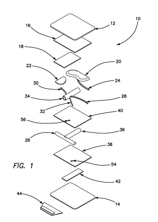
[00013] FIG. 1 is an exploded view of a renal hilum surgical simulation system
in accordance
with various embodiments of the present invention.
[00014] FIG. 2 is a top view of portions of the renal hilum surgical
simulation system in
accordance with various embodiments of the present invention.
[00015] FIG. 3 is a cross-sectional view of a renal vein and artery in
accordance with various
embodiments of the present invention.
4
CA 03101014 2020-11-19
WO 2019/232480 PCT/US2019/035056
[00016] FIG. 4A is a side view of portions of the renal hilum surgical
simulation system in
accordance with various embodiments of the present invention.
[00017] FIG. 4B is a top view of assembled portions of the renal hilum
surgical simulation
system in accordance with various embodiments of the present invention.
[00018] FIG. 5A is a top view of portions of the renal hilum surgical
simulation system in
accordance with various embodiments of the present invention.
[00019] FIG. 5B is a top view of assembled portions of the renal hilum
surgical simulation
system in accordance with various embodiments of the present invention.
[00020] FIG. 6 is a top view of portions of the renal hilum surgical
simulation system in
accordance with various embodiments of the present invention.
[00021] FIG. 7 is a top view of assembled portions of the renal hilum surgical
simulation
system in accordance with various embodiments of the present invention.
[00022] FIG. 8 is a top view of portions of the renal hilum surgical
simulation system in
accordance with various embodiments of the present invention.
[00023] FIG. 9 is a top view of assembled portions of the renal hilum surgical
simulation
system in accordance with various embodiments of the present invention.
[00024] FIG. 10 is a top view of portions of the renal hilum surgical
simulation system in
accordance with various embodiments of the present invention.
[00025] FIG. 11 is a top view of assembled portions of the renal hilum
surgical simulation
system in accordance with various embodiments of the present invention.
[00026] FIG. 12 is a top view of portions of the renal hilum surgical
simulation system in
accordance with various embodiments of the present invention.
[00027] FIG. 13 is an exploded perspective view of the renal hilum surgical
simulation system
in accordance with various embodiments of the present invention.
[00028] FIG. 14 is an exploded side view of the renal hilum surgical
simulation system in
accordance with various embodiments of the present invention.
[00029] FIG. 15 is a top view of portions of the renal hilum surgical
simulation system in
accordance with various embodiments of the present invention.
[00030] FIG. 16 is a top view of the renal hilum surgical simulation system in
accordance with
various embodiments of the present invention.
CA 03101014 2020-11-19
WO 2019/232480 PCT/US2019/035056
[00031] FIG. 17 is a perspective view of the renal hilum surgical simulation
system in
accordance with various embodiments of the present invention.
[00032] FIG. 18 is a perspective view of the renal hilum surgical simulation
system in
accordance with various embodiments of the present invention.
[00033] FIG. 19 is a side view of the renal hilum surgical simulation system
in accordance with
various embodiments of the present invention.
Detailed Description
[00034] In a LDN procedure, renal hilum dissection is one of the more
challenging and high-
risk steps due to the need to mobilize multiple critical structures.
Currently, there is an unmet
need for simulated models or surgical simulation systems that trainees can
practice on to become
proficient at this step of the operation. A simulated model or surgical
simulation system of the
renal hilum would reduce the learning curve by allowing surgical trainees to
practice the required
dissection repeatedly in a low-risk environment. To be effective, the surgical
simulation system
should allow for complete dissection of specific structures within the renal
hilum from a
laparoscopic approach, which includes one or more of the following simulated
anatomy and
landmarks to be present and identifiable in the model or surgical simulation
system: kidney,
adrenal gland, renal vein, renal artery, ureter, gonadal vein, adrenal vein,
lumbar vein, and aorta.
These structures should be anatomically correct and/or be made of materials
that have a similar
simulated tissue reaction encountered in the LDN procedure. In addition, these
structures may be
surrounded by simulated dissectible areolar tissue of appropriate density to
provide realistic
tactile feedback. Practice on the surgical simulation system can promote
identification of the
appropriate anatomy and acquisition of appropriate tissue handling and
dissection skills required
for the procedure.
[00035] The renal hilum surgical simulation system in accordance with various
embodiments
allows a trainee to focus on the skills necessary to practice the most
challenging steps within a
LDN procedure. To provide a realistic procedural training environment, in
various embodiments,
the surgical simulation system is positioned appropriately. To further enhance
the training
environment, the surgical simulation system uses simulated materials to
represent the various
anatomical landmarks as well as materials to simulate areas of dissectible
tissue, which provide
key visual and tactile feedback useful for the training of an LDN procedure.
In order to simulate
6
CA 03101014 2020-11-19
WO 2019/232480 PCT/US2019/035056
the tactile feel of the anatomical structures encountered during the LDN
procedure, in
accordance with various embodiments, specific combinations of materials,
construction, and
design have been chosen for various components found within the surgical
simulation system.
[00036] Turning now to FIG. 1, an exploded perspective view of a renal hilum
surgical
simulation model or system 10 according to various embodiments of the present
invention is
shown. The inner contents (anatomical structures and fibers) of the surgical
simulation system 10
are encapsulated between two layers of silicone, a top penetrable layer 12 and
a bottom
penetrable layer 14, that are adhered together to create a closed pocket.
Inside the pocket, the top
outermost layer is a top fibrous layer 16 constructed of a simulated
dissectible tissue area made
of multiple layers of sheets of polyester fibers, e.g., batting, adhered using
small amount of
silicone or adhesive that the surgeon is to dissect or cut through in order to
uncover and reach the
anatomical structures encountered in the LDN procedure. This dissection area
comprising of the
multiple layers of polyester fibers, such as a half-fibrous layer 18, that are
adhered together, fiber
to fiber, e.g., batting to batting, as well as fiber, e.g., batting, to
anatomical simulated structures
are created to demonstrate the varying densities of the anatomy found in the
body. In accordance
with various embodiments, one or more of the layers are planar and/or stacked
relative to each
other.
[00037] In accordance with various embodiments, a layer of simulated
anatomical landmarks is
provided. The simulated anatomical landmarks in various embodiments comprise a
simulated
kidney 20, adrenal gland 22, ureter 24, and/or aorta 26. While none of these
components should
be dissected or cut during the simulated procedure, these landmarks are
included in the surgical
simulation system 10 to help orientate and/or educate the trainee. For
example, the simulated
ureter 24 should be identified but not touched, and is used as a tool to
navigate to the location of
the gonadal vein 28. Although the simulated landmarks should not be touched or
manipulated by
the trainee, one or more of these simulated anatomical structures includes one
or more visual
characteristics such as size, shape, color and/or any combination thereof, to
simulate anatomy
and/or to pose as indicators to allow for orientation within the simulated
environment. In various
embodiments, one or more of these simulated anatomical structures also
comprises one or more
tactile characteristics, such as texture, resiliency, elasticity and/or any
combination thereof to
further enhance identification of the simulated landmarks and/or as assessment
and/or
educational indicators. For example, in various embodiments, one or more of
the simulated
7
CA 03101014 2020-11-19
WO 2019/232480 PCT/US2019/035056
landmarks holds its shape until cut or excessively manipulated and thus if
inadvertently cut or
otherwise unduly manipulated, the simulated landmarks would reflect this
treatment and thereby
providing an assessment for an evaluator and/or educational indicator for a
trainee.
[00038] During the simulated procedure, the simulated gonadal vein 28, adrenal
vein 30, and
lumbar vein 32 are located and circumferentially dissected, or skeletonized.
During this
skeletonization, the surgeon may pull up on the veins in order to make cuts
and dissect through
the fibers or batting. This is one of the most challenging steps in the
procedure as the veins are
very fragile and will break or tear if incised or if too much force is put on
them. For surgeons to
become comfortable or proficient in these steps of the procedure, they must
understand the force
required to manipulate the veins during dissection without harming them and
thus the necessity
to simulate the fragility of the veins.
[00039] In accordance with various embodiments, the simulated gonadal vein 28,
adrenal vein
30, and/or lumbar vein 32 are made of a silicone or silicone foam that is
molded into thin flat
structures to simulate fragility of the various veins. It should be noted that
gonadal, adrenal, and
lumbar veins found within the human body are hollow cylindrical structures
through which
blood flows and have diameters of 3mm, 4mm, 2mm respectively. As such, in
accordance with
various embodiments, while the simulated gonadal vein 28, adrenal vein 30,
and/or lumbar vein
32 are not exact replicas of anatomy, e.g., in size and/or shape, these
simulated veins are
provided, for example, in size and/or shape along with the choice of material,
e.g., silicone, to
aid in the manufacturing process and replicate the tactile feel of the
corresponding structures.
[00040] In various embodiments, the simulated gonadal, adrenal, and lumbar
veins, 28, 30, 32
includes one or more cuts or notches 50 along their lengths in predetermined
locations as shown
in FIG. 2. These predetermined notches 50 create weak or break points at
specific locations,
allowing the simulated vessels to simulate the fragility of such vessels.
Additionally, in various
embodiments, if excessive force or manipulation is applied to the simulated
veins, the simulated
veins will separate at one or more of the notches 50. A separated or torn
vessel can provide an
assessment and/or educational indication for or regarding the trainee's
specific performance of or
during the simulated procedure. Furthermore, the location of where the tear
occurred, as
indicated at a particular notch or weak point, can further assist in providing
a more detailed
assessment and/or educational indicator of the force or manipulation applied
to the torn
simulated vessel. It should however be noted that simulated vessels with
predetermined notches
8
CA 03101014 2020-11-19
WO 2019/232480 PCT/US2019/035056
may inhibit assessment of the simulated vessels after the procedure is
performed, e.g.,
identifying new versus old or pre-installed notches may prove difficult, and
as such
predetermining the location and/or size of the notches or weak points can
assist in reducing or
eliminating this inhibition.
[00041] In various embodiments, the simulated lumbar vein within the surgical
simulation
system is under tension. The simulated lumbar vein, in various embodiments, is
pulled taut and
attached to the back of the model or surgical simulation system, putting it on
tension. Placing
the simulated lumbar vein under tension allows the simulated vein or portions
thereof to snap
when nicked or excessively tugged during circumferentially dissection. This
snapping simulates
or represents the fragility of the simulated lumbar vein as the amount of
force used to snap the
simulated vessel is similar to the amount of force to similarly affect a non-
simulated lumbar vein.
[00042] In various embodiments, the surgical simulation system 10 comprises a
simulated renal
vein 34 and a simulated renal artery 36. The simulated renal vein 34 and renal
artery 36 are
separated from the surrounding fibers or batting (i.e. skeletonized) during
the simulated
procedure. The simulated renal vein 34 and renal artery 36 have much larger
diameters
(approximately 1.2 cm and 6 mm, respectively) than that of the simulated
gonadal, adrenal, and
lumbar veins 28, 30, 32 giving them more integrity and/or strength to simulate
the tactile
differences in the simulated renal vein 34 and renal artery 36.
[00043] In accordance with various embodiments, with reference to FIG. 3, the
illustrated
simulated renal artery 36 has a smaller overall diameter but thicker wall
relative to the simulated
renal vein 34 having a larger diameter and thinner wall. In various
embodiments, the simulated
renal artery and vein are made of silicone and, in various embodiments, the
simulated renal
artery comprises a thick layer of silicone providing a thicker wall thickness
of the simulated
vessel. In various embodiments, the layer of silicone is made thicker by
applying multiple thin
layers or coats of wet or dry silicone. As a result of a thicker wall, the
vessel will be harder to
penetrate, i.e., the simulated renal artery is harder to penetrate versus the
simulated renal vein.
The simulated renal vein, in various embodiments, has a thin layer of silicone
to provide a thin
wall thickness. As a result, the vessel, e.g., the simulated renal vein, will
be easier to puncture or
nick.
[00044] Providing a contrast in structural integrity of the renal vein and
renal artery further
provides or enhances the simulation and/or the training and/or assessment
indications as the
9
CA 03101014 2020-11-19
WO 2019/232480 PCT/US2019/035056
tactile force allowed during the simulated procedure to circumferentially
dissect around each of
the structures without puncturing or otherwise unduly disrupting them is
different for each
vessel. In various embodiments, the thinner walls of renal vein 34 are fragile
and/or made with a
thinner layer of material. In contrast, in various embodiments, the simulated
renal artery 36 is
made of a thicker layer or layers of material. Both vessels are made of or
molded from silicone
and/or a similar fragile material that will hold its shape including
conductive material.
[00045] In various embodiments, the simulated renal vein 34 and/or renal
artery 36 are filled
with fluid or the like to further mimic anatomy and/or for assessment or
training indicators. For
example, if either of the vasculature is punctured, fluid may be expelled or
trickle out of the
simulated vessels and thereby provide a visual indication of punctured
vasculature and
potentially indicating further training or decreased proficiency of the
trainee.
[00046] In FIGS. 4A-B, the simulated adrenal vein 30, gonadal vein 28, and
lumbar vein 32 are
adhered or otherwise attached to the simulated renal vein 34 at a renal vein
adhesion area 52 and,
in various embodiments, through adhesion of silicone to silicone. The renal
vein adhesion area
52 is depicted by a rectangular box in FIG. 4B. Even though the adhesion area
is depicted as a
rectangular shape, the adhesion area may be any shape. The attachment area 52
is illustrated or
referred throughout as a guide and as an exemplary way to show where the
components are
adhered or otherwise attached or where adhesive or the like is applied. In
various embodiments,
the simulated gonadal vein 28, adrenal vein 30, and lumbar vein 32, are molded
separately and
are minimally and/or weakly adhered to the renal vein 34 to increase the
fragility of the
simulated veins for, e.g., assessment and/or training, when the simulated
vessel is put on tension
and dissected around. The weak adhesion in various embodiments is achieved by
using a weak
adhesive or similar attachment, such as a silicone with a softer durometer,
and/or removing
connector 33 and attaching the simulated veins 28, 30, 32 directly to the
simulated renal vein 34.
[00047] With reference to FIGS. 5A-B, a second vasculature subassembly is
illustrated in
accordance with various embodiments. As illustrated, the simulated renal
artery 36 is adhered or
otherwise attached to the simulated aorta 26 by a silicone-to-silicone
adhesion and, in various
embodiments, with consistent hard durometer silicone. In various embodiments,
wet silicone is
employed as an adhesive and allowed to cure to solidify the connection. The
aorta adhesion area
52 is depicted by a rectangular box in FIG. 5B. In various embodiments, the
simulated aorta 26
has a semi-cylindrical shape as seen for example in FIG. 14.
CA 03101014 2020-11-19
WO 2019/232480 PCT/US2019/035056
[00048] Turning now to FIG. 6, a back fibrous layer 38 is provided. The back
fibrous layer 38
in various embodiments is made of or includes batting. In various embodiments,
the back
fibrous layer is a rectangular, substantially planar layer of polyfill or
other fibrous material. The
back fibrous layer 38 includes a hole or opening 54 through which the lumbar
vein 32 is passed.
The opening 54 is unique to the surgical simulation system 10 and is not
anatomically correct.
The second vasculature subassembly comprising the renal artery 36 and aorta 26
is adhered to
the back or first fibrous layer 38 using adhesive as shown in FIG. 7. The
adhesion area 52 is
shown to be substantially under the entire second subassembly.
[00049] Turning now to FIG. 8, in accordance with various embodiments, a
second fibrous
layer 40 is adhered to the simulated renal artery 36 and aorta 26 of the
second vasculature
subassembly. The second fibrous layer 40 is made of or includes batting. In
various
embodiments, the second fibrous layer is a rectangular, substantially planar
layer of polyfill or
other fibrous material. The second fibrous layer 40 is also adhered to the
back or first fibrous
layer 38 with an adhesion area 52 indicated by the large rectangle. The second
fibrous layer 40
also contains a hole or aperture 56 extending from the top and through to the
bottom surface of
the second fibrous layer 40. The simulated lumbar vein 32 passes through this
hole 56 and the
hole 54 in the back fibrous layer 38 and, as such, the holes 54, 56 are
aligned when the layers are
stacked such that their perimeters are substantially congruent to fit inside
the pocket. Turning
now to FIG. 9, the first vasculature assembly, comprising the simulated
gonadal vein 28, adrenal
vein 30, lumbar vein 32 and renal vein 34, are adhered to the second fibrous
layer 40 with an
adhesion area 52 being under the renal vein 34, adrenal vein 30 and gonadal
vein 28 as shown in
FIG. 9 with the adhesion area 52 shown by three rectangles. The simulated
lumbar vein 32 is
passed through the holes 56 and 54 in the fibrous layers 40, 38.
[00050] Turning now to FIG. 10, the simulated kidney 20, ureter 24 and adrenal
gland 22 are
connected to the simulated renal vein 34 and adrenal vein 30 and to the second
fibrous layer 40.
The simulated ureter 24 is adhered to or otherwise attached to the back of the
simulated kidney
20. The kidney 20 is adhered to the top end of the simulated renal vein 34 as
well as the second
fibrous layer 40. The simulated ureter 24 is adhered to the second fibrous
layer 40. The simulated
adrenal gland 22 is adhered to the simulated adrenal vein 30 as well as the
second fibrous layer
40. The simulated adrenal gland 22 is not adhered to the simulated kidney 20.
The adhesion areas
11
CA 03101014 2020-11-19
WO 2019/232480 PCT/US2019/035056
52 are demonstrated by the rectangular shapes in FIG. 10 and the non-adhesion
area 58 between
the adrenal gland 22 and the kidney is demonstrated by the ellipse in FIG. 10.
[00051] Turning now to FIG. 11, the half fibrous layer 18 is adhered to the
simulated kidney 20,
the simulated adrenal gland 22, the adrenal vein 30, the renal vein 34, and
the second fibrous
layer 40. The adhesion area 52 is shown by a rectangle substantially
completely underneath the
half fibrous layer 18. The half fibrous layer 18 is provided to simulate a
denser dissectible
areolar tissue found within a patient. In various embodiments, the half
fibrous layer 18 is created
from cutting the larger piece of fibrous material, e.g., batting, in half,
length-wise and pulling
apart the layers of the batting to create a thinner piece to add to the
density of the dissectible
tissue. In accordance with various embodiments, the fibers or fibrous material
encapsulate and
surround one or more or every simulated anatomical structure. The multiple
layers of fibrous
material, e.g., batting, provide varying density of dissectible material in
which a surgeon has to
navigate. As stated previously, the simulated lumbar vein 32 passes through
the holes 54, 56 in
the fibrous layers 38, 40. When the surgical simulation system 10 is flipped
over, back side
facing up, as shown in FIG. 12, the simulated lumbar vein 32 is pulled through
the holes 54, 56
to expose it on the back side.
[00052] With reference to FIGS. 12-13, the surgeon must circumferentially
dissect around the
renal vein 34. In accordance with various embodiments, the contents of the
surgical simulation
system 10 are encapsulated between the top silicone layer 12 and the bottom
silicone layer 14.
The bottom silicone layer 14 of the surgical simulation system 10, in various
embodiments, is
constructed of uncured silicone, which is adhered to the top fibrous layer 16
around the outside
border, creating a pocket upon curing together with all of the components
retained by and located
inside the pocket. Because the bottom silicone layer 14 of silicone is uncured
during
manufacturing of the assembly, the back fibrous layer 38 will also adhere to
the wet silicone. If
the back fibrous layer 38 becomes too saturated with uncured silicone, it can
undesirably start to
adhere the simulated renal artery 36 and aorta 26 to the bottom silicone layer
14, which would
prevent the ability of the surgeon trainee to circumferentially dissect around
the renal artery of
the simulated LDN procedure. To prevent or reduce this undesirable adhesion,
an adhesion
blocker 42 is used to ensure that the simulated renal artery 36 can be
dissected circumferentially
around as shown in FIG. 13 with the dissection area 60 demarked with a
ellipse. The adhesion
blocker 42, in various embodiments, is made of a silicone sheet, molded to the
approximate
12
CA 03101014 2020-11-19
WO 2019/232480 PCT/US2019/035056
thickness of the bottom silicone layer 14, and cut to the size of the renal
artery 36 to prevent any
undesired adhesion. In various embodiments, the adhesion blocker 42 is placed
or used such that
it does not obstruct the lumbar vein 32, since the lumbar vein 32 will
ultimately be adhered to the
back of the surgical simulation system 10, bottom silicone layer 14. The
adhesion blocker 42, in
various embodiments, is adhered to the back fibrous layer 38 shown, for
example, by the
rectangular adhesion area, without excess force applied, so as not to saturate
the fibrous material,
e.g., batting, through and adhere the simulated renal artery 36 or aorta 26.
[00053] With reference to FIG. 14, the simulated lumbar vein 32, in various
embodiments, is
adhered to the simulated renal vein 34 and then passes through the second
fibrous layer 40 and
back fibrous layer 38 and adhered to the bottom silicone layer 14. In
accordance with various
embodiments, the adherence of the lumbar vein 32 to the bottom silicone layer
14 occurs while
the model or surgical simulation system contents are placed on the uncured
bottom silicone layer
14. Upon curing of the bottom silicone layer 14, the contact of the lumbar
vein 32 with the
uncured bottom silicone layer 14 will form the necessary adhesion. In various
other
embodiments, the lumbar vein 32 is adhered to the second fibrous layer 40 and
back fibrous
layer 38 at their respective holes 56, 54.
[00054] In various embodiments, in the surgical simulation system, the layers
are adhered
together by intertwining the surrounding fibrous layers, holding simulated
structures in place
with or without the use of silicone or silicone adhesive.
[00055] In various embodiments, fibers of the fibrous, e.g., batting, layers
are mesh through one
another to create a knit matrix and/or when push through the silicone
components a slight
adhesion of batting to silicone is created. As such, adequate adhesion of
tissue (e.g., batting) to
the organs (e.g., silicone) for a surgeon to dissect through in the simulated
procedure is provided.
Such knit matrix can also avoid or reduce the use of silicone glue layers that
can be difficult to
control for consistency throughout the surgical simulation system or cause
unwanted residues.
[00056] With reference now to FIGS. 15 and 16, in various embodiments, to
ensure
identification of the simulated ureter 24 and gonadal vein 28, the simulated
ureter 24 and gonadal
vein 28 are visible through the border/perimeter 62 of the surgical simulation
system 10. In
accordance with various embodiments, the border/perimeter 62 is formed by the
top silicone
layer 12 adhering together with the bottom silicone layer 14 to form a pocket
64. The simulated
ureter 24 and gonadal vein 28 are visible through the top silicone layer 12 at
the border/perimeter
13
CA 03101014 2020-11-19
WO 2019/232480 PCT/US2019/035056
62 of the surgical simulation system 10. These landmarks pose as an indicator
as to where the
surgeon should start dissection of the surgical simulation system 10. In order
for these
landmarks to be visible through the border 62, the simulated ureter 24 and
gonadal vein 28
extend outwardly past the fibrous layers and into the border, highlighted by
circle 63 in FIGS.
15-16. In various other embodiments, the color and/or opacity of the top
silicone layer 12 is
distinguished with respect to the simulated ureter 24 and gonadal vein 28 to
allow for visibility
of the landmarks through the top silicone layer 12.
[00057] With reference to FIGS. 17-19, in accordance with various embodiments,
the renal
hilum dissection surgical simulation model 10 may include two or more holes
along the border
62 for mounting on a stand 66 having a base 68 with at least two upstanding
posts 70 extending
upwardly from the base 68. The posts 70 are passed through the holes in the
border 62. The stand
66 with the surgical simulation model 10 can then be located inside a cavity
of a laparoscopic
trainer 72 for the procedural practice to begin. The trainer defines a cavity
between a top cover
and a base. The cavity is obscured from direct view by the practitioner and a
scope is inserted
through the top cover to capture a live video feed of the cavity, which is
displayed on a monitor
to the practitioner. The practitioner or trainee inserts various instruments
through the top cover
and performs the simulated procedure on the surgical simulation system 10
inside the cavity. The
stand 66 serves to support the surgical simulation model or system 10 inside
the trainer 72. In
various embodiments, the surgical simulation system 10 contains one or more
holes or apertures
in each of the top two corners of the border 62. These holes interface with
the posts 70. In
various embodiments, the stand 66 includes four posts 70. In various
embodiments, the border 62
is made from elastic silicone material that stretches and returns to its
original shape and the holes
of the border are stretched to fit over the post 70 and then return to a tight
fit to secure the
surgical simulation system 10 into place on the posts 70 of the base 68. The
placement of the
holes on the posts 70, along with the angled position of a flap 44, allow for
the surgical
simulation system 10 to be placed in a variety of angles with respect to the
base 68 that may be
necessary to complete the simulated procedure. In various embodiments, in
order to stabilize the
upper corners of the surgical simulation system 10, clips 74 within the
trainer 72 are used to pull
the surgical simulation system upright and/or hold it in position. In
accordance with various
embodiments, a stand or stable structure and/or similar attachments to the
surgical simulation
14
CA 03101014 2020-11-19
WO 2019/232480 PCT/US2019/035056
system and/or the trainer may hold the surgical simulation system stable in an
angled position for
the simulated surgical procedure.
[00058] In various embodiments, the surgical simulation system includes, is
integrated or is
embedded with a frame that supports, suspends and/or angles the surgical
simulation system and
in various embodiments in order to replicate or simulate the angled position
of a patient. The
surgical simulation system is removably attached to the frame and in various
embodiments, the
frame is removably attached to a surgical trainer. In such embodiments, the
apertures within the
border and/or the additional portion provided by the border may be removed
along with the flap,
the associated attachment and/or the additional portions provided by the
surgical simulation
system providing the flap, attachment and/or border.
[00059] During an LDN procedure, the patient is situated lying down on their
side with a slight
backwards tilt. In order to replicate or simulate the angled position of a
patient, the renal hilum
dissection surgical simulation system 10, according to various embodiments,
incorporates a flap
44 designed to be used as a support stand. Looped side of a hook-and-loop type
fastener 46, such
as VELCRO , is adhered to the flap 44 and configured to mate with the opposite
or hooked side
of the hook-and-loop type fastener 46 located on the bottom floor of the
trainer 72. The flap 44
extends from the bottom side of the surgical simulation system 10 and in
various embodiments,
is constructed a soft and flexible yet durable silicone that allows it to bend
while maintaining its
structural integrity. In various embodiments, the flap 44 is flexible so that
two pieces of the
hook-and-loop type fastener 46 can mate, while creating a bent stand for which
to hold the
surgical simulation system into the desired angle and position within the
laparoscopic trainer 72.
The flap 44 is used in conjunction with or without the stand 66. Attachment of
the flap 44 to the
floor of the trainer may vary and in various embodiments, the hook-and-loop
type fastener may
be replaced with or further include, for example, one or more snaps, magnets,
posts or clips,
and/or may extend through, attach to or be adhered to an intermediary
component, e.g., an
extension of base 68, between the attachment/surgical simulation system and
the floor of the
trainer. The attachment of the surgical simulation system allows the surgical
simulation system
to be removable and thus eases replace-ability, repositioning or reorientation
of the surgical
simulation system. Such attachment or positioning of the various portions of
the surgical
simulation system relative to the trainer ensures that the orientation or
angled position of the
surgical simulation system replicates the orientation or position of the
patient and in various
CA 03101014 2020-11-19
WO 2019/232480 PCT/US2019/035056
embodiments ensures the tactile feedback, flexibility or other features
provided by the surgical
simulation system are not sacrificed and/or the simulated LDN procedure
compromised.
[00060] In various embodiments, other variations to the surgical simulation
system 10 may
include alteration of the anatomical structures inside the pocket to include
abnormal, diseased, or
varying anatomy. Such anatomy could include the right renal hilum or the
inclusion of additional
lumbar veins and/or tumors. In other embodiments, the surgical simulation
system 10 is dipped
or soaked in water or other liquid to better represent the environment of a
patient. For example,
when the fibrous or batting layers become saturated with liquid they tend to
become denser and
more adhered. This allows, in various embodiments, for more applicable and
accurate
representation of the difficulty of the LDN procedure. Instead of a liquid
such as water, the
pocket 64 could also be filled with a gel like substance.
[00061] In various embodiments, the arrangement and/or composition of the
various portions
and components are provided to vary the difficulty of the surgical simulation
system and thereby
vary the simulated surgical procedure to enhance surgical training and
surgical skill assessment.
Such examples are described throughout the description and provided in the
claims that may
seem arbitrary but again are included or excluded to vary and adjust the
difficulty the surgical
simulation system to enhance surgical training and skill assessment. Some of
these examples
can include varying fibrous layer densities, exaggerating or underplaying
simulated renal
vasculature and/or organ shapes, dimensions and/or tactile response,
saturating fibrous layers
with liquid, creating simulated vasculature paths, e.g., a simulated renal
vasculature threaded or
extended through at least one opening in one or more or different fibrous
layers, and/or varying
the coloring and/or composition of the simulated renal vasculature, organs
and/or surrounding
structures.
[00062] In various embodiments, both sides or layers of the surgical
simulation system are
penetrable to ensure or further assess surgical skill such that if mishandling
or manipulation of
the simulated tissue, e.g., too much force is used, a noticeable puncture or
opening in the
opposing side of the surgical simulation system would be visible. Likewise,
the thickness or
distance between the layers are minimal, e.g., a fraction of the length or
width of the surgical
simulation system or the pocket contained therein, to further test or enhance
the assessment of
the surgical skill or effective operation of the simulated surgical procedure.
16
CA 03101014 2020-11-19
WO 2019/232480 PCT/US2019/035056
[00063] In various embodiments, the surgical simulation system is so confined
to limit the
working space available to simulate the surgical procedures. Likewise, the
size of the pocket, for
example, can be modified to further limit the operational space and thereby
increase the
difficulties of the simulated surgical procedure. Additionally, the number
and/or size of the
components and combinations thereof are further limited to enhance portability
of the surgical
simulation system, operation within a trainer, e.g., a portable laparoscopic
trainer and/or further
focus the surgical trainee on the specific simulated procedure. Similarly,
omitted features or
reduction of sizes or shapes are provided to enhance the surgical simulation
system, e.g., increase
difficulties or focus on the specific simulated surgical procedure, even
though such differences or
changes may not be anatomically correct. In various embodiments, the surgical
simulation
system includes at least one simulated renal vasculature, e.g., renal vein,
renal artery, and/or the
like and/or other vasculature/vessels provided herein, and/or at least one
simulated renal organ,
e.g., adrenal gland, kidney and/or the like and/or other organs/glands
provided herein.
[00064] The above description is provided to enable any person skilled in the
art to make and
use the surgical simulation system or systems and perform the methods
described herein and sets
forth the best modes contemplated by the inventors of carrying out their
inventions. Various
modifications, however, will remain apparent to those skilled in the art. It
is contemplated that
these modifications are within the scope of the present disclosure. Different
embodiments or
aspects of such embodiments may be shown in various figures and described
throughout the
specification. However, it should be noted that although shown or described
separately each
embodiment and aspects thereof may be combined with one or more of the other
embodiments
and aspects thereof unless expressly stated otherwise. It is merely for easing
readability of the
specification that each combination is not expressly set forth.
[00065] Although the present invention has been described in certain specific
aspects, many
additional modifications and variations would be apparent to those skilled in
the art. It is
therefore to be understood that the present invention may be practiced
otherwise than specifically
described, including various changes in the size, shape and materials, without
departing from the
scope and spirit of the present invention. Thus, embodiments of the present
invention should be
considered in all respects as illustrative and not restrictive.
17