Note: Descriptions are shown in the official language in which they were submitted.
CA 03101624 2020-11-25
WO 2019/232397 PCT/US2019/034942
SOFTWARE FOR MICROFLUIDIC SYSTEMS INTERFACING WITH MASS
SPECTROMETRY
CROSS-REFERENCE
[0001] This application claims the benefit of U.S. Provisional Application No.
62/678,265, filed
on May 31, 2018, and U.S. Provisional Application No. 62/684,090, filed on
June 12, 2018, both
of which applications are incorporated herein by reference.
BACKGROUND
[0002] The present disclosure relates to the field of chemical analysis, and
in particular, to the
separation of analytes in a mixture and their subsequent analysis by mass
spectrometry (MS).
Separation of analyte components from a more complex analyte mixture on the
basis of an
inherent quality of the analytes, and providing sets of fractions that are
enriched for states of that
quality, is a key part of analytical chemistry. Simplifying complex mixtures
in this manner
reduces the complexity of downstream analysis. However, complications can
arise when
attempting to interface known enrichment methods and/or devices with
analytical equipment
and/or techniques.
[0003] A variety of methods have been used, for example, to interface protein
sample
preparation techniques with downstream detection systems such as mass
spectrometers. A
common method is to prepare samples using liquid chromatography and collect
fractions for
mass spectrometry (LC-MS). This has the disadvantage of requiring protein
samples to be
digested into peptide fragments, leading to a large number of sample fractions
which must be
analyzed and complex data reconstruction post-run. While certain forms of
liquid
chromatography can be coupled to a mass spectrometer, for example peptide map
reversed-phase
chromatography, these known techniques are restricted to using peptide
fragments, rather than
intact proteins, which limits their utility.
[0004] Another method to introduce samples into a mass spectrometer is
electrospray ionization
(ESI). In ESI, small droplets of sample and solution are emitted from a distal
end of a capillary
or microfluidic device comprising an electrospray feature, such as an emitter
tip or orifice, by the
application of an electric field between the capillary tip or emitter tip and
the mass spectrometer
source plate. The droplet stretches and expands in this induced electric field
to form a cone
shaped emission (i.e., a "Taylor cone") which comprises increasingly small
droplets that
evaporate and produce the gas phase ions that are introduced into the mass
spectrometer for
further separation and detection. Typically, emitter tips are formed from a
capillary, which
provides a convenient droplet volume for ESI. Capillaries, however, are
limited to a linear flow
- 1 -
CA 03101624 2020-11-25
WO 2019/232397 PCT/US2019/034942
path that does not allow for multi-step sample processing. ESI also depends on
the voltage at the
ESI tip to remain constant throughout the analysis, which can be a challenge
in many assays,
where internal fluid resistances can change over time, altering the voltage
drop in different parts
of the electrical circuit and thereby changing the voltage at the ESI tip.
[0005] Other work has been pursued with microfluidic devices. Microfluidic
devices may be
produced by various known techniques and provide fluidic channels of defined
dimensions that
can make up a channel network designed to perform different fluid
manipulations. These devices
offer an additional level of control and complexity than capillaries, making
them a better choice
for sample prep. However, like capillaries, these tools often provide limited
characterization of
separated analyte fractions prior to introduction to a mass spectrometer, if
any. Also, systems
with capillaries or microfluidic devices generally provide no tools for
calibrating the system to
reestablish a Taylor cone during operation.
[0006] Methods, devices, systems, and software for improving the quality of
electrospray
ionization mass spectrometry (ESI-MS) data are described, as are methods,
devices, systems, and
software for achieving more quantitative characterization of and improved
correlation between
chemical separation data and mass spectrometry data.
SUMMARY
[0007] Disclosed herein are methods for maintaining an electrospray ionization
(ESI) tip at a
constant voltage relative to ground while performing a separation reaction,
the method
comprising: a) applying a first voltage to a proximal end of a separation
channel, wherein a distal
end of the separation channel is in fluid and electrical communication with
the ESI tip; b)
applying a second voltage to a proximal end of an auxiliary fluid channel,
wherein a distal end of
the auxiliary fluid channel is in fluid and electrical communication with the
distal end of the
separation channel; c) performing the separation reaction to separate a
mixture of analytes,
wherein the separation reaction takes place within the separation channel; and
d) monitoring a
change in resistance of the separation channel or a change in voltage at the
ESI tip in a feedback
loop that adjusts the first and second voltages to maintain a constant voltage
drop across the
separation channel and a constant voltage at the ESI tip. In some embodiments,
the separation
channel is a lumen of a capillary. In some embodiments, the capillary
comprises a microvial
spray tip. In some embodiments, the separation channel is a fluid channel
within a microfluidic
device. In some embodiments, the separation reaction comprises an isoelectric
focusing
reaction. In some embodiments, the separation reaction comprises an
electrophoretic separation
reaction. In some embodiments, the first voltage is applied at a cathode and
the second voltage
is applied at an anode. In some embodiments, the voltage at the ESI tip is
held at ground. In
- 2 -
CA 03101624 2020-11-25
WO 2019/232397 PCT/US2019/034942
some embodiments, the voltage at the ESI tip is held at the second voltage. In
some
embodiments, the adjustment to the first and second voltages comprises
subtracting a transient
voltage change measured at the ESI tip from the first voltage and second
voltage. In some
embodiments, the voltage at the ESI tip is measured using a power supply that
provides the first
or second voltage. In some embodiments, the feedback loop operates at a
frequency of at least
0.1 Hz. In some embodiments, the feedback loop operates at a frequency of at
least 10 Hz. In
some embodiments, the feedback loop maintains the voltage at the ESI tip to
within 10% of a
pre-set value. In some embodiments, the feedback loop maintains the voltage at
the ESI tip to
within 1% of a pre-set value. In some embodiments, the feedback loop
maintains the voltage
drop across the separation channel to within 10% of a pre-set value. In some
embodiments, the
feedback loop maintains the voltage drop across the separation channel to
within 1% of a pre-
set value.
[0008] Also disclosed herein are methods comprising: a) providing a sample
comprising a
mixture of two or more analytes; b) performing a separation within a fluid
channel containing the
sample to resolve individual analyte peaks from the mixture of two or more
analytes; c)
calculating a velocity of an analyte peak upon mobilization of the fluid
channel's contents
towards a fluid channel exit; and d) using the velocity of the analyte peak to
determine a time at
which the analyte peak reaches the fluid channel exit. In some embodiments,
the fluid channel is
a lumen of a capillary. In some embodiments, the fluid channel is part of a
microfluidic device.
In some embodiments, the separation is based on isoelectric focusing (IEF),
capillary zone
electrophoresis (CZE), capillary gel electrophoresis (CGE), capillary
isotachophoresis (CITP), or
micellar electrokinetic chromatography (MEKC). In some embodiments, the
velocity of the
analyte peak is calculated from a time interval required for the analyte peak
to move from a first
position to a second position. In some embodiments, the first position, second
position, and time
interval are determined from a series of two or more images of the fluid
channel. In some
embodiments, the series of two or more images comprise ultraviolet light
absorbance images,
visible light absorbance images, or fluorescence images. In some embodiments,
the fluid
channel exit comprises an electrospray interface with a mass spectrometer. In
some
embodiments, the time at which the analyte peak reaches the fluid channel exit
is used to
correlate mass spectrometer data with the analyte peak. In some embodiments,
the mobilization
of the fluid channel's contents comprises the use of an electroosmotic
mobilization technique, a
chemical mobilization technique, a hydrodynamic mobilization technique, or any
combination
thereof. In some embodiments, the two or more analytes comprise proteins,
protein-drug
conjugates, peptides, nucleic acid molecules, carbohydrate molecules, lipid
molecules,
- 3 -
CA 03101624 2020-11-25
WO 2019/232397 PCT/US2019/034942
metabolite molecules, small organic compounds, or any combination thereof. In
some
embodiments, a comparison of mass spectrometer data collected for samples of a
biologic drug
candidate and a reference drug is used to make a determination of
biosimilarity. In some
embodiments, the velocity of the analyte peak is used in a feedback loop to
adjust a control
parameter for the separation or mobilization of the analyte peaks. In some
embodiments, the
control parameter is a voltage. In some embodiments, the feedback loop
operates at a frequency
of at least 0.1 Hz.
[0009] Disclosed herein are methods comprising: a) providing a sample
comprising a mixture of
two or more analytes; b) performing a separation within a fluid channel
containing the sample to
resolve individual analyte peaks from the mixture of two or more analytes; and
c) collecting
mass spectrometer data for the two or more individual analyte peaks emitted
from the fluid
channel via an electrospray interface with a mass spectrometer; wherein a data
collection mode
for the mass spectrometer is alternated between a high mass scan and a low
mass scan. In
some embodiments, the mass spectrometer is switched between the high mass scan
and low mass
scan data collection modes at a frequency of at least 0.5 Hz. In some
embodiments, the fluid
channel is a lumen of a capillary. In some embodiments, the fluid channel is
part of a
microfluidic device. In some embodiments, the separation is based on
isoelectric focusing (IEF),
capillary zone electrophoresis (CZE), capillary gel electrophoresis (CGE),
capillary
isotachophoresis (CITP), or micellar electrokinetic chromatography (MEKC). In
some
embodiments, the high mass scan captures mass spectral data for biological
macromolecules. In
some embodiments, the biological macromolecules comprise proteins, protein-
drug conjugates,
nucleic acid molecules, reduced proteins, fusion proteins, protein complexes,
or any combination
thereof. In some embodiments, the m/z ratio for the high mass scan ranges from
1500 to 6000.
In some embodiments, the low mass scan captures mass spectral data for
solution-phase
ampholytes used in performing an isoelectric focusing separation. In some
embodiments, the
m/z ratio for the low mass scan ranges from 150 to 1500. In some embodiments,
the mass
spectra of one or more solution-phase ampholytes are used to calibrate the
isoelectric points (pIs)
for biological macromolecules identified in the high mass scans.
[0010] Disclosed herein are methods comprising: a) performing a separation
within a fluid
channel containing a sample, wherein the sample comprises a mixture of two or
more analytes,
and wherein the separation resolves individual analyte peaks from the mixture
of two or more
analytes; b) mobilizing the fluid channel's contents towards a fluid channel
exit, wherein the
fluid channel exit comprises an electrospray interface with a mass
spectrometer; and c)
simultaneously or alternately imaging: (i) at least a portion of the fluid
channel to monitor a
- 4 -
CA 03101624 2020-11-25
WO 2019/232397 PCT/US2019/034942
position of an analyte peak during (a) and (b), and (ii) a Taylor cone
existing between the fluid
channel exit and an inlet to the mass spectrometer to monitor electrospray
performance. In some
embodiments, the positions of the analyte peak in two or more images of at
least a portion of the
fluid channel are used to calculate velocity for the analyte peak. In some
embodiments, the
velocity of the analyte peak is used to determine a time at which the analyte
peak will reach the
fluid channel exit. In some embodiments, the time at which the analyte peak
reaches the fluid
channel exit is used to correlate mass spectrometer data with the analyte
peak. In some
embodiments, data derived from the imaging of the Taylor cone is used in a
feedback loop to
adjust electrospray performance. In some embodiments, the feedback loop
operates at a
frequency of at least 0.1 Hz. In some embodiments, the fluid channel is a
lumen of a capillary.
In some embodiments, the fluid channel is part of a microfluidic device. In
some embodiments,
the separation is based on isoelectric focusing (IEF), capillary zone
electrophoresis (CZE),
capillary gel electrophoresis (CGE), capillary isotachophoresis (CITP), or
micellar electrokinetic
chromatography (MEKC). In some embodiments, the imaging comprises ultraviolet
light
absorbance imaging, visible light absorbance imaging, or fluorescence imaging.
In some
embodiments, the mobilization of the fluid channel's contents comprises the
use of an
electroosmotic mobilization technique, a chemical mobilization technique, a
hydrodynamic
mobilization technique, or any combination thereof. In some embodiments, the
two or more
analytes comprise proteins, protein-drug conjugates, peptides, nucleic acid
molecules,
carbohydrate molecules, lipid molecules, metabolite molecules, small organic
compounds, or
any combination thereof
[0011] Disclosed herein are computer-implemented methods for maintaining an
electrospray
ionization (ESI) tip at a constant voltage relative to ground while performing
a separation
reaction, the method comprising: a) receiving, using a processor, a first
measurement of a
voltage at the ESI tip, wherein a distal end of the separation channel is in
fluid and electrical
communication with the ESI tip; b) receiving, using the processor, a second
measurement of the
voltage at the ESI tip; c) comparing the second measurement to the first
measurement using the
processor, wherein if the second measurement differs from the first
measurement, the processor
causes a voltage at a proximal end of the separation channel and a voltage at
a proximal end of
an auxiliary fluid channel comprising a distal end that is in fluid and
electrical communication
with the distal end of the separation channel, to be adjusted such that the
voltage at the ESI tip is
returned to the first measurement value; and d) repeating steps (a) through
(c) at a specified
frequency. In some embodiments, the separation channel comprises a lumen of a
capillary or a
fluid channel within a microfluidic device. In some embodiments, the
separation reaction
- 5 -
CA 03101624 2020-11-25
WO 2019/232397 PCT/US2019/034942
comprises an isoelectric focusing reaction. In some embodiments, the
separation reaction
comprises an electrophoretic separation reaction. In some embodiments, the
voltage at the ESI
tip is held at ground. In some embodiments, the specified frequency is at
least 1 Hz. In some
embodiments, the voltage at the ESI tip is maintained to within 5% of a
specified value.
[0012] Also disclosed herein are computer-implemented methods comprising: a)
receiving,
using a processor, image data comprising two or more images acquired using a
detector
configured to image all or a portion of a separation channel in a capillary or
a microfluidic
device; b) processing the image data using the same or a different processor
to determine a
position of an analyte peak within the separation channel in the two or more
images; c)
calculating, using the same or a different processor, a velocity of the
analyte peak based on the
position of the analyte peak in the two or more images and a known time
interval between
acquisition of the two or more images; and d) determining, using the same or a
different
processor, a time at which the analyte peak will reach a separation channel
outlet. In some
embodiments, a separation reaction performed within the separation channel
comprises
isoelectric focusing (IEF), capillary zone electrophoresis (CZE), capillary
gel electrophoresis
(CGE), capillary isotachophoresis (CITP), or micellar electrokinetic
chromatography (MEKC).
In some embodiments, the two or more images comprise ultraviolet light
absorbance images,
visible light absorbance images, or fluorescence images. In some embodiments,
the separation
channel outlet is in fluid communication with or comprises an electrospray
interface with a mass
spectrometer. In some embodiments, the time at which the analyte peak reaches
the separation
channel outlet is used to correlate mass spectrometer data with the analyte
peak. In some
embodiments, the analyte is separated from a mixture and comprises a protein,
a protein-drug
conjugate, a peptide, a nucleic acid molecule, a carbohydrate molecule, a
lipid molecule, a
metabolite molecule, or a small organic compound. In some embodiments, a
comparison of
mass spectrometer data collected for samples of a biologic drug candidate and
a reference drug is
used to make a determination of biosimilarity. In some embodiments, the
velocity of the analyte
peak is used in a feedback loop to adjust a control parameter for a separation
reaction performed
in the separation channel. In some embodiments, the control parameter is a
voltage. In some
embodiments, the feedback loop operates at a frequency of at least 0.1 Hz.
INCORPORATION BY REFERENCE
[0013] All publications, patents, and patent applications mentioned in this
specification are
herein incorporated by reference in their entirety to the same extent as if
each individual
publication, patent, or patent application was specifically and individually
indicated to be
- 6 -
CA 03101624 2020-11-25
WO 2019/232397 PCT/US2019/034942
incorporated by reference in its entirety. In the event of a conflict between
a term herein and a
term in an incorporated reference, the term herein controls.
BRIEF DESCRIPTION OF THE DRAWINGS
[0014] The novel features of the invention are set forth with particularity in
the appended claims.
A better understanding of the features and advantages of the present invention
will be obtained
by reference to the following detailed description that sets forth
illustrative embodiments, in
which the principles of the invention are utilized, and the accompanying
drawings of which:
[0015] FIG. 1 provides a schematic illustration of a device for isoelectric
focusing (IEF) and
electrospray ionization (ESI) of an automatically loaded sample, according to
one embodiment
of the present disclosure.
[0016] FIG. 2 provides an example flowchart of a computer-implemented method
for
calculating isoelectric points for separated analyte bands.
[0017] FIG. 3 provides another example flowchart for a computer-implemented
method for
determining a velocity for one or more separated analyte bands and calculating
an exit time.
[0018] FIG. 4 provides another example flowchart for a computer-implemented
method for
implementing imaging-based feedback and control of one or more operating
parameters for an
ESI-MS analysis system.
[0019] FIG. 5 provides a schematic block diagram of the hardware components
for one
embodiment of the disclosed systems.
[0020] FIG. 6 provides a schematic block diagram of the software components
for one
embodiment of the disclosed systems.
[0021] FIGS. 7A-B illustrate a microfluidic device for use in some embodiments
of the
invention. FIG. 7A provides a schematic illustration of a fluid channel
network of an exemplary
microfluidic device. FIG. 7B provides a computer aided design (CAD) drawing of
an assembled
microfluidic device. The fluid channel layer shown in FIG. 7A is sandwiched
between two clear
layers to seal the fluid channels.
[0022] FIG. 8 provides an image of the Taylor cone and electrospray ionization
(ESI) plume
during mobilization of a separated sample.
[0023] FIGS. 9A-F provide non-limiting examples of data for mobilization of a
sample
following separation of analytes in a mixture of analytes using isoelectric
focusing. FIG. 9A
shows an absorbance trace at t = 0 minutes (completion of isoelectric
focusing). FIG. 9B shows
an absorbance trace at t = 1 minute. FIG. 9C shows an absorbance trace at t =
2 minutes. FIG.
9D shows an absorbance trace at t = 3 minutes. FIG. 9E shows an absorbance
trace at t = 4
minutes. FIG. 9F shows an absorbance trace at t = 5 minutes.
- 7 -
CA 03101624 2020-11-25
WO 2019/232397 PCT/US2019/034942
[0024] FIGS. 10A-B provide representative circuit diagrams for a microfluidic
device designed
to perform isoelectric focusing to separate analytes and subsequent
mobilization of the separated
analyte mixture. FIG. 10A provides a representative circuit diagram for the
microfluidic device
shown in FIG. 7A during isoelectric focusing, in the case where the ESI tip
will be held at or
close to ground. FIG. 10B shows a representative circuit diagram for the
microfluidic device
shown in FIG. 7A during chemical mobilization of a separated analyte mixture.
The resistance
of channel 114 (shown in FIG. 7A) is assumed to be negligible in this example.
[0025] FIGS. 11A-B provide representative data for mobilization while keeping
the ESI tip at
OV. FIG. 11A shows a plot of voltage as a function of time. FIG. 11B shows a
plot of current as
a function of time.
[0026] FIG. 12 provides an example flowchart of voltage feedback loop where
the ESI tip is
held at +3000V.
[0027] FIGS. 13A-E provide examples of representative circuit diagrams for
microfluidic
devices of the present disclosure. FIG. 13A provides a representative circuit
diagram for the
microfluidic device shown in FIG. 7A during chemical mobilization, where the
ESI tip will be
held at a positive voltage, using an additional resistor to sink current to
ground. FIG. 13B shows
a representative circuit diagram for the microfluidic device shown in FIG. 7A
during chemical
mobilization, where the ESI tip will be held at a positive voltage using an
additional resistor to
sink current to a third power supply. FIG. 13C shows a representative circuit
diagram for the
microfluidic device shown in FIG. 7A during chemical mobilization, where the
ESI tip will be
held at a positive voltage using a field-effect transistor (FET) to sink
current. FIG. 13D shows a
representative circuit diagram for the microfluidic device shown in FIG. 7A
during chemical
mobilization, where the ESI tip will be held at a positive voltage using a
bipolar junction
transistor (BJT) to sink current. FIG. 13E provides a representative circuit
diagram for the
microfluidic device shown in FIG 7A during chemical mobilization of a
separated analyte
mixture, where the ESI tip will be held at or close to ground.
[0028] FIG. 14A provides a representative diagram of a capillary junction
sprayer. FIG. 14B
shows the representative resistor circuit diagram for the capillary junction
sprayer diagram in
FIG. 14A.
[0029] FIG. 15 provides an exemplary flowchart of a computer-controlled
voltage feedback
loop where the ESI tip is held at OV.
[0030] FIGS. 16A-E provide examples of analyte separation data and the
corresponding mass
spectrometry data for separated analyte species. FIG. 16A shows an
electropherogram of a
separated analyte mixture. FIG. 16B shows a mass spectrum for an acidic peak
of the separated
- 8 -
CA 03101624 2020-11-25
WO 2019/232397 PCT/US2019/034942
species. FIG. 16C shows a mass spectrum of the main peak present in the
electropherogram of
FIG. 16A. FIG. 16D and FIG. 16E show mass spectra of two basic peaks from the
electropherogram shown in FIG. 16A.
DETAILED DESCRIPTION
[0031] Some embodiments described herein relate to innovative software and
systems for
analyzing data from and directing the operation of capillary- and microfluidic-
based separation
systems integrated with mass spectrometric detection. In some embodiments,
analytes are
imaged during separation in capillaries or on microfluidic devices, and
molecular weight or
mass-to-charge ratio is measured in a mass spectrometer post separation. The
disclosed methods,
devices, systems, and software provide for more accurate characterization of
separated analyte
peaks, and for achieving improved correlation between chemical separation data
and mass
spectrometry (MS) data. Also disclosed are methods, devices, systems, and
software for
improving the quality of electrospray ionization mass spectrometry (ESI-MS)
data. The
disclosed methods, devices, systems, and software have potential application
in a variety of
fields including, but not limited to, proteomics research, drug discovery and
development, and
clinical diagnostics. For example, in some embodiments, the disclosed methods,
devices,
systems, and software may be utilized for the characterization of biologic and
biosimilar
pharmaceuticals during development and/or manufacturing, as will be discussed
in more detail
below. Biologics and biosimilars are a class of drugs which include, for
example, recombinant
proteins, antibodies, live virus vaccines, human plasma-derived proteins, cell-
based medicines,
naturally-sourced proteins, antibody-drug conjugates, protein-drug conjugates
and other protein
drugs.
[0032] Microfluidic devices designed to perform any of a variety of chemical
separation
techniques and that also comprise an electrospray ionization interface for
performing
downstream mass spectrometry-based analysis are described. In a preferred
embodiment, the
disclosed devices are designed to perform isoelectric focusing of proteins or
other biological
macromolecules. In another preferred embodiment, the disclosed devices are
designed to be used
with imaging techniques. Devices and methods for integration of imaged
microfluidic
separations with mass spectrometry have been previously described in, for
example, published
PCT Patent Application Publication No. WO 2017/095813, and U.S. Patent
Application
Publication No. US 2017/0176386, which are hereby incorporated by reference
for all purposes.
These applications describe, among other things, systems for performing imaged
separation in
conjunction with MS analysis. Such microfluidic systems represent a
significant advancement in
biologics characterization. However, in order for such a system to provide
maximal benefit it
- 9 -
CA 03101624 2020-11-25
WO 2019/232397 PCT/US2019/034942
would be beneficial to have innovative software and systems to aid in the
operation of these
systems and downstream integration of imaged and MS data, as is disclosed
herein.
[0033] Accordingly, in a preferred embodiment, the disclosed microfluidic
devices may be used
in combination with imaging techniques to, for example, make an accurate
determination of the
isoelectric point (pI) for one or more analytes that have been isoelectrically-
separated from a
mixture of analytes in a separation channel to form a series of enriched
fractions comprising
substantially pure individual analyte components (also referred to herein as
"peaks" or "bands").
Imaging all or a portion of a separation channel allows one to determine the
location of two or
more pI standards (or pI markers) that have been injected along with the
sample to be separated,
and thus allows one to calculate a more accurate pI for each of the separated
analyte peaks by
extrapolation to determine the local pH. In some embodiments, the imaging of
the analyte
mixture within a separation channel is performed while the separation is being
performed and,
optionally, a determination of isoelectric points for one or more of the
analytes that are being
separated is performed and iteratively updated while the separation is being
performed. In some
embodiments, the imaging-based determination of isoelectric points for one or
more analytes that
have been isoelectrically-focused is performed after the separation is
complete. In some
embodiments, the imaging-based determination of isoelectric points for one or
more analytes that
have been isoelectrically-focused is performed after the separation is
complete, and before the
separated analyte mixture has been mobilized towards an electrospray tip. In
some
embodiments, the imaging-based methods disclosed herein may be used with
capillary-based
ESI-MS systems rather than microfluidic device-based ESI-MS systems. In some
embodiments,
the determination of isoelectric points for one or more analyte peaks may be
performed by a
computer-implemented method.
[0034] In another preferred embodiment, the disclosed microfluidic devices may
be used in
combination with imaging techniques to image separated analyte peaks after
mobilization of the
separated analyte mixture, i.e., as the peaks move out of the separation
channel and towards an
electrospray tip. In some embodiments, the imaged mobilization step is the
same step as the
imaged separation step, such as when implementing a separation step comprising
capillary gel
electrophoresis, capillary zone electrophoresis, isotachophoresis, capillary
electrokinetic
chromatography, micellar electrokinetic chromatography, flow counterbalanced
capillary
electrophoresis, or any other separation technique that separates components
of an analyte
mixture by differential velocity. In some embodiments, the imaged mobilization
step will be
analyzed to correlate enriched fractions in the imaged separation with mass
spectrum. Imaging
of the mobilized analyte peaks may be utilized to, for example, determine a
velocity for one or
- 10 -
CA 03101624 2020-11-25
WO 2019/232397 PCT/US2019/034942
more analyte peaks based on their positions in a series of mobilization
images, which may then
be used to determine the time point at which the analyte peak(s) will exit the
separation channel,
or be emitted by the electrospray tip, and may thus be used to correlate mass
spectrometer data
with specific analyte peaks. In some cases, the velocity of the analyte
peak(s) is calculated from
the time interval required for the analyte peak to move a certain displacement
value (e.g., from a
first position to a second position). In some embodiments, imaging of the
mobilized analyte
peaks may allow direct monitoring of the peak(s) as they travel through a
fluid channel and are
emitted by the electrospray tip, and may thus be used to directly correlate
mass spectrometer data
with specific analyte peaks. In some embodiments, the imaging-based methods
disclosed herein
may be used with capillary-based ESI-MS systems rather than microfluidic
device-based ESI-
MS systems. In some embodiments, the determination of velocities for one or
more analyte
peaks, their actual or predicted separation channel exit times, and/or their
electrospray emission
times, may be performed by a computer-implemented method.
[0035] In some embodiments, the mobilization of separated analyte peaks may be
initiated by a
change in electric field or flow parameters in a microfluidic device. In some
embodiments, one
or more electrodes connecting a power supply to the microfluidic device will
be connected or
disconnected to initiate mobilization through a computer-implemented method.
In some
embodiments, the Taylor cone formed at the electrospray tip may be imaged
during the
mobilization step. In some embodiments, computer implemented image analysis
may be used to
identify a stable electrospray operating condition. In some embodiments, the
image analysis may
be performed by an operator. In some embodiments, the image analysis may be
performed using
automated image processing software. In some embodiments, one or more of the
operating
parameters known to affect electrospray performance will be adjusted to regain
a stable
electrospray operating condition. Examples of operating parameters that may be
adjusted
include, but are not limited to, electrophoresis voltage, flow rate, distance
from the electrospray
tip to the MS inlet, MS voltage, and the like. In some embodiments, a computer-
implemented
method may be used to adjust the electrospray parameters.
[0036] In some embodiments, more than one power supply may be used to generate
an
electrophoresis electric field. In some embodiments, two power supplies having
positive polarity
may be used. In some embodiments, one or more power supplies may have negative
polarity. In
some embodiments, the voltage setting on the power supplies may be changed in
unison to
maintain the same voltage gradient in a separation channel for an
electrophoretic separation. In
some embodiments, the voltage settings on the power supplies may be changed in
order to
maintain a constant voltage at an electrospray tip. In some embodiments, the
multiple power
- 11 -
CA 03101624 2020-11-25
WO 2019/232397 PCT/US2019/034942
supplies may be different channels in a single multi-channel power supply. In
some
embodiments isoelectric focusing may be performed in the separation channel,
and the resistance
in the channel may increase over time. In some embodiments chemical
mobilization may be
performed in the separation channel, and the resistance in the channel may
decrease over time. In
some embodiments, pressure driven mobilization may be performed, and the
resistance in the
channel may change over time as new reagent is pushed into the channel. In
some embodiments,
the electrospray tip may be kept at ground. In some embodiments, the
electrospray tip may be
kept at a specific voltage relative to the mass spectrometer. In some
embodiments, the
electrospray tip may be kept at a specific voltage relative to ground. In some
embodiments, a
computer-implemented method may adjust voltages to maintain a constant
electric field strength
in the separation channel (or a constant voltage drop between anode and
cathode), and a constant
voltage at the electrospray tip. In some embodiments, the voltage at the tip
may be measured
using a volt-meter. In some embodiments, the voltage at the tip may be
measured using an
electrode positioned at or inside the tip. In some embodiments, an additional
power supply may
be set to OpA using current control and used as a volt-meter to read the tip
voltage. In some
embodiments, a computer implemented method will read the value of the voltage
at the tip, and
adjust voltages to maintain a constant electric field strength in the
separation channel (or a
constant voltage drop between anode and cathode), and maintain a constant
voltage at the tip. In
some embodiments, a computer implemented method will calculate the voltage at
the ESI tip
based on current flow through the separation electric field circuit. In some
embodiments, the
voltage drop across the separation channel will be adjusted such that a
constant power or a
maximum power is applied in the separation channel, where the power applied in
the separation
channel is calculated as:
power = voltage across separation channel x current in separation channel
where the current can be measured constantly or periodically during separation
and the current
measurements can be used to adjust the voltage across the separation channel.
This method of
controlling the power in the separation channel may be useful for managing
temperature effects
in the separation channel.
[0037] In some embodiments, the separation path will be a length of linear
coated or uncoated
capillary, tube or line with the inlet inserted in a vial containing an acidic
anolyte and positive
electrode or basic catholyte and negative electrode. In some embodiments, the
outlet of the
separation path will be inserted into junction sprayer. In some embodiments,
the junction
sprayer houses both a Tee for a secondary tube, line, or capillary that can
introduce another
- 12 -
CA 03101624 2020-11-25
WO 2019/232397 PCT/US2019/034942
conductive make-up solution to the capillary outlet providing for a liquid to
liquid electrical
contact and liquid flow to support electrospray and transport analytes
emerging from the
separation channel to the tip for introduction into a mass spectrometer by
electrospray ionization.
In some embodiments, the system may be configured with anolyte and positive
electrode at the
separation path inlet and the junction or distal portion of the separation
path may be loaded with
catholyte just prior to focusing. After focusing is completed, a mobilization
agent with
competing anion may be introduced into the junction by either hydrodynamic or
electroosmotic
force. In some embodiments, the separation path inlet may be immersed in a
vial with catholyte
and a negative electrode, and the junction or distal portion of the capillary
may be loaded with
anolyte just prior to focusing. After focusing is completed, a mobilization
agent with competing
cation may be introduced into the junction by either hydrodynamic or
electroosmotic force. In
some embodiments, the separation channel will be a length of a linear
capillary, with one end
inserted into an anolyte reservoir connecting the capillary to a positive
electrode and the other
end inserted into a catholyte reservoir connecting the capillary to a negative
electrode for
isoelectric focusing. In some embodiments, after focusing, the catholyte end
of the capillary will
be removed from the catholyte and inserted into a junction sprayer (e.g., a
microvial sprayer) in
proximity to a mass spectrometer, as shown in FIG. 14A. In some embodiments,
the junction
sprayer may provide a volume of mobilizer to charge analyte in ESI and
mobilize focused
analytes. In some embodiments, the junction sprayer may provide electrical
connection to
complete mobilization circuit. In some embodiments, the voltage at the anolyte
and junction
sprayer will be adjusted so that the change in voltage (AV) or electric field
between the anolyte
and junction sprayer remains constant, and the voltage at the ESI tip remains
constant.
[0038] In some embodiments, the separation channel (e.g., capillary) comprises
a microvial,
which may facilitate the transfer of the mobilized effluent to the ESI. The
microvial may be a
part of the capillary or may be appended and/or fused to the separation
channel. The microvial
may be a part of the ESI tip. In some instances, the microvial may comprise or
be a part of a
junction sprayer. The microvial may provide a fluid flow path (e.g., for
sheath fluid) in a portion
of the channel or at the ESI tip.
[0039] In some embodiments, one power supply may be connected to a resistor to
create a
current sink. In some embodiments, the resistor may sink current by connecting
the
electrophoresis circuit to ground. In some embodiments, the resistor is a
field effect transistor
(FET) adjustable resistor. In some embodiments, the resistor may be a
precision variable resistor,
a relay resistor network, a resistor ladder, or any other resistive element
capable of providing a
path to sink current. In some embodiments the current sink can be a FET, where
the FET is
- 13 -
CA 03101624 2020-11-25
WO 2019/232397 PCT/US2019/034942
controlled such that it provides a constant current flow through the FET or
can be controlled to
function as an open or as a short circuit when required. In some embodiments,
a bipolar junction
transistor (BJT) can be used for the current sink function. In some
embodiments, the resistor may
sink current by connecting the electrophoresis circuit to a current sinking
power supply. In some
embodiments, the voltage setting of the current-sinking power supply will be
adjusted as the
resistance in the separation channel changes over time. In some embodiments,
the voltage on the
current-sinking power supply will be adjusted to maintain constant current
across the resistor. In
some embodiments, a resistor, or set of resistors, resistive circuit, or the
like, may be used as a
current sink
[0040] In some embodiments, the mass-to-charge (m/z) range being scanned may
be changed
during the mobilization/ESI step. In some embodiments, a computer-implemented
method may
be used to switch between a high m/z range and low m/z range. In some
embodiments, a mass
spectrum in the one m/z range may be used as an internal standard for the
separation of the
analyte in a different mass range. This spectrum may comprise data for free
solution isoelectric
gradient ampholytes, which may be used as a standard for isoelectric point
(pI), or this spectrum
may comprise data for electrophoretic mobility standards which may be used as
a standard in
electrophoresis, e.g., capillary zone electrophoresis. In some instances, this
spectrum may
comprise data for any molecule which can be resolved in the separation step,
for example, by pI,
charge to mass ratio, reputation through gel, electrophoretic mobility, etc.,
which is in a different
mass range than the analyte of interest.
[0041] A system of the present disclosure may comprise one or more of: (i) a
capillary or
microfluidic device designed to perform an analyte separation, e.g., an
isoelectric focusing-based
separation, that provides an electrospray interface with a mass spectrometer,
(ii) a mass
spectrometer, (iii) an imaging device or system, (iv) a processor or computer,
(v) software for
coordinating the operation of the capillary- or microfluidic device-based
analyte separation with
image acquisition, (vi) software for processing images and determining the
position(s) of one or
more pI standards or analyte peaks in a separation channel while the
separation is being
performed, after the separation is complete, or after mobilization of the pI
standards and analyte
peaks towards the electrospray tip; (vii) software for processing images and
determining a
velocity, an exit time, and/or an electrospray emission time for one or more
pI standard or
analyte peaks, (viii) software for simultaneously or alternately acquiring
images of the separation
channel to monitor a position of an analyte peak and the Taylor cone existing
between the
electrospray tip and the inlet to the mass spectrometer to monitor
electrospray performance; (ix)
software for processing images of a Taylor cone and adjusting one or more of
the position of the
- 14 -
CA 03101624 2020-11-25
WO 2019/232397 PCT/US2019/034942
electrospray tip relative to the mass spectrometer inlet, the fluid flow
through the electrospray
tip, the voltage between the electrospray tip and the mass spectrometer, or
any combination
thereof, to affect a change in a quality of the mass spectrometer data; (x)
software for controlling
the collection of mass spectrometer data for individual analyte peaks emitted
from the
electrospray interface, where the data collection mode for the mass
spectrometer is alternated
between a high mass scan and a low mass scan; (xi) software for reading the
voltage at the
electrospray tip and adjusting separation channel voltages to maintain
constant field strength in
the channel (or a constant voltage drop between anode and cathode), while
maintaining a
constant voltage on the tip; or any combination thereof. In some embodiments,
the system may
comprise an integrated system in which a selection of these functional
components are packaged
in a fixed configuration. In some embodiments, the system may comprise a
modular system in
which the selection of functional components may be changed in order to
reconfigure the system
for new applications. In some embodiments, some of these functional system
components, e.g.,
capillaries or microfluidic devices, are replaceable or disposable components.
[0042] It is to be understood that both the foregoing general overview and the
following
description are exemplary and explanatory only, and are not restrictive of the
methods and
devices described herein.
[0043] Definitions: Unless otherwise defined, all technical terms used herein
have the same
meaning as commonly understood by one of ordinary skill in the art in the
field to which this
disclosure belongs.
[0044] As used in this specification and the appended claims, the singular
forms "a", "an", and
"the" include plural references unless the context clearly dictates otherwise.
Any reference to
"or" herein is intended to encompass "and/or" unless otherwise stated.
Similarly, the phrases
"comprise", "comprises", "comprising", "include", "includes", and "including"
are not intended
to be limiting.
[0045] As used herein, the term "about" a number refers to that number plus or
minus 10% of
that number. The term "about" when used in the context of a range refers to
that range minus
10% of its lowest value and plus 10% of its greatest value.
[0046] Analytes: As noted above, the disclosed methods, devices, systems, and
software enable
more accurate characterization of separated analyte peaks, and improved
correlation between
chemical separation data and mass spectrometry data. In some instances, these
analytes can be,
for example, released glycans, carbohydrates, lipids or derivatives thereof
(e.g., extracellular
vesicles, liposomes, etc.), DNA, RNA, intact proteins, digested proteins,
protein complexes,
antibody-drug conjugates, protein-drug conjugates, peptides, metabolites,
organic compounds, or
- 15 -
CA 03101624 2020-11-25
WO 2019/232397 PCT/US2019/034942
other biologically relevant molecules, or any combination thereof. In some
instances, these
analytes can be small molecule drugs. In some instances, these analytes can be
protein molecules
in a protein mixture, such as a biologic protein pharmaceutical and/or a
lysate collected from
cells isolated from culture or in vivo.
[0047] Samples: The disclosed methods, devices, systems, and software may be
used for
separation and characterization of analytes obtained from any of a variety of
biological or non-
biological samples. Examples include, but are not limited to, tissue samples,
cell culture
samples, whole blood samples (e.g., venous blood, arterial blood, or capillary
blood samples),
plasma, serum, saliva, interstitial fluid, urine, sweat, tears, protein
samples derived from
industrial enzyme or biologic drug manufacturing processes, environmental
samples (e.g., air
samples, water samples, soil samples, surface swipe samples), and the like. In
some
embodiments, the samples may be processed using any of a variety of techniques
known to those
of skill in the art prior to analysis using the disclosed methods and devices
for integrated
chemical separation and mass spectrometric characterization. For example, in
some
embodiments the samples may be processed to extract proteins or nucleic acids.
Samples may be
collected from any of a variety of sources or subjects, e.g., bacteria, virus,
plants, animals, or
humans.
[0048] Sample volumes: In some embodiments of the disclosed methods and
devices, the
miniaturization that may be achieved through the use of microfabrication
techniques enables the
processing of very small sample volumes. In some embodiments, the sample
volume used for
analysis may range from about 0.1 pi to about 1 ml. In some embodiments, the
sample volume
used for analysis may be at least 0.1 pi, at least 1 pi, at least 2.5 pi, at
least 5 pi, at least 7.5 pi, at
least 10 pi, at least 25 pi, at least 50 pi, at least 75 pi, at least 100 pi,
at least 250 pi, at least 500
pi, at least 750 pi, or at least 1 ml. In some embodiments, the sample volume
used for analysis
may be at most 1 ml, at most 750 pi, at most 500 pi, at most 250 pi, at most
100 jil, at most 75
pi, at most 50 pi, at most 25 pi, at most 10 pi, at most 7.5 pi, at most 5 pi,
at most 2.5 pi, at most
1 jil, or at most 0.1 pl. Any of the lower and upper values described in this
paragraph may be
combined to form a range included within the present disclosure, for example,
in some
embodiments the sample volume used for analysis may range from about 5 pi to
about 500 pl.
Those of skill in the art will recognize that sample volume used for analysis
may have any value
within this range, e.g., about 10 pl.
[0049] Separation techniques: The disclosed methods, devices, systems, and
software may
utilize any of a variety of analyte separation techniques known to those of
skill in the art. For
- 16 -
CA 03101624 2020-11-25
WO 2019/232397 PCT/US2019/034942
example, in some embodiments, the imaged separation may be an electrophoretic
separation,
such as, isoelectric focusing, capillary gel electrophoresis, capillary zone
electrophoresis,
isotachophoresis, capillary electrokinetic chromatography, micellar
electrokinetic
chromatography, flow counterbalanced capillary electrophoresis, electric field
gradient focusing,
dynamic field gradient focusing, and the like, that produces one or more
separated analyte
fractions from an analyte mixture.
[0050] Capillary isoelectric focusing (CIEF): In some embodiments, the
separation technique
may comprise isoelectric focusing (IEF), e.g., capillary isoelectric focusing
(CIEF). Isoelectric
focusing (or "electrofocusing") is a technique for separating molecules by
differences in their
isoelectric point (pI), i.e., the pH at which they have a net zero charge.
CIEF involves adding
ampholyte (amphoteric electrolyte) solutions to a sample channel between
reagent reservoirs
containing an anode or a cathode to generate a pH gradient within a separation
channel (i.e., the
fluid channel connecting the electrode-containing wells) across which a
separation voltage is
applied. The ampholytes can be solution phase or immobilized on the surface of
the channel
wall. Negatively charged molecules migrate through the pH gradient in the
medium toward the
positive electrode while positively charged molecules move toward the negative
electrode. A
protein (or other molecule) that is in a pH region below its isoelectric point
(pI) will be positively
charged and so will migrate towards the cathode (i.e., the negatively charged
electrode). The
protein's overall net charge will decrease as it migrates through a gradient
of increasing pH (due,
for example, to protonation of carboxyl groups or other negatively charged
functional groups)
until it reaches the pH region that corresponds to its pI, at which point it
has no net charge and so
migration ceases. As a result, a mixture of proteins separates based on their
relative content of
acidic and basic residues and becomes focused into sharp stationary bands with
each protein
positioned at a point in the pH gradient corresponding to its pI. The
technique is capable of
extremely high resolution with proteins differing by a single charge being
fractionated into
separate bands. In some embodiments, isoelectric focusing may be performed in
a separation
channel that has been permanently or dynamically coated, e.g., with a neutral
and hydrophilic
polymer coating, to eliminate electroosmotic flow (EOF). Examples of suitable
coatings include,
but are not limited to, amino modifiers, hydroxypropylcellulose (HPC) and
polyvinylalcohol
(PVA), Guarant (Alcor Bioseparations), linear polyacrylamide, polyacrylamide,
dimethyl
acrylamide, polyvinylpyrrolidine (PVP), methylcellulose, hydroxyethylcellulose
(HEC),
hydroxyprpylmethylcellulose (HPMC), triethylamine, proylamine, morpholine,
diethanolamine,
triethanolamine, diaminopropane, ethylenediamine, chitosan, polyethyleneimine,
cadaverine,
putrescine, spermidine, diethylenetriamine, tetraethylenepentamine, cellulose,
dextran,
- 17 -
CA 03101624 2020-11-25
WO 2019/232397 PCT/US2019/034942
polyethylene oxide (PEO), cellulose acetate, amylopectin, ethylpyrrolidine
methacrylate,
dimethyl methacryl ate, didodecyldimethylammonium bromide, Brij 35,
sulfobetains, 1,2-
dilauryloylsn-phosphatidylcholine, 1,4-didecy1-1,4-
diazoniabicyclo[2,2,2]octane dibromide ,
agarose, poly(iVhydroxyethylacrylamide), pole-323, hyperbranched polyamino
esters, pullalan,
glycerol, adsorbed coatings, covalent coatings, dynamic coatings, etc. In some
embodiments,
isoelectric focusing may be performed (e.g., in uncoated separation channel)
using additives such
as methylcellulose, glycerol, urea, formamide, surfactants (e.g., Triton-X
100, CHAPS,
digitonin) in the separation medium to significantly decrease the
electroosmotic flow, allow
better protein solubilization, and limit diffusion inside the capillary of
fluid channel by
increasing the viscosity of the electrolyte.
[0051] As noted above, the pH gradient used for capillary isoelectric focusing
techniques is
generated through the use of ampholytes, i.e., amphoteric molecules that
contain both acidic and
basic groups and that exist mostly as zwitterions within a certain range of
pH. The portion of the
electrolyte solution on the anode side of the separation channel is known as
an "anolyte". That
portion of the electrolyte solution on the cathode side of the separation
channel is known as a
"catholyte". A variety of electrolytes may be used in the disclosed methods
and devices
including, but not limited to, phosphoric acid, sodium hydroxide, ammonium
hydroxide,
glutamic acid, lysine, formic acid, dimethylamine, triethylamine, acetic acid,
piperidine,
diethylamine, and/or any combination thereof. The electrolytes may be used at
any suitable
concentration, such as 0.0001%, 0.001%, 0.01%, 0.1%, 1%, 10%, 20%, 30%, 40%,
50%, 60%,
70%, 80%, 90%, etc. The concentration of the electrolytes may be at least
0.0001%, 0.001%,
0.01%, 0.1%, 1%, 10%, 20%, 30%, 40%, 50%, 60%, 70%, 80%, 90%. The
concentration of the
electrolytes may be at most 90%, 80%, 70%, 60%, 50%, 40%, 30%, 20%, 10%, 1%,
0.1%,
0.01%, 0.001%, 0.0001%. A range of concentrations of the electrolytes may be
used, e.g., 0.1%-
2%. Ampholytes can be selected from any commercial or non-commercial carrier
ampholytes
mixtures (e.g., Servalyt pH 4-9 (Serva, Heildelberg, Germany), Beckman pH 3-10
(Beckman
Instruments, Fullerton, CA, USA), Ampholine 3.5-9.5 and Pharmalyte 3-10 (both
from General
Electric Healthcare, Orsay, France), AESlytes (AES), FLUKA ampholyte (Thomas
Scientific,
Swedesboro, NJ), Biolyte (Bio-Rad, Hercules, CA)), and the like. Carrier
ampholyte mixtures
may comprise mixtures of small molecules (about 300- 1,000 Da) containing
multiple aliphatic
amino and carboxylate groups that have closely spaced pI values and good
buffering capacity.
In the presence of an applied electric field, carrier ampholytes partition
into smooth linear or
non-linear pH gradients that increase progressively from the anode to the
cathode.
- 18 -
CA 03101624 2020-11-25
WO 2019/232397 PCT/US2019/034942
[0052] Any of a variety of pI standards may be used in the disclosed methods
and devices for
calculating the isoelectric point for separated analyte peaks. For example, pI
markers generally
used in CIEF applications, e.g., protein pI markers and synthetic small
molecule pI markers, may
be used. In some instances, protein pI markers may be specific proteins with
commonly accepted
pI values. In some instances, the pI markers may be detectable, e.g., via
imaging. A variety or
combination of protein pI markers or synthetic small molecule pI markers that
are commercially
available, e.g., the small molecule pI markers available from Advanced
Electrophoresis
Solutions, Ltd. (Cambridge, Ontario, Canada), ProteinSimple, the peptide
library designed by
Shimura, and Slais dyes (Alcor Biosepartions), may be used.
[0053] Mobilization techniques: In some embodiments, e.g., in those instances
where isoelectric
focusing is employed, the separated analyte bands may be mobilized towards an
end of the
separation channel that interfaces with a downstream analytical device, e.g.,
an electrospray
ionization interface with a mass spectrometer. In some embodiments, e.g., in
those instances
where capillary gel electrophoresis, capillary zone electrophoresis,
isotachophoresis, capillary
electrokinetic chromatography, micellar electrokinetic chromatography, flow
counterbalanced
capillary electrophoresis, or any other separation technique that separates
components of an
analyte mixture by differential velocity is employed, the separation step may
be viewed as the
mobilization step.
[0054] In some embodiments, mobilization of the analyte bands may be
implemented by
applying hydrodynamic pressure to one end of the separation channel. In some
embodiments,
mobilization of the analyte bands may be implemented by orienting the
separation channel in a
vertical position so that gravity may be employed. In some embodiments,
mobilization of the
analyte bands may be implemented using EOF-assisted mobilization. In some
embodiments,
mobilization of the analyte bands may be implemented using chemical
mobilization. In some
embodiments, any combination of these mobilization techniques may be employed.
[0055] In one embodiment, the mobilization step for isoelectrically-focused
analyte bands
comprises chemical mobilization. Compared with pressure-based mobilization,
chemical
mobilization has the advantage of exhibiting minimal band broadening by
overcoming the
hydrodynamic parabolic flow profile induced by the use of pressure. Chemical
mobilization may
be implemented by introducing either the inlet or outlet of a separation path
containing a
completely or partially focused pH gradient to a conductive solution with an
ion that competes
with either hydronium or hydroxyl for electrophoresis into the separation
path. This results in
the stepwise electrokinetic displacement of the pH gradient components by
disrupting the
approximate zero net charge state. In the case of cathodic chemical
mobilization, the supply of
- 19 -
CA 03101624 2020-11-25
WO 2019/232397 PCT/US2019/034942
hydroxyls, the catholyte solution, may be replaced with a mobilization
solution containing a
competing anion. The competing anion can cause a drop in pH in the separation
path developing
a positive charge on the pH gradient components allowing them to migrate
towards the cathode.
Correspondingly, in anodic mobilization the supply of hydroniums, the anolyte
solution is
replaced with a mobilization solution containing a competing cation which
increases the pH in
the separation developing a negative charge of the pH gradient components
allowing them to
migrate towards the anode. In some embodiments, cathodic mobilization may be
initiated using
acidic electrolytes such as formic acid, acetic acid, carbonic acid,
phosphoric acid and the like, at
any suitable concentration. In some embodiments, anodic mobilization may be
initiated using
basic electrolytes such as ammonium hydroxide, dimethylamine, diethylamine,
piperidine,
sodium hydroxide and the like. In some embodiments, chemical mobilization may
be initiated by
adding salt, such as sodium chloride, or any other salt to the anolyte or
catholye solution.
[0056] In a preferred embodiment, a chemical mobilization step may be
initiated within a
microfluidic device designed to integrate CIEF with ESI-MS by changing an
electric field within
the device to electrophorese a mobilization electrolyte into the separation
channel. In some
embodiments, the change in electric field may be implemented by connecting or
disconnecting
one or more electrodes attached to one or more power supplies, wherein the one
or more
electrodes are positioned in reagent wells on the device or integrated with
fluid channels of the
device. In some embodiments, the connecting or disconnecting of one or more
electrodes may
be controlled using a computer-implemented method and programmable switches,
such that the
timing and duration of the mobilization step may be coordinated with the
separation step, the
electrospray ionization step, and/or mass spectrometry data collection. In
some embodiments, the
disconnecting of one or more electrodes from the separation circuit may be
implemented by
using current control and setting the current to 0 p.A.
[0057] Capillary zone electrophoresis (CZE): In some embodiments, the
separation technique
may comprise capillary zone electrophoresis, a method for separation of
charged analytes in
solution in an applied electric field. The net velocity of charged analyte
molecules is influenced
both by the electroosmotic flow (EOF) mobility, uE0F, exhibited by the
separation system and the
electrophoretic mobility, [1E13, for the individual analyte (dependent on the
molecule's size, shape,
and charge), such that analyte molecules exhibiting different size, shape, or
charge exhibit
differential migration velocities and separate into bands.
[0058] Capillary gel electrophoresis (CGE): In some embodiments, the
separation technique
may comprise capillary gel electrophoresis, a method for separation and
analysis of
macromolecules (e.g., DNA, RNA, and proteins) and their fragments based on
their size and
- 20 -
CA 03101624 2020-11-25
WO 2019/232397 PCT/US2019/034942
charge. The method comprises use of a gel-filled separation channel, where the
gel acts as an
anti-convective and/or sieving medium during electrophoretic movement of
charged analyte
molecules in an applied electric field. The gel functions to suppress thermal
convection caused
by application of the electric field, and also acts as a sieving medium that
retards the passage of
molecules, thereby resulting in a differential migration velocity for
molecules of different size or
charge.
[0059] Capillary isotachophoresis (CITP): In some embodiments, the separation
technique may
comprise capillary isotachophoresis, a method for separation of charged
analytes that uses a
discontinuous system of two electrolytes (known as the leading electrolyte and
the terminating
electrolyte) within a capillary or fluid channel of suitable dimensions. The
leading electrolyte
may contain ions with the highest electrophoretic mobility, while the
terminating electrolyte may
contain ions with the lowest electrophoretic mobility. The analyte mixture
(i.e., the sample) to
be separated can be sandwiched between these two electrolytes, and application
of an electric
field results in partitioning of the charged analyte molecules within the
capillary or fluid channel
into closely contiguous zones in order of decreasing electrophoretic mobility.
The zones move
with constant velocity in the applied electric field such that a detector,
e.g., a conductivity
detector, photodetector, or imaging device, may be utilized to record their
passage along the
separation channel. Unlike capillary zone electrophoresis, simultaneous
determination or
detection of anionic and cationic analytes is not feasible in a single
analysis performed using
capillary isotachophoresis.
[0060] Capillary electrokinetic chromatography (CEC): In some embodiments, the
separation
technique may comprise capillary electrokinetic chromatography, a method for
separation of
analyte mixtures based on a combination of liquid chromatographic and
electrophoretic
separation methods. CEC offers both the efficiency of capillary
electrophoresis (CE) and the
selectivity and sample capacity of packed capillary high performance liquid
chromatography
(HPLC). Because the capillaries used in CEC are packed with HPLC packing
materials, the
wide variety of analyte selectivities available in HPLC are also available in
CEC. The high
surface area of these packing materials enables CEC capillaries to accommodate
relatively large
amounts of sample, making detection of the subsequently eluted analytes a
somewhat simpler
task than it is in capillary zone electrophoresis (CZE).
[0061] Micellar electrokinetic chromatography (MEKC): In some embodiments, the
separation
technique may comprise micellar electrokinetic chromatography, a method for
separation of
analyte mixtures based on differential partitioning between surfactant
micelles (a pseudo-
-21 -
CA 03101624 2020-11-25
WO 2019/232397 PCT/US2019/034942
stationary phase) and a surrounding aqueous buffer solution (a mobile phase).
In MEKC, the
buffer solution may contain a surfactant at a concentration that is greater
than the critical micelle
concentration (CMC), such that surfactant monomers are in equilibrium with
micelles. MEKC
may be performed in open capillaries or fluid channels using alkaline
conditions to generate a
strong electroosmotic flow. A variety of surfactants, e.g., sodium dodecyl
sulfate (SDS) may be
used in MEKC applications. For example, the anionic sulfate groups of SDS
cause the surfactant
and micelles to have electrophoretic mobility that is counter to the direction
of the strong
electroosmotic flow. As a result, the surfactant monomers and micelles migrate
slowly, though
their net movement is still in the direction of the electoosmotic flow, i.e.,
toward the cathode.
During MEKC separations, analytes may distribute between the hydrophobic
interior of the
micelle and hydrophilic buffer solution. Hydrophilic analytes that are
insoluble in the micelle
interior migrate at the electroosmotic flow velocity, uo, and will be detected
at the retention time
of the buffer, tm. Hydrophobic analytes that solubilize completely within the
micelles migrate at
the micelle velocity, u, and elute at the final elution time, tc.
[0062] Flow counterbalanced capillary electrophoresis (FCCE): In some
embodiments, the
separation technique may comprise flow counterbalanced capillary
electrophoresis, a method for
increasing the efficiency and resolving power of capillary electrophoresis
that utilizes a pressure-
induced counter-flow to actively retard, halt, or reverse the electrokinetic
migration of an analyte
through a capillary. By retarding, halting, or moving the analytes back and
forth across a
detection window, the analytes of interest may effectively be confined to the
separation channel
for much longer periods of time than under normal separation conditions,
thereby increasing
both the efficiency and the resolving power of the separation.
[0063] Separation times and separation resolution: In general, the separation
time required to
achieve complete separation will vary depending on the specific separation
technique and
operational parameters (e.g., separation channel length, microfluidic device
design, buffer
compositions, applied voltages, etc.) utilized. In some embodiments, the
software will determine
when separation is complete based on an imaging-based analysis of analyte
peaks, as described
in co-pending U.S. Patent Application No. 16/261,382. In some embodiments, the
separation
time may range from about 0.1 minutes to about 30 minutes. In some
embodiments, the
separation time may be at least 0.1 minutes, at least 0.5 minutes, at least 1
minute, at least 5
minutes, at least 10 minutes, at least 15 minutes, at least 20 minutes, at
least 25 minutes, or at
least 30 minutes. In some embodiments, the separation time may be at most 30
minutes, at most
25 minutes, at most 20 minutes, at most 15 minutes, at most 10 minutes, at
most 5 minutes, at
most 1 minute, at most 0.5 minutes, or at most 0.1 minutes. Any of the lower
and upper values
- 22 -
CA 03101624 2020-11-25
WO 2019/232397 PCT/US2019/034942
described in this paragraph may be combined to form a range included within
the present
disclosure, for example, in some embodiments the separation time may range
from about 1
minute to about 20 minutes. The separation time may have any value within this
range, e.g.,
about 7 minutes.
[0064] Similarly, the separation efficiency and resolution achieved using the
disclosed methods
and devices may vary depending on the specific separation technique and
operational parameters
(e.g., separation channel length, microfluidic device design, buffer
compositions, applied
voltages, etc.) utilized. In some embodiments, the separation efficiency
(e.g., number of
theoretical plates) achieved may range from about 1,000 to 1,000,000. In some
instances, the
separation efficiency may be at least 1,000, at least 5,000, at least 10,000,
at least 20,000, at least
30,000, at least 40,000, at least 50,000, at least 60,000, at least 70,000, at
least 80,000, at least
90,000, at least 100,000, at least 200,000, at least 300,000, at least
400,000, at least 500,000, at
least 600,000, at least 700,000, at least 800,000, at least 900,000, or at
least 1,000,000. The
separation resolution of efficiency may vary, depending on one or more
properties (e.g.,
molecular mass, diffusivity, electrophoretic or isoelectric mobility, etc.) of
the analytes in the
mixture.
[0065] Microfhtidic device design and fabrication: In some embodiments of the
disclosed
methods, devices, and systems, the separation of analytes from a mixture and,
optionally, their
subsequent analysis using ESI-MS may be performed using a microfluidic device
designed to
integrate one or more sample preparation steps (e.g., filtration, pre-
concentration, or extraction
steps, and the like) and/or separation steps (e.g., as outlined above) with an
electrospray
ionization step.
[0066] In some embodiments, the disclosed microfluidic device may comprise one
or more
sample or reagent ports (also referred to as inlet ports, sample wells, or
reagent wells), one or
more waste ports (also referred to as outlet ports), one or more fluid
channels connecting said
inlet and outlet ports with each other or with intermediate fluid channels
(e.g., separation
channels), or any combination thereof In some embodiments, the disclosed
microfluidic devices
may further comprise one or more reaction chambers or mixing chambers, one or
more
microfabricated valves, one or more microfabricated pumps, one or more vent
structures, one or
more membranes (e.g., filtration membranes), one or more micro-column
structures (e.g., fluid
channels or modified fluid channels that have been packed with a
chromatographic separation
medium), or any combination thereof
[0067] In a preferred embodiment, the disclosed microfluidic devices
incorporate an electrospray
orifice or electrospray tip to provide an electrospray ionization interface
with a mass
- 23 -
CA 03101624 2020-11-25
WO 2019/232397 PCT/US2019/034942
spectrometer. One non-limiting example of such an interface is described in co-
pending U.S.
Patent Application Publication Nos. U.S. 2017/0176386 Al and U.S. 2018/0003674
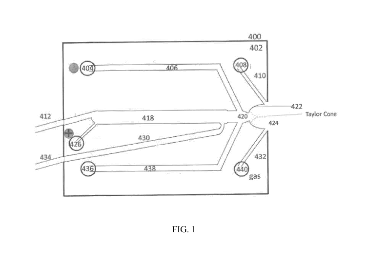
Al. FIG. 1
illustrates one non-limiting example of a microfluidic device designed to
perform isoelectric
focusing followed by ESI-MS characterization. The fluid channel network shown
in FIG. 1 is
fabricated from a plate of soda lime glass, which has very low transmission of
280 nm light
using a standard photolithographic etching technique. The depth of the
separation (or
enrichment) channel 418 is the same as the thickness of the glass layer 402,
i.e., the enrichment
channel 418 passes all the way from the top to bottom of glass plate 402. The
device 400 can be
illuminated by a light source disposed on one side of device 400 and imaged by
a detector
disposed on an opposite side of device 400. Because substrate 402 is opaque,
but enrichment
channel 418 defines an optical slit, the substrate 402 can block light that
does not pass through
the enrichment channel 418, blocking stray light and improving resolution of
the imaging
process. The glass layer 402 is sandwiched between two fused silica plates,
which are
transmissive (e.g., transparent) to 280 nm light. The top plate contains
through holes for the
instrument and user to interface with the channel network, while the bottom
plate is solid. The
three plates are bonded together at 520 C for 30 minutes. The inlet and
outlet tubing is
manufactured from cleaved capillaries (1001.tm ID, Polymicro) bonded to the
channel network.
The operation of this device in performing isoelectric focusing of proteins
and subsequent mass
spectrometry characterization will be described in Example 1 below.
[0068] Any of a variety of fluid actuation mechanisms known to those of skill
in the art may be
used to control fluid flow of samples and reagents through the device.
Examples of suitable fluid
actuation mechanisms for use in the disclosed methods, devices, and systems
include, but are not
limited to, application of positive or negative pressure to one or more inlet
ports or outlet ports,
gravitational or centrifugal forces, electrokinetic forces, electrowetting
forces, or any
combination thereof. In some embodiments, positive or negative pressure may be
applied
directly, e.g., through the use of mechanical actuators or pistons that are
coupled to the inlet
and/or outlet ports to actuate flow of the sample or reagents through the
fluidic channels. In
some embodiments, the mechanical actuators or pistons may exert force on a
flexible membrane
or septum that is used to seal the inlet and/or outlet ports. In some
embodiments, positive or
negative pressure may be applied indirectly, e.g., through the use of
pressurized gas lines or
vacuum lines connected with one or more inlet and/or outlet ports. In some
embodiment, pumps,
e.g., programmable syringe pumps, HPLC pumps, or peristaltic pumps, connected
with one or
more inlet and/or outlet ports may be used to drive fluid flow. In some
embodiments,
electrokinetic forces and/or electrowetting forces may be applied through the
use of electric field
- 24 -
CA 03101624 2020-11-25
WO 2019/232397 PCT/US2019/034942
and control of surface properties within the device. Electric fields may be
applied by means of
electrodes inserted into one or more inlet and/or outlet ports, or by means of
electrodes
integrated into one or more fluid channels within the device. The electrodes
may be connected
with one or more DC or AC power supplies for controlling voltages and/or
currents within the
device.
[0069] In general, the inlet ports, outlet ports, fluid channels, or other
components of the
disclosed microfluidic devices, including the main body of the device, may be
fabricated using
any of a variety of materials, including, but not limited to glass, fused-
silica, silicon,
polycarbonate, polymethylmethacrylate, cyclic olefin copolymer (COC) or cyclic
olefin polymer
(COP), polydimethylsiloxane (PDMS), or other elastomeric materials. Suitable
fabrication
techniques will generally depend on the choice of material, and vice versa.
Examples include,
but are not limited to, CNC machining, photolithography and chemical etching,
laser photo-
ablation, injection molding, hot embossing, die cutting, 3D printing, and the
like. In some
embodiments, the microfluidic device may comprise a layered structure in
which, for example, a
fluidics layer comprising fluid channels is sandwiched between an upper layer
and/or a lower
layer to seal the channels. The upper layer and/or lower layer may comprise
openings that align
with fluid channels in the fluidics layer to create inlet and/or outlet ports,
etc. Two or more
device layers may be clamped together to form a device which may be
disassembled, or may be
permanently bonded. Suitable bonding techniques will generally depend on the
choice of
materials used to fabricate the layers. Examples include, but are not limited
to, anodic bonding,
thermal bonding, laser welding, or the use of curable adhesives (e.g.,
thermally- or photo-curable
adhesives).
[0070] In some embodiments, all or a portion of the inlet ports, outlet ports,
or fluid channels
within the microfluidic device may comprise a surface coating used to modify
the electroosmotic
flow properties (e.g., HPC or PVA coatings) and/or
hydrophobicity/hydrophilicity properties
(e.g., polyethylene glycol (PEG) coatings) of the inlet port, outlet port, or
fluid channel walls.
[0071] The inlet and/or outlet ports of the disclosed devices can be
fabricated in a variety of
shapes and sizes. Appropriate inlet and/or outlet port geometries include, but
are not limited to,
spherical, cylindrical, elliptical, cubic, conical, hemispherical,
rectangular, or polyhedral (e.g.,
three dimensional geometries comprised of several planar faces, for example,
rectangular cuboid,
hexagonal columns, octagonal columns, inverted triangular pyramids, inverted
square pyramids,
inverted pentagonal pyramids, inverted hexagonal pyramids, or inverted
truncated pyramids), or
any combination thereof
- 25 -
CA 03101624 2020-11-25
WO 2019/232397 PCT/US2019/034942
[0072] Inlet and/or outlet port dimensions may be characterized in terms of an
average diameter
and depth. As used herein, the average diameter of the inlet or outlet port
refers to the largest
circle that can be inscribed within the planar cross-section of the inlet
and/or outlet port
geometry. In some embodiments of the present disclosure, the average diameter
of the inlet
and/or outlet ports may range from about 0.1 mm to about 10 mm. In some
embodiments, the
average diameter of the inlet and/or outlet ports may be at least 0.5 mm, at
least 1 mm, at least 2
mm, at least 4 mm, at least 8 mm, or at least 10 mm. In some embodiments, the
average
diameter may be at most 10 mm, at most 8 mm, at most 6 mm, at most 4 mm, at
most 2 mm, at
most 1 mm, or at most 0.5 mm. Any of the lower and upper values described in
this paragraph
may be combined to form a range included within the present disclosure, for
example, in some
embodiments, the average diameter may range from about 2 mm to about 8 mm.
Those of skill
in the art will recognize that the average diameter of the inlet and/or outlet
ports have any value
within this range, e.g., about 5.5 mm.
[0073] In some embodiments, the depth of the inlet and/or outlet ports (e.g.,
the sample or
reagent wells) may range from about 51.tm to about 500 jim. In some
embodiments, the depth
may be at least 5 jim, at least 10 jim, at least 25 jim, at least 50 jim, at
least 75 jim, at least 100
at least 200 jim, at least 300 jim, at least 400 jim, or at least 500 jim. In
some embodiments,
the depth may be at most 500 jim, at most 400 jim, at most 300 jim, at most
200 jim, at most 100
at most 50 jim, at most 25 jim, at most 10 jim, or at most 5 jim. Any of the
lower and upper
values described in this paragraph may be combined to form a range included
within the present
disclosure, for example, in some embodiments the depth of the inlet and/or
outlet ports may
range from about 501.tm to about 200 jim. Those of skill in the art will
recognize that the depth
may have any value within this range, e.g., about 130 jim. In some
embodiments, the depth of
the inlet and/or outlet ports (e.g., the sample or reagent wells) may range
from about 5001.tm to
about 50 mm. In some embodiments, the depth may be at least 1 mm, at least 5
mm, at least 10
mm, at least 15 mm, at least 20 mm, or at least 50 mm. In some embodiments,
the depth may be
at most 50 mm, at most 20 mm, at most 15 mm, at most 10 mm, at most 5 mm, or
at most 1 mm.
Any of the lower and upper values described in this paragraph may be combined
to form a range
included within the present disclosure, for example, in some embodiments the
depth of the inlet
and/or outlet ports may range from about 501.tm to about 5 mm.
[0074] In some embodiments, the fluid channels of the disclosed devices may
have any of a
variety of cross-sectional geometries, such as square, rectangular, circular,
and the like. In
general, the cross-sectional geometry of the fluid channels will be dependent
on the fabrication
technique used to create them, and vice versa. In some embodiments, a cross-
sectional
- 26 -
CA 03101624 2020-11-25
WO 2019/232397 PCT/US2019/034942
dimension of the fluid channels (e.g., the height, the width, or an average
diameter for a fluid
channel of non-rectangular cross-section, where the average diameter is
defined as the diameter
of the largest circle that can be inscribed within the cross-sectional
geometry of the fluid
channel) may range from about 51.tm to about 500 tm. In some embodiments, a
dimension the
fluid channel may be at least 5 jim, at least 10 jim, at least 25 jim, at
least 50 jim, at least 75
at least 100 jim, at least 200 jim, at least 300 jim, at least 400 jim, at
least 500 jim, or at least
1000 jim. In some embodiments, a dimension of the fluid channel may be at most
1000 1.tm , at
most 500 jim, at most 400 jim, at most 300 jim, at most 200 jim, at most 100
jim, at most 50
at most 25 jim, at most 10 jim, or at most 5 jim. Any of the lower and upper
values described in
this paragraph may be combined to form a range included within the present
disclosure, for
example, in some embodiments a dimension of the fluid channel may range from
about 751.tm to
about 300 jim. Those of skill in the art will recognize that the dimension may
have any value
within this range, e.g., about 95 jim. In some embodiments, a depth of the
fluid channel may be
equal to that for the inlet and/or outlet ports of the device,
[0075] Imaging techniques: In some embodiments of the disclosed methods and
devices, the
imaging of an analyte separation step and/or mobilization step may be
performed using an
optical detection technique, such as ultraviolet (UV) light absorbance,
visible light absorbance,
fluorescence, Fourier transform infrared spectroscopy, Fourier transform near
infrared
spectroscopy, Raman spectroscopy, optical spectroscopy, and the like. In some
embodiments, all
or a portion of a separation (or enrichment) channel, a junction or connecting
channel that
connects an end of the separation channel and a downstream analytical
instrument or an
electrospray orifice or tip, the electrospray orifice or tip itself, or any
combination thereof may
be imaged. In some embodiments the separation (or enrichment) channel may be
the lumen of a
capillary. In some embodiments, the separation (or enrichment) channel may be
a fluid channel
within a microfluidic device.
[0076] The wavelength range(s) used for detection of separated analyte bands
will typically
depend on the choice of imaging technique and the material(s) out of which the
device or portion
thereof are fabricated. For example, in the case that UV light absorbance is
used for imaging all
or a portion of the separation channel or other part of the microfluidic
device, detection at about
220 nm (due to a native absorbance of peptide bonds) and/or at about 280 nm
(due to a native
absorbance of aromatic amino acid residues) may allow one to visualize protein
bands during
separation and/or mobilization provided that at least a portion of the device,
e.g., the separation
channel, is transparent to light at these wavelengths. In some embodiments,
the analytes to be
separated and characterized via ESI-MS may be labeled prior to separation
with, e.g., a
- 27 -
CA 03101624 2020-11-25
WO 2019/232397 PCT/US2019/034942
fluorophore, chemiluminescent tag, or other suitable label, such that they may
be imaged using
fluorescence imaging or other suitable imaging techniques. In some
embodiments, e.g., wherein
the analytes comprise proteins produced by a commercial manufacturing process,
the proteins
may be genetically-engineered to incorporate a green fluorescence protein
(GFP) domain or
variant thereof, so that they may be imaged using fluorescence. In some
embodiments, proteins
may be tagged or labeled. The labeled proteins may be configured such that the
label does not
interfere with or perturb the analyte property on which the chosen separation
technique is based.
[0077] Any of a variety of imaging system components may be utilized for the
purpose of
implementing the disclosed methods, devices, and systems. Examples include,
but are not
limited to, one or more light sources (e.g., light emitting diodes (LEDs),
diode lasers, fiber
lasers, gas lasers, halogen lamps, arc lamps, etc.), condenser lenses,
objective lenses, mirrors,
filters, beam splitters, prisms, image sensors (e.g., CCD image sensors or
cameras, CMOS image
sensors or cameras, Diode Arrays, thermal imaging sensors, FTIR, etc.), and
the like, or any
combination thereof. Depending on the imaging mode utilized, the light source
and image
sensor may be positioned on opposite sides of the microfluidic device, e.g.,
so that absorbance-
based images may be acquired. In some embodiments, the light source and image
sensor may be
positioned on the same side of the microfluidic device, e.g., so that
epifluorescence images may
be acquired.
[0078] Images may be acquired continuously during the separation,
mobilization, and/or
electrospray steps, or may be acquired at random or specified time intervals.
In some
embodiments, a series of one or more images are acquired continuously, at
random time
intervals, or at specified time intervals. In some embodiments, the series of
one or more images
may comprise video images.
[0079] Imaging of pI markers for determination of protein isoelectric points
prior to
electrospray: In some embodiments, as noted above, the positions of two or
more pI markers in
images of a separation channel comprising a separated analyte mixture that has
been subjected to
CIEF may be used to determine an isoelectric point for one or more individual
analyte peaks
(e.g., protein analyte peaks). In some embodiments, the isoelectric point for
one or more analyte
peaks is calculated from the positions of two or more pI markers on the basis
of an assumed
linear relationship between local pH and position along the separation
channel. In some
embodiments, the isoelectric point for one or more analyte peaks is calculated
from the positions
of three or more pI markers on the basis of a nonlinear fitting function
(e.g., a nonlinear
polynomial) that describes the relationship between local pH and position
along the separation
channel. In some embodiments, the isolelectric point for one or more analytes
is calculated on
- 28 -
CA 03101624 2020-11-25
WO 2019/232397 PCT/US2019/034942
the basis of the positions of 2, 3, 4, 5, 6, 7, 8, 9, or 10 or more pI
standards that are determined
from images of the separation channel.
[0080] In some embodiments, the images used for determining the positions of
the two or more
pI markers are acquired as the analyte mixture is being separated, and the
calculation of pI for
each analyte band is iteratively updated as the separation continues. In some
embodiments, the
images used for determining the positions of the two or more pI markers are
acquired after
separation is complete and prior to initiation of a mobilization step. In some
embodiments, the
images used for determining the positions of the two or more pI markers are
acquired as the
separated mixture is mobilized and expelled through an electrospray tip or
orifice. In some
embodiments, the images used for determining the positions of the two or more
pI markers are
acquired as the separated mixture is mobilized and expelled through a fluid
channel that connects
the separation channel to a downstream analytical instrument.
[0081] In some embodiments, the images used to determine the positions of two
or more pI
markers and of analyte band(s) in a separated mixture are acquired using a
computer-
implemented method (e.g., a software package). In some embodiments, the
positions of the two
or more pI markers as well as of the analyte band(s) are determined using a
computer-
implemented method that comprises automated image processing. In some
embodiments, the
computer-implemented method further comprises performing a calculation of
isoelectric point
for one or more analyte bands based on the position data derived from the
automated image
processing.
[0082] FIG. 2 provides an example process flow chart for a computer-
implemented method to
acquire image(s) of a separation channel (or other portion of a microfluidic
device), determine
the positions of pI markers and analyte bands in the image(s) (i.e., in the
case where the
separation step comprises CIEF), and calculate a pI for one or more analyte
bands in the
separated mixture of analytes. In some embodiments, the computer-implemented
method may
comprise controlling the acquisition of a series of one or more images which
are then processed
to identify the positions of pI markers and separated analyte bands. Examples
of suitable
automated image processing algorithms will be discussed in more detail below.
In some
embodiments, predetermined knowledge for the predicted position of the pI
markers, e.g., the
positions of pI markers as determined from images of a "control" sample
comprising only the pI
markers, may be used to discriminate between bands corresponding to pI markers
and bands
corresponding to separated analytes. In some embodiments, the images of pI
markers may be
acquired at a different wavelength or using a different imaging mode than that
used to acquire
the images of the separated analyte bands. As illustrated in FIG. 2, if the
image processing step
- 29 -
CA 03101624 2020-11-25
WO 2019/232397 PCT/US2019/034942
fails to determine the positions for the known number of pI markers and/or for
the separated
analyte bands, the system may be instructed to acquire new image(s) so that
the image
processing step may be repeated. Once the positions of the pI markers and
separated analyte
bands are determined, the data for the positions of the pI markers is fit to a
user-selected model
for the pH gradient (e.g., a linear or nonlinear model) and the resulting
fitted relationship
between local pH and position along the separation channel is then used to
calculate the
isoelectric point for one or more analyte bands.
[0083] In some embodiments, the computer-implemented method may be an
iterative process, in
which the steps of detection of pI marker and analyte band positions, fitting
of the position data
to a pH gradient model, and calculation of isoelectric points for one or more
analyte bands is
repeated so that the latter is continuously updated and refined (e.g., through
averaging of several
determinations). In some embodiments, a cycle comprising the steps of image
acquisition and
processing, detection of pI marker and analyte band positions, fitting of pI
marker position data
to a pH gradient model, and calculation of isoelectric points for one or more
analyte bands may
be completed in a sufficiently short time that the calculation of isoelectric
points may be updated
and refined at a rate of at least 0.01 Hz, 0.1 Hz, 0.2 Hz, 0.3 Hz, 0.4 Hz, 0.5
Hz, 0.6 Hz, 0.7 Hz,
0.8 Hz, 0.9 Hz, 1 Hz, 10 Hz, 100 Hz, or 1,000 Hz, or any other relevant rate,
e.g., at a rate of at
least the Nyquist rate.
[0084] Imaging of analyte bands to determine velocities: In some embodiments,
as noted above,
the position of one or more analyte bands may be determined from a series of
two or more
images of the separation channel (or other portion of a microfluidic device),
such that a velocity
for one or more analyte bands may be calculated from the difference in its
relative position in the
two or more images and the known time interval between the acquisition times
for the two or
more images. In some embodiments, the two or more images of at least a portion
of the
separation channel may be acquired while a separation step is being performed.
In some
embodiments, the two or more images may be acquired during a mobilization
step. In some
embodiments, the two or more images may be acquired while a separated sample
is being
expelled through a fluid channel that connects an end of the separation
channel to a downstream
analytical instrument. In some embodiments, the two or more images may be
acquired while a
separated sample is being expelled through an electrospray tip or orifice to
form a Taylor cone.
In some embodiments, the velocity determined for one or more analyte bands may
be used to
calculate the time at which a given analyte band exits the separation channel.
In some
embodiments, e.g., when there is one or more interconnecting fluid junctions
or fluid channels
that connect an end of the separation channel with an outlet port, e.g., an
electrospray orifice or
- 30 -
CA 03101624 2020-11-25
WO 2019/232397 PCT/US2019/034942
tip, the velocity determined for the one or more analyte bands may be used to
calculate the time
at which a given analyte band reaches the outlet port and exits the device. In
some
embodiments, the velocity determined for the one or more analyte bands may be
used to
calculate the time at which a given analyte band exits an electrospray tip or
electrospray orifice
and enters a Taylor cone formed between the electrospray tip or orifice and
the inlet of a mass
spectrometer.
[0085] In some embodiments, the sequence of images used to determine a
velocity for one or
more analyte bands may be acquired using a computer-implemented method (e.g.,
a software
package). In some embodiments, the velocities of one or more analyte bands are
determined
using a computer-implemented method that comprises automated image processing.
In some
embodiments, the computer-implemented method further comprises performing a
calculation of
the time at which a given analyte band will exit the separation channel. In
some embodiments,
the computer-implemented method further comprises performing a calculation of
the time at
which a given analyte band will reach an outlet port and exit the device. In
some embodiments,
the computer-implemented method further comprises performing a calculation of
the time at
which a given analyte band will exit an electrospray tip or electrospray
orifice and enter a Taylor
cone formed between an electrospray tip or orifice and the inlet of a mass
spectrometer. In some
embodiments the exit time(s) determined for one or more analyte bands are used
to correlate
specific analyte bands with mass spectrometry data or data collected using
other analytical
instruments.
[0086] FIG. 3 provides another example process flow chart for a computer-
implemented method
to acquire image(s) of a separation channel (or other portion of a
microfluidic device), determine
the velocity of one or more analyte bands, and calculate at time at which a
given analyte band
will reach a specified point in the device, e.g., the end of the separation
channel, a junction point
between the separation channel and a secondary fluid channel, an outlet port
of the device, or the
outlet of an electrospray tip or orifice. In some embodiments, the computer-
implemented
method may comprise controlling the acquisition of a series of one or more
images which are
then processed to identify the positions of separated analyte bands. Examples
of suitable
automated image processing algorithms will be discussed in more detail below.
As illustrated in
FIG. 3, if the image processing step fails to determine the positions for the
separated analyte
bands, the system may be instructed to acquire new image(s) so that the image
processing step
may be repeated. Once the positions of the separated analyte bands are
determined for a series
of two or more images, a velocity is calculated for one or more of the analyte
peaks based on
their relative positions in the two or more images and the known time
interval(s) between the
- 3 1 -
CA 03101624 2020-11-25
WO 2019/232397 PCT/US2019/034942
acquisition times of the two or more images. In some embodiments, the tracking
of one or more
analyte bands from one image to the next in a series of images may be used to
distinguish
between several separated analyte bands, and to refine the velocity
calculation (e.g., through
averaging the velocity values calculated from several pairs of images in the
series). In some
embodiments, pI markers or other internal standards that may be detected using
the selected
imaging mode may be used as "velocity standards". The analyte band velocities
thus determined
may be used to calculate the time at which a given band will reach a user-
specified point in the
device, e.g., the outlet end of the separation channel, a particular fluid
junction within the device,
an outlet port of the device, an electrospray ionization tip or orifice where
the analyte enters a
Taylor cone, and the like.
[0087] In some embodiments, the computer-implemented method may be an
iterative process, in
which the steps of detection of analyte band positions, determination of
analyte band velocities,
and calculation of exit times is repeated so that the exit time prediction is
continuously updated
and the correlation of chemical separation data with mass spectrometry data
(or other types of
downstream analytical data) is further improved. In some embodiments, a cycle
comprising the
steps of image acquisition and processing, velocity calculation, and exit time
prediction(s) may
be completed in a sufficiently short time that the exit time prediction(s) may
be updated at a rate
of at least 0.01 Hz, 0.1 Hz, 0.2 Hz, 0.3 Hz, 0.4 Hz, 0.5 Hz, 0.6 Hz, 0.7 Hz,
0.8 Hz, 0.9 Hz, 1 Hz,
Hz, 100 Hz, or 1,000 Hz, or any other relevant rate, e.g., at a rate of at
least the Nyquist rate.
[0088] Correlation of separation data with mass spectrometry data: In some
embodiments, the
computer-implemented methods described above for performing imaging-based
determination of
accurate isoelectric points for isoelectrically-focused analyte bands enables
one to correlate
isoelectric point data with specific m/z peaks in mass spectrometry data (or
other analytical
data), thereby improving the information content of the data set (even for
single run experiments)
and allowing more quantitative characterization of an analyte sample.
[0089] In some embodiments, the computer-implemented methods described above
for using
image-derived data to calculate velocities and predict exit times for
separated analyte bands
(using any of a variety of different separation techniques) enables one to
improve the time
correlation between chemical separation data (e.g., retention times,
electrophoretic mobilities,
isoelectric points, etc.) and specific m/z peaks in mass spectrometry data (or
other analytical
data), thereby improving both the information content of the data set (even
for single run
experiments) and allowing more quantitative comparisons of data collected for
different sample
runs, different samples, or data collected on different instruments due to the
ability to correct for
experiment-to-experiment or instrument-to-instrument variations in separation
times. The
- 32 -
CA 03101624 2020-11-25
WO 2019/232397 PCT/US2019/034942
disclosed methods, devices, and systems may thus be particularly advantageous
for a variety of
metabolomics, proteomics, and drug development or manufacturing applications.
[0090] In some embodiments (e.g., those comprising a CIEF step), the computer-
implemented
methods of the present disclosure may perform both imaging-based determination
of precise
isoelectric points and imaging-based determination of the velocities of the
separated analyte
bands.
[0091] Mass spectrometry and electrospray ionization: In some embodiments, the
methods,
devices, and systems of the present disclosure may be configured for
performing electrospray
ionization of a separated analyte mixture and its injection into a mass
spectrometer. Mass
spectrometry (MS) is an analytical technique that measures the "mass" of
analyte molecules in a
sample by ionizing them and sorting the resultant ions based on their mass-to-
charge (m/z) ratio.
Combined with an upfront liquid- or gas-phase sample separation system, mass
spectrometry
provides one of the most effective means available for analyzing complex
samples comprising a
plurality of low abundance analytes, as is common, for example, in biological
samples.
[0092] All mass spectrometers share the requirement that the ions be in the
gas phase prior to
introduction into a mass analyzer. A variety of sample ionization modes have
been developed
including, but not limited to, matrix-assisted laser desorption and ionization
(MALDI) and
electrospray ionization (ESI). In the MALDI technique, the sample (e.g., a
biological sample
comprising a mixture of proteins) is mixed with an energy absorbing matrix
(EAM) such as
sinapinic acid or a-cyano-4-hydroxycinnamic acid and crystallized onto a metal
plate. Surface
enhanced laser desorption and ionization (SELDI) is a common variant of the
technique that
incorporates additional surface chemistry on the metal plate to promote
specific binding of
certain classes of proteins. The plate is inserted into a vacuum chamber, and
the matrix crystals
are struck with light pulses from a nitrogen laser. The energy absorbed by the
matrix molecules
is transferred to the proteins, causing them to desorb, ionize, and produce a
plume of ions in the
gas phase that are accelerated in the presence of an electric field and drawn
into a flight tube
where they drift until they strike a detector that records the time of flight.
The time of flight may
in turn be used to calculate the m/z ratio for the ionized species. In some
embodiments of the
disclosed devices, an outlet port of the device may comprise a capillary or
other feature used to
deposit separated analyte bands (or fractions thereof) onto a MALDI plate in
preparation for
mass spectrometric analysis, e.g., to correlate isoelectric points for
specific analyte bands with
MALDI mass spectrometer data.
[0093] Electrospray ionization (ESI; also referred to herein simply as
"electrospray") is another
widely used technique due to its inherent compatibility for interfacing liquid
chromatographic or
- 33 -
CA 03101624 2020-11-25
WO 2019/232397 PCT/US2019/034942
electrokinetic chromatographic separation techniques with a mass spectrometer.
As noted above,
in electrospray ionization, small droplets of sample and solution are emitted
from a distal end of
a capillary or microfluidic device comprising an electrospray feature (e.g.,
an emitter tip or
orifice) by the application of an electric field between the tip or orifice
and the mass
spectrometer source plate. The droplet then stretches and expands in this
induced electric field to
form a cone shaped emission (i.e., a "Taylor cone"), which comprises
increasingly small droplets
that evaporate and produce the gas phase ions that are introduced into the
mass spectrometer for
further separation and detection. Emitter tips may be formed from a capillary
or a corner or ESI
tip built into microfluidic chip design, which provides a convenient droplet
volume for ESI.
Emitter tips may be sharpened to provide a small surface and drop volume using
a lapping
wheel, file, machining tools, CNC machining tools, water jet cutting, or other
tools or process to
shape the ESI tip to provide a small surface volume, and the like. In some
embodiments, the tip
may be drawn by heating and stretching the tip portion of the chip. In some
embodiments, the tip
may then be cut to a desired length or diameter. In some embodiments, the
electrospray tip may
be coated with a hydrophobic coating which may minimize the size of droplets
formed on the tip.
In some embodiments, the system may electrospray mobilizer, catholyte, or any
other liquid
during a separation step, when no analyte is being eluted from the device.
[0094] In some embodiments of the disclosed methods, devices, and systems,
other ionization
methods are used, such as inductive coupled laser ionization, fast atom
bombardment, soft laser
desorption, atmospheric pressure chemical ionization, secondary ion mass
spectrometry, spark
ionization, thermal ionization, and the like.
[0095] With respect to electrospray ionization, in some embodiments the
disclosed microfluidic
devices comprise features designed to promote efficient electrospray
ionization and convenient
interfacing with downstream mass spectrometric analysis, as illustrated in
FIG. 1. The mass-to-
charge ratio (or "mass") for analytes expelled from the microfluidic device
(e.g., a biologic or
biosimilar) and introduced into a mass spectrometer can be measured using any
of a variety of
different mass spectrometer designs. Examples include, but are not limited to,
time-of-flight
mass spectrometry, quadrupole mass spectrometry, ion trap or orbitrap mass
spectrometry,
distance-of-flight mass spectrometry, Fourier transform ion cyclotron
resonance, resonance mass
measurement, and nanomechanical mass spectrometry.
[0096] In some embodiments, the electrospray feature of a microfluidic device
may be in-line
with a separation channel. In some embodiments, the electrospray feature of a
microfluidic
device may be oriented at a right angle or at an intermediate angle relative
to a separation
channel. In some embodiments of the disclosed methods, substantially all of
the separated
- 34 -
CA 03101624 2020-11-25
WO 2019/232397 PCT/US2019/034942
and/or enriched analyte fractions from a final separation or enrichment step
performed in a
capillary or microfluidic device are expelled from the electrospray tip or
feature in a continuous
stream. In some embodiments, a portion of the analyte mixture (e.g., a
fraction of interest) may
be expelled from a microfluidic device via an outlet configured to interface
with an analytical
instrument, such as a mass spectrometer or another device configured to
fractionate and/or enrich
at least a portion of the sample. Another portion of the analyte mixture
(e.g., containing fractions
other than the fraction of interest) can be expelled via a waste channel.
[0097] In some embodiments, the expulsion from the capillary or microfluidic
device is
performed using pressure, electric force, ionization, or any combination of
these. In some
embodiments, the expulsion coincides with a mobilization step as described
above. In some
embodiments a sheath liquid used for electrospray ionization is used as an
electrolyte for an
electrophoretic separation. In some embodiments, a nebulizing gas is provided
to reduce the
analyte fraction to a fine spray.
[0098] Imaging-based feedback of electrospray ionization performance:
Conventional ESI-MS
systems using capillaries or microfluidic devices generally provide no tools
for calibrating the
system to reestablish a Taylor cone during operation. Maintaining a stable
Taylor cone can be
complicated by the electrophoresis electric field applied across the
separation channel in the
microfluidic device or capillary. Changes in the conductivity of reagents
between runs, or during
a run, can change the voltage potential at the interface with the mass
spectrometer. Changes in
potential at the interface may adversely affect the Taylor cone and can lead
to loss of
electrospray ionization efficiency. Disclosed herein are methods and systems
for improving the
electrospray ionization performance and thus the quality of mass spectrometry
data collected for
capillary-based or microfluidic device-based ESI-MS systems. In some
embodiments, for
example, imaging of the Taylor cone in an electrospray ionization setup may be
used in a
computer implemented method to provide feedback control of one or more
operating parameters
such that the shape, density, or other characteristic of the Taylor cone is
maintained within a
specified range. In some embodiments, the operating parameters that may be
controlled through
such a feedback process include, but are not limited to, the alignment of the
electrospray tip or
orifice with the mass spectrometer inlet, the distance between the
electrospray tip and the mass
spectrometer inlet (e.g., by mounting the capillary tip or microfluidic device
comprising an
integrated electrospray feature on a programmable precision X-Y-Z translation
stage), the flow
rate of analyte sample through the electrospray tip (e.g., by adjusting the
pressure, electric field
strength, or combination thereof that are used to drive the expulsion of
analyte sample), the
voltage applied, e.g., at a proximal end of the channel, e.g., between the
electrospray tip or
- 35 -
CA 03101624 2020-11-25
WO 2019/232397 PCT/US2019/034942
orifice and the mass spectrometer inlet, the volumetric flowrate of a sheath
liquid or sheath gas
surrounding the expulsed analyte sample, or any combination thereof.
[0099] FIG. 4 provides an example process flow chart for a computer-
implemented method used
to: (i) acquire images of the Taylor cone (using any of a variety of image
sensors, e.g., CCD
image sensors or CMOS image sensors), (ii) process the images to determine a
shape, density, or
other characteristic of the Taylor cone, (iii) compare the shape, density, or
other characteristic of
the Taylor cone with a set of specified or target values, and (iv) based on
said comparison, use a
mathematical algorithm that relates the shape, density, or other
characteristic of the Taylor cone
to one or more operating parameters to determine an appropriate adjustment to
the one or more
operating parameters to restore the Taylor cone to the specified or target
values. In some
embodiments, data acquired from the mass spectrometer (e.g., total ion current
data) may be
used in addition to data derived from images of the Taylor cone to monitor
system performance
and make adjustments to one or more operational parameters.
[0100] In some embodiments, the cyclical process illustrated in FIG. 4,
comprising the steps of
image acquisition and processing, identification of Taylor cone
characteristics, comparison of the
said Taylor cone characteristics with a set of target values, and calculation
of the adjustments
needed to one or more ESI-MS systems operating parameters, may be completed in
a sufficiently
short time that the one or more operating parameters may be updated at a rate
of at least 0.01 Hz,
0.1 Hz, 0.2 Hz, 0.3 Hz, 0.4 Hz, 0.5 Hz, 0.6 Hz, 0.7 Hz, 0.8 Hz, 0.9 Hz, 1 Hz,
10 Hz, 100 Hz, or
1,000 Hz, or any other relevant rate, e.g., at a rate of at least the Nyquist
rate.
[0101] Alternating high mass /low mass scanning: In some embodiments of the
disclosed
methods, devices, and systems, the mass spectrometer may be set to alternate
between a high
mass scan range (e.g., an m/z range of about 1500 ¨6000), or "high mass scan",
and a low mass
scan range (e.g., an m/z range of about 150¨ 1500), or "low mass scan", such
that the low mass
scan may be used to identify low mass markers, e.g., free solution ampholytes
in the instance
that an isoelectric focusing separation step was performed, that can be
identified in the mass
spectrometry data and used to calibrate it with respect to a property
indicated by the low mass
marker (e.g., a specific range of isoelectric point in the case that free
solution ampholytes are
detected, peptides, small molecule markers). The switching between high mass
scans and low
mass scans and the scan rates should be fast relative to the efflux of analyte
sample from the
electrospray interface. In some instances, the switching rate between high
mass scans and low
mass scans may range from about 0.5 Hz to about 50 Hz. In some instances, the
switching rate
may be at least 0.5 Hz, at least 1 Hz, at least 5 Hz, at least 10 Hz, at least
20 Hz, at least 30 Hz, at
least 40 Hz, or at least 50 Hz.
- 36 -
CA 03101624 2020-11-25
WO 2019/232397 PCT/US2019/034942
[0102] Altering high and low separation/mobilization voltage to keep ESI tip
voltage constant:
In some embodiments, the ESI ion source on the mass spectrometer will have an
adjustable
power supply capable of setting a negative voltage on the mass spectrometer.
In some
embodiments, the ESI ion source on the mass spectrometer will have an
adjustable power supply
capable of setting a positive voltage on the mass spectrometer. In some
embodiments, the ESI
ion source on the mass spectrometer will be held at ground. In some
embodiments, the ESI tip on
the capillary or microfluidic device will be held at or close to ground to
generate an electric field
between the ESI tip and the charged ESI ion source on the mass spectrometer.
In some
embodiments, the ESI tip on the capillary or microfluidic device will be held
at a positive or
negative voltage to generate an electric field between the ESI tip and the
grounded ESI ion
source on the mass spectrometer.
[0103] FIG. 15 provides an exemplary flowchart of a computer-controlled
feedback loop to
maintain a constant voltage drop of 3000V between the anode and cathode while
keeping the ESI
tip voltage at OV during mobilization. In some embodiments, this feedback loop
may be
implemented when the mass spectrometer ESI ion source is set at a positive or
negative voltage
relative to ground (for example, -3500V). In this example, AV between anolyte
port 110 and
mobilizer port 104 is kept at 3000V by initially setting anolyte port 110 at
+3000V and mobilizer
port 104 at OV in FIG. 7A. In some embodiments, a different AV may be set by
setting anolyte
port 110 to a different value. In some embodiments, anodic mobilization may be
used, and port
110 would be a catholyte port, set to, for example, -3000V. In the example
outlined in FIG. 15,
during mobilization, the resistance in separation channel 112 is dropping due
to analyte and
ampholytes in the separation regaining charge. This causes the voltage drop
across channel 112
to drop, leading to an increase in voltage at ESI tip 116, according to
equation 1:
V116¨ (AV110-104)*(R105)/(R109 + R112 + R105)
However, by measuring or calculating ESI tip voltage 116, the voltage settings
at anolyte port
110 and mobilizer port 104 can be adjusted. By subtracting ESI tip voltage 116
from both
anolyte port 110 and mobilizer port 104 settings, AV110-104 remains 3000V so
the mobilization is
unaffected, but ESI tip 116 voltage is set to 0 according to equation 2:
V116¨ (AV110-104)*(R105)/(R109 + R112 + R105) + V104
This feedback loop continues to operate until the mobilization is complete,
adjusting ESI tip 116
voltage to 0 at a regular frequency, e.g., the Nyquist rate, or about 0.2 Hz.
In some instances, the
voltage at ESI tip 116 may be adjusted to 0 at a rate of at least 0.01 Hz, 0.1
Hz, 0.2 Hz, 0.3 Hz,
0.4 Hz, 0.5 Hz, 0.6 Hz, 0.7 Hz, 0.8 Hz, 0.9 Hz, 1 Hz, 10 Hz, 100 Hz, or 1,000
Hz. Maintaining a
- 37 -
CA 03101624 2020-11-25
WO 2019/232397 PCT/US2019/034942
constant stable voltage at ESI tip 116 can be critical to maintaining stable
electrospray during the
mobilization process.
[0104] In some instances, the feedback loop operates to maintain the voltage
at the ESI tip to
within a specified percentage of a pre-set value. For example, in some
instances, the feedback
loop operates to maintain the voltage at the ESI tip to within 10%, 9%, 8%,
7%, 6%, 5%, 4%,
3%, 2%, 1%, 0.5%, or 0.1% of a pre-set value. In some instances, the feedback
loop operates to
maintain the ESI tip voltage to within 1000V, 500V, 100V, 75V, 50V, 25V, 10V,
5V, or 1V of a
pre-set value.
[0105] In some embodiments, the mass spectrometer ESI ion source is held at
ground, and ESI
tip 116 will need to be kept at a constant positive or negative voltage in
order to create an electric
field between ESI tip 116 and the mass spectrometer. In some embodiments, ESI
tip voltage
(e.g., the pre-set value) may be around +5000V, around +4000V, around +3500V,
around
+3000V, around +2500V, around +2000V, around+1500V around +1000V, around
+500V, or
around -5000V, around -4000V, around -3500V, around -3000V, around -2500V,
around -
2000V, around-1500V, around -1000V, or around -500V. FIG. 12 provides an
example
flowchart of a computer-controlled feedback loop to maintain a constant
voltage drop of 3000V
between the anode and cathode while keeping the ESI tip voltage at 3000V
during mobilization.
Operation of the computer-controlled feedback loop is the same as in FIG. 15,
except voltages at
anolyte port 110 and mobilizer port 104 are offset by +3000V, which offsets
the voltage at ESI
tip 116 to +3000V, still obeying equation 2. In some embodiments control of
the electric field
strength can be accomplished using analog circuitry. In some embodiments, the
control of
voltages at one or more electrodes in contact with the capillary-based or
microfluidic device-
based separation system may be provided by using one, two, three, or four or
more independent
high-voltage power supplies. In some instance, the control of voltages at one
or more electrodes
in contact with the capillary-based or microfluidic device-based separation
system may be
provided, e.g., by using a single, multiplexed high-voltage power supply.
[0106] In some instances, the feedback loop operates to maintain the electric
field strength
within the separation channel, or the voltage drop between the anode and
cathode, to within a
specified percentage of a pre-set value. For example, in some instances, the
feedback loop
operates to maintain the electric field strength within the separation
channel, or the voltage drop
between the anode and cathode, to within 10%, 9%, 8%, 7%, 6%, 5%, 4%, 3%, 2%,
1%, 0.5%,
0.1%, or 0.01% of a pre-set value. In some instances, the feedback loop
operates to maintain the
electric field strength within the separation channel, or the voltage drop
between the anode and
cathode, to within 1000V, 500V, 100V, 75V, 50V, 25V, by, 5V, or 1V of a pre-
set value.
- 38 -
CA 03101624 2020-11-25
WO 2019/232397 PCT/US2019/034942
[0107] System hardware: FIG. 5 provides a schematic illustration of a system
hardware block
diagram for one embodiment of the disclosed methods, devices, and systems. As
illustrated, a
system of the present disclosure may comprise one or more of the following
hardware
components: (i) a chemical separation system (e.g., a capillary or
microfluidic device designed to
perform an analyte separation, e.g., an isoelectric focusing-based separation,
and one or more
high-voltage power supplies), (ii) an electrospray interface for a mass
spectrometer that, in some
cases, may be directly integrated with the separation system (as indicated by
the dashed line),
(iii) a mass spectrometer, (iv) an imaging device or system, (v) a processor
or computer, and (vi)
a computer memory device, or any combination thereof. In some embodiments, the
system may
further comprise one or more capillary or microfluidic device flow controllers
(e.g.,
programmable syringe pumps, peristaltic pumps, HPLC pumps, etc.), temperature
controllers
configured to maintain a specified temperature for all or a portion of a
capillary or microfluidic
device, additional photo sensors or image sensors (e.g., photodiodes,
avalanche photodiodes,
CMOS image sensors and cameras, CCD image sensors and cameras, etc.), light
sources (e.g.,
light emitting diodes (LEDs), diode lasers, fiber lasers, gas lasers, halogen
lamps, arc lamps,
etc.), other types of sensors (e.g., temperature sensors, flow sensors, pH
sensors, conductivity
sensors, etc.), computer memory devices, computer display devices (e.g.,
comprising a graphical
user interface), digital communication devices (e.g., intranet, internet,
WiFi, Bluetooth , or other
hardwired or wireless communication hardware), and the like.
[0108] In some embodiments, the system may comprise an integrated system in
which a
selection of functional hardware components are packaged in a fixed
configuration. In some
embodiments, the system may comprise a modular system in which the selection
of functional
hardware components may be changed in order to reconfigure the system for new
applications.
In some embodiments, some of these functional system components, e.g.,
capillaries or
microfluidic devices, are replaceable or disposable components.
[0109] As noted above, any of a variety of different mass spectrometers may be
utilized in
different embodiments of the disclosed systems including, but not limited to,
time-of-flight mass
spectrometers, quadrupole mass spectrometers, ion trap or orbitrap mass
spectrometers, distance-
of-flight mass spectrometers, Fourier transform ion cyclotron resonance
spectrometers,
resonance mass measurement spectrometers, and nanomechanical mass
spectrometers.
[0110] System & application software: As illustrated in FIG. 6, a system of
the present
disclosure may comprise a plurality of software modules. For example, a system
may comprise
a system control software module, a data acquisition software module, a data
processing
software module, or any combination thereof. In general, these software
modules will be
- 39 -
CA 03101624 2020-11-25
WO 2019/232397 PCT/US2019/034942
configured to operate within an operating system or environment hosted by a
computer
processor, and may communicate and share data with each other and/or the
operating system.
[0111] In some embodiments, a system control software module may comprise
software for: (i)
coordinating the operation of the capillary- or microfluidic device-based
analyte separation
system with image acquisition by an imaging system, (ii) coordinating the
operation of capillary-
or microfluidic device-based analyte separation system with data acquisition
by the mass
spectrometer system, (iii) coordinating image acquisition by an imaging system
with operation of
the capillary- or microfluidic device-based analyte separation system and/or
mass spectrometer
system, (iv) providing feedback control of one or more operating parameters of
an electrospray
ionization setup and/or mass spectrometer based on data derived from imaging
of a separation
channel and/or a Taylor cone, (v) controlling data acquisition by the mass
spectrometer while
switching between high mass and low mass scan ranges in an alternating
fashion, (vi) monitoring
voltage at ESI tip and adjusting separation circuit voltages to maintain a
constant separation
electric field strength (or voltage drop between the anode and cathode) and
constant voltage at
ESI tip, or any combination thereof
[0112] In some embodiments, a data acquisition module may comprise software
for: (i)
controlling image acquisition by one or more image sensors or imaging systems,
storing said
image data, and providing a software interface with system control and/or data
processing
software modules, and (ii) controlling data acquisition by one or more mass
spectrometer
systems, storing said mass spectrometer data (or other downstream analytical
instrument), and
providing a software interface with system control and/or data processing
software, or any
combination thereof.
[0113] In some embodiments, a data processing module may comprise software
for: (i)
processing images and determining the position(s) of one or more pI standards
or analyte peaks
in a separation channel while the separation is being performed, after the
separation is complete,
or after mobilization of the pI standards and analyte peaks towards a
separation channel outlet or
electrospray tip, (ii) processing images and determining a velocity, an exit
time, and/or an
electrospray emission time for one or more pI standard or analyte peaks, (iii)
processing of
images of a separation channel to monitor a position of an analyte peak and
images of a Taylor
cone to monitor electrospray performance, where the images of the separation
channel and
Taylor cone are acquired either simultaneously or alternately, (iv) processing
images of a Taylor
cone, determining a shape, density, or other characteristic of the Taylor
cone, and calculating an
adjustment to be made to one or more operating parameters comprising the
position (i.e.,
alignment and/or separation distance) of the electrospray tip or orifice
relative to the mass
- 40 -
CA 03101624 2020-11-25
WO 2019/232397 PCT/US2019/034942
spectrometer inlet, the fluid flow rate through the electrospray tip or
orifice, the voltage between
the electrospray tip or orifice and the mass spectrometer, etc., or any
combination thereof, to
affect a change in a quality of the mass spectrometer data; or any combination
thereof.
[0114] The disclosed system and application software may be implemented using
any of a
variety or programming languages and environments known to those of skill in
the art.
Examples include, but are not limited to, C, C++, C#, PL/I, PL/S, PL/8, PL-6,
SYMPL, Python,
Java, Lab View, Visual Basic, .NET and the like.
[0115] Image processing software: In some embodiments, as noted above, the
data processing
module may comprise image processing software for determining the positions of
pI markers or
separated analyte bands, for characterizing the shape, density, or other
visual indicator of Taylor
cone function, etc. Any of a variety of image processing algorithms known to
those of skill in
the art may be utilized for image pre-processing or image processing in
implementing the
disclosed methods and systems. Examples include, but are not limited to, Canny
edge detection
methods, Canny-Deriche edge detection methods, first-order gradient edge
detection methods
(e.g., the Sobel operator), second order differential edge detection methods,
phase congruency
(phase coherence) edge detection methods, other image segmentation algorithms
(e.g., intensity
thresholding, intensity clustering methods, intensity histogram-based methods,
etc.), feature and
pattern recognition algorithms (e.g., the generalized Hough transform for
detecting arbitrary
shapes, the circular Hough transform, etc.), and mathematical analysis
algorithms (e.g., Fourier
transform, fast Fourier transform, wavelet analysis, auto-correlation,
Savitzky-Golay smoothing,
Eigen analysis, etc.), or any combination thereof.
[0116] Processors and computer systems: One or more processors or computers
may be
employed to implement the methods disclosed herein. The one or more processors
may
comprise a hardware processor such as a central processing unit (CPU), a
graphic processing unit
(GPU), a general-purpose processing unit, or computing platform. The one or
more processors
may be comprised of any of a variety of suitable integrated circuits (e.g.,
application specific
integrated circuits (ASICs) designed specifically for implementing deep
learning network
architectures, or field-programmable gate arrays (FPGAs) to accelerate compute
time, etc.,
and/or to facilitate deployment), microprocessors, emerging next-generation
microprocessor
designs (e.g., memristor-based processors), logic devices and the like.
Although the disclosure is
described with reference to a processor, other types of integrated circuits
and logic devices may
also be applicable. The processor may have any suitable data operation
capability. For example,
the processor may perform 512 bit, 256 bit, 128 bit, 64 bit, 32 bit, or 16 bit
data operations. The
-41 -
CA 03101624 2020-11-25
WO 2019/232397 PCT/US2019/034942
one or more processors may be single core or multi core processors, or a
plurality of processors
configured for parallel processing.
[0117] The one or more processors or computers used to implement the disclosed
methods may
be part of a larger computer system and/or may be operatively coupled to a
computer network (a
"network") with the aid of a communication interface to facilitate
transmission of and sharing of
data. The network may be a local area network, an intranet and/or extranet, an
intranet and/or
extranet that is in communication with the Internet, or the Internet. The
network in some cases is
a telecommunication and/or data network. The network may include one or more
computer
servers, which in some cases enables distributed computing, such as cloud
computing. The
network, in some cases with the aid of the computer system, may implement a
peer-to-peer
network, which may enable devices coupled to the computer system to behave as
a client or a
server.
[0118] The computer system may also include memory or memory locations (e.g.,
random-
access memory, read-only memory, flash memory, Intel OptaneTM technology),
electronic
storage units (e.g., hard disks), communication interfaces (e.g., network
adapters) for
communicating with one or more other systems, and peripheral devices, such as
cache, other
memory, data storage and/or electronic display adapters. The memory, storage
units, interfaces
and peripheral devices may be in communication with the one or more
processors, e.g., a CPU,
through a communication bus, e.g., as is found on a motherboard. The storage
unit(s) may be
data storage unit(s) (or data repositories) for storing data.
[0119] The one or more processors, e.g., a CPU, execute a sequence of machine-
readable
instructions, which are embodied in a program (or software). The instructions
are stored in a
memory location. The instructions are directed to the CPU, which subsequently
program or
otherwise configure the CPU to implement the methods of the present
disclosure. Examples of
operations performed by the CPU include fetch, decode, execute, and write
back. The CPU may
be part of a circuit, such as an integrated circuit. One or more other
components of the system
may be included in the circuit. In some cases, the circuit is an application
specific integrated
circuit (ASIC).
[0120] The storage unit stores files, such as drivers, libraries and saved
programs. The storage
unit stores user data, e.g., user-specified preferences and user-specified
programs. The computer
system in some cases may include one or more additional data storage units
that are external to
the computer system, such as located on a remote server that is in
communication with the
computer system through an intranet or the Internet.
- 42 -
CA 03101624 2020-11-25
WO 2019/232397 PCT/US2019/034942
[0121] Some aspects of the methods and systems provided herein are implemented
by way of
machine (e.g., processor) executable code stored in an electronic storage
location of the
computer system, such as, for example, in the memory or electronic storage
unit. The machine
executable or machine readable code is provided in the form of software.
During use, the code is
executed by the one or more processors. In some cases, the code is retrieved
from the storage
unit and stored in the memory for ready access by the one or more processors.
In some
situations, the electronic storage unit is precluded, and machine-executable
instructions are
stored in memory. The code may be pre-compiled and configured for use with a
machine having
one or more processors adapted to execute the code, or may be compiled at run
time. The code
may be supplied in a programming language that is selected to enable the code
to execute in a
pre-compiled or as-compiled fashion.
[0122] Various aspects of the disclosed methods and devices may be thought of
as "products" or
"articles of manufacture", e.g., "computer program or software products",
typically in the form
of machine (or processor) executable code and/or associated data that is
stored in a type of
machine readable medium, where the executable code comprises a plurality of
instructions for
controlling a computer or computer system in performing one or more of the
methods disclosed
herein. Machine-executable code may be stored in an optical storage unit
comprising an
optically readable medium such as an optical disc, CD-ROM, DVD, or Blu-Ray
disc. Machine-
executable code may be stored in an electronic storage unit, such as memory
(e.g., read-only
memory, random-access memory, flash memory) or on a hard disk. "Storage" type
media
include any or all of the tangible memory of the computers, processors or the
like, or associated
modules thereof, such as various semiconductor memory chips, optical drives,
tape drives, disk
drives and the like, which may provide non-transitory storage at any time for
the software that
encodes the methods and algorithms disclosed herein.
[0123] All or a portion of the software code may at times be communicated via
the Internet or
various other telecommunication networks. Such communications, for example,
enable loading
of the software from one computer or processor into another, for example, from
a management
server or host computer into the computer platform of an application server.
Thus, other types of
media that are used to convey the software encoded instructions include
optical, electrical and
electromagnetic waves, such as those used across physical interfaces between
local devices,
through wired and optical landline networks, and over various atmospheric
links. The physical
elements that carry such waves, such as wired or wireless links, optical
links, or the like, are also
considered media that convey the software encoded instructions for performing
the methods
disclosed herein. As used herein, unless restricted to non-transitory,
tangible "storage" media,
- 43 -
CA 03101624 2020-11-25
WO 2019/232397 PCT/US2019/034942
terms such as computer or machine "readable medium" refer to any medium that
participates in
providing instructions to a processor for execution.
[0124] The computer system typically includes, or may be in communication
with, an electronic
display for providing, for example, images captured by a machine vision
system. The display is
typically also capable of providing a user interface (UI). Examples of UI' s
include but are not
limited to graphical user interfaces (GUIs), web-based user interfaces, and
the like.
[0125] Applications: As noted above, the disclosed methods, devices, systems,
and software
have potential application in a variety of fields including, but not limited
to, proteomics research,
drug discovery and development, and clinical diagnostics. For example, the
improved
information content and data quality that may be achieved for separation-based
ESI-MS analysis
of analyte samples using the disclosed methods may be of great benefit for the
characterization
of biologic and biosimilar pharmaceuticals during development and/or
manufacturing. Other
applications may include, but are not limited to, analysis of environmental
pollutants, pesticides,
small molecules, metabolites, peptides, post-translational modifications,
glycoforms, antibody-
drug conjugates, fusion proteins, viruses, allergans, single cell organisms,
and other applications.
[0126] Biologics and biosimilars are a class of drugs which include, for
example, recombinant
proteins, antibodies, live virus vaccines, human plasma-derived proteins, cell-
based medicines,
naturally-sourced proteins, antibody-drug conjugates, protein-drug conjugates
and other protein
drugs. The FDA and other regulatory agencies require the use of a stepwise
approach to
demonstrating biosimilarity, which may include a comparison of the proposed
product and a
reference product with respect to structure, function, animal toxicity, human
pharmacokinetics
(PK) and pharmacodynamics (PD), clinical immunogenicity, and clinical safety
and
effectiveness (see "Scientific Considerations in Demonstrating Biosimilarity
to a Reference
Product: Guidance for Industry", U.S. Department of Health and Human Services,
Food and
Drug Administration, April 2015). Examples of the structural characterization
data that may be
required for protein products include primary structure (i.e., amino acid
sequence), secondary
structure (i.e., the degree of folding to form alpha helix or beta sheet
structures), tertiary structure
(i.e., the three dimensional shape of the protein produced by folding of the
polypetide backbone
and secondary structural domains), and quaternary structure (e.g., the number
of subunits
required to form an active protein complex, or the protein's aggregation
state)). In many cases,
this information may not be available without employing laborious, time-
intensive, and costly
techniques such as x-ray crystallography. Thus there is a need for
experimental techniques that
allow for convenient, real-time, and relatively high-throughput
characterization of protein
- 44 -
CA 03101624 2020-11-25
WO 2019/232397 PCT/US2019/034942
structure for the purposes of establishing biosimilarity between candidate
biological drugs and
reference drugs.
[0127] In some embodiments, the disclosed methods, devices, and systems may be
used to
provide structural comparison data for biological drug candidates (e.g.,
monoclonal antibodies
(mAb)) and reference biological drugs for the purpose of establishing
biosimilarity. For
example, in some instances, isoelectric point data and/or mass spectrometry
data for a drug
candidate and a reference drug may provide important evidence in support of a
demonstration of
biosimilarity. In some embodiments, isoelectric point data and/or mass
spectrometry data for a
drug candidate and a reference drug that have both been treated with a site-
specific protease
under identical reaction conditions may provide important evidence in support
of a
demonstration of biosimilarity. In some embodiments, the disclosed methods,
devices, and
systems may be used to monitor a biologic drug manufacturing process to ensure
the quality and
consistency of the product by analyzing samples drawn at different points in
the production
process, or samples drawn from different production runs. In some embodiments,
the disclosed
methods, devices, and systems may be used to evaluate stability of formulation
buffers. In some
embodiments, the disclosed methods, devices, and systems may be used to
evaluate cloned cell
lines for production and quality of biological drug candidates.
EXAMPLES
[0128] These examples are provided for illustrative purposes only and not to
limit the scope of
the claims provided herein.
Example 1 - Characterization of protein charge on chip before performing mass
spectrometry
[0129] The fabrication of the microfluidic device illustrated in FIG. 1 has
been described above.
To operate, the device is mounted on an instrument containing a nitrogen gas
source, heater,
positive pressure pump (e.g., Parker, T5-1IC-03-1EEP), electrophoresis power
supply (Gamm
High Voltage, MC30) terminating in two platinum-iridium electrodes (e.g.,
Sigma-Aldrich,
357383), UV light source (e.g., LED, qphotonics, UVTOP280), CCD camera (e.g.,
ThorLabs,
3401JV-GE) and an autosampler for loading samples onto the device. The power
supply shares a
common earth ground with the mass spectrometer. The instrument is controlled
through software
(e.g., Lab View).
[0130] Protein samples are pre-mixed with ampholyte pH gradient and pI markers
before placing
into vials and loading onto the autosampler. They are serially loaded from an
autosampler via the
inlet 412 onto the microfluidic device 400 through the enrichment channel 418
and out of the
device to waste 430 through the outlet 434.
- 45 -
CA 03101624 2020-11-25
WO 2019/232397 PCT/US2019/034942
[0131] The sheath/catholyte fluid (50% Me0H, N4OH/H20) is loaded onto the two
catholyte
wells 404, 436, anolyte (10 mM H3PO4) onto the anolyte well 426, and the
source of heated
nitrogen gas is attached to the two gas wells 408, 440.
[0132] After all reagents are loaded, an electric field of +600V/cm is applied
from anolyte well
426 to catholyte wells 404, 436 by connecting the electrodes to the anolyte
well 426 and
catholyte wells 404, 436 to initiate isoelectric focusing. The UV light source
is aligned under the
enrichment channel 418, and the camera is placed above the enrichment channel
418 to measure
the light that passes through the enrichment channel 418, thereby detecting
the focusing proteins
by means of their absorbance. The glass plate 402, being constructed of soda-
lime glass, acts to
block any stray light from the camera, so light not passing through the
enrichment channel 418 is
inhibited from reaching the camera, increasing sensitivity of the measurement.
[0133] Images of the focusing proteins can be captured continuously and/or
periodically during
IEF. When focusing is complete, low pressure will be applied from the inlet
412, mobilizing the
pH gradient toward the orifice 424. The electric field can be maintained at
this time to maintain
the high resolution IEF separation. Continuing to image the enrichment channel
418 during the
ESI process can be used to determine the pI of each protein as it is expelled
from the orifice 424.
[0134] As the enriched protein fraction moves from the enrichment channel 418
into the
confluence 420, it will mix with the sheath fluid, which can flow from the
catholyte wells 404,
436 to the confluence 420 via sheath/catholyte fluid channels 406, 438. Mixing
enriched protein
fractions with the sheath fluid can put the protein fraction in a mass
spectrometry compatible
solution, and restore charge to the focused protein (IEF drives proteins to an
uncharged state),
improving the ionization.
[0135] The enriched protein fraction then continues on to the orifice 424,
which can be defined
by a countersunk surface 422 of the glass plate 402. The enriched protein
fraction can create a
Taylor cone once caught in the electric field between the sheath fluid well
ground and mass
spectrometer negative pole.
[0136] As solution continues to push at the Taylor cone from the enrichment
channel 418, small
droplets of fluid will be expelled from the Taylor cone and fly towards the
mass spectrometer
inlet. Nitrogen gas (e.g., at 150 C.) can flow from the gas wells 408, 440,
down gas channels
410, 432 and form nitrogen gas jets which flank the Taylor cone which can
convert droplets
emanating from the Taylor cone to a fine mist before leaving the microfluidic
device, which can
aid detection in the mass spectrometer. Adjusting pressure from the inlet 412
can adapt Taylor
cone size as needed to improve detection in mass spectrometer.
- 46 -
CA 03101624 2020-11-25
WO 2019/232397 PCT/US2019/034942
Example 2 - Tracking velocity of analyte peaks as they leave the microfluidic
chip
and enter the mass spectrometer
[0137] For this example, microfluidic channel network 100 in FIG. 7A is
fabricated in a 250-
micron thick layer of opaque cyclic olefin polymer. Channel 112 is 250 microns
deep, so it cuts
all the way through the 250-micron layer. All other channels are 50 microns
deep. The channel
layer is sandwiched between two transparent layers of cyclic olefin polymer as
in FIG. 7B to
fabricate a planar microfluidic device. Ports 102, 104, 106, 108 and 110
provide access to the
channel network for reagent introduction from external reservoirs and
electrical contact. Port 102
is connected to a vacuum source, allowing channel 103 to act as a waste
channel, enabling the
priming of the other reagents through the channel network to "waste". Acid (1%
formic acid) is
primed through port 108 to channels 109, 112, 114, and 103, and out to port
102. Sample (4%
Pharmalyte 3-10, 12.5mM pI standard 3.38 (purified peptide, sequence: Trp-Asp-
Asp-Asp), 12.5
mM pI standard 10.17 (purified peptide, sequence: Trp-Tyr-Lys-Arg), NIST
monoclonal
antibody standard (part number 8671, NIST)) is primed through port 106 into
channels 107, 112,
114, and 103 and out to port 102. This leaves channel 112 containing the
sample analyte. Base
(1% dimethylamine) is primed through port 104 into channels 105, 114, and 103
and out to port
102. Mobilizer (1% formic acid, 49% methanol) is primed through port 110 into
channels 111,
114, and 103, and out channel 103 to port 102.
[0138] Electrophoresis of the analyte sample in channel 112 is performed by
applying 4000V to
port 108 and connecting port 110 to ground. The ampholytes in the analyte
sample establish a pH
gradient spanning channel 112. Absorbance imaging of the separation is
performed using a
280nm light source aligned to channel 112 and measuring the transmission of
280 light through
the channel 112 with a CCD camera. Software calculates the absorbance by
comparing light
transmission during separation or mobilization compared to a "blank" reference
measurement
taken in the absence of focused analyte before the analyte is run, then
displays the absorbance
per pixel over the length of channel 112. Locations where standards or analyte
has focused are
displayed as peaks, as indicated in FIGS. 9A ¨ 9F.
[0139] Once the analyte has completed focusing, a final focused absorbance
image is captured.
Software will identify the spatial position of the pI markers and interpolate
in between the
markers to calculate the pI of the focused analyte fraction peaks. At this
point, the control
software will trigger a relay disconnecting the ground at port 110, and
connecting port 104 to
ground, as well as setting pressure on the mobilizer reservoir connected to
port 104 to establish
flow of 100 nL/min of mobilizer solution through port 104 into channels 105
and 114, and out of
- 47 -
CA 03101624 2020-11-25
WO 2019/232397 PCT/US2019/034942
the chip at orifice 116. Orifice 116 is positioned 2 mm away from a mass
spectrometer ESI inlet,
with an inlet voltage of -3500V to -4500V.
[0140] While the pressure driven flow directs mobilizer from port 104 to
orifice 116, some of
the formic acid in the mobilizer reagent will electrophorese in the form of
formate from channel
105, through channel 112 to the anode at port 108. As the formate travels
through channel 112 it
will disrupt the isoelectric pH gradient, causing the ampholytes, standards
and analyte sample to
increase charge and migrate electrophoretically out of channel 112 into
channel 114, where
pressure driven flow from port 110 will carry them into the ESI spray out of
orifice 116.
[0141] While mobilization occurs, the software continues to capture absorbance
images, and
identifies peaks, tracking their migration out of the imaging channel 112 into
channel 114. By
tracking the time each peak leaves imaging channel 112, its velocity, and the
flow rate in channel
114 the software can calculate the time the peak traverses channel 114 is
introduced to the mass
spectrometer via orifice 116, allowing direct correlation between the original
focused peak and
the resulting mass spectrum.
[0142] FIGS. 9A-F provide examples of a series of absorbance traces, taken 1
minute apart,
showing the mobilization of isoelectric point (pI) standards as determined
from images of a
separation channel. FIG. 9A shows a plot of absorbance 910 as a function of
channel distance
905 after isoelectric focusing of five pI standards (peaks 915, 920, 925, 930,
935) has been
completed, prior to mobilization. As shown in FIG. 9B, after 1 min of
mobilization, peak 915,
corresponding to the pI = 9.99 standard, is at the edge of the field-of-view
of the imaging system.
As shown in FIG. 9C, after 2 min of mobilization, the peak 915 (pI = 9.99
standard) has exited
the portion of the channel being imaged. As shown in FIG. 9D, after 3 min. of
mobilization,
peak 920 (pI = 8.40 standard) has exited the portion of the channel being
imaged. As shown in
FIG. 9E, after 4 min. of mobilization, peak 925 (pI = 7.00 standard) has
exited the portion of the
channel being imaged. As shown in FIG. 9F, after 5 min. of mobilization, peak
930 (pI = 4.05
standard) has left the portion of the channel being imaged.
Example 3- Using feedback to adjust MS and ESI parameters
[0143] In example 3, the chip, instrument and software perform all the same
procedures as in
example 2. In addition, a second CCD camera is used to image the Taylor cone
during ESI, as
illustrated in FIG. 8. These images are used to evaluate the quality and
consistency of the Taylor
cone. Evaluating the image and/or total in count on the mass spectrometer
allows for
identification of ESI Taylor cone failure and diagnosis of cause.
[0144] Taylor cone formation in ESI is dependent on maintaining an input flow
into the cone
that matches the rate of fluid being lost to evaporation and ESI. The size of
the Taylor cone is
- 48 -
CA 03101624 2020-11-25
WO 2019/232397 PCT/US2019/034942
dependent on flowrate, voltage gradient between microfluidic device and MS,
distance between
microfluidic device and MS, as well as subtle variation in the ESI tip of the
microfluidic device
and local environment.
[0145] Imaging of the Taylor cone allows diagnosis of the cause of ESI
failure. For example,
loss of Taylor cone is indicative of not enough flow, and software can
increase flow of mobilizer
into microfluidic device. Likewise, coronal discharge indicates the voltage is
too high, and
software can reduce voltage. Expansion of ESI cloud indicates too high a
voltage, while forming
a droplet rather than a Taylor cone indicates voltage is too low. These
differences, and any other
visual differences, can be identified in images and the software can
automatically compensate to
reestablish the Taylor cone.
Example 4 -Low mass scan as marker for separation
[0146] In example 4, the chip, instrument and software perform all the same
procedures as in
example 2. In addition, once mobilization occurs and analyte peaks begin to
migrate to the MS,
the MS is set to alternate between m/z ranges of 1500-6000 and 150-1500. The
1500-6000 range
is used to identify NIST Antibody analyte fraction peaks as they are
introduced to the MS. The
150-1500 m/z range scan is used to identify the free solution ampholytes
(Pharmalytes) as they
are introduced to the MS. The ampholytes can be identified in the mass scan
and used to
calibrate the total ion chromatograph from the MS, because the presence of
particular
ampholytes defines portion of the isoelectric pH gradient being analyzed in
the MS at any
timepoint.
Example 5 -Altering high and low voltage to maintain electric field strength
and
constant voltage at tip
[0147] For this example, microfluidic channel network 100 in FIG. 7A is
fabricated in a 250-
micron thick layer of opaque cyclic olefin polymer. Channel 112 is 250 microns
deep, so it cuts
all the way through the 250-micron layer. All other channels are 50 microns
deep. The channel
layer is sandwiched between two transparent layers of cyclic olefin polymer as
in FIG. 7B to
fabricate a planar microfluidic device. Ports 102, 104, 106, 108 and 110
provide access to the
channel network for reagent introduction from external reservoirs and
electrical contact. Port 102
is connected to a vacuum source, allowing channel 103 to act as a waste
channel, enabling the
priming of the other reagents through the channel network to "waste". Acid (1%
formic acid) is
primed through port 108 to channels 109, 112, 114, and 103, and out to port
102. Sample (4%
Pharmalyte 3-10, 12.5mM pI standard 3.38 (purified peptide, sequence: Trp-Asp-
Asp-Asp), 12.5
mM pI standard 10.17 (purified peptide, sequence: Trp-Tyr-Lys-Arg), NIST
monoclonal
- 49 -
CA 03101624 2020-11-25
WO 2019/232397 PCT/US2019/034942
antibody standard (part number 8671, NIST)) is primed through port 106 into
channels 107, 112,
and 114 and out to port 102. This leaves channel 112 containing the sample
analyte. Base (1%
dimethylamine) is primed through port 104 into channels 105, 114, and 103 and
out to port 102.
Mobilizer (1% formic acid, 49% methanol) is primed through port 110 into
channels 110, 114,
and 103 and out channel 102 to port 102. Pressure is applied to the base
reservoir to produce a
flow of 100 nL/minute through port 104 into channels 105 and 114 and out the
orifice 116.
[0148] Isoelectric focusing of the analyte sample in channel 112 is initiated
by applying 2000V
to port 108 using power supply 1005, and connecting port 110 to high-voltage
power supply
1010 and applying -2000V. This establishes the circuit represented in FIG.
10A, which includes
high-voltage power supply 1005 and high voltage power supply 1010 (in some
instances, supply
1005 and supply 1010 may comprise two channels of a single, multiplexed high-
voltage power
supply), to generate a voltage drop between the anode and cathode of 4000V.
The electrical
resistances of the channels are dependent of the dimensions of the channels
and the conductivity
of the reagents. In this example, the electrical resistance of the acid
channel, R109,
corresponding to channel 109 (see FIG. 7A) is 10 megaohm, the electrical
resistance of the
sample channel, R112, corresponding to channel 112 (see FIG. 7A) starts at 40
megaohm, and
the resistance of the base in channel, R111, corresponding to channel 111 (see
FIG. 7A) is 50
megaohm. The resistance of the electrospray ionization (ESI) interface, R113,
between orifice
116 (see FIG. 7A) and mass spectrometer 1015 is 2 gigaohm. The total voltage
drop across
channels 109, 112 and 111 (see FIG. 7A) is 4000V, and since these channels
represent three
resistors in series, the voltage at the tip (Vii6) is calculated according to
equation 1:
V116= AV108-110 *(R111)/(R109 R112 R111) (high voltage-power supply 1010
voltage setting).
At the initiation of isoelectric focusing, V116 = 0 volts. Orifice 116 (see
FIG. 7A) is positioned
2 mm away from a mass spectrometer ESI inlet, with an inlet voltage of -3500V
to -4500V to
form the Taylor cone. FIG. 10B shows another embodiment of the circuit
represented in FIG.
10A, comprising the resistance R105 of channel 105.
[0149] The ampholytes in the analyte sample establish a pH gradient spanning
channel 112.
Absorbance imaging of the separation is performed using a 280nm light source
aligned to
channel 112 and measuring the transmission of 280 nm light through the channel
112 with a
CCD camera. Software calculates the absorbance by comparing light transmission
during
separation or mobilization compared to a "blank" reference measurement taken
in the absence of
focused analyte before the analyte is run, then displays the absorbance per
pixel over the length
of channel 112. Locations where standards or analyte has focused are displayed
as peaks, as
illustrated in FIG. 9A ¨ 9F.
- 50 -
CA 03101624 2020-11-25
WO 2019/232397 PCT/US2019/034942
[0150] As the sample is focusing, the resistance of the sample channel 112
increases, as the
ampholytes, antibody isoforms and standards reach their isoelectric points and
lose their charge,
while resistance in channels 109 and 111 and at the ESI interface remain
unchanged. The
computer implemented method monitors the current at power supply 1005, and can
calculate the
resistance at any point in time in channel 112. The computer implemented
method uses this
information to adjust power supplies 1005 and 1010. For example, when the
resistance in
channel 112 has climbed to 140 megaohm, if the power supplies were not
adjusted, the voltage at
orifice 116 would be -1000V, which would disrupt the Taylor cone. But, by
adjusting power
supply 1005 to +3000V, and power supply 1010 to -1000V, the tip would remain
at OV and the
total voltage drop across channels 109, 112 and 111 would remain at 4000V.
These adjustments
are made on the fly as the resistance in channel 112 changes.
[0151] Once the analyte has completed focusing, a final focused absorbance
image is captured.
Software will identify the spatial position of the pI markers and interpolate
in between the
markers to calculate the pI of the focused analyte fraction peaks. At this
point, the control
software will trigger a relay disconnecting power supply 1010 at port 110, and
connecting port
104 to power supply 1010, as well as setting pressure on the mobilizer
reservoir connected to
port 104 to establish flow of 100 nL/min of mobilizer solution through port
104 into channels
105 and 114, and out of the chip at orifice 116 (see chip schematic in FIG. 7A
and the electrical
circuit illustrated in FIG. 10B). Orifice 116 is positioned 2 mm away from a
mass spectrometer
ESI inlet, with an inlet voltage of -3500V to -4500V.
[0152] While the pressure driven flow directs mobilizer from port 104 to
orifice 116, some of
the formic acid in the mobilizer reagent will electrophorese in the form of
formate from channel
105, through channel 112 to the anode at port 108. As the formate travels
through channel 112 it
will disrupt the isoelectric pH gradient, causing the ampholytes, standards
and analyte sample to
increase charge and migrate electrophoretically out of channel 112 into
channel 114, where
pressure driven flow from port 110 will carry them into the ESI spray out of
orifice 116.
[0153] While mobilization occurs, the resistance of channel 112 will drop.
FIGS. 11 A-B show
examples of voltage and current data for channel 112, which may be used to
derive the resistance
of the channel. FIG. 11A shows a plot of the voltage as a function of time.
FIG. 11B shows a
plot of the current as a function of time. Software monitors the change of
current, and adjusts the
power supplies to maintain a voltage drop between the anode and cathode of
3000V and OVat tip
116, as described in FIG. 15. The voltage change may be transient or stable.
[0154] While mobilization occurs, the software continues to capture absorbance
images, and
identifies peaks, tracking their migration out of the imaging channel 112 into
channel 114. By
-51 -
CA 03101624 2020-11-25
WO 2019/232397 PCT/US2019/034942
tracking the time each peak leaves imaging channel 112, its velocity, and the
flow rate in channel
114 the software can calculate the time the peak traverses channel 114 is
introduced to the mass
spectrometer via orifice 116, allowing direct correlation between the original
focused peak and
the resulting mass spectrum.
[0155] FIG. 13A provides a representative circuit diagram for the microfluidic
device shown in
FIG. 7A during chemical mobilization, where the ESI tip will be held at a
positive voltage using
an additional resistor R120 to sink current to ground. The circuit may
comprise high-voltage
power supply 1305, which may be substantially similar to 1005, and high-
voltage power supply
1310, which may be substantially similar to 1010, to generate a specified
voltage drop between
the anode and cathod (e.g., 4000V). The circuit may additionally comprise a
third high-voltage
power supply 1307. The electrical resistances of the channel are dependent of
the dimensions of
the channels and the conductivity of the reagents. Also integrated in the
circuit is the electrical
resistance of the acid channel R109, corresponding to channel 109 (see FIG.
7A), the electrical
resistance of the sample channel R112, corresponding to channel 112 (see FIG.
7A), and the
resistance of the base in channel R111, corresponding to channel 111 (see FIG.
7A), and the
resistance of the electrospray ionization (ESI) R113 interface between orifice
116 (see FIG. 7A)
and the voltage supply of the mass spectrometer 1315, which may be
substantially similar to
1015. The circuit may also comprise the electrical resistance R105 of channel
105. Power supply
1307 can be connected to channel 111 (see FIG. 7A) and use current control set
to 0 [tA during
mobilization. This power supply may read voltage at the tip and used for
implementing a
computer-controlled feedback loop to maintain a constant voltage at the tip.
[0156] FIG. 13B shows a representative circuit diagram for the microfluidic
device shown in
FIG 7A during chemical mobilization, where the ESI tip will be held at a
positive voltage using
a resistor R120 to sink current to power supply 1320. FIG. 13C shows a
representative circuit
diagram for the microfluidic device shown in FIG 7A during chemical
mobilization, where the
ESI tip will be held at a positive voltage using a field-effect transistor
(FET) 1325 to sink
current. The electrical circuit may additionally comprise an amplifier 1330, a
voltage reference
1335, and an additional resistor R200. FIG. 13D shows a representative circuit
diagram for the
microfluidic device shown in FIG 7A during chemical mobilization, where the
ESI tip will be
held at a positive voltage using a bipolar junction transistor (BJT) 1340 to
sink current. Power
supply 1307 can be connected to channel 111 (see FIG. 7A) and use current
control set to 0 A.
This power supply may read voltage at the tip and used for implementing a
computer-controlled
feedback loop to maintain a constant voltage at the tip. FIG. 13E provides a
representative
circuit diagram for the microfluidic device shown in FIG 7A during chemical
mobilization of a
- 52 -
CA 03101624 2020-11-25
WO 2019/232397 PCT/US2019/034942
separated analyte mixture, where ESI tip will be held at or close to ground.
Power supply 1307
can be connected to channel 111 (see FIG. 7A) and use current control set to 0
A. This power
supply may read voltage at the tip and used for implementing a computer-
controlled feedback
loop to maintain a constant voltage at the tip.
Example 6 -Altering high and low voltage to maintain electric field strength
and
constant voltage at Up based on measuring Up voltage
[0157] For this example, microfluidic channel network 100 in FIG. 7A is
fabricated in a 250-
micron thick layer of opaque cyclic olefin polymer. Channel 112 is 250 microns
deep, so it cuts
all the way through the 250-micron layer. All other channels are 50 microns
deep. The channel
layer is sandwiched between two transparent layers of cyclic olefin polymer as
in FIG. 7B to
fabricate a planar microfluidic device. Ports 102, 104, 106, 108, and 110
provide access to the
channel network for reagent introduction from external reservoirs and
electrical contact. Port
102 is connected to a vacuum source, allowing channel 103 to act as a waste
channel, enabling
the priming of the other reagents through the channel network to "waste". Acid
(1% formic acid)
is primed through port 108 to channels 109, 112, 114, and 103, and out to port
102. Sample (4%
Pharmalyte 3-10, 12.5mM pI standard 3.38 (purified peptide, sequence: Trp-Asp-
Asp-Asp), 12.5
mM pI standard 10.17 (purified peptide, sequence: Trp-Tyr-Lys-Arg), NIST
monoclonal
antibody standard (part number 8671, NIST)) is primed through port 106 into
channels 107, 112,
and 114, and out to port 102. This leaves channel 112 containing the sample
analyte. Base (1%
dimethylamine) is primed through port 104 into channels 105, 114, and 103, and
out to port 102.
Mobilizer (1% formic acid, 49% methanol) is primed through port 110 into
channels 110, 114,
and 103, and out channel 102 to port 102 (see chip schematic of FIG. 7A and
the electrical
circuit illustrated in FIG. 13E).
[0158] Electrophoresis of the analyte sample in channel 112 is initiated by
applying 1500V to
port 108 using power supply 1305, and connecting port 110 to power supply
1307, set to OV.
After 5 minutes, power supply 1305 is increased to 3000V for 3 minutes to
complete focusing.
[0159] The ampholytes in the analyte sample establish a pH gradient spanning
channel 112.
Absorbance imaging of the separation is performed using a 280nm light source
aligned to
channel 112 and measuring the transmission of 280 light through the channel
112 with a CCD
camera. Software calculates the absorbance by comparing light transmission
during separation or
mobilization compared to a "blank" reference measurement taken in the absence
of focused
analyte before the analyte is run, then displays the absorbance per pixel over
the length of
channel 112. Locations where standards or analyte has focused are displayed as
peaks, as
illustrated in FIG. 9A ¨ 9F.
- 53 -
CA 03101624 2020-11-25
WO 2019/232397 PCT/US2019/034942
[0160] Once the analyte has completed focusing, a final focused absorbance
image is captured.
Software will identify the spatial position of the pI markers and interpolate
in between the
markers to calculate the pI of the focused analyte fraction peaks. At this
point, the control
software will trigger a relay connecting port 104 to power supply 1310, as
well as setting
pressure on the mobilizer reservoir connected to port 104 to establish flow of
100 nL/min of
mobilizer solution through port 104 into channels 105 and 114, and out of the
chip at orifice 116.
Orifice 116 is positioned 2 mm away from a mass spectrometer ESI inlet 1315,
with an inlet
voltage of -3500V to -4500V. Power supply 1307 is set to 0 A using current
control, power
supply 1305 to 3000V and power supply 1310 to OV, and the MS ESI ion source is
set between -
3500V and -4500V.
[0161] While the pressure driven flow directs mobilizer from port 104 to
orifice 116, some of
the formic acid in the mobilizer reagent will electrophorese in the form of
formate from channel
105, through channel 112 to the anode at port 108. As the formate travels
through channel 112, it
will disrupt the isoelectric pH gradient, causing the ampholytes, standards
and analyte sample to
increase charge and migrate electrophoretically out of channel 112 into
channel 114, where
pressure driven flow from port 110 will carry them into the ESI spray out of
orifice 116.
[0162] While mobilization occurs, the resistance of channel 112 will drop.
Power supply 1307,
which is set to O[tA, will equal the voltage at V116 in FIG. 13E, because the
voltage drop across
channel 111 is now 0 (AV = IR = 0 * R111 = OV). As shown in the data in FIGS.
11A-B, at 8
minutes (480 seconds) after the focusing is complete, the software monitors
change of current,
and adjusts the power supplies to maintain a constant voltage drop between the
anode and
cathode of 3000V and 0 volt at tip 116, as described in FIG. 15. The voltage
at the tip (V116) is
described by equation 2:
V116= AV108-110 *(R111)/(R109 R112 R105) (power supply 1310 voltage
setting).
[0163] While mobilization occurs, the software continues to capture absorbance
images, and
identifies peaks, tracking their migration out of the imaging channel 112 into
channel 114. By
tracking the time each peak leaves imaging channel 112, its velocity, and the
flow rate in channel
114 the software can calculate the time the peak traverses channel 114 is
introduced to the mass
spectrometer via orifice 116, allowing direct correlation between the original
focused peak and
the resulting mass spectrum.
- 54 -
CA 03101624 2020-11-25
WO 2019/232397 PCT/US2019/034942
Example 7- Altering high and low voltage to maintain electric field strength
and
constant voltage at Up based on measuring Up voltage, and resistor
[0164] For this example, microfluidic channel network 100 in FIG. 7A is
fabricated in a 250-
micron thick layer of opaque cyclic olefin polymer. Channel 112 is 250 microns
deep, so it cuts
all the way through the 250-micron layer. All other channels are 50 microns
deep. The channel
layer is sandwiched between two transparent layers of cyclic olefin polymer as
in FIG. 7B to
fabricate a planar microfluidic device. Ports 102, 104, 106, 108 and 110
provide access to the
channel network for reagent introduction from external reservoirs and
electrical contact. Port 102
is connected to a vacuum source, allowing channel 103 to act as a waste
channel, enabling the
priming of the other reagents through the channel network to "waste". Acid (1%
formic acid) is
primed through port 108 to channels 109, 112, 114, and 103, and out to port
102. Sample (4%
Pharmalyte 3-10, 12.5mM pI standard 5.52 (purified peptide, sequence: Trp-Glu-
His), 12.5 mM
pI standard 8.4 (purified peptide, sequence: Trp-Tyr-Lys), Infliximab
biosimilar monoclonal
antibody standard (part number MCA6090, Bio-rad)) is primed through port 106
into channels
107, 112, and 114, and out to port 102. This leaves channel 112 containing the
sample analyte.
Base (1% dimethylamine) is primed through port 104 into channel 105, 114, 103
and out to port
102. Mobilizer (1% Formic acid, 49% Methanol) is primed through port 110 into
channels 110,
114, and 103, and out channel 102 to port 102 (see chip schematic of FIG. 7A
and the electrical
circuit illustrated in FIG. 13B).
[0165] Electrophoresis of the analyte sample in channel 112 is initiated by
applying 1500V to
port 108 using power supply 1305, and connecting port 110 to power supply
1307, set to OV.
After 5 minutes, power supply 1305 is increased to 3000V.
[0166] The ampholytes in the analyte sample establish a pH gradient spanning
channel 112.
Absorbance imaging of the separation is performed using a 280 nm light source
aligned to
channel 112 and measuring the transmission of 280 nm light through the channel
112 with a
CCD camera. Software calculates the absorbance by comparing light transmission
during
separation or mobilization compared to a "blank" reference measurement taken
in the absence of
focused analyte before the analyte is run, then displays the absorbance per
pixel over the length
of channel 112. Locations where standards or analyte has focused are displayed
as peaks, as
illustrated in FIG. 9A ¨ 9F.
[0167] Once the analyte has completed focusing, the charge variants of
infliximab are separated
as shown in FIG. 16A, and a final focused absorbance image is captured.
Software will identify
the spatial position of the pI markers and interpolate in between the markers
to calculate the pI of
the focused analyte fraction peaks. At this point, the control software will
trigger a relay
- 55 -
CA 03101624 2020-11-25
WO 2019/232397 PCT/US2019/034942
connecting port 104 to power supply 1310, as well as setting pressure on the
mobilizer reservoir
connected to port 104 to establish flow of 100nL/min of mobilizer solution
through port 104 into
channels 105 and 114, and out of the chip at orifice 116. Orifice 116 is
positioned 2 mm away
from a mass spectrometer ESI inlet, 1315. Power supply 1307 is set to 01.tA
using current
control, power supply 1305 is set to 7000V, power supply 1310 is set to 4000V,
and the MS ESI
ion source 1315 is held at ground. An additional resistor R120 is connected to
the system
between power supply 1310 and channel 105 (R current sink), and the other side
of resistor R120
is connected to power supply 1320 as shown in FIG. 13B. Power supply 1320 will
be set at a
minimum of 4000V less than power supply 1310 in order to act as current sink.
Resistor R120
could instead connect the electrical circuit to ground, as in FIG. 13A, could
be a field-effect
transistor (FET) as shown in FIG. 13C, could be a bipolar-junction transistor
(BJT) as shown in
FIG. 13D, or any other resistive element which could sink current from power
supply 1310 to
create a functioning electrophoresis circuit.
[0168] While the pressure driven flow directs mobilizer from port 104 to
orifice 116, some of
the formic acid in the mobilizer reagent will electrophorese in the form of
formate from channel
105, through channel 112 to the anode at port 108. As the formate travels
through channel 112 it
will disrupt the isoelectric pH gradient, causing the ampholytes, standards
and analyte sample to
increase charge and migrate electrophoretically out of channel 112 into
channel 114, where
pressure driven flow from port 110 will carry them into the ESI spray out of
orifice 116.
[0169] While mobilization occurs, the resistance of channel 112 will drop.
Power supply 1307,
which is set to 0 A, will equal the voltage at V116, because the voltage drop
across channel 111
is now 0 (AV = IR = 0 * R111 = OV). As shown in data in FIGS. 11A and 11B, the
software
monitors change of current, and adjusts the power supplies to maintain a
constant voltage drop
between the anode and cathode of 3000V, and 3000 volt at tip 116, as described
in FIG. 12. The
voltage at the tip (V116) is described by equation 2:
V116= AV108-110 *(R111)/(R109 R112 R105) (power supply 1310 voltage
setting).
[0170] While mobilization occurs, the software continues to capture absorbance
images, and
identifies peaks, tracking their migration out of the imaging channel 112 into
channel 114. By
tracking the time each peak leaves imaging channel 112, its velocity, and the
flow rate in channel
114 the software can calculate the time the peak traverses channel 114 is
introduced to the mass
spectrometer via orifice 116, allowing direct correlation between the original
focused peak and
the resulting mass spectra. For example, FIG. 16B shows the mass of the
glycoforms
electrosprayed into the mass spectrometer that were contained in the acidic
peak of the
electropherogram shown in FIG. 16A. FIG. 16C shows the mass of glycoforms in
the main
- 56 -
CA 03101624 2020-11-25
WO 2019/232397 PCT/US2019/034942
infliximab peak from FIG. 16A. FIG. 16D and FIG. 16E show the masses of the
basic peaks
from the electropherogram shown in FIG. 16A.
Example 8 ¨ Altering high and low voltage to maintain electric field strength
and constant voltage in 2-step capillary IEF
[0171] In Example 8, 2-step IEF (isoelectric focusing followed by
mobilization) is performed in
a 60cm capillary, and mobilized into ESI-MS through a junction sprayer, as
outlined in FIG.
14A. Separation capillary 1808 is immersed in anolyte vial 1806. High voltage
power supply
1802 is connected to anolyte vial 1806 through electrode 1804. The other end
of capillary 1808
is connected through tee union 1812 to junction sprayer 1814. Capillary 1808
is inserted into
junction sprayer 1814 so the capillary outlet is in close proximity to ESI tip
1824. The third arm
of tee union 1812 is connected to mobilizer capillary 1816 which is immersed
in pressurized
mobilizer vial 1818. Pressurized mobilizer vial 1818 is also grounded via
electrode 1817 so it
may act as a current sink. In addition, junction sprayer 1814 is connected to
power supply 1810
through wire 1820 which connects to the outside of sprayer 1814. In this
example, the mass
spectrometer ion source is held at ground.
[0172] Reagents are prepared as follows. Anolyte vial 1806 is filled with 1%
formic acid in
water, separation capillary 1808 is filled with aqueous sample (250 g/mL NIST
mAb, 1.5%
Pharmalyte 5-8 ampholyte, 1.5% Pharmalyte 8-10.5, 5 mg/mL pI standard 7.00 and
10.17),
junction sprayer chamber 1826 and mobilizer capillary 1816 are filled with 1%
diethylamine in
water, and pressurized mobilizer vial 1818 is filled with 1% formic acid, 50%
acetonitrile, and
49% water.
[0173] In this example, the mass spectrometer ion source is held at ground. To
initiate focusing,
power supply 1802 is set to +30kV, power supply 1810 is set to 4kV. And
pressure driven flow
from mobilizer vial 1818 is initiated at 100 nL/min. In this way, ESI is
initiated using the
diethylamine in the junction sprayer cavity 1826, and the diethylamine also
acts as catholyte for
the isoelectric focusing step.
[0174] As focusing proceeds in capillary 1808, the sample loses charge
carrying capacity and
resistance increases in capillary 1808. As the ESI tip is positioned
electrically between capillary
1808 and diethylamine in chamber 1826 (See FIG. 14B), the ESI tip voltage
(V1824) will drop in
accordance with equation 3:
V1824 - AV1806-1814 * R1826/(R1808 R1826) V1814
- 57 -
CA 03101624 2020-11-25
WO 2019/232397 PCT/US2019/034942
[0175] In addition, as the resistance in capillary 1808 increases, the current
passing through the
capillary will decrease, which can be measured at power supply 1802. The
increased current will
be directly related to resistance change in capillary 1808 by equation 4:
I1806 ¨ AV1806-1814 / (R1808 R1826)
[0176] Using a computer-controlled feedback loop as described in FIG. 12, the
system can
calculate the change in resistance in capillary 1808 (and therefore the change
in voltage drop
across capillary 1808, which defines voltage at ESI tip 1824), the system can
adjust power
supplies 1802 and 1810 to retain the AV of 26kV, and maintain a ESI tip
voltage of 4000kV.
[0177] After focusing is complete (-30 minutes), the mobilizer solution in
pressurized mobilizer
vial 1818 will have replaced the diethylamine in junction sprayer chamber
1826, initiating
mobilization of the NIST mAb protein isoforms in capillary 1808. In similar
fashion but opposite
to isoelectric focusing, as mobilization proceeds, resistance in capillary
1808 will drop, affecting
the voltage at ESI tip 1824. Once again, the computer-controlled feedback loop
will use
equations 3 and 4 to calculate the necessary change to power supplies 1802 and
1810 to maintain
a 26kV electric field while keeping ESI tip 1824 voltage at 4kV.
[0178] While preferred embodiments of the present invention have been shown
and described
herein, it will be obvious to those skilled in the art that such embodiments
are provided by way
of example only. Numerous variations, changes, and substitutions will now
occur to those
skilled in the art without departing from the invention. It should be
understood that various
alternatives to the embodiments of the invention described herein may be
employed in any
combination in practicing the invention. It is intended that the following
claims define the scope
of the invention and that methods and structures within the scope of these
claims and their
equivalents be covered thereby.
- 58 -