Note: Descriptions are shown in the official language in which they were submitted.
CA 03102240 2020-12-01
WO 2019/232448 PCT/US2019/035014
DETECTING AN ANALYTE IN A MEDIUM
Cross-Reference to Related Application(s)
This application claims the benefit of and priority to U.S. Provisional Patent
Application
No. 62/679,609 filed June 1, 2018, the contents of which are incorporated by
reference herein in
their entirety.
Field of the Invention
The invention generally relates to apparatuses and methods for detecting an
analyte in a
medium.
Background
The presence of target analytes in a sample, along with the identification and
levels of
target analytes that are present in the sample can provide valuable
information in a number of
industries. For example, analysis of a water sample for various components,
such as pathogens or
chemicals, and their respective concentrations can be indicative of the
presence of potentially
dangerous levels of a certain component or contamination. The contamination
information may
be helpful when determining treatment options for the water source.
Currently available testing methods for contaminants in water sources, on
food, or on
surfaces are complex, expensive, and slow. The testing methods and related
equipment usually
have a high capital cost and are not portable, but instead must be set up
within a laboratory. In
addition to being bulky and expensive, the testing processes may include
several steps that are
not able to be carried out by a general member of the public, such as
filtering, culturing,
incubation, and staining.
Further, the testing process itself can take days or weeks to produce results.
In that time, a
minor contamination problem may turn into a major event. Public water
resources may be
infected, food production lines may be contaminated, and hospital infections
may spread quickly.
Summary
The invention recognizes that there is a need for quick, affordable, easy to
use testing
systems and methods for detecting a target analyte (e.g., contaminating
pathogen), in a medium
1
CA 03102240 2020-12-01
WO 2019/232448 PCT/US2019/035014
(e.g., water). The present invention leverages advances in optical technology,
along with
proprietary optical configurations and proprietary algorithms and databases to
provide systems
and methods for testing the quality of various media and identifying and
quantifying
contaminating agents / analytes with the media. In certain embodiments, the
systems and
methods of the invention are small, portable, point-and-shoot detection
systems that can provide
results in seconds without destroying or damaging the media to be tested
(i.e., non-destructive
optical scanning). The invention delivers real-time biological safety
monitoring of process waters
and surfaces for the water, pharmaceutical, semiconductor and food and
beverage industries.
Particularly, the invention takes advantage of the fact that certain analytes
in a medium
auto-fluoresce when excited with ultraviolet light (e.g., deep ultraviolet
light (deep UV)). Using
the proprietary algorithms and databases of the invention, a unique deep UV
signature of an
analyte in a medium can be identified and quantified. In that manner, the
invention allows users
to cost effectively, quickly and easily ensure that media and certain surfaces
are safe and without
contamination. With systems and methods of the invention, needless
contamination is now
preventable.
For example, the present invention, in certain embodiments, provides small,
portable,
point-and-shoot detection systems that can detect and provide water quality to
a user in seconds.
As such, a user will not have to wait anywhere from 24 hours to two weeks or
longer to obtain
contamination testing results. In this example, the present invention detects
a range of targets, or
analytes, within medium water source or sample. Examples of target analytes
include pathogens,
amino acids, hormones, industrial chemicals, biomarkers, and pharmaceuticals.
As such, this
exemplary embodiment provides a solution to the bulky, time-consuming testing
methods
offered for water quality and contamination testing. The system may also
provide an indication
and concentration of analytes, such as pathogens or contaminants, in the
water. A system
according to the present invention may allow users to have results in seconds
instead of hours,
days, or weeks. This will allow users to ensure the water, they use is safe
and without
contamination.
In an aspect, the present invention provides a system for detecting a target
in a medium.
The system comprises a light-emitting diode (e.g., one or more light emitting
diodes) operating
at a single wavelength in a deep ultraviolet (UV) range for excitation of a
target in a medium and
a plurality of semiconductor photodetectors. Optionally, one or more
wavelengths for excitation
2
CA 03102240 2020-12-01
WO 2019/232448 PCT/US2019/035014
may be outside of the deep UV region, for example at 340 nm. The system is
configured such
that each semiconductor photodetector detects only a subset of emission from
the excited target.
While excitation may be in the deep UV region, emission may be in the UV
region, such as in
the UVA and UVB regions. In a preferred embodiment, the emission is in a
detection range of
300-400 nm.
In certain embodiments, the system configuration for each semiconductor
photodetector
detecting only a subset of emission from the excited target comprises each
semiconductor
photodetector having a different filter applied thereto or a grating element
to split the emission
from the excited target such that each semiconductor photodetector detecting
only a subset of
emission from the excited target. In a preferred embodiment, the system
comprises at least six
semiconductor photodetectors. In an exemplary embodiment, the plurality of
semiconductor
photodetectors are avalanche photodiode detectors or silicon sensors.
In an embodiment, the system further comprises a processor configured to
process data
received from the plurality of semiconductor photodetectors. The processor may
be integrated
into the system. The processor may be remote from the system. The processor
may be a
computer, smart phone, or microcontroller.
In certain embodiments, the system of the present invention is a portable,
handheld,
point-and-shoot system.
In another aspect, the present invention is directed to methods of providing
information
regarding a medium. The methods may involve providing a system comprising a
light-emitting
diode operating at a single wavelength in a deep ultraviolet (UV) range for
excitation of a target
in a medium and a plurality of semiconductor photodetectors. Optionally, one
or more
wavelengths for excitation may be outside of the deep UV region, for example
at 340 nm. The
system may be configured such that each semiconductor photodetector detects
only a subset of
emission from the excited target. A medium comprising one or more target
analytes may be
exposed to at least a single wavelength in the deep UV spectrum from the light-
emitting diode of
the system to thereby excite the target analyte in the medium. The method may
further comprise
detecting emission from the excited one or more target analytes via the
plurality of
semiconductor photodetectors of the system to thereby produce emission data
and processing the
emission data, thereby providing information regarding the medium.
3
CA 03102240 2020-12-01
WO 2019/232448 PCT/US2019/035014
In certain embodiments, the medium may be selected from the group consisting
of a
biofluid, water, an aluminum surface, a stainless steel surface, a granite
surface, a ceramic
surface, a plastic surface, and a metallic surface. In an embodiment, the
target analyte may be
selected from the group consisting of a microorganism, a biomolecule, and a
chemical. In a
preferred embodiment, the medium is water and the target analyte is one or
more pathogens.
In an embodiment, the method is performed in Earth's atmospheric conditions.
In certain
embodiments, the method is performed outside of Earth's atmospheric
conditions. In an
embodiment, processing the emission data may comprise identifying presence of
one or more
target analytes in the medium. Processing the emission data may further
comprise identifying the
one or more target analytes in the medium. Processing the emission data may
further comprise
quantifying the one or more target analytes in the medium.
In certain embodiments, the invention is directed to a system for detecting a
target in a
water source. The system comprises a light-emitting diode operating at a
single wavelength in a
deep ultraviolet (UV) range for excitation of a target in a water source; and
a semiconductor
photodetector that detects emission from the excited target and provides a
readout if a detection
level exceeds a threshold. The system is provided in a housing sized and
configured to mate with
a top of a drinking glass. In some embodiments, the housing has a unitary
configuration with a
conical shape. In some embodiments, the housing comprises a plurality of
components including
a base or tripod. The system is a portable, handheld, point-and-shoot system.
The threshold
detection level is a total microbial load or a bioburden. The emission is in a
detection range of
300-400 nm.
The semiconductor photodetector is an avalanche photodiode detector or a
silicon sensor.
The system further comprises a processor configured to process data received
from the
semiconductor photodetector. The processor is integrated into the system. The
processor is
remote from the system. The processor is a computer, smart phone, or
microcontroller.
In certain embodiments, the invention is directed to a system for detecting a
target in a
water source. The system comprises a light-emitting diode operating at a
single wavelength in a
deep ultraviolet (UV) range for excitation of a target in a water source; and
a semiconductor
photodetector that detects emission from the excited target and provides a
readout if a detection
level exceeds a threshold. The system is configured to be coupled in-line to
the water source.
4
CA 03102240 2020-12-01
WO 2019/232448 PCT/US2019/035014
The threshold detection level is a total microbial load or a bioburden. The
emission is in a
detection range of 300-400 nm. The semiconductor photodetector is an avalanche
photodiode
detector or a silicon sensor.
The system further comprises a processor configured to process data received
from the
semiconductor photodetector. The processor is integrated into the system. The
processor is
remote from the system. The processor is a computer, smart phone, or
microcontroller.
In certain embodiments, the invention is directed to a method of providing
information
regarding a medium. The method comprises providing a system comprising a light-
emitting
diode operating at a single wavelength in a deep ultraviolet (UV) range for
excitation of a target
in a medium, and a semiconductor photodetector that detects emission from the
excited target,
the system configured to be coupled in-line to the medium.
The method further comprises exposing a medium comprising one or more target
analytes to at least a single wavelength in the deep UV spectrum from the
light-emitting diode of
the system to thereby excite the target analyte in the medium; detecting
emission from the
excited one or more target analytes via the semiconductor photodetector to
thereby produce
emission data; and outputting a read if the emission data exceeds a threshold
detection level,
thereby providing information regarding the medium. The method further
comprises displaying
on a graphical user interface results of the processing step.
Processing the emission data comprises identifying presence of one or more
target
analytes in the medium. Processing the emission data further comprises
identifying the one or
more target analytes in the medium. Processing the emission data further
comprises quantifying
the one or more target analytes in the medium.
The medium is selected from the group consisting of a biofluid, water, an
aluminum
surface, a stainless steel surface, a granite surface, a ceramic surface, a
plastic surface, and a
metallic surface. The method may be performed in Earth's atmospheric
conditions. The method
may be performed outside of Earth's atmospheric conditions.
In some embodiments, the threshold detection level is a total microbial load
or a
bioburden. The target analyte is selected from the group consisting of a
microorganism, a
biomolecule, and a chemical. In some embodiments, the medium is water and the
target analyte
is one or more pathogens.
CA 03102240 2020-12-01
WO 2019/232448 PCT/US2019/035014
In certain embodiments, the invention is directed to a method of providing
information
regarding a medium. The method comprises providing a system comprising a light-
emitting
diode operating at a single wavelength in a deep ultraviolet (UV) range for
excitation of a target
in a medium, and a semiconductor photodetector that detects emission from the
excited target,
the system provided in a housing sized and configured to mate with a top of a
drinking glass. In
some embodiments, the housing has a unitary configuration with a conical
shape. In certain
embodiments, the housing comprises a plurality of components including a base
or tripod. The
method comprises exposing a medium comprising one or more target analytes to
at least a single
wavelength in the deep UV spectrum from the light-emitting diode of the system
to thereby
excite the target analyte in the medium. The method comprises detecting
emission from the
excited one or more target analytes via the semiconductor photodetector to
thereby produce
emission data. The method further comprises outputting a read if the emission
data exceeds a
threshold detection level, thereby providing information regarding the medium.
In some
embodiments, the method further comprises displaying on a graphical user
interface results of
the processing step.
In some embodiments, the threshold detection level is a total microbial load
or a
bioburden. The medium may be selected from the group consisting of a biofluid,
water, an
aluminum surface, a stainless steel surface, a granite surface, a ceramic
surface, a plastic surface,
and a metallic surface. The target analyte may be selected from the group
consisting of a
microorganism, a biomolecule, and a chemical. In some examples, the medium is
water and the
target analyte is one or more pathogens. The method may be performed in
Earth's atmospheric
conditions. The method may be performed outside of Earth's atmospheric
conditions.
In an embodiment, processing the emission data comprises identifying presence
of one or
more target analytes in the medium. Processing the emission data further
comprises identifying
the one or more target analytes in the medium. Processing the emission data
further comprises
quantifying the one or more target analytes in the medium.
Moreover, certain embodiments of the invention use emission data to determine
total
microbial load and bioburden measurements. The present invention comprises
directing one or
more wavelengths of light that are each within a deep ultraviolet (UV)
spectrum into a medium
comprising a biological substance to thereby excite the biological substance
in the medium.
Emission is detected from the excited biological substance via one or more
semiconductor
6
CA 03102240 2020-12-01
WO 2019/232448 PCT/US2019/035014
photodetectors, thereby producing deep UV emission data. The deep UV emission
data is
analyzed for presence of a deep UV spectral signature indicative of the
biological substance,
wherein presence of the deep UV spectral signature indicates that the medium
comprises a
biological substance. While excitation may be in the deep UV region, emission
may be in the UV
region, such as in the UVA and UVB regions.
The emission data may be used to determine total microbial load. Microbial
load is the
number and type of microorganisms contaminating an object or organism, such as
non-specific
biological and microbiological contamination. Total microbial load indicates
the microbiology
present in the sample. Emission data may be analyzed for deep UV spectral
signatures indicative
of microbiology. Emission data may be analyzed for deep UV spectral signatures
indicative of
presence and quantity of microbiology. For example, analyzing may include
comparing the UV
spectral signature with a library of UV spectral signatures of varying amounts
and types of
microbiology on or in a variety of media. Systems of the invention may
indicate the total
microbial load in the sample after detecting the UV spectral signatures
indicative of
microbiology.
In certain embodiments, the invention is used to detect total microbial load
(TML). The
invention is a real-time monitoring indicator of water safety complimenting
the randomized spot-
check of E. coli or Coliform test. For example, WHO and EPA waterborne disease
initial
screening methods do not detect non-coliform or protozoan pathogens such as
Salmonella,
Cryptosporidium, Giardia, and Listeria, among others. The invention can be
used to detect all
microbiology present in a given sample in order to provide insights that are
typically undetected,
even when the microbiology cannot be specified. Thus, the invention adds a
complimentary layer
of intelligence to current methods, such as indicating when to actually
conduct a coliform test.
The emission data may be used to determine bioburden, or the number of
bacteria living
on a surface or within a liquid. Often, bioburden refers to the number of
microorganisms on an
unsterilized surface. Emission data may be analyzed for deep UV spectral
signatures indicative
of presence and quantity of microorganisms. For example, analyzing may include
comparing the
UV spectral signature with a library of UV spectral signatures of varying
amounts and types of
microorganisms on or in a variety of media. Systems of the invention may
indicate the bioburden
in the sample after detecting the UV spectral signatures indicative of the
presence or quantity of
microorganisms.
7
CA 03102240 2020-12-01
WO 2019/232448 PCT/US2019/035014
In certain embodiments, the method may further comprise displaying on a
graphical user
interface results of the processing step.
In an aspect, the present invention is directed to a system for analyzing a
sample medium.
The system comprises a processor coupled to a non-transitory memory configured
to cause the
system to receive sample data associated with a sample medium, wherein the
sample data
comprises identification of a source of the sample medium and spectral data of
the sample
medium comprising one or more analytes. The sample data is compared to a
reference dataset
comprising a plurality of reference spectra, wherein each of the plurality of
reference spectra
comprises a spectral profile associated with an identified medium that
comprises an identified
level of one or more identified analytes in the identified medium. The system
according to the
present invention determines whether the sample data matches one of the
plurality of reference
spectra.
In certain embodiments, if the processor determines that the sample data
matches one of
the plurality of reference spectra, the processor may be further configured to
generate a sample
medium quality score for the sample medium based on the identification of the
one or more
analytes in the sample medium and a level of the one or more analytes in the
sample medium.
The processor may be further configured to output the sample medium quality
score to a user
interface.
In an embodiment, if the processor determines that the sample data does not
match any of
the plurality of reference spectra in the reference dataset, the processor may
be further
configured to compare the sample data to the reference spectra in the
reference dataset for an
identified contaminant in one or more of the reference spectra; and determine
whether the sample
data matches an identified contaminant in one or more of the plurality of
reference spectra,
wherein one or more matches identifies one or more contaminants in the sample
medium.
In certain embodiments, the processor may be further configured to quantify an
amount
of at least one of the one or more contaminants in the sample medium. The
processor may be
further configured to output an identification and quantification of the one
or more contaminants
in the sample medium to a user interface. The processor may be further
configured to output the
sample medium quality score to a user interface. The user interface may be
integrated into the
system comprising the processor. The user interface may be remote from the
system comprising
the processor.
8
CA 03102240 2020-12-01
WO 2019/232448 PCT/US2019/035014
In certain aspects, the present invention is directed to a system for
analyzing a sample.
The system may include an excitation source for exciting a sample medium
comprising one or
more analytes. The system may also include a detector for receiving spectral
data of the sample
medium comprising the one or more analytes, and a processor operably
associated with the
sample. The processor may be coupled to a non-transitory memory configured to
cause the
system to receive sample data associated with the sample medium. The sample
data may
comprise identification of a source of the sample medium and the spectral data
of the sample
medium comprising the one or more analytes. The sample data may be compared to
a reference
dataset comprising a plurality of reference spectra, wherein each of the
plurality of reference
spectra comprises a spectral profile associated with an identified medium that
comprises an
identified level of one or more identified analytes in the identified medium.
The system of the
present invention may further determine whether the sample data matches one of
the plurality of
reference spectra.
In embodiments of the present invention, the processor and the user interface
may be
integrated into the system. The processor and/or the user interface may be
remote from the
system and/or each other. The user interface may be integrated into the system
comprising the
processor. The user interface may be remote from the system comprising the
processor. The
processor may be any suitable means, such as, e.g., a computer, smart phone,
or microcontroller.
In embodiments of the present embodiment, the spectral data of the sample
medium
including one or more analytes may be deep ultraviolet (UV) spectral data and
each of the first
plurality of first reference spectra may be deep ultraviolet (UV) reference
spectra.
In embodiments of the present embodiment, the system according to the present
invention may be a portable, handheld, point-and-shoot system, which allows
for ease of use for
consumers.
Brief Description of the Drawings
FIG. 1 shows the scanner or detector according to the present invention.
FIG. 2 shows side and top views of the OLED display scanner or detector
according to
the present application.
FIG. 3 shows the dimensions of the scanner or detector.
FIG. 4 shows the display for results of the sample.
9
CA 03102240 2020-12-01
WO 2019/232448
PCT/US2019/035014
FIG. 5 shows the user interface on an external source such as a smartphone.
FIG. 6 shows a minilab embodiment of the present invention.
FIG. 7 shows a minilab embodiment of the present invention
FIG. 8 shows wedge embodiments of the present invention.
FIG. 9 shows an embodiment using a small sample cup.
FIG. 10 shows an embodiment using a large sample.
FIG. 11 shows an embodiment using the detector as a toilet adapter.
FIG. 12 shows an embodiment using the detector using as a sink adapter.
FIG. 13 shows an embodiment of an on-line detector.
FIG. 14 shows an embodiment of an in-line detector probe.
FIG. 15 shows an embodiment of an off-line stand-off detector.
FIG. 16 shows an embodiment of a target list, or database, according to the
invention.
FIG. 17 shows limits of detection with noise and without noise.
FIG. 18 shows the signatures for filtering out microbiology.
FIG. 19 shows the water quality scaling.
FIG. 20 shows an embodiment of hardware specifications according to the
breadboard
setup.
FIG. 21 shows the reference calibration target of biphenyl.
FIG. 22 shows the system block diagram of the present invention.
FIG. 23 shows the timing concept of the present invention.
FIG. 24 shows an embodiment of an algorithm used in the present invention.
FIG. 25 shows EEM for tap water and pure water.
FIG. 26 shows bacteria spectral signatures in tap water.
FIG. 27 shows fruit and vegetable pesticide scans.
FIG. 28 shows the ecosystem and communication between users, the cloud and
blockchain, and the detector and processor.
FIG. 29 shows how data is secured on an embodiment using blockchain.
FIG. 30 shows monitoring devices upstream and downstream of a polluter.
FIG. 31 shows the Mahalanobis Distance plots of emission-excitation matrix
(EEM)
spectra for bacterial and amino acid signatures.
FIG. 32 shows clustering of spectra of gram+ and gram- bacteria species.
CA 03102240 2020-12-01
WO 2019/232448 PCT/US2019/035014
FIG. 33 shows fluorescence of clean, distilled water.
FIG. 34 shows fluorescence of contaminated restaurant water.
FIG. 35 shows fluorescence signatures of amino acids and microbiology.
FIG. 36 compares current technology to advancements of the invention (Orb).
FIG. 37 shows a concentration curve.
FIG. 38 shows deconvolution of a bacterial mixture.
FIG. 39 shows a table of R2 predicted vs. actual composition.
FIG. 40 shows the spectral profile for E. coli when viable (live) and
confirmed non-viable
(dead) after autoclaving.
FIG. 41 shows the emission center wavelength for various bacterial species
when viable
(live) and confirmed non-viable (dead) after autoclaving.
FIG. 42 shows a table of R2 predicted vs actual viability.
FIG. 43 shows different sources for detection using the invention (Orb) and
the EPA
approved method (coliform/E. coli).
FIG. 44 shows an outline of a test method of the invention where a source was
doped
with salmonella, the invention was used to detect contamination, and the
approved Gold
Standard EPA method was used to detect contamination.
FIG. 45 shows results of the comparison of detection using the invention (Orb)
to the
Gold Standard detection.
FIG. 46 shows a selection of detection capabilities to date.
Detailed Description
Various compounds with certain chemical structures can give strong auto-
fluorescence or
"native" fluorescence when excited with ultraviolet light. This can be quite
strong for some
interesting compounds such as plasticizers that have been identified as
endocrine disrupters as
well as amino acids that are found in bacterial cells. By using this
phenomenon, a detection
apparatus can be assembled with relatively inexpensive and robust components
that use a
technique that allow the final device to be non-invasive, portable, and easy
to use for the
consumer. Taken together, the ideal application for this technique is in the
detection,
identification, and quantification of one or more analytes in a medium, e.g.,
pathogen and other
contaminating agents / analytes in water, bio-fluids, and surfaces,
particularly where the current
11
CA 03102240 2020-12-01
WO 2019/232448 PCT/US2019/035014
EPA/FDA approved process involves laboratory testing.
The present invention allows for detection results in seconds. In certain
embodiments,
devices of the present invention are portable and achieve non-contact
analysis. No preparation or
reagents are required, and the present invention may detect multiple
contaminants. The present
invention allows detection of targets in media such as water, and also allows
for detection of
targets on surfaces such as aluminum and stainless steel surfaces. The
invention delivers real-
time biological safety monitoring of process waters and surfaces for the
water, pharmaceutical,
semiconductor and food and beverage industries.
Hardware
With the advent of cheaper and more powerful ultraviolet light emitting diodes
(UV
LEDS) and sensitive detectors, the present invention may be used to identify
specific molecules
with a high degree of accuracy in a portable, reagent-less, non-invasive
manner.
In an aspect, the present invention provides a system for detecting a target
in a medium.
The system comprises a light-emitting diode operating at a single wavelength
in a deep
ultraviolet (UV) range for excitation of a target in a medium and a plurality
of semiconductor
photodetectors. The system is configured such that each semiconductor
photodetector detects
only a subset of emission from the excited target. In a preferred embodiment,
the emission is in a
detection range of 300-400 nm. Deep UV is ultraviolet light below 280 nm, or
ultraviolet light in
the 240-280 nm range. Autofluorescence is "native" fluorescence or emission of
light by
biological structures when the biological structures have absorbed light or
have been excited
with ultraviolet light. In the present invention, the pathogens or
contaminants autofluorescence
after being excited by, or absorbing, deep ultraviolet light. The emission of
the autofluorescence
is then detected by the plurality of detectors in the range of 300-400 nm.
In certain embodiments, the system configuration for each semiconductor
photodetector
detecting only a subset of emission from the excited target comprises each
semiconductor
photodetector having a different filter applied thereto or a grating element
to split the emission
from the excited target such that each semiconductor photodetector detecting
only a subset of
emission from the excited target. In a preferred embodiment, the system
comprises at least six
semiconductor photodetectors. In an embodiment, the plurality of semiconductor
photodetectors
are avalanche photodiode detectors or silicon sensors.
12
CA 03102240 2020-12-01
WO 2019/232448 PCT/US2019/035014
In an embodiment, the system further comprises a processor configured to
process data
received from the plurality of semiconductor photodetectors. The processor may
be integrated
into the system. The processor may be remote from the system. The processor
may be a
computer, smart phone, or microcontroller.
In certain embodiments, the system of the present invention is a portable,
handheld,
point-and-shoot system.
In certain embodiments, the invention is directed to a system for detecting a
target in a
water source. The system comprises a light-emitting diode operating at a
single wavelength in a
deep ultraviolet (UV) range for excitation of a target in a water source; and
a semiconductor
photodetector that detects emission from the excited target and provides a
readout if a detection
level exceeds a threshold. The system is provided in a housing sized and
configured to mate with
a top of a drinking glass. In some embodiments, the housing has a unitary
configuration with a
conical shape. In some embodiments, the housing comprises a plurality of
components including
a base or tripod. The system is a portable, handheld, point-and-shoot system.
The threshold
detection level is a total microbial load or a bioburden. The emission is in a
detection range of
300-400 nm.
The semiconductor photodetector is an avalanche photodiode detector or a
silicon sensor.
The system further comprises a processor configured to process data received
from the
semiconductor photodetector. The processor is integrated into the system. The
processor is
remote from the system. The processor is a computer, smart phone, or
microcontroller.
In certain embodiments, the invention is directed to a system for detecting a
target in a
water source. The system comprises a light-emitting diode operating at a
single wavelength in a
deep ultraviolet (UV) range for excitation of a target in a water source; and
a semiconductor
photodetector that detects emission from the excited target and provides a
readout if a detection
level exceeds a threshold. The system is configured to be coupled in-line to
the water source.
The threshold detection level is a total microbial load or a bioburden. The
emission is in a
detection range of 300-400 nm. The semiconductor photodetector is an avalanche
photodiode
detector or a silicon sensor.
The system further comprises a processor configured to process data received
from the
semiconductor photodetector. The processor is integrated into the system. The
processor is
remote from the system. The processor is a computer, smart phone, or
microcontroller.
13
CA 03102240 2020-12-01
WO 2019/232448 PCT/US2019/035014
In certain embodiments, the invention is directed to a method of providing
information
regarding a medium. The method comprises providing a system comprising a light-
emitting
diode operating at a single wavelength in a deep ultraviolet (UV) range for
excitation of a target
in a medium, and a semiconductor photodetector that detects emission from the
excited target,
the system configured to be coupled in-line to the medium.
The method further comprises exposing a medium comprising one or more target
analytes to at least a single wavelength in the deep UV spectrum from the
light-emitting diode of
the system to thereby excite the target analyte in the medium; detecting
emission from the
excited one or more target analytes via the semiconductor photodetector to
thereby produce
emission data; and outputting a read if the emission data exceeds a threshold
detection level,
thereby providing information regarding the medium. The method further
comprises displaying
on a graphical user interface results of the processing step.
Processing the emission data comprises identifying presence of one or more
target
analytes in the medium. Processing the emission data further comprises
identifying the one or
more target analytes in the medium. Processing the emission data further
comprises quantifying
the one or more target analytes in the medium.
The medium is selected from the group consisting of a biofluid, water, an
aluminum
surface, a stainless steel surface, a granite surface, a ceramic surface, a
plastic surface, and a
metallic surface. The method may be performed in Earth's atmospheric
conditions. The method
may be performed outside of Earth's atmospheric conditions.
In some embodiments, the threshold detection level is a total microbial load
or a
bioburden. The target analyte is selected from the group consisting of a
microorganism, a
biomolecule, and a chemical. In some embodiments, the medium is water and the
target analyte
is one or more pathogens.
In certain embodiments, the invention is directed to a method of providing
information
regarding a medium. The method comprises providing a system comprising a light-
emitting
diode operating at a single wavelength in a deep ultraviolet (UV) range for
excitation of a target
in a medium, and a semiconductor photodetector that detects emission from the
excited target,
the system provided in a housing sized and configured to mate with a top of a
drinking glass. In
some embodiments, the housing has a unitary configuration with a conical
shape. In certain
embodiments, the housing comprises a plurality of components including a base
or tripod. The
14
CA 03102240 2020-12-01
WO 2019/232448 PCT/US2019/035014
method comprises exposing a medium comprising one or more target analytes to
at least a single
wavelength in the deep UV spectrum from the light-emitting diode of the system
to thereby
excite the target analyte in the medium. The method comprises detecting
emission from the
excited one or more target analytes via the semiconductor photodetector to
thereby produce
emission data. The method further comprises outputting a read if the emission
data exceeds a
threshold detection level, thereby providing information regarding the medium.
In some
embodiments, the method further comprises displaying on a graphical user
interface results of
the processing step.
In some embodiments, the threshold detection level is a total microbial load
or a
bioburden. The medium may be selected from the group consisting of a biofluid,
water, an
aluminum surface, a stainless steel surface, a granite surface, a ceramic
surface, a plastic surface,
and a metallic surface. The target analyte may be selected from the group
consisting of a
microorganism, a biomolecule, and a chemical. In some examples, the medium is
water and the
target analyte is one or more pathogens. The method may be performed in
Earth's atmospheric
conditions. The method may be performed outside of Earth's atmospheric
conditions.
In an embodiment, processing the emission data comprises identifying presence
of one or
more target analytes in the medium. Processing the emission data further
comprises identifying
the one or more target analytes in the medium. Processing the emission data
further comprises
quantifying the one or more target analytes in the medium.
In certain embodiments, the invention is directed to a system for determining
that a
medium comprises a biological substance. The system comprises a housing with a
built-in
display, the housing sized and configured to mate with a top of a drinking
glass. In certain
embodiments, the housing has a unitary configuration with a conical shape. In
some
embodiments, the housing has a plurality of components including a base or
tripod.
The system comprises one or more excitation sources disposed in the housing,
each
operating in a deep ultraviolet (UV) range for excitation of a biological
substance in a medium.
The system further comprises one or more detectors comprising a semiconductor
photodetector,
the one or more detectors disposed in the housing. The system is configured
such that the
semiconductor photodetector detects emission from the excited biological
substances and
displays a reading on the built-in display, wherein the reading is dependent
on whether the
CA 03102240 2020-12-01
WO 2019/232448 PCT/US2019/035014
emission exceeds a threshold detection level. The emission is in a detection
range of 300-400
nm. The system is a portable, handheld, point-and-shoot system.
The system further comprises a processor configured to process data received
from the
semiconductor photodetector. In certain embodiments, the processor is
integrated into the
system. In some embodiments, the processor is remote from the system. The
processor may be a
computer, smart phone, or microcontroller.
The threshold detection level may be a bioburden or total microbial load. The
biological
substance may be a pathogen and the system may be configured such that the
semiconductor
photodetector detects only a subset of emission from the excited pathogen to
produce a deep UV
spectral signature indicative of presence of the pathogen in the medium.
In an embodiment, the invention is directed to a system for determining that a
medium
comprises a biological substance. The system comprises one or more excitation
sources, each
operating in a deep ultraviolet (UV) range for excitation of a biological
substance in a medium.
The system comprises one or more detectors comprising a semiconductor
photodetector. In
embodiments of the invention, the emission is in a detection range of 300-400
nm.
The system further comprises a housing, the one or more excitation sources and
the one
or more detectors disposed in the housing, and an adapter operable with the
housing, the adapter
configured to be releasably attachable to a supply source for the medium. In
certain
embodiments, the housing has a unitary configuration with a conical shape. In
some
embodiments, the housing has a plurality of components including a base or
tripod. The system
is configured such that the semiconductor photodetector detects emission from
the excited
biological substances and outputs a reading, the reading dependent on whether
the emission
exceeds a threshold detection level. In some embodiments, the adapter is
releasably attachable to
a pipe. In some embodiments, the adapter is a tap mount for a faucet.
The system further comprises a processor configured to process data received
from the
semiconductor photodetector. In certain embodiments, the processor is
integrated into the
system. In some embodiments, the processor is remote from the system. The
processor may be a
computer, smart phone, or microcontroller.
The threshold detection level may be a bioburden or total microbial load. The
biological
substance may be a pathogen and the system may be configured such that the
semiconductor
16
CA 03102240 2020-12-01
WO 2019/232448 PCT/US2019/035014
photodetector detects only a subset of emission from the excited pathogen to
produce a deep UV
spectral signature indicative of presence of the pathogen in the medium.
In an embodiment, the invention is directed to a system for determining that a
medium
comprises a biological substance. The system comprises one or more excitation
sources, each
operating in a deep ultraviolet (UV) range for excitation of a biological
substance in a medium.
The system comprises one or more detectors comprising a semiconductor
photodetector. In
embodiments of the invention, the emission is in a detection range of 300-400
nm.
The system further comprises a housing, the one or more excitation sources and
the one
or more detectors disposed in the housing, and an adapter operable with the
housing, the adapter
configured to be releasably attachable to a supply source for the medium. The
system is
configured such that the semiconductor photodetector detects emission from the
excited
biological substances and outputs a reading, the reading dependent on whether
the emission
exceeds a threshold detection level.
In certain embodiments, the housing has a unitary configuration with a conical
shape. In
some embodiments, the housing has a plurality of components including a base
or tripod. In
some embodiments, the adapter is releasably attachable to a pipe. In some
embodiments, the
adapter is a tap mount for a faucet.
The system further comprises a processor configured to process data received
from the
semiconductor photodetector. In certain embodiments, the processor is
integrated into the
system. In some embodiments, the processor is remote from the system. The
processor may be a
computer, smart phone, or microcontroller.
The threshold detection level may be a bioburden or total microbial load. The
biological
substance may be a pathogen and the system may be configured such that the
semiconductor
photodetector detects only a subset of emission from the excited pathogen to
produce a deep UV
spectral signature indicative of presence of the pathogen in the medium.
In an embodiment, the invention is directed to a method for determining that a
medium
comprises a biological substance. The method comprises directing one or more
wavelengths of
light that are each within a deep ultraviolet (UV) spectrum into a medium
comprising a
biological substance to thereby excite the biological substance in the medium.
The method
comprises detecting emission from the excited biological substance via one or
more
semiconductor photodetectors, each operating in a deep ultraviolet (UV) range
for excitation of
17
CA 03102240 2020-12-01
WO 2019/232448 PCT/US2019/035014
the biological substance in the medium, thereby producing deep UV emission
data. The method
further comprises analyzing the deep UV emission data for presence of a deep
UV spectral
signature indicative of the biological substance, wherein presence of the deep
UV spectral
signature indicates that the medium comprises a biological substance.
In an embodiment, the emission is in a detection range of 300-400 nm. The one
or more
semiconductor photodetectors is an avalanche photodiode detector or a silicon
sensor.
In certain aspects, the medium is selected from the group consisting of a
biofluid, water,
an aluminum surface, a stainless steel surface, and a metallic surface. In
some examples, the
biological substance is a pathogen. In some instances, the biological
substance is a pathogen and
the medium is water. The method may be performed in Earth's atmospheric
conditions. The
method may be performed outside of Earth's atmospheric conditions.
In an embodiment, the invention is directed to a method for identifying a
pathogen in a
medium. The method comprises directing one or more wavelengths of light into a
medium
comprising a pathogen and a non-pathogen biological substance to thereby
excite the pathogen
and the non-pathogen biological substance in the medium; and detecting
emission using one or
more detectors comprising a semiconductor photodetector that detects different
wavelengths of
emission such that a spectral signature unique to the pathogen is detected and
distinguished from
a spectral signature of the non-pathogen biological substance, thereby
identifying the pathogen in
the medium. The method further comprises quantifying an amount of the pathogen
in the
medium. The method further comprises generating a quality value of the medium.
In some embodiments, the non-pathogen biological substance is a protein. In
some
embodiments, the pathogen is a live pathogen. In certain examples, the
spectral signature unique
to the pathogen is a spectral signature unique to the live pathogen. In some
examples, the spectral
signature unique to the live pathogen is detected and distinguished from a
spectral signature of
the pathogen when dead.
In certain embodiments, the medium is selected from the group consisting of a
biofluid,
water, an aluminum surface, a stainless steel surface, a granite surface, a
ceramic surface, a
plastic surface, and a metallic surface. The one or more wavelengths of light
are within a deep
ultraviolet (UV) range. The emission is detected at a range of 300-400 nm.
In an embodiment, the invention is directed to a method for identifying a
plurality of
pathogens in a medium. The method comprises directing one or more wavelengths
of light into a
18
CA 03102240 2020-12-01
WO 2019/232448 PCT/US2019/035014
medium comprising a plurality of pathogens and a non-pathogen biological
substance to thereby
excite the plurality of pathogens and the non-pathogen biological substance in
the medium; and
detecting emission using one or more detectors comprising a semiconductor
photodetector that
detects different wavelengths of emission such that a spectral signature
unique to each of the
plurality of the pathogens is detected and the spectral signature unique to
each of the plurality of
the pathogens is distinguished from each other and a spectral signature of the
non-pathogen
biological substance, thereby identifying each of the plurality of pathogens
in the medium. The
method further comprises quantifying an amount of the each of the plurality of
pathogens in the
medium. The method further comprises generating a quality value of the medium.
In certain embodiments, the non-pathogen biological substance is an amino
acid. In
certain embodiments, at least one pathogen of the plurality of pathogens is a
live pathogen. The
spectral signature unique to the pathogen may be a spectral signature unique
to the live pathogen.
In some instances, the spectral signature unique to the live pathogen is
detected and
distinguished from a spectral signature of the pathogen when dead.
In certain embodiments, the medium is selected from the group consisting of a
biofluid,
water, an aluminum surface, a stainless steel surface, a granite surface, a
ceramic surface, a
plastic surface, and a metallic surface. The one or more wavelengths of light
are within a deep
ultraviolet (UV) range. The emission is detected at a range of 300-400 nm. As
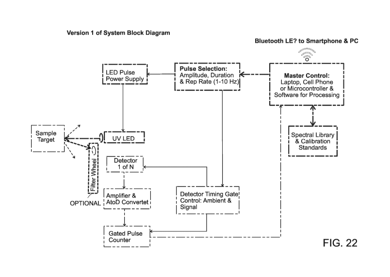
shown in FIG. 22,
the system block diagram depicts a sample target being subjected to UV LED.
Detector 1 of N
detectors detects the signal from the sample target and sends the signal to
the Amplifier & AtoD
(analog to digital) Converter to be amplified and converted to digital. The
signal then goes to the
gated pulse controller and then on to the master control. The master control
may be any suitable
means and preferably may be a laptop, cell phone, or microcontroller and
software for
processing. The master control is in communication with the spectral library
and the calibration
standards of the present invention. The master control may send results via
Bluetooth LE,
smartphones, and personal computers. The master control also communicates with
the pulse
selection for amplitude, duration and rep rate (1-10 Hz). The pulse selection
communications to
the LED pulse power supply which inputs to the UV LED. The pulse selection
also
communicates with the detector timing gate control for the ambient and signal
timing. A filter
wheel may optionally be arranged between the sample target and Detector 1 of N
detectors.
19
CA 03102240 2020-12-01
WO 2019/232448 PCT/US2019/035014
The timing concept of the present invention is shown in FIG. 23. A nominal
full cycle is
1 to 5 Hz. the LED duty cycle is less than 3%. At the zero time reference
point, the LED and
Detectors are OFF. Once the LED and Detectors are ON, the LED rise/fall should
be less than 5
[Ls, with 0 to full, or full to 0 as options. The ambient and signal
measurements are the same
length of time.
Multiple system configurations are further discussed and exemplified herein
and the
skilled artisan will appreciate that the configurations are exemplary and non-
limiting
embodiments of the invention. In a particular exemplary embodiment, the
present invention
identifies and quantifies certain targets using a single wavelength excitation
and six (6) channel
detection between the 300-400 nm range. This involves characterizing the
spectral properties of
these targets. In certain embodiments, the invention uses single channel
detection, or 1 channel
detection. Single channel detection allows for indication of presence or
absence of a biological
substance or microorganism.
In order to determine the feasibility of using native fluorescence to detect
potential
targets at low concentrations, a screening protocol was constructed to
determine various criteria
to identify possible targets. A photometric standard was created to correlate
various detection
schemes (spectrophotometers / various detectors / various optical layout),
which is not
commercially available. This allows for determination of possible limits of
detection (in parts per
million (ppm)/parts per billion) depending on hardware parameters (PMT/Si
Detector). The
potential targets may be determined (see FIG. 16 for exemplary targets).
Potential challenges
addressed and overcome include environmental factors (pH, salt, temperature),
and quenching at
lower concentrations than previously reported.
A model was created based on the concentration study of individual targets in
tap water.
The robustness of the model was tested by artificially added noise and
determining limits of
detection. A simulated engine was created based on real data to generate
initial hardware
parameters (band passes, optimized laser excitation signal/noise, etc.) and
test robustness of
initial quantification algorithm. From samples made in lab, BPA was able to be
detected and
quantified down to 0.023 ppm cross-validated with optimized laser excitation
and band passes. A
key finding for the algorithm development was to be able to construct a
library for quantification
of new samples. The calibration library in house may quantify BPA down to
0.023 ppm in water
with a similar environment (FIG. 17).
CA 03102240 2020-12-01
WO 2019/232448 PCT/US2019/035014
The bandpass configuration was determined that would separate out microbiology
from
each other and from amino acid signatures. Certain excitations separate out
different
microbiological strains from each other as well as raw amino acid signatures
using six (6)
channel detection (FIG. 18).
Water quality may be discerned with a single excitation and six (6) channel
detection of
native auto fluorescence. Various hardware configurations provide the water
quality information.
Final determination of water quality may be verified by a water lab (FIG. 19).
In certain embodiments, the hardware specifications included the following
examples for
LED and detectors. As an exemplary embodiment, the LED was selected from
continuous mode
¨ 100mA ¨ lmW and pulsed mode, 4Hz, 2% Duty cycle (5ms on). The max drive
current was
¨350mA with Thor Lab power supply and Rigol pulse generator.
As an exemplary embodiment, the detector was selected from Hamamatsu S12698-01
photodiode, Hamamatsu MPPC, and STS-UV Ocean optics fiber coupled spectrometer
(FIG.
20).
The configuration utilizes the front surface detection with an LED light
source (278nm)
focused onto a cuvette holder and detector assembly placed at approximately 35
degrees away
from the incoming beam to collect the fluorescence light. With this
configuration, the resulting
fluorescence output of the standard biphenyl in ethanol was determined in
absolute values
(uW/nanojoules). Therefore, the range of signals expected to be found as a
function of target of
interest (FIG. 21) was calculated. The silicon detector was suitable and a
preferable detector may
be the MPPC (APD). At lmW (100mA) a sample, 101\2 CFU (colony forming units of
bacteria)
produce about 3 pW of fluorescence in the 300-400 nm range. The angle
dependence may further
be optimized.
In certain embodiments, a grating system may be used instead of filters and
how the light
is split and filtered would change slightly. Using a grating option may allow
access to more
wavelengths of interest.
Algorithms and Software
The present invention also uses algorithms in detection, identification, and
quantification
of target analytes. The initial target screens for the algorithm include
determination of whether
the target fluoresce in the region of interest, e.g. in water, whether the
fluorescence is strong
21
CA 03102240 2020-12-01
WO 2019/232448 PCT/US2019/035014
enough, e.g. to EPA/FDA limits, and whether samples contaminated with targets
that are
indicative of real world scenarios may be experimentally created.
Identification, classification, and quantification are then based on
fluorescence spectrum.
This requires a model based on experimentally derived data. The data in the
model is correlated
to and indicative of real world scenarios in order to ensure robustness and
high confidence levels
of the models. For example, fluorescence can change based on
temperature/pH/salt other
molecular interactions and models of the invention account for various
conditions (i.e. only tap
water, only pool water, etc.).
In an embodiment of the present invention, an algorithm may used by a user who
identifies a source for the medium. The sample may then be scanned and
compared to an in-
house database of sources. If the source is within the "threshold", the water
quality value may be
reported. If the source is outside of the "threshold", the source may be
identified as an outlier.
The source may then be compared to an in-house database of contaminants, which
is in
communication with the samples in the contaminant and chemical libraries. If
the contaminant is
identified, then the contaminant may further be quantified.
FIG. 24 shows an embodiment of an algorithm used in the present invention. The
user
identifies a source for the medium. The sample is scanned. The sample is then
compared to
defined source, a step which uses a library of sources that is already in-
house. If the sample is not
detected as an outlier, then the water quality value is reported. If the
sample is detected as an
outlier, the sample is compared to the library of contaminants, which uses a
library of
contaminants in various sources. If there is a single contaminant
classification, then
quantification commences using the in-house concentration study of the
contaminant. If there
was no single contaminant classification, then it is considered whether the
spectra may be broken
down to various components. If a known contaminant has not been identified,
then a water
quality value is output. If a known contaminant has been identified, then a
determination of
whether the quantification is within error is made. If the quantification is
within error, then the
contaminant is identified with a concentration. If the quantification is not
within error, then
merely the contaminant is identified.
Various known statistical pattern recognition methods can be used in
conjunction with
the present invention. For example, the following statistical methods,
training sets, machine
learning techniques, and comparisons to known spectra may be used.
22
CA 03102240 2020-12-01
WO 2019/232448 PCT/US2019/035014
An important feature of the methods of the invention is the ability to analyze
heterogeneous samples using a fluorescence or an absorption spectrum.
Fluorescence
microscopy measures the fluorescence of a particular compound when given a
particular
wavelength. As such, the wavelength that reaches the detector is a different
wavelength than
used to shine the sample. Fluorescent compounds can absorb light at a
particular wavelength and
emit light at a higher wavelength, with some energy being lost by the compound
to the
surroundings. Absorbance spectroscopy measures how much of a particular
wavelength of light
gets absorbed by a sample. It's usually used to measure the concentration of a
compound in a
sample. As such, the more light that is absorbed, the higher the concentration
of the compound in
the sample.
The methods for analyzing the fluorescence or absorption spectrum are based
upon the
principles that each element in a mixture has its own spectrum and that each
element has a
specific absorption coefficient. The methods of the invention then correlate
concentration with
absorption. Particularly, the concentration of a compound can be determined
with the knowledge
of the compound's absorption coefficient. This relationship, in the most basic
sense, can be
illustrated by Beer's Law:
A = cbc,
wherein A is absorbance, c is concentration (mol/L;M), b is pathlength, and
is the molar
absorptivity (or extinction coefficient). Molar absorptivity is the
characteristic of a substance that
tells how much light is absorbed at a particular wavelength.
When measuring the fluorescence or absorption of a heterogeneous mixture, the
sum of
the absorption coefficient values for each element is measured at the same
time. Thus, in order to
determine the concentration, the linear combination of all spectra of the
elements needs to be
determined. The analysis then takes into account the interaction of elements
with one another.
The analysis then accounts for the fact that despite each element having a
different spectrum,
their optical absorbance can be the same. For example, one element may be
present at 1mM and
another may also be present at 1mM, both of which can be 1000 times less than
the total value,
or signal, of the mixture.
In one embodiment, deconvolution can be used to enable determination of
concentrations. Deconvolution is an algorithm-based process used to reverse
the effects of
23
CA 03102240 2020-12-01
WO 2019/232448 PCT/US2019/035014
convolution on recorded data. See, e.g., O'Haver T. "Intro to Signal
Processing - Deconvolution".
University of Maryland at College Park. Retrieved 2016-09-13, the content of
which is
incorporated by reference herein in its entirety. In general, the object of
deconvolution is to find
the solution of a convolution equation of the form: f * g = h, wherein h is
some recorded value,
and f is the desired value, but has been convolved with some other value g
before it was
recorded. The function g might represent the interaction between two elements.
If g is known,
then deterministic deconvolution can be performed. However, if g is not known
in advance, then
it will need to be estimated using, for example, statistical estimation. In
actual practice, the
situation is usually closer to: (f* g) + c = h, wherein is noise that has
entered the recorded
value. The lower the signal-to-noise ratio, the worse the estimate of the
deconvolved value will
be.
Methods for deconvoluting the data in accordance with the present disclosure
include the
use of, for example, principal component analysis (PCA). PCA is a statistical
procedure that
reduces the dimensionality of a data set by transforming the data to a new set
of variable
(principal components) that summarize the features of the data. See, for
example, Jolliffe, 1986,
Principal Component Analysis, Springer, New York. PCA uses an orthogonal
transformation to
convert a set of observations of possibly correlated variables into a set of
values of linearly
uncorrelated variables called principal components. The number of principal
components is less
than or equal to the number of original variables. This transformation is
defined in such a way
that the first principal component has the largest possible variance (that is,
accounts for as much
of the variability in the data as possible), and each succeeding component in
turn has the highest
variance possible under the constraint that it is orthogonal to the preceding
components. The
resulting vectors are an uncorrelated orthogonal basis set. PCA is sensitive
to the relative scaling
of the original variables. The first few principal components ("PCs") capture
most of the
variation in the data set. In contrast, the last few PCs are often assumed to
capture only the
residual 'noise' in the data. PCA is discussed in more detail below with
respect to use of
databases in the analysis of data. It is also to be understood that other
statistical analysis methods
known in the art, such as those discussed in more detail below, can be used.
Exemplary analyses
are also described below.
In the present invention, the presence of a target analyte and its
concentration may be
reported. In certain embodiments, the methods of the invention can involve the
use of a computer
24
CA 03102240 2020-12-01
WO 2019/232448 PCT/US2019/035014
system (described in more detail below) to generate a report that includes a
determination of the
presence of and concentration of the target analyte. The computer system may
perform one or
more of the following steps: analyzing the sample to provide spectral data on
the one or more
target analytes received by the single detector, retrieving known spectral and
concentration data,
applying the known data to the spectral data received by the detector, and
generating a report
comprising the concentration of the one or more target analytes. The report
may be sent to an
output device such as a display monitor or a printer.
Converting a fluorescence or an absorption spectrum to a concentration reading
Sample analysis results are generally reported in concentrations of different
analytes in a
sample. The present disclosure provides for a method in which spectral data
can be converted
into concentration for a target analyte through the comparison of the spectral
data to a database
comprising known spectra already associated with concentration levels of the
target analyte.
Because methods of the present invention may involve the use of a single
detector that receives a
light beam after it has passed through the sample, the spectral data may
include total absorption
or fluorescence data. Optionally, more than one detector, e.g. inclusive up to
at least six or more
detectors, may be used. Typically, when converting spectral data to
concentration, careful
measurement of a "training set" of samples is performed. A mathematical
multivariate model is
then constructed for individual components to be eventually used to evaluate
unknown
concentrations.
In certain embodiments, the database will contain chemical composition and
spectral data
from a training set. The training set can comprise a number of samples from
which the chemical
composition and spectral behavior are known. Chemical composition data can be
determined
through any means known in the art, such as, for example, a chemical component
analyzer
(CCA). Spectral behavior can be determined through any means known in the art,
including the
apparatuses and methods described herein.
Using the spectral data obtained, the concentration of the components (e.g.
elements of
blood plasma) can be determined. This information is compiled in a database
and absorption or
fluorescence/concentration curves for the various components/elements can be
determined and
also contained in the database.
CA 03102240 2020-12-01
WO 2019/232448 PCT/US2019/035014
Once the database is compiled, the concentration of one or more target
analytes in a
heterogeneous sample can be determined. This is done by comparing the spectral
data obtained
according to the present disclosure to the database comprising the known
spectra already
associated with concentration levels of the target analyte.
This aspect of the present disclosure is especially amenable for
implementation using a
computer. The computer or CPU is able to compare the spectral data of the
target analyte(s) to
the reference spectral data to thereby provide the concentration of the target
analyte(s). Such
systems generally include a central processing unit (CPU) and storage coupled
to the CPU. The
storage stores instructions that when executed by the CPU, cause the CPU to
accept as input,
spectral data obtained by the detector. The executed instructions also cause
the computer to
provide the concentration of the target analyte as a result of inputting the
sample data into an
algorithm, or pattern recognition platform, trained on the reference set of
known spectral data.
In certain embodiments, the reference set is stored at a remote location
separate from the
computer and the computer communicates across a network to access the
reference set in order
to determine the concentration. In other embodiments, the reference set is
stored locally within
the computer and the computer accesses the reference set within the computer
in order to make
the determination.
The pattern recognition platform can be based on any appropriate pattern
recognition
method that is capable of receiving input data representative of a spectral
data from the sample
being analyzed and providing the concentration of the target analyte in the
sample as an output.
The pattern recognition program is trained with training data from a reference
set of known
spectral data and concentrations from various analytes. In some embodiments, a
test sample
having known concentration and spectral data can be used to test the accuracy
of the platform
recognition platform obtained using the training data.
Various known statistical pattern recognition methods can be used in
conjunction with
the present disclosed methods. Suitable statistical methods include, without
limitation, principal
component analysis (PCA), logic regression, ordinal logistic regression,
linear or quadratic
discriminant analysis, clustering, nearest neighbor classifier analysis, and
Cox Proportional
Handling. Non-limiting examples of implementing particular pattern recognition
platforms using
the various statistical are provided herein.
26
CA 03102240 2020-12-01
WO 2019/232448 PCT/US2019/035014
In some embodiments, the pattern recognition platform is based on a regression
model,
preferably a logistic regression model. Some embodiments of the present
invention provide
generalizations of the logistic regression model that handle multicategory
(polychotomous)
responses. Such embodiments can be used to discriminate between three or more
elements. Such
regression models use multicategory logit models that simultaneously refer to
all pairs of
categories, and describe the odds of response in one category instead of
another. Once the model
specifies logits for a certain (J-1) pairs of categories, the rest are
redundant. See, for example,
Agresti, An Introduction to Categorical Data Analysis, John Wiley & Sons,
Inc., 1996, New
York, Chapter 8, which is hereby incorporated by reference.
Linear discriminant analysis (LDA) attempts to classify sample according to
its elemental
composition based on certain spectral properties. In other words, LDA tests
whether measured
spectral data predicts categorization. LDA typically requires continuous
independent variables
and a dichotomous categorical dependent variable. In the present disclosure,
the spectral data for
select wavelengths across a number of elements in the training population
serve as the requisite
continuous independent variables. The concentration of each of the elements of
the training
population serves as the dichotomous categorical dependent variable.
LDA seeks the linear combination of variables that maximizes the ratio of
between-group
variance and within-group variance by using the grouping information.
Implicitly, the linear
weights used by LDA depend on how the spectral data for a wavelength separates
between, for
example, two different elements and how the spectral data correlates with
spectral data for other
wavelengths. For example, LDA can be applied to the data matrix of the N
members (e.g.
elements) in the training sample by K wavelengths in a number of wavelengths
described in the
present invention. Then, the linear discriminant of each member of the
training population is
plotted. Ideally, those members of the training population representing a
first subgroup (e.g. a
first element) will cluster into one range of linear discriminant values and
those members of the
training population representing a second subgroup (e.g. a second element)
will cluster into a
second range of linear discriminant values. The LDA is considered more
successful when the
separation between the clusters of discriminant values is larger. For more
information on linear
discriminant analysis, see Duda, Pattern Classification, Second Edition, 2001,
John Wiley &
Sons, Inc; and Hastie, 2001, The Elements of Statistical Learning, Springer,
New York;
Venables & Ripley, 1997, Modern Applied Statistics with s-plus, Springer, New
York.
27
CA 03102240 2020-12-01
WO 2019/232448 PCT/US2019/035014
Quadratic discriminant analysis (QDA) takes the same input parameters and
returns the
same results as LDA. QDA uses quadratic equations, rather than linear
equations, to produce
results. LDA and QDA are interchangeable, and which to use is a matter of
preference and/or
availability of software to support the analysis. Logistic regression takes
the same input
parameters and returns the same results as LDA and QDA.
In some embodiments of the present disclosure, decision trees are used to
classify
elements using spectral data for a selected set of wavelengths. Decision tree
algorithms belong to
the class of supervised learning algorithms. The aim of a decision tree is to
induce a classifier (a
tree) from real-world example data. This tree can be used to classify unseen
examples (determine
elements in a sample of unknown composition) which have not been used to
derive the decision
tree. A decision tree is derived from training data. An example contains
values for the different
attributes and what class the example belongs. In one embodiment, the training
data is spectral
data from a number of wavelengths across the training population (e.g. various
elements)
In general there are a number of different decision tree algorithms, many of
which are
described in Duda, Pattern Classification, Second Edition, 2001, John Wiley &
Sons, Inc.
Decision tree algorithms often require consideration of feature processing,
impurity measure,
stopping criterion, and pruning. Specific decision tree algorithms include,
cut are not limited to
classification and regression trees (CART), multivariate decision trees, ID3,
and C4.5.
In one approach, when an exemplary embodiment of a decision tree is used, the
spectral
data for a representative number of wavelengths across a training population
is standardized to
have mean zero and unit variance. The members (e.g. elements) of the training
population are
randomly divided into a training set and a test set. For example, in one
embodiment, two thirds
of the members of the training population are placed in the training set and
one third of the
members of the training population are placed in the test set. The spectral
data for a
representative number of wavelengths are used to construct the decision tree.
Then, the ability
for the decision tree to correctly classify members in the test set is
determined. In some
embodiments, this computation is performed several times for a given number of
wavelengths. In
each iteration of the computation, the members of the training population are
randomly assigned
to the training set and the test set. Then, the quality of the combination of
traits is taken as the
average of each such iteration of the decision tree computation.
28
CA 03102240 2020-12-01
WO 2019/232448 PCT/US2019/035014
In some embodiments, the spectral data across a representative number of
wavelengths is
used to cluster a training set. For example, consider the case in which ten
wavelengths are used.
Each member m (e.g. element) of the training population will have absorption
or
fluorescence/concentration values for each of the ten wavelengths. Such values
from a member
m in the training population define the vector:
Xi m X2m X3m X4m X5m X6m X7m X8m X9m X 10m
where X. is the fluorescence or absorbance/concentration of the ith wavelength
in element m. If
there are m elements in the training set, selection of i wavelengths will
define m vectors. Those
members of the training population that exhibit similar absorption or
fluorescence/concentration
curves across the training group will tend to cluster together. A particular
combination of
wavelengths of the present invention is considered to be a good classifier in
this aspect of the
present disclosure when the vectors cluster into the trait groups (elements)
found in the training
population. For instance, if the training population includes two different
elements, a clustering
classifier will cluster the population into two groups, with each group
uniquely representing
either element.
Clustering is described on pages 211-256 of Duda and Hart, Pattern
Classification and
Scene Analysis, 1973, John Wiley & Sons, Inc., New York. As described in
Section 6.7 of
Duda, the clustering problem is described as one of finding natural groupings
in a dataset. To
identify natural groupings, two issues are addressed. First, a way to measure
similarity (or
dissimilarity) between two samples is determined. This metric (similarity
measure) is used to
ensure that the samples in one cluster are more like one another than they are
to samples in other
clusters. Second, a mechanism for partitioning the data into clusters using
the similarity measure
is determined.
Similarity measures are discussed in Section 6.7 of Duda, where it is stated
that one way
to begin a clustering investigation is to define a distance function and to
compute the matrix of
distances between all pairs of samples in a dataset. If distance is a good
measure of similarity,
then the distance between samples in the same cluster will be significantly
less than the distance
between samples in different clusters. However, as stated on page 215 of Duda,
clustering does
not require the use of a distance metric. For example, a nonmetric similarity
function s(x, x') can
be used to compare two vectors x and x'. Conventionally, s(x, x') is a
symmetric function whose
29
CA 03102240 2020-12-01
WO 2019/232448 PCT/US2019/035014
value is large when x and x' are somehow "similar". An example of a nonmetric
similarity
function s(x, x') is provided on page 216 of Duda.
Once a method for measuring "similarity" or "dissimilarity" between points in
a dataset
has been selected, clustering requires a criterion function that measures the
clustering quality of
any partition of the data. Partitions of the data set that extremize the
criterion function are used to
cluster the data. See page 217 of Duda. Criterion functions are discussed in
Section 6.8 of Duda.
More recently, Duda et al., Pattern Classification, 2nd edition, John Wiley &
Sons, Inc.
New York, has been published. Pages 537-563 describe clustering in detail.
More information on
clustering techniques can be found in Kaufman and Rousseeuw, 1990, Finding
Groups in Data:
An Introduction to Cluster Analysis, Wiley, New York, N.Y.; Everitt, 1993,
Cluster analysis (3d
ed.), Wiley, New York, N.Y.; and Backer, 1995, Computer-Assisted Reasoning in
Cluster
Analysis, Prentice Hall, Upper Saddle River, N.J. Particular exemplary
clustering techniques that
can be used in the present invention include, but are not limited to,
hierarchical clustering
(agglomerative clustering using nearest-neighbor algorithm, farthest-neighbor
algorithm, the
average linkage algorithm, the centroid algorithm, or the sum-of-squares
algorithm), k-means
clustering, fuzzy k-means clustering algorithm, and Jarvis-Patrick clustering.
In some embodiments, the pattern recognition platform is based on PCA, as
briefly
described above. In such an approach, vectors for a selected set of
wavelengths can be selected in
the same manner described for clustering above. In fact, the set of vectors,
where each vector
represents spectral data for the select wavelengths from a particular member
(e.g. element) of the
training populations, can be considered a matrix. In some embodiments, this
matrix is
represented in a Free-Wilson method of qualitative binary description of
monomers (Kubinyi,
1990, 3D QSAR in drug design theory methods and applications, Pergamon Press,
Oxford, pp
589-638), and distributed in a maximally compressed space using PCA so that
the first principal
component (PC) captures the largest amount of variance information possible,
the second
principal component (PC) captures the second largest amount of all variance
information, and so
forth until all variance information in the matrix has been accounted for.
Then, each of the vectors (where each vector represents a member of the
training
population) is plotted. Many different types of plots are possible. In some
embodiments, a one-
dimensional plot is made. In this one-dimensional plot, the value for the
first principal
component from each of the wavelengths is plotted. In this form of plot, the
expectation is that
CA 03102240 2020-12-01
WO 2019/232448 PCT/US2019/035014
members of a first group (e.g. a first element within the blood plasma) will
cluster in one range
of first principal component values and members of a second group (e.g., a
second element
within the blood plasma) will cluster in a second range of first principal
component values.
In one example, the training population comprises two groups: a first element
and a
second element. The first principal component is computed using the spectral
data for the select
wavelengths of the present disclosure across the entire training population
data set. Then, each
member of the training set is plotted as a function of the value for the first
principal component.
In this example, those members of the training population in which the first
principal component
is positive are the first element and those members of the training population
in which the first
principal component is negative are the second element.
In some embodiments, the members of the training population are plotted
against more
than one principal component. For example, in some embodiments, the members of
the training
population are plotted on a two-dimensional plot in which the first dimension
is the first
principal component and the second dimension is the second principal
component. In such a two-
dimensional plot, the expectation is that members of each subgroup represented
in the training
population will cluster into discrete groups. For example, a first cluster of
members in the two-
dimensional plot will represent a first element, a second cluster of members
in the two-
dimensional plot will represent a second element, and so forth.
In some embodiments, the members of the training population are plotted
against more
than two principal components and a determination is made as to whether the
members of the
training population are clustering into groups that each uniquely represents a
subgroup found in
the training population. In some embodiments, principal component analysis is
performed by
using the R mva package (Anderson, 1973, Cluster Analysis for applications,
Academic Press,
New York 1973; Gordon, Classification, Second Edition, Chapman and Hall, CRC,
1999.).
Principal component analysis is further described in Duda, Pattern
Classification, Second
Edition, 2001, John Wiley & Sons, Inc.
Nearest neighbor classifiers are another statistical method on which the
pattern
recognition platform can be based. Nearest neighbor classifiers are memory-
based and require no
model to be fit. Given a query point x0, the k training points x(r), r, . . .
, k closest in distance to
x0 are identified and then the point x0 is classified using the k nearest
neighbors. Ties can be
31
CA 03102240 2020-12-01
WO 2019/232448 PCT/US2019/035014
broken at random. In some embodiments, Euclidean distance in feature space is
used to
determine distance as:
d(i)=11x(i)¨x011.
Typically, when the nearest neighbor algorithm is used, the expression data
used to
compute the linear discriminant is standardized to have mean zero and variance
1. In the present
disclosure, the members of the training population are randomly divided into a
training set and a
test set. For example, in one embodiment, two thirds of the members of the
training population
are placed in the training set and one third of the members of the training
population are placed
in the test set. Profiles represent the feature space into which members of
the test set are plotted.
Next, the ability of the training set to correctly characterize the members of
the test set is
computed. In some embodiments, nearest neighbor computation is performed
several times for a
set number of wavelengths. In each iteration of the computation, the members
of the training
population are randomly assigned to the training set and the test set. Then,
the quality of the
spectral data for the set number of wavelengths is taken as the average of
each such iteration of
the nearest neighbor computation.
The nearest neighbor rule can be refined to deal with issues of unequal class
priors,
differential misclassification costs, and feature selection. Many of these
refinements involve
some form of weighted voting for the neighbors. For more information on
nearest neighbor
analysis, see Duda, Pattern Classification, Second Edition, 2001, John Wiley &
Sons, Inc; and
Hastie, 2001, The Elements of Statistical Learning, Springer, New York.
The pattern classification and statistical techniques described above are
merely examples
of the types of models that can be used to construct a model for
classification. It is to be
understood that any statistical method can be used in accordance with the
present disclosure.
Moreover, combinations of these described above also can be used. Further
detail on other
statistical methods and their implementation are described in U.S. Patent
Application No.
11/134,688, incorporated by reference herein in its entirety.
Computer implementation
Other embodiments are within the scope and spirit of the invention. For
example, due to
the nature of software, functions described above can be implemented using
software, hardware,
32
CA 03102240 2020-12-01
WO 2019/232448 PCT/US2019/035014
firmware, hardwiring, or combinations of any of these. Features implementing
functions can also
be physically located at various positions, including being distributed such
that portions of
functions are implemented at different physical locations. Steps of the
invention may be
performed using dedicated medical imaging hardware, general purpose computers,
or both. As
one skilled in the art would recognize as necessary or best-suited for
performance of the methods
of the invention, computer systems or machines of the invention include one or
more processors
(e.g., a central processing unit (CPU) a graphics processing unit (GPU) or
both), a main memory
and a static memory, which communicate with each other via a bus. A computer
device generally
includes memory coupled to a processor and operable via an input/output
device.
Exemplary input/output devices include a video display unit (e.g., a liquid
crystal display
(LCD) or a cathode ray tube (CRT)). Computer systems or machines according to
the invention
can also include an alphanumeric input device (e.g., a keyboard), a cursor
control device (e.g., a
mouse), a disk drive unit, a signal generation device (e.g., a speaker), a
touchscreen, an
accelerometer, a microphone, a cellular radio frequency antenna, and a network
interface device,
which can be, for example, a network interface card (NIC), Wi-Fi card, or
cellular modem.
Memory according to the invention can include a machine-readable medium on
which is
stored one or more sets of instructions (e.g., software), data, or both
embodying any one or more
of the methodologies or functions described herein. The software may also
reside, completely or
at least partially, within the main memory and/or within the processor during
execution thereof
by the computer system, the main memory and the processor also constituting
machine-readable
media. The software may further be transmitted or received over a network via
the network
interface device.
While the machine-readable medium can in an exemplary embodiment be a single
medium, the term "machine-readable medium" should be taken to include a single
medium or
multiple media (e.g., a centralized or distributed database, and/or associated
caches and servers)
that store the one or more sets of instructions. The term "machine-readable
medium" shall also
be taken to include any medium that is capable of storing, encoding or
carrying a set of
instructions for execution by the machine and that cause the machine to
perform any of the
methodologies of the present invention. The term "machine-readable medium"
shall accordingly
be taken to include, but not be limited to, solid-state memories (e.g.,
subscriber identity module
(SIM) card, secure digital card (SD card), micro SD card, or solid-state drive
(SSD)), optical and
33
CA 03102240 2020-12-01
WO 2019/232448 PCT/US2019/035014
magnetic media, and any other tangible storage media. Preferably, computer
memory is a
tangible, non-transitory medium, such as any of the foregoing, and may be
operably coupled to a
processor by a bus. Methods of the invention include writing data to
memory¨i.e., physically
transforming arrangements of particles in computer memory so that the
transformed tangible
medium represents the tangible physical objects¨e.g., the arterial plaque in a
patient's vessel.
As used herein, the word "or" means "and or or", sometimes seen or referred to
as
"and/or", unless indicated otherwise.
As used in any embodiment herein, the term "module" may refer to software,
firmware
and/or circuitry configured to perform any of the aforementioned operations.
Software may be
embodied as a software package, code, instructions, instruction sets and/or
data recorded on non-
transitory computer readable storage medium. Firmware may be embodied as code,
instructions
or instruction sets and/or data that are hard-coded (e.g., nonvolatile) in
memory devices.
"Circuitry", as used in any embodiment herein, may comprise, for example,
singly or in
any combination, hardwired circuitry, programmable circuitry such as computer
processors
comprising one or more individual instruction processing cores, state machine
circuitry, and/or
firmware that stores instructions executed by programmable circuitry. The
modules may,
collectively or individually, be embodied as circuitry that forms part of a
larger system, for
example, an integrated circuit (IC), system on-chip (SoC), desktop computers,
laptop computers,
tablet computers, servers, smart phones, etc.
Any of the operations described herein may be implemented in a system that
includes one
or more storage mediums having stored thereon, individually or in combination,
instructions that
when executed by one or more processors perform the methods. Here, the
processor may
include, for example, a server CPU, a mobile device CPU, and/or other
programmable circuitry.
Also, it is intended that operations described herein may be distributed
across a plurality of
physical devices, such as processing structures at more than one different
physical location.
The storage medium may include any type of tangible medium, for example, any
type of
disk including hard disks, floppy disks, optical disks, compact disk read-only
memories (CD-
ROMs), compact disk rewritables (CD-RWs), and magneto-optical disks,
semiconductor devices
such as read-only memories (ROMs), random access memories (RAMs) such as
dynamic and
static RAMs, erasable programmable read-only memories (EPROMs), electrically
erasable
programmable read-only memories (EEPROMs), flash memories, Solid State Disks
(SSDs),
34
CA 03102240 2020-12-01
WO 2019/232448 PCT/US2019/035014
magnetic or optical cards, or any type of media suitable for storing
electronic instructions.
Other embodiments may be implemented as software modules executed by a
programmable
control device. The storage medium may be non-transitory.
As described herein, various embodiments may be implemented using hardware
elements, software elements, or any combination thereof. Examples of hardware
elements may
include processors, microprocessors, circuits, circuit elements (e.g.,
transistors, resistors,
capacitors, inductors, and so forth), integrated circuits, application
specific integrated circuits
(ASIC), programmable logic devices (PLD), digital signal processors (DSP),
field programmable
gate array (FPGA), logic gates, registers, semiconductor device, chips,
microchips, chip sets, and
so forth.
Methods of Use
In certain aspects, the present invention is directed to methods of providing
information
regarding a medium. The method comprises providing a system comprising a light-
emitting
diode operating at a single wavelength in a deep ultraviolet (UV) range for
excitation of a target
in a medium and a plurality of semiconductor photodetectors. The system may be
configured
such that each semiconductor photodetector detects only a subset of emission
from the excited
target. A medium comprising one or more target analytes may be exposed to at
least a single
wavelength in the deep UV spectrum from the light-emitting diode of the system
to thereby
excite the target analyte in the medium. The method may further comprise
detecting emission
from the excited one or more target analytes via the plurality of
semiconductor photodetectors of
the system to thereby produce emission data and processing the emission data,
thereby providing
information regarding the medium.
In certain embodiments, the medium may be selected from the group consisting
of a
biofluid, water, an aluminum surface, a stainless steel surface, a granite
surface, a ceramic
surface, a plastic surface, and a metallic surface. In an embodiment, the
target analyte may be
selected from the group consisting of a microorganism, a biomolecule, and a
chemical. In a
preferred embodiment, the medium is water and the target analyte is one or
more pathogens.
In an embodiment, processing the emission data may comprise identifying
presence of
one or more target analytes in the medium. Processing the emission data may
further comprise
CA 03102240 2020-12-01
WO 2019/232448 PCT/US2019/035014
identifying the one or more target analytes in the medium. Processing the
emission data may
further comprise quantifying the one or more target analytes in the medium.
In certain embodiments, the method may further comprise displaying on a
graphical user
interface results of the processing step.
Moreover, certain embodiments of the invention use emission data to determine
total
microbial load and bioburden measurements. The present invention comprises
directing one or
more wavelengths of light that are each within a deep ultraviolet (UV)
spectrum into a medium
comprising a biological substance to thereby excite the biological substance
in the medium.
Emission is detected from the excited biological substance via one or more
semiconductor
photodetectors, thereby producing deep UV emission data. The deep UV emission
data is
analyzed for presence of a deep UV spectral signature indicative of the
biological substance,
wherein presence of the deep UV spectral signature indicates that the medium
comprises a
biological substance. While excitation may be in the deep UV region, emission
may be in the UV
region, such as in the UVA and UVB regions.
The emission data may be used to determine total microbial load. Microbial
load is the
number and type of microorganisms contaminating an object or organism, such as
non-specific
biological and microbiological contamination. Total microbial load indicates
the microbiology
present in the sample. Emission data may be analyzed for deep UV spectral
signatures indicative
of microbiology. Emission data may be analyzed for deep UV spectral signatures
indicative of
presence and quantity of microbiology. For example, analyzing may include
comparing the UV
spectral signature with a library of UV spectral signatures of varying amounts
and types of
microbiology on or in a variety of media. Systems of the invention may
indicate the total
microbial load in the sample after detecting the UV spectral signatures
indicative of
microbiology.
The emission data may be used to determine bioburden, or the number of
bacteria living
on a surface or within a liquid. Often, bioburden refers to the number of
microorganisms on an
unsterilized surface. Emission data may be analyzed for deep UV spectral
signatures indicative
of presence and quantity of microorganisms. For example, analyzing may include
comparing the
UV spectral signature with a library of UV spectral signatures of varying
amounts and types of
microorganisms on or in a variety of media. Systems of the invention may
indicate the bioburden
36
CA 03102240 2020-12-01
WO 2019/232448 PCT/US2019/035014
in the sample after detecting the UV spectral signatures indicative of the
presence or quantity of
microorganisms.
In some embodiments, the invention provides different detector and filter
configurations
for detection using the target thresholds. For example, a first configuration
uses a single detector
to determine the presence or absence of a target, such as a non-specific
contamination. Such a
single-detector embodiment may encompass a portable detector, such as a
detector used while
traveling. Another configuration uses two detectors to determine and
distinguish between the
presence of microbiology and general biology targets. To specify certain
strains of targets in a
mixture or sample, a configuration of the invention uses seven detectors.
Another configuration
uses seven or more detectors to distinguish between dead and live specified
strains of targets.
As a non-limiting example, the target may be selected from bacteria, fungi,
protein, a
cell, a virus, a nucleic acid, a receptor, a ligand, a hormone, a drug, a
chemical substance, or any
molecule known in the art. For example, the target may be selected from
Benzimidazole, 1-
Naphthol, Carbofuran, Bisphenol A, Carbaryl-d7, Naphthalene, p-xylene,
Tryptophan,
Phenanthrene, Tyrosine, ethylestradiol, Propoxur, Ibuprofen, Beta-estradiol,
Dimethyl phthalate,
Chlopyrifos, Ethylbenzene, Dibutyl phthalate, Benzo[a]pyrene, Benzene,
Biphenyl, 3,5,6-
Trichloro-2-pyridinol, Bisphenol S, Imidazole, hydrocortisone, Toluene,
Alachlor, Atrazine,
QB3 Tap water, Di n octyl phthalate, Acetaminophen, Estrone, Glyphosate, Lead,
Bis(2-
ethylhexyl) phthalate, clarithromycin, Trihalomethane, diisodecylphthalate.
phenylalanine,
Heptachlor, testosterone, dieldrin, Tozaphenel, Aldrin, DTT, cortisol, and
Endrin.
In certain embodiments, the target is a pathogen, or pathogenic bacteria or
fungi. A
pathogen is a biological agent, such as a microorganism (e.g. bacterium or
protozoan), that
causes disease or illness to its host. In other embodiments, the target is a
gram positive or gram
negative bacteria.
Exemplary fungal species include species from the Candida genus, Aspergillus
genus,
and Cryptococcus genus. In particular embodiments, the specific fungal species
include C.
albicans, C. glabarata, C. parapsilosis, C. tropicalis, C. krusei,
Cryptococcus neoformans, and
Cryptococcus gattii.
Exemplary bacteria include bacteria of the Escherichia genus, Listeria genus,
Clostridium
genus, Enterobacteriaceae family, Mycobacterium genus, Shigella genus,
Borrelia genus,
Campylobacter genus, Bacillus genus, Salmonella genus, Enterococcus genus,
Streptococcus
37
CA 03102240 2020-12-01
WO 2019/232448 PCT/US2019/035014
genus (such as Pneumococcus), Acinetobacter genus, Strenotrophomonas genus,
Pseudomonas
genus, Neisseria genus, and Haemophilus genus, and a combination thereof. The
method may
also be used to detect the mecA gene, which is a bacterial gene associated
with antibiotic
resistance.
Pathogen Detection and Identification
The present inventions allow for pathogen detection and identification.
Available
technologies merely provide an indication of yes or no as to whether there is
biological material
present. With the present invention, bandpass configuration allows for
separation of
microbiology from each other and from amino acid signatures. For example, the
excitation used
in six channel detection can separate out different microbiological strains
from each other, as
well as from raw amino acid signatures. The ability to separate the bacteria
signature without an
amino acid is possible due to separation of the channels.
The unique signature associated with three pathogens was used in the
invention. By
combining known amino acids with pathogen signatures, a configuration was
determined for
detection at ranges where there are no overlapping parts (FIG. 18). As shown
in FIG. 18, when
comparing the excitation of Pathogen 1, Pathogen 2, and Pathogen 3, there were
overlapping
regions and non-overlapping regions. For example, the overlapping regions were
observed at
particular excitations. The non-overlapping regions indicated the unique
signature for each
pathogen. By using separate channels within the deep UV range, emission may be
detected that
differentiates bacteria and other pathogens from one another, thereby allowing
for identification
of the bacteria. If the signals could not be separated, the bacteria and other
pathogens could not
be distinguished and there would only be an indication of whether or not a
biological substance
was present in the sample.
To separate the microbiology targets, Mahalanobis Distance (see FIG. 31) was
implemented using the bacterial and amino acid signatures emission-excitation
matrix (EEM)
spectra. The bacterial signatures and amino acid signatures are shown in FIG.
31. The EEM
signatures were normalized and binned into six channels. The Mahalanobis
Distance analysis
looked for the largest difference between the bacterial and amino acids. The
wavelengths
indicated separated the microbiology from the amino acid signatures.
38
CA 03102240 2020-12-01
WO 2019/232448 PCT/US2019/035014
As an example, the present invention may be used to test for presence of
Staphylococcus
Aureus in tap water. A user may input a source of the medium being tested,
e.g. tap water. The
sample (e.g. tap water) would then be scanned by the apparatus of the present
invention. Results
would be processed by comparing the spectra from the scanned tap water sample
to a database of
known spectra from known sources. If the spectrum for the sample is within the
threshold for a
known source, then the medium quality (e.g. the water quality of the tap
water) is output.
However, if the sample is not within the threshold, then the spectrum for the
sample is compared
to a database of contaminants. For example, the database of contaminants
includes lab-produced
samples of varying concentrations for various bacterial and chemical
components within various
sources.
If the spectrum for the sample is within a threshold for a contamination
source from the
contaminant database, then the contamination may be identified. For instance,
the tap water
sample may match up with a database entry of a known bacterial contamination,
e.g.
Staphylococcus Aureus. The tap water sample would then be identified as having
a contaminant
which is Staphylococcus Aureus.
The contamination may be further quantified based on calculations from
concentration
studies of the contaminants. For example, the concentration of Staphylococcus
Aureus in the tap
water sample would be determined based on concentration studies of
Staphylococcus Aureus in
samples from tap water. The results of the scan in the tap water example would
indicate the
presence of Staphylococcus Aureus as a contaminant and the concentration of
Staphylococcus
Aureus in the tap water sample.
Water analysis
The systems and methods of the invention are applicable to many different
types of
media and surfaces, as already mentioned throughout this application. A
particular area of
interest is water analysis and water quality. In that manner, the present
invention provides a
range of targets that can be detected within different water sources and water
types, such as tap,
bottle, and well water and on aluminum and stainless steel surfaces. Target
contaminants may be
selected from the group consisting of pathogens, amino acids, hormones,
industrial chemicals,
pharmaceuticals, and biomarkers. In an embodiment, urine and saliva matrices
may be analyzed
for the human biomarker analysis. In certain embodiments, for a deeper water
analysis, users
39
CA 03102240 2020-12-01
WO 2019/232448 PCT/US2019/035014
may request a collection kit and EPA certified facilities will email them a
full, easy to
understand, report. Quantification according to the present invention may
positively impact the
creation of cleaner rivers and water sources.
For example, devices of the invention may be used for monitoring and detection
of water
quality in industrial and manufacturing processes. Devices of the invention
may be used for
water quality detection in water kiosks that provide sale of tap water, such
as in developing
countries. Utilities providers may use devices of the invention to ensure
water quality being
provided to customers. Similarly, building owners may use devices of the
invention for
monitoring and detection of water quality within a building, such as to ensure
safe water for
tenants. Devices of the invention may also be used in the food and beverage
industry,
pharmaceutical industry, and healthcare industry.
There is bacterial contamination in tap water. If the water was pure water, no
fluorescence would show when doing an EEM (FIG. 25). Tap water has a residual
signature in
region that is linked to decomposing biological substances. However, pathogens
such as bacteria
and various other targets fluorescence in the region marked by the circle
(FIG. 26).
For example, the present invention may be used to test water quality. A user
may input a
source of the water being tested, e.g. bottled water, tap water from a
particular location, or well
water. The water sample (e.g. bottled water) would then be scanned by the
apparatus of the
present invention. Results would be processed by comparing the spectra from
the scanned water
sample to a database of known spectra from known sources. If the spectrum for
the sample is
within the threshold for a known source, then the water quality is output.
However, if the sample
is not within the threshold, then the spectrum for the sample is compared to a
database of
contaminants. For example, the database of contaminants includes lab-produced
samples of
varying concentrations for various bacterial and chemical components within
various sources.
If the spectrum for the sample is within a threshold for a contamination
source from the
contaminant database, then the contamination may be identified. For instance,
the bottled water
sample may match up with a database entry of a known E. coli contamination in
bottled water.
The water sample would then be identified as having a contaminant which is E.
coli.
The contamination may be further quantified based on calculations from
concentration
studies of the contaminants. For example, the concentration of E. coli in the
water bottle sample
would be determined based on concentration studies of E. coli in water samples
from water
CA 03102240 2020-12-01
WO 2019/232448 PCT/US2019/035014
bottles. The results of the scan in the water bottle example would indicate
the presence of E. coli
as a contaminant and the concentration of E. coli in the water bottle sample.
Food
The present invention may help to shift global agricultural land to being more
than 1%
certified organic. By using detection of the present invention, safe and
environmental practices
may flourish. Awareness may drive markets to efficiency and innovation, and
new markets may
be created for other technologies.
In an example, in consumer signaling, consumers may be empowered by molecular
insight into their food and water. The consumer interest may incentivize
retailers to invest in
detection technology for the present invention. Retailers may drive adoption
throughout the food
and water supply chain such as through the distributor, processor/supplier, or
grower/water
source.
In an embodiment, the spectral database of the present invention may be
valuable to
retailers wanting to gain customer confidence. In an embodiment, the present
invention may
become the standard in molecular scanning. In an embodiment, the present
invention may create
store-specific scanning technology. In an embodiment, a database according to
the present
invention may be accessed for a monthly or yearly fee. In an embodiment, the
present invention
may monetize throughout the supply chain on the back of consumer knowledge and
demand for
cleaner products. Retail stores may rely on such a clean supply chain
reputation and may be
incentivized to integrate the present invention in stores and throughout
suppliers.
Fruit and vegetable pesticide scans are shown in FIG. 27.
For example, the present invention may be used to test for presence of a
pesticide on a
fruit or vegetable. A user may input a source of the medium being tested, e.g.
a fruit or
vegetable. The sample (e.g. an apple) would then be scanned by the apparatus
of the present
invention. Results would be processed by comparing the spectra from the
scanned apple sample
to a database of known spectra from known sources. If the spectrum for the
sample is within the
threshold for a known source, then the medium quality is output. However, if
the sample is not
within the threshold, then the spectrum for the sample is compared to a
database of
contaminants. For example, the database of contaminants includes lab-produced
samples of
varying concentrations for various bacterial and chemical components within
various sources.
41
CA 03102240 2020-12-01
WO 2019/232448 PCT/US2019/035014
If the spectrum for the sample is within a threshold for a contamination
source from the
contaminant database, then the contamination may be identified. For instance,
the apple sample
may match up with a database entry of a known pesticide contamination, e.g.
flazasulfuron. The
apple sample would then be identified as having a contaminant which is
flazasulfuron.
The contamination may be further quantified based on calculations from
concentration
studies of the contaminants. For example, the concentration of flazasulfuron
in the apple sample
would be determined based on concentration studies of flazasulfuron in similar
samples from
apples. The results of the scan in the apple example would indicate the
presence of flazasulfuron
as a contaminant and the concentration of flazasulfuron in the apple sample.
Embodiments of the invention may be used to detect contaminants on foods. As
an
example, the invention may be used to detect pesticides on fruits and
vegetables. For example,
the invention may be used to detect a pesticide on a kale leaf. As an example,
the invention may
be used to detect a bacterial increase on the surface of meats and fish to
determine freshness or
lack thereof.
In an embodiment, the invention may be used to monitor the process wash water
quality
for fresh produce cleaning, such as that completed by food producers and
suppliers, can give
early indications of contamination events. For instance, tested sources may
include well water
directly from a tank, well water at tap, a first wash with biocide, direct
runoff from produce,
waste water at the end of a process line, or water from plastic crate wash. As
another example,
the invention can be used by individuals who wash a head of romaine lettuce
and then scan the
captured water to get an indication of sanity level or safety level of the
romaine lettuce.
Healthcare
The present invention may help to reduce hospital-acquired infections. Each
year, 1.7
million patients are infected by hospitals. A staggering 1 in 25 patients
resulted in approximately
99,000 deaths unrelated to conditions for which the patients were admitted to
the hospitals.
Hospital-acquired infections in the United States result in $38 billion in
extra costs each year.
This needless contamination may be due to unsterilized surfaces in the
hospitals. With the
present invention, target analytes may be detected on surfaces such as
stainless steel and
aluminum. These surfaces are prevalent in hospital settings.
42
CA 03102240 2020-12-01
WO 2019/232448 PCT/US2019/035014
For example, the present invention may be used to test for presence of
Staphylococcus
Aureus on surfaces in a hospital. A user may input a source of the medium
being tested, e.g. an
aluminum surface. The sample (e.g. an aluminum surface) would then be scanned
by the
apparatus of the present invention. Results would be processed by comparing
the spectra from
the scanned aluminum surface sample to a database of known spectra from known
sources. If the
spectrum for the sample is within the threshold for a known source, then the
medium quality is
output. However, if the sample is not within the threshold, then the spectrum
for the sample is
compared to a database of contaminants. For example, the database of
contaminants includes
lab-produced samples of varying concentrations for various bacterial and
chemical components
within various sources.
If the spectrum for the sample is within a threshold for a contamination
source from the
contaminant database, then the contamination may be identified. For instance,
the aluminum
surface sample may match up with a database entry of a known bacterial
contamination, e.g.
Staphylococcus Aureus. The aluminum surface sample would then be identified as
having a
contaminant which is Staphylococcus Aureus.
The contamination may be further quantified based on calculations from
concentration
studies of the contaminants. For example, the concentration of Staphylococcus
Aureus in the
aluminum surface sample would be determined based on concentration studies of
Staphylococcus Aureus in samples from aluminum surfaces. The results of the
scan in the
aluminum surface example would indicate the presence of Staphylococcus Aureus
as a
contaminant and the concentration of Staphylococcus Aureus in the aluminum
surface sample.
Blockchain
In certain embodiments, blockchain technology may be used. Blockchain is a
digital,
decentralized transaction and data management technology, such as described in
Yli-Huumo et
al, Where Is Current Research on Blockchain Technology?¨A Systematic Review,
PLOS ONE,
2016, incorporated herein. Data integrity and authentication are essential
issues in the
Blockchain environment. It is necessary that when data gets sent and verified,
it has not been
altered or tampered with. A private key may be an authentication element. A
smart phone may be
used as a second authentication factor. By using blockchain in the present
invention, sample data
may be shared with the database and tampering of that sample data may be
avoided.
43
CA 03102240 2020-12-01
WO 2019/232448 PCT/US2019/035014
The use of blockchain may be directed to quantified water, quantified food &
agriculture,
quantified environment, and quantified health-home diagnostics. In the
ecosystem according to
the present invention, product, data, and people interact (FIG. 28). Further,
cloud and blockchain
provide an intelligent spectral database of key environmental and human health
markers. The
value and utility of the library of biological/molecular 'signatures'
increases with every scan by a
user. The community of consumers is also important, as users are incentivized
to discover the
molecular universe that surrounds them. These consumers allow for gaining of
insight into their
personal and environmental health while strengthening the product-data
performance of the
present invention.
In certain embodiments, data may be secured on the blockchain, which may be
the first
global intelligent database of key environmental and human health markers
(FIG. 29). Users may
choose what gets written to the blockchain (using a RES token). Scanned data
is private and
hashed by default. Data is immutable and cannot be erased or altered by
anyone. To view data a
"View key" is needed that only the user will have. All data may be geotagged
and time-stamped.
Users may earn RES tokens on data if written to the blockchain and made
available for analysis
(pending SEC rules).
In an embodiment, time-stamped, geo-tagged, and encrypted data may be written
to the
blockchain. Monitoring devices according to the present invention may be
stationed before and
after point-source polluters (FIG. 30). This will establish a guardian network
of real-time,
verified data that cannot be manipulated or deleted.
In an embodiment, consumers may scan whole foods for contaminants. Consumers
may
scan bio-fluids for health markers. In an embodiment, government agencies may
monitor public
waterways. In an embodiment, supermarkets may monitor growers and suppliers.
In an
embodiment, food and beverage processors may scan water and surfaces. In an
embodiment,
hospitals may scan facilities.
Atmospheric Conditions
In particular embodiments, the present invention may be used in different
atmospheric
conditions. Preferably the invention is used under Earth's atmospheric
conditions. The invention
may be used under other atmospheric conditions. As non-limiting examples, the
present
44
CA 03102240 2020-12-01
WO 2019/232448 PCT/US2019/035014
invention may be used for detection of target analytes in media on a space
station, a rocket, on
Mars, or under water.
Detection of Gram+ / Gram-
In certain embodiments, the invention is used to tell the difference between
Granil+ and
Gram/- bacteria. In a non-limiting study, the invention was used on six
bacteria strains. The
gram+ and gram- bacteria species used in a non-limiting study include Listeria
welshimeri,
Listeria seeligeri, Staphylococcus epidermidis, Klebsiella aero genes,
Pseudomonas putida,
Enterobacter, and Escherichia coli. FIG. 32 shows the clustering of spectra of
the gram + and
gram- bacteria species.
EXAMPLES
Example 1
FIG. 1 shows the scanner or detector according to the present invention. The
device is
small and portable. The device includes an indicator, which may be an OLED
display. The
device also includes the lens and detectors, as well as a scan button.
Optionally, there may be a
micro grip texture on the device.
Example 2
FIG. 2 shows side and top views of the OLED display scanner or detector
according to
the present application. The display may be an E-ink display in certain
embodiments. The
display screen may be the user interface for the device, with all components
of the system
included in the detector device. The device is small and portable. The device
is also user-
friendly, as evidenced by the simple scanning button located on a side of the
device.
Example 3
FIG. 3 shows the dimensions of the scanner or detector. The top of the device,
or the
display screen, may measure 76 mm in diameter. The scanning side of the
device, or the side
containing the detector and lens, may measure 45 mm in diameter. The device
may have a
thickness of 35 mm.
CA 03102240 2020-12-01
WO 2019/232448 PCT/US2019/035014
Example 4
FIG. 4 shows the display for results of the sample. The display screen may
have simple,
easy-to-read features. For example, a check may mean that the sample is
cleared, while an X
may mean that the sample is contaminated.
Example 5
FIG. 5 shows the user interface on an external source. The user interface may
be on a
smartphone. Such an embodiment differs from having the user interface
integrated in the detector
device itself. The user interface may also be on other suitable means, such as
an external laptop
computer or tablet.
Example 6
In certain embodiments, the detector of the present invention may be used in a
minilab
setup. As shown in FIG. 6, the minilab may have a housing, a detector, and a
sample slide plate.
As shown in FIG. 7, the minilab may have a housing, a detector, and a sample
cup. The sample
may be placed on the sample slide plate or sample cup and then inserted into
the minilab
housing. The detector may be fitted within the housing to stabilize the
detector while scanning.
Example 7
In certain embodiments, the detector of the present invention may be used in a
wedge
setup. As shown in FIG. 8, the wedge may come in different angles to fit any
surface. The wedge
may use cases (sink, toilet, shelf, wall). The back side of the wedge may
include a VHB tape
layer to strongly stick to any type of surface.
Example 8
In certain embodiments, systems of the invention comprise a housing that has
multiple
components, and one of the components is a base or tripod. In such an
embodiment, the detector
of the present invention may be used in a tripod setup. As shown in FIG. 9,
the detector may be
used for detection of a sample in a small sample cup. As shown in FIG. 10, the
detector may be
used to for detection of a large sample. The tripod setup allows for the
detector to be stabilized
while scanning.
46
CA 03102240 2020-12-01
WO 2019/232448 PCT/US2019/035014
Example 9
As shown in FIG. 11, the detector may be used as a toilet adapter. The toilet
adapter
system may be used to analyze urine within the toilet bowl water. For example,
the adapter may
detect anomalies or diseases within the urine. Measurements may include high
protein content
and urinary tract infections (UTI). In addition, the device may be used as an
early warning
system for diseases and health markers. For example, the device may be used
for disease
detection, as certain cancers may shift the spectral signature of urine in the
UV region. As
another example, the device may be used to detect hormones from ovulation
cycles and
pregnancy markers, such as hCG. Users may be notified of the detection results
by smartphone
notification.
Example 10
As shown in FIG. 12, the detector may be used a sink adapter. This allows
users access to
the water quality within their environment, such as in a home, hospital
setting, or work
environment. People use sinks frequently throughout the day. If a contaminant
is present in the
water source for the sink, there may be increased safety concerns. By
installing the detector on
the plumbing leading to the faucet of a sink, users have the opportunity to
detect the water source
before using the water in the sink. Further, users may opt to obtain sample
data more frequently
or set the detector to scan the water source at specified time periods to
monitor the water quality.
Example 11
FIG. 13 shows an embodiment of an on-line detector. The on-line detector is
useful for
any suitable detection of any water supply source. For example, in an
embodiment, the on-line
detector is coupled to a water source, such as a pipe. In some cases, the
detector is mounted to a
pipe attachment, such as a 2 inch pipe attachment or a T-junction pipe
fitting. The device
provides detection of the liquid inside the pipe. In some examples, similar to
Example 10, the
device is coupled to a pipe for a sink and detects bioburden or total
microbial load in the liquid
leading to the faucet of the sink. The on-line embodiment provides a low
profile with a detection
window for non-disruption mounting and un-mounting of the device.
47
CA 03102240 2020-12-01
WO 2019/232448 PCT/US2019/035014
Example 12
FIG. 14 shows an embodiment of an in-line detector probe. As shown in FIG. 14,
the
probe is inserted into the pipe, with part of the probe disposed within the
pipe, for detection of
bioburden or total microbial load in the liquid flowing through the pipe. The
in-line detector
probe is useful for any suitable detection of any water supply source. In one
non-limiting
example, the probe may be used in industrial settings to monitor water
quality, particularly where
a sink or end-point to a line is unavailable or if water quality should be
monitored in a particular
closed section of a manufacturing process. In another non-limiting example,
the probe may be
used in a building, such as a hotel, to monitor water quality provided in the
building.
Example 13
FIG. 15 shows an embodiment of an off-line, stand-off detector, such as a tap
attachment.
The device monitors water quality from a tap mount on the line, but the device
itself is off-line,
or off of the pipe. The off-line, stand-off detector is useful for any
suitable detection of any water
supply source. Providing the device off-line as a stand-off detector is non-
invasive and
eliminates detector biofouling and allows high accuracy sample readings.
Having a tap mount
that does not contact the water gives the end user the nearest to a pure
result as possible at the
point-of-use and point-of-care. In one non-limiting example, the off-line,
stand-off detector is
used as a tap attachment in a residential setting. In another non-limiting
example, the detector is
used as a tap attachment in a water kiosk.
Example 14
The invention uses deep UV autofluorescence to detect and identify various
strains of
bacteria. Bacteria cells have unique autofluorescence signatures when excited
in the UV region
(see Label-Free bacterial imaging with Deep-UV-laser induced native
fluorescence, Bhartia,
Salas, Hug, Reid, A. Lane, Edwards, Nealson, 2010). Excitation Emission Matrix
(EEM) has
been proposed as a potential tool for water monitoring, but EEM only makes
inferences (see
Fluorescence as a potential monitoring tool for recycled water systems: A
review, Henderson et
al. 2009).
Real-World Example of Contaminated Tap Water
48
CA 03102240 2020-12-01
WO 2019/232448 PCT/US2019/035014
Pure water would show no fluorescence signature when doing an EEM. However,
bacteria and various other contaminants fluoresce in the region marked by the
blue circle. Some
tap waters show a residual signature (green perforated circle) in a region
that is linked to
dissolved organic matter (DOM). This sample was taken from a restaurant in
Mann County, CA.
USA. FIG. 33 shows clean, distilled water, while FIG. 34 shows contaminated
restaurant water.
Discovered Unique Signatures Between Different Biologies
Fluorescence profiles of the cells are due to the amino acids signatures. Not
only is there
a difference in amino acid composition for each strain, but they are most
likely dynamically
changing depending on life-cycle of the cell (alive > growing > dying > dead).
FIG. 35 shows
fluorescence signatures of amino acids and microbiology.
Al Has Advanced Native-Induced Fluorescence Technology To Identify
Microbiology
Fluorescence signatures from bacteria are mainly thought to be from
tryptophan;
however, we have found a stark difference between the signatures for E. coli,
Salmonella,
Staphylococcus aureus, Listeria monocytogenes and other dissolved organic
matter (DOM).FIG.
36 compares current technology to advancements of the invention (Orb).
Ability to quantify
Correlation data has been geared towards testing the invention's
classification and
quantification algorithm against known enumerated bacteria. To date, the
invention has >98%
accuracy when identifying bacteria (E. coli, Salmonella, Staphylococcus
aureus, Listeria) in tap
water that contained other fluorescing biological contaminants to challenge
the system. FIG. 37
shows a concentration curve.
Deconvolution Study: Specifying Individual Bacteria From a Multi-Species
Mixture
With the invention's library of bacteria signatures, it is possible to break
down individual
bacterial species in a mixture and predict the presence and quantity of each.
FIG. 38 shows
deconvolution of a bacterial mixture. FIG. 39 shows a table of R2 predicted
vs. actual
composition.
Deconvolution Study: Dead vs Live Bacteria
The invention can accurately differentiate and determine the quantity of
viable bacteria in
a mixture of dead vs. live cells. FIG. 40 shows the spectral profile for E.
coli when viable (live)
and confirmed non-viable (dead) after autoclaving. FIG. 41 shows the emission
center
49
CA 03102240 2020-12-01
WO 2019/232448 PCT/US2019/035014
wavelength for various bacterial species when viable (live) and confirmed non-
viable (dead)
after autoclaving. FIG. 42 shows a table of R2 predicted vs actual viability.
Cross Validation Study: Food Process Wash Water
An aim of the invention was to predict the presence/absence of E. coli from
samples
collected from various waters in one of North America's largest fresh produce
processing plants.
The invention's algorithm classified the samples in relation to the
fluorescence database we have
curated from measuring known pathogens in our facility. The highlighted region
was the only
discrepancy of the invention (Orb) detection vs EPA approved method for
detection of E.coli.
This could be due to the sample containing bacteria that were non-culturable.
FIG. 43 shows
different sources for detection using the invention (Orb) and the EPA approved
method
(coliform/E. coli).
Total Microbial Load (TML) can be the real-time monitoring indicator of water
safety
complimenting the randomized spot-check E. coli or Cohform test
WHO and EPA waterborne disease initial screening methods do not detect non-
coliform
or protozoan pathogens such as: Salmonella, Cryptosporidium, Giardia, Listeria
etc. Orb can
detect all microbiology present in a given sample - even if we can't specify
them - giving
insights normally never detected and adding a complimentary layer of
intelligence to current
methods such as when to actually take a coliform test. The Gold Standard
failed to detect
pathogens after 24 hours, while the invention (Orb) detects in seconds, e.g. 3
seconds. FIG. 44
shows an outline of a test method of the invention where a source was doped
with salmonella,
the invention was used to detect contamination, and the approved Gold Standard
EPA method
was used to detect contamination. FIG. 45 shows results of the comparison of
detection using the
invention (Orb) to the Gold Standard detection.
Target Library Continually Grows
FIG. 46 shows a selection of detection capabilities to date. Moreover, the
invention
provides additional capabilities of surface scanning, food scanning, and bio-
fluid scanning. In
surface scanning, the invention is used to scan stainless steel and aluminum
surfaces for
pathogen monitoring and cleanliness proof statements. In food scanning, the
invention is used to
monitor food contamination from pathogens and select chemicals. In bio-fluid
scanning, the
invention is used to monitor urine for protein levels and infection.
CA 03102240 2020-12-01
WO 2019/232448 PCT/US2019/035014
Incorporation by Reference
References and citations to other documents, such as patents, patent
applications, patent
publications, journals, books, papers, web contents, have been made throughout
this disclosure.
All such documents are hereby incorporated herein by reference in their
entirety for all purposes.
Equivalents
The invention may be embodied in other specific forms without departing from
the spirit
or essential characteristics thereof. The foregoing embodiments are therefore
to be considered in
all respects illustrative rather than limiting on the invention described
herein. Scope of the
invention is thus indicated by the appended claims rather than by the
foregoing description, and
all changes which come within the meaning and range of equivalency of the
claims are therefore
intended to be embraced therein.
51