Note: Descriptions are shown in the official language in which they were submitted.
DENTAL IMPLANT WITH POROUS INGROWTH MATERIAL
BACKGROUND OF THE INVENTION
1. Field of the Invention
[0001] The present disclosure relates to dental implants and, more
particularly, to dental
implants incorporating features to encourage stable fixation following
implantation.
2. Description of the Related Art
[0002] Dental implants are known that can be used to treat defect regions in a
mouth of a
patient. Known dental implants include a base portion that is fixated in the
mouth before a
replacement tooth is mounted to the base portion. Before the replacement tooth
can be mounted
to the base portion, the base portion must be firmly and stably fixated in the
mouth. In many
cases, the base portion is fixated in the mouth using, for example, fixation
screws that are driven
into solid tissue, such as tissue of the jawbone. Not only is this fixation
painful for the patient,
but such fixation is not always sufficient to hold the dental implant in
place, which requires a
revision surgery and additional pain and recovery time for the patient.
[0003] What is needed in the art is a reliable way to treat a defect region in
a patient's mouth.
SUMMARY OF THE INVENTION
[0004] Exemplary embodiments disclosed herein provide dental implants that
have porous
ingrowth material on one or more surfaces of the implant to encourage tissue
ingrowth into the
material and fixation of the dental implant.
[0005] In some exemplary embodiments provided according to the present
disclosure, a dental
implant includes: a base having exterior surfaces and a plug opening formed
therein; a plug
assembly inserted into the plug opening; and at least one region of porous
ingrowth material
1
Date Recue/Date Received 2020-12-16
associated with at least one of the exterior surfaces of the base.
[0006] In some exemplary embodiments provided according to the present
disclosure, a
method of treating a defect region in a mouth include placing an implantation
bore in the defect
region and implanting a dental implant into the implantation bore. The dental
implant includes: a
base having exterior surfaces and a plug opening formed therein; a plug
assembly inserted into
the plug opening; and at least one region of porous ingrowth material
associated with at least one
of the exterior surfaces of the base and in contact with tissue of the defect
region.
[0007] One possible advantage that may be realized by exemplary embodiments
provided
according to the present disclosure is that the region of ingrowth material
can allow the implant
to quickly fixate and stabilize in tissue of the defect region.
[0008] Another possible advantage that may be realized by exemplary
embodiments provided
according to the present disclosure is that the implant can be easily adjusted
to treat both
contained and uncontained defect regions.
[0009] Yet another possible advantage that may be realized by exemplary
embodiments
provided according to the present disclosure is that the base can include a
reservoir to elute one
or more therapeutic agents into the defect region and further increase the
recovery speed.
BRIEF DESCRIPTION OF THE DRAWINGS
[0010] The above-mentioned and other features and advantages of this
invention, and the
manner of attaining them, will become more apparent and the invention will be
better understood
by reference to the following description of embodiments of the invention
taken in conjunction
with the accompanying drawings, wherein:
[0011] FIG. 1 is an exploded perspective view of an exemplary embodiment of a
dental
implant provided according to the present disclosure;
[0012] FIG. 2 is an illustration of the dental implant of FIG. 1 implanted in
a defect region of a
2
Date Recue/Date Received 2020-12-16
mouth of a patient;
[0013] FIG. 3 is an exploded perspective view of another exemplary embodiment
of a dental
implant provided according to the present disclosure;
[0014] FIG. 4 is a perspective view of the dental implant of FIG. 3; and
[0015] FIG. 5 is an illustration of the dental implant of FIGS. 3-4 implanted
in a defect region
of a mouth of a patient.
[0016] Corresponding reference characters indicate corresponding parts
throughout the several
views. The exemplifications set out herein illustrate embodiments of the
invention and such
exemplifications are not to be construed as limiting the scope of the
invention in any manner.
DESCRIPTION OF THE INVENTION
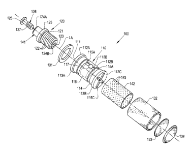
[0001] Referring now to the drawings, and more particularly to FIGS. 1-2, an
exemplary
embodiment of a dental implant 100 for implantation within gingival tissue is
illustrated. The
dental implant 100 generally includes a base 110 with a plug opening 111
formed therein, a plug
assembly 120 inserted into the plug opening 111, and at least one region of
ingrowth material
131, 132, 133, 134, which are also referred to as "ingrowth regions,"
associated with one or more
respective surfaces 112A, 112B, 112C of the base 110. In some embodiments, as
illustrated in
FIGS. 1-2, the base 110 defines a generally cylindrical shape with a pair of
increased diameter
ends 113A, 113B and a body portion 114 between and connecting the ends 113A,
113B. In some
embodiments, the base 110 comprises a generally non-porous material, which may
have a
porosity of between about 0% and about 10%. The base 110 may comprise one or
more
biocompatible materials suitable for short-term or long-term placement within
an animal body,
human or otherwise, which can include, but are not limited to: metals such as
titanium, stainless
steel, cobalt chrome, and/or tantalum; polymers such as ultra-high molecular
weight
polyethylene (UHMWPE), other forms of polyethylene, polyether ether ketone
(PEEK),
3
Date Recue/Date Received 2020-12-16
polylactic acid (PLA),and/or polyglycolic acid (PGA); and/or ceramics such as
hydroxyapatite
(HA), high-density alumina, so-called "Bioglass," and graphite. It should be
appreciated that all
of the previously mentioned materials are exemplary only, and many other types
of biomaterials
can be incorporated in the base 110.
[0002] In some embodiments, one or more fluid delivery grooves 115A, 115B are
formed in
the body portion 114 to deliver fluid from a reservoir 116 formed inside the
base 110 to
surrounding tissue, as will be described further herein. The surfaces 112A and
112C of the base
110 may also include one or more respective fluid delivery grooves 115C. In
some embodiments,
two or more of the delivery grooves, such as grooves 115A, extend
circumferentially about a
longitudinal axis LA defined through the base 110 and one or more of the
delivery grooves, such
as groove 115B, extend parallel to the longitudinal axis LA to connect the
circumferentially
extending delivery grooves 115A. The shape and size of the delivery grooves
115A, 115B may
be adjusted to give differing fluid delivery behavior from the reservoir 116
of the implant 100 to
surrounding tissue. The fluid delivery grooves 115A, 115B may fluidly
communicate with the
reservoir 116 formed in the base 110 via a reservoir opening 117 that is
formed in the base 110
to the reservoir 116 and partially surrounded by the delivery groove 115B.
[0003] A plug assembly 120 is inserted into the plug opening 111 of the base
110 to close the
plug opening 111. In some embodiments, the plug assembly 120 comprises a base
engaging
portion 121 with threads 122 that thread into corresponding threads of the
base 110 to couple the
base engaging portion 121 with the base 110. The base engaging portion 121 may
have a
through-hole 123 extending therethrough that extends from one end 124A of the
base engaging
portion 121 to an opposite end 124B. In some embodiments, a stop 125 is formed
in the base
engaging portion 121 between the ends 124A, 124B to prevent the base engaging
portion 121
from over-threading into the base 110. The base engaging portion 121 may be
unthreaded on one
4
Date Recue/Date Received 2020-12-16
side of the stop 125, such as adjacent to the end 124A, and threaded on the
other side of the stop
125, such as adjacent to the end 124B.
[0004] The plug assembly 120 may further include a plug portion 126 that is
placed in the
through-hole 123 of the base engaging portion 121. The plug portion 126 may,
for example,
include a stem 127 with a diameter less than a diameter of the through-hole
123 and a head 128
having a diameter greater than the diameter of the through-hole 123 that is
connected to the stem
127. In some embodiments, the plug portion 126 may comprise a generally
inelastic material,
such as a thermoset polymer. Alternatively, the plug portion 126 may comprise
an elastic
material, such as rubber. It should be appreciated that the plug portion 126
should sufficiently fill
the through-hole 123 of the base engaging portion 121 to plug the through-hole
123 so fluid from
the reservoir 116 does not rapidly leak out of the through-hole 123 when
subjected to increase
fluid pressure.
[0005] In some embodiments, the through-hole 123 is formed in a mount 141 of
the base
engaging portion 121 that is shaped and sized to mount, for example, a
replacement tooth
thereon. In other words, the plug assembly 120 is configured to mount a
replacement tooth
thereon. The replacement tooth may fit directly on the mount 141 for fixation
within the patient
or, alternatively, an adapter may be fit on the mount 141, with the
replacement tooth then being
mounted to the adapter. Many such mounts for replacement teeth are known, so
further
description of the mount 141 is omitted for brevity.
[0006] At least one region of ingrowth material, illustrated as four regions
131, 132, 133, 134,
is associated with one or more respective surfaces 112A, 112B, 112C of the
base 110. As used
herein, the ingrowth regions 131, 132, 133, 134 are "associated with" a
respective surface of the
base 110 in the sense that each ingrowth region 131, 132, 133, 134 is directly
or indirectly
fixated to its respective surface 112A, 112B, 112C of the base 110. To
encourage tissue
Date Recue/Date Received 2020-12-16
ingrowth, the ingrowth material of each region 131, 132, 133, 134 is porous
and comprises a
biocompatible material. Each region 131, 132, 133, 134 may comprise the same
ingrowth
material or, alternatively, one or more of the regions 131, 132, 133, 134 may
comprise a different
ingrowth material than one or more of the other regions 131, 132, 133, 134. In
some
embodiments, the ingrowth material comprises titanium or polyether ether
ketone (PEEK)
having a porosity of between about 50% and 70%, a mean pore size of between
about 500 gm
and about 550 gm, a pore interconnectivity of between about 210 gm and about
250 gm, and a
nominal thickness of between about 0.25 mm and 1.25 mm. One exemplary ingrowth
material
that may be used in one or more of the regions 131, 132, 133, 134 is
commercially available
under the tradename OSTEOSYNC CD from SITES MEDICAL 0 of Columbia City,
Indiana. It
should be appreciated that these characteristics of the ingrowth material(s)
are exemplary only.
In some embodiments, one of the regions, such as region 131, is associated
with the top surface
112A of the base 110; one of the regions, such as region 132, is associated
with the
circumferential surface 112B of the base 110 between terminal end regions
113A, 113B of the
base 110, which are not covered by any ingrowth material; and two of the
regions, such as
regions 133 and 134, are associated with the bottom surface 112C of the base
110. Each of the
surfaces 112A, 112B, 112C may have a through-hole formed through to the
reservoir 116 to
allow fluid delivery from the reservoir 116 to surrounding tissues through
pores of the ingrowth
regions 131, 132, 133, 134.
[0007] In some embodiments, two of the ingrowth regions associated with the
same surface,
such as the ingrowth regions 133 and 134 associated with the bottom surface
112C of the base
110, differ from one another. For example, the ingrowth region 133, which may
be referred to as
a "first region of ingrowth material" and sandwiched between the ingrowth
region 134 and the
bottom surface 112C of the base 110, may comprise a first ingrowth material
that has a lower
6
Date Recue/Date Received 2020-12-16
porosity compared to a second ingrowth material of the ingrowth region 134,
which may be
referred to as a "second region of ingrowth material." In other words, the
first ingrowth material
of the first region of ingrowth material 133 may have a first porosity and the
second ingrowth
material of the second region of ingrowth material 134 may have a second
porosity that differs
from the first porosity, e.g., by being greater than the first porosity. By
providing the ingrowth
region 133 with a lower porosity than the ingrowth region 134, the fluid
delivery rate to
surrounding tissues from the reservoir 116 may be limited by the porosity of
the ingrowth region
133 while the porosity of the ingrowth region 134 may be greater to encourage
tissue ingrowth
into the pores of the ingrowth region 134. Such a configuration can allow long-
term fluid
delivery from the reservoir 116 to surrounding tissue, due to the relatively
low porosity of the
ingrowth region 133, while providing larger pores in the ingrowth region 134
to encourage tissue
ingrowth to stabilize the implant 100. Alternatively, or in addition to, the
two ingrowth regions
133, 134 may differ from one another by, for example, having different
compositions and/or
shapes. It should be appreciated that ingrowth regions associated with the
same surface may
differ from each other in other ways, and the foregoing examples represent
only a few of the
possible ways that the regions may differ from each other.
[0008] In some embodiments, a perforated sleeve 142 is placed between the
circumferential
surface 112B of the base 110 and the ingrowth region 132 to provide a uniform
surface for
associating the ingrowth region 132 with the circumferential surface 112B. As
illustrated in FIG.
1, the circumferential surface 112B may be relatively non-uniform due to the
formation of the
fluid delivery grooves 115A, 115B, which makes securing the ingrowth region
132 to the surface
112B difficult. The perforated sleeve 142, therefore, fits on the
circumferential surface 112B,
such as by press fitting, between the terminal end regions 113A, 113B of the
base 110 to provide
a uniformly sized surface for securing the ingrowth region 132. Further, the
size of apertures 143
7
Date Recue/Date Received 2020-12-16
formed in the perforated sleeve 142 may be adjusted to control fluid delivery
through pores of
the ingrowth region 132 by controlling the rate of fluid flow to the ingrowth
region 132 from the
reservoir 116 via the reservoir opening 117. In this respect, the perforated
sleeve 142 assists with
associating the ingrowth region 132 with the circumferential surface 112B of
the base 110 as
well as controlling fluid delivery from the reservoir 116 to surrounding
tissues following
implantation.
[0009] Referring specifically now to FIG. 2, the implant 100 is illustrated
following
implantation into gingival tissue T of a mouth of a patient, which may be a
human or other
animal. As illustrated in FIG. 2, a defect region 200 is present between two
adjacent teeth 201,
202 in the patient. The defect region 200 previously held a tooth, which has
fallen out or
otherwise been removed due to, for example, disease or trauma. To replace the
tooth, the defect
region 200 is cleaned to remove remaining fragments of the removed tooth,
other debris, and
pathogens. In the case illustrated in FIG. 2, the defect region 200 is a
contained defect that has
sufficient amounts of local gingival and bone tissue to support and fixate the
implant 100 without
requiring, for example, one or more screws anchoring the implant 100.
[0010] An implantation bore 203 is formed in the tissue of the defect region
200 and the
implant 100 is placed within the implantation bore 203. In some embodiments,
the implantation
bore 203 is formed by removing the tooth (or tooth fragments) that is present
in the defect region
200 and is being replaced by the implant 100. As illustrated, the mount 141
protrudes out of the
implantation bore 203 following placement of the implant 100 to accept, for
example, a
replacement tooth after the implant 100 has sufficiently fixated within the
tissue. Initially,
however, the mount 141 may be left uncovered in the defect region 200,
exposing the mount 141
and the head 128 of the plug portion 126. Before, during, or after
implantation, the plug portion
126 may be removed from the through-hole 123 in the mount 141 and one or more
therapeutic
8
Date Recue/Date Received 2020-12-16
agents may be filled into the reservoir 116 within the base 110 via the
through-hole 123. The
therapeutic agent(s) may be, but is not limited to, an antibiotic or other
antimicrobial agent, an
anti-inflammatory agent, an analgesic, a growth factor, a solution comprising
regenerative cells
such as stem cells, or any other substance that provides a therapeutic effect
on the tissue
surrounding the implant 100 following implantation. Alternatively, the
therapeutic agent(s) may
be filled into the reservoir 116 via, for example, the reservoir opening 117
prior to covering the
reservoir opening 117 with the perforated sleeve 142 and the ingrowth region
132. Thus, it
should be appreciated that the reservoir 116 may be initially filled with one
or more therapeutic
agents in a variety of ways.
[0011] In some embodiments, the reservoir 116 is initially filled with a first
therapeutic agent,
which may be one or more antimicrobial agents to reduce the risk of infection,
and then refilled
with the first therapeutic agent, a second therapeutic agent that is different
from the first
therapeutic agent, or both while the implant 100 is implanted in the patient.
For example, the
reservoir 116 may be refilled partially with the first therapeutic agent (an
antimicrobial) to
continue delivering antimicrobial agent to surrounding tissue to reduce the
risk of infection but
also filled with a second therapeutic agent, such as a growth factor, to
encourage tissue
infiltration and ingrowth into the ingrowth regions 131, 132, 133, 134 to
encourage a faster and
more stable fixation of the implant 100 in the defect region 200. To refill
the reservoir 116, the
plug portion 126 may be pulled out of the through-hole 123 and a tip of a
syringe containing the
therapeutic agent(s) placed within the through-hole 123 to inject the
therapeutic agent(s) from
the syringe into the through-hole 123 and the fluidly coupled reservoir 116.
Alternatively, the
syringe may also be used to remove fluid, which may be therapeutic agent or
biological fluid,
from the reservoir 116.
[0012] After partially or fully filling the reservoir 116, the plug portion
126 is replaced in the
9
Date Recue/Date Received 2020-12-16
through-hole 123. In some embodiments, the plug portion 126 is shaped and
sized such that
replacement of the plug portion 126 in the through-hole 123 pressurizes the
fluid in the reservoir
116, urging the fluid in the reservoir 116 out to the ingrowth regions 131,
132, 133, 134 via, for
example, the reservoir opening 117 formed in the base 110. In this respect,
the plug portion 126
not only seals the through-hole 123, but also provides an initial
pressurization of the reservoir
116 to deliver a bolus of the therapeutic agent(s) to surrounding tissue while
the implant 100 is
implanted in the patient. After the initial bolus of therapeutic agent(s) is
delivered, the remaining
therapeutic agent(s) in the reservoir 116 may then travel out of the reservoir
116 into the
surrounding tissue by compressive force exerted on the implant 100 "squeezing"
out the
therapeutic agent(s) as well as by natural diffusion of the therapeutic
agent(s) into the tissue. It
should be appreciated that the plug portion 126 may be removed from and
replaced in the
through-hole 123 multiple times throughout the implantation to refill the
reservoir 116. Due to
the simplicity of removing and replacing the plug portion 126 to refill the
reservoir 116, the
patient in which the implant 100 is implanted may refill the reservoir 116 at
home or in other
non-clinical settings.
[0013] While the implant 100 is implanted, native tissues adjacent to the
defect region 200,
such as gingival tissue and bone tissue, may infiltrate and grow into the
ingrowth regions 131,
132, 133, 134 of the implant 100. As the native tissues grow into the ingrowth
regions 131, 132,
133, 134, the implant 100 becomes stably fixated within the tissue. After the
implant 100 is
stably fixated within the tissue, the replacement tooth may be mounted to the
mount 141 to finish
replacement of the removed tooth. In some embodiments, the replacement tooth
may be mounted
directly on the mount 141 with the plug portion 126 placed in the through-hole
123 of the base
engaging portion 121. In some embodiments, the plug portion 126 may be
replaced with a
different plug in the through-hole 123 that is shaped to both fill the through-
hole 123 and engage,
Date Recue/Date Received 2020-12-16
for example, a socket of the replacement tooth to mount the replacement tooth
to the mount 141.
In other embodiments, the base engaging portion 121 may be replaced with a
different base
engaging portion having a mount for mounting the replacement tooth. It should
thus be
appreciated that many different ways of mounting a replacement tooth to the
implant 100 may be
used to fixate the replacement tooth to the implant 100.
[0014] It has been discovered that certain ingrowth materials, such as the
previously described
OSTEOSYNC CD, provide surprisingly good tissue ingrowth and fixation
characteristics to the
implant 100. When OSTEOSYNC CD is used to form the ingrowth regions 131, 132,
133, 134,
the push-out strength of the implant 100 may be roughly equivalent to the push-
out strength of
native bone after only 5 weeks of implantation. The results were surprising
because of the high
degree of improvement that such an implant provided compared to known dental
implants.
Without being bound to any particular theory as to why such results were
observed, it is
theorized that the ingrowth regions 131, 132, 133, 134, when comprising
OSTEOSYNC CD or
similar materials, provide an excellent substrate for cortical bone tissue
ingrowth. Considering
the high concentration of cortical bone tissue adjacent to the jawbone, it is
theorized that the
stable fixation of the implant 100 at five weeks is at least partly
attributable to rapid ingrowth
and proliferation of cortical bone tissue in the ingrowth regions 131, 132,
133, 134. It is thus
theorized that implants provided in accordance with the present disclosure are
well-suited as
dental implants because they simulate the natural fixation of teeth in the
gingival tissue and the
bone tissue present in the mouth of a patient, i.e., predominantly by ingrowth
of and integration
with adjacent cortical bone tissue.
[0015] It has also been found that providing one or more therapeutic agents in
the reservoir
116 for therapeutic agent delivery during implantation encourages rapid,
stable fixation of the
implant 100. As previously described, the reservoir 116 may be initially
filled with one or more
11
Date Recue/Date Received 2020-12-16
antimicrobial agents to reduce the risk of pathogens infecting the defect
region 200 and
interfering with tissue growth fixating the implant 100. The reservoir 116 may
be refilled one or
more times with the antimicrobial agent(s), or other therapeutic agent(s),
throughout the
implantation, as previously described, to encourage tissue ingrowth into the
ingrowth regions
131, 132, 133, 134 and stable fixation of the implant 100. Therefore, the
implants provided in
accordance with the present disclosure may also encourage rapid, stable
fixation in the
surrounding native tissue by delivering one or more therapeutic agents into
the surrounding
tissue from the reservoir 116 to provide a favorable environment for tissue
ingrowth into the
ingrowth regions 131, 132, 133, 134.
[0016] Referring now to FIGS. 3-5, another exemplary embodiment of a dental
implant 300 for
implantation within gingival tissue that has an uncontained defect is
illustrated. Similarly to the
previously described implant 100, the implant 300 includes a base 310 with a
plug opening 311
formed therein, a plug assembly 320 inserted into the plug opening 311, and at
least one region
of ingrowth material 331, 332, 333, 334, 335 associated with one or more
respective surfaces
312A, 312B, 312C of the base 310. As illustrated in FIG. 3, for example, the
ingrowth regions
331 and 332 may be associated with the top surface 312A of the base 310; the
ingrowth region
333 may be associated with the peripheral surface 312B of the base 310; and
the ingrowth
regions 334 and 335 may be associated with the bottom surface 312C of the base
310. Compared
to the implant 100, with a cylindrical base 110 having a generally circular
cross-section, the
implant 300 has a relatively larger base 310 with an oval cross-section to
stabilize the implant
300 in an uncontained defect region 510, which is illustrated in FIG. 5 and
described further
herein.
[0017] Similarly to the base 110, the base 310 can have fluid delivery grooves
315A, 315B,
315C formed therein that communicate fluid from a reservoir 316 formed in the
base 310 to, for
12
Date Recue/Date Received 2020-12-16
example, the ingrowth regions 331, 332, 333, 334, 335 via openings, such as a
reservoir opening
317, formed in the base 310 to the reservoir 316. The reservoir 316 may be
refilled by removing
a plug portion 326 from the plug assembly 320 and delivering fluid to the
reservoir 316 through
a through-hole 323 of the plug assembly 320 using, for example, a syringe or
other fluid delivery
device. The implant 300 may also include a perforated sleeve 342 between the
ingrowth region
333 and the circumferential surface 312B and have a mount 341 for mounting a
replacement
tooth, similarly to the implant 100.
[0018] The implant 300 is configured to be implanted in an uncontained defect,
i.e., a defect
where adjacent tissue is diseased, destroyed, and/or otherwise unsuitable for
fixating the implant
300 without additional fixation. To provide the additional fixation needed to
stably fixate the
implant 300, one or more fixation openings 351, 352 may be formed in the base
310, such as in
the circumferential surface 312B, to accept a respective fixator, such as one
or more orthopaedic
screws 501 (illustrated in FIG. 5). Corresponding openings 353, 354 may also
be formed in the
perforated sleeve 342 and the ingrowth region 333 so the fixation openings
351, 352 are
uncovered to accept the screw(s) 501. The fixation opening(s) 351, 352 may be,
for example,
threaded to couple with the screw(s) 501 in order to fixate the implant 300
within the mouth of
the patient. In this respect, implantation and use of the implant 300 is
similar to that of the
implant 100, but also includes the additional step of fixating the fixator(s)
501 in a defect region,
such as defect region 500 illustrated in FIG. 5, and coupling the fixator(s)
501 with the implant
300 by, for example, threading the fixator(s) 501 into the fixation opening(s)
351, 352. In all
other respects, implantation and use of the implant 300 may be similar to
implantation and use of
the implantation 100, which is previously described.
[0019] While this invention has been described with respect to at least one
embodiment, the
present invention can be further modified within the spirit and scope of this
disclosure. This
13
Date Recue/Date Received 2020-12-16
application is therefore intended to cover any variations, uses, or
adaptations of the invention
using its general principles. Further, this application is intended to cover
such departures from
the present disclosure as come within known or customary practice in the art
to which this
invention pertains and which fall within the limits of the appended claims.
14
Date Recue/Date Received 2020-12-16