Note: Descriptions are shown in the official language in which they were submitted.
Modular Intraocular Lens Designs and Methods
Cross-Reference to Related Applications
[0001]
This application claims the benefits under 35 U.S.C. 119(e) of priority
of
U.S. Provisional Patent Application No. 61/589,981 filed on January 24, 2012,
entitled
"LASER ETCHING OF IN SITU INTRAOCULAR LENS AND SUCCESSIVE
SECONDARY LENS IMPLANTATION," and of U.S. Provisional Patent Application No.
61/677,213 filed on July 30, 2012, entitled "MODULAR INTRAOCULAR LENS
DESIGNS & METHODS". This application is a divisional application of Canadian
application no. 2,861,865.
Field of the Invention
[0002] The
present disclosure generally relates to embodiments of intraocular
lenses (10Ls). More specifically, the present disclosure relates to
embodiments of
modular IOL designs and methods.
Background
[0003] The
human eye functions to provide vision by transmitting light through a
clear outer portion called the cornea, and focusing the image by way of a
crystalline
lens onto a retina. The quality of the focused image depends on many factors
including
the size and shape of the eye, and the transparency of the cornea and the
lens.
[0004]
When age or disease causes the lens to become less transparent (e.g.,
cloudy), vision deteriorates because of the diminished light, which can be
transmitted to
the retina. This deficiency in the lens of the eye is medically known as a
cataract. An
accepted treatment for this condition is surgical removal of the lens from the
capsular
bag and placement of an artificial intraocular lens (I0L) in the capsular bag.
In the
1
Date Recue/Date Received 2020-12-18
United States, the majority of cataractous lenses are removed by a surgical
technique
called phacoemulsification. During this procedure, an opening (capsulorhexis)
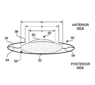
is made
in the anterior side of the capsular bag and a thin phacoemulsification-
cutting tip is
inserted into the diseased lens and vibrated ultrasonically. The vibrating
cutting tip
liquefies or emulsifies the lens so that the lens may be aspirated out of the
capsular
bag. The diseased lens, once removed, is replaced by an 10L.
[0005]
After cataract surgery to implant an 10L, the optical result may be
suboptimal or may need adjustment over time. For example, shortly after the
procedure, it may be determined that the refractive correction is erroneous
leading to
what is sometimes called "refractive surprise." Also for example, long after
the
procedure, it may be determined that the patient needs or desires a different
correction,
such as a stronger refractive correction, an astigmatism correction, or a
multifocal
correction.
[0006]
In each of these cases, a surgeon may be reluctant to attempt removal of
the suboptimal IOL from the capsular bag and replacement with a new 10L. In
general,
manipulation of the capsular bag to remove an IOL risks damage to the capsular
bag
including posterior rupture. This risk increases over time as the capsular bag
collapses
around the IOL and tissue ingrowth surrounds the haptics of the 10L. Thus, it
would be
desirable to be able to correct or modify the optical result without the need
to remove
the IOL or manipulate the capsular bag.
[0007]
A variety of secondary lenses have been proposed to address the
aforementioned drawbacks. For example, one possible solution includes a
secondary
lens that resides anterior to the capsular bag with haptics that engage the
ciliary sulcus.
While this design may have the advantage of avoiding manipulation of the
capsular bag,
2
Date Recue/Date Received 2020-12-18
its primary disadvantage is engaging the ciliary sulcus. The ciliary sulcus is
composed
of soft vascularized tissue that is susceptible to injury when engaged by
haptics or other
materials. Such injury may result in complications such as bleeding,
inflammation and
hyphema. Thus, in general, it may be desirable to avoid placing a secondary
lens in the
ciliary sulcus to avoid the potential for complications.
[0008]
Another potential solution may include a lens system that avoids the
potential problems associated with the ciliary sulcus. The lens system may
include a
primary lens and a secondary lens, where the secondary lens may be attached to
the
primary lens, both within the capsular bag. The primary lens may have a recess
into
which an edge of the secondary lens may be inserted for attachment. The recess
is
preferably located radially outwardly of the opening (capsulorhexis) in the
capsular bag
to avoid interfering with light transmission. To attach the secondary lens in-
situ, the
capsular bag must be manipulated around the perimeter of the capsulorhexis to
gain
access to the recess in the primary lens. As stated previously, manipulation
of the
capsular bag may be undesirable given the risks associated therewith.
Therefore, while
such lens systems may avoid the potential for injury to the ciliary sulcus by
implanting
both the primary lens and the secondary lens in the capsular bag, these
systems do not
avoid manipulation of the capsular bag to attach the secondary lens.
[0009]
Thus, there remains a need for an IOL system and method that allows for
correction or modification of the optical result using a secondary lens that
can be
attached to a primary lens without the need manipulate the capsular bag.
3
Date Recue/Date Received 2020-12-18
Summary of the Invention
[0010]
Embodiments of the present disclosure provide a modular IOL system
including intraocular primary and secondary components, which, when combined,
form
an intraocular optical correction device. The primary component may comprise
an
intraocular base, and the secondary component may comprise an intraocular
lens,
wherein the base is configured to releasably receive the intraocular lens. In
some
embodiments, the base may be configured as a lens, in which case the modular
IOL
system may be described as including a primary lens and a secondary lens. The
primary component (e.g., base or primary lens) may be placed in the capsular
bag using
conventional cataract surgery techniques. The primary component may have a
diameter greater than the diameter of the capsulorhexis to retain the primary
component
in the capsular bag. The secondary component (e.g., secondary lens) may have a
diameter less than the diameter of the capsulorhexis such that the secondary
component may be attached to the primary component without manipulation of the
capsular bag. The secondary component may also be manipulated to correct or
modify
the optical result, intra-operatively or post-operatively, without the need to
remove the
primary component and without the need to manipulate the capsular bag. For
example,
the secondary component may be removed, repositioned, and/or exchanged to
correct,
modify, and/or fine tune the optical result.
[0011] Common
indications for exchanging the secondary component may be
residual refractive error (e.g., for monofocal lenses), decentration error
(e.g., for
multifocal lenses) due to post-operative healing, astigmatism error (e.g., for
toric lenses)
induced by surgery, changing optical correction needs due to progressive
disease,
changing optical correction desires due to lifestyle changes, injury, age,
etc.
4
Date Recue/Date Received 2020-12-18
[0012]
The primary component may have haptics (e.g., projections) extending
therefrom for centration in the capsular bag, and the secondary component may
exclude haptics, relying instead on attachment to the primary component for
stability.
Such attachment may reside radially inside the perimeter of the capsulorhexis
and
radially outside the field of view to avoid interference with light
transmission.
Alternatively or in addition, the attachment may comprise a small fraction of
the
perimeter (e.g., less than 20%) of the secondary component to minimize the
potential
for interference in light transmission.
[0013]
The primary component may have an anterior surface that is in intimate
contact with a posterior surface of the secondary component to prevent fluid
ingress,
tissue ingrowth, and/or optical interference. The secondary component may be
removably secured to the primary component by mechanical attachment and/or
chemical attraction, for example. Mechanical attachment may be facilitated by
mating
or interlocking geometries corresponding to each of the primary and the
secondary
components. Such geometries may be pre-formed by molding or cutting, for
example,
or formed in-situ by laser etching, for example. Chemical attraction may be
facilitated
by using similar materials with a smooth surface finish activated by a surface
treatment,
for example. In some instances, it may be desirable to reduce chemical
attraction and
rely more on mechanical attachment for stability. In this case, the primary
and
secondary components may be formed of dissimilar materials or otherwise have
adjacent surfaces that do not have a chemical attraction.
[0014]
The modular IOL systems and methods according to embodiments of the
present disclosure may be applied to a variety of IOL types, including fixed
monofocal,
multifocal, toric, accommodative, and combinations thereof. In addition, the
modular
5
Date Recue/Date Received 2020-12-18
IOL systems and methods according to embodiments of the present disclosure may
be
used to treat, for example: cataracts, large optical errors in myopic (near-
sighted),
hyperopic (far-sighted), and astigmatic eyes, ectopia lentis, aphakia,
pseudophakia, and
nuclear sclerosis.
[0014a] According to an embodiment of the present invention, there is provided
an
intraocular lens system, comprising:
a. an intraocular primary component having a body and one or more haptics,
wherein the body is annular and defines a through opening having an inside
perimeter,
the body further includes a continuous recess extending around an entirety of
the inside
perimeter, the continuous recess being defined by an anterior-facing posterior
wall and
a radially-inward-facing lateral wall, and wherein the body further includes
diametrically
spaced extensions extending anteriorly of and radially inward from the
radially-inward-
facing lateral wall, the diametrically spaced extensions terminating with a
radially-
inward-facing concave free edge; and
b. an intraocular secondary component having an optical body, wherein the
secondary component is releasably attachable to the primary component by
insertion of
the secondary component into the continuous recess of the primary component
such
that an entire posterior periphery of the secondary component contacts the
anterior-
facing posterior wall of the recess, the diametrically spaced extensions
extend over an
anterior peripheral portion of the secondary component at discrete,
diametrically spaced
apart junctions, so as to interlock to thereby limit relative anterior and
posterior
movement between the primary and the second components and to leave the
anterior peripheral portion exposed between the diametrically spaced
extensions, and
- 6 -
Date Recue/Date Received 2020-12-18
the secondary component is compressible for releasable attachment to the
primary
component;
c. wherein the intraocular lens system is configured to provide optical
correction
to an eye solely via the optical body of the secondary component.
[0014b] According to another embodiment of the present invention, there is
provided an intraocular lens system, comprising:
a. a base having a body, a plurality of haptics extending from the body, a
center
opening extending in an anterior-posterior direction through the body to form
a
continuous annular ring having an insider perimeter, a continuous recess
extending
around an entirety of the inside perimeter, the continuous recess being
defined by an
anterior-facing posterior wall and a radially-inward-facing lateral wall,
wherein the
radially-inward-facing lateral wall includes diametrically spaced extensions
extending
anteriorly of and radially inward from the radially-inward-facing lateral
wall, the
diametrically spaced extensions terminating with a radially-inward-facing
concave free
edge, and the base being configured to fit within a lens capsule; and
b. a lens having an optical body, wherein the lens is releasably attachable to
the
base by insertion of the lens into the continuous recess of the base such that
an entire
posterior periphery of the lens contacts the anterior-facing posterior wall of
the recess,
the diametrically spaced extensions extend over an anterior peripheral portion
of the
lens to form discrete, diametrically spaced apart junctions so as to interlock
to thereby
limit relative anterior and posterior movement between the base and the lens,
and the
lens is radially compressible for releasable attachment to the base;
c. wherein the intraocular lens system is configured to provide optical
correction
to an eye solely via the lens, a surface of each of the diametrically spaced
extensions is
- 6a -
Date Recue/Date Received 2020-12-18
even with a portion of an anterior surface of the lens, and the radially-
inward-facing
lateral wall of the continuous recess, except at the diametrically spaced
extensions,
exposes a portion of the anterior peripheral portion of the lens.
[0014c] According to another embodiment of the present invention, there is
provided a modular intraocular lens system for implantation in an eye,
comprising:
(a) an annular base having an inner perimeter defining a center hole and an
outer perimeter with two or more haptics extending outwardly therefrom, the
inner
perimeter including a continuous circumferential recess having a ledge
extending
around an entirety of the inner perimeter, the continuous circumferential
recess having
an anterior rim and a posterior rim, wherein an inside diameter of the
anterior rim is
greater than an inside diameter of the posterior rim;
(b) a lens having an optic portion and a curvilinear extension protruding away
from the optic portion, the optic portion having an optical axis, and the
optic portion
disposed over the center hole and the curvilinear extension disposed in the
continuous
circumferential recess on the ledge of the annular base to form an overlapping
joint,
wherein the joint is configured to permit insertion and removal of the lens to
and from
the annular base, the lens also having a pair of extension members extending
in a
direction away from the curvilinear extension, wherein each of the pair of
extension
members extends from a different location on the curvilinear extension;
(c) wherein the lens has a resting position along the optical axis relative to
the
annular base when disposed in the continuous circumferential recess; and
(d) wherein the pair of extension members are configured to allow a change in
rotational position of the lens relative to the annular base and maintain the
resting
position.
- 6b -
Date Recue/Date Received 2020-12-18
[0014d] According to another embodiment of the present invention, there is
provided a modular intraocular lens system for implantation in an eye,
comprising:
(a) an annular base defining a through hole and including a plurality of
haptics
extending radially away from the annular base, the through hole being
surrounded by a
continuous circumferential recess, the continuous circumferential recess
having an
anterior rim portion and a posterior rim portion, wherein an inside diameter
of the
anterior rim portion is greater than an inside diameter of the posterior rim
portion such
that the posterior rim portion forms an anterior-facing posterior ledge
extending around
an entirety of the continuous circumferential recess;
(b) a lens disposed over the hole, in the continuous circumferential recess,
and
on the anterior-facing posterior ledge to form an overlapping joint between
the lens and
the annular base, wherein the joint is configured to permit insertion and
removal of the
lens to and from the annular base;
(c) wherein the lens has a resting position relative to at least one of an
anterior
end and a posterior end of the annular base when disposed in the continuous
circumferential recess; and
(d) wherein the lens includes a pair of extensions disposed in the continuous
circumferential recess that are configured to allow rotation of the lens
relative to the
annular base from a first rotational position in which the lens is in the
resting position to
a second rotational position in which the lens is in the resting position, the
second
rotational position being different than the first rotational position.
[0014e] According to another embodiment of the present invention, there is
provided
a modular intraocular lens system for implantation in an eye having a visual
axis,
comprising:
- 6c -
Date Recue/Date Received 2020-12-18
(a) an annular base defining a center through hole and including a plurality
of haptics
extending from the annular base, the center through hole being surrounded by a
continuous circumferential recess, the continuous circumferential recess
having an
anterior rim portion and a posterior rim portion, wherein an inside diameter
of the
anterior rim portion is greater than an inside diameter of the posterior rim
portion to form
a posterior ledge extending around an entirety of the continuous
circumferential recess;
(b) a lens disposed over the hole, in the recess, and on the posterior ledge
of the
annular base to form an overlapping joint between the lens and the annular
base,
wherein the joint is configured to (i) permit insertion and removal of the
lens to and from
the annular base, (ii) permit adjustment of a position of the lens relative to
the base
around the visual axis, and (iii) limit movement of the lens relative to the
base along the
visual axis;
(c) wherein the lens includes a pair of opposing protrusions disposed in the
recess
that are configured to support the lens on the posterior ledge while allowing
an
adjustment in rotational position of the lens relative to the base.
[0014f] In another aspect, there is provided an intraocular lens system,
comprising:
a base having a body including an annular recess, wherein the annular recess
has an outer diameter; and
an intraocular lens, comprising:
an optical body having an edge extending between an anterior surface of
the optical body and a posterior surface of the optical body,
a first extension projecting from a first portion of the edge of the optical
body, and
- 6d -
Date Recue/Date Received 2020-12-18
a second extension projecting from a second portion of the edge of the
optical body, wherein the first and second portions of the edge of the optical
body are
located opposite one another, wherein a distance between an outermost portion
of the
first extension and an outermost portion of the second extension is greater
than the
outer diameter of the annular recess, and wherein the body of the base is
configured to
receive the intraocular lens, with the first and second extensions extending
into the
annular recess, to removably secure the intraocular lens to the base.
[0014g] In another aspect, there is provided an intraocular lens system,
comprising:
an annular base having a equatorial plane, the annular base comprising:
a first end section surrounding a first end opening, wherein the first end
section includes:
(a) a first surface, wherein the first surface is angled relative to the
equatorial plane, and
(b) a second surface opposite the first surface, wherein the second
surface is angled relative to the first surface,
a second end section surrounding a second end opening,
a wall extending between the first end section and the second end section,
a passage extending through the base from the first end opening to the
second end opening, and
a recess surrounding the passage, wherein the recess is bordered by
surfaces of the first end section, the second end section, and the wall; and
a lens configured for insertion into and removal from the annular base, the
lens
comprising:
an optic, and
- 6e -
Date Recue/Date Received 2020-12-18
at least one extension protruding from the optic, wherein, when the lens is
inserted into the annular base, the extension is received by the recess of the
annular
base, and the optic extends across the passage of the annular base.
[0015] Various other aspects of embodiments of the present disclosure
are
.. described in the following detailed description and drawings.
Brief Description of the Drawings
[0016] The drawings illustrate example embodiments of the present
disclosure.
The drawings are not necessarily to scale, may include similar elements that
are
numbered the same, and may include dimensions (in millimeters) and angles (in
degrees) by way of example, not necessarily limitation. In the drawings:
[0017] Figure 1 is a schematic diagram of the human eye shown in
cross section;
[0018] Figures 2A and 2B are front and side cross-sectional views,
respectively,
of a modular IOL disposed in a capsular bag according to an embodiment of the
present
disclosure;
[0019] Figures 3A-3D and 4A-4D are front and side cross-sectional views,
respectively, schematically illustrating a method for implanting a modular IOL
according
to an embodiment of the present disclosure;
[0020] Figure 5 is a front view of a modular 10L, according to an
embodiment of
the present disclosure, wherein subsurface attachment mechanisms are provided
for
connection between the primary and secondary lenses;
[0021] Figures 6A and 6B are cross-sectional views taken along line 6-
6 in Figure
5, showing two embodiments of subsurface attachment mechanisms;
- 6f -
Date Recue/Date Received 2020-12-18
[0022]
Figure 7 is a front view of a modular 10L, according to an embodiment of
the present disclosure, wherein extension attachment mechanisms are provided
to
connect the primary and secondary lenses;
[0023]
Figures 8A-8C are cross-sectional views taken along line 8-8 in Figure 7,
showing three embodiments of extension attachment mechanisms;
[0024]
Figures 9A-9D are front views showing various positions of the attachment
mechanisms to adjust the position of the secondary lens relative to the
primary lens;
[0025]
Figure 10 is a front view of a modular 10L, according to an embodiment of
the present disclosure, wherein etched subsurface attachment mechanisms are
provided for connection between the primary and secondary lenses;
[0026]
Figures 11A-11F are cross-sectional views of the modular IOL shown in
Figure 10, showing various embodiments of etched subsurface attachment
mechanisms;
[0027]
Figures 12A-12C are schematic illustrations of front, sectional and detail
views, respectively, of an alternative modular 10L, according to an embodiment
of the
present disclosure;
[0028]
Figures 13A and 13B show representative photomicrographs at 4X and
40X magnification, respectively, of a groove (see, arrow) formed by laser
etching;
[0029]
Figures 14-22 are various views of alternative modular 10Ls according to
embodiments of the present disclosure;
[0030]
Figures 23A-23D are schematic illustrations of a lens removal system for a
modular IOL according to an embodiment of the present disclosure;
7
Date Recue/Date Received 2020-12-18
[0031]
Figure 24 is a schematic flow chart of a method for using a modular 10L,
according to an embodiment of the present disclosure, wherein an exchange of
the
secondary lens is motivated by a sub-optimal optical result detected intra-
operatively;
[0032]
Figure 25 is a schematic flow chart of a method for using a modular 10L,
according to an embodiment of the present disclosure, wherein an exchange of
the
secondary lens is motivated by a sub-optimal optical result detected post-
operatively;
[0033]
Figure 26 is a schematic flow chart of a method for using a modular 10L,
according to an embodiment of the present disclosure, wherein a secondary lens
is
attached to a primary lens by forming the attachment means in-situ; and
[0034]
Figures 27 and 27A-27D are various views of a further embodiment of a
modular 10L, according to the present disclosure.
Detailed Description
[0035]
With reference to Figure 1, the human eye 10 is shown in cross section.
The eye 10 has been described as an organ that reacts to light for several
purposes.
As a conscious sense organ, the eye allows vision. Rod and cone cells in the
retina 24
allow conscious light perception and vision including color differentiation
and the
perception of depth. In addition, the human eye's non-image-forming
photosensitive
ganglion cells in the retina 24 receive light signals which affect adjustment
of the size of
the pupil, regulation and suppression of the hormone melatonin, and
entrainment of the
body clock.
[0036]
The eye 10 is not properly a sphere; rather it is a fused two-piece unit.
The
smaller frontal unit, more curved, called the cornea 12 is linked to the
larger unit called
the sclera 14. The corneal segment 12 is typically about 8 mm (0.3 in) in
radius. The
8
Date Recue/Date Received 2020-12-18
sclera 14 constitutes the remaining five-sixths; its radius is typically about
12 mm. The
cornea 12 and sclera 14 are connected by a ring called the limbus. The iris
16, the
color of the eye, and its black center, the pupil, are seen instead of the
cornea 12 due to
the cornea's 12 transparency. To see inside the eye 10, an ophthalmoscope is
needed,
since light is not reflected out. The fundus (area opposite the pupil), which
includes the
macula 28, shows the characteristic pale optic disk (papilla), where vessels
entering the
eye pass across and optic nerve fibers 18 depart the globe.
[0037]
Thus, the eye 10 is made up of three coats, enclosing three transparent
structures. The outermost layer is composed of the cornea 12 and sclera 14.
The
middle layer consists of the choroid 20, ciliary body 22, and iris 16. The
innermost layer
is the retina 24, which gets its circulation from the vessels of the choroid
20 as well as
the retinal vessels, which can be seen within an ophthalmoscope. Within these
coats
are the aqueous humor, the vitreous body 26, and the flexible lens 30. The
aqueous
humor is a clear fluid that is contained in two areas: the anterior chamber
between the
cornea 12 and the iris 16 and the exposed area of the lens 30; and the
posterior
chamber, between the iris 16 and the lens 30. The lens 30 is suspended to the
ciliary
body 22 by the suspensory ciliary ligament 32 (Zonule of Zinn), made up of
fine
transparent fibers. The vitreous body 26 is a clear jelly that is much larger
than the
aqueous humor.
[0038] The
crystalline lens 30 is a transparent, biconvex structure in the eye that,
along with the cornea 12, helps to refract light to be focused on the retina
24. The lens
30, by changing its shape, functions to change the focal distance of the eye
so that it
can focus on objects at various distances, thus allowing a sharp real image of
the object
of interest to be formed on the retina 24. This adjustment of the lens 30 is
known as
9
Date Recue/Date Received 2020-12-18
accommodation, and is similar to the focusing of a photographic camera via
movement
of its lenses.
[0039]
The lens has three main parts: the lens capsule, the lens epithelium, and
the lens fibers. The lens capsule forms the outermost layer of the lens and
the lens
fibers form the bulk of the interior of the lens. The cells of the lens
epithelium, located
between the lens capsule and the outermost layer of lens fibers, are found
predominantly on the anterior side of the lens but extend posteriorly just
beyond the
equator.
[0040]
The lens capsule is a smooth, transparent basement membrane that
completely surrounds the lens. The capsule is elastic and is composed of
collagen. It is
synthesized by the lens epithelium and its main components are Type IV
collagen and
sulfated glycosaminoglycans (GAGs). The capsule is very elastic and so causes
the
lens to assume a more globular shape when not under the tension of the zonular
fibers,
which connect the lens capsule to the ciliary body 22. The capsule varies
between
approximately 2-28 micrometers in thickness, being thickest near the equator
and
thinnest near the posterior pole. The lens capsule may be involved with the
higher
anterior curvature than posterior of the lens.
[0041]
Various diseases and disorders of the lens 30 may be treated with an 10L.
By way of example, not necessarily limitation, a modular IOL according to
embodiments
of the present disclosure may be used to treat cataracts, large optical errors
in myopic
(near-sighted), hyperopic (far-sighted), and astigmatic eyes, ectopia lentis,
aphakia,
pseudophakia, and nuclear sclerosis. However, for purposes of description, the
modular IOL embodiments of the present disclosure are described with reference
to
cataracts.
Date Recue/Date Received 2020-12-18
[0042]
The following detailed description describes various embodiments of a
modular IOL system including primary and secondary intraocular components,
namely
an intraocular base configured to releasably receive an intraocular lens. In
some
embodiments, the base may be configured to provide optical correction, in
which case
the modular IOL system may be described as including a primary lens and a
secondary
lens. The principles and features described with reference to embodiments
where the
base is configured for optical correction may be applied to embodiments where
the base
is not configured for optical correction, and vice versa. Stated more broadly,
features
described with reference to any one embodiment may be applied to and
incorporated
into other embodiments.
[0043]
With reference to Figures 2A and 2B, a modular IOL system 50/60 is
shown implanted in the capsular bag 34 of lens 30 having a capsulorhexis 36
formed
therein. The modular IOL system may include a primary lens 50 and a secondary
lens
60. The primary lens 50 may include a body portion 52, a pair of haptics 54
for
anchoring and centering the primary lens 50 in the capsular bag 34, and means
for
attachment (not shown here, but described later) to the secondary lens 60. The
secondary lens 60 may include an optic body portion 62, no haptics, and
corresponding
means for attachment (not shown here, but described later) to the primary lens
50. The
anterior surface of the body portion 52 of the primary lens 50 may be in
intimate contact
with the posterior surface of the body portion 62 of the secondary lens 60,
without any
intervening material (e.g., adhesive, aqueous humor, tissue ingrowth, etc.) in
between.
For example, the anterior surface of the body portion 52 may be in directed
contact with
the posterior surface of body portion 62. The secondary lens 60 may be acutely
and
chronically releasably attached to the primary lens 50 to facilitate exchange
of the
11
Date Recue/Date Received 2020-12-18
secondary lens 60 while the primary lens 50 remains in the capsular bag 34 of
the lens
30.
[0044]
The body portion 52 of the primary lens 50 may provide some refractive
correction, but less than required for an optimal optical result. The optimal
optical result
may be provided by the combination of the correction provided by the optical
body
portion 52 of the primary lens 50 together with the optical body portion 62 of
the
secondary lens 60. For example, the optical body portion 62 of the secondary
lens 60
may change (e.g., add or subtract) refractive power (for monofocal
correction), toric
features (for astigmatism correction), and/or diffractive features (for
multifocal
correction).
[0045]
The secondary lens 60 may have an outside diameter dl, the
capsulorhexis 36 may have an inside diameter d2, and the body 52 of the
primary lens
50 may have an outside diameter d3, where dl <d2 d3. This arrangement provides
a
gap between the secondary lens 60 and the perimeter of the capsulorhexis 36
such that
the secondary lens 60 may be attached or detached from the primary lens 50
without
touching or otherwise disturbing any portion of the capsular bag 34.
By way of
example, not limitation, assuming the capsulorhexis has a diameter of
approximately 5
to 6 mm, the body of the primary lens (i.e., excluding the haptics) may have a
diameter
of approximately 5 to 8 mm, and the secondary lens may have a diameter of
approximately 3 to less than 5 mm, thereby providing a radial gap up to
approximately
1.5 mm between the secondary lens and the perimeter of the capsulorhexis.
Notwithstanding this example, any suitable dimensions may be selected to
provide a
gap between the secondary lens and the perimeter of the capsulorhexis in order
to
12
Date Recue/Date Received 2020-12-18
mitigate the need to manipulate the lens capsule to attach the secondary lens
to the
primary lens.
[0046]
With reference to Figures 3A-3D (front views) and 4A-4D (side cross-
sectional views), a method for implanting a modular IOL system 50/60 is shown
schematically. As seen in Figures 3A and 4A, a lens 30 with cataracts includes
an
opaque or clouded center 38 inside a capsular bag 34. Access to the lens 30
for
cataract surgery may be provided by one or more lateral incisions in the
cornea. A
capsulorhexis (circular hole) 36 may be formed in the anterior capsular bag 34
using
manual tools or a femtosecond laser. As seen in Figures 3B and 4B, the opaque
center
38 is removed by phacoemulsification and/or aspiration through the
capsulorhexis 36.
The primary lens 50 is delivered in a rolled configuration using a tube
inserted through
the capsulorhexis 36 and into the capsular bag 34. The primary lens 50 is
ejected from
the delivery tube and allowed to unfurl. With gentle manipulation, the haptics
54 of the
primary lens engage the inside equator of the lens capsule 34 and center the
lens body
52 relative to the capsulorhexis 36 as seen in Figures 3C and 4C. The
secondary lens
60 is delivered in a rolled configuration using a tube, positioning the distal
tip thereof
adjacent the primary lens 50. The secondary lens 60 is ejected from the
delivery tube
and allowed to unfurl. With gentle manipulation, the secondary lens 60 is
centered
relative to the capsulorhexis 36. Without manipulating the capsular bag 34 or
the
primary lens 50, the secondary lens 60 is then attached to the primary lens 50
as seen
in Figures 3D and 4D. If necessary, the secondary lens 60 may be removed
and/or
replaced in a similar manner, reversing the steps where appropriate. As an
alternative,
the primary 50 and secondary 60 lenses may be implanted as a unit, thus
eliminating a
delivery step.
13
Date Recue/Date Received 2020-12-18
[0047]
Because it may be difficult to ascertain which side of the secondary lens
60 should face the primary lens 50, the secondary lens may include a marking
indicative
of proper position. For example, a clockwise arrow may be placed along the
perimeter
of the anterior surface of the secondary lens 60, which appears as a clockwise
arrow if
positioned right-side-up and a counter-clockwise arrow if positioned wrong-
side-up.
Alternatively, a two-layered color marking may be placed along the perimeter
of the
anterior surface of the secondary lens 60, which appears as a first color if
positioned
right-side-up and a second color if positioned wrong-side-down. Other
positionally
indicative markings may be employed on the secondary lens 60, and similar
marking
schemes may be applied to the primary lens 50.
[0048]
With reference to Figure 5, subsurface attachment mechanisms 70 may
be used to releasably secure the secondary lens 60 to the primary lens 50. The
attachment mechanisms 70 may be positioned radially inside the perimeter of
the
capsulorhexis 36 and radially outside the field of view to avoid interference
with light
transmission. Alternatively or in addition, the attachment mechanism 70 may
have
radial and lateral extents limited to a small fraction (e.g., less than 10-
20%) of the
perimeter of the secondary lens 50 to minimize the potential for interference
in light
transmission. Two diametrically opposed attachment mechanisms 70 are shown,
but
any suitable number may be used, uniformly or non-uniformly distributed about
the
circumference of the secondary lens 60.
[0049]
If the primary lens 50 and the secondary lens 60 are delivered at the same
time, it may be desirable to align the attachment mechanisms 70 with the roll
axis 80,
around which the lenses 50 and 60 may be rolled for insertion via a delivery
tool.
Because the secondary lens 60 may shift relative to the primary lens 50 when
rolled
14
Date Recue/Date Received 2020-12-18
about axis 80, providing the attachment mechanisms 70 along the roll axis 80
minimizes
stress to the attachment mechanisms 70. To this end, the attachment mechanisms
70
may be coaxially aligned relative to the roll axis 80 and may be configured to
extend a
limited distance (e.g., less than 10-20% of the perimeter of the secondary
lens 60) from
.. the axis 80.
[0050]
The attachment mechanisms 70 may be configured to have mating or
interlocking geometries as shown in Figures 6A and 6B. Generally, the
geometries
include a male portion and female portion that are releasably connectable. The
female
portion is configured to receive the male portion and limit relative motion
between the
primary lens 50 and the secondary lens 60 in at least two dimensions (e.g.,
superior-
inferior and right-left). The female and male portions may be configured to
have an
interlocking geometry such that relative motion between the primary lens 50
and the
secondary lens 60 is limited in three dimensions (e.g., superior-inferior,
right-left,
anterior-posterior). The attachment mechanisms 70 may be engaged and
disengaged
by applying orthogonal force in a posterior (push) and anterior (pull)
direction,
respectively. The attachment mechanisms 70 may be pre-formed by molding,
cutting,
etching, or a combination thereof, for example.
[0051]
In the examples shown, each attachment mechanism 70 comprises an
interlocking cylindrical protrusion 72 and cylindrical recess or groove 74.
Other mating
or interlocking geometries may be used as well. The cylindrical geometry shown
has
the advantage of allowing slight rotation of the secondary lens 60 relative to
the primary
lens 50 when rolled for delivery, thus further reducing stress thereon. As
shown in
Figure 6A, the cylindrical protrusion 72 may extend anteriorly from the
anterior surface
of the body 52 of the primary lens 50, and the cylindrical recess 74 may
extend
Date Recue/Date Received 2020-12-18
anteriorly through the posterior surface of the body 62 of the secondary lens
60
adjacent a radial peripheral zone thereof. Alternatively, as shown in Figure
6B, the
cylindrical protrusion 72 may extend posteriorly from the posterior surface of
the body
62 of the secondary lens 60 adjacent a radial peripheral zone thereof, and the
cylindrical recess 74 may extend posteriorly through the anterior surface of
the body 52
of the primary lens 50. The configuration shown in Figure 6B may be
particularly suited
for the case where the primary lens 50 is a pre-existing implanted IOL into
which the
recess 74 may be etched in-situ, by laser, for example.
[0052]
With reference to Figure 7, extension attachment mechanisms 90 may be
used to releasably connect the primary 50 and secondary 60 lenses. Extension
attachment mechanisms 90 may be similar to subsurface attachment mechanisms 70
except as shown and described. Extension attachment mechanisms 90 may extend
radially from the perimeter of the secondary lens 60, with each including
mating or
interlocking geometries, examples of which are shown in Figures 8A-8C. In
Figure 8A,
a cylindrical portion 92 extends from the outer edge of the secondary lens 60,
and a
cylindrical recess 94 extends from the outer edge of the primary lens 50. In
Figure 8B,
the corollary is shown, with the cylindrical portion 92 extending from the
outer edge of
the primary lens 50, and the cylindrical recess 94 extending from the outer
edge of the
secondary lens 60. In both embodiments shown in Figures 8A and 8B, the
attachment
mechanisms 90 may be engaged and disengaged by applying orthogonal force in a
posterior (push) and anterior (pull) direction, respectively. Alternatively,
in the
embodiment shown in Figure 8C, the attachment mechanisms 90 may be engaged and
disengaged by applying rotational force in a clockwise or counterclockwise
direction,
depending on which lens 50/60 is attached to each of the cylindrical portion
92 and the
16
Date Recue/Date Received 2020-12-18
cylindrical recess 94. In addition, although the embodiment of Figure 7 only
depicts the
use of two attachment mechanisms 90, any suitable number of attachment
mechanisms
90 may be utilized within the principles of the present disclosure.
[0053] With reference to Figures 9A-9D, the portion of attachment
mechanism 90
associated with the secondary lens 60 may be positioned such that the center
of the
secondary lens 60 is aligned with the center of the primary lens 50.
Alternatively, to
adjust for misalignment of the primary lens 50 due to imbalanced post-
operative
healing, for example, the portion of attachment mechanism 90 associated with
the
secondary lens 60 may be offset as shown in Figures 9B-9D. In Figure 9B, the
portion
of attachment mechanism 90 associated with the secondary lens 60 is
rotationally
offset. In Figure 9C, the portion of attachment mechanism 90 associated with
the
secondary lens 60 is superiorly offset. In Figure 9D, the portion of
attachment
mechanism 90 associated with the secondary lens 60 is laterally offset. An
anterior-
posterior offset may also be employed as described in more detail with
reference to
Figures 11C and 11F. Each of the embodiments shown in Figures 9B, 9C, 9D, 11C
and
11F are provided by way of example, and the offset may be made in any
direction
(anterior, posterior, superior, inferior, right, left, clockwise,
counterclockwise) or
combination thereof, to varying magnitudes depending on the misalignment of
the
primary lens 50. In addition, attachment mechanism 90 is shown by way of
example,
but the same principles may be applied to other attachment means described
herein.
[0054] With reference to Figure 10, alternative subsurface attachment
mechanisms 100 may be used to releasably connect the secondary lens 50 to the
primary lens 60. Subsurface attachment mechanisms 100 may be similar to
subsurface
attachment mechanisms 70 except as shown and described. Subsurface attachment
17
Date Recue/Date Received 2020-12-18
mechanisms 100 may comprise mating or interlocking geometries extending along
an
arcuate path adjacent the peripheral edge of the secondary lens 60. The
subsurface
attachment mechanism 100 may include a protrusion 102 and a corresponding
recess
or groove 104 into which the protrusion 102 may be received. The protrusion
102 may
extend from the posterior surface of the secondary lens 60 and the
corresponding
recess or groove 104 may extend into the anterior surface of the primary lens
50 as
shown in Figures 11A (separated) and 11D (attached). Alternatively, the
protrusion 102
may extend from the anterior surface of the primary lens 50 and the
corresponding the
recess or groove 104 may extend into the posterior surface of the secondary
lens 60 as
shown in Figures 11B (separated) and 11E (attached). In either embodiment, the
anterior-posterior dimension of the protrusion 102 may match the same
dimension of
the recess or groove 104 to provide intimate contact between the anterior
surface of the
primary lens 50 and the posterior surface of the secondary lens 60.
Alternatively, the
anterior-posterior dimension of the protrusion 102 may exceed the same
dimension of
the recess or groove 104 to provide an anterior-posterior offset as shown in
Figures
11C (separated) and 11F (attached). Further, those of ordinary skill in the
art will
readily recognize that any suitable number of attachment mechanisms 100 may be
utilized within the principles of the present disclosure.
[0055] With reference to Figure 12A, alternative subsurface attachment
mechanisms 105 may be used to connect the secondary lens 60 to the primary
lens 50.
Subsurface attachment mechanisms 105 may be similar to subsurface attachment
mechanisms 100 except as shown and described. As seen in Figure 12B, which is
a
cross-sectional view taken along line B-B in Figure 12A, the subsurface
attachment
mechanism 105 may comprise mating or interlocking geometries including a
protrusion
18
Date Recue/Date Received 2020-12-18
107 and a series of holes 109 into which the protrusion 107 may be received.
The
holes 109 may be distributed in a pattern as seen in Figure 12C, which shows
several
alternative detail views of box C in Figure 12A. In Figure 12C, the protrusion
107
resides in a hole 109 designated as a black circle while the remaining holes
109
designated as white circles remain open. With this arrangement, the
protrusions 107
may be placed in a corresponding pair of holes 109 to achieve the desired
alignment
between the primary 50 and secondary 60 lenses. For example, and with
continued
reference to Figure 12C, the protrusions 107 may be placed in a corresponding
pair of
holes 109 to achieve centered (nominal), shift right, shift left, shift up,
shift down, rotate
clockwise or rotate counterclockwise (labeled C1-C7, respectively) alignment
between
the primary 50 and secondary 60 lenses. This arrangement provides a range of
adjustments as described with reference to Figures 9A-9D. In addition, any
suitable
number of attachment mechanisms 105 may be disposed uniformly or non-uniformly
about a perimeter of lenses 50 and 60.
[0056] All or
a portion of the various subsurface attachment means described
herein may be formed by molding, cutting, milling, etching or a combination
thereof. For
example, with particular reference to Figure 11A, the groove 104 may be formed
by in-
situ laser etching a pre-existing implanted primary lens 50, and the
protrusion may be
pre-formed by molding, milling or cutting the secondary lens 60.
[0057]
Examples of lasers that may be used for in-situ etching include
femtosecond lasers, ti/saph lasers, diode lasers, YAG lasers, argon lasers and
other
lasers in the visible, infrared and ultraviolet range. Such lasers may be
controlled in
terms of energy output, spatial control and temporal control to achieve the
desired etch
geometry and pattern. In-situ etching may be accomplished, for example, by
19
Date Recue/Date Received 2020-12-18
transmitting a laser beam from an external laser source, through the cornea
and past
the pupil. Alternatively, in-situ etching may be accomplished by transmitting
a laser
beam from a flexible fiber optic probe inserted into the eye.
[0058]
With reference to Figures 13A and 13B, photomicrographs at 4X and 40X
magnification, respectively, show how a groove (see, arrow) was experimentally
etched
in a primary lens by laser etching. A femtosecond laser set within the
following ranges
may be used to etch the groove: power of 1 nJ to 100 uJ; pulse duration of 20
fs up to
the picosecond range; and a frequency of 1 to 250 kHz.
[0059]
The primary and secondary components of the modular IOL systems
disclosed herein may be formed of the same, similar or dissimilar materials.
Suitable
materials may include, for example, acrylate-based materials, silicone
materials,
hydrophobic polymers or hydrophilic polymers, and such materials may have
shape-
memory characteristics. For example, materials comprising the optical portions
of the
modular lens system can be silicone, PMMA, hydrogels, hydrophobic acrylic,
hydrophilic
acrylic or other transparent materials commonly used for intraocular lenses.
Non-optical
components of the modular IOL might include nitinol, polyethylene sulfone
and/or
polyim ide.
[0060]
Materials can be selected to aid performance of certain features of the
modular lens system notably the attachment and detachment features necessary
for the
primary and secondary lenses as previously described. Other features of the
modular
lens that can be enhanced with specific material selections include
manufacturability,
intraoperative and post-operative handling, fixation (both intraoperative and
at time of
post-operative modification), reaching
micro-incision sizes (<2.4mm ) and
exchangeability (minimal trauma on explantation of lenses).
Date Recue/Date Received 2020-12-18
[0061]
For example, in one embodiment the primary lens and the secondary lens
are made from hydrophobic acrylic material having a glass transition
temperature
between approximately 5 and 300 C and a refractive index between approximately
1.41-
1.60.
In another embodiment, the primary and secondary lens can be made from
different materials having different glass transition temperatures and
mechanical
properties to aid fixation and detachment properties of the modular system. In
another
embodiment, both or either of the modular lens system is made from materials
allowing
for compression to an outer diameter equal to or smaller than approximately
2.4 mm.
[0062]
Material properties that are generally desirable in the modular IOL system
include minimal to no glistening formation, minimal pitting when exposed to
YAG laser
application and passing standard MEM elution testing and other
biocompatibility testing
as per industry standards. The material may contain various chromophores that
will
enhance UV blocking capabilities of the base material. Generally, wavelengths
that are
sub 400nm are blocked with standard chromophores at concentrations <1%.
Alternatively or in addition, the material may contain blue light blocking
chromophores,
e.g., yellow dyes which block the desired region of the blue-light spectrum.
Suitable
materials are generally resistant to damage, e.g., surface abrasion, cracking,
or hazing,
incurred by mechanical trauma under standard implantation techniques.
[0063]
The components of the modular IOL may be formed by conventional
techniques such as molding, cutting, milling, etching or a combination
thereof.
[0064]
As an alternative to mechanical attachment, chemical attraction between
the primary and secondary components may be utilized. Using similar materials
with a
smooth surface finish may facilitate chemical attraction. Chemical attraction
may be
enhanced by surface activation techniques such as plasma or chemical
activation. In
21
Date Recue/Date Received 2020-12-18
some instances, it may be desirable to reduce chemical attraction to avoid
sticking
between the materials and rely more on mechanical attachment for stability. In
this
case, the primary and secondary components may be formed of dissimilar
materials or
otherwise have adjacent surfaces that do not have a chemical attraction.
[0065] With
reference to Figures 14-14C, an alternative modular IOL 140 is
shown in front, sectional and detailed views, respectively. Figure 14A shows a
cross-
sectional view taken along line A-A in Figure 14, Figure 14B shows a cross
sectional
view taken along line B-B in Figure 14, and Figure 14C shows a detail view of
circle C in
Figure 14B. Modular IOL 140 may include a primary lens 50 with haptics 54 and
a
secondary lens 60. The interfacing surfaces of the primary lens 50 (anterior
surface)
and secondary lens 60 (posterior surface) may be in intimate contact as best
seen in
Figures 14A and 14B. Maintaining intimate contact (i.e., avoiding a gap) or
maintaining
a consistent gap between the interfacing surfaces of the primary lens 50 and
the
secondary lens 60 may reduce the likelihood of induced astigmatism. In some
embodiments, however, a substance (e.g., an adhesive agent) may be disposed
between the respective surfaces of lenses 50 and 60. A circular extension may
be
formed in the secondary lens 60, with a correspondingly sized and shaped
circular
recess formed in the primary lens 50 to form an interference fit therebetween,
thus
securely connecting the two components. The depth of the recess in the primary
lens
50 may be a fraction of the thickness of the secondary lens 60, with a
circular extension
of the secondary lens 60 extending over a portion of the primary lens 50,
thereby
forming an overlap joint 142 as best seen in Figure 14C. The overlap joint 142
may
extend 360 degrees around the circumference of the secondary lens 60 as shown,
or a
fraction thereof. The circular extension of the secondary lens 60 rises above
the
22
Date Recue/Date Received 2020-12-18
anterior surface of the primary lens 50 to form a raised portion. In some
embodiments,
the raised portion may have a radially tapering configuration. The raised
portion may be
radially compressed with forceps to facilitate connection and disconnection of
the
primary lens 50 and the secondary lens 60. Using radial compression to insert
the
secondary lens 60 into the primary lens 50 reduces the anterior-posterior
forces applied
to the capsular bag during insertion, thereby reducing the risk of capsular
rupture.
[0066]
With reference to Figures 15-15D, an alternative modular IOL 150 is
shown in front, sectional and detailed views, respectively. Figure 15A shows a
cross-
sectional view taken along line A-A in Figure 15, Figure 15B shows a cross
sectional
view taken along line B-B in Figure 15, Figure 15C shows a detail view of
circle C in
Figure 15B, and Figure 15D shows an alternative detail view of circle C in
Figure 15B.
Modular IOL 150 may include a primary lens 50 with haptics 54 and a secondary
lens
60. The interfacing surfaces of the primary lens 50 (anterior surface) and
secondary
lens 60 (posterior surface) may be in intimate contact as best seen in Figures
15A and
15B. The primary lens 50 may include a recess defining a wall into which the
correspondingly sized and shaped circular secondary lens 60 may be placed. The
wall
defined by the recess in the primary lens 50 may extend around the entire
perimeter of
the primary lens with the exception of two diametrically opposed gaps 152. The
gaps
152 thus expose the perimeter edge of the secondary lens 60 as seen in Figure
15A to
facilitate insertion and removal by radial compression of the secondary lens
60 using
forceps, for example. The remainder of the wall defined by the recess in the
primary
lens provides for a flush joint as seen in Figures 15B and 15C, where the
anterior
surface of the secondary lens 60 may be flush with the anterior surface of the
primary
lens 50. As seen in Figure 15C, the wall defined by the recess in the primary
lens 50
23
Date Recue/Date Received 2020-12-18
and the interfacing edge of the secondary lens 60 may be canted inwardly to
provide a
joint 154 with positive mechanical capture and secure connection therebetween.
Alternatively, as seen in Figure 15D, the wall defined by the recess in the
primary lens
50 and the interfacing edge of the secondary lens 60 may be "S" shaped to
provide a
joint 156 with positive mechanical capture and secure connection therebetween.
Alternative interlocking geometries may be employed.
[0067]
With reference to Figures 16-16D, an alternative modular IOL 160 is
shown in front, sectional and detailed views, respectively. Figure 16A shows a
cross-
sectional view taken along line A-A in Figure 16, Figure 16B shows a cross
sectional
view taken along line B-B in Figure 16, Figure 16C shows a detail view of
circle C in
Figure 16B, and Figure 16D shows a detail view of circle D in Figure 16A.
Modular IOL
160 may be configured similar to modular IOL 150 shown in Figures 15-15D with
primary lens 50 including a recess defining a wall into which the
correspondingly sized
and shaped circular secondary lens 60 may be placed. However, in this
embodiment,
an angular gap 162 (rather than gap 152) is provided along a fraction of the
perimeter of
the secondary lens 60. The wall defined by a circumferential portion of the
perimeter
edge of the secondary lens 60 may have the same geometry as the wall defined
by the
recess in the primary lens 50 to provide a flush joint 154 as best seen in
Figure 16C.
The wall defined by another (e.g., the remainder) circumferential portion of
the
perimeter edge of the secondary lens 60 may have a more inwardly angled
geometry to
provide an angled gap 162 as best seen in Figure 16D. The angled gap 162 thus
exposes the perimeter edge of the secondary lens 60 as seen in Figure 16D into
which
forceps may be placed to facilitate insertion and removal by radial
compression of the
secondary lens 60. Alternative gap geometries may be employed.
24
Date Recue/Date Received 2020-12-18
[0068]
With reference to Figures 17-17C, an alternative modular IOL 170 is
shown in front, sectional, detailed and isometric views, respectively. Figure
17A shows
a cross-sectional view taken along line A-A in Figure 17, Figure 17B shows a
detail view
of circle B in Figure 17A, and Figure 17C shows an isometric view of the
assembled
components. Modular IOL 170 may be configured similar to modular IOL 150 shown
in
Figures 15-15D with primary lens 50 including a recess defining a wall into
which the
correspondingly sized and shaped circular secondary lens 60 may be placed.
However,
in this embodiment, the wall defining the recess in the primary lens 50
includes a portion
thereof that is milled down to define two diametrically opposed tabs 172. The
inside
circumferential walls of the tabs 172 provide for a flush joint 174 as seen in
Figure 17B,
such that the anterior surface of the secondary lens 60 is flush with the
anterior surface
of the primary lens 50. The interface of the joint 174 along the tabs 172 may
be canted,
"S" shaped, or "C" shaped as shown, for example. Elsewhere along the
perimeter,
away from the tabs 172, in the area where the wall is milled down, the
perimeter edge of
the secondary lens 60 is exposed as seen in Figure 17C, to facilitate
insertion and
removal of the secondary lens 60 by radial compression thereof using forceps,
for
example.
[0069]
With reference to Figures 18-18C, an alternative modular IOL 180 is
shown in front, sectional, detailed and isometric views, respectively. Figure
18A shows
a cross-sectional view taken along line A-A in Figure 18, Figure 18B shows a
detail view
of circle B in Figure 18A, and Figure 18C shows an isometric view of the
assembled
components. Modular IOL 180 may be configured similar to modular IOL 170 shown
in
Figures 17-17C with primary lens 50 including a recess defining a partial wall
into which
the correspondingly sized and shaped circular secondary lens 60 may be placed,
Date Recue/Date Received 2020-12-18
interlocking via flush joint 174 in tabs 172. However, in this embodiment,
grasping
recesses or holes 182 are provided in each of the tabs 172 and in the adjacent
portions
of secondary lens 60. In one embodiment, the grasping recesses or holes 182
may not
extend through an entire thickness of primary 50 and secondary 60 lenses. The
grasping holes 182 in the secondary lens 60 facilitate insertion and removal
by radial
compression of the secondary lens 60 using forceps, for example. Adjacent
grasping
holes 182 in the tab portion 172 and the secondary lens 60 may be pulled
together or
pushed apart in a radial direction to facilitate connection and disconnection,
respectively, of the joint 174 using forceps, for example.
[0070] Using
radial forces applied via the grasping holes 182 to connect and
disconnect (or lock and unlock) the joint 174 between the primary lens 50 and
the
secondary lens 60 reduces the anterior-posterior forces applied to the
capsular bag,
thereby reducing the risk of capsular rupture. Grasping holes 182 may also be
used to
facilitate connecting and disconnecting different interlocking geometries
while
minimizing anterior-posterior forces. For example, a recess in the primary
lens 50 may
include internal threads that engage corresponding external threads on the
perimeter
edge of the secondary lens 60. In this embodiment, forceps inserted into the
grasping
holes 182 may be used to facilitate rotation of the secondary lens 60 relative
to the
primary lens 50 to screw and unscrew the primary 50 and secondary 60 lenses.
In an
alternative embodiment, a keyed extension of the secondary lens 60 may be
inserted
into an keyed opening in the primary lens 50 and rotated using forceps
inserted into the
grasping holes 182 to lock and unlock the primary 50 and secondary 60 lenses.
In
another alternative embodiment, forceps or the like may be inserted
posteriorly through
a hole in the secondary lens 60 to grasp an anterior protrusion on the primary
lens 50
26
Date Recue/Date Received 2020-12-18
like a handle (not shown), followed by applying posterior pressure to the
secondary lens
60 while holding the primary lens 50 stationary. The grasping holes 182 may
also be
used to rotate the secondary lens 60 relative to the primary lens 50 for
purposes of
rotational adjustment in toric applications, for example.
[0071] With
reference to Figures 19-19D, an alternative modular IOL 190 is
shown in front, sectional, detailed, isometric exploded and isometric
assembled views,
respectively. Figure 19A shows a cross-sectional view taken along line A-A in
Figure
19, Figure 19B shows a detail view of circle B in Figure 19A, Figure 19C shows
an
exploded isometric view of the components, and Figure 19D shows an assembled
isometric view of the components. Modular IOL 190 differs from some of the
previously
described embodiments in that the primary component serves as a base 55 but
does
not necessarily provide for optical correction, whereas the secondary
component serves
as a lens 65 and provides for optical correction. Base 55 may be configured in
the
shape of an annulus or ring with a center opening 57 extending therethrough in
an
anterior-posterior direction. In some embodiments, base 55 may not define a
complete
ring or annulus. Base 55 may also include haptics 59, which are similar in
function to
haptics 54 described previously but differ in geometric configuration.
Generally, haptics
54/59 function to center the base 55 in the capsular bag. Such haptics may
also be
configured to apply outward tension against the inside equatorial surface of
the capsular
bag, similar to capsular tension rings, to aid in symmetric healing and
maintain
centration of the base. The haptics 59 may include one or more openings
therein.
[0072]
Because the base 55 includes a center opening 57, the posterior optical
surface of the lens 65 is not in contact with the base 55. A circular
extension may be
formed in the lens 65, with a correspondingly sized and shaped circular recess
formed
27
Date Recue/Date Received 2020-12-18
in the base 55 to form a ledge on the base 55 and an overlapping joint 192
with an
interference and/or friction fit therebetween, thus securely connecting the
two
components. Alternatively, the shape of the overlapping joint 192 may form a
canted
angle or an "S" shape as described previously to form an interlock
therebetween. The
joint or junction 192 may include a modified surface to reduce light
scattering caused by
the junction 192. For example, one or both of the interfacing surfaces of the
joint 192
may be partially to totally opaque or frosted (i.e., roughened surface) to
reduce light
scattering caused by the junction 192.
[0073]
The depth of the recess in the base 55 may be the same thickness of the
circular extension of the lens 65 such that the anterior surface of the lens
65 and the
anterior surface of the base 55 are flush as best seen in Figure 19B. With
this
arrangement, the posterior surface of the lens 65 extends more posteriorly
than the
anterior surface of the base 55. In some embodiments, however, the anterior
surface
of lens 65 may be disposed relatively higher or lower than the anterior
surface of base
55. The dimensions of the recess and the corresponding ledge in the base 55
may be
selected relative to the thickness of the lens 65 such that at least a portion
of the
posterior-most surface of the lens 65 is coplanar with the posterior-most
surface of the
base 55, or such that at least a portion of the posterior-most surface of the
lens 65 is
more posterior than the posterior-most surface of the base 55.
[0074] As
with prior embodiments, the lens may be exchanged for a different lens
either intra-operatively or post-operatively. This may be desirable, for
example, if the
first lens does not provide for the desired refractive correction, in which
case the first
lens may be exchanged for a second lens with a different refractive
correction, without
disturbing the lens capsule. In cases where the lens 65 does not have the
desired
28
Date Recue/Date Received 2020-12-18
optical alignment due to movement or misalignment of the base, for example, it
may be
exchanged for a different lens with an optical portion that is manufactured
such that it is
offset relative to the base 55. For example, the optical portion of the second
lens may
be offset in a rotational, lateral and/or axial direction, similar to the
embodiments
described with reference to Figures 9A-9D. This general concept may be applied
to
other embodiments herein where the secondary component (e.g., lens) has
limited
positional adjustability relative to the primary component (e.g., base).
[0075]
A number of advantages are associated with the general configuration of
this embodiment, some of which are mentioned hereinafter. For example, because
the
posterior optical surface of the lens 65 is not in contact with the base 55,
the potential
for debris entrapment therebetween is eliminated. Also, by way of example,
because
the base 55 includes a center opening 57 that is devoid of material, the base
55 may be
rolled into a smaller diameter than a primary lens 50 as described previously
to facilitate
delivery through a smaller incision in the cornea. Alternatively, the base 55
may have a
larger outside diameter and be rolled into a similar diameter as primary lens
50. For
example, the base lens 55 may have an outside diameter (excluding haptics) of
approximately 8mm and be rolled into the same diameter as a primary lens 50
with an
outside diameter 6mm. This may allow at least a portion of the junction
between the
base 55 and lens 65 to be moved radially outward away from the circumferential
perimeter of the capsulorhexis, which typically has a diameter of 5-6mm.
Moving at
least a portion of the junction between the base 55 and the lens 65 radially
outward
from the perimeter of the capsulorhexis may reduce the amount of the junction
that is in
the field of view and thus reduce the potential for light scattering or
optical aberrations
(e.g., dysphotopsias) created thereby. Of course, notwithstanding this
example, any
29
Date Recue/Date Received 2020-12-18
suitable dimensions may be selected to provide a gap between the lens 65 and
the
perimeter edge of the capsulorhexis in order to mitigate the need to
manipulate the lens
capsule to connect or disconnect the lens 65 to or from the base 55.
[0076]
With reference to Figures 20-20D, an alternative modular IOL 200 is
shown in front, sectional, detailed, isometric exploded and isometric
assembled views,
respectively. Figure 20A shows a cross-sectional view taken along line A-A in
Figure
20, Figure 20B shows a detail view of circle B in Figure 20A, Figure 20C shows
an
exploded isometric view of the components, and Figure 20D shows an assembled
isometric view of the components. Modular IOL 200 includes a base 55 with
associated
haptics 59 and a lens 65. The base 55 includes a center hole 57 such that the
posterior
optical surface of the lens 65 is not in contact with the base 55. The lens 65
includes a
circular extension that is sized and shaped to fit in a circular recess formed
in the base
55 to form a ledge on the base 55 and an overlapping joint 202. The
overlapping joint
202 may be configured with an "S" shaped interface to securely connect the two
components. Thus, modular IOL 200 is similar to modular IOL 190, except that
the joint
202 between the base 55 and the lens 65 may include a peg-and-hole
arrangement. In
this arrangement, a pair of diametrically opposed pegs 204 may extend
posteriorly from
the posterior perimeter of the lens 65 and fit within a selected pair of holes
206 from a
series of holes 206 formed in the ledge of the joint 202 in the base 55.
[0077]
Figures 20E-201 show additional detail of modular IOL 200. Figure 20E
shows a side view of the lens 65, Figure 20F shows a rear view of the
posterior surface
of the lens 65, Figure 20G is a detailed view of circle G in Figure 20E,
Figure 20H is a
front view of the anterior surface of the base 55, and Figure 201 is a
detailed view of
circle 1 in Figure 20H. As seen in Figures 20E-20F, a pair of diametrically
opposed pegs
Date Recue/Date Received 2020-12-18
204 may extend posteriorly from the posterior perimeter of the lens 65. As
seen in
Figures 20H-20I, the inside diameter of the base 55 along the ledge of the
joint 202
includes a series of holes 206, into a selected pair of which the pair of pegs
204 may be
inserted. With this arrangement, the lens 65 may be selectively rotated
relative to the
base 55 for purposes of rotational adjustment in toric applications, for
example.
[0078] With reference to Figures 21-21E, an alternative modular IOL
210 is
shown in front, sectional, detailed and isometric views, respectively. Figures
21A and
21B show a cross-sectional views taken along line A-A and line B-B,
respectively, in
Figure 21. Figures 21C and 21D show detail views of circle C in Figure 21A and
circle
D in Figure 21B, respectively. Figure 21E shows an isometric view of the
assembled
components of the modular IOL 210. Modular IOL 210 may be configured similar
to a
combination of modular IOL 190 shown in Figures 19-19D and modular IOL 170
shown
in Figures 17-17C. Like modular IOL 190, modular IOL 210 includes a base 55
configured in the shape of an annulus or ring with a center opening and a
recess
defining a wall into which the correspondingly sized and shaped circular lens
65 may be
placed. Like modular IOL 170, the wall defining the recess extends along the
inside
perimeter of the base 55, with a portion thereof milled down to define two
diametrically
opposed tabs 212. The inside circumferential walls of the tabs 212 provide for
a flush
joint 214 as seen in Figure 21C, such that the anterior surface of the lens 65
is flush
with the anterior surface of the base 55. The interface of the joint 214 along
the tabs
212 may be canted, "S" shaped, or "C" shaped as shown, for example. Elsewhere
along the perimeter, away from the tabs 212, in the area where the wall is
milled down,
the perimeter edge of the lens 65 is exposed as seen in Figure 21D, to
facilitate
insertion and removal of the lens 65 by radial compression using forceps, for
example.
31
Date Recue/Date Received 2020-12-18
[0079]
With reference to Figures 22-22D, an alternative modular IOL 220 is
shown in front, sectional, and detailed views, respectively. Figure 22A shows
a cross-
sectional view taken along line A-A in Figure 22, Figure 22B a cross-sectional
view
taken along line B-B in Figure 22, Figure 22C shows a detail view of circle C
in Figure
22A, and Figure 22D shows a detail view of circle D in Figure 22B. Modular IOL
220
includes a base 55 with associated haptics 59 and a lens 65. The base 55
includes a
center hole such that the posterior optical surface of the lens 65 is not in
contact with
the base 55. The perimeter of the lens 65 is sized and shaped to fit in a
circular recess
formed in the base 55 to form a ledge on the base 55 and a flush joint 222.
The flush
joint 222 may be configured with an "S" shaped interface to securely connect
the two
components. A pair of pegs 224 extend anteriorly from the base 55 adjacent the
inside
perimeter thereof, and through a pair of arc-shaped slots 226 adjacent the
perimeter of
the lens 65. The arc-shaped slots may extend along a fraction of the
circumference of
the lens 65 as shown in Figure 22. With this arrangement, the lens 65 may be
selectively rotated relative to the base 55 for purposes of rotational
adjustment in toric
applications, for example.
[0080]
The pegs 224 may be sized and configured to rise above the anterior
surface of the lens 65 as shown in Figure 22C. Forceps or the like may be
inserted
posteriorly through the arc-shaped slots 226 in the lens 65 to grasp the pegs
224 like a
handle, followed by applying posterior pressure to the lens 65 while holding
the pegs
224 stationary. By holding the pegs 224 and thus stabilizing the base 55
during
connection of the lens 65 to the base 55, anterior-posterior forces applied to
the
capsular bag are reduced, thereby reducing the risk of capsular rupture.
32
Date Recue/Date Received 2020-12-18
[0081]
With reference Figures 23A-23D, a lens removal system for a modular IOL
according to an embodiment of the present disclosure is shown schematically.
Figures
23A and 23B are side and top views, respectively, of the lens removal system.
Figures
23C and 23D are top views showing how the lens removal system may be used to
remove lens 60/65. The lens removal or extractor system may include a cannula
230
and a pair of forceps 235. The cannula 230 may include a lumen sized to
slidably
receive the forceps 235. The cannula 230 may include a tubular shaft portion
232 and a
contoured distal opening 234. The cannula 230 may be formed and configured
similar
to conventional IOL insertion devices, for example. The forceps 235 include a
pair of
atraumatic grasping tips 237 and a tubular shaft 239. The tubular shaft 239
may be
advanced to compress the tips 237 and grasp the lens 60/65. The forceps 235
may be
formed and configured similar to conventional ophthalmology forceps, for
example,
except that the tips 237 may be formed of or covered by a relatively soft
polymeric
material to avoid damage to the lens 60/65. Generally, any devices used to
manipulate
the modular IOL components described herein may be formed of or covered by a
relatively soft polymeric material to avoid damage to the components thereof.
[0082]
With reference to Figures 23C and 23D, the cannula 230 may be inserted
through a corneal incision until its distal end is adjacent the capsulorhexis.
The forceps
235 may be inserted into and through the cannula 230, until the distal tips
237 extend
distally beyond the distal end of the cannula 230. The lens 60/65 to be
extracted may
be grasped with the forceps 235 as shown in Figure 23C. With the lens 60/65
securely
held by the forceps 235, the forceps 235 may be retracted proximally into the
cannula
230. As the forceps 235 are retracted into the cannula 230, the lens 60/65
enters the
contoured opening 234. The contoured opening 234 encourages the edges of the
lens
33
Date Recue/Date Received 2020-12-18
60/65 to roll and fold as seen in Figure 23D. Complete retraction of the
forceps 235 into
the cannula 230 thus captures the lens 60/65 safely in the lumen of the
cannula 230
after which it may be removed from the eye. A similar approach may also be
used to
insert the lens 60/65, reversing the relevant steps.
[0083]
Figures 24-26 describe example methods of using modular 10Ls
according to embodiments of the present disclosure. Although described with
reference
to a primary lens and a secondary lens by way of example, not necessarily
limitation,
the same or similar methods may be applied other modular IOL embodiments,
including
modular IOL embodiments described herein that comprise a base and a lens.
[0084] With
reference to Figure 24, a method for using a modular IOL according
to an embodiment of the present disclosure is shown in a schematic flow chart.
In this
example, the secondary lens may be exchanged in the event of a sub-optimal
optical
result detected intra-operatively. An IOL implant procedure, such as cataract
surgery,
may be started 110 according to conventional practice. The native lens may
then be
prepared 112 to receive a modular IOL using conventional steps such as making
corneal access incisions, cutting the capsulorhexis in the anterior capsular
bag and
removing the cataract lens by phacoemulsification. The base lens (i.e.,
primary lens 50)
is then placed 114 in the lens capsule. The secondary lens (i.e., secondary
lens 60) is
then placed 116 on the base lens within the perimeter of the capsulorhexis
without
touching or otherwise disturbing the capsular bag. The attachment means is
then
engaged 118 to releasably connect the secondary lens to the base lens.
Alternatively,
the secondary lens may be attached to the base lens before placement in the
lens
capsule, such that the base lens and the secondary lens are inserted together
as a unit.
With both the base lens and the secondary lens in place, the optical result
may be
34
Date Recue/Date Received 2020-12-18
measured 120, for example by intra-operative aberrometry. The optical result
may take
into consideration refractive correction, centricity, toric correction, etc. A
decision 122
is then made as to whether the optical result is optimal or sub-optimal. If
the optical
result is optimal or otherwise adequate, the IOL procedure is completed 124.
However,
if the optical result is sub-optimal or otherwise inadequate, the attachment
means may
be disengaged 126 and the secondary lens may be removed 128. A different
secondary lens may be then placed 116 on the base lens, following the same
subsequent steps as shown. The different secondary lens may have, for example,
a
different refractive power to correct refractive error, a different offset to
correct for
decentration, or a different toric power to correct for toric error.
[0085]
With reference to Figure 25, an alternative method for using a modular
IOL according to an embodiment of the present disclosure is shown in a
schematic flow
chart. In this example, the secondary lens may be exchanged in the event of a
sub-
optimal optical result detected post-operatively. The same steps 110-118, and
124 may
be performed as described previously, except that the patient is allowed to
acclimate
130 to the modular IOL for a period of 1-4 weeks or more, for example. Upon a
return
visit, the optical result is measured 120 and a determination 122 is made as
to whether
the optical result is optimal or sub-optimal. If the optical result is optimal
or otherwise
adequate, the procedure is stopped 132. If the optical result is sub-optimal
or otherwise
inadequate, a revision procedure may be initiated 134 to replace the secondary
lens
following steps 126, 128, 116 and 118 as described previously.
[0086]
This method allows the lens capsule to heal before deciding whether the
optical result is sufficient, which may be advantageous to the extent the
healing process
alters the position of the primary and/or secondary lens. This method may also
be
Date Recue/Date Received 2020-12-18
applied on a chronic basis, where the optical needs or desires of the patient
change
over the course of a longer period of time (e.g., > 1 year). In this example,
the patient
may require or desire a different correction such as a stronger refractive
correction, a
toric correction, or a multifocal correction, each of which may be addressed
with a
different secondary lens.
[0087]
With reference to Figure 26, another alternative method for using a
modular IOL according to an embodiment of the present disclosure is shown in a
schematic flow chart. In this example, the secondary lens may be implanted in
a patient
138 having a pre-existing IOL that is optically sub-optimal or otherwise
doesn't meet the
needs and desires of the patient. After the procedure starts 110, an
attachment
mechanism may be formed in-situ in the pre-existing (base) IOL (step 140)
using laser
etching, for example, to form a groove as described previously. Formation of
the
groove may be performed within the perimeter of the previously cut
capsulorhexis to
avoid touching or otherwise disturbing the lens capsule. The secondary lens
may then
be placed 116 on the base lens within the perimeter of the capsulorhexis, and
the
attachment means may be engaged 118 to connect the secondary lens to the base
lens, and the procedure may be completed 124 as described previously.
[0088]
With reference to Figures 27-27D, an alternative modular IOL 270 is
shown in front, sectional and detailed views, respectively. Figures 27A and
27B show
cross-sectional views taken along line A-A and line B-B, respectively, in
Figure 27.
Figures 27C and 27D show detail views of circle C in Figure 27A and circle D
Figure
27B, respectively. Modular IOL 270 may be configured similar to modular IOL
210
shown in Figures 21-21D. Like modular IOL 210, modular IOL 270 includes a base
55
configured in the shape of an annulus or ring with a center opening and a
recess
36
Date Recue/Date Received 2020-12-18
defining a wall into which the correspondingly sized and shaped circular lens
65 may be
placed. Also like modular IOL 210, the wall defining the recess extends along
the inside
perimeter of the base 55, with a portion thereof milled down to define two
diametrically
opposed tabs 272. The inside circumferential walls of the tabs 272 provide for
a flush
joint 274 as seen in Figure 27C, such that the anterior surface of the lens 65
is flush
with the anterior surface of the base 55. The interface of the joint 274 along
the tabs
272 may be canted, "S" shaped, or "C" shaped as shown, for example. Elsewhere
along the perimeter, away from the tabs 272, in the area where the wall is
milled down,
the perimeter edge of the lens 65 is exposed as seen in Figure 27D, to
facilitate
insertion and removal of the lens 65 by radial compression using forceps, for
example.
[0089] Because the base 55 includes a center opening that is devoid
of material,
the base 55 may have a larger outside optic diameter (excluding haptics) of
approximately 8mm, for example, and still be rolled into a delivery profile
that is
sufficiently small to fit through a corneal incision of less than
approximately 2.4mm, for
.. example. This may allow at least a portion of the junction between the base
55 and
lens 65 to be moved radially outward away from the circumferential perimeter
of the
capsulorhexis, which typically has a diameter of 5-6mm. Moving at least a
portion of the
junction between the base 55 and the lens 65 radially outward from the
perimeter of the
capsulorhexis may reduce the amount of the junction that is in the field of
view and thus
reduce the potential for light scattering or optical aberrations (e.g.,
dysphotopsias)
created thereby.
[0090] To further illustrate this advantage, consider a standard (single
component) 10L, which typically has an optic diameter of conventional lenses
is 6mm.
An IOL with a 6mm diameter optic may be rolled and delivered through a 2.2mm
37
Date Recue/Date Received 2020-12-18
corneal incision. In order to secure the standard IOL in the capsular bag, the
capsulorhexis is typically sized to allow the capsular bag to fully capture
the standard
IOL after the bag collapses and heals down. This drives surgeons to form a
capsulorhexis having a diameter of approximately 4.5mm to 5.5mm.
[0091] Now consider IOL 270 by comparison. The modular (two piece) nature
of
IOL 270 and the hole in the base 55 allow both components (base 55 and lens
65) to be
rolled and delivered through a small corneal incision (e.g., 2.2mm), but don't
require a
capsulorhexis of 4.5mm to 5.5mm. Rather, because the base has a diameter of
8mm
(excluding haptics), the capsulorhexis diameter may be larger (e.g., 6.0mm to
6.5mm),
which allows the lens 65 to comfortably fit inside the perimeter of the
capsulorhexis and
allows the junction 274 to be more peripheral to further minimize light
scatter. Of
course, notwithstanding these examples, any suitable dimensions may be
selected to
provide a gap between the lens 65 and the perimeter edge of the capsulorhexis
in order
to mitigate the need to manipulate the lens capsule to connect or disconnect
the lens 65
to or from the base 55.
38
Date Recue/Date Received 2020-12-18