Note: Descriptions are shown in the official language in which they were submitted.
CA 03103478 2020-12-10
WO 2019/246305
PCT/US2019/038049
Systems and Methods for System Identification
Cross-Reference to Related Applications
[0001] This application claims priority to and the benefit of U.S. Provisional
Patent
Application No. 62/687,133, filed on June 19, 2018, and entitled "METHODS AND
SYSTEMS FOR IMPROVED ASSESSMENT OF VASCULAR AND CARDIAC STATE";
U.S. Provisional Patent Application No. 62/863,136, filed on June 18, 2019,
and entitled
"SYSTEMS AND METHODS FOR SYSTEM IDENTIFICATION"; and U.S. Provisional
Patent Application No. 62/863,146, filed on June 18, 2019, and entitled
"SYSTEMS AND
METHODS FOR DETERMINING CARDIAC PERFORMANCE". The entire contents of
the above-referenced applications are incorporated herein by reference.
Background
[0002] Cardiovascular diseases are a leading cause of morbidity, mortality,
and burden on
healthcare around the world. A variety of treatment modalities have been
developed for heart
health, ranging from pharmaceuticals to mechanical devices and
transplantation. Temporary
cardiac support devices, such as heart pump systems, provide hemodynamic
support, and
facilitate heart recovery. Some heart pump systems are percutaneously inserted
into the heart
and can run in parallel with the native heart to supplement cardiac output,
such as the
IMPELLA 0 family of devices (Abiomed, Inc., Danvers MA). Such heart pump
systems
may measure and/or calculate heart pump parameters useful for determining
patient health
and judging operation of the heart pump system. The pump may be positioned
across the
heart's aortic valve such that a blood inlet to the pump is within the left
ventricle and an
outlet from the pump is within the aorta. In some implementations, the pump is
positioned
within the right ventricle of the heart. If the pump is positioned across the
aortic valve such
that a blood inlet to the pump is within the left ventricle and an outlet from
the pump is
within the aorta, the pump contributes to native heart operation by unloading
the left
ventricle.
[0003] The cardiac support, as measured by the volumetric flow of blood
delivered by the
pumping device, or the duration of cardiac support that each patient needs can
vary. It is
difficult for clinicians to directly and quantitatively determine how much
support a device
CA 03103478 2020-12-10
WO 2019/246305
PCT/US2019/038049
should deliver or when to terminate use of a heart pump system, particularly
for patients who
recover from intervention or other cardiac care. Thus, clinicians tend to rely
on judgments
and indirect estimates of cardiac function, such as measuring intracardiac or
intravascular
pressures using fluid filled catheters. Cardiac output (CO) in particular is
difficult to
quantify. Pulmonary artery catheters (PAC) may provide real-time measures of
central
venous pressure and pulmonary artery pressure, and may estimate CO using
Fick's laws
through measures of systemic oxygen consumption or the bolus thermodilution
method.
Because of the assumptions that must be made to arrive at CO metrics and the
corresponding
lack of fidelity with more invasive metrics, PACs have been unable to
establish reliable
association with clinical outcomes. Measurements through PACs discount dynamic
changes
in cardiac function and are not continuous, while non-linear aspects of
systemic ventricular
vascular coupling are not adequately captured.
Summary
[0004] The systems and methods described herein determine metrics of cardiac
performance, such as CO, for a single heartbeat of a patient, and can use the
metrics to
determine appropriate levels of mechanical circulatory support to be provided
to the patient.
The cardiac performance metrics can be measured in multiple beats and
processed
mathematically to arrive at a model for the performance of that patient's
heart in general.
The determinations can be done using a mechanical circulatory support system,
such as an
intravascular blood pump system. The systems and methods characterize cardiac
performance
from pressure and flow measurements or estimations of pressure and flow during
use of the
mechanical circulatory support system, as determined within the period of a
single heartbeat
of one or more heartbeats. The systems and methods described herein may be
readily
validated and utilized in clinical applications because they utilize existing
measurements
acquired by the mechanical circulatory support system. The systems and methods
described
herein leverage the operation of an indwelling mechanical circulatory support
device without
the need for additional measurements or catheters to determine CO. The
potential to
continuously and accurately track changes in systemic vascular resistance and
compliance as
well as estimate cardiac stroke volume marks a significant advancement over
traditional
measures obtained from a PAC or other diagnostics readily deployed in clinical
practice.
[0005] The systems and methods described determine cardiac performance by
determining
aortic pressure measurements (or other physiologic measurements) within a
single heartbeat
or across multiple heartbeats and using such measurements in conjunction with
flow
2
CA 03103478 2020-12-10
WO 2019/246305
PCT/US2019/038049
estimations or flow measurements made during the single heartbeat or multiple
heartbeats to
determine the cardiac performance, including determining the cardiac output.
By utilizing a
mechanical circulatory support system placed within the vasculature, the need
to place a
separate measurement device within a patient is reduced or eliminated. Because
measurements may be made within a single heartbeat, the heart's performance
within one or
more beats may be characterized in a continuous manner from one beat to
another ¨ e.g.., the
heart performance may be measured for each heartbeat in a series of
heartbeats.
Additionally, the operation of the heart pump is not impaired by the
acquisition of these
measurements. The system and methods described herein may characterize cardiac
performance without altering the operation of the heart pump (e.g., without
increasing or
decreasing pump speed). This may be particularly beneficial if a patient is
entirely reliant on
the heart pump's blood flow contribution, such that the speed of the heart
pump cannot be
decreased without potentially harming the patient, or if other instrumentation
(e.g.,
extracorporeal membrane oxygenation (ECMO) systems) prevent an increase in the
heart
pump speed. In some applications, the systems and methods described herein are
used in
conjunction with such other instrumentation. The systems and methods described
herein
may thus provide continuous measurements of heart performance while also
providing
appropriate heart support.
[0006] Hemodynamic support may be provided to a patient's heart via mechanical
circulatory support systems, which may include a blood pump and a hemodynamic
parameter
may be measured during operation of the blood pump. The blood pump may be an
intravascular blood pump, intra-aortic balloon pump, ECM device, or other
blood pump
(e.g., the Impella0 family of devices from Abiomed Inc in Danvers, MA or the
TandemHeart0 family of devices from CardiacAssist Inc. in Pittsburgh, PA).
Multiple
measurements of the hemodynamic parameter may be acquired during a single
heartbeat. For
example, multiple measurements (e.g., three, four, five, six, seven, ten,
twenty, thirty, one
hundred or any suitable number of measurements) may be acquired during the
diastolic fall of
the heartbeat at different times. If the heart performance is modeled as a
mathematical
system (e.g., via a Windkessel model), the pressure and rates of blood flow at
these different
times allow a system of equations to be configured, which may then be solved
to determine
functional values such as systemic vascular resistance and compliance, which
are indicative
of cardiac performance. Cardiac output (and other metrics indicative of
cardiac and/or
vascular performance) may then be calculated from the resistance or compliance
values.
These calculations are not limited to computing resistance and compliance only
once for a
3
CA 03103478 2020-12-10
WO 2019/246305
PCT/US2019/038049
single heartbeat ¨ for example, the calculations described herein may include
computing
resistance and compliance for multiple pressure measurements (or groups of
pressure
measurements) within a single heartbeat, determining resistance and compliance
from those
measurements, and then averaging or otherwise processing those resistance and
compliance
values to determine representative resistance, compliance, or other metric
values
representative of the overall vasculature or heart health for the single
heartbeat. Similar
measurements can be made of multiple heartbeats, and used to determine an
average or other
combined measurement that models the cardiac performance of that patient's
heart.
[0007] In some aspects, hemodynamic support may be applied or adjusted based
on the
determined cardiac performance measurements. Hemodynamic support is applied to
a
patient's heart via an mechanical circulatory support device (MCS). In some
implementations, the device is an intravascular blood pump placed within the
patient's heart
via percutaneous insertion. The MCS may be a surgically implanted device, a
left ventricular
assist device, a counterpulsation device, an expandable heart pump, an
extracorporeal
membrane oxygenation device, an intra-aortic balloon pump, or any other
suitable device.
The pump may be introduced to the patient because the patient is in
cardiogenic shock,
undergoing coronary intervention, having a heart attack, or otherwise
experiencing a decline
in heart health. The pump contributes to native heart operation such that the
CO from the
heart is equal to native CO plus pump output.
[0008] Providing hemodynamic support may include operating the intravascular
blood
pump at a first pumping rate or pump speed. The pumping rate is the speed of
operation of
the pump and corresponds to the amount of blood flow provided by the pump's
operation. In
some implementations, the pumping rate may correspond to a speed of rotation
of a rotor.
For example, the pump speed may be 10,000 RPM, 20,000 RPM, 30,000 RPM, 40,000
RPM,
50,000 RPM, 60,000 RPM, 70,000 RPM, 80,000 RPM, 90,000 RPM, 100,000 RPM, or
any
suitable speed. A pump speed may correspond to a power level, or P-level, as
described
below in relation to FIG. 1. For example, the pump speed may be P-1, P-2, P-3,
P-4, P-5, P-
6, P-7, P-8, or P-9. In some implementations, the pumping rate may instead
correspond to
the rate at which a chamber of the pump fills up with and releases blood.
[0009] In some implementations, the hemodynamic support is provided at the
first pumping
rate during a plurality of beats of the heart. Each beat includes a systolic
rise, a dicrotic
notch, and a diastolic fall that occurs after the dicrotic notch. For example,
the hemodynamic
pumping rate may be provided over two, three, four, ten, twenty, thirty, one
hundred, two
hundred, or any other suitable number of heartbeats. The dicrotic notch marks
the start of
4
CA 03103478 2020-12-10
WO 2019/246305
PCT/US2019/038049
diastole, which is the phase of the heartbeat when the heart muscle relaxes
and allows the
chambers to fill with blood. If the intravascular blood pump is a left heart
system, while the
blood pump is operating, the only substantial flow out of the patient's left
ventricle into the
aorta during diastole is the flow contributed by the blood pump. The diastolic
period is the
time for a heart to complete diastole ¨ the phase of the heartbeat when the
heart muscle
relaxes and allows the chambers to fill with blood. For example, the diastolic
period may be
0.05 seconds, 0.1 seconds, 0.2 seconds, 0.3 seconds, 0.4 seconds, 0.5 seconds,
0.6 seconds,
0.7 seconds, 0.8 seconds, 0.9 seconds, 1 second or any suitable length of
time.
[0010] In some implementations, a hemodynamic parameter is measured to monitor
the
positioning and performance of the device, as well as the well-being of the
patient while on
the device. For example, the hemodynamic parameter may be measured with a
sensor
included in the intravascular blood pump, or may be measured by a separate
device. A
hemodynamic parameter may be any parameter relating to the flow of blood
within the
organs and tissues of the body. For example, the hemodynamic parameter may
include at
least one or more of heart rate, blood pressure, arterial oxygen saturation,
mixed venous
saturation, central venous oxygen saturation, arterial blood pressure, mean
arterial pressure,
right arterial pressure, central venous pressure, right ventricular pressure,
pulmonary artery
pressure, mean pulmonary artery pressure, pulmonary artery occlusion pressure,
left atrial
pressure, aortic pressure, differential pressure, left ventricular end
pressure, stroke volume,
stroke volume index, stroke volume variation, systemic vascular resistance,
systemic vascular
resistance index, pulmonary vascular resistance, pulmonary vascular resistance
index,
pulmonary vascular resistance, pulmonary vascular resistance index, left
ventricular stroke
work, left ventricular stoke work index, right ventricular stroke work, right
ventricular stroke
work indexõ coronary artery perfusion pressure, right ventricular end
diastolic volume, right
ventricular end diastolic volume index, right ventricular end systolic volume,
right ventricular
ejection fraction, arterial oxygen content, venous oxygen content, arterial-
venous oxygen
content difference, oxygen delivery, oxygen delivery index, oxygen
consumption, oxygen
consumption index, oxygen extraction fraction, oxygen extraction index, total
peripheral
resistance, CO, cardiac index, and CPO.
[0011] In some implementations, the hemodynamic parameter is aortic pressure.
Multiple
aortic pressure measurements may be made at respective different times and the
results used
to detect the location and performance of the pump, and to configure the pump
for operation.
In some implementations, three or more aortic pressure measurements are
detected, all may
be within the diastole of the same heartbeat of the plurality of beats or
during different beats
CA 03103478 2020-12-10
WO 2019/246305
PCT/US2019/038049
or times. If the blood pump is a left heart system, the pressure measurements
may be optimal
during diastole because the only substantial flow through the aorta during
diastole is
contributed by the blood pump. So determination of pump performance and its
contribution
to the heart may be easier then.
[0012] In some implementations, at least three rates of blood flow pumped by
the
intravascular blood pump are determined at respective three different times.
The flow output
from the pump (ip) can be determined by the speed of the pump (rotations per
minute or
RPM) and the motor current supplied to the pump to maintain that pump speed.
The technical
relationship between pump speed and motor current allows estimation of flow by
mathematical correlation or a look-up table, where the pump speed and motor
current are
indices to the look-up table. The flow values in the look-up table may be pre-
populated
through bench testing. Another way to determine flow output from the pump is
to determine
flow for a sub-set of possible combinations of pump speed and motor current
values before
placing the pump (or a similar pump) in a patient. For example, if the flow at
a pump speed
of 40,000 RPM and a motor current of 500 mA is represented by ii and the flow
at a pump
speed of 40,000 RPM and motor current of a 510 mA is represented by i2, the
flow at pump
speed of 40,000 RPM and motor current of 505 mA can be calculated by taking
the average
of 11 and i2.
[0013] CO is determined based on multiple aortic pressure measurements and
rates of blood
flow. Some adaptations use at least three aortic pressure measurements and at
least three
corresponding rates of blood flow. In an example, a Windkessel model is used
to simulate
the vascular system, with two current sources, in and ip in parallel with each
other and with a
resistance R and compliance C. The governing equation for technical
relationship reflected
in this model is:
dP P
C = th+lp (1)
where C is compliance, P is pressure, R is systemic vascular resistance, in is
flow from native
heart operation and ip is flow from the pump. During diastole, the aortic
valve is closed, so
the only flow through the left ventricle is from the pump positioned across
the valve. By
discounting the heart current source and assuming pump flow is constant, the
model can thus
be simplified as follows:
_ t
P = Poe RC + ipR (2)
_ t
where Po is a scaling factor of the exponential decay term (Poe Rc) of the
diastolic pressure.
For example, the scaling factor P0 may be proportional to the reciprocal of a
corresponding
6
CA 03103478 2020-12-10
WO 2019/246305
PCT/US2019/038049
P01 P02
pump speed, such that - = , where Poi is the scaling factor at a first pump
speed
speed2 speed,
speedi and Poz is the scaling factor at a second pump speed speedz. Thus, once
Pox has been
clinically determined for a single pump speed x, the scaling factor Pox may be
extrapolated for
a range of pump speeds. In some implementations, flow from the pump ip is
estimated from
current flow to the motor of the heart pump system to maintain constant speed.
Pressure P
may be measured at a variety of points within a single diastolic period to
characterize and
deconstruct the pressure waveform. For example, pressure may be known (e.g.,
measured) at
multiple times and, in the case of the Winkessel model, at three different
times. Flow ip may
be estimated at the same times as the pressure measurements. Setting up
multiple pressure
equations, one each for the times pressure is measured, based on Equation (2),
R and C
values may be calculated. In some implementations, the heart pump is operated
at a constant
speed.
[0014] Systemic vascular resistance and compliance values may be used to
calculate other
metrics indicative of heart performance. For example, once R and C have been
determined
using the Windkessel model described above, CO for the heart may be determined
by
inserting the calculated Rand C values into Equation (1) above and solving for
in to
determine volumetric flow contributed by native heart function. CO may be
calculated by
taking the average of the total cardiac flow (in + ip) over a desired period
of time (e.g., 5
seconds, 10 seconds, 30 seconds, etc.).
[0015] In some implementations, other metrics indicative of cardiac
performance may be
determined. For example, the metric indicative of cardiac performance may be
ventricular
resistance, ventricular compliance, CO, CPO, stroke volume, stroke work,
ejection fraction,
cardiac index, or a prediction of patient survival. Many metrics indicative of
cardiac
performance are interrelated. For example, CO is determined based on the flow
rate of the
blood through and past the pump. The stroke volume is an index of left
ventricular function
which formula SV= CO/HR, where SV is the stroke volume, CO is the cardiac
output, and
HR is the heart rate. Stroke work is the work done by the ventricle to eject a
volume of blood
and can be calculated from the stroke volume according to the equation SW = SV
* MAP,
where SW is the stroke work, SV is the stroke volume, and MAP is the mean
arterial
pressure. Cardiac work is calculated by the product of stroke work and heart
rate. CPO is a
measure of the heart function representing cardiac pumping ability in Watts.
CPO is
calculated using the equation CPO = mAoP * C0/451, where CPO is the cardiac
power
output, mAoP is the mean aortic pressure, CO is the cardiac output, and 451 is
a constant
7
CA 03103478 2020-12-10
WO 2019/246305
PCT/US2019/038049
used to convert mmHg x L/min into Watts. The ejection fraction can be
calculated by
dividing the stroke volume by the volume of blood in the ventricle. Other
parameters, such as
chamber pressure, preload state, afterload state, heart recovery, flow load
state, variable
volume load state, and/or heartbeat cycle flow state can be calculated from
these values or
determined via these parameters.
[0016] Operation of the pump may be adjusted based on the metric indicative of
cardiac
performance. Adjusting pump operation may include increasing pump speed,
decreasing
pump speed, adjusting pump placement, turning the pump off, or any other
suitable
adjustment. For example, if total volume of blood pumped is below a threshold,
the pump
speed may be increased, while if the blood volume is above a threshold, the
pump speed may
be decreased.
[0017] In some implementations, a plurality of aortic pressure measurements
are detected
during the diastolic fall during a specific beat of the plurality of beats.
For example, pressure
may be sampled at a rate of 1, 2, 3, 10, 20, 30, 100, 200, 300, 1000, 2000,
3000, or any other
suitable number of samples per second. In some examples, aortic pressure is
only sampled
during the diastolic fall. In some examples, aortic pressure is constantly or
periodically
measured. In some examples, the sampling rate of aortic pressure is altered
during the
diastolic fall. In some implementations, at least one aortic pressure
measurement is taken at
the end of diastole, when the cardiac output occurs solely by the operation of
a blood pump.
In some implementations, the plurality of aortic pressure measurements may be
acquired via
a pressure sensor. For example, the pressure sensor may be part of an
intravascular blood
pump providing hemodynamic support to the heart, or the pressure sensor may be
separate
from the intravascular blood pump.
[0018] In some implementations, determining cardiac output for a patient's
heart includes
processing multiple cardiac output values for a single heartbeat or several
heartbeats. For
example, as described above, a plurality of aortic pressure measurements may
be acquired
during diastole of a specific heartbeat. For each pressure measurement in the
plurality of
aortic pressure measurements, pressure may be measured and flow may be
estimated. The
pressure and flow values, in combination with the known time of measurement,
may be
compared between two times to calculate heart parameters such as vascular
resistance and
compliance and then used to determine CO. Even within a single heartbeat, the
calculated
CO values across the diastolic fall may vary due to fluctuations in the
patient's heart and
differences in flow estimation. Processing the plurality of CO values may
include performing
at least one of a summation, average, or linear regression on the determined
plurality of
8
CA 03103478 2020-12-10
WO 2019/246305
PCT/US2019/038049
cardiac output values to calculate a first cumulative indicator of cardiac
output of the heart.
By processing a plurality of CO values for a plurality of aortic pressure
measurements in a
single heart, the systems and methods described herein provide an accurate
representation of
CO (and heart performance) for a heart. In some implementations, the first
cumulative
indicator of cardiac output of the heart is indicative of cardiac performance
or overall patient
health during the specific heartbeat.
[0019] In some implementations, the systems and methods herein may determine
cardiac
performance by computing CO for multiple heartbeats, and the measurements and
determinations of CO can be assessed to identify a cumulative indicator of
cardiac
performance of the heart. In some implementations, a second cumulative
indicator of cardiac
output of the heart is determined for a second heartbeat after the specific
heartbeat discussed
above ¨ i.e., the first cumulative indicator of cardiac output may be
representative of a first
heartbeat at a first time and the second cumulative cardiac output may be
representative of a
second heartbeat at a second time later than the first time. In some
implementations, the
second heartbeat is directly after the first heartbeat. In some
implementations, a period of
time elapses between the end of the first heartbeat and the start of the
second heartbeat. The
period of time may be 1 second, 1 minute, 10 minutes, 1 hour, 10 hours, or any
other suitable
length of time. For example, the first cumulative indicator may be calculated
for a heartbeat
starting at time 12:00 PM, and the second cumulative indicated may be
calculated for a
heartbeat starting at 1:00 PM that same day. Investigating the cardiac output
of heartbeats at
different points in time may allow a clinician or computer system to find
overall trends in
patient health.
[0020] In some implementations, the first cumulative indicator of cardiac
output is
compared to the second cumulative indicator of cardiac output. Similar to the
first
cumulative indicator described above, the second cumulative indicator may be
determined by
calculating a second plurality of cardiac output values, wherein each cardiac
output value of
the second plurality of cardiac output values corresponds to a beat of the
second set of beats.
A summation, average, or linear regression is applied to the determined
plurality of cardiac
output values to calculate the second cumulative indicator, which may be
indicative of the
overall cardiac performance of the patient's heart.
[0021] Based on the comparison between the first cumulative indicator and the
second
cumulative indicator, either (i) an increase in cardiac performance of the
heart or (ii) a
decrease in cardiac performance of the heart is determined. The increase or
decrease in
cardiac performance may be indicative of a patient's cardiac or overall
health. Similarly, CO
9
CA 03103478 2020-12-10
WO 2019/246305
PCT/US2019/038049
values may be determined for a plurality of heartbeats for the patient and may
be used to
track cardiac performance over time. The hemodynamic support provided to the
patient may
be adjusted based on determining whether cardiac performance of the heart is
increasing or
decreasing over time. That indicator may be used to identify when to apply or
adjust
mechanical circulatory support levels, and to what extent. In some
implementations, if an
increase in cardiac performance is observed, the hemodynamic support provided
to the
patient may be decreased; but if a decrease in cardiac performance is
observed, the
hemodynamic support provided to the patient may be increased.
[0022] In some aspects, a hemodynamic parameter is monitored during operation
of a heart
pump at a first pump speed. In some implementations, the pump is an
intravascular blood
pump device placed within the patient's heart via percutaneous insertion. The
pump may be
introduced to the patient because the patient is in cardiogenic shock or
otherwise
experiencing a decline in health. The pump may be positioned across the aortic
valve such
that a blood inlet (e.g., blood inlet 172 of FIG. 1) to the pump is within the
left ventricle and
an outlet (e.g., outlet openings 170 of FIG. 1) from the pump is within the
aorta. The pump
contributes with native heart operation such that CO from the heart is equal
to native CO plus
pump output.
[0023] A hemodynamic parameter may be any parameter relating to the flow of
blood
within the organs and tissues of the body. For example, the hemodynamic
parameter may
include at least one of heart rate, blood pressure, arterial oxygen
saturation, mixed venous
saturation, central venous oxygen saturation, arterial blood pressure, mean
arterial pressure,
right arterial pressure, central venous pressure, right ventricular pressure,
pulmonary artery
pressure, mean pulmonary artery pressure, pulmonary artery occlusion pressure,
left atrial
pressure, aortic pressure, differential pressure, left ventricular end
pressure, stroke volume,
stroke volume index, stroke volume variation, systemic vascular resistance,
systemic vascular
resistance index, pulmonary vascular resistance, pulmonary vascular resistance
index,
pulmonary vascular resistance, pulmonary vascular resistance index, left
ventricular stroke
work, left ventricular stoke work index, right ventricular stroke work, right
ventricular stroke
work indexõ coronary artery perfusion pressure, right ventricular end
diastolic volume, right
ventricular end diastolic volume index, right ventricular end systolic volume,
right ventricular
ejection fraction, arterial oxygen content, venous oxygen content, arterial-
venous oxygen
content difference, oxygen delivery, oxygen delivery index, oxygen
consumption, oxygen
consumption index, oxygen extraction ration, oxygen extraction index, total
peripheral
resistance, CO, cardiac index, and cardiac power output (CPO). A pump speed is
the speed
CA 03103478 2020-12-10
WO 2019/246305
PCT/US2019/038049
of operation of the pump and corresponds to the amount of blood flow provided
by the
pump's operation. In some implementations, the pump speed may correspond to a
speed of
rotation of a rotor. For example, the pump speed may be 10,000 RPM, 20,000
RPM, 30,000
RPM, 40,000 RPM, 50,000 RPM, 60,000 RPM, 70,000 RPM, 80,000 RPM, 90,000 RPM,
100,000 RPM, or any suitable speed. A pump speed may correspond to a power
level, or P-
level, as described above in relation to FIG. 1. For example, the pump speed
may be P-1, P-
2, P-3, P-4, P-5, P-6, P-7, P-8, P-9, or any other suitable value. In some
implementations, the
pump speed may instead correspond to the rate at which a chamber of the pump
fills up with
and releases blood. By monitoring a hemodynamic parameter, the systems and
methods
described herein may investigate changes in that hemodynamic parameter over
time. Such
comparisons may be used to quantify heart performance.
[0024] In some implementations, a diastolic period of a heartbeat cycle is
identified, based
on a shape of the hemodynamic parameter over time. In particular, the dicrotic
notch
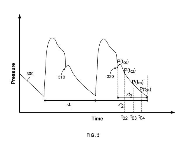
(evident in an aortic pressure waveform, e.g., notch 310 of FIG. 3) indicates
the start of
diastole. If the patient's heart rate is relatively steady, the start of a
heartbeat may be accurate
predicted. As the heartbeat completes systole, the aortic pressure decreases
before increasing
to form the dicrotic notch. Identifying this waveform shape allows the system
to determine
the start of diastole. The diastolic period is the time for a heart to
complete diastole ¨ the
phase of the heartbeat when the heart muscle relaxes and allows the chambers
to fill with
blood. For example, the diastolic period may be 0.05 seconds, 0.1 seconds, 0.2
seconds, 0.3
seconds, 0.4 seconds, 0.5 seconds, 0.6 seconds, 0.7 seconds, 0.8 seconds, 0.9
seconds, 1
second or any suitable length of time.
[0025] In some implementations, a time-variant relationship between aortic
pressure and
blood flow during the diastolic period is determined. The time-variant
relationship may be a
Windkessel model with two current sources, in and ip in parallel with each
other and with a
resistance R and compliance C. The governing equation for this model is:
dP P
C = th+lp (1)
where C is compliance, P is pressure, R is vascular resistance, in is flow
from native heart
operation and ip is flow from the pump. During diastole, however, the aortic
valve is closed,
so the only flow through the left ventricle is from the pump positioned across
the valve. By
discounting the heart current source and assuming pump flow is constant, the
model can thus
be simplified as follows:
_ t
P = Poe RC + ipR (2)
11
CA 03103478 2020-12-10
WO 2019/246305
PCT/US2019/038049
where Po is the scaling factor of the exponential decay part of the diastolic
pressure. In some
implementations, flow from the pump ip is estimated from current flow to the
motor of the
heart pump system to maintain constant speed. Pressure P may be measured at a
variety of
points within a single diastolic period to characterize and deconstruct the
pressure waveform.
For example, pressure may be known at a first, second, and third time. Flow ip
may be
estimated at the first, second and third time as well. Setting up three
pressure equations, one
each for the three times respectively, based on Equation (2), R and C values
may be
calculated. In some implementations, the heart pump is operated at a constant
speed.
[0026] In some implementations, a total volume of blood pumped per heartbeat,
which is
representative of cardiac performance, is calculated based on the time-variant
relationship
between the aortic pressure and blood flow during the diastolic period. For
example, once R
and C have been determined, CO for the heart may be determined by plugging the
calculated
Rand C values into Equation (1) above and solving for in to determine
volumetric flow
contributed by native heart function.
[0027] In some implementations, other metrics indicative of cardiac
performance may be
computed. For example, the metric indicative of cardiac performance may be
ventricular
resistance, ventricular compliance, CO, CPO, stroke volume, stroke work,
ejection fraction,
cardiac index, or a prediction of patient survival. Many metrics indicative of
cardiac
performance are interrelated. For example, CO is determined based on the flow
rate of the
blood through and past the pump. The stroke volume is an index of left
ventricular function
which formula SV= CO/HR, where SV is the stroke volume, CO is the cardiac
output, and
HR is the heart rate. Stroke work is the work done by the ventricle to eject a
volume of blood
and can be calculated from the stroke volume according to the equation SW = SV
* MAP,
where SW is the stroke work, SV is the stroke volume, and MAP is the mean
arterial
pressure. Cardiac work is calculated by the product of stroke work and heart
rate. CPO is a
measure of the heart function representing cardiac pumping ability in Watts.
CPO is
calculated using the equation CPO = mAoP * C0/451, where CPO is the cardiac
power
output, mAoP is the mean aortic pressure, CO is the cardiac output, and 451 is
a constant
used to convert mmHg x L/min into Watts. The ejection fraction can be
calculated by
dividing the stroke volume by the volume of blood in the ventricle. Other
parameters, such as
chamber pressure, preload state, afterload state, heart recovery, flow load
state, variable
volume load state, and/or heartbeat cycle flow state can be calculated from
these values or
determined via these parameters.
12
CA 03103478 2020-12-10
WO 2019/246305
PCT/US2019/038049
[0028] In some implementations, operation of the pump is adjusted, based on
the metric
indicative of cardiac performance. Adjusting pump operation may include
increasing pump
speed, decreasing pump speed, adjusting pump placement, turning the pump off,
or any other
suitable adjustment. For example, if total volume of blood pumped is below a
threshold, the
pump speed may be increased, while if the blood volume is above a threshold,
the pump
speed may be decreased.
[0029] In some implementations, the above-described methods include actuating
a blood
pump within the patient's vasculature, and determining cardiac output of the
patient's heart
using any of the foregoing systems and sensors. The blood pump's pumping speed
may be
adjusted based on the determined cardiac output. In some implementations, the
hemodynamic support applied may be based on determining whether cardiac
performance of
the heart is increasing or decreasing over time.
[0030] In some implementations, a blood vessel sensor is provided. The blood
vessel
sensor may include a system for inducing blood flow within a patient's blood
vessel. The
system may be an intravascular system. The system may includea motor, and an
impeller. In
some implementations, the system for inducing blood flow within the patient's
blood vessel
comprises a cannula that is configured to extend within the left ventricle of
a heart and a
pressure sensor configured to detect at least one of: aortic pressure, left
ventricular pressure,
or differential pressure. The system for inducing blood flow within the
patient's blood vessel
may be intracardiac blood pump incorporating the impeller within a shroud. For
example, the
shroud may be a pump housing. The shroud may be sized for passage through the
patient's
blood vessel and may be coupled to the motor or other pump elements. The
shroud may
comprise one or more blood exhaust apertures or outlets.
[0031] The blood vessel sensor may also include a controller. The controller
may be
configured to detect changes in resistance of impeller rotation within the
blood vessel. In
some implementations, a constant impeller rotational speed is maintained based
on the
detected resistance of impeller rotation. [0032] In some implementations,
vascular
compliance and vascular resistance may be calculated based on the change in
resistance of
impeller rotation using a transfer function. In some implementations, the
transfer function is
a Windkessel model. In some implementations, pump operation data may be
transmitted to a
computing device. The computing device may be located separately from the
controller or
onboard the controller. For example, the computing device may be a server
stored remotely.
In some implementations, the pump operation data includes at least one of
pressure
measurements, current measurements, change in resistance of impeller rotation,
and flow
13
CA 03103478 2020-12-10
WO 2019/246305
PCT/US2019/038049
estimations. In some implementations, the controller is further configured to
receive pump
operation commands from the computing device, wherein the pump operation
commands are
based on the pump operation data. For example, the computing device may
calculate
vascular resistance and compliance and alter pump operation accordingly.
[0033] In some implementations, the controller or the computing device is
configured to
determine a metric indicative of cardiac performance based on the vascular
compliance and
the vascular resistance. The metric indicative of cardiac performance may be
at least one of:
cardiac output, cardiac power output, stroke volume, stroke work, ejection
fraction, cardiac
contractility, ventricular elastance, cardiac index, a prediction of patient
survival, or any
suitable metric.
[0034] In some implementations, the controller is configured to adjust the
impeller
rotational speed based on at least one of: the vascular resistance, the
vascular compliance, or
the cardiac output. For example, the impeller rotational speed may be
increased or decreased
to provide more or less blood flow based on the patient's cardiac or vascular
health.
[0035] In some implementations, the controller is configured to receive
measurements
indicative of aortic pressure for a time period, detect current delivered to
the pump, and
determine, based on the current delivered to the pump, rates of blood flow
pumped by the
system for the time period. The calculation of the vascular compliance and the
vascular
resistance may be based on the measurements indicative of aortic pressure and
the rates of
blood flow.
[0036] In some implementations, a controller is configured to perform any of
the
implementations, aspects, and methods described herein. For example, the
controller may be
the Automated Impella Controller (AIC) of Abiomed, Inc or any other suitable
controller. In
some implementations, the heart pump system includes a catheter; a motor; a
rotor
operatively coupled to the motor; a pump housing at least partially
surrounding the rotor so
that the actuating motor drives the rotor and pumps blood through the pump
housing; one or
more sensors, including a differential pressure sensor; and the controller.
For example, the
heart pump system may comprise the Impella 5.0 heart pump of Abiomed, Inc
connected to
an AIC or any other suitable system.
[0037] In some implementations, the controller comprises a display. Any of the
foregoing
calculations or features may be configured for display. For example, an aortic
pressure
waveform may be presented on a graphical user interface. Clinicians may view
such displays
and adjust operation of the pump based on their observations of hemodynamic
parameters
over time.
14
CA 03103478 2020-12-10
WO 2019/246305
PCT/US2019/038049
Brief Description of the Drawings
[0038] FIG. 1 show an illustrative heart pump system inserted into a blood
vessel of a
patient;
[0039] FIG. 2 illustrates a process for determining the volume of blood pumped
per
heartbeat according to certain implementations;
[0040] FIG. 3 shows a plot of pressure versus time for a heart pump system
according to
certain implementations;
[0041] FIG. 4 shows a CO sensor coupled to a patient according to certain
implementations;
[0042] FIG. 5 illustrates a process for determining total volume of blood
pumped per
heartbeat according to certain implementations;
[0043] FIG. 6 illustrates a process for determining cardiac output according
to certain
implementations; and
[0044] FIG. 7 shows plots of pressure versus time and flow versus time for a
heart pump
system according to certain implementations.
Detailed Description
[0045] To provide an overall understanding of the systems and methods describe
herein,
certain illustrative embodiments will be described. Although the embodiments
and features
described herein are specifically described for use in connection with a
percutaneous heart
pump system, it will be understood that all the components and other features
outlined below
may be combined with one another in any suitable manner and may be adapted and
applied to
other types of cardiac therapy and heart pump systems, including heart pump
systems
implanted using a surgical incision, and the like.
[0046] The systems, devices, and methods described herein enable a support
device
residing completely or partially within an organ to assess that organ's
function. In particular,
the systems, devices and methods enable mechanical circulatory support
systems, such as
percutaneous ventricular assist devices, to be used to assess the function of
the heart. For
example, support devices such as blood pumps may be used in the treatment of
cardiogenic
shock, heart attack, or used to support a heart generally during coronary
intervention.
[0047] Assessing the function of the heart using a mechanical circulatory
support system
can alert health professionals of changes in cardiac function and allow the
degree of/level of
support provided by the assist device (i.e., flow rate of blood pumped by the
device) to be
CA 03103478 2020-12-10
WO 2019/246305
PCT/US2019/038049
tailored to a particular patient's needs. For example, the degree of support
can be increased
when a patient's heart function is deteriorating, or the degree of support can
be decreased
when a patient's heart function is recovering and returning to a baseline of
normal heart
function. This can allow the device to dynamically respond to changes in heart
function to
promote heart recovery and can allow the patient to be gradually weaned off
the therapy.
Furthermore, assessment of the heart function can indicate when it is
appropriate to terminate
use of the heart pump system. Although some embodiments presented herein are
directed to
heart pump systems implanted across the aortic valve and residing partially in
the left
ventricle, the concepts can be applied to devices in other regions of the
heart, the
cardiovascular system, or the body.
[0048] Assessment of cardiac function may include leveraging heart-device
interactions to
determine heart parameters. The systems and methods described herein determine
cardiac
output based on aortic pressure measurements and flow output from a blood pump
system.
The flow may be a measurement or estimate determined from the motor current
supplied to a
motor at a given pump speed in an intravascular blood pump system. At least
one advantage
of the systems and methods described herein is that they allow the heart pump
system to
assess cardiac function without changing operation of the pump (e.g., pump
speed), thereby
minimizing risks associated with changing pump speeds. A decrease in pump
speed involves
a decrease in patient support, while an increase in pump speed may result in
suction or other
risks. Frequent and/or fast changes in pump speed may also lead to hemolysis
or decrease of
pump/motor performance. The use of an intravascular blood pump system to
measure or
estimate the necessary parameters to determine metrics indicative of cardiac
performance
also allows for continuous measurements of the heart's performance, because
these metrics
are acquired by the blood pump system already placed within the patient's
vasculature.
[0049] Continuous measurement of vascular and cardiac performance by
leveraging the
effects of a heart pump system is a crucial step to provide additional
clinical data to aid in
titration of appropriate device support. However, more importantly, the
systems and methods
described herein demonstrate the impact and potential of device-arterial
coupling to
determine cardiac and vascular state. Unlike some invasive heart pump systems,
which shunt
blood out of the heart, the heart pump systems presented herein reside within
the heart and
work in parallel with native ventricular function. This allows the heart pump
systems
presented herein to be sensitive enough to detect native ventricular function
unlike some
more invasive devices. Thus, the systems, devices, and methods enable the use
of heart pump
systems not only as support devices, but also as diagnostic and prognostic
tools. The heart
16
CA 03103478 2020-12-10
WO 2019/246305
PCT/US2019/038049
pump systems can essentially function as active catheters that extract
information about
cardiac function by hydraulically coupling with the heart. In some
implementations, the heart
pump systems operate at a constant level (e.g., constant rotational speed of a
rotor), while
power delivered to the assist device is measured. In certain implementations,
the speed of the
rotor of the heart pump system may be varied (e.g., as a delta, step, or ramp
function) to
further probe the native heart function.
[0050] FIG. 1 show an illustrative heart pump system inserted into a blood
vessel of a
patient. Heart pump systems compatible with the present disclosure are
disclosed in U.S.
Patent Application Publication No. 2018-0078159-Al, the contents of which are
hereby
incorporated by reference in their entirety. Generally, any other heart pump
system or system
for obtaining physiological data from a patient may be used with the present
disclosure. In
some implementations, the systems and methods described herein may relate to
the
IMPELLA 0 family of devices (Abiomed, Inc., Danvers MA).
[0051] The heart pump system 100 may operate within a heart, partially within
the heart,
outside the heart, partially outside the heart, partially outside the vascular
system, or in any
other suitable location in a patient's vascular system. The heart pump system
may be
considered "in position" when cannula 173 is placed across the aortic valve
such that a blood
inlet (e.g., blood inlet 172) to the pump is within the left ventricle and an
outlet (e.g., outlet
openings 170) from the pump is within the aorta. The heart pump system 100
includes a
heart pump 106 and a control system 104. All or part of the control system 104
may be in a
controller unit separate/remote from the heart pump 106. In some
implementations, the
control system 104 is internal to the heart pump 106. The control system 104
and the heart
pump 106 are not shown to scale. The pump system 100 includes an elongate
catheter body
105, a motor housing 102 and a drive shaft in which a pump element is formed.
The pump
100 includes a pump housing 134, and a motor housing 102 coupled to a cannula
173 at a
distal end 111 of the motor housing 102. An impeller blade on the drive shaft
may be rotated
within a pump housing 134 to induce a flow of blood into the cannula 173 at a
suction head
174. The suction head 174 provides a blood inlet 172 at the distal end portion
171 of the
cannula 173. The flow 109 of blood passes through the cannula 173 in a first
direction 108
and exits the cannula 173 at one or more outlet openings 170 of the cannula
173.
[0052] The rotation of the drive shaft within the pump housing 134 may also
rotate a pump
element within a bearing gap. A hemocompatible fluid may be delivered through
the
elongate catheter 105 through the motor housing 102 to a proximal end portion
of the cannula
173 where the fluid is pressurized by the rotation of a pump element. The flow
of
17
CA 03103478 2020-12-10
WO 2019/246305
PCT/US2019/038049
hemocompatible fluid has a second direction 122 through the bearing gap of the
pump. After
exiting the bearing gap, the hemocompatible fluid may follow flow direction
123 and become
entrained in the flow of blood and flows into the aorta with the blood.
[0053] The heart pump 100 is inserted into a vessel of the patient through a
sheath 175.
The pump housing 134 may enclose the rotor and internal bearings and may be
sized for
percutaneous insertion into a vessel of a patient. In some implementations,
the pump may be
advanced through the vasculature and over the aortic arch 164. Although the
pump is shown
in the left ventricle, the pump may alternatively be placed in the right
heart, such that the
blood is pumped from the patient's inferior vena cava or right atrium, through
the right
ventricle into the pulmonary artery.
[0054] A flexible projection 176 may also be included at a distal end portion
171 of the
cannula 173, distal to the suction head 174, in order to position the heart
pump 100 optimally
in a vessel or chamber of the heart. The flexible projection 176 may prevent
the suction head
174 from approaching the wall of the vessel where it may become stuck due to
suction. The
flexible projection 176 may extend the pump 100 mechanically, but not
hydraulically, as the
flexible projection 176 may be non-sucking. In some implementations, the
flexible
projection may be formed as a pigtail. In some aspects, the pump need not
include a flexible
projection.
[0055] The elongate catheter 105 houses a connection 126 that may comprise a
fluid supply
line and may also house electrical connection cables. The connection 126 may
supply a
hemocompatible fluid to the pump from a fluid reservoir that may be contained
within control
system 104.
[0056] The control system 104 includes controller 182 controls pump 106,
including, for
example, controlling power to the motor or controlling the motor speed. In
some
implementations, the control system 104 includes display screens to show
measurements such
as differential pressure signal and motor current. The control system 104 may
include
circuitry for monitoring the motor current for drops in current indicating air
in the line,
changes in differential pressure signal, flow position, suction, or any other
suitable
measurement. The control system 104 may include warning sounds, lights or
indicators to
alert an operator of sensor failures, disconnects or breaks in the connection
126, or sudden
changes to patient health.
[0057] The motor 108 may operate at a speed required to maintain the rotor at
a set speed.
As a result and as further described below, the motor current drawn by the
motor to maintain
18
CA 03103478 2020-12-10
WO 2019/246305
PCT/US2019/038049
the rotor speed can be monitored and used to understand the underlying cardiac
state. For
example, motor current may be used to determine flow through the heart.
[0058] The heart pump may operate at a variety of pump speeds or P-levels. P-
level is the
performance level of the heart pump system and related to flow control of the
system. As P-
level increases, the flow rate, motor current, and revolutions per minute
associated with the
heart pump system increase; thus, higher P-levels correspond to higher flow
rates and
revolutions per minute associated with the heart pump system. For example,
power level P-1
may corresponds to a first number of rotations per minute (RPM) for the rotor,
while power
level P-2 corresponds to a second number of RPM. In some examples, the pump
operates at
ten different power levels ranging from P-0 through P-9. These P-levels may
correspond to 0
RPM through 100,000 RPM or any suitable number. Changing the speed of the
rotor
changes the CO of the heart.
[0059] The control system 104 can include a current sensor (not shown). The
controller
182 supplies current to the motor 108 by the connection 126 such as through
one or more
electrical wires. The current supplied to the motor 108 via the connection 126
is measured by
the current sensor. The load that the motor of a mechanical pump experiences
is pressure
head, or the difference between the aortic and left ventricular pressure. The
heart pump 106
experiences a nominal load during steady state operation for a given pressure
head, and
variations from this nominal load are a result of changing external load
conditions, for
example the dynamics of left ventricular contraction. Changes to the dynamic
load conditions
alter the motor current required to operate the pump rotor at a constant, or
substantially
constant, speed. As described above, the motor may operate at a speed required
to maintain
the rotor at a set speed, and the motor current drawn by the motor to maintain
the rotor speed
can be monitored and used to understand the underlying cardiac state. The
cardiac state can
be even more precisely quantified and understood by simultaneously monitoring
the pressure
head during the heartbeat cycle using a pressure sensor 112. The heart
parameter estimator
185 receives current signals from the current sensor as well as pressure
signals from the
pressure sensor 112. The heart parameter estimator 185 uses these current and
pressure
signals to characterize the heart's function. The heart parameter estimator
185 may access
stored look-up tables to obtain additional information to characterize the
heart's function
based on the pressure and current signals. For example, the heart parameter
estimator 185
may receive an aortic pressure from the pressure sensor 112, and using look-up
tables, may
use the motor current and pump speed to determine a delta pressure between the
aorta and the
ventricle.
19
CA 03103478 2020-12-10
WO 2019/246305
PCT/US2019/038049
[0060] In some implementations, pressure sensor 112 is an aortic pressure
sensor. In some
implementations, pressure sensor 112 is a flexible membrane integrated into
the cannula 172
configured to measure differential pressure. One side of the sensor is exposed
to the blood
pressure on the outside of the cannula and the other side is exposed to the
pressure of the
blood inside of the cannula. The sensor generates an electrical signal (the
differential
pressure signal) proportional to the difference between the pressure outside
the cannula and
the pressure inside, which may be displayed by the heart pump system. When the
heart pump
system is placed in the correct position across the aortic valve, the top
(outer surface) of the
sensor is exposed to the aortic pressure and the bottom (inner surface) of the
sensor is
exposed to the ventricular pressure. Therefore, the differential pressure
signal is
approximately equal to the difference between the aortic pressure and the
ventricular
pressure. In some implementations, the system includes both differential and
aortic pressure
sensors.
[0061] FIG. 2 illustrates a process 200 for determining cardiac output. The
process 200 can
be performed using the heart pump system 100 of FIG. 1 or any other suitable
pump. In
some implementations, the pump is an intravascular blood pump device placed
within the
patient's heart via percutaneous insertion. The pump may be introduced to the
patient
because the patient is in cardiogenic shock or otherwise experiencing a
decline in health. The
pump may be positioned across the aortic valve such that a blood inlet (e.g.,
blood inlet 172
of FIG. 1) to the pump is within the left ventricle and an outlet (e.g.,
outlet openings 170 of
FIG. 1) from the pump is within the aorta. The pump contributes with native
heart operation
such that CO from the heart is equal to native CO plus pump output.
[0062] In step 202, hemodynamic support is applied to a heart at a first
pumping rate. In
some implementations, the pumping rate may correspond to a speed of rotation
of a rotor.
For example, the pump speed may be 10,000 RPM, 20,000 RPM, 30,000 RPM, 40,000
RPM,
50,000 RPM, 60,000 RPM, 70,000 RPM, 80,000 RPM, 90,000 RPM, 100,000 RPM, or
any
suitable speed. A pump speed may correspond to a power level, or P-level, as
described
below in relation to FIG. 1. For example, the pump speed may be P-1, P-2, P-3,
P-4, P-5, P-
6, P-7, P-8, or P-9. In some implementations, the pumping rate may instead
correspond to
the rate at which a chamber of the pump fills up with and releases blood. The
pumping rate is
supplied over a plurality of heartbeats of the heart. Each heartbeat includes
a systolic rise, a
dicrotic notch, and a diastolic fall that occurs after the dicrotic notch.
[0063] In step 204, at least three aortic pressure measurements are detected
during the
diastolic fall of a specific beat of the plurality of beats. In some
implementations, aortic
CA 03103478 2020-12-10
WO 2019/246305
PCT/US2019/038049
pressure is continuously measured or is periodically sampled, and a plurality
of aortic
pressure measurements are detected. For example, pressure may be sampled at a
rate of 1, 2,
3, 10, 20, 30, 100, 200, 300, 1000, 2000, 3000, or any other suitable number
of samples per
second. In some examples, aortic pressure is only sampled during the diastolic
fall. In some
examples, aortic pressure is constantly or periodically measured.
[0064] In step 206, at least threee blood flow pumped by the intravascular
blood pump are
determined. As shown in FIG. 3 and described above, pressure may be measured
at a series
of points during a diastolic time period. For each of these pressure
measurements, pressure is
measured and flow may be estimated based on current supplied to the pump to
maintain a
rotor speed. This mathematical relationship between pump speed and motor
current to a flow
estimate may be implemented by setting up a look-up table where the pump speed
and motor
current are the indices to the table and the flow values in the table is pre-
populated through
bench testing. Another way is to pre-determine flow for a sub-set of possible
combinations of
pump speed and motor current. For example, if the flow at a pump speed of
40,000 RPM and
a motor current of 500 mA and the flow at a pump speed of 40,000 RPM and a
motor current
of 510 mA are known as ii and i2, respectively, then the flow at a pump speed
of 40,000 RPM
and a motor current of 505 mA can be calculated by taking the average of ii
and i2. The
pressure and flow measurements, in combination with the known time of
measurement, are
compared between two times to calculate heart parameters such as systemic
vascular
resistance and compliance.
[0065] In step 208, cardiac output during the specific beat is determined
based on the aortic
pressure and blood flow measurements. A Windkessel model with two current
sources, in
and ip in parallel with each other and with a resistance R and compliance C,
may be used to
simulate the aortic pressure. The governing equation for this model is:
dP P
C = th+lp (1)
where C is compliance, P is pressure, R is vascular resistance, in is flow
from native heart
operation and ip is flow from the pump. During diastole, however, the aortic
valve is closed,
so the only flow through the left ventricle is from the pump positioned across
the valve. By
discounting the heart current source and assuming pump flow is constant, the
model can thus
be simplified as follows:
_ t
P = Poe RC + ipR (2)
where Po is the initial aortic pressure during diastole. In some
implementations, flow from
the pump ip is estimated from current flow to the motor of the heart pump
system to maintain
21
CA 03103478 2020-12-10
WO 2019/246305
PCT/US2019/038049
constant speed. Pressure P may be measured at a variety of points within a
single diastolic
period to characterize and deconstruct the pressure waveform, as shown in FIG.
3 and
described below. For example, pressure may be known at three times. Flow ip
may be
estimated at the same three times as well. Setting up three pressure
equations, one each for
the three times respectively, based on Equation (2), R and C values may be
calculated. For
example, once R and C have been determined using the Windkessel model
described above,
CO for the heart may be determined by plugging the calculated R and C values
into Equation
(1) above and solving for in to determine volumetric flow contributed by
native heart
function.
[0066] Operation of the pump may be adjusted, based on the calculated CO
value.
Adjusting pump operation may include increasing pump speed, decreasing pump
speed,
adjusting pump placement, turning the pump off, or any other suitable
adjustment. For
example, if the CO is below a threshold, the pump speed may be increased,
while if CO is
above a threshold, the pump speed may be decreased.
[0067] FIG. 3 shows a plot 300 of pressure versus time for a heart pump
system, according
to certain implementations. The y-axis of plot 300 represents aortic pressure
in mmHg, while
the x-axis represents time as a percentage of a heartbeat length. In
particular, plot 300 shows
that pressure may be measured at a series of points Po-Ps during diastole of a
heartbeat. Ati
represents a time of a first heartbeat and At represents a time of a second
heartbeat after the
first heartbeat. Time periods Ati and At occur while the heart pump system is
placed at least
partially within the patient's heart. Point 310 represents the dicrotic notch
during the first
heartbeat and point 320 represents the dicrotic notch during the second
heartbeat. Diastolic
time period At3 represent the diastolic period of the second heartbeat. During
time periods
At, At, and At3, the pump operates at a first pump speed. In some
implementations, the
pump operates at a second pump speed during time period At3. For example, pump
speed
may be increased during time period At3. At higher pump speeds, the measured
aortic
pressure and total flow are higher compared to lower pump speeds.
[0068] At a given known point in time to2 within diastolic period At3,
pressure P(t02) is
known; at a second known point in time t03 within diastolic period At3,
pressure P(t03) is
known; and at a third known point in time to4 within diastolic period At3,
pressure P(t04) is
known. At each of these times within the diastolic period At3, the pump flow
is known from
motor current supplied to the pump motor at that point in time. Thus, the
following equations
may be used to calculate Po, R and C:
22
CA 03103478 2020-12-10
WO 2019/246305
PCT/US2019/038049
_t02
P(t02) = POe RC ip (t02) * R (3)
_t03
P(t03) = Poe RC + ip(t03)* R (4)
_t04
P(t04) = POe RC ip (t04) * R (5)
These steps may be repeated for each time point within diastolic period t3.
Rand C values
calculated for each set of times (e.g., t02 and to4, t02 and to3, etc.) may
differ slightly. The
measured R and C values may be averaged to arrive at representative systemic
vascular
resistance and compliance values for the heart. In some implementations, R and
C values
may be periodically calculated to determine how the values change over time as
a patient is
treated. In some implementations, cardiac output may be determined using the
calculated R
and C values. For example, determining cardiac output may include determining
cardiac
output of a plurality of specific beats within the plurality of beats and
applying at least one of
a summation, average, or linear regression on the determined cardiac outputs
to determine a
cumulative indicator of cardiac output of the heart.
[0069] FIG. 4 shows a compliance sensor 410 coupled to a patient 400.
Compliance sensor
410 may comprise a variety of hardware elements configured to perform the
methods
described herein, as well as additional processes. In some implementations,
the compliance
sensor includes an intravascular blood pump (e.g., pump 202 of FIG. 1). The
intravascular
blood pump may be configured to be placed at least partially within a
patient's heart. In
some implementations, the intravascular blood pump includes a cannula, an
impeller
configured to be rotated within a blood vessel and pump blood through the
cannula, and a
drive mechanism configured to impart power to turn the impeller. In some
implementations,
the cannula may be configured to extend across an aortic valve such that a
distal end of the
cannula is within a left ventricle and a proximal end of the cannula is within
the aorta. For
example, the heart pump system may be considered "in position" when the
cannula is placed
across the aortic valve such that a blood inlet to the pump is within the left
ventricle and an
outlet from the pump is within the aorta. The drive mechanism may include an
onboard
motor, a drive cable, a drive shaft, or any other suitable element or
combination thereof
[0070] In some implementations, compliance sensor 410 includes an elongate
catheter body
coupled to a cannula. The elongate catheter may include a drive cable,
electrical wiring
connecting the blood pump to a control system, any suitable element, or any
combination
thereof In some implementations, the blood pump includes a pump housing and a
motor
23
CA 03103478 2020-12-10
WO 2019/246305
PCT/US2019/038049
housing coupled to the cannula at a distal end of the motor housing. The
impeller may be
rotated within the pump housing to induce a flow of blood into the cannula.
[0071] Compliance sensor 410 includes a pressure sensor configured to detect
pressure
within the blood vessel arising at least in part from the pumping of blood
within the vessel.
For example, the pressure sensor may be a differential pressure sensor that is
part of a blood
pump. One side or surface of the differential pressure sensor may be exposed
to the aortic
pressure, a second side or surface of the differential pressure sensor may be
exposed to the
ventricular pressure, and the differential pressure sensor may measure the
difference between
the aortic and ventricular pressures. As another example, pressure sensor 412
may comprise
a pressure measurement lumen configured to measure aortic pressure.
[0072] Compliance sensor 410 includes controller 414. Controller 414 is
coupled to
pressure sensor 412. Controller 414 may coupled directly or indirectly to
pressure sensor
412. For example, control 414 may be connected to pressure sensor 412 via
electrical wiring,
a wireless signal, or any other suitable means. Controller 414 is configured
to detect signals
from the pressure sensor indicative of blood pressure. All or part of
controller 414 may be in
a controller unit separate/remote from an intravascular blood pump. In some
implementations, the control system is internal to an intravascular blood
pump.
[0073] In some implementations, controller 414 is configured to detect changes
in
resistance of impeller rotation within the blood vessel. For example,
resistance may be
calculated at a variety of points in time based on pressure and flow
measurements of the
heart, as described above in relation to FIG. 1.
[0074] In some implementations, controller 414 is configured to maintain a
constant
impeller rotational speed, based on the detected resistance of impeller
rotation. Current
supplied to the impeller motor may change based on the necessary current
needed to maintain
motor speed. Thus, motor current may be correlated to flow through the heart.
[0075] In some implementations, controller 414 is configured to calculate,
based on the
change in resistance of impeller rotation, vascular compliance and vascular
resistance using a
transfer function. For example, the vascular compliance and resistance may be
determined as
described above in relation to FIG. 3. In another example, while the pump
speed is maintained
at a constant speed (speedi), a set of diastolic aortic pressure measurements
Pi(t) and a set of
pump flow ii(t) measurements are determined for a set of times (e.g., t equals
tloi, tl o2, t103,
etc.). Then the controller may set the pump to a different constant speed
(speed2) and obtain a
second set of diastolic aortic pressure measurements P2(t) and a second set of
pump flow
measurements i2(t) for a second set of times (e.g., t equals t201, (202, (203,
etc.). The difference
24
CA 03103478 2020-12-10
WO 2019/246305
PCT/US2019/038049
in the two sets of pressure measurements Pi(t) and P2(t) and the difference in
the two sets of
pump flow ii(t) and i2(t) can be used to calculate the vascular resistance via
the following
equation:
mean(Pi(t))-mean(P2(t))
R ¨ __________________________________________ (6)
mean(i1(t))-mean(i2(t))
[0076] FIG. 5 illustrates a process 500 for determining total volume of blood
pumped per
heartbeat. The process 500 can be performed using the heart pump system 100 of
FIG. 1 or
any other suitable pump. In some implementations, the pump is an intravascular
blood pump
device placed within the patient's heart via percutaneous insertion. In some
implementations,
the pump may be a surgically implanted device, a left ventricular assist
device, a
counterpulsation device, an expandable heart pump, or any other suitable
device. The pump
may be introduced to the patient because the patient is in cardiogenic shock
or otherwise
experiencing a decline in health. The pump may be positioned across the aortic
valve such
that a blood inlet (e.g., blood inlet 172 of FIG. 1) to the pump is within the
left ventricle and
an outlet (e.g., outlet openings 170 of FIG. 1) from the pump is within the
aorta. The pump
contributes with native heart operation such that CO from the heart is equal
to native CO plus
pump output.
[0077] At step 502, a hemodynamic parameter is monitored during operation of a
heart
pump at a first pump speed. A hemodynamic parameter may be any parameter
relating to the
flow of blood within the organs and tissues of the body. For example, the
hemodynamic
parameter may include at least one of heart rate, blood pressure, arterial
oxygen saturation,
mixed venous saturation, central venous oxygen saturation, arterial blood
pressure, mean
arterial pressure, right arterial pressure, central venous pressure, right
ventricular pressure,
pulmonary artery pressure, mean pulmonary artery pressure, pulmonary artery
occlusion
pressure, left atrial pressure, aortic pressure, differential pressure, left
ventricular end
pressure, stroke volume, stroke volume index, stroke volume variation,
systemic vascular
resistance, systemic vascular resistance index, pulmonary vascular resistance,
pulmonary
vascular resistance index, pulmonary vascular resistance, pulmonary vascular
resistance
index, left ventricular stroke work, left ventricular stoke work index, right
ventricular stroke
work, right ventricular stroke work indexõ coronary artery perfusion pressure,
right
ventricular end diastolic volume, right ventricular end diastolic volume
index, right
ventricular end systolic volume, right ventricular ejection fraction, arterial
oxygen content,
venous oxygen content, arterial-venous oxygen content difference, oxygen
delivery, oxygen
delivery index, oxygen consumption, oxygen consumption index, oxygen
extraction ration,
CA 03103478 2020-12-10
WO 2019/246305
PCT/US2019/038049
oxygen extraction index, total peripheral resistance, CO, cardiac index, and
CPO. A pump
speed is the speed of operation of the pump and corresponds to the amount of
blood flow
provided by the pump's operation. In some implementations, the pump speed may
correspond to a speed of rotation of a rotor. For example, the pump speed may
be 10,000
RPM, 20,000 RPM, 30,000 RPM, 40,000 RPM, 50,000 RPM, 60,000 RPM, 70,000 RPM,
80,000 RPM, 90,000 RPM, 100,000 RPM, or any suitable speed. A pump speed may
correspond to a power level, or P-level, as described above in relation to
FIG. 1. For
example, the pump speed may be P-1, P-2, P-3, P-4, P-5, P-6, P-7, P-8, P-9, or
any other
suitable value. In some implementations, the pump speed may instead correspond
to the rate
at which a chamber of the pump fills up with and releases blood.
[0078] At step 504, a diastolic period of a heartbeat cycle is identified,
based on a shape of
the hemodynamic parameter over time. The diastolic period is the time for a
heart to
complete diastole ¨ the phase of the heartbeat when the heart muscle relaxes
and allows the
chambers to fill with blood. For example, the diastolic period may be 0.05
seconds, 0.1
seconds, 0.2 seconds, 0.3 seconds, 0.4 seconds, 0.5 seconds, 0.6 seconds, 0.7
seconds, 0.8
seconds, 0.9 seconds, 1 second or any suitable length of time.
[0079] At step 506, a time-variant relationship between aortic pressure and
blood flow
during the diastolic period is determined. The time-variant relationship may
be a Windkessel
model with two current sources, in and ip in parallel with each other and with
a resistance R
and compliance C. The governing equation for this model is:
dP P
C = th+lp (1)
where C is compliance, P is pressure, R is systemic vascular resistance, in is
flow from native
heart operation and ip is flow from the pump. During diastole, however, the
aortic valve is
closed, so the only flow through the left ventricle is from the pump
positioned across the
valve. By discounting the heart current source and assuming pump flow is
constant, the
model can thus be simplified as follows:
_ t
P = Poe RC + ipR (2)
where Po is the initial aortic pressure during diastole. In some
implementations, flow from
the pump ip is estimated from current flow to the motor of the heart pump
system to maintain
constant speed. Pressure P may be measured at a variety of points within a
single diastolic
period to characterize and deconstruct the pressure waveform, as describe
below in relation to
FIG. 3. In some implementations, the heart pump is operated at a constant
speed. In some
implementations described herein, the speed of the pump may be altered to
"ping" the heart.
26
CA 03103478 2020-12-10
WO 2019/246305
PCT/US2019/038049
[0080] At step 508, a total volume of blood pumped per heartbeat, which is
representative
of cardiac performance, calculated based on the time-variant relationship
between aortic
pressure and blood flow during the diastolic period. For example, once R and C
have been
determined, CO for the heart may be determined (e.g., as described below in
relation to FIG.
7). In some implementations, other metrics indicative of cardiac performance
may be
computed. For example, the metric indicative of cardiac performance may be
ventricular
resistance, ventricular compliance, CO, CPO, stroke volume, stroke work,
ejection fraction,
cardiac index, or a prediction of patient survival. Many metrics indicative of
cardiac
performance are interrelated. For example, CO is determined based on the flow
rate of the
blood through and past the pump. The stroke volume is an index of left
ventricular function
which formula SV= CO/HR, where SV is the stroke volume, CO is the cardiac
output, and
HR is the heart rate. Stroke work is the work done by the ventricle to eject a
volume of blood
and can be calculated from the stroke volume according to the equation SW = SV
* MAP,
where SW is the stroke work, SV is the stroke volume, and MAP is the mean
arterial
pressure. Cardiac work is calculated by the product of stroke work and heart
rate. CPO is a
measure of the heart function representing cardiac pumping ability in Watts.
CPO is
calculated using the equation CPO = mAoP * C0/451, where CPO is the cardiac
power
output, mAoP is the mean aortic pressure, CO is the cardiac output, and 451 is
a constant
used to convert mmHg x L/min into Watts. The ejection fraction can be
calculated by
dividing the stroke volume by the volume of blood in the ventricle. Other
parameters, such as
chamber pressure, preload state, afterload state, heart recovery, flow load
state, variable
volume load state, and/or heartbeat cycle flow state can be calculated from
these values or
determined via these parameters.
[0081] In some implementations, operation of the pump is adjusted, based on
the metric
indicative of cardiac performance. Adjusting pump operation may include
increasing pump
speed, decreasing pump speed, adjusting pump placement, turning the pump off,
or any other
suitable adjustment. For example, if total volume of blood pumped is below a
threshold, the
pump speed may be increased, while if the blood volume is above a threshold,
the pump
speed may be decreased.
[0082] In some implementations, the above-described methods include actuating
a blood
pump within the patient's vasculature, and determining cardiac output of the
patient's heart
using any of the foregoing systems and sensors. The blood pump's pumping speed
may be
adjusted based on the determined cardiac output.
27
CA 03103478 2020-12-10
WO 2019/246305
PCT/US2019/038049
[0083] FIG. 6 illustrates a process 600 for determining cardiac output. The
process 600 can
be performed using the heart pump system 100 of FIG. 1 or any other suitable
pump. In
some implementations, the pump is an intravascular blood pump device placed
within the
patient's heart via percutaneous insertion. The pump may be introduced to the
patient
because the patient is in cardiogenic shock or otherwise experiencing a
decline in health. The
pump may be positioned across the aortic valve such that a blood inlet (e.g.,
blood inlet 172
of FIG. 1) to the pump is within the left ventricle and an outlet (e.g.,
outlet openings 170 of
FIG. 1) from the pump is within the aorta. The pump contributes with native
heart operation
such that CO from the heart is equal to native CO plus pump output.
[0084] In step 602, hemodynamic support is applied to a heart at a first
pumping rate. The
hemodynamic pumping rate is supplied over a plurality of heartbeats of the
heart. Each
heartbeat includes a systolic rise, a dicrotic notch, and a diastolic fall
that occurs after the
dicrotic notch. For example, the hemodynamic pumping rate may be provided over
two,
three, four, ten, twenty, thirty, one hundred, two hundred, or any other
suitable number of
heartbeats.
[0085] In step 604, a plurality of aortic pressure measurements are detected
during the
diastolic fall during a specific beat of the plurality of beats. For example,
pressure may be
sampled at a rate of 1, 2, 3, 10, 20, 30, 100, 200, 300, 1000, 2000, 3000, or
any other suitable
number of samples per second. In some examples, aortic pressure is only
sampled during the
diastolic fall. In some examples, aortic pressure is constantly or
periodically measured. In
some examples, the sampling rate of aortic pressure is altered during the
diastolic fall.
[0086] In step 606, a first of the plurality of aortic pressure measurements
is compared to a
second of the plurality of aortic pressure measurements in a time dependent
non-linear model
of the vascular system to determine systemic vascular resistance and
compliance. In some
implementations, at least one aortic pressure measurement is taken at the end
of diastole,
when the cardiac output occurs solely by the operation of a blood pump. For
example, as
shown in FIG. 3 and described above, pressure may be measured at a series of
points during a
diastolic time period. For each of these pressure measurements, pressure may
be measured
and flow may be estimated. The pressure and flow measurements, in combination
with the
known time of measurement, may be compared between two times to calculate
heart
parameters such as aortic resistance and compliance.
[0087] In step 608, cardiac output during the specific beat is determined as a
function of the
determined systemic vascular resistance and compliance. In some
implementations,
determining cardiac output includes determining cardiac output of a plurality
of specific beats
28
CA 03103478 2020-12-10
WO 2019/246305
PCT/US2019/038049
within the plurality of beats and applying at least one of a summation,
average, or linear
regression on the determined cardiac outputs to determine a cumulative
indicator of cardiac
output of the heart. The cumulative indicator of cardiac output of the heart
may be indicative
of cardiac performance or overall patient health.
[0088] FIG. 7 shows two plots, one of aortic pressure and one of cardiac flow
for the same
ten-second period. The y-axis of the upper plot represents aortic pressure in
mmHg, while
the x-axis represents time in seconds. The y-axis of the lower plot represents
calculated total
cardiac flow in liters per minute, while the x-axis represents time in
seconds. In this
example, systemic vascular resistance R and compliance C are known. For
example, R and C
may be calculated using aortic pressure measurements taken during the depicted
ten second
time period in combination with pump data as described above. The total
cardiac flow ih-Fip
is calculated using R, C, and the aortic pressure waveform by applying
Equation (1):
dP P
C ¨dt ¨R= th+lp (1)
CO can be calculated by taking the average of the total cardiac flow ih+ip
resulting from
Equation (1) over a period of time (e.g., 5 seconds, 10 seconds, or 30
seconds). In the
example in FIG. 7, the period of time is 10 seconds. The average R value for
the time period
is 0.6143 mmHg*sec/m1 and the average C value is 1.5 mL/mmHg, resulting in a
calculated
CO of 6.9 L/min.
[0089] The foregoing is merely illustrative of the principles of the
disclosure, and the
apparatuses can be practiced by other than the described aspects, which are
presented for
purposes of illustration and not of limitation. It is to be understood that
the apparatuses
disclosed herein, while shown for use in percutaneous insertion of heart
pumps, may be
applied to apparatuses in other applications requiring hemostasis.
[0090] Variations and modifications will occur to those of skill in the art
after reviewing
this disclosure. The disclosed features may be implemented, in any combination
and
subcombination (including multiple dependent combinations and
subcombinations), with one
or more other features described herein. The various features described or
illustrated above,
including any components thereof, may be combined or integrated in other
systems.
Moreover, certain features may be omitted or not implemented.
[0091] The systems and methods described may be implemented locally on a heart
pump
system or a controller of a heart pump system, such as the AIC. The heart pump
system may
comprise a data processing apparatus. The systems and methods described herein
may be
implemented remotely on a separate data processing apparatus. The separate
data processing
29
CA 03103478 2020-12-10
WO 2019/246305
PCT/US2019/038049
apparatus may be connected directly or indirectly to the heart pump system
through cloud
applications. The heart pump system may communicate with the separate data
processing
apparatus in real-time (or near real-time).
[0092] In general, aspects of the subject matter and the functional operations
described in
this specification can be implemented in digital electronic circuitry, or in
computer software,
firmware, or hardware, including the structures disclosed in this
specification and their
structural equivalents, or in combinations of one or more of them. Aspects of
the subject
matter described in this specification can be implemented as one or more
computer program
products, i.e., one or more modules of computer program instructions encoded
on a computer
readable medium for execution by, or to control the operation of, data
processing apparatus.
The computer readable medium can be a machine-readable storage device, a
machine-
readable storage substrate, a memory device, a composition of matter affecting
a machine-
readable propagated signal, or a combination of one or more of them. The term
"data
processing apparatus" encompasses all apparatus, devices, and machines for
processing data,
including by way of example a programmable processor, a computer, or multiple
processors
or computers. The apparatus can include, in addition to hardware, code that
creates an
execution environment for the computer program in question, e.g., code that
constitutes
processor firmware, a protocol stack, a database management system, an
operating system, or
a combination of one or more of them. A propagated signal is an artificially
generated signal,
e.g., a machine-generated electrical, optical, or electromagnetic signal that
is generated to
encode information for transmission to suitable receiver apparatus.
[0093] A computer program (also known as a program, software, software
application,
script, or code) can be written in any form of programming language, including
compiled or
interpreted languages, and it can be deployed in any form, including as a
stand-alone program
or as a module, component, subroutine, or other unit suitable for use in a
computing
environment. A computer program may correspond to a file in a file system. A
program can
be stored in a portion of a file that holds other programs or data (e.g., one
or more scripts
stored in a markup language document), in a single file dedicated to the
program in question,
or in multiple coordinated files (e.g., files that store one or more modules,
sub programs, or
portions of code). A computer program can be deployed to be executed on one
computer or
on multiple computers that are located at one site or distributed across
multiple sites and
interconnected by a communication network.
[0094] The processes and logic flows described in this specification can be
performed by
one or more programmable processors executing one or more computer programs to
perform
CA 03103478 2020-12-10
WO 2019/246305
PCT/US2019/038049
functions by operating on input data and generating output. The processes and
logic flows
can also be performed by, and apparatus can also be implemented as, special
purpose logic
circuitry, e.g., an FPGA (field programmable gate array) or an ASIC
(application specific
integrated circuit).
[0095] Processors suitable for the execution of a computer program include, by
way of
example, both general and special purpose microprocessors, and any one or more
processors
of any kind of digital computer. Generally, a processor will receive
instructions and data
from a read-only memory or a random access memory or both. The essential
elements of a
computer are a processor for performing instructions and one or more memory
devices for
storing instructions and data. Generally, a computer will also include, or be
operatively
coupled to receive data from or transfer data to, or both, one or more mass
storage devices for
storing data, e.g., magnetic, magneto optical disks, or optical disks.
However, a computer
need not have such devices.
[0096] Examples of changes, substitutions, and alterations are ascertainable
by one skilled
in the art and could be made without departing from the scope of the
information disclosed
herein. All references cited herein are incorporated by reference in their
entirety and made
part of this application.
31