Note: Descriptions are shown in the official language in which they were submitted.
CA 03104655 2020-12-21
WO 2019/246559
PCT/US2019/038536
SYSTEMS AND METHODS FOR SIMULATION AND MODELING OF COMBINED
AUGMENTATION PROCEDURES
CROSS-REFERENCE TO RELATED APPLICATIONS
[001] This application claims priority to U.S. Provisional Application
No. 62/688,778, filed on June 22, 2018, which is incorporated by reference
herein in its
entirety.
TECHNICAL FIELD
[002] The present disclosure relates to systems and methods useful for medical
procedures, such as, e.g., aesthetic and/or reconstructive surgeries.
BACKGROUND
[003] Aesthetic, cosmetic, and reconstructive surgeries refer to surgeries
performed
in order to repair, restore, or change the appearance of a subject's anatomy.
For example, the
field of cosmetic surgery includes surgeries such rhytidectomy (facelifts),
mammoplasty
(changing the size of the breasts), and gluteoplasty (changing the size of the
buttocks), and
the field of reconstructive surgery includes implanting a prosthesis and
procedures such as
the reattachment of an amputated body part. In some such procedures, a surgeon
inserts a
suitable implant at a desired region of the subject's body. In some cases, an
implant alone
may not provide a desired size, shape, or change in physical appearance or
feel of the
subject's body part. Additionally, an implant alone may have an undesirable
weight or feel to
a subject. Moreover, in some cases, the subject may have to wait for the
conclusion of the
procedure to visualize the results of the procedure.
SUMMARY
[004] Systems and methods for simulating an outcome of a surgical procedure
are
disclosed herein. In some aspects, a method for simulating an outcome of a
surgical
procedure includes receiving parameters for a pre-operative state of an
implantation site,
receiving parameters for a post-operative state of the implantation site,
based on the
parameters for the pre-operative and post-operative states, automatically
generating a hybrid
strategy for achieving the post-operative state from the pre-operative state,
wherein the
hybrid strategy includes a proposed implant volume and a proposed volume of a
secondary
material, and generating a simulation of the post-operative state of the
implantation site using
the proposed implant volume and the proposed volume of the secondary material.
[005] Receiving parameters for the post-operative state of the implantation
site may
include providing a catalog of potential implants for use at the implantation
site, and
CA 03104655 2020-12-21
WO 2019/246559
PCT/US2019/038536
receiving a selection of an initial implant from the catalog of potential
implants. For
example, the parameters for the pre-operative state of the implantation site
may include a pre-
operative volume, and the parameters for the post-operative state of the
implantation site may
include a post-operative volume.
[006] According to some aspects of the present disclosure, automatically
generating
the hybrid strategy includes calculating a difference in volume between the
post-operative
state and the pre-operative state, determining the proposed implant volume by
applying a skin
quality coefficient to the difference in volume, and determining the proposed
volume of the
secondary material by subtracting the proposed implant volume from the
difference in
volume. In some aspects, a volume of the post-operative state may be less than
or equal to
twice a volume of the pre-operative state, wherein determining the proposed
implant volume
further includes applying the skin quality coefficient to the volume
difference to obtain a first
value, and applying the skin quality coefficient to the first value to obtain
the proposed
implant volume. For example, the skin quality coefficient may be between about
0.5 and
about 0.6. Further, according to some aspects, determining the proposed volume
of the
secondary material includes factoring in a reabsorption rate of the secondary
material. The
secondary material may comprise, for example, fat and/or a synthetic filler.
[007] The present disclosure also includes a method for simulating an outcome
of a
surgical procedure that comprises receiving parameters for a pre-operative
state of an
implantation site, receiving a selection of an initial implant from a digital
catalog, generating
a first visual simulation of the implantation site in a first post-operative
state, wherein the first
post-operative state includes the selected initial implant, generating a
second visual
simulation of the implantation site in a second post-operative state, wherein
the second post-
operative state includes a second implant, optionally selected from a digital
catalog or
database, and a volume of secondary material, receiving an input adjusting a
parameter of the
second post-operative state, and generating an adjusted second visual
simulation of the
implantation site in the second post-operative state to account for the
adjusted parameter.
[008] The adjusted parameter may include a change in a distribution of the
volume
of secondary material at the implantation site. Further, for example, the
input adjusting the
parameter of the second post-operative state may include a third implant
different from each
of the initial implant and the second implant. The steps of receiving
parameters for a pre-
operative state of an implantation site, receiving selection of an initial
implant from a digital
catalog, and receiving an input adjusting a parameter of the second post-
operative state may
include receiving data from a graphical user interface.
2
CA 03104655 2020-12-21
WO 2019/246559
PCT/US2019/038536
[009] According to some aspects of the present disclosure, the second visual
simulation includes a distribution of the volume of secondary material in one
or more
quadrants of the implantation site, and receiving the input adjusting the
parameter of the
second post-operative state includes receiving an indicated amount of
secondary material for
addition to or subtraction from at least one quadrant of the one or more
quadrants of the
implantation site. The method may further include displaying a side-by-side
view of the first
visual simulation and the second visual simulation. In some examples,
receiving parameters
for the pre-operative state of the implantation site includes receiving
digital imaging data of
the implantation site.
[010] In some aspects of the present disclosure, there is a method for
simulating an
outcome of a surgical procedure that includes receiving parameters for a pre-
operative state
of an implantation site, generating a simulation of the implantation site
using the parameters
for the pre-operative state and a proposed implant volume, receiving placement
parameters
for a volume of secondary material to be added to the implantation site, and
modifying the
simulation of the implantation site to include the volume of secondary
material. Generating
the simulation of the implantation site may include, for example, dividing the
implantation
site into segments. In some examples, the placement parameters for the volume
of secondary
material includes identification of a segment for placement of the volume of
secondary
material.
[011] Further, for example, modifying the simulation of the implantation site
includes increasing a volume of the identified segment corresponding to the
volume of
secondary material, and generating a resulting displacement at a plurality of
points on a
perimeter of the implantation site, wherein a magnitude of displacement for
each point of the
plurality of points negatively correlates to a distance between the identified
segment and the
point. The proposed implant volume may correspond to an implant from a catalog
of
potential implants. As mentioned above, the secondary material optionally may
comprise fat
or a synthetic filler.
BRIEF DESCRIPTION OF THE DRAWINGS
[012] The accompanying drawings, which are incorporated in and constitute a
part
of this specification, illustrate exemplary embodiments of the present
disclosure. In these
drawings, where appropriate, reference numerals illustrating similar elements
are labeled
similarly. For simplicity and clarity of illustration, the figures depict the
general structure
and/or manner of construction of the various embodiments. Descriptions and
details of well-
known features and techniques may be omitted to avoid obscuring other
features. Elements
3
CA 03104655 2020-12-21
WO 2019/246559
PCT/US2019/038536
in the figures are not necessarily drawn to scale. The dimensions of some
features may be
exaggerated relative to other features to improve understanding of the
exemplary
embodiments. For example, one of ordinary skill in the art will appreciate
that some views
may not be drawn to scale. Further, even if it is not specifically mentioned
in the text,
aspects described with reference to one embodiment may also be applicable to,
and may be
used with, other embodiments.
[013] FIG. 1 depicts, in flow chart form, an exemplary method according to
some
aspects of the present disclosure.
[014] FIG. 2 depicts, in flow chart form, another exemplary method according
to
some aspects of the present disclosure.
[015] FIG. 3 depicts, in flow chart form, another exemplary method according
to
some aspects of the present disclosure.
[016] FIG. 4 depicts, in flow chart form, another exemplary method according
to
some aspects of the present disclosure.
[017] FIGS. 5-7 depict views of an exemplary user interface according to some
aspects of the present disclosure.
[018] FIGS. 8-11 depict views of another exemplary user interface according to
some aspects of the present disclosure.
[019] FIG. 12 depicts, in flow-chart form, a further exemplary method
according to
some aspects of the present disclosure.
[020] FIG. 13 depicts, in schematic form, an exemplary system according to
some
aspects of the present disclosure.
DETAILED DESCRIPTION
[021] Aspects of the present disclosure may be used to visualize physical
features of
a subject, such as a patient contemplating a medical procedure, and simulate
changes in the
subject's appearance resulting from the medical procedure. Aspects of the
present disclosure
may be used to simulate the results of aesthetic or reconstructive surgeries.
Advantageously,
aspects of the present disclosure may allow for a hybrid approach to cosmetic
procedures
such as breast augmentation surgery, gluteal augmentation surgery, and the
like, where the
hybrid approach may have improved reproducibility, improved predictability,
and/or
improved surgical outcomes. Aspects of the present disclosure may offer, for
example, a
procedure that reduces complications related to greater implant volume and the
associated
weight of an implant having a greater volume, and/or a better alternative for
surgeons and
patients who may appreciate lightweight implants, along with the capability
of, e.g., not only
4
CA 03104655 2020-12-21
WO 2019/246559
PCT/US2019/038536
enlarging, constructing, or reconstructing subject anatomy, but being able to
sculpt a final
outcome for an individual subject.
[022] Embodiments of the present disclosure may provide one or more additional
benefits, such as simulation capabilities (e.g., three-dimensional simulation
capabilities) for
surgeons who perform breast augmentation and other cosmetic surgeries and
their patients,
allowing such surgeons to model hybrid augmentation strategies combining an
implant with
one or more secondary materials. Moreover, projected outcomes achieved by the
systems
and methods disclosed herein may assist surgeons and patients by providing pre-
surgical
consultation recommendations and procedural guidance as to insertion/injection
locations,
and volume(s) of secondary material(s) to be used in augmenting an implant
having a lower
volume and a smaller size than would otherwise be needed to achieve a desired
result.
[023] Aspects of the present disclosure are described in greater detail below.
The
terms and definitions as used and clarified herein are intended to represent
the meaning
within the present disclosure. The terms and definitions provided herein
control, if in conflict
with terms and/or definitions incorporated by reference.
[024] In the discussion that follows, relative terms such as "about,"
"substantially,"
"approximately," etc. are used to indicate a possible variation of 5% in a
stated numeric
value. It should be noted that the description set forth herein is merely
illustrative in nature
and is not intended to limit the embodiments of the subject matter, or the
application and uses
of such embodiments. Any implementation described herein as exemplary is not
to be
construed as preferred or advantageous over other implementations. Rather, the
term
"exemplary" is used in the sense of example or illustrative. The terms
"comprise," "include,"
"have," "with," and any variations thereof are used synonymously to denote or
describe non-
exclusive inclusion. As such, a process, method, system, or device that uses
such terms does
not include only those steps, structure or elements but may include other
steps, structures or
elements not expressly listed or inherent to such process, method, system, or
device. Further,
terms such as "first," "second," and the like, if used herein, do not denote
any order, quantity,
or importance, but rather are used to distinguish one element from another.
Similarly, terms
of relative orientation, such as "front side, "top side," "back side," "bottom
side," "upper,"
"lower," etc., are referenced relative to the described figures.
[025] The term "implantation site" as used herein may refer to a portion of a
body
(e.g., a body of a human or animal subject) where use (e.g., implantation) of
an implant in a
surgical procedure is being considered. For example, an implantation site
according to the
CA 03104655 2020-12-21
WO 2019/246559
PCT/US2019/038536
present disclosure may include an area of a chest, a gluteal area, a genital
area, an arm, leg,
hand, foot, or other limb, or any other area of a body.
[026] The term "implant" as used herein may refer to any biocompatible
implantable
device designed for body contouring, such as a breast, gluteal, pectoral,
penile, calf, or facial
implant. Such implantable devices may be made from silicone (e.g., silicone
gel), saline,
plastic or other polymer(s) or copolymer(s), and/or other materials, e.g.,
biocompatible
materials, useful in the field of aesthetics and plastic surgery.
[027] The present disclosure generally relates to surgical procedures
including the
use of medical implants, including aesthetic and reconstructive surgery.
Various aspects of
the present disclosure may be used with and/or include one or more features
disclosed in
International Application No. PCT/IB2017/0000247, entitled "Transponders and
Sensors for
Implantable Medical Devices and Methods of Use Thereof," filed on February 8,
2017 and
published as W02017/137853; International Application No. PCT/IB2017/000380,
entitled
"Medical Imaging Systems, Devices, and Methods," filed on April 4, 2017, and
published as
W02017/175055; International Application No. PCT/US2017/027807, entitled
"Apparatus
for the Implantation of Medical Devices and Methods of Use Thereof," filed on
April 14,
2017; International Application No. PCT/US2017/031948, entitled "Medical
Implants and
Methods of Preparation Thereof," filed on May 10, 2017, and published as
W02017/196973;
U.S. Application Publication No. 2015/0282926; U.S. Application Publication
No.
2014/0081398; and/or U.S. Application Publication No. 2014/0078013, each
incorporated
herein by reference.
[028] Methods according to the present disclosure may be performed using
computing hardware and/or software. For example, one or more algorithms may be
programmed on, e.g., a computer or series of computers, to automatically
execute aspects of
the methods disclosed herein. Additionally, in some embodiments, the present
disclosure
may include imaging and/or simulation systems that may be used in, or in
preparation for,
cosmetic surgery (or another medical procedure). The systems may be used to
capture,
measure, and/or calculate a subject's pre-operative state, and/or visualize
and/or simulate
expected changes in a subject's appearance resulting from a contemplated
medical procedure.
Additionally, the systems may be used in conjunction with data storage devices
(e.g.,
computer storage, cloud storage, and/or databases) housing data identifying
implants,
secondary materials, and their characteristics, in order to identify or
suggest implants,
secondary materials, potential implant sizes, and/or volumes or quantities of
secondary
6
CA 03104655 2020-12-21
WO 2019/246559
PCT/US2019/038536
materials for use in a surgery. Moreover, systems disclosed herein may be able
to simulate
the use of suggested implants and/or secondary materials at an implantation
site.
[029] An exemplary system including imaging, modeling, recommendation,
computing, and user interface components is described herein with respect to
FIG. 13,
discussed below. Such an exemplary system or parts thereof may be used to
perform all or
part of any method disclosed herein. It will be understood by one of ordinary
skill in the art
that any suitable computing system, imaging system, and/or user interface
device may be
programmed or adapted to perform part or all of the methods disclosed herein.
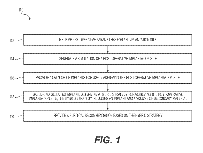
[030] FIG. 1 illustrates an exemplary method 100 according to aspects of the
present
disclosure. Method 100 may include receiving pre-operative parameters for an
implantation
site (step 102), generating a simulation of a post-operative implantation site
(step 104),
providing a catalog or database of potential implants for use in achieving the
post-operative
implantation site (step 106), based on a selected implant, determining a
hybrid strategy for
achieving the target post-operative implantation site, the hybrid strategy
including an implant
and a volume of secondary material (step 108), and providing a surgical
recommendation
based on the hybrid strategy (step 110).
[031] Generally, method 100 may result in generation of one or more
simulations of
an implantation site, and may use input information (e.g., selection of an
implant by a user) to
determine and output a recommended hybrid strategy for a surgical procedure
involving an
implant. The hybrid strategy may include both an implant volume (also referred
to herein as
a first volume) and a volume of secondary material (also referred to herein as
a second
volume), and may be tailored to achieve a particular result at an implantation
site. In some
embodiments, the hybrid strategy may be configured to provide a naturalistic,
visually and
tactilely smooth, and/or comfortable post-operative result at an implantation
site.
[032] Receiving pre-operative parameters for an implantation site according to
step
102 may include receiving information to assist in simulating, preparing for,
or performing a
surgery involving an implant. For example, pre-operative parameters may
include, e.g.,
measurements of an implantation site, data regarding the individual whose body
includes the
implantation site (e.g., the subject), such as demographic data, age, sex,
gender, height,
weight, physical conditions, reasons for wanting or needing a surgical
procedure, prior
surgical procedures, etc. In some embodiments, pre-operative parameters may
include
characteristics of an implantation site on a subject's body, such as skin
quality (e.g., laxity),
tissue health, prior surgical history, or other characteristics. In some
embodiments, pre-
operative parameters may include a series of dimensions of an implantation
site, such as the
7
CA 03104655 2020-12-21
WO 2019/246559
PCT/US2019/038536
depth of an incision, incision width and/or length, incision location, and/or
a volume of tissue
at or proximate the implantation site. In some embodiments, pre-operative
parameters may
include images taken by one or more imaging devices (e.g., a scanner, camera,
etc.). For
example, pre-operative parameters may be collected using high-resolution
scanning,
ultrasound imaging, high-resolution photography, three-dimensional imaging, or
visual
inspection, among other methods. In some embodiments, pre-operative parameters
may
include images taken using devices, systems and methods disclosed in
International
Application Publication No. WO/2017/175055. Additionally, pre-operative
parameters may
be collected by examining deeper tissue properties of an implantation site and
the
surrounding area, using, e.g., ultrasonic elasto-graphic techniques. In other
variations,
measurements may be taken by Eulerian Video Magnification techniques and
variations
thereon. In some embodiments, pre-operative parameters may include images that
have been
processed to determine, e.g., one or more dimensions of an implantation site.
[033] Generating a simulation of the post-operative implantation site
according to
step 104 may include, e.g., using the received pre-operative parameters in
conjunction with
anticipated post-operation parameters to create a visual representation of the
implantation
site. Post-operative parameters may include data characterizing a desired
outcome of a
surgical procedure at the implantation site (e.g., an augmentation procedure
including an
implant), such as desired measurements of a post-operative implantation site
(e.g., height,
width, volume, shape, and/or implant type). To generate a simulation of the
post-operative
implantation site, the pre-operative parameters may be used as a base or
starting point from
which post-operative conditions may be determined. For example, in the case of
a
mammoplasty, a subject's pre-operative breast size (e.g., shape and volume),
in conjunction
with a known shape and/or volume of a desired post-operative breast size, may
be used to
depict a post-operative breast size on a simulation of a subject's torso. In
some
embodiments, the generated simulation of the post-operative implantation site
may include,
e.g., a three-dimensional visual simulation which may be output to a user
device and/or saved
for reference or later use.
[034] Providing a catalog or database of implants for use in achieving the
post-
operative implantation site according to step 106 may include processing the
pre-operative
parameters of the implantation site and the simulation of the post-operative
implantation site
to determine implants that may be used to achieve the characteristics of the
post-operative
implantation site. For example, to achieve a desired volume at an implantation
site in a post-
operative state (e.g., volume of a bodily part, such as a breast or buttock)
that is greater than
8
CA 03104655 2020-12-21
WO 2019/246559
PCT/US2019/038536
the volume of the pre-operative implantation site, it may be determined that
implants of a
particular volume range may be useful. Moreover, to achieve a desired shape,
texture,
weight, or compatibility with a subject's body, it may be determined that
implants having
particular shapes and material compositions may be appropriate.
[035] For example, in some embodiments, round implants may be selected,
whereas
in others, an oval, teardrop, or other shape may be selected. As a further
example, in some
embodiments, implants having a fluid filling such as silicone gel or saline
liquid may be
selected, whereas in other embodiments, implants having a structured interior,
less viscous
filling material, and/or a more solid interior (including, e.g. high viscosity
materials, e.g.,
providing a "gummy bear" interior) may be selected. In some embodiments,
implants having
a filling material with a viscosity and elasticity providing for gravity-
sensitive characteristics
may be selected. In some embodiments, implants having smooth-textured
surfaces, micro-
textured surfaces, rough-textured surfaces, or a combination thereof may be
selected.
Exemplary characteristics of implants which may be found to be suitable
according to the
present disclosure are disclosed in, e.g., U.S. Application Publication No.
2015/0282926,
U.S. Application Publication No. 2014/0081398, and/or International
Application Publication
No. W02017/196973. Providing a catalog of implants may include, e.g.,
providing a list,
database, group of images, etc., identifying the implants available, so that
one or more
implants may be selected by a user. In some embodiments, providing a catalog
of implants
may include exporting or outputting a list, database, or group of implants to,
e.g., a user
device (e.g., user device 1306 depicted in FIG. 13).
[036] In some embodiments, method 100 may include receiving a selection of an
implant from the catalog of implants. Such a selection may be made
automatically, e.g., by a
computer system performing method 100, or may be made manually, e.g., by a
physician,
patient, or other individual. In some embodiments, selection of an implant may
assist in, e.g.,
performing further steps according to method 100.
[037] Generally, determining a hybrid strategy for achieving the target post-
operative implantation site according to step 108 may include executing one or
more
algorithms using, e.g., pre-operative parameters of the implantation site and
a simulation of
the post-operative implantation site to calculate a combination of an implant
and one or more
secondary materials that, when implanted, injected, or otherwise added to the
implantation
site, aid in achieving a size (volume), a shape, and/or other characteristics
of the post-
operative implantation site. One such type of algorithm is described in
further detail below
with reference to FIG. 2. In embodiments in which an implant is selected from
the catalog of
9
CA 03104655 2020-12-21
WO 2019/246559
PCT/US2019/038536
implants, performing step 108 optionally may include using parameters of the
selected
implant, such as the volume of the selected implant, as a basis for
determining the hybrid
strategy. Step 108 may involve using the simulation of the post-operative
implantation site
and the pre-operative parameters for the implantation site to determine a
difference between
the pre-operative implantation site and the post-operative implantation site,
such as a
difference in size (volume) and/or shape. The determined difference may serve
as a basis for
determining a hybrid strategy. Additionally or alternatively, a target post-
operative
parameter (e.g., shape or volume) may serve as a basis for determining a
hybrid strategy.
[038] In some embodiments, step 108 may include identifying a target post-
operative volume to be added to the implantation site (also referred to herein
as a total
volume), including identifying a first volume to be added as an implant, and
identifying a
second volume of a secondary material to be added in addition to the implant,
e.g., each of
the first volume and the second volume being a fraction of the total volume to
be added to the
implantation site. Advantageously, adding a second volume of a secondary
material to an
implantation site along with an implant, instead of adding an implant that
comprises the
entire volume to be added to the implantation site, may result in a more
naturalistic, better
supported, and/or customizable result from the surgical procedure.
[039] The secondary material may be any suitable biocompatible material for
injecting, implanting, or otherwise supplementing at an implantation site
along with the
implant. Exemplary suitable materials include, e.g., fat (such as heterologous
or autologous
fat), natural fillers, synthetic fillers, combinations thereof, and/or
combinations of scaffolding
materials useful in the field of aesthetics and cosmetic surgery. Particular
advantages may
vary depending on a type of a secondary material selected. For example, fat
grafts may
provide for a more natural result at the post-operative implantation site
and/or better
acceptance (biocompatibility) of an implant at the implantation site. As a
further example,
scaffolding materials may allow for improved structuring of a post-operative
implantation
site, and/or improved positioning and/or anchoring of an implant within the
implantation site.
Additionally, placement and division of a secondary material at an
implantation site may be
customizable, to provide a bespoke shape or size to a post-operative
implantation site.
[040] In some embodiments, in addition to determining an implant volume (first
volume) and a volume of secondary material (second volume), determining a
hybrid strategy
according to step 108 may include determining parameters for placing the
volume of
secondary material at the implantation site. In some embodiments, for example,
the
implantation site may be divided into different regions (e.g., different
segments and/or
CA 03104655 2020-12-21
WO 2019/246559
PCT/US2019/038536
subsections). The volume of secondary material may be divided amongst
different regions.
In some embodiments, an algorithm may be utilized to automatically divide the
volume of
secondary material amongst the different regions (see, e.g., FIG. 12 and the
associated
discussion herein). In further embodiments, input (e.g., from a user, such as
a physician or
patient, or from a digital source, such as a database) may be used to
determine and/or adjust
the division of the volume of secondary material amongst the different
regions.
[041] As mentioned above, determining the hybrid strategy may include
determining
a total volume to be added to an implantation site. For example, the total
volume may be
determined by calculating a pre-operative implantation site volume using,
e.g., pre-operative
implantation parameters, calculating a post-operative implantation site volume
using, e.g.,
parameters of the post-operative implantation site simulated in step 104, and
calculating a
difference between the post-operative implantation site volume and the pre-
operative
implantation site volume. Additionally or alternatively, the total volume may
be assumed to
be the volume of an initial implant selected from the catalog of implants,
which may be
independently selected or may be selected based on the calculated difference
of post-
operative implantation site volume and the pre-operative implantation site
volume (e.g., an
initial implant having a volume closest to the calculated difference).
[042] In some embodiments, determining the hybrid strategy may include running
through a series of "optional" scenarios (e.g., 2, 3, 4, 5, or more
scenarios), which may result
in one or more simulations of an implantation site reflecting an implant of
predetermined
dimensions and volume, supplemented with a volume of one or more secondary
materials.
[043] Providing a surgical recommendation based on the hybrid strategy
according
to step 110 may include selecting and outputting a hybrid strategy to, e.g., a
physician,
patient, digital repository, or other recipient. In some embodiments,
providing a surgical
recommendation may include outputting the hybrid strategy to a user interface.
In some
embodiments, providing the surgical recommendation may include recommending an
implant
shape, type, and/or volume along with a volume and type of secondary material.
In some
embodiments, a surgical recommendation may also include recommending an
incision site,
placement parameters for the volume of secondary material, and/or
injection/insertion
locations for the volume of secondary material or for the implant based on
physical and/or
biological properties of, e.g., the subject, the implant, and/or the secondary
material, to
enhance a likelihood of replicating a desired outcome.
[044] FIG. 2 illustrates an exemplary method 200 according to aspects of the
present
disclosure. As discussed above, FIG. 2 shows in further detail steps in
developing a hybrid
11
CA 03104655 2020-12-21
WO 2019/246559
PCT/US2019/038536
strategy (e.g., step 108 of method 100). Method 200 may include receiving pre-
operative
parameters for an implantation site (step 202), receiving a target post-
operative volume for
the implantation site (step 204), determining an approximate implant volume
using the pre-
operative parameters and the target post-operative volume (step 206),
selecting an implant
based on the approximate implant volume (step 208), determining a target
volume of
secondary material based on the selected implant and the target post-operative
volume (step
210), and adjusting the target volume of secondary material based on secondary
material
characteristics (step 212).
[045] Receiving pre-operative parameters for an implantation site according to
step
202 may include any and/or all aspects of step 102 described with respect to
method 100.
Receiving a target post-operative volume for the implantation site according
to step 204 may
include, e.g., receiving of a target post-operative volume for the
implantation site from, e.g., a
user, and/or may include calculating, simulating, or extrapolating a target
post-operative
volume. For example, step 204 may include receiving a selection of an implant
(e.g., from a
catalog of implants provided according to step 106 of method 100), and
extrapolating a post-
operative volume using the received pre-operative parameters for the
implantation site and
the volume of the selected implant. In some embodiments, step 204 may include
generating a
simulation of a post-operative implantation site which a user can adjust (by,
e.g., selecting a
particular implant), and then calculating a target post-operative volume using
the adjusted
simulation.
[046] Determining an approximate implant volume using the pre-operative
parameters and the target post-operative volume according to step 206 may
include
determining what fraction or percentage of the target post-operative volume
(total volume)
should comprise an implant. This fraction may vary based on, e.g.,
characteristics of the
subject. For example, in some embodiments, a skin laxity of a subject may
affect the
selection of implant volume. For example, a subject having a higher skin
laxity may desire or
require that an implant make up a greater percentage (e.g., about 65%) of a
target post-
operative volume in order to, e.g., properly shape and support the subject's
skin, as opposed
to a subject having a lower skin laxity, who may desire or require that an
implant make up a
smaller percentage (e.g., about 55%) of a target post-operative volume. This
parameter of
skin laxity may be subjectively calculated using a "pinch test", or may be
determined by
other suitable methods of quantifying/qualifying the amount of elasticity
(elastin and/or
collagen) in the skin and underlying tissue equating to laxity.
12
CA 03104655 2020-12-21
WO 2019/246559
PCT/US2019/038536
[047] The fraction or percentage of the target post-operative volume to be
accounted
for by an implant may also or alternatively vary depending on, e.g., physician
recommendations, patient preferences, and/or a combination thereof In some
embodiments,
the percentage of the target post-operative volume to be added to the
implantation site as an
implant (i.e., implant volume) may vary from, e.g., about 45% to about 80%,
such as from
about 50% to about 70%, from about 55% to about 65%, or about 45%, about 50%,
about
55%, about 60%, about 65%, about 70%, about 75%, or about 80%. In some
embodiments,
the percentage of the target post-operative volume to be added to the
implantation site as an
implant may be divided by 100 to arrive at a coefficient. In embodiments in
which skin
quality (e.g., skin laxity) factors into the percentage of the target post-
operative volume to be
added as an implant, the resulting coefficient may be referred to as a skin
quality coefficient.
[048] To arrive at an approximate implant volume, the target post-operative
volume
may be multiplied by the determined coefficient. For example, if a determined
percentage of
a target post-operative volume to be accounted for by an implant is 65% and
the target post-
operative volume is 400 cc, then the approximate implant volume may be 0.65 x
400 cc, or
260 cc. In some circumstances, as is described further with respect to FIG. 3,
the target post-
operative volume may be multiplied by the determined coefficient twice (or
more times) to
arrive at a suitable approximate implant volume.
[049] Selecting an implant based on the approximate implant volume according
to
step 208 may include reviewing one or more catalogs or databases of available
implants and
selecting an implant having a volume close to the approximate implant volume.
For example,
an implant having a smaller or larger volume than the approximate implant
volume may be
selected. This may account for situations in which an implant having precisely
the
approximate implant volume is unavailable. Implants are often produced in a
limited variety
of sizes, for example. Further, multiple factors may limit the availability of
implants, such as
manufacturer or distributor inventory, a desired implant shape, surface
texture, filling texture,
viscosity, etc. In some cases, a physician may only selectively work with one
or a few brands
of implants, further limiting the availability of a wide variety of implant
volumes.
[050] In some embodiments, step 208 may be performed automatically; for
example,
a computer system performing method 200 may review one or more digital implant
catalogs
and may automatically select an implant from the reviewed catalogs.
Additionally or
alternatively, step 208 may include receiving a selection of an implant from,
e.g., a user via a
user device or interface. For example, the computer system may select an
implant that can
then be proposed to a user, who may accept or reject the proposed implant. If
the user rejects
13
CA 03104655 2020-12-21
WO 2019/246559
PCT/US2019/038536
the implant, or independently of a computer selecting an implant, a user may
be provided
with a list of implants having volumes close to the approximate implant
volume, and their
characteristics (e.g., on a user device). The user may then select a desired
implant from the
list. Optionally, a user may be able to select a magnitude of variation in
volume from the
approximate implant volume, and may be able to view implants falling within
the selected
magnitude of variation in volume or less.
[051] Determining a target volume of secondary material based on the selected
implant and the target post-operative volume according to step 210 may include
subtracting
the volume of the implant selected according to step 208, as well as the pre-
operative volume
of the implantation site, from the target post-operative volume. In other
words, in determining
a hybrid strategy, once an implant is selected for use in the hybrid strategy,
the remaining
volume of the target post-operative volume may be achieved by adding a
corresponding
amount of secondary material to the implantation site.
[052] Characteristics of the secondary material may affect the extent to which
it may
supplement an implant at an implantation site. Thus, method 200 may then
continue to step
212, which includes adjusting the target volume of secondary material based on
secondary
material characteristics. In this step, the volume of secondary material to be
added to the
implantation site may be adjusted to account for a property or behavior of the
secondary
material, such as an uptake rate, a reabsorption rate, and/or a survival rate.
A reabsorption
rate of a secondary material (e.g., a fat graft) may be, for example, between
about 30% and
about 60%, such as about 30%, about 35%, about 40%, about 45%, about 50%,
about 55%,
and about 60%. Therefore, an additional amount of the secondary material may
be added to
the implantation site to account for the reabsorption rate. In such cases, the
total secondary
material volume to be added to the implantation site may be calculated as
follows:
Total Added Volume = Desired Volume * (1+ (reabsorption rate(%))) Equation
1
100%
[053] For example, for a secondary material having a reabsorption rate of
about
35%, the volume of the secondary material to be added to the implantation site
may be
multiplied by 1.35, such that upon expected reabsorption of a portion of the
secondary
material, the remaining volume of secondary material at the implantation site
will be the
desired volume.
[054] Once a hybrid strategy is developed that includes both an implant and an
adjusted volume of secondary material, processes may continue to, e.g., saving
and/or
providing a recommended surgical strategy to a user, or any other step(s).
14
CA 03104655 2020-12-21
WO 2019/246559
PCT/US2019/038536
[055] In some embodiments, the method may be modified from that depicted in
FIG. 1 and the algorithm depicted and described with respect to FIG. 2 in
order to account for
certain characteristics of a subject. Specifically, in some cases, applying a
determined
fraction or percentage to a target post-operative volume (e.g., step 206 of
method 200)
provides an approximate implant volume greater than the target post-operative
volume. This
may be true, e.g., when the target post-operative volume is less than twice
the volume of the
pre-operative volume at the implantation site. In some such cases, applying a
determined
fraction or percentage to a target post-operative volume may arrive at a
supposed
approximate implant volume that, when added to the pre-operative volume at the
implantation site, would result in a total post-operative volume that is
greater than desired. In
other such cases, this step may arrive at a supposed approximate implant
volume that leaves
nearly no room for secondary materials. Method 300, while sharing several
steps in common
with method 200, includes a query and an additional step to account for such
cases with a
correction coefficient. Method 300 may include receiving pre-operative
parameters for an
implantation site (step 302), receiving a target post-operative volume for the
implantation site
(step 304), and applying a correction coefficient to the target post-operative
volume based on
a pre-operative parameter to determine an approximate implant volume (step
306). Method
300 may further include determining whether the post-operative volume is less
than or equal
to twice the pre-operative volume (step 308). If yes, then method 300 may
include applying
the correction-coefficient to the approximate implant volume to re-determine
the approximate
implant volume (step 310). If not, method 300 may include skipping step 310.
Method 300
may then include selecting an implant based on the approximate implant volume
(step 312).
[056] Receiving pre-operative parameters for an implantation site according to
step
302 may include any and/or all aspects of step 102 described with respect to
method 100.
Receiving a target post-operative volume for the implantation site according
to step 304 may
include any and/or all aspects of step 204 described with respect to method
200. Applying a
correction coefficient to the target post-operative volume based on a pre-
operative parameter
to determine an approximate implant volume according to step 306 may share any
and/or all
aspects of step 206 described with respect to method 200, where the correction
coefficient of
step 306 is the determined fraction or percentage of the target post-operative
volume that
should be filled by an implant, of step 206, and wherein the pre-operative
parameter is a
characteristic of the subject, such as skin laxity.
[057] According to step 308, method 300 may then include determining whether
the
target post-operative volume is less than or equal to twice the pre-operative
volume. If not,
CA 03104655 2020-12-21
WO 2019/246559
PCT/US2019/038536
then method 300 may continue to step 312. However, if the target post-
operative volume is
indeed less than or equal to twice the pre-operative volume at the target
implantation site,
then method 300 proceeds to step 310, which may include applying the
correction coefficient
to the approximate implant volume, to re-determine the approximate implant
volume. This
step may include multiplying the approximate implant volume by the determined
fraction or
percentage of the target post-operative volume that should be filled by an
implant, to account
for the relatively smaller overall volume difference between the pre-operative
volume and the
target post-operative volume.
[058] For example, if a determined percentage of a target post-operative
volume to
be accounted for by an implant is 65% and the target post-operative volume is
400 cc, and the
pre-operative volume of the implantation site is 200 cc (i.e., the target post-
operative volume
is less than or equal to twice the pre-operative volume at the target
implantation site), then the
approximate implant volume may be 400 cc x 0.65 x 0.65, or 169 cc. Therefore,
this step
reduces the approximate implant volume to account for the fact that the
overall volume
difference between the pre-operative volume and the target post-operative
volume is only
200 cc.
[059] In some cases, an overall volume difference between the pre-operative
volume
and the target post-operative volume may be even smaller, such that applying
the correction
coefficient twice is insufficient. For example, a target post-operative volume
may be 400 cc
and the pre-operative volume of the implantation site may be 250 cc. In such a
case,
applying the correction coefficient twice (to arrive at an approximate implant
volume of 169
cc) still results in an approximate implant volume greater than the overall
volume difference
between the pre-operative volume and the target post-operative volume (in this
case, 150 cc).
In such a case, the correction coefficient may be applied more than twice,
until the
approximate implant volume is less than the overall volume difference between
the pre-
operative volume and the target post-operative volume. Finally, selecting an
implant based
on the approximate implant volume according to step 312 may include any and/or
all aspects
of step 208 described with respect to method 200.
[060] As has been alluded to in the discussion of FIGS. 1-3, aspects of
methods
disclosed herein may include receiving input from and/or sending output to,
e.g., a user
device having a user interface, which may be a visual user interface. This
input and output
may allow for a user (e.g., a physician, a subject, or other user) to view and
change aspects of
a potential surgical procedure to, e.g., achieve a more customized final
result. FIG. 4
illustrates an exemplary method 400 according to aspects of the present
disclosure. Method
16
CA 03104655 2020-12-21
WO 2019/246559
PCT/US2019/038536
400 may include receiving selection of an initial implant, e.g., from a
digital catalog (step
402), generating a first visual simulation of a post-operative implantation
site including the
selected initial implant (step 404), generating a second visual simulation of
a post-operative
implantation site including a second implant and a volume of a secondary
material (step 406),
receiving an input adjusting a parameter of the second visual simulation (step
408), and
generating an adjusted second visual simulation to account for the adjusted
parameter (step
410).
[061] As with the other methods disclosed herein, steps of method 400 may be
performed by, e.g., a system including computing hardware and software (e.g.,
system 1300).
In some embodiments, steps of receiving input (e.g., step 402 and step 408)
may include
receiving input from a user device (e.g., device 1306) and steps of generating
or adjusting a
visual simulation may include the use of a computer system (e.g., computer
system 1304),
and/or more specifically a modeling engine (e.g., modeling engine 1310).
Configurations and
relative locations of a user device and a computer system are described
further with respect to
system 1300, but in general may have any suitable configuration or location
for performing
the steps of method 400.
[062] Receiving selection of an initial implant from a digital catalog
according to
step 402 may include, e.g., receiving identification of a particular implant
in a digital catalog
and/or receiving a volume, size, filling type, and/or other characteristic of
an initial implant.
The digital catalog may be, e.g., a tailored list or database provided
according to step 106 of
method 100, or may be a pre-existing list or database. In some embodiments,
for example, a
digital catalog may be provided to a user interface, and the user interface
may subsequently
receive a user selection of an initial implant.
[063] Generating a first visual simulation of a post-operative implantation
site
including the selected initial implant according to step 404 may include using
parameters of a
pre-operative implantation site (e.g., received according to step 102 of
method 100, step 202
of method 200, or step 302 of method 300) in combination with parameters of
the selected
initial implant to construct an image of the implantation site into which the
selected initial
implant has been implanted or placed. In some embodiments, generating this
visual
simulation may include, e.g., generating a three dimensional visual simulation
using, e.g.,
imaging data of a subject and adjusting it to simulate the addition of the
selected initial
implant. Any of the three-dimensional simulations and/or related methods and
algorithms
disclosed in International Application No. PCT/US2019/034667 filed on May 30,
2019,
incorporated by reference herein, may be used in the present disclosure.
17
CA 03104655 2020-12-21
WO 2019/246559
PCT/US2019/038536
[064] For example, the methods herein may include generating and/or
manipulating
a simulation using a three-dimensional model that includes a plurality of
tetrahedra used to
describe tissue volume (breast tissue in a breast volume model). The
simulation may be
based on a three-dimensional model that corresponds to an initial
configuration, wherein the
model may be modified to simulate an expansion or stretching of tissue to
accommodate
placement of an implant and secondary materials as disclosed herein. For
example, a set of
reference tetrahedra of increased volume may be defined, wherein the reference
tetrahedra
correspond to the planned region of volume increase. Further details on
generating and
modifying such three-dimensional models to simulate the results of
contemplated medical
procedures are provided in PCT/US2019/034667 filed on May 30, 2019.
[065] Generating a second visual simulation of a post-operative implantation
site
including a second implant and a volume of a secondary material according to
step 406 may
include, e.g., receiving or calculating a potential hybrid strategy including
an implant volume
(with a selected initial implant having the implant volume) and a volume of
secondary
material, and constructing a simulation of the hybrid strategy. In some
embodiments, a visual
simulation generated according to step 406 may include a standard or default
distribution of
the volume of the secondary material in the implantation site. In other
embodiments, a visual
simulation generated according to step 406 does not include the volume of the
secondary
material distributed at the implantation site, and may simply note an
available volume of
secondary material which may be added to the simulation in a customized
distribution.
[066] In some embodiments, the first visual simulation and/or the second
visual
simulation may be output to, e.g., a user interface. For example, a
comparative view of the
first visual simulation and the second visual simulation may be provided to,
e.g., allow a user
to view similarities and differences between the use of an initial selected
implant and the use
of a hybrid strategy at an implantation site. Presentation of the first visual
simulation and/or
the second visual simulation optionally may include interactive elements
(e.g., sliders,
buttons, meters, color coding, and the like) on a user interface of a device,
to allow a user to
change or otherwise interact with and manipulate the visual simulations.
[067] Receiving an input adjusting a parameter of the second visual simulation
according to step 408 may include receiving a change from a user through such
an interactive
element. This step may include receiving a change to any parameter of the
second visual
simulation corresponding to a change in surgical procedure or materials used.
For example, a
change to an implant size, an implant shape, an implant type, an implant
placement position,
a volume of secondary material, and/or a distribution of secondary material
may be received.
18
CA 03104655 2020-12-21
WO 2019/246559
PCT/US2019/038536
[068] Generating an adjusted second visual simulation to account for the
adjusted
parameter according to step 410 may include making any recalculations
necessary in
response to the received input to reflect the adjustment in the second visual
simulation, re-
generating or changing the second visual simulation according to the
recalculations, and/or
outputting the adjusted second visual simulation. In this manner, a user may
interact with the
visual simulation to observe the effect of various options and adjustments on
the outcome of
a surgery, and to view similarities and differences between an approach using
only a selected
initial implant and an approach using a second implant and a volume of
secondary material
(e.g., a hybrid approach).
[069] In some embodiments, adjustments may be received for both the first and
second visual simulations. In other embodiments, adjustments may be received
for only the
second visual simulation. Generally, method 400 may assist a user in
visualizing,
customizing, and otherwise preparing for a contemplated surgical procedure
including an
implant.
[070] Some embodiments of the present disclosure may facilitate collaboration
between a subject (e.g., a patient), and a physician (e.g., a surgeon). For
example, in some
embodiments of the present disclosure, a patient may be able to select a
desired shape and
size of a post-operative implantation site. Measurements of the patient may be
performed on,
e.g., a three-dimensional image of the implantation site on the client
(wherein the three-
dimensional image may be acquired by a camera, e.g., of a scanner), and a
simulation of the
patient's desired post-operative implantation site may be combined with the
patient
measurements to obtain a simulation of a target post-operative implantation
site. Thereupon,
a computer-implemented variation on surgeon selection of the breast implant
may be output
based upon one or more methods described herein. An algorithm may compare
volumes and
shapes of different implants (e.g., surface curvatures, area, etc.) to find a
potential best match
to a final desired shape and size. Additionally, an algorithm may rank order
implants based
on the initial measurements of the patient and additional geometric
calculations, which may
be based on physical and/or mechanical properties of the implant,
physicochemical properties
of the secondary materials, and/or may be based on other considerations.
[071] Reference will now be made to views of exemplary user interfaces which
may
be used with aspects of the present disclosure. User interfaces suitable for
combining with
methods of the present disclosure may generally allow for users to view
generated
simulations, make selections and adjustments to them, and/or to save, load,
transfer, or
otherwise use the generated simulations in contemplating or preparing for a
surgical
19
CA 03104655 2020-12-21
WO 2019/246559
PCT/US2019/038536
procedure. As such, user interfaces according to the present disclosure may
include any
suitable displays, interactive elements, options, etc. to achieve these goals.
The views
depicted herein are merely limited examples, and one of ordinary skill in the
art will
understand that many more variations on exemplary user interfaces are possible
and
contemplated herein.
[072] FIGs. 5-7 depict views 500, 600, 700 of an exemplary user interface for
use in,
e.g., simulating, modelling, designing, and preparing for aesthetic or
reconstructive surgeries.
View 500 may include a pre-operative visual simulation 510 of an implantation
site (in this
case, a torso is depicted). View 500 also includes a simulation setting menu
520 including
various general simulation settings, an implant selection menu 530 listing a
plurality of
implants (e.g., listing dimensions of each of a plurality of implants) that
may be selected and
added to the simulation, and a simulation use menu 540, including options
which may assist
in the use of a generated simulation in various ways.
[073] Pre-operative visual simulation 510 may be a simulation generated
according
to aspects of the present disclosure and any suitable method (e.g., using
images, parameters,
measurements, or other data pertaining to a subject, as disclosed elsewhere
herein).
Generally, pre-operative visual simulation 510 may depict an implantation site
of a subject in
a pre-operative state, i.e., before a contemplated surgical procedure to
insert one or more
implants to the subject's body. In some embodiments, simulation 510 may be a
three
dimensional simulation, such as a three-dimensional rendering. In some
embodiments,
simulation 510 may be interactive (e.g., rotatable, scalable, expandable, and
the like).
Simulation 510 may assist a user in visualizing and analyzing an initial
status (e.g., size,
shape, look) of an implantation site.
[074] Simulation setting menu 520 may include one or more selectable options
to
customize a contemplated surgical procedure. As an example, menu 520 includes
an option
to select an implant position or positions, select a type of surgical
procedure, and view a type
of digital catalog. Types of digital catalogs may differ by, e.g., implant
brand (manufacturer),
implant model, shape, texture, etc. Implant selection menu 530 may include a
list or other
presentation of potential implants to include in the implantation site. For
example, implant
selection menu 530 may include a variety of dimensions (e.g., volumes,
diameters, shapes,
etc.). Simulation use menu 540 may include options for implementing, changing,
comparing,
etc. one or more implants in the simulation.
[075] FIG. 6 depicts view 600, including a post-operative visual simulation
610 of
an implantation site including an implant of a selected size. View 600 may
include one or
CA 03104655 2020-12-21
WO 2019/246559
PCT/US2019/038536
more of the same menus as view 500, such as, e.g., simulation setting menu
520, and may
include one or more views which differ or vary from menus shown in view 500.
For
example, an exemplary implant size menu 630 is shown, which may list a variety
of volumes
of a single implant shape.
[076] Post-operative visual simulation 610 may be a simulation generated
according
to aspects of the present disclosure and any suitable method (e.g., using
images, parameters,
measurements, or other data pertaining to a subject, as disclosed elsewhere
herein).
Generally, post-operative visual simulation 610 may depict an implantation
site of a subject
in a post-operative state, i.e., including one or more implants. In some
embodiments, post-
operative visual simulation 610 may depict a standard implantation procedure
in which, e.g.,
an implant accounts for an entire difference in volume at an implantation site
(as opposed to a
hybrid strategy including an implant and a volume of a secondary material). As
with
simulation 510, simulation 610 may be a three dimensional simulation, such as
a three-
dimensional rendering, and may similarly be interactive (e.g., rotatable,
scalable, expandable,
and the like). Simulation 610 may assist a user in visualizing and analyzing
an initial status
(e.g., size, shape, look) of an implantation site.
[077] Implant size menu 630 may be, e.g., a variation on or a sub-menu of
implant
selection menu 530 depicted in view 500. Implant size menu 630 may be depicted
upon
selection of an option to view implants having a particular shape (e.g., round
implants, as
opposed to anatomically shaped implants). In some embodiments, implant size
menu 630
may include a smaller number of implant options than, e.g., implant selection
menu 530, to
allow for a user to review and select implants having a particular
characteristic (in this case, a
round shape). One of ordinary skill in the art will understand that many other
types of
implant selection menus are possible and contemplated herein.
[078] FIG. 7 depicts view 700, which may include a hybrid post-operative
visual
simulation 710 of an implantation site including an implant of a selected size
and a volume of
a secondary material (e.g., fat). View 700 may also include one or more of the
same menus
as views 500 and 600. View 700 may include menus specific to a hybrid post-
operative
simulation, such as, e.g., a sculpting settings menu 720 and a secondary
material distribution
menu 730.
[079] Hybrid post-operative visual simulation 710 may be a simulation
generated
according to aspects of the present disclosure and any suitable method (e.g.,
using images,
parameters, measurements, or other data pertaining to a subject, as disclosed
elsewhere
herein, in combination with a calculated combination of an implant and
secondary material).
21
CA 03104655 2020-12-21
WO 2019/246559
PCT/US2019/038536
Generally, hybrid post-operative visual simulation 710 may depict an
implantation site of a
subject in a post-operative state including both an implant (or two implants,
in the case of a
double mammoplasty as shown) and a volume of secondary material. As with
simulations
510 and 610, simulation 710 may be a three dimensional simulation, such as a
three-
dimensional rendering, and may similarly be interactive (e.g., rotatable,
scalable, expandable,
and the like). Simulation 710 may assist a user in visualizing and analyzing a
hybrid
approach to a surgical procedure, and may allow a user to "sculpt" or
otherwise alter the
hybrid approach (e.g., by changing a distribution of the secondary material)
to derive a
customized target post-operative result.
[080] Sculpting settings menu 720 may include options to, e.g., change an
implant
size or a volume of secondary material at a selected implantation site (here,
a left breast or a
right breast). As shown, a recommended implant size may be displayed. The
recommended
implant size may be calculated according to algorithms and/or methods
disclosed herein (e.g.,
according to methods 100, 200, and/or 300). An interactive component (e.g., a
slider, meter,
button, or numerical input field) may allow for variation of the desired post-
operative
volume. In response to variation of the desired post-operative volume, a
recommended
implant size may change (e.g., the user interface may dynamically provide a
recommended
implant size based on changes to the desired post-operative volume).
[081] Secondary material distribution menu 730 may include, e.g., division of
the
implantation site into sections (e.g., quadrants, as shown, or other sections)
and may allow for
a user to add to, subtract from, or otherwise alter the distribution of
secondary material at the
implantation site via interactive components (e.g., sliders, meters, buttons,
numerical input
fields, etc.) for each section. Algorithms may be employed to, e.g.,
dynamically update a
simulation according to adjustments made in the secondary material
distribution menu. In
some embodiments, smoothing algorithms may also be employed to maintain a
desirably
smooth (e.g., well-integrated) look to an implantation site, regardless of
changes made to the
simulation in secondary material distribution menu 730. An exemplary method of
updating a
simulation according to changes made to secondary material distribution is
described herein
with respect to FIG. 12.
[082] In some embodiments, views 500, 600, and 700 may all be displayable
simultaneously (e.g., in separate windows). In some embodiments, in may be
possible to
toggle between views 500, 600, and 700, to compare the simulations shown in
each view. In
other embodiments, an option to directly view a side-by-side comparison of the
different
simulations may be available (see, e.g., FIG. 11).
22
CA 03104655 2020-12-21
WO 2019/246559
PCT/US2019/038536
[083] FIGs. 8-11 depict views 800, 900, 1000, 1100 of another exemplary user
interface for use in, e.g., simulating, modelling, designing, and preparing
for cosmetic
surgeries. View 800, for example, may include a pre-operative visual
simulation 810 of an
implantation site, and a comparative post-operative visual simulation 820 of
the implantation
site. A post-operative visual simulation adjustment menu 830 may allow for
changes to post-
operative visual simulation 820. Simulation use menu 840 may include options
to assist in
the use of generated simulations in various ways.
[084] Pre-operative visual simulation 810 may share characteristics with,
e.g., pre-
operative visual simulation 510 of view 500. Post-operative visual simulation
820 may
likewise share characteristics with, e.g., post-operative visual simulation
610 of view 600.
View 800 allows for the pre- and post-operative visual simulations to be
viewed
simultaneously for a more direct comparison between the two. In some
embodiments, pre-
operative visual simulation 810 and post-operative visual simulation 820 may
both be
rotatable, expandable, or otherwise viewable in tandem, so that similar views
of the two
simulations may be examined simultaneously.
[085] Adjustment menu 830 may be an interactive element or collection of
interactive elements allowing a user to adjust post-operative visual
simulation 820. In some
embodiments, post-operative visual simulation 820 may dynamically change
depending on
adjustments made in adjustment menu 830. Adjustment menu 830 may include
options to
change, e.g., post-operative volume, shape, height, projection, or other
characteristics of an
implantation site. Additionally, post-operative visual simulation 820 and/or
adjustment menu
830 may show numeric characteristics of, e.g., an implantation site in post-
operative visual
simulation 820. For example, a post-operative volume and/or other dimension of
an
implantation site may be dynamically calculated as post-operative visual
simulation 820 is
adjusted, by calculating, e.g., differences between pre-operative visual
simulation 810 and
post-operative visual simulation 820 in real time or periodically. Thus, a
post-operative
volume and/or diameter (and/or other dimensions) of an implantation site may
be indicated as
a part of post-operative visual simulation 820 and/or adjustment menu 830. In
this manner,
view 800 may allow a user to adjust and view characteristics of a post-
operative implantation
site until a desirable post-operative implantation site is achieved. In some
embodiments,
arrival at a desirable post-operative implantation site via, e.g., adjustments
made using
adjustment menu 830 may also serve as a selection of a post-operative volume.
[086] Simulation use menu 840 may include various selectable options to aid in
use
of the simulation(s). For example, simulation use menu 840 may include options
to load,
23
CA 03104655 2020-12-21
WO 2019/246559
PCT/US2019/038536
print, save, analyze, annotate, or visually present a simulation or
simulations. Simulation use
menu 840 may be available on, e.g., multiple views of a user interface to
allow for saving,
loading, and otherwise manipulating simulations from any of the multiple
views.
[087] FIG. 9 depicts view 900, which may include post-operative visual
simulation
820, adjustment menu 830 an implant search menu 930, and an implant selection
menu 940.
View 900 may include one or more of the same menus as view 800, such as
simulation use
menu 840.
[088] Implant search menu 930 may accept input from a user to populate implant
selection menu 940 with a list of implants compatible with characteristics of
post-operative
visual simulation 820. In some embodiments, for example, implant search menu
930 may
accept input of search criteria, such as a particular implant brand, shape, or
having a
particular texture or filling type. Implant selection menu 940 may list
implants fitting the
search criteria that also have measurements that may be suitable for achieving
the post-
operative visual simulation from, e.g., a pre-operative implantation site.
Some implants in
implant selection menu 940 may be identified as hybrid-compatible
implants¨i.e., they may
be used in combination with a volume of a secondary material, as a part of a
hybrid strategy.
A user may then be able to select an implant from implant selection menu 940
for further
refinement of the post-operative visual simulation 820.
[089] FIG. 10 depicts a close-up portion of view 900, in which a hybrid-
compatible
implant has been selected from implant selection menu 940. Upon selection of a
hybrid-
compatible implant, a button 1010 allowing for the generation of a hybrid
strategy may be
digitally added to view 900. Button 1010 may generate a new post-operative
hybrid
simulation, as depicted in FIG. 11.
[090] FIG. 11 depicts view 1100, which may include a comparison of several
simulations, such as pre-operative visual simulation 810, post-operative
visual simulation
820, and a hybrid post-operative visual simulation 1110. View 1100 may include
an
adjustment tool 1120, which may allow a user to adjust parameters of, e.g.,
post-operative
visual simulation 920 and/or hybrid post-operative visual simulation 1110.
Moreover, view
1100 depicts measurement lines 815, which may be applied by a user or
digitally to identify
different sections of an implantation site. While measurement lines 815 are
depicted on pre-
operative visual simulation 815, similar measurement lines may be applied to
post-operative
visual simulation 820 or hybrid post-operative visual simulation 1110.
[091] Hybrid post-operative visual simulation 1110 may share any or all
characteristics with, e.g., hybrid post-operative visual simulation 710 of
view 700. Generally,
24
CA 03104655 2020-12-21
WO 2019/246559
PCT/US2019/038536
hybrid post-operative visual simulation 1110 may depict an implantation site
of a subject in a
post-operative state including both an implant based on a selection made from
implant
selection menu 940 and a volume of secondary material. As with other
simulations disclosed
herein, simulation 1110 may be a three dimensional simulation, such as a three-
dimensional
rendering, and may similarly be interactive (e.g., rotatable, scalable,
expandable, and the
like). Simulation 1110 may assist a user in visualizing and analyzing a hybrid
approach to a
surgical procedure. Adjustment tool 1120 may allow a user to input commands to
"sculpt" or
otherwise alter the hybrid approach (e.g., by changing a volume, distribution,
or type of the
secondary material) to derive a customized target post-operative result.
[092] Methods for simulating the addition of a volume of secondary material to
an
implantation site may benefit from, e.g., algorithms that may automatically
apportion the
secondary material in a realistic and suitable manner around an
implant/implantation site.
Such algorithms may be run as a part of, e.g., a modeling engine (see, e.g.,
modeling engine
1310 of system 1300). Generally, algorithms may help a modeling engine to
simulate the
addition of secondary materials into (or removal of secondary materials from)
one or more
segments of an implantation site, and may advantageously allow for greater
control and
customization in the simulation of secondary materials at the implantation
site. For example,
segmentation of an implantation site may provide greater precision in
parameters for
placement of the secondary materials at the implantation site. Segments of an
implantation
site may be digitally determined once, e.g., a perimeter and/or a center or
epicenter of an
implantation site are identified. For example, for a mammoplasty, a simulated
breast area
may be digitally separated into four quadrants which may intersect at a point
of greatest
projection or at a nipple portion of the breast.
[093] In simulating the addition or subtraction of secondary materials at a
segment
of an implantation site, methods according to the present disclosure and
algorithms for
implementing such methods may address two goals: simulating a realistic
increase (or
decrease) in rest volume of one or more digital segments of an implantation
site, and
realistically contouring the perimeter of such segments and/or the
implantation site, so that a
smooth transition between the area inside of a segment and the area outside of
a segment
(e.g., either within the implantation site or external to the implantation
site) is maintained.
[094] To achieve both of these goals, segments of an implantation site may be
further divided into geometric subsections (e.g., tetrahedra or other suitable
shapes). In some
embodiments, for example, a tetrahedral model of an implantation site (e.g., a
breast model)
may be constructed from a triangle mesh surface. Each subsection may be
identified or
CA 03104655 2020-12-21
WO 2019/246559
PCT/US2019/038536
characterized by a distance between the sub-section (e.g., a center point of
the subsection)
and a reference point (e.g., a center or epicenter of an implantation site, a
point on a perimeter
of an implantation site, or both). Segments may be divided into small enough
subsections to
allow for realistic modeling, without overtaxing processing power of, e.g., a
modeling engine.
[095] In some embodiments, segments and/or subsections may be selected and
oriented based on the general location of an implantation site. For example,
in the case of a
breast implantation site, a chest wall of a subject may be used to orient a
coordinate system
for dividing the implantation site into segments and/or subsections. A
rotation matrix may be
formed that rotates center points of subsections (e.g., tetrahedral center
points) from a global
space to an orientation of the chest wall. Points in the coordinate system may
be translated so
that a nipple location is located at the origin of the coordinate system. Each
of the four
quadrants of the coordinate system may then be translated into one of four
segments that
make up the breast implantation site (e.g., such that points where x < 0 and y
<0 correspond
to a southwest quadrant, points where x> 0 and y <0 correspond to a southeast
quadrant,
points where x < 0 and y > 0 correspond to a northwest quadrant, and points
where x> 0 and
y > 0 correspond to a northeast quadrant).
[096] To simulate an increase in volume in a segment of an implantation site,
a
modeling engine may increase a volume of subsections in the segment by some
fraction of
their pre-operative or otherwise original volume. A scaling factor for the
volume increase
may be computed from the original volume of the segment, and from the volume
of added
secondary material. The volume increase may be tapered on a subsection-by-
subsection
basis, depending on the characterization of each subsection, such that a
greater volume
increase is modeled at a center of a segment than at a periphery of the
segment. This may
allow for smoother transitions between segments. Moreover, the volume increase
may be
tapered depending on additional reference points, such as a body surface of
the subject, a
direction of gravitational pull, and the like.
[097] To simulate a realistic/smooth contour at a perimeter of an implantation
site, a
modeling engine may separate a border area of a segment into a plurality of
points, and may
apply a displacement to each point depending on a distance between the point
and the nearest
point of a surface outside of the segment (e.g., a perimeter point outside of
the implantation
site). The border area may be determined, e.g., algorithmically, or may be
determined using
user input. The magnitude of displacement for each point of the plurality of
points may
negatively correlate to a distance between the segment and the point. For
example, a fraction
of a displacement towards a perimeter point may be applied to each point
within border area,
26
CA 03104655 2020-12-21
WO 2019/246559
PCT/US2019/038536
where the fraction decays to zero as the distance between each point and the
nearest
perimeter point decreases. An exemplary function that has been implemented to
achieve this
is:
f = - )e Equation
2
Dmax
where d is a distance to the nearest perimeter point, Dmax is a width of the
border area (i.e.,
d < Dmax), and e is a decay exponent parameter that may be chosen.
[098] FIG. 12 illustrates an exemplary perimeter contouring method 1200
according
to aspects of the present disclosure. Method 1200 may include receiving data
regarding a
volume of material to add to an implantation site (step 1202), separating the
implantation site
into regions (step 1204), separating each region into sub-regions (step 1206),
simulating
addition of the volume of material to a region of the implantation site by
increasing a volume
of each sub-region in the region, wherein the increase in volume for each sub-
region is
dependent on a distance between the sub-region and an origin point (step
1208), and
performing a smoothing algorithm to simulate a smooth change in volume between
the region
of the implant site and an area outside of the region (step 1210).
[099] Receiving data regarding a volume of material to add to an implantation
site
according to step 1202 may include receiving, e.g., a manually-input or
automatically-
suggested volume of a secondary material to add to an implantation site.
Separating the
implantation site into regions according to step 1204 may be done as described
above, e.g.,
by identifying quadrants or other segments of the implantation site based on a
center point, an
epicenter, or a border of the implantation site. Separating each region into
sub-regions
according to step 1206 may also be done as described above, e.g., by
separating each segment
into subsections and characterizing each subsection by a center point and a
distance between
the center point and one or more reference points (e.g., a center, epicenter,
or periphery of a
segment or of the implantation site). Simulating addition of the volume of
material to a
region of the implantation site according to step 1208 may include identifying
subsections
lying within the region (e.g., the segment) in question, and increasing their
reference
volumes. For example, if a total starting volume of subsections in a region or
segment is
characterized by Vo, and vf is a volume of a secondary material to be added to
the region or
segment, then the reference volume of each subsection in the region or segment
may increase
by a factor of s:
s = (Vo + vf) / Vo Equation 3
27
CA 03104655 2020-12-21
WO 2019/246559
PCT/US2019/038536
[0100] In some embodiments, a modeling engine may apply this or a similar
algorithm to a segment or region and will move towards a solution in that
segment or region
in which the subsections are a factor of s larger than their initial sizes. As
described above, a
volume added to each subsection may taper in correlation with a distance
between a center
point of the subsection and a perimeter or other reference point of the region
or segment.
Performing a smoothing algorithm to simulate a smooth change in volume between
the
region of the implant site and an area outside of the region according to step
1210 may
include applying Equation 2 to, e.g., points in a border area of the region.
[0101] Embodiments disclosed herein may be created, executed, displayed etc.
using
any suitable technology or system. In some embodiments, methods disclosed
herein may be
executed using one or more computer systems. FIG. 13 depicts a high-level
schematic
diagram of an exemplary system 1300 for executing the methods described herein
(e.g.,
methods 100, 200, 300, 400, 1200). Specifically, FIG. 13 depicts an imaging
system 1302,
computer system 1304, and user device 1306, all of which may be connected to
an electronic
network 1320.
[0102] System 1300 is merely exemplary, and it may be understood by one of
ordinary skill in the art that system 1300 may have any configuration suitable
for facilitating
execution, display etc. of embodiments disclosed herein. In some embodiments,
components
of system 1300 (e.g., imaging system 1302, computer system 1304, and user
device 1306)
may be part of a unitary device or system. In other embodiments, components of
system
1300 may each include a separate device, computer, or group of computers.
[0103] Imaging system 1302 may be any imaging system suitable for, e.g.,
receiving
imaging data characterizing an implantation site. As has been described
elsewhere herein,
imaging data may include two- or three- dimensional images, physical
measurements, or any
other data suitable for creating a simulation of, and/or analyzing, an
implantation site. In
some embodiments, imaging system 1302 may include one or more image capture
devices
designed to capture two- and three- dimensional images of anatomy, such as
cameras,
scanners (e.g., including one or more cameras), x-ray devices, computerized
tomography
devices, magnetic resonance imaging devices, positron emission tomography
devices, and/or
other devices. In some embodiments, imaging system 1302 may include a scanner
specifically designed to obtain detailed visual data to aid in simulating an
implantation site.
For example, imaging system 1302 may include aspects of systems disclosed in
International
Application Publication No. W02017/175055, incorporated by reference herein in
its
entirety. Imaging system 1302 may be located, e.g., in a medical facility,
office, educational
28
CA 03104655 2020-12-21
WO 2019/246559
PCT/US2019/038536
facility, home, or any other suitable location. In some embodiments, imaging
system 1302
may be portable. In some embodiments, it is contemplated that system 1300 may
include
multiple types of imaging systems 1302.
[0104] Computer system 1304 may include one or more computers configured to
process, store, create, and/or manipulate images and data received from, e.g.,
imaging system
1302 and/or user device 1306. In some embodiments, computer system 1304 may be
combined with imaging system 1302 and/or user device 1306 into a single device
or system.
In other embodiments, computer system 1304 may receive and/or send data over
network
1320 to and/or from imaging system 1302 and/or user device 1306. Computer
system 1304
may include, for example, a storage module 1312 for storing images and data,
and a
processing module 1308 for processing and manipulating images and data.
Computer system
1304 may also include a modeling engine 1310 which may create and/or update
simulations
using images and data received from imaging system 1302 and/or user device
1306, and/or
stored in storage module 1312. Computer system 1304 may also include, for
example, a
recommendations engine 1314 which may generate and/or update one or more
suggestions or
recommendations of parameters for a surgical procedure (e.g., an implant size
and/or a
volume of secondary material). In some embodiments, modeling engine 1310 and
recommendations engine 1314 may, together or separately, receive and send data
and/or
perform aspects of the methods disclosed herein (e.g., methods 100, 200, 300,
400, 1200).
[0105] Storage module 1312 may include one or more computers, computer
processors, hard drives, cloud-based storage systems, and/or other system
configured to
electronically store data and/or images. Storage module 1312 may store
received data in, for
example, one or more databases, digital file systems, and/or cloud-based
storage systems.
Further, computer system 1304 may process received data in processing module
1308.
Processing module 1308 may process data by, for example, cataloguing received
data, sorting
data by one or more categories, such as by patient, anatomical feature,
intervention,
measurement device, date created, date received, etc. Such processing may also
include
analyzing data and/or selecting data to send to modeling engine 1310 and/or
recommendations engine 1314. Storage module 1312 and/or processing module 1308
may
send received data, before or after it is stored and/or processed, to modeling
engine 1310
and/or recommendations engine 1314.
[0106] Storage module 1312, processing module 1308, modeling engine 1310, and
recommendations engine 1314 may each or all be, for example, one or more
computer
processors, computer storage devices, and/or combinations thereof Modeling
engine 1310
29
CA 03104655 2020-12-21
WO 2019/246559
PCT/US2019/038536
may generate and/or update a simulation (e.g., a three-dimensional visual
simulation)
according to embodiments of the present disclosure e.g., a three-dimensional
simulation using
relevant data received from storage module 1312 and/or processing module 1308.
Recommendations engine 1314 may run one or more algorithms to generate one or
more
recommended strategies for surgical procedures (e.g., hybrid surgical
procedures) according
to aspects of embodiments disclosed herein, based at least in part on data
received from
storage module 1312 processing module 1308, and/or a simulation created or
updated by
modeling engine 1310.
[0107] Modeling engine 1310 and/or recommendation engine 1314 may be located
on
any hardware capable of allowing them to perform the functions described
herein. For
example, modeling engine 1310 and recommendation engine 1314 may operate on a
single
computer, or may be a set of networked computers, working, for example, in
series or in
parallel. In further embodiments, functions of modeling engine 1310 and
recommendation
engine 1314 may be shared by two computational machines, or four or more
computational
machines.
[0108] User device 1306 may be any device suitable for facilitating user
interaction
with a simulation of an implantation site¨e.g., creating, modifying, or
viewing a simulation
of a pre-operative or post-operative implantation site in order to create,
customize, or
otherwise prepare for a surgical procedure including an implant. User device
1306 may
include, for example, a device allowing for output of a simulation,
suggestions, and/or
recommendations for a surgical procedure to a user. User device 1306 may also
allow for
input of subject data, implant data, secondary material data, parameters,
measurements,
and/or adjustments to measurements for creating or modifying a simulation. A
user may be,
e.g., a physician, a patient, a prospective patient, or any other individual.
In some
embodiments user device 1306 may be a computing device, such as, e.g., a
personal
computer or a computer associated with a medical facility, a tablet, or a
mobile device.
Generally, user device 1306 may be any computing device having a graphical
user interface.
[0109] Network 1320 may be, for example, a wired or wireless network of
computer
processors, electronic storage devices, etc., such as the Internet, a local
area network, a wide
area network, or any other computer network configuration known in the art. In
some
embodiments, network 1320 may be entirely local to a single geographic area,
e.g., a single
medical facility, office, or server system. In other embodiments, network 1320
may connect
various aspects of system 1300 across different geographic areas, e.g.,
different medical
facilities, offices, cities, countries, or continents.
CA 03104655 2020-12-21
WO 2019/246559
PCT/US2019/038536
[0110] The following examples are intended to illustrate the present
disclosure
without, however, being limiting in nature. It is understood that the present
disclosure
encompasses additional embodiments consistent with the foregoing description
and following
examples.
EXAMPLES
Example 1
[0111] A patient has a pre-operative breast volume of 150 cc, and the
patient's
surgeon has chosen 400 cc implants. Based on the patient's skin laxity, a
desired volume of
an implant as compared to total breast volume is 65% (i.e., the desired
implant volume is
65% of the total desired breast volume, based on the skin laxity of the
patient). A computer
system runs an algorithm which makes the following determinations:
= 150 cc + 400 cc = 550 cc (total desired breast volume)
= 500 cc (total desired breast volume) x 0.65 (coefficient based on the
desired
implant volume) = 357.5 cc (total desired implant volume, given skin laxity of
the patient).
[0112] The implant having the nearest available volume below the 550 cc
desired
implant volume is a 350 cc implant. The computer system includes this implant
as part of a
hybrid strategy recommendation. The computer system then determines the
remaining
volume by subtracting the implant volume from the total desired breast volume
and the
existing patient tissue (pre-operative breast volume).
= 550 cc (total desired breast volume) ¨ 350 cc (volume of available
implant) ¨
150 cc (pre-operative breast volume) = 50 cc (calculated volume of secondary
material).
[0113] The secondary material selected is autologous fat, having a
reabsorption rate
of 35% or 0.35. Thus, the computer system calculates the volume of secondary
material
based on the reabsorption rate as follows: 50 cc x 1.35 (to account for
predicted
reabsorption) = 67 cc (recommended volume of fat). The computer system
includes this
volume as a part of the hybrid strategy recommendation.
[0114] Thus, where a surgeon originally chooses 400 cc implants, the algorithm
recommends a hybrid approach of a 350 cc implant and 77 cc of autologous fat.
This hybrid
approach provides the patient with a more naturalistic look and feel.
Example 2
[0115] A patient has a pre-operative breast volume of 200 cc, and the
patient's
surgeon has chosen 200 cc implants. Based on the patient's skin laxity, a
desired implant
31
CA 03104655 2020-12-21
WO 2019/246559
PCT/US2019/038536
volume as compared to total breast volume is 65% (i.e., the desired implant
volume is 65% of
the total desired breast volume, given skin laxity of the patient). A computer
system runs an
algorithm which makes the following determinations:
= 200 cc + 200 cc = 400 cc (total desired breast volume)
= 400 cc (total desired breast volume) x 0.65 (coefficient based on the
desired
implant volume) = 260 cc.
[0116] As the total desired breast volume is less than or equal to twice the
patient's
pre-operative breast volume, the coefficient is applied again:
= 260 cc x 0.65 = 169 cc (total desired implant volume, given skin laxity
of the
patient).
[0117] The implant having the nearest available volume below the desired
implant
volume is a 155 cc implant. The computer system includes this implant as part
of a hybrid
strategy recommendation. The computer system then determines the remaining
volume by
subtracting the implant volume from the total desired breast volume and the
existing patient
tissue (pre-operative breast volume).
= 400 cc (total desired breast volume) ¨ 155 cc (volume of available
implant) ¨
200 cc (pre-operative breast volume) = 45 cc (calculated volume of secondary
material).
[0118] The secondary material selected is autologous fat, having a
reabsorption rate
of 50% or 0.5. Thus, the computer system calculates the volume of secondary
material based
on the reabsorption rate as follows: 45 cc x 1.5 (to account for predicted
reabsorption) =
67.5 cc (recommended volume of fat). The computer system includes this volume
as a part
of the hybrid strategy recommendation.
[0119] Thus, where a surgeon originally chooses 200 cc implants, the algorithm
recommends a hybrid approach of a 133 cc implant and about 67 cc of autologous
fat.
[0120] While the figures and disclosure herein depict several exemplary
configurations of transponders, sensors, assemblies, readers, implants, and
several exemplary
methods of use thereof, one of ordinary skill in the art will understand that
many other
configurations and variations on methods are possible and may be appropriate
for a given
implant, patient, procedure, or application, based on implant size, shape,
orientation and
intended location in the patient body. The examples of devices, systems, and
methods herein
are intended to be exemplary and are not comprehensive; one of ordinary skill
in the art will
also understand that some variations on the disclosed devices, systems, and
methods herein
are also contemplated within this disclosure.
32