Note: Descriptions are shown in the official language in which they were submitted.
CA 03104813 2020-12-22
WO 2020/007865
PCT/EP2019/067749
DEVICE FOR PERFORATING A DENSE BONE LAYER
FIELD OF THE INVENTION
The invention resides in the field of medical technology and regards a device
suitable
for perforating a dense bone layer with the aim of enabling transport of
blood, oxygen
.. and/or viable cells through the dense bone layer from trabecular bone
tissue and/or
bone marrow on an inner side of the dense bone layer to a repair site on an
opposite,
outer side of the dense bone layer, and for therewith enhancing tissue repair
or tissue
regeneration and tissue growth in the repair site. The invention also relates
to a method
for perforating a dense bone layer with the aid of the named device.
BACKGROUND OF THE INVENTION
Methods in the field of the invention are known as marrow-stimulation and
comprise
microdrilling or microfracturing the dense bone layer situated between
vascularized
trabecular bone and/or bone marrow (inner side of the dense bone layer) and a
tissue
layer in need of tissue repair, tissue regeneration or tissue growth to
compensate tissue
CA 03104813 2020-12-22
WO 2020/007865 PCT/EP2019/067749
- 2 -
loss due to injury, illness, degeneration or surgical intervention, or to
augment tissue
(outer side of the dense bone layer). The dense bone layer to be perforated is
in
particular a subchondral bone plate in a joint of a human or animal patient.
The
subchondral plate is perforated for enhancing cartilage repair or growth of
cartilage or
suitable repair tissue (e.g. vascularized granulation tissue or fibrous or
fibrocartilaginous tissue), or for growth of bone tissue or ossification of
cartilage tissue
for fusion of a suitably prepared and stabilized joint (arthrodesis).
The known process of microdrilling usually comprises producing with the aid of
a
rotating drill bit a plurality of bores through the dense bone layer, the
bores having a
diameter of e.g. 0.5 to 2mm, and, depending on the thickness of the dense bone
layer,
a depth in the range of e.g. 2 to lOmm. The known process of microfracturing
comprises producing openings in the dense bone layer by impacting a usually
conical
awl into the dense bone layer with the aim of not only producing an opening in
the
dense bone layer but, in particular, of producing microfractures in the bone
tissue
surrounding this opening, wherein these microfractures are thought to
complement
perforation of the dense bone layer, and therefore the openings produced by
microfracturing usually have a rather smaller depth than the openings produced
by
microdrilling. The advantage of the microdrilling process is the fact that
debris are
easily removed from the bore along the grooves of the drill bit and therefore
do not
obstruct blood flow through the produced bore; its disadvantage is the
inevitable
thermal load on the bone tissue and the possibly non-negligible mechanical
weakening
of the dense bone plate. The advantages of the microfracturing process are its
simplicity, the possibly smaller opening depth, and the fact that
substantially no debris
remains in the opening leaving the latter unobstructed; its disadvantages are
the
compaction of the bone tissue adjoining the opening (damage to and sealing of
trabecular bone structure), the friction between the bone tissue and the tool,
and in
particular the dependence of the achievable microfracturing on the usually not
very
well known quality of the bone tissue at hand, which renders the perforation
quality
and the mechanical weakening of the dense bone plate hardly predictable.
CA 03104813 2020-12-22
WO 2020/007865 PCT/EP2019/067749
- 3 -
Known instruments for microdrilling and microfracturing usually comprise a
shaft
with a distal end portion designed as drill bit for microdrilling or designed
as conical
prick for microfracturing. Depending on the site and the method in which
microdrilling
or microfracturing is to be performed, the approach for such instruments is
substantially perpendicular to the dense bone layer, i.e. in the same
direction, in which
the dense bone layer is to be perforated ("direct" approach). This approach is
in
particular possible in open joint surgery and for similar surgical methods.
More often
however, and in particular for minimally invasive joint surgery, the approach
is
substantially parallel to the dense bone layer to be perforated, i.e.
substantially angled
relative to the perforation to be produced ("indirect" approach). Instruments
suitable
for the direct approach may comprise a substantially straight shaft with a
coaxially
arranged distal end portion. Instruments suitable for the indirect approach
need to be
angled, curved or otherwise adapted such that the principal direction in which
the distal
end portion acts on the dense bone layer is non-parallel with the principal
axis of the
shaft.
The following shortly commented publications constitute background for the
present
invention, wherein:
EP1698285 discloses a straight instrument for microfracturing a subchondral
bone
plate e.g. during open joint surgery, the tool comprising a distal end portion
equipped
with a plurality of conical pricks to be impacted into the dense bone layer
for
simultaneous creation of a plurality of openings.
US9237894, W02014/150193 and W02015/179646 disclose angled awl-shaped
instruments suitable for microfracturing subchondral bone in an indirect
approach.
CA 03104813 2020-12-22
WO 2020/007865 PCT/EP2019/067749
- 4 -
US9072528 discloses a microfracturing instrument which comprises, on two
opposite
lateral sides of its distal end portion, bent conical pricks, the tool being
suitable for the
indirect approach when driven in oscillating rotation around its longitudinal
axis.
W02017/073970 discloses a microfracturing instrument suitable for arthroscopic
surgery (indirect approach) and comprising a tube-shaped distal end with a
cutting
edge suitable for punching through the dense bone layer, the tube shaped
distal end
being arranged on the shaft at an angle.
US8409230 discloses a variety of microfracturing or microdrilling instruments
which
all comprise a shaft and a distal end piece arranged at an angle to each
other, the distal
end piece not being rigidly connected with the shaft but arranged for combined
operation (shaft impacting or rotating the distal end piece), wherein
impaction may be
driven or assisted by ultrasonic energy.
US 2004/0127925 describes percutaneous surgical instruments for de-bulking
calculi
or drilling bone including actuators for generating vibrations at ultrasonic
frequencies
and a horn for amplifying the actuator vibrations. A fixed probe is used for
disintegrating calculi which may be aspirated through a lumen through the
fixed probe
and the horn.
W02016/057576 discloses an instrument comprising a shaft carrying at its
distal end
a conical prick for microfracturing or a ring curette for scraping the bone
layer, prick
and curette having an axis perpendicular to the shaft. The proximal end of the
shaft is
connected to an ultrasound generator, wherein the generator is adapted to the
instrument for longitudinal vibration of the shaft and therewith vibration of
the prick
CA 03104813 2020-12-22
WO 2020/007865 PCT/EP2019/067749
- 5 -
or curette parallel to the shaft axis, i.e., in operation, substantially
parallel to the surface
of the dense bone layer.
SUMMARY OF THE INVENTION
It is the object of the present invention to improve the above shortly
described known
devices and methods for perforating a dense bone layer without rendering
device and
method more involved. With the aid of device and method according to the
invention
it is to be possible to produce accurately defined openings (as with
microdrilling) of a
sufficient depth for the stated aim, wherein creation of these openings is to
leave a
substantial part of the bone tissue adjoining the opening unaffected
(substantially no
thermal damage, nor tissue compaction, nor uncontrolled fracturing) and to
leave the
opening free of bone debris.
This object is achieved by device and method according to the invention.
Important in
microfracturing is that on is able to penetrate through the dense subchondral
bone layer
into the trabecular bone and not just into the interface - in the knee this
means, for
example, that the cannula penetrates 7-8 mm into the bone - the object is to
create
access to the trabecular bone marrow and leave this open as long as possible
in a small
defect being attraumatic as possible. Therefore cauterizing effects by the
instrument
should be minimized (minimization of heat).
The device according to the invention comprises a perforation instrument with
a shaft
and a distal end piece, and it further comprises a vibration generator,
preferably a
generator of ultrasonic vibration, e.g. a piezoelectric vibration generator.
The distal
end piece is arranged at the distal end of the shaft and it comprises a solid
(non-hollow)
or a hollow, substantially cylindrical perforator, which, for the perforation
process, is
CA 03104813 2020-12-22
WO 2020/007865 PCT/EP2019/067749
- 6 -
impacted into or through the dense bone layer. The distal end piece may be
constituted
by the perforator alone or may comprise further elements, e.g. coupling
elements for
coupling the perforator to the shaft. The cylinder axis of the perforator
(principal
perforator axis) is parallel or angled relative to a principal longitudinal
shaft axis. The
proximal end of the shaft is directly or indirectly connected or connectable
to the
vibration generator. The vibration generator, the shaft and the distal end
piece are
adapted to each other such that a principal vibration direction of the
perforator is
oriented parallel to the cylinder axis of the latter or parallel to the
impaction direction
respectively. The device according to the invention may further comprise a
cutting
tube as part of the perforator. The cutting tube may an integral part of the
perforator
and forms preferably the distal end of the perforator. The cutting tube may
also be
coupled, in particular rigidly coupled, to the distal end of the perforator.
The cutting
tube may be coupled to the perforator in a way that the principal perforator
axis (B)
and the principal cutting tube axis coincide or are parallel to each other.
Another
embodiment of the invention refers to a device, wherein the principal
perforator axis
(B) and the principal cutting tube axis build an angle. It is preferred that
the angle is >
90 and < 180 and is particularly preferred between 120 and 1500. The
perforator
comprising a cutting tube may have an oblique-cut distal end.
The term "proximal" as used herein refers to the nearest to the point of
attachment to
the housing of an ultrasonic surgical instrument or respectively to the user
of that
instrument. The term "distal" as used herein refers to being situated away
from the
point of attachment to the housing of an ultrasonic surgical instrument or
respectively
to the user of that instrument. The distal edge is used to punch or cut the
bone.
Using the method according to the invention, the distal face of the perforator
is
positioned and held against the dense bone layer and is impacted into the
latter by the
effect of being vibrated in the impaction direction and possibly, in addition,
by being
urged against the dense bone layer. On impaction, a solid perforator will
compact the
CA 03104813 2020-12-22
WO 2020/007865 PCT/EP2019/067749
- 7 -
bone tissue from the opening being created at the bottom of this opening and a
hollow
perforator will take up bone tissue in its inside to be removed from the
created
cylindrical opening through the hollow perforator or stored within the
perforator and
removed together with the perforator. Due to the combination of a cylindrical
perforator being vibrated in impaction direction, which prevents at least
lateral bone
compaction, and which reduces lateral friction between perforator and bone
tissue, the
opening such created may be substantially cylindrical and at least its lateral
walls are
constituted by uncompromised and therefore fully viable bone tissue, and, as
bone
tissue from the inside of the opening is left compacted at the bottom of the
opening or
is removed "automatically", the opening remains unobstructed in any case,
without
necessitating a further method or control step. In case that the perforator of
the
inventive device is hollow it is even possible to introduce ring-shaped
openings
(openings in form of a cylinder barrel) within the bone, where a bone core is
left (cf.
Fig. 24). This method allows to increase the surface for bleeding despite the
same
number of perforations. Nevertheless it is important not to destroy the bone
core,
neither by fracturing nor by heat. In addition, the device has to be designed
in a way
to break off the bone core from the underlying bone tissue, such as trabecular
bone,
without damage of the bone core.
Compared with the known method of microfracturing, the effect of the method
according to the invention is better predictable regarding achievable flow
through the
perforation and regarding mechanical weakening of the dense bone layer and
therewith
allows better optimization, i.e. better achievement of optimum flow at minimum
mechanical weakening.
Preferable diameters of the cylindrical openings to be created with device and
method
according to the invention are similar to the openings produced with known
such
devices and methods, i.e. they are in the range of e.g. 0.5 to 2mm. Preferable
diameters
of the ring-shaped openings to be created with device and method according to
the
CA 03104813 2020-12-22
WO 2020/007865 PCT/EP2019/067749
- 8 -
invention are larger than the cylindrical openings, i.e. they are in the range
of e.g. 1.5
to 3mm, wherein the width of the gap between bone core and surrounding bone
may
between 0.1 and 0.5mm. The depth of the openings is dependent on the dense
bone
layer to be perforated and is e.g. for the subchondral bone plate in a knee of
an adult
human patient in the range of 5 and 8mm. In the smaller ankle joint, opening
diameter
and depth will be smaller. Preferably a plurality of openings is produced,
wherein for
the above example of a human knee joint, a distance between neighboring
openings is
e.g. in the range of between 5 and lOmm.
Preferably, the shaft and the distal end piece (comprising the perforator) of
the device
according to the invention are rigidly coupled to each other or form together
one single
piece, wherein the shaft is designed to transmit the vibration energy as fully
as possible
from its proximal end to the distal end piece or the perforator respectively.
However,
it is possible also that shaft and distal end piece are designed as two
separate or only
loosely connected pieces such that the vibrating shaft acts in the way of a
hammer on
the distal end portion or the perforator respectively (transmitting only the
forward
directed half of the vibration).
Preferably, the material of the shaft and the perforator of the device
according to the
invention is a metallic material, such as e.g. stainless steel or titanium.
Preferably, the
connection between the shaft and the vibration generator is releasable and the
instrument comprising the shaft and the distal end piece is disposable.
The device according to the invention is e.g. a hand-held device wherein a
handle
portion thereof houses the vibration generator being supplied with the
necessary
energy by a battery or through a corresponding cable connecting the hand piece
to a
control and supply unit. The preferred frequency for the vibration is in the
ultrasonic
range, preferably in the range 15 and 40 kHz or between 20 and 30 kHz and of
an
CA 03104813 2020-12-22
WO 2020/007865 PCT/EP2019/067749
- 9 -
energy sufficient for achieving an amplitude in the micrometer range for the
distal end
of the perforator, between 20 and 120pm or preferably between 60 and 100 m.
For achieving a vibration parallel to a perforator axis for a perforator being
rigidly
fixed to the shaft and with a perforator axis substantially coinciding with
the shaft axis,
the vibrating system of generator, shaft and perforator is designed and
activated to
vibrate in a standing wave mainly longitudinally and with an anti-node
position at the
distal end of the perforator. If the perforator is rigidly connected to the
shaft at an angle
(perforator axis or impaction direction non-parallel to principal shaft axis)
the same is
achieved by designing and activating the vibrating system of vibration
generator, shaft
and perforator to vibrate in a flexural mode with an anti-node position at the
distal end
of the shaft or by connecting the distal end piece to a mainly longitudinally
vibrating
shaft at a node position of the latter. The same (redirection of vibration
direction from
parallel to the shaft axis to parallel to the perforator axis) can be achieved
by
corresponding design of the distal end piece and its connection to the shaft,
e.g. as
disclosed in the publication W02007/101362. If the perforator is only loosely
or not
attached to the shaft, the anti-node position is to be situated at the distal
end of the
shaft.
The present invention refers to a device, wherein the shaft and the distal end
piece with
the perforator are loosely connected or separate and are arranged for the
distal end of
the shaft to be able to hammer against the perforator. Another embodiment of
the
invention refers to a device, wherein the principal shaft axis and the
principal
perforator axis (B) coincide or are parallel to each other. Another embodiment
of the
invention refers to a device, wherein the shaft is designed to vibrate in a
longitudinal
mode.
CA 03104813 2020-12-22
WO 2020/007865 PCT/EP2019/067749
- 10 -
When perforating bone there are several possibilities to dispose the removed
bone
fragments. One possibility is to impact the cut dense bone core into the
underlying
trabecular bone (cf. Fig. 15) which is compacted so that the bleeding can be
impaired.
The fragments may further be aspirated by special devices or the devices used
for
perforation. This often results in obstruction of the aspirating device. Using
the devices
according to the present invention the bone may be removed as one bone core or
fragment and not as debris. This bone core may remain within the distal end of
the
perforator or the cutting tube. In the following the bone core may either be
removed to
the outside and flush away from the operation side together with the blood
etc. or it
may be sucked further into a space of the shaft or the perforator and stored
until all
perforations are made and the instrument will be removed from the operation
side. In
this case the perforator may be a disposable article which is removed from the
device
and through away after each surgery together with the extracted bone
particles. This
makes a laborious cleaning of the device redundant. There is no bone debris
which
may obstruct a liquid supply or a suction device and has to be pushed away.
One objective of the invention is to provide a device suitable to reduce the
blockage
by bone debris within the instrument. The invention refers therefore to a
device,
wherein the perforator or the shaft comprises an opening suitable for storage
of
removed bone fragments. Another embodiment of the invention refers to a
device,
wherein the shaft or the perforator comprises a pipeline suitable for liquid
supply to
the opening or applying vacuum to the opening. Using the liquid supply it is
possible
to remove the bone core from the cutting tube or the perforator and the vacuum
is
suitable to support retaining the bone core within the opening.
There may be specific elements located interior of the perforator or its
cutting tube
which facilitates the breakage of the bone core after cutting into the dense
bone. Such
elements may be ribs. Cross-threading of the ribs exert force to the bone core
removing
it from the underlying layers of the bone tissue. Consequently, another aspect
of the
CA 03104813 2020-12-22
WO 2020/007865 PCT/EP2019/067749
- 11 -
invention refers to a device, wherein the perforator or the cutting tube
comprises
protruding elements, such as ribs, on the inner surface suitable to exert
torsion force to
the cut bone. It may also be useful to shred the bone cores after being
removed.
Therefore the perforator or the cutting edge may have slicing elements within
the
interior.
The perforator may have e.g. a circular, an oval, or regularly or irregularly
polygonal
cross section. A solid perforator preferably further comprises a pointed tip,
i.e. it has
the form of an awl, wherein the largest cross section of the tip is the same
or slightly
larger than the cross section of the cylinder. The perforator may also have
the form of
a blade or chisel (narrow elongated cross section) and comprise a distal edge.
A hollow
perforator has the form of a bone punch having a distal edge. Therein, edges
of the
perforator, which, in operation, are in narrow contact with the bone tissue
are
preferably sharp or serrated, i.e. comprise a saw-like structure of a size in
the visible
or sub-visible range. This is in particular applicable for distal edges of
blade-, chisel-,
and punch-shaped perforators, but may also be applicable for lateral edges of
any
perforator having a polygonal cross section and for the edges of a pyramid-
shaped tip
of a solid perforator.
One preferred embodiment of the invention refers to a device, wherein the
perforator
or its cutting tube is a hollow cylinder and comprises a distal edge. This
distal edge is
preferably formed as a sharp cutting end suitable to cut dense bone. Therefore
one
embodiment refers to a device according, wherein the distal edge terminates an
inner
and/or outer tapering of a cylinder wall. It is further preferred that the
distal edge
comprises a groove at the inner or outer side. The groove may represent a
depression
in the lateral wall of the perforator or its cutting tube. The width of said
groove may
be 0.3 to 1.5 mm. The groove around may have a constant or unvaried thickness.
It is
preferred that the transition of the groove to the distal edge or the groove
is smooth.
Therefore it is preferred that the transition between the groove and the
surface of the
CA 03104813 2020-12-22
WO 2020/007865 PCT/EP2019/067749
- 12 -
edge is continuously or in form of a curvature. In case that the transition is
curved, it
is possible to create a defined but stable cutting edge. Alternatively, there
may be a
step near the distal edge where the thickness of the lateral wall increases
towards the
distal end.
The sharp cutting edge may also be formed by a separate element attached to
the distal
end of the perforator or its cutting tube. Such an element may be a ring, e.g.
made of a
ceramic material, the same material or another alloy than the perforator,
clamped into
the opening of the hollow perforator or the hollow cutting tube. The ring may
also be
welded to the perforator or the cutting tube after being put onto the distal
end thereof.
It is also possible to form a sharp cutting edge by cutting into the lateral
wall of the
perforator or its cutting tube and bend or fold the resulting parts
altematingly outwards
and inwards.
The device according to the invention may further comprise a cannula or a
guide shaft
around the shaft of the perforation instrument. Thereby transmission of
vibrational
energy though the instrument shaft is to be as efficient as possible and
friction between
the shaft and the cannula as low as possible. This can be achieved by
providing, in an
axial position in which the most distal node position of the shaft is
situated, a region
in which radial clearance between instrument and cannula is at a minimum. This
is
realized by the shaft comprising, at least in the most distal node position an
increased
cross section, and by the through opening of the guide shaft portion extending
form
the distal end portion to beyond the most distal node position of the
sonotrode
comprising a constant cross section. The shaft may further comprise portions
of a
larger cross section at other node positions. In addition also the cannula may
have
regions of larger cross sections at one or more node positions. It is further
preferred
that the arrangement comprising shaft and cannula has polymeric sliding
surfaces to
minimize friction loss. These surfaces may be formed by a polymeric ring
around the
distal end of the shaft or the perforator, respectively its cutting tube. The
polymeric
CA 03104813 2020-12-22
WO 2020/007865 PCT/EP2019/067749
- 13 -
ring may be made of PEEK. Alternatively, there may be a polymeric bushing
attached
at the inner surface of the cannula, which can also be made of PEEK.
Consequently,
the region o larger cross section may be formed as polymeric attachment to the
cannula
or the shaft. The region of larger cross section may also have a polymeric
coating.
BRIEF DESCRIPTION OF THE DRAWINGS
Exemplary embodiments of device and method according to the invention are
described in further detail in connection with the appended Figs., wherein:
Figs. 1 and 2 illustrate the direct and the indirect approach for perforation
of a dense bone layer using a device according to the
invention;
Figs. 3 to 8 show exemplary embodiments of arrangements of shaft
and
perforator suitable for the device according to the invention;
Fig. 9 to 13 show exemplary embodiments of perforators suitable
for
the device according to the invention;
Fig. 14 shows the distal end of a shaft carrying a plurality of
perforators;
Fig. 15 is an axial section through an opening in a dense
bone layer
created with a solid perforator;
Fig. 16 illustrates creation of an opening in a dense bone
layer and
removal of bone tissue from the opening with the aid of a
hollow perforator;
CA 03104813 2020-12-22
WO 2020/007865 PCT/EP2019/067749
- 14 -
Fig. 17 and 18 illustrate exemplary embodiments of the arrangement of
shaft and perforator suitable for the device according to the
invention and suitable for removal of the punched bone
cone.
Figs. 19 to 21 illustrate further exemplary embodiments of the instrument
suitable for a device according to the invention, which
instruments further comprise a protective cannula.
Fig. 22
illustrates another exemplary embodiment of the
arrangement of shaft and perforator suitable for the device
according to the invention and suitable for removal of the
punched bone cone from the operations side.
Fig. 23
illustrates an exemplary embodiment of a perforator
suitable for the device according to the invention, in an axial
section and a cross-section.
Fig. 24 illustrates two
alternative approaches for perforation of a
dense bone layer using a device according to the invention.
Fig. 25 shows
exemplary cross-sections of shafts suitable for a
device according to the invention and used for stiffening of
a long shaft.
Fig. 26 shows exemplary
distal ends of a perforator suitable for a
device according to the invention.
CA 03104813 2020-12-22
WO 2020/007865 PCT/EP2019/067749
- 15 -
Fig. 27
illustrates another variation of the distal end of a perforator
suitable for a device according to the invention.
Fig. 28
illustrate an exemplary embodiments of the instrument
suitable for a device according to the invention.
Fig. 29 illustrates
another exemplary embodiment of the
arrangement of shaft and perforator suitable for the device
according to the invention.
Fig. 30
illustrates another exemplary embodiment of the
arrangement of shaft and perforator suitable for the device
according to the invention.
DESCRIPTION OF THE PREFERRED EMBODIMENTS
In all appended Figs., same reference numerals designate same elements or
similar
elements serving same functions.
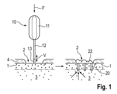
Figure 1 illustrates in a very schematic manner, the direct approach for
perforating a
dense bone layer 1 using a device 10 according to the invention, wherein the
left hand
side of Fig. 1 shows the device 10 positioned for the perforation process, and
the right
hand side shows the perforated dense bone layer and illustrates the desired
flow of
blood, oxygen and/or cells through the perforation. As already mentioned
further
above, the dense bone layer is situated between a repair site 2 (outer side of
the dense
bone layer) and a region 3 or layer of trabecular bone and/or bone marrow
(inner side
CA 03104813 2020-12-22
WO 2020/007865 PCT/EP2019/067749
- 16 -
of bone layer), wherein there is usually a gradual transition between dense
and
trabecular bone tissue and not, as illustrated, a sharp line separating the
two. The dense
bone layer 1 is e.g. a subchondral bone plate and the repair site 2 is a
location, in which
cartilage 4 covering the subchondral bone plate 1 is missing or in need of
repair,
strengthening, augmentation or possibly ossification. The perforation process
is e.g. a
part of an open surgery on a human or animal patient.
The device 10 as illustrated in Fig. 1 has the form of an ultrasonic hand
piece and
comprises a handle 11 housing the vibration generator (not shown), a shaft 12
coupled
to the vibration generator and a substantially cylindrical perforator 13
coupled to the
distal end of the shaft 12 in a substantially coaxial manner. For the
perforation process,
the distal face of the perforator 13 is positioned and held against the outer
surface of
the dense bone layer 1 (arrow F), such that the principal perforator axis
forms an angle
of e.g. about 90 with the bone surface. Furthermore, the vibration generator
is
activated to vibrate (double arrow V) the perforator 13, and the device is
possibly urged
against the dense bone layer 1 (direction of arrow F), therewith driving the
perforator
13 into and preferably through the dense bone layer 1 to form an opening 20
from
which the perforator 13 is then removed. Preferably, as shown on the right
hand side
of Fig. 1, a plurality of openings 20 is created. As soon as the perforator 13
is removed
from the opening 20, fluid material is flowing from the inner side of the
dense bone
layer 1 to its outer side as shown with small arrows, in particular blood
carrying oxygen
and cells, forming, on the outer side of the dense bone layer 1, a blood clot
22, which
enables and/or enhances tissue growth or tissue repair in the repair site 2.
Figure 2 illustrates, in the same schematic manner as Fig. 1, the indirect
approach for
perforating a dense bone layer 1 using a device according to the invention.
For this
perforation process, the perforator 13 is coupled at an angle to the distal
end of the
shaft 12 and is positioned against the dense bone layer 1 by approaching the
device 10
in a direction as indicated with arrow F, i.e. substantially parallel to the
dense bone
CA 03104813 2020-12-22
WO 2020/007865 PCT/EP2019/067749
- 17 -
layer 1. The perforation process as illustrated in Fig. 2 is e.g. part of a
minimally
invasive surgical operation on a joint of a human or animal patient, wherein
the dense
bone layer to be perforated is one of a pair of subchondral bone plates (1 and
1')
situated opposite each other in the joint. In another, exemplary application
of the
process as illustrated in Fig. 2, the dense bone layer 1 to be perforated is
the cortical
bone layer of one of a pair of neighboring vertebrae facing the intervertebral
space,
from which the intervertebral disc and possibly also the cartilage tissue
covering the
subchondral bone plates has been partly or fully removed.
Figures 3 to 8 illustrate various embodiments of instruments suitable for the
device
according to the invention and comprising a shaft 12 and a distal end piece
with a
perforator 13, wherein only a distal portion of the shaft 12 is shown. For
further
illustrating the desired vibration mode of the instruments, Figs. 3 to 5 show
on their
right hand side corresponding schemata. In all Figs. 3 to 8, the perforator 13
has the
form of a cylinder with a tapering tip as further illustrated in Fig. 9.
However, all
further embodiments of perforators as illustrated in the following Figs. 10 to
14 are
also applicable for the instruments as illustrated in Figs. 3 to 8. In
particular, the
perforator may have the form of a prism.
The instrument according to Fig. 3 comprises a perforator 13 and a shaft 12,
the shaft
having at least in its distal end region substantially the same cross section
as the
perforator 13. Shaft 12 and perforator 13 are made as one piece or as two
rigidly
coupled pieces having one common principal axis A, rendering the instrument
suitable
for the direct approach according to Fig. 1. Shaft 12 and perforator 13 are
designed to
vibrate principally longitudinally (double arrows SA and PA) in a standing
wave
having an anti-node position in the region of the distal end of the perforator
13, as
indicated in the vibration scheme on the right hand side of the figure.
CA 03104813 2020-12-22
WO 2020/007865 PCT/EP2019/067749
- 18 -
The instrument shown in Fig. 4 is similar to the instrument of Fig. 3 but the
perforator
13 is only loosely or not coupled to the shaft 12. Therein the shaft 12 is
designed and
activated to vibrate mainly longitudinally (double arrow SA) in a standing
wave having
an anti-node position at the distal face of the shaft, wherein only the
distally directed
half wave is transmitted to the perforator 13 resulting in a series of
intermittent blows
to the latter (arrows PI).
Both instruments illustrated in Figs. 3 and 4 may be further equipped for
creation of
openings with a limited depth. Elements for such depth limitation may comprise
frill
or partial collars extending at a corresponding axial distance from the distal
face of the
perforator radially from the perforator 13 or from the distal shaft end region
(see also
Fig. 10). In the embodiment according to Fig. 4, an outer ring of the distal
face of shaft
12 may serve as depth limiting element, if the shaft 12 has a correspondingly
larger
cross section than the perforator 13.
Figs. 5 and 6 illustrate an instrument suitable for the device according to
the invention
and comprising a shaft 12 and distal end piece with a perforator 13, the
instrument
being suitable for the indirect approach (Fig. 2). The perforator 13 is
arranged relative
to the shaft 12 for the principal shaft axis A to not coincide with the
principal perforator
axis B but to form an angle therewith, as illustrated e.g. an angle of 90 ,
and the
perforator 13 is rigidly coupled to the shaft 12 (Fig. 5) or is loosely
coupled or not
coupled (Fig. 6) thereto. The shaft 12 is designed and activated for vibrating
principally or in addition to the longitudinal or axial vibration (double
arrow SA) in a
transvers or bending vibration mode (double arrow ST), the bending vibration
ST
having node positions in anti-node positions of the axial vibration SA. The
shaft
location in which the perforator 13 is coupled to the shaft 12 or with which
the shaft
is acting on the perforator is a node position of the axial shaft vibration
(SA), i.e. an
anti-node position of its bending vibration (ST), such effecting an axial or
longitudinal
vibration in the rigidly fixed perforator 13 (double arrow PA, Fig. 5), the
perforator 13
CA 03104813 2020-12-22
WO 2020/007865 PCT/EP2019/067749
- 19 -
being preferably designed for an anti-node position of the named vibration PA
in the
region of its distal face. In the case of the perforator 13 not being rigidly
fixed to the
shaft 12 (Fig. 6) the vibration of shaft 12 is only partially transmitted to
the perforator
13 in the same manner as discussed in connection with Fig. 4, i.e. the shaft
12 will act
on the perforator 13 with a series of intermittent hammer blows (arrows PI).
In a process of perforating a dense bone layer using an instrument as
illustrated in Figs.
5 and 6, the depth of the openings to be created is limited by the shaft 12,
i.e. this depth
is limited to be at the most as large as the axial length of the perforator
13. However,
for limiting the depth of the openings to be created, further depth limiting
elements as
e.g. shown in Fig 10 may be provided on the perforator 13.
Figs. 7 and 8 illustrate, very schematically, two further examples of
instruments
suitable for the device according to the invention, the instruments again
comprising a
shaft 12 and a distal end piece with a perforator 13, wherein the principal
shaft axis
A forms an acute angle (Fig. 7) or an obtuse angle (Fig. 8) with the principal
perforator axis B. All comments and explanations in connection with Figs. 5
and 6
are applicable also for Figs. 7 and 8.
Figures 9 to 13 show exemplary embodiments of perforators 13 suitable for the
device
according to the invention, all the perforators shown being applicable in any
of the
instruments as illustrated in Figs. 3 to 8.
Figs. 9 and 10 show solid perforators 13.1 comprising in addition to the
cylindrical or
prismatic main portion a tapering distal tip 14. If the tip 14 has the form of
a pyramid,
the edges of this pyramid are preferably sharp or serrated and together with
the surface
portions between the edges may be plane or concave. Instead of comprising a
tip 14,
CA 03104813 2020-12-22
WO 2020/007865 PCT/EP2019/067749
- 20 -
the distal face of the solid cylinder may have a concave form as indicated in
Fig. 9
with a broken line designated with the numeral 14.1, and comprise a sharp or
serrated
distal outer edge.
In the perforator according to Fig. 9 the largest (most proximal) cross
section of the
tip 14 is the same as the cross section of the cylinder portion, but in the
perforator
according to Fig. 10 it is slightly larger (e.g. by at least 0.1mm), which
results in even
further reduction of lateral friction on impaction. In addition, the
perforator according
to Fig. 10 comprises a depth limiting element 15 in form of a step-shaped
enlargement
of the cross section. Such symmetric or asymmetric depth limiting element can
be
provided on all perforators shown in Figs 9 to 13.
Figs. 11 and 12 show blade- or chisel-shaped, solid perforators 13.2 having a
narrow
elongated cross section and a distal edge 18, which may be straight (Fig. 11),
or
forming a middle point (Fig. 12), but which may also be curved or forming a
lateral
point.
Fig. 13 shows a hollow perforator 13.3 whose distal wall portion is tapering
towards a
distal edge 18, the taper preferably being restricted to the inside of the
hollow cylinder,
which, compared with outer tapering results in even less lateral bone
compaction. As
above mentioned for the solid perforator, a slight reduction of diameter of a
main
proximal cylinder portion caused by the taper of e.g. 0.02 to 0.1mm may help
to further
reduce lateral friction on impaction.
As mentioned already further above, the perforators as shown in Figs. 9, 10
and 13
may have circular cross sections as illustrated. However, this is not a
condition for the
invention. These cross sections may as well be oval, polygonal or of any
desired shape.
CA 03104813 2020-12-22
WO 2020/007865 PCT/EP2019/067749
- 21 -
As also mentioned already further above, the distal edges of the perforators
13.2 and
13.3 as illustrated in Figs 11 to 13 are preferably sharp or serrated, the
serration being
regular or substantially random and having a size in the visible or sub-
visible region.
Fig. 14 shows a further exemplary embodiment of an instrument suitable for the
device
according to the invention, the instrument comprising a shaft 12 (only distal
end
portion shown) carrying at its distal face a plurality of perforators 13,
wherein these
perforators 13 may e.g. have the shape of any of the perforators 13.1, 13.2 or
13.3 as
illustrated in Figs. 9 to 13 or described herein. The combination of shaft 12
and a
plurality of perforators 13 serves for creating simultaneously a plurality of
openings in
the dense bone layer, wherein the shaft 12 has a correspondingly larger cross
section
than each single perforator 13, and wherein the distal shaft face may serve as
depth
limiting element.
Figure 15 shows an opening 20 through a dense bone layer 1, the opening
created with
the aid of a solid perforator as e.g. shown in Figs 9 or 10. The opening 20
has e.g. a
depth reaching through the dense bone layer 1 into the trabecular bone tissue
3,
wherein bone compaction is found only below the bottom of the opening 20
(region
21 of compacted bone tissue), leaving the lateral walls 22 of the opening
fully
uncompromised.
Figure 16 is a cross section through the distal portion of an exemplary,
angled
instrument suitable for the device according to the invention, the instrument
comprising a distal end piece with a hollow perforator 13.3 and a shaft 12,
the two
being rigidly fixed to each other at an angle of e.g. 90 . The illustrated
instrument is
shown in the process of creating an opening through a dense bone layer 1, the
distal
edge of the perforator 13.3. having reached the trabecular bone tissue 3
beneath the
dense bone layer 1.
CA 03104813 2020-12-22
WO 2020/007865 PCT/EP2019/067749
- 22 -
The instrument as illustrated in Fig. 16 is particularly suitable for creating
a series of
openings after each other before being removed from the repair site in which
the dense
bone layer is to be perforated, and for removal of bone tissue from the
openings
together with the instrument. For this purpose, the shaft 12 comprises, in its
distal end
portion, a transversal through opening 23 being aligned with the channel of
the hollow
perforator 13.3 and comprising at least adjacent to the perforator 13.3 a
cross section
similar to the cross section of the inner perforator channel. This through
opening may
further comprise a portion 23' with a larger cross section. Bone debris 25
punched out
of a first opening created with the aid of the perforator 13.3 is e.g. held
within the
perforator, to be pushed out of the latter into the through opening 23 and 23'
of the
shaft 12 by bone debris of a second or third opening being created after the
first
opening. Therein, the vibration of the shaft 12 and the perforator 13.3
facilitates
transport of the bone debris through the hollow perforator 13.3.
If the transversal opening 23 through the shaft 12 does not comprise an
enlarged
portion as illustrated in Fig. 16, bone debris may be removed from shaft 12
and hollow
perforator 13.3 after removal from the repair site using a suitable needle. In
a straight
instrument as e.g. illustrated in Fig. 3 comprising a hollow perforator, the
bone debris
may be collected in a correspondingly hollow distal end of the shaft and may
be
removed by disconnecting the perforator from the shaft after removal of the
instrument
from the repair site. For debris removal it is possible also to provide a
lateral opening
in the hollow perforator 13.3 through which the debris is pushed and from
where it is
transported away together with water which, in particular in arthroscopic
surgery, is
used for continually rinsing the repair site.
Figures 17 and 18 show further exemplified embodiments of an instrument
suitable
for the device according to the invention. The instruments as shown in Fig. 17
and Fig.
18 are particularly suitable for creating a series of bone openings before
being removed
from the operation site, and for removal of bone tissue (in particular the
complete bone
CA 03104813 2020-12-22
WO 2020/007865 PCT/EP2019/067749
- 23 -
fragments punched out) from the openings created using the instrument. For
this
purpose, the hollow perforator 13 comprises, in its distal end portion, a
cutting tube
40. This cutting tube may end in an opening 41 of the perforator having a
larger cross
section than the cutting tube. Bone fragments 42 punched out of an opening
created
with the aid of the perforator 13 may be stored within that opening 41. Thus
the
perforator may comprise an opening 41 suitable as depot for bone fragments or
bone
debris. The perforator 13 may be designed as disposable item, suitable to
create several
bone openings within one operation side and store the resulting (stamped or
punched
out) bone fragments, After removal from the operation side the bone fragments
may
be used as sample for tests or as allogenic transplant material. The
perforator may
through away (with or without the bone fragments) or may easily be cleaned.
The
embodiment of Fig. 17 is designed so that the vibration of the shaft 12 and
the
perforator 13 facilitates transport of the bone fragments 42 through the
hollow
perforator and retain them within the opening 41. The shaft 12 may have a male
thread
25 and the perforator a female thread 26 to ensure a proper connection of both
elements.
In the embodiment shown in Figure 18 the bone fragments may further be
transported
using hydraulic pressure. Therefore, there may be a channel within the shaft
12
suppling a liquid such as water from the handle to the perforator. This liquid
flow can
be stopped or reversed to generate negative pressure. Alternatively, the
liquid may be
used to transport (or eject) the bone fragment out of the cutting tube. The
bone
fragment can be ejected into the operation side and flush away using the
liquid
generally used to clean the operation side (e.g. from blood). When using this
alternative the liquid flow into the perforator is not stopped, but in the
moment the
perforator cuts into the bone the outflow of the liquid is stopped. Therefore,
the
pressure within the perforator increases and this pressure can be used to
remove the
bone fragment from the perforator. In case that a negative pressure is used to
transport
and store bone fragments punched out, the bone fragments are suctioned. This
supports
the process of breaking away the portion of the dense bone punched out.
Therefore, it
CA 03104813 2020-12-22
WO 2020/007865 PCT/EP2019/067749
- 24 -
is possible to remove the bone within the cutting tube in one piece and
without less
damage to the surrounding tissue.
The liquid fed through the instrument may further be used to cool the
instrument and
the perforator, to fill the joint and/or to rinse the operation side. A space
between the
instrument and a tube or channel (cf. Figs. 19 to 21) may be used to suck
liquid and
debris of the bone out of the operational side.
Figures 19 to 21 illustrate exemplary embodiments of the device according to
the
invention which are particularly suitable for arthroscopic or minimally
invasive
surgery by comprising a longish and slender shaft 12 situated in a cannula 30,
the
perforator 13 protruding from the distal end of the cannula and preferably
being
retractable into the cannula before and after the perforation process. Figs 19
to 21 only
show the distal end portions of shaft 12 and cannula 30, and the perforator
13.
The cannula 30 guides the shaft, which for such minimally invasive
applications is
usually quite long (in the range of 50 to 100 mm) and as slender as possible,
and,
furthermore, keeps it substantially dry and free of contamination by tissue
components,
but it also protects surrounding tissue from undesired interaction with the
vibrating
shaft. The guiding function of the cannula may be further improved by
designing the
combination of a shaft 12 to be activated in a mainly longitudinal vibration
and a
cannula 30 for protecting the shaft, to have a reduced radial clearance in
positions in
which the longitudinal vibration of the slender shaft comprises a node
position (anti-
node position of undesired radial vibration). For achieving this improved
guiding
function, the cannula may comprise a reduced inner cross section or the shaft
an
enlarged cross section in the named positions.
CA 03104813 2020-12-22
WO 2020/007865 PCT/EP2019/067749
- 25 -
The distal edge of the cannula 30 may be a sharp and/or serrated edge or may
comprise
pin-shaped protrusions for rendering the cannula 30 suitable for being secured
in the
repair site by lightly embedding this distal edge in tissue.
Furthermore, the cannula may be equipped with fluid conducts for supplying the
repair
site with fluid (e.g. saline solution for rinsing the repair site) and/or for
removing fluid
from the repair site (e.g. saline solution or surplus blood, and, as mentioned
further
above, possibly bone debris).
According to Fig. 19, the instrument comprising a shaft 12 and one perforator
13 or a
plurality thereof protrudes from the distal cannula end for the perforation
process and
before and/or after the perforation process may be retracted such that the
perforator 13
is protected inside the distal end portion of the cannula 30 (retracted
position illustrated
in broken lines).
Figure 20 B shows in an axial section a distal part of shaft 12 within a
cannula 30 (e.g.
an arthroscopic tube). The instrument can be moved in a distal direction
relative to the
channel. Within cannula 30 is a lumen being for example a lumen as used for
optical
instruments (camera) used within an arthroscopy. At the distal end of the
instrument a
perforator 13 is comprised. The instrument is equipped with at least one
portion 18 of
a larger cross section in the node position N (V4). This larger cross section
may be
caused by a ring around the instrument wall. This ring may be a polymeric ring
or a
ring made of the same material as the instrument. The portion 18 may also be
rather at
set spots. There may be three or four spots within the circumference of the
instrument.
Alternatively, or in addition, cannula 30 may comprise at least one portion 19
of a
larger cross section in the node position N (V4). The larger cross section may
be built
CA 03104813 2020-12-22
WO 2020/007865 PCT/EP2019/067749
- 26 -
by a bulge of the wall of the cannula. The bulge may be an integral part of
the cannula
wall and can be formed as a ring having a square cross section. Alternatively,
there
may be several rectangles sitting on one cycle around the sonotrode with some
space
between them. These rectangles can be arranged within a consistent interval on
that
circle. Instead of rectangles there may also be spherical bulges attached on
one circle
around the sonotrode. The circle is always located in a node position N. This
embodiment is illustrated in the cross-sections of the cannula 30 shown in
Figure 20
A. As can be seen, three or four rectangle bulges causes a larger cross
section of the
channel. Between these bulges is free space, which may serve as space for a
liquid
(cooling/flushing). At the distal end of cannula 30 may be attached a
polymeric
bushing 31 which is able to lower frictional loss. The bushing may be made of
a
polymer such as PEEK and is attached to the inner surface of cannula 30.
Alternatively,
shaft 12 or perforator 13 may have a polymeric rind attached to its distal
end.
According to Fig. 21, the shaft 12 has a larger cross section than the
perforator 13 and
the cannula 30 has a partly closed distal end, its distal opening 31 being
reduced to a
cross section smaller than the cross section of the shaft 12 and therewith
limiting
distally oriented axial movement of the instrument in the cannula 30 such
limiting a
depth to which the perforator 13 can be impacted into a dense bone layer, i.e.
limiting
the depth of an opening to be created.
Figure 22 shows an embodiment of the arrangement of shaft 12 and perforator 13
similar to the one of Figure 18 and 19 being suitable for creating a series of
bone
openings before being removed from the operation site, and for removal of bone
tissue
(in particular the complete bone fragments punched out) from the openings
created
using the instrument. For this purpose, the hollow perforator 13 comprises, in
its distal
end portion, a cutting tube 40.1. This cutting tube may end in an opening of
the
perforator having a larger cross section than the cutting tube and being
suitable for
storage of bone fragments 42. The perforator 13 may be designed for single
use,
CA 03104813 2020-12-22
WO 2020/007865 PCT/EP2019/067749
- 27 -
suitable to create several bone openings within one operation side and store
the
resulting (stamped or punched out) bone fragments.
The embodiment of Fig. 22 is designed so that vacuum facilitates transport of
the bone
fragments 42 through the cutting tube 40.1 and retains them within the
perforator.
Therefore, there may be a channel 43.1 within the shaft 12 for pulling out air
and
applying vacuum within the perforator. This channel 43.1 may be created to run
along
the longitudinal axis of shaft 12. The channel may leave the shaft 12 lateral.
This means
the proximal part of the channel 43.1 runs perpendicular to the longitudinal
axis and
the distal part of the channel. Alternatively, a channel 43.2 may be attached
to a flange
which protrudes from the perforator 13. At the height where the flange
protrudes from
the perforator a peripheral groove 44 may be located within the perforator
wall. This
groove extends once around the complete circumference (dotted line). It is
preferred
that the channel 43.1 or 43.2 is attached to the instrument in a node position
(V4). A
channel 43. 1 comprised within the shaft 12 and perhaps the handle has to be
cleaned
after each operation (each treatment of a patient). A channel 43.2 integrated
in the
perforator 13 has the advantage that it has not be cleaned in case the
perforator is a
disposable item or is easier to clean because it is very short and without
bend structure.
On the other side it is more expensive to include such a channel in the
perforator, in
particular in case that it is a disposable item. The vacuum may also be used
to supports
or facilitates breaking away the portion of the dense bone punched out.
Therefore, it is
possible to remove the bone within the cutting tube in one piece and without
less
damage to the surrounding tissue.
As shown in Figure 22 the cutting tube of a perforator may reach into the
space within
the perforator, such as a syphon. Fig. 22 shows also a possible cross section
of a cutting
tube. The cutting tube may have ribs 50 on its inner side. These ribs may run
parallel
to the longitudinal axis of the cutting tube and along the complete length of
the cutting
tube or are arranged only in a distal part which lays outside of the
perforator opening.
CA 03104813 2020-12-22
WO 2020/007865 PCT/EP2019/067749
- 28 -
The ribs may alternatively form spirals or curves on the inner side of the
cutting tube.
The ribs or protruding spikes or alternative protruding elements attached to
the inner
side of the cutting tube may facilitate breaking away of bone fragments after
introduction of the cutting tube into the dense bone. Therefore the cutting
tube or the
complete instrument has to be rotated, so that torsion is applied to the bone
fragment
within the cutting tube. These cutting tubes may be made by extrusion or
additive
manufacturing processes.
Figure 23 illustrates another embodiment of a cutting tube or perforator.
Slicing
elements 51 are arranged within the tube or perforator. These elements are
suitable for
cutting or breaking the bone core into small fragments respectively bone
debris which
may be aspirate from the perforator. The slicing elements may be formed as
thin blades
(e.g. two, three, four or five blades) which are arranged in a way to meet one
another
at the central axis of the tube as illustrated in the cross section of Figure
23. The cutting
tube may cut into the dense bone with its distal end and when cutting deeper
into the
bone the slicing elements chop the bone core into smaller fragments. The
distal ends
of the slicing elements or blades may be sharp and may be slanting.
Figure 24 illustrates two different methods of perforating a dense bone layer
1 with the
aim of enabling transport of blood, viable cells and/or oxygen from an inner
side of
the dense bone layer to an opposite outer side of the dense bone layer using a
device
according to the invention. In Figure 24 A the used device introduces several
micro
holes. The holes can have different cross sections, e.g. rectangular, oval or
irregular.
The cross section is preferably round and may have a diameter between 1 and 2
mm.
The perforator used within this method may have a solid distal end and pushes
the
dense bone material into the trabecular part of the bone which is compacted
thereby.
Alternatively, as shown in Figure 24 B, the device used comprises a hollow
perforator
or at least a hollow cutting tube of the perforator and the bone cone is not
eliminated.
Figure 26 B illustrates the result of a method of perforating a dense bone
layer with
CA 03104813 2020-12-22
WO 2020/007865 PCT/EP2019/067749
- 29 -
the aim of enabling transport of blood, viable cells and/or oxygen from an
inner side
of the dense bone layer to an opposite outer side of the dense bone layer, the
method
comprising:
providing a device according to the invention, the device comprising a
vibration
generator, a shaft defining a principal shaft axis and a distal end piece with
a hollow,
substantially cylindrical perforator defining a principal perforator axis,
positioning the
device such that the principal perforator axis is oriented substantially
perpendicular,
relative to the dense bone layer and a distal end of the perforator is
positioned against
the dense bone layer, activating the vibration generator and holding the
perforator
against the dense bone layer for a time sufficient to create a substantially
cylindrical
opening through or at least into the dense bone layer, and removing the
perforator from
the opening, whereby the cross section of the substantially cylindrical
opening is
substantially ring-shaped. This means that a bone core is left within the
substantially
cylindrical opening. This bone structure has contact to the underlying
trabecular bone
and optionally also to deeper parts of the dense bone but there is a cut with
a closed
geometry within the dense bone and with bone structure in the middle having
the
complete (original) thickness of the treated bone. The ring-shaped cross
section of the
cylindrical opening may have a diameter of 0.1 to 0.75 mm, and preferably of
0.25 to
0.5 mm.
To create substantially cylindrical openings with a substantially ring-shaped
cross
section results in a larger surface for bleeding with the same number of
perforations.
The circumference of the cylindrical opening is a source of bleeding and also
the bone
core in the middle. At the same time the bone is not weaker or even less
fragile. The
devices of the present invention are suitable for this method because the
perforator
geometry is optimized for minimal heat impact to the bone and to minimize
debris
formation.
CA 03104813 2020-12-22
WO 2020/007865 PCT/EP2019/067749
- 30 -
Figure 25 shows three alternative cross sections of the shaft which are
suitable to
increase stiffness of the shaft. The shaft may have a cross section in form of
a double
T, Z or X of course the cross section may also be oval, round or rectangular.
Also an
instrument having a shaft not being round may be adapted to fit to a round or
oval
perforator. This may be done by a respective design of the proximal end of the
perforator or the distal end of the shaft. Alternatively, there may be an
adaptor between
both elements transmitting vibration from the shaft to the perforator.
Figure 26 shows alternative arrangements of the distal end of a perforator
suitable for
a device according to the invention. In Figure 26 A the hollow distal end of
the
perforator or the cutting tube may be sharpened to create peaks or tips. Such
peaks
may enhance the piercing capacity of the penetrator according to the
invention. The
peak should preferably be located in a way that it is the first structure to
penetrate the
bone. The peak may, therefore, be located at the outer, the inner edge or in
the middle
of the perforator wall. Alternatively, the distal end portion of the
perforator may be
formed to have different facets. Each facet is a small plane surface at the
distal edge.
Thus, three or more peaks may result at the distal end, where different facets
hit each
other.
Figure 26 B shows also a sharp distal end of the perforator or respectively
the cutting
tube comprised by the perforator. The distal end of the perforator or its
cutting tube
shows a groove 52, which bounds a sharp, distal cutting edge. The groove
consists of
a groove 16 which runs on the inner side of the perforator or its cutting tube
and runs
parallel to the distal cutting edge. The wall of the perforator within the
groove 52 is
thinner as the wall of the distal cutting edge. The wall within the groove may
be
between 0.05 and 0.5 mm lower than the edge. The minimal depth of the groove
is
sufficient to adequately lower the friction. It is preferred that the wall of
the perforator
or respectively its cutting tube increases abruptly or gradually towards the
proximal
end. Alternatively, the distal cutting edge has a thicker wall than the
remaining cutting
CA 03104813 2020-12-22
WO 2020/007865 PCT/EP2019/067749
- 31 -
tube or perforator. In this case 52 refers to a step and not a groove. Thus,
the distal end
of the perforator or its cutting tube may comprise a step 52, where the wall
thickness
increases in a way that distal of the step the wall is thicker than proximal
of the step.
The groove or respectively step 52 may also be located on the outer side of
the
perforator or its cutting tube.
The distal end illustrated in Figure 26 C is similar to the one of Figure 26
B, but there
is a groove 52 on the inner side of the distal end of the perforator or its
cutting tube
and another groove 53 on the outer side. Again 52 and or 53 may also refer to
a step
within the wall of the perforator or its cutting tube. Steps 52 and 53 may
also be
designed to form a curvature.
In the embodiment illustrated in Figure 26 D, the distal cutting edge of the
perforator
or its cutting tube are formed by a ring 54 inserted or pressed in the distal
end of the
perforator or its cutting edge. The ring may be made of a material different
from the
perforator or its cutting tube. The ring may be made of metal, an alloy or a
ceramic or
any material hart enough to cut bone. The ring may be designed to form a sharp
distal
edge.
Similar to the ring 54 in Figure 26 D, Figure 26 E shows a ring or part of a
ring 55 put
on the distal end of the perforator or its cutting tube and being welded (by
laser or
ultrasound) to the wall of the perforator or its cutting tube. The material 56
remaining
after welding sits on the proximal end of the ring 55. The ring 55 may form a
sharp
cutting edge for introducing into the bone, wherein the wall of the perforator
or its
cutting tube may be thinner. The minimal thickness of the wall is sufficient
to
adequately lower the friction.
CA 03104813 2020-12-22
WO 2020/007865 PCT/EP2019/067749
- 32 -
Figure 27 shows another embodiment of an arrangement of the distal end of a
perforator or its cutting tube suitable for a device according to the
invention. The distal
end is alternatingly folded. Therefore, the tubular distal end has at least
two incisions.
The parts between the incisions are alternating folded inwards or outwards.
Figure 27
A illustrates the distal end with incisions before folding. Figure 27 B shows
parts 57
and 57' being folded outwards, wherein the part in-between has been folded
inwards.
Figure 27 C shows a cross section of the folded distal end having 4 incisions
and parts
57, 57' being folded outwards and parts 58 and 58' being folded inwards. The
fold
should be in a way that a sharp cutting edge 18 results, as illustrated in
Figure 27 D
being a cut at A to A'.
Figure 28 shows an instrument according to the invention comprising a shaft 12
and
one perforator 13. The perforator has been pressed in the shaft 12
(illustrated in broken
lines) and laser welded to be secured. The perforator may be a hollow tube
with a
length of 10 to 14 mm, an outer cross section of 1 to 1.4 mm and an inner
cross section
of 0.6 to 1 mm. the length of the perforator part being outside the shaft is
about 7 mm.
The outer diameter of the cross section of the shaft may be 3 to 5 mm. The
shaft has
been press fit into a base 61 having a proximal element 60 (thread or plug-in)
for
connection to the handle and/or the vibration generator.
Figure 29 illustrates an embodiment of the arrangement of shaft 12 and hollow
perforator 13, similar to Fig. 28. The angle a as well as the mass of element
17
influence the axial amplitude of the vibration. The mass of element 17 may be
chosen
in a way that the maximal amplitude is at point 62. In case that the angle a
is nearly 00
position 62 is at V4. Element 17 may be a ring-shaped increase of the wall
thickness.
Alternatively, it may consists of at least two solid objects 17 and 17'
attached
symmetrically to shaft 12.
CA 03104813 2020-12-22
WO 2020/007865 PCT/EP2019/067749
- 33 -
Figure 30 illustrates an embodiment of the arrangement of shaft 12 and hollow
perforator 13, similar to Fig. 28. The shaft 12 is a hollow tube with an
oblique-cut
distal end. The distal end may be closed by a plate which may be welded to the
tube.
The hollow perforator (having the form of a tube) can be pressed into said
plate and
welded to the plate (cf. remains 56.1). The plate can serve as a depth
limiting element.
The outer diameter of the perforator may between 6 and 10 mm. The angle a may
be
> 900 and < 180 and is preferably between 120 and 150 . The geometry of the
shaft
12 can have anyone as described herein, e.g. there may be space for storage of
bone
fragments 42. There may be a supply for a cooling liquid from the handle to
the inner
pace of the shaft 12 (not shown). In addition the distal end of the perforator
may be
designed as shown in Figures 26 A to E and Figure 27.
Analogous to the arrangements of shaft 12 and hollow perforator 13 shown in
Figures
29 and 30 also a cutting tube 40 may be attached oblique to a perforator 13.