Note: Descriptions are shown in the official language in which they were submitted.
CA 03105025 2020-12-23
87567721
STACKED SENSOR ASSEMBLY
FOR FLUID ANALYZER
[0001] The subject application claims priority to US provisional Application
No. 62/692,053, filed June 29, 2018.
FIELD OF THE DISCLOSURE
[0002] The disclosure herein relates generally to the field of sensors used in
the analysis
of fluid properties. The disclosed sensor assembly is embodied in a sensor
cartridge which is
especially adapted for use in biomedical applications so as to assist in the
analysis of multiple
physical parameters and/or chemical constituents of small volume samples of
bodily fluids such as
whole blood.
BACKGROUND
[0003] In a variety of instances it is desirable to measure the constituents
in a bodily fluid
to Include, for example, partial pressure of blood gasses in a whole blood
sample, concentrations
of electrolytes in the blood sample, and the hematocrit value of the blood
sample. For example,
measuring pCO2, p02, pH, Nat, Kt, Ca2+ and hematocrit value are primary
clinical indications in
assessing the condition of a medical patient. In addition, in an attempt to
use as little of the
patient's blood as possible in each analysis performed, the devices which are
employed to analyze
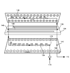
a blood sample are preferably relatively small. Performing blood analysis
using a small blood
sample is important, for example, when a relatively large number of samples
must be taken in a
relatively short amount of time or if the volume of blood is limited, as in
neonates.
[0004] For example, patients in intensive care may require a sampling
frequency of 15-20
per day for blood gas and clinical chemistry measurements, leading to a
potentially large loss of
blood during patient assessment. In addition, by reducing the size of the
analyzer sufficiently to
make the unit portable, analysis can be performed at the point of care. Also,
reduced size
typically means reduced turnaround time. Furthermore, in order to limit the
number of tests
which must be performed it is desirable to gather as much information as
possible upon
completion of each test. However, size limitations are imposed upon the
sensors that are used to
measure blood chemistry. These size limitations are in large part due to
physical geometries of
the sensors and the connections to the sensors.
1
Date Recue/Date Received 2020-12-23
CA 03105025 2020-12-23
WO 2020/005683
PCT/US2019/038135
[0005] Point of care blood gas analyzers permit in vitro analysis at the
patient's
bedside, in the emergency room, or in the intensive care unit. These units use
solid state
sensors with thin-film electrodes. The microchips, reagents, calibrators, and
a sampling
device are all contained within a disposable cartridge system. Healthcare
facilities can select
cartridges with additional test options, including potassium, glucose, BUN and
lactate.
Because whole blood can be tested, minimal specimen processing is needed; the
sample does
not have to be centrifuged and the plasma separated from the red blood cells
prior to testing.
[0006] In settings with medium-to high volume sample testing, a multi-use
cartridge
system is used. These cartridges can be customized to the specific analyte
menu and to the
volume of testing. The number of samples measured on a cartridge may vary from
25 to 750
and once loaded into the analyzer, the cartridge typically has an in-use life
of between 14 and
30 days.
[0007] The basic principle of operation for blood gas analyzers has not
changed
significantly from earlier units. In about 2005 self-contained cartridges were
introduced into
several analytical systems, paving the way for point of care testing and
compact units. Whole
blood can be analyzed for many analytes, including the electrolytes potassium
(1C+), sodium
(Na'), and calcium (Ca? ') and metabolites such as glucose, lactate, blood
urea nitrogen
(BUN), and creatine. The sensors used for these measurements are ion-specific
or ion-
selective electrodes (ISE) These sensors are membrane-based electrochemical
transducers
that respond to a specific ion. Biosensors are used in analyzers in the
traditional clinical
laboratory, but also in point-of-care testing devices. Biosensors convert the
biochemical
signal into an electrical signal.
[0008] Electrolytes are determined by potentiometric measurements, a form of
electrochemical analysis. In potentiometry, the potential or voltage is
measured between the
two electrodes in a solution. These potentials can also be produced when a
metal and ions of
that metal are present in a solution. By using a membrane that is
semipermeable to the ion,
different concentrations of the ion can be separated. These systems use a
reference and a
measuring electrode. A constant voltage is applied to the reference electrode;
the difference
in voltage between the reference and measuring electrode is used to calculate
the
concentration of the ion in solution.
[0009] Ion-selective electrodes are based on a modification of the principle
of
potentiometry. The potential difference or electron flow is created by
selectively transferring
the ion to be measured from the sample solution to the membrane phase. The ion-
selective
electrode measures the free ion concentration of the desired analvte on a
selectively produced
2
CA 03105025 2020-12-23
WO 2020/005683
PCT/US2019/038135
membrane. Membranes have a complex composition and contain organic solvents,
inert
polymers, plasticizers, and ionophores wherein the ionophores are molecules
that increase the
membrane's permeability to the specific ion.
[0010] Amperometric methods measure the current flow produced from oxidation-
reduction reactions. Types of amperometry include enzyme electrodes, such as
the glucose
oxidase method and the Clark p02 electrode. These types of designs are well
known as
biosensors and are adaptable for testing in the clinical laboratory as well as
for point of care
testing. Enzyme-based biosensor technology was first developed to measure
blood glucose.
A solution of glucose oxidase is placed between the gas permeable membrane of
the p02
electrode and an outer membrane that is semipermeable. Glucose in the blood
diffuses
through the semipermeable membrane and reacts with the glucose oxidase.
Glucose is
converted by glucose oxidase to hydrogen peroxide and gluconic acid.
[0011] A polarizing voltage is applied to the electrode, which oxidizes the
hydrogen
peroxide and contributes to the loss of electrons. Oxygen is consumed near the
surface of the
p02 electrode and its rate of consumption is measured. The loss of electrons
and rate of
decrease of p02 is directly proportional to the glucose concentration in the
sample. Enzyme-
based biosensors are also used to measure cholesterol, creatine, and pyruvate.
[0012] The basic principles of operation for laboratory blood gas analyzers
are the
same as for the previously described electrodes for pH; pCO2, and p02; and ion
specific,
electrodes for the measurement of electrolytes. Approximately 50-120 I of a
well-mixed
arterial blood sample are typically injected through the inlet and sample
probe into the
measuring chamber. The specimen then contacts the surface of each electrode
for several
seconds.
[0013] One of the principal challenges with existing sensor assemblies is that
performing blood analysis using a small blood sample is important when a
relatively large
number of samples must be taken in a relatively short amount of time or if the
volume of
blood is limited, as in neonates.
[0014] Accordingly, it would be desirable to provide a sensor assembly which
remains accurate over a relatively long period of exposure to electrolytes and
blood samples,
uses a very small sample size, and detects the concentration of a number of
different
electrolytes as well as the partial pressure of a number of blood gases all in
a single analysis.
3
CA 03105025 2020-12-23
WO 2020/005683
PCT/US2019/038135
SUMMARY
[0015] Heel sticks and draws from arterial lines are the most commonly used
sites for
blood draws. Heel sticks require a high degree of technical expertise to be
done properly and
without inflicting unnecessary pain or harm to the patient. Frequent blood
draws for
laboratory testing create the risk of iatrogenic anemia. It has been estimated
that 64 % of
infants < 1500 g receive transfusions for anemia due in part to frequent or
excessive blood
draws. With a plasma volume of 4-5 % of body weight, a 1,500 g infant has a
total of 70 mL
of plasma. Blood transfusion may be required when 10% or more of a neonate's
blood
volume is withdrawn in 2-3 days. This amount represents about 80 mL/kg of body
weight for
a full-term infant; and about 100 mL/kg for a preterm infant.
[0016] The volume and number of blood draws have been reduced in recent years
due
to trans cutaneous monitoring and newer equipment. Minimizing the volume of
blood draws
reduces the subsequent need for transfusion as well as the risk associated
with
transfusion. Many of the current clinical chemistry analyzers require small
blood sample
volumes for testing, with many sensor arrays requiring between 451.tt to
4000,, depending
on the number of analytes being measured (e.g., blood gases, electrolytes,
etc.). '[he
hematocrit of an infant can be > 60 %, reducing the volume of serum or plasma
in the
collection container. The "dead volume-, consisting of the volume of specimen
that must be
in the instrument's sampling container, is required in addition to the
specimen volume and
must be minimal for neonatal applications.
[0017] The sensor array disclosed herein requires a sample volume of no
greater than
301.tt +/-11,11 in order to pass a sufficient quantity of fluid past each of
the analyte sensors.
The sensor assembly is capable of supporting numerous analyte sensors with the
sensor
assembly including a molded separation panel, a potentiometric chip disposed
atop the
separation panel, an amperometric chip disposed beneath the separation panel,
and a bonding
media disposed beneath the amperometric chip. The separation panel includes an
upper
surface and a lower surface and first and second longitudinally disposed ends.
[0018] A fluid channel is molded into the upper surface and spans
substantially
between the first and second longitudinally disposed ends. A second fluid
channel is molded
into the lower molded surface and spans substantially between the first and
second
longitudinally disposed ends. The first and second fluid channels have a total
volume of 301.L1
+/-11,11. Analyte sensors are strategically located above and below the upper
and lower fluid
chambers to quantify the concentration or pressure of the constituents of
interest.
4
87567721
[0019] It is an object of the sensor assembly disclosed herein to provide a
low cost
disposable sensor assembly.
[0020] It is a further object of the sensor assembly disclosed herein to
compactly provide
a disposable sensor assembly capable of housing a large number of analyte
sensors.
[0021] It is a further object of the sensor assembly disclosed herein to
provide a sensor
assembly that requires a blood volume of no greater than 30[tL.
[0021a] According to one aspect of the present invention, there is provided a
sensor
assembly for analysis of physical parameters and chemical constituents of
small volume samples
of bodily fluids with at least two analyte sensors comprising: a separation
panel, the separation
panel further comprising an upper surface with an upper fluid channel for
passage there through of
the sample volume as well as a lower surface with a lower fluid channel in
fluid communication
with the upper fluid channel; a first chip disposed atop the separation panel,
the first chip
including at least one analyte sensor disposed over the upper fluid channel
and one or more
electrical contact points for connecting the analyte sensor with an analyzer;
and a second chip
disposed beneath the separation panel, the second chip including at least one
analyte sensor
disposed over the lower fluid channel and one or more electrical contact
points for connecting the
analyte sensor with an analyzer; wherein a bodily fluid sample traverses
through the entire extent
of the upper and lower fluid channels in fluid communication with the sensors
of the first and
second chips.
[0021b] According to another aspect of the present invention, there is
provided a sensor
assembly for analysis of physical parameters and chemical constituents of
small volume samples
of bodily fluids with at least two analyte sensors comprising: a separation
panel with an upper
surface and a lower surface, the separation panel further comprising first and
second
longitudinally disposed ends with upper and lower fluid channels disposed
within the upper and
lower surfaces respectively and extending substantially between the first and
second ends and
when in an operating mode bodily fluid is in fluid communication with both the
upper and lower
fluid channels; a first chip positioned atop the separation panel with at
least one analyte sensor
positioned over the upper fluid channel and when the sensor assembly is in an
operating mode the
bodily fluid is in fluid communication with the at least one analyte sensor;
and a second chip
positioned beneath the lower separation channel with at least one analyte
sensor positioned
Date Re9ue/Date Received 2021-03-05
87567721
beneath the fluid channel and when the sensor assembly is in an operating mode
the bodily fluid is
in fluid communication with the at least one analyte sensor, wherein the
bodily fluid sample
traverses through the entire extent of the upper and lower fluid channels in
fluid communication
with the sensors of the first and second chips.
[0022] These, together with other aspects of the disclosed sensor array, along
with the
various features of novelty that characterize the technology, are pointed out
below and form a part
of this disclosed technology. For a better understanding of the disclosed
technology, its operating
advantages and the specific objects attained by its uses, reference should be
made to the
accompanying drawings and descriptive matter in which there are illustrated
exemplary
embodiments of the disclosed technology.
BRIEF DESCRIPTION OF THE DRAWINGS
[0023] Illustrative embodiments of the disclosed technology are described in
detail below
with reference to the attached drawing figures, wherein:
[0024] FIG. I is a perspective view of an embodiment of an exploded cartridge
with case
and cover and including an embodiment of a sensor assembly;
[0025] FIG. 2 is an exploded topside view of an embodiment of the sensor
assembly
components;
[0026] FIG. 3 is an exploded bottom side view of an embodiment of the sensor
assembly
components;
[0027] FIG. 4 is a plan view of an embodiment of a separation panel of the
sensor
assembly disclosed herein;
[0028] FIG. 5 is a cross-sectional view of the separation panel of FIG. 4
taken along line
5-5;
[0029] FIG. 6 is a cross-sectional view of the separation panel of FIG. 4
taken along line
6-6;
[0030] FIG. 7 is an exploded view of the potentiometric chip, separation panel
and
amperometric chip;
[0031] FIG. 8 is a cross sectional view of the fully assembled cartridge with
sensor
assembly installed therein; and
[0032] FIG. 9 is a perspective view of a fully assembled fluid sensor
assembly.
5a
Date Re9ue/Date Received 2021-03-05
CA 03105025 2020-12-23
WO 2020/005683
PCT/US2019/038135
DETAILED DESCRIPTION
[0033] Disclosed herein is a stacked sensor assembly 10 for determining
partial
pressures of gases, concentrations of electrolytes and metabolites in a fluid
sample. The
stacked sensor configuration is ideal for minimizing the surface area required
for the sensor
assembly without sacrificing the functionality of the sensor assembly. In
clinical laboratory
settings where available space may be at a premium due to the large number of
instruments
utilized, this stacked sensor configuration offers an attractive option for
reducing the footprint
of the sensor assembly. Fluids, such as whole blood, can be analyzed for many
analytes,
including the electrolytes potassium (10, sodium (Nat), and calcium (Ca24) and
metabolites
such as glucose, lactate, blood urea nitrogen (BUN), and creatine. The sensors
used for these
measurements are ion-specific or ion-selective electrodes (ISE).
[0034] An embodiment of the stacked sensor assembly 10 disclosed herein is
depicted
in FIG. 1. The stacked sensor assembly 10 is shown ready for loading onto the
cartridge base
20 and the cartridge cover 30 located atop the stacked sensor assembly 10. The
fully
assembled cartridge 40 includes the stacked sensor assembly 10 as well as the
cartridge base
20 and the cartridge cover 30. The cartridge 40 is sold as a unit for
installation in a fluid gas
analyzer, such as a blood gas analyzer, that is well known in the industry and
sold by several
manufacturers.
[0035] As shown in FIG 2, the stacked sensor assembly 10 is comprised of
multiple
layers. The uppermost layer may be comprised of solely a potentiometric chip
102 or a
combination of potentiometric sensors and other types of sensors. The
discussion below
details the utilization of potentiometric and amperometric chip sets; however,
it should be
understood that the disclosure herein contemplates the combination of many
types of sensors
to include potentiometric, and amperometric, sensors on each chip. The
potentiometric chip
102 operates pursuant to a form of electrochemical analysis. In potentiometry,
the potential
or voltage is measured between the two electrodes in a solution. These
potentials can also be
produced when a metal and ions of that metal are present in a solution. By
using a membrane
that is semipermeable to the ion, different concentrations of the ion can be
separated. These
systems use a reference and a measuring electrode as is well understood by
those skilled in
the art. A constant voltage is applied to the reference electrode; the
difference in voltage
between the reference and measuring electrode is used to calculate the
concentration of the
ion in solution.
[0036] Ion-selective electrodes are based on a modification of the principle
of
potentiometry. The potential difference or electron flow is created by
selectively transferring
6
87567721
the ion to be measured from the sample solution to the membrane phase. The ion-
selective
electrode measures the free ion concentration of the desired analyte on a
selectively produced
membrane. Membranes have a complex composition and contain organic solvents,
inert
polymers, plasticizers, and ionophores wherein the ionophores are molecules
that increase the
membrane's permeability to the specific ion.
[0037] As seen in FIG. 2, disposed beneath the potentiometric chip 102 is the
separation panel 104. The separation panel 104 includes an upper surface 106
with an upper
fluid channel 108 for passage there through of the sample fluid. The fluid
sample traverses
through an opening 148 in the separation panel prior to entering the upper
fluid channel 108.
The separation panel 104 also reduces the potential for unintended
electromagnetic cross-talk
between sensors located on the oppositely disposed chip thereby improving the
accuracy and
reliability of the sensor data. As seen in FIG. 3, the separation panel 104
includes a lower
surface 110 with a lower fluid channel 112. The lower fluid channel 112 is in
fluid
communication with the upper fluid channel 108, as will be discussed in
greater detail below.
Optionally disposed between the potentiometric chip 102 and the separation
panel 104 may be
an upper gasket 114. The upper gasket 114 seals the separation panel 104
against leakage of
the sample fluid and is preferably comprised of a flexible fluid resistant
material capable of
forming a seal against leakage. The upper gasket 114 may include a series of
perforations 116
located on each side of a centralized cutout 118. The perforations 116 in the
upper gasket 114
may provide an opening for the lower protruding surface 122 of the analyte
sensor contact
points 124, 125.
[0038] As previously discussed, the potentiometric chip 102 is positioned atop
the
separation panel 104 and includes, as shown in FIG. 3, at least one analyte
sensor 136, and
preferably many more are positioned over the upper fluid channel 108. Each
analyte sensor
136 includes two electrical contact points 124, 125 for connecting the analyte
sensor 136 to an
analyzer (not shown).
[0039] As seen in FIG. 4, the upper surface 106 also includes a pair of
optional
adjacent channels 128, 130 that straddle the upper fluid channel 108. These
channels may
facilitate the fabrication of the upper surface 106 by inhibiting warping that
my otherwise
result from excess molded material. The upper fluid channel 108 serves as a
conduit for the
7
Date Recue/Date Received 2021-04-29
87567721
fluid being measured by an upper analyte sensor 136. The fluid sample
traverses through an
opening 148 in the separation panel prior to entering the upper fluid channel.
108. This fluid
channel 108 is narrow and generally linear in configuration and is preferably
rectangular as
shown at cross section 5-5 and 6-6as seen in FIGS. 4-6. Other cross-sectional
configurations,
such as arcuate, are also contemplated by this disclosure. In order to reduce
the volume of the
fluid sample required for analysis to no more than roughly 30111, the upper
fluid channel must
be very narrow. Cross section 5-5 also details the through hole 138 that leads
from the upper
fluid channel 108 to the exit point 140 on the lower chip (as seen in FIG. 7)
which is
discussed in greater detail below.
[0040] As seen in FIGS. 2 and 3 a second chip 152 is disposed beneath the
separation
panel 104. In a preferred embodiment, the chip 152 is comprised of all
amperometric sensors;
7a
Date Recue/Date Received 2021-04-29
CA 03105025 2020-12-23
WO 2020/005683
PCT/US2019/038135
however, a combination of amperometric, potentiometric and other sensor
options placed
upon the chip 152 are also contemplated with this disclosure. The discussion
below is
directed to a chip comprised solely of amperometric sensors; however, this
characterization
should not be considered limiting. Amperometric methods measure the electrical
current flow
produced from oxidation-reduction reactions. Types of amperometry include
enzyme
electrodes, such as the glucose oxidase method and the Clark p02 electrode.
These types of
designs are well known as biosensors and are adaptable for testing in the
clinical laboratory
as well as for point of care testing.
[0041] Enzyme-based biosensor technology was first developed to measure blood
glucose. A solution of glucose oxidase is placed between the gas permeable
membrane of the
p02 electrode and an outer membrane that is semipermeable. Glucose in the
blood diffuses
through the semipermeable membrane and reacts with the glucose oxidase.
Glucose is
converted by glucose oxidase to hydrogen peroxide and gluconic acid.
[0042] A polarizing voltage is applied to the electrode, which oxidizes the
hydrogen
peroxide and contributes to the loss of electrons. Oxygen is consumed near the
surface of the
p02 electrode and its rate of consumption is measured. The loss of electrons
and rate of
decrease of p02 is directly proportional to the glucose concentration in the
sample. The basic
principles of operation for laboratory fluid analyzers are the same as for the
previously
described electrodes for pH, p(702 and p02; and ion specific electrodes for
the measurement
of electrolytes.
[0043] As seen in FIGS. 2 and 3, the amperometric chip 152 includes at least
one
analyte sensor 154 disposed over the lower fluid channel 112 and two
electrical contact
points 156, 158 for connecting the analyte sensor 154 with an analyzer (not
shown). As seen
in FIG. 3, positioned above the amperometric chip 152 is the separation panel
104. The
separation panel 104 includes a lower surface 110 with a lower fluid channel
112 for passage
of the sample fluid. As seen in FIG. 3, the separation panel 104 includes a
lower surface 110
with a lower fluid channel 112, in fluid communication with the upper fluid
channel 108.
Optionally disposed between the amperometric chip 152 and the separation panel
104 is a
lower gasket 160. The lower gasket 160 seals the separation panel 104 against
leakage of the
sample fluid and is preferably comprised of flexible fluid resistant material
capable of
forming a seal against leakage. The gasket 160 includes a cutout area 162 that
coincides with
the location and configuration of the lower fluid channel 112.
[0044] As seen in FIG. 3, the separation panel 104 also includes a pair of
optional
adjacent channels 164, 166 that straddle the lower fluid channel 112. These
channels may
8
87567721
facilitate the fabrication of the separation panel 104 by inhibiting warping
that may otherwise result
from excess molded material. The amperometric chip 152 is positioned beneath
the separation panel
104 and includes, as shown in FIG. 2, at least one analyte sensor 154, and
preferably many more, are
positioned beneath the lower fluid channel 112. Each analyte sensor 154
includes two electrical
contact points 156, 158 for connecting each analyte sensor 154 with an
analyzer (not shown).
[0045] The lower fluid channel 112 serves as a conduit for the fluid being
analyzed by at least
one lower analyte sensor 154. This fluid channel 112 is narrow and generally
linear in configuration
and may be viewed at cross sections 5-5 and 6-6 as seen in FIGS. 5-6. In order
to reduce the volume
of the fluid sample required for analysis to no more than about 30 1, the
lower fluid channel 112, just
like the upper fluid channel 108, must be very narrow. For example, at cross
section 5-5, as shown in
FIG. 5, the lower fluid channel 112 has a very narrow rectangular profile.
Cross section 5-5 also details
the through hole 138 that leads from the upper fluid channel 108 to the exit
point 140 on the
amperometric chip (as seen in FIG. 7) which is discussed in greater detail
below.
[0046] Fluid 170 undergoing analysis enters the channel 112, as best seen in
FIGS. 7 and 8
at the far extent of the channel through an opening 172. The fluid 170 then
travels along the lower
fluid channel 112 providing access to one or more amperometric analyte sensors
154. The fluid
sample 170 then traverses through an opening 148 in the separation panel prior
to entering the upper
fluid channel 108. After entering the upper fluid channel 108 the fluid sample
170 traverses beneath at
least one analyte sensor 136 prior to transiting through the exit opening 138
in the separation panel
104. FIG. 8 reveals a cross section view of the overall cartridge assembly 40
and details the fluid path
through the sensor assembly 10.
[0047] As seen in FIG. 9, the sensor assembly 10 when fully assembled reveals
analyte
sensor contacts 124, 125 156, 158. These sensor contacts feed electrical
signals to contact points
located on the analyzer (not shown) where the voltage and current levels from
each analyte sensor are
separately analyzed. Following analysis, the pertinent details regarding the
fluid analytes are reported
out to the user to effectuate a diagnostic assessment.
[0048] Many different arrangements of the various components depicted, as well
as
components not shown, are possible without departing from the spirit and scope
of the disclosed
technology. Embodiments of the disclosed technology have been described with
the intent to be
illustrative rather than restrictive. Alternative embodiments will become
apparent to those skilled in
the art that do not depart from its scope. A skilled artisan may
9
Date Recue/Date Received 2021-04-29
CA 03105025 2020-12-23
87567721
develop alternative means of implementing the aforementioned improvements
without
departing from the scope of the disclosed technology.
[0049] It will be understood that certain features and sub combinations are of
utility
and may be employed without reference to other features and sub combinations
and are
contemplated below. Not all steps listed in the various figures need be
carried out in the
specific order described.
Date Recue/Date Received 2020-12-23