Note: Descriptions are shown in the official language in which they were submitted.
1
PHOTODYNAMIC THERAPY APPARATUS FOR LOCAL TARGETING IN
CANCER TREATMENT AND CONTROL METHOD THEREFOR
Technical Field
The present invention relates to a photodynamic
therapy apparatus for local targeting in cancer
treatment and a control method therefor. More
specifically, an endoscope is placed in the center of an
end portion of a probe used in photodynamic therapy and
a plurality of optical fibers are arranged along the
edge thereof, so that a lesion site is irradiated with a
plurality of lights while the plurality of optical
fibers of the probe receive lights from individual light
sources, respectively, to perform light irradiation
individually. Furthermore, the present invention relates
to an apparatus and a control method therefor, wherein
unnecessary damage to normal tissues is minimized by
controlling light irradiation regions, for instance, by
defining light irradiation regions encompassing a lesion
site from an image provided in real time through an
endoscope disposed at an end portion of a probe and
allowing only individual light sources irradiating the
defined light irradiation regions to emit lights to
achieve local light irradiation. Therefore, the
photodynamic therapy apparatus for local targeting and
the control method therefor of the present invention are
capable of treating various types of cancers or tumors
to which local treatment is applicable due to small-
sized lesion sites, including cervical cancer, female
cancers (endometrial cancer, ovarian cancer, and breast
cancer), skin cancer, brain cancer, and the like.
Background Art
Cervical cancer is the fourth most common female
cancer in the world. According to cancer statistics 2012
Date Recue/Date Received 2021-01-06
2
from the World Health Organization, there were 500,000
or more cases diagnosed with cancer and approximately
50% of the cases were dead.
Considering the facts that cervical cancer can be
completely cured if detected early since the cervical
cancer goes through stages of precancerous lesions for a
longer period of time compared with other cancers and
that the presence or absence of cancer can be determined
through simple tests compared with diagnostic
examinations of stomach, lung, colon, thyroid cancers,
early detection and aggressive treatment for cervical
precancerous lesions is important.
There are currently no therapeutic drugs for these
cervical precancerous lesions, and the only treatment is
surgical excision, and examples of the surgical excision
are a loop electrosurgical excision procedure (LEEP),
cervical conization, laser excision, and hysterectomy.
However, such surgical therapies cause premature
birth, miscarriage, infertility, and the resulting
social problems, such as a reduction in fertility rate,
and incomplete surgery has a problem regarding a risk of
recurrence. Although not high probability, cervical
precancerous lesions cause a risk of birth with cerebral
palsy caused by premature birth, retinopathy, and
decreased lung maturity.
As such, cervical precancerous lesions may increase
rapidly among young people and cause serious pregnancy-
related complications in existing conization, and thus
considering current social circumstances of low
fertility, the development of new safe and effective
treatments is urgent.
There has recently been newly proposed photodynamic
therapy (PDT) in which surgery is performed using a
laser, wherein treatment is achieved by selective
response to only necessary sites to destroy lesions
Date Recue/Date Received 2021-01-06
3
encompassing cancer cells or precancerous lesions. That
is, photodynamic therapy is receiving attention as a
treatment method wherein singlet oxygen or free
radicals, which are derived by the overall chemical
reaction of abundant oxygen in the body, a light (laser)
supplied from the outside, and a photo-sensitizer as a
material sensitive to a light, treat inflammations of
various lesion sites or destroy cancer cells, and thus
photodynamic therapy is on the rise as a treatment
method capable of pregnancy-related problems due to
conventional wide excision.
International Publication No. WO 2009/095912 Al
(published 06 August 2009) discloses, as photodynamic
therapy, a method in which a photosensitizer is
selectively accumulated only in cancer cells and tumors
by conventional injection through an endoscopic catheter
and the cervical lesion sites are destroyed by light
irradiation.
Korean Patent Publication No. 2002-0060020
(published 16 July 2002) discloses a method for
photodynamic therapy and diagnosis, in which two or more
laser diodes are provided as a light source. Also in the
present case, the lights output from a plurality of
laser diodes are collected and condensed to one, and
then allowed to exit to a target tissue through an
optical cable, thereby achieving treatment.
In most of the PDT apparatuses including ones
disclosed in the prior art documents, the lights from a
plurality of light sources are condensed to one and
allowed to perform light irradiation through one exit,
or the lights from a plurality of light sources are
directly irradiated at the same time.
Such a light irradiation manner in which the
condensed lights are allowed to exit through one exit
can increase treatment effects by increasing light
Date Recue/Date Received 2021-01-06
4
intensity, but may cause a problem of skin damage due to
the irradiation to tissues in the vicinity of lesion
sites. In
addition, a manner in which a transferred
light is divided into a plurality of lights and
irradiated (Korean Patent Registration No. 10-1500620)
or a manner in which a plurality of light irradiation
parts are allowed to protrude from an endoscope and a
plurality of light sources are installed in the
protruding light irradiation surfaces, thereby achieving
individual light irradiation from each light source
(Korean Patent Registration No. 10-1670401) may be
applied, but these manners result in light irradiation
to a wide area encompassing lesion sites.
Therefore, there has been a need for an apparatus
capable of achieving local treatment by intensive light
irradiation to only a lesion site while minimizing the
damage to adjacent normal tissues when a tumor is
located locally in the epithelium of the cervix, like in
the treatment of early cervical cancer.
Detailed Description of the Invention
Technical Problem
Therefore, the present invention has been made in
view of the above-mentioned problems, and an aspect of
the present invention is to provide a photodynamic
therapy apparatus for local targeting in cancer
treatment, wherein in the photodynamic therapy apparatus
capable of local treatment, light irradiation regions
encompassing a lesion site are selected from an image
provided in real time through an endoscope and only the
selected light irradiation regions are irradiated with
lights, so that damage to normal tissues adjacent to the
lesion site can be minimized.
Technical Solution
Date Recue/Date Received 2021-01-06
5
In accordance with an aspect of the present
invention, there is provided a photodynamic therapy
apparatus for local targeting in cancer treatment, the
apparatus including: a light supply device including: a
light source body; a light condenser installed to
correspond to the light source body to condense a light
irradiated from the light source body; and an optical
fiber connected to the light condenser to receive the
condensed light through one end thereof and allow the
condensed light to exit through the other end thereof;
an optical cable configured to extend the optical fiber
to the outside of the light supply device; a probe
connected to the optical cable to allow the condensed
light to exit and receive an internal image of the human
body; an image supply device including an image sensor
configured to receive the image input from the probe to
convert the input image to image data; a monitor
configured to receive the converted image data from the
image supply device to display the image data; an input
device configured to receive selection information; and
a controller configured to process the image data from
the image supply device to allow an input value of the
input device to be contained in the image data and
configured to control the light supply device and
regulate signal transmission and power supply for the
constituent elements of the apparatus.
Furthermore, the light supply device may include: a
light source body having a plurality of individual light
sources, of which the light on and off and the light
intensity are individually controllable; a plurality of
light condensers installed to correspond to the
plurality of individual light sources of the light
source body to condense lights irradiated from the
individual light sources, respectively; and optical
fibers individually connected to the plurality of light
Date Recue/Date Received 2021-01-06
6
condensers to receive the condensed lights through one
ends thereof and allow the condensed lights to exit
through the other ends thereof, and the optical cable
may bundle the plurality of optical fibers into one
bundle and extend the bundled optical fibers to the
outside of the light supply device. The probe may
include: a body connected to the optical cable to be
fixed by an external device or an operator; an insertion
tube protruding from the front end of the body to have a
rod shape to be inserted into the human body, and having
a light exit surface at the end, through which the
condensed lights through the plurality of optical fibers
exit individually; and a lens installed in the light
exit surface of the insertion tube to receive an image
at the front.
Furthermore, another form of the light supply
device may include: a light source body, of which the
light on and off and the light intensity are
controllable; one or a plurality of light condensers
installed to correspond to the light source body to
condense lights irradiated from the light source body,
respectively; an entrance light optical fiber
individually connected to each of the light condensers
to receive the condensed light through one end thereof
and allow the condensed light to exit through the other
end thereof; a light switch configured to receive the
light from the entrance light optical fiber to divide
the light into a plurality of lights and allow the
divided lights to exit, the divided lights being
individually regulated by the light switch; and a
plurality of exit light optical fibers configured to
receive the lights exiting from the light switch through
one ends thereof to allow the lights to exit through the
other ends thereof, and the optical cable may bundle the
plurality of optical fibers into one bundle and extend
Date Recue/Date Received 2021-01-06
7
the bundled optical fibers to the outside of the light
supply device.
Furthermore, in the light source body of the light
supply device, the plurality of individual light sources
may be installed at equal intervals in a support while
the individual light sources are arranged in a lattice
arrangement, an arrangement of triangles, or a
concentric arrangement in which a plurality of circles
are arranged concentrically.
Furthermore, the individual light sources may have
any one or two types selected from a laser diode (LD),
an injection laser diode (ILD), and a light-emitting
diode (LED).
Furthermore, the light condenser of the light
supply device may be configured such that an inside
surface is formed to be a reflection surface to condense
a light to an end of the optical fiber or a condensing
lens is used to condense a light to an end of the
optical fiber.
Furthermore, the lens of the probe may be an
endoscope.
Furthermore, the cancer may be a target of
treatment and may be a cancer or tumor to which local
treatment is applicable due to a small-sized lesion site.
Then, the controller may include: an image data
module configured to receive an image in a lighting
state to allow the image sensor to convert the image to
image data; an interest area selection module configured
to output the image data, converted from the image in
the lighting state, to a monitor, allow the input device
to select an interest area that is suspected, and apply
the interest area to the image data; a target area
setting module configured to set the interest area as a
target area to be irradiated with light; a light
irradiation region setting module configured to check
Date Recue/Date Received 2021-01-06
8
light irradiation regions of the individual light
sources through the light exit surface of the probe; and
a local light irradiation module configured to select
individual light sources, which are to irradiate the set
target area with light, and supply power to the selected
individual light sources to emit lights.
Furthermore, the image data module may further
perform a process of receiving a fluorescent image in a
dark state to allow the image sensor to convert the
fluorescent image to image data; a fluorescent area
setting module may automatically set a fluorescent area
94 with predetermined brightness or higher from the
image data converted from the image in the dark state;
and the target area setting module may overlap images,
to which the interest area and the fluorescent area are
applied, to set an overlapping area as a target area.
Furthermore, the local light irradiation module may
set individual light sources, which are to be supplied
with power, based on whether the center of a light
irradiation region of an individual light source is
included in the target area.
Furthermore, the local light irradiation module may
calculate the minimization of the area of light
irradiation regions encompassing the entire target area
and then set individual light sources, which are to be
supplied with power, based on whether light irradiation
regions having a calculated area are irradiated with
lights of the individual light sources.
Furthermore, the controller may further include a
light output control module configured to control the
output intensity of each of individual light sources, of
which the turning on and off is determined.
Furthermore, by, among the selected individuals,
setting the output intensity of an individual light
source to be increased when a light irradiation region
Date Recue/Date Received 2021-01-06
9
of the individual light source is included in the target
area by 50-100% and setting the output intensity of an
individual light source to be reduced when a light
irradiation region of the individual light source is
included in the target area by 50% or less, the light
output control module may differently apply the light
output intensity according to the degree to which a
light irradiation region of an individual light source
overlaps the target area.
Advantageous Effects
In the photodynamic therapy apparatus for local
targeting in cancer treatment of the present invention,
an image is acquired through the endoscope installed in
the probe, light irradiation regions encompassing a
lesion site are selected from the acquired image, and
only the selected regions are irradiated with lights,
thereby achieving photodynamic therapy while minimizing
damage to normal tissues.
In particular, to achieve local light irradiation,
a plurality of optical fibers are disposed in the probe,
the respective optical fibers that are disposed are
respectively provided with the individual light sources,
which can be individually regulated, to perform light
irradiation, while individual light sources irradiating
normal tissues except for a selected area are turned
off, thereby reducing the light irradiation regions.
Accordingly, there can be provided an apparatus
capable of achieving customized local treatment on
various types of cancers and tumors to which local
treatment is applicable due to small-sized lesion sites,
including not only early cervical cancer but also
representative female cancers (endometrial cancer,
ovarian cancer, and breast cancer), skin cancer, brain
cancer, and the like.
Date Recue/Date Received 2021-01-06
10
Brief Description of the Drawings
FIG. 1 is a schematic diagram showing a
photodynamic therapy apparatus for local targeting
according to a preferable embodiment of the present
invention.
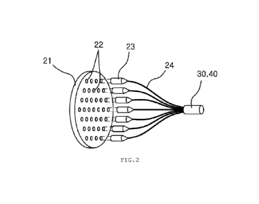
FIG. 2 is a schematic view showing a light supply
device according to an embodiment of the present
invention.
FIGS. 3A to 3C are plane views showing light source
bodies having various individual light source
arrangement forms according to the present invention.
FIGS. 4A and 4B are operational views showing
operations of light condensers according to embodiments
of the present invention.
FIGS. 4C to 4E are plane views showing light supply
devices according to other embodiments of the present
invention.
FIGS. 5A and 5B are plane views showing light exit
surfaces of probes employing an endoscope as a lens.
FIGS. 6A and 6B are block diagrams showing a
controller according to the present invention.
FIGS. 7A and 7B are schematic views showing a
lesion site and a selected interest area on a cervical
image data screen.
FIG. 8 is a schematic view showing a fluorescent
image data screen of the cervix and a selected
fluorescent area.
FIGS. 9A and 9B are schematic views showing two
image data of an interest area and a fluorescent area,
and a target area set by combining the two image data.
FIG. 10A is a view in which light irradiation
regions of respective individual light sources are
matched to a target area according to an embodiment of
the present invention.
Date Recue/Date Received 2021-01-06
11
FIG. 10B is an image view in which only individual
light sources irradiating a target area are selected to
emit lights according to an embodiment of the present
invention.
FIG. 10C is a schematic view depicting that
selective light emission is performed based on the
position of the center of a light irradiation region of
an individual light source according to an embodiment of
the present invention.
FIG. 11A is a block diagram showing the
configuration of a controller including a light output
control module according to an embodiment of the present
invention.
FIG. 11B is a schematic view showing output
intensity setting states of individual light sources by
the relationship between an individual light source and
a target area according to an embodiment of the present
invention.
Mode for Carrying Out the Invention
Hereinafter, embodiments of the present invention
will be described in more detail with reference to the
accompanying drawings. While the present invention can
be variously modified and have alternative forms,
specific embodiments have been shown by way of example
in the drawings and will be described in detail herein.
However, it is to be understood that the present
invention is not intended to be limited to the
particular forms disclosed. Rather, the intention is to
cover all modifications, equivalents, and alternatives
falling within the spirit and scope of the invention as
defined by the claims. In describing each drawing, like
reference numerals are used for like elements. In the
accompanying drawings, the dimensions of the structures
are shown enlarged or reduced in order to clarify the
Date Recue/Date Received 2021-01-06
12
present invention.
The terms used herein are simply used to describe
particular embodiments and are not intended to limit the
present invention. A singular expression includes a
plural expression unless clearly construed in a
different way in the context. It should be understood
that the terms, such as "includes", "comprises", or
"has" are used to specify the presence of features,
numbers, steps, operations, elements, or combinations
thereof described in the specification, rather than
excluding the possibility of presence or addition of one
or more other features, numbers, steps, operations,
elements, or combinations thereof in advance.
Unless otherwise defined, all the terms used
herein, including technical or scientific terms, have
the same meaning as commonly understood by a person
having an ordinary skill in the art to which the present
invention belongs. Terms as defined in dictionaries
generally used should be construed as including meanings
which accord with the contextual meanings of related
technology, and unless clearly defined herein, the terms
should not be construed as having ideal or excessively
formal meanings.
FIG. 1 is a diagram showing a photodynamic therapy
apparatus for local targeting according to a preferable
embodiment of the present invention.
As referenced, a photodynamic therapy apparatus 10
for local targeting according to the present invention
includes: a light supply device 20, an optical cable 30
configured to transfer emitted light; a probe 40
configured to irradiate the transferred light and
receive an image at the front; an image supply device 50
configured to convert the image received from the probe
to image data; a monitor 60 configured to display the
Date Recue/Date Received 2021-01-06
13
converted image data; an input device 70 configured to
receive selection information, and a controller 80
configured to regulate various signals.
The photodynamic therapy apparatus 10 for local
targeting may be provided in the form in which the
respective elements are separated as shown but connected
to each other by connection jacks or some elements are
integrally coupled, and the photodynamic therapy
apparatus 10 may be mounted on a mobile table to thereby
facilitate movement.
Referring to FIG. 2, the light supply device 20
includes: a light source body 21 in which a plurality of
individual light sources 22 are arranged; light
condensers 23 configured to receive and condense lights
irradiated from the light source body; and optical
fibers 24 configured to transfer the lights condensed in
the light condensers. These elements are connected to a
cable 30 or a probe 40.
First, the light source body 21 is a device in
which the plurality of individual light sources 22 are
mounted on a substrate to supply lights by individual
regulation. The light source body may be provided in the
form in which a plurality of individual light sources
are mounted on a single substrate or in the form in
which two or more substrates each having a plurality of
individual light sources are assembled.
The light source body 21 is formed by a plurality
of individual light sources arranged at predetermined
intervals in a substrate. The individual light sources
22 may be arranged in a lattice form on a circular
substrate to perform light irradiation as shown in FIG.
3A, may be arranged in the form of consecutive triangles
to perform light irradiation as shown in FIG. 3B, or may
be arranged in a concentric form in which a plurality of
circles having different diameters are arranged
Date Recue/Date Received 2021-01-06
14
concentrically, to perform light irradiation as shown in
FIG. 3C.
The number of the individual light sources 22
corresponds to the number of optical fibers exposed to a
light exit surface at an end of the probe. When the
number of optical fibers 24 that are disposed is
increased by enlargement of the area of the light exit
surface 421, the individual light sources 22
corresponding thereto can be increasingly disposed to
perform individual light irradiation.
The individual light sources 22 may have any one
type or a mixture of two or more types of a laser diode
(LD), an injection laser diode (ILD), and a light-
emitting diode (LED).
In addition, the individual light sources 22 each
are connected so as to receive power independently, and
a regulation unit controlled by the controller 80 is
installed on each power supply line, so that the turning
on and off of the individual light sources can be
independently regulated and the intensities of the
individual light sources can be individually controlled
by the controller.
Among the plurality of individual light sources 33
arranged, any one may irradiate a visible light to
acquire an internal image of the human body, and another
may irradiate the light to be diffused, thereby checking
the location where a photosensitizer is concentrated in
the inside of the human body. The output of the light
may be lowered to minimize the irritation to the skin
and facilitate fluorescence visualization of the
corresponding skin. Individual light sources offering
such functionality may be disposed in the center of the
light source body or may be deflected to one side,
thereby providing such functionality.
Date Recue/Date Received 2021-01-06
15
Among the individual light sources with
functionality, individual light sources irradiating
visible light may be used as light sources for an
endoscope. That is, while a light is received from the
light source body, an internal image of the human body
may be separately transferred to the image supply
device. In addition, an endoscope line is separately
prepared, so that a light source irradiating visible
light may be provided from the endoscope line itself but
not from the light source body.
In addition, the plurality of light condensers 23
are installed adjacent to the light source body 21. The
light condenser 23 receives a large area of light
irradiated from the individual light source 22 and
condenses the light to one point to allow the light to
exit. A single light condenser 23 is installed to
correspond to a single individual light source 22 as a
set, so individual light irradiation can be achieved
through the optical fibers 24 on a light exit surface
421 of the probe 40.
The light condenser 23 may be differently provided
in a light condensing manner by a condensing lens 232
and in a light condensing manner by a light reflection
surface 231. FIG. 4A is a schematic diagram depicting a
light condensing form by a condensing lens 232. As shown
in FIG. 4A, a condensing lens 232 is formed toward an
individual light source 22, so that a light horizontally
entering from the individual light source is condensed
to one point by the refraction through the condensing
lens 232, and an end of the optical fiber 24 is disposed
at the point to which the light is condensed, so that a
wide area of light from the individual light source is
condensed in a point form, inserted into the end surface
of the optical fiber, and moved along the optical fiber.
Date Recue/Date Received 2021-01-06
16
Alternatively, in FIG. 4B, an inside surface of a
light condenser 23 has a gradually reduced diameter and
formed to be a reflection surface 231, so that a light
horizontally entering from an individual light source 22
is refracted to the center along the reflection surface
231 on the inside of the light condenser 23 and thus
condensed to one point, and the light condensed to one
point is inserted into one end of the optical fiber 24
and moved along the optical fiber.
Therefore, the light condenser 23 is provided in
the form in which one end is disposed adjacent to the
individual light source 22 and the other end is coupled
with the end of the optical fiber 24, so that a wide
area of light exiting from the individual light source
22 can be condensed to one point and transferred to the
optical fiber 24. The individual light sources are
individually regulated, so that the lights irradiated
from the probe to a skin site are controlled by the
optical fiber unit, thereby minimizing the light
irradiation area to normal tissues.
Last, the rear end of the optical fiber 24 is
coupled with the front end of the light condenser 23, so
that the optical fiber 24 receives the condensed light
and allows the condensed light to exit forward through
the front end thereof.
As mentioned above, the light supply device 20
including the light source body 21, the light condensers
23, and the optical fibers 24 is extended to the outside
by the optical cable 30.
The optical cable 30 bundles the plurality of
optical fibers connected to the light condensers into
one bundle and extends the bundled optical fibers to the
outside of the light supply device. The plurality of
optical fibers are bundled inside the light supply
Date Recue/Date Received 2021-01-06
17
device 20, and exposed and extended by the optical cable
30 outside the light supply device.
This optical cable may be partially diverged or may
be joined with another cable at one portion, as
necessary, thereby transmitting various types of signals
or image data, and may be extended by additional
installation of an extension cable through a connector.
The cable that is joined or diverged may be a cable
connected to the image supply device, and a
representative example of the cable may be an endoscope.
In another embodiment of the light supply device of
the present invention, a light switch is installed on
the optical fiber transferring the light condensed by
the light condenser, thereby dividing one entrance light
into a plurality of exit lights.
As shown in FIG. 4C, a light supply device 20a
according to another embodiment of the present invention
includes: a light source body 21, of which the light on
and off and the light intensity are controllable; a
light condenser 23 installed to correspond to the light
source body to condense a light irradiated from the
light source body; an entrance light optical fiber 24a
connected to the light condenser to receive the
condensed light and allow the condensed light to exit to
the other end; a light switch 25 configured to receive
the light from the entrance light optical fiber to
divide the light into a plurality of lights and allow
the divided lights to exit, the divided lights being
individually regulated by the light switch; and a
plurality of exit light optical fibers 24b configured to
receive the lights exiting from the light switch to
allow the lights to exit to the other ends. The
plurality of exit light optical fibers are bundled into
one bundle to be made into an optical cable, which is
then extended to the outside and connected to the probe.
Date Recue/Date Received 2021-01-06
18
Since the light switch can perform the same role as
an individual light source in the previous embodiment,
the light switch is allowed to regulate any one of the
divided lights, thereby producing the same effect as
regulating individual light emission of the individual
light sources. In addition, the number of lights divided
by the light switch may be varied as needed, besides
four divided lights as shown in the drawing.
In addition, as shown in FIG. 4D, the light supply
device 20b may be provided such that a plurality of
light condensers 23 are disposed for a light source body
21 formed of one light source, and light switches 25 are
installed on optical fibers 24a connected to the light
condensers, respectively, so that the division of light
is achieved through a plurality of optical fibers 24b.
As shown in FIG. 4E, in a light supply device 20c,
a plurality of individual light sources 22 are provided
as a light source body 21, and a plurality of light
condensers 23 are disposed for the individual light
sources, respectively, and optical fibers 24a connected
to the respective light condensers are connected to
light switches 25, and the lights divided by each of the
light switches may be transferred through a plurality of
optical fibers 24b.
Hereinafter, embodiments of the present invention
will be described by an example in which optical fibers
are separately regulated through the individual
regulation of individual light sources, but a form in
which light switches are mounted on optical fibers to
provide a function of regulating individual light
sources is included within the right scope of the
present invention.
The optical cable may be divided into the rear end
at the light entrance side and the front end at the
light exit side, and the probe 40 is installed on the
Date Recue/Date Received 2021-01-06
19
front end at the light exit side. The probe 40 includes:
a body 41; an insertion tube 42 protruding from the
front end of the body to be inserted into the human
body; and a lens 43 installed in the front end of the
insertion tube to receive an internal image of the human
body.
The body 41, while containing and fixing the
optical cable, may have an expand end surface to thereby
provide a portion which a user can grasp, or may be
coupled to a separate support or device by forming a
fixing unit.
The insertion tube 42 is a portion that protrudes
forward from the front end of the body, and the
insertion tube is inserted into the human body and
disposed adjacent to a skin as a target of testing or
treatment. It is therefore preferable that the insertion
tube is formed in the form or formed of a material
capable of fixing a position, like an endoscope tube
body.
A light exit surface 421 is formed on the end of
the insertion tube 42. The plurality of optical fibers
24 provided from the optical cable are disposed on the
light exit surface 421. The light exit surface 421 may
allow lights to exit by direct exposure of the end of
the optical cable, or may be provided in the form in
which optical fibers are arranged at predetermined
intervals by a separate support. The light exit surface
may have a structure sealed by a transparent cap in
order to prevent the infiltration of foreign materials,
and when the light exit surface includes an endoscope,
only a portion of the light exit surface, which
corresponds to the endoscope, may be partially opened to
draw out an endoscope device inside.
When the end of the insertion tube is formed to be
a light exit surface having a support, the optical
Date Recue/Date Received 2021-01-06
20
fibers in the light irradiation part of the light exit
surface may be arranged in the same forms as the light
source bodies in FIGS. 3A and 3B. Also, the individual
light sources of the light source body may be arranged
in various forms to achieve light condensation by
individual regulation, and the optical fibers
irradiating the lights through the light exit surface of
the insertion tube may be arranged at predetermined
intervals inside the circular light exit surface to
perform light irradiation according to the shape of a
target area, which is an area in need of treatment.
A lens 43 is further installed in the center or at
one side of the insertion tube 42 to receive an image,
which corresponds to light reflected from the inside of
the human body. In addition, a light irradiation part
emitting visible light but not treatment wavelengths may
be further formed adjacent to the lens to performing
light irradiation for providing a reflection light to
the lens. The light irradiation part may be provided by
any one of the plurality of optical fibers, and an
individual light source corresponding to the
corresponding optical fiber may be configured to
irradiate visible light.
In addition, the lens 43 may be an endoscope. The
endoscope may be used in various forms, such as a form
of having only a lens, a form of having an outlet at one
side, through which a separate surgical instrument can
be withdrawn, or a form of configuring a gas outlet
together. A driving unit for operating the endoscope may
be inserted into a main body of the probe to enable a
precise operation of the endoscope.
The optical fibers inside the probe 40 may be wired
by direct insertion of the optical cable, or the
plurality of optical fibers corresponding to the optical
fiber inside the probe are already wired and the optical
Date Recue/Date Received 2021-01-06
21
cable is connected to the rear end of the probe, so that
the optical fibers of the optical cable may be
correspondingly connected to the optical fibers inside
the probe.
FIGS. 5A and 5B show a light exit surface 421 of a
probe employing an endoscope 43a as a lens. As shown in
FIG. 5A, the endoscope 43a is placed in the center of
the light exit surface of the probe, thereby performing
image acquisition. However, the placement of the
endoscope in the center keeps the individual optical
fibers 24 from being disposed in the endoscope placement
region, and thus individual light irradiation may be
difficult to control at the center of the light
irradiation regions. It is therefore preferable to
secure a wide area of light irradiation regions capable
of individually controlling light irradiation by placing
the endoscope 43a to be deflected to one side on the
light exist surface 421 of the probe, as shown in FIG.
5B.
An image inputted to the lens 43 of the probe is
transmitted to the image supply device 50 and converted
to image data. The image supply device 50 includes an
image sensor to convert a reflective-light type of
image, inputted from the lens, to image data by using a
program.
The image supply device 50 is connected to the
monitor 60 to display the converted image data. The
monitor usually includes all image output devices
capable of outputting images.
An input device 70 is further installed in linkage
with the monitor 60. The input device is used as a unit
configured to enlarge a portion of an output image or
display a portion of the output image. Such an input
device commonly includes keyboards and mice, and
includes touch screen forms combined with monitors, or
Date Recue/Date Received 2021-01-06
22
communication equipments (smartphones and notebooks)
that can be connected by communication.
The controller 80 is configured to process the
image data from the image supply device to allow an
input value of the input device to be contained in the
image data and configured to control the individual
light sources of the light supply device and regulate
signal transmission and power supply for the constituent
elements of the apparatus. Therefore, the controller is
connected to all of the light supply device, the probe,
the image supply device, the monitor, and the input
device, so that the controller analyzes the signals
transmitted from each thereof, to thereby perform
suitable device operations.
FIG. 6A is a block diagram showing a representative
configuration of the controller.
As referenced, the controller 80 of the present
invention includes an image data module 81, an interest
area selection module 81, a target area setting module
84, a light irradiation region setting module 85, and a
local light irradiation module 86.
A typical configuration in the controller is
described on the basis of the execution process with the
cervix as a target site to be treated.
First, the image data module 81 performs a process
of receiving an image or a fluorescent image in a
lighting state and a dark state to allow an image sensor
to convert the image to image data.
The image data module 81 inserts a probe into the
body such that the probe is inserted into the vicinity
of the cervix, which is a site to be treated, supplies
power to any one individual light source, preferably an
individual light source irradiating visible light, to
irradiate a light from a light exit surface at the front
end of the probe, allows a lens to receive an internal
Date Recue/Date Received 2021-01-06
23
image obtained by light irradiation and an image sensor
to convert the image to generate image data, and
transmits and outputs the image data to a monitor.
In addition, a photosensitizer is concentrated in
the cancer or tumor after a predetermined time after
administration, and thus the photosensitizer can be
confirmed in a dark state.
Therefore, the image data module may additionally
receive a fluorescent image by creating a dark state
after acquiring the internal image by visible light.
Also, the image data module may allow the lens to
receive the fluorescent image, allow the image sensor to
convert the fluorescent image to generate fluorescent
image data, and transmit and output the fluorescent
image data to the monitor.
The interest area selection module outputs optical
image data, acquired by visible light, to the monitor
and selects an interest area suspected with cancer or
tumor through an input device connected to the monitor.
The interest area selection module may select a
partial area on the monitor screen by a signal input
through the input device and enlarge and output the
selected area. The enlarged and output screen image may
be processed and corrected without breaks by a known
image editing program.
In FIG. 7A, a lesion site 92 can be defined on an
image screen 90 of the cervix 91. As shown in FIG. 7B,
an operator selects an interest area 93 by using a
mouse, a direct touch, or other selection means. Further
enlargement of a portion can facilitate selection of the
interest area. The selected information is displayed on
the monitor and the selected interest area 93 is stored
in combination with image data.
As shown in FIG. 6B, a process by a fluorescent
area setting module 83 may be further performed. That
Date Recue/Date Received 2021-01-06
24
is, when a fluorescent image is further received by the
image data module 81, the fluorescent area setting
module performs a process of automatically setting
fluorescent areas, which are parts exhibiting
fluorescence, from the fluorescent image data, acquired
in a dark state, by using an image editing program.
As an example of fluorescent area selection, a
fluorescent area with predetermined brightness or higher
may be selected. The fluorescent image data may also be
checked by output to the monitor. The brightness setting
is gradually changed between low brightness and high
brightness through the input device, thereby checking
changes of the automatically selected fluorescent areas
according to the brightness setting and finally
selecting any one of the changed fluorescent areas. When
the automatically selected fluorescent area is
displayed, the corresponding area is selected as a
fluorescent area through the input device using a mouse,
a direct touch, or other selection means, and the
selected information is displayed on the monitor and the
selected fluorescent area is stored in combination with
image data.
FIG. 8 shows fluorescent image data in a dark
state. In most
cases, after a predetermined time, a
photosensitizer is concentrated only in the lesion site
92 and is removed in most of the other regions. However,
in some cases, as shown in the drawing, a small amount
of the photosensitizer may remain in areas other than
the lesion site. An operator may additionally determine
the presence or absence of another lesion site by
comparison with the image data obtained using visible
light. An error of the fluorescent image data is
considered to temporarily appear as if the
photosensitizer is concentrated by a lateral surface
protruding or depressed in the direction of the lens
Date Recue/Date Received 2021-01-06
25
while a three-dimensional conformation is expressed on a
plane. The image data, in which all of the parts
exhibiting fluorescence, including the fluorescent site
shown due to the error, are selected as a fluorescent
area 94, was displayed.
The target area setting module 84 sets the interest
area 93 per se as a target area when only the interest
area is selected by the interest area selection module.
When the fluorescent area is additionally set by
the fluorescent area setting module 83, the target area
setting module 84 overlaps two image data, to which the
interest area 93 and the fluorescent area 94 are
applied, and sets the overlapped area as a target area
95 to be irradiated with light.
Since the images in the lighting state and the dark
state are acquired at approximately the same time, the
magnification and the location in the cervix can be
considered to be almost identical between the two
images. However, when the magnifications are different
by image enlargement during the selection of the
interest area and the fluorescent area, it is preferable
that conversion is performed at the same magnification
through a previously known graphic program and then a
target area is set by overlapping the two image data.
FIG. 9A shows image data with an interested area 93
selected and image data with a fluorescent area 94 set,
and FIG. 9B shows an image in which a target area 95 is
set by converting two image data at the same
magnification and then arranging the two to overlap each
other to display an overlapping area of the interest
area 93 and the fluorescent area 94.
The light irradiation region setting module 85
performs a process of checking respective light
irradiation regions 96 of the individual light sources
through the light exit surface of the probe. That is, in
Date Recue/Date Received 2021-01-06
26
the set arrangement structure, the individual light
sources are sequentially allowed to emit lights to check
which regions of the cervix are irradiated with the
lights irradiated from the individual light sources,
respectively, and the individual light sources and the
light irradiation regions are matched and stored.
The local light irradiation module 86, upon the
completion of the setting of the light irradiation
regions 96, supplies power to only individual light
sources, which perform light irradiation on the set
target area 95, to achieve partial light emission, and
such partial light emission enables a treatment while
the light irradiation to the normal tissues is
minimized.
FIG. 10A is a view in which the target area 95 is
matched to the light irradiation regions 96 of the
individual light sources. In FIG. 10B, only individual
light sources corresponding to the light irradiation
regions 96 overlapping at least a portion of the target
area 95 were supplied with power and operated, and the
other individual light sources were turned off, thereby
minimizing damage to the normal tissue.
As for a method of controlling the plurality of
individual light sources, the individual light sources
may be selectively supplied with power based on whether
or not the center of a light irradiation region 96 of an
individual light source is included in the target area
95. As shown in FIG. 10C, the individual light sources
can be allowed to selectively emit lights by a method in
which some lights are irradiated to the target area 95
wherein the individual light sources corresponding to
the light irradiation regions 96 with centers deviating
from the target area are powered off and only the
individual light sources with centers included in the
target area 95 are powered on. Such a method of
Date Recue/Date Received 2021-01-06
27
determining whether an individual light source is
operated according to the center of the light
irradiation region should be applied when the light
irradiation regions of the individual light sources
partially overlap, preferably adjacent light irradiation
regions overlap by at least 40% in diameter, and in such
cases, the target area can be included in the overall
light irradiation regions of the operating individual
light sources even on the basis of the centers of the
light irradiation regions of the individual light
sources.
As for another method of controlling the plurality
of individual light sources, the minimization of the
area of a plurality of light irradiation regions
encompassing the entire target area is calculated by a
program and then only individual light sources
corresponding to the calculated light irradiation area
are selectively supplied with power to emit lights. That
is, light irradiation regions of all of individual light
sources which irradiate light to at least a portion of
the target area are first selected, and among the
selected light irradiation regions of the individual
light sources, light irradiation regions of individual
light sources, which, even though removed, can be
compensated for by other light irradiation regions to
result in light irradiation to the target area, are
second selected and removed, so that the light
irradiation regions overlapping the target area are
finally selected and only the individual light sources
corresponding to the selected light irradiation regions
are operated to emit lights.
The local light irradiation module 86 may set the
light irradiation regions by moving a probe back and
forth according to the size of the target area and then
selectively operating the individual light sources,
Date Recue/Date Received 2021-01-06
28
besides a method in which a plurality of individual
light sources corresponding to the target area are
selected while the probe is fixed. The probe may be
moved forward and backward and the target area may be
irradiated with a plurality of lights at one time, or
the target area may be divided into a plurality of areas
and then the probe may be moved to the divided target
areas to perform light irradiation.
The controller 80 may further include a light
output control module 87 as shown in FIG. 11A. The light
output control module 87 may be set as a sub-module
subordinating to the local light irradiation module.
The light output control module 87 controls the
output intensity of each of the individual light
sources, of which the turning on and off is determined,
thereby irradiating a normal tissue and a target area-
forming tissue with lights of different intensities, so
that the normal tissue adjacent to the target area is
minimally damaged and the cancer or tumor in the target
area are irradiated with lights of strong intensities.
The output intensities are set in advance such that
light irradiation may be performed by stages according
to the set values.
The light output intensity is differentiated
according to the degree to which the light irradiation
region of the individual light source overlaps the
target area. For example, the light irradiation may be
performed such that the output intensity of an
individual light source is set to be high when a light
irradiation region of the corresponding individual light
source is included in the target area by 50-100%, and
the output intensity of an individual light source is
set to be low when a light irradiation region of the
corresponding individual light source is contained in
the target area by 50% or less.
Date Recue/Date Received 2021-01-06
29
Referring to FIG. 11B, when a light irradiation
region 96 (st-on) of an individual light source is
entirely included in the target area 95, the output
intensity of the corresponding individual light source
is increased. When a light irradiation region 96 (off)
of an individual light source contains a portion of the
target area but can be compensated for by other light
irradiation regions, the corresponding individual light
source is powered off to remove the light irradiation
region. When a light irradiation region 96 (on) of an
individual light source contains a portion of the target
area but cannot be compensated for by other light
irradiation regions, the corresponding individual light
source is powered on and the output intensity of the
corresponding individual light source is maintained to
be medium or low.
The embodiments of the present invention have been
described for cervical cancer, but the present invention
can be used in photodynamic therapies for various types
of cancers and tumors to which local treatment is
applicable due to small-sized lesion sites, including
other female cancers (endometrial cancer, ovarian
cancer, and breast cancer), skin cancer, and brain
cancer.
According to a control method for the photodynamic
therapy apparatus for local targeting according to the
present invention, the target area, which corresponds to
a tissue to be treated, is defined and treated by
controlling power transmitted to each of the plurality
of individual light sources through image analysis of
tissue surface.
Specifically, the method may include: an image data
step of receiving an image in a lighting state to allow
the image sensor to convert to the image to image data;
an interest area selection step of outputting the image
Date Recue/Date Received 2021-01-06
30
data, converted from the image in the lighting state, to
the monitor, allowing the input device to select a
suspected interest area 93, and applying the interest
area to the image data; a target area setting step of
setting the interest area as a target area 95 to be
irradiated with lights; a light irradiation region
setting step of checking light irradiation regions 96 of
individual light sources through the light exit surface
of the probe; and a local light irradiation step of
selecting individual light sources, which are to
irradiate the set target area with lights, and supplying
power to the selected individual light sources to emit
lights.
In the image data step, a process of receiving a
fluorescent image in a dark state to allow the image
sensor to convert the fluorescent image to image data is
further performed. In a fluorescent area setting step, a
fluorescent area 94 with predetermined brightness or
higher is automatically set from the image data
converted from the image in the dark state.
In addition, in the target area setting step,
images, to which the interest area 93 and the
fluorescent area 94 are applied, are allowed to overlap
each other to set an overlapping area as a target area
95.
In the local light irradiation step, individual
light sources to be supplied with power are set based on
whether the center of a light irradiation region 96 of
an individual light source is included in the target
area 95.
In the local light irradiation step, the
minimization of the area of light irradiation regions
encompassing the entire target area 95 is calculated and
then individual light sources to be supplied with power
are set based on whether light irradiation regions
Date Recue/Date Received 2021-01-06
31
having a calculated area are irradiated with lights of
the individual light sources.
The method may further include a light output
control step of controlling the output intensity of each
of individual light sources, of which the turning on and
off is determined.
In the light output control step, by, among the
selected individuals, setting the output intensity of an
individual light source to be increased when a light
irradiation region 96 of the individual light source is
included in the target area by 50-100% and setting the
output intensity of an individual light source to be
reduced when a light irradiation region of the
individual light source is included in the target area
by 50% or less, the light output intensity may be
differently applied according to the degree to which a
light irradiation region 96 of an individual light
source overlaps the target area 95.
Date Recue/Date Received 2021-01-06