Note: Claims are shown in the official language in which they were submitted.
What is claimed:
1. A method of identifying a plant protoplast that lacks pathogen
resistance, the method
comprising:
introducing a first fluidic medium containing one or more protoplasts into a
microfluidic device comprising an enclosure having a flow region and at least
one growth
chamber;
moving a first protoplast of the one or more protoplasts into a first growth
chamber of
the at least one growth chamber;
contacting the first protoplast with a pathogenic agent; and
monitoring viability of the first protoplast during a first time period after
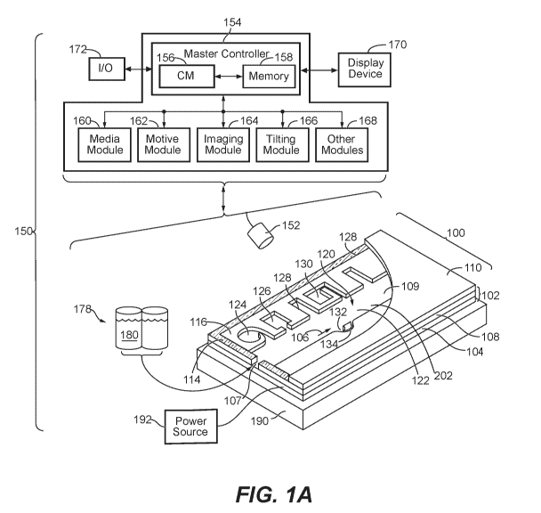
contacting
the first protoplast with the pathogenic agent,
wherein protoplast viability at the end of the first time period indicates
that the
protoplast lacks resistance to the pathogenic agent.
2. The method of claim 1, wherein the one or more protoplasts are from a
broad acre
crop plant.
3. The method of claim 2, wherein the broad acre crop plant is a wheat,
corn, soy, or
cotton plant.
4. The method of claim 1, wherein the one or more protoplasts are from a
high value or
ornamental crop plant.
5. The method of claim 4, wherein the high value crop plant is a tomato,
lettuce, pepper,
or squash plant.
6. The method of claim 1, wherein the one or more protoplasts are from a
turf or forage
plant.
7. The method of claim 6, wherein the turf or forage plant is a grass or
alfalfa plant.
8. The method of claim 1, wherein the one or more protoplasts are from an
experimental
plant.
9. The method of any one of claims 1 to 8, wherein the pathogenic agent is
a plant
pathogen or a molecule derived therefrom.
92
10. The method of claim 9, wherein the plant pathogen is a virus, a
bacterium, or a fungal
cell.
11. The method of claim 9, wherein the pathogenic agent is a molecular
agent or a
fragment thereof
12. The method of any one of claims 1 to 8, wherein contacting the first
protoplast with
the pathogenic agent comprises flowing a second fluidic medium containing the
pathogenic
agent into the flow region of the microfluidic device.
13. The method of claim 12, wherein contacting the first protoplast with
the pathogenic
agent further comprises moving the pathogenic agent into the isolation region
of the first
growth chamber or allowing the pathogenic agent to diffuse from the flow
region into the
isolation region of the first growth chamber.
14. The method of any one of claims 1 to 8, wherein said enclosure further
comprises a
base, a microfluidic circuit structure disposed on the base, and a cover.
15. The method of claim 14, wherein the cover and the base are part of a
dielectrophoresis
(DEP) mechanism for selective inducing DEP forces on micro-objects, and
wherein moving
the first protoplast into the first growth chamber comprises applying DEP
force on the first
protoplast.
16. The method of any one of claims 1 to 8, wherein the microfluidic device
further
comprises a first electrode, an electrode activation substrate, and a second
electrode, wherein
the first electrode is part of a first wall of the enclosure and the electrode
activation substrate
and the second electrode are part of a second wall of the enclosure, wherein
the electrode
activation substrate comprises a photoconductive material, semiconductor
integrated circuits,
or phototransistors, and wherein moving the first protoplast into the first
growth chamber
comprises applying DEP force on the first protoplast.
17. The method of claim 16, wherein the first wall is a cover, and wherein
the second wall
is a base.
18. The method of claim 16, wherein the electrode activation substrate
comprises
phototransistors.
93
19. The method of claim 16, wherein the cover and/or the base is
transparent to light.
20. The method of any one of claims 1 to 8, wherein the first growth
chamber is a
sequestration pen that comprises an isolation region and a connection region
that fluidically
connects the isolation region to the flow region, and wherein the isolation
region is an
unswept region of the micro-fluidic device.
21. The method of claim 20, wherein the enclosure further comprises a
microfluidic
channel comprising at least a portion of the flow region, wherein the
connection region of the
sequestration pen comprises a proximal opening into the microfluidic channel
having a width
W con ranging from about 50 microns to about 150 microns and a distal opening
into the
isolation region, and wherein a length L con of the connection region from the
proximal
opening to the distal opening is as least 1.0 times the width W con of the
proximal opening of
the connection region.
22. The method of claim 21, wherein the length L con of the connection
region from the
proximal opening to the distal opening is at least 1.5 times the width W con
of the proximal
opening of the connection region.
23. The method of claim 21, wherein the length L con of the connection
region from the
proximal opening to the distal opening is at least 2.0 times the width W con
of the proximal
opening of the connection region.
24. The method of claim 21, wherein the width W con of the proximal opening
of the
connection region ranges from about 50 microns to about 100 microns.
25. The method of claim 21, wherein the length L con of the connection
region from the
proximal opening to the distal opening is between about 50 microns and about
500 microns.
26. The method of claim 21, wherein a height H ch of the microfluidic
channel at the
proximal opening of the connection region is between 20 microns and 100
microns.
27. The method of claim 21, wherein a width W ch of the microfluidic
channel at the
proximal opening of the connection region is between about 50 microns and
about 500
microns.
94
28. The method of claim 20, wherein the volume of the isolation region of
the
sequestration pen ranges from about 5x10 5 to about 5x10 6 cubic microns.
29. The method of claim 20, wherein the volume of the isolation region of
the
sequestration pen ranges from about 1x10 6 to about 2x10 6 cubic microns.
30. The method of claim 20, wherein the proximal opening of the connection
region is
parallel to a direction of bulk flow in the flow region.
31. The method of any one of claim 1 to 8, wherein monitoring viability of
the first
protoplast during the first time period comprises monitoring cell division of
the first
protoplast, and wherein cell division of the first protoplast indicates that
the protoplast lacks
resistance to the pathogenic agent.
32. The method of any one of claims 1 to 8, wherein monitoring viability of
the first
protoplast during the first time period comprises maintaining the microfluidic
chip at a
temperature of about 20°C to about 30°C during the first time
period and/or minimizing the
amount of light to which the first protoplast is exposed during the first time
period.
33. The method of any one of claims 1 to 8, wherein monitoring viability of
the first
protoplast during the first time period comprises periodically perfusing
protoplast growth
medium through the flow region of the microfluidic device during the first
time period.
34. The method of claim 33, wherein the protoplast growth medium is
perfused through
the flow region no more than once every three days.
35. The method of any one of claims 1 to 8, wherein monitoring viability of
the first
protoplast during the first time period comprises staining the first
protoplast with a cell
viability dye.
36. The method of any one of claims 1 to 8, wherein monitoring viability of
the first
protoplast during the first time period comprises staining the first
protoplast with a
chlorophyll stain and/or a cell wall stain.
37. The method of any one of claims 1 to 8, wherein the first time period
is at least 12
hours.
38. The method of claim 37, wherein the first time period is at least 96
hours.
39. The method of any one of claims 1 to 8 further comprising:
determining that the first protoplast lacks resistance to the pathogenic
agent; and
exporting the first protoplast from the first growth chamber and the
microfluidic
device.
40. The method of any one of claims 1 to 8 further comprising:
determining that the first protoplast lacks resistance to the pathogenic
agent; and
sequencing one or more disease resistance genes of the first protoplast.
41. The method of any one of claims 1 to 8 further comprising:
determining that the first protoplast lacks resistance to the pathogenic
agent; and
sequencing the transcriptome of the first protoplast.
42. The method of any one of claims 1 to 8 further comprising:
determining that the first protoplast lacks resistance to the pathogenic
agent; and
sequencing the genome of the first protoplast.
43. The method of claim 40 further comprising:
identifying a molecular change or defect in the sequence of one or more
disease
resistance genes, the transcriptome, and/or the genome associated with the
lack of pathogen
resistance.
44. The method of any one of claims 1 to 8, the method further comprising:
moving at least one protoplast into each of a plurality of growth chambers in
the
microfluidic device; and
performing the remaining steps of the method on each of the protoplasts moved
into
the plurality of growth chambers.
45. A kit for screening a plant protoplast for a disease resistance trait,
the kit comprising:
a microfluidic chip, wherein the microfluidic chip comprises an enclosure
having a
flow region and at least one growth chamber; and
a reagent for detecting viability of the plant protoplast.
46. The kit of claim 45 further comprising a surface conditioning reagent.
47. The kit of claim 45 further comprising a conditioning modification
reagent, and
wherein at least one surface of the growth chamber comprises a surface
modifying ligand.
96
48. The kit of claim 45, wherein at least one surface of the growth chamber
comprises a
covalently linked coating material.
49. The kit of any one of claims 45 to 48, wherein the reagent for
detecting the viability
of the plant protoplast is a fluorescent stain.
97
