Note: Descriptions are shown in the official language in which they were submitted.
CA 03105282 2020-12-28
WO 2020/039442
PCT/IL2019/050941
CATHETER ULTRASOUND TRANSDUCER CONTAINER
CROSS REFERENCE TO RELATED APPLICATIONS
[0001] This application claims the benefit of priority to U.S. Provisional
Patent
Application No. 62/720,995, filed August 22, 2018, entitled "CATHETER
ULTRASOUND
TRANSDUCER CONTAINER". The contents of the above application is all
incorporated
by reference as if fully set forth herein in its entirety.
FIELD OF THE INVENTION
[0002] The invention, in some embodiments thereof, relates to catheter
ultrasound (US)
transducers.
BACKGROUND
[0003] Catheter ablation is a procedure used to remove or terminate a faulty
electrical
pathway from sections of the heart, especially in those who are prone to
developing cardiac
arrhythmias and to restore the heart to its normal rhythm. Ablation procedures
are
commonly carried out by radiofrequency (RF) ablation and cryoablation.
[0004] Catheter ablation is a specialist catheter-based procedure that ablates
abnormal
heart muscle tissue. The procedure is used particularly in patients whose
cardiac arrhythmia
cannot be controlled with medication.
[0005] Catheter ablation involves advancing several flexible catheters into
the
patient's blood vessels, usually either in the femoral vein, internal jugular
vein,
or subclavian vein. The catheters are then advanced towards the heart.
Electrical impulses
are then used to induce the arrhythmia and local heating or freezing is used
to ablate the
abnormal tissue that is causing it. Catheter ablation is usually performed by
an electrophysiologist (a specially trained cardiologist) in a catheter lab or
a specialized EP
lab.
1
CA 03105282 2020-12-28
WO 2020/039442
PCT/IL2019/050941
[0006] The foregoing examples of the related art and limitations related
therewith are
intended to be illustrative and not exclusive. Other limitations of the
related art will become
apparent to those of skill in the art upon a reading of the specification and
a study of the
figures.
SUMMARY
[0007] There is provided, in accordance with some embodiments of the invention
a
catheter US transducer container, including a housing, one or more cooling
channels,
oriented longitudinally along a longitudinal axis of the container, a sealing
cooling channel
cover, one or more PE elements positioned on a floor of the cooling channel
and having an
emitting surface facing the cover, wherein the cooling channel has a trapezoid
cross section
at any point along the PE element.
[0008] In some embodiments, an emitting surface of at least one PE element is
oriented
in parallel to the cooling channel cover. In some embodiments, the floor
includes the short
base of the trapezoid. In some embodiments, the housing further includes at
least one fluid
inlet opening to a proximal end of the cooling channel and at least one fluid
outlet located
at a distal end of the cooling channel and/or located inside the fluid
collecting and diverting
chamber and a fluid collecting and diverting chamber coupled to a distal end
of the cooling
channel.
[0009] In some embodiments, the housing includes at least one post coupled to
the floor
of the cooling channel and supports the PE element, forming a gap between the
floor and
the PE element. In some embodiments, the PE element is angled with respect to
the floor of
the cooling channel. In some embodiments, the cooling channel cover and the
emitting
surface of the PE element are parallel. In some embodiments, the cooling
channel is
configured to promote laminar flow of fluid flowing between the cover and the
emitting
surface of the PE element.
[0010] In some embodiments, the rate of flow of the fluid flowing the cooling
channel is
adjusted to the viscosity of the fluid and fluid velocity within the cooling
channel is
maintained below a threshold at which it becomes turbulent. In some
embodiments, in
2
CA 03105282 2020-12-28
WO 2020/039442
PCT/IL2019/050941
operation, the laminar flow promoted by the geometry and dimensions of the
fluid channel.
In some embodiments, the laminar flow effected by the geometry and design of
the cooling
channel forms a temperature gradient in the fluid in the cooling channel along
a distance
(L) between the emitting surface of the PE element and the fluid channel cover
and the
temperature gradient maintains a temperature at the cooling fluid channel
cover at or below
temperature of blood surrounding the container.
[0011] In some embodiments, the container includes a plurality of PE elements
angled
with respect to one another. In some embodiments, the container includes a
plurality of PE
elements at least one of which is angled with respect to the cooling channel
floor. In some
embodiments, at least one of the PE elements is an ablative PE element and at
least one of
the PE elements is an imaging PE element. In some embodiments, a depth (d) of
the cross-
section of the cooling channel is smaller than the radius of the housing. In
some
embodiments, a diameter of the container is unchangeable. In some embodiments,
the
housing includes at least one temperature sensor.
[0012] In some embodiments, the PE element includes a first and a second
electrodes, the
first electrode disposed along the emitting surface and a second electrode
disposed along an
opposite surface of PE element, wherein the PE element includes a first and a
second
electrodes, the first electrode is disposed along at least a portion of the PE
element emitting
surface and around one end of the PE element and a second electrode disposed
along at least
a portion of an opposite surface of PE element and around an opposite end of
the PE
element.
[0013] In some embodiments, the electrodes are isolated from each other by at
least one
gap on the emitting surface and the opposite surface of the PE element,
wherein the at least
one gap is bridged by an insulating adhesive.
[0014] In some embodiments, the catheter includes at least one positioner. In
some
embodiments, the positioner is in a form of a basket. In some embodiments, and
as shown
in Fig. 7E, the positioner is in a form of a coil. In some embodiments, and as
shown in Figs.
7F and 7G, the positioner is in a form of an umbrella. In some embodiments,
the positioner
may comprise and opening facing towards US transducer container 100 (Fig. 7F).
In some
embodiments, the positioner may comprise and opening facing away from US
transducer
container 100 (Fig. 7G). In some embodiments, the positioner includes at least
one opening.
3
CA 03105282 2020-12-28
WO 2020/039442
PCT/IL2019/050941
In some embodiments, the positioner 702 is non-occluding. In some embodiments,
the
positioner 702 is disposed over the container. In some embodiments, the
container is
disposed between two positioners 702.
[0015] In some embodiments, the container is rotatable about the catheter. In
some
embodiments, the container includes a beam collimating acoustic lens. In some
embodiments, the beam collimating acoustic lens is configured to collimate an
US beam
and generate a jet effect in surrounding blood along the beam pathway through
the blood.
In some embodiments, the collimating acoustic lens is configured to direct the
jet effect
towards and cool the ablated tissue.
[0016] In some embodiments, the catheter includes a medicament outlet in
propinquity to
the container and wherein the collimating acoustic lens is configured to
direct the jet effect
towards and drive the medicament into tissue.
[0017] In some embodiments, the processor is configured to adjust the level of
energy
emitted from the PE element based on at least one of distance measured from
the emitting
surface of the PE element to the tissue wall, tissue thickness, duration of
energy delivery,
change in amplitude and/or phase of ultrasound signal returning from the
tissue and
reduction of recorded electrical potential signals. In some embodiments, the
processor is
configured to adjust fluid flow velocity in the cooling channel based on the
temperature
reading and beam energy level. In some embodiments, the cooling fluid channel
includes a
fluid inlet and wherein the processor is configured to adjust fluid
temperature at the inlet
based on the temperature reading and beam energy level.
[0018] In some embodiments there is provided a method of manufacture of a
catheter US
transducer container , including molding a housing having at least one cooling
fluid channel,
at least one PE element mounting post, and at least one wiring conduit, laying
electrical and
data wiring inside the wiring conduits, mounting at least one PE element on
the at least one
mounting post and within designated cavities in cooling channel and connecting
wiring,
sealing a perimeter of the PE element to walls of the cooling channel
attaching a cooling
fluid collecting and diverting chamber to a distal end of the housing, and
placing an
insulating cover over the housing and cooling channel, shrinking the cover and
tightly
sealing the housing and the cooling channel.
4
CA 03105282 2020-12-28
WO 2020/039442
PCT/IL2019/050941
[0019] The following embodiments and aspects thereof are described and
illustrated in
conjunction with systems, tools and methods which are meant to be exemplary
and
illustrative, not limiting in scope.
[0020] In addition to the exemplary aspects and embodiments described above,
further
aspects and embodiments will become apparent by reference to the figures and
by study of
the following detailed description.
BRIEF DESCRIPTION OF THE FIGURES
[0021] Exemplary embodiments are illustrated in referenced figures. Dimensions
of
components and features shown in the figures are generally chosen for
convenience and
clarity of presentation and are not necessarily shown to scale. The figures
are listed below.
[0022] Figs. 1A, 1B, 1C, 1D, 1E, 1F, 1G, 1H, 11, 1J and 1K are perspective
view and
cross section view simplified illustration of an US transducer container,
according to some
embodiments of the invention;
[0023] Figs. 2A, 2B and 2C are cross section view simplified illustrations of
the US
transducer container cooling system in accordance with some embodiments of the
current
invention;
[0024] Fig. 3A is a perspective view simplified illustration of US transducer
container
cooling system and Figs. 3B and 3C are graphs demonstrating heat distribution
within the
cooling system in accordance with some embodiments of the invention;
[0025] Figs. 4A and 4B are longitudinal cross-section view and transverse
cross section
view simplified illustrations of the effect of laminar cooling fluid flow on
bubbles in
accordance with some embodiments of the current invention;
[0026] Figs. 5A, 5B, SC, 5D, 5E, 5F, 5G, 5H and 51, which are perspective view
and cross
section view simplified illustration of method of manufacturing a container
transducer in
accordance with some embodiments of the invention;
[0027] Fig. 6 is a flow chart of a method of manufacture and assembly of an US
transducer
container in accordance with some embodiments of the invention;
CA 03105282 2020-12-28
WO 2020/039442
PCT/IL2019/050941
[0028] Figs. 7A, 7B, 7C, 7D, 7E, 7F, 7G and 7H are plan view and perspective
view
simplified illustrations of a positioner 702 for an US transducer container in
accordance
with some embodiments of the invention; and
[0029] Fig. 8 is a cross section view simplified illustration of a jet effect
generated by an
US transducer container in accordance with some embodiments of the invention;
[0030] Fig. 9 is a transverse cross-section simplified illustration of a
multidirectional US
transducer container, according to some embodiments of the invention; and
[0031] Figs. 10A and 10B which are perspective view simplified illustrations
of a
combination US transducer/RF electrode catheter container, according to some
embodiments of the invention.
DETAILED DESCRIPTION
[0032] According to an aspect of some embodiments of the invention there is
provided a
catheter US transducer having one or more PiezoElectric (PE) elements
(ceramics) and one
or more cooling systems that regulate the temperature of the transducer and/or
volumes
adjacent to the US transducer (e.g., cooling fluid). According to some
embodiments, the US
transducer and the cooling system are housed within a container. In some
embodiments, the
container comprises one or more apertures.
[0033] In some embodiments, the cooling systems comprises cooling fluid. In
some
embodiments, the cooling system is circulated within the container. In some
embodiments,
the container is sealed from the environment. In some embodiments, the cooling
fluid does
not contact fluid surrounding the catheter and/or the container. In some
embodiments, the
external diameter of the container is smaller than the external diameter of
the catheter. In
some embodiments, the external diameter of the container is the same as the
external
diameter of the catheter. In some embodiments, the external diameter of the
container is
larger than the external diameter of the catheter. In some embodiments, the
temperature of
the external surface of the US transducer container is maintained below 45 C.
In some
embodiments, the container is rigid. In some embodiments, the external
diameter of the US
transducer container is unchanged during operation.
6
CA 03105282 2020-12-28
WO 2020/039442
PCT/IL2019/050941
[0034] According to an aspect of some embodiments of the invention there is
provided an
US transducer container sized and fitted to be positioned along a catheter
and/or within a
delivery catheter. In some embodiments, the US transducer emitting surface
comprises a
plane one dimension of which is oriented in parallel to a longitudinal axis of
the catheter.
In some embodiments, the US transducer emitting surface comprises a plane one
dimension
of which is angled with respect to the longitudinal axis of the catheter. In
some
embodiments, the US transducer comprises a plurality of emitting surfaces, in
which at least
one emitting surface comprises a plane one dimension of which is oriented in
parallel to a
longitudinal axis of the catheter and at least a second emitting surface
comprises a plane
having at least one dimension that is angled with respect to the longitudinal
axis of the
catheter. In some embodiments, the US transducer comprises a plurality of
emitting
surfaces, in which at least two emitting surfaces are angled with respect to
the longitudinal
axis of the catheter. In some embodiments, the at least two emitting surfaces
are inclined
towards each other with respect to the longitudinal axis of the catheter.
[0035] According to an aspect of some embodiments of the invention there is
provided an
US transducer container sized and fitted to be positioned along a catheter
and/or within a
delivery catheter. In some embodiments, the US transducer container comprises
a
collimating acoustic lens. In some embodiments, the US transducer emits a
collimated
beam. In some embodiments, the collimated beam generates one or more jets in
the blood
stream (a jet effect). In some embodiments, a collimated beam generates the
jet effect in
surrounding blood along the beam pathway through the blood. In some
embodiments, the
generated jets are at the same temperature as the medium in which they are
generated.
[0036] According to an aspect of some embodiments of the invention there is
provided
one or more US transducer positioners 702s. In some embodiments, the
positioner 702 is in
a form of a basket. In some embodiments, the positioner 702 is in a form of a
cage. In some
embodiments, the positioner 702 is in a form of a coil. In some embodiments,
the transducer
catheter comprises two positioners 702s disposed one on either side of the US
transducer
container. In some embodiments, the positioner 702 is made of a shape memory
alloy. In
some embodiments, the positioner 702 envelops the US transducer container. In
some
embodiments, the positioner 702 comprises an aperture. In some embodiments,
the diameter
of the aperture is greater than the diameter of the US beam emitted through
the aperture.
7
CA 03105282 2020-12-28
WO 2020/039442
PCT/IL2019/050941
[0037] According to some embodiments of the invention, the catheter comprises
one or
more therapeutic agent delivery nozzles configured to deliver a therapeutic
agent into a
volume within an emitted US beam. In some embodiments, the US transducer emits
collimated beam energy. In some embodiments, the collimated beam energy
generates one
or more jets in the blood stream (a jet effect) that drive the therapeutic
agent via the jet
stream towards the tissue surface.
General
[0038] Reference is now made to Figs. 1A, 1B, 1C, 1D, 1E, 1F, 1G, 1H, 11, 1J
and 1K,
which are perspective view and cross section view simplified illustrations of
a catheter US
transducer container according to some embodiments of the invention. According
to some
embodiments of the invention there is provided a catheter US transducer 175
housed in
container 100. In some embodiments, container 100 comprises one or more
cooling systems
200 that regulate the temperature of the transducer 175 by streaming cooling
fluid over and
around the US transducer. According to some embodiments, the US transducer 175
and the
cooling system 200 are housed within the container 100. In some embodiments,
the
container 100 comprises one or more apertures 118. In some embodiments, one or
more of
the apertures 118 comprise one or more blood-contact surfaces 116.
[0039] In some embodiments, the container 100 is fluidly sealed from the
environment.
In some embodiments, the cooling fluid does not contact fluid surrounding the
catheter
and/or the container 100. In some embodiments, the external diameter of the
container 100
is smaller than the external diameter of the catheter 106. In some
embodiments, the external
diameter of the container 100 is the same as the external diameter of the
catheter. In some
embodiments, the external diameter of the container is larger than the
external diameter of
the catheter 106. In some embodiments, the temperature of the external surface
of the US
transducer container 100 is maintained below 45 C.
[0040] In some embodiments, US transducer container 100 is attached to a
catheter 106
end and functionally coupled to one or more sources of cooling fluid, power
(e.g., electric
power), vacuum and unidirectional and/or bidirectional data communication
conduits.
Catheter 106 comprises a main lumen 126. The term "Cooling Fluid" as used
herein relates
8
CA 03105282 2020-12-28
WO 2020/039442
PCT/IL2019/050941
to a fluid having a temperature configured to maintain a temperature of a
blood-contact
surface 116 no higher than the surrounding blood temperature.
[0041] In some embodiments, the US transducer 175 container 100 is mounted at
a distal
end of a catheter 106. In some embodiments, the US transducer container 100 is
mounted
proximally to the catheter tip. In some embodiments, the external diameter of
the US
transducer container 100 is unchanged before, during and/or post operation.
[0042] As used herein the term "Proximal" means closer to the user of the US
catheter
and the term "Distal" means closer to the tip of the US catheter. The term
"proximally"
means towards the user of the US catheter and the term "Distally" means away
from the
user of the US catheter and towards the tip of the US catheter.
[0043] In some embodiments, and as shown in the exemplary embodiments depicted
in
Figs. 1A and 1B, catheter ultrasound transducer container 100 has a
cylindrical geometry
and comprises a housing 502. In some embodiments, at least one or more
portions of
housing 502 are solid. In some embodiments, housing 502 comprises one or more
hollow
conduits that provide passageways for example, for electrical and/or data
communication
wiring, a coolant, medicament and/or any other fluid from a source to the US
transducer
container 100. In some embodiments, a solid portion of housing 502 fills over
50% of the
cross-section of housing 502. In some embodiments, the solid portion of
housing 502 fills
between 50% and 75% of the cross-section of housing 502.
[0044] In some embodiments, housing 502 comprises one or more trough-form
cooling
channels 120, disposed longitudinally along a longitudinal axis of container
100 and
catheter 106 and configured to promote laminar fluid flow. In some
embodiments, cooling
channel 120 comprises a trapezoid cross-section (Fig. 2A) defined by a floor
108 and walls
122/124, on lateral sides of floor 108 forming an obtuse angle between floor
108 and walls
122/124. In some embodiments, walls 122/124 are positioned parallel to the
longitudinal
axis of container 100 and catheter 106. In some embodiments, the trapezoid is
an isosceles
trapezoid. In some embodiments, floor 108 comprises the short base of the
trapezoid.
[0045] In some embodiments, housing 502 comprises one or more posts 102 that
protrude
from floor 108 and support one or more piezoelectric (PE) elements 140 Forming
a gap
between PE element 140 and floor 108. The length of cooling channel 120 is at
least the
9
CA 03105282 2020-12-28
WO 2020/039442
PCT/IL2019/050941
same as the length of PE element 140. In some embodiments, cooling channel 120
has a
trapezoid cross section at any point along at least one or more PE elements
140.
[0046] In some embodiments, catheter ultrasound transducer container 100
cooling
channel 120 comprises one or more cooling fluid inlets 152 disposed at a
proximal and of
cooling channel 120. Cooling channel 120 opens distally to a fluid (e.g.,
coolant) cooling
fluid diverting chamber 156. In some embodiments, housing 502 comprises a
cooling fluid
outlet 154 disposed at a distal end of cooling channel 120 and/or inside a
fluid cooling fluid
diverting chamber 156. In some embodiments, fluid cooling fluid diverting
chamber 156 is
configured to collect fluid flowing through cooling channel 120 over an
emitting surface
142 of PE element 140 and exiting from the distal end thereof, and divert the
fluid to drain
into fluid outlet 154 and catheter 106 to a fluid collection reservoir.
[0047] In some embodiments, catheter US transducer container 100 is fluidly
sealed and
isolated from the surroundings e.g., blood. In some embodiments, catheter US
transducer
container 100 comprises a sealing cooling channel cover 130. In some
embodiments, and
as explained in greater detail herein, cover 130 comprises at least two
surfaces: a PE element
140-facing surface and a blood contact surface 116 facing away from PE element
140. In
some embodiments, fluid (e.g., coolant) inlet 152 disposed between cover 130
and the
emitting surface 142 of PE element 140. In some embodiments, cover 130 is
parallel to
emitting surface 142 of PE element 140. In some embodiments, fluid flowing
from inlet 152
through cooling channel 120 and between two flat surfaces of PE element 140
and cover
130 flows at a laminar flow. The rate of flow of the coolant fluid is adjusted
to the viscosity
of the fluid and fluid velocity is maintained below a threshold at which it
becomes turbulent.
[0048] In some embodiments, and optionally, cooling channel 120 comprises one
or more
cooling fluid side inlets 128 in walls 122/124 and fluid flowing from side
inlets 128 through
cooling channel 120 and between two flat surfaces of PE element 140 and cover
130 flows
at a laminar flow.
[0049] In some embodiments, cooling channel 120 cover 130 completes the
trapezoid
cross-section. In some embodiments, cover 130 is flat. In some embodiments,
cover 130 is
curved. In some embodiments, the depth (d) (Figs. 2A and 2C) of the cross-
section of
cooling channel 120 is smaller than the radius of housing 502. In some
embodiments, the
depth (d) of the cross-section of cooling channel 120 is less than two thirds
of the radius of
CA 03105282 2020-12-28
WO 2020/039442
PCT/IL2019/050941
housing 502. In some embodiments, the depth (d) of the cross-section of
cooling channel
120 is between two thirds and half of the radius of housing 502.
[0050] In some embodiments, cover 130 spans less than 50% of the circumference
of
housing 502. In some embodiments, cover 130 spans between 40% and 50% of the
circumference of housing 502. In some embodiments, cover 130 spans between 30%
and
40% of the circumference of housing 502. In some embodiments, cover 130 spans
between
20% and 30% of the circumference of housing 502. In some embodiments, cover
130 spans
less than 20% of the circumference of housing 502.
[0051] PE element 140 emitting surface 142 is positioned parallel to a floor
108 of cooling
channel 120 and to container 100 longitudinal axis and emits US energy
radially outwards
in a direction generally perpendicular to the emitting surface 142 of PE
element 140. In
some embodiments, PE element 140 is mounted on posts 102 defining a gap 104
between
PE element 140 and floor 108 of cooling channel 120. In some embodiments, gap
104
comprises air that forms a buffer that blocks ultrasonic energy from being
emitted in the
direction of channel floor 108 and increases the energy emitted radially
outward.
[0052] In some embodiments, and as shown in Fig. IC, PE element 140 is
inclined
sloping generally forwards (distally) towards the catheter tip 158 with
respect to floor 108
of cooling channel 120 and to container 100 longitudinal axis and is
configured to emit US
energy generally angled forward (distally) with respect to floor 108 of
cooling channel 120
and container 100 longitudinal axis. In some embodiments, and as depicted in
Fig. ID an
angle of inclination (a) between 1 and 80 degrees. In some embodiments, the
angle of
inclination (a) is between 20 and 70 degrees, between 30 and 60 degrees or
between 40 and
50 degrees.
[0053] In some embodiments, and as depicted in Fig. 1E, PE element 140 is
inclined
sloping generally proximally (away from catheter tip 158) with respect to
floor 108 of
cooling channel 120 and to container 100 longitudinal axis and is configured
to emit US
energy generally angled backwards (proximally) with respect to floor 108 of
cooling
channel 120 and container 100 longitudinal axis at an angle of inclination (0)
between 1 and
80 degrees. In some embodiments, the angle of inclination (0) is between 20
and 70 degrees,
between 30 and 60 degrees or between 40 and 50 degrees.
11
CA 03105282 2020-12-28
WO 2020/039442
PCT/IL2019/050941
[0054] A potential advantage of this configuration is in that US energy can be
emitted
generally perpendicularly towards inclined or sloppy anatomical tissue e.g.,
openings or
ostia 112 of narrowing blood vessels 110 wall for ablation purposes. As
demonstrated in
Fig. 1F, tip 158 of catheter 106 is limited from further introduction by a
wall 114 of blood
vessel 110 and in some cases treatment of tissue in the ostium 112 of a blood
vessel 110 can
be difficult to impossible.
[0055] In some embodiments, and as shown in Fig. 1F, US transducer container
100
comprises one or more PE elements 140-1 inclined sloping generally forwards
(distally)
towards the catheter tip 158 and one or more PE elements 140-2 parallel to
floor 108 of
cooling channel 120 and container 100 longitudinal axis. A potential advantage
of this
configuration is in that US energy can be emitted generally forward towards
areas having
limited access, e.g., ostium 112 of narrowing blood vessel 110, for ablation
purposes. In this
configuration ablation US energy is emitted from PE element 140-1 from a safe
distance
but may still be imaged by PE element 140-2 without harm to the treated
tissue.
[0056] In some embodiments, and as shown in Fig. 1G, US transducer container
100
comprises one or more PE elements 140-1 inclined sloping generally forwards
(distally)
towards the catheter tip 158 and one or more PE elements 140-2 inclined
sloping generally
backwards (proximally) away from the catheter tip 158. A potential advantage
of this
configuration is in that ablation energy can be emitted by one of PE elements
140 (e.g., PE
element 140-1) and the progress of the ablative procedure imaged by the second
PE element
(e.g., PE element 140-2). In this configuration ablation US energy is emitted
from PE
element 140-1 from a safe distance but may still be imaged by PE element 140-2
without
harm to the treated tissue.
[0057] In some embodiments, and as shown in Fig. 1H, US transducer container
100
comprises one or more PE elements 140-1 inclined sloping generally forwards
(distally)
towards the catheter tip 158, one or more PE elements 140-2 parallel to floor
108 of cooling
channel 120 and container 100 longitudinal axis and one or more PE elements
140-3
inclined sloping generally proximally (away from catheter tip 158). A
potential advantage
of this configuration is in that US energy can be emitted generally forward
and
perpendicularly towards areas having angled or steeped anatomy (e.g., ostia
112 of
narrowing blood vessels 110) for ablation purposes, or generally backward
towards areas
12
CA 03105282 2020-12-28
WO 2020/039442
PCT/IL2019/050941
having angled or steeped anatomy (e.g., ostia 112 of narrowing blood vessels
110) for
ablation purposes. In this configuration ablation US energy is emitted from PE
elements
140-1 and/or 140-3 from a safe distance but may still be imaged by PE element
140-2
without harm to the treated tissue.
[0058] In some embodiments, two ablating PE elements 140 (e.g., Fig. 1J, 140-1
and 140-
2) are set in container 100 spaced from one another by a gap e.g., wider than
1 mm. A
potential advantage in this configuration is in that concurrent activation of
the PE elements
and concurrent full rotation of the US transducer container 100 forms two
adjacent
circumferential lesion rings effecting a dual lesion block.
[0059] A potential advantage of the configuration depicted in Figs. 1G ¨ 1J
are in that by
adding an additional PE element 140 e.g., on the proximal and distal sides of
the cooling
channel 120 enables to measure the alignment of the PE element 140-2 with
respect to the
tissue.
[0060] In cases in which a PE emitting surface is at an angle with respect to
the tissue
(i.e., not parallel), the acoustic foot print on the tissue will be larger
(like a shadow of a
flashlight aimed at an angle onto a surface). The implication of a larger
acoustic footprint is
that the energy per area distributed on the tissue is smaller. Therefore, it
is more difficult to
ablate the tissue at the same energy level. If the angle of the emitting
surface with respect
to the tissue target surface is known, the required increase in the energy
level can be
calculated.
[0061] Hence, a potential advantage of a configuration having two or more
inclined
emitting surfaces is in that a system processor is in that it provides e.g., a
system processor
to measure the parallelism, the angles of the emitting surfaces with respect
to the target
tissue, compute the US beam energy required to ablate and adjust accordingly
the PE
element emitted US beam. In some embodiments, for example, an angle of the
emitting
surface 142 with respect to the target tissue 100 surface above 10 degrees, 15
degrees or 20
degrees requires an increase of 7%, 14% or 25% respectively.
[0062] A potential advantage in having an emitting surface positioned at an
angle with
respect to a second leveled emitting surface is in that such a configuration
improves the
detection of a signal emitted from the angled emitting surface and reflected
from the tissue
towards leveled emitting surface.
13
CA 03105282 2020-12-28
WO 2020/039442
PCT/IL2019/050941
[0063] In some embodiments, the angled emitting surface is angled such that a
first axis
perpendicular to the angled emitting surface crosses a second axis
perpendicular to the
leveled emitting surface at a distance between 5mm and 25mm, 7 and 20mm or
lOmm and
17mm.
[0064] In some embodiments, and as shown in Figs. 1G, 1H, 11, 1J and 1K, PE
element
140-1 is configured to detect an US signal emitted from PE element 140-2 and
reflected off
targeted tissue 110. Optionally and alternatively, PE element 140-2 is
configured to detect
an US signal emitted from PE element 140-1 and reflected off targeted tissue
110.
[0065] In some embodiments, a first PE element e.g., 140-1 is positioned such
that it faces
an expected US signal emitted from a second PE element e.g., 140-2 and
reflected off
targeted tissue 110.
[0066] In the exemplary embodiments depicted in Fig. 1K, US transducer
container 100
comprises two pair of PE elements 140-1/140-2 and 140-1a/140-2a placed side-by-
side. A
potential advantage ion this configuration is in that PE elements 140-1/140-2
and 140-
1a/140-2a can be positioned and angled to provide imaging and ablative results
suitable for
any desired specific procedure.
[0067] In some embodiments, US transducer container 100 comprises a plurality
of PE
elements arranged axially along US transducer container 100. In some
embodiments, two
or more consecutive PE elements of the plurality of PE elements comprise at
least two
ablative PE elements. In some embodiments, the two or more consecutive PE
elements
define between them a gap (e.g., 528, Figs. 5G, 5H, 51) greater than lmm in
width. In some
embodiments, a first PE element comprises both an ablative and a sensor
(imaging)
configured to send and receive an US signal during ablation. In some
embodiments, the
ablative and a sensor (imaging) PE element is configured to detect signals
returning directly
to the PE element along an ablative US emission line.
[0068] In some embodiments, a second PE element acts only as sensor (imaging)
that
only receives signals between ablation pulses. A potential advantage in a
second PE element
acts only as sensor (imaging) is in that the treatment area is larger and
there is an increased
ability to detect returning signals that are deflected away from the direct
ablation line.
Additionally, a second PE element acts only as sensor (imaging) can detect
signals from a
close distance because the PE element it is at a resting state before the
signal arrives and
14
CA 03105282 2020-12-28
WO 2020/039442
PCT/IL2019/050941
therefore, the arrived signal is cleaner (has less noise/ringing that are
typically associated
with an element that vibrate when it receives a signal).
[0069] In some embodiments, different PE elements of US transducer container
100
operate at different frequencies. E.g., ablative PE element's operate in a
frequency range
greater than 8mHz, while an imaging PE element/s operates at a different,
lower range and
works in pulse-echo mode. In this configuration, the ablative PE element
ablates tissue and
the imaging PE element transmits and receives its own imaging signal from the
ablated area
(pulse-echo mode). The pulse-echo mode configuration stems from the imaging PE
element
operates on lower frequencies and hence cannot detect the higher frequency
signal of the
ablative PE element. A potential advantage in this configuration is in that
lower frequency
PE elements allow for deeper signal penetration. Low frequency PE elements
cannot be
used for ablation purposes because of the greater difficulty in forming
ablative lesions with
low frequency US signals.
[0070] In some embodiments, PE elements used for imaging comprise an array of
at least
four smaller PE elements. A potential advantage in this configuration is in
increased image
resolution.
[0071] In some embodiments, a method for use of a combination of a scanning PE
element
and an ablating PE element or a single scanning and ablating PE element e.g.,
in ablating
one or more ostia of the pulmonary veins and as depicted in Figs. 1F to 1K,
comprises:
Positioning US transducer container 100 at an ostium of a blood vessel;
rotating the transducer about its axis and scanning the vein ostium;
recording one or more returned signal/s from the tissue for creating a
baseline
image of the vein ostium;
concurrently or consecutively, measuring the vessel wall thickness;
ablating vessel tissue in the vein ostium in consecutive segments by rotating
the transducer segmentally until full rotation is completed;
recording the returned signal/s of the ablated segments in real-time and
creating a real-time image based on the one or more returned signals;
comparing the returned signal/s and/or images acquired during-ablation to
the acquired baseline return signal/s and /or image created therefrom;
CA 03105282 2020-12-28
WO 2020/039442
PCT/IL2019/050941
identifying changes in the return signal/s and / or image acquired during -
ablation that represent changes in the tissue that correspond to ablation
lesion formation;
and
terminating ablation after returned signal/s and / or image changes between
baseline returned signal/s and / or image and acquired returned signal/s and /
or image
indicate an achieved predetermined level of ablation.
Catheter Ultrasound transducer cooling system
[0072] In some embodiments, and as shown in Fig. I catheter US transducer
container
100 comprises a cooling system 200 configured to cool PE element 140 and
maintain a
container blood-contact surface 116 temperature at or below 45 degrees
Celsius. In some
embodiments, cooling system 200 comprises a cooling fluid inlet 152, a cooling
fluid outlet
154 and a trough-form cooling channel 120 in between. In some embodiments,
trough-form
cooling channel 120 is defined by a floor 108, bordered by a first and a
second side walls
122/124 extending from both sides of floor 108 and along both lateral sides of
emitting
surface 142. First and a second side walls 122/124 span between floor 108 and
cover 130
and sealingly meet edges of container blood-contact surface 116 to form an
aperture 118 in
container 100.
[0073] In some embodiments, cover 130 comprises at least two surfaces: a PE
element
140-facing surface and a blood contact surface 116 facing away from PE element
140. In
some embodiments, blood-contact surface 116 is the outermost surface of
cooling channel
120. In some embodiments, blood-contact surface 116 comprises an interface
between
cooling channel 120 and blood surrounding container 100 and catheter 106. In
some
embodiments, cover 130 forms a barrier that maintains the coolant fluid within
cooling
channel 120 and prevents blood from making contact with PE element 140 and/or
cooling
system 200. Such contact may lead to blood clotting.
[0074] In some embodiments, and as explained in greater detail elsewhere
herein, walls
122/124 are inclined imparting a trapezoid cross-section to cooling channel
120 the smaller
trapeze base forming floor 108. In some embodiments, the cross section of the
cooling
channel 120 has trapezoid geometry at least over 50% of its length. In some
embodiments,
16
CA 03105282 2020-12-28
WO 2020/039442
PCT/IL2019/050941
the cross section of the cooling channel 120 has trapezoid geometry at least
over 75% of its
length.
[0075] Any one of PE elements 140/140-1/140-2/140-3 can function as an US
ablating
element and/or an US imaging transducer. For example, in Fig. 1G, PE element
140-2 may
function as an US transducer whereas PE elements 140-1 and 140-3 may function
as US
ablation elements. Optionally and alternatively, and as described in detail
elsewhere herein,
in the embodiments depicted in Figs. 1A-1G as well as embodiments described
elsewhere
herein PE element 140 may function as an US ablation element and/or an US
transducer
element. In some embodiments, and as discussed elsewhere herein, PE element
140 is
disposed inside cooling channel 120 and is mounted on one or more posts 102.
In some
embodiments, dimensions of cooling channel 120 are equal to or larger
dimensions of PE
element 140. E.g., In some embodiments, a length of cooling channel 120 is at
least the
same as the length of PE element 140. In some embodiments, it is shorter than
PE element
140.
[0076] In some embodiments, and as shown in Figs. 2A, 2B and 2C, walls 122/124
are
inclined sloping radially inwards at an angle (7) between 1 and 45 degrees
from the
perpendicular 202 to floor 108. In some embodiments, angle (7) is between 10
and 30
degrees or 15 and 25 degrees from the perpendicular to floor 108.
[0077] A potential advantage in a trapezoid cross-section of cooling channel
120 is in that
inclined walls 122/124 form an unobstructed pathway for an US beam 204 emitted
from
emitting surface 142 of PE element 140. A potential advantage in a trapezoid
cross-section
of cooling channel 120 is in that inclined walls 122/124 provide easy access
to floor 108 for
mounting of PE element 140 during manufacturing.
Catheter US transducer container
[0078] Fig. 2C, which is a thermal image of an US beam distribution pattern of
an acoustic
beam emitted from an US PE element 140 in perpendicular to the emitting
surface 142 via
cooling channel 120, depicts the pressure (Pmax) of the emitted beam along an
X-axis (i.e.,
along a transverse cross-section of PE emitting surface 142) as a function of
a height (h)
(Fig. 2A) between emitting surface 142 and cooling channel 120 cover 130. As
depicted in
the exemplary embodiment shown in Fig. 2C, a margin clear of any US acoustic
pressure is
17
CA 03105282 2020-12-28
WO 2020/039442
PCT/IL2019/050941
represented by a deep blue color 148 on both sides of an emitted beam 150
showing the full
beam 150 span to be emitted with no interference. As shown in Fig. 2C, the
acoustic beam
emitted from an US PE element 140 is unobstructed as it travels through and
out of cooling
channel 120.
[0079] Reference is now made to Fig. 3A, which is a perspective view
simplified
illustration of catheter US transducer container 100 and US transducer
container 100 cooling
system 200 and to Figs. 3B and 3C, which are graphs demonstrating heat
distribution within
cooling system 200 in accordance with some embodiments of the invention. In
some
embodiments, cooling system 200 is configured to cool PE element 140 as well
as form a
closed-circuit system, heat transfer buffer zone 160 between PE element 140
and blood-
contact surface 116 configured to maintain a container 100 blood-contact
surface 116
temperature at or below 45 degrees Celsius to decrease the risk of blood
clotting and emboli
generation. As shown in Fig. 3C, the temperature of the cooling fluid in
buffer zone 160
drops as the distance of the fluid from PE element 140 increases as indicated
by an arrow
350.
[0080] In some embodiments, buffer zone 160 is formed inside cooling channel
120
between emitting surface 142 and cover 130 by generating a temperature
gradient in cooling
fluid within cooling channel 120 as explained in greater detail elsewhere
herein. In some
embodiments, the cooling gradient is achieved by a laminar-uniform flow of the
cooling
fluid at least over emitting surface 142 of PE element 140 and formed by
cooling channel
120 generally flat floor, flat emitting surface 142 of PE element 140 and flat
cover 130,
supplied by an acoustically matched dedicated cooling fluid inlet 152 at one
end of channel
120 and evacuated by a dedicated cooling fluid outlet 154 at the other,
opposite end of
channel 120. In some embodiments, the rate of flow of the coolant fluid is
adjusted to the
viscosity of the fluid and fluid velocity is maintained below a threshold at
which it becomes
turbulent.
[0081] In some embodiments, a temperature sensor 166 at the blood-contact
surface 116-
blood interface (or temperature within the flow channel) controls the rate of
flow rate needed
to maintain a temperature of the blood barrier below a target temperature
needed to prevent
blood coagulation.
18
CA 03105282 2020-12-28
WO 2020/039442
PCT/IL2019/050941
[0082] In some embodiments, the system is configured to vary the cooling fluid
flow rate
and change the effective temperature at the blood-contact surface 116-blood
interface. For
example, in some embodiments, the flow rate is increased to cool down the
blood-contact
surface 116-blood interface.
[0083] In some embodiments, the system is configured to vary the temperature
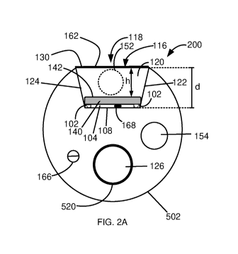
of or at
the fluid inlet 152 based on temperature readings of temperature sensor 166 at
the blood-
contact surface 116-blood interface (or temperature within the flow channel)
and maintain
an unchanged flow velocity.
[0084] Optionally, the system is configured to vary the temperature of or at
the fluid inlet
152 and vary the flow of the cooling fluid based on temperature readings of
temperature
sensor 166 at the blood-contact surface 116-blood interface (or temperature
within the flow
channel).
[0085] The flow rate and variation in flow rate depends on at least one of the
area cross-
section of cooling channel 120, the area of blood-contact surface 116-blood
interface,
temperature of the cooling fluid and variation in vessel blood temperature. To
cool down
blood-contact surface 116-blood interface and given cooling channel 120
channel
dimensions, the velocity of the cooling fluid over the ablating element in
some
embodiments, is between 5cm/sec ¨ 60cm/sec. In some embodiments, the velocity
of flow
is between 15cm/sec ¨ 50cm/sec. In some embodiments, the velocity of flow is
between
20cm/sec ¨ 30cm/sec. is 25cm/sec.
[0086] A potential advantage in this system configuration is in that the
system cooling
channel has a small cross-section e.g., smaller than a diameter of catheter
106 (between
0.01-0.5 of the diameter of catheter 106) configured to generate a velocity of
flow
sufficiently high to achieve efficient cooling, below 45 degrees Celsius at
the blood-contact
surface 116-blood interface.
[0087] The structure of cooling system 200 provides laminar-uniform flow over
emitting
surface 142 of PE element 140 in a direction indicated by arrow 180. In some
embodiments,
the flow rate of the cooling fluid is between 5m1/sec and 400m1/sec. In some
embodiments,
the flow rate of the cooling fluid is between 50m1/sec and 300m1/sec. In some
embodiments,
the flow rate of the cooling fluid is between 75m1/sec and 200m1/sec.
19
CA 03105282 2020-12-28
WO 2020/039442
PCT/IL2019/050941
[0088] In some embodiments, container 100 comprises one or more temperature
sensor
166 at the blood-contact surface 116 of container 100 cover 130 and the flow
rate is adjusted
in accordance with a temperature measured at blood contact surface 116. For
example, when
the measured temperature at blood-contact surface 116 exceeds 45 degrees
Celsius, blood
flow from inlet 152 is increased accordingly.
[0089] In some embodiments, container 100 comprises one or more temperature
sensors
168 in gap 104 between PE element 140 and floor 108 of cooling channel 120 or
adjacent
to PE element 140. In some embodiments, the flow rate is adjusted in
accordance with a
temperature measured in gap 104 to monitor and control PE element 140
temperature during
operation.
[0090] Figs. 3B and 3C are a graph (Fig. 3B) and heat distribution map (Fig.
3C) depicting
a temperature gradient in cooling channel 120 with respect to the level of the
fluid layer
measured by distance (L) from PE element 140 emitting surface 142. As shown in
Fig. 3B,
the greater the distance (L) between a fluid layer and PE element 140 emitting
surface 142,
the lower the temperature, dropping as shown in Fig. 3B from approximately 120
degrees
Celsius at emitting surface 142 to approximately 45 degrees at blood-contact
surface 116
which is at the greatest distance (d.x) from US beam emitting surface 142.
[0091] It is also noted in Fig. 3B, that the temperature continues to drop to
below 45
degrees Celsius beyond blood-contact surface 116 in blood flow layers adjacent
to blood
contact surface 116. Optionally, the coolant fluid is cooled to below 37deg C
at blood
contact surface 116, in which case the blood temperature which is normally at
37degC will
not drop in the layers beyond the blood contact surface 116.
[0092] A potential advantage in the cross-section profile of cooling system
200 is in that
the laminar flow of cooling fluid in cooling channel 120 generates an
effective and uniform
blood-contact surface 116-blood interface and provides for a rapidly formed
homogeneous
temperature profile of the blood-contact surface 116 -blood interface with no
heat zones.
[0093] A potential advantage in the cross-section profile of cooling system
200 is in that
the laminar flow of cooling fluid in cooling channel 120 is configured to and
effective in
removal of gas (e.g., air) bubbles formed in cooling channel 120, e.g.,
bubbles adhered to
PE element 140-facing surface of cover 130.
CA 03105282 2020-12-28
WO 2020/039442
PCT/IL2019/050941
Component of the PE element cooling system
[0094] In some embodiments, a processor (not shown) is used to calculate and
optimize
signal transmission and sensing data (e.g., temperature, distance from organ
wall, wall
thickness, power application time, change in amplitude and phase of returned
signal) to
optimize power output (e.g., for ablation), transducer reliability and lesion
size. In some
embodiments, container 100 comprises a PE element temperature sensor 166 that
communicates with the processor. The processor is configured to increase or
decrease power
input based on the data received from the piezoelectric temperature sensor.
[0095] In some embodiments, the system processor is configured to adjust the
level of
energy emitted from the PE element based on one or more of distance measured
from the
emitting surface of the PE element to said tissue wall, tissue thickness,
duration of energy
delivery, change in amplitude and/or phase of ultrasound signal returning from
the tissue
and reduction of recorded electrical potential signals.
[0096] In some embodiments, adjustment of the energy level is based on
impedance
measurement between one or more tissue contact electrodes 725 on positioner
702 and on
one or more electrical electrodes, not in contact with the tissue located on
the catheter 106
shaft or US transducer container 100.
[0097] In some embodiment, a processor (not shown) is configured to calculate
speed of
transducer rotation via a motor (not shown) positioned in the catheter handle
(not shown)
to optimize power output for optimal lesion creation based on sensing data
(e.g., distance
from organ wall, wall thickness, power application time, change in amplitude
and phase of
returned signal). Alternatively, and optionally, the processor is configured
to calculate
transducer rotation based on a gyroscope embedded within a handle (not shown)
of catheter
106. A potential advantage of a gyroscope is in its ability to show absolute
angles of the
catheter/ultrasound transceiver that allows physicians to return to a
registered angular
position during the procedure.
[0098] In some embodiments, the processor receives data from both blood-
contact surface
temperature sensor 164 and PE element temperature sensor 166 and based on the
current
cooling fluid flow rate extrapolates a temperature gradient between emitting
surface 142
and blood contact surface 116 and increases or decreases power input to PE
element 140
accordingly.
21
CA 03105282 2020-12-28
WO 2020/039442
PCT/IL2019/050941
[0099] In some embodiments, US transmission duty cycle is maintained greater
than 60%
to cool down US transducer PE element 140 without effecting the rate of energy
transfer to
tissue required to elevate tissue temperature above 50deg needed to form
tissue lesion.
[0100] Other means used to maintain a relatively cool temperature of PE
element 140
comprise using a pulse repetition frequency to lower transducer temperature,
increase flow
rate, decrease coolant fluid temperature, lower duty cycle, and regulate
voltage based on
distance from tissue wall to regulate time needed for successful ablation.
Bubble Control
[0101] Bubbles commonly formed by cavitation effect or air trapped in the
inlet and/or
outlet tubes pose a common interference issue in US transmission by forming
one or more
non-acoustically matched surfaces that reflect portions of the emitted US beam
in
unexpected directions. This is especially found in configurations that involve
cooling
systems that circulate a coolant within a balloon enveloping the US
transducer. Bubbles are
often trapped and adhered to a curved wall of the balloon, where circulation
is insufficient
to dislodge the bubbles and when successful, the coolant fluid flow in the
vicinity of the
bubbles is turbulent and just arbitrarily moves the bubbles from one location
to another.
[0102] As shown in the exemplary embodiment depicted in Figs. 4A and 4B, which
are
side cross-section view and transverse cross section view taken along line B-
B, simplified
illustrations of the effect of laminar cooling fluid flow on bubbles in
accordance with some
embodiments of the invention, a bubble 402, formed within cooling channel 120
is
maintained away from emitting surface 142 and cooling channel cover 130 by
laminar flow
450 and is carried towards cooling fluid outlet 154 positioned in cooling
fluid diverting
chamber 156 at tip 158 of the container 100 where it is suctioned out of the
catheter 106 by
vacuum within cooling fluid outlet 154 or by means of pressure head forcing
the fluid
towards the cooling fluid outlet 154.
[0103] In some embodiments, the confined channel cross-section adds to the
effect of the
laminar cooling fluid flow by limiting the wall surface to which a bubble may
adhere as
well as increase the fluid pressure applied to a bubble that appears. As shown
in Figs. 4A
and 4B, bubbles that appear are urged into cooling fluid cooling fluid
diverting chamber
156 at tip 158 of container 100 and by a down flow towards fluid outlet 154.
An additional
22
CA 03105282 2020-12-28
WO 2020/039442
PCT/IL2019/050941
advantage in the configuration of the laminar flow in cooling channel 120 as
well as the
flow directionality is in that it removed risk of ultrasound transmission
interreference due
to air bubbles and negates the need for use of degassed fluid or in-line
bubble detection
sensors and/or traps.
[0104] In some embodiments, an area of a cross-section of cooling channel 120
constitutes between 0.01 and 0.5 of an area of a cross-section of the catheter
106 at the same
location. In some embodiments, an area of a cross-section of cooling channel
120 constitutes
between 0.1 and 0.4 of an area of a cross-section of the catheter 106 at the
same location.
In some embodiments, and at least one ultrasound transducer one or more PE
elements 140
are disposed within and on a floor 108 of the channel 120.
[0105] A potential advantage of laminar cooling fluid flow within the cooling
channel is
in that heat transfer by the coolant is predictable and controlled by manually
or
automatically adjusting the flow rate and the flow parameters can be
predefined (and
simulated) with respect to the required ultrasound parameters.
[0106] A potential advantage of laminar cooling fluid flow within the cooling
channel is
in a uniform temp distribution throughout US transducer container 100 and
faster flow
adjustment expressed by faster control of blood contact surface temperature
adjustment.
Uniform temperature eliminates hot zones from forming at the blood contact
surface 116.
[0107] In some embodiments, the maximal volume of the coolant within the US
transducer container 100 is lower than 14,200 mmA3. In some embodiments, the
maximal
volume of the coolant within the US transducer container 100 is lower than 25
1mm^3. In
some embodiments, the volume of the coolant within the US transducer container
100 at
any given time is between 1 mmA3 to 40 mmA3.
[0108] According to some embodiments, the US transducer is configured to be
inserted
into an organ (e.g., a blood vessel) via a catheter. In some embodiments, the
external
diameter of the US transducer container 100 is smaller than the diameter of a
catheter 106
configured to insert the US transducer into an organ. In some embodiments, as
shown in
section A-A of Fig. 1, the cross section of container 100 is reduced at the
level of cooling
channel 120 cover 130. In some embodiments the cover 130 is made of a high
heat
absorbing material. In some embodiments the cover 130 is made of a low
acoustic
23
CA 03105282 2020-12-28
WO 2020/039442
PCT/IL2019/050941
attenuation material. In some embodiments, cover 130 thickness is below 1 mm,
below
0.5mm or below 0.3mm.
[0109] In some embodiments, the distance between the external surface of the
US
transducer and the tissue is monitored, such as the power supplied to the
transducer is
increased or decreased based on the monitored distance. In some embodiments,
the distance
between the transducer and the tissue is monitored, such as the power supplied
to the
transducer is increased or decreased based on the monitored distance. In some
embodiments,
the distance between the transducer and the tissue is monitored, such as the
power supplied
to the transducer is manually or automatically stopped if the monitored
distance is below a
predetermined safe distance. In some embodiments, a safe distance is defined
by a distance
above 1 mm. In some embodiments, a safe distance is defined by a distance
above 2mm. In
some embodiments, a safe distance is defined by a distance above 5mm.In some
embodiments, the treatment duration and/or power is regulated based on
analysis of the
signals returned from tissue, detection of lesion formation in the tissue and
completion of
lesion created.
In some embodiments, the treatment duration and/or power is regulated based on
one or
more of the following measurements and calculations: distance from tissue,
tissue thickness,
transducer duty cycle, transducer pulse repetition frequency, voltage,
amplitude of return
signal from targeted area, rate of change of amplitude of returned signal,
phase change of
signal return from targeted area, reduction of recorded electrical potential
signals e.g.,
signals recorded from the pulmonary veins and/or impedance measurement between
a tissue
contact electrode attached to the positioner 702 and a non-contact electrode
attached to the
catheter shaft or US housing . In some embodiments the US transducer comprises
one or
more computing units which receive sensors data as an input and outputs
transducer
operation parameters.
Transducer design and manufacture
[0110] Reference is now made to Figs. 5A, 5B, 5C, 5D, 5E, 5F, 5G, 5H and 51,
which are
perspective view and cross section view simplified illustration of method of
manufacturing
a container transducer in accordance with some embodiments of the invention.
As shown
24
CA 03105282 2020-12-28
WO 2020/039442
PCT/IL2019/050941
in Fig. 5A, a container 100 comprises a housing 502 comprises a trough-shaped
fluid
channel 120 having one or more supports 504 for PE element 140.
[0111] In some embodiments, PE element 140 support 504 are made of non-
electrically
conductive high temperature capacity material so that heat produced by PE
element 140,
positioned on supports 504, during operation is absorbed by the proximal and
distal PE
element 140 supports 504. In some embodiments, US PE element 140 comprises a
middle
partition made of a non-electrically conductive material that insulates
between transducer
electrodes connected at the distal end and the proximal end of the transducer
ceramic.
[0112] In some embodiments of the current invention, the catheter US
transducer
comprises an internal heat conducting lumen, connected at its distal end to
one or more of:
US transducer surface, transducer support, thereby transferring heat out of
the US
transducer.
[0113] In some embodiments, housing 502 comprises an electrical conduit 506
for a PE
element 140 temperature sensor 166 and a conduit 508 for electrical wiring as
will be
explained in greater detail herein. In the exemplary embodiment shown in Fig.
5B, an
electrical wire 510 has been inserted into housing 502 and laid out prior to
being connected
to PE element 140.
[0114] Figs. 5C and 5D, which are side cross-section view simplified
illustrations of
wiring options for PE element 140 in accordance with some embodiments of the
invention.
As shown in Fig. 5C, wiring of PE element 140 comprises two or more
electrodes, a first
electrode 512 along PE element 140 emitting surface 142 and a second electrode
514 along
an opposite surface of PE element 140 facing floor 108 of cooling channel 120.
Electrodes
512 and 514 are isolated from each other.
[0115] Alternatively, and optionally, and as shown in Fig. 5D, wiring of PE
element 140
comprises two or more electrodes, a first electrode 516 along at least a
portion of PE element
140 emitting surface 142 and around one end of PE element 140 and a second
electrode 518
along at least a portion of an opposite surface of PE element 140 facing floor
108 of cooling
channel 120 and around an opposite tip 158-facing end of PE element 140.
Electrodes 516
and 518 are isolated from each other by one or more gaps 530/536 on the
emitting surface
142 as well as on the opposite surface facing floor 108 respectively.
CA 03105282 2020-12-28
WO 2020/039442
PCT/IL2019/050941
[0116] In some embodiments, one or more gaps 530/536 are bridged by an
insulating
adhesive. In Fig. 5D, the gap 536 on the emitting surface 142 of PE element
140 is bridged
by an insulating adhesive 532.
[0117] A potential advantage of the wiring configurations is in that this
configuration
nullifies the need to isolate PE element 140 with non-conductive material,
e.g., Parylene.
This is achieved by positioning at least two contacts on generally opposite
sides of the PE
element 140, while maintaining and the PE element 140 circumferentially
insulated with
insulating material e.g., an electrical insulating adhesive. This prevents any
potential
electrical short between the two sides of the PE element.
[0118] A potential advantage in the use of a non-conductive material, e.g.,
Parylene to
isolate PE element 140 is in that it simplifies the manufacturing process and
is less
expensive than other commonly used techniques. Reference is now made to Fig.
5E, which
is a cross section of US transducer container 100 as taken along section C-C
shown in Fig.
5B and shows wire 510 exiting conduit 508 and placed in contact with tip 158-
facing end
of PE element 140. In some embodiments, container cover 130 comprises one or
more micro
outlet ports 195 that allow fluid outflow from cooling channel 120 into the
surrounding
blood stream. A potential advantage of micro outlet ports is in that fluid
exiting the micro
ports washes off any blood residue/charring that may form during the ablation
process.
[0119] Fig. 5F, is a cross section view simplified illustration of housing 502
electrical and
fluid passages to catheter 106 as viewed from a direction indicated in Fig. 5B
by an arrow
550. As shown in the exemplary embodiments depicted in Fig. 5F, housing 502
comprises
conduits for cooling fluid inlet 152 tube and cooling fluid outlet 154 tube
and a a transducer
housing support tube 520 having a lumen 126. In some embodiments, transducer
housing
support tube 520 and lumen 126 are sized to accommodate a guidewire 524
conducting tube
526. . . In some embodiments, housing 502 comprises one or more conduits 508
for one or
more ablation PE elements 140 coaxial cables and one or more conduits 522 for
one or more
inclined PE elements 140 coaxial cables.
[0120] Figs. 5G, 5H and 51 depict the manufacturing process of US transducer
container
100 following the electrical wiring of US transducer container 100. Fig. 5G
shows the stage
of manufacturing following the previous stages described herein and comprises
connecting
electrical conductors506/510 to the corresponding ends of PE element 140 in
accordance
26
CA 03105282 2020-12-28
WO 2020/039442
PCT/IL2019/050941
with the connection options described elsewhere herein. The step of connection
of electrical
wires is followed in some embodiments, and as shown in Fig. 5H by attaching
fluid inlet
152 and fluid outlet 154 to a cooling fluid diverting chamber 156 within tip
158 of the
container as shown in Fig. 51. The perimeter 534 of PE element 140 is sealed
to walls
122/124 of cooling channel 120 and posts 102 with a flexible isolating and
fluid proofing
adhesive e.g., Epoxy adhesive, UV adhesive or Silicon adhesive (e.g., Dymax
204-CTH,
Dymax 1191, Epo-Tek 301 or Epo-Tek 353ND) thus Insulating cover 130 is
comprises a
polymer (e.g., Polyester or Pebax@) is then placed over housing 502 as in
shrunken (e.g.,
by exposure to heat) to tightly seal housing 502.
[0121] The process is finalized by attaching cooling channel cover 130 over
housing 502
and non-spherical part of the container tip 156.
[0122] Reference is now made to Fig. 6, which is a flow chart of a method of
manufacture
and assembly of a US transducer container 100 in accordance with some
embodiments of
the invention. As shown in Fig. 6, the method comprises at 602 molding a
housing 502
comprising one or more fluid conduits 152/154, one or more electrical conduits
506/510,
one or more temperature sensor 166 conduits, one or more main catheter lumen
126, and
one or more trough-shaped cooling channels 120.
[0123] In some embodiments, cooling channel 120 comprises a trough-form
cooling
channel 120 defined by a floor 108 including one or more posts 102 and
bordered by a first
and a second side walls 122/124 extending from both sides of floor 108 and
along both
lateral sides of emitting surface 142 and meet edges of container blood-
contact surface 116
to form an aperture 118 in container blood-contact surface116.
[0124] At 604, electrical conduit 506/510 of PE element 140 is laid within the
respective
conduits, in communication with and leading through catheter 106 to a
respective source/s
of power and/or communication (not shown).
At 606, PE element 140 is mounted on one or more posts 102 and connected to
electrical
conductors506/510 as explained in detail elsewhere herein. In some
embodiments, and
optionally, the method comprises coating PE element 140 with a dielectric
layer. In some
embodiments, and optionally, the method comprises applying a dielectric
material between
ends of electrical conductors506/510 connected to PE element 140. At 608,
sealing the
perimeter of PE element 140 to walls 122/124 of cooling channel 120 with a
flexible
27
CA 03105282 2020-12-28
WO 2020/039442
PCT/IL2019/050941
isolating and fluid proofing adhesive and at 610 attaching a cooling fluid
diverting chamber
156 and tip 158 of the container. In some embodiments, steps 606 and 608 are
combined to
a single step. The process is completed by placing over housing 502 an
insulating covers a
portion of which, in some embodiments, comprises cover 130, shrinking the
cover (e.g., by
exposure to heat) and tightly sealing housing 502 and cooling channel 120.
Positioner
[0125]
Reference is now made to Figs. 7A, 7B, 7C and 7D which are plan view and
perspective view simplified illustrations of a positioner 702 for a catheter
106 carrying a US
transducer container 100 as disclosed herein. In some embodiments, catheter
106 comprises
an expandable positioner 702 enveloping at least a portion of US transducer
container 100.
In some embodiments, positioner 702 is mounted on a catheter inserted through
catheter
106. In some embodiments, positioner 702 is an integral part of catheter 106.
In some
embodiments, and as shown in Figs. 7A, 7B and 7C, positioner 702 envelops US
transducer
container 100. In some embodiments, positioner 100 comprises one or more
openings 704,
the diameter of which is greater than the diameter of the US beam emitted
through the
opening 704 so that positioner 702 does not interfere with propagation of the
beam. In some
embodiments, positioner 702 is made of a shape memory resilient biocompatible
material,
e.g., Nitinol. In some embodiments, positioner 702 is a non-occluding
positioner configured
to allow blood flow therethrough.
[0126] In some
embodiments, positioner 702 comprises a cage-like geometry. In
some embodiments, positioner 702 comprises a basket-like geometry. In some
embodiments, positioner 702 comprises a cylinder-like geometry. In some
embodiments,
dimensions of a cylindrical positioner 702 are between l0mm-30mm in diameter
and 7mm-
60mm in length. In some embodiments, dimensions of a cylindrical positioner
702 are
between 15mm-25mm in diameter and l0mm-50mm in length. In some embodiments,
dimensions of a cylindrical positioner 702 are between 17mm-20mm in diameter
and 8mm-
40mm in length.
[0127] In some
embodiments, the geometry of positioner 702 and location of
openings 704 in positioner 702 is non-uniform e.g., the openings 704 are
located at the distal
portion of positioner 702 such that one portion of positioner 702 e.g., a
proximal portion,
28
CA 03105282 2020-12-28
WO 2020/039442
PCT/IL2019/050941
provides mechanical support and another portion e.g., a distal portion
provides less
mechanical support and more exposure (more openings 704) to allow for more
effective
acoustic ablation.
[0128] In some
embodiments, positioner 702 comprises a detachable from the
catheter. In some embodiments, positioner 702 comprises a detachable plug,
e.g.,
configured to plug cavities in the left atrium such as Left Atrial Appendage
following an
ablation treatment.
[0129] In some
embodiments, positioner 702 comprises contact and/or non-contact
electrodes 725 and is configured to record electrical activity before, during
and/or after
ablation to monitor procedure effectiveness.
[0130] In some
embodiments, and as depicted in Figs. 7A and 7B, positioner 702
has an ovoid geometry. In some embodiments, and as depicted in Fig. 7C,
positioner 702
comprises a positioner 702 has a diamond geometry or any other suitable
geometry.
[0131] Fig. 7D,
which is a perspective view simplified illustration of
implementation of US transducer container 100 and positioner 702 in accordance
with some
embodiments of the invention. As shown in the exemplary embodiment depicted in
Fig. 7D,
US transducer container is implemented in contactless ablation of ostia of the
pulmonary
veins in the left atrium of the heart. In this procedure, a US transducer
container 100
configuration can be employed using an ablative inclined PE element 140 and an
imaging
PE 140-1 as described elsewhere herein. Positioner 702 is expanded inside the
left atrium
lumen and directed towards one of the four main pulmonary veins ostia. Once
the positioner
702 is positioned in contact with walls of the pulmonary vein ostium, US
transducer
container 100 is automatically positioned, generally centered in the ostium to
allow safe
ablation of the ostium margins. In some embodiments, US transducer container
is
configured to be rotatable within positioner 702 as indicated by arrow 750 and
ablate a ring
encompassing the margin of the pulmonary vein ostium. In some embodiments, US
transducer container is configured to axially translate in a bidirectional
manner within
positioner 702 to better position US transducer container 100 within, for
example, a
pulmonary vein ostium. A potential advantage of this feature is in that linear
movement of
US transducer container 100 provides for linear ablation (e.g., in parallel to
the axis of
translation of US transducer container 100) of the tissue in selected
anatomies.
29
CA 03105282 2020-12-28
WO 2020/039442
PCT/IL2019/050941
[0132] In some cases, such as, for example atrial fibrillation treatment, the
pulmonary
vein is ablated to stop the ectopic cardiac action potential trigger. In such
treatments, a
balloon-type positioner or cooling balloon, commonly used in such procedures,
is inflated
to a point at which the balloon surface is urged against a vessel wall thus
stabilizing the
ablating element. However, when a cooling balloon is used to cool and center a
transducer
a blood vessel (e.g., within the pulmonary vein) and the balloon wall contacts
the pulmonary
vein tissue, blood might be trapped and pooled between the balloon and the
tissue. The
pooled blood may potentially coagulate due to heat generated by the tissue
during ablation.
Deflation of the balloon at the end of the procedure may release the newly
formed blood
clot which may become a stroke risk.
A potential advantage of a non-occluding positioner is in that it is
configured to allow blood
to flow therethrough significantly reducing or altogether preventing blood
pooling and/or
clotting and formation of blood embolism.
Jet Effect
[0133] Reference is now made to Fig. 8, which is a cross-section view
simplified
illustration of implementation of a catheter US transducer container in
accordance with
some embodiments of the invention. In some embodiments, US transducer
container 100 is
sized and fitted to be positioned along or within a catheter 106. In some
embodiments, US
transducer container 100 comprises a collimating acoustic lens 802.
[0134] To the surprise of the authors of this disclosure it was observed that
in some
embodiments, collimated beam energy generated from a suitably designed US
transducer
PE element 140 generates a jet effect 850 in sun-ounding blood stream having
the same
temperature as that of the surrounding blood stream. In some embodiments, the
collimated
beam energy is above 50W/cm^2. In some embodiments, the collimated beam energy
is
above 70W/cm^2. In some embodiments, the collimated beam energy is above
90W/cm^2.
[0135] A potential advantage in such a jet effect 850 is in that a jet aimed
at a treatment
area cools the tissue wall 808 at the point of penetration of the ultrasound
beam into the
tissue and prevents tissue charring.
[0136] In some embodiments, catheter 106 comprises one or more therapeutic
agent
delivery nozzles 804 configured to deliver a therapeutic agent 806 into the
blood stream,
CA 03105282 2020-12-28
WO 2020/039442
PCT/IL2019/050941
e.g., up-stream to US transducer container 100 so that therapeutic agent 806
flows into
emitted US beam 204 and is driven by the jet effect 850 towards the tissue
808.
[0137] It has also come to be known to the authors of this disclosure that too
small a cross-
section dimension (e.g., area) does not generate a jet effect or that the
generated jet would
not be effective in driving a therapeutic agent 806 towards a small tissue 808
area.
Alternatively, a too large cross-section dimension (e.g., area) would require
a high level of
driving energy, beyond the maximal energy requirement for the device.
[0138] It was found that in some embodiments, an optimal cross-section
dimension (e.g.,
area) for generating a jet effect sufficiently effective in driving a
therapeutic agent 806
towards a small tissue 808 area should be sufficiently small (e.g., high
energy per cross-
section area) and is in the range between 8mmA2 to 30mm^2. In some
embodiments, an
optimal cross-section dimension (e.g., area) is in the range between 12mmA2 to
20mm^2.
In some embodiments, an optimal cross-section dimension (e.g., area) is in the
range
between 14mmA2 to 16mm^2.
[0139] Fig. 9 is a transverse cross-section simplified illustration of a
multidirectional US
transducer container, according to some embodiments of the invention. In some
embodiments, a multidirectional US scanner/ablating transducer container 900
includes a
plurality of PE Elements 140, arranged circumferentially about a longitudinal
axis of
container 900. In some embodiments, each PE element is arranged within a
cooling channel
120 and includes coolant fluid inlet and outlet and electrical conductors as
explained
elsewhere herein.
[0140] In some circumstances, in addition to catheter ablation for atrial
fibrillation
treatment as explained in detail elsewhere herein, there is a need to form
lesions (e.g., lesion
lines) in non-pulmonary vein ostium locations e.g., between the left inferior
pulmonary vein
to the mitral valve, between the left pulmonary veins to the right pulmonary
veins along the
posterior wall (LA roof line & LA floor lines) and/or in selected areas in the
left atrium
where ectopic cardiac action potential triggers are identified.
[0141] Reference is now made to Figs. 10A and 10B, which are perspective view
simplified illustrations of a catheter US transducer container including one
or more RF
electrode tips forming an US transducer/RF combination catheter container
1000. In some
embodiments, and as shown in Figs. 10A and 10B, the US transducer/RF container
1000 tip
31
CA 03105282 2020-12-28
WO 2020/039442
PCT/IL2019/050941
158 comprises a metallic material such as, for example, iridium/platinum,
platinum, copper
or gold. In some embodiments, the US transducer/RF combination catheter
container 1000
tip 158 comprises an RF electrode 1002 electrically connected via one or more
electrical
conductors, similar to electrical conductors 506/510, to an RF power source
(not shown). In
some embodiments, container tip 158 comprises one or more irrigation ports
1004
configured to eject cooling fluid (e.g., saline) to cool RF treated lesions
and/or tissue
surrounding the treated lesions. In some embodiments, US transducer/RF
combination
catheter container 1000 RF electrode 1002 is configured to come in contact
with tissue and
form lesions at the tissue.
[0142] A potential advantage in an US transducer/RF combination catheter
container is
in the ability of the device to treat not only pulmonary veins (PV) ostia but
also to form
additional lesion lines that might be required or desired after completion of
pulmonary vein
electrical isolation.
[0143] A potential advantage in a US transducer /RF combination catheter
container is in
that such a combination container is configured to effect:
a. Radially outward directed US ablation, resulting in circumferential
pulmonary vein (PV) electrical isolation; and
b. Point-by-point RF ablation targeting non-PV ectopic cardiac action
potential triggers.
[0144] A potential advantage in a US transducer /RF combination catheter
container
is in that such a combination container is configured for combining different
types of energy
(e.g., US and RF energies) to increase treatment diversity of the device:
a. Radially outward directed US ablation, resulting in circumferential
pulmonary vein (PV) electrical isolation; and
b. Forward directed contact RF ablation for specific non-PV ectopic cardiac
action potential triggers.
In some embodiments, and as shown in Fig. 10B, US transducer /RF combination
catheter
container comprises a positioner 702. In some embodiments, positioner 702 is
detachable.
A potential advantage in this configuration is in that positioner 702 is
detachable and
configured to plug a cavity e.g., the left atrial appendage.
32
CA 03105282 2020-12-28
WO 2020/039442
PCT/IL2019/050941
In some embodiments, positioner 702 is collapsible. In some embodiments,
positioner 702
in the collapsed configuration is configured to be drawn into catheter 106. A
potential
advantage in this configuration is in that a tissue location can be treated
initially with an US
transducer, being maintained in place by positioner 702 as explained in detail
elsewhere
herein, followed by removal of positioner 702 e.g., by collapse and retrieval
into catheter
106, followed by contact RF treatment employing container tip 158 RF electrode
1002.
[0145] Throughout this application, various embodiments of this invention may
be
presented in a range format. It should be understood that the description in
range format is
merely for convenience and brevity and should not be construed as an
inflexible limitation
on the scope of the invention. Accordingly, the description of a range should
be considered
to have specifically disclosed all the possible subranges as well as
individual numerical
values within that range. For example, description of a range such as from 1
to 6 should be
considered to have specifically disclosed subranges such as from 1 to 3, from
1 to 4, from
1 to 5, from 2 to 4, from 2 to 6, from 3 to 6 etc., as well as individual
numbers within that
range, for example, 1, 2, 3, 4, 5, and 6. This applies regardless of the
breadth of the range.
[0146] Whenever a numerical range is indicated herein, it is meant to include
any cited
numeral (fractional or integral) within the indicated range. The phrases
"ranging/ranges
between" a first indicate number and a second indicate number and
"ranging/ranges from"
a first indicate number "to" a second indicate number are used herein
interchangeably and
are meant to include the first and second indicated numbers and all the
fractional and
integral numerals therebetween.
[0147] In the description and claims of the application, each of the words
"comprise"
"include" and "have", and forms thereof, are not necessarily limited to
members in a list
with which the words may be associated. In addition, where there are
inconsistencies
between this application and any document incorporated by reference, it is
hereby intended
that the present application controls.
[0148] The descriptions of the various embodiments of the invention have been
presented
for purposes of illustration but are not intended to be exhaustive or limited
to the
embodiments disclosed. Many modifications and variations will be apparent to
those of
ordinary skill in the art without departing from the scope and spirit of the
described
embodiments. The terminology used herein was chosen to best explain the
principles of the
33
CA 03105282 2020-12-28
WO 2020/039442
PCT/IL2019/050941
embodiments, the practical application or technical improvement over
technologies found
in the marketplace, or to enable others of ordinary skill in the art to
understand the
embodiments disclosed herein.
34