Note: Descriptions are shown in the official language in which they were submitted.
CA 03105415 2020-12-30
WO 2020/016661 PCT/IB2019/000873
ANTIBODIES SPECIFIC TO FOLATE RECEPTOR ALPHA
BACKGROUND OF THE INVENTION
Folate receptor alpha, also known as folate receptor 1 (FOLR1) belongs to the
folate
receptor family, members of which have high binding affinity to folic acid
and/or derivatives
thereof (e.g., 5-methyletrahydrofolate). FOLR1 has been reported to be overly
expressed in a
number of epithelial-derived tumors, such as ovarian, breast, renal, lung,
colorectal, and
brain. This receptor therefore can be a target for the treatment of such
epithelial-derived
tumors.
It is therefore of great importance to develop effective FOLR1 antagonists,
such as
anti-FOLR1 antibodies for use in both cancer treatment and diagnosis.
SUMMARY OF THE INVENTION
The present disclosure is based, at least in part, on the development of a
number of
antibodies specific to FOLR1. Such antibodies showed high binding affinity to
the target
FOLR1 antigen and/or high inhibitory activity against FOLR1 + cells.
Accordingly, one aspect of the present disclosure features an isolated
antibody that
binds to FOLR1 (anti-FOLR1 antibody), wherein the antibody binds the same
epitope of
human FOLR1 as a reference antibody, which is FOLR1-Ab1, FOLR1-Ab4, FOLR1-
Ab14,
FOLR1-Ab20, or FOLR1-Ab23, the structure features of each of which are
provided herein.
In some embodiments, the anti-FOLR1 antibody described herein may comprise
heavy chain variable region (VH), which comprises one or more of the
following:
(a) a heavy chain complementary determining region 1 (HC CDR 1)
set forth as
XiYTFTX2YX3, in which Xi is G or I, X2 is D or S, and X3 is W, N, or S;
(b) a heavy chain complementary determining region 2 (HC CDR2) set forth as
INXiX2X3X4X5X6, in which Xi is P or T, X2 is N, Y, or E, X3 is N, D, or T, X4
is
G or S, X5 is G or E, and X6 is T or P; and
(c) a heavy chain complementary determining region 3 (HC CDR3) set
forth as
ARX1X2X3YX4X5X6X7X8X9Xio, in which Xi is S, K or M, X2 is G or P, X3 is G
or Y, X4 is G or absent, X5 is P or absent, X6 is A, R, or K, X7 is W, Y or I,
X8 is F
or M, X9 is D or A, and Xio is Y or V.
1
CA 03105415 2020-12-30
WO 2020/016661
PCT/IB2019/000873
Such an anti-FOLR1 antibody may comprises a VH, in which the HC CDR1, HC
CDR2, and HC CDR3, collectively, are at least 85% (e.g., at least 90%, at
least 95%, at least
98% or more) identical to the HC CDR1, HC CDR2, and HC CDR3 of the reference
antibody. In some instances, the antibody may comprise a VH that includes the
same HC
CDR1, HC CDR2, and HC CDR3 as one of the reference antibodies noted above. In
other
embodiments, the anti-FOLR1 antibody described herein may comprise a VH that
comprises
the HC CDR1, HC CDR2, and HC CDR3, which collective contain up to 5, 4, 3, 2,
or 1
mutation relative to the HC CDR1, HC CDR2, and HC CDR3 of the reference
antibody.
Alternatively or in addition, the anti-FOLR1 antibody described herein may
comprise
a light chain variable region (VL), which comprises one or more of the
following:
(a) a light chain complementary determining region 1 (LC CDR 1) set forth
as
ESVDNYGISF or QSLLYSSSQKNY;
(b) a light chain complementary determining region 2 (LC CDR 2) set forth
as
XiAS, in which Xi is V, A, or W; and
(c) a light
chain complementary determining region 3 (LC CDR 3) set forth as
QQX1X2X3X4PX5T, in which Xi is Y or S, X2 is Y or K, X3 is E or S, X4 is Y or
V, and X5 is W, Y, or absent.
Such an antibody may comprise a VL, in which the LC CDR1, LC CDR2, and LC
CDR3, collectively, are at least 85% (e.g., at least 90%, at least 95%, at
least 98% or more)
identical to the LC CDR1, LC CDR2, and LC CDR3 of the reference antibody. In
some
instances, the antibody may comprise the same LC CDR1, LC CDR2, and LC CDR3 as
one
of the reference antibodies noted above. In other embodiments, the anti-FOLR1
antibody
described herein may comprise the LC CDR1, LC CDR2, and LC CDR3, which
collective
contain up to 5, 4, 3, 2, or 1 mutation relative to the LC CDR1, LC CDR2, and
LC CDR3 of
the reference antibody.
In some examples, the anti-FOLR1 antibody described herein comprises the same
heavy chain and/or light chain CDRs as one of the reference antibodies noted
above. In some
instances, such an anti-FOLR1 antibody may comprise the same VH and/or VL as
the
reference antibody.
Any of the anti-FOLR1 antibodies described herein may specifically binds to
human
2
CA 03105415 2020-12-30
WO 2020/016661 PCT/IB2019/000873
FOLR1. In some instances, the anti-FOLR1 antibody may cross-react with human
FOLR1
and a non-human FOLR1, such as a rodent FOLR1 or a primate FOLR1. The antibody
may
be a human antibody or a humanized antibody. In some examples, it can be a
chimeric
antibody.
In some embodiments, the anti-FOLR1 antibody may be a full-length antibody
(e.g.,
an IgG molecule) or an antigen-binding fragment thereof. Alternatively, it can
be a single-
chain antibody.
In another aspect, the present disclosure features a nucleic acid or set of
nucleic acids
(e.g., two nucleic acids), which collectively encodes any of the anti-FOLR1
antibodies
described herein, and a vector or set of vectors (e.g., two vectors)
comprising the nucleic
acid(s) coding for the anti-FOLR1 antibodies. In some instances, the vector or
vector set can
be an expression vector(s). Also provided herein are host cells comprising the
nucleic acid(s)
or vector(s). Further, the present disclosure provides a method for making an
anti-FOLR1
antibody described herein, comprising culturing the host cell that comprises
the vector or
vector set comprising coding sequences for the antibody, wherein the coding
sequences are in
operably linkage to a suitable promoter, and harvesting the antibodies thus
produced, for
example, from the host cell or the culture medium.
In addition, the present disclosure provides an antibody-drug conjugate (ADC)
comprising: any of the anti-FOLR1 antibodies described herein, and at least
one therapeutic
agent, which is covalently conjugated to the antibody. In some examples, the
therapeutic
agent can be a cytotoxic agent, for example, monomethyl auristatin E.
In some embodiments, the antibody and the therapeutic agent may be conjugated
through a linker. In some examples, the linker can be a cleavable linker, for
example, a
protease-sensitive linker, a pH-sensitive linker, or a glutathione-sensitive
linker. In some
instances, the linker can be a protease-sensitive linker, which may comprise a
peptide having
2-5 amino acids. The peptide may comprise naturally-occurring amino acid
residues, non-
naturally-occurring amino acid residues, or a combination thereof. In one
example, the
peptide may comprise valine-citrulline. In other examples, the linker can be a
non-cleavable
linker. Such a non-cleavable linker may comprise an optionally substituted
alkane or a
thioether.
3
CA 03105415 2020-12-30
WO 2020/016661
PCT/IB2019/000873
In some embodiments, the linker may comprise a functional group that forms a
covalent bond between the antibody and the linker. Exemplary functional groups
include, but
are not limited to, a maleimide group, an iodoacetamide group, a vinyl sulfone
group, an
acrylate group, an acrylamide group, an acrylonitrile group, and a
methacrylate group. In one
example, the linker may further a molecular spacer of Formula I:
X
H
0
(Formula I),
in which
R1 is optionally substituted C1-6 alkyl, optionally substituted phenyl,
optionally
substituted C2-6 alkylene, optionally substituted C2-6 alkenylene, optionally
substituted C2-6
alkynylene, or optionally substituted triazole; and X is 0, S, or N.
Further, the present disclosure provides a chimeric antigen receptor (CAR),
which
may comprise: (i) an extracellular domain comprising an antigen binding
fragment that binds
FOLR1, (ii) a transmembrane domain, and (iii) one or more intracellular
stimulatory
domains. The antigen binding fragment may binds the same epitope of human
FOLR1 as any
of the reference antibodies described herein. In some examples, the antigen
binding fragment
may comprise the same HC CDRs and/or LC CDRs of any of the reference
antibodies. Such
an antigen binding fragment may comprise the same VH and VL as the reference
antibody. In
some examples, the antigen binding fragment can be a single chain antibody
(scFv).
In any of the CARs described herein, the transmembrane domain may comprise a
transmembrane domain derived from CD28 or CD8. Alternatively or in addition,
the one or
more intracellular stimulatory domains may comprise a signaling domain from
CD3t and
optionally a co-stimulatory signaling domain, which may be from 4-1BB, CD7,
CD27,
CD28, CD40, 0X40, ICOS, GITR, HVEM, TIM1, or LFA-1. Nucleic acids encoding any
of
the CAR described herein, vectors comprising such, and host cells expressing
the CAR are
also within the scope of the present disclosure. In some examples, the host
cell expressing
the CAR is an immune cell such as a T cell.
In yet another aspect, the present disclosure provides a pharmaceutical
composition
4
CA 03105415 2020-12-30
WO 2020/016661 PCT/IB2019/000873
comprising (i) one or more of the anti-FOLR1 antibodies described herein, a
nucleic acid or
set of nucleic acids encoding such, an antibody-drug conjugate as described
herein, or a host
cell expressing any of the CAR constructs described herein, and (ii) a
pharmaceutically
acceptable carrier.
Moreover, the present disclosure features a method of reducing the number of
FOLR1+ cells, the method comprising administering to a subject in need thereof
an effective
amount of any of the pharmaceutical compositions described herein. In some
embodiments,
the subject may be a human patient has or is suspected of having cancer, for
example, an
epithelial cancer. Also within the scope of the present disclosure are
pharmaceutical
compositions as described herein for use in treating any of the target
diseases also described
herein (e.g., cancer such as an epithelial cancer) or for use in manufacturing
a medicament for
the treatment of the target disease.
In addition, the present disclosure features a method of detecting presence of
FOLR1+
cells, the method comprising: i. contacting a sample suspected of having
FOLR1+ cells with
any of the anti-FOLR1 antibodies described herein, which is conjugated with a
labeling
agent; and ii. detecting presence FOLR1+ cells in the sample based on binding
of the
antibody to cells in the sample. In some instances, the sample is derived from
a human
patient at risk for or suspected of having a cancer, such as an epithelial
cancer.
The details of one or more embodiments of the invention are set forth in the
description below. Other features or advantages of the present invention will
be apparent
from the following drawings and detailed description of several embodiments,
and also from
the appended claims.
BRIEF DESCRIPTION OF THE DRAWINGS
FIGs 1A-1E include diagrams showing that a number of anti-FOLR1 antibodies,
including FOLR1-Abl, FOLR1-Ab4, FOLR1-Ab14, FOLR1-Ab20, and FOLR1-Ab23,
bound to cells expressing surface FOLR1.
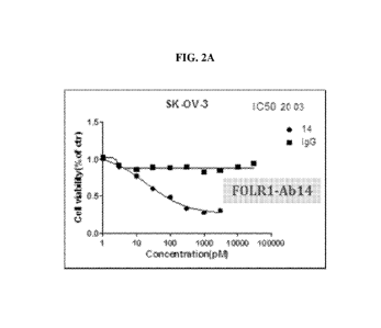
FIGs. 2A-2D including diagrams showing inhibitory effect of exemplary anti-
FOLR1
antibodies as indicated against SK-OV-3 cells, which are FOLR1 .
5
CA 03105415 2020-12-30
WO 2020/016661 PCT/IB2019/000873
DETAILED DESCRIPTION OF THE INVENTION
Disclosed herein are a number of anti-FOLR1 antibodies, which showed superior
features, including high binding affinity to the target FOLR1 antigen, and/or
high inhibitory
activity against FOLR1 + cells.
Accordingly, provided herein are antibodies capable of binding to FOLR1,
nucleic
acids encoding such, antibody-drug conjugates (ADCs) and chimeric antigen
receptors
(CARs) comprising the anti-FOLR1 antibodies, and uses thereof for both
therapeutic and
diagnostic purposes. Also provided herein are kits for therapeutic and/or
diagnostic use of
the antibodies and/or ADCs and CARs comprising such, as well as methods for
producing the
anti-FOLR1 antibodies.
Antibodies binding to FOLK'
The present disclosure provides antibodies that bind folate receptor alpha,
which is
also known as folate receptor 1 (FOLR1). As a member of the folate receptor
family, FOLR1
has a high binding affinity to folic acid and its derivatives. In humans,
FOLR1 is encoded by
the FOLR1 gene. FOLR1 was found to be overly expressed on various epithelial
tumors, for
example, ovarian cancer. Thus this receptor can serve as a target and/or a
biomarker for
treatment and diagnosis of the target cancer. Accordingly, the anti-FOLR1
antibodies
disclosed herein may be used in treating and/or diagnosing a target cancer as
described
herein, either by itself or being conjugated to other moieties, for example,
being conjugated
to a therapeutic agent to form an antibody-drug conjugate or being the
extracellular antigen-
binding domain in a chimeric antigen receptor.
An antibody (interchangeably used in plural form) is an immunoglobulin
molecule
capable of specific binding to a target, such as a carbohydrate,
polynucleotide, lipid,
polypeptide, etc., through at least one antigen recognition site, located in
the variable region
of the immunoglobulin molecule. As used herein, the term "antibody"
encompasses not only
intact (i.e., full-length) polyclonal or monoclonal antibodies, but also
antigen-binding
fragments thereof (such as Fab, Fab', F(ab')2, Fv), single chain (scFv),
mutants thereof, fusion
proteins comprising an antibody portion, humanized antibodies, chimeric
antibodies,
diabodies, nanobodies, linear antibodies, single chain antibodies,
multispecific antibodies
6
CA 03105415 2020-12-30
WO 2020/016661 PCT/IB2019/000873
(e.g., bispecific antibodies) and any other modified configuration of the
immunoglobulin
molecule that comprises an antigen recognition site of the required
specificity, including
glycosylation variants of antibodies, amino acid sequence variants of
antibodies, and
covalently modified antibodies. An antibody includes an antibody of any class,
such as IgD,
IgE, IgG, IgA, or IgM (or sub-class thereof), and the antibody need not be of
any particular
class. Depending on the antibody amino acid sequence of the constant domain of
its heavy
chains, immunoglobulins can be assigned to different classes. There are five
major classes of
immunoglobulins: IgA, IgD, IgE, IgG, and IgM, and several of these may be
further divided
into subclasses (isotypes), e.g., IgGl, IgG2, IgG3, IgG4, IgAl and IgA2. The
heavy-chain
constant domains that correspond to the different classes of immunoglobulins
are called
alpha, delta, epsilon, gamma, and mu, respectively. The subunit structures and
three-
dimensional configurations of different classes of immunoglobulins are well
known.
A typical antibody molecule comprises a heavy chain variable region (VH) and a
light
chain variable region (VL), which are usually involved in antigen binding. The
VH and VL
regions can be further subdivided into regions of hypervariability, also known
as
"complementarity determining regions" ("CDR"), interspersed with regions that
are more
conserved, which are known as "framework regions" ("FR"). Each VH and VL is
typically
composed of three CDRs and four FRs, arranged from amino-terminus to carboxy-
terminus
in the following order: FR1, CDR1, FR2, CDR2, FR3, CDR3, FR4. The extent of
the
framework region and CDRs can be precisely identified using methodology known
in the art,
for example, by the Kabat definition, the Chothia definition, the AbM
definition, and/or the
contact definition, all of which are well known in the art. See, e.g., Kabat,
E.A., et al. (1991)
Sequences of Proteins of Immunological Interest, Fifth Edition, U.S.
Department of Health
and Human Services, NIH Publication No. 91-3242, Chothia et al., (1989) Nature
342:877;
Chothia, C. et al. (1987) J. Mol. Biol. 196:901-917, Al-lazikani et al (1997)
J. Molec. Biol.
273:927-948; and Almagro, J. Mol. Recognit. 17:132-143 (2004). See also
hgmp.mrc.ac.uk
and bioinf.org.uk/abs.
In some embodiments, the anti-FOLR1 antibody as described herein can bind and
inhibit the activity of FOLR1 by at least 50% (e.g., 60%, 70%, 80%, 90%, 95%
or greater).
The apparent inhibition constant (KiaPP or Icapp), which provides a measure of
inhibitor
7
CA 03105415 2020-12-30
WO 2020/016661
PCT/IB2019/000873
potency, is related to the concentration of inhibitor required to reduce
enzyme activity and is
not dependent on enzyme concentrations. The inhibitory activity of an anti-
FOLR1 antibody
described herein can be determined by routine methods known in the art.
The KLaPP value of an antibody may be determined by measuring the inhibitory
effect
of different concentrations of the antibody on the extent of the reaction
(e.g., enzyme
activity); fitting the change in pseudo-first order rate constant (v) as a
function of inhibitor
concentration to the modified Morrison equation (Equation 1) yields an
estimate of the
apparent Ki value. For a competitive inhibitor, the KiaPP can be obtained from
the y-intercept
extracted from a linear regression analysis of a plot of KLaPP versus
substrate concentration.
([E]-[I] - Kr ) + 11([E] -[I] - K ia"" +4[E] = K
v = A= ____________________________________________________________________
2 (Equation 1)
Where A is equivalent to v0/E, the initial velocity (v0) of the enzymatic
reaction in the
absence of inhibitor (I) divided by the total enzyme concentration (E).
In some embodiments, the anti-FOLR1 antibody described herein may have a KiaPP
value of 1000, 900, 800, 700, 600, 500, 400, 300, 200, 100, 50, 40, 30, 20,
19, 18, 17, 16, 15,
14, 13, 12, 11, 10, 9, 8, 7, 6, 5 pM or less for a FOLR1 antigen or an
antigenic epitope
thereof. In some embodiments, the anti-FOLR1 antibody may have a lower KiaPP
for a first
target relative to a second target. Differences in KiaPP (e.g., for
specificity or other
comparisons) can be at least 1.5, 2, 3, 4, 5, 10, 15, 20, 37.5, 50, 70, 80,
91, 100, 500, 1000,
10,000 or 105 fold.
The antibodies described herein can be murine, rat, human, or any other origin
(including chimeric or humanized antibodies). Such antibodies are non-
naturally occurring,
i.e., would not be produced in an animal without human act (e.g., immunizing
such an animal
with a desired antigen or fragment thereof).
Any of the antibodies described herein can be either monoclonal or polyclonal.
A
"monoclonal antibody" refers to a homogenous antibody population and a
"polyclonal
antibody" refers to a heterogeneous antibody population. These two terms do
not limit the
source of an antibody or the manner in which it is made.
8
CA 03105415 2020-12-30
WO 2020/016661 PCT/IB2019/000873
In one example, the antibody used in the methods described herein is a
humanized
antibody. Humanized antibodies refer to forms of non-human (e.g., murine)
antibodies that
are specific chimeric immunoglobulins, immunoglobulin chains, or antigen-
binding
fragments thereof that contain minimal sequence derived from non-human
immunoglobulin.
For the most part, humanized antibodies are human immunoglobulins (recipient
antibody) in
which residues from a complementary determining region (CDR) of the recipient
are replaced
by residues from a CDR of a non-human species (donor antibody) such as mouse,
rat, or
rabbit having the desired specificity, affinity, and capacity. In some
instances, Fv framework
region (FR) residues of the human immunoglobulin are replaced by corresponding
non-
human residues. Furthermore, the humanized antibody may comprise residues that
are found
neither in the recipient antibody nor in the imported CDR or framework
sequences, but are
included to further refine and optimize antibody performance. In general, the
humanized
antibody will comprise substantially all of at least one, and typically two,
variable domains,
in which all or substantially all of the CDR regions correspond to those of a
non-human
immunoglobulin and all or substantially all of the FR regions are those of a
human
immunoglobulin consensus sequence. The humanized antibody optimally also will
comprise
at least a portion of an immunoglobulin constant region or domain (Fc),
typically that of a
human immunoglobulin. Antibodies may have Fc regions modified as described in
WO
99/58572. Other forms of humanized antibodies have one or more CDRs (one, two,
three,
four, five, and/or six), which are altered with respect to the original
antibody, which are also
termed one or more CDRs "derived from" one or more CDRs from the original
antibody.
Humanized antibodies may also involve affinity maturation.
In another example, the antibody described herein can be a chimeric antibody,
which
can include a heavy constant region and a light constant region from a human
antibody.
.. Chimeric antibodies refer to antibodies having a variable region or part of
variable region
from a first species and a constant region from a second species. Typically,
in these chimeric
antibodies, the variable region of both light and heavy chains mimics the
variable regions of
antibodies derived from one species of mammals (e.g., a non-human mammal such
as mouse,
rabbit, and rat), while the constant portions are homologous to the sequences
in antibodies
9
CA 03105415 2020-12-30
WO 2020/016661 PCT/IB2019/000873
derived from another mammal such as human. In some embodiments, amino acid
modifications can be made in the variable region and/or the constant region.
In some embodiments, the anti-FOLR1 antibodies described herein specifically
bind
to the corresponding target antigen or an epitope thereof. An antibody that
"specifically
binds" to an antigen or an epitope is a term well understood in the art. A
molecule is said to
exhibit "specific binding" if it reacts more frequently, more rapidly, with
greater duration
and/or with greater affinity with a particular target antigen than it does
with alternative
targets. An antibody "specifically binds" to a target antigen or epitope if it
binds with greater
affinity, avidity, more readily, and/or with greater duration than it binds to
other substances.
For example, an antibody that specifically (or preferentially) binds to a
FOLR1 antigen or an
antigenic epitope therein is an antibody that binds this target antigen with
greater affinity,
avidity, more readily, and/or with greater duration than it binds to other
antigens or other
epitopes in the same antigen. It is also understood with this definition that,
for example, an
antibody that specifically binds to a first target antigen may or may not
specifically or
preferentially bind to a second target antigen. As such, "specific binding" or
"preferential
binding" does not necessarily require (although it can include) exclusive
binding. In some
examples, an antibody that "specifically binds" to a target antigen or an
epitope thereof may
not bind to other antigens or other epitopes in the same antigen. In some
embodiments, the
anti-FOLR1 antibody described herein specifically binds human FOLR1. In some
examples,
.. its binding activity to a non-human FOLR1 antigen is not detectable in a
conventional assay
or is very low such that it would have no significant biological significance
as known to those
skilled in the art. In other examples, the anti-FOLR1 antibody described
herein may cross-
react with FOLR1 from different species, for example, between human FOLR1 and
a non-
human FOLR1 (e.g., FOLR1 from an experimental animal such as a non-human
primate,
mouse, or rat).
As used herein, the term "folate receptor alpha," "folate receptor 1", or
"FOLR1"
refers to a folate receptor alpha of any suitable species, e.g., human, a non-
human mammal
such as a non-human primate, or a rodent (e.g., mouse or rat). FOLR1 is a
single chain
membrane protein capable of binding to folic acid and its derivatives. The
amino acid
CA 03105415 2020-12-30
WO 2020/016661 PCT/IB2019/000873
sequence of an exemplary human FOLR1 is provided below (see also GenBank
accession no.
P15328):
1 maqrmttqll 111vwvavvg eaqtriawar tellnvcmna khhkekpgpe dklheqcrpw
61 rknaccstnt sqeahkdvsy lyrfnwnhcg emapackrhf iqdtclyecs pnlgpwiqqv
121 dqswrkervl nvplckedce qwwedcrtsy tcksnwhkgw nwtsgfnkca vgaacqpfhf
181 yfptptvlcn eiwthsykvs nysrgsgrci qmwfdpaqgn pneevarfya aamsgagpwa
241 awpfllslal mllwlls
FOLR1 molecules from other species were well known in the art and the amino
acid
sequences thereof can be retrieved from a publically available database, for
example,
GenBank.
In some embodiments, an anti-FOLR1 antibody as described herein has a suitable
binding affinity for the target antigen (e.g., human FOLR1) or antigenic
epitopes thereof. As
used herein, "binding affinity" refers to the apparent association constant or
KA. The KA is
the reciprocal of the dissociation constant (KD). The anti-FOLR1 antibody
described herein
may have a binding affinity (KD) of at least 10-5, 10-6, 10-7, 10-8, 10-9, 10-
10 M, or lower for the
target antigen or antigenic epitope. An increased binding affinity corresponds
to a decreased
KD. Higher affinity binding of an antibody for a first antigen relative to a
second antigen can
be indicated by a higher KA (or a smaller numerical value KD) for binding the
first antigen
than the KA (or numerical value KD) for binding the second antigen. In such
cases, the
antibody has specificity for the first antigen (e.g., a first protein in a
first conformation or
mimic thereof) relative to the second antigen (e.g., the same first protein in
a second
conformation or mimic thereof; or a second protein). In some embodiments, the
anti-FOLR1
antibodies described herein have a higher binding affinity (a higher KA or
smaller KD) to
human FOLR1 as compared to the binding affinity to FOLR1 of a different
species.
Differences in binding affinity (e.g., for specificity or other comparisons)
can be at least 1.5,
2, 3, 4, 5, 10, 15, 20, 37.5, 50, 70, 80, 91, 100, 500, 1000, 10,000 or 105
fold. In some
embodiments, any of the anti-FOLR1 antibodies may be further affinity matured
to increase
the binding affinity of the antibody to the target antigen or antigenic
epitope thereof.
Binding affinity (or binding specificity) can be determined by a variety of
methods
including equilibrium dialysis, equilibrium binding, gel filtration, ELISA,
surface plasmon
resonance, or spectroscopy (e.g., using a fluorescence assay). Exemplary
conditions for
evaluating binding affinity are in HBS-P buffer (10 mM HEPES pH7.4, 150 mM
NaCl,
11
CA 03105415 2020-12-30
WO 2020/016661 PCT/IB2019/000873
0.005% (v/v) Surfactant P20). These techniques can be used to measure the
concentration of
bound binding protein as a function of target protein concentration. The
concentration of
bound binding protein ([Bound]) is generally related to the concentration of
free target
protein ([Free]) by the following equation:
[Bound] = [Free]/(Kd+[Free])
It is not always necessary to make an exact determination of KA, though, since
sometimes it is sufficient to obtain a quantitative measurement of affinity,
e.g., determined
using a method such as ELISA or FACS analysis, is proportional to KA, and thus
can be used
for comparisons, such as determining whether a higher affinity is, e.g., 2-
fold higher, to
obtain a qualitative measurement of affinity, or to obtain an inference of
affinity, e.g., by
activity in a functional assay, e.g., an in vitro or in vivo assay.
In some embodiments, the anti-FOLR1 antibodies described herein bind to the
same
epitope in a FOLR1 antigen (e.g., human FOLR1) as one of the reference
antibodies provided
herein or compete against the reference antibody from binding to the FOLR1
antigen.
Reference antibodies provided herein include FOLR1-Ab217, FOLR1-Ab218, FOLR1-
Ab220, FOLR1-Ab221, and FOLR1-Ab222, the structural features of each of which
are
provided herein. An antibody that binds the same epitope as a reference
antibody described
herein may bind to exactly the same epitope or a substantially overlapping
epitope (e.g.,
containing less than 3 non-overlapping amino acid residue, less than 2 non-
overlapping
amino acid residues, or only 1 non-overlapping amino acid residue) as the
reference antibody.
Whether two antibodies compete against each other from binding to the cognate
antigen can
be determined by a competition assay, which is well known in the art. Such
antibodies can
be identified as known to those skilled in the art, e.g.,, those having
substantially similar
structural features (e.g., complementary determining regions), and/or those
identified by
assays known in the art. For example, competition assays can be performed
using one of the
reference antibodies to determine whether a candidate antibody binds to the
same epitope as
the reference antibody or competes against its binding to the FOLR1 antigen.
The anti-FOLR1 antibodies described herein may comprise a heavy chain variable
region (VH), which may comprise (a) a heavy chain complementary determining
region 1
12
CA 03105415 2020-12-30
WO 2020/016661 PCT/IB2019/000873
(HC CDR 1) set forth as XiYTFTX2YX3, in which Xi is G or I, X2 is D or S, and
X3 is W, N,
or S; (b)
a heavy chain complementary determining region 2 (HC CDR2) set forth as
INXiX2X3X4X5X6, in which Xi is P or T, X2 is N, Y, or E, X3 is N, D, or T, X4
is G or S, X5
is G or E, and X6 is T or P; (c) a heavy chain complementary determining
region 3 (HC
CDR3) set forth as ARX1X2X3YX4X5X6X7X8X9X10, in which Xi is S, K or M, X2 is G
or P,
X3 is G or Y, X4 is G or absent, X5 is P or absent, X6 is A, R, or K, X7 is W,
Y or I, X8 is F or
M, X9 is D or A, and Xio is Y or V, or (d) a combination of any one of (a)-
(c). In some
instances, the antibody may comprise a HC CDR1 of (a), a HC CDR2 of (b), and a
HC CDR3
of (c).
In some examples, the HC CDR1 motif XiYTFTX2YX3 may contain G at position Xi,
D at position X2, and/or N at position X3. Alternatively or in addition, the
HC CDR2 motif
INXiX2X3X4X5X6 may contain P at position Xi, N at position X2 and/or position
X3, G at
position X4, G or S at position X5, and/or T at position X6. Alternatively or
in addition, the
HC CDR3 motif ARX1X2X3YX4X5X6X7X8X9Xio may include K at position Xi, P or G at
position X2, Y at position X3, G at position X4, P at position X5, K or R at
position X6, Y at
position X7, F at position X8, D at position X9, and/or Y or V at position
Xio.
Table 1 provides the amino acid sequences of the heavy chain CDRs (by IMGT
definitions) for exemplary anti-FOLR1 antibodies. Antibodies having the same
heavy chain
.. CDR1, CDR2, and CDR3 regions as those exemplary anti-FOLR1 antibodies are
also within
the scope of the present disclosure.
Table 1: Heavy chain CDR sequences of anti-FOLR1 antibodies
Exemplary
CDR1 CDR2 CDR3
Antibody
FOLR1-Ab 1 GYTFTSYW INPYDSET ARSGGYAWFAY
FOLR1-Ab4 IYTFTDYS INTETGEP ARMGYYGPKIMDY
FOLR 1 -Ab 14 GYTFTDYN INPNNGGT ARKPYYGPRYFDV
FOLR 1 -Ab20 GYTFTDYN INPNNGGT ARKPYYGPRYFDV
FOLR 1 -Ab23 GYTFTDYN INPNNGGT ARKPYYGPRYFDV
Alternatively or in addition, the anti-FOLR1 antibodies described herein may
comprise a light chain variable domain (VI) that comprises comprises a light
chain variable
region (VI), which comprises (a) a light chain complementary determining
region 1 (LC
13
CA 03105415 2020-12-30
WO 2020/016661 PCT/IB2019/000873
CDR 1) set forth as ESVDNYGISF or QSLLYSSSQKNY; (b) a light chain
complementary
determining region 2 (LC CDR 2) set forth as XiAS, in which Xi is V, A, or W;
(c) a light
chain complementary determining region 3 (LC CDR 3) set forth as
QQX1X2X3X4PX5X6, in
which Xi is Y or S, X2 is Y or K, X3 is E or S, X4 is Y or V, X5 is W, Y, or
T, and X6 is T or
.. absent, or (d) any combination of (a)-(c).
In some examples, the LC CDR1 of the anti-FOLR1 antibody is ESVDNYGISF. In
other examples, the LC CDR1 of the antibody is QSLLYSSSQKNY. Alternatively or
in
addition, the LC CDR2 motif XiAS contains V at position Xi. Alternatively or
in addition,
the LC CDR3 motif QQX1X2X3X4PX5T includes S at position Xi, K at position X2,
E at
position X3, V at position X4, and/or no residue at position X5.
Table 2 provides the amino acid sequences of the light chain CDRs for
exemplary
anti-FOLR1 antibodies. Antibodies having the same light chain CDR1, CDR2, and
CDR3
regions as those exemplary anti-FOLR1 antibodies are also within the scope of
the present
disclosure.
Table 2: Light chain CDR sequences of anti-FOLR1 antibodies
Exemplary
CDR1 CDR2 CDR3
Antibody
FOLR1-Ab217 QSLLYSSSQKNY WAS QQYYSYPWT
FOLR1-Ab218 ESVDNYGISF AAS QQSKEVPYT
FOLR1-Ab220 ESVDNYGISF VAS QQSKEVPT
FOLR1-Ab221 ESVDNYGISF VAS QQSKEVPT
FOLR1-Ab222 ESVDNYGISF VAS QQSKEVPT
The heavy chain and light chain CDRs of the reference antibodies provided
herein are
determined based on the IMGT approach, which is well known in the art. In some
instances,
the anti-FOLR1 antibodies disclosed herein may comprise the same heavy chain
and light
chain CDRs of any of the reference antibodies disclosed herein. Two antibodies
having the
same VH and/or VL CDRs means that their CDRs are identical when determined by
the same
approach (e.g., those described herein and/or known in the art).
In some examples, the anti-FOLR1 antibodies disclosed herein may comprise the
same VH and/or VL sequence as one of the reference antibodies, which are
provided below
(CDRs in boldface):
14
CA 03105415 2020-12-30
WO 2020/016661 PCT/IB2019/000873
FOLR1-Ab 1:
VH:
QVQLQQPGAELVRPGASVKLSCKASGYTFTSYWMNWVKQRPEQGLEWIGRINPYDSETHSNQK
FKDKAIL
TVDKSSTTAYMQLSSLTSEDSAVYYCARSGGYAWFAYCARSGGYAWFAYWWFAYWGQGTLVTVSAWGQG
TLVTVSA
VL
DIVMSQSPSSLAVSVGEKVTMSCKSSQSLLYSSSQKNYLAWYQQKPGQSPKLLIYWASTRESG
VPDRFTGS
GSGTDFTLTISSVKAEDLAVYYCQQYYSYPWTCQQYYSYPWTFWTFGGGTKLEIKFGGGTKLEIK
FOLR1-Ab4
VH:
QIQLVQSGPELKKPGETVKISCKASIYTFTDYSIQWVKQAPGKGLKWMGWINTETGEPTYADD
FKGRFAFS
LESSASTAFLQINNLKNEDTATYFCARMGYYGPKIMDYCARMGYYGPKIMDYWMDYWGQGTSVTVSSWG
QGTSVTVSS
VL:
DIVLTQSPASLAVSLGQRATISCRASESVDNYGISFMNWFQQKPGQPPKLLIYAASNQGSGVP
ARFSDSGS
GTDFSLNIHPMEEDDTAMYFCQQSKEVPYTCQQSKEVPYTFYTFGGGTKLEIKFGGGTKLEIK
FOLR1-Ab 14
VH:
EVLLQQSGPELVKPGASVKIPCKASGYTFTDYNMDWVKQSHGKSLEWIGDINPNNGGTIYNQK
FKGKATLT
VDKSSSTAYMELRSLTSEDTAVYYCARKPYYGPRYFDVCARKPYYGPRYFDVWYFDVWGAGTTVTVSSW
GA GTTVTVSS
VL:
DIVLTQSPASLAVSLGQRATISCRASESVDNYGISFMNWFQQKPGQPPKLLIYVASNQGSGVP
ARFSGSGS GTDFSLNIHPMEEDDTAMYFCQQSKEVPTCQQSKEVPTFTFGGGTKLEIKFGGGTKLEIK
FOLR1-Ab20
VH:
EVLLQQSGPELVKPGASVKIPCKASGYTFTDYNMDWVKQSHGKSLEWIGDINPNNGGTIYNQK
FKGKATLT
VDKSSSTAYMELRSLTSEDTAVYYCARKPYYGPRYFDVCARKPYYGPRYFDVWYFDVWGAGTTVTVSSW
GA GTTVTVSS
VL:
DIVLTQFPASLAVSLGQRATISCRASESVDNYGISFMNWFQQKPGQPPKLLIYVASNQGSGVP
ARFSGSGF GTDFSLNIHPMEEDDTAMYFCQQSKEVPTCQQSKEVPTFTFGGGTKLEIKFGGGTKLEIK
FOLR1-Ab23
CA 03105415 2020-12-30
WO 2020/016661 PCT/IB2019/000873
VH:
EVLLQQSGPELVKPGASVKIPCKASGYTFTDYNMDWVKQSHGKSLEWIGDINPNNGGTIYNQK
FKGKATLT
VDKSSSTAYMELRSLTSEDTAVYYCARKPYYGPRYFDVCARKPYYGPRYFDVWYFDVWGAGTTVTVSSW
GA GTTVTVSS
VL:
DIVLTQFPAFLAVFLGQRATISCRASESVDNYGISFMNWFQQKPGQPPKLLIYVASNQGSGVP
ARFSGSGF GTEFSLNIHPMEEDDSAMYFCQQSKEVPTCQQSKEVPTFTFGGGTKLEIKFGGGTKLEIK
Also within the scope of the present disclosure are functional variants of any
of the
reference anti-FOLR1 antibodies as disclosed herein (e.g., those listed in
Tables 1 and 2
above). A functional variant can comprise up to 5 (e.g., 4, 3, 2, or 1) amino
acid residue
variations in one or more of the heavy chain and light chain CDR regions of
the reference
antibody and binds the same epitope of the FOLR1 antigen with substantially
similar affinity
(e.g., having a KD value in the same order). In some instances, each of the
heavy chain
and/or light chain CDR in a functional variant contain no more than 2 amino
acid residue
variations as relative to the counterpart CDR in the reference antibody. In
some examples,
each of the heavy chain and/or light chain CDR in a functional variant contain
no more than 1
amino acid residue variations as relative to the counterpart CDR in the
reference antibody.
In one example, the amino acid residue variations are conservative amino acid
residue
substitutions. As used herein, a "conservative amino acid substitution" refers
to an amino
acid substitution that does not alter the relative charge or size
characteristics of the protein in
which the amino acid substitution is made. Variants can be prepared according
to methods
for altering polypeptide sequence known to one of ordinary skill in the art
such as are found
in references which compile such methods, e.g. Molecular Cloning: A Laboratory
Manual, J.
Sambrook, et al., eds., Second Edition, Cold Spring Harbor Laboratory Press,
Cold Spring
Harbor, New York, 1989, or Current Protocols in Molecular Biology, F.M.
Ausubel, et al.,
eds., John Wiley & Sons, Inc., New York. Conservative substitutions of amino
acids include
substitutions made amongst amino acids within the following groups: (a) M, I,
L, V; (b) F, Y,
W; (c) K, R, H; (d) A, G; (e) S, T; (f) Q, N; and (g) E, D.
In some embodiments, the anti-FOLR1 antibody comprises heavy chain CDRs that,
collectively, are at least 80% (e.g., 85%, 90%, 95%, or 98%) identical to the
heavy chain
CDRs of a reference antibody, and/or light chain CDRs that, collectively, are
at least 80%
(e.g., 85%, 90%, 95%, or 98%) identical to the light chain CDRs of the
reference antibody.
16
CA 03105415 2020-12-30
WO 2020/016661 PCT/IB2019/000873
In some embodiments, the anti-FOLR1 antibody comprises a heavy chain variable
region
(VH) that is at least 80% (e.g., 85%, 90%, 95%, or 98%) identical to the heavy
chain variable
region of any of the reference antibody and/or a light chain variable region
(VI) that is at
least 80% (e.g., 85%, 90%, 95%, or 98%) identical to the light chain variable
region of the
reference antibody.
The "percent identity" of two amino acid sequences is determined using the
algorithm
of Karlin and Altschul Proc. Natl. Acad. Sci. USA 87:2264-68, 1990, modified
as in Karlin
and Altschul Proc. Natl. Acad. Sci. USA 90:5873-77, 1993. Such an algorithm is
incorporated into the NBLAST and XBLAST programs (version 2.0) of Altschul, et
al. J.
Mol. Biol. 215:403-10, 1990. BLAST protein searches can be performed with the
XBLAST
program, score=50, wordlength=3 to obtain amino acid sequences homologous to
the protein
molecules of interest. Where gaps exist between two sequences, Gapped BLAST
can be
utilized as described in Altschul et al., Nucleic Acids Res. 25(17):3389-3402,
1997. When
utilizing BLAST and Gapped BLAST programs, the default parameters of the
respective
programs (e.g., XBLAST and NBLAST) can be used.
The present disclosure also provides germlined variants of any of the
reference anti-
FOLR1 antibodies disclosed herein. A germlined variant contains one or more
mutations in
the framework regions as relative to its parent antibody towards the
corresponding germline
sequence. To make a germline variant, the heavy or light chain variable region
sequence of
the parent antibody or a portion thereof (e.g., a framework sequence) can be
used as a query
against an antibody germline sequence database (e.g., bioinfo.org.uk/abs/,
www.vbase2.org,
or imgt.org) to identify the corresponding germline sequence used by the
parent antibody and
amino acid residue variations in one or more of the framework regions between
the germline
sequence and the parent antibody. One or more amino acid substitutions can
then be
introduced into the parent antibody based on the germline sequence to produce
a germlined
variant.
In some embodiments, the heavy chain of any of the anti-FOLR1 antibodies as
described herein may further comprise a heavy chain constant region (CH) or a
portion
thereof (e.g., CH1, CH2, CH3, or a combination thereof). The heavy chain
constant region
can of any suitable origin, e.g., human, mouse, rat, or rabbit. In one
specific example, the
17
CA 03105415 2020-12-30
WO 2020/016661
PCT/IB2019/000873
heavy chain constant region is from a human IgG (a gamma heavy chain). When
needed, the
anti-FOLR1 antibody as described herein may comprise a modified constant
region. For
example, it may comprise a modified constant region that is immunologically
inert, e.g., does
not trigger complement mediated lysis, or does not stimulate antibody-
dependent cell
mediated cytotoxicity (ADCC). ADCC activity can be assessed using methods
disclosed in
U.S. Pat. No. 5,500,362. In other embodiments, the constant region is modified
as described
in Eur. J. Immunol. (1999) 29:2613-2624; PCT Application No. PCT/GB99/01441;
and/or
UK Patent Application No. 9809951.8.
Any of the anti-FOLR1 antibodies described herein may further comprise a light
chain that includes a light chain variable region and optionally, a light
chain constant region,
which can be any CL known in the art. In some examples, the CL is a kappa
light chain. In
other examples, the CL is a lambda light chain. Antibody heavy and light chain
constant
regions are well known in the art, e.g., those provided in the IMGT database
(www.imgt.org)
or at www.vbase2.org/vbstat.php., both of which are incorporated by reference
herein.
Preparation of anti-FOLR1 antibodies
Antibodies capable of binding FOLR1 as described herein can be made by any
method known in the art. See, for example, Harlow and Lane, (1998) Antibodies:
A
Laboratory Manual, Cold Spring Harbor Laboratory, New York.
In some embodiments, antibodies specific to a target FOLR1 antigen (e.g.,
human
FOLR1) can be made by the conventional hybridoma technology. The full-length
target
antigen or a fragment thereof, optionally coupled to a carrier protein such as
KLH, can be
used to immunize a host animal for generating antibodies binding to that
antigen. The route
and schedule of immunization of the host animal are generally in keeping with
established
and conventional techniques for antibody stimulation and production, as
further described
herein. General techniques for production of mouse, humanized, and human
antibodies are
known in the art and are described herein. It is contemplated that any
mammalian subject
including humans or antibody producing cells therefrom can be manipulated to
serve as the
basis for production of mammalian, including human hybridoma cell lines.
Typically, the
host animal is inoculated intraperitoneally, intramuscularly, orally,
subcutaneously,
18
CA 03105415 2020-12-30
WO 2020/016661 PCT/IB2019/000873
intraplantar, and/or intradermally with an amount of immunogen, including as
described
herein.
Hybridomas can be prepared from the lymphocytes and immortalized myeloma cells
using the general somatic cell hybridization technique of Kohler, B. and
Milstein, C. (1975)
Nature 256:495-497 or as modified by Buck, D. W., et al., In Vitro, 18:377-381
(1982).
Available myeloma lines, including but not limited to X63-Ag8.653 and those
from the Salk
Institute, Cell Distribution Center, San Diego, Calif., USA, may be used in
the hybridization.
Generally, the technique involves fusing myeloma cells and lymphoid cells
using a fusogen
such as polyethylene glycol, or by electrical means well known to those
skilled in the art.
After the fusion, the cells are separated from the fusion medium and grown in
a selective
growth medium, such as hypoxanthine-aminopterin-thymidine (HAT) medium, to
eliminate
unhybridized parent cells. Any of the media described herein, supplemented
with or without
serum, can be used for culturing hybridomas that secrete monoclonal
antibodies. As another
alternative to the cell fusion technique, EBV immortalized B cells may be used
to produce the
.. anti-FOLR1 monoclonal antibodies described herein. The hybridomas are
expanded and
subcloned, if desired, and supernatants are assayed for anti-immunogen
activity by
conventional immunoassay procedures (e.g., radioimmunoas say, enzyme
immunoassay, or
fluorescence immunoassay).
Hybridomas that may be used as source of antibodies encompass all derivatives,
.. progeny cells of the parent hybridomas that produce monoclonal antibodies
capable of
interfering with the FOLR1 activity. Hybridomas that produce such antibodies
may be grown
in vitro or in vivo using known procedures. The monoclonal antibodies may be
isolated from
the culture media or body fluids, by conventional immunoglobulin purification
procedures
such as ammonium sulfate precipitation, gel electrophoresis, dialysis,
chromatography, and
ultrafiltration, if desired. Undesired activity if present, can be removed,
for example, by
running the preparation over adsorbents made of the immunogen attached to a
solid phase
and eluting or releasing the desired antibodies off the immunogen.
Immunization of a host
animal with a target antigen or a fragment containing the target amino acid
sequence
conjugated to a protein that is immunogenic in the species to be immunized,
e.g., keyhole
limpet hemocyanin, serum albumin, bovine thyroglobulin, or soybean trypsin
inhibitor using
19
CA 03105415 2020-12-30
WO 2020/016661
PCT/IB2019/000873
a bifunctional or derivatizing agent, for example maleimidobenzoyl
sulfosuccinimide ester
(conjugation through cysteine residues), N-hydroxysuccinimide (through lysine
residues),
glutaraldehyde, succinic anhydride, SOC1, or R1N=C=NR, where R and R1 are
different
alkyl groups, can yield a population of antibodies (e.g., monoclonal
antibodies).
If desired, an antibody (monoclonal or polyclonal) of interest (e.g., produced
by a
hybridoma) may be sequenced and the polynucleotide sequence may then be cloned
into a
vector for expression or propagation. The sequence encoding the antibody of
interest may be
maintained in vector in a host cell and the host cell can then be expanded and
frozen for
future use. In an alternative, the polynucleotide sequence may be used for
genetic
manipulation to "humanize" the antibody or to improve the affinity (affinity
maturation), or
other characteristics of the antibody. For example, the constant region may be
engineered to
more resemble human constant regions to avoid immune response if the antibody
is used in
clinical trials and treatments in humans. It may be desirable to genetically
manipulate the
antibody sequence to obtain greater affinity to the target antigen and greater
efficacy in
inhibiting the activity of FOLR1. It will be apparent to one of skill in the
art that one or more
polynucleotide changes can be made to the antibody and still maintain its
binding specificity
to the target antigen.
In other embodiments, fully human antibodies can be obtained by using
commercially
available mice that have been engineered to express specific human
immunoglobulin
proteins. Transgenic animals that are designed to produce a more desirable
(e.g., fully human
antibodies) or more robust immune response may also be used for generation of
humanized
or human antibodies. Examples of such technology are XenomouseRTM from Amgen,
Inc.
(Fremont, Calif.) and HuMAb-MouseRTm and TC MouseTM from Medarex, Inc.
(Princeton,
N.J.). In another alternative, antibodies may be made recombinantly by phage
display or
yeast technology. See, for example, U.S. Pat. Nos. 5,565,332; 5,580,717;
5,733,743; and
6,265,150; and Winter et al., (1994) Annu. Rev. Immunol. 12:433-455, and.
Alternatively,
the phage display technology (McCafferty et al., (1990) Nature 348:552-553)
can be used to
produce human antibodies and antibody fragments in vitro, from immunoglobulin
variable
(V) domain gene repertoires from unimmunized donors.
CA 03105415 2020-12-30
WO 2020/016661 PCT/IB2019/000873
In some embodiments, antibodies capable of binding to a FOLR1 antigen can be
isolated from an antibody library, for example, a phage display antibody
library or a yeast
display antibody library. In one example, the anti-FOLR1 antibody described
herein can be
isolated from a monoclonal antibody library, for example, following the
methods disclosed in
US 2015/0153356, the relevant disclosures of which are incorporated by
reference herein for
the purposes or subject matter referenced herein.
Antigen-binding fragments of an intact antibody (full-length antibody) can be
prepared via routine methods. For example, F(ab')2 fragments can be produced
by pepsin
digestion of an antibody molecule, and Fab fragments that can be generated by
reducing the
disulfide bridges of F(ab')2 fragments.
Genetically engineered antibodies, such as humanized antibodies, chimeric
antibodies, single-chain antibodies, and bi-specific antibodies, can be
produced via, e.g.,
conventional recombinant technology. In one example, DNA encoding a monoclonal
antibodies specific to a target antigen can be readily isolated and sequenced
using
conventional procedures (e.g., by using oligonucleotide probes that are
capable of binding
specifically to genes encoding the heavy and light chains of the monoclonal
antibodies). The
hybridoma cells serve as a preferred source of such DNA. Once isolated, the
DNA may be
placed into one or more expression vectors, which are then transfected into
host cells such as
E. coli cells, simian COS cells, Chinese hamster ovary (CHO) cells, or myeloma
cells that do
not otherwise produce immunoglobulin protein, to obtain the synthesis of
monoclonal
antibodies in the recombinant host cells. See, e.g., PCT Publication No. WO
87/04462. The
DNA can then be modified, for example, by substituting the coding sequence for
human
heavy and light chain constant domains in place of the homologous murine
sequences,
Morrison et al., (1984) Proc. Nat. Acad. Sci. 81:6851, or by covalently
joining to the
immunoglobulin coding sequence all or part of the coding sequence for a non-
immunoglobulin polypeptide. In that manner, genetically engineered antibodies,
such as
"chimeric" or "hybrid" antibodies; can be prepared that have the binding
specificity of a
target antigen.
Techniques developed for the production of "chimeric antibodies" are well
known in
the art. See, e.g., Morrison et al. (1984) Proc. Natl. Acad. Sci. USA 81,
6851; Neuberger et
21
CA 03105415 2020-12-30
WO 2020/016661 PCT/IB2019/000873
al. (1984) Nature 312, 604; and Takeda et al. (1984) Nature 314:452.
Methods for constructing humanized antibodies are also well known in the art.
See,
e.g., Queen et al., Proc. Natl. Acad. Sci. USA, 86:10029-10033 (1989). In one
example,
variable regions of VH and VL of a parent non-human antibody are subjected to
three-
s dimensional molecular modeling analysis following methods known in the
art. Next,
framework amino acid residues predicted to be important for the formation of
the correct
CDR structures are identified using the same molecular modeling analysis. In
parallel,
human VH and VL chains having amino acid sequences that are homologous to
those of the
parent non-human antibody are identified from any antibody gene database using
the parent
VH and VL sequences as search queries. Human VH and VL acceptor genes are then
selected.
The CDR regions within the selected human acceptor genes can be replaced with
the
CDR regions from the parent non-human antibody or functional variants thereof.
When
necessary, residues within the framework regions of the parent chain that are
predicted to be
important in interacting with the CDR regions (see above description) can be
used to
substitute for the corresponding residues in the human acceptor genes.
A single-chain antibody can be prepared via recombinant technology by linking
a
nucleotide sequence coding for a heavy chain variable region and a nucleotide
sequence
coding for a light chain variable region. Preferably, a flexible linker is
incorporated between
the two variable regions. Alternatively, techniques described for the
production of single
.. chain antibodies (U.S. Patent Nos. 4,946,778 and 4,704,692) can be adapted
to produce a
phage or yeast scFv library and scFv clones specific to a FOLR1 can be
identified from the
library following routine procedures. Positive clones can be subjected to
further screening to
identify those that inhibit FOLR1 activity.
Antibodies obtained following a method known in the art and described herein
can be
characterized using methods well known in the art. For example, one method is
to identify
the epitope to which the antigen binds, or "epitope mapping." There are many
methods
known in the art for mapping and characterizing the location of epitopes on
proteins,
including solving the crystal structure of an antibody-antigen complex,
competition assays,
gene fragment expression assays, and synthetic peptide-based assays, as
described, for
example, in Chapter 11 of Harlow and Lane, Using Antibodies, a Laboratory
Manual, Cold
22
CA 03105415 2020-12-30
WO 2020/016661 PCT/IB2019/000873
Spring Harbor Laboratory Press, Cold Spring Harbor, N.Y., 1999. In an
additional example,
epitope mapping can be used to determine the sequence to which an antibody
binds. The
epitope can be a linear epitope, i.e., contained in a single stretch of amino
acids, or a
conformational epitope formed by a three-dimensional interaction of amino
acids that may
not necessarily be contained in a single stretch (primary structure linear
sequence). Peptides
of varying lengths (e.g., at least 4-6 amino acids long) can be isolated or
synthesized (e.g.,
recombinantly) and used for binding assays with an antibody. In another
example, the
epitope to which the antibody binds can be determined in a systematic
screening by using
overlapping peptides derived from the target antigen sequence and determining
binding by
the antibody. According to the gene fragment expression assays, the open
reading frame
encoding the target antigen is fragmented either randomly or by specific
genetic constructions
and the reactivity of the expressed fragments of the antigen with the antibody
to be tested is
determined. The gene fragments may, for example, be produced by PCR and then
transcribed and translated into protein in vitro, in the presence of
radioactive amino acids.
The binding of the antibody to the radioactively labeled antigen fragments is
then determined
by immunoprecipitation and gel electrophoresis. Certain epitopes can also be
identified by
using large libraries of random peptide sequences displayed on the surface of
phage particles
(phage libraries). Alternatively, a defined library of overlapping peptide
fragments can be
tested for binding to the test antibody in simple binding assays. In an
additional example,
mutagenesis of an antigen binding domain, domain swapping experiments and
alanine
scanning mutagenesis can be performed to identify residues required,
sufficient, and/or
necessary for epitope binding. For example, domain swapping experiments can be
performed
using a mutant of a target antigen in which various fragments of the FOLR1
polypeptide have
been replaced (swapped) with sequences from a closely related, but
antigenically distinct
protein. By assessing binding of the antibody to the mutant FOLR1, the
importance of the
particular antigen fragment to antibody binding can be assessed.
Alternatively, competition assays can be performed using other antibodies
known to
bind to the same antigen to determine whether an antibody binds to the same
epitope as the
other antibodies. Competition assays are well known to those of skill in the
art.
In some examples, an anti-FOLR1 antibody is prepared by recombinant technology
as
23
CA 03105415 2020-12-30
WO 2020/016661 PCT/IB2019/000873
exemplified below.
Nucleic acids encoding the heavy and light chain of an anti-FOLR1 antibody as
described herein can be cloned into one expression vector, each nucleotide
sequence being in
operable linkage to a suitable promoter. In one example, each of the
nucleotide sequences
encoding the heavy chain and light chain is in operable linkage to a distinct
prompter.
Alternatively, the nucleotide sequences encoding the heavy chain and the light
chain can be
in operable linkage with a single promoter, such that both heavy and light
chains are
expressed from the same promoter. When necessary, an internal ribosomal entry
site (IRES)
can be inserted between the heavy chain and light chain encoding sequences.
In some examples, the nucleotide sequences encoding the two chains of the
antibody
are cloned into two vectors, which can be introduced into the same or
different cells. When
the two chains are expressed in different cells, each of them can be isolated
from the host
cells expressing such and the isolated heavy chains and light chains can be
mixed and
incubated under suitable conditions allowing for the formation of the
antibody.
Generally, a nucleic acid sequence encoding one or all chains of an antibody
can be
cloned into a suitable expression vector in operable linkage with a suitable
promoter using
methods known in the art. For example, the nucleotide sequence and vector can
be contacted,
under suitable conditions, with a restriction enzyme to create complementary
ends on each
molecule that can pair with each other and be joined together with a ligase.
Alternatively,
synthetic nucleic acid linkers can be ligated to the termini of a gene. These
synthetic linkers
contain nucleic acid sequences that correspond to a particular restriction
site in the vector.
The selection of expression vectors/promoter would depend on the type of host
cells for use
in producing the antibodies.
A variety of promoters can be used for expression of the antibodies described
herein,
including, but not limited to, cytomegalovirus (CMV) intermediate early
promoter, a viral
LTR such as the Rous sarcoma virus LTR, HIV-LTR, HTLV-1 LTR, the simian virus
40
(SV40) early promoter, E. coli lac UV5 promoter, and the herpes simplex tk
virus promoter.
Regulatable promoters can also be used. Such regulatable promoters include
those
using the lac repressor from E. coli as a transcription modulator to regulate
transcription from
lac operator-bearing mammalian cell promoters [Brown, M. et al., Cell, 49:603-
612 (1987)],
24
CA 03105415 2020-12-30
WO 2020/016661 PCT/IB2019/000873
those using the tetracycline repressor (tetR) [Gossen, M., and Bujard, H.,
Proc. Natl. Acad.
Sci. USA 89:5547-5551 (1992); Yao, F. et al., Human Gene Therapy, 9:1939-1950
(1998);
Shockelt, P., et al., Proc. Natl. Acad. Sci. USA, 92:6522-6526 (1995)]. Other
systems
include FK506 dimer, VP16 or p65 using astradiol, RU486, diphenol murislerone,
or
rapamycin. Inducible systems are available from Invitrogen, Clontech and
Ariad.
Regulatable promoters that include a repressor with the operon can be used. In
one
embodiment, the lac repressor from E. coli can function as a transcriptional
modulator to
regulate transcription from lac operator-bearing mammalian cell promoters [M.
Brown et al.,
Cell, 49:603-612 (1987)]; Gossen and Bujard (1992); [M. Gossen et al., Natl.
Acad. Sci.
USA, 89:5547-5551(1992)] combined the tetracycline repressor (tetR) with the
transcription
activator (VP 16) to create a tetR-mammalian cell transcription activator
fusion protein, tTa
(tetR-VP 16), with the tet0-bearing minimal promoter derived from the human
cytomegalovirus (hCMV) major immediate-early promoter to create a tetR-tet
operator
system to control gene expression in mammalian cells. In one embodiment, a
tetracycline
inducible switch is used. The tetracycline repressor (tetR) alone, rather than
the tetR-
mammalian cell transcription factor fusion derivatives can function as potent
trans-modulator
to regulate gene expression in mammalian cells when the tetracycline operator
is properly
positioned downstream for the TATA element of the CMVIE promoter (Yao et al.,
Human
Gene Therapy). One particular advantage of this tetracycline inducible switch
is that it does
not require the use of a tetracycline repressor-mammalian cells transactivator
or repressor
fusion protein, which in some instances can be toxic to cells (Gossen et al.,
Natl. Acad. Sci.
USA, 89:5547-5551 (1992); Shockett et al., Proc. Natl. Acad. Sci. USA, 92:6522-
6526
(1995)), to achieve its regulatable effects.
Additionally, the vector can contain, for example, some or all of the
following: a
selectable marker gene, such as the neomycin gene for selection of stable or
transient
transfectants in mammalian cells; enhancer/promoter sequences from the
immediate early
gene of human CMV for high levels of transcription; transcription termination
and RNA
processing signals from 5V40 for mRNA stability; 5V40 polyoma origins of
replication and
ColE1 for proper episomal replication; internal ribosome binding sites
(IRESes), versatile
multiple cloning sites; and T7 and 5P6 RNA promoters for in vitro
transcription of sense and
CA 03105415 2020-12-30
WO 2020/016661 PCT/IB2019/000873
antisense RNA. Suitable vectors and methods for producing vectors containing
transgenes
are well known and available in the art.
Examples of polyadenylation signals useful to practice the methods described
herein
include, but are not limited to, human collagen I polyadenylation signal,
human collagen II
polyadenylation signal, and 5V40 polyadenylation signal.
One or more vectors (e.g., expression vectors) comprising nucleic acids
encoding any
of the antibodies may be introduced into suitable host cells for producing the
antibodies. The
host cells can be cultured under suitable conditions for expression of the
antibody or any
polypeptide chain thereof. Such antibodies or polypeptide chains thereof can
be recovered by
the cultured cells (e.g., from the cells or the culture supernatant) via a
conventional method,
e.g., affinity purification. If necessary, polypeptide chains of the antibody
can be incubated
under suitable conditions for a suitable period of time allowing for
production of the
antibody.
In some embodiments, methods for preparing an antibody described herein
involve a
recombinant expression vector that encodes both the heavy chain and the light
chain of an
anti-FOLR1 antibody, as also described herein. The recombinant expression
vector can be
introduced into a suitable host cell (e.g., a dhfr- CHO cell) by a
conventional method, e.g.,
calcium phosphate-mediated transfection. Positive transformant host cells can
be selected
and cultured under suitable conditions allowing for the expression of the two
polypeptide
chains that form the antibody, which can be recovered from the cells or from
the culture
medium. When necessary, the two chains recovered from the host cells can be
incubated
under suitable conditions allowing for the formation of the antibody.
In one example, two recombinant expression vectors are provided, one encoding
the
heavy chain of the anti-FOLR1 antibody and the other encoding the light chain
of the anti-
FOLR1 antibody. Both of the two recombinant expression vectors can be
introduced into a
suitable host cell (e.g., dhfr- CHO cell) by a conventional method, e.g.,
calcium phosphate-
mediated transfection. Alternatively, each of the expression vectors can be
introduced into a
suitable host cells. Positive transformants can be selected and cultured under
suitable
conditions allowing for the expression of the polypeptide chains of the
antibody. When the
two expression vectors are introduced into the same host cells, the antibody
produced therein
26
CA 03105415 2020-12-30
WO 2020/016661 PCT/IB2019/000873
can be recovered from the host cells or from the culture medium. If necessary,
the
polypeptide chains can be recovered from the host cells or from the culture
medium and then
incubated under suitable conditions allowing for formation of the antibody.
When the two
expression vectors are introduced into different host cells, each of them can
be recovered
from the corresponding host cells or from the corresponding culture media. The
two
polypeptide chains can then be incubated under suitable conditions for
formation of the
antibody.
Standard molecular biology techniques are used to prepare the recombinant
expression vector, transfect the host cells, select for transformants, culture
the host cells and
recovery of the antibodies from the culture medium. For example, some
antibodies can be
isolated by affinity chromatography with a Protein A or Protein G coupled
matrix.
Any of the nucleic acids encoding the heavy chain, the light chain, or both of
an anti-
FOLR1 antibody as described herein, vectors (e.g., expression vectors)
containing such; and
host cells comprising the vectors are within the scope of the present
disclosure.
Antibody-Drug Conjugate
The present disclosure also provides antibody-drug conjugates comprising any
of the
anti-FOLR1 antibodies described herein, which is in covalent linkage to a
therapeutic agent.
The term "antibody-drug conjugate" or "ADC" used herein refers to a conjugate
wherein the
anti-FOLR1 antibody described herein and a therapeutic agent are covalently
linked.
Generally, this antibody-drug conjugate may include the anti-FOLR1 antibody,
the
therapeutic agent, and optionally a linker between the antibody and the
therapeutic agent.
The ADS may increase therapeutic effects by delivering the therapeutic agent
to a FOLR1+
cell, which is targeted by the antibody, in particular, a FOLR1+ cancer cell.
The antibody-
drug conjugate may be prepared by various methods of preparing antibody-drug
conjugates,
which are known in the art.
The therapeutic agent in the ADC described herein may be a toxin, a
chemotherapeutic agent, an antibiotic, ADP-ribosyl transferase, a radioactive
isotope or a
nucleolytic enzyme. In some instances, the therapeutic agent is a cytotoxic
agent. Examples
include, but are not limited to, anthracycline, an auristatin (e.g.,
auristatin E), a camptothecin,
a combretastain, a dolastatin, a duocarmycin, an enediyne, a geldanamycin, an
indolino-
27
CA 03105415 2020-12-30
WO 2020/016661 PCT/IB2019/000873
benzodiazepine dimer, a maytansine, a puromycin, a pyrrolobenzodiazepine
dimer, a taxane,
a vinca alkaloid, a tubulysin, a hemiasterlin, a spliceostatin, a
pladienolide, and
calicheamicin.
In some embodiments, the anti-FOLR1 antibody and the therapeutic agent are
connected via a linker. Such a linker may be a cleavable linker, for example,
cleavable under
a certain pH condition (a pH-sensitive linker), cleavable by a protease (a
protease-sensitive
linker), or cleavable in the presence of glutathione (a glutathione- sensitive
linker). In some
examples, the linker comprises a protease cleavage site, which may contain 2-5
amino acid
residues that are recognizable and/or cleavable by a suitable protease. Such a
peptide may
comprise naturally-occurring amino acid residues, non-naturally occurring
amino acid
residues, or a combination thereof. In one example, the peptide linker can be
a dipeptide
linker. Examples include a valine-citrulline (val-cit) linker, a phenylalanine-
lysine (phe-lys)
linker, or maleimidocapronic-valine-citruline-p-aminobenzyloxycarbonyl (vc)
linker.
Alternatively, the linker may be non-cleavable, e.g., a linker comprising
optionally
substituted alkane or thioether.
In some examples, the linker may comprise a functional group that can form a
covalent bond with the antibody. Exemplary functional groups include, but are
not limited
to, a maleimide group, an iodoacetamide group, a vinyl sulfone group, an
acrylate group, an
acrylamide group, an acrylonitrile group, or a methacrylate group. In some
instances, the
linker can contain one or more reactive amines include, but are not limited
to, acetyl-lysine-
valine-citrulline-p-aminobenzyloxycarbonyl (AcLys-VC-PABC) or amino PEG6-
propionyl.
See, e.g., W02012/059882. Other exemplary linkers include Sulfosuccinimidy1-4-
[Nmaleimidomethyl]cyclohexane-1-carboxylate (smcc). Sulfo-smcc conjugation
occurs via a
maleimide group which reacts with sulfhydryls (thiols, --SH), while its Sulfo-
NHS ester is
reactive toward primary amines (as found in Lysine and the protein or peptide
N terminus).
In some examples, the linker may comprise a molecular spacer, for example, a
moiety
,Ri X
H
of Formula I: 0
, in which Ri can be optionally substituted Ci-
6 alkyl (e.g., C1-3 alkyl), optionally substituted phenyl, optionally
substituted C2_6 alkylene,
28
CA 03105415 2020-12-30
WO 2020/016661 PCT/IB2019/000873
optionally substituted C2-6 alkenylene, optionally substituted C2-6
alkynylene, or optionally
substituted triazole; and/or X can be 0, S, or N.
Methods for conjugating cytotoxic agent or other therapeutic agents to
antibodies
were known in the art and have been described in various publications. For
example,
.. chemical modification can be made in the antibodies either through lysine
side chain amines
or through cysteine sulfhydryl groups activated by reducing interchain
disulfide bonds for the
conjugation reaction to occur. See, e.g., Tanaka et al., FEBS Letters 579:2092-
2096, (2005),
and Gentle et al., Bioconjug. Chem. 15:658-663, (2004). Reactive cysteine
residues
engineered at specific sites of antibodies for specific drug conjugation with
defined
stoichiometry have also been described. See, e.g., Junutula et al., Nature
Biotechnology,
26:925-932, (2008). Conjugation using an acyl donor glutamine-containing tag
and/or an
endogenous glutamine made reactive by polypeptide engineering in the presence
of
transglutaminase and an amine, for example, a cytotoxic agent modified with a
reactive
amine, is also described in W02012/059882, Strop et al., Chem. Biol. 20(2):161-
167 (2013),
and Farias et al., Bioconjug. Chem. 25(2):245-250 (2014). The relevant
disclosures of such
publications are herein incorporated by reference for the purpose and subject
matter
referenced therein.
Chimeric Antigen Receptor (CAR) and Immune Cells Expressing Such
The present disclosure also features chimeric antigen receptors targeting
FOLR1 and
immune cells expressing such. Chimeric antigen receptors (CARs) as disclosed
herein are
artificial cell-surface receptors that redirect binding specificity of immune
cells (e.g., T cells)
expressing such to F0LR1+ cells, such as epithelium-derived cancer cells,
thereby
eliminating the target disease cells via, e.g., the effector activity of the
immune cells. A CAR
construct often comprises an extracellular antigen binding domain fused to at
least an
intracellular signaling domain. Cartellieri et al., J Biomed Biotechnol
2010:956304, 2010.
The extracellular antigen binding domain, which can be a single-chain antibody
fragment
(scFv), is specific to a FOLR1 antigen and the intracellular signaling domain
can mediate a
cell signaling that lead to activation of immune cells. As such, immune cells
expressing a
CAR construct specific to FOLR1 can bind to diseased cells (e.g., tumor cells)
expressing
FOLR1, leading to activation of the immune cells and elimination of the
diseased cells.
29
CA 03105415 2020-12-30
WO 2020/016661 PCT/IB2019/000873
Any of the anti-FOLR1 antibodies described herein can be used to produce the
CAR
constructs also described herein. For example, the VH and VL domains of an
anti-FOLR1
antibody can be fused to the intracellular signaling domain(s) to produce a
CAR construct
using the conventional recombinant technology. In some examples, the VH and VL
domains
of an anti-FOLR1 are connected via a peptide linker to form a scFv fragment.
The CAR construct disclosed herein may comprise one or more intracellular
signaling
domains. In some examples, CAR comprises an intracellular signaling domain
that includes
an immunoreceptor tyrosine-based activation motif (ITAM). Such an
intracellular signaling
domain may be from CD3. In addition, the CAR construct may further comprise
one or
more co-stimulatory signaling domains, which may be from a co-stimulatory
receptor, for
example, from 4-1BB (CD137), CD7, CD27, CD28, CD40, 0X40, ICOS, GITR, HVEM,
TIM1, or LFA-1.
The CAR construct disclosed herein may further comprise a transmembrane-hinge
domain, which can be obtained from a suitable cell-surface receptor, for
example, CD28 or
CD8.
Also provided are isolated nucleic acid molecules and vectors encoding any of
the
anti-FOLR1 CARs as disclosed herein, and host cells, such as host immune cells
(e.g., T cells
and natural killer cells), comprising the nucleic acid molecules or vectors.
Immune cells
expressing anti-FOLR1 CARs, which comprises a FOLR1-specific antibody binding
fragment, can be used for the treatment of cancers that express FOLR1. Thus,
also provided
herein are methods of treating a subject with FOLR1+ cancer by selecting a
subject with a
cancer that expresses FOLR1, and administering to the subject a
therapeutically effective
amount of the immune cells expressing the FOLR1-targeted CARs.
.. Pharmaceutical Compositions
The anti-FOLR1 antibodies, the encoding nucleic acids or nucleic acid sets,
vectors
comprising such, or host cells comprising the vectors, as described herein, as
well as the
ADCs comprising the anti-FOLR1 antibodies and/or immune cells expressing FOLR1-
targeting CARs, can be mixed with a pharmaceutically acceptable carrier
(excipient) to form
a pharmaceutical composition for use in treating a target disease.
"Acceptable" means that
the carrier must be compatible with the active ingredient of the composition
(and preferably,
CA 03105415 2020-12-30
WO 2020/016661
PCT/IB2019/000873
capable of stabilizing the active ingredient) and not deleterious to the
subject to be treated.
Pharmaceutically acceptable excipients (carriers) including buffers, which are
well known in
the art. See, e.g., Remington: The Science and Practice of Pharmacy 20th Ed.
(2000)
Lippincott Williams and Wilkins, Ed. K. E. Hoover.
The pharmaceutical compositions to be used in the present methods can comprise
pharmaceutically acceptable carriers, excipients, or stabilizers in the form
of lyophilized
formulations or aqueous solutions. (Remington: The Science and Practice of
Pharmacy 20th
Ed. (2000) Lippincott Williams and Wilkins, Ed. K. E. Hoover). Acceptable
carriers,
excipients, or stabilizers are nontoxic to recipients at the dosages and
concentrations used,
.. and may comprise buffers such as phosphate, citrate, and other organic
acids; antioxidants
including ascorbic acid and methionine; preservatives (such as
octadecyldimethylbenzyl
ammonium chloride; hexamethonium chloride; benzalkonium chloride, benzethonium
chloride; phenol, butyl or benzyl alcohol; alkyl parabens such as methyl or
propyl paraben;
catechol; resorcinol; cyclohexanol; 3-pentanol; and m-cresol); low molecular
weight (less
.. than about 10 residues) polypeptides; proteins, such as serum albumin,
gelatin, or
immunoglobulins; hydrophilic polymers such as polyvinylpyrrolidone; amino
acids such as
glycine, glutamine, asparagine, histidine, arginine, or lysine;
monosaccharides, disaccharides,
and other carbohydrates including glucose, mannose, or dextrans; chelating
agents such as
EDTA; sugars such as sucrose, mannitol, trehalose or sorbitol; salt-forming
counter-ions such
.. as sodium; metal complexes (e.g. Zn-protein complexes); and/or non-ionic
surfactants such
as TWEENTm, PLURONICSTm or polyethylene glycol (PEG).
In some examples, the pharmaceutical composition described herein comprises
liposomes containing the antibodies (or the encoding nucleic acids, or the
ADCs), which can
be prepared by methods known in the art, such as described in Epstein, et al.,
Proc. Natl.
Acad. Sci. USA 82:3688 (1985); Hwang, et al., Proc. Natl. Acad. Sci. USA
77:4030 (1980);
and U.S. Pat. Nos. 4,485,045 and 4,544,545. Liposomes with enhanced
circulation time are
disclosed in U.S. Pat. No. 5,013,556. Particularly useful liposomes can be
generated by the
reverse phase evaporation method with a lipid composition comprising
phosphatidylcholine,
cholesterol and PEG-derivatized phosphatidylethanolamine (PEG-PE). Liposomes
are
.. extruded through filters of defined pore size to yield liposomes with the
desired diameter.
31
CA 03105415 2020-12-30
WO 2020/016661 PCT/IB2019/000873
The antibodies, the encoding nucleic acid(s), or the ADCs may also be
entrapped in
microcapsules prepared, for example, by coacervation techniques or by
interfacial
polymerization, for example, hydroxymethylcellulose or gelatin-microcapsules
and poly-
(methylmethacylate) microcapsules, respectively, in colloidal drug delivery
systems (for
example, liposomes, albumin microspheres, microemulsions, nano-particles and
nanocapsules) or in macroemulsions. Such techniques are known in the art, see,
e.g.,
Remington, The Science and Practice of Pharmacy 20th Ed. Mack Publishing
(2000).
In other examples, the pharmaceutical composition described herein can be
formulated in sustained-release format. Suitable examples of sustained-release
preparations
include semipermeable matrices of solid hydrophobic polymers containing the
antibody,
which matrices are in the form of shaped articles, e.g. films, or
microcapsules. Examples of
sustained-release matrices include polyesters, hydrogels (for example, poly(2-
hydroxyethyl-
methacrylate), or poly(v nylalcohol)), polylactides (U.S. Pat. No. 3,773,919),
copolymers of
L-glutamic acid and 7 ethyl-L-glutamate, non-degradable ethylene-vinyl
acetate, degradable
lactic acid-glycolic acid copolymers such as the LUPRON DEPOTTm (injectable
microspheres composed of lactic acid-glycolic acid copolymer and leuprolide
acetate),
sucrose acetate isobutyrate, and poly-D-(-)-3-hydroxybutyric acid.
The pharmaceutical compositions to be used for in vivo administration must be
sterile.
This is readily accomplished by, for example, filtration through sterile
filtration membranes.
Therapeutic antibody compositions are generally placed into a container having
a sterile
access port, for example, an intravenous solution bag or vial having a stopper
pierceable by a
hypodermic injection needle.
The pharmaceutical compositions described herein can be in unit dosage forms
such
as tablets, pills, capsules, powders, granules, solutions or suspensions, or
suppositories, for
oral, parenteral or rectal administration, or administration by inhalation or
insufflation.
For preparing solid compositions such as tablets, the principal active
ingredient can be
mixed with a pharmaceutical carrier, e.g., conventional tableting ingredients
such as corn
starch, lactose, sucrose, sorbitol, talc, stearic acid, magnesium stearate,
dicalcium phosphate
or gums, and other pharmaceutical diluents, e.g., water, to form a solid
preformulation
composition containing a homogeneous mixture of a compound of the present
invention, or a
32
CA 03105415 2020-12-30
WO 2020/016661 PCT/IB2019/000873
non-toxic pharmaceutically acceptable salt thereof. When referring to these
preformulation
compositions as homogeneous, it is meant that the active ingredient is
dispersed evenly
throughout the composition so that the composition may be readily subdivided
into equally
effective unit dosage forms such as tablets, pills and capsules. This solid
preformulation
composition is then subdivided into unit dosage forms of the type described
above containing
from 0.1 to about 500 mg of the active ingredient of the present invention.
The tablets or pills
of the novel composition can be coated or otherwise compounded to provide a
dosage form
affording the advantage of prolonged action. For example, the tablet or pill
can comprise an
inner dosage and an outer dosage component, the latter being in the form of an
envelope over
the former. The two components can be separated by an enteric layer that
serves to resist
disintegration in the stomach and permits the inner component to pass intact
into the
duodenum or to be delayed in release. A variety of materials can be used for
such enteric
layers or coatings, such materials including a number of polymeric acids and
mixtures of
polymeric acids with such materials as shellac, cetyl alcohol and cellulose
acetate.
Suitable surface-active agents include, in particular, non-ionic agents, such
as
polyoxyethylenesorbitans (e.g., TweenTm 20, 40, 60, 80 or 85) and other
sorbitans (e.g.,
SpanTM 20, 40, 60, 80 or 85). Compositions with a surface-active agent will
conveniently
comprise between 0.05 and 5% surface-active agent, and can be between 0.1 and
2.5%. It will
be appreciated that other ingredients may be added, for example mannitol or
other
pharmaceutically acceptable vehicles, if necessary.
Suitable emulsions may be prepared using commercially available fat emulsions,
such
as IntralipidTM, LiposynTM, lrifonutrolTM, LipofundinTM and LipiphysanTM. The
active
ingredient may be either dissolved in a pre-mixed emulsion composition or
alternatively it
may be dissolved in an oil (e.g., soybean oil, safflower oil, cottonseed oil,
sesame oil, corn oil
or almond oil) and an emulsion formed upon mixing with a phospholipid (e.g.
egg
phospholipids, soybean phospholipids or soybean lecithin) and water. It will
be appreciated
that other ingredients may be added, for example glycerol or glucose, to
adjust the tonicity of
the emulsion. Suitable emulsions will typically contain up to 20% oil, for
example, between
5 and 20%.
33
CA 03105415 2020-12-30
WO 2020/016661 PCT/IB2019/000873
The emulsion compositions can be those prepared by mixing an antibody with
IntralipidTM or the components thereof (soybean oil, egg phospholipids,
glycerol and water).
Pharmaceutical compositions for inhalation or insufflation include solutions
and
suspensions in pharmaceutically acceptable, aqueous or organic solvents, or
mixtures thereof,
.. and powders. The liquid or solid compositions may contain suitable
pharmaceutically
acceptable excipients as set out above. In some embodiments, the compositions
are
administered by the oral or nasal respiratory route for local or systemic
effect.
Compositions in preferably sterile pharmaceutically acceptable solvents may be
nebulised by use of gases. Nebulised solutions may be breathed directly from
the nebulising
device or the nebulising device may be attached to a face mask, tent or
intermittent positive
pressure breathing machine. Solution, suspension or powder compositions may be
administered, preferably orally or nasally, from devices which deliver the
formulation in an
appropriate manner.
Methods of Treatment and Diagnosis
Any of the anti-FOLR1 antibodies, the encoding nucleic acids or nucleic acid
sets,
vectors comprising such, the ADCs comprising the anti-FOLR1 antibodies, and
immune cells
(e.g., T cells or NK cells) expressing FOLR1-targeting CARs, as described
herein, are useful
for inhibiting and/or eliminating FOLR1+ disease cells, such as FOLR1+ cancer
cells, thereby
benefiting treatment of a disease or disorder associated with the FOLR1+
disease cells.
To practice the method disclosed herein, an effective amount of the
pharmaceutical
composition described herein can be administered to a subject (e.g., a human)
in need of the
treatment via a suitable route, such as intravenous administration, e.g., as a
bolus or by
continuous infusion over a period of time, by intramuscular, intraperitoneal,
intracerebrospinal, subcutaneous, intra-articular, intrasynovial, intrathecal,
oral, inhalation or
topical routes. Commercially available nebulizers for liquid formulations,
including jet
nebulizers and ultrasonic nebulizers are useful for administration. Liquid
formulations can be
directly nebulized and lyophilized powder can be nebulized after
reconstitution.
Alternatively, the antibodies as described herein can be aerosolized using a
fluorocarbon
formulation and a metered dose inhaler, or inhaled as a lyophilized and milled
powder.
34
CA 03105415 2020-12-30
WO 2020/016661 PCT/IB2019/000873
The subject to be treated by the methods described herein can be a mammal,
more
preferably a human. Mammals include, but are not limited to, farm animals,
sport animals,
pets, primates, horses, dogs, cats, mice and rats. A human subject who needs
the treatment
may be a human patient having, at risk for, or suspected of having a target
disease/disorder
associated with FOLR1+ disease cells. In some embodiments, the FOLR1+ disease
cells are
cancer cells, for example, epithelial cancer cells (i.e., derived from
epithelial cells).
Examples include, but are not limited to, ovarian cancer cells, breast cancer
cells, renal
cancer cells, lung cancer cells, colorectal cancer cells, and brain cancer
cells. A subject
having a target disease or disorder can be identified by routine medical
examination, e.g.,
laboratory tests, organ functional tests, CT scans, or ultrasounds. A subject
suspected of
having any of such target disease/disorder might show one or more symptoms of
the
disease/disorder. A subject at risk for the disease/disorder can be a subject
having one or
more of the risk factors for that disease/disorder.
As used herein, "an effective amount" refers to the amount of each active
agent
required to confer therapeutic effect on the subject, either alone or in
combination with one or
more other active agents. In some embodiments, the therapeutic effect is
reduced FOLR1
activity or the activity of FOLR1+ cells. Determination of whether an amount
of the antibody
or other therapeutic agents comprising such (e.g., ADC or CAR-T cells)
achieved the
therapeutic effect would be evident to one of skill in the art. Effective
amounts vary, as
recognized by those skilled in the art, depending on the particular condition
being treated, the
severity of the condition, the individual patient parameters including age,
physical condition,
size, gender and weight, the duration of the treatment, the nature of
concurrent therapy (if
any), the specific route of administration and like factors within the
knowledge and expertise
of the health practitioner. These factors are well known to those of ordinary
skill in the art
and can be addressed with no more than routine experimentation. It is
generally preferred
that a maximum dose of the individual components or combinations thereof be
used, that is,
the highest safe dose according to sound medical judgment.
Empirical considerations, such as the half-life, generally will contribute to
the
determination of the dosage. For example, antibodies that are compatible with
the human
immune system, such as humanized antibodies or fully human antibodies, may be
used to
CA 03105415 2020-12-30
WO 2020/016661 PCT/IB2019/000873
prolong half-life of the antibody and to prevent the antibody being attacked
by the host's
immune system. Frequency of administration may be determined and adjusted over
the
course of therapy, and is generally, but not necessarily, based on treatment
and/or suppression
and/or amelioration and/or delay of a target disease/disorder. Alternatively,
sustained
.. continuous release formulations of an antibody may be appropriate. Various
formulations
and devices for achieving sustained release are known in the art.
In one example, dosages for an antibody as described herein may be determined
empirically in individuals who have been given one or more administration(s)
of the
antibody. Individuals are given incremental dosages of the antagonist. To
assess efficacy of
the antagonist, an indicator of the disease/disorder can be followed.
Generally, for administration of any of the anti-FOLR1 antibodies or ADCs
comprising such as described herein, an initial candidate dosage can be about
2 mg/kg. For
the purpose of the present disclosure, a typical daily dosage might range from
about any of
0.1 t.g/kg to 3 t.g/kg to 30 t.g/kg to 300 t.g/kg to 3 mg/kg, to 30 mg/kg to
100 mg/kg or
more, depending on the factors mentioned above. For repeated administrations
over several
days or longer, depending on the condition, the treatment is sustained until a
desired
suppression of symptoms occurs or until sufficient therapeutic levels are
achieved to alleviate
a target disease or disorder, or a symptom thereof. An exemplary dosing
regimen comprises
administering an initial dose of about 2 mg/kg, followed by a weekly
maintenance dose of
about 1 mg/kg of the antibody, or followed by a maintenance dose of about 1
mg/kg every
other week. However, other dosage regimens may be useful, depending on the
pattern of
pharmacokinetic decay that the practitioner wishes to achieve. For example,
dosing from one-
four times a week is contemplated. In some embodiments, dosing ranging from
about 3
i.t.g/mg to about 2 mg/kg (such as about 3 iig/mg, about 10 iig/mg, about 30
iig/mg, about 100
iig/mg, about 300 iig/mg, about 1 mg/kg, and about 2 mg/kg) may be used. In
some
embodiments, dosing frequency is once every week, every 2 weeks, every 4
weeks, every 5
weeks, every 6 weeks, every 7 weeks, every 8 weeks, every 9 weeks, or every 10
weeks; or
once every month, every 2 months, or every 3 months, or longer. The progress
of this
therapy is easily monitored by conventional techniques and assays. The dosing
regimen
.. (including the antibody used) can vary over time.
36
CA 03105415 2020-12-30
WO 2020/016661 PCT/IB2019/000873
For the purpose of the present disclosure, the appropriate dosage of an
antibody as
described herein will depend on the specific antibody, antibodies, and/or non-
antibody
peptide (or compositions thereof) employed, the type and severity of the
disease/disorder,
whether the antibody is administered for preventive or therapeutic purposes,
previous
therapy, the patient's clinical history and response to the antagonist, and
the discretion of the
attending physician. Typically the clinician will administer an antibody,
until a dosage is
reached that achieves the desired result. In some embodiments, the desired
result is a
decrease in thrombosis. Methods of determining whether a dosage resulted in
the desired
result would be evident to one of skill in the art. Administration of one or
more antibodies
can be continuous or intermittent, depending, for example, upon the
recipient's physiological
condition, whether the purpose of the administration is therapeutic or
prophylactic, and other
factors known to skilled practitioners. The administration of an antibody may
be essentially
continuous over a preselected period of time or may be in a series of spaced
dose, e.g., either
before, during, or after developing a target disease or disorder.
As used herein, the term "treating" refers to the application or
administration of a
composition including one or more active agents to a subject, who has a target
disease or
disorder, a symptom of the disease/disorder, or a predisposition toward the
disease/disorder,
with the purpose to cure, heal, alleviate, relieve, alter, remedy, ameliorate,
improve, or affect
the disorder, the symptom of the disease, or the predisposition toward the
disease or disorder.
Alleviating a target disease/disorder includes delaying the development or
progression
of the disease, or reducing disease severity. Alleviating the disease does not
necessarily
require curative results. As used therein, "delaying" the development of a
target disease or
disorder means to defer, hinder, slow, retard, stabilize, and/or postpone
progression of the
disease. This delay can be of varying lengths of time, depending on the
history of the disease
and/or individuals being treated. A method that "delays" or alleviates the
development of a
disease, or delays the onset of the disease, is a method that reduces
probability of developing
one or more symptoms of the disease in a given time frame and/or reduces
extent of the
symptoms in a given time frame, when compared to not using the method. Such
comparisons
are typically based on clinical studies, using a number of subjects sufficient
to give a
statistically significant result.
37
CA 03105415 2020-12-30
WO 2020/016661 PCT/IB2019/000873
"Development" or "progression" of a disease means initial manifestations
and/or
ensuing progression of the disease. Development of the disease can be
detectable and
assessed using standard clinical techniques as well known in the art. However,
development
also refers to progression that may be undetectable. For purpose of this
disclosure,
development or progression refers to the biological course of the symptoms.
"Development"
includes occurrence, recurrence, and onset. As used herein "onset" or
"occurrence" of a
target disease or disorder includes initial onset and/or recurrence.
In some embodiments, the antibodies described herein are administered to a
subject in
need of the treatment at an amount sufficient to inhibit the activity of one
or both of the target
antigen by at least 20% (e.g., 30%, 40%, 50%, 60%, 70%, 80%, 90% or greater)
in vivo. In
other embodiments, the antibodies are administered in an amount effective in
reducing the
activity level of a target antigens by at least 20% (e.g., 30%, 40%, 50%, 60%,
70%, 80%,
90% or greater).
Conventional methods, known to those of ordinary skill in the art of medicine,
can be
.. used to administer the pharmaceutical composition to the subject, depending
upon the type of
disease to be treated or the site of the disease. This composition can also be
administered via
other conventional routes, e.g., administered orally, parenterally, by
inhalation spray,
topically, rectally, nasally, buccally, vaginally or via an implanted
reservoir. The term
"parenteral" as used herein includes subcutaneous, intracutaneous,
intravenous,
.. intramuscular, intraarticular, intraarterial, intrasynovial, intrasternal,
intrathecal, intralesional,
and intracranial injection or infusion techniques. In addition, it can be
administered to the
subject via injectable depot routes of administration such as using 1-, 3-, or
6-month depot
injectable or biodegradable materials and methods. In some examples, the
pharmaceutical
composition is administered intraocularly or intravitreally.
Injectable compositions may contain various carriers such as vegetable oils,
dimethylactamide, dimethyformamide, ethyl lactate, ethyl carbonate, isopropyl
myristate,
ethanol, and polyols (glycerol, propylene glycol, liquid polyethylene glycol,
and the like).
For intravenous injection, water soluble antibodies can be administered by the
drip method,
whereby a pharmaceutical formulation containing the antibody and a
physiologically
acceptable excipient is infused. Physiologically acceptable excipients may
include, for
38
CA 03105415 2020-12-30
WO 2020/016661
PCT/IB2019/000873
example, 5% dextrose, 0.9% saline, Ringer's solution or other suitable
excipients.
Intramuscular preparations, e.g., a sterile formulation of a suitable soluble
salt form of the
antibody, can be dissolved and administered in a pharmaceutical excipient such
as Water-for-
Injection, 0.9% saline, or 5% glucose solution.
In one embodiment, an antibody is administered via site-specific or targeted
local
delivery techniques. Examples of site-specific or targeted local delivery
techniques include
various implantable depot sources of the antibody or local delivery catheters,
such as infusion
catheters, an indwelling catheter, or a needle catheter, synthetic grafts,
adventitial wraps,
shunts and stents or other implantable devices, site specific carriers, direct
injection, or direct
application. See, e.g., PCT Publication No. WO 00/53211 and U.S. Pat. No.
5,981,568.
Targeted delivery of therapeutic compositions containing an antisense
polynucleotide,
expression vector, or subgenomic polynucleotides can also be used. Receptor-
mediated DNA
delivery techniques are described in, for example, Findeis et al., Trends
Biotechnol. (1993)
11:202; Chiou et al., Gene Therapeutics: Methods And Applications Of Direct
Gene Transfer
(J. A. Wolff, ed.) (1994); Wu et al., J. Biol. Chem. (1988) 263:621; Wu et
al., J. Biol. Chem.
(1994) 269:542; Zenke et al., Proc. Natl. Acad. Sci. USA (1990) 87:3655; Wu et
al., J. Biol.
Chem. (1991) 266:338.
Therapeutic compositions containing a polynucleotide (e.g., those encoding the
antibodies described herein) are administered in a range of about 100 ng to
about 200 mg of
DNA for local administration in a gene therapy protocol. In some embodiments,
concentration ranges of about 500 ng to about 50 mg, about 1 i.t.g to about 2
mg, about 5 i.t.g to
about 500 .g, and about 20 g to about 100 g of DNA or more can also be used
during a
gene therapy protocol.
The therapeutic polynucleotides and polypeptides described herein can be
delivered
using gene delivery vehicles. The gene delivery vehicle can be of viral or non-
viral origin
(see generally, Jolly, Cancer Gene Therapy (1994) 1:51; Kimura, Human Gene
Therapy
(1994) 5:845; Connelly, Human Gene Therapy (1995) 1:185; and Kaplitt, Nature
Genetics
(1994) 6:148). Expression of such coding sequences can be induced using
endogenous
mammalian or heterologous promoters and/or enhancers. Expression of the coding
sequence
can be either constitutive or regulated.
39
CA 03105415 2020-12-30
WO 2020/016661 PCT/IB2019/000873
Viral-based vectors for delivery of a desired polynucleotide and expression in
a
desired cell are well known in the art. Exemplary viral-based vehicles
include, but are not
limited to, recombinant retroviruses (see, e.g., PCT Publication Nos. WO
90/07936; WO
94/03622; WO 93/25698; WO 93/25234; WO 93/11230; WO 93/10218; WO 91/02805;
U.S.
Pat. Nos. 5,219,740 and 4,777,127; GB Patent No. 2,200,651; and EP Patent No.
0 345 242),
alphavirus-based vectors (e.g., Sindbis virus vectors, Semliki forest virus
(ATCC VR-67;
ATCC VR-1247), Ross River virus (ATCC VR-373; ATCC VR-1246) and Venezuelan
equine encephalitis virus (ATCC VR-923; ATCC VR-1250; ATCC VR 1249; ATCC VR-
532)), and adeno-associated virus (AAV) vectors (see, e.g., PCT Publication
Nos. WO
.. 94/12649, WO 93/03769; WO 93/19191; WO 94/28938; WO 95/11984 and WO
95/00655).
Administration of DNA linked to killed adenovirus as described in Curiel, Hum.
Gene Ther.
(1992) 3:147 can also be employed.
Non-viral delivery vehicles and methods can also be employed, including, but
not
limited to, polycationic condensed DNA linked or unlinked to killed adenovirus
alone (see,
e.g., Curiel, Hum. Gene Ther. (1992) 3:147); ligand-linked DNA (see, e.g., Wu,
J. Biol.
Chem. (1989) 264:16985); eukaryotic cell delivery vehicles cells (see, e.g.,U
U.S. Pat. No.
5,814,482; PCT Publication Nos. WO 95/07994; WO 96/17072; WO 95/30763; and WO
97/42338) and nucleic charge neutralization or fusion with cell membranes.
Naked DNA can
also be employed. Exemplary naked DNA introduction methods are described in
PCT
Publication No. WO 90/11092 and U.S. Pat. No. 5,580,859. Liposomes that can
act as gene
delivery vehicles are described in U.S. Pat. No. 5,422,120; PCT Publication
Nos. WO
95/13796; WO 94/23697; WO 91/14445; and EP Patent No. 0524968. Additional
approaches
are described in Philip, Mol. Cell. Biol. (1994) 14:2411, and in Woffendin,
Proc. Natl. Acad.
Sci. (1994) 91:1581.
The particular dosage regimen, i.e., dose, timing and repetition, used in the
method
described herein will depend on the particular subject and that subject's
medical history.
When immune cells expressing a FOLR1-targeting CAR are used for disease
treatment, patients can be treated by infusing therapeutically effective doses
of such immune
cells such as T lymphocytes or NK cells in the range of about 105 to 1010 or
more cells per
kilogram of body weight (cells/Kg). The infusion can be repeated as often and
as many times
CA 03105415 2020-12-30
WO 2020/016661
PCT/IB2019/000873
as the patient can tolerate until the desired response is achieved. The
appropriate infusion
dose and schedule will vary from patient to patient, but can be determined by
the treating
physician for a particular patient. Typically, initial doses of approximately
106 cells/Kg will
be infused, escalating to 108 or more cells/Kg. IL-2 can be co-administered to
expand
infused cells. The amount of IL-2 can about 1-5 x 10⁶ international units per
square meter of
body surface.
In some embodiments, more than one antibody, or a combination of an antibody
and
another suitable therapeutic agent, may be administered to a subject in need
of the treatment.
The antibody, ADCs and/or CAR-T cells comprising such, can also be used in
conjunction
with other agents that serve to enhance and/or complement the effectiveness of
the agents.
Treatment efficacy for a target disease/disorder can be assessed by methods
well-
known in the art.
Any of the anti-FOLR1 antibodies described herein may also be used for
detecting the
presence or level of FOLR1+ cells in a sample. Such a diagnostic assay may be
performed m
vitro or in vivo.
For diagnostic uses, an anti-FOLR1 antibody as described herein may be
conjugated
with a detectable label (e.g., an imaging agent such as a contrast agent) for
diagnostic
purposes, either in vivo or in vitro. As used herein, "conjugated" or
"attached" means two
entities are associated, preferably with sufficient affinity that the
therapeutic/diagnostic
benefit of the association between the two entities is realized. The
association between the
two entities can be either direct or via a linker, such as a polymer linker.
Conjugated or
attached can include covalent or noncovalent bonding as well as other forms of
association,
such as entrapment, e.g., of one entity on or within the other, or of either
or both entities on or
within a third entity, such as a micelle.
In one example, an anti-FOLR1 antibody as described herein can be attached to
a
detectable label, which is a compound that is capable of releasing a
detectable signal, either
directly or indirectly, such that the aptamer can be detected, measured,
and/or qualified, in
vitro or in vivo. Examples of such "detectable labels" are intended to
include, but are not
limited to, fluorescent labels, chemiluminescent labels, colorimetric labels,
enzymatic
markers, radioactive isotopes, and affinity tags such as biotin. Such labels
can be conjugated
41
CA 03105415 2020-12-30
WO 2020/016661 PCT/IB2019/000873
to the aptamer, directly or indirectly, by conventional methods.
In some embodiments, the detectable label is an agent suitable for imaging
FOLR1+
cells in vivo, which can be a radioactive molecule, a radiopharmaceutical, or
an iron oxide
particle. Radioactive molecules suitable for in vivo imaging include, but are
not limited to,
1221, 1231,1241, 1251, 1311, 18¨,
75Br, 76Br, 76Br, 77m., 211At, 225Ac, 177Lu, 153sm, 186Re, 188Re, 67cu,
213Bi, 212Bi,
yip and 67Ga. Exemplary radiopharmaceuticals suitable for in vivo imaging
include 111In Oxyquinoline, 131I Sodium iodide, 99mTc Mebrofenin, and 99mTc
Red Blood
Cells, 123I Sodium iodide, 99mTc Exametazime, 99mTc Macroaggregate Albumin,
99mTc
Medronate, 99mTc Mertiatide, 99mTc Oxidronate, 99mTc Pentetate, 99mTc
Pertechnetate, 99mTc
Sestamibi, 99mTc Sulfur Colloid, 99mTc Tetrofosmin, Thallium-201, and Xenon-
133. The
reporting agent can also be a dye, e.g., a fluorophore, which is useful in
detecting a disease
mediated by FOLR1+ cells in tissue samples.
To perform a diagnostic assay in vitro, an anti-FOLR1 antibody can be brought
in
contact with a sample suspected of containing FOLR1+ cells. The antibody and
the sample
may be incubated under suitable conditions for a suitable period to allow for
binding of the
antibody to the FOLR1 antigen. Such an interaction can then be detected via
routine
methods, e.g., ELISA or FACS. To perform a diagnostic assay in vivo, a
suitable amount of
anti-FOLR1 antibodies, conjugated with a label, can be administered to a
subject in need of
the examination. Presence of the labeled antibody can be detected based on the
signal
released from the label by routine methods.
Kits for Use in Treatment and Diagnosis
The present disclosure also provides kits for use in inhibiting and/or
eliminating
FOLR1+ disease cells and thus alleviating diseases/disorders associated with
such disease
cells. Such kits can include one or more containers comprising an anti-FOLR1
antibody, an
ADC comprising such, or immune cells expressing FOLR1-targeting CAR
polypeptide, e.g.,
any of those described herein.
In some embodiments, the kit can comprise instructions for use in accordance
with
any of the methods described herein. The included instructions can comprise a
description of
administration of the anti-FOLR1 antibody, the ADC, or the immune cells to
treat, delay the
onset, or alleviate a target disease as those described herein. The kit may
further comprise a
42
CA 03105415 2020-12-30
WO 2020/016661 PCT/IB2019/000873
description of selecting an individual suitable for treatment based on
identifying whether that
individual has the target disease. In still other embodiments, the
instructions comprise a
description of administering an antibody, an ADC, or immune cells, to an
individual at risk of
the target disease.
The instructions relating to the use of an anti-FOLR1 antibody, an ADC
comprising
such, or immune cells expressing FOLR1-targeting CAR, generally include
information as to
dosage, dosing schedule, and route of administration for the intended
treatment. The
containers may be unit doses, bulk packages (e.g., multi-dose packages) or sub-
unit doses.
Instructions supplied in the kits of the invention are typically written
instructions on a label or
package insert (e.g., a paper sheet included in the kit), but machine-readable
instructions
(e.g., instructions carried on a magnetic or optical storage disk) are also
acceptable.
The label or package insert indicates that the composition is used for
treating,
delaying the onset and/or alleviating a disease or disorder associated with
FOLR1+ cells, such
as epithelial cancer. Instructions may be provided for practicing any of the
methods
described herein.
The kits of this invention are in suitable packaging. Suitable packaging
includes, but
is not limited to, vials, bottles, jars, flexible packaging (e.g., sealed
Mylar or plastic bags),
and the like. Also contemplated are packages for use in combination with a
specific device,
such as an inhaler, nasal administration device (e.g., an atomizer) or an
infusion device such
.. as a minipump. A kit may have a sterile access port (for example the
container may be an
intravenous solution bag or a vial having a stopper pierceable by a hypodermic
injection
needle). The container may also have a sterile access port (for example the
container may be
an intravenous solution bag or a vial having a stopper pierceable by a
hypodermic injection
needle). At least one active agent in the composition is an anti-FOLR1
antibody, an ADC
comprising such, or immune cells expressing FOLR1-targeting CAR as those
described
herein.
Kits may optionally provide additional components such as buffers and
interpretive
information. Normally, the kit comprises a container and a label or package
insert(s) on or
associated with the container. In some embodiments, the invention provides
articles of
manufacture comprising contents of the kits described above.
43
CA 03105415 2020-12-30
WO 2020/016661
PCT/IB2019/000873
Also provided herein are kits for use in detecting FOLR1+ cells in a sample.
Such a
kit may comprise any of the anti-FOLR1 antibodies described herein. In some
instances, the
anti-FOLR1 antibody can be conjugated with a detectable label as those
described herein. As
used herein, "conjugated" or "attached" means two entities are associated,
preferably with
sufficient affinity that the therapeutic/diagnostic benefit of the association
between the two
entities is realized. The association between the two entities can be either
direct or via a
linker, such as a polymer linker. Conjugated or attached can include covalent
or noncovalent
bonding as well as other forms of association, such as entrapment, e.g., of
one entity on or
within the other, or of either or both entities on or within a third entity,
such as a micelle.
Alternatively or in addition, the kit may comprise a secondary antibody
capable of
binding to anti-FOLR1 antibody. The kit may further comprise instructions for
using the
anti-FOLR1 antibody for detecting FOLR1 .
General techniques
The practice of the present disclosure will employ, unless otherwise
indicated,
conventional techniques of molecular biology (including recombinant
techniques),
microbiology, cell biology, biochemistry, and immunology, which are within the
skill of
the art. Such techniques are explained fully in the literature, such as
Molecular Cloning: A
Laboratory Manual, second edition (Sambrook, et al., 1989) Cold Spring Harbor
Press;
Oligonucleotide Synthesis (M. J. Gait, ed. 1984); Methods in Molecular
Biology, Humana
Press; Cell Biology: A Laboratory Notebook (J. E. Cellis, ed., 1989) Academic
Press;
Animal Cell Culture (R. I. Freshney, ed. 1987); Introuction to Cell and Tissue
Culture (J.
P. Mather and P. E. Roberts, 1998) Plenum Press; Cell and Tissue Culture:
Laboratory
Procedures (A. Doyle, J. B. Griffiths, and D. G. Newell, eds. 1993-8) J. Wiley
and Sons;
Methods in Enzymology (Academic Press, Inc.); Handbook of Experimental
Immunology
(D. M. Weir and C. C. Blackwell, eds.): Gene Transfer Vectors for Mammalian
Cells (J.
M. Miller and M. P. Cabs, eds., 1987); Current Protocols in Molecular Biology
(F. M.
Ausubel, et al. eds. 1987); PCR: The Polymerase Chain Reaction, (Mullis, et
al., eds.
1994); Current Protocols in Immunology (J. E. Coligan et al., eds., 1991);
Short Protocols
in Molecular Biology (Wiley and Sons, 1999); Immunobiology (C. A. Janeway and
P.
Travers, 1997); Antibodies (P. Finch, 1997); Antibodies: a practice approach
(D. Catty.,
44
CA 03105415 2020-12-30
WO 2020/016661 PCT/IB2019/000873
ed., IRL Press, 1988-1989); Monoclonal antibodies: a practical approach (P.
Shepherd and
C. Dean, eds., Oxford University Press, 2000); Using antibodies: a laboratory
manual (E.
Harlow and D. Lane (Cold Spring Harbor Laboratory Press, 1999); The Antibodies
(M.
Zanetti and J. D. Capra, eds. Harwood Academic Publishers, 1995); DNA Cloning:
A
practical Approach, Volumes I and II (D.N. Glover ed. 1985); Nucleic Acid
Hybridization
(B.D. Hames & S.J. Higgins eds.(1985 ; Transcription and Translation (B.D.
Hames &
S.J. Higgins, eds. (1984 ; Animal Cell Culture (R.I. Freshney, ed. (1986 ;
Immobilized
Cells and Enzymes (1RL Press, (1986 ; and B. Perbal, A practical Guide To
Molecular
Cloning (1984); F.M. Ausubel et al. (eds.).
Without further elaboration, it is believed that one skilled in the art can,
based on the
above description, utilize the present invention to its fullest extent. The
following specific
embodiments are, therefore, to be construed as merely illustrative, and not
limitative of the
remainder of the disclosure in any way whatsoever. All publications cited
herein are
incorporated by reference for the purposes or subject matter referenced
herein.
EXAMPLES
Example 1: Generation of Anti-FOLR1 Antibodies
Reagents and General Methods
Hybridoma cell culture medium (PFHM-II Protein-Free Hybridoma Medium;
#12040077) was purchased from Thermo Fisher.
RNA was isolated using standard protocols and the TRIzol Reagent from Thermo
Fisher (#15596018). Resultant cDNA molecules were generated using the cDNA
synthesis
kit (PrimeScript II 1st strand cDNA synthesis kit; #6210A) from Takara.
Antibody V-region
amplification was performed using Premix Taq (#RR901A)from Takara. Standard
PCR
primer sets (Ig-Primer Sets #TB326) were obtained from Novagen. Genes were
cloned into
pET28a (Novagen; #69864) using standard techniques, including use of EcoRI,
HindIII, Sall,
and T4 Ligase (all from NEB). QIAEX II Gel Extraction Kit (QIAgen #20021) was
utilized
to purify some, but not all, oligonucleotide molecules.
SK-OV-3 and Daudi cell cultures were maintained in vitro as a monolayer
culture at
37 C in an atmosphere of 5% CO2. The tumor cells were passaged regularly, as
needed.
CA 03105415 2020-12-30
WO 2020/016661 PCT/IB2019/000873
Screening of antibody library to discover anti-FOLR1 antibodies
As previously described in US 2015/0153356, a large library of monoclonal
antibodies (>100,000 in total) was generated using a mixture of proteome and
peptide
antigens. The library was segmented into a series of high density antibody
arrays before
being screened against cancer tumor samples and the FDA Normal Tissue Panel.
A number of antibodies isolated from the library, including FOLR1-Ab1, FOLR1-
Ab4, FOLR1-Ab14, FOLR1-Ab20, and FOLR1-Ab23, were found to differentially
target
SK-OV-3 cell lines. FOLR1 was identified as the target antigen via
immunoprecipitation
with the antibodies followed by mass spectroscopy. Subsequent knockdown of
FOLR1 using
standard antisense oligonucleotide techniques and overexpression of FOLR1
confirmed that
FOLR1 is the target antigen to which the antibodies bind.
Production of Antibody Clones
Individual hybridoma clones were cultured in a T25 flask with 10 mL hybridoma
cell
culture medium (PFHM-II Protein-Free Hybridoma Medium). Cells were grown at 37
C until
80% confluent. Culture medium was then removed and cells were twice washed
with lx PBS.
TRIzol reagent (1 mL vol.) was added directly to the flask and cells were
lysed by pipette-
mixing. The cell lysate was then recovered from the T25 flask and total RNA
was isolated
using standard methods. RNA concentrations were subsequently measured with a
Nanodrop
2000 (Thermo Fisher). Strand cDNA was then generated from the isolated RNA
according to
the Takara PrimeScript II 1st strand cDNA synthesis kit protocol.
Amplification of the
hybridoma V-region of the resultant cDNA was then performed following Novagen
user
protocol TB326. Primer pairs, as shown in Table 3 below, were used for
amplification:
Table 3. Primers for amplifying nucleic acids encoding anti-FOLR1 antibodies
Primer Sequence (5' to 3')
1 MuIgVH5'-A GGGAATTCATGRASTTSKGGYTMARCTKGRTTT
2 MuIgVH5'-B GGGAATTCATGRAATGSASCTGGGTYWTYCTCTT
3 MuIgVH5'-C1 ACTAGTCGACATGGACTCCAGGCTCAATTTAGTTTTCCT
4 MuIgVH5'-C2 ACTAGTCGACATGGCTGTCYTRGBGCTGYTCYTCTG
5 MuIgVH5'-C3 ACTAGTCGACATGGVTTGGSTGTGGAMCTTGCYATTCCT
46
CA 03105415 2020-12-30
WO 2020/016661 PCT/IB2019/000873
6 MuIgVH5'-D1 ACTAGTCGACATGAAATGCAGCTGGRTYATSTTCTT
7 MuIgVH5'-D2 ACTAGTCGACATGGRCAGRCTTACWTYYTCATTCCT
8 MuIgVH5'-D3 ACTAGTCGACATGATGGTGTTAAGTCTTCTGTACCT
9 MuIgVH5'-E1 ACTAGTCGACATGGGATGGAGCTRTATCATSYTCTT
MuIgVH5'-E2 ACTAGTCGACATGAAGWTGTGGBTRAACTGGRT
ii MuIgVH5'-E3 ACTAGTCGACATGGRATGGASCKKIRTCTTTMTCT
12 MuIgVH5'-F1 ACTAGTCGACATGAACTTYGGGYTSAGMTTGRTTT
13 MuIgVH5'-F2 ACTAGTCGACATGTACTTGGGACTGAGCTGTGTAT
14 MuIgVH5'-F3 ACTAGTCGACATGAGAGTGCTGATTCTTTTGTG
MuIgVH5'-F4 ACTAGTCGACATGGATTTTGGGCTGATTTTTTTTATTG
16 MuIgMVH3'-1 CCCAAGCTTACGAGGGGGAAGACATTTGGGAA
17 MuIgGVH3'-2 CCCAAGCTTCCAGGGRCCARKGGATARACIGRTGG
18 MuIgkVL5'-A GGGAATTCATGRAGWCACAKWCYCAGGTCTTT
19 MuIgkVL5'-B GGGAATTCATGGAGACAGACACACTCCTGCTAT
MuIgkVL5'-C ACTAGTCGACATGGAGWCAGACACACTSCTGYTATGGGT
21 MuIgkVL5'-D1 ACTAGTCGACATGAGGRCCCCTGCTCAGWTTYTTGGIWTCTT
22 MuIgkVL5'-D2 ACTAGTCGACATGGGCWTCAAGATGRAGTCACAKWYYCWGG
23 MuIgkVL5'-E1 ACTAGTCGACATGAGTGTGCYCACTCAGGTCCTGGSGTT
24 MuIgkVL5'-E2 ACTAGTCGACATGTGGGGAYCGKTTTYAMMCTTTTCAATTG
MuIgkVL5'-E3 ACTAGTCGACATGGAAGCCCCAGCTCAGCTTCTCTTCC
26 MuIgkVL5'-F1 ACTAGTCGACATGAGIMMKTCIMTTCAITTCYTGGG
27 MuIgkVL5'-F2 ACTAGTCGACATGAKGTHCYCIGCTCAGYTYCTIRG
28 MuIgkVL5'-F3 ACTAGTCGACATGGTRTCCWCASCTCAGTTCCTTG
29 MuIgkVL5'-F4 ACTAGTCGACATGTATATATGTTTGTTGTCTATTTCT
MuIgkVL5'-G1 ACTAGTCGACATGAAGTTGCCTGTTAGGCTGTTGGTGCT
31 MuIgkVL5'-G2 ACTAGTCGACATGGATTTWCARGTGCAGATTWTCAGCTT
32 MuIgkVL5'-G3 ACTAGTCGACATGGTYCTYATVTCCTTGCTGTTCTGG
33 MuIgkVL5'-G4 ACTAGTCGACATGGTYCTYATVTTRCTGCTGCTATGG
34 MuIgkVL3'-1 CCCAAGCTTACTGGATGGTGGGAAGATGGA
MuIgIVL5'-A GGGAATTCATGGCCTGGAYTYCWCTYWTMYTCT
36 MuIgIVL3'-1 CCCAAGCTTAGCTCYTCWGWGGAIGGYGGRAA
PCR products were checked with 1% Agarose gel. Positive PCR products were
recovered using QIAgen gel extraction kits and subsequently cloned into pET28a
vector
using restriction enzymes (from NEB) corresponding to the primer sequence.
pET28a vectors
5 with PCR product insertion were transformed into DH5a bacterial cells and
cultured on
Ampicillin-positive agar plates. Each bacterial clone was sent for Sanger
sequencing using
47
CA 03105415 2020-12-30
WO 2020/016661 PCT/IB2019/000873
the MuIgGVH3'-2, MuIgkVL3'-1 or MuIglVL3'-1 primers. Obtained sequences were
compared for consistence to confirm target VH and VL sequences, respectively.
VH and VL
sequences were then analyzed on the IGMT database (http://www.imgt.org/) to
provide the
V-region, Frame and CDR elements of VH and VL.
Example 2: Evaluation of anti-FOLR1 antibodies
SK-OV-3 cells over-expressing FOLR1 (FOLR1 + SK-OV-3) and Daudi cells negative
for expression of FOLR1 (FOLR1- Daudi) were harvested using trypsin-EDTA
partial
digestion followed by centrifugation at 1000 rpm for 5 minutes. The cells were
re-suspended
in cold PBS and aliquoted. The anti-FOLR1 antibodies were diluted in PBS and
added to
either the FOLR1 + SK-OV-3 cells or the FOLR1- Daudi cells. The cell solutions
were mixed,
incubated at 4 C in the dark and washed with PBS prior to addition of
secondary antibody
conjugates (for detection purposes). After incubation, the cells were washed
with PBS, fixed
with a fixative, and then subjected to FACS analysis. As shown in FIGs 1A-1E,
these
antibodies exhibited saturable binding to the FOLR1 + SK-OV-3. These
antibodies did not
exhibit saturable binding to the FOLR1- Daudi.
Antibodies were tested in an antigen binding assay using ELISA titration
experiments.
Antibodies were incubated with varying concentrations of recombinant FOLR1
protein
(rProtein) All antibodies tested bound with 0.39-12.5 nM affinity, as shown in
Table 4
below.
SK-OV-3 cells were dosed with four of the anti-FOLR1 antibody clones. For the
purposes of this experiment, dosing with an IgG alone functioned as a control
experiment.
After dosing, cell viability was determined to assess the indirect
cytotoxicity of all antibody
clones. In both cell lines, the anti-FOLR1 antibodies caused a decrease in
cellular viability
down to 26-32% total viability, with IC50 values between 26-33.73 pM, as shown
in Table 4
below and in FIGs 2A-2D. The IgG control antibody led to no significant losses
in cellular
viability.
48
CA 03105415 2020-12-30
WO 2020/016661 PCT/IB2019/000873
Table 4. Characteristics of Anti-FOLR1 Antibodies
Indirect Cytotoxicity rProtein ELISA
ICso (pM) Cell viability % Affinity (nM)
FOLR1-Ab14 20.03 28 3.125
FOLR1-Ab4 33.73 32 0.78
FOLR1-Ab20 27 27 12.5
FOLR1-Ab23 26 26 6.25
FOLR1-Ab1 X X 0.39
OTHER EMBODIMENTS
All of the features disclosed in this specification may be combined in any
combination. Each feature disclosed in this specification may be replaced by
an alternative
feature serving the same, equivalent, or similar purpose. Thus, unless
expressly stated
otherwise, each feature disclosed is only an example of a generic series of
equivalent or
similar features.
From the above description, one skilled in the art can easily ascertain the
essential
characteristics of the present invention, and without departing from the
spirit and scope
thereof, can make various changes and modifications of the invention to adapt
it to various
usages and conditions. Thus, other embodiments are also within the claims.
EQUIVALENTS
While several inventive embodiments have been described and illustrated
herein,
those of ordinary skill in the art will readily envision a variety of other
means and/or
structures for performing the function and/or obtaining the results and/or one
or more of the
advantages described herein, and each of such variations and/or modifications
is deemed to
be within the scope of the inventive embodiments described herein. More
generally, those
skilled in the art will readily appreciate that all parameters, dimensions,
materials, and
49
CA 03105415 2020-12-30
WO 2020/016661 PCT/IB2019/000873
configurations described herein are meant to be exemplary and that the actual
parameters,
dimensions, materials, and/or configurations will depend upon the specific
application or
applications for which the inventive teachings is/are used. Those skilled in
the art will
recognize, or be able to ascertain using no more than routine experimentation,
many
equivalents to the specific inventive embodiments described herein. It is,
therefore, to be
understood that the foregoing embodiments are presented by way of example only
and that,
within the scope of the appended claims and equivalents thereto, inventive
embodiments may
be practiced otherwise than as specifically described and claimed. Inventive
embodiments of
the present disclosure are directed to each individual feature, system,
article, material, kit,
and/or method described herein. In addition, any combination of two or more
such features,
systems, articles, materials, kits, and/or methods, if such features, systems,
articles, materials,
kits, and/or methods are not mutually inconsistent, is included within the
inventive scope of
the present disclosure.
All definitions, as defined and used herein, should be understood to control
over
dictionary definitions, definitions in documents incorporated by reference,
and/or ordinary
meanings of the defined terms.
All references, patents and patent applications disclosed herein are
incorporated by
reference with respect to the subject matter for which each is cited, which in
some cases may
encompass the entirety of the document.
The indefinite articles "a" and "an," as used herein in the specification and
in the
claims, unless clearly indicated to the contrary, should be understood to mean
"at least one."
The phrase "and/or," as used herein in the specification and in the claims,
should be
understood to mean "either or both" of the elements so conjoined, i.e.,
elements that are
conjunctively present in some cases and disjunctively present in other cases.
Multiple
elements listed with "and/or" should be construed in the same fashion, i.e.,
"one or more" of
the elements so conjoined. Other elements may optionally be present other than
the elements
specifically identified by the "and/or" clause, whether related or unrelated
to those elements
specifically identified. Thus, as a non-limiting example, a reference to "A
and/or B", when
used in conjunction with open-ended language such as "comprising" can refer,
in one
embodiment, to A only (optionally including elements other than B); in another
embodiment,
CA 03105415 2020-12-30
WO 2020/016661 PCT/IB2019/000873
to B only (optionally including elements other than A); in yet another
embodiment, to both A
and B (optionally including other elements); etc.
As used herein in the specification and in the claims, "or" should be
understood to
have the same meaning as "and/or" as defined above. For example, when
separating items in
.. a list, "or" or "and/or" shall be interpreted as being inclusive, i.e., the
inclusion of at least
one, but also including more than one, of a number or list of elements, and,
optionally,
additional unlisted items. Only terms clearly indicated to the contrary, such
as "only one of'
or "exactly one of," or, when used in the claims, "consisting of," will refer
to the inclusion of
exactly one element of a number or list of elements. In general, the term "or"
as used herein
shall only be interpreted as indicating exclusive alternatives (i.e. "one or
the other but not
both") when preceded by terms of exclusivity, such as "either," "one of,"
"only one of," or
"exactly one of." "Consisting essentially of," when used in the claims, shall
have its ordinary
meaning as used in the field of patent law.
As used herein in the specification and in the claims, the phrase "at least
one," in
reference to a list of one or more elements, should be understood to mean at
least one element
selected from any one or more of the elements in the list of elements, but not
necessarily
including at least one of each and every element specifically listed within
the list of elements
and not excluding any combinations of elements in the list of elements. This
definition also
allows that elements may optionally be present other than the elements
specifically identified
within the list of elements to which the phrase "at least one" refers, whether
related or
unrelated to those elements specifically identified. Thus, as a non-limiting
example, "at least
one of A and B" (or, equivalently, "at least one of A or B," or, equivalently
"at least one of A
and/or B") can refer, in one embodiment, to at least one, optionally including
more than one,
A, with no B present (and optionally including elements other than B); in
another
embodiment, to at least one, optionally including more than one, B, with no A
present (and
optionally including elements other than A); in yet another embodiment, to at
least one,
optionally including more than one, A, and at least one, optionally including
more than one,
B (and optionally including other elements); etc.
It should also be understood that, unless clearly indicated to the contrary,
in any
methods claimed herein that include more than one step or act, the order of
the steps or acts
51
CA 03105415 2020-12-30
WO 2020/016661 PCT/IB2019/000873
of the method is not necessarily limited to the order in which the steps or
acts of the method
are recited.
52