Note: Descriptions are shown in the official language in which they were submitted.
CA 03105491 2020-12-31
WO 2020/014440
PCT/US2019/041342
-1-
SELECTIVE ESTROGEN RECEPTOR DEGRADERS
Background
Selective estrogen receptor degraders (SERDs) bind to the estrogen receptor
(ER) and
downregulate ER-mediated transcriptional activity. This degradation and
downregulation
caused by SERDs can be useful in the treatment of cell proliferation
disorders, such as
cancer. Some small molecule examples of SERDs have been disclosed in the
literature (see,
e.g., W02005073204, W02014205136, and W02016097071). However, known SERDs
have not yet been as useful as is needed to effectively treat cancer. For
example, finding
SERDs with better pharmacokinetic (PK) and pharmacodynamic (PD) properties,
higher
efficiency in the clinic, and good oral bioavailability would be very helpful
in treating cancer.
A pure antagonist SERD with potent inhibition of ER-mediated transcription
would be
expressly beneficial in treating cancer. There is a need for new SERDs to
treat cancers such
as breast cancer, ovarian cancer, endometrial cancer, prostate cancer, uterine
cancer, gastric
cancer, and lung cancer as well as mutations due to emerging resistance. In
particular there
is a need for new SERDs to treat ER positive breast cancer, gastric cancer,
and/or the lung
cancer
Summary
A compound of the formula:
FL1çp
HO
and pharmaceutically acceptable salts thereof, and pharmaceutical compositions
thereof, are
provided herein.
CA 03105491 2020-12-31
WO 2020/014440
PCT/US2019/041342
-2-
Methods of using a compound as described herein, pharmaceutically acceptable
salts
thereof, and pharmaceutical compositions thereof, to treat breast cancer,
ovarian cancer,
endometrial cancer, prostate cancer, uterine cancer, gastric cancer, or lung
cancer are also
provided. The methods include administering a therapeutically effective amount
of a
compound as described herein, or a pharmaceutically acceptable salt thereof,
to a patient in
need.
Further provided is the compound as described herein, and a pharmaceutically
acceptable salt thereof, for use in therapy. The compound described herein,
and
pharmaceutically acceptable salts thereof, can be used in the treatment of
breast cancer,
ovarian cancer, endometrial cancer, prostate cancer, uterine cancer, gastric
cancer, or lung
cancer.
The use of a compound as described herein, and pharmaceutically acceptable
salts
thereof, for the manufacture of a medicament for treating breast cancer,
ovarian cancer,
endometrial cancer, prostate cancer, uterine cancer, gastric cancer, or lung
cancer is further
provided.
Description
A novel tetracyclic compound and pharmaceutical salts thereof that act as
SERDs are
disclosed herein. SERDs can be used either as single agents or in combination
with other
classes of drugs including selective estrogen receptor modulators (SERMs)
aromatase
inhibitors, CDK4 inhibitors, CDK6 inhibitors, PI3K inhibitors, and mTOR
inhibitors to treat
hormone receptor-positive breast cancer.
The novel compound described herein is a compound of the formula:
0
HO N =
CA 03105491 2020-12-31
WO 2020/014440
PCT/US2019/041342
-3-
Pharmaceutically acceptable salts of the compound are also described. The
compound can be
named using IUPAC nomenclature as (4-{2-[3-(fluoromethyl)azetidin-1-
yl]ethoxylpheny1){342-fluoro-4-(trifluoromethyl)phenyll-7-hydroxyquinolin-4-
yllmethanone.
Also described herein is a pharmaceutical composition including a compound as
described herein, or a pharmaceutically acceptable salt thereof, in
combination with a
pharmaceutically acceptable excipient, carrier, or diluent. The pharmaceutical
compositions
described herein may be prepared using pharmaceutically acceptable additives.
The term
"phainiaceutically acceptable additive(s)" as used herein, refers to one or
more carriers,
diluents, and excipients that are compatible with the other additives of the
compositions or
formulations and not deleterious to the patient. The compound, or
pharmaceutically
acceptable salts thereof, described herein can be formulated as pharmaceutical
compositions
administered by a variety of routes, such as oral or IV. Bioavailability is
often a factor in
cancer treatment and the ability to tailor administration methods and
pharmaceutical
compositions to control or optimize the bioavailability of an active
ingredient is useful. An
orally bioavailable SERD composition would be particularly useful. The
compound, or
pharmaceutically acceptable salts thereof, as described herein are believed to
have oral
bioavailability. Examples of pharmaceutical compositions and processes for
their
preparation can be found in "Remington: The Science and Practice of Pharmacy",
L. V.
Allen Jr, Editor, 22'' Ed., Mack Publishing Co., 2012. Non-limiting examples
of
pharmaceutically acceptable carriers, diluents, and excipients include the
following: saline,
water, starch, sugars, mannitol, and silica derivatives; binding agents such
as carboxymethyl
cellulose and other cellulose derivatives, alginates, gelatin, and polyvinyl-
pyrrolidone; kaolin
and bentonite; polyethyl glycols.
Further described herein are methods of treating a cancer. The methods
described
herein include administering to a patient in need of such treatment an
effective amount of a
compound as described herein, or a pharmaceutically acceptable salt thereof.
For example,
the method of administering the effective amount of a compound as described
herein, or
pharmaceutically acceptable salt thereof, can be oral administration. The
cancer can be an
CA 03105491 2020-12-31
WO 2020/014440
PCT/US2019/041342
-4-
estrogen responsive cancer. Additionally, the cancer can be breast cancer,
ovarian cancer,
endometrial cancer, prostate cancer, uterine cancer, gastric cancer, or lung
cancer. For
example, the cancer can be ER positive breast cancer, ER positive gastric
cancer, or ER
positive lung cancer.
Also described herein is a compound as described herein, or a phamiaceutically
acceptable salt thereof, for use in therapy. The compound as described herein,
or
pharmaceutically acceptable salts thereof, as described herein can be used in
the treatment of
breast cancer, ovarian cancer, endometrial cancer, prostate cancer, uterine
cancer, gastric
cancer, or lung cancer. In particular, the cancer can be ER positive breast
cancer, ER
positive gastric cancer, or ER positive lung cancer. For example, the
compound, or
pharmaceutically acceptable salt thereof, can be orally administered.
Additionally, for the compound as described herein, or pharmaceutically
acceptable
salts thereof, can be used in the manufacture of a medicament for the
treatment of a cancer.
For example, the medicament can be orally administered. The types of cancer
the
medicaments as described herein can be used to treat include breast cancer,
ovarian cancer,
endometrial cancer, prostate cancer, uterine cancer, gastric cancer, or lung
cancer. In
particular the cancer can be ER positive breast cancer, ER positive gastric
cancer, or ER
positive lung cancer.
The compound as described herein, and pharmaceutically acceptable salts
thereof,
may have clinical utility as a single agent or in combination with other anti-
cancer agents, for
the treatment of cancers such as breast cancer, ovarian cancer, endometrial
cancer, prostate
cancer, uterine cancer, gastric cancer, and lung cancer. When used in
combination with other
anti-cancer agents, the compound as described herein, or pharmaceutically
acceptable salts
thereof, can be used simultaneously, sequentially, or separately with the
other anti-cancer
agents. An example of an other anti-cancer agent that can be combined with the
compound
as described herein is 5-(4-{243-(fluoromethypazetidin-1-yl]ethoxy}pheny1)-8-
(trifluoromethyl)-5H-[1]benzopyrano[4,3-c]quinolin-2-ol.
As used herein, the term "effective amount" refers to the amount or dose of
the
compound as described herein, or pharmaceutically acceptable salts thereof,
which, upon
CA 03105491 2020-12-31
WO 2020/014440
PCT/US2019/041342
-5-
single or multiple dose administration to the patient, provides the desired
effect in the patient
under diagnosis or treatment. Preferably, a desired effect is inhibition of
tumor cell
proliferation, tumor cell death, or both. The compound as described herein, or
pharmaceutically acceptable salts thereof, as described herein are generally
effective over a
wide dosage range. For example, dosages per day normally fall within the daily
range of
about 100 mg to about 2000 mg.
As used herein, "treat", "treating" or "treatment" refers to restraining,
slowing,
stopping, or reversing the progression or severity of an existing symptom or
disorder.
As used herein, the term "patient" refers to a human which is afflicted with a
particular disease, disorder, or condition.
The compound as described herein, or pharmaceutically acceptable salts
thereof, as
described herein may be prepared by a variety of procedures known in the art,
some of which
are illustrated in the Preparations and Example below. The specific synthetic
steps for each
of the routes described may be combined in different ways, or in conjunction
with steps from
different procedures, to prepare the compound as described herein, or
pharmaceutically
acceptable salts thereof. The products can be recovered by conventional
methods well
known in the art, including extraction, evaporation, precipitation,
chromatography, filtration,
trituration, and crystallization. The reagents and starting materials are
readily available to
one of ordinary skill in the art.
In an optional step, a pharmaceutically acceptable salt of a compound as
described
herein can be formed by reaction of an appropriate free base of the present
invention with an
appropriate pharmaceutically acceptable acid in a suitable solvent under
standard conditions.
Additionally, the formation of such salts can occur simultaneously upon
deprotection of a
nitrogen-protecting group. The possible formation of pharmaceutically
acceptable salts is
well known. See, for example, Gould, P.L., "Salt selection for basic drugs,"
International
Journal of Pharmaceutics, 33: 201-217 (1986); Bastin, R.J., et al. "Salt
Selection and
Optimization Procedures for Pharmaceutical New Chemical Entities," Organic
Process
Research and Development, 4: 427-435 (2000); and Berge, S.M., etal.,
"Pharmaceutical
Salts," Journal of Pharmaceutical Sciences, 66: 1-19, (1977). One of ordinary
skill in the art
CA 03105491 2020-12-31
WO 2020/014440
PCT/US2019/041342
-6-
will appreciate that a compound as described herein is readily converted to
and may be
isolated as a pharmaceutically acceptable salt.
Unless specifically noted, abbreviations used herein are defined according to
Aldrichimica Acta, Vol. 17, No. 1, 1984, or the commonly accepted meaning of
those of skill
in the art. Other abbreviations are defined as follows: "AUC" refers to area
under the curve;
"BSA" refers to Bovine Serum Albumin; "DCM" refers to dichloromethane or
methylene
chloride; "DMA" refers to dimethylamine; "DMEM" refers to Dulbecco's Modified
Eagle's
Medium; "DMF" refers to N,N-dimethylformamide; "DMSO" refers to dimethyl
sulfoxide;
"DNA" refers to deoxyribonucleic acid; "cDNA" refers to complementary DNA;
"DNase"
refers to deoxyribonuclease; "DTT" refers to dithiothreitol; "EC50" refers to
the
concentration of an agent which produces 50 % response of the target activity
compared to a
predefined positive control compound (absolute EC50); "EDTA" refers to
ethylenediaminetetraacetic acid; "ERa" refers to estrogen receptor alpha;
"Et0Ac" refers to
ethyl acetate; "Et0H" refers to ethanol; "FBS" refers to Fetal Bovine Serum;
"HESS" refers
to Hank's Balanced Salt Solution; "HEPES" refers to 4-(2-hydroxyethyl)-1-
pipazineethanesulfonic acid; "IC50" refers to the concentration of an agent
which produces
50% of the maximal inhibitory response possible for that agent, (relative
IC50), or the
concentration of an agent which produces 50% inhibition of the target enzyme
activity
compared to placebo control (absolute IC50); "iPrOH" refers to isopropanol or
isopropyl
alcohol; "IV" refers to intravenous administration; "Ki" refers to inhibition
constant;
"Me0H" refers to methyl alcohol or methanol; "MTBE" refers to methyl (-butyl
ether;
"PBS" refers to Phosphate Buffered Saline; "PO" refers to oral administration;
"PRa" refers
to progesterone receptor alpha; "QD" refers to once a day dosing; "RNA" refers
to
ribonucleic acid; "RNase" refers to ribonuclease; "RT-PCR" refers to reverse
transcription
polymerase chain reaction; "RT-qPCR" refers to reverse transcription
quantitative
polymerase chain reaction; "TI-IF" refers to tetrahydrofuran; and "XPhos Pd
G2" refers to
chloro(2-dicyclohexylphosphino-2',4',6'-triisopropy1-1,11-biphenyl)[2-(2'-
amino-1,1'-
biphenyl)]palladium(II).
CA 03105491 2020-12-31
WO 2020/014440
PCT/US2019/041342
-7-
The following Preparations and Examples further illustrate the invention.
Preparations and Examples
Preparation 1
2-[3-(Fluoromethyl)azetidin-l-yl]ethan-1-ol
FN H
Add sodium triacetoxyborohydride (405 g, 1.91 mol) portion-wise over a period
of 15
minutes to a stirred 0 C solution of 3-(fluoromethyl)azetidine hydrochloride
(160 g, 1.28
mol) in DCM (2.4 L) under N2 and stir at 0 C for 10 minutes. Add 1,4-dioxane-
2,5-diol (99
g, 0.83 mol) at 0 C in 6 portions over a period of 1 hour then stir at 0-5 C
for 15 minutes.
Allow the reaction to warm to room temperature and stir for 2 hours under N2.
Cool the
reaction to 10-15 C over a period of 20 minutes. Add water (800 mL) over a
period of 25-
30 minutes at 10-15 C, allow to warm to room temperature for 5-10 minutes and
then
separate the layers. Wash the aqueous layer with DCM (800 mL), separate the
layers then
cool the combined aqueous layers to 10-15 C and adjust the pH to 13-14 using
50% sodium
hydroxide solution (-540 mL). Allow the aqueous layer to warm to room
temperature,
extract with DCM (4 X 800 mL), dry with sodium sulfate (80 g), filter, and
concentrate to
dryness to obtain the title compound. Following this preparation gave 139 g
(82%) of the
title compound as a thick yellow oil with an ES/1\4S (m/z) of 134.1 (M H).
Preparation 2
2-[3-(Fluoromethyl)azetidin-l-yl]ethan-1-ol hydrochloride
HCI
Dissolve 2[3-(fluoromethyl)azetidin-1-yl]ethan-1-ol (529 g, 4 mol) in MTBE
(2.6 L)
and cool to 0 C. Add HC1/Et0H solution (492 mL, 30 wt%) drop-wise over 30
minutes
then stir at 0 C for 30 minutes. Filter the solids and wash the filter cake
with MTBE (2 X
CA 03105491 2020-12-31
WO 2020/014440
PCT/US2019/041342
-8-
200 mL). Dry under N2 for 8 hours to obtain the title compound. Following this
preparation
gave 580 g (86%) of the title compound as a white solid with an ES/MS (m/z) of
134.0
(M+H).
Preparation 3
(3-Chloro-7-methoxyquinolin-4-y1)-(4-fluorophenyl)methanone
0
CI
0
Cool a mixture of 4-bromo-3-chloro-7-methoxyquinoline (70 g, 254 mmol) and
Ttlf
(1 L) to -40 C under N2 resulting in precipitation of the material. Add
isopropylmagnesium
chloride (2 M in THF, 254 mL, 509 mmol) over 20 minutes and stir the mixture
for 1 hour.
Add a solution of 4-fluorobenzoyl chloride (66 mL, 559 mmol) in TI-IF (140 mL)
drop-wise
then allow to warm to room temperature. Quench the reaction with saturated
ammonium
chloride solution (300 mL) and water (200 mL) and separate the layers. Wash
the organic
layer with saturated ammonium chloride solution (300 mL), dry over MgSO4,
filter, and
concentrate to provide an oily residue. Filter the crude brown oil through
silica gel eluting
with a mixture of MTBE/hexane (1:1) to obtain the crude product as a yellow
solid (84 g).
Treat the solid with 10% methylacetate/heptane (800 mL) and stir at room
temperature
overnight. Filter to collect the solids and reserve. Concentrate the filtrate
and purify on
silica eluting with 10-40% Et0Ac/hexanes then treat the product with 10%
methylacetate/heptane (200 mL) and stir at room temperature for 3 hours.
Filter the resulting
solids, combine with solids from the previous filtration and dry under vacuum
overnight to
obtain the title compound. Following this preparation gave 31 g (38%) of the
title compound
as a yellow solid with an ES/MS (m/z) of 316.0 (VI+H).
CA 03105491 2020-12-31
WO 2020/014440
PCT/US2019/041342
-9-
Preparation 4
(3-Chloro-7-hydroxyquinolin-4-y1)-(4-fluorophenyl)methanone
0
CI
HO
Add boron tribromide (1 M in DCM, 295 mL, 295 mmol) to a mixture of (3-chloro-
7-
methoxyquinolin-4-y1)-(4-fluorophenyl)methanone (31 g, 98 mmol) in DCM (217
ml) and
stir the mixture at room temperature for 3 days. Pour the mixture slowly into
a 0 C solution
of dibasic potassium phosphate (2 M in water, 700 mL) and water (200 mL).
Allow the
mixture to warm to room temperature and stir for 1 hour. Concentrate the
solution in vacuo
to remove organic solvents, filter, collect the filtrate and dry the filtrate
under vacuum at 45
C overnight. Treat the solids with DCM/heptane (1:1, 450 mL) and stir
overnight. Collect
the solids and dry under vacuum overnight to obtain the title compound.
Following this
preparation gave 32 g (quantitative yield) of the title compound as a light
brown solid with an
ES/MS (m/z) of 302.0 (M+H).
Preparation 5
(3 -Chloro-7-hydroxyquinolin-4-y1)-(4- { 243 -(fluoromethypazetidin-1-
yl]ethoxylphenyl)methanone
0
0
CI
HO N
Add 2[3-(fluoromethypazetidin-1-yl]ethan-1-ol hydrochloride (3.90 g, 23.0
mmol)
to a stirred solution of (3-chloro-7-hydroxyquinolin-4-y1)-(4-
fluorophenyl)methanone (5.00
g, 15.3 mmol) in DMF (75 ml) followed by sodium hydride (60% in mineral oil,
3.07 g, 76.6
CA 03105491 2020-12-31
WO 2020/014440
PCT/US2019/041342
-10-
mmol). Stir under N2 and warm to 40 C for 45 minutes. Quench the solution
with water
and concentrate. Partition the residue between 20% iPrOH/CHC13 and saturated
aqueous
sodium bicarbonate solution and separate, extract the aqueous with 2 X 20%
iPrOH/CHC13,
combine the organic extracts, dry the combined organic layers over magnesium
sulfate, filter
and concentrate the filtrate to obtain the crude product as a dark red oil.
Purify the crude
material by silica gel column chromatography eluting with a gradient of 5-10%
7 N NH3 in
Me0H/DCM to give the title compound. Following this preparation gave 5.31 g
(84%) of
the title compound as a yellow solid with an ES/MS (m/z) of 415.0 (M+H).
Preparation 6
(4-Fluoropheny1)[342-fluoro-4-(trifluoromethyl)pheny1]-7-hydroxy-4-
quinolyl]methanone
0
HO
Degas/purge with 5 X N2 a mixture of (3-chloro-7-hydroxy-4-quinoly1)-(4-
fluorophenyl)methanone (140 g, 440.8 mmol), 2-fluoro-4-
(trifluoromethyl)phenylboronic
acid (1833 g, 881.7 mmol), potassium carbonate (184.6 g, 1.3 mol), 2-methyl-2-
butanol (1.7
L) and water (0.56 L). Add XPhos Pd G2 (7.1 g, 8.82 mmol) and heat at 80 C
for 2 hours.
Cool the mixture to room temperature and evaporate the organic solvent. Add
Et0Ac (1 L)
and water (0.2 L). Separate the organic layer and dry it over magnesium
sulfate. Filter this
material through silica gel and concentrate to dryness. Triturate the crude
material with a
mixture of hexanes (1.25 L) and MTBE (0.25 L) to provide a solid. Filter the
solid and dry
under vacuum. Dissolve the solid in THE' (1.5 L) and add a scavenger of
SiliaMetS Thiol
(150 g). Stir the mixture at room temperature overnight. Filter the scavenger
and evaporate
the filtrate to dryness to give the title compound. Following this preparation
gave 185.5 g
(96%) of the title compound as a white solid with an ES/MS (m/z) of 430.0
(M+H).
CA 03105491 2020-12-31
WO 2020/014440
PCT/US2019/041342
-11-
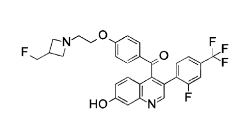
Example 1
(4-{243-(Fluoromethyl)azetidin-1-yl]ethoxy) phenyl ){342-fluoro-4-
(trifluoromethyl )phenyll -7-hydroxyquinolin-4-yllmethanone
o
0
I
HO N F
To a vessel, equipped with a N2 inlet, add THF (2.8 L), potassium tert-
butoxide
(274.5 g, 2.45 mol) and 2-(3-(fluoromethyl)azetidin-1-yl)ethan-1-ol (168 g,
1.22 mol). Stir
the mixture for 10 minutes. Add dropwise a solution of (4-fluoropheny1)-[342-
fluoro-4-
(trifluoromethyl)pheny1]-7-hydroxy-4-quinolyl]methanone (350 g, 0.81 mol) in
TI-1F (0.7 L).
Stir at room temperature for one hour. Quench the reaction with 1 N HC1 until
pH 8 and
dilute with Et0Ac (4 L). Separate the organic layer and wash it with brine (2
L). Dry the
solution over magnesium sulfate, filter the solution, and concentrate to
dryness to give the
title compound. Following this preparation gave 415 g (93.8%) of the title
compound as a
pale brown solid with an ES/1\4S (m/z) of 543.2 (M+H).
Alternate Example 1
Degas/purge with N2 x 5 a mixture (3-chloro-7-hydroxyquinolin-4-y1)-(4-{243-
(fluoromethyl)azetidin-1-yl]ethoxy}phenyl)methanone (200 mg, 0.48 mmol), 2-
fluoro-4-
(trifluoromethyl)phenylboronic acid (158 mg, 0.72 mmol), potassium carbonate
(202 mg,
1.45 mmol), 2-methyl-2-butanol (3 ml), and water (1 ml) in a microwave vial.
Add XPhos
Pd G2 (12 mg, 0.015 mmol), seal the mixture, and microwave at 80 C for 2
hours. Partition
the residue between MTBE and saturated ammonium chloride solution. Separate
the layers
and extract the aqueous with MTBE. Combine the organic extracts, dry them over
magnesium sulfate, filter, and concentrate the filtrate to obtain an orange
residue. Purify the
crude material by silica gel column chromatography eluting with of 5% Me0H/DCM
to give
CA 03105491 2020-12-31
WO 2020/014440
PCT/US2019/041342
-12-
the title compound. Following this preparation gave 205 mg (78%) of the title
compound as
a yellow solid with ES/MS (m/z) of 543.2 (M+H).
EXAMPLE 2
Racemic 5-(4-{ 243-(Fluoromethyl)azetidin-1-yl]ethoxy}pheny1)-8-
(trifluoromethyl)-5H-
[1]benzopyrano[4,3-c]quinolin-2-ol
0
H 0
Cool a solution of (4-1243-(fluoromethyl)azetidin-1-yl]ethoxy}pheny1){342-
fluoro-
4-(trifluoromethyl)phenyl]-7-hydroxyquinolin-4-yllmethanone (5.27 g, 9.71
mmol) in 1,4-
dioxane (100 mL) to 5 C. Add lithium triethylborohydride (1 M in THF, 30.0
mL, 30.0
mmol). Remove the cooling bath and stir for 1.5 hours at room temperature.
Quench the
mixture with water. Add saturated NH4C1 solution and Et0Ac. Separate the
layers and
extract the aqueous layer with Et0Ac. Combine the organic extracts, dry over
anhydrous
MgSO4, filter, and concentrate the filtrate. Dissolve the crude residue in TI-
1F (100 mL).
Add sodium hydride (60% in mineral oil, 1.94 g, 48.5 mmol). Heat Reflux the
solution for
1.5 hours. Add additional sodium hydride (60% in mineral oil, 1.94 g, 48.5
mmol), then
reflux for an additional 30 minutes. Cool the solution to room temperature and
quench with
water. Add Et0Ac and saturated NH4C1 solution. Separate the layers and extract
the
aqueous layer with Et0Ac. Combine the organic extract, dry over anhydrous
MgSO4, filter,
and concentrate the filtrate. Purify the residue by silica gel column
chromatography eluting
with a gradient of 5-7% Me0H in DCM to give the title compound (3.70 g, 72%)
as a light
yellow foam. ES/1\4S (m/z): 525.2 (M+H).
Biological Assays
The relationship between estrogen receptor expression and certain cancers has
been
reported in the literature, (for breast cancer see, e.g., Puhalla S,
Bhattacharya S, Davidson N.,
Hormonal therapy in breast cancer: A model disease for the personalization of
cancer care,
CA 03105491 2020-12-31
WO 2020/014440
PCT/US2019/041342
-13-
Molecular Oncology, 2012, 6:222-236; Kennecke H, Yerushalmi R, Woods R, Cheang
MCU, Voduc D, Speers CH, Nielsen TO, Gelmon K, Metastatic behavior of breast
cancer
subtypes, J Clin Oncol, 2010, 28(20):3271-3277; for ovarian cancer see, e.g.,
O'Donnell AJ,
Macleod KG, Bums DJ, Smyth JF, Langdon SP, Estrogen receptor-alpha mediates
gene
expression changes and growth response in ovarian cancer cells exposed to
estrogen, Endocr
Relat Cancer, 2005;12(4):851-66; Walker G, MacLeod K, Williams AR, Cameron DA,
Smyth JF, Langdon SP, Estrogen regulated gene expression predicts response to
endocrine
therapy in patients with ovarian cancer, Gynecol Oncol, 2007, 106(3):461-8;
Smyth JF,
Gourley C, Walker G, MacKean MJ, Stevenson A, Williams AR, et al.,
Antiestrogen therapy
is active in selected ovarian cancer cases: The use of letrozole in estrogen
receptor-positive
patients, Clin Cancer Res, 2007, 13(12):3617-22; for prostate cancer see,
e.g., Bonkohoff H,
Fixemer T, Hunsicker I and Remberger K, Estrogen receptor expression in
prostate cancer
and premalignant prostate lesions, Am J Pathol, 1999, 155:641-647; for
endometrial and
uterine cancer see, e.g., Krasner C, Aromatase inhibitors in gynecologic
cancer, J Steroid
Biochem Mol Biol, 2007, Aug¨Sep;106(1-5):76-80; Boisen MM, Andersen CL,
Sreekumar
S, et al., Treating gynecologic malignancies with selective estrogen receptor
downregulators
(SERDs): Promise and challenges, Mol Cell Endocrinol, 2015, 418:322-3330; For
lung
cancer see, e.g., Baik CS, Eaton KD et al., Estrogen signaling in lung cancer:
An opportunity
for novel therapy, Cancer, 2012, 4:969-988; Marquez-Garban DC, Chen H-W,
Goodglick L,
Fishbein MC and Pietras RJ, Targeting aromatase and estrogen signaling in
human non-small
cell lung cancer. Steroid enzymes and cancer, Ann. N. Y. Acad Sci, 2009,
1155:194-205;
Hamilton DI-I, Griner LM, Keller JM, Hu X, Southall N, Marugan J, David JM,
Ferrer M and
Palena C, Targeting estrogen receptor signaling with fulvestrant enhances
immune and
chemotherapy mediated cytotoxicity of human lung cancer, Clin Cancer Res,
2016,
22(24):6204-16; Rodriguez-Lara V, Hernandez-Martinez JM, Arrieta 0, Influence
of
estrogen in non-small cell lung cancer and its clinical implications, J
Thoracic Disease, 2018,
10(1):482-497; for gastric cancer see, e.g., Tang W, Liu R, Yan Y, Pan X, Wang
M, Han X,
Ren H, and Zhang Z, Expression of estrogen receptors and androgen receptor and
their
clinical significance in gastric cancer, Oncotarget, 2017, 8(25) 40765-777).
-14-
The following assays demonstrate that the exemplified compounds are potent
degraders of ERa wild type and mutant proteins. The results of the assays also
demonstrate
that the exemplified compounds are potent antagonists of ERa wild type and
mutant
receptors and inhibit ER-mediated transcriptional activity. Additionally, the
assays
.. demonstrate that the compound of example 1 inhibits proliferation of ER+
breast cancer cell
lines, and ERa signalling and tumor growth inhibition in a ER-positive breast
cancer
xenograft model.
ERa (wild type) and ERa (Y537S mutant) competition binding assay
The purpose of the following ER competition binding assays is to determine the
binding affinity of a test compound against ERa (wild type) and ERa (Y537S
mutant) See
Fanning et al., "Estrogen receptor alpha somatic mutations Y537S and D538G
confer breast
cancer endocrine resistance by stabilizing the activating function-2 binding
conformation,"
eLife 2016;5:e12792.
Run the competition binding assay in a buffer containing 50 mM HEPES, pH 7.5,
1.5
mM EDTA, 150 mM NaCl, 10% glycerol, 1 mg/mL ovalbumin, and 5 mM DTT, using
0.025
ttCi per well 3H-estradiol (118 Ci/mmol, 1 mCi/mL), 7.2 ng/well ERa (wild
type), or 7.2
ng/well ERa (Y537S mutant). Add the test compound at 10 different
concentrations ranging
from 10,000 nM to 0.5 nM, and determine nonspecific binding in the presence of
1 1.1A4 of
17-13 estradiol. Incubate the binding reaction (140 L) for 4 hours at room
temperature, and
then add cold dextran-charcoal buffer (70 L) (containing per 50 mL of assay
buffer, 0.75 g
of charcoal and 0.25 g of dextran) to each reaction. Mix the plates for 8
minutes on an
orbital shaker at 4 C and then centrifuge at 3000 rpm at 4 C for 10 minutes.
Transfer an
aliquot (120 ttL) of the mixture to another 96-well, white flat bottom plate
(Costar) and
.. add Perkin Elmer Optiphase Supermix scintillation fluid (175 L) to each
well. Seal the
plates and shake vigorously on an orbital shaker. After an incubation of 2.5
hours, read the
plates in a Wallac Microbeta counter. Calculate the IC50 using a 4-parameter
logistic curve
fit and calculate % inhibition at 10 M. Convert the IC50 values for the
compound to Ki
using Cheng-Prusoff equation. The results of this assay demonstrate that the
compound of
Date Recue/Date Received 2022-11-15
CA 03105491 2020-12-31
WO 2020/014440
PCT/US2019/041342
-15-
Example 1 binds to recombinant ERa wild type with a Ki (nM) of 3.78 + 0.74
(n=3) and
binds to ERa mutant (Y537S) with a Ki (nM) of 21.24 + 2.12 (n=3).
The results of this assay demonstrate the binding affinity and potency of
exemplified
compound against ERa wild type and mutant (ESR1 Y537S) proteins.
ERa degradation assay in MCF7 cells
The purpose of the following ERa degradation assay is to measure the
degradation of
ERa by a test compound in an ERa positive breast cancer cell line such as
MCF7.
Culture MCF7 (purchased from ATCC HTB-22) cells in DMEM media supplemented
with 10% FBS, 0.01 mg/mL human insulin 1 and 1% penicillin/streptomycin
antibiotics and
plate in 384-well flat-bottom plates at a density of 4,000 cells per well in
phenol red free
DMEM media (20 L) containing 10% charcoal stripped FBS. Incubate the cells
overnight
in a cell culture incubator (5% CO2, 95% relative humidity and 37 C) and
allow the cells to
attach to the plate. The following day dose the cells with the test compound.
Use an Echo
555 acoustic dispenser to prepare test compound serial dilutions (1:3) in a
range from 6 pM
to 0.0003 pM. Dose the cells with the addition of 5 pL from the serial
dilution plate to the
cell plate producing a final DMSO concentration of 0.2% with a final test
compound
concentration dose range between 2 and 0.0001 p.M. For the maximum point, use
media
containing 0.2% of DMSO and for the minimum point, use fulvestrant diluted at
2 pM final
concentrations in the growth media containing 0.2% DMSO. After dosing with the
test
compound, incubate the cell plates at 37 C and 5% CO2 for 24 hours. Fix the
cells by
adding 14% para-formaldehyde (10 pL) for 30 minutes at room temperature. Wash
the cells
once with PBS (20 pt) and incubate with PBS (20 pL) containing 0.5% (v/v)
TWEEN 20
for 1 hour. Wash the cells with PBS containing 0.05% TWEEN 20 (2x) and block
with 3%
BSA in PBS containing 0.05% TWEEN 20 and 0.1% TRITONTm X-100 (20 pL/well) for
1
hour at room temperature. Add 1:500 Primary antibody (20 pL) (ERa (Clone SP1)
monoclonal rabbit antibody #RM-9101-S, Thermo Scientific) dilution in 1% BSA
in PBS
containing 0.05% TWEEN 20 per well, seal the plates and incubate overnight at
4 C. The
following day wash the cells with PBS containing 0.05% TWEEN 20 (2x) and
incubate
CA 03105491 2020-12-31
WO 2020/014440
PCT/US2019/041342
-16-
with secondary antibody (20i...it/well) (1:1000 dilution, Goat anti-rabbit IgM
ALEXA
FLUORTM 488) in PBS 1% BSA for 105 minutes at room temperature. After washing
plates
with PBS (2x20 L), add RNase (Sigma) (20 RL of 50 g/mL) and 1:1000 propidium
iodide
dilution in PBS per well (20 L). Seal the plates and incubate 1 hour at room
temperature on
.. the bench (preserved from light). Scan the plates with ACUMEN EXPLORERTM
[Laser-
scanning fluorescence microplate cytometer manufactured by TTP LABTECH LTD] to
measure ERa. Image analysis is based on cellular fluorescent signals for
identifying positive
cells. Identify estrogen receptor positive cells by mean intensity. Use total
intensity at 575-
640 nm from propidium iodide/DNA to identify individual cells. Assay output is
% estrogen
.. receptor positive cells. Determine the IC50 by curve fitting to a four
parameter logistic for
each output using GENE DATATm.
The results of this assay demonstrate that the compound of formula (I) is a
SERD
with potent ERa degradation activity in cells. Specifically, the results show
potent
degradation of ERa by the compound of Example 1 in MCF7 breast cancer cells.
Using this
assay, the Relative IC50 ( M) value for the compound of Example 1 is 2.16 +
0.96 nM
(n=15).
PRa, induction assay in MCF7 cells
The purpose of the following PRa induction assay is to determine whether a
test
compound has agonistic activity against ERa receptor (an agonist would be
expected to
activate the receptor.)
Culture MCF7 (purchased from ATCC HTB-22) in DMEM media supplemented
with 10% FBS, 0.01 mg/mL human insulin 1 and 1% penicillin/streptomycin
antibiotics and
plate the cells (prior to becoming 70% confluent) in 384-well flat-bottom
plates at a density
of 4,000 cells per well in 20 !IL volume in DMEM phenol red free media
containing 10%
FBS (charcoal stripped). Incubate the cells overnight in a cell culture
incubator (5% CO2,
95% relative humidity at 37 C) and allow the cells to attach to the plate.
The following day,
dose the cells with test compound. Use an Echo 555 acoustic dispenser to
prepare compound
serial dilutions (1:3) in a range from 6 [.EM to 0.0003 M. Dose the cells
with the addition of
CA 03105491 2020-12-31
WO 2020/014440
PCT/US2019/041342
-17-
the test compound (5 ttL) from the serial dilution plate to the cell plate
producing a final
DMSO concentration of 0.2% with a final concentration of the test compound
dose range
between 2 and 0.0001 RM. For the maximum point use media containing 0.2% of
DMSO
and for the minimum point, use fulvestrant diluted at 2 jiM final
concentrations in the growth
media containing 0.2% DMSO. After dosing with the test compound, incubate the
cell plates
at 37 C and 5% CO2 for 24 hours. Fix the cells by adding 14% para-
formaldehyde (10 tiL)
for 30 minutes at room temperature. Wash cells once with PBS (20 p.L) and
incubate with
PBS (20 [iL) containing 0.5% (v/v) TWEEN 20 for 1 hour. Wash cells twice with
PBS (20
L) containing 0.05% TWEEN 20 and block with 3% BSA in PBS containing 0.05%
TWEEN 20 and 0.1% TRITONTm X-100 (20 pt/well) for 1 hour at room temperature.
Add 1:500 primary antibody (20 [IL) (Progesterone receptor monoclonal mouse
anti-human
antibody, clone PgR 636 Dako, M3569) dilution in 1% BSA/PBS with 0.05 TWEEN
20
per well, seal the plates and incubate overnight at 4 C.
The following day, wash cells with PBS 0.05% TWEEN 20 (2><20 [IL) and
incubate
with secondary antibody (20 [IL/well) (1:1000 dilution, Goat anti-rabbit IgM
ALEXA
FLUORTM 488) in PBS 1% BSA for 105 minutes at room temperature. After washing
with
PBS (2x20 !IL), add RNase (20 pt of 50 pg/mL) (Sigma) and 1:1000 propidium
iodide
dilution in PBS per well. Seal plates and incubate 1 hour at room temperature
on the bench
(preserved from light). Scan plates with ACUMEN EXPLORERTM [Laser-scanning
fluorescence microplate cytometer manufactured by TTP LABTECH LTD] to measure
progesterone receptor alpha. Image analysis is based on cellular fluorescent
signals for
identifying positive cells. Identify progesterone receptor positive cells by
mean intensity.
Use total intensity at 575-640 nm from propidium iodide/DNA to identify
individual cells.
Assay output is % progesterone receptor positive cells. Determine the IC50 by
curve fitting to
a four parameter logistic for each output using GENE DATATm.
Using this assay, the Relative IC50 (tiM) of the compound of Example 1 is > 2
p.M.
The results of this assay demonstrate no significant agonistic activity of
Example 1 in MCF7
breast cancer cells. These results also demonstrate that the compound of
Example 1 is a pure
antagonist of ERa in MCF7 breast cancer cells.
CA 03105491 2020-12-31
WO 2020/014440
PCT/US2019/041342
-18-
PRa inhibition (ERa, functional antagonism) cell assay in MCF7-ESR1 Y537N 682
CRISPR cells
The purpose of the following PRa. inhibition (ERa functional antagonism) cell
assay
is to determine the antagonistic activity of a test compound against the Y537N
mutant ERa
receptor. An antagonist in this assay is expected to block the function of the
ERa receptor.
PRa (PGR) is a downstream transcriptional target of ERa and hence an
antagonist of ERa is
expected to inhibit the expression of PRa.
Culture MCF7-ESR1 Y537N-682 (generated by CRISPR/Cas9 gene editing of ESR1
gene in MCF7 cells, clone#682) in DMEM media supplemented with 10% FBS and 1%
penicillin/streptomycin antibiotics and plate the cells (prior to becoming 70%
confluent) in
384-well flat-bottom plates at a density of 4,000 cells per well in DMEM
phenol red free
media 10% FBS (20 [IL volume) (charcoal stripped). Incubate the cells
overnight in a cell
culture incubator (5% CO2, 95% relative humidity and 37 C) and allow the
cells to attach to
the plate. The following day dose the cells with the test compound. Use an
Echo 555
acoustic dispenser to prepare compound serial dilutions (1:3) in a range from
6 RM to 0.0003
pIVI. Dose the cells with the addition of 5 pL from the serial dilution plate
to the cell plate
producing a final DMSO concentration of 0.2cY0 with a final test compound
concentration
dose range between 2 and 0.0001 pM. For the maximum point use media containing
0.2% of
DMSO and for the minimum point, use fulvestrant diluted at 2 pM final
concentrations in the
growth media containing 0.2% DMSO. After dosing with test compound, incubate
the cell
plates at 37 C and 5% CO2 for 72 hours. Fix the cells by adding 14% para-
formaldehyde
(10 ML) for 30 minutes at room temperature. Wash the cells with PBS (1x20 ML)
and
incubate with PBS (20 [IL) of containing 0.5% (v/v) TWEEN 20 for 1 hour. Wash
the cells
with PBS (2x20 ML), 0.05% TWEEN 20, and block with 3% BSA/PBS 0.05% TWEEN
20, 0.1% TRITONTm X-100 (20 p.L/well) for 1 hour at room temperature. Add
1:500
primary antibody (20 1,) (Progesterone receptor monoclonal mouse anti-human
antibody,
clone PgR 636 Dako, M3569) dilution in 1% BSA/PBS 0.05 TWEEN 20 per well,
seal the
plates and incubate overnight at 4 C.
CA 03105491 2020-12-31
WO 2020/014440
PCT/US2019/041342
-19-
The following day, wash the cells with PBS 0.05% (2x20 L) and incubate with
secondary antibody (20 ML/well) (1:1000 dilution, Goat anti-rabbit IgM ALEXA
FLUORTM
488) in PBS 1% BSA for 105 minutes at room temperature. After washing with PBS
(2x20
L), add RNase (20 L of 50[1g/mL) (Sigma) and 1:1000 propidium iodide dilution
in PBS
per well. Seal the plates and incubate 1 hour at room temperature on the bench
(preserved
from light). Scan the plates with ACUMEN EXPLORERTM [Laser-scanning
fluorescence
microplate cytometer manufactured by TTP LABTECH LTD] to measure progesterone
receptor alpha. Image analysis is based on cellular fluorescent signals for
identifying
positive cells. Identify progesterone receptor positive cells by mean
intensity. Use total
intensity at 575-640 nm from propidium iodide/DNA to identify individual
cells. Assay
output is % progesterone receptor positive cells. Determine the IC50 by curve
fitting to a four
parameter logistic for each output using GENE DATATm.
Using this assay, the Relative IC50 (nM) of the compound of Example 1 is 7.602
+
4.804 nM (n=14). These results demonstrate potent inhibition of PRa and
functional
antagonism by Example 1 in MCF7 (ESR1 Y537N, heterozygous mutant) breast
cancer cells.
As such, the compound of Example 1 is a potent antagonist of ERa mutant
(Y537N) and a
potent inhibitor of ERa mediated transcription. PRa (PGR) is also a
transcriptional target of
ERa and the results from this assay demonstrate potent inhibition of ERa-
mediated
transcription of PRa.
PRa inhibition (ERa functional antagonism) cell assay in MCF7 cells
The purpose of the following Plta inhibition (ERa functional antagonism) cell
assay
is to determine the antagonistic activity of a test compound against the ERa
receptor. An
antagonist in this assay is expected to block the function of the ERa
receptor. Pita is a
downstream transcriptional target of ERa and hence an antagonist of ERa is
expected to
inhibit the expression of PRa.
Carry out the assay conditions as detailed in the ERa degradation Cell base
Acumen
assay above, using the MCF7 cell line except that, prior to test compound
dispensing, remove
the media from the cell plate and pretreat all wells except for the negative
control wells
CA 03105491 2020-12-31
WO 2020/014440
PCT/US2019/041342
-20-
(column 24 of the plate) with assay media containing 0.47 nM estradiol for 30
minutes. In
this assay, carry out immunostaining for the detection of PRa and scan the
plates with
ACUMEN EXPLORERTM [Laser-scanning fluorescence microplate cytometer
manufactured
by TTP LABTECH LTD] to measure PRa. Image analysis is based on cellular
fluorescent
signals for identifying positive cells. Identify PRa positive cells by mean
intensity. Use total
intensity at 575-640 from propidium iodide/DNA to identify individual cells.
Assay output is
% PRa positive cells. Determine the IC50 by curve fitting to a four parameter
logistic for
each output using GENE DATATm.
Using this assay, the Relative IC50 (nM) of the compound of Example 1 in this
assay
is 15.75 + 9.037 nM (n=15). The results of this assay demonstrate potent
inhibition of PRa
and functional antagonism by Example 1 in MCF7 breast cancer cells. As such,
the
compound of Example 1 is a potent antagonist of ERa wild-type protein and a
potent
inhibitor of ERa mediated transcription. PRa (PGR) is also a transcriptional
target of ERa
and the results from this assay demonstrate potent inhibition of ERa-mediated
transcription
of PRa.
Cell Proliferation Assay in MCF7 and MCF7-ESR1 Y537N-682
The purpose of the following cell proliferation assays generally is to detect
whether a
test compound has effects on cell proliferation, cell viability, and
cytotoxicity in response to
treatment in cell culture experiments. Cell proliferation is monitored by
monitoring the
number of cells over time and the propodeum iodide assay used allows
continuous
measurement of cell viability over time.
Seed MCF7 (purchased from ATCC HTB-22) cells at a density of 2,000 cells per
well in DMEM phenol red free media 10% FBS (20 [IL volume) (charcoal stripped)
into a
clear bottom 384-well cell culture plate. Plate MCF7-ESRY537N -682 (generated
by
CRISPR/Cas9 gene editing of ESR1 gene in MCF7 cells, clone#682) in DMEM media
CA 03105491 2020-12-31
WO 2020/014440
PCT/US2019/041342
-21-
supplemented with 10% FBS, and 1% penicillin/streptomycin antibiotics at a
density of 1000
cells per well. Incubate the plates at 37 C and 5% CO2. The following day
dose the cells
with the test compound. Use an Echo 555 acoustic dispenser to prepare test
compound serial
dilutions (1:3) in a range from 60 jiM to 0.003 p.M. Dose the cells with the
addition of 5 ?AL
from the serial dilution plate to the cell plate, producing a final DMSO
concentration of 0.2%
with a final test compound concentration dose range between 20 and 0.001 !.EM.
For the
maximum point use media containing 0.2% of DMSO and for the minimum point use
fulvestrant diluted at 2 1.tM final concentrations in the growth media
containing 0.2% DMSO.
After dosing with the test compound, incubate the cell plates at 37 C and 5%
CO2. Seven
days after test compound addition, remove the plates from the incubator and
add cold ethanol
96% (65 p.L) to each well. After 30 minutes, remove the media and add RNase
(20 [IL of 50
[tg/mL) (Sigma) and 1:1000 propidium iodide dilution in PBS per well. Seal the
plates and
incubate 1 hour at room temperature on the bench (preserved from light). Scan
the plates
with ACUMEN EXPLORERTM [Laser-scanning fluorescence microplate cytometer
manufactured by TTP LABTECH LTD]. The MCF-7 cell line grows forming
aggregates,
cell number as number of objects may not be able to be used as readout; so the
cell number
may be evaluated through estimated number of cells (calculated through the
area parameter
(ratio of total area of the total cells population (a designated range of peak
intensity of FL-1
(PI) and the mean area of the single cells population (defined by perimeter)).
Determine the
IC50 by curve fitting to a four parameter logistic for each output using GENE
DATATm.
Using this assay, the Relative IC50 (nM) of the compound of Example 1 in MCF7
ESR1 wild type is 9.243 + 1.741 nM (n=2) and in MCF7-ESR1 Y537N mutant cells
is 7.960
+ 3.691 nM (n=6). These results demonstrate potent anti-proliferative activity
and cell
growth inhibition by Example 1 in MCF7 (ESR1 wild type) and MCF7 (ESR1 Y537N
mutant) breast cancer cells.
In Vivo Target inhibition (IVTI) Assay (PGR RT-qPCR assay) in MCF7 tumors
CA 03105491 2020-12-31
WO 2020/014440
PCT/US2019/041342
-22-
The purpose of this IVTI assay is to measure the ability of a test compound
(SERD)
to inhibit PGR (Progesterone Receptor alpha) gene expression (transcription)
downstream of
ERa in xenograft tumors implanted in mice.
Implant female NOD SC1D mice (22-25 g) from Envigo RMS, Inc., Madison,
Wisconsin with 5 xl0e6 MCF7 ER+ve breast cancer cells (ATCC, # HTB-22)
subcutaneously
in the right flank region in 1:1 HBSS + MATRIGELTm solution (200 1.1L).
Implant a 17-
13 pellet (0.18 mg/pellet, 90 day release, from Innovative research)
subcutaneously 1
day prior to tumor cell implantation. Measure tumor growth and body weight
twice per week
beginning the seventh day after the implantation. When tumor sizes reach 250-
350 mm3,
.. randomize animals and group into groups of five animals. Dose animals with
either the test
compound in a specific vehicle (1% hydroxyethylcellulose/0.25% TWEEN 80/0.05%
Antifoam in purified water) or vehicle alone orally for 3 days and collect
tumors and blood at
desired time intervals after last dose. Sacrifice animals using isoflurane
anesthesia plus
cervical dislocation. Flash freeze tumors and store at -80 C until processing
for RNA
isolation and RT-qPCR assay. Collect blood in EDTA tubes, spin down for
plasma, and
freeze at -80 C in a 96-well plate. Determine test compound exposures using
mass
spectrometry.
Pulverize tumors in liquid nitrogen and lyse in lxRNA lysis buffer (from RNA
isolation kits) using Matrix D beads (MP Biomedical, #6913-500) in a FASTPREP-
24m4 Cell
Disrupter machine (MP Biomedical). Transfer tumor lysates to fresh tubes after
spinning at
14000 rpm for 20 minutes at 4 C. Isolate RNA from tumor lysates using
PURELINK
RNA Mini Kit (Invitrogen #12183018A) or RNeasy Mini Kit (Qiagen #74104 and
#74106).
Remove DNA contaminants using PURELINK DNase Set (Invitrogen #12185010) or
RNase-Free DNase Set (Qiagen #79254). Measure isolated RNA concentration by
diluting
samples in RNase free water and measuring the absorbance at 260 nm on a plate
reader
(SpectraMax190). Subtract the average 260 nm absorbance measurement of the
blank
(RNase free water only) from the 260 nm measurements of all other RNA samples.
Dilute
RNA samples to equal concentrations in RNase free water. Synthesize cDNA from
diluted
RNA using First-Strand Synthesis System for RT-PCR (Invitrogen, #18080-051).
To
CA 03105491 2020-12-31
WO 2020/014440
PCT/US2019/041342
-23-
perform RT-qPCR, first dilute cDNA in RNase free water. Combine 2x Absolute
Blue
qPCR ROX Mix (Thermo, #AB-4139/A), PGR primer (Thermo, Hs01556702 ml), and
diluted cDNA for each reaction in a PCR plate (Applied Biosystems, #4309849).
Amplify
cDNA by incubating the samples for 2 minutes at 50 C followed by 15 minutes
at 95 C in
the thermocycler (ABI Prism 7900HT Sequence Detection System). Continue to
incubate at
95 C for 15 seconds followed by 50 C for 60 seconds for a total of 40
cycles. Cycles are
normalized to the housekeeping gene and used to calculate % PGR inhibition
compared to
the vehicle alone. Analyze each sample in duplicate and use average numbers
for
calculations. Calculate the percent target (PGR) inhibition using Excel and XL
Fit.
The results of this assay demonstrates that the compound of Example 1 inhibits
PRa
(PGR) expression in the tumor xenograft model. Additionally, the compound of
Example 1
inhibits PRa (PGR) expression by 57% in the tumor xenograft model for 24 hours
with 30
mg/kg dose when administered orally. These results demonstrate significant and
sustained
inhibition of ERa antagonistic activity and ERa-mediate transcriptional
activity in vivo in a
.. tumor xenograft model.
In vivo tumor growth inhibition study in MCF7 xenograft tumor implanted in
mice
The purpose of the following xenograft tumor growth inhibition assay is to
measure
reduction in tumor volume in response to test compound administration.
Expand human breast cancer cells MCF7 (ATCC # HTB-22) in culture, harvest and
inject 5 xl0e6 cells in 1:1 HBSS+MATRIGELTmsolution (200 ,L) subcutaneously
on to the
rear right flank of female NOD SC1D mice (22-25 g, Envigo RMS, Inc). Twenty-
four hours
prior to implantation of MCF7 cells, implant estrogen pellets (0.18 mg/pellet,
1713 estradiol,
90-day release, Innovative Research) subcutaneously. Measure tumor growth and
body
weight twice per week beginning the seventh day after the implantation. When
tumor sizes
reach 250-350 mm3, randomize animals and group into groups of 5 animals.
Prepare the test
compound in an appropriate vehicle (1% hydroxyethylcellulose/0.25% TWEENO
80/0.05%
Antifoam in purified water) and administer by oral gavage for 42 days.
Determine tumor
response by tumor volume measurement performed twice a week during the course
of
CA 03105491 2020-12-31
WO 2020/014440
PCT/US2019/041342
-24-
treatment. Take the body weight as a general measure of toxicity whenever
tumor volume is
measured.
When used in this assay, the compound of Example 1 is found to have delta T/C%
values as provided in Table 6 below. These results indicate that the compound
of Example 1
demonstrates good oral bioavailability in mice and significant anti-tumor
activity or tumor
regressions in an MCF7 human breast cancer xenograft model.
In vivo tumor growth inhibition study in MCF7 xenograft tumor implanted in
mice
Table 6
Delta T/C% or
Tumor Model Dose (mg/kg) Schedule p-value
Regression /0
MCF7 (Breast
Cancer 30 QD -36 <0.001*
Xenograft)
Analysis for tumor volume is based on Log 10 and SpatialPower covariance
structure.
*: significant (p<0.05) compared to vehicle control.
Delta T/C% is calculated when the endpoint tumor volume in a treated group is
at or above
baseline tumor volume. The formula is 100*(T-To)/(C-Co), where T and C are
mean
endpoint tumor volumes in the treated or control group, respectively. To and
Co are mean
baseline tumor volumes in those groups.
Regression% is calculated when the endpoint volume is below baseline. The
formula is
100*(T-To)/To, where To is the mean baseline tumor volume for the treated
group.
Grand mean of all groups from baseline (randomization) at day 32 is used to
compute %
change of TIC.
Rat Oral Bioavailability Assay
The purpose of the following assay is to demonstrate whether a test compound
is
orally bioavailable.
CA 03105491 2020-12-31
WO 2020/014440
PCT/US2019/041342
-25-
Administer the test compound to Sprague-Dawley rats IV at 1 mg/kg (using
vehicles
of either: 20% CAPTISOL in 25 mM sodium phosphate buffer, pH2 quantum sails;
or 25%
DMA, 15% Et0H, 10% propylene glycol, 25% 2-pyrrolidone, and 25% purified
water) and
PO at 10 mg/kg (using a vehicle of 1% hydroxyethyl cellulose, 0.25%
polysorbate 80, 0.05%
Antifoam 1510-US, and purified water quantum sails). Collect serial blood
samples at 0.08,
0.25, 0.5, 1, 2, 4, 8, and 12 hours post dose for IV bolus and at 0.25, 0.5,
1, 2, 4, 8, and 12
hours post dose after oral administration. After treatment with an EDTA
coagulant, obtain
plasma by centrifugation and stored at -70 C until analysis by LC-MS/MS.
Determine the
test compound concentration in plasma and upload into the Watson LrmsTm system
where
noncompartmental analysis is used to calculate Area Under the Curve (AUC) for
both IV and
PO arms. Calculate oral bioavailability (%F) via the following equation,
%F = (A UCpo X Doseiv) / (A UCry X Dosepo) X 100.
Using this assay, the compound of Example 1 displays a %F value of 27%. This
assay demonstrates that Example 1 has good oral bioavailability.