Note: Descriptions are shown in the official language in which they were submitted.
CA 03106002 2021-01-08
WO 2020/011973
PCT/EP2019/068804
Antibody molecules
Field of the Invention
The invention relates to antibodies and antigen-binding fragments thereof that
bind to
programmed-death ligand 1 (PD-L1). The antibodies or antigen-binding fragments
thereof
comprise a CDR-based antigen-binding site for PD-L1. Antibodies or antigen-
binding fragments
thereof of the invention may find application, for example, in cancer therapy.
Background to the invention
Programmed cell death 1 (PD-1) is a cell-surface receptor, its ligands PD-L1
(0D274, B7-H1) and
PD-L2 (B7-DC) deliver inhibitory signals that regulate the balance between T
cell activation,
tolerance, and immunopathology. PD-L1 is transiently expressed on all immune
cells and some
tumour cells.
PD-L1 is a type I transmembrane protein with two Ig-like domains within the
extracellular region,
a transmembrane domain and a short cytoplasmic domain. The complete human PD-
L1 (hPD-
L1) sequence can be found under GENBANK Accession No. Q9NZQ7. The cytoplasmic
domain
has no known signal transduction motif suggesting that there is no signalling
by PD-L1 on
interaction of the ligand with its receptor. The molecular weight of PD-L1 is
40 kDa (290 amino
acids), it is encoded by the 0D274 gene on human chromosome 9 and on mouse
chromosome
19. PD-L1 is a member of the B7 protein family and shares approximately 20 %
amino acid
sequence identity with B7.1 and B7.2. Human PD-L1 shares 70% and 93% amino
acid identity
with the murine and cynomolgus orthologs of PD-L1, respectively.
Human PD-L1 binds to its receptor, PD-1, with an affinity (KD) of 770 nM. PD-1
is expressed on
activated T cells, B cells, and myeloid cells; it modulates activation or
inhibition of cellular immune
responses. Binding of PD-L1 to PD-1 delivers an inhibitory signal, reducing
cytokine production
and suppressing proliferation of T cells. Consequently, PD-L1 expression by
cells can mediate
protection against cytotoxic T lymphocyte (CTL) killing and is a regulatory
mechanism that
dampens chronic immune responses during viral infections. Cancer, as a chronic
and pro-
inflammatory disease, subverts this immune-protective pathway through up-
regulation of PD-L1
expression to evade the host immune response. In the context of an active
immune response,
IFNy also upregulates the expression of PD-L1. PD-L1 also mediates immune
suppression
through interaction with another protein, B7.1 (also known as CD80), blocking
its ability to deliver
one of the secondary signals of activation on T cells through CD28. In terms
of PD-L1 expression
1
CA 03106002 2021-01-08
WO 2020/011973
PCT/EP2019/068804
on tumour cells and its engagement with B7.1, the relevance of this specific
interaction in tumour
immune resistance is still unclear.
PD-L1 expression has been shown in a wide variety of solid tumours. Of 654
samples examined
in one study, spanning 19 tumours from different sites, 89 (14%) were PD-L1
positive (5%
frequency). The highest PD-L1 positive frequencies were seen in head and neck
(17/54; 31%),
cervical (10/34; 29%), cancer of unknown primary origin (CUP; 8/29; 28%),
glioblastoma
multiforme (GBM; 5/20; 25%), bladder (8/37; 21%), oesophageal (16/80; 20%),
triple negative
(TN) breast (6/33; 18%), and hepatocarcinoma (6/41; 15%) (Grosso et al.,
2013). Tumour-
associated expression of PD-L1 has been shown to confer immune resistance and
potentially
protect tumour cells from T cell mediated apoptosis.
Therapies targeting PD-L1 have shown excellent results in murine in vivo
studies. In the B16
murine model of melanoma, treatment with anti-PD-L1 therapy combined with
either GVAX or
FVAX vaccination strategies resulted in a significant effect both on survival
(30 days for control
vs 52 days for PD-L1-treated) and percentage of tumour-free (5%) animals upon
conclusion of
the study (Curran et al., 2010). Anti-PD-L1 therapy has also been used to
study the mechanism
of immune suppression in the P815 murine mastoma model. P815 cells injected
into mice
normally trigger a strong immune response, which results in their rejection.
When PD-L1 is
expressed on P815 cells, these cells escape immune attack, which in turn can
be negated through
administration of anti-PD-L1 antibodies (lwai et al., 2002). It is evident
that targeting the PD-1/PD-
L1 axis in immunogenic human cancers (Herbst et al, 2014) results in a
survival advantage
through stimulation of an anti-cancer immune response (Wolchok et al., 2013;
Larkin et al., 2015).
Atezolizumab (MPDL3280A, RG7466, TECENTRIQTm) is a humanized IgG1 antibody
which
binds to PD-L1. It is in clinical trials as a monotherapy and also in
combination with other biologic
and/or small molecule therapies for treatment of solid cancers, including
colorectal cancer, breast
cancer, non-small-cell lung carcinoma, bladder cancer, and renal cell
carcinoma. Treatment with
atezolizumab resulted in objective response rates (ORR) of 23% in NSCLC, 36%
melanoma, 33%
bladder, 14% in RCC, and 13% in head and neck cancers (Herbst et al., 2014;
Powles et al.,
2014).
In May 2016 the FDA granted accelerated approval to atezolizumab for locally
advanced or
metastatic urothelial carcinoma treatment after failure of cisplatin-based
chemotherapy; however,
the confirmatory trial failed to achieve its primary endpoint of overall
survival. In October 2016,
the FDA approved atezolizumab for the treatment of patients with metastatic
non-small cell lung
2
CA 03106002 2021-01-08
WO 2020/011973
PCT/EP2019/068804
cancer (NSCLC) who had disease progression during or following platinum-
containing
chemotherapy. Patients with EGFR or ALK genomic tumor aberrations are required
to have
disease progression on FDA-approved therapy for these aberrations prior to
receiving
atezolizumab. Atezolizumab in combination with avastin and standard
chemotherapy for some
patients with lung cancer is under FDA priority review, a decision being
expected by 5 September
2018. The most common adverse effects reported in clinical studies of
atezolizumab were fatigue,
decreased appetite, nausea, and infections; urinary tract infection was the
most common severe
adverse effect.
Avelumab (MSB0010718C, BAVENCIOTM) is a fully human IgG1 antibody which binds
to PD-L1
and is undergoing clinical testing in a number of cancers including bladder
cancer, gastric cancer,
head and neck cancer, mesothelioma, non-small-cell lung carcinoma, ovarian
cancer, renal
cancer and Merkel-cell carcinoma. Avelumab received orphan drug designation by
the European
Medicines Agency (EMA) for the treatment of gastric cancer in January 2017. In
2017 the FDA
and the EMA approved avelumab for Merkel-cell carcinoma (an aggressive skin
cancer) in adults
and pediatric patients 12 years and older. Approval was based on data from an
open-label, single-
arm, multi-center clinical trial (JAVELIN Merkel 200 trial). All patients had
histologically-confirmed
metastatic MCC with disease progression on or after chemotherapy administered
for metastatic
disease. The overall response rate (ORR) was assessed by an independent review
committee
according to Response Evaluation Criteria in Solid Tumors (RECIST) 1.1. The
ORR was 33%
(95% confidence interval [CI]: 23.3, 43.8), with 11% complete and 22% partial
response rates.
Among the 29 responding patients, the response duration ranged from 2.8 to
23.3+ months with
86% of responses durable for 6 months or longer. Responses were observed in
patients
regardless of PD-L1 tumor expression or presence of Merkel cell polyomavirus.
Safety data were
evaluated in 1738 patients. The most common serious adverse reactions to
avelumab were
immune-mediated adverse reactions (pneumonitis, hepatitis, colitis, adrenal
insufficiency, hypo-
and hyperthyroidism, diabetes mellitus, and nephritis) and life-threatening
infusion reactions.
Among the 88 patients enrolled in the JAVELIN Merkel 200 trial, the most
common adverse
reactions were fatigue, musculoskeletal pain, diarrhea, nausea, infusion-
related reaction, rash,
decreased appetite, and peripheral edema. Serious adverse reactions that
occurred in more than
one patient in the trial were acute kidney injury, anemia, abdominal pain,
ileus, asthenia, and
cellulitis.
Durvalumab (MEDI4736, IMFINZITm) is a human IgG1 antibody which binds to PD-L1
and is being
tested in clinical trials alone or in combination with tremelimumab in non-
small-cell lung cancer,
3
CA 03106002 2021-01-08
WO 2020/011973
PCT/EP2019/068804
squamous cell carcinoma of the head and neck, bladder cancer, pancreatic
cancer and with other
biologic and small molecules in trials for additional solid cancers such as
gastric cancers,
melanoma and unresectable hepatocellular carcinoma.
Durvalumab was approved by the FDA for the treatment of patients with locally
advanced or
metastatic urothelial carcinoma who either have disease progression during or
following platinum-
containing chemotherapy or have disease progression within 12 months of
neoadjuvant or
adjuvant treatment with platinum-containing chemotherapy.
A phase 1B clinical trial of durvalumab and tremelimumab showed some activity
in non-small cell
lung cancer (NSCLC). However, in July 2017, AstraZeneca announced that a phase
III trial of
durvalumab with tremelimumab as a first-line treatment of non-small cell lung
cancer had failed
to meet its primary endpoint of progression-free survival.
Early results of a phase I trial combining durvalumab and gefitinib in lung
cancer patients were
reported to have "showed promise". A phase 1 clinical trial is in progress
using durvalumab with
a TLR 7/8 agonist (MEDI 9197) for solid tumors. A Phase 1b/2a trial is in
progress combining
durvalumab with an HPV DNA vaccine (MEDI 0457) in patients with HPV-associated
recurrent/metastatic head and neck cancer.
In November 2017, the double-blinded phase 3 AstraZeneca PACIFIC clinical
trial reported the
efficacy of durvalumab in the treatment of stage III non-small cell lung
cancer. A cohort of 709
patients with stage III NSCLC, who did not have disease progression after two
or more cycles of
a platinum-based chemotherapy, were randomly assigned to receive durvalumab or
a placebo as
consolidation therapy for their lung cancer. Durvalumab increased the median
progression-free
survival from 5.6 months (placebo) to 16.8 months (durvalumab); the 12 month
progression-free
survival rate was increased from 35.3% (placebo) to 55.9% (durvalumab), and
the 18 month
progression-free survival rate was increased from 27.0% (placebo) to 44.2%
(durvalumab). The
median time to death or distant metastases increased from 14.6 months
(placebo) to 23.2 months
(durvalumab). However, extreme side effects were also increased from 26.1% of
patients
(placebo) to 29.9% of patients (durvalumab).
Adverse effects were reported following exposure to durvalumab in 182 patients
with locally
advanced or metastatic urothelial carcinoma whose disease has progressed
during or after one
standard platinum-based regimen. Patients received 10 mg/kg durvalumab via
intravenous
infusion every 2 weeks. The median duration of exposure was 10.2 weeks (range:
0.14, 52.4).
Thirty-one percent (31%) of patients had a drug delay or interruption for an
adverse reaction. The
4
CA 03106002 2021-01-08
WO 2020/011973
PCT/EP2019/068804
most common (>2%) were liver injury (4.9%), urinary tract infection (3.3%),
acute kidney injury
(3.3%), and musculoskeletal pain (2.7%). The most common adverse reactions
15%) were
fatigue (39%), musculoskeletal pain (24%), constipation (21%), decreased
appetite (19%),
nausea (16%), peripheral edema (15%) and urinary tract infection (15%). The
most common
Grade 3 or 4 adverse reactions 3%) were fatigue, urinary tract infection,
musculoskeletal pain,
abdominal pain, dehydration, and general physical health deterioration. Eight
patients (4.4%)
who were treated with durvalumab experienced Grade 5 adverse events of
cardiorespiratory
arrest, general physical health deterioration, sepsis, ileus, pneumonitis, or
immune-mediated
hepatitis. Three additional patients were experiencing infection and disease
progression at the
time of death. Durvalumab was discontinued for adverse reactions in 3.3% of
patients. Serious
adverse reactions occurred in 46% of patients. The most frequent serious
adverse reactions (>
2%) were acute kidney injury (4.9%), urinary tract infection (4.4%),
musculoskeletal pain (4.4%),
liver injury (3.3%), general physical health deterioration (3.3%), sepsis,
abdominal pain,
pyrexia/tumor associated fever (2.7% each). Immune-mediated adverse reactions
requiring
systemic corticosteroids or hormone replacement therapy occurred in 8.2%
(15/182) patients,
including 5.5% (10/182) patients who required systemic corticosteroid therapy
and 2.7% (5/182)
patients who required only hormone replacement therapy. Seven patients (3.8%)
received an oral
prednisone dose equivalent to > 40 mg daily for an immune-mediated adverse
reaction.
Further anti-PD-L1 antibodies including BMS-936559 have been tested in
clinical trials, and
others are in preclinical testing.
W02013181634 (Sorrento Therapeutics) describes PD-L1 antibodies. Only one
antibody
disclosed, "SH1E2" (SEQ ID NO: 147/148 in that application), is said to
exhibit improved T-cell
activation, measured by the percentage of 0D25 positive cells, when compared
to PD-L1
antibodies 10A5 and YW234.55570 disclosed in the art.
Infectious diseases show many parallels with oncology. It is thought that the
role of PD-L1 in
immune regulation could be harnessed to maximize the immune response against
pathogens.
Immunomodulation in infectious disease is an emerging area of medicine and
early reviews
suggest that PD-L1 blockade may improve biological responses to infection, in
particular, by
helping to counteract T cell exhaustion, manage immune-mediated clearance and
generate long-
term immunity (Wykes and Lewin, 2017). Thus, there also remains a need in the
art for additional
molecules which can target PD-L1 and which find application in the treatment
of infectious
diseases.
5
CA 03106002 2021-01-08
WO 2020/011973
PCT/EP2019/068804
Antibodies that target PD-L1 may also be useful to treat conditions associated
with inflammation,
such as vascular inflammation and stroke.
Whilst there are various anti-PD-L1 therapeutics in development, current data
shows that overall
treatment with existing anti-PD-L1 monotherapies results in a response in less
than 50% of cancer
.. patients. The spectrum and severity of reported adverse reactions differs
between antibodies in
clinical testing. To increase the objective response rate (ORR), and/or seek
to reduce adverse
effects, anti-PD-L1 antibodies may be combined with other biologics, such as
antibodies against
other checkpoint regulators, as well as with small molecule therapies and
other immune system
activating approaches, such as tumour vaccines.
Thus, there remains a need in the art for additional molecules which can
target PD-L1 and which
find application in cancer therapy.
Statements of Invention
The present inventors have prepared anti-PD-L1 antibodies by screening a phage
library, followed
by mutagenesis, screening, selection, and light chain shuffling to isolate
anti-PD-L1 antibodies
with affinity for PD-L1 and activity in a T cell activation assay.
Further rounds of mutagenesis, screening and selection were performed to
remove potential sites
for post-translational modifications and to improve the biophysical properties
of the selected
antibodies.
The above approach enabled identification of anti-PD-L1 antibodies which
showed excellent
binding to PD-L1 and activity in T cell activation assays. Based on these
characteristics, it is
expected that the antibodies of the invention will find application in the
treatment of human
cancers, as well as infectious and inflammatory diseases, through inhibition
of PD-L1.
Antibodies of the invention were also shown to have a high affinity for
cynomolgus PD-L1,
comparable to their affinity for human PD-L1. Antibodies of the invention also
showed
measurable affinity for mouse PD-L1.
In addition, antibodies to PD-L1 were identified that had a relatively high
melting temperature,
which can be expected to have enhanced stability, beneficial in the
manufacture and storage of
the antibodies.
6
CA 03106002 2021-01-08
WO 2020/011973
PCT/EP2019/068804
The invention provides:
I. An antibody or antigen-binding fragment thereof, capable of binding
specifically to PD-L1,
comprising a variable heavy (VH) domain comprising heavy chain complementarity
determining
regions (CDRs): HCDR1, HCRD2 and HCDR3, characterised in that the amino acid
sequence of
HCDR1 (amino acids 31 to 35) is SYGIS (SEQ ID NO: 1); the amino acid sequence
of HCDR2 is
WI5AYX1X2X3X4NYAQKLQG (SEQ ID NO: 2); and the amino acid sequence of HCDR3 is
DLFPTIFGVSYYYY (SEQ ID NO: 3); wherein X1 is S or N or G; X2 is G or S; X3 is
or G, N or S;
and X4 is T or A, and wherein the sequences are defined by Kabat nomenclature.
2. An antibody or antigen-binding fragment thereof according to clause 1,
characterised in that
the amino acid sequence of HCDR1 (amino acids 31 to 35) is SYGIS (SEQ ID NO:
1); the amino
acid sequence of HCDR2 is WI5AYX1X2X3X4NYAQKLQG (SEQ ID NO: 2); and the amino
acid
sequence of HCDR3 is DLFPTIFGVSYYYY (SEQ ID NO: 3); wherein X1 is S or N; X2
is G or S;
X3 is G or N; and X4 is T, and wherein the sequences are defined by Kabat
nomenclature.
3. An antibody or antigen-binding fragment thereof according to clause 1 or
clause 2, wherein the
amino acid at position 28 preceding HCDR1 is P or T.
4. An antibody or antigen-binding fragment thereof according to any preceding
clause, wherein
the sequence X1X2X3X4 (SEQ ID NO: 4) (residues 54-57) of HCDR2 is selected
from SGGT (SEQ
ID NO: 5), NSNT (SEQ ID NO: 6), GGST (SEQ ID NO: 7) and SGNA (SEQ ID NO: 8).
5. An antibody or antigen-binding fragment thereof according to any preceding
clause, wherein
the residue at HCDR1 position 28 (Kabat nomenclature) is P and the sequence
X1X2X3X4 (SEQ
ID NO: 4 (residues 54-57) of HCDR2 is SGGT (SEQ ID NO: 5).
6. An antibody or antigen-binding fragment thereof according to any preceding
clause, comprising
a variable light (VL) domain comprising light chain complementarity
determining regions: LCDR1,
LCDR2 and LCDR3, characterised in that:
(a) the VL is a kappa VL and the amino acid sequence of LCDR1 is RA5Q5IX5X6RLA
(SEQ ID
NO: 9); the amino acid sequence of LCDR2 is EASX7X8EX6(SEQ ID NO: 10); and the
amino acid
sequence of LCDR3 is QQX1oXiiXi2X13PX14X15X16 (SEQ ID NO: 11); wherein X5 is G
or S; X6 is
N or G; X7 is T or N; Xs is S or L; X9 is T or S; Xio is S or A;
is Y or N; X12 iS S or T; Xi3 is T, W
or F; X14 is absent or R; X15 is Y, R or V; and X16 is T or S; or,
(b) the VL is a lambda VL and the amino acid sequence of LCDR1 is
TGTSSDVGGYNXiNS
(SEQ ID NO: 12); the amino acid sequence of LCDR2 is EVTNRPS (SEQ ID NO: 13);
and the
7
CA 03106002 2021-01-08
WO 2020/011973
PCT/EP2019/068804
amino acid sequence of LCDR3 is SSFKRGSTLVV (SEQ ID NO: 14); wherein X17 is Y
or S; and
wherein the sequences are defined by Kabat nomenclature.
7. An antibody or antigen-binding fragment thereof according to clause 6,
wherein the VL domain
is a kappa VL and the amino acid sequence of LCDR1 is RASQSIGNRLA (SEQ ID NO:
15), the
amino acid sequence of LCDR2 is EASTSET (SEQ ID NO: 16), and the amino acid
sequence of
LCDR3 is QQSYSTPYT (SEQ ID NO: 17).
8. An antibody or antigen-binding fragment thereof according to any preceding
clause comprising
an antigen-binding site comprising the CDRs (HCDR1, HCRD2, HCDR3, LCDR1, LCDR2
and
LCDR3, respectively) of antibody:
(a) G1AA/E12v2 of SEQ ID NO: 1, 18, 3, 15, 16 and 17;
(b) G1AA/G12v2 of SEQ ID NO: 1, 18,3, 19,20 and 21;
(c) G1AA/E05v2 of SEQ ID NO: 1, 18, 3, 19,20 and 22;
(d) G1/887_04_E12 of SEQ ID NO: 1,23, 3, 15, 16 and 17;
(e) G1/887_04_G12 of SEQ ID NO: 1, 23, 3, 19,20 and 21;
(f) G1/894_08_E05 of SEQ ID NO: 1, 23, 3, 19,20 and 22;
(g) G1/894_08_A05 of SEQ ID NO: 1, 23, 3, 19, 20 and 24;
(h) G1AA/1ambdav3 of SEQ ID NO: 1, 18, 3, 25, 13 and 14;
(i) G1/280_02_G02_NS of SEQ ID NO: 1, 23, 3, 26, 13 and 14 or
G1/280_02_G02 of SEQ ID NO: 1,78, 3,26, 13 and 14;
wherein the sequences are defined according to Kabat nomenclature.
9. An antibody or antigen-binding fragment according to any preceding clause,
wherein the
antigen-binding site comprises the VH and / or VL domain of antibody:
(a) G1AA/E12v2 of SEQ ID NO: 27 and 28, respectively;
(b) G1AA/G12v2 of SEQ ID NO: 29 and 30, respectively;
(c) G1AA/E05v2 of SEQ ID NO: 31 and 32, respectively;
(d) G1/887_04_E12 of SEQ ID NO: 33 and 34, respectively;
8
CA 03106002 2021-01-08
WO 2020/011973
PCT/EP2019/068804
(e) G1/887_04_G12 of SEQ ID NO: 35 and 36, respectively;
(f) G1/894_08_E05 of SEQ ID NO: 37 and 38, respectively;
(g) G1/894_08_A05 of SEQ ID NO: 39 and 40, respectively;
(h) G1AA/1ambday3 of SEQ ID NO: 41 and 42, respectively;
(i) G1/280_02_G02_NS of SEQ ID NO: 43 and 44, respectively; or
G1/280_02_G02 of SEQ ID NO: 45 and 46, respectively;
wherein the sequences are defined according to the Kabat nomenclature.
10. The antibody molecule according to any preceding clause, wherein the
antibody molecule
comprises the heavy chain and / or light chain of antibody:
(a) G1AA/E12v2 of SEQ ID NO: 47 and 48, respectively;
(b) G1AA/G12v2 of SEQ ID NO: 49 and 50, respectively;
(c) G1AA/E05v2 of SEQ ID NO: 51 and 52, respectively;
(d) G1/887_04_E12 of SEQ ID NO: 53 and 54, respectively;
(e) G1/887_04_G12 of SEQ ID NO: 55 and 56, respectively;
(f) G1/894_08_E05 of SEQ ID NO: 57 and 58, respectively;
(g) G1/894_08_A05 of SEQ ID NO: 59 and 60, respectively;
(h) G1AA/1ambday3 of SEQ ID NO: 61 and 62, respectively;
(i) G1/280_02_G02_NS of SEQ ID NO: 63 and 64, respectively; or
G1/280_02_G02 of SEQ ID NO: 65 and 66, respectively;
wherein the sequences are defined according to Kabat nomenclature.
11. An antibody or antigen-binding fragment thereof according to any preceding
clause,
comprising the HCDRs (HCDR1, HCDR2 and HCDR3) and / or LCDRs (LCDR1, LCDR2 and
LCDR3); VH and / or VL; Fab; light chain and / or heavy chain of antibody
G1AA/E12v2,
G1AA/G12v2 or G1AA/E05v2.
9
CA 03106002 2021-01-08
WO 2020/011973
PCT/EP2019/068804
12. The antibody or antigen-binding fragment thereof, according to any
preceding clause,
comprising the HCDRs (HCDR1, HCDR2 and HCDR3) and / or LCDRs (LCDR1, LCDR2 and
LCDR3); VH and / or VL; Fab; light chain and / or heavy chain of antibody
G1AA/E12v2 or
G1/E12v2.
13. An antibody or antigen-binding fragment thereof according to any preceding
clause, wherein
the VH has at least 95, 96, 97, 98 or 99 % identity to the VH of an antibody
selected from
G1AA/E12v2 of SEQ ID NO: 27, G1AA/G12v2 of SEQ ID NO: 29, G1AA/E05v2 of SEQ ID
NO:
31, G1/887_04_E12 of SEQ ID NO: 33, G1/887_04_G12 of SEQ ID NO: 35,
G1/894_08_E05 of
SEQ ID NO: 37, G11894 08A05 of SEQ ID NO: 39 G1AA/1ambday3 of SEQ ID NO: 41,
G1/280_02_G02_N5 of SEQ ID NO: 43 and G1/280_02_G02 of SEQ ID NO: 45.
14. An antibody or antigen-binding fragment thereof, according to any
preceding clause, wherein
the antibody molecule, or antigen-binding fragment, binds to human PD-L1.
15. An antibody or antigen-binding fragment thereof, according to any
preceding clause, wherein
the antibody molecule, or antigen-binding fragment, binds to cynomolgus PD-L1.
16. An antibody or antigen-binding fragment thereof, according to any
preceding clause, wherein
the antibody or antigen-binding fragment, binds to mouse PD-L1.
17. An antibody or antigen-binding fragment thereof, according to any
preceding clause, wherein
the antibody or antigen-binding fragment has an affinity (KD) for recombinant
human PD-L1 and
for recombinant cynomolgus PD-L1 of less than 2 nM, preferably less than 1 nM,
more preferably
less than 0.75 nM, yet more preferably less than 0.5 nM when measured by SPR
(e.g., Biacore).
18. An antibody or antigen-binding fragment thereof, according to any
preceding clause, wherein
the antibody or antigen-binding fragment thereof enhances T-cell activation
when assessed in a
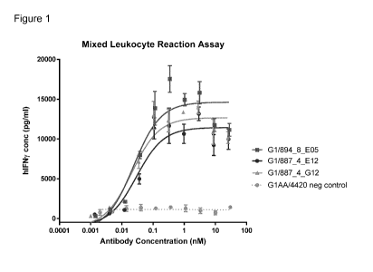
Mixed Lymphocyte Reaction (MLR) assay.
19. An antibody or antigen-binding fragment thereof, according to any
preceding clause, wherein
the antibody or antigen-binding fragment, is a multispecific, preferably a
bispecific, molecule
comprising at least a second antigen-binding site.
20. An antibody or antigen-binding fragment, according to any preceding
clause, wherein the
antibody or antigen-binding fragment thereof, comprises a second antigen-
binding site located in
a constant domain of the antibody or antigen-binding fragment.
10
CA 03106002 2021-01-08
WO 2020/011973
PCT/EP2019/068804
21. An antibody or antigen-binding fragment thereof, according to clause 20,
wherein the second
antigen-binding site comprises:
(a) a first sequence in the AB structural loop and / or a second sequence in
the EF structural loop
of a constant heavy domain,
(b) a first sequence in the AB structural loop and a second sequence in the EF
structural loop of
a constant heavy domain,
(c) a first sequence in the AB structural loop and / or a second sequence in
the EF structural loop
and / or a third sequence in the CD structural loop of a constant heavy domain
(d) a first sequence in the AB structural loop, a second sequence in the EF
structural loop and a
third sequence in the CD structural loop of a constant heavy domain
22. An antibody or antigen-binding fragment thereof, according to clause 20 or
21, wherein the
constant heavy domain is a CH3 domain.
23. An antibody or antigen-binding fragment thereof, according to any
preceding clause, wherein
the antibody is an immunoglobulin G (IgG), or antigen-binding fragment
thereof.
24. An antibody or antigen-binding fragment thereof, according to clause 23,
wherein the antibody
is an IgG1 or fragment thereof, or an IgG4 or fragment thereof.
25. An antibody or antigen-binding fragment thereof, according to clause 23 or
clause 24, wherein
the antibody is an IgG1 or fragment thereof with a modified Fc region.
26. An antibody or antigen-binding fragment thereof, according to clause 24 or
clause 25, wherein
the antibody is an IgG1 or fragment thereof with a modified Fc region with
reduced immune
effector function.
27. An antibody or antigen-binding fragment thereof, according to clause 25 or
26, wherein the
modified Fc has reduced ADCC and / or CDC relative to IgG1.
28. An antibody or antigen-binding fragment thereof, according to any of
clauses 25 to 27, wherein
.. the modified Fc region comprises a LALA, LALA-PA or LALA-PG modification.
29. An antibody or antigen-binding fragment thereof, according to any of
clauses 25 to 28, wherein
the antibody is an IgG1 or antigen-binding fragment thereof comprising a LALA
modification in
the Fc region.
11
CA 03106002 2021-01-08
WO 2020/011973
PCT/EP2019/068804
30. An antibody or antigen-binding fragment thereof, according to any of
clauses 19 to 29, wherein
the second antigen-binding site binds to an inhibitory checkpoint molecule,
costimulatory
molecule or tumour-associated antigen.
In an antibody or antigen-binding fragment thereof, according to the
invention, the second
antigen-binding site may bind to an inhibitory checkpoint molecule, such as
CTLA-4, LAG-3,
TIGIT, TIM-3, VISTA, 0D73, CSF-1R, KIR. 67-H3, 67-H4, 264, NKG2A, 0D47, SIRPa,
BTLA,
CCR4, CD200R, or TGFbeta.
In an antibody or antigen-binding fragment thereof, according to the
invention, the second
antigen-binding site may bind to and be an agonist for a costimulatory
molecule expressed by T
cells such as 0X40, ICOS, CD40, HVEM, NKG2D, or TNFR2.
In an antibody or antigen-binding fragment thereof, according to the
invention, the second
antigen-binding site may bind to a tumour-associated antigen (TAA), such as c-
Met, 137-H3, 67-
H4, EGFR, HER-2, EPCAM, CEACAM, FAP, VEGF, MSLN, GPC3, 0D38, CD19, or CD20.
31. An antibody or antigen-binding fragment thereof, according to any of
clauses 19 to 30, wherein
the second antigen-binding site does not bind to 0X40, Inducible T-cell
COStimulator (ICOS) or
CD137.
32. An antibody or antigen-binding fragment thereof, according to any of
clauses 19 to 30, wherein
the second antigen-binding site does not bind to 0D27 or glucocorticoid-
induced TNFR-related
protein (GITR).
33. An antibody or antigen-binding fragment thereof, according to any of
clauses 19 to 30, wherein
the second antigen-binding site does not bind to lymphocyte-activation gene 3
(LAG-3).
34. A conjugate or fusion comprising an antibody or antigen-binding fragment
thereof according
to any preceding clause and an immune system modulator (agonist or
antagonist), a cytotoxic
molecule, or a radioisotope.
35. An antibody, antigen-binding fragment thereof, conjugate or fusion
according to any
preceding clause having a detectable label.
36. A nucleic acid molecule or set of nucleic acid molecules encoding an
antibody, antigen-
binding fragment thereof, conjugate or fusion according to any preceding
clause.
12
CA 03106002 2021-01-08
WO 2020/011973
PCT/EP2019/068804
37. A nucleic acid molecule or set of nucleic acid molecules according to
clause 36, wherein the
nucleic acid molecule or set of nucleic acid molecules comprises cDNA sequence
encoding one
or more of the VH and / or VL, Fab, heavy and / or light chain of:
(a) G1AA/E12v2 or G1/E12v2;
(b) G1AA/E05v2 or G1/E05v2;
(c) G1AA/G12v2 or G1/G12v2;
(d) G1/887_04_E12;
(e) G1/894_08_E05;
(f) G1/887_04_G12;
(g) G1/894_08_A05;
(h) G1AA/1ambday3;
(i) G1/280_02_G02_NS; or
G1/280_02_G02.
38. A nucleic acid molecule or set of nucleic acid molecules according to
clause 37, comprising a
first nucleic acid sequence and a second nucleic acid sequence, wherein:
(a) the first nucleic acid sequence comprises a VH cDNA sequence encoding the
VH of antibody
G1AA/E12v2 of SEQ ID NO: 27 and the second nucleic acid sequence comprises a
VL cDNA
sequence encoding the VL of antibody G1AA/E12v2 of SEQ ID NO: 28;
(b) the first nucleic acid sequence comprises a VH cDNA sequence encoding the
VH of antibody
G1AA/G12v2 of SEQ ID NO: 29 and the second nucleic acid sequence comprises a
VL cDNA
sequence encoding the VL of antibody G1AA/G12v2 of SEQ ID NO: 30;
(c) the first nucleic acid sequence comprises a VH cDNA sequence encoding the
VH of antibody
G1AA/E05v2 of SEQ ID NO: 31 and the second nucleic acid sequence comprises a
VL cDNA
sequence encoding the VL of antibody G1AA/E05v2 of SEQ ID NO: 32;
(d) the first nucleic acid sequence comprises a VH cDNA sequence encoding the
VH of antibody
G1/887_04_E12 of SEQ ID NO: 33 and the second nucleic acid sequence comprises
the VL
cDNA sequence of antibody G1/887_04_E12 of SEQ ID NO: 34;
13
CA 03106002 2021-01-08
WO 2020/011973
PCT/EP2019/068804
(e) the first nucleic acid sequence comprises a VH cDNA sequence encoding the
VH of antibody
G1/887_04_G12 of SEQ ID NO: 35 and the second nucleic acid sequence comprises
a VL cDNA
sequence encoding the VL of antibody G1/887_04_G12 of SEQ ID NO: 36;
(f) the first nucleic acid sequence comprises a VH cDNA sequence encoding the
VH of antibody
G1/894_08_E05 of SEQ ID NO: 37 and the second nucleic acid sequence comprises
a VL cDNA
sequence encoding the VL of antibody G1/894_08_E05 of SEQ ID NO: 38;
(g) the first nucleic acid sequence comprises a VH cDNA sequence encoding the
VH of antibody
G1/894_08_A05 of SEQ ID NO: 39 and the second nucleic acid sequence comprises
a VL cDNA
sequence encoding the VL of antibody G1/894_08_A05 of SEQ ID NO: 40;
(h) the first nucleic acid sequence comprises a VH cDNA sequence encoding the
VH of antibody
G1AA/1ambdav3 of SEQ ID NO: 41 and the second nucleic acid sequence comprises
a VL cDNA
sequence encoding the VL of antibody G1AA/1ambdav3 of SEQ ID NO: 42;
(i) the first nucleic acid sequence comprises a VH cDNA sequence encoding the
VH of antibody
G1/280_02_G02_NS of SEQ ID NO: 43 and the second nucleic acid sequence
comprises a VL
cDNA sequence encoding the VL of antibody G1/280_02_G02_NS of SEQ ID NO: 44;
or
(i) the first nucleic acid sequence comprises a VH cDNA sequence encoding the
VH of antibody
G1/280_02_G02 of SEQ ID NO: 45 and the second nucleic acid sequence comprises
a VL cDNA
sequence encoding the VL of antibody G1/280_02_G02 of SEQ ID NO: 46.
39. A vector or set of vectors comprising the nucleic acid molecule or set of
nucleic acid molecules
of any of clauses 36 to 38.
40. A recombinant host cell comprising a nucleic acid molecule or set of
nucleic acid molecules
of any of clauses 36 to 38, or the vector or set of vectors of clause 39.
41. A method of producing an antibody antigen-binding fragment thereof,
conjugate or fusion
according to any preceding clause, comprising culturing the recombinant host
cell of clause 40
under conditions suitable for production of the antibody, antigen-binding
fragment, conjugate or
fusion.
42. The method of clause 41 further comprising isolating and/or purifying the
antibody, antigen-
binding fragment, conjugate or fusion.
14
CA 03106002 2021-01-08
WO 2020/011973
PCT/EP2019/068804
43. A composition (e.g., pharmaceutical composition) comprising the antibody,
antigen-binding
fragment, conjugate or fusion according to any of clauses 1 to 42 and an
excipient (e.g.,
pharmaceutically-acceptable excipient).
44. An antibody, antigen-binding fragment, conjugate or fusion according to
any of clauses 1 to
35 or composition according to clause 43 for use in a method for treatment of
the human or animal
body by therapy.
45. A method for treatment of a disease or disorder in a patient comprising
administering to the
patient a therapeutically-effective amount of an antibody, antigen-binding
fragment thereof,
conjugate or fusion according to any of clauses 1 to 35 or composition
according to clause 43.
46. The use of an antibody, antigen-binding fragment thereof, conjugate or
fusion according to
any of clauses 1 to 35 or composition according to clause 43 in the
manufacture of a medicament
for the treatment of the human or animal body.
47. An antibody, antigen-binding fragment thereof, conjugate or fusion
according to any of clauses
1 to 35 or composition according to clause 43 for use according to clause 44
in a method of
treatment that comprises administering the antibody, antigen-binding fragment
thereof, conjugate,
fusion or composition to the human or animal body in combination with a second
therapeutic.
48. A method of clause 45, or use of clause 46, wherein the method further
comprises
administering a therapeutically-effective amount of a second therapeutic to
the patient.
49. An antibody, antigen-binding fragment thereof, conjugate, fusion or
composition for use
according to clause 47, or in a method of clause 48, wherein the second
therapeutic is a
radiotherapy, preferably targeted radiotherapy.
50. An antibody, antigen-binding fragment, conjugate or fusion according to
any of clauses 1 to
35 or composition according to clause 43 for use in a diagnostic method
practised on the human
or animal body or practised in vitro on a sample from on the human or animal
body.
51. A method of detecting a disease or disorder in a patient, the method
comprising the use of an
antibody, antigen-binding fragment thereof, conjugate or fusion according to
any of clauses 1 to
or of a composition according to clause 43.
CA 03106002 2021-01-08
WO 2020/011973
PCT/EP2019/068804
52. The use of an antibody, antigen-binding fragment thereof, conjugate or
fusion according to
any of clauses 1 to 35 or of a composition according to clause 43 in the
manufacture of a
diagnostic product.
Treatment against various types of cancer using anti-PD-L1 or anti-PD-1
antibodies has been
investigated in clinical trials and shown promising results. These include
solid tumours such as
ovarian cancer, prostate cancer, colorectal cancer, fibrosarcoma, renal cell
carcinoma, melanoma
(advanced and metastatic melanoma), pancreatic cancer, breast cancer,
glioblastoma
multiforme, lung cancer (such as non-small cell lung cancer and small cell
lung cancer), head and
neck cancer (such as head and neck squamous cell carcinoma), stomach cancer
(gastric cancer),
bladder cancer, cervical cancer, uterine cancer (uterine endometrial cancer,
uterine cervical
cancer), vulvar cancer, testicular cancer, penile cancer, esophageal cancer,
hepatocellular
carcinoma, nasopharyngeal cancer, Merkel cell carcinoma, mesothelioma, DNA
mismatch repair
deficient colorectal cancer, DNA mismatch repair deficient endometrial cancer,
thyroid cancer,
Hodgkin's lymphoma, non-Hodgkin's lymphoma (such as diffuse large B-cell
lymphoma, follicular
lymphoma, indolent non-Hodgkin's lymphoma, mantle cell lymphoma), leukaemia
(such as
chronic lymphocytic leukaemia, myeloid leukaemia, acute lymphoblastoid
leukaemia, or chronic
lymphoblastoid leukaemia), multiple myeloma, and peripheral T-cell lymphoma.
The antibody or
antigen-binding fragment thereof of the invention thus may find application in
the treatment of
these cancers. Tumours of these cancers are known, or expected, to contain
immune cells, such
as TILs, expressing PD-L1.
In particular, treatment of melanoma, colorectal cancer, breast cancer,
bladder cancer, renal cell
carcinoma, gastric cancer, head and neck cancer (such as squamous cell
carcinoma of the head
and neck), mesothelioma, lung cancer (such as non-small-cell lung cancer),
ovarian cancer,
Merkel-cell carcinoma, pancreatic cancer, melanoma and hepatocellular
carcinoma using anti-
PD-L1 antibodies has been investigated in clinical trials and shown promising
results. Thus, the
cancer to be treated using an antibody or antigen-binding fragment thereof of
the invention may
be a melanoma, colorectal cancer, breast cancer, bladder cancer, renal cell
carcinoma, bladder
cancer, gastric cancer, head and neck cancer (such as squamous cell carcinoma
of the head and
neck), mesothelioma, lung cancer (such as non-small-cell lung cancer), ovarian
cancer, Merkel-
cell carcinoma, pancreatic cancer, melanoma, or hepatocellular carcinoma.
16
CA 03106002 2021-01-08
WO 2020/011973
PCT/EP2019/068804
Cancer may be characterised by the abnormal proliferation of malignant cancer
cells. Where the
application refers to a particular type of cancer, such as breast cancer, this
refers to a malignant
transformation of the relevant tissue, in this case a breast tissue. A cancer
which originates from
malignant transformation of a different tissue, e.g., ovarian tissue, may
result in metastatic lesions
in another location in the body, such as the breast, but is not thereby a
breast cancer as referred
to herein but an ovarian cancer.
The cancer may be a primary or secondary cancer. Thus, an antibody or antigen-
binding fragment
thereof of the invention may be for use in a method of treating cancer in a
patient, wherein the
cancer is a primary tumour and/or a tumour metastasis.
An antibody or antigen-binding fragment thereof of the invention may also be
expected to find
application in the treatment of infectious diseases, such as viral, bacterial,
fungal and/or parasitic
infections. Preferably, the infectious disease is a viral, bacterial or fungal
disease, more preferably
a viral or bacterial disease, most preferably a viral disease. The infectious
disease may be chronic
or acute, but is preferably chronic.
Examples of viral diseases which may be treated with an antibody or antigen-
binding fragment
thereof according to the invention include: human immunodeficiency virus
(HIV), influenza virus,
enterovirus, hepatitis B virus (HBV), hepatitis C virus (HCV), hepatitis A
virus (HAV), hepatitis D
virus (HDV), and hepatitis E virus (HEV), respiratory syncytial virus (RSV),
herpesvirus (such as
Epstein-Barr virus, herpes simplex virus 1 (HSV-1), herpes simplex virus 2
(HSV-2),
cytomegalovirus (CMV)), and papillomavirus infection.
Examples of bacterial diseases which may be treated with an antibody or
antigen-binding
fragment thereof of the invention include: Mycobacterium tuberculosis, gram-
negative bacteria
(such as Acinetobacter, Klebisella, Enterobacter), gram-positive bacteria
(such as Clostridium
difficile, Staphylococcus aureus), and Listeria (e.g., Listeria monocytogenes)
infection.
Examples of fungal diseases which may be treated with an antibody or antigen-
binding fragment
thereof of the invention include: Aspergillus and Candida infection.
Examples of parasitic diseases which may be treated with an antibody or
antigen-binding
fragment thereof of the invention include: Malaria, Toxoplasma, and Leishmania
infection.
An antibody or antigen-binding fragment thereof according to the invention is
designed to be used
in methods of treatment of patients, preferably human patients. An antibody or
antigen-binding
fragment thereof of the invention will usually be administered in the form of
a pharmaceutical
17
CA 03106002 2021-01-08
WO 2020/011973
PCT/EP2019/068804
composition, which may comprise at least one additional component, such as a
pharmaceutically
acceptable excipient. For example, a pharmaceutical composition of the
invention, may comprise,
in addition to the antibody or antigen-binding fragment thereof, a
pharmaceutically-acceptable
excipient, carrier, buffer, stabiliser or other materials well known to those
skilled in the art. Such
materials should be non-toxic and should not interfere with the efficacy of
the antibody or antigen-
binding fragment thereof. The precise nature of the carrier or other material
will depend on the
route of administration, which may be by injection, e.g., intravenous or
subcutaneous. The
antibody or antigen-binding fragment thereof may be administered
intravenously, or
subcutaneously.
Liquid pharmaceutical compositions generally comprise a liquid carrier such as
water or
physiological saline solution. For subcutaneous or intravenous injection, or
injection at the site of
affliction, the antibody or antigen-binding fragment thereof, or
pharmaceutical composition
comprising the antibody or antigen-binding fragment thereof, is preferably in
the form of a
parenterally acceptable aqueous solution which is pyrogen-free and has
suitable pH, isotonicity
and stability.
A composition comprising an antibody or antigen-binding fragment thereof
according to the
invention may be administered alone or in combination with other treatments,
concurrently or
sequentially or as a combined preparation with another therapeutic agent or
agents, dependent
upon the condition to be treated. For example, an antibody or fragment thereof
of the invention
may be administered in combination with an existing therapeutic agent for the
disease to be
treated, e.g., a cancer as mentioned above. For example, an antibody or
fragment thereof of the
invention may be administered to the patient in combination with a second anti-
cancer therapy,
such as chemotherapy, anti-tumour vaccination (also referred to as a cancer
vaccination),
radiotherapy, immunotherapy, an oncolytic virus, chimeric antigen receptor
(CAR) T-cell therapy,
or hormone therapy.
It is expected that the antibody or fragment thereof of the invention may act
as an adjuvant in anti-
cancer therapy, such as chemotherapy, anti-tumour vaccination, or
radiotherapy. Without wishing
to be bound by theory, it is thought that administration of the antibody or
fragment thereof to the
patient as part of chemotherapy, anti-tumour vaccination, or radiotherapy will
trigger a greater
immune response against the cancer associated antigen PD-L1, than is achieved
with
chemotherapy, anti-tumour vaccination, or radiotherapy alone.
18
CA 03106002 2021-01-08
WO 2020/011973
PCT/EP2019/068804
A method of treating cancer in a patient may thus comprise administering to
the patient a
therapeutically effective amount of an antibody or fragment thereof according
to the invention in
combination with a chemotherapeutic agent, anti-tumour vaccine, radionuclide,
immunotherapeutic agent, oncolytic virus, CAR-T cell, or agent for hormone
therapy. The
chemotherapeutic agent, anti-tumour vaccine, radionuclide, immunotherapeutic
agent, oncolytic
virus, CAR-T cell, or agent for hormone therapy is preferably a
chemotherapeutic agent, anti-
tumour vaccine, radionuclide, immunotherapeutic agent, oncolytic virus, CAR-T
cell, or agent for
hormone therapy for the cancer in question, i.e., a chemotherapeutic agent,
anti-tumour vaccine,
radionuclide, immunotherapeutic agent, oncolytic virus, CAR-T cell, or agent
for hormone therapy
which has been shown to be effective in the treatment of the cancer in
question. The selection of
a suitable chemotherapeutic agent, anti-tumour vaccine, radionuclide,
immunotherapeutic agent,
oncolytic virus, CAR-T cell, or agent for hormone therapy, which have been
shown to be effective
for the cancer in question, is well within the capabilities of the skilled
practitioner.
For example, where the method comprises administering to the patient a
therapeutically effective
amount of an antibody or fragment thereof according to the invention in
combination with a
chemotherapeutic agent, the chemotherapeutic agent may be selected from the
group consisting
of: taxanes, cytotoxic antibiotics, tyrosine kinase inhibitors, PARP
inhibitors, B_RAF enzyme
inhibitors, alkylating agents, platinum analogues, nucleoside analogues,
thalidomide derivatives,
antineoplastic chemotherapeutic agents and others. Taxanes include docetaxel,
paclitaxel and
nab-paclitaxel; cytotoxic antibiotics include actinomycin, bleomycin,
anthracyclines, doxorubicin
and valrubicin; tyrosine kinase inhibitors include sunitinib, erlotinib,
gefitinib, axitinib, PLX3397,
imatinib, cobemitinib and trametinib; PARP inhibitors include piraparib; B-Raf
enzyme inhibitors
include vemurafenib and dabrafenib; alkylating agents include dacarbazine,
cyclophosphamide,
temozolomide; platinum analogues include carboplatin, cisplatin and
oxaliplatin; nucleoside
analogues include gemcitabine and azacitidine; antineoplastics include
fludarabine. Other
chemotherapeutic agents suitable for use in the invention include
methotrexate, defactinib,
entinostat, pemetrexed, capecitabine, eribulin, irinotecan, fluorouracil, and
vinblastine.
Vaccination strategies for the treatment of cancers have been implemented in
the clinic and
discussed in detail within scientific literature (such as Rosenberg S.
Development of Cancer
.. Vaccines. ASCO Educational Book Spring: 60-62 (2000)). This mainly involves
strategies to
prompt the immune system to respond to various cellular markers expressed by
autologous or
allogenic cancer cells by using those cells as a vaccination method, both with
or without
19
CA 03106002 2021-01-08
WO 2020/011973
PCT/EP2019/068804
granulocyte-macrophage colony-stimulating factor (GM-CSF). GM-CSF provokes a
strong
response in antigen presentation and works particularly well when employed
with said strategies.
Where a method of the invention comprises administering to the patient a
therapeutically-effective
amount of an antibody or fragment thereof according to the invention in
combination with an
immunotherapeutic agent, the immunotherapeutic agent may be selected from the
group
consisting of: antibodies binding to a checkpoint inhibitor, costimulatory
molecule or soluble factor,
such as antibodies binding to CTLA-4, LAG-3, TIGIT, TIM-3, VISTA, 0D73, CSF-
1R, KIR, 0X40,
CD40, HEVM, TGFB, IL-10, CSF-1. Alternatively, the immunotherapeutic agent may
one or more
cytokines or cytokine-based therapies selected from the group consisting of IL-
2, prodrug of
conjugated IL2, GM-CSF, IL-7, IL-12, IL-9, IL-15, IL-18, IL-21, and type I
interferon.
Administration may be in a "therapeutically effective amount", this being an
amount which is
sufficient to show benefit to a patient. Such benefit may be at least
amelioration of at least one
symptom. Thus, "treatment" of a specified disease refers to amelioration of at
least one symptom.
The actual amount administered, and rate and time-course of administration,
will depend on the
nature and severity of what is being treated, the particular patient being
treated, the clinical
condition of the individual patient, the cause of the disorder, the site of
delivery of the composition,
the type of antibody or fragment thereof, the method of administration, the
scheduling of
administration and other factors known to medical practitioners. Prescription
of treatment, e.g.,
decisions on dosage etc., is within the responsibility of general
practitioners and other medical
doctors, and may depend on the severity of the symptoms and/or progression of
a disease being
treated. Appropriate doses of antibody or fragment thereof are well known in
the art (Ledermann
et al. (1991) Int. J. Cancer 47: 659-664; and Bagshawe et al. (1991) Antibody,
I mmunoconjugates
and Radiopharmaceuticals 4: 915-922). Specific dosages indicated herein, or in
the Physician's
Desk Reference (2003) as appropriate for an antibody or fragment thereof being
administered,
may be used. A therapeutically-effective amount or suitable dose of an
antibody or fragment
thereof can be determined by comparing its in vitro activity and in vivo
activity in an animal model.
Methods for extrapolation of effective dosages in mice and other test animals
to humans are
known. The precise dose will depend upon a number of factors, including the
size and location of
the area to be treated, and the precise nature of the specific binding member.
Treatments may
be repeated at daily, twice-weekly, weekly or monthly intervals, at the
discretion of the physician.
Treatment may be given before and/or after surgery, and may be administered or
applied directly
at the anatomical site of surgical treatment.
CA 03106002 2021-01-08
WO 2020/011973
PCT/EP2019/068804
Detailed Description
The invention relates to antibodies and antigen-binding fragments thereof that
comprise a CDR-
based antigen-binding site for PD-L1. An antibody or antigen-binding fragment
thereof of the
invention may be produced by recombinant means. A "recombinant antibody" is an
antibody which
has been produced by a recombinantly engineered host cell. An antibody or
antigen-binding
fragment thereof in accordance with the invention is optionally isolated or
purified.
The term "PD-L1" may refer to human PD-L1, murine, in particular mouse PD-L1,
and/or
cynomolgus monkey PD-L1, unless the context requires otherwise. Preferably the
term "PD-L1"
refers to human PD-L1, unless the context requires otherwise.
The term "antibody molecule" describes an immunoglobulin whether natural or
partly or wholly
synthetically produced. The antibody molecule may be human or humanised. The
antibody
molecule is preferably a monoclonal antibody molecule. Examples of antibodies
are the
immunoglobulin isotypes, such as immunoglobulin G, and their isotypic
subclasses, such as IgG1,
IgG2, IgG3 and IgG4, as well as fragments thereof. The four human subclasses
(IgG1, IgG2,
IgG3 and IgG4) each contain a different heavy chain; but they are highly
homologous and differ
mainly in the hinge region and the extent to which they activate the host
immune system. IgG1
and IgG4 contain two inter-chain disulphide bonds in the hinge region, IgG2
has 4 and IgG3 has
11 inter-chain disulphide bonds.
The terms "antibody" and "antibody molecule", as used herein, includes
antibody fragments, such
as Fab and scFy fragments, provided that said fragments comprise a CDR-based
antigen binding
site for PD-L1. Unless the context requires otherwise, the terms "antibody" or
"antibody molecule",
as used herein, is thus equivalent to "antibody or antigen-binding fragment
thereof'.
Antibodies are immunoglobulins, which have the same basic structure consisting
of two heavy
and two light chains forming two Fab arms containing identical domains that
are attached by a
flexible hinge region to the stem of the antibody, the Fc domain, giving the
classical 'Y' shape.
The Fab domains consist of two variable and two constant domains, with a
variable heavy (VH)
and constant heavy 1 (OH I) domain on the heavy chain and a variable light
(VL) and constant
light (CL) domain on the light chain. The two variable domains (VH and VL)
form the variable
fragment (Fv), which provides the CDR-based antigen specificity of the
antibody, with the constant
domains (CHI and VL) acting as a structural framework. Each variable domain
contains three
hypervariable loops, known as complementarity determining regions (CDRs). On
each of the VH
and VL the three CDRs (CDR1, CDR2, and CDR3) are flanked by four less-variable
framework
21
CA 03106002 2021-01-08
WO 2020/011973
PCT/EP2019/068804
(FR) regions (FR1, FW2, FW3 and FW4) to give a structure FW1-CDR1-FW2-CDR2-FW3-
CDR3-
FW4. The CDRs provide a specific antigen recognition site on the surface of
the antibody.
Both Kabat and ImMunoGeneTics (IMGT) numbering nomenclature is used herein.
Generally,
unless otherwise indicated (explicitly or by context) amino acid residues are
numbered herein
.. according to the Kabat numbering scheme (Kabat et al., 1991). For those
instances when IMGT
numbering is used, amino acid residues are numbered herein according to the
ImMunoGeneTics
(IMGT) numbering scheme. The IMGT numbering scheme is described in Lefranc et
al., 2005.
When the sequences are defined by IMGT nomenclature, the invention provides:
1A. An antibody or antigen-binding fragment thereof, capable of binding
specifically to PD-L1
comprising a variable heavy (VH) domain comprising heavy chain CDRs: HCDR1,
HCRD2 and
HCDR3, each flanked by framework (FW) regions, characterised in that the amino
acid sequence
of HCDR1 is GYX1FTSYG (SEQ ID NO: 67); the amino acid sequence of HCDR2 is
I5AYX2X3X4X5
(SEQ ID NO: 68); and the amino acid sequence of HCDR3 is ARDLFPTIFGVSYYYY (SEQ
ID
NO: 69); wherein X1 is P or T; X2 is S, N or G, preferably S or N; X3 is G or
S; X4 is G, N or S,
.. preferably G or N; and X5 is T or A, preferably T, and wherein the
sequences are defined by the
ImMunoGeneTics (IMGT) nomenclature.
2A. An antibody or antigen-binding fragment thereof according to clause 1A,
wherein the
sequence X2X3X4X5 (SEQ ID NO: 4) (residues 62-65) of HCDR2 is selected from
SGGT (SEQ ID
NO: 5), NSNT (SEQ ID NO: 6), GGST (SEQ ID NO: 7) and SGNA (SEQ ID NO: 8).
(IMGT
.. nomenclature).
3A. An antibody or antigen-binding fragment thereof according to clause 1A or
2A, wherein X1 is
P and X2X3X4X5 of HCDR2 is SGGT (SEQ ID NO: 5) (IMGT nomenclature).
4A. An antibody or antigen-binding fragment thereof according to any of
clauses 1A to 3A
comprising a variable light (VL) domain comprising a LCDR1, LCDR2 and LCDR3,
characterised
in that:
(a) the VL is a kappa VL and the amino acid sequence of LCDR1 is Q5IX6X7R (SEQ
ID NO: 70);
the amino acid sequence of LCDR2 is EAS (SEQ ID NO: 71); and the amino acid
sequence of
LCDR3 is QQX8X9X1oTPYT (SEQ ID NO: 72), QQX8X9X1oTPRVT (SEQ ID NO: 73),
QQX8X9X10FPRVS (SEQ ID NO: 74), or QQX8X9X1oWPRT (SEQ ID NO: 75); wherein X6
is G or
S; X7 is N or G; X5 is S or A; X9 is Y or N; and X10 is S or T; or,
22
CA 03106002 2021-01-08
WO 2020/011973
PCT/EP2019/068804
(b) the VL is a lambda VL and the amino acid sequence of LCDR1 is SSDVGGYNXii
(SEQ ID
NO: 76), the amino acid sequence of LCDR2 is EVT (SEQ ID NO: 77) and the amino
acid
sequence of LCDR3 is SSFKRGSTLVV (SEQ ID NO: 14); wherein X11 is Y or S; and
wherein the
sequences are defined by IMGT nomenclature.
5A. An antibody antigen-binding fragment thereof according to any of clauses
1A to 4A,
comprising an antigen-binding site comprising:
(a) the CDRs of antibody G1AA/E12v2;
(b) the CDRs of antibody G1AA/G12v2;
(c) the CDRs of antibody G1AA/E05v2;
(d) the CDRs of antibody G1/887_04_E12;
(e) the CDRs of antibody G1/887_04_G12;
(f) the CDRs of antibody G1/894_08_E05;
(g) the CDRs of antibody G1/894_08_A05;
(h) the CDRs of antibody G1AA/lambdav3; or
(i) the CDRs of antibody G1/280_02_G02_NS;
U) the CDRs of antibody G1/280_02_G02;
wherein the sequences are defined according to the ImMunoGeneTics (IMGT)
numbering
scheme
It is possible to take monoclonal and other antibodies and use techniques of
recombinant DNA
technology to produce other antibodies or chimeric molecules which generally
retain the
specificity of the original antibody. Such techniques may involve introducing
the CDRs into a
different immunoglobulin framework, or grafting variable regions onto a
different immunoglobulin
constant region. Introduction of the CDRs of one immunoglobulin into another
immunoglobulin is
described for example in EP-A-184187, GB2188638A or EP-A-239400.
Alternatively, a
hybridoma or other cell producing an antibody molecule may be subject to
genetic mutation or
other changes, which may or may not alter the binding specificity of
antibodies produced.
As antibodies can be modified in a number of ways, the term "antibody" should
be construed as
covering antibody fragments, derivatives, functional equivalents and
homologues of antibodies,
23
CA 03106002 2021-01-08
WO 2020/011973
PCT/EP2019/068804
including any polypeptide comprising an immunoglobulin binding domain, whether
natural or
wholly or partially synthetic. Chimeric molecules comprising an immunoglobulin
binding domain,
or equivalent, fused to another polypeptide are therefore included. Cloning
and expression of
chimeric antibodies are described in EP-A- 0120694 and EP-A-0125023.
An example of an antibody fragment comprising both CDR sequences and CH3
domain is a
minibody, which comprises a scFy joined to a CH3 domain (Hu et al., 1996).
An antibody or antigen-binding fragment of the invention binds to PD-L1, in
particular human PD-
L1. Binding in this context may refer to specific binding. The term "specific"
may refer to the
situation in which the antibody molecule will not show any significant binding
to molecules other
than its specific binding partner(s), here PD-L1. The term "specific" is also
applicable where the
antibody molecule is specific for particular epitopes, such as epitopes on PD-
L1, that are carried
by a number of antigens in which case the antibody molecule will be able to
bind to the various
antigens carrying the epitope.
Amino acids may be referred to by their one letter or three letter codes, or
by their full name. The
one and three letter codes, as well as the full names, of each of the twenty
standard amino acids
are set out below.
Amino acid One letter code Three letter code
alanine A Ala
arginine R Arg
asparagine N Asn
aspartic acid D Asp
cysteine C Cys
glutamic acid E Glu
glutamine Q Gln
glycine G Gly
histidine H His
isoleucine I Ile
24
CA 03106002 2021-01-08
WO 2020/011973
PCT/EP2019/068804
leucine L Leu
lysine K Lys
methionine M Met
phenylalanine F Phe
proline P Pro
serine S Ser
threonine T Thr
tryptophan W Trp
tyrosine Y Tyr
valine V Val
Amino acids, one and three-letter codes
In preferred embodiments, the PD-L1 antibody of the invention comprises the
HCDR3 sequence
of E12v2 (SEQ ID NO: 3); it is preferred that the antibody further comprises
the HCDR2 sequence
of E12v2 (SEQ ID NO: 18); it is preferred that the PD-L1 antibody of the
invention yet further
comprises the HCDR1 sequence of E12v2 (SEQ ID NO: 1). In preferred embodiments
the HCDR2
sequence is HCDR2 sequence of E12v2 (SEQ ID NO: 18) and the amino acid at
position 28 in
the VH (Kabat) is proline. In particularly preferred embodiments the PD-L1
antibody of the
invention comprises the HCDR3 sequence of SEQ ID NO: 3, the HCDR2 sequence of
SEQ ID
NO: 18 and the amino acid at position 28 in the VH (Kabat) is proline. In more
particularly
preferred embodiments the PD-L1 antibody of the invention comprises the HCDR3
sequence of
SEQ ID NO: 3, the HCDR2 sequence of SEQ ID NO: 18, and the HCDR1 sequence of
SEQ ID
NO: 1 and the amino acid at position 28 in the VH (Kabat) is proline.
Antibodies of the invention may comprise one or more, e.g., 1, 2, 3, 4, 5, 6,
7, 8,9 or 10 further
amino acid modifications in the VH and / or VL sequences, provided that
functional properties of
the antibody are retained.
A modification may be an amino acid substitution, deletion or insertion.
Preferably, the
modification is a substitution.
CA 03106002 2021-01-08
WO 2020/011973
PCT/EP2019/068804
In preferred embodiments in which one or more amino acids are substituted with
another amino
acid, the substitutions may conservative substitutions, for example according
to the following
chart. In some embodiments, amino acids in the same category in the middle
column are
substituted for one another, i.e. a non-polar amino acid is substituted with
another non-polar
amino acid, for example. In some embodiments, amino acids in the same line in
the rightmost
column are substituted for one another.
ALIPHATIC Non-polar GAP
ILV
Polar- CSTM
uncharged
NQ
Polar - charged D E
KR
AROMATIC H F WY
In some embodiments, substitution(s) may be functionally conservative. That
is, in some
embodiments the substitution may not affect (or may not substantially affect)
one or more
functional properties (e.g., binding affinity) of the antibody molecule
comprising the substitution
as compared to the equivalent unsubstituted antibody molecule.
In a preferred embodiment, a PD-L1 antibody of the invention may comprise a VH
and / or VL
domain sequence with one or more amino acid sequence alterations (addition,
deletion,
substitution and/or insertion of an amino acid residue), preferably 20
alterations or fewer, 15
alterations or fewer, 10 alterations or fewer, 5 alterations or fewer, 4
alterations or fewer, 3
alterations or fewer, 2 alterations or fewer, or 1 alteration compared with
the VH and / or VL
sequences of the invention set forth herein.
In a preferred embodiment, an antibody of the invention comprises the HCDR3
domain of E12v2
.. set forth in SEQ ID NO: 3.
In another preferred embodiment, an antibody of the invention comprises the VH
domain of E12v2
set forth in SEQ ID NO: 27 or a VH domain with an amino acid sequence which
has at least 95%,
at least 96%, at least 97%, at least 98%, or at least 99% sequence identity to
the sequence set
forth in SEQ ID NO: 27.
26
CA 03106002 2021-01-08
WO 2020/011973
PCT/EP2019/068804
In a preferred embodiment, an antibody of the invention comprises a VH domain
comprising the
HCDR3 set forth in SEQ ID NO: 3 and the VH domain has an amino acid sequence
with at least
70%, at least 75%, at least 80%, at least 85%, at least 90%, at least 95%, at
least 96%, at least
97%, at least 98%, or at least 99% sequence identity to the sequence set forth
in SEQ ID NO: 27.
In a preferred embodiment, an antibody of the invention comprises a VH domain
comprising the
HCDR3 of E12v2 set forth in SEQ ID NO: 3 and a HCDR2 selected from those set
forth in SEQ
ID NO: 18, 23 or 24 and the VH domain has an amino acid sequence with at least
70%, at least
75%, at least 80%, at least 85%, at least 90%, at least 95%, at least 96%, at
least 97%, at least
98%, or at least 99% sequence identity to the sequence set forth in SEQ ID NO:
27.
In a preferred embodiment, an antibody of the invention comprises a VH domain
comprising the
HCDR3 of E12v2 set forth in SEQ ID NO: 3, a HCDR2 domain selected from those
set forth in
SEQ ID NO: 18, 23 or 24, a proline at position 28, and the VH domain has an
amino acid sequence
with at least 70%, at least 75%, at least 80%, at least 85%, at least 90%, at
least 95%, at least
96%, at least 97%, at least 98%, or at least 99% sequence identity to the
sequence set forth in
SEQ ID NO: 27.
In a preferred embodiment, an antibody of the invention comprises a VL domain
comprising a VL
domain of E12v2 set forth in SEQ ID NO: 28 or an amino acid sequence with at
least 70%, at
least 75%, at least 80%, at least 85%, at least 90%, at least 95%, at least
96%, at least 97%, at
least 98%, or at least 99% sequence identity to the sequence set forth in SEQ
ID NO: 28.
Sequence identity is commonly defined with reference to the algorithm GAP
(Wisconsin GCG
package, Accelerys Inc, San Diego USA). GAP uses the Needleman and Wunsch
algorithm to
align two complete sequences, maximising the number of matches and minimising
the number of
gaps. Generally, default parameters are used, with a gap creation penalty
equalling 12 and a gap
extension penalty equalling 4. Use of GAP may be preferred but other
algorithms may be used,
e.g., BLAST (which uses the method of Altschul et al. (1990) J. Mol. Biol.
215: 405-410), FASTA
(which uses the method of Pearson and Lipman (1988) PNAS USA 85: 2444-2448),
or the Smith-
Waterman algorithm (Smith and Waterman (1981) J. Mol Biol. 147: 195-197), or
the TBLASTN
program, of Altschul et al. (1990) supra, generally employing default
parameters. In particular, the
psi-Blast algorithm (Nucl. Acids Res. (1997) 25 3389-3402) may be used.
Sequence identity may
be defined using the Bioedit, ClustalW algorithm.
The antibody may comprise a CH2 domain. The CH2 domain is preferably located
at the N-
terminus of the CH3 domain, as in the case in a human IgG molecule. The CH2
domain of the
27
CA 03106002 2021-01-08
WO 2020/011973
PCT/EP2019/068804
antibody is preferably the CH2 domain of human IgG1, IgG2, IgG3, or IgG4, more
preferably the
CH2 domain of human IgG1. The sequences of human IgG domains are known in the
art.
The antibody may comprise an immunoglobulin hinge region, or part thereof, at
the N-terminus of
the CH2 domain. The immunoglobulin hinge region allows the two CH2-CH3 domain
sequences
to associate and form a dimer. Preferably, the hinge region, or part thereof,
is a human IgG1,
IgG2, IgG3 or IgG4 hinge region, or part thereof. More preferably, the hinge
region, or part thereof,
is an IgG1 hinge region, or part thereof.
The sequence of the CH3 domain, is not particularly limited. Preferably, the
CH3 domain is a
human immunoglobulin G domain, such as a human IgG1, IgG2, IgG3, or IgG4 CH3
domain,
.. most preferably a human IgG1 CH3 domain.
An antibody of the invention may comprise a human IgG1, IgG2, IgG3, or IgG4
constant region.
The sequences of human IgG1, IgG2, IgG3, or IgG4 CH3 domains are known in the
art.
The heavy chain of the antibody molecule may optionally comprise an additional
lysine residue
(K) at the C-terminus of the heavy chain CH3 domain sequence.
Immunoglobulins are known to have a modular architecture comprising discrete
domains, which
can be combined in a multitude of different ways to create multispecific,
e.g., bispecific, trispecific,
or tetraspecific antibody formats. Exemplary multispecific antibody formats
are described in
Spiess et al., 2015 and Kontermann, 2012, for example. The antibodies of the
invention may be
employed in such multispecific formats.
For example, an antibody of the invention may be a heterodimeric antibody
molecule, such as a
heterodimeric complete immunoglobulin molecule, or a fragment thereof. In this
case, one part of
the antibody will have a sequence or sequences as described herein. For
example, where the
antibody of the invention is a bispecific heterodimeric antibody molecule, the
antibody may
comprise a heavy chain and light chain as described herein paired with a heavy
chain and light
chain comprising a VH domain and a VL domain, respectively, which bind an
antigen other than
PD-Ll. Techniques for preparing heterodimeric antibodies are known in the art
and include knobs-
into-holes (KIHs) technology, which involves engineering the CH3 domains of an
antibody
molecule to create either a "knob" or a "hole" to promote chain
heterodimerization. Alternatively,
heterodimeric antibodies can be prepared through the introduction of charge
pairs into the
antibody molecule to avoid homodimerization of CH3 domains by electrostatic
repulsion and to
28
CA 03106002 2021-01-08
WO 2020/011973
PCT/EP2019/068804
direct heterodimerization by electrostatic attraction. Examples of
heterodimeric antibody formats
include CrossMab, mAb-Fv, SEED-body, and KIH IgG.
Alternatively, a multispecific antibody molecule may comprise a complete
immunoglobulin
molecule or a fragment thereof and an additional antigen-binding moiety or
moieties. The antigen-
binding moiety may for example be an Fv, scFy or single domain antibody, and
may be fused to
the complete immunoglobulin molecule or a fragment thereof. Examples of
multispecific antibody
molecules comprising additional antigen-binding moieties fused to a complete
immunoglobulin
molecule include DVD-IgG, DVI-IgG, scFv4-IgG, IgG-scFv, and scFv-IgG molecules
(Spiess et
al., 2015; Figure 1). Examples of multispecific antibody molecules comprising
additional antigen-
binding moieties fused to an immunoglobulin fragment include BiTE molecules,
diabodies, and
DART molecules, for example (Spiess et al., 2015; Figure 1). Other suitable
formats would be
readily apparent to the skilled person.
In addition to the CDR-based PD-L1-binding site, e.g., in the VH of an
antibody, the antibody may
further comprise one or more additional antigen-binding sites to create a bi-
or multi-specific
molecule. The antibody may comprise a CH3-based or CH2-based antigen-binding
site. CDR-
based antigen binding sites are found in naturally-occurring immunoglobulin
molecules and their
structure is well-known in the art. Where the antibody or antigen-binding
fragment thereof
comprises a CDR-based antigen binding site, the antibody or antigen-binding
fragment thereof is
preferably an antibody molecule. The bi- or multispecific antibody molecule
may comprise a CDR-
based antigen binding site for PD-L1 and a CH3-based or CH2-based binding site
for a second
target. In a preferred embodiment, the antibody molecule is a human
immunoglobulin G molecule,
such as a human IgG1, IgG2, IgG3 or IgG4 molecule, more preferably a human
IgG1 molecule.
Optionally, antibody or antigen-binding fragments thereof of the invention may
have a second
antigen-binding site located in a constant domain, preferably CH3 or CH2, of
the antibody.
Alternatively or additionally, an antibody or antigen-binding fragment thereof
of the invention may
comprise a further CDR-based antigen-binding site (e.g., as formed by a VH and
a VL) for a
second or third target antigen. Thus, an antibody molecule, or antigen-binding
fragment thereof,
according to the invention may be a multispecific, preferably a bispecific,
molecule comprising a
second antigen-binding site.
The second antigen binding site, when present, may be a CH3-based or CH2-based
antigen-
binding site or a CDR-based antigen-binding site, and may bind an antigen such
that the binding
of said antigen is expected to be beneficial in the context of cancer
treatment.
29
CA 03106002 2021-01-08
WO 2020/011973
PCT/EP2019/068804
The antibody molecule may be a mAb2 (TM) bispecific antibody. A mAb2
bispecific antibody, as
referred to herein, is an IgG immunoglobulin which includes a CDR-based
antigen binding site in
each of its variable regions and at least one antigen binding site in a
constant domain of the
antibody molecule.
In one embodiment, when the antibody or antigen-binding fragment thereof
comprises a second
antigen-binding site, e.g., a CH3-based, CH2-based or CDR-based antigen-
binding site, the
second antigen binding site may bind to a non-redundant and complementary
inhibitory
checkpoint molecule, such as CTLA-4, LAG-3, TIGIT, TIM-3, VISTA, 0D73, CSF-1R,
KIR, 67-
H3, 67-H4, 264, NKG2A, 0D47, SIRPa, BTLA, CCR4, CD200R, or TGFbeta.
The inhibition of the PD-1/PD-L1 axis and the stimulation of costimulatory
molecules represent
complementary strategies to enhance immune responses in human patients. The
reversal of T
cell exhaustion through checkpoint blockade may allow these cells to be
activated more potently
and to develop full anti-tumour activity. Thus, in another embodiment, an
antibody or antigen-
binding fragment thereof of the invention may comprise a second antigen-
binding site, e.g., a
CH3-based, CH2-based or CDR-based antigen-binding site, and the second antigen
binding site
may bind to, and be an agonist for, a costimulatory molecule expressed by T
cells such as 0X40,
ICOS, CD40, HVEM, NKG2D, or TNFR2.
In a further embodiment, the antibody or antigen-binding fragment thereof of
the invention may
comprise a second antigen-binding site, e.g., a CH3-based, CH2-based or CDR-
based antigen-
binding site, and the second antigen-binding site may bind to a tumour
associated antigen (TAA).
Such antibody or antigen-binding fragment thereof is expected to result in
tumour-specific T cell
responses through localised immune activation. Examples of TAAs are c-Met, 137-
H3, 137-H4,
EGFR, HER-2, EPCAM, CEACAM, FAP, VEGF, MSLN, GPC3, 0D38, CD19, and CD20.
As detailed above, infectious diseases show many parallels with oncology. The
role of PD-L1 in
.. immune regulation could be harnessed to maximise the immune response
against pathogens.
Immunomodulation in the context of treatment of infectious diseases is an
emerging area of
medicine and early reviews suggest that PD-L1 blockade may improve biological
responses to
infection, in particular, helping to counteract T-cell exhaustion, manage
immune-mediated
clearance, and generate long-term immunity (Wykes and Lewin, 2017).
In some infectious diseases, exaggerated pro-inflammatory responses and
suboptimal antigen-
specific T-cell activity are the causes of severe tissue damage (Rao et al.,
2017). Without wishing
to be bound by theory, it is thought that the use of an antibody or antigen-
binding fragment thereof
CA 03106002 2021-01-08
WO 2020/011973
PCT/EP2019/068804
of the invention comprising a second antigen-binding site may find application
in the treatment of
these diseases by localising beneficial immunomodulatory activity to the
pathogen environment.
Alternatively, use of an antibody or antigen-binding fragment thereof of the
invention comprising
a second antigen-binding site which binds to an immune cell target, either for
agonism or
antagonism, may result in increased T-cell specificity and activity.
Thus, in one embodiment, where the antibody or antigen-binding fragment
thereof comprises a
second antigen-binding site, the second antigen binding site may bind to an
immune cell target,
such as PD-1, PD-L2, CTLA-4, LAG-3, TIGIT, TIM3, 0X40, CD40, ICOS, 0D28, or
CD80.
Alternatively, where the antibody or antigen-binding fragment thereof
comprises a second
antigen-binding site, the second antigen-binding site may bind to a pathogenic
target, namely an
antigen expressed by a human pathogen. The pathogen may be a virus, bacterium,
fungus, or
parasite. Preferably the pathogen is a virus, bacterium or fungus. More
preferably, the pathogen
is a virus or bacterium. Most preferably, the pathogen is a virus. Examples of
viral antigens include
proteins p24, gp120, and gp41 expressed by human immunodeficiency virus (HIV),
hepatitis B
surface antigen (HBsAg) expressed by hepatitis B virus (HBV), and
hemagglutinin and
neuraminidase expressed by influenza virus. Examples of bacterial antigens
include Ry1733,
Ry2389 and Ry2435n expressed by Mycobacterium tuberculosis.
In some embodiments, the second antigen-binding site of the antibody of the
invention may not
bind to 0X40. In addition, or alternatively, the second antigen-binding site
of an antibody of the
invention may not bind to CD137. In addition, or alternatively, the second
antigen-binding site of
an antibody of the invention may not bind to CD27. In addition, or
alternatively, the second
antigen-binding site of an antibody of the invention may not bind to
glucocorticoid-induced TN FR-
related protein (GITR). In addition, or alternatively, the second antigen-
binding site of an antibody
of the invention may not bind to lymphocyte-activation gene 3 (LAG-3). In
addition, or alternatively,
.. the second antigen-binding site of an antibody of the invention may not
bind to Inducible T-cell
COStimulator (ICOS).
An antibody of the invention may be conjugated to an immune system modulator,
cytotoxic
molecule, radioisotope or detectable label. The immune system modulator may be
a cytotoxic
molecule, such as a cytokine.
31
CA 03106002 2021-01-08
WO 2020/011973
PCT/EP2019/068804
The antibody molecule may be conjugated to a bioactive molecule or a
detectable label. In this
case, the antibody molecule may be referred to as a conjugate. Such conjugates
find application
in the treatment and/or diagnosis of diseases as described herein.
For example, the bioactive molecule may be an immune system modulator, such as
a cytokine,
preferably a human cytokine. For example, the cytokine may be a cytokine which
stimulates T
cell activation and/or proliferation. Examples of cytokines for conjugation to
the antibody molecule
include IL-2, IL-10, IL-12, IL-15, IL-21, GM-CSF and IFN-gamma.
Alternatively, the bioactive molecule may be a ligand trap, such as a ligand
trap of a cytokine,
e.g., of TGF-beta or IL-6.
Suitable detectable labels which may be conjugated to antibody molecules are
known in the art
and include radioisotopes such as iodine-125, iodine-131, yttrium-90, indium-
111 and technetium-
99; fluorochromes, such as fluorescein, rhodamine, phycoerythrin, Texas Red
and cyanine dye
derivatives for example, Cy7 and Alexa750; chromogenic dyes, such as
diaminobenzidine; latex
beads; enzyme labels such as horseradish peroxidase; phosphor or laser dyes
with spectrally
isolated absorption or emission characteristics; and chemical moieties, such
as biotin, which may
be detected via binding to a specific cognate detectable moiety, e.g.,
labelled avidin.
The antibody of the invention may be conjugated to the bioactive molecule or
detectable label by
means of any suitable covalent or non-covalent linkage, such as a disulphide
or peptide bond.
Where the bioactive molecule is a cytokine, the cytokine may be joined to the
antibody molecule
by means of a peptide linker. Suitable peptide linkers are known in the art
and may be 5 to 25, 5
to 20, 5 to 15, 10 to 25, 10 to 20, or 10 to 15 amino acids in length.
In some embodiments, the bioactive molecule may be conjugated to the antibody
by a cleavable
linker. The linker may allow release of the bioactive molecule from the
antibody at a site of therapy.
Linkers may include amide bonds (e.g., peptidic linkers), disulphide bonds or
hydrazones. Peptide
linkers for example may be cleaved by site-specific proteases, disulphide
bonds may be cleaved
by the reducing environment of the cytosol and hydrazones may be cleaved by
acid-mediated
hydrolysis.
The conjugate may be a fusion protein comprising the antibody of the invention
and the bioactive
molecule. In this case the bioactive molecule may be conjugated to the
antibody by means of a
peptide linker or peptide bond. Where the antibody is a multichain molecule,
such as where the
antibody molecule is or comprises an Fcab or is a mAb2, the bioactive molecule
may be
32
CA 03106002 2021-01-08
WO 2020/011973
PCT/EP2019/068804
conjugated to one or more chains of the antibody molecule. For example, the
bioactive molecule
may be conjugated to one or both of the heavy chains of the mAb2 molecule.
Fusion proteins have
the advantage of being easier to produce and purify, facilitating the
production of clinical-grade
material.
The invention also provides a nucleic acid or set of nucleic acids encoding an
antibody or antigen-
binding fragment of the invention, as well as a vector comprising such a
nucleic acid or set of
nucleic acids.
Where the nucleic acid encodes the VH and VL domain, or heavy and light chain,
of an antibody
molecule of the invention, the two domains or chains may be encoded on two
separate nucleic
acid molecules.
An isolated nucleic acid molecule may be used to express an antibody molecule
of the invention.
The nucleic acid will generally be provided in the form of a recombinant
vector for expression.
Another aspect of the invention thus provides a vector comprising a nucleic
acid as described
above. Suitable vectors can be chosen or constructed, containing appropriate
regulatory
sequences, including promoter sequences, terminator fragments, polyadenylation
sequences,
enhancer sequences, marker genes and other sequences as appropriate.
Preferably, the vector
contains appropriate regulatory sequences to drive the expression of the
nucleic acid in a host
cell. Vectors may be plasmids, viral e.g., phage, or phagemid, as appropriate.
A nucleic acid molecule or vector as described herein may be introduced into a
host cell.
Techniques for the introduction of nucleic acid or vectors into host cells are
well established in
the art and any suitable technique may be employed. A range of host cells
suitable for the
production of recombinant antibody molecules are known in the art, and include
bacterial, yeast,
insect or mammalian host cells. A preferred host cell is a mammalian cell,
such as a CHO, NSO,
or HEK cell, for example a HEK293 cell.
A recombinant host cell comprising a nucleic acid or the vector of the
invention is also provided.
Such a recombinant host cell may be used to produce an antibody of the
invention. Thus, also
provided is a method of producing an antibody of the invention, the method
comprising culturing
the recombinant host cell under conditions suitable for production of the
antibody. The method
may further comprise a step of isolating and/or purifying the antibody
molecule.
Thus the invention provides a method of producing an antibody molecule of the
invention
comprising expressing a nucleic acid encoding the antibody molecule in a host
cell and optionally
33
CA 03106002 2021-01-08
WO 2020/011973
PCT/EP2019/068804
isolating and/or purifying the antibody molecule thus produced. Methods for
culturing host cells
are well-known in the art. Techniques for the purification of recombinant
antibody molecules are
well-known in the art and include, for example HPLC, FPLC or affinity
chromatography, e.g., using
Protein A or Protein L. In some embodiments, purification may be performed
using an affinity tag
on antibody molecule. The method may also comprise formulating the antibody
molecule into a
pharmaceutical composition, optionally with a pharmaceutically-acceptable
excipient or other
substance as described below.
The antibodies of the invention are expected to find application in
therapeutic applications, in
particular therapeutic applications in humans, such as cancer treatment and
the treatment of
infectious diseases. Thus, also provided is a composition such as a
pharmaceutical composition
comprising an antibody molecule according to the invention and an excipient,
such as a
pharmaceutically-acceptable excipient.
The invention further provides an antibody molecule of the invention, for use
in a method of
treatment. Also provided is a method of treating a patient, wherein the method
comprises
administering to the patient a therapeutically-effective amount of an antibody
molecule according
to the invention. Further provided is the use of an antibody molecule
according to the invention
for use in the manufacture of a medicament. A patient, as referred to herein,
is preferably a human
patient.
The invention also provides an antibody molecule of the invention, for use in
a method of treating
cancer in a patient. Also provided is a method of treating cancer in a
patient, wherein the method
comprises administering to the patient a therapeutically-effective amount of
an antibody molecule
according to the invention. Further provided is the use of an antibody
molecule according to the
invention for use in the manufacture of a medicament for the treatment of
cancer in a patient. The
treatment may further comprise administering to the patient a second anti-
cancer agent and/or
therapy, such as an anti-tumour vaccine and/or a chemotherapeutic agent. The
second anti-
cancer agent and/or therapy may be administered to the patient simultaneously,
separately, or
sequentially to the antibody molecule of the invention.
In another aspect, the invention relates to an antibody that binds to PD-L1
for use in
a) treating cancer, b) delaying progression of cancer, c) prolonging the
survival of a patient
suffering from cancer, or d) stimulating a cell-mediated immune response.
The invention also provides an antibody of the invention, for use in a method
of treating an
infectious disease in a patient. Also provided is a method of treating an
infectious disease in a
34
CA 03106002 2021-01-08
WO 2020/011973
PCT/EP2019/068804
patient, wherein the method comprises administering to the patient a
therapeutically-effective
amount of an antibody according to the invention. Further provided is the use
of an antibody
according to the invention for use in the manufacture of a medicament for the
treatment of an
infectious disease in a patient. The treatment may further comprise
administering to the patient a
second agent and/or therapy for the treatment of the infectious disease. The
second agent and/or
therapy may be administered to the patient simultaneously, separately, or
sequentially to the
antibody or antigen-binding fragment thereof or antibody molecule of the
invention.
The antibody molecules as described herein may thus be useful for therapeutic
applications, in
particular in the treatment of cancer. In addition, the antibody molecules are
expected to be useful
in the treatment of infectious diseases, such as persistent infectious
diseases.
An antibody molecule as described herein may be used in a method of treatment
of the human
or animal body. Related aspects of the invention provide;
(i) an antibody molecule described herein for use as a medicament,
(ii) an antibody molecule described herein for use in a method of treatment of
a disease or
disorder,
(iii) the use of an antibody molecule described herein in the manufacture of a
medicament for use
in the treatment of a disease or disorder; and,
(iv) a method of treating a disease or disorder in an individual, wherein the
method comprises
administering to the individual a therapeutically effective amount of an
antibody molecule as
described herein.
The individual may be a patient, preferably a human patient.
Treatment may be any treatment or therapy in which some desired therapeutic
effect is achieved,
for example, the inhibition or delay of the progress of the condition, and
includes a reduction in
the rate of progress, a halt in the rate of progress, amelioration of the
condition, cure or remission
(whether partial or total) of the condition, preventing, ameliorating,
delaying, abating or arresting
one or more symptoms and/or signs of the condition or prolonging survival of
an individual or
patient beyond that expected in the absence of treatment.
Treatment as a prophylactic measure (i.e., prophylaxis) is also included. For
example, an
individual susceptible to or at risk of the occurrence or re-occurrence of a
disease such as cancer
may be treated as described herein. Such treatment may prevent or delay the
occurrence or re-
occurrence of the disease in the individual.
CA 03106002 2021-01-08
WO 2020/011973
PCT/EP2019/068804
A method of treatment as described may be comprise administering at least one
further treatment
to the individual in addition to the antibody molecule. The antibody molecule
described herein
may thus be administered to an individual alone or in combination with one or
more other
treatments. Where the antibody molecule is administered to the individual in
combination with
another treatment, the additional treatment may be administered to the
individual concurrently
with, sequentially to, or separately from the administration of the antibody
molecule. Where the
additional treatment is administered concurrently with the antibody molecule,
the antibody
molecule and additional treatment may be administered to the individual as a
combined
preparation. For example, the additional therapy may be a known therapy or
therapeutic agent
for the disease to be treated.
Whilst an antibody molecule may be administered alone, antibody molecules will
usually be
administered in the form of a pharmaceutical composition, which may comprise
at least one
component in addition to the antibody molecule. Another aspect of the
invention therefore
provides a pharmaceutical composition comprising an antibody molecule as
described herein. A
method comprising formulating an antibody molecule into a pharmaceutical
composition is also
provided.
Pharmaceutical compositions may comprise, in addition to the antibody
molecule, a
pharmaceutically acceptable excipient, carrier, buffer, stabilizer or other
materials well known to
those skilled in the art. The term "pharmaceutically acceptable" as used
herein pertains to
compounds, materials, compositions, and/or dosage forms which are, within the
scope of sound
medical judgement, suitable for use in contact with the tissues of a subject
(e.g., human) without
excessive toxicity, irritation, allergic response, or other problem or
complication, commensurate
with a reasonable benefit/risk ratio. Each carrier, excipient, etc. must also
be "acceptable" in the
sense of being compatible with the other ingredients of the formulation. The
precise nature of the
carrier or other material will depend on the route of administration, which
may be by infusion,
injection or any other suitable route, as discussed below.
For parenteral, for example subcutaneous or intravenous administration, e.g.,
by injection, the
pharmaceutical composition comprising the antibody molecule may be in the form
of a
parenterally acceptable aqueous solution which is pyrogen-free and has
suitable pH, isotonicity
and stability. Those of relevant skill in the art are well able to prepare
suitable solutions using, for
example, isotonic vehicles, such as Sodium Chloride Injection, Ringer's
Injection, Lactated
Ringer's Injection. Preservatives, stabilizers, buffers, antioxidants and/or
other additives may be
employed as required including buffers such as phosphate, citrate and other
organic acids;
36
CA 03106002 2021-01-08
WO 2020/011973
PCT/EP2019/068804
antioxidants, such as ascorbic acid and methionine; preservatives (such as
octadecyldimethylbenzyl ammonium chloride; hexamethonium chloride;
benzalkonium chloride;
benzethonium chloride; phenol, butyl or benzyl alcohol; alkyl parabens, such
as methyl or propyl
paraben; catechol; resorcinol; cyclohexanol; 3'-pentanol; and m-cresol); low
molecular weight
polypeptides; proteins, such as serum albumin, gelatin or immunoglobulins;
hydrophilic polymers,
such as polyvinylpyrrolidone; amino acids, such as glycine, glutamine,
asparagines, histidine,
arginine, or lysine; monosaccharides, disaccharides and other carbohydrates
including glucose,
mannose or dextrins; chelating agents, such as EDTA; sugars, such as sucrose,
mannitol,
trehalose or sorbitol; salt-forming counter-ions, such as sodium; metal
complexes (e.g., Zn-protein
complexes); and/or non-ionic surfactants, such as TWEENTm, PLURONICSTM or
polyethylene
glycol (PEG).
In some embodiments, antibody molecules may be provided in a lyophilised form
for reconstitution
prior to administration. For example, lyophilised antibody molecules may be re-
constituted in
sterile water or saline prior to administration to an individual.
Administration may be in a "therapeutically effective amount", this being
sufficient to show benefit
to an individual. The actual amount administered, and rate and time-course of
administration, will
depend on the nature and severity of what is being treated, the particular
individual being treated,
the clinical condition of the individual, the cause of the disorder, the site
of delivery of the
composition, the type of antibody molecule, the method of administration, the
scheduling of
administration and other factors known to medical practitioners. Prescription
of treatment, e.g.,
decisions on dosage etc., is within the responsibility of general
practitioners and other medical
doctors, and may depend on the severity of the symptoms and/or progression of
a disease being
treated. Appropriate doses of antibody molecules are well known in the art
(Ledermann et al.,
1991; Bagshawe et al., 1991). Specific dosages indicated herein, or in the
Physician's Desk
Reference (2003) as appropriate for an antibody molecule being administered,
may be used. A
therapeutically effective amount or suitable dose of an antibody molecule can
be determined by
comparing in vitro activity and in vivo activity in an animal model. Methods
for extrapolation of
effective dosages in mice and other test animals to humans are known. The
precise dose will
depend upon a number of factors, including whether the size and location of
the area to be treated,
and the precise nature of the antibody molecule.
A typical antibody dose is in the range 100 pg to 1 g for systemic
applications, and 1 pg to 1 mg
for topical applications. An initial higher loading dose, followed by one or
more lower doses, may
be administered. This is a dose for a single treatment of an adult individual,
which may be
37
CA 03106002 2021-01-08
WO 2020/011973
PCT/EP2019/068804
proportionally adjusted for children and infants, and also adjusted for other
antibody formats in
proportion to molecular weight.
Treatments may be repeated at daily, twice-weekly, weekly or monthly
intervals, at the discretion
of the physician. The treatment schedule for an individual may be dependent on
the
pharmocokinetic and pharmacodynamic properties of the antibody composition,
the route of
administration and the nature of the condition being treated.
Treatment may be periodic, and the period between administrations may be about
two weeks or
more, e.g., about three weeks or more, about four weeks or more, about once a
month or more,
about five weeks or more, or about six weeks or more. For example, treatment
may be every two
to four weeks or every four to eight weeks. Suitable formulations and routes
of administration are
described above.
In a preferred embodiment, an antibody molecule as described herein may be for
use in a method
of treating cancer.
Cancer may be characterised by the abnormal proliferation of malignant cancer
cells. Where a
particular type of cancer, such as breast cancer, is referred to, this refers
to an abnormal
proliferation of malignant cells of the relevant tissue, such as breast
tissue. A secondary cancer
which is located in the breast but is the result of abnormal proliferation of
malignant cells of
another tissue, such as ovarian tissue, is not a breast cancer as referred to
herein but an ovarian
cancer.
The cancer may be a primary or a secondary cancer. Thus, an antibody molecule
as described
herein may be for use in a method of treating cancer in an individual, wherein
the cancer is a
primary tumour and/or a tumour metastasis.
A tumour of a cancer to be treated using an antibody molecule as described
herein may comprise
cells that express PD-L1, e.g., on their cell surface. In one embodiment, the
tumour may have
been determined to comprise cells that express PD-L1. Methods for determining
the expression
of an antigen on a cell surface are known in the art and include, for example,
flow cytometry.
For example, the cancer to be treated using an antibody molecule as described
herein may be
selected from the group consisting of leukaemias, such as acute myeloid
leukaemia (AML),
chronic myeloid leukaemia (CML), acute lymphoblastic leukaemia (ALL) and
chronic lymphocytic
.. leukaemia (CLL); lymphomas, such as Hodgkin lymphoma, non-Hodgkin lymphoma
and multiple
myeloma; and solid cancers, such as sarcomas (e.g., soft tissue sarcomas),
skin cancer (e.g.,
38
CA 03106002 2021-01-08
WO 2020/011973
PCT/EP2019/068804
Merkel cell carcinoma), melanoma, bladder cancer (e.g., urothelial carcinoma),
brain cancer
(glioblastoma multiforme), breast cancer, uterine/endometrial cancer, ovarian
cancer (e.g.,
ovarian serous cystadenoma), prostate cancer, lung cancer (e.g., non-small
cell lung carcinoma
(NSCLC) and small cell lung cancer (SOLO)), colorectal cancer (e.g.,
colorectal adenocarcinoma),
cervical cancer (e.g., cervical squamous cell cancer and cervical
adenocarcinoma), liver cancer
(e.g., hepatocellular carcinoma), head and neck cancer (e.g., head and neck
squamous-cell
carcinoma), oesophageal cancer, pancreatic cancer, renal cancer (e.g., renal
cell cancer),
adrenal cancer, stomach cancer (e.g., stomach adenocarcinoma), testicular
cancer, cancer of the
gall bladder and biliary tracts (e.g., cholangiocarcinoma), thyroid cancer,
thymus cancer, bone
cancer, and cerebral cancer.
In a preferred embodiment, the cancer to be treated using an antibody molecule
as described
herein is a solid cancer. More preferably, the cancer to be treated using an
antibody molecule as
described herein is a solid cancer selected from the group consisting of:
sarcoma, melanoma,
bladder cancer, brain cancer, breast cancer, ovarian cancer,
uterine/endometrial cancer, prostate
cancer, lung cancer, colorectal cancer, cervical cancer, liver cancer, head
and neck cancer,
pancreatic cancer, renal cancer and stomach cancer.
In the context of cancer, treatment may include inhibiting cancer growth,
including complete
cancer remission, and/or inhibiting cancer metastasis, as well as inhibiting
cancer recurrence.
Cancer growth generally refers to any one of a number of indices that indicate
change within the
cancer to a more developed form. Thus, indices for measuring an inhibition of
cancer growth
include a decrease in cancer cell survival, a decrease in tumour volume or
morphology (for
example, as determined using computed tomographic (CT), sonography, or other
imaging
method), a delayed tumour growth, a destruction of tumour vasculature,
improved performance
in delayed hypersensitivity skin test, an increase in the activity of anti-
cancer immune cells or
other anti-cancer immune responses, and a decrease in levels of tumour-
specific antigens.
Activating or enhancing immune responses to cancerous tumours in an individual
may improve
the capacity of the individual to resist cancer growth, in particular growth
of a cancer already
present in the subject and/or decrease the propensity for cancer growth in the
individual.
In the context of cancer treatment, an antibody molecule as described herein
may be administered
.. to an individual in combination with another anti-cancer therapy or
therapeutic agent, such as an
anti-cancer therapy or therapeutic agent which has been shown to be suitable,
or is expected to
be suitable, for the treatment of the cancer in question. For example, the
antibody molecule may
be administered to the individual in combination with a chemotherapeutic
agent, radiotherapy, an
39
CA 03106002 2021-01-08
WO 2020/011973
PCT/EP2019/068804
immunotherapeutic agent, an anti-tumour vaccine, an oncolytic virus, an
adoptive cell transfer
(ACT) therapy (such as adoptive NK cell therapy or therapy with chimeric
antigen receptor (CAR)
T-cells, autologous tumour infiltrating lymphocytes (TILs), or gamma/delta T
cells, or an agent for
hormone therapy.
Without wishing to be bound by theory, it is thought that the antibody
molecule described herein
may act as an adjuvant in anti-cancer therapy. Specifically, it is thought
that administration of the
antibody molecule to an in individual in combination with chemotherapy and/or
radiotherapy, or in
combination with an anti-tumour vaccine, for example, will trigger a greater
immune response
against the cancer than is achieved with chemotherapy and/or radiotherapy, or
with an anti-
tumour vaccine, alone.
One or more chemotherapeutic agents for administration in combination with an
antibody of the
invention as described herein may be selected from the group consisting of:
taxanes, cytotoxic
antibiotics, tyrosine kinase inhibitors, PARP inhibitors, B-Raf enzyme
inhibitors, MEK inhibitors,
c-MET inhibitors, VEGFR inhibitors, PDGFR inhibitors, alkylating agents,
platinum analogues,
nucleoside analogues, antifolates, thalidomide derivatives, antineoplastic
chemotherapeutic
agents and others. Taxanes include docetaxel, paclitaxel and nab-paclitaxel;
cytotoxic antibiotics
include actinomycin, bleomycin, and anthracyclines such as doxorubicin,
mitoxantrone and
valrubicin; tyrosine kinase inhibitors include erlotinib, gefitinib, axitinib,
PLX3397, imatinib,
cobemitinib and trametinib; PARP inhibitors include piraparib; B-Raf enzyme
inhibitors include
vemurafenib and dabrafenib; alkylating agents include dacarbazine,
cyclophosphamide and
temozolomide; platinum analogues include carboplatin, cisplatin and
oxaliplatin; nucleoside
analogues include azacitidine, capecitabine, fludarabine, fluorouracil and
gemcitabine; antifolates
include methotrexate and pemetrexed. Other chemotherapeutic agents suitable
for use in the
present invention include defactinib, entinostat, eribulin, irinotecan and
vinblastine.
Preferred therapeutic agents for administration with an antibody molecule as
described herein are
doxorubicin, mitoxantrone, cyclophosphamide, cisplatin, and oxaliplatin.
A radiotherapy for administration in combination with an antibody molecule as
described herein
may be external beam radiotherapy or brachytherapy.
An immunotherapeutic agent for administration in combination with an antibody
molecule as
described herein may be a therapeutic antibody molecule, nucleic acid,
cytokine, or cytokine-
based therapy. For example, the therapeutic antibody molecule may bind to an
immune regulatory
molecule, e.g., an inhibitory checkpoint molecule or an immune costimulatory
molecule, a
CA 03106002 2021-01-08
WO 2020/011973
PCT/EP2019/068804
receptor of the innate immune system, or a tumour antigen, e.g., a cell
surface tumour antigen or
a soluble tumour antigen. Examples of immune regulatory molecules to which the
therapeutic
antibody molecule may bind include CTLA-4, LAG-3, TIGIT, TIM-3, VISTA, PD-1,
0D47, 0D73,
CSF-1R, KIR, 0X40, CD40, HVEM, IL-10 and CSF-1. Examples of receptors of the
innate
immune system to which the therapeutic antibody molecule may bind include
TLR1, TLR2, TLR4,
TLR5, TLR7, TLR9, RIG-I-like receptors (e.g., RIG-I and MDA-5), and STING.
Examples of
tumour antigens to which the therapeutic antibody molecule may bind include
HER2, EGFR,
CD20 and TGF-beta.
The nucleic acid for administration in combination with an antibody molecule
as described herein
may be a siRNA.
The cytokines or cytokine-based therapy may be selected from the group
consisting of: IL-2,
prodrug of conjugated IL-2, GM-CSF, IL-7, IL-12, IL-9, IL-15, IL-18, IL-21,
and type I interferon.
Anti-tumour vaccines for the treatment of cancer have both been implemented in
the clinic and
discussed in detail within scientific literature (such as Rosenberg S.
Development of Cancer
Vaccines. ASCO Educational Book Spring: 60-62 (2000)). This mainly involves
strategies to
prompt the immune system to respond to various cellular markers expressed by
autologous or
allogenic cancer cells by using those cells as a vaccination method, both with
or without
granulocyte-macrophage colony-stimulating factor (GM-CSF). GM-CSF provokes a
strong
response in antigen presentation and works particularly well when employed
with said strategies.
The chemotherapeutic agent, radiotherapy, immunotherapeutic agent, anti-tumour
vaccine,
oncolytic virus, ACT therapy, or agent for hormone therapy is preferably a
chemotherapeutic
agent, radiotherapy, immunotherapeutic agent, anti-tumour vaccine, oncolytic
virus, ACT therapy,
or agent for hormone therapy for the cancer in question, i.e. a
chemotherapeutic agent,
radiotherapy, immunotherapeutic agent, anti-tumour vaccine, oncolytic virus,
ACT therapy, or
agent for hormone therapy which has been shown to be effective in the
treatment of the cancer
in question. The selection of a suitable chemotherapeutic agent, radiotherapy,
immunotherapeutic
agent, anti-tumour vaccine, oncolytic virus, ACT therapy, or agent for hormone
therapy which has
been shown to be effective for the cancer in question is well within the
capabilities of the skilled
practitioner.
In some embodiments for potential therapeutic use, an antibody that does not
activate effector
functions is preferred.
41
CA 03106002 2021-01-08
WO 2020/011973
PCT/EP2019/068804
IgG4 has been used but this sub-class to undergo Fab-arm exchange, where heavy
chains can
be swapped between IgG4 in vivo. Due to their lack of effector functions, IgG4
antibodies
represent the preferred IgG subclass for receptor blocking without cell
depletion. IgG4 molecules
can exchange half-molecules in a dynamic process termed Fab-arm exchange. This
phenomenon
can occur between therapeutic antibodies and endogenous IgG4. The S228P
mutation has been
shown to prevent this recombination process allowing the design of less
unpredictable therapeutic
IgG4 antibodies.
The CH2 domain is known bind to Fcy receptors and complement. Binding of the
CH2 domain to
Fey receptors is required antibody-dependent cell-mediated cytotoxicity
(ADCC), while binding to
complement is required complement-dependent cytotoxicity (CDC). The CH2 domain
of the
antibody molecule preferably comprise one or more mutations that reduce or
abrogate binding of
the CH2 domain to one or more Fcy receptors, such as FeyRI, FeyRIla, FeyRIlb,
FeyRIII, and/or
to complement. The inventors postulate that reducing or abrogating binding to
Fey receptors will
decrease or eliminate ADCC mediated by the antibody molecule. Similarly,
reducing or abrogating
binding to complement is expected to reduce or eliminate CDC mediated by the
antibody
molecule. Without wishing to be bound by theory, this is expected to reduce or
avoid liver
inflammation when the antibody molecule is administered to a patient. In
addition, reducing or
abrogating binding to Fey receptors is expected to be useful where the
antibody molecule
comprises a second antigen-binding site for an immune cell antigen as
described herein, where
.. ADCC and/or CDC-mediated killing of immune cells bound by the antibody
molecule should be
avoided. Mutations to decrease or abrogate binding of the CH2 domain to one or
more Fey
receptors and/or complement are known in the art (Wang et al., 2018). These
mutations include
the "LALA mutation" described in Bruhns et al., 2009 and Hezareh et al., 2001,
which involves
substitution of the leucine residues at IMGT positions 1.3 and 1.2 of the CH2
domain with alanine
(L1 .3A and L1 .2A). Alternatively, the generation of a-glycosyl antibodies
through mutation of the
conserved N-linked glycosylation site by mutating the asparagine (N) at IMGT
position 84.4 of the
CH2 domain to alanine, glycine or glutamine (N84.4A, N84.4G or N84.4Q) is also
known to
decrease IgG1 effector function (Wang et al., 2018). As a further alternative,
complement
activation (C1q binding) and ADCC are known to be reduced through mutation of
the proline at
IMGT position 114 of the CH2 domain to alanine or glycine (P114A or P114G)
(Idusogie et al.,
2000; Klein et al., 2016). These mutations may also be combined in order to
generate antibody
molecules with further reduced or no ADCC or CDC activity.
42
CA 03106002 2021-01-08
WO 2020/011973
PCT/EP2019/068804
Thus, the antibody molecule may comprise a CH2 domain, wherein the CH2 domain
preferably
comprises:
(i) alanine residues at positions 1.3 and 1.2; and/or
(ii) an alanine or glycine at position 114; and/or
(iii) an alanine, glutamine or glycine at position 84.4;
wherein the amino acid residue numbering is according to the IMGT numbering
scheme.
In a preferred embodiment, the antibody molecule comprises a CH2 domain,
wherein the CH2
domain comprises:
(i) an alanine residue at position 1.3; and
(ii) an alanine residue at position 1.2;
wherein the amino acid residue numbering is according to the IMGT numbering
scheme.
In an alternative preferred embodiment, the antibody molecule comprises a CH2
domain, wherein
the CH2 domain comprises:
(i) an alanine residue at position 1.3;
(ii) an alanine residue at position 1.2; and
(iii) an alanine at position 114;
wherein the amino acid residue numbering is according to the IMGT numbering
scheme.
IgG naturally persists for a prolonged period in serum due to FcRn-mediated
recycling, giving it a
typical half life of approximately 21 days. To prolong half life the pH
dependant interaction of the
Fc domain with FcRn has been engineered to increase affinity at pH 6.0 while
retaining minimal
binding at pH 7.4. The mutations T250Q/M428L, conferred an approximately 2-
fold increase in
IgG half-life in rhesus monkeys. The M252Y/S254T/T256E variant (dubbed YTE),
conferred an
approximately 4-fold increase in IgG half-life in cynomolgus monkeys. A longer
half-life is
desirable in some circumstances to decrease the frequency of administration
whilst maintaining
or improving efficacy of the administered antibody. Antibodies of the
invention may be provided
as half-life extended variants, engineered to extend half-life in vivo serum
following administration,
thus antibodies of the invention may be provided as T250Q/M428L or
M252Y/S254T/T256E
variants.
The antibody molecules of the invention may be useful in the detection of PD-
L1, in particular in
the detection of cells comprising PD-L1 at their cell surface, i.e. cells
expressing cell-surface
43
CA 03106002 2021-01-08
WO 2020/011973
PCT/EP2019/068804
bound PD-L1. The cells may be immune cells, such as CD8+ T cells, CD4+ T
cells, Treg cells, B
cells, NK cells, NKT cells, dendritic cells, or TILs, but preferably are CD8+
T cells or TILs.
Thus, the present invention relates to the use of an antibody molecule for
detecting the presence
of PD-L1, preferably the presence of cells comprising PD-L1 at their cell
surface, in a sample.
The antibody molecule may be conjugated to a detectable label as described
elsewhere herein.
Also provided is an in vitro method of detecting PD-L1, wherein the method
comprises incubating
the antibody molecule with a sample of interest, and detecting binding of the
antibody molecule
to the sample, wherein binding of the antibody to the sample indicates the
presence of PD-L1.
Binding of the antibody molecule to a sample may be detected using an ELISA,
for example.
In a preferred embodiment, the present invention relates to an in vitro method
of detecting cells
comprising PD-L1 at their cell surface, wherein the method comprises
incubating the antibody
molecule with a cell sample of interest, and determining binding of the
antibody molecule to cells
present in the sample, wherein binding of the antibody to cells present in
sample indicates the
presence of cells comprising PD-L1 at their cell surface. Methods for
detecting binding of an
antibody molecule to cells are known in the art and include ELISAs, and flow-
cytometry.
The cell sample of interest may be a tumour sample obtained from an
individual.
The antibody molecules of the invention may thus be useful in the detection or
diagnosis of
disease or disorder, in particular the detection or diagnosis of cancer. The
cancer may be a cancer
which can be treated with an antibody molecule of the invention as described
herein.
Related aspects of the invention thus provide;
(i) an antibody molecule described herein for use as a diagnostic,
(ii) an antibody molecule described herein for use in a method of detecting or
diagnosing a disease
or disorder, such as cancer,
(iii) the use of an antibody molecule described herein in the manufacture of a
diagnostic product
for use in the detection or diagnosis of a disease or disorder;
(iv) a method of detecting or diagnosing a disease or disorder in an
individual; and
(v) a kit for use in a method of detecting or diagnosing a disease or disorder
in an individual, the
kit comprising an antibody molecule as described herein.
Further aspects and embodiments of the invention will be apparent to those
skilled in the art given
the present disclosure, including the following experimental exemplification.
44
CA 03106002 2021-01-08
WO 2020/011973
PCT/EP2019/068804
All documents mentioned in this specification are incorporated herein by
reference in their
entirety.
"and/or" where used herein is to be taken as specific disclosure of each of
the two specified
features or components with or without the other. For example, "A and/or B" is
to be taken as
specific disclosure of each of (i) A, (ii) B and (iii) A and B, just as if
each is set out individually
herein.
Unless context dictates otherwise, the descriptions and definitions of the
features set out above
are not limited to any particular aspect or embodiment of the invention and
apply equally to all
aspects and embodiments which are described.
Certain aspects and embodiments of the invention will now be illustrated by
way of example and
with reference to the figures provided herein.
List of Figures
Figure 1: Mixed Leukocyte Reaction Assay. The functional activity of the anti-
PD-L1 kappa clones,
G1/894_8_E05, G1/887_4_E12 and G1/887_4_G12, was tested in a mixed leukocyte
reaction
assay. All anti-PD-L1 mAbs showed potent activity with EC50 values lower than
0.030 nM. No
activity was observed for the negative control G1AA/4420.
Figure 2: D011.10 mouse T cell activation assay. The functional activity of
the anti-PD-L1 kappa
clones, G1/894_8_E05, G1/887_4_E12 and G1/887_4_G12, towards mouse PD-L1 was
tested
in a T cell assay with LK35.2 overexpressing mouse PD-L1 and D011.10 T cells.
All anti-PD-L1
mAbs showed potent activity with low nanomolar EC50 values. No activity was
observed for the
negative control G1AA/4420.
Figure 3: Pharmacokinetics of anti-PD-L1 mAbs in non-tumour bearing mice.
Figure 4: Mixed Leukocyte Reaction Assay. The functional activity of the anti-
PD-L1 kappa clones,
.. G1AA/E05v2, G1AA/E12v2 and G1AA/G12v2 was tested in a mixed leukocyte
reaction assay. All
anti-PD-L1 mAbs showed potent activity with EC50 values below 0.055 nM. No
activity was
observed for the negative control G1AA/4420.
Examples
CA 03106002 2021-01-08
WO 2020/011973
PCT/EP2019/068804
The aim of these experiments was to generate an anti-human-PD-L1 mAb that was
cross-reactive
with mouse and/or cynomolgus PD-L1 and which was a potent inhibitor (blocker)
of PD-1/PD-L1
activity.
Example 1: Isolation of naïve anti-PD-L1 binding mAb: 280_02_G02
1.1 Antigens: CD4 and Fc tagged human and mouse PD-L1
Human and mouse PD-L1 antigens with fusion proteins were generated for use in
antibody
selections and screening. Antigens were expressed with either a monomeric C-
terminal rat CD4,
domains 3 and 4 (rCd4) tag (Brown and Barclay, 1994) or a dimeric human IgG1
Fc domain
(resulting in hPD-L1-rCD4-His (SEQ ID NO: 79), hPD-L1-Fc-His (SEQ ID NO: 80),
mPD-L1-rCD4-
His (SEQ ID NO: 81) and mPD-L1-Fc-His (SEQ ID NO: 82). The production of
antigens in two
different formats enabled the elimination of tag binders during sequential
antibody phage display
pannings. Expression plasmids encoding the antigens were transfected into
HEK293 cells as
described by Chapple et al., 2006. Supernatants were harvested 5 days after
transfection and the
secreted antigens were purified by Ni-NTA sepharose affinity chromatography
(Schofield et al.,
2007). Biotinylated antigen was prepared using EZ-link Sulfo-N HS-Biotin
reagent (Thermo Fisher
Scientific, product code 21326) following the manufacturer's recommendations.
The biotinylation
reaction product was gel filtered and the monomeric fraction was collected.
The monomeric
fraction was used for all solution-phase phage-display selections. The average
number of biotins
per molecule was 1 to 3 biotins per PD-L1 monomer as determined using
Fluorescence Biotin
Quantitation kit (Thermo Fisher Scientific, product code 46610).
1.2 Selections
1.2.1 Library design
The "IONTAS 1" human antibody phage display library (IONTAS Ltd.) was employed
to select for
anti-PD-L1 clones. The antibody genes used to construct the IONTAS 1 library
were derived from
human lymphocytes (42 buffy coat donations) and one tonsil tissue sample. Both
the buffy coats
and tonsil tissue were obtained under Local Research Ethical Committee
approval.
1.2.2 Naïve solid phase selection
Three rounds of solid phase selections were performed with the IONTAS 1
antibody phage display
library using antigen that was directly coated onto polystyrene Nunc tubes as
described by
Schofield et al., 2007. The first, second and third selection rounds employed
human PD-L1-Fc-
His (SEQ ID NO: 80), mouse PD-L1-rCD4-His (SEQ ID NO: 81) and human PD-L1-Fc-
His (SEQ
ID NO: 80), respectively. In the first round, Nunc Maxisorp Immunotubes
(Thermo Scientific,
46
CA 03106002 2021-01-08
WO 2020/011973
PCT/EP2019/068804
444202) were coated overnight with 10 pg/ml of human PD-L1-Fc-His for direct
selection. The
next day the tubes were rinsed 2x in PBS and then blocked by filling to the
top with PBS 2%
Marvel/PBS (MPBS) and incubated for 1 hour, then the tubes were washed three
times with PBS.
Also, the IONTAS 1 antibody phage display library (500 pl) was blocked with 4%
MPBS (500 pl)
for 1 hour. To each antigen coated immunotube 2% MPBS (179.25 pl), Fc-His
(10.75 pl, 2.8
mg/ml) and blocked IONTAS 1 antibody phage display library (110 pl, 2 x 10¹²
colony forming
units, 2% MPBS) was added. Fc-His was added to the selection to remove anti-Fc
binders binding
to the solid phase immobilized Fc tagged antigen. The antibody phage display
library was allowed
to bind to the directly-immobilized antigen for 1.5 hours at room temperature.
After this time, the
immuno-tubes were washed 6 times with PBS-T (PBS, pH7.4, 0.1% TweenTm-20),
then washed
6 times with PBS. Bound phages were eluted and propagated using standard phage
recovery
procedures. The second round of antibody phage display selection was performed
as above,
except that mouse PD-L1-rCd4-His (SEQ ID NO: 81) was used to coat the Nunc
immunosorb
tube, rCd4-His was used instead of Fc-His for the de-selection and the round 1
output phage,
selected against human PD-L1-Fc-His, was used as the input phage population.
The third round
with human PD-L1-Fc-His was performed exactly as round 1.
1.2.3 Chain shuffling and solution phase selections
The selected variable heavy (VH) anti-PD-L1 antibody population was shuffled
with a naïve
variable light (VL) antibody population as described by Dyson et al., 2011,
and this shuffled,
rescued, antibody-phage-display population was employed in solution phase
selections. Briefly,
panning was performed with human PD-L1-rCd4-His (SEQ ID NO: 79) (10 nM), human
PD-L1-
rCd4-His (SEQ ID NO: 79) (200 pM) and mouse PD-L1-rCd4-His (SEQ ID NO: 81) (10
nM) at
rounds 1, 2 and 3, respectively, and this resulted in an output anti-PD-L1
scFv population termed
"Selection 280". This scFv population contained human and mouse anti-PD-L1
binding scFvs, as
determined by a phage polyclonal ELISA performed as described by Dyson et al.,
2011, and
displayed minimal cross-reactivity with human PD1 or with rCd4 or Fc tags.
1.3 Screening: ELISA, recombinant blocking assay, cell-based blocking assay
1.3.1 Monoclonal scFv ELISA
The Selection 280 scFv population from Example 1.2.3 was screened by ELISA to
identify the
.. clones which bound best to human PD-L1. The scFv population was subcloned
into the soluble
scFv vector pSANG10 and E.coli cultures containing soluble scFvs were prepared
as described
(Martin et al., 2006; Studier, 2005). Soluble scFv were then used in a
monoclonal ELISA with
47
CA 03106002 2021-01-08
WO 2020/011973
PCT/EP2019/068804
immobilised human PD-L1-rCd4-His (SEQ ID NO: 79). Briefly, Nunc Maxisorp
plates (Thermo
Fisher Scientific, 437111) were coated with human PD-L1-rCd4-His (SEQ ID NO:
79) (5 pg/ml,
PBS) overnight, blocked with 2% MPBS for 1 hour and the E. co//culture
supernatant (1:2 dilution
with 2x 2% MPBS) was added and scFy were allowed to bind for 1 hour at room
temperature.
Bound scFy were detected with anti-FLAG M2 antibody (Sigma, F1804) labelled
with europium.
A total of 470 clones were screened and this resulted in the identification of
346 anti-PD-L1 clones
with a binding signal for PD-L1 at least 10-fold above background compared
with "empty" blocked
wells containing no antigens. The 192 best anti-human PD-L1 clones assessed by
primary ELISA
signal were selected for further analysis.
1.3.2 Screening scFy in ELISA-based PD-L1/PD-1 blocking assay
To identify clones that blocked the interaction between PD-L1 and PD1, an
ELISA was performed
to screen for blocking scFvs. Briefly, nunc maxisorp plates (437111, Thermo
Fisher Scientific)
were coated with anti-rCd4 (domains 3 and 4) antibody (MCA1022, OX-68, Bio-
Rad) overnight,
blocked with 3% MPBS and incubated with human PD1-rCd4-His (SEQ ID NO: 79) (5
pg/ml in
3% MPBS) for 1 hour at room temperature. Biotinylated human PD-L1-Fc-His (SEQ
ID NO: 79)
(50 pl, 0.2 nM), was pre-mixed with E. co//culture supernatant containing
scFv. The Nunc 96-well
plates were washed 3 times with PBS, 0.1% TweenTm-20 (PBS-T) and 3 times with
PBS, then the
human PD-L1-Fc-His / scFy mix was added and incubated for 1 hour at room
temperature. The
plates were washed and bound human PD-L1-Fc-His was detected using goat-anti-
Fc-biotin
(Jackson ImmunoResearch, 109-065-098, Laboratories, 0.1 pg/ml, 3% MPBS) and
Streptavidin-
Europium (Perkin Elmer, 1244-360) followed by DELFIA enhancement solution
(Perkin Elmer,
4001-0010). Of 192 clones screened, 183 displayed at least 90% blocking
activity compared with
the medium control. The 183 anti-PD-L1 scFy clones identified were screened
further for mouse
PD-L1 cross-reactivity in a primary ELISA, as described in Example 1.3.1, but
using immobilised
mouse PD-L1-rCD4-His instead of human PD-L1. This identified 50 mouse cross-
reactive anti-
PD-L1 clones, which were candidates for conversion to IgG1 format.
1.3.3 Conversion of blocking anti-PD-L1 scFy clones to IgG1 format
The anti-PD-L1 scFy clones which blocked the interaction between PD-L1 and PD1
were
converted to IgG1 format by sub-cloning the VL and VH genes into the IgG1
expression plasmid
pINT3-IgG1 and were expressed in HEK293 at 4 ml scale as described by Chapple
et al., 2006.
The antibodies were batch affinity purified with Protein A sepharose beads (PC-
A100) and
Proteus "1-step batch" midi-spin columns (Generon, GEN-1SB08) according to the
48
CA 03106002 2021-01-08
WO 2020/011973
PCT/EP2019/068804
manufacturer's instructions. Dialysis of the purified antibodies was performed
with GeBAflex maxi
tubes, with an 8 kDa cut-off (Generon, D045). If necessary, the antibodies
were concentrated to
2 pM by ultrafiltration.
1.3.4 Screening for PD-L1/PD-1 blocking activity in Jurkat-NFAT reporter co-
culture assay
The functional activity of the purified anti-PD-L1 mAbs was then assessed in a
co-culture reporter
assay screen. This screen was performed using the GloResponse NFAT-1uc2/PD-1
stable Jurkat
cell line (Promega, 0S187102) and Thaw-and-Use PD-L1 cells (Promega, 0S178103)
in
accordance with the manufacturer's instructions. The PD-L1 cells were plated
in HAM'S-F12
medium containing 10% FBS. The next day PD-1 Jurkat reporter cells (Promega,
0S187102)
were resuspended in assay medium (90% RPMI1640, 1% FBS). Media was removed
from the
plate containing adhered PD-L1 cells and 40 pl of assay media containing
different antibodies at
a 2x concentration (200 nM) followed by 40 pl of the PD-1 cell mix was added
to the adhered
cells. The plate was incubated for 6 hours at 37 C, 5% 002. BioGlo reagent
(Promega, G7940,
80 pl) was added to each well and the luciferase output was read using a BMG
pherastar plate
reader. This identified antibody G1/280_02_G02 as capable of blocking the
interaction of PD-L1
with PD1 in a co-culture assay, as determined by increased luciferase activity
compared to
controls with no antibody. This activity was confirmed in a dose-response co-
culture assay
(doubling concentration range: 200 to 1.56 nM) resulting in a calculated half
maximal effective
concentration (E050) of 4.2 nM.
1.4 Sequence optimisation
Preliminary analysis of the sequence of the G1/280_02_G02 antibody resulted in
the identification
of a potential deamidation site in the VH-CDR2, specifically an NG motif at
Kabat positions 54 to
55. As deamidation at this site could potentially affect binding, variant
clones were produced in
which the NG motif was changed to either NA, NS, SG or GG. These modifications
did not result
in any significant reduction in affinity for recombinant PD-L1 or potency in
PD-L1 blocking activity,
and the variant clone containing the NS modification, designated
G1/280_02_G02_NS, was
chosen for use in a light-chain shuffle.
1.5 Summary of naïve selections
The phage selections strategies employed identified more than 50 anti-human PD-
L1 binding
clones with potent in vitro PD-1/PD-L1 blocking activity as well as mouse PD-
L1 cross-reactivity.
49
CA 03106002 2021-01-08
WO 2020/011973
PCT/EP2019/068804
In particular, G1/280_02_G02 showed potent activation in a cell-based PD-L1
reporter assay and
was therefore selected for further optimisation.
Example 2: Chain shuffling to generate kappa light chain-containing anti-PD-L1
clones
The G1/280_02_G02_NS antibody possesses a lambda light chain. As most
monoclonal
antibodies used in a clinical context to date have kappa light chains (Jain et
al., 2017), it was
sought, by the use of a chain-shuffling campaign, to generate clones
comprising the heavy chain
of the G1/280_02_G02_NS antibody but paired with kappa light chains, which
retained affinity for
human PD-L1 and mouse cross-reactivity. The IONTASTm kappa-light-chain library
in the phage
display plasmid plONTAS-1 (kappa-library) was used to prepare a light-chain-
shuffled library of
scFv clones comprising the heavy chain of the G1/280_02_G02_NS antibody
coupled with light
chain variants.
2.1 Phage selections and screening strategy
A number of phage-display solution selections were performed in three rounds
using biotinylated
human PD-L1-rCD4-His (SEQ ID NO: 79) and mouse PD-L1-rCD4-His antigens (SEQ ID
NO:
81). The selections were performed by decreasing the antigen concentrations in
every round
(varying from 100 to 0.02 nM) and for each round of selection a "no-antigen"
control was used.
Details of the selections are shown in Table 2.
Table 2.
Selection no. Antigen Input library Round
866 hPD-L1; 100 nM Chain shuffled library 1
867 hPD-L1; 10 nM Chain shuffled library 1
868 hPD-L1; 1 nM Chain shuffled library 1
869 hPD-L1; 1 nM Output of 867 2
870 hPD-L; 0.2 nM Output of 867 2
871 hPD-L1; 0.1 nM Output of 868 2
872 hPD-L1; 0.02 nM Output of 868 2
CA 03106002 2021-01-08
WO 2020/011973
PCT/EP2019/068804
887 hPD-L1; 0.1 nM Output of 869 3
888 mPD-L1; 1.0 nM Output of 869 3
889 mPD-L1; 0.1 nM Output of 869 3
890 hPD-L1; 0.02 nM Output of 870 3
891 mPD-L1; 1.0 nM Output of 870 3
892 mPD-L1; 0.1 nM Output of 870 3
893 mPD-L1; 1.0 nM Output of 872 3
894 mPD-L1; 0.1 nM Output of 872 3
Six selection outputs were selected for screening, two from round 2 (nos. 871
and 872) and four
from round 3 (nos. 887, 890, 891 and 894) using the soluble scFv expression
system as described
in section 1.3.1. A total of 1692 soluble scFv clones were screened for
binding to immobilised
antigen in ELISA (hu-PD-L1-rCD4-His (SEQ ID NO: 79) antigen coated at 3 pg/mL
in Dulbecco
PBS, 50 pl, onto Maxisorb plates) using the assay described in Example 1.3.1
above using
DELFIA enhancement solution.
Of the 1692 clones screened, 1029 clones yielded a signal of more than 2000
RFU in the DELFIA
assay, giving a success rate of around 61%. The top 736 clones were then
selected and analysed
using a secondary assay (affinity ranking) employing three concentrations of
hPD-L1-rCD4-His
antigen (1.0 nM, 0.2 nM, and 0.04 nM). From the 736 clones screened, the 48
clones which
showed the greatest signal were selected for cloning and expression in IgG1
format. Clones were
expressed in Expi293FTM (Fisher Scientific cat. no. 13479756) cells at 800 pl
scale, and the
culture supernatants were harvested on the 5th day post transfection for
further screening in IgG1
format.
2.2 SPR screening
All 48 antibodies were ranked by affinity using SPR (Biacore T200 instrument).
For ranking,
diluted supernatants (1:10 in running buffer made of 1X PBS and 0.002% TweenTm-
20) were
immobilised onto a Protein-A chip (GE healthcare, product code: 29127556) and
human PD-L1-
rCD4-His (SEQ ID NO: 79 ) was flowed over the prepared surface at 50 nM
concentration. The
association (IQ and dissociation (kd) rate constants generated using this
single injection were
used to determine an estimated dissociation constant (KD). The KD values of
the clones were
51
CA 03106002 2021-01-08
WO 2020/011973
PCT/EP2019/068804
compared with that of clone 280_02_G02_NS in Ig1 format (G1/280_02_G02_NS).
Ten clones of
unique sequence were identified that showed higher affinity for human PD-L1
than clone
G1/280_02_G02_NS and were therefore subjected to full kinetic analysis
together with clone
G1/280_02_G02_NS.
Briefly, SPR experiments were performed using a BlAcore T200 instrument.
Antibodies from
diluted culture supernatant were captured on a Protein A chip (GE Healthcare,
29127556) over
FC2 at a flow rate of 10 pl/min, with 60 seconds contact time. Typically, this
resulted in 500-800
RU of antibody captured. Doubling dilutions of PD-L1-rCd4-His, ranging from 50
to 0.05 nM, were
injected from 50 nM at a flow rate of 30 pl/min, (Concentration range: 50 nM-
0.05 nM) over FC1
and FC2. Association was measured over 180 seconds, and dissociation was
measured over
300 seconds. All measurements were performed at 25 C in PBS, pH 7.4, 0.05 %
TweenTm-20.
Kinetic parameters were determined by reference cell subtraction and fitting
the sensogram
experimental data assuming a 1:1 interaction using the BlAevaluation software
(GE, BR-1005-
97). The resulting data was fitted using BlAevaluation software and
corresponding Ica, kd, and KD
values were calculated. Out of the ten clones tested, four antibodies,
designated
"G1/887_04_E12", "894_08_A05", "G1/894_08_E05" and "G1/887_04_G12", exhibited
sub-
nanomolar KD values, which were lower than the KD for G1/280_02_G02_NS. The
affinity data
obtained under the described screening conditions showed that the kappa light
chain shuffle
described in Example 2 allowed the heavy chain of the G1/280_02_G02_NS
antibody to be paired
not only with a lambda light chain but also with kappa light chains to produce
antibodies with
good, and in fact improved, affinity for recombinant human PD-L1. Accurate
affinities generated
with low levels of captured mAb are reported in Table 3 Section 3.2.
Example 3: Characterisation of kappa clones in IgG1 format
3.1 Cell based PD-1 / PD-L1 blocking assay
The ability of the anti-PD-L1 clones containing a kappa light chain,
G1/887_04_E12,
G1/894_08_E05 and G1/887_04_G12, to block the interaction between PD1 and PD-
L1 was
assessed in a bioluminescent cell-based assay using a PD1/PD-L1 Blockade
Bioassay product
(Promega, J1250/J1255) in accordance with the manufacturer's recommendations.
The blocking
activity was compared to the G1/280_02_G02_NS clone.
52
CA 03106002 2021-01-08
WO 2020/011973
PCT/EP2019/068804
Briefly, all antibodies were expressed and purified as described in section
1.3.3 and tested at 3-
fold dilutions from 100 nM to 35 pM (eight concentrations) in duplicates. The
calculated 1050 values
are shown in Table 3. All clones tested were shown to be potent inhibitors of
the PD1/PD-L1
interaction, with the three kappa light chain-containing clones G1/887_04_E12,
G1/894_08_E05
and G1/884_04_G12 exhibiting even better IC50 values than the lambda light
chain-containing
clone G1/280_02_G02_NS.
Table 3.
Clone ID IC50 (nM)
G1/280_02_G02_NS 0.51
G1/887_04_E12 0.35
G1/894_08_E05 0.36
G1/887_04_G12 0.33
3.2 Affinities
The binding of the anti-PD-L1 mAbs G1/887_04_E12, G1/887_04_G12 and
G1/894_08_E05 to
recombinant human biotinylated human PD-L1-Avi-His (Acro Biosystems, PD1-
H82E5),
cynomolgus PD-L1-His (Acro Biosystems, PD1-052H4) and mouse PD-L1-His (Acro
Biosystems,
PD1-M5220) was then measured by SPR using a Biacore T200 instrument (GE
Healthcare).
Affinities were compared to the 280_02_G02_NS clone in IgG1 format
(G1AA/280_02_G02_NS;
the "AA" in this clone name denotes that this clone also contained the "LALA"
mutation in the CH2
domain).
Briefly, the anti-PD-L1 mAbs, diluted in HBS-EP buffer (GE Healthcare,
BR100188) at 2 pg/ml,
were injected individually on flows cell 2, 3 and 4 of a Protein A chip (GE
Healthcare, 29127556)
at 30 pl/min to achieve a final response of approximately 110 RU. The
recombinant human,
cynomolgus and mouse PD-L1-His antigens, diluted in HBS-EP buffer, were
injected on flow cell
1, 2, 3 or 4 as appropriate at a concentration range of 81 nM to 0.037 nM with
3-fold dilutions for
4 min at 75 pl/min and then allowed to dissociate in buffer for 10 min.
Regeneration was achieved
by injecting 10 mM glycine-HCL pH1.5 (GE Healthcare, Human Antibody Capture
Kit, BR00356)
for 30 sec at a rate of 30 pl/min. Subtracted data (flow cell 2 ¨ flow cell 1,
flow cell 3 ¨ flow cell 1,
or flow cell 4 ¨ flow cell 1) were analysed using BlAevaluation 3.2 Software
(GE Healthcare) to
identify binding using the model 1:1 binding with mass transfer, with
refractive index (RI) constant
53
CA 03106002 2021-01-08
WO 2020/011973
PCT/EP2019/068804
0. To determine the affinities of the mouse PD-L1 binding curves, the Rmax of
the corresponding
human binding profiles was used.
The binding data demonstrated that the G1AA/280_02_G02_NS clone and the
G1/894_08_E05,
G1/887_04_E12 and G1/887_04_G12 clones bound to human and cynomolgus PD-L1
with low
single-digit nanomolar or subnanomolar affinities and were fully
human/cynomolgus cross-
reactive (Table 4). In comparison to the G1AA/280_02_G02_NS clone, the binding
affinities of
the G1/894_08_E05, G1/887_04_E12 and G1/887_04_G12 clones were about 1.8 to
4.8 fold
higher for human PD-L1 and 2.7 to 4.7 fold higher for cynomolgus PD-L1. The
affinities of the
clones for recombinant mouse PD-L1 were lower, with KD values ranging from 38
to 225 nM, with
the highest affinity being observed for the G1/887_04_E12 clone. These data
show that the heavy
chain of the G1AA/280_02_G02_NS antibody can be paired with both lambda and
kappa light
chains to produce antibodies with good (and in the case of kappa light chain
pairing, sub-
nanomolar) affinities for recombinant human and cynomolgus PD-L1, as well as
some, albeit
lower, affinity for recombinant mouse PD-L1.
Table 4.
Clone Human PD-L1-His Cynomolgus PD- Mouse PD-L1-His
KD (nM) L1-His KD (nM)
KD (nM)
G1AA/280_2_G02_NS 1.10 1.32 137
G1/894_08_E05 0.60 0.49 223
G1/887_04_E12 0.23 0.28 38
G1/887_04_G12 0.39 0.45 225
3.3 Binding of anti-PD-L1 mAbs to cell expressed PD-L1
3.3.1 Generation of cells overexpressing human PD-L1
To assess binding of the anti-PD-L1 mAbs G1/894_08_E05, G1/887_04_E12 and
G1/887_04_G12 to cell surface PD-L1, HEK293 cells overexpressing human PD-L1
were
generated.
Human PD-L1 sequence (SEQ ID NO: 83) was subcloned into pcDNATm5/FRT vector
(ThermoFisher Scientific Cat. No. V601020) using Kpnl and Notl restriction
sites and the vector
was then transformed into Flp-In T-REx 293 cell line (Life Technologies, R780-
07) using
54
CA 03106002 2021-01-08
WO 2020/011973
PCT/EP2019/068804
Lipofectamine 2000 (Life Technologies, 11668-019). Cells were grown in DMEM
containing 10%
FBS, 100 pg/ml Hygromycin B (Melford Laboratories Ltd, Z2475) and 15 pg/ml
Blasticidin (Melford
Laboratories Ltd, B1105) for 3-4 weeks until colonies of stably transformed
cells had formed.
These colonies were amplified in the presence of 1 pg/ml doxycycline (Sigma
Aldrich, D9891)
and tested for expression of PD-L1 using PE-conjugated anti-human PD-L1 (MIH1)
antibody (BD
Biosciences, 557924). Cells were detached using cell dissociation buffer,
washed once with PBS,
plated at 2x10⁵ cells in wells of a 96-well plate and then incubated with
antibody diluted 1:20 in
PBS for 1 hour at 4 C, before being washed again in PBS and measured using an
Accuri C6
cytometer (BD Biosciences). The data was analysed using FlowJoX software.
Expression of
human PD-L1 was detected in the cell line.
3.3.2 Cell binding assay: cell binding to HEK293-hPD-L1 and control HEK-FRT
shows specific
binding.
The anti-human PD-L1 mAbs, G1/894_08_E05, G1/887_04_E12 and G1/887_04_G12 were
then
tested for binding to HEK293 cells expressing human PD-L1 using flow
cytometry. Non-specific
binding was also assessed by testing binding to HEK293 parental cells lacking
human PD-L1
(Flp-In T-Rex 293 cell line, Life Technologies, R780-07).
HEK293 and HEK293.hPD-L1 suspensions were prepared in PBS containing 0.5% BSA
(Sigma,
A7906) and seeded at 1 x 10⁵ cell/well in 100 pl in round bottomed 96-well
plates (VWR, 734-
1797). Cells were washed once in 100 pl 1x DPBS and mAbs G1/894_08_E05,
G1/887_04_E12
and G1/887_04_G12 were diluted (1.10-6- 0.013 nM, 5-fold dilutions) in 100 pl
1x DPBS (Gibco,
14190-094). The washed cells were resuspended in the diluted antibody mixture,
incubated at
4 C for 30 minutes, and then washed once in PBS. 100 p1/well of secondary
antibody (Alexa Fluor
647-AffiniPure Goat Anti-Human IgG, F(ab')2 Fragment Specific, Stratech
Scientific, 109-605-
006-JIR) diluted 1:1000 in PBS was then added, the cells/antibody mixture was
incubated for 20
mins at 4 C, and the cells were then washed again with PBS and resuspended in
100 pl of PBS
containing 7AAD (1:1000, Biotium, 40043) before being analysed using a Canto
II flow cytometer
(BD Bioscience). Dead cells were excluded and the fluorescence in the FITC
channel
(488nm/530/30) was measured. The geometric mean fluorescence intensity (GMFI)
values were
plotted against the log concentration of antibody and the resulting curves
were fitted using the log
(agonist) versus response equation in GraphPad Prism.
The G1/894_08_E05, G1/887_04_E12 and G1/887_04_G12 clones were found to bind
to cell
surface human PD-L1 with EC50 values in the range of 0.26 - 0.29 nM (see Table
5). No binding
CA 03106002 2021-01-08
WO 2020/011973
PCT/EP2019/068804
to parental HEK293 cells was observed showing the specificity of the binding.
Therefore, all mAb
clones tested bound specifically to PD-L1, with no non-specific binding
observed.
Table 5.
Clone HEK293:hPD-L1 H EK293-FRT negative
EC50 (nM) control cells
G1/894_08_E05 0.29 No binding
G1/887_04_E12 0.26 No binding
G1/887_04_G12 0.27 No binding
.
3.4 Activity of anti-PD-L1 mAbs in Mixed Lymphocyte Reaction assay
The activity of the anti-PD-L1 mAbs was tested in a Mixed Lymphocyte Reaction
(MLR) assay.
An MLR assay measures the cellular immune response that occurs between
two allogeneic lymphocyte populations (same species but genetically distinct).
The assay uses
CD4+ T cells from one donor and monocyte derived dendritic cells (iDCs) from
another donor. As
the immune cells contain physiological levels of immune checkpoint regulators,
the MLR assay
can be used to confirm that T cell activation is enhanced by the mAb in a
human system.
3.4.1 Generation of expanded CD4+ T cells
PBMCs were isolated from leukocyte cones by Ficoll gradient separation. CD4+ T
cells were
isolated using a Human CD4+ T Cell Isolation Kit (Miltenyi Biotec Ltd, 130-096-
533) according to
the manufacturer's instructions. Human T-Activator CD3/CD28 Dynabeads (Life
Technologies,
11131D) were resuspended by vortexing. Beads were transferred to a sterile
15ml tube and 10m1
RPM! (Life Technologies, 61870044) with 10% FBS (Life Technologies, 10270106)
and 1x
Penicillin Streptomycin (Life Technologies, 15140122) was added to wash the
Dynabeads. The
supernatant was discarded. The required amount of CD4+ T cells at 1.0 x10⁶
cells/ml in RPM!
with 10% FBS and 1x Penicillin Streptomycin Solution and 50 Umi recombinant
human IL2
(Peprotech, 200-02-50pg) with 3:1 bead to cell ratio was transferred to a T75
flask (Greiner Bio-
one, 690195) and incubated at 37 C + 5% CO2. After 3 days the cells were
gently resuspended
.. and counted. The cell density was maintained between 0.8-1 x 10⁶ cells/ml
by adding fresh media
(RPMI-10% FBS + Penicillin Streptomycin Solution 1X + 501U/m1 rhulL2) as
needed. On day 7 or
8, the CD3/28 beads were removed and CD4+ T cells were rested overnight at 1 x
106 cells/ml
fresh media RPMI-10% FBS + Penicillin Streptomycin Solution 1X with reduced
101U/m1 rhulL2.
The cells were stored frozen until required.
56
CA 03106002 2021-01-08
WO 2020/011973
PCT/EP2019/068804
3.4.2 Generation of iDC
Untouched monocytes were isolated from human PBMCs using a Human Pan Monocyte
Isolation
Kit, (Miltenyi Biotec Ltd, 130-096-537) following the manufacturer's
instructions. Monocytes were
differentiated to iDCs using Human Mo-DC Differentiation Medium (Miltenyi
Biotec Ltd, 130-094-
812) following the manufacturer's instructions.
3.4.3 MLR assay
Expanded T cells were thawed one day before the experiment, washed with AIM V
Medium
(Gibco, 12055-091) and incubated at 37 C, 5% CO2 in AIM V Medium overnight.
The anti-human
PD-L1 mAbs, G1/894_08_E05, G1/887_04_E12 and G1/887_04_G12 were diluted at 4x
the final
concentration in triplicate in 50 pl AIM V Medium in 96 well round bottom
plates (VWR, 734-1797).
An anti-FITC antibody, designated 4420 (Bedzyk et al., 1989; Bedzyk et al.,
1990), containing the
LALA mutation was included as negative control. A 3-fold dilution series
starting from 30 nM to
0.002 nM was tested. Both 1x10⁴ iDC cells suspended in 50 pl AIM V Medium and
1x10⁵
expanded CD4+ T cells suspended in 100 pl AIM V Medium were added to the
antibody dilutions
and incubated for 5 days at 37 C + 5% CO2 The following controls were
included: CD4+ T cells
alone, iDC alone, CD4+ T cells + iDCs, and AIM V Medium only. Supernatants
were harvested,
samples were diluted (1:25) and interferon gamma (IFN-y) concentrations
measured using
Human IFN gamma ELISA Ready-SET-Go! Kit (Life Technologies, 88-7316-77).
Plates were read
at 450 nm using the plate reader with the Gen5 Software, BioTek. Absorbance
values of 630 nm
were subtracted from those of 450 nm (Correction). The standard curve for
calculation of cytokine
concentration was based on a four parameter logistic curve fit (Gen5 Software,
BioTek). The
concentration of human IFN-y was plotted vs the log concentration of antibody
and the resulting
curves were fitted using the log (agonist) vs response equation in GraphPad
Prism.
The anti-human PD-L1 mAbs, G1/894_08_E05, G1/887_04_E12 and G1/887_04_G12,
showed
potent activity in the MLR assay with EC50 values of less than 0.030 nM and a
maximum level of
IFN-y (Emax) of greater than 10000 pg/ml (Table 6, representative Figure 1).
The EC50 indicates
the concentration of mAb at which half of the response is achieved, whereas
the Emax is an
absolute value that indicates the maximum concentration of IFN-y achieved in
the assay. No
activity was observed with the negative control G1AA/4420 mAb, as expected.
57
CA 03106002 2021-01-08
WO 2020/011973
PCT/EP2019/068804
Table 6.
Clone Functional activity in MLR assay
EC50 (nM) En-,õ(pg/m1)
G1/894_08_E05 0.024 14620
G1/887_04_E12 0.029 11440
G1/887_04_G12 0.021 12670
G1AA/4420 negative control No activity
3.5 Activity of anti-PD-L1 mAbs in a mouse D011.10 T cell activation
assay
As the anti-human PD-L1 mAbs G1/887_04_E12, G1/887_4_G12 and G1/894_08_E05
were
shown to be weakly crossreactive to mouse PD-L1 (see Example 3.2, Table 4)
their functional
activity towards mouse PD-L1 was examined in an interleukin-2 (IL-2) release
assay based on
the D011.10 OVA T-lymphocyte and LK35.2 B-lymphocyte hybridoma cell lines. IL-
2 release is
a marker of T cell activation. T cells expressing endogenous murine PD-1 were
transfected with
empty vector (pLVX). B-cells were transfected with a mouse PD-L1 construct.
3.5.1 Production of T cell lines with an empty vector
Lentiviral transduction methodology was used to generate D011.10 cells
(National Jewish Health)
containing the empty lentiviral vector pLVX using the Lenti-X HTX Packaging
System (Clontech,
631249). Lenti-X expression vector (pLVX) (Clontech, 631253) was co-
transfected with a Lenti-X
HTX Packaging Mix into the Lenti-X 293T Cell Line (Clontech, 632180) to
generate virus. The
D011.10 cell line was transduced using the lentiviral particles produced with
the Lenti-X HTX
Packaging System.
3.5.2 Production of antigen presenting cells over-expressing PD-L1
Lentiviral transduction methodology was used to generate LK35.2 B cell
lymphoma cells (ATCC,
HB-98) over-expressing mouse PD-L1 using the Lenti-X HTX Packaging System
(Clontech,
631249). Lenti-X expression vector (pLVX) (Clontech, 631253) containing, mouse
PD-L1 cDNA
(encoding the mouse PD-L1 of SEQ ID NO: 84), was co-transfected with a Lenti-X
HTX Packaging
Mix into the Lenti-X 293T Cell Line (Clontech, 632180) to generate virus. The
LK35.2 cell line was
transduced using the lentiviral vectors produced with the Lenti-X HTX
Packaging System.
58
CA 03106002 2021-01-08
WO 2020/011973
PCT/EP2019/068804
3.5.3 Mouse D011.10 T cell activation assay
Dilutions of the anti-PD-L1 mAbs G1/887_04_E12, G1/887_04_G12 and
G1/894_08_E05 or the
anti-FITC negative control mAb (G1AA/4420) were prepared in experimental media
(DMEM
(Gibco, 61965-026), 10% FBS (Gibco, 10270-106), 1 mM Sodium Pyruvate (Gibco,
11360-070)).
The mAbs were mixed 1:1 with 4x10⁵/mILK35.2 mPD-L1 cells in experimental media
in presence
of 2.46 pM OVA peptide (H-ISQAVHAAHAEINEAGR-OH, Pepscan) (100 pL LK35.2 mPD-L1
cells (B cell hybridoma transduced with a lentiviral vector containing mPD-L1
to overexpress
mouse PD-L1)/mAb mix per well in 96-round bottom plate) and incubated at 37 C,
5% CO2 for 1
hour. 2x10⁵ D011.10 pLVX cells (D011.10 T cell hybridoma transduced with an
empty lentiviral
vector) per ml in 100 pl volume experimental media were added to 100 pl of the
LK35.2 mPD-L1/
(mAbs) mix. The cells were then mixed before being incubated at 37 C, 5% CO2
for 24 hours.
Supernatants were collected and assayed with mouse IL-2 ELISA kit
(eBioscience, 88-7024-88
or R&D systems, 5M2000) following the manufacturer's instructions. Plates were
read at 450 nm
using the plate reader with the Gen5 Software, BioTek. Absorbance values of
570 nm were
subtracted from those of 450 nm (Correction). The standard curve for
calculation of cytokine
concentration was based on a four parameter logistic curve fit (Gen5 Software,
BioTek). The
concentration of mouse IL-2 was plotted vs the log concentration of mAb and
the resulting curves
were fitted using the log (agonist) vs response equation in GraphPad Prism.
The results are shown in Figure 2 and Table 7. The anti-human PD-L1 mAbs
showed significant
activity in the mouse T cell activation assay with potencies (E050) in the
range of 1-4.4 nM. No
activity was observed with the negative control mAb as expected. Of the three
clones tested,
G1/887_04_E12, which showed the highest affinity for recombinant mouse PD-L1
(see Table 4),
was also the most potent clone in the T cell activation assay. The differences
in potency were
smaller than the measured affinities which is likely due to the high
overexpression of mouse PD-
L1 on the LK35.2 cells in this assay.
Table 7
Clone Functional activity in mPD-L1 D011.10 T Cell
Activation
Assay
EC50 (nM) Emax (pg/ml)
G1/894_08_E05 4.37 2112
G1/887_04_E12 1.02 1966
G1/887_04_G12 2.71 1912
59
CA 03106002 2021-01-08
WO 2020/011973
PCT/EP2019/068804
G1AA/4420 negative No activity
control
3.6 Pharmacokinetics in naïve mice
To investigate the in vivo pharmacokinetics (PK), the anti-PD-L1 mAbs
G1/894_08_E05,
G1/887_04_E12, and G1/887_04_G12 were tested in a research-grade PK study in
which the
mAbs were administered to non-tumour bearing mice and the concentrations in
the blood serum
were measured over time.
057/BL6 mice (female 9-10 weeks old) were divided into 4 groups of 3 animals
to receive a single
dose of the test antibody administered intravenously. The animals were dosed
once with the anti-
PD-L1 mAbs at 8 mg/kg. Antibodies were administered intravenously (100 pl,
tail vein) and then
blood samples (20 pl, tail vein) were collected at 7 different time points,
from 3 mice per time
point. The time points were 0.5, 1, 6, 24, 48, 96 and 144 hours post-dosing.
Blood was allowed
to clot at room temperature for 2 hours, spun in a centrifuge at 2000 g for 20
min, then the serum
was recovered and stored at -80 C. For the analysis, all the serum samples
were thawed and
analysed at the same time on a Gyrolab xPlore machine (Gyrolab Technologies)
using the 200-
3W-002-A program. Biotinylated goat anti-human IgG-heavy and light chain
monkey-adsorbed
(Cambridge Bioscience, A80-319B) was used as the capture reagent and goat anti-
human IgG-
AF647 (Cambridge Bioscience, 2040-31) as the detection reagent. The
concentration of human
IgG was measured in each serum sample and real drug concentration was
calculated based on
titration curves for each compound to eliminate potential differences in
detection. Additional
standard curves were performed to validate that binding to the capture or
detection anti-human
IgG mAb was not altered by the mAbs.
The anti-PD-L1 mAbs showed no initial rapid clearance and exposure levels were
maintained at
more than 24 pg/ml during the 6-day period (Figure 3). This data is as
expected for mAbs and in
line with published anti-PD-L1 mAb data (Deng et al., 2016).
3.7 Summary of characterisation of kappa clones in IgG1 format
CA 03106002 2021-01-08
WO 2020/011973
PCT/EP2019/068804
The anti-PD-L1 mAbs G1/894_08_E05, G1/887_04_E12, and G1/887_04_G12,
containing the
selected kappa light chains demonstrated cynomolgus and mouse PD-L1 cross-
reactivity,
showed specific binding to cell surface-expressed PD-L1, and showed even
higher affinity for
recombinant human and cynomolgus PD-L1 than the lambda light chain-containing
clone
G1/280_02_G02. The anti-PD-L1 mAbs G1/894_08_E05, G1/887_04_E12, and
G1/887_04_G12
were shown to be potent activators of human T cells in vitro, to have
functional mouse
crossreactivity, and to have satisfactory PK profiles in non-tumour bearing
mice.
Example 4: Sequence optimisation
4.1 Identification and removal of potential protein deamidation sites
Analysis of the sequence of the G1/280_02_G02_NS clone resulted in the
identification of the
sequence NSNT (SEQ ID NO: 6) in the H-CDR2 loop (at Kabat positions 54-57) as
a potential
deamidation site, which, if deamidated, could affect binding. The heavy chain
of this clone was
retained in all kappa light chain-containing clones obtained by the chain
shuffling campaign
described in Example 2, so this potential deamidation site was also present in
clones
G1/887_04_E12, G1/894_08_A05, G1/894_08_E05 and G1/887_4_G12. Using specific
primers
closest to germline sequence, the NSNT (SEQ ID NO: 6) sequence was changed in
the four
kappa light chain-containing clones by site-directed mutagenesis to either
GGST (SEQ ID NO:
7), SGGT (SEQ ID NO: 5) or SGNA (SEQ ID NO: 8) to produce the variant clones
identified in
Table 8. At the same time as removing this potential deamidation site, the
role of the proline
residue at Kabat position 28 in the VH region of the G1/887_04_E12 clone,
which was
unintentionally introduced into the sequence of this antibody during the kappa
light chain shuffle,
was also investigated by reverting it back to a threonine residue as contained
in the
G1/280_02_G02_NS clone. The parent and resulting variant clones (all in IgG1
format) were
transfected at 0.8 ml scale, and culture supernatants harvested five days
after transfections were
used to determine the affinities of the clones for human and cynomolgus PD-L1-
rCD4-His by SPR.
Cyno PD-L1-rCD4-His was generated as described in Example 1.1. With the
exception of the
variant clones derived from the G1/887_04_E12 clone, all variant clones
retained their sub-
nanomolar affinities for human and cynomolgus PD-L1 as compared to their
respective parent
clone (see Table 8).
Table 8.
61
CA 03106002 2021-01-08
WO 2020/011973
PCT/EP2019/068804
Clone ID H-CDR2 (position 54- H-CDR1 hPDL1 cPDL1
57) "NSNT" motif or Proline KD (n M) KD (n
M)
germline (GGST, (position
SGGT, or SGNA) 28)
G1/280_02_G02_NS - - 1.20 1.80
G1/887_04_E12 (Parent) NSNT (SEQ ID NO: 6) + 0.21 0.25
G1/929_01_A01 GGST (SEQ ID NO: 7) - 2.66 3.18
G1/929_01_A02 SGGT (SEQ ID NO: 5) - 3.07 3.64
G1/929_01_A03 SGNA (SEQ ID NO: 8) - 2.66 3.31
G1/894_08_A05 (Parent) NSNT (SEQ ID NO: 6) - 0.42 0.57
G1/929_01_A04 GGST (SEQ ID NO: 7) - 0.47 0.60
G1/929_01_A05 SGGT (SEQ ID NO: 5) - 0.55 0.62
G1/929_01_A06 SGNA (SEQ ID NO: 8) - 0.45 0.58
G1/894_08_E05 (Parent) NSNT (SEQ ID NO: 6) - 0.48 0.59
G1/929_01_A07 GGST (SEQ ID NO: 7) - 0.47 0.59
G1/929_01_A08 SGGT (SEQ ID NO: 5) - 0.50 0.65
G1/929_01_A09 SGNA (SEQ ID NO: 8) - 0.49 0.57
G1/887_04_G12 (Parent) NSNT (SEQ ID NO: 6) - 0.36 0.50
G1/929_01_A10 GGST (SEQ ID NO: 7) - 0.42 0.60
G1/929_01_Al All SGGT (SEQ ID NO: 5) - 0.51 0.57
G1/929_01_Al2 SGNA (SEQ ID NO: 8) - 0.51 0.66
The much-reduced affinities of the G1/929_01_A01, G1/929_01_A02 and
G1/929_01_A03
clones compared to their parent (G1/887_04_E12) were considered likely to be
due to the removal
of the proline in the VH region at Kabat position 28 rather than to the
presence of the GGST (SEQ
ID NO: 7), SGGT (SEQ ID NO: 5) and SGNA (SEQ ID NO: 8) substitutions in H-
CDR2. It was
62
CA 03106002 2021-01-08
WO 2020/011973
PCT/EP2019/068804
surprising that this proline residue in the G1/887_04_E12 clone appeared to be
important for its
affinity for PD-L1. The variants derived from the three parent clones
G1/887_04_E12,
G1/894_08_E05 and G1/887_04_G12 which contained the SGGT (SEQ ID NO: 5)
substitution in
their H-CDR2 (positions 54-57), namely clones G1/929_01_A02, G1/929_01_A08 and
G1/929_01_A11, were selected for further characterisation on the basis that
this SGGT (SEQ ID
NO: 5) substitution was closest to germline sequence.
Using site-directed mutagenesis, the potential deamidation site (NSNT (SEQ ID
NO: 6) at Kabat
position 54 to 57) in the H-CDR2 loop of the G1/280_02_G02_NS clone was also
modified to
SGGT (SEQ ID NO: 5). Additionally, a further potential deamidation site (NS
motif) identified at
Kabat positions 31 to 32 in the CDR1 of the lambda light chain of this clone
was modified to NY
by mutating serine 32 (Kabat numbering) to a tyrosine, as tyrosine is found at
this position in
several germline sequences, such as IGLV2-8-01, IGLV2-8-02, IGLV2-8-03, IGLV2-
11-01,
IGLV2-11-02, IGLV2-11-03 and IGLV2-14-01, IGLV2-14-02, IGLV2-14-03, IGLV2-14-
04. The
combination of these modifications yielded the lambda light chain-containing
clone G1/1ambday3,
which was also selected for further characterisation.
Example 5: Characterisation of mAbs/mAb2
5.1 Cloning and production of clones in mAb and mAb2 format
The threonine residue at Kabat position 28 in the VH region of the
G1/929_01_A02 "SGGT"
variant clone identified in Example 4 was mutated to a proline, as is present
at the same position
in its parent clone G1/887_04_E12, with a view to improving its affinity for
human and cynomolgus
PD-L1. Transient expression in HEK293-6E cells and purification using mAb
Select SuRe protein
A columns was used to produce this modified variant clone and the other three
"SGGT" variant
clones (G1/929_01_A08, G1/929_01_A11 and G1/1ambday3) identified in Example 4
in IgG1
format and with the LALA mutation to enable testing of their functional
activity in the absence of
effector function. The resulting mAbs were designated G1AA/E12v2, G1AA/E05v2,
G1AA/G12v2
and G1AA/1ambdav3. The heavy and light chain sequences respectively are shown
in SEQ ID
NO: 47 and SEQ ID NO: 48 for G1AA/E12v2; SEQ ID NO: 49 and SEQ ID NO: 50 for
G1AA/G12v2; SEQ ID NO: 51 and SEQ ID NO: 52 for G1AA/E05v2, and SEQ ID NO: 61
and
SEQ ID NO: 62 for G1AA/1ambday3.
63
CA 03106002 2021-01-08
WO 2020/011973
PCT/EP2019/068804
The CDR-based antigen-binding sites of a mAb can be combined with Fcab
(fragment
crystallisable antigen-binding) moieties generated in a constant domain to
provide bispecific
antibodies referred to as mAb2.
The anti-PD-L1 antibodies of the invention were produced in anti-CD137/anti-PD-
L1 mAb2 format
to test their specificity for human PD-L1. The mAb2 were produced in IgG1 LALA
format, with the
heavy chain having an anti-human CD137 binding site in the CH3 domain of the
Fcab moiety and
a VH domain from anti-PD-L1 mAb clone G1AA/E12v2, G1AA/E05v2, G1AA/G12v2 or
G1AA/1ambday3. To generate the mAb2, the heavy chains were co-transfected with
the
corresponding light chain of the anti-PD-L1 mAbs. The mAb2 were produced by
transient
expression in HEK293-6E cells and purified using mAb Select SuRe protein A
columns to yield
clones F522-172-003AA/E12v2, F522-172-003AA/G12v2, F522-172-003AA/E05v2 and
F522-
172-003AA/1ambday3. The heavy and light chain sequences respectively are shown
in SEQ ID
NO: 85 and SEQ ID NO: 86 for F522-172-003AA/E12v2, SEQ ID NO: 87 and SEQ ID
NO: 88 for
F522-172-003AA/G12v2, SEQ ID NO: 89 and SEQ ID NO: 90 for F522-172-
003AA/E05v2, and
SEQ ID NO: 91 and SEQ ID NO: 92 for F522-172-003AA/1ambdav3.
5.2 Affinities of mAb for human and cynomolgus PD-L1
To determine whether the further sequence modifications present in
G1AA/1ambday3 (namely,
NSNT (SEQ ID NO: 6) to SGGT (SEQ ID NO: 5) in the VH-CDR2, NS to NY in the VL-
CDR1, and
the LALA mutation) and the kappa light chain-containing mAbs G1AA/E12v2,
G1AA/E05v2, and
G1AA/G12v2 (namely, the LALA mutation and, in G1AA/E12v2 only, threonine to
proline at Kabat
position 28 in the VH region) had affected binding kinetics, the affinities of
these anti-PD-L1 mAbs
for human and cynomolgus PD-L1 were determined as described in Example 3.2.
The mAbs
G1AA/1amday3, G1AA/E05v2, G1AA/E12v2 and G1AA/G12v2 exhibited affinities for
human and
cynomolgus PD-L1 similar to those observed in Example 3.2 (Table 4) for mAbs
G1AA/280_02_G02_NS, G1/894_08_E05, G1/887_04_E12 and G1/887_04_G12,
demonstrating
that the binding affinities of the mAbs and mAb2 tested were not affected by
the modification of
the potential deamidation sites or the introduction of the LALA mutation. The
G1AA/E12v2 mAb
showed the lowest KD value of all four mAbs tested (0.21 nM for human PD-L1,
and 0.37 nM for
cynomolgus PD-L1).
64
CA 03106002 2021-01-08
WO 2020/011973
PCT/EP2019/068804
The VH of G1AA/E12v2 differs from that of G1/929_01_A02 (Example 4, Table 8)
by one residue;
G1AA/E12v2 has a proline at Kabat position 28 whereas G1/929_01_A02 has a
threonine at this
position. G1/929_01_A02 had a greater than 10-fold lower affinity for both
human and
cynomolgus PD-L1 when compared to G1AA/E12v2; this data demonstrates the
importance of
the proline residue at position 28 (Kabat nomenclature) in the VH of clone
G1AA/E12v2 for its
affinity for human and cynomolgus PD-L1.
Table 9.
mAb Human PD-L1-His Cyno PD-L1-His
KD (nM) KD (nM)
G1AA/1ambdav3 1.34 2.45
G1AA/E05v2 0.50 0.89
G1AA/E12v2 0.21 0.37
G1AA/G12v2 0.44 0.75
5.3 Specificity for PD-L1 family members
PD-L1 belongs to the 67 homology family of immune checkpoint regulators (Ni
and Dong, 2017).
To analyse specificity of the anti-PD-L1 Fab arms of the mAb2 clones F522-172-
003AA/lambdav3,
F522-172-003AA/E05v2, F522-172-003AA/E12v2 and F522-172-003AA/G12v2, their
ability to
bind to closely-related family members was tested using SPR. The aim was to
demonstrate
specificity by showing no binding of the mAb2 to closely-related antigens at a
concentration of 1
pM, but showing binding to PD-L1 receptors at a concentration of 1 nM.
Flow cells on CMS chips were immobilised with approximately 1000 RU of either
human PD-L2-
Fc (R&D Biosystems, 1224-PL), CD8O-Fc (R&D Biosystems, 140-61), PD-1-His (R&D
Biosystems, 8986-PD), 67-H3-His (F-star in-house production), PD-L1-Fc (R&D
Biosystems,
156-67) or PD-L1-His (Acrobiosystems, PD1-H83F3). Flow cell 1 was run as a
blank
immobilisation. The mAb2 were diluted to 1 pM and 1 nM in 1x HBS-EP buffer (GE
Healthcare,
product code BR100188), allowed to flow over the chip for 3 min and then
allowed to dissociate
for 4 min. A 30-seconds injection of 10 mM glycine pH 1.5 was used for
regeneration. Positive
control mAbs were injected at 50-100 nM to demonstrate the coating of each
antigen. Binding
levels were determined at the end of the association phase and compared.
CA 03106002 2021-01-08
WO 2020/011973
PCT/EP2019/068804
All the mAb2 clones tested showed a high level of specificity, with less than
10 RU of mAb2 binding
to the four antigens detected at 1 pM compared to a range of 105 to 570 RU of
binding response
detected at 1 nM for binding to either human PD-L1-Fc or PD-L1-His. These
results showed that
specificity of the Fab arms for PD-L1 was retained regardless of the
modifications made to the
CDRs to remove potential deamidation sites, and that the introduction of the
LALA mutation and
producing the Fabs in mAb2 format did not affect their binding to PD-L1.
5.4 Activity of anti-human PD-L1 mAbs in MLR
The anti-PD-L1 mAbs, G1AA/E05v2, G1AA/E12v2 and G1AA/G05v2 were tested in a
Mixed
Lymphocyte Reaction (MLR) assay as described in Example 3.4. G1AA/4420 was
used as a
negative control. The data are shown in Table 10 and Figure 4. The mAbs
G1AA/E05v2,
G1AA/E12v2 and G1AA/G12v2 showed potent activity in the MLR assay with E050
values of less
than 0.054 nM and a maximum level of IFN-y (Emax) of greater than 600 pg/ml
(Table 10, Figure
4. The E050 and especially the Emax values were significantly different from
those described in
Example 3.4. This difference is believed to be due to donor variability, as
the response depends
on the allogenic reaction between T cells from one donor and the monocyte
derived dendritic cells
from another donor. The potency of the anti-human PD-L1 mAbs was consistent
with the data
described in Example 3.4, as was the ranking of the clones by order of
potency. No activity was
observed for the negative control G1AA/4420 mAb, as expected.
Table 10.
Functional activity in MLR Assay
Clone EC50 (nM) Emax (pg/ml)
G1AA/E05v2 0.047 632
G1AA/E12v2 0.054 666
G1AA/G12v2 0.040 686
G1AA/4420 negative No activity
control
5.5 Expression, purification and analytical characterisation of anti-PD-
L1 mAbs
66
CA 03106002 2021-01-08
WO 2020/011973
PCT/EP2019/068804
The mAbs G1AA/E05v2, G1AA/E12v2 and G1AA/G12v2 were produced at lab-scale and
characterised by the standard analytical methods of SEC and Differential
Scanning Calorimetry
(DSC).
5.5.1 Lab scale expression and purification of anti-PD-L1 mAbs
DNA sequences encoding the mAbs G1AA/E05v2, G1AA/E12v2 and G1AA/G12v2 were
transfected into HEK293 6E (National Research Council Canada) cells using
PElpro (Polyplus,
France). After 5 days, cell culture fluids were harvested, and purified on
MabSelect Protein-A pre-
packed columns using AKTAxpress instrument (both GE Healthcare, Uppsala,
Sweden).
Equilibration of the columns was carried out in 50mM Tris, 250 mM NaCI at pH
7.0 followed by
loading with harvested cell culture fluid. The resin was washed using 50mM
Tris, 250 mM NaCI
at pH 7.0 and this was followed by eluting the mAb using buffer at pH of less
than 3.5.
5.5.2 Analysis by SE-UPLC
Post-purification SE-UPLC was performed within 24 hours of purification
(material was stored at
4 C) using an Acquity H-Class Bio UPLC (Waters Corp. UK) to measure the
percentage of
monomer. An Acquity UPLC BEH200 SEC 1.7mm column (4.6 x 150mm) was used, the
mobile
phase consisted of 250mM sodium phosphate, 100 mM L-Arginine at pH 6.8.
Quantification of
monomer, low molecular and high molecular weight species was performed using
Empower
software (Waters Corp, UK).
5.5.3 Thermostability
The melting temperature (Tm) of G1AA/E05v2, G1AA/E12v2 and G1AA/G12v2 was
measured
using a Microcal VP-capillary differential scanning calorimeter (DSC).
G1AA/1ambdav3 was
included to assess the difference between the kappa and lambda light chain-
containing mAbs.
Samples were measured in sample buffer at a concentration of 0.2 mg/ml. The
scan rate was set
at 60 C/hr and data were collected between 35 C and 100 C. Data analysis was
performed with
Origin 7.0 software. As the DSC peaks of the Fab and CH3 were overlapping, one
value was
reported.
Table 11.
67
CA 03106002 2021-01-08
WO 2020/011973
PCT/EP2019/068804
Monomer purity
mAb Tm of Fab/CH3
post-Protein A %
G1AA/E05v2 99.48 0.01% .. 80.4-82.8 C
G1AA/E12v2 98.85 0.07% 81.4-84.1 C
G1AA/G12v2 99.83 0.11% .. 78.1-81.3 C
G1AA/lambdav3 99.75 9.25% 68.1 C
A summary of the results is shown in Table 11. The three mAbs: G1AA/E05v2,
G1AA/E12v2 and
G1AA/G12v2 showed favourable analytical characterisation parameters; monomer
purity post-
protein A was greater than 98% and the thermal stability of the Fab transition
(Tm) was found to
be at the higher end of transitions typically reported for IgG1, with
G1AA/E12v2 appearing to be
the most thermally stable (Fab/CH3 TM= 81.4-84.1 C). The lambda light chain
mAb,
G1AA/1ambdav3, had a lower Tm than the three kappa light chain-containing
mAbs.
68
CA 03106002 2021-01-08
WO 2020/011973
PCT/EP2019/068804
References
Bedzyk WD, Johnson LS, Riordan GS, Voss EW Jr. Comparison of variable region
primary
structures within an anti-fluorescein idiotype family. J. Biol. Chem. 264(3),
1565-69 (1989).
Bedzyk WD, Weidner KM, Denzin LK, Johnson LS, Hardman KD, Pantoliano MW, Asel
ED, Voss
EW Jr. Immunological and structural characterization of a high affinity anti-
fluorescein single-
chain antibody. J Biol Chem. 265(30), 18615-20 (1990).
Brown MH, Barclay AN. Expression of immunoglobulin and scavenger receptor
superfamily
domains as chimeric proteins with domains 3 and 4 of CD4 for ligand analysis.
Protein Eng. 7(4),
515-21 (1994).
Bruhns P, lannascoli B, England P, Mancardi DA, Fernandez N, Jorieux S, Daeron
M. Specificity
and affinity of human Fcy receptors and their polymorphic variants for human
IgG subclasses.
Blood 113(16), 3716-25 (2009).
Chapple SD, Crofts AM, Shadbolt SP, McCafferty J, Dyson MR. Multiplexed
expression and
screening for recombinant protein production in mammalian cells. BMC
Biotechnol. 6:49 (2006).
Curran MA, Montalvo W, Yagita H, Allison JP. PD-1 and CTLA-4 combination
blockade expands
infiltrating T cells and reduces regulatory T and myeloid cells within B16
melanoma tumors. Proc.
Natl. Acad. Sci. U.S.A. 107(9), 4275-80 (2010).
Deng R, Bumbaca D, Pastuskovas CV, Boswell CA, West D, Cowan KJ, Chiu H,
McBride J,
Johnson C, Xin Y, Koeppen H, Leabman M and lyer S. Preclinical
pharmacokinetics,
pharmacodynamics, tissue distribution, and tumor penetration of anti-PD-L1
monoclonal
antibody, an immune checkpoint inhibitor. MAbs 8(3), 593-603 (2016).
Dyson MR, Zheng Y, Zhang C, Colwill K, Pershad K, Kay BK, Pawson T, McCafferty
J: Mapping
protein interactions by combining antibody affinity maturation and mass
spectrometry. Anal.
Biochem. 417(1), 25-35 (2011).
Grosso J, Inzunza D, Wu Q, Simon J, Singh P, Zhang X, Phillips T, Simmons P,
Cogswell J.
Programmed death-ligand 1 (PD-L1) expression in various tumor types. J.
Immunother Cancer.
1(Suppl 1), 53 (2013).
Herbst RS, Soria JC, Kowanetz M, Fine GD, Hamid 0, Gordon MS, Sosman JA,
McDermott DF,
Powderly JD, Gettinger SN, Kohrt HE, Horn L, Lawrence DP, Rost S, Leabman M,
Xiao Y,
Mokatrin A, Koeppen H, Hegde PS, Mailman I, Chen DS, Hodi FS. Predictive
correlates of
69
CA 03106002 2021-01-08
WO 2020/011973
PCT/EP2019/068804
response to the anti-PD-L1 antibody MPDL3280A in cancer patients. Nature
515(7528), 563-7
(2014).
Hezareh M, HesseII AJ, Jensen RC, van de Winkel JG, Parren PW. Effector
function activities of
a panel of mutants of a broadly neutralizing antibody against human
immunodeficiency virus type
1. J. Virol. 75(24), 12161-8 (2001).
Hu S, Shively L, Raubitschek A, Sherman M, Williams LE, Wong JY, Shively JE,
Wu AM.
Minibody: A novel engineered anti-carcinoembryonic antigen antibody fragment
(single-chain Fv-
CH3) which exhibits rapid, high-level targeting of xenografts. Cancer Res.
56(13), 3055-61 (1996).
Idusogie EE, Presta LG, Gazzano-Santoro H, Totpal K, Wong PY, Ultsch M, Meng
YG, Mulkerrin
MG. Mapping of the C1q binding site on rituxan, a chimeric antibody with a
human IgG1 Fc. J.
Immunol. 164(8), 4178-84 (2000).
lwai Y, Ishida M, Tanaka Y, Okazaki T, Honjo T, Minato N. Involvement of PD-L1
on tumor cells
in the escape from host immune system and tumor immunotherapy by PD-L1
blockade. Proc.
Natl. Acad. Sci. U.S.A. 99(19), 12293-7 (2002).
Jain T, Sun T, Durand S, Hall A, Houston NR, Nett JH, Sharkey B, Bobrowicz B,
Caffry I, Yu Y,
Cao Y, Lynaugh H, Brown M, Baruah H, Gray LT, Krauland EM, Xu Y, Vasquez M and
Wittrup
KD. Biophysical properties of the clinical-stage antibody landscape. PNAS 114
(5), 944-949
(2017).
Kabat EA, Wu TT, Perry HM, Gottesman KS, FoeIler C. Sequences of Proteins of
Immunological
.. Interest, 5th ed. NIH Publication No. 91-3242. Washington, D.C.: U.S.
Department of Health and
Human Services (1991).
Kontermann RE. Dual targeting strategies with bispecific antibodies. MAbs
4(2),182-97 (2012).
Klein C, Schaefer W, Regula JT. The use of CrossMAb technology for the
generation of bi- and
multispecific antibodies. MAbs. 8(6), 1010-20 (2016).
Larkin J, Hodi FS, Wolchok JD. Combined nivolumab and ipilimumab or
monotherapy in untreated
melanoma. N Engl J Med. 373(13), 1270-1 (2015).
Lefranc MP, Pommie C, Kaas Q, Duprat E, Bosc N, Guiraudou D, Jean C, Ruiz M,
Da Piedade I,
Rouard M, Foulquier E, Thouvenin V, Lefranc G. IMGT unique numbering for
immunoglobulin and
T cell receptor constant domains and Ig superfamily C-like domains. Dev. Comp.
Immunol. 29(3),
185-203 (2005).
CA 03106002 2021-01-08
WO 2020/011973
PCT/EP2019/068804
Martin CD, Rojas G, Mitchell JN, Vincent KJ, Wu J, McCafferty J, Schofield DJ.
A simple vector
system to improve performance and utilisation of recombinant antibodies. BMC
Biotechnol. 6, 46
(2006).
Ni L, Dong C. New B7 family checkpoints in human cancers. Mol. Cancer Ther.
16(7), 1203-11
(2017).
Powles T, Eder JP, Fine GD, Braiteh FS, Loriot Y, Cruz C, Bellmunt J, Burris
HA, Petrylak DP,
Teng SL, Shen X, Boyd Z, Hegde PS, Chen DS, Vogelzang NJ. MPDL3280A (anti-PD-
L1)
treatment leads to clinical activity in metastatic bladder cancer. Nature
515(7528), 558-62 (2014).
Rao M, Valentini D, Dodoo E, Zumla A, Maeurer M. Anti-PD-1/PD-L1 therapy for
infectious
diseases: learning from the cancer paradigm. Int. J. Infect. Dis. 56, 221-228
(2017).
Schofield DJ, Pope AR, Clemente! V, Buckell J, Chapple SDj, Clarke KF, Conquer
JS, Crofts AM,
Crowther SR, Dyson MR, Flack G, Griffin GJ, Hooks Y, Howat WJ, Kolb-Kokocinski
A, Kunze S,
Martin CD, Maslen GL, Mitchell JN, O'Sullivan M, Perera RL, Roake W, Shadbolt
SP, Vincent KJ,
Watford A, Wilson WE, Xie J, Young JL, McCafferty J. Application of phage
display to high
throughput antibody generation and characterization. Genome Biol. 8(11), R254
(2007).
Spiess C, Zhai Q, Carter PJ. Alternative molecular formats and therapeutic
applications for
bispecific antibodies. Mol. Immunol. 67(2 Pt A), 95-106 (2015).
Studier FW. Protein production by auto-induction in high density shaking
cultures. Protein Expr.
Purif. 41(1), 207-34 (2005).
Wang X, Mathieu M, Brerski RJ. IgG Fc engineering to modulate antibody
effector functions.
Protein Cell. 9(1), 63-73 (2018).
Wolchok JD, Kluger H, Callahan MK, Postow MA, Rizvi NA, Lesokhin AM, Segal NH,
Ariyan CE,
Gordon RA, Reed K, Burke MM, Caldwell A, et al. Nivolumab plus ipilimumab in
advanced
melanoma. N. Engl. J. Med. 369(2), 122-33 (2013).
Wykes MN, Lewin SR. Immune checkpoint blockade in infectious diseases. Nat.
Rev. Immunol.
18(2), 91-104 (2017).
71
CA 03106002 2021-01-08
WO 2020/011973
PCT/EP2019/068804
Sequence listing information
Antibody sequences
Notes:
The complete heavy chain, variable domain is shown in italics, CDRs according
to the
Kabat scheme are shown in italics and underlined, CDRs according to the IMGT
scheme are
shown in bold italics, therefore any overlapping IMGT and Kabat CDR sequences
are shown in
bold, italics and underlined, and, where applicable, location of LALA mutation
is shown in bold
and underlined.
ii. Amino acid and cDNA sequences for complete heavy chain are provided
without optional
C-terminal lysine.
iii. Complete light chain, variable domain are shown in italics, CDRs
according to the Kabat
scheme are shown in italics and underlined, CDRs according to the IMGT scheme
are shown in
bold italics, therefore any overlapping IMGT and Kabat CDR sequences are shown
in bold, italics
and underlined.
iv. In the amino acid sequence of variable domains, CDRs according to the
Kabat scheme
are shown in italics and underlined, CDRs according to the IMGT scheme are
shown in bold
italics, therefore any overlapping IMGT and Kabat CDR sequences are shown in
bold, italics and
underlined.
v. CDR amino acid sequences according to both Kabat and IMGT schemes
are provided.
Amino acid sequence of heavy and light chains, variable domains and CDRs of
G1/280_02_G02
Heavy chain AA EVOLVOSGAEVKRPGASVKVSCKASGYTFTSYGISWVROAPGQG
(without LALA)
LEWMGWISAYNGNTNYACKLOGRVTMTTDTSTSTAYMELRSLRS
(SEQ ID NO: 65)
DDTAVYYCARDLFPTIFGVSYYYYWGOGTL VTVSSASTKG PSVF PL
APSSKSTSGGTAALGCLVKDYFPEPVTVSWNSGALTSGVHTFPAV
LQSSGLYSLSSVVTVPSSSLGTQTYI CNVN H KPSNTKVDKKVE PKS
CDKTHTCPPCPAPELLGGPSVFLFPPKPKDTLMISRTPEVTCVVVD
VSH ED PEVKFNWYVDGVEVH NAKTKPRE EQYNSTYRVVSVLTVLH
QDWLNGKEYKCKVSNKALPAPI EKTISKAKGQPREPQVYTLPPSRD
ELTKNQVSLTCLVKGFYPSDIAVEWESNGQPENNYKTTPPVLDSD
GSFFLYSKLTVDKSRWQQGNVFSCSVMH EALHNHYTQKSLSLSPG
CA 03106002 2021-01-08
WO 2020/011973
PCT/EP2019/068804
Heavy chain AA (with EVOLVOSGAEVKRPGASVKVSCKASGYTFTSYGISWVROAPGQG
LALA)
LEWMGWISAYNGNTNYAOKLOGRVTMTTDTSTSTAYMELRSLRS
(SEQ ID NO: 93)
DDTAVYYCARDLFPTIFGVSYYYYWGQGTL VTVSSASTKGPSVF PL
APSSKSTSGGTAALGCLVKDYFPEPVTVSWNSGALTSGVHTFPAV
LQSSGLYSLSSVVTVPSSSLGTQTYICNVN HKPSNTKVDKKVEPKS
CDKTHTCPPCPAPEAAGGPSVFLFPPKPKDTLMISRTPEVTCVVVD
VSH ED PEVKF NWYVDGVEVH NAKTKP RE EQYNSTYRVVSVLTVLH
QDWLN GKEYKCKVSN KAL PAP I EKTI SKAKGQP RE PQVYTL PPS RD
ELTKNQVSLTCLVKGFYPSDIAVEWESNGQPENNYKTTPPVLDSD
GSFFLYSKLTVDKSRWQQGNVFSCSVMH EALHN HYTQKSLSLSPG
VH domain AA
EVOLVOSGAEVKRPGASVKVSCKASGYTFTSYGISWVROAPGQG
(SEQ ID NO: 45)
LEWMGWISAYNGNTNYAOKLOGRVTMTTDTSTSTAYMELRSLRS
DDTAVYYCARDLFPTIFGVSYYYYWGQGTLVTVSS
HCDR1 (AA) (IMGT) GYTFTSYG
(SEQ ID NO: 94)
HCDR1 (AA) (Kabat) SYGIS
(SEQ ID NO: 1)
HCDR2 (AA) (IMGT) ISAYNGNT
(SEQ ID NO: 95)
HCDR2 (AA) (Kabat) WISAYNGNTNYAQKLQG
(SEQ ID NO: 78)
HCDR3 (AA) (IMGT) ARDLFPTIFGVSYYYY
(SEQ ID NO: 69)
HCDR3 (AA) (Kabat) DLFPTIFGVSYYYY
(SEQ ID NO: 3)
Light chain AA
QSALTOPASVSGSPGOSITISCTGTSSDVGGYNSVSWYQQFPGKA
(SEQ ID NO: 66)
PKLMIFEVTNRPSGVSDRFSGSKSDNTASLTISGLQAEDEAEYYCS
SFKRGSTLVVFGGGTKLTVLGQPAAAPSVTLFPPSSEELQANKATL
VCLISDFYPGAVTVAWKADSSPVKAGVETTTPSKQSNNKYAASSYL
SLTPEQWKSH RSYSCQVTH EGSTVEKTVAPTECS
VL domain AA
QSALTOPASVSGSPGOSITISCTGTSSDVGGYNSVSWYQQFPGKA
(SEQ ID NO: 46)
PKLMIFEVTNRPSGVSDRFSGSKSDNTASLTISGLQAEDEAEYYCS
SFKRGSTLVVFGGGTKLTVL
LCDR1 (AA) (IMGT) SSDVGGYNS
73
CA 03106002 2021-01-08
WO 2020/011973
PCT/EP2019/068804
(SEQ ID NO: 96)
LCDR1 (AA) (Kabat) TGTSSDVGGYNSVS
(SEQ ID NO: 26)
LCDR2 (AA) (IMGT) EVT
(SEQ ID NO: 77)
LCDR2 (AA) (Kabat) EVTNRPS
(SEQ ID NO: 13)
LCDR3 (AA) (IMGT) SSFKRGSTLVV
(SEQ ID NO: 14)
LCDR3 (AA) (Kabat) SSFKRGSTLVV
(SEQ ID NO: 14)
Amino acid sequence of heavy and light chains, variable domains and CDRs of
G1/280_02_G02_NS
Heavy chain AA EVOLVOSGAEVKRPGASVKVSCKASGYTFTSYGISWVROARGOG
(without LALA)
LEWMGWISAYNSNTNYAOKLOGRVTMTTDTSTSTAYMELRSLRS
(SEQ ID NO: 63)
DDTAVYYCARDLFPTIFGVSYYYYWGOGTL VTVSSASTKGPSVFPL
APSSKSTSGGTAALGCLVKDYFPEPVTVSWNSGALTSGVHTFPAV
LQSSGLYSLSSVVTVPSSSLGTQTYICNVNHKPSNTKVDKKVEPKS
CDKTHTCPPCPAPELLGGPSVFLFPPKPKDTLMISRTPEVTCVVVD
VSH ED PEVKFNWYVDGVEVH NAKTKPRE EQYNSTYRVVSVLTVLH
QDWLNGKEYKCKVSNKALPAPI EKTISKAKGQPREPQVYTLPPSRD
ELTKNQVSLTCLVKGFYPSDIAVEWESNGQPENNYKTTPPVLDSD
GSFFLYSKLTVDKSRWQQGNVFSCSVMH EALHNHYTQKSLSLSPG
Heavy chain AA (with EVOLVOSGAEVKRPGASVKVSCKASGYTFTSYGISWVROARGOG
LALA)
LEWMGWISAYNSNTNYACKLOGRVTMTTDTSTSTAYMELRSLRS
(SEQ ID NO: 97)
DDTAVYYCARDLFPTIFGVSYYYYWGOGTL VTVSSASTKGPSVFPL
APSSKSTSGGTAALGCLVKDYFPEPVTVSWNSGALTSGVHTFPAV
LQSSGLYSLSSVVTVPSSSLGTQTYICNVNHKPSNTKVDKKVEPKS
CDKTHTCPPCPAPEAAGGPSVFLFPPKPKDTLMISRTPEVTCVVVD
VSH ED PEVKFNWYVDGVEVH NAKTKPRE EQYNSTYRVVSVLTVLH
QDWLNGKEYKCKVSNKALPAPI EKTISKAKGQPREPQVYTLPPSRD
74
CA 03106002 2021-01-08
WO 2020/011973
PCT/EP2019/068804
ELTKNQVSLTCLVKGFYPSDIAVEWESNGQPENNYKTTPPVLDSD
GSFFLYSKLTVDKSRWQQGNVFSCSVMHEALHNHYTQKSLSLSPG
VH domain AA
EVOLVOSGAEVKRPGASVKVSCKASGYTFTSYGISWVROAPGQG
(SEQ ID NO: 43)
LEWMGWISAYNSNTNYAOKLOGRVTMTTDTSTSTAYMELRSLRS
DDTAVYYCARDLFPTIFGVSYYYYWGQGTLVTVSS
HCDR1 (AA) (IMGT) GYTFTSYG
(SEQ ID NO: 94)
HCDR1 (AA) (Kabat) SYGIS
(SEQ ID NO: 1)
HCDR2 (AA) (IMGT) ISAYNSNT
(SEQ ID NO: 98)
HCDR2 (AA) Kabat) WISAYNSNTNYAQKLQG
(SEQ ID NO: 23)
HCDR3 (AA) (IMGT) ARDLFPTIFGVSYYYY
(SEQ ID NO: 69)
HCDR3 (AA) (Kabat) DLFPTIFGVSYYYY
(SEQ ID NO: 3)
Light chain AA
QSALTOPASVSGSPGOSITISCTGTSSDVGGYNSVSWYQQFPGKA
(SEQ ID NO: 64)
PKLMIFEVTNRPSGVSDRFSGSKSDNTASLTISGLQAEDEAEYYCS
SFKRGSTLVVFGGGTKLTVLGQPAAAPSVTLFPPSSEELQANKATL
VCLISDFYPGAVTVAWKADSSPVKAGVETTTPSKQSNNKYAASSYL
SLTPEQWKSHRSYSCQVTHEGSTVEKTVAPTECS
VL domain AA
QSALTOPASVSGSPGOSITISCTGTSSDVGGYNSVSWYQQFPGKA
(SEQ ID NO: 44)
PKLMIFEVTNRPSGVSDRFSGSKSDNTASLTISGLQAEDEAEYYCS
SFKRGSTLVVFGGGTKLTVL
LCDR1 (AA) (IMGT) SSDVGGYNS
(SEQ ID NO: 96)
LCDR1 (AA) (Kabat) TGTSSDVGGYNSVS
(SEQ ID NO: 26)
LCDR2 (AA) (IMGT) EVT
(SEQ ID NO: 77)
LCDR2 (AA) (Kabat) EVTNRPS
(SEQ ID NO: 13)
CA 03106002 2021-01-08
WO 2020/011973
PCT/EP2019/068804
LCDR3 (AA) (IMGT) SSFKRGSTLVV
(SEQ ID NO: 14)
LCDR3 (AA) (Kabat) SSFKRGSTLVV
(SEQ ID NO: 14)
Amino acid sequence of heavy and light chains, variable domains and CDRs of
G1/894_08_E05
Heavy chain AA EVOLVOSGAEVKRPGASVKVSCKASGYTFTSYGISWVROAPGOG
(without LALA)
LEWMGWISAYNSNTNYAOKLOGRVTMTTDTSTSTAYMELRSLRS
(SEQ ID NO: 57)
DDTAVYYCARDLFPTIFGVSYYYYWGOGTL VTVSSASTKGPSVF PL
APSSKSTSGGTAALGCLVKDYFPEPVTVSWNSGALTSGVHTFPAV
LQSSGLYSLSSVVTVPSSSLGTQTYICNVNHKPSNTKVDKKVEPKS
CDKTHTCPPCPAPELLGGPSVFLFPPKPKDTLMISRTPEVTCVVVD
VSH ED PEVKFNWYVDGVEVH NAKTKPRE EQYNSTYRVVSVLTVLH
QDWLNGKEYKCKVSNKALPAPI EKTISKAKGQPREPQVYTLPPSRD
ELTKNQVSLTCLVKGFYPSDIAVEWESNGQPENNYKTTPPVLDSD
GSFFLYSKLTVDKSRWQQGNVFSCSVMH EALHNHYTQKSLSLSPG
Heavy chain AA (with EVOLVOSGAEVKRPGASVKVSCKASGYTFTSYGISWVROAPGOG
LALA)
LEWMGWISAYNSNTNYAOKLOGRVTMTTDTSTSTAYMELRSLRS
(SEQ ID NO: 99)
DDTAVYYCARDLFPTIFGVSYYYYWGOGTL VTVSSASTKGPSVF PL
APSSKSTSGGTAALGCLVKDYFPEPVTVSWNSGALTSGVHTFPAV
LQSSGLYSLSSVVTVPSSSLGTQTYICNVNHKPSNTKVDKKVEPKS
CDKTHTCPPCPAPEAAGGPSVFLFPPKPKDTLMISRTPEVTCVVVD
VSH ED PEVKFNWYVDGVEVH NAKTKPRE EQYNSTYRVVSVLTVLH
QDWLNGKEYKCKVSNKALPAPI EKTISKAKGQPREPQVYTLPPSRD
ELTKNQVSLTCLVKGFYPSDIAVEWESNGQPENNYKTTPPVLDSD
GSFFLYSKLTVDKSRWQQGNVFSCSVMH EALHNHYTQKSLSLSPG
VH domain AA
EVOLVOSGAEVKRPGASVKVSCKASGYTFTSYGISWVROAPGOG
(SEQ ID NO: 37)
LEWMGWISAYNSNTNYAOKLOGRVTMTTDTSTSTAYMELRSLRS
DDTAVYYCARDLFPTIFGVSYYYYWGOGTLVTVSS
HCDR1 (AA) (IMGT) GYTFTSYG
(SEQ ID NO: 94)
HCDR1 (AA) (Kabat) SYGIS
76
CA 03106002 2021-01-08
WO 2020/011973
PCT/EP2019/068804
(SEQ ID NO: 1)
HCDR2 (AA) (IMGT) ISAYNSNT
(SEQ ID NO: 98)
HCDR2 (AA) Kabat) WISAYNSNTNYAQKLQG
(SEQ ID NO: 23)
HCDR3 (AA) (IMGT) ARDLFPTIFGVSYYYY
(SEQ ID NO: 69)
HCDR3 (AA) (Kabat) DLFPTIFGVSYYYY
(SEQ ID NO: 3)
Light chain AA
DIOMTOSPSTLSASVGDRVTITCRASOSISGRLAWYOCKPGKAPNL
(SEQ ID NO: 58)
LIYEASNLESGVPSRFSGSGSGTEFTLTISSLOPEDFATYYCQQSY
STPRVTFGOGTKVEIKRTAAAAPSVFIFPPSDEQLKSGTASVVCLLN
NFYPREAKVQWKVDNALQSGNSQESVTEQDSKDSTYSLSSTLTLS
KADYEKHKLYACEVTHQGLSSPVTKSFNRGEC
VL domain AA
DIOMTOSPSTLSASVGDRVTITCRASOSISGRLAWYOCKPGKAPNL
(SEQ ID NO: 38)
LIYEASNLESGVPSRFSGSGSGTEFTLTISSLOPEDFATYYCQQSY
STPRVTFGQGTKVEIK
LCDR1 (AA) (IMGT) QSISGR
(SEQ ID NO: 100)
LCDR1 (AA) (Kabat) RASQSISGRLA
(SEQ ID NO: 19)
LCDR2 (AA) (IMGT) EAS
(SEQ ID NO: 71)
LCDR2 (AA) (Kabat) EASNLES
(SEQ ID NO: 20)
LCDR3 (AA) (IMGT) QQSYSTPRVT
(SEQ ID NO: 22)
LCDR3 (AA) (Kabat) QQSYSTPRVT
(SEQ ID NO: 22)
Amino acid sequence of heavy and light chains, variable domains and CDRs of
G1/887_04_E12
77
CA 03106002 2021-01-08
WO 2020/011973
PCT/EP2019/068804
Heavy chain AA EVOLVOSGAEVKRPGASVKVSCKASGYPFTSYGISWVROAPGOG
(without LALA)
LEWMGWISAYNSNTNYACKLOGRVTMTTDTSTSTAYMELRSLRS
(SEQ ID NO: 53)
DDTAVYYCARDLFPTIFGVSYYYYWGOGTL VTVSSASTKGPSVF PL
APSSKSTSGGTAALGCLVKDYFPEPVTVSWNSGALTSGVHTFPAV
LQSSGLYSLSSVVTVPSSSLGTQTYICNVNHKPSNTKVDKKVEPKS
CDKTHTCPPCPAPELLGGPSVFLFPPKPKDTLMISRTPEVTCVVVD
VSH ED PEVKFNWYVDGVEVH NAKTKPRE EQYNSTYRVVSVLTVLH
QDWLNGKEYKCKVSNKALPAPI EKTISKAKGQPREPQVYTLPPSRD
ELTKNQVSLTCLVKGFYPSDIAVEWESNGQPENNYKTTPPVLDSD
GSFFLYSKLTVDKSRWQQGNVFSCSVMH EALHNHYTQKSLSLSPG
Heavy chain AA (with EVOLVOSGAEVKRPGASVKVSCKASGYPFTSYGISWVROAPGOG
LALA)
LEWMGWISAYNSNTNYACKLOGRVTMTTDTSTSTAYMELRSLRS
(SEQ ID NO: 101)
DDTAVYYCARDLFPTIFGVSYYYYWGOGTL VTVSSASTKGPSVFPL
APSSKSTSGGTAALGCLVKDYFPEPVTVSWNSGALTSGVHTFPAV
LQSSGLYSLSSVVTVPSSSLGTQTYICNVNHKPSNTKVDKKVEPKS
CDKTHTCPPCPAPEAAGGPSVFLFPPKPKDTLMISRTPEVTCVVVD
VSH ED PEVKFNWYVDGVEVH NAKTKPRE EQYNSTYRVVSVLTVLH
QDWLNGKEYKCKVSNKALPAPI EKTISKAKGQPREPQVYTLPPSRD
ELTKNQVSLTCLVKGFYPSDIAVEWESNGQPENNYKTTPPVLDSD
GSFFLYSKLTVDKSRWQQGNVFSCSVMH EALHNHYTQKSLSLSPG
VH domain AA
EVOLVOSGAEVKRPGASVKVSCKASGYPFTSYGISWVROAPGOG
(SEQ ID NO: 33)
LEWMGWISAYNSNTNYACKLOGRVTMTTDTSTSTAYMELRSLRS
DDTAVYYCARDLFPTIFGVSYYYYWGOGTLVTVSS
HCDR1 (AA) (IMGT) GYPFTSYG
(SEQ ID NO: 102)
HCDR1 (AA) (Kabat) SYGIS
(SEQ ID NO: 1)
HCDR2 (AA) (IMGT) ISAYNSNT
(SEQ ID NO: 98)
HCDR2 (AA) (Kabat) WISAYNSNTNYAQKLQG
(SEQ ID NO: 23)
HCDR3 (AA) (IMGT) ARDLFPTIFGVSYYYY
(SEQ ID NO: 69)
78
CA 03106002 2021-01-08
WO 2020/011973
PCT/EP2019/068804
HCDR3 (AA) (Kabat) DLFPTIFGVSYYYY
(SEQ ID NO: 3)
Light chain AA
DIOMTOSPSTLSASVRDRVIITCRASQSIGNRLAWYOHKPGKAPKL
(SEQ ID NO: 54)
LIYEASTSETGVPSRFSGSGSGTDFTLTISSLOPEDFATYYCQQSY
STPYTFGOG TKLEIKRTAAAAPSVF I F P PS D EQLKS GTASVVC LLN N
FYPREAKVQWKVDNALQSGNSQESVTEQDSKDSTYSLSSTLTLSK
ADYEKHKLYACEVTHQGLSSPVTKSFNRGEC
VL domain AA
DIOMTOSPSTLSASVRDRVIITCRASQSIGNRLAWYOHKPGKAPKL
(SEQ ID NO: 34)
LIYEASTSETGVPSRFSGSGSGTDFTLTISSLOPEDFATYYCQQSY
STPYTFGOGTKLEIK
LCDR1 (AA) (IMGT) QSIGNR
(SEQ ID NO: 103)
LCDR1 (AA) (Kabat) RASQSIGNRLA
(SEQ ID NO: 15)
LCDR2 (AA) (IMGT) EAS
(SEQ ID NO: 71)
LCDR2 (AA) (Kabat) EASTSET
(SEQ ID NO: 16)
LCDR3 (AA) (IMGT) QQSYSTPYT
(SEQ ID NO: 17)
LCDR3 (AA) (Kabat) QQSYSTPYT
(SEQ ID NO: 17)
Amino acid sequence of heavy and light chains, variable domains and CDRs of
G1/887_04_G12
Heavy chain AA EVOLVOSGAEVKRPGASVKVSCKASGYTFTSYGISWVROAPGOG
(without LALA)
LEWMGWISAYNSNTNYAOKLOGRVTMTTDTSTSTAYMELRSLRS
(SEQ ID NO: 55)
DDTAVYYCARDLFPTIFGVSYYYYWGOGTL VTVSSASTKGPSVFPL
APSSKSTSGGTAALGCLVKDYFPEPVTVSWNSGALTSGVHTFPAV
LQSSGLYSLSSVVTVPSSSLGTQTYICNVNHKPSNTKVDKKVEPKS
CDKTHTCPPCPAPELLGGPSVFLFPPKPKDTLMISRTPEVTCVVVD
VSH ED PEVKFNWYVDGVEVH NAKTKPRE EQYNSTYRVVSVLTVLH
79
CA 03106002 2021-01-08
WO 2020/011973
PCT/EP2019/068804
QDWLN GKEYKCKVSNKALPAPI EKTI SKAKGQPRE PQVYTL PPS RD
ELTKNQVSLTCLVKGFYPSDIAVEWESNGQPENNYKTTPPVLDSD
GSFFLYSKLTVDKSRWQQGNVFSCSVMH EALHN HYTQKSLSLSPG
Heavy chain AA (with EVOLVOSGAEVKRPGASVKVSCKASGYTFTSYGISWVROAPGOG
LALA)
LEWMGWISAYNSNTNYAOKLOGRVTMTTDTSTSTAYMELRSLRS
(SEQ ID NO: 104)
DDTAVYYCARDLFPTIFGVSYYYYWGOGTLVTVSSASTKGPSVFPL
APSSKSTSGGTAALGCLVKDYFPEPVTVSWNSGALTSGVHTFPAV
LQSSGLYSLSSVVTVPSSSLGTQTYICNVN HKPSNTKVDKKVEPKS
CDKTHTCPPCPAPEAAGGPSVFLFPPKPKDTLMISRTPEVTCVVVD
VSH ED PEVKFNWYVDGVEVH NAKTKPRE EQYNSTYRVVSVLTVLH
QDWLN GKEYKCKVSNKALPAPI EKTI SKAKGQPRE PQVYTL PPS RD
ELTKNQVSLTCLVKGFYPSDIAVEWESNGQPENNYKTTPPVLDSD
GSFFLYSKLTVDKSRWQQGNVFSCSVMH EALHN HYTQKSLSLSPG
VH domain AA
EVOLVOSGAEVKRPGASVKVSCKASGYTFTSYGISWVROAPGOG
(SEQ ID NO: 35)
LEWMGWISAYNSNTNYACKLOGRVTMTTDTSTSTAYMELRSLRS
DDTAVYYCARDLFPTIFGVSYYYYWGOGTLVTVSS
HCDR1 (AA) (IMGT) GYTFTSYG
(SEQ ID NO: 94)
HCDR1 (AA) (Kabat) SYGIS
(SEQ ID NO: 1)
HCDR2 (AA) (IMGT) ISAYNSNT
(SEQ ID NO: 98)
HCDR2 (AA) Kabat) WISAYNSNTNYAQKLQG
(SEQ ID NO: 23)
HCDR3 (AA) (IMGT) ARDLFPTIFGVSYYYY
(SEQ ID NO: 69)
HCDR3 (AA) (Kabat) DLFPTIFGVSYYYY
(SEQ ID NO: 3)
Light chain AA
DIOMTOSPSTLSASVGDRVTITCRASOSISGRLAWYOOKPGKAPNL
(SEQ ID NO: 56)
LIYEASNLESGVPSRFSGSGSGTEFTLTISSLOPEDFATYYCQQSY
SWPRTFGOGTKVEIKRTAAAAPSVF I F P PS D EQLKSGTASVVCLLN
NFYPREAKVQWKVDNALQSGNSQESVTEQDSKDSTYSLSSTLTLS
KADYEKHKLYACEVTHQGLSSPVTKSFNRGEC
CA 03106002 2021-01-08
WO 2020/011973
PCT/EP2019/068804
VL domain AA
DIOMTOSPSTLSASVGDRVTITCRASOSISGRLAWYOOKPGKAPNL
(SEQ ID NO: 36)
LIYEASNLESGVPSRFSGSGSGTEFTLTISSLOPEDFATYYCQQSY
SWPRTFGQGTKVEIK
LCDR1 (AA) (IMGT) QSISGR
(SEQ ID NO: 100)
LCDR1 (AA) (Kabat) RASQSISGRLA
(SEQ ID NO: 19)
LCDR2 (AA) (IMGT) EASN
(SEQ ID NO: 77)
LCDR2 (AA) (Kabat) EASNLES
(SEQ ID NO: 20)
LCDR3 (AA) (IMGT) QQSYSWPRT
(SEQ ID NO: 21)
LCDR3 (AA) (Kabat) QQSYSWPRT
(SEQ ID NO: 21)
Amino acid sequence of heavy and light chains, variable domains and CDRs of
G1/894_08_A05
Heavy chain AA EVOLVOSGAEVKRPGASVKVSCKASGYTFTSYGISWVROARGOG
(without LALA)
LEWMGWISAYNSNTNYACKLOGRVTMTTDTSTSTAYMELRSLRS
(SEQ ID NO: 59)
DDTAVYYCARDLFPTIFGVSYYYYWGOGTL VTVSSASTKGPSVFPL
APSSKSTSGGTAALGCLVKDYFPEPVTVSWNSGALTSGVHTFPAV
LQSSGLYSLSSVVTVPSSSLGTQTYICNVNHKPSNTKVDKKVEPKS
CDKTHTCPPCPAPELLGGPSVFLFPPKPKDTLMISRTPEVTCVVVD
VSH ED PEVKFNWYVDGVEVH NAKTKPRE EQYNSTYRVVSVLTVLH
QDWLNGKEYKCKVSNKALPAPI EKTISKAKGQPREPQVYTLPPSRD
ELTKNQVSLTCLVKGFYPSDIAVEWESNGQPENNYKTTPPVLDSD
GSFFLYSKLTVDKSRWQQGNVFSCSVMH EALHNHYTQKSLSLSPG
Heavy chain AA (with EVOLVOSGAEVKRPGASVKVSCKASGYTFTSYGISWVROARGOG
LALA)
LEWMGWISAYNSNTNYACKLOGRVTMTTDTSTSTAYMELRSLRS
(SEQ ID NO: 105)
DDTAVYYCARDLFPTIFGVSYYYYWGOGTLVTVSSASTKGPSVFPL
APSSKSTSGGTAALGCLVKDYFPEPVTVSWNSGALTSGVHTFPAV
LQSSGLYSLSSVVTVPSSSLGTQTYICNVNHKPSNTKVDKKVEPKS
81
CA 03106002 2021-01-08
WO 2020/011973
PCT/EP2019/068804
CDKTHTCPPCPAPEAAGGPSVFLFPPKPKDTLMISRTPEVTCVVVD
VSH ED PEVKFNWYVDGVEVH NAKTKPRE EQYNSTYRVVSVLTVLH
QDWLNGKEYKCKVSNKALPAPI EKTISKAKGQPREPQVYTLPPSRD
ELTKNQVSLTCLVKGFYPSDIAVEWESNGQPENNYKTTPPVLDSD
GSFFLYSKLTVDKSRWQQGNVFSCSVMH EALHNHYTQKSLSLSPG
VH domain AA
EVOLVOSGAEVKRPGASVKVSCKASGYTFTSYGISWVROAPGQG
(SEQ ID NO: 39)
LEWMGWISAYNSNTNYACKLOGRVTMTTDTSTSTAYMELRSLRS
DDTAVYYCARDLFPTIFGVSYYYYWGOGTLVTVSS
HCDR1 (AA) (IMGT) GYTFTSYG
(SEQ ID NO: 94)
HCDR1 (AA) (Kabat) SYGIS
(SEQ ID NO: 1)
HCDR2 (AA) (IMGT) ISAYNSNT
(SEQ ID NO: 98)
HCDR2 (AA) Kabat) WISAYNSNTNYAQKLQG
(SEQ ID NO: 23)
HCDR3 (AA) (IMGT) ARDLFPTIFGVSYYYY
(SEQ ID NO: 69)
HCDR3 (AA) (Kabat) DLFPTIFGVSYYYY
(SEQ ID NO: 3)
Light chain AA
DIOMTOSPSTLSASVGDRVTITCRASOSISGRLAWYOOKPGKAPNL
(SEQ ID NO: 60)
LIYEASNLESGVPSRFSGSGSGTEFTLTINSLOPDDFATYYCQQAN
TFPRVSFGGGTKVEIKRTAAAAPSVFI F PPS DEQLKS GTASVVCLLN
NFYPREAKVQWKVDNALQSGNSQESVTEQDSKDSTYSLSSTLTLS
KADYEKHKLYACEVTHQGLSSPVTKSFNRGEC
VL domain AA
DIOMTOSPSTLSASVGDRVTITCRASOSISGRLAWYOOKPGKAPNL
(SEQ ID NO: 40)
LIYEASNLESGVPSRFSGSGSGTEFTLTINSLOPDDFATYYCQQAN
TFPRVSFGGGTKVEIK
LCDR1 (AA) (IMGT) QSISGR
(SEQ ID NO: 100)
LCDR1 (AA) (Kabat) RASQSISGRLA
(SEQ ID NO: 19)
LCDR2 (AA) (IMGT) EASN
82
CA 03106002 2021-01-08
WO 2020/011973
PCT/EP2019/068804
(SEQ ID NO: 106)
LCDR2 (AA) Kabat) EASNLES
(SEQ ID NO: 20)
LCDR3 (AA) (IMGT) QQANTFPRVS
(SEQ ID NO: 24)
LCDR3 (AA) (Kabat) QQANTFPRVS
(SEQ ID NO: 24)
Amino acid and cDNA sequences of heavy and light chains and variable domains
of
G1AA/E05v2 and amino acid sequence of CDRs
Heavy chain AA EVOLVOSGAEVKRPGASVKVSCKASGYTFTSYGISWVROAPGQG
(without LALA)
LEWMGWISAYSGGTNYACKLOGRVTMTTDTSTSTAYMELRSLRS
(SEQ ID NO: 107)
DDTAVYYCARDLFPTIFGVSYYYYWGQGTL VTVSSASTKGPSVFPL
APSSKSTSGGTAALGCLVKDYF PE PVTVSWN SGALTSGVHTFPAV
LQSSGLYSLSSVVTVPSSSLGTQTYICNVNHKPSNTKVDKKVEPKS
CDKTHTCPPCPAPELLGGPSVFLFPPKPKDTLMISRTPEVTCVVVD
VSH ED PEVKFNWYVDGVEVH NAKTKPRE EQYNSTYRVVSVLTVLH
QDWLNGKEYKCKVSNKALPAPI EKTISKAKGQPREPQVYTLPPSRD
ELTKNQVSLTCLVKGFYPSDIAVEWESNGQPENNYKTTPPVLDSD
GSFFLYSKLTVDKSRWQQGNVFSCSVMH EALHNHYTQKSLSLSPG
Heavy chain DNA GAAGTGCAGCTGGTGCAGTCCGGAGCCGAAGTCAAGAGGCCT
(without LALA)
GGAGCGTCCGTGAAGGTGTCCTGCAAAGCCTCAGGATACACCT
(SEQ ID NO: 108) TCACTTCGTACGGGATTTCCTGGGTCCGCCAAGCACCGGGTCA
AGGCTTGGAGTGGATGGGATGGATCAGCGCGTATTCCGGGGGA
ACCAACTACGCTCAAAAGCTGCAGGGTCGCGTGACCATGACCA
CCGATACCTCCACCTCAACGGCCTACATGGAACTGAGATCTCTG
CGGAGCGACGACACTGCCGTGTACTACTGTGCCCGGGACCTGT
TCCCCACTATCTTCGGAGTGTCGTACTACTACTACTGGGGCCAG
GGGACTCTCGTGACCGTGTCGAGCGCTAGCACTAAGGGCCCGT
CGGTGTTCCCGCTGGCCCCATCGTCCAAGAGCACATCAGGGGG
TACCGCCGCCCTGGGCTGCCTTGTGAAGGATTACTTTCCCGAG
CCCGTCACAGTGTCCTGGAACAGCGGAGCCCTGACCTCCGGAG
TGCATACTTTCCCGGCTGTGCTTCAGTCCTCTGGCCTGTACTCA
83
CA 03106002 2021-01-08
WO 2020/011973
PCT/EP2019/068804
TTGTCCTCCGTGGTCACCGTCCCTTCGTCCTCCCTGGGCACCC
AGACCTATATCTGTAATGTCAACCATAAGCCCTCGAACACCAAG
GTCGACAAGAAGGTCGAGCCGAAGTCGTGCGACAAGACTCACA
CTTGCCCGCCTTGCCCAGCCCCGGAACTGCTGGGTGGTCCTTC
GGTGTTCCTCTTCCCGCCCAAGCCGAAGGATACCCTGATGATCT
CACGGACCCCCGAAGTGACCTGTGTGGTGGTGGACGTGTCCCA
CGAGGACCCGGAAGTGAAATTCAATTGGTACGTGGATGGAGTG
GAAGTGCACAACGCCAAGACCAAGCCACGGGAAGAACAGTACA
ACTCTACCTACCGCGTGGTGTCCGTGCTCACTGTGCTGCACCAA
GACTGGCTGAACGGGAAGGAGTACAAGTGCAAAGTGTCCAACA
AGGCGCTGCCTGCCCCAATTGAGAAAACTATCTCGAAAGCCAA
GGGACAGCCTCGAGAGCCTCAAGTGTACACCCTGCCTCCCTCT
CGGGACGAGCTGACCAAGAACCAAGTCTCCCTGACCTGTCTGG
TCAAGGGATTCTACCCATCGGATATCGCCGTGGAATGGGAAAG
CAACGGACAGCCCGAGAACAACTACAAGACGACTCCGCCCGTG
CTGGATTCCGACGGGAGCTTCTTCTTGTACTCCAAGCTGACCGT
CGACAAGAGCAGATGGCAGCAGGGAAACGTGTTCTCCTGCTCC
GTGATGCATGAGGCGCTGCACAACCACTACACTCAGAAGAGCT
TGTCCCTGTCGCCCGGA
Heavy chain AA (with EVOLVOSGAEVKRPGASVKVSCKASGYTFTSYGISWVROAPGQG
LALA)
LEWMGWISAYSGGTNYACKLOGRVTMTTDTSTSTAYMELRSLRS
(SEQ ID NO: 51)
DDTAVYYCARDLFPTIFGVSYYYYWGQGTL VTVSSASTKGPSVF PL
APSSKSTSGGTAALGCLVKDYF PE PVTVS WN SGALTS GVHT F PAV
LQSS G LYS LSSVVTVPSSS LGTQTYI CNVN HKPSNTKVDKKVEPKS
CDKTHTCPPCPAPEAAGGPSVFLFPPKPKDTLMISRTPEVTCVVVD
VS H E D PEVKF NWYVDGVEVH NAKTKP RE EQYN STYRVVSVLTVLH
QDWLN GKEYKC KVS N KAL PAP I EKTI SKAKGQP RE PQVYTL PPS RD
ELTKN QVSLTCLVKGFYPSDIAVEWESN GQ P E N NYKTTP PVL DS D
GS F FLYSKLTVDKSRWQQGNVFSCSVMH EALHN HYTQKSLS LS PG
Heavy chain DNA GAAGTGCAGCTGGTGCAGTCCGGAGCCGAAGTCAAGAGGCCT
(with LALA)
GGAGCGTCCGTGAAGGTGTCCTGCAAAGCCTCAGGATACACCT
(SEQ ID NO: 109) TCACTTCGTACGGGATTTCCTGGGTCCGCCAAGCACCGGGTCA
AGGCTTGGAGTGGATGGGATGGATCAGCGCGTATTCCGGGGGA
ACCAACTACGCTCAAAAGCTGCAGGGTCGCGTGACCATGACCA
84
CA 03106002 2021-01-08
WO 2020/011973
PCT/EP2019/068804
CCGATACCTCCACCTCAACGGCCTACATGGAACTGAGATCTCTG
CGGAGCGACGACACTGCCGTGTACTACTGTGCCCGGGACCTGT
TCCCCACTATCTTCGGAGTGTCGTACTACTACTACTGGGGCCAG
GGGACTCTCGTGACCGTGTCGAGCGCTAGCACTAAGGGCCCGT
CGGTGTTCCCGCTGGCCCCATCGTCCAAGAGCACATCAGGGGG
TACCGCCGCCCTGGGCTGCCTTGTGAAGGATTACTTTCCCGAG
CCCGTCACAGTGTCCTGGAACAGCGGAGCCCTGACCTCCGGAG
TGCATACTTTCCCGGCTGTGCTTCAGTCCTCTGGCCTGTACTCA
TTGTCCTCCGTGGTCACCGTCCCTTCGTCCTCCCTGGGCACCC
AGACCTATATCTGTAATGTCAACCATAAGCCCTCGAACACCAAG
GTCGACAAGAAGGTCGAGCCGAAGTCGTGCGACAAGACTCACA
CTTGCCCGCCTTGCCCAGCCCCGGAAGCTGCCGGTGGTCCTTC
GGTGTTCCTCTTCCCGCCCAAGCCGAAGGATACCCTGATGATCT
CACGGACCCCCGAAGTGACCTGTGTGGTGGTGGACGTGTCCCA
CGAGGACCCGGAAGTGAAATTCAATTGGTACGTGGATGGAGTG
GAAGTGCACAACGCCAAGACCAAGCCACGGGAAGAACAGTACA
ACTCTACCTACCGCGTGGTGTCCGTGCTCACTGTGCTGCACCAA
GACTGGCTGAACGGGAAGGAGTACAAGTGCAAAGTGTCCAACA
AGGCGCTGCCTGCCCCAATTGAGAAAACTATCTCGAAAGCCAA
GGGACAGCCTCGAGAGCCTCAAGTGTACACCCTGCCTCCCTCT
CGGGACGAGCTGACCAAGAACCAAGTCTCCCTGACCTGTCTGG
TCAAGGGATTCTACCCATCGGATATCGCCGTGGAATGGGAAAG
CAACGGACAGCCCGAGAACAACTACAAGACGACTCCGCCCGTG
CTGGATTCCGACGGGAGCTTCTTCTTGTACTCCAAGCTGACCGT
CGACAAGAGCAGATGGCAGCAGGGAAACGTGTTCTCCTGCTCC
GTGATGCATGAGGCGCTGCACAACCACTACACTCAGAAGAGCT
TGTCCCTGTCGCCCGGA
VH domain AA EVOLVOSGAEVKRPGASVKVSCKASGYTFTSYGISWVROAPGQG
(SEQ ID NO: 31) LEWMGWISAYSGGTNYACKLOGRVTMTTDTSTSTAYMELRSLRS
DDTAVYYCARDLFPTIFGVSYYYYWGOGTLVTVSS
VH domain DNA GAAGTGCAGCTGGTGCAGTCCGGAGCCGAAGTCAAGAGGCCT
(SEQ ID NO: 110) GGAGCGTCCGTGAAGGTGTCCTGCAAAGCCTCAGGATACACCT
TCACTTCGTACGGGATTTCCTGGGTCCGCCAAGCACCGGGTCA
AGGCTTGGAGTGGATGGGATGGATCAGCGCGTATTCCGGGGGA
CA 03106002 2021-01-08
WO 2020/011973
PCT/EP2019/068804
ACCAACTACGCTCAAAAGCTGCAGGGTCGCGTGACCATGACCA
CCGATACCTCCACCTCAACGGCCTACATGGAACTGAGATCTCTG
CGGAGCGACGACACTGCCGTGTACTACTGTGCCCGGGACCTGT
TCCCCACTATCTTCGGAGTGTCGTACTACTACTACTGGGGCCAG
GGGACTCTCGTGACCGTGTCGAGC
HCDR1 (AA) (IMGT) GYTFTSYG
(SEQ ID NO: 94)
HCDR1 (AA) (Kabat) SYGIS
(SEQ ID NO: 1)
HCDR2 (AA) (IMGT) ISAYSGGT
(SEQ ID NO: 111)
HCDR2 (AA) (Kabat) WISAYSGGTNYAQKLQG
(SEQ ID NO: 18)
HCDR3 (AA) (IMGT) ARDLFPTIFGVSYYYY
(SEQ ID NO: 69)
HCDR3 (AA) (Kabat) DLFPTIFGVSYYYY
(SEQ ID NO: 3)
Light chain AA
DIOMTOSPSTLSASVGDRVTITCRASOSISGRLAWYOOKPGKAPNL
(SEQ ID NO: 52)
LIYEASNLESGVPSRFSGSGSGTEFTLTISSLOPEDFATYYCQQSY
STPRVTFGQGTKVEIKRTVAAPSVF I F P PS D EQLKS GTASVVCLLN N
FYPREAKVQWKVDNALQSGNSQESVTEQDSKDSTYSLSSTLTLSK
ADYEKHKVYACEVTHQGLSSPVTKSFN RG EC
Light chain DNA
GACATTCAGATGACCCAATCCCCGTCCACGCTGAGCGCCTCCG
(SEQ ID NO: 112)
TCGGTGATCGCGTGACAATCACTTGTCGGGCGTCGCAGTCCAT
CTCTGGAAGGCTCGCCTGGTACCAGCAGAAGCCTGGAAAGGCT
CCCAACCTCCTTATCTACGAAGCCAGCAACCTGGAGTCCGGAG
TGCCTAGCCGGTTCAGCGGATCAGGGTCCGGTACCGAGTTCAC
CCTGACCATTTCCTCGCTCCAACCTGAGGACTTCGCCACCTACT
ACTGCCAACAGTCCTATTCAACTCCGCGCGTGACCTTCGGCCA
GGGCACTAAGGTCGAAATCAAAAGAACCGTGGCAGCCCCATCG
GTGTTTATCTTCCCGCCCTCGGACGAACAGCTGAAGTCAGGCA
CTGCTAGCGTGGTCTGTCTCCTGAACAATTTCTACCCGCGCGAA
GCTAAGGTCCAGTGGAAGGTCGACAACGCGCTGCAGTCCGGAA
86
CA 03106002 2021-01-08
WO 2020/011973
PCT/EP2019/068804
ACAGCCAGGAGTCAGTGACCGAGCAGGACTCCAAGGATTCCAC
TTATTCCCTGTCCTCCACCCTGACTTTGAGCAAGGCCGACTACG
AGAAGCACAAAGTGTACGCCTGCGAAGTGACCCATCAAGGGCT
TTCGTCGCCCGTGACCAAGAGCTTCAACCGGGGCGAATGC
VL domain AA
DIOMTOSPSTLSASVGDRVTITCRASOSISGRLAWYOOKPGKAPNL
(SEQ ID NO: 32)
LIYEASNLESGVPSRFSGSGSGTEFTLTISSLOPEDFATYYCQQSY
STPRVTFGQGTKVEIK
VL domain DNA
GACATTCAGATGACCCAATCCCCGTCCACGCTGAGCGCCTCCG
(SEQ ID NO: 113)
TCGGTGATCGCGTGACAATCACTTGTCGGGCGTCGCAGTCCAT
CTCTGGAAGGCTCGCCTGGTACCAGCAGAAGCCTGGAAAGGCT
CCCAACCTCCTTATCTACGAAGCCAGCAACCTGGAGTCCGGAG
TGCCTAGCCGGTTCAGCGGATCAGGGTCCGGTACCGAGTTCAC
CCTGACCATTTCCTCGCTCCAACCTGAGGACTTCGCCACCTACT
ACTGCCAACAGTCCTATTCAACTCCGCGCGTGACCTTCGGCCA
GGGCACTAAGGTCGAAATCAAA
LCDR1 (AA) (IMGT) QSISGR
(SEQ ID NO: 100)
LCDR1 (AA) (Kabat) RASQSISGRLA
(SEQ ID NO: 19)
LCDR2 (AA) (IMGT) EAS
(SEQ ID NO: 71)
LCDR2 (AA) (Kabat) EASNLES
(SEQ ID NO: 20)
LCDR3 (AA) (IMGT) QQSYSTPRVT
(SEQ ID NO: 22)
LCDR3 (AA) (Kabat) QQSYSTPRVT
(SEQ ID NO: 22)
Amino acid and cDNA sequences of heavy and light chains and variable domains
of
G1AA/E12v2 and amino acid sequence of CDRs
Heavy chain AA EVOLVOSGAEVKRPGASVKVSCKASGYPFTSYGISWVROAPGQG
(without LALA)
LEWMGWISAYSGGTNYACKLOGRVTMTTDTSTSTAYMELRSLRS
87
CA 03106002 2021-01-08
WO 2020/011973
PCT/EP2019/068804
(SEQ ID NO: 114)
DDTAVYYCARDLFPTIFGVSYYYYWGOGTLVTVSSASTKGPSVFPL
APSSKSTSGGTAALGCLVKDYF PE PVTVSWN SGALTSGVHT F PAV
LQSSGLYSLSSVVTVPSSSLGTQTYICNVN HKPSNTKVDKKVEPKS
CDKTHTCPPCPAPELLGGPSVFLFPPKPKDTLMISRTPEVTCVVVD
VSH ED PEVKF NWYVDGVEVH NAKTKP RE EQYNSTYRVVSVLTVLH
QDWLN GKEYKCKVSN KAL PAP I EKTI SKAKGQP RE PQVYTL PPS RD
ELTKNQVSLTCLVKGFYPSDIAVEWESN GQP EN NYKTTP PVL DSD
GSF FLYSKLTVDKSRWQQGNVFSCSVMH EALHN HYTQKSLSLSPG
Heavy chain DNA GAAGTGCAGCTGGTGCAGTCCGGAGCCGAAGTCAAGAGGCCT
(without LALA)
GGAGCGTCCGTGAAGGTGTCCTGCAAAGCCTCAGGATACCCCT
(SEQ ID NO: 115)
TCACTTCGTACGGGATTTCCTGGGTCCGCCAAGCACCGGGTCA
AGGCTTGGAGTGGATGGGATGGATCAGCGCGTATTCCGGGGGA
ACCAACTACGCTCAAAAGCTGCAGGGTCGCGTGACCATGACCA
CCGATACCTCCACCTCAACGGCCTACATGGAACTGAGATCTCTG
CGGAGCGACGACACTGCCGTGTACTACTGTGCCCGGGACCTGT
TCCCCACTATCTTCGGAGTGTCGTACTACTACTACTGGGGCCAG
GGGACTCTCGTGACCGTGTCGAGCGCTAGCACTAAGGGCCCGT
CGGTGTTCCCGCTGGCCCCATCGTCCAAGAGCACATCAGGGGG
TACCGCCGCCCTGGGCTGCCTTGTGAAGGATTACTTTCCCGAG
CCCGTCACAGTGTCCTGGAACAGCGGAGCCCTGACCTCCGGAG
TGCATACTTTCCCGGCTGTGCTTCAGTCCTCTGGCCTGTACTCA
TTGTCCTCCGTGGTCACCGTCCCTTCGTCCTCCCTGGGCACCC
AGACCTATATCTGTAATGTCAACCATAAGCCCTCGAACACCAAG
GTCGACAAGAAGGTCGAGCCGAAGTCGTGCGACAAGACTCACA
CTTGCCCGCCTTGCCCAGCCCCGGAACTGCTGGGTGGTCCTTC
GGTGTTCCTCTTCCCGCCCAAGCCGAAGGATACCCTGATGATCT
CACGGACCCCCGAAGTGACCTGTGTGGTGGTGGACGTGTCCCA
CGAGGACCCGGAAGTGAAATTCAATTGGTACGTGGATGGAGTG
GAAGTGCACAACGCCAAGACCAAGCCACGGGAAGAACAGTACA
ACTCTACCTACCGCGTGGTGTCCGTGCTCACTGTGCTGCACCAA
GACTGGCTGAACGGGAAGGAGTACAAGTGCAAAGTGTCCAACA
AGGCGCTGCCTGCCCCAATTGAGAAAACTATCTCGAAAGCCAA
GGGACAGCCTCGAGAGCCTCAAGTGTACACCCTGCCTCCCTCT
CGGGACGAGCTGACCAAGAACCAAGTCTCCCTGACCTGTCTGG
88
CA 03106002 2021-01-08
WO 2020/011973
PCT/EP2019/068804
TCAAGGGATTCTACCCATCGGATATCGCCGTGGAATGGGAAAG
CAACGGACAGCCCGAGAACAACTACAAGACGACTCCGCCCGTG
CTGGATTCCGACGGGAGCTTCTTCTTGTACTCCAAGCTGACCGT
CGACAAGAGCAGATGGCAGCAGGGAAACGTGTTCTCCTGCTCC
GTGATGCATGAGGCGCTGCACAACCACTACACTCAGAAGAGCT
TGTCCCTGTCGCCCGGA
Heavy chain AA (with EVOLVOSGAEVKRPGASVKVSCKASGYPFTSYGISWVROAPGQG
LALA)
LEWMGWISAYSGGTNYACKLOGRVTMTTDTSTSTAYMELRSLRS
(SEQ ID NO: 47)
DDTAVYYCARDLFPTIFGVSYYYYWGQGTL VTVSSASTKGPSVF PL
APSSKSTSGGTAALGCLVKDYF PE PVTVSWN SGALTS GVHT F PAV
LQSS G LYS LSSVVTVPSSS LGTQTYI CNVN HKPSNTKVDKKVEPKS
CDKTHTCPPCPAPEAAGGPSVFLFPPKPKDTLMISRTPEVTCVVVD
VS H E D PEVKF NWYVDGVEVH NAKTKP RE EQYN STYRVVSVLTVLH
QDWLN GKEYKC KVS N KAL PAP I EKTI SKAKGQP RE PQVYTL PPS RD
ELTKNQVSLTCLVKGFYPSDIAVEWESNGQPENNYKTTPPVLDSD
GS F FLYSKLTVDKSRWQQGNVFSCSVMH EALHN HYTQKSLS LS PG
Heavy chain DNA GAAGTGCAGCTGGTGCAGTCCGGAGCCGAAGTCAAGAGGCCT
(with LALA)
GGAGCGTCCGTGAAGGTGTCCTGCAAAGCCTCAGGATACCCCT
(SEQ ID NO: 116)
TCACTTCGTACGGGATTTCCTGGGTCCGCCAAGCACCGGGTCA
AGGCTTGGAGTGGATGGGATGGATCAGCGCGTATTCCGGGGGA
ACCAACTACGCTCAAAAGCTGCAGGGTCGCGTGACCATGACCA
CCGATACCTCCACCTCAACGGCCTACATGGAACTGAGATCTCTG
CGGAGCGACGACACTGCCGTGTACTACTGTGCCCGGGACCTGT
TCCCCACTATCTTCGGAGTGTCGTACTACTACTACTGGGGCCAG
GGGACTCTCGTGACCGTGTCGAGCGCTAGCACTAAGGGCCCGT
CGGTGTTCCCGCTGGCCCCATCGTCCAAGAGCACATCAGGGGG
TACCGCCGCCCTGGGCTGCCTTGTGAAGGATTACTTTCCCGAG
CCCGTCACAGTGTCCTGGAACAGCGGAGCCCTGACCTCCGGAG
TGCATACTTTCCCGGCTGTGCTTCAGTCCTCTGGCCTGTACTCA
TTGTCCTCCGTGGTCACCGTCCCTTCGTCCTCCCTGGGCACCC
AGACCTATATCTGTAATGTCAACCATAAGCCCTCGAACACCAAG
GTCGACAAGAAGGTCGAGCCGAAGTCGTGCGACAAGACTCACA
CTTGCCCGCCTTGCCCAGCCCCGGAAGCTGCCGGTGGTCCTTC
GGTGTTCCTCTTCCCGCCCAAGCCGAAGGATACCCTGATGATCT
89
CA 03106002 2021-01-08
WO 2020/011973
PCT/EP2019/068804
CACGGACCCCCGAAGTGACCTGTGTGGTGGTGGACGTGTCCCA
CGAGGACCCGGAAGTGAAATTCAATTGGTACGTGGATGGAGTG
GAAGTGCACAACGCCAAGACCAAGCCACGGGAAGAACAGTACA
ACTCTACCTACCGCGTGGTGTCCGTGCTCACTGTGCTGCACCAA
GACTGGCTGAACGGGAAGGAGTACAAGTGCAAAGTGTCCAACA
AGGCGCTGCCTGCCCCAATTGAGAAAACTATCTCGAAAGCCAA
GGGACAGCCTCGAGAGCCTCAAGTGTACACCCTGCCTCCCTCT
CGGGACGAGCTGACCAAGAACCAAGTCTCCCTGACCTGTCTGG
TCAAGGGATTCTACCCATCGGATATCGCCGTGGAATGGGAAAG
CAACGGACAGCCCGAGAACAACTACAAGACGACTCCGCCCGTG
CTGGATTCCGACGGGAGCTTCTTCTTGTACTCCAAGCTGACCGT
CGACAAGAGCAGATGGCAGCAGGGAAACGTGTTCTCCTGCTCC
GTGATGCATGAGGCGCTGCACAACCACTACACTCAGAAGAGCT
TGTCCCTGTCGCCCGGA
VH domain AA EVOLVOSGAEVKRPGASVKVSCKASGYPFTSYGISWVROAPGQG
(SEQ ID NO: 27) LEWMGWISAYSGGTNYACKLOGRVTMTTDTSTSTAYMELRSLRS
DDTAVYYCARDLFPTIFGVSYYYYWGOGTLVTVSS
VH domain DNA GAAGTGCAGCTGGTGCAGTCCGGAGCCGAAGTCAAGAGGCCT
(SEQ ID NO: 117) GGAGCGTCCGTGAAGGTGTCCTGCAAAGCCTCAGGATACCCCT
TCACTTCGTACGGGATTTCCTGGGTCCGCCAAGCACCGGGTCA
AGGCTTGGAGTGGATGGGATGGATCAGCGCGTATTCCGGGGGA
ACCAACTACGCTCAAAAGCTGCAGGGTCGCGTGACCATGACCA
CCGATACCTCCACCTCAACGGCCTACATGGAACTGAGATCTCTG
CGGAGCGACGACACTGCCGTGTACTACTGTGCCCGGGACCTGT
TCCCCACTATCTTCGGAGTGTCGTACTACTACTACTGGGGCCAG
GGGACTCTCGTGACCGTGTCGAGC
HCDR1 (AA) (IMGT) GYPFTSYG
(SEQ ID NO: 102)
HCDR1 (AA) (Kabat) SYGIS
(SEQ ID NO: 1)
HCDR2 (AA) (IMGT) ISAYSGGT
(SEQ ID NO: 111)
HCDR2 (AA) (Kabat) WISAYSGGTNYAQKLQG
CA 03106002 2021-01-08
WO 2020/011973
PCT/EP2019/068804
(SEQ ID NO: 18)
HCDR3 (AA) (IMGT) ARDLFPTIFGVSYYYY
(SEQ ID NO: 69)
HCDR3 (AA) (Kabat) DLFPTIFGVSYYYY
(SEQ ID NO: 3)
Light chain AA
DIOMTOSPSTLSASVRDRVIITCRASOSIGNRLAWYOHKPGKAPKL
(SEQ ID NO: 48)
LIYEASTSETGVPSRFSGSGSGTDFTLTISSLOPEDFATYYCQQSY
STPYTFGQGTKLEIKRTVAAPSVF I FPPSDEQLKSGTASVVCLLNN F
YPREAKVQWKVDNALQSGNSQESVTEQDSKDSTYSLSSTLTLSKA
DYEKH KVYACEVTHQGLSSPVTKSFN RG EC
Light chain DNA
GACATCCAGATGACGCAGAGCCCGTCTACCCTGTCCGCCTCCG
(SEQ ID NO: 118) TGAGAGATCGCGTGATCATCACCTGTCGGGCCAGCCAGTCCAT
CGGAAACCGCTTGGCGTGGTACCAGCACAAGCCTGGGAAGGCT
CCGAAGCTGCTCATCTACGAAGCCTCGACTTCGGAGACTGGTG
TCCCTAGCCGGTTCAGCGGATCGGGATCAGGGACCGATTTCAC
TCTGACCATTTCCTCCCTGCAACCCGAGGACTTCGCCACCTACT
ACTGCCAACAGTCATATTCCACCCCGTACACCTTCGGACAAGGC
ACCAAGCTCGAAATCAAGCGGACTGTCGCCGCACCTTCCGTGT
TCATTTTCCCACCCTCCGACGAACAGCTGAAATCGGGTACAGCT
AGCGTGGTCTGTCTCCTGAACAATTTCTACCCGCGCGAAGCTAA
GGTCCAGTGGAAGGTCGACAACGCGCTGCAGTCCGGAAACAGC
CAGGAGTCAGTGACCGAGCAGGACTCCAAGGATTCCACTTATTC
CCTGTCCTCCACCCTGACTTTGAGCAAGGCCGACTACGAGAAG
CACAAAGTGTACGCCTGCGAAGTGACCCATCAAGGGCTTTCGT
CGCCCGTGACCAAGAGCTTCAACCGGGGCGAATGC
VL domain AA
DIOMTOSPSTLSASVRDRVIITCRASOSIGNRLAWYOHKPGKAPKL
(SEQ ID NO: 28)
LIYEASTSETGVPSRFSGSGSGTDFTLTISSLOPEDFATYYCQQSY
STPYTFGQGTKLEIK
VL domain DNA
GACATCCAGATGACGCAGAGCCCGTCTACCCTGTCCGCCTCCG
(SEQ ID NO: 119) TGAGAGATCGCGTGATCATCACCTGTCGGGCCAGCCAGTCCAT
CGGAAACCGCTTGGCGTGGTACCAGCACAAGCCTGGGAAGGCT
CCGAAGCTGCTCATCTACGAAGCCTCGACTTCGGAGACTGGTG
TCCCTAGCCGGTTCAGCGGATCGGGATCAGGGACCGATTTCAC
91
CA 03106002 2021-01-08
WO 2020/011973
PCT/EP2019/068804
TCTGACCATTTCCTCCCTGCAACCCGAGGACTTCGCCACCTACT
ACTGCCAACAGTCATATTCCACCCCGTACACCTTCGGACAAGGC
ACCAAGCTCGAAATCAAG
LCDR1 (AA) (IMGT) QSIGNR
(SEQ ID NO: 103)
LCDR1 (AA) (Kabat) RASQSIGNRLA
(SEQ ID NO: 15)
LCDR2 (AA) (IMGT) EAS
(SEQ ID NO: 71)
LCDR2 (AA) (Kabat) EASTSET
(SEQ ID NO: 16)
LCDR3 (AA) (IMGT) QQSYSTPYT
(SEQ ID NO: 17)
LCDR3 (AA) (Kabat) QQSYSTPYT
(SEQ ID NO: 17)
Amino acid and cDNA sequences of heavy and light chains and variable domains
of
G1AA/G12v2 and amino acid sequence of CDRs
Heavy chain AA EVOLVOSGAEVKRPGASVKVSCKASGYTFTSYGISWVROAPGQG
(without LALA)
LEWMGWISAYSGGTNYACKLOGRVTMTTDTSTSTAYMELRSLRS
(SEQ ID NO: 120)
DDTAVYYCARDLFPTIFGVSYYYYWGQGTL VTVSSASTKGPSVFPL
APSSKSTSGGTAALGCLVKDYFPEPVTVSWNSGALTSGVHTFPAV
LQSSGLYSLSSVVTVPSSSLGTQTYICNVNHKPSNTKVDKKVEPKS
CDKTHTCPPCPAPELLGGPSVFLFPPKPKDTLMISRTPEVTCVVVD
VSH ED PEVKFNWYVDGVEVH NAKTKPRE EQYNSTYRVVSVLTVLH
QDWLNGKEYKCKVSNKALPAPI EKTISKAKGQPREPQVYTLPPSRD
ELTKNQVSLTCLVKGFYPSDIAVEWESNGQPENNYKTTPPVLDSD
GSFFLYSKLTVDKSRWQQGNVFSCSVMH EALHNHYTQKSLSLSPG
Heavy chain DNA GAAGTGCAGCTGGTGCAGTCCGGAGCCGAAGTCAAGAGGCCT
(without LALA)
GGAGCGTCCGTGAAGGTGTCCTGCAAAGCCTCAGGATACACCT
(SEQ ID NO: 121)
TCACTTCGTACGGGATTTCCTGGGTCCGCCAAGCACCGGGTCA
AGGCTTGGAGTGGATGGGATGGATCAGCGCGTATTCCGGGGGA
ACCAACTACGCTCAAAAGCTGCAGGGTCGCGTGACCATGACCA
92
CA 03106002 2021-01-08
WO 2020/011973
PCT/EP2019/068804
CCGATACCTCCACCTCAACGGCCTACATGGAACTGAGATCTCTG
CGGAGCGACGACACTGCCGTGTACTACTGTGCCCGGGACCTGT
TCCCCACTATCTTCGGAGTGTCGTACTACTACTACTGGGGCCAG
GGGACTCTCGTGACCGTGTCGAGCGCTAGCACTAAGGGCCCGT
CGGTGTTCCCGCTGGCCCCATCGTCCAAGAGCACATCAGGGGG
TACCGCCGCCCTGGGCTGCCTTGTGAAGGATTACTTTCCCGAG
CCCGTCACAGTGTCCTGGAACAGCGGAGCCCTGACCTCCGGAG
TGCATACTTTCCCGGCTGTGCTTCAGTCCTCTGGCCTGTACTCA
TTGTCCTCCGTGGTCACCGTCCCTTCGTCCTCCCTGGGCACCC
AGACCTATATCTGTAATGTCAACCATAAGCCCTCGAACACCAAG
GTCGACAAGAAGGTCGAGCCGAAGTCGTGCGACAAGACTCACA
CTTGCCCGCCTTGCCCAGCCCCGGAACTGCTGGGTGGTCCTTC
GGTGTTCCTCTTCCCGCCCAAGCCGAAGGATACCCTGATGATCT
CACGGACCCCCGAAGTGACCTGTGTGGTGGTGGACGTGTCCCA
CGAGGACCCGGAAGTGAAATTCAATTGGTACGTGGATGGAGTG
GAAGTGCACAACGCCAAGACCAAGCCACGGGAAGAACAGTACA
ACTCTACCTACCGCGTGGTGTCCGTGCTCACTGTGCTGCACCAA
GACTGGCTGAACGGGAAGGAGTACAAGTGCAAAGTGTCCAACA
AGGCGCTGCCTGCCCCAATTGAGAAAACTATCTCGAAAGCCAA
GGGACAGCCTCGAGAGCCTCAAGTGTACACCCTGCCTCCCTCT
CGGGACGAGCTGACCAAGAACCAAGTCTCCCTGACCTGTCTGG
TCAAGGGATTCTACCCATCGGATATCGCCGTGGAATGGGAAAG
CAACGGACAGCCCGAGAACAACTACAAGACGACTCCGCCCGTG
CTGGATTCCGACGGGAGCTTCTTCTTGTACTCCAAGCTGACCGT
CGACAAGAGCAGATGGCAGCAGGGAAACGTGTTCTCCTGCTCC
GTGATGCATGAGGCGCTGCACAACCACTACACTCAGAAGAGCT
TGTCCCTGTCGCCCGGA
Heavy chain AA (with EVOLVOSGAEVKRPGASVKVSCKASGYTFTSYGISWVROAPGQG
LALA)
LEWMGWISAYSGGTNYACKLOGRVTMTTDTSTSTAYMELRSLRS
(SEQ ID NO: 49)
DDTAVYYCARDLFPTIFGVSYYYYWGQGTL VTVSSASTKGPSVFPL
APSSKSTSGGTAALGCLVKDYF PE PVTVSWN SGALTSGVHTFPAV
LQSS G LYS LSSVVTVPSSS LGTQTYI CNVN HKPSNTKVDKKVE PKS
CDKTHTCPPCPAPEAAGGPSVFLFPPKPKDTLMISRTPEVTCVVVD
VS H ED PEVKF NWYVDGVEVH NAKTKP RE EQYNSTYRVVSVLTVLH
93
CA 03106002 2021-01-08
WO 2020/011973
PCT/EP2019/068804
QDWLN GKEYKCKVSN KAL PAP I EKTI SKAKGQP RE PQVYTL PPS RD
ELTKNQVSLTCLVKGFYPSDIAVEWESNGQPENNYKTTPPVLDSD
GSF FLYSKLTVDKSRWQQGNVFSCSVMH EALHN HYTQKSLSLSPG
Heavy chain DNA GAAGTGCAGCTGGTGCAGTCCGGAGCCGAAGTCAAGAGGCCT
(with LALA)
GGAGCGTCCGTGAAGGTGTCCTGCAAAGCCTCAGGATACACCT
(SEQ ID NO: 122) TCACTTCGTACGGGATTTCCTGGGTCCGCCAAGCACCGGGTCA
AGGCTTGGAGTGGATGGGATGGATCAGCGCGTATTCCGGGGGA
ACCAACTACGCTCAAAAGCTGCAGGGTCGCGTGACCATGACCA
CCGATACCTCCACCTCAACGGCCTACATGGAACTGAGATCTCTG
CGGAGCGACGACACTGCCGTGTACTACTGTGCCCGGGACCTGT
TCCCCACTATCTTCGGAGTGTCGTACTACTACTACTGGGGCCAG
GGGACTCTCGTGACCGTGTCGAGCGCTAGCACTAAGGGCCCGT
CGGTGTTCCCGCTGGCCCCATCGTCCAAGAGCACATCAGGGGG
TACCGCCGCCCTGGGCTGCCTTGTGAAGGATTACTTTCCCGAG
CCCGTCACAGTGTCCTGGAACAGCGGAGCCCTGACCTCCGGAG
TGCATACTTTCCCGGCTGTGCTTCAGTCCTCTGGCCTGTACTCA
TTGTCCTCCGTGGTCACCGTCCCTTCGTCCTCCCTGGGCACCC
AGACCTATATCTGTAATGTCAACCATAAGCCCTCGAACACCAAG
GTCGACAAGAAGGTCGAGCCGAAGTCGTGCGACAAGACTCACA
CTTGCCCGCCTTGCCCAGCCCCGGAAGCTGCCGGTGGTCCTTC
GGTGTTCCTCTTCCCGCCCAAGCCGAAGGATACCCTGATGATCT
CACGGACCCCCGAAGTGACCTGTGTGGTGGTGGACGTGTCCCA
CGAGGACCCGGAAGTGAAATTCAATTGGTACGTGGATGGAGTG
GAAGTGCACAACGCCAAGACCAAGCCACGGGAAGAACAGTACA
ACTCTACCTACCGCGTGGTGTCCGTGCTCACTGTGCTGCACCAA
GACTGGCTGAACGGGAAGGAGTACAAGTGCAAAGTGTCCAACA
AGGCGCTGCCTGCCCCAATTGAGAAAACTATCTCGAAAGCCAA
GGGACAGCCTCGAGAGCCTCAAGTGTACACCCTGCCTCCCTCT
CGGGACGAGCTGACCAAGAACCAAGTCTCCCTGACCTGTCTGG
TCAAGGGATTCTACCCATCGGATATCGCCGTGGAATGGGAAAG
CAACGGACAGCCCGAGAACAACTACAAGACGACTCCGCCCGTG
CTGGATTCCGACGGGAGCTTCTTCTTGTACTCCAAGCTGACCGT
CGACAAGAGCAGATGGCAGCAGGGAAACGTGTTCTCCTGCTCC
94
CA 03106002 2021-01-08
WO 2020/011973
PCT/EP2019/068804
GTGATGCATGAGGCGCTGCACAACCACTACACTCAGAAGAGCT
TGTCCCTGTCGCCCGGA
VH domain AA EVOLVOSGAEVKRPGASVKVSCKASGYTFTSYGISWVROAPGQG
(SEQ ID NO: 29) LEWMGWISAYSGGTNYACKLOGRVTMTTDTSTSTAYMELRSLRS
DDTAVYYCARDLFPTIFGVSYYYYWGQGTLVTVSS
VH domain DNA GAAGTGCAGCTGGTGCAGTCCGGAGCCGAAGTCAAGAGGCCT
(SEQ ID NO: 123) GGAGCGTCCGTGAAGGTGTCCTGCAAAGCCTCAGGATACACCT
TCACTTCGTACGGGATTTCCTGGGTCCGCCAAGCACCGGGTCA
AGGCTTGGAGTGGATGGGATGGATCAGCGCGTATTCCGGGGGA
ACCAACTACGCTCAAAAGCTGCAGGGTCGCGTGACCATGACCA
CCGATACCTCCACCTCAACGGCCTACATGGAACTGAGATCTCTG
CGGAGCGACGACACTGCCGTGTACTACTGTGCCCGGGACCTGT
TCCCCACTATCTTCGGAGTGTCGTACTACTACTACTGGGGCCAG
GGGACTCTCGTGACCGTGTCGAGC
HCDR1 (AA) (IMGT) GYTFTSYG
(SEQ ID NO: 94)
HCDR1 (AA) (Kabat) SYGIS
(SEQ ID NO: 1)
HCDR2 (AA) (IMGT) ISAYSGGT
(SEQ ID NO: 111)
HCDR2 (AA) Kabat) WISAYSGGTNYAQKLQG
(SEQ ID NO: 18)
HCDR3 (AA) (IMGT) ARDLFPTIFGVSYYYY
(SEQ ID NO: 69)
HCDR3 (AA) (Kabat) DLFPTIFGVSYYYY
(SEQ ID NO: 3)
Light chain AA DIOMTOSPSTLSASVGDRVTITCRASOSISGRLAWYOCKPGKAPNL
(SEQ ID NO: 50) LIYEASNLESGVPSRFSGSGSGTEFTLTISSLOPEDFATYYCQQSY
SWPRTFGQGTKVEIKRTVAAPSVF I F P PS D EQLKS GTASVVCLLN N
FYPREAKVQWKVDNALQSGNSQESVTEQDSKDSTYSLSSTLTLSK
ADYEKHKVYACEVTHQGLSSPVTKSFN RG EC
Light chain DNA GACATTCAGATGACCCAGTCCCCGAGCACGCTGTCCGCAAGCG
(SEQ ID NO: 124) TGGGGGACAGAGTGACCATCACTTGCCGCGCCTCACAATCCAT
CA 03106002 2021-01-08
WO 2020/011973
PCT/EP2019/068804
CAGCGGACGCTTGGCCTGGTACCAGCAGAAGCCCGGAAAGGC
CCCAAACCTTCTGATCTACGAAGCCTCGAACCTGGAGTCAGGC
GTCCCTTCGCGGTTCTCTGGCTCCGGTTCCGGAACTGAGTTCA
CCCTCACCATCTCGTCCCTGCAACCGGAAGATTTCGCCACCTAC
TACTGCCAACAGTCGTACTCCTGGCCCCGGACATTCGGACAGG
GAACCAAAGTCGAGATTAAGCGGACTGTGGCGGCTCCTAGCGT
GTTCATCTTTCCCCCGTCCGACGAACAGCTGAAGTCCGGTACC
GCTAGCGTGGTCTGTCTCCTGAACAATTTCTACCCGCGCGAAGC
TAAGGTCCAGTGGAAGGTCGACAACGCGCTGCAGTCCGGAAAC
AGCCAGGAGTCAGTGACCGAGCAGGACTCCAAGGATTCCACTT
ATTCCCTGTCCTCCACCCTGACTTTGAGCAAGGCCGACTACGAG
AAGCACAAAGTGTACGCCTGCGAAGTGACCCATCAAGGGCTTT
CGTCGCCCGTGACCAAGAGCTTCAACCGGGGCGAATGC
VL domain AA
DIOMTOSPSTLSASVGDRVTITCRASOSISGRLAWYOCKPGKAPNL
(SEQ ID NO: 30)
LIYEASNLESGVPSRFSGSGSGTEFTLTISSLOPEDFATYYCQQSY
SWPRTFGQGTKVEIK
VL domain DNA
GACATTCAGATGACCCAGTCCCCGAGCACGCTGTCCGCAAGCG
(SEQ ID NO: 125) TGGGGGACAGAGTGACCATCACTTGCCGCGCCTCACAATCCAT
CAGCGGACGCTTGGCCTGGTACCAGCAGAAGCCCGGAAAGGC
CCCAAACCTTCTGATCTACGAAGCCTCGAACCTGGAGTCAGGC
GTCCCTTCGCGGTTCTCTGGCTCCGGTTCCGGAACTGAGTTCA
CCCTCACCATCTCGTCCCTGCAACCGGAAGATTTCGCCACCTAC
TACTGCCAACAGTCGTACTCCTGGCCCCGGACATTCGGACAGG
GAACCAAAGTCGAGATTAAG
LCDR1 (AA) (IMGT) QSISGR
(SEQ ID NO: 100)
LCDR1 (AA) (Kabat) RASQSISGRLA
(SEQ ID NO: 19)
LCDR2 (AA) (IMGT) EAS
(SEQ ID NO: 71)
LCDR2 (AA) Kabat) EASNLES
(SEQ ID NO: 20)
LCDR3 (AA) (IMGT) QQSYSWPRT
96
CA 03106002 2021-01-08
WO 2020/011973
PCT/EP2019/068804
(SEQ ID NO: 21)
LCDR3 (AA) (Kabat) QQSYSWPRT
(SEQ ID NO: 21)
Amino acid and cDNA sequences of heavy and light chains and variable domains
of
G1AA/lambdav3 and amino acid sequence of CDRs
Heavy chain AA EVOLVOSGAEVKRPGASVKVSCKASGYTFTSYGISWVROAPGQG
(without LALA)
LEWMGWISAYSGGTNYACKLOGRVTMTTDTSTSTAYMELRSLRS
(SEQ ID NO: 126)
DDTAVYYCARDLFPTIFGVSYYYYWGQGTL VTVSSASTKGPSVFPL
APSSKSTSGGTAALGCLVKDYFPEPVTVSWNSGALTSGVHTFPAV
LQSSGLYSLSSVVTVPSSSLGTQTYICNVNHKPSNTKVDKKVEPKS
CDKTHTCPPCPAPELLGGPSVFLFPPKPKDTLMISRTPEVTCVVVD
VSHEDPEVKFNWYVDGVEVHNAKTKPREEQYNSTYRVVSVLTVLH
QDWLNGKEYKCKVSNKALPAPIEKTISKAKGQPREPQVYTLPPSRD
ELTKNQVSLTCLVKGFYPSDIAVEWESNGQPENNYKTTPPVLDSD
GSFFLYSKLTVDKSRWQQGNVFSCSVMHEALHNHYTQKSLSLSPG
Heavy chain DNA GAAGTGCAGCTGGTGCAGTCCGGAGCCGAAGTCAAGAGGCCT
(without LALA)
GGAGCGTCCGTGAAGGTGTCCTGCAAAGCCTCAGGATACACCT
(SEQ ID NO: 127) TCACTTCGTACGGGATTTCCTGGGTCCGCCAAGCACCGGGTCA
AGGCTTGGAGTGGATGGGATGGATCAGCGCGTATTCCGGGGGA
ACCAACTACGCTCAAAAGCTGCAGGGTCGCGTGACCATGACCA
CCGATACCTCCACCTCAACGGCCTACATGGAACTGAGATCTCTG
CGGAGCGACGACACTGCCGTGTACTACTGTGCCCGGGACCTGT
TCCCCACTATCTTCGGAGTGTCGTACTACTACTACTGGGGCCAG
GGGACTCTCGTGACCGTGTCGAGCGCTAGCACTAAGGGCCCGT
CGGTGTTCCCGCTGGCCCCATCGTCCAAGAGCACATCAGGGGG
TACCGCCGCCCTGGGCTGCCTTGTGAAGGATTACTTTCCCGAG
CCCGTCACAGTGTCCTGGAACAGCGGAGCCCTGACCTCCGGAG
TGCATACTTTCCCGGCTGTGCTTCAGTCCTCTGGCCTGTACTCA
TTGTCCTCCGTGGTCACCGTCCCTTCGTCCTCCCTGGGCACCC
AGACCTATATCTGTAATGTCAACCATAAGCCCTCGAACACCAAG
GTCGACAAGAAGGTCGAGCCGAAGTCGTGCGACAAGACTCACA
CTTGCCCGCCTTGCCCAGCCCCGGAACTGCTGGGTGGTCCTTC
97
CA 03106002 2021-01-08
WO 2020/011973
PCT/EP2019/068804
GGTGTTCCTCTTCCCGCCCAAGCCGAAGGATACCCTGATGATCT
CACGGACCCCCGAAGTGACCTGTGTGGTGGTGGACGTGTCCCA
CGAGGACCCGGAAGTGAAATTCAATTGGTACGTGGATGGAGTG
GAAGTGCACAACGCCAAGACCAAGCCACGGGAAGAACAGTACA
ACTCTACCTACCGCGTGGTGTCCGTGCTCACTGTGCTGCACCAA
GACTGGCTGAACGGGAAGGAGTACAAGTGCAAAGTGTCCAACA
AGGCGCTGCCTGCCCCAATTGAGAAAACTATCTCGAAAGCCAA
GGGACAGCCTCGAGAGCCTCAAGTGTACACCCTGCCTCCCTCT
CGGGACGAGCTGACCAAGAACCAAGTCTCCCTGACCTGTCTGG
TCAAGGGATTCTACCCATCGGATATCGCCGTGGAATGGGAAAG
CAACGGACAGCCCGAGAACAACTACAAGACGACTCCGCCCGTG
CTGGATTCCGACGGGAGCTTCTTCTTGTACTCCAAGCTGACCGT
CGACAAGAGCAGATGGCAGCAGGGAAACGTGTTCTCCTGCTCC
GTGATGCATGAGGCGCTGCACAACCACTACACTCAGAAGAGCT
TGTCCCTGTCGCCCGGA
Heavy chain AA (with EVOLVOSGAEVKRPGASVKVSCKASGYTFTSYGISWVROAPGQG
LALA)
LEWMGWISAYSGGTNYACKLOGRVTMTTDTSTSTAYMELRSLRS
(SEQ ID NO: 61)
DDTAVYYCARDLFPTIFGVSYYYYWGQGTL VTVSSASTKGPSVF PL
APSSKSTSGGTAALGCLVKDYF PE PVTVSWN SGALTS GVHT F PAV
LQSS G LYS LSSVVTVPSSS LGTQTYI CNVN HKPSNTKVDKKVEPKS
CDKTHTCPPCPAPEAAGGPSVFLFPPKPKDTLMISRTPEVTCVVVD
VS H E D PEVKF NWYVDGVEVH NAKTKP RE EQYN STYRVVSVLTVLH
QDWLN GKEYKC KVS N KAL PAP I EKTI SKAKGQP RE PQVYTL PPS RD
ELTKN QVSLTCLVKGFYPSDIAVEWESN GQ P E N NYKTTP PVL DS D
GS F FLYSKLTVDKSRWQQGNVFSCSVMH EALHN HYTQKSLS LS PG
Heavy chain DNA GAAGTGCAGCTGGTGCAGTCCGGAGCCGAAGTCAAGAGGCCT
(with LALA)
GGAGCGTCCGTGAAGGTGTCCTGCAAAGCCTCAGGATACACCT
(SEQ ID NO: 128) TCACTTCGTACGGGATTTCCTGGGTCCGCCAAGCACCGGGTCA
AGGCTTGGAGTGGATGGGATGGATCAGCGCGTATTCCGGGGGA
ACCAACTACGCTCAAAAGCTGCAGGGTCGCGTGACCATGACCA
CCGATACCTCCACCTCAACGGCCTACATGGAACTGAGATCTCTG
CGGAGCGACGACACTGCCGTGTACTACTGTGCCCGGGACCTGT
TCCCCACTATCTTCGGAGTGTCGTACTACTACTACTGGGGCCAG
GGGACTCTCGTGACCGTGTCGAGCGCTAGCACTAAGGGCCCGT
98
CA 03106002 2021-01-08
WO 2020/011973
PCT/EP2019/068804
CGGTGTTCCCGCTGGCCCCATCGTCCAAGAGCACATCAGGGGG
TACCGCCGCCCTGGGCTGCCTTGTGAAGGATTACTTTCCCGAG
CCCGTCACAGTGTCCTGGAACAGCGGAGCCCTGACCTCCGGAG
TGCATACTTTCCCGGCTGTGCTTCAGTCCTCTGGCCTGTACTCA
TTGTCCTCCGTGGTCACCGTCCCTTCGTCCTCCCTGGGCACCC
AGACCTATATCTGTAATGTCAACCATAAGCCCTCGAACACCAAG
GTCGACAAGAAGGTCGAGCCGAAGTCGTGCGACAAGACTCACA
CTTGCCCGCCTTGCCCAGCCCCGGAAGCTGCCGGTGGTCCTTC
GGTGTTCCTCTTCCCGCCCAAGCCGAAGGATACCCTGATGATCT
CACGGACCCCCGAAGTGACCTGTGTGGTGGTGGACGTGTCCCA
CGAGGACCCGGAAGTGAAATTCAATTGGTACGTGGATGGAGTG
GAAGTGCACAACGCCAAGACCAAGCCACGGGAAGAACAGTACA
ACTCTACCTACCGCGTGGTGTCCGTGCTCACTGTGCTGCACCAA
GACTGGCTGAACGGGAAGGAGTACAAGTGCAAAGTGTCCAACA
AGGCGCTGCCTGCCCCAATTGAGAAAACTATCTCGAAAGCCAA
GGGACAGCCTCGAGAGCCTCAAGTGTACACCCTGCCTCCCTCT
CGGGACGAGCTGACCAAGAACCAAGTCTCCCTGACCTGTCTGG
TCAAGGGATTCTACCCATCGGATATCGCCGTGGAATGGGAAAG
CAACGGACAGCCCGAGAACAACTACAAGACGACTCCGCCCGTG
CTGGATTCCGACGGGAGCTTCTTCTTGTACTCCAAGCTGACCGT
CGACAAGAGCAGATGGCAGCAGGGAAACGTGTTCTCCTGCTCC
GTGATGCATGAGGCGCTGCACAACCACTACACTCAGAAGAGCT
TGTCCCTGTCGCCCGGA
VH domain AA EVQLVQS GAEVKRP GASVKVSCKAS GYTFTSYG I SWVRQAPGQG
(SEQ ID NO: 41) LEWMGWISAYSGGTNYAQKLQGRVTMTTDTSTSTAYMELRSLRS
DDTAVYYCARDLF PTI FGVSYYYYWGQGTLVTVSS
VH domain DNA GAAGTGCAGCTGGTGCAGTCCGGAGCCGAAGTCAAGAGGCCT
(SEQ ID NO: 129) GGAGCGTCCGTGAAGGTGTCCTGCAAAGCCTCAGGATACACCT
TCACTTCGTACGGGATTTCCTGGGTCCGCCAAGCACCGGGTCA
AGGCTTGGAGTGGATGGGATGGATCAGCGCGTATTCCGGGGGA
ACCAACTACGCTCAAAAGCTGCAGGGTCGCGTGACCATGACCA
CCGATACCTCCACCTCAACGGCCTACATGGAACTGAGATCTCTG
CGGAGCGACGACACTGCCGTGTACTACTGTGCCCGGGACCTGT
99
CA 03106002 2021-01-08
WO 2020/011973
PCT/EP2019/068804
TCCCCACTATCTTCGGAGTGTCGTACTACTACTACTGGGGCCAG
GGGACTCTCGTGACCGTGTCGAGC
HCDR1 (AA) (IMGT) GYTFTSYG
(SEQ ID NO: 94)
HCDR1 (AA) (Kabat) SYGIS
(SEQ ID NO: 1)
HCDR2 (AA) (IMGT) ISAYSGGT
(SEQ ID NO: 111)
HCDR2 (AA) (Kabat) WISAYSGGTNYAQKLQG
(SEQ ID NO: 18)
HCDR3 (AA) (IMGT) ARDLFPTIFGVSYYYY
(SEQ ID NO: 69)
HCDR3 (AA) (Kabat) DLFPTIFGVSYYYY
(SEQ ID NO: 3)
Light chain AA
OSALTOPASVSGSPGOSITISCTGTSSDVGGYNYVSWYQQFPGKA
(SEQ ID NO: 62)
PKLMIFEVTNRPSGVSDRFSGSKSDNTASLTISGLQAEDEAEYYCS
SFKRGSTLVVFGGGTKLTVLGQPAAAPSVTLFPPSSEELQANKATL
VCL I S D FYPGAVTVAWKADSS PVKAGVETTTPS KQS N N KYAASSYL
SLTPEQWKSH RSYSCQVTH EGSTVEKTVAPTECS
Light chain DNA
CAGTCGGCCCTTACTCAACCCGCGTCAGTCTCCGGTAGCCCCG
(SEQ ID NO: 130)
GACAGTCCATCACGATTTCGTGCACCGGAACCAGCAGCGATGT
CGGGGGATACAACTACGTGTCCTGGTACCAGCAGTTCCCGGGA
AAGGCCCCTAAGCTGATGATCTTCGAAGTCACTAACAGACCTTC
CGGAGTGTCGGACCGGTTCTCCGGCTCCAAGTCCGACAACACT
GCGAGCCTGACCATCTCGGGCCTGCAAGCCGAGGACGAAGCC
GAGTACTACTGTAGCTCATTCAAGCGCGGTTCCACCCTCGTGGT
GTTCGGCGGTGGCACTAAGCTCACCGTGCTGGGACAGCCAGCC
GCAGCTCCTAGCGTGACCTTGTTCCCCCCGTCGAGCGAAGAAC
TGCAGGCCAACAAGGCCACCCTCGTCTGCCTGATCTCCGACTT
CTACCCTGGGGCCGTGACTGTGGCTTGGAAGGCCGATTCAAGC
CCAGTGAAAGCCGGAGTGGAAACCACCACTCCGTCCAAGCAGT
CGAACAATAAGTATGCCGCGTCCTCCTACCTGTCGCTGACCCC
GGAGCAGTGGAAGTCCCATCGGTCCTACTCCTGCCAAGTCACC
100
CA 03106002 2021-01-08
WO 2020/011973
PCT/EP2019/068804
CACGAAGGGTCCACTGTGGAGAAAACAGTGGCTCCCACCGAGT
GCTCT
VL domain AA
QSALTOPASVSGSPGOSITISCTGTSSDVGGYNYVSWYQQFPGKA
(SEQ ID NO: 42)
PKLMIFEVTNRPSGVSDRFSGSKSDNTASLTISGLQAEDEAEYYCS
SFKRGSTLVVFGGGTKLTVL
VL domain DNA
CAGTCGGCCCTTACTCAACCCGCGTCAGTCTCCGGTAGCCCCG
(SEQ ID NO: 131)
GACAGTCCATCACGATTTCGTGCACCGGAACCAGCAGCGATGT
CGGGGGATACAACTACGTGTCCTGGTACCAGCAGTTCCCGGGA
AAGGCCCCTAAGCTGATGATCTTCGAAGTCACTAACAGACCTTC
CGGAGTGTCGGACCGGTTCTCCGGCTCCAAGTCCGACAACACT
GCGAGCCTGACCATCTCGGGCCTGCAAGCCGAGGACGAAGCC
GAGTACTACTGTAGCTCATTCAAGCGCGGTTCCACCCTCGTGGT
GTTCGGCGGTGGCACTAAGCTCACCGTGCTGGGA
LCDR1 (AA) (IMGT) SSDVGGYNY
(SEQ ID NO: 132)
LCDR1 (AA) (Kabat) TGTSSDVGGYNYVS
(SEQ ID NO: 25)
LCDR2 (AA) (IMGT) EVT
(SEQ ID NO: 77)
LCDR2 (AA) (Kabat) EVTNRPS
(SEQ ID NO: 13)
LCDR3 (AA) (IMGT) SSFKRGSTLVV
(SEQ ID NO: 14)
LCDR3 (AA) (Kabat) SSFKRGSTLVV
(SEQ ID NO: 14)
Amino acid sequence of heavy and light chains, variable domains and CDRs of
G1/929_01_A07
Heavy chain AA EVOLVOSGAEVKRPGASVKVSCKASGYTFTSYGISWVROAPGQG
(without LALA)
LEWMGWISAYGGSTNYACKLOGRVTMTTDTSTSTAYMELRSLRS
(SEQ ID NO: 133)
DDTAVYYCARDLFPTIFGVSYYYYWGQGTL VTVSSASTKGPSVFPL
APSSKSTSGGTAALGCLVKDYF PE PVTVSWN SGALTSGVHT F PAV
101
CA 03106002 2021-01-08
WO 2020/011973
PCT/EP2019/068804
LQSSGLYSLSSVVTVPSSSLGTQTYICNVNHKPSNTKVDKKVEPKS
CDKTHTCPPCPAPELLGGPSVFLFPPKPKDTLMISRTPEVTCVVVD
VSH ED PEVKFNWYVDGVEVH NAKTKPRE EQYNSTYRVVSVLTVLH
QDWLNGKEYKCKVSNKALPAPI EKTISKAKGQPREPQVYTLPPSRD
ELTKNQVSLTCLVKGFYPSDIAVEWESNGQPENNYKTTPPVLDSD
GSFFLYSKLTVDKSRWQQGNVFSCSVMH EALHNHYTQKSLSLSPG
VH domain AA
EVOLVOSGAEVKRPGASVKVSCKASGYTFTSYGISWVROAPGQG
(SEQ ID NO: 134) LEWMGWISAYGGSTNYACKLOGRVTMTTDTSTSTAYMELRSLRS
DDTAVYYCARDLFPTIFGVSYYYYWGQGTLVTVSS
HCDR1 (AA) (IMGT) GYTFTSYG
(SEQ ID NO: 94)
HCDR1 (AA) (Kabat) SYGIS
(SEQ ID NO: 1)
HCDR2 (AA) (IMGT) ISAYGGST
(SEQ ID NO: 135)
HCDR2 (AA) (Kabat) WISAYGGSTNYAQKLQG
(SEQ ID NO: 136)
HCDR3 (AA) (IMGT) ARDLFPTIFGVSYYYY
(SEQ ID NO: 69)
HCDR3 (AA) (Kabat) DLFPTIFGVSYYYY
(SEQ ID NO: 3)
Light chain AA
DIOWITOSPSTLSASVGDRVTITCRASOSISGRLAWYOOKPGKAPNL
(SEQ ID NO: 137)
LIYEASNLESGVPSRFSGSGSGTEFTLTISSLOPEDFATYYCQQSY
STPRVTFGOGTKVEIKRTAAAAPSVF1 F PPS DEQLKS GTASVVCLLN
NFYPREAKVQWKVDNALQSGNSQESVTEQDSKDSTYSLSSTLTLS
KADYEKHKLYACEVTHQGLSSPVTKSFNRGEC
VL domain AA
DIOWITOSPSTLSASVGDRVTITCRASOSISGRLAWYOOKPGKAPNL
(SEQ ID NO: 138)
LIYEASNLESGVPSRFSGSGSGTEFTLTISSLOPEDFATYYCQQSY
STPRVTFGQGTKVEIK
LCDR1 (AA) (IMGT) QSISGR
(SEQ ID NO: 100)
LCDR1 (AA) (Kabat) RASQSISGRLA
(SEQ ID NO: 19)
102
CA 03106002 2021-01-08
WO 2020/011973
PCT/EP2019/068804
LCDR2 (AA) (IMGT) EAS
(SEQ ID NO: 71)
LCDR2 (AA) (Kabat) EASNLES
(SEQ ID NO: 20)
LCDR3 (AA) (IMGT) QQSYSTPRVT
(SEQ ID NO: 22)
LCDR3 (AA) (Kabat) QQSYSTPRVT
(SEQ ID NO: 22)
Amino acid sequence of heavy and light chains, variable domains and CDRs of
G1/929_01_A08
(This clone is the same as G1AA/E05v2 but without the LALA mutation.)
Heavy chain AA EVOLVOSGAEVKRPGASVKVSCKASGYTFTSYGISWVROAPGQG
(without LALA)
LEWMGWISAYSGGTNYACKLOGRVTMTTDTSTSTAYMELRSLRS
(SEQ ID NO: 139)
DDTAVYYCARDLFPTIFGVSYYYYWGQGTL VTVSSASTKGPSVFPL
APSSKSTSGGTAALGCLVKDYFPEPVTVSWNSGALTSGVHTFPAV
LQSSGLYSLSSVVTVPSSSLGTQTYICNVNHKPSNTKVDKKVEPKS
CDKTHTCPPCPAPELLGGPSVFLFPPKPKDTLMISRTPEVTCVVVD
VSH ED PEVKFNWYVDGVEVH NAKTKPRE EQYNSTYRVVSVLTVLH
QDWLN GKEYKCKVSNKALPAPI EKTISKAKGQPRE PQVYTLPPS RD
ELTKNQVSLTCLVKGFYPSDIAVEWESN GQPENNYKTTPPVLDSD
GSFFLYSKLTVDKSRWQQGNVFSCSVMH EALHN HYTQKSLSLSPG
VH domain AA
EVOLVOSGAEVKRPGASVKVSCKASGYTFTSYGISWVROAPGQG
(SEQ ID NO: 140) LEWMGWISAYSGGTNYACKLOGRVTMTTDTSTSTAYMELRSLRS
DDTAVYYCARDLFPTIFGVSYYYYWGQGTLVTVSS
HCDR1 (AA) (IMGT) GYTFTSYG
(SEQ ID NO: 94)
HCDR1 (AA) (Kabat) SYGIS
(SEQ ID NO: 1)
HCDR2 (AA) (IMGT) ISAYSGGT
(SEQ ID NO: 111)
HCDR2 (AA) Kabat) WISAYSGGTNYAQKLQG
103
CA 03106002 2021-01-08
WO 2020/011973
PCT/EP2019/068804
(SEQ ID NO: 18)
HCDR3 (AA) (IMGT) ARDLFPTIFGVSYYYY
(SEQ ID NO: 69)
HCDR3 (AA) (Kabat) DLFPTIFGVSYYYY
(SEQ ID NO: 3)
Light chain AA
DIOMTOSPSTLSASVGDRVTITCRASOSISGRLAWYOOKPGKAPNL
(SEQ ID NO: 141)
LIYEASNLESGVPSRFSGSGSGTEFTLTISSLOPEDFATYYCQQSY
STPRVTFGQGTKVEIKRTAAAAPSVFIFPPSDEQLKSGTASVVCLLN
NFYPREAKVQWKVDNALQSGNSQESVTEQDSKDSTYSLSSTLTLS
KADYEKHKLYACEVTHQGLSSPVTKSFNRGEC
VL domain AA
DIOMTOSPSTLSASVGDRVTITCRASOSISGRLAWYOOKPGKAPNL
(SEQ ID NO: 142)
LIYEASNLESGVPSRFSGSGSGTEFTLTISSLOPEDFATYYCQQSY
STPRVTFGQGTKVEIK
LCDR1 (AA) (IMGT) QSISGR
(SEQ ID NO: 100)
LCDR1 (AA) (Kabat) RASQSISGRLA
(SEQ ID NO: 19)
LCDR2 (AA) (IMGT) EAS
(SEQ ID NO: 71)
LCDR2 (AA) (Kabat) EASNLES
(SEQ ID NO: 20)
LCDR3 (AA) (IMGT) QQSYSTPRVT
(SEQ ID NO: 22)
LCDR3 (AA) (Kabat) QQSYSTPRVT
(SEQ ID NO: 22)
Amino acid sequence of heavy and light chains, variable domains and CDRs of
G1/929_01_A09
Heavy chain AA EVOLVOSGAEVKRPGASVKVSCKASGYTFTSYGISWVROAPGQG
(without LALA)
LEWMGWISAYSGNANYACKLOGRVTMTTDTSTSTAYMELRSLRS
(SEQ ID NO: 143)
DDTAVYYCARDLFPTIFGVSYYYYWGOGTLVTVSSASTKGPSVFPL
APSSKSTSGGTAALGCLVKDYFPEPVTVSWNSGALTSGVHTFPAV
104
CA 03106002 2021-01-08
WO 2020/011973
PCT/EP2019/068804
LQSSGLYSLSSVVTVPSSSLGTQTYICNVNHKPSNTKVDKKVEPKS
CDKTHTCPPCPAPELLGGPSVFLFPPKPKDTLMISRTPEVTCVVVD
VSH ED PEVKFNWYVDGVEVH NAKTKPRE EQYNSTYRVVSVLTVLH
QDWLNGKEYKCKVSNKALPAPI EKTISKAKGQPREPQVYTLPPSRD
ELTKNQVSLTCLVKGFYPSDIAVEWESNGQPENNYKTTPPVLDSD
GSFFLYSKLTVDKSRWQQGNVFSCSVMH EALHNHYTQKSLSLSPG
VH domain AA
EVOLVOSGAEVKRPGASVKVSCKASGYTFTSYGISWVROAPGQG
(SEQ ID NO: 144) LEWMGWISAYSGNANYAOKLOGRVTMTTDTSTSTAYMELRSLRS
DDTAVYYCARDLFPTIFGVSYYYYWGQGTLVTVSS
HCDR1 (AA) (IMGT) GYTFTSYG
(SEQ ID NO: 94)
HCDR1 (AA) (Kabat) SYGIS
(SEQ ID NO: 1)
HCDR2 (AA) (IMGT) ISAYSGNA
(SEQ ID NO: 145)
HCDR2 (AA) (Kabat) WISAYSGNANYAQKLQG
(SEQ ID NO: 146)
HCDR3 (AA) (IMGT) ARDLFPTIFGVSYYYY
(SEQ ID NO: 69)
HCDR3 (AA) (Kabat) DLFPTIFGVSYYYY
(SEQ ID NO: 3)
Light chain AA
DIQMTOSPSTLSASVGDRVTITCRASQSISGRLAWYQQKPGKAPNL
(SEQ ID NO: 147)
LIYEASNLESGVPSRFSGSGSGTEFTLTISSLOPEDFATYYCQQSY
STPRVTFGOGTKVEIKRTAAAAPSVF1 F PPS DEQLKS GTASVVCLLN
NFYPREAKVQWKVDNALQSGNSQESVTEQDSKDSTYSLSSTLTLS
KADYEKHKLYACEVTHQGLSSPVTKSFNRGEC
VL domain AA
DIQMTOSPSTLSASVGDRVTITCRASQSISGRLAWYQQKPGKAPNL
(SEQ ID NO: 148)
LIYEASNLESGVPSRFSGSGSGTEFTLTISSLOPEDFATYYCQQSY
STPRVTFGQGTKVEIK
LCDR1 (AA) (IMGT) QSISGR
(SEQ ID NO: 100)
LCDR1 (AA) (Kabat) RASQSISGRLA
(SEQ ID NO: 19)
105
CA 03106002 2021-01-08
WO 2020/011973
PCT/EP2019/068804
LCDR2 (AA) (IMGT) EAS
(SEQ ID NO: 71)
LCDR2 (AA) (Kabat) EASNLES
(SEQ ID NO: 20)
LCDR3 (AA) (IMGT) QQSYSTPRVT
(SEQ ID NO: 22)
LCDR3 (AA) (Kabat) QQSYSTPRVT
(SEQ ID NO: 22)
Amino acid sequence of heavy and light chains, variable domains and CDRs of
G1/929_01_A01
Heavy chain AA EVOLVOSGAEVKRPGASVKVSCKASGYTFTSYGISWVROAPGQG
(without LALA)
LEWMGWISAYGGSTNYACKLOGRVTMTTDTSTSTAYMELRSLRS
(SEQ ID NO: 149)
DDTAVYYCARDLFPTIFGVSYYYYWGQGTL VTVSSASTKGPSVFPL
APSSKSTSGGTAALGCLVKDYFPEPVTVSWNSGALTSGVHTFPAV
LQSSGLYSLSSVVTVPSSSLGTQTYICNVNHKPSNTKVDKKVEPKS
CDKTHTCPPCPAPELLGGPSVFLFPPKPKDTLMISRTPEVTCVVVD
VSH ED PEVKFNWYVDGVEVH NAKTKPRE EQYNSTYRVVSVLTVLH
QDWLNGKEYKCKVSNKALPAPI EKTISKAKGQPREPQVYTLPPSRD
ELTKNQVSLTCLVKGFYPSDIAVEWESNGQPENNYKTTPPVLDSD
GSFFLYSKLTVDKSRWQQGNVFSCSVMH EALHNHYTQKSLSLSPG
VH domain AA
EVOLVOSGAEVKRPGASVKVSCKASGYPFTSYGISWVROAPGQG
(SEQ ID NO: 150) LEWMGWISAYGGSTNYACKLOGRVTMTTDTSTSTAYMELRSLRS
DDTAVYYCARDLFPTIFGVSYYYYWGQGTLVTVSS
HCDR1 (AA) (IMGT) GYPFTSYG
(SEQ ID NO: 102)
HCDR1 (AA) (Kabat) SYGIS
(SEQ ID NO: 1)
HCDR2 (AA) (IMGT) ISAYGGST
(SEQ ID NO: 135)
HCDR2 (AA) (Kabat) WISAYGGSTNYAQKLQG
(SEQ ID NO: 136)
106
CA 03106002 2021-01-08
WO 2020/011973
PCT/EP2019/068804
HCDR3 (AA) (IMGT) ARDLFPTIFGVSYYYY
(SEQ ID NO: 69)
HCDR3 (AA) (Kabat) DLFPTIFGVSYYYY
(SEQ ID NO: 3)
Light chain AA
DIOMTOSPSTLSASVRDRVIITCRASOSIGNRLAWYOHKPGKAPKL
(SEQ ID NO: 151)
LIYEASTSETGVPSRFSGSGSGTDFTLTISSLOPEDFATYYCQQSY
STPYTFGQG TKLEIKRTAAAAPSVF I F P PS D EQLKS GTASVVC LLN N
FYPREAKVQWKVDNALQSGNSQESVTEQDSKDSTYSLSSTLTLSK
ADYEKHKLYACEVTHQGLSSPVTKSFNRGEC
VL domain AA
DIOMTOSPSTLSASVRDRVIITCRASOSIGNRLAWYOHKPGKAPKL
(SEQ ID NO: 152)
LIYEASTSETGVPSRFSGSGSGTDFTLTISSLOPEDFATYYCQQSY
STPYTFGQGTKLEIK
LCDR1 (AA) (IMGT) QSIGNR
(SEQ ID NO: 103)
LCDR1 (AA) (Kabat) RASQSIGNRLA
(SEQ ID NO: 15)
LCDR2 (AA) (IMGT) EAS
(SEQ ID NO: 71)
LCDR2 (AA) (Kabat) EASTSET
(SEQ ID NO: 16)
LCDR3 (AA) (IMGT) QQSYSTPYT
(SEQ ID NO: 17)
LCDR3 (AA) (Kabat) QQSYSTPYT
(SEQ ID NO: 17)
Amino acid sequence of heavy and light chains, variable domains and CDRs of
G1/929_01_A02
Heavy chain AA EVOLVOSGAEVKRPGASVKVSCKASGYTFTSYGISWVROAPGQG
(without LALA)
LEWMGWISAYSGGTNYACKLOGRVTMTTDTSTSTAYMELRSLRS
(SEQ ID NO: 153)
DDTAVYYCARDLFPTIFGVSYYYYWGQGTLVTVSSASTKGPSVFPL
APSSKSTSGGTAALGCLVKDYFPEPVTVSWNSGALTSGVHTFPAV
LQSSGLYSLSSVVTVPSSSLGTQTYICNVNHKPSNTKVDKKVEPKS
107
CA 03106002 2021-01-08
WO 2020/011973
PCT/EP2019/068804
CDKTHTCPPCPAPELLGGPSVFLFPPKPKDTLMISRTPEVTCVVVD
VSH ED PEVKFNWYVDGVEVH NAKTKPRE EQYNSTYRVVSVLTVLH
QDWLNGKEYKCKVSNKALPAPI EKTISKAKGQPREPQVYTLPPSRD
ELTKNQVSLTCLVKGFYPSDIAVEWESNGQPENNYKTTPPVLDSD
GSFFLYSKLTVDKSRWQQGNVFSCSVMH EALHNHYTQKSLSLSPG
VH domain AA
EVOLVOSGAEVKRPGASVKVSCKASGYPFTSYGISWVROAPGQG
(SEQ ID NO: 154) LEWMGWISAYSGGTNYACKLOGRVTMTTDTSTSTAYMELRSLRS
DDTAVYYCARDLFPTIFGVSYYYYWGQGTLVTVSS
HCDR1 (AA) (IMGT) GYPFTSYG
(SEQ ID NO: 102)
HCDR1 (AA) (Kabat) SYGIS
(SEQ ID NO: 1)
HCDR2 (AA) (IMGT) ISAYSGGT
(SEQ ID NO: 111)
HCDR2 (AA) Kabat) WISAYSGGTNYAQKLQG
(SEQ ID NO: 18)
HCDR3 (AA) (IMGT) ARDLFPTIFGVSYYYY
(SEQ ID NO: 69)
HCDR3 (AA) (Kabat) DLFPTIFGVSYYYY
(SEQ ID NO: 3)
Light chain AA
DIOMTOSPSTLSASVRDRVIITCRASOSIGNRLAWYOHKPGKAPKL
(SEQ ID NO: 155)
LIYEASTSETGVPSRFSGSGSGTDFTLTISSLOPEDFATYYCQQSY
STPYTFGQG TKLEIKRTAAAAPSVF I F P PS D EQLKS GTASVVC LLN N
FYPREAKVQWKVDNALQSGNSQESVTEQDSKDSTYSLSSTLTLSK
ADYEKHKLYACEVTHQGLSSPVTKSFNRGEC
VL domain AA
DIOMTOSPSTLSASVRDRVIITCRASOSIGNRLAWYOHKPGKAPKL
(SEQ ID NO: 156)
LIYEASTSETGVPSRFSGSGSGTDFTLTISSLOPEDFATYYCQQSY
STPYTFGQGTKLEIK
LCDR1 (AA) (IMGT) QSIGNR
(SEQ ID NO: 103)
LCDR1 (AA) (Kabat) RASQSIGNRLA
(SEQ ID NO: 15)
LCDR2 (AA) (IMGT) EAS
108
CA 03106002 2021-01-08
WO 2020/011973
PCT/EP2019/068804
(SEQ ID NO: 71)
LCDR2 (AA) (Kabat) EASTSET
(SEQ ID NO: 16)
LCDR3 (AA) (IMGT) QQSYSTPYT
(SEQ ID NO: 17)
LCDR3 (AA) (Kabat) QQSYSTPYT
(SEQ ID NO: 17)
Amino acid sequence of heavy and light chains, variable domains and CDRs of
G1/929_01_A03
Heavy chain AA EVOLVOSGAEVKRPGASVKVSCKASGYTFTSYGISWVROAPGQG
(without LALA)
LEWMGWISAYSGNANYACKLOGRVTMTTDTSTSTAYMELRSLRS
(SEQ ID NO: 157)
DDTAVYYCARDLFPTIFGVSYYYYWGQGTL VTVSSASTKGPSVFPL
APSSKSTSGGTAALGCLVKDYFPEPVTVSWNSGALTSGVHTFPAV
LQSSGLYSLSSVVTVPSSSLGTQTYICNVNHKPSNTKVDKKVEPKS
CDKTHTCPPCPAPELLGGPSVFLFPPKPKDTLMISRTPEVTCVVVD
VSH ED PEVKFNWYVDGVEVH NAKTKPRE EQYNSTYRVVSVLTVLH
QDWLNGKEYKCKVSNKALPAPI EKTISKAKGQPREPQVYTLPPSRD
ELTKNQVSLTCLVKGFYPSDIAVEWESNGQPENNYKTTPPVLDSD
GSFFLYSKLTVDKSRWQQGNVFSCSVMH EALHNHYTQKSLSLSPG
VH domain AA
EVOLVOSGAEVKRPGASVKVSCKASGYPFTSYGISWVROAPGQG
(SEQ ID NO: 158) LEWMGWISAYSGNANYACKLOGRVTMTTDTSTSTAYMELRSLRS
DDTAVYYCARDLFPTIFGVSYYYYWGOGTLVTVSS
HCDR1 (AA) (IMGT) GYPFTSYG
(SEQ ID NO: 102)
HCDR1 (AA) (Kabat) SYGIS
(SEQ ID NO: 1)
HCDR2 (AA) (IMGT) ISAYSGNA
(SEQ ID NO: 145)
HCDR2 (AA) (Kabat) WISAYSGNANYAQKLQG
(SEQ ID NO: 146)
HCDR3 (AA) (IMGT) ARDLFPTIFGVSYYYY
109
CA 03106002 2021-01-08
WO 2020/011973
PCT/EP2019/068804
(SEQ ID NO: 69)
HCDR3 (AA) (Kabat) DLFPTIFGVSYYYY
(SEQ ID NO: 3)
Light chain AA
DIOMTOSPSTLSASVRDRVIITCRASOSIGNRLAWYOHKPGKAPKL
(SEQ ID NO: 159)
LIYEASTSETGVPSRFSGSGSGTDFTLTISSLOPEDFATYYCQQSY
STPYTFGQG TKLEIKRTAAAAPSVF I F P PS D EQLKS GTASVVC LLN N
FYPREAKVQWKVDNALQSGNSQESVTEQDSKDSTYSLSSTLTLSK
ADYEKHKLYACEVTHQGLSSPVTKSFNRGEC
VL domain AA
DIOMTOSPSTLSASVRDRVIITCRASOSIGNRLAWYOHKPGKAPKL
(SEQ ID NO: 160)
LIYEASTSETGVPSRFSGSGSGTDFTLTISSLOPEDFATYYCQQSY
STPYTFGQGTKLEIK
LCDR1 (AA) (IMGT) QSIGNR
(SEQ ID NO: 103)
LCDR1 (AA) (Kabat) RASQSIGNRLA
(SEQ ID NO: 15)
LCDR2 (AA) (IMGT) EAS
(SEQ ID NO: 71)
LCDR2 (AA) (Kabat) EASTSET
(SEQ ID NO: 16)
LCDR3 (AA) (IMGT) QQSYSTPYT
(SEQ ID NO: 17)
LCDR3 (AA) (Kabat) QQSYSTPYT
(SEQ ID NO: 17)
Amino acid sequence of heavy and light chains, variable domains and CDRs of
G1/929_01_A10
Heavy chain AA EVOLVOSGAEVKRPGASVKVSCKASGYTFTSYGISWVROAPGQG
(without LALA)
LEWMGWISAYGGSTNYACKLOGRVTMTTDTSTSTAYMELRSLRS
(SEQ ID NO: 161)
DDTAVYYCARDLFPTIFGVSYYYYWGQGTLVTVSSASTKGPSVFPL
APSSKSTSGGTAALGCLVKDYFPEPVTVSWNSGALTSGVHTFPAV
LQSSGLYSLSSVVTVPSSSLGTQTYICNVNHKPSNTKVDKKVEPKS
CDKTHTCPPCPAPELLGGPSVFLFPPKPKDTLMISRTPEVTCVVVD
110
CA 03106002 2021-01-08
WO 2020/011973
PCT/EP2019/068804
VSH ED PEVKFNWYVDGVEVH NAKTKPRE EQYNSTYRVVSVLTVLH
QDWLNGKEYKCKVSNKALPAPI EKTISKAKGQPREPQVYTLPPSRD
ELTKNQVSLTCLVKGFYPSDIAVEWESNGQPENNYKTTPPVLDSD
GSFFLYSKLTVDKSRWQQGNVFSCSVMH EALHNHYTQKSLSLSPG
VH domain AA
EVOLVOSGAEVKRPGASVKVSCKASGYTFTSYGISWVROAPGQG
(SEQ ID NO: 162) LEWMGWISAYGGSTNYACKLOGRVTMTTDTSTSTAYMELRSLRS
DDTAVYYCARDLFPTIFGVSYYYYWGQGTLVTVSS
HCDR1 (AA) (IMGT) GYTFTSYG
(SEQ ID NO: 94)
HCDR1 (AA) (Kabat) SYGIS
(SEQ ID NO: 1)
HCDR2 (AA) (IMGT) ISAYGGST
(SEQ ID NO: 135)
HCDR2 (AA) (Kabat) WISAYGGSTNYAQKLQG
(SEQ ID NO: 136)
HCDR3 (AA) (IMGT) ARDLFPTIFGVSYYYY
(SEQ ID NO: 69)
HCDR3 (AA) (Kabat) DLFPTIFGVSYYYY
(SEQ ID NO: 3)
Light chain AA
DIOWITOSPSTLSASVGDRVTITCRASOSISGRLAWYOOKPGKAPNL
(SEQ ID NO: 163)
LIYEASNLESGVPSRFSGSGSGTEFTLTISSLOPEDFATYYCQQSY
SWPRTFGOGTKVEIKRTAAAAPSVF1 FPPSDEQLKSGTASVVCLLN
NFYPREAKVQWKVDNALQSGNSQESVTEQDSKDSTYSLSSTLTLS
KADYEKHKLYACEVTHQGLSSPVTKSFNRGEC
VL domain AA
DIOWITOSPSTLSASVGDRVTITCRASOSISGRLAWYOOKPGKAPNL
(SEQ ID NO: 164)
LIYEASNLESGVPSRFSGSGSGTEFTLTISSLOPEDFATYYCQQSY
SWPRTFGQGTKVEIK
LCDR1 (AA) (IMGT) QSISGR
(SEQ ID NO: 100)
LCDR1 (AA) (Kabat) RASQSISGRLA
(SEQ ID NO: 19)
LCDR2 (AA) (IMGT) EASN
(SEQ ID NO: 106)
111
CA 03106002 2021-01-08
WO 2020/011973
PCT/EP2019/068804
LCDR2 (AA) (Kabat) EASNLES
(SEQ ID NO: 20)
LCDR3 (AA) (IMGT) QQSYSWPRT
(SEQ ID NO: 21)
LCDR3 (AA) (Kabat) QQSYSWPRT
(SEQ ID NO: 21)
Amino acid sequence of heavy and light chains, variable domains and CDRs of
G1/929_01_A11
(This clone is the same as G1AA/G12v2 but without the LALA mutation.)
Heavy chain AA EVOLVOSGAEVKRPGASVKVSCKASGYTFTSYGISWVROAPGQG
(without LALA)
LEWMGWISAYSGGTNYACKLOGRVTMTTDTSTSTAYMELRSLRS
(SEQ ID NO: 165)
DDTAVYYCARDLFPTIFGVSYYYYWGQGTL VTVSSASTKGPSVFPL
APSSKSTSGGTAALGCLVKDYFPEPVTVSWNSGALTSGVHTFPAV
LQSSGLYSLSSVVTVPSSSLGTQTYICNVNHKPSNTKVDKKVEPKS
CDKTHTCPPCPAPELLGGPSVFLFPPKPKDTLMISRTPEVTCVVVD
VSH ED PEVKFNWYVDGVEVH NAKTKPRE EQYNSTYRVVSVLTVLH
QDWLNGKEYKCKVSNKALPAPI EKTISKAKGQPREPQVYTLPPSRD
ELTKNQVSLTCLVKGFYPSDIAVEWESNGQPENNYKTTPPVLDSD
GSFFLYSKLTVDKSRWQQGNVFSCSVMH EALHNHYTQKSLSLSPG
VH domain AA
EVOLVOSGAEVKRPGASVKVSCKASGYTFTSYGISWVROAPGQG
(SEQ ID NO: 29)
LEWMGWISAYSGGTNYACKLOGRVTMTTDTSTSTAYMELRSLRS
DDTAVYYCARDLFPTIFGVSYYYYWGOGTLVTVSS
HCDR1 (AA) (IMGT) GYTFTSYG
(SEQ ID NO: 94)
HCDR1 (AA) (Kabat) SYGIS
(SEQ ID NO: 1)
HCDR2 (AA) (IMGT) ISAYSGGT
(SEQ ID NO: 111)
HCDR2 (AA) (Kabat) WISAYSGGTNYAQKLQG
(SEQ ID NO: 18)
HCDR3 (AA) (IMGT) ARDLFPTIFGVSYYYY
112
CA 03106002 2021-01-08
WO 2020/011973
PCT/EP2019/068804
(SEQ ID NO: 69)
HCDR3 (AA) (Kabat) DLFPTIFGVSYYYY
(SEQ ID NO: 3)
Light chain AA
DIOMTOSPSTLSASVGDRVTITCRASOSISGRLAWYOOKPGKAPNL
(SEQ ID NO: 166)
LIYEASNLESGVPSRFSGSGSGTEFTLTISSLOPEDFATYYCQQSY
SWPRTFGOGTKVEIKRTAAAAPSVF1 FPPSDEQLKSGTASVVCLLN
NFYPREAKVQWKVDNALQSGNSQESVTEQDSKDSTYSLSSTLTLS
KADYEKHKLYACEVTHQGLSSPVTKSFNRGEC
VL domain AA
DIOMTOSPSTLSASVGDRVTITCRASOSISGRLAWYOOKPGKAPNL
(SEQ ID NO: 167)
LIYEASNLESGVPSRFSGSGSGTEFTLTISSLOPEDFATYYCQQSY
SWPRTFGQGTKVEIK
LCDR1 (AA) (IMGT) QSISGR
(SEQ ID NO: 100)
LCDR1 (AA) (Kabat) RASQSISGRLA
(SEQ ID NO: 19)
LCDR2 (AA) (IMGT) EASN
(SEQ ID NO: 106)
LCDR2 (AA) (Kabat) EASNLES
(SEQ ID NO: 20)
LCDR3 (AA) (IMGT) QQSYSWPRT
(SEQ ID NO: 21)
LCDR3 (AA) (Kabat) QQSYSWPRT
(SEQ ID NO: 21)
Amino acid sequence of heavy and light chains, variable domains and CDRs of
G1/929_01_Al2
Heavy chain AA EVOLVOSGAEVKRPGASVKVSCKASGYTFTSYGISWVROAPGQG
(without LALA)
LEWMGWISAYSGNANYACKLOGRVTMTTDTSTSTAYMELRSLRS
(SEQ ID NO: 168)
DDTAVYYCARDLFPTIFGVSYYYYWGOGTLVTVSSASTKGPSVFPL
APSSKSTSGGTAALGCLVKDYFPEPVTVSWNSGALTSGVHTFPAV
LQSSGLYSLSSVVTVPSSSLGTQTYICNVNHKPSNTKVDKKVEPKS
CDKTHTCPPCPAPELLGGPSVFLFPPKPKDTLMISRTPEVTCVVVD
113
CA 03106002 2021-01-08
WO 2020/011973
PCT/EP2019/068804
VSH ED PEVKFNWYVDGVEVH NAKTKPRE EQYNSTYRVVSVLTVLH
QDWLNGKEYKCKVSNKALPAPI EKTISKAKGQPREPQVYTLPPSRD
ELTKNQVSLTCLVKGFYPSDIAVEWESNGQPENNYKTTPPVLDSD
GSFFLYSKLTVDKSRWQQGNVFSCSVMH EALHNHYTQKSLSLSPG
VH domain AA
EVOLVOSGAEVKRPGASVKVSCKASGYTFTSYGISWVROAPGQG
(SEQ ID NO: 169) LEWMGWISAYSGNANYAOKLOGRVTMTTDTSTSTAYMELRSLRS
DDTAVYYCARDLFPTIFGVSYYYYWGQGTLVTVSS
HCDR1 (AA) (IMGT) GYTFTSYG
(SEQ ID NO: 94)
HCDR1 (AA) (Kabat) SYGIS
(SEQ ID NO: 1)
HCDR2 (AA) (IMGT) ISAYSGNA
(SEQ ID NO: 145)
HCDR2 (AA) (Kabat) WISAYSGNANYAQKLQG
(SEQ ID NO: 146)
HCDR3 (AA) (IMGT) ARDLFPTIFGVSYYYY
(SEQ ID NO: 69)
HCDR3 (AA) (Kabat) DLFPTIFGVSYYYY
(SEQ ID NO: 3)
Light chain AA
DIQMTOSPSTLSASVGDRVTITCRASQSISGRLAWYQQKPGKAPNL
(SEQ ID NO: 170)
LIYEASNLESGVPSRFSGSGSGTEFTLTISSLOPEDFATYYCQQSY
SWPRTFGOGTKVEIKRTAAAAPSVF1 FPPSDEQLKSGTASVVCLLN
NFYPREAKVQWKVDNALQSGNSQESVTEQDSKDSTYSLSSTLTLS
KADYEKHKLYACEVTHQGLSSPVTKSFNRGEC
VL domain AA
DIQMTOSPSTLSASVGDRVTITCRASQSISGRLAWYQQKPGKAPNL
(SEQ ID NO: 171) LIYEASNLESGVPSRFSGSGSGTEFTLTISSLOPEDFATYYCQQSY
SWPRTFGQGTKVEIK
LCDR1 (AA) (IMGT) QSISGR
(SEQ ID NO: 100)
LCDR1 (AA) (Kabat) RASQSISGRLA
(SEQ ID NO: 19)
LCDR2 (AA) (IMGT) EASN
(SEQ ID NO: 106)
114
CA 03106002 2021-01-08
WO 2020/011973
PCT/EP2019/068804
LCDR2 (AA) (Kabat) EASNLES
(SEQ ID NO: 20)
LCDR3 (AA) (IMGT) QQSYSWPRT
(SEQ ID NO: 21)
LCDR3 (AA) (Kabat) QQSYSWPRT
(SEQ ID NO: 21)
Amino acid sequence of heavy and light chains, variable domains and CDRs of
G1/929_01_A04
Heavy chain AA EVOLVOSGAEVKRPGASVKVSCKASGYTFTSYGISWVROAPGQG
(without LALA)
LEWMGWISAYGGSTNYACKLOGRVTMTTDTSTSTAYMELRSLRS
(SEQ ID NO: 172)
DDTAVYYCARDLFPTIFGVSYYYYWGQGTL VTVSSASTKGPSVFPL
APSSKSTSGGTAALGCLVKDYFPEPVTVSWNSGALTSGVHTFPAV
LQSSGLYSLSSVVTVPSSSLGTQTYICNVNHKPSNTKVDKKVEPKS
CDKTHTCPPCPAPELLGGPSVFLFPPKPKDTLMISRTPEVTCVVVD
VSH ED PEVKFNWYVDGVEVH NAKTKPRE EQYNSTYRVVSVLTVLH
QDWLNGKEYKCKVSNKALPAPI EKTISKAKGQPREPQVYTLPPSRD
ELTKNQVSLTCLVKGFYPSDIAVEWESNGQPENNYKTTPPVLDSD
GSFFLYSKLTVDKSRWQQGNVFSCSVMH EALHNHYTQKSLSLSPG
VH domain AA
EVOLVOSGAEVKRPGASVKVSCKASGYTFTSYGISWVROAPGQG
(SEQ ID NO: 173) LEWMGWISAYGGSTNYACKLOGRVTMTTDTSTSTAYMELRSLRS
DDTAVYYCARDLFPTIFGVSYYYYWGQGTLVTVSS
HCDR1 (AA) (IMGT) GYTFTSYG
(SEQ ID NO: 94)
HCDR1 (AA) (Kabat) SYGIS
(SEQ ID NO: 1)
HCDR2 (AA) (IMGT) ISAYGGST
(SEQ ID NO: 135)
HCDR2 (AA) (Kabat) WISAYGGSTNYAQKLQG
(SEQ ID NO: 136)
HCDR3 (AA) (IMGT) ARDLFPTIFGVSYYYY
(SEQ ID NO: 69)
115
CA 03106002 2021-01-08
WO 2020/011973
PCT/EP2019/068804
HCDR3 (AA) (Kabat) DLFPTIFGVSYYYY
(SEQ ID NO: 3)
Light chain AA
DIOMTOSPSTLSASVGDRVTITCRASOSISGRLAWYOOKPGKAPNL
(SEQ ID NO: 174)
LIYEASNLESGVPSRFSGSGSGTEFTLTINSLOPDDFATYYCQQAN
TFPRVSFGGGTKVEIKRTAAAAPSVFI F PPS DEQLKS GTASVVCLLN
NFYPREAKVQWKVDNALQSGNSQESVTEQDSKDSTYSLSSTLTLS
KADYEKHKLYACEVTHQGLSSPVTKSFNRGEC
VL domain AA
DIOMTOSPSTLSASVGDRVTITCRASOSISGRLAWYOOKPGKAPNL
(SEQ ID NO: 175)
LIYEASNLESGVPSRFSGSGSGTEFTLTINSLOPDDFATYYCQQAN
TFPRVSFGGGTKVEIK
LCDR1 (AA) (IMGT QSISGR
(SEQ ID NO: 100)
LCDR1 (AA) (Kabat) RASQSISGRLA
(SEQ ID NO: 19)
LCDR2 (AA) (IMGT) EASN
(SEQ ID NO: 106)
LCDR2 (AA) (Kabat) EASNLES
(SEQ ID NO: 20)
LCDR3 (AA) (IMGT) QQANTFPRVS
(SEQ ID NO: 24)
LCDR3 (AA) (Kabat) QQANTFPRVS
(SEQ ID NO: 24)
Amino acid sequence of heavy and light chains, variable domains and CDRs of
G1/929_01_A05
Heavy chain AA EVOLVOSGAEVKRPGASVKVSCKASGYTFTSYGISWVROAPGQG
(without LALA)
LEWMGWISAYSGGTNYACKLOGRVTMTTDTSTSTAYMELRSLRS
(SEQ ID NO: 176)
DDTAVYYCARDLFPTIFGVSYYYYWGOGTLVTVSSASTKGPSVFPL
APSSKSTSGGTAALGCLVKDYFPEPVTVSWNSGALTSGVHTFPAV
LQSSGLYSLSSVVTVPSSSLGTQTYICNVNHKPSNTKVDKKVEPKS
CDKTHTCPPCPAPELLGGPSVFLFPPKPKDTLMISRTPEVTCVVVD
VSH ED PEVKFNWYVDGVEVH NAKTKPRE EQYNSTYRVVSVLTVLH
116
CA 03106002 2021-01-08
WO 2020/011973
PCT/EP2019/068804
QDWLNGKEYKCKVSNKALPAPI EKTISKAKGQPREPQVYTLPPSRD
ELTKNQVSLTCLVKGFYPSDIAVEWESNGQPENNYKTTPPVLDSD
GSFFLYSKLTVDKSRWQQGNVFSCSVMH EALHNHYTQKSLSLSPG
VH domain AA
EVOLVOSGAEVKRPGASVKVSCKASGYTFTSYGISWVRQAPGQG
(SEQ ID NO: 177) LEWMGWISAYSGGTNYACKLOGRVTMTTDTSTSTAYMELRSLRS
DDTAVYYCARDLFPTIFGVSYYYYWGQGTLVTVSS
HCDR1 (AA) (IMGT) GYTFTSYG
(SEQ ID NO: 94)
HCDR1 (AA) (Kabat) SYGIS
(SEQ ID NO: 1)
HCDR2 (AA) (IMGT) ISAYSGGT
(SEQ ID NO: 111)
HCDR2 (AA) (Kabat) WISAYSGGTNYAQKLQG
(SEQ ID NO: 18)
HCDR3 (AA) (IMGT) ARDLFPTIFGVSYYYY
(SEQ ID NO: 69)
HCDR3 (AA) (Kabat) DLFPTIFGVSYYYY
(SEQ ID NO: 3)
Light chain AA
DIOMTOSPSTLSASVGDRVTITCRASOSISGRLAWYOOKPGKAPNL
(SEQ ID NO: 178)
LIYEASNLESGVPSRFSGSGSGTEFTLTINSLOPDDFATYYCQQAN
TFPRVSFGGGTKVEIKRTAAAAPSVFIFPPSDEQLKSGTASVVCLLN
NFYPREAKVQWKVDNALQSGNSQESVTEQDSKDSTYSLSSTLTLS
KADYEKHKLYACEVTHQGLSSPVTKSFNRGEC
VL domain AA
DIOMTOSPSTLSASVGDRVTITCRASOSISGRLAWYOOKPGKAPNL
(SEQ ID NO: 179)
LIYEASNLESGVPSRFSGSGSGTEFTLTINSLOPDDFATYYCQQAN
TFPRVSFGGGTKVEIK
LCDR1 (AA) (IMGT) QSISGR
(SEQ ID NO: 100)
LCDR1 (AA) (Kabat) RASQSISGRLA
(SEQ ID NO: 19)
LCDR2 (AA) (IMGT) EASN
(SEQ ID NO: 106)
LCDR2 (AA) (Kabat) EASNLES
117
CA 03106002 2021-01-08
WO 2020/011973
PCT/EP2019/068804
(SEQ ID NO: 20)
LCDR3 (AA) (IMGT) QQANTFPRVS
(SEQ ID NO: 24)
LCDR3 (AA) (Kabat) QQANTFPRVS
(SEQ ID NO: 24)
Amino acid sequence of heavy and light chains, variable domains and CDRs of
G1/929_01_A06
Heavy chain AA EVOLVOSGAEVKRPGASVKVSCKASGYTFTSYGISWVROAPGQG
(without LALA)
LEWMGWISAYSGNANYACKLOGRVTMTTDTSTSTAYMELRSLRS
(SEQ ID NO: 180)
DDTAVYYCARDLFPTIFGVSYYYYWGQGTL VTVSSASTKGPSVFPL
APSSKSTSGGTAALGCLVKDYFPEPVTVSWNSGALTSGVHTFPAV
LQSSGLYSLSSVVTVPSSSLGTQTYICNVNHKPSNTKVDKKVEPKS
CDKTHTCPPCPAPELLGGPSVFLFPPKPKDTLMISRTPEVTCVVVD
VSH ED PEVKFNWYVDGVEVH NAKTKPRE EQYNSTYRVVSVLTVLH
QDWLNGKEYKCKVSNKALPAPI EKTISKAKGQPREPQVYTLPPSRD
ELTKNQVSLTCLVKGFYPSDIAVEWESNGQPENNYKTTPPVLDSD
GSFFLYSKLTVDKSRWQQGNVFSCSVMH EALHNHYTQKSLSLSPG
VH domain AA
EVOLVOSGAEVKRPGASVKVSCKASGYTFTSYGISWVROAPGQG
(SEQ ID NO: 181) LEWMGWISAYSGNANYACKLOGRVTMTTDTSTSTAYMELRSLRS
DDTAVYYCARDLFPTIFGVSYYYYWGQGTLVTVSS
HCDR1 (AA) (IMGT) GYTFTSYG
(SEQ ID NO: 94)
HCDR1 (AA) (Kabat) SYGIS
(SEQ ID NO: 1)
HCDR2 (AA) (IMGT) ISAYSGNA
(SEQ ID NO: 145)
HCDR2 (AA) (Kabat) WISAYSGNANYAQKLQG
(SEQ ID NO: 146)
HCDR3 (AA) (IMGT) ARDLFPTIFGVSYYYY
(SEQ ID NO: 69)
HCDR3 (AA) (Kabat) DLFPTIFGVSYYYY
118
CA 03106002 2021-01-08
WO 2020/011973
PCT/EP2019/068804
(SEQ ID NO: 3)
Light chain AA
DIOMTOSPSTLSASVGDRVTITCRASOSISGRLAWYOOKPGKAPNL
(SEQ ID NO: 182)
LIYEASNLESGVPSRFSGSGSGTEFTLTINSLOPDDFATYYCQQAN
TFPRVSFGGGTKVEIKRTAAAAPSVFIFPPSDEQLKSGTASVVCLLN
NFYPREAKVQWKVDNALQSGNSQESVTEQDSKDSTYSLSSTLTLS
KADYEKHKLYACEVTHQGLSSPVTKSFNRGEC
VL domain AA
DIOMTOSPSTLSASVGDRVTITCRASOSISGRLAWYOOKPGKAPNL
(SEQ ID NO: 183)
LIYEASNLESGVPSRFSGSGSGTEFTLTINSLOPDDFATYYCQQAN
TFPRVSFGGGTKVEIK
LCDR1 (AA) (IMGT) QSISGR
(SEQ ID NO: 100)
LCDR1 (AA) (Kabat) RASQSISGRLA
(SEQ ID NO: 19)
LCDR2 (AA) (IMGT EASN
(SEQ ID NO: 106)
LCDR2 (AA) (Kabat) EASNLES
(SEQ ID NO: 20)
LCDR3 (AA) (IMGT) QQANTFPRVS
(SEQ ID NO: 24)
LCDR3 (AA) (Kabat) QQANTFPRVS
(SEQ ID NO: 24)
mAbs tested in mAb2 format
Notes:
In the heavy chain sequence, variable domain in italics and, where applicable,
location of
LALA mutation in bold and underlined.
ii. In the light chain sequence, variable domain shown in italics.
119
CA 03106002 2021-01-08
WO 2020/011973
PCT/EP2019/068804
Amino acid sequence of heavy and light chain of FS22-172-003AA/E05v2 mAb2
Heavy chain AA (with EVOLVOSGAEVKRPGASVKVSCKASGYTFTSYGISWVROAPGOG
LALA) LEWMGWISAYSGGTNYAOKLOGRVTMTTDTSTSTAYMELRSLRS
(SEQ ID NO: 89) DDTAVYYCARDLFPTIFGVSYYYYWGOGTLVTVSSASTKGPSVFPL
APSSKSTSGGTAALGCLVKDYFPEPVTVSWNSGALTSGVHTFPAV
LQSSGLYSLSSVVTVPSSSLGTQTYICNVNHKPSNTKVDKKVEPKS
CDKTHTCPPCPAPEAAGGPSVFLFPPKPKDTLMISRTPEVTCVVVD
VSH ED PEVKFNWYVDGVEVH NAKTKPRE EQYNSTYRVVSVLTVLH
QDWLN GKEYKCKVSNKALPAPI EKTISKAKGQPRE PQVYTLPPS RD
ELPYI I PPYNQVSLTCLVKGFYPS DIAVEWESN GQPEN NYKTTPPVL
DS DGS FFLYSKLTVGADRWL EGNVFSCSVM H EALHNHYTQKSLSL
SPG
Light chain AA DIOMTOSPSTLSASVGDRVTITCRASOSISGRLAWYOOKPGKAPNL
(SEQ ID NO: 90) LIYEASNLESGVPSRFSGSGSGTEFTLTISSLOPEDFATYYCOOSYS
TPRVTFGOGTKVEIKRTVAAPSVF IF PPS D EQLKSGTASVVC LLN N F
YPREAKVQWKVDNALQSGNSQESVTEQDSKDSTYSLSSTLTLSKA
DYEKH KVYAC EVTHQGLSS PVTKSFN RG EC
Amino acid sequence of heavy and light chain of FS22-172-003AA/E12v2 mAb2
Heavy chain AA (with EVOLVOSGAEVKRPGASVKVSCKASGYPFTSYGISWVROAPGOG
LALA) LEWMGWISAYSGGTNYAOKLOGRVTMTTDTSTSTAYMELRSLRS
(SEQ ID NO: 85) DDTAVYYCARDLFPTIFGVSYYYYWGOGTLVTVSSASTKGPSVFPL
APSSKSTSGGTAALGCLVKDYFPEPVTVSWNSGALTSGVHTFPAV
LQSSGLYSLSSVVTVPSSSLGTQTYICNVNHKPSNTKVDKKVEPKS
CDKTHTCPPCPAPEAAGGPSVFLFPPKPKDTLMISRTPEVTCVVVD
VSH ED PEVKFNWYVDGVEVH NAKTKPRE EQYNSTYRVVSVLTVLH
QDWLN GKEYKCKVSNKALPAPI EKTISKAKGQPRE PQVYTLPPS RD
ELPYI I PPYNQVSLTCLVKGFYPS DIAVEWESN GQPEN NYKTTPPVL
DS DGS FFLYSKLTVGADRWL EGNVFSCSVM H EALHNHYTQKSLSL
SPG
Light chain AA DIOMTOSPSTLSASVRDRVIITCRASOSIGNRLAWYOHKPGKAPKL
(SEQ ID NO: 86) LIYEASTSETGVPSRFSGSGSGTDFTLTISSLOPEDFATYYCOOSYS
120
CA 03106002 2021-01-08
WO 2020/011973
PCT/EP2019/068804
TPYTFGOGTKLEIKRTVAAPSVF IFPPSDEQLKSGTASVVOLLNN FY
PREAKVQWKVDNALQSGNSQESVTEQDSKDSTYSLSSTLTLSKAD
YEKHKVYACEVTHQGLSSPVTKSFNRGEC
Amino acid sequence of heavy and light chain of FS22-172-003AA/G12v2 mAb2
Heavy chain AA (with EVOLVOSGAEVKRPGASVKVSCKASGYTFTSYGISWVROAPGOG
LALA) LEWMGWISAYSGGTNYAOKLOGRVTMTTDTSTSTAYMELRSLRS
(SEQ ID NO: 87) DDTAVYYCARDLFPTIFGVSYYYYWGOGTLVTVSSASTKGPSVFPL
APSSKSTSGGTAALGCLVKDYFPEPVTVSWNSGALTSGVHTFPAV
LQSSGLYSLSSVVTVPSSSLGTQTYICNVNHKPSNTKVDKKVEPKS
CDKTHTCPPCPAPEAAGGPSVFLFPPKPKDTLMISRTPEVTCVVVD
VSHEDPEVKFNWYVDGVEVHNAKTKPREEQYNSTYRVVSVLTVLH
QDWLNGKEYKCKVSNKALPAPIEKTISKAKGQPREPQVYTLPPSRD
ELPYIIPPYNQVSLTCLVKGFYPSDIAVEWESNGQPENNYKTTPPVL
DSDGSFFLYSKLTVGADRWLEGNVFSCSVMHEALHNHYTQKSLSL
SPG
Light chain AA DIOMTOSPSTLSASVGDRVTITCRASOSISGRLAWYOOKPGKAPNL
(SEQ ID NO: 88) LIYEASNLESGVPSRFSGSGSGTEFTLTISSLOPEDFATYYCOOSYS
WPRTFGOGTKVEIKRTVAAPSVFI FPPSDEQLKSGTASVVCLLNN F
YPREAKVQWKVDNALQSGNSQESVTEQDSKDSTYSLSSTLTLSKA
DYEKHKVYACEVTHQGLSSPVTKSFNRGEC
Amino acid sequence of heavy and light chain of FS22-172-003AA/1ambdav3 mAb2
Heavy chain AA (with EVOLVOSGAEVKRPGASVKVSCKASGYTFTSYGISWVROAPGOG
LALA) LEWMGWISAYSGGTNYAOKLOGRVTMTTDTSTSTAYMELRSLRS
(SEQ ID NO: 91) DDTAVYYCARDLFPTIFGVSYYYYWGOGTLVTVSSASTKGPSVFPL
APSSKSTSGGTAALGCLVKDYFPEPVTVSWNSGALTSGVHTFPAV
LQSSGLYSLSSVVTVPSSSLGTQTYICNVNHKPSNTKVDKKVEPKS
CDKTHTCPPCPAPEAAGGPSVFLFPPKPKDTLMISRTPEVTCVVVD
VSHEDPEVKFNWYVDGVEVHNAKTKPREEQYNSTYRVVSVLTVLH
QDWLNGKEYKCKVSNKALPAPIEKTISKAKGQPREPQVYTLPPSRD
ELPYIIPPYNQVSLTCLVKGFYPSDIAVEWESNGQPENNYKTTPPVL
121
CA 03106002 2021-01-08
WO 2020/011973
PCT/EP2019/068804
DSDGSFFLYSKLTVGADRWLEGNVFSCSVMHEALHNHYTQKSLSL
SPG
Light chain AA OSALTOPASVSGSPGOSITISCTGTSSDVGGYNYVSWYOOFPGKA
(SEQ ID NO: 92) PKLMIFEVTNRPSGVSDRFSGSKSDNTASLTISGLOAEDEAEYYCS
SFKRGSTLVVFGGGTKLTVLGQPAAAPSVTLFPPSSEELQANKATL
VCLISDFYPGAVTVAWKADSSPVKAGVETTTPSKQSNNKYAASSYL
SLTPEQWKSHRSYSCQVTHEGSTVEKTVAPTECS
Amino acid sequences of recombinant antigens
PD-Ll-rCd4-His
Signal peptide (underlined)
Extracellular domain of PD-L1 (regular font)
C-terminal rat CD4 (domains 3 and 4) (italics)
Junction between antigen and C-terminal fusion encoding a Notl restriction
site (bold and
underlined)
C-terminal hexahistidine tag (italics and underlined)
Human MRIFAVFIFMTYWHLLNAFTVTVPKDLYVVEYGSNMTIECKFPVEKQ
(SEQ ID NO: 79) LDLAALIVYWEMEDKNIIQFVHGEEDLKVQHSSYRQRARLLKDQLS
LGNAALQITDVKLQDAGVYRCMISYGGADYKRITVKVNAPYNKINQ
RILVVDPVTSEHELTCQAEGYPKAEVIWTSSDHQVLSGKTTTTNSK
REEKLFNVTSTLRINTTTNEIFYCTFRRLDPEENHTAELVIPELPLAH
PPNERTAAATSITAYKSEGESAEFSFPLNLGEESLOGELRWKAEKA
PSSOSWITFSLKNOKVSVOKSTSNPKFOLSETLPLTLOIPOVSLOFA
GSGNLTLTLDRGILYOEVNLVVMKVTOPDSNTLTCEVMGPTSPKM
RLILKOENOEARVSROEKVIOVOAPEAGVWOCLLSEGEEVKMDSKI
OVLSKGLNGSHHHHHH
Mouse MRIFAGIIFTACCHLLRAFTITAPKDLYVVEYGSNVTMECRFPVEREL
(SEQ ID NO: 81) DLLALVVYWEKEDEQVIQFVAGEEDLKPQHSNFRGRASLPKDQLL
KGNAALQITDVKLQDAGVYCCIISYGGADYKRITLKVNAPYRKINQRI
SVDPATSEHELICQAEGYPEAEVIWTNSDHQPVSGKRSVTTSRTE
GMLLNVTSSLRVNATANDVFYCTFWRSQPGQNHTAELIIPELPATH
122
CA 03106002 2021-01-08
WO 2020/011973
PCT/EP2019/068804
PPQNRTAAATSITA YKSEGESAEFSFPLNLGEESLOGELRWKAEK
APSSOSWITFSLKNOKVSVOKSTSNPKFOLSETLPLTLOIPOVSLOF
AGSGNLTLTLDRGILYOEVNLVVMKVTOPDSNTLTCEVMGPTSPK
MRLILKOENOEARVSROEKVIOVOAPEAGVWOCLLSEGEEVKMDS
KIOVLSKGLNGSHHHHHH
Cyno
MRIFAVFIFTIYWHLLNAFTVTVPKDLYVVEYGSNMTIECKFPVEKQL
(SEQ ID NO: 184)
DLTSLIVYWEMEDKNIIQFVHGEEDLKVQHSNYRQRAQLLKDQLSL
GNAALRITDVKLQDAGVYRCMISYGGADYKRITVKVNAPYNKINQRI
LVVDPVTSEHELTCQAEGYPKAEVIWTSSDHQVLSGKTTTTNSKRE
EKLLNVTSTLRINTTANEIFYCIFRRLDPEENHTAELVIPELPLALPPN
ERTAAATSITAYKSEGESAEFSFPLNLGEESLOGELRWKAEKAPSS
OSWITFSLKNOKVSVOKSTSNPKFOLSETLPLTLOIPOVSLOFAGS
GNLTLTLDRGILYOEVNLVVMKVTOPDSNTLTCEVMGPTSPKMRLI
LKOENOEARVSROEKVIOVOAPEAGVWOCLLSEGEEVKMDSKIOV
LSKGLNGSHHHHHH
PD-Ll-Fc-His
Signal peptide (underlined)
Extracellular domain of PD-L1 (regular font)
Human IgG1 Fc (italics)
Junction between antigen and C-terminal fusion encoding a Notl restriction
site (bold and
underlined)
C-terminal hexahistidine tag (italics and underlined)
Human
MRIFAVFIFMTYWHLLNAFTVTVPKDLYVVEYGSNMTIECKFPVEKQ
(SEQ ID NO: 80)
LDLAALIVYWEMEDKNIIQFVHGEEDLKVQHSSYRQRARLLKDQLS
LGNAALQITDVKLQDAGVYRCMISYGGADYKRITVKVNAPYNKINQ
RILVVDPVTSEHELTCQAEGYPKAEVIWTSSDHQVLSGKTTTTNSK
REEKLFNVTSTLRINTTTNEIFYCTFRRLDPEENHTAELVIPELPLAH
PPNERTAAADKTHTCPPCPAPELLGGPSVFLFPPKPKDTLMISRTP
EVTCVVVDVSHEDPEVKFNWYVDGVEVHNAKTKPREEOYNSTYR
VVSVLTVLHODWLNGKEYKCKVSNKALPAPIEKTISKAKGOPREPO
VYTLPPSRDELTKNOVSLTCLVKGFYPSDIAVEWESNGOPENNYKT
123
CA 03106002 2021-01-08
WO 2020/011973
PCT/EP2019/068804
TPPVLDSDGSFFLYSKLTVDKSRWOOGNVFSCSVMHEALHNHYT
QKSLSLSPGKGSHHHHHH
Mouse MRIFAGIIFTACCHLLRAFTITAPKDLYVVEYGSNVTMECRFPVEREL
(SEQ ID NO: 82) DLLALVVYWEKEDEQVIQFVAGEEDLKPQHSNFRGRASLPKDQLL
KGNAALQITDVKLQDAGVYCCIISYGGADYKRITLKVNAPYRKINQRI
SVDPATSEHELICQAEGYPEAEVIWTNSDHQPVSGKRSVTTSRTE
GMLLNVTSSLRVNATANDVFYCTFWRSQPGQNHTAELIIPELPATH
PPQNRTAAADKTHTCPPCPAPELLGGPSVFLFPPKPKDTLMISRTP
EVTCVVVDVSHEDPEVKFNWYVDGVEVHNAKTKPREEQYNSTYR
VVSVLTVLHODWLNGKEYKCKVSNKALPAPIEKTISKAKGQPREPQ
VYTLPPSRDELTKNOVSLTCLVKGFYPSDIAVEWESNGOPENNYKT
TPPVLDSDGSFFLYSKLTVDKSRWQQGNVFSCSVMHEALHNHYT
QKSLSLSPGKGSHHHHHH
PD-L1
Extracellular domain (italics)
Transmembrane and intracellular domains (bold)
Human FTVTVPKDLYVVEYGSNMTIECKFPVEKOLDLAALIVYWEMEDKNII
(SEQ ID NO: 83) OFVHGEEDLKVQHSSYRORARLLKDOLSLGNAALQITDVKLODAG
VYRCMISYGGADYKRITVKVNAPYNKINORILVVDPVTSEHELTCQA
EGYPKAEVIWTSSDHQVLSGKTTTTNSKREEKLFNVTSTLRINTTTN
EIFYCTFRRLDPEENHTAELVIPELPLAHPPNERTHLVILGAILLCLG
VALTFIFRLRKGRMMDVKKCGIQDTNSKKQSDTHLEET
Mouse FTITAPKDLYVVEYGSNVTMECRFPVERELDLLALVVYWEKEDEQV
(SEQ ID NO: 84) IQFVAGEEDLKPOHSNFRGRASLPKDOLLKGNAALOITDVKLODAG
VYCCIISYGGADYKRITLKVNAPYRKINORISVDPATSEHELICOAEG
YPEAEVIWTNSDHQPVSGKRSVTTSRTEGMLLNVTSSLRVNATAN
DVFYCTFWRSOPGONHTAELIIPELPATHPPONRTHWVLLGSILLF
LIVVSTVLLFLRKQVRMLDVEKCGVEDTSSKNRNDTQFEET
124