Note: Descriptions are shown in the official language in which they were submitted.
CA 03106022 2021-01-07
WO 2020/018620
PCT/US2019/042123
CHIMERIC ANTIGEN RECEPTOR T CELLS DERIVED FROM
IMMUNOENGINEERED PLURIPOTENT STEM CELLS
I. CROSS REFERENCE TO RELATED APPLICATIONS
[0001] This application claims priority to 62/698,941 filed on July 17, 2018,
incorporated by
reference herein in its entirety.
FIELD OF THE INVENTION
[0002] The present invention relates to the field of adoptive immunotherapy.
The invention
provides chimeric antigen receptor (CAR) expressing immune cells, e.g., T
cells that have
been differentiated from hypoimmunogenic pluripotent (HIP) stem cells
comprising a nucleic
acid encoding the CAR. The engineered HIP cells are genetically modified to be
homozygous null for the beta-2 microglobulin (B2M) gene, homozygous null for
the class II
transactivator (CIITA) gene, and to overexpress CD47.
III. BACKGROUND OF THE INVENTION
[0003] Adoptive cell immunotherapy utilizes antigen-specific immune cells,
e.g., T cell or
natural killer (NK) cells, to treat a number of diseases including cancer and
antibody-
mediated transplant rejection. Unfortunately, current adoptive T cell
therapies are limited by
the lack of universal tumor-specific T cells. For instance, KymriahTM
(tisagenlecleucel,
Novartis) and YescartaTM (axicabtagene ciloleucel, Kite) uses a patient's own
T cells to
produce the CAR-T therapy.
[0004] Such adoptive T cell therapies are based on autologous cell transfer. T
lymphocytes
are recovered from a patient, genetically modified or selected ex vivo,
cultivated in vitro in
order to amplify the number of cells, and finally infused into the patient. In
addition to
lymphocyte infusion, the patient may also be pre-conditioned with radiation or
chemotherapy
and administration of lymphocyte growth factors such as IL-2 to promote and
support
engraftment of the T cells and/or a therapeutic response
[0005] Each patient receives an individually manufactured treatment, using the
patient's own
lymphocytes. Such autologous therapies face substantial technical and logistic
problems.
For instance, the therapeutic cells must be generated in expensive dedicated
facilities staffed
with expert personnel and they must be generated in a short time following a
patient's
diagnosis. In some cases, due to pretreatment of the patient the isolated
lymphocytes may be
1
CA 03106022 2021-01-07
WO 2020/018620
PCT/US2019/042123
poorly functional and present in very low numbers, thus making it challenging
to produce an
effective amount of therapeutic cells for treating the patient.
[0006] Therefore, there is a need for "off-the-shelf" therapeutic antigen-
specific T cells for
use in adoptive immunotherapies.
IV. SUMMARY OF THE INVENTION
[0007] In one aspect, the present invention provides an isolated
hypoimmunogenic or
hypoimmune pluripotent stem cell (HIP cell) comprising a nucleic acid encoding
a chimeric
antigen receptor (CAR), wherein endogenous 13-2 microglobulin (B2M) gene
activity and
endogenous class II transactivator (CIITA) gene activity have been eliminated
and CD47
expression has been increased. The CAR can comprise an extracellular domain, a
transmembrane domain, and an intracellular signaling domain.
[0008] In some embodiments, the extracellular domain binds to an antigen
selected from the
group consisting of CD19, CD20, CD22, CD38, CD123, CS1, CD171, BCMA, MUC16,
ROR1, and WT1. In certain embodiments, the extracellular domain comprises a
single chain
variable fragment (scFv). In some embodiments, the transmembrane domain
comprises
CD3, CD4, CD8a, CD28, 4-1BB, 0X40, ICOS, CTLA-4, PD-1, LAG-3, and BTLA. In
certain embodiments, the intracellular signaling domain comprises CD3, CD28, 4-
1BB,
0X40, ICOS, CTLA-4, PD-1, LAG-3, and BTLA.
[0009] In certain embodiments, the CAR comprises an anti-CD19 scFv domain, a
CD28
transmembrane domain, and a CD3 zeta signaling intracellular domain. In some
embodiments, the CAR comprises anti-CD19 scFv domain, a CD28 transmembrane
domain,
a 4-1BB signaling intracellular domain, and a CD3 zeta signaling intracellular
domain.
[0010] In various embodiments, the nucleic acid encoding the CAR is introduced
into the
HIP cell after B2M gene activity and CIITA gene have been eliminated and CD47
expression
has been increased.
[0011] In particular embodiments, the human HIP cell is a human engineered
induced
pluripotent stem cell (human engineered iPSC), the B2M gene is human B2M gene,
the
CIITA gene is human B2M gene, and the increased CD47 expression results from
introducing into the human engineered iPSC at least one copy of a human CD47
gene under
the control of a promoter. In other embodiments, the mouse HIP cell is a mouse
engineered
iPSC, the B2M gene is mouse B2M gene, the CIITA gene is mouse B2M gene, and
the
2
CA 03106022 2021-01-07
WO 2020/018620
PCT/US2019/042123
increased CD47 expression results from introducing into the mouse engineered
iPSC at least
one copy of a mouse CD47 gene under the control of a promoter. The promoter
can be a
constitutive promoter.
[0012] In some embodiments, elimination of B2M gene activity results from a
Clustered
Regularly Interspaced Short Palindromic Repeats (CRISPR)/Cas9 reaction that
disrupts both
alleles of the B2M gene. In certain embodiments, elimination of CIITA gene
activity results
from a CRISPR/Cas9 reaction that disrupts both alleles of the CIITA gene.
[0013] In some embodiments, the suicide gene is a herpes simplex virus
thymidine kinase
(HSV-tk) gene and the trigger agent is ganciclovir. In some instances, the HSV-
tk gene
encodes a protein comprising at least 90% sequence identity to SEQ ID NO:4. In
certain
instances, the HSV-tk gene encodes a protein comprising the amino acid
sequence of SEQ ID
NO:4.
[0014] In certain embodiments, the suicide gene is an Escherichia coil
cytosine deaminase
(CD) gene and the trigger agent is 5-fluorocytosine (5-FC). The CD gene can
encode a
protein comprising at least 90% sequence identity to SEQ ID NO:5. In some
cases, the CD
gene encodes a protein comprising the amino acid sequence of SEQ ID NO:5.
[0015] In various embodiments, the suicide gene encodes an inducible caspase 9
protein and
the trigger agent is a chemical inducer of dimerization (CID). In certain
instances, the
inducible caspase 9 protein comprises at least 90% sequence identity to SEQ ID
NO:6. In
other instances, the inducible caspase 9 protein comprises the amino acid
sequence of SEQ
ID NO:6.
[0016] In another aspect of the invention, provided is an isolated hypoimmune
CAR-T (HI-
CAR-T) cell produced by in vitro differentiation of any one of the HIP cells
described herein.
[0017] In some embodiments, the HI-CAR-T cell is a cytotoxic hypoimmune CAR-T
cell.
[0018] In various embodiments, the in vitro differentiation comprises
culturing the HIP cell
carrying a CAR construct in a culture media comprising one or more growth
factors or
cytokines selected from the group consisting of bFGF, EPO, Flt3L, IGF, IL-3,
IL-6, IL-15,
GM-CSF, SCF, and VEGF. In some embodiments, the culture media further
comprises one
or more selected from the group consisting of a BMP activator, a GSK3
inhibitor, a ROCK
inhibitor, a TGFr3 receptor/ALK inhibitor, and a NOTCH activator.
3
CA 03106022 2021-01-07
WO 2020/018620
PCT/US2019/042123
[0019] In particular embodiments, isolated HI-CAR-T cell produced by in vitro
differentiation of any one of the HIP carrying the CAR-T construct is for use
as a treatment
of cancer.
[0020] In another aspect of the invention, provided is a method of treating a
patient with
cancer by administering a composition comprising a therapeutically effective
amount of any
of the isolated HI-CAR-T cells described herein. In some embodiments, the
composition
further comprises a therapeutically effective carrier.
[0021] In some embodiments, the administration step comprises intravenous
administration,
subcutaneous administration, intranodal administration, intratumoral
administration,
intrathecal administration, intrapleural administration, and intraperitoneal
administration. In
certain instances, the administration further comprises a bolus or by
continuous perfusion.
[0022] In some embodiments, the cancer is a blood cancer selected from the
group consisting
of leukemia, lymphoma, and myeloma. In various embodiments, the cancer is a
solid tumor
cancer or a liquid tumor cancer.
[0023] In another aspect, the present invention provides a pure population of
HI-CAR-T cells
derived from a population of isolated HIP cells carrying the CAR construct by
a method
comprising in vitro differentiation, wherein the isolated HIP cells comprise a
nucleic acid
encoding a chimeric antigen receptor (CAR) and a suicide gene that is
activated by a trigger
agent that can induce the HIP cells to die, and wherein endogenous 13-2
microglobulin (B2M)
gene activity and endogenous class II transactivator (CIITA) gene activity
have been
eliminated and CD47 expression has been increased in the HIP cells.
[0024] In some embodiments, the suicide gene is a herpes simplex virus
thymidine kinase
(HSV-tk) gene and the trigger agent is ganciclovir, the suicide gene is an
Escherichia coli
cytosine deaminase (CD) gene and the trigger agent is 5-fluorocytosine (5-FC),
or the suicide
gene is an inducible caspase 9 protein and the trigger agent is a chemical
inducer of
dimerization (CID).
[0025] In some embodiments, the HI-CAR-T cells are a cytotoxic hypoimmune CAR-
T cells.
[0026] In some embodiments, the in vitro differentiation comprises culturing
the HIP cells in
a culture media comprising one or more growth factors or cytokines selected
from the group
consisting of bFGF, EPO, Flt3L, IGF, IL-3, IL-6, IL-15, GM-CSF, SCF, and VEGF.
In some
embodiments, the culture media further comprises one or more selected from the
group
consisting of a BMP activator, a GSK3 inhibitor, a ROCK inhibitor, a TGF13
receptor/ALK
4
CA 03106022 2021-01-07
WO 2020/018620
PCT/US2019/042123
inhibitor, and a NOTCH activator. In some instances, the in vitro
differentiation comprises
culturing the HIP cells on feeder cells. In some embodiments, the feeder cells
are endothelial
cells. In certain embodiments, the feeder cells are endothelial cells derived
from HIP cells,
such as but not limited to human HIP cells. In some embodiments, the in vitro
differentiation
comprises culturing in simulated microgravity. In certain embodiments, the
culturing in
simulated microgravity is for at least 72 hours. In various embodiments, the
method further
comprises culturing the HI-CAR-T cells in a negative selection media
comprising the trigger
agent to induce the HIP cells to die, thereby producing a population of
isolated HI-CAR-T
cells that is substantially free or free of the HIP cells. Such isolated HI-
CAR-T cells can be
for use as a treatment of cancer.
[0027] In some embodiments, provided herein is a method of treating a patient
with cancer
by administering a composition comprising a therapeutically effective amount
of any one of
the pure population of isolated HI-CAR-T cells. The compositions can also
include a
therapeutically effective carrier.
[0028] In some embodiments, the administration step comprises intravenous
administration,
subcutaneous administration, intranodal administration, intratumoral
administration,
intrathecal administration, intrapleural administration, and intraperitoneal
administration. In
certain instances, the administration further comprises a bolus or by
continuous perfusion.
[0029] In some embodiments, the cancer is a blood cancer selected from the
group consisting
of leukemia, lymphoma, and myeloma. In various embodiments, the cancer is a
solid tumor
cancer or a liquid tumor cancer.
[0030] In another aspect, the present invention provides a method of making
any one of the
isolated hypoimmune CAR-T cells (HI-CAR-T cells) described herein. The method
includes
in vitro differentiating of any one of the HIP cells of the invention wherein
in vitro
differentiating comprises culturing the HIP cell in a culture media comprising
one or more
growth factors or cytokines selected from the group consisting of bFGF, EPO,
Flt3L, IGF,
IL-2, IL-3, IL-6, IL-7, IL-15, GM-CSF, SCF, and VEGF. In some embodiments, the
culture
media further comprises one or more selected from the group consisting of a
BMP activator,
a GSK3 inhibitor, a ROCK inhibitor, a TGFr3 receptor/ALK inhibitor, and a
NOTCH
activator.
[0031] In some embodiments, the in vitro differentiating comprises culturing
the HIP cells on
feeder cells. In various embodiments, the in vitro differentiating comprises
culturing in
CA 03106022 2021-01-07
WO 2020/018620
PCT/US2019/042123
simulated microgravity. In certain instances, the culturing in simulated
microgravity is for at
least 72 hours.
V. BRIEF DESCRIPTION OF THE DRAWINGS
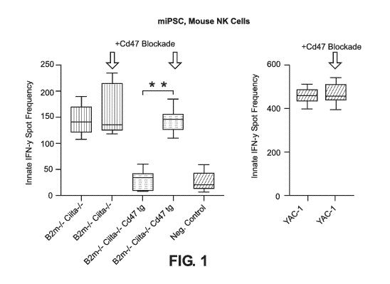
[0032] FIG. 1 shows Elispot results of mouse B2m-/-Ciita-/-CD47 tg iPSCs
incubated with
mouse natural killer (NK) cells (approximately 95% NK cells and 5%
macrophages).
[0033] FIG. 2 shows Elispot results of human B2M-/-CIITA-/-CD47 tg iPSCs
incubated with
human NK cells (approximately 95% NK cells and 5% macrophages).
[0034] FIG. 3 shows Elispot results of mouse B2m-/-Ciita-/-CD47 tg iPSCs
incubated with
human NK cells (approximately 95% NK cells and 5% macrophages).
[0035] FIG. 4 shows Elispot results of human B2M-/-CIITA-/-CD47 tg iPSCs
incubated with
mouse NK cells (approximately 95% NK cells and 5% macrophages).
[0036] FIG. 5 shows phagocytosis assay results of firefly luciferase labeled
human B2M-/-
CIITA-/-CD47 tg iPSCs co-cultured with human macrophages.
[0037] FIG. 6 shows phagocytosis assay results of firefly luciferase labeled
mouse B2m-/-
Ciita-/-CD47 tg iPSCs co-cultured with mouse macrophages.
[0038] FIG. 7 shows phagocytosis assay results of firefly luciferase labeled
human B2M-/-
CIITA-/-CD47 tg iPSCs co-cultured with mouse macrophages.
[0039] FIG. 8 shows phagocytosis assay results of firefly luciferase labeled
mouse B2m-/-
Ciita-/-CD47 tg iPSCs co-cultured with human macrophages.
[0040] FIG. 9 shows differentiation of HIP cells described herein into T
cells.
[0041] FIGS 10A and 10B show differentiation of HIP cells into CD3+ cells,
CD4+ cells,
and CD8+ cells. FIG. 10A shows cells at day 23 (D23) of differentiation on 0P9-
DL1 cells.
FIG. 10B shows cells at day 30 (D30) of differentiation off feeder cells and
with CD3/CD28
stimulation.
[0042] FIG. 11 shows differentiation of HIP cells into T cells (e.g., CD3+
cells, CD4+ cells,
and CD8+ cells) at day 23 (D23) of differentiation on feeder cells with
CD3/CD28
stimulation.
[0043] FIG. 12 shows endothelial progenitor cells derived from HIP cells.
6
CA 03106022 2021-01-07
WO 2020/018620
PCT/US2019/042123
[0044] FIGS. 13A-13C show human HIP cells cultured with endothelial progenitor
cells
(EPCs) that were differentiated into CD4+ T cells (FIG. 13A), naive CD4+ cells
(CD45RA+CCR7+CD4+ cells; FIG. 13B), and central memory CD4+ T cells
(CD45RA-CCR7+CD4+ cells; FIG. 13C). ** denotes p<0.001; unpaired student's t-
test.
[0045] FIGS. 14A and 14B show human T cells derived from human HIP cells using
simulated microgravity (sug) for 72 hours. FIG. 14A shows the morphology of
the human T
cells derived from human HIP cells. FIG. 14B shows the cell viability of the
human T cells.
P=n.s.; unpaired student's t-test.
[0046] FIG. 15 shows human CD8+ T cells derived from human HIP cells using
simulated
microgravity (sug) for 72 hours. * denotes p<0.05; unpaired student's t-test.
[0047] FIG. 16 shows human CD8+ T cells derived from human HIP cells using
simulated
microgravity (sug) for 72 hours and 10 days.
[0048] FIG. 17 shows human CD8+CD45RA+CCR7+ T cells and human
CD8+CD45RA+CCR7- T cells derived from human HIP cells using simulated
microgravity
(sug) for 72 hours followed by treatment at lg for 72 hours. * denotes p<0.05;
unpaired
student's t-test.
[0049] FIG. 18 shows human CD8+ T cells derived from human HIP cells using
simulated
microgravity and cytokine stimulation.
VI. DETAILED DESCRIPTION OF THE INVENTION
A. Introduction
[0050] The invention provides LlypoImmunogenic Pluripotent ("HIP") cells that
avoid host
immune responses due to several genetic manipulations as outlined herein. The
cells lack
major immune antigens that trigger immune responses and are engineered to
avoid
phagocytosis. This allows the derivation of "off-the-shelf' cell products for
generating
specific tissues and organs. The benefit of being able to use human allogeneic
HIP cell
derivatives in human patients results in significant benefits, including the
ability to avoid
long-term adjunct immunosuppressive therapy and drug use generally seen in
allogeneic
transplantations. It also provides significant cost savings as cell therapies
can be used
without requiring individual treatments for each patient. Recently, it was
shown that cell
products generated from autologous cell sources may become subject to immune
rejection
with few or even one single antigenic mutation. Thus, autologous cell products
are not
7
CA 03106022 2021-01-07
WO 2020/018620
PCT/US2019/042123
inherently non-immunogenic. Also, cell engineering and quality control is very
labor and
cost intensive and autologous cells are not available for acute treatment
options. Only
allogeneic cell products will be able to be used for a bigger patient
population if the immune
hurdle can be overcome. HIP cells will serve as a universal cell source for
the generation of
universally-acceptable derivatives.
[0051] The present invention is directed to the exploitation of the
fetomaternal tolerance that
exists in pregnant women. Although half of a fetus' human leukocyte antigens
(HLA) are
paternally inherited and the fetus expresses major HLA mismatched antigens,
the maternal
immune system does not recognize the fetus as an allogeneic entity and does
not initiate an
immune response, e.g. as is seen in a "host versus graft" type of immune
reaction.
Fetomaternal tolerance is mainly mediated by syncytiotrophoblast cells in the
fetal-maternal
interface. Syncytiotrophoblast cells show little or no proteins of the major
histocompatibility
complexes I and II (MIC-I and MHC-II), as well as increased expression of
CD47, known as
the "don't eat me" protein that suppresses phagocytic innate immune
surveillance and
elimination of HLA-devoid cells. Surprisingly, the same tolerogenic mechanisms
that
prevent rejection of the fetus during pregnancy also allow the HIP cells of
the invention to
escape rejection and facilitate long-term survival and engraftment of these
cells after
allogeneic transplantation.
[0052] These results are additionally surprising in that this fetomaternal
tolerance can be
introduced with as little as three genetic modifications (as compared to the
starting iPSCs,
e.g. human iPSCs), two reductions in activity ("knock outs" as further
described herein) and
one increase in activity (a "knock in" as described herein). Generally, others
of skill in the
art have attempted to suppress immunogenicity of iPSCs but have been only
partially
successful; see Rong etal., Cell Stem Cell 14:121-130 (2014) and Gornalusse
etal., Nature
Biotech doi:10.1038/nbt.3860).
[0053] This application is related to International Application No.
PCT/U518/13688, filed on
January 14, 2018 and U.S. Provisional Application No. 62/445,969, filed
January 13, 2017,
the disclosures in their entirety are herein incorporated by reference, in
particular, the
examples, figures, figure descriptions, and descriptions of producing
hypoimmunogenic
pluripotent stem cells and differentiating such cells into other cell types.
8
CA 03106022 2021-01-07
WO 2020/018620
PCT/US2019/042123
[0054] Thus, the invention provides for the generation of HIP cells from
pluripotent stem
cells, and then their maintenance, differentiation and ultimately
transplantation of their
derivatives into patients in need thereof
B. Definitions
[0055] The term "pluripotent cells" refers to cells that can self-renew and
proliferate while
remaining in an undifferentiated state and that can, under the proper
conditions, be induced to
differentiate into specialized cell types. The term "pluripotent cells," as
used herein,
encompass embryonic stem cells and other types of stem cells, including fetal,
amnionic, or
somatic stem cells. Exemplary human stem cell lines include the H9 human
embryonic stem
cell line. Additional exemplary stem cell lines include those made available
through the
National Institutes of Health Human Embryonic Stem Cell Registry and the
Howard Hughes
Medical Institute HUES collection (as described in Cowan, C. A. et. al, New
England I Med.
350:13. (2004), incorporated by reference herein in its entirety.)
[0056] "Pluripotent stem cells" as used herein have the potential to
differentiate into any of
the three germ layers: endoderm (e.g. the stomach linking, gastrointestinal
tract, lungs, etc),
mesoderm (e.g. muscle, bone, blood, urogenital tissue, etc) or ectoderm (e.g.
epidermal
tissues and nervous system tissues). The term "pluripotent stem cells," as
used herein, also
encompasses "induced pluripotent stem cells", or "iPSCs", a type of
pluripotent stem cell
derived from a non-pluripotent cell. Examples of parent cells include somatic
cells that have
been reprogrammed to induce a pluripotent, undifferentiated phenotype by
various means.
Such "iPS" or "iPSC" cells can be created by inducing the expression of
certain regulatory
genes or by the exogenous application of certain proteins. Methods for the
induction of iPS
cells are known in the art and are further described below. (See, e.g., Zhou
etal., Stem Cells
27 (11): 2667-74 (2009); Huangfu etal., Nature Biotechnol. 26 (7): 795 (2008);
Woltjen et
al., Nature 458 (7239): 766-770 (2009); and Zhou etal., Cell Stem Cell 8:381-
384 (2009);
each of which is incorporated by reference herein in their entirety.) The
generation of
induced pluripotent stem cells (iPSCs) is outlined below. As used herein,
"hiPSCs" are
human induced pluripotent stem cells, and "miPSCs" are murine induced
pluripotent stem
cells.
[0057] "Pluripotent stem cell characteristics" refer to characteristics of a
cell that distinguish
pluripotent stem cells from other cells. The ability to give rise to progeny
that can undergo
differentiation, under the appropriate conditions, into cell types that
collectively demonstrate
9
CA 03106022 2021-01-07
WO 2020/018620
PCT/US2019/042123
characteristics associated with cell lineages from all of the three germinal
layers (endoderm,
mesoderm, and ectoderm) is a pluripotent stem cell characteristic. Expression
or non-
expression of certain combinations of molecular markers are also pluripotent
stem cell
characteristics. For example, human pluripotent stem cells express at least
several, and in
some embodiments, all of the markers from the following non-limiting list: S
SEA-3, S SEA-
4, TRA-1-60, TRA-1-81, TRA-2-49/6E, ALP, 5ox2, E-cadherin, UTF-1, 0ct4, Rexl,
and
Nanog. Cell morphologies associated with pluripotent stem cells are also
pluripotent stem
cell characteristics. As described herein, cells do not need to pass through
pluripotency to be
reprogrammed into endodermal progenitor cells and/or hepatocytes.
[0058] As used herein, "multipotent" or "multipotent cell" refers to a cell
type that can give
rise to a limited number of other particular cell types. For example, induced
multipotent cells
are capable of forming endodermal cells. Additionally, multipotent blood stem
cells can
differentiate itself into several types of blood cells, including lymphocytes,
monocytes,
neutrophils, etc.
[0059] As used herein, the term "oligopotent" refers to the ability of an
adult stem cell to
differentiate into only a few different cell types. For example, lymphoid or
myeloid stem cells
are capable of forming cells of either the lymphoid or myeloid lineages,
respectively.
[0060] As used herein, the term "unipotent" means the ability of a cell to
form a single cell
type. For example, spermatogonial stem cells are only capable of forming sperm
cells.
[0061] As used herein, the term "totipotent" means the ability of a cell to
form an entire
organism. For example, in mammals, only the zygote and the first cleavage
stage blastomeres
are totipotent.
[0062] As used herein, "non-pluripotent cells" refer to mammalian cells that
are not
pluripotent cells. Examples of such cells include differentiated cells as well
as progenitor
cells. Examples of differentiated cells include, but are not limited to, cells
from a tissue
selected from bone marrow, skin, skeletal muscle, fat tissue and peripheral
blood. Exemplary
cell types include, but are not limited to, fibroblasts, hepatocytes,
myoblasts, neurons,
osteoblasts, osteoclasts, and T-cells. The starting cells employed for
generating the induced
multipotent cells, the endodermal progenitor cells, and the hepatocytes can be
non-pluripotent
cells.
[0063] Differentiated cells include, but are not limited to, multipotent
cells, oligopotent cells,
unipotent cells, progenitor cells, and terminally differentiated cells. In
particular
CA 03106022 2021-01-07
WO 2020/018620
PCT/US2019/042123
embodiments, a less potent cell is considered "differentiated" in reference to
a more potent
cell.
[0064] A "somatic cell" is a cell forming the body of an organism. Somatic
cells include cells
making up organs, skin, blood, bones and connective tissue in an organism, but
not germ
cells.
[0065] Cells can be from, for example, human or non-human mammals. Exemplary
non-
human mammals include, but are not limited to, mice, rats, cats, dogs,
rabbits, guinea pigs,
hamsters, sheep, pigs, horses, bovines, and non-human primates. In some
embodiments, a cell
is from an adult human or non-human mammal. In some embodiments, a cell is
from a
neonatal human, an adult human, or non-human mammal.
[0066] As used herein, the terms "subject" or "patient" refers to any animal,
such as a
domesticated animal, a zoo animal, or a human. The "subject" or "patient" can
be a mammal
like a dog, cat, bird, livestock, or a human. Specific examples of "subjects"
and "patients"
include, but are not limited to, individuals (particularly human) with a
disease or disorder
related to the liver, heart, lung, kidney, pancreas, brain, neural tissue,
blood, bone, bone
marrow, and the like.
[0067] Mammalian cells can be from humans or non-human mammals. Exemplary non-
human mammals include, but are not limited to, mice, rats, cats, dogs,
rabbits, guinea pigs,
hamsters, sheep, pigs, horses, bovines, and non-human primates (e.g.,
chimpanzees,
macaques, and apes).
[0068] By "hypo-immunogenic pluripotent cell," "hypoimmune pluripotent stem
cell,"
"hypoimmune pluripotent cell," or "HIP cell" herein is meant a pluripotent
cell that retains its
pluripotent characteristics and yet gives rise to a reduced immunological
rejection response
when transferred into an allogeneic host. In preferred embodiments, HIP cells
do not give
rise to an immune response. Thus, "hypo-immunogenic" or "hypoimmune" refers to
a
significantly reduced or eliminated immune response when compared to the
immune
response of a parental (i.e. "wild-type" or "wt") cell prior to
immunoengineering as outlined
herein. In many cases, the HIP cells are immunologically silent and yet retain
pluripotent
capabilities. Assays for HIP characteristics are outlined below.
[0069] By "HLA" or "human leukocyte antigen" complex is a gene complex
encoding the
major histocompatibility complex (MHC) proteins in humans. These cell-surface
proteins
that make up the HLA complex are responsible for the regulation of the immune
response to
11
CA 03106022 2021-01-07
WO 2020/018620
PCT/US2019/042123
antigens. In humans, there are two MHCs, class I and class II, "HLA-I" and
"HLA-II".
HLA-I includes three proteins, HLA-A, HLA-B and HLA-C, which present peptides
from the
inside of the cell, and antigens presented by the HLA-I complex attract killer
T-cells (also
known as CD8+ T-cells or cytotoxic T cells). The HLA-I proteins are associated
with 13-2
microglobulin (B2M). HLA-II includes five proteins, HLA-DP, HLA-DM, HLA-DOB,
HLA-DQ and HLA-DR, which present antigens from outside the cell to T
lymphocytes. This
stimulates CD4+ cells (also known as T-helper cells). It should be understood
that the use of
either "MHC" or "HLA" is not meant to be limiting, as it depends on whether
the genes are
from humans (HLA) or murine (MHC). Thus, as it relates to mammalian cells,
these terms
may be used interchangeably herein.
[0070] By "gene knock out" herein is meant a process that renders a particular
gene inactive
in the host cell in which it resides, resulting either in no protein of
interest being produced or
an inactive form. As will be appreciated by those in the art and further
described below, this
can be accomplished in a number of different ways, including removing nucleic
acid
sequences from a gene, or interrupting the sequence with other sequences,
altering the
reading frame, or altering the regulatory components of the nucleic acid. For
example, all or
part of a coding region of the gene of interest can be removed or replaced
with "nonsense"
sequences, all or part of a regulatory sequence such as a promoter can be
removed or
replaced, translation initiation sequences can be removed or replaced, etc.
[0071] By "gene knock in" herein is meant a process that adds a genetic
function to a host
cell. This causes increased levels of the encoded protein. As will be
appreciated by those in
the art, this can be accomplished in several ways, including adding one or
more additional
copies of the gene to the host cell or altering a regulatory component of the
endogenous gene
increasing expression of the protein is made. This may be accomplished by
modifying the
promoter, adding a different promoter, adding an enhancer, or modifying other
gene
expression sequences.
[0072] "13-2 microglobulin" or "132M" or "B2M" protein refers to the human
132M protein that
has the amino acid and nucleic acid sequences shown below; the human gene has
accession
number NC 000015.10:44711487-44718159.
[0073] "CD47 protein" protein refers to the human CD47 protein that has the
amino acid and
nucleic acid sequences shown below; the human gene has accession number
NC 000003.12:108043094-108094200.
12
CA 03106022 2021-01-07
WO 2020/018620
PCT/US2019/042123
[0074] "CIITA protein" protein refers to the human CIITA protein that has the
amino acid
and nucleic acid sequences shown below; the human gene has accession number
NC 000016.10:10866208-10941562.
[0075] By "wild type" in the context of a cell means a cell found in nature.
However, in the
context of a pluripotent stem cell, as used herein, it also means an iPSC that
may contain
nucleic acid changes resulting in pluripotency but did not undergo the gene
editing
procedures of the invention to achieve hypo-immunogenicity.
[0076] By "syngeneic" herein refers to the genetic similarity or identity of a
host organism
and a cellular transplant where there is immunological compatibility; e.g. no
immune
response is generated.
[0077] By "allogeneic" herein refers to the genetic dissimilarity of a host
organism and a
cellular transplant where an immune response is generated.
[0078] By "B2M-/-" herein is meant that a diploid cell has had the B2M gene
inactivated in
both chromosomes. As described herein, this can be done in a variety of ways.
[0079] By "CIITA -/-" herein is meant that a diploid cell has had the CIITA
gene inactivated
in both chromosomes. As described herein, this can be done in a variety of
ways.
[0080] By "CD47 tg" (standing for "transgene") or "CD47+") herein is meant
that the host
cell expresses CD47, in some cases by having at least one additional copy of
the CD47 gene.
[0081] An "Oct polypeptide" refers to any of the naturally-occurring members
of Octamer
family of transcription factors, or variants thereof that maintain
transcription factor activity,
similar (within at least 50%, 80%, or 90% activity) compared to the closest
related naturally
occurring family member, or polypeptides comprising at least the DNA-binding
domain of
the naturally occurring family member, and can further comprise a
transcriptional activation
domain. Exemplary Oct polypeptides include Oct-1, Oct-2, Oct-3/4, Oct-6, Oct-
7, Oct-8, Oct-
9, and Oct-11. 0ct3/4 (referred to herein as "0ct4") contains the POU domain,
a 150 amino
acid sequence conserved among Pit-1, Oct-1, Oct-2, and uric-86. (See, Ryan, A.
K. &
Rosenfeld, M. G., Genes Dev. 11:1207-1225 (1997), incorporated herein by
reference in its
entirety.) In some embodiments, variants have at least 85%, 90%, or 95% amino
acid
sequence identity across their whole sequence compared to a naturally
occurring Oct
polypeptide family member such as to those listed above or such as listed in
GenBank
accession number NP-002692.2 (human 0ct4) or NP-038661.1 (mouse 0ct4). Oct
polypeptides (e.g., 0ct3/4 or Oct 4) can be from human, mouse, rat, bovine,
porcine, or other
13
CA 03106022 2021-01-07
WO 2020/018620
PCT/US2019/042123
animals. Generally, the same species of protein will be used with the species
of cells being
manipulated. The Oct polypeptide(s) can be a pluripotency factor that can help
induce
multipotency in non-pluripotent cells.
[0082] A "Klf polypeptide" refers to any of the naturally-occurring members of
the family of
Krtippel-like factors (Klfs), zinc-finger proteins that contain amino acid
sequences similar to
those of the Drosophila embryonic pattern regulator Krtippel, or variants of
the naturally-
occurring members that maintain transcription factor activity similar (within
at least 50%,
80%, or 90% activity) compared to the closest related naturally occurring
family member, or
polypeptides comprising at least the DNA-binding domain of the naturally
occurring family
member, and can further comprise a transcriptional activation domain. (See,
Dang, D. T.,
Pevsner, J. & Yang, V. W., Cell Biol. 32:1103-1121 (2000), incorporated by
reference herein
in its entirety.) Exemplary Klf family members include, Klfl, Klf2, Klf3, Klf-
4, Klf5, Klf6,
Klf7, Klf8, Klf9, Klfl 0, Klf11, Klf12, Klf13, Klf14, K1f15, Klf16, and Klf17.
Klf2 and Klf-4
were found to be factors capable of generating iPS cells in mice, and related
genes Klfl and
Klf5 did as well, although with reduced efficiency. (See, Nakagawa, et al.,
Nature
Biotechnology 26:101-106 (2007), incorporated by reference herein in its
entirety.) In some
embodiments, variants have at least 85%, 90%, or 95% amino acid sequence
identity across
their whole sequence compared to a naturally occurring Klf polypeptide family
member such
as to those listed above or such as listed in GenBank accession number
CAX16088 (mouse
Klf4) or CAX14962 (human Klf4). Klf polypeptides (e.g., Klfl, Klf4, and Klf5)
can be from
human, mouse, rat, bovine, porcine, or other animals. Generally, the same
species of protein
will be used with the species of cells being manipulated. The Klf
polypeptide(s) can be a
pluripotency factor. The expression of the Klf4 gene or polypeptide can help
induce
multipotency in a starting cell or a population of starting cells.
[0083] A "Myc polypeptide" refers to any of the naturally-occurring members of
the Myc
family. (See, e.g., Adhikary, S. & Eilers, M., Nat. Rev. Mol. Cell Biol. 6:635-
645 (2005),
incorporated by reference herein in its entirety.) It also includes variants
that maintain
similar transcription factor activity when compared to the closest related
naturally occurring
family member (i.e., within at least 50%, 80%, or 90% activity). It further
includes
polypeptides comprising at least the DNA-binding domain of a naturally
occurring family
member, and can further comprise a transcriptional activation domain.
Exemplary Myc
polypeptides include, e.g., c-Myc, N-Myc and L-Myc. In some embodiments,
variants have at
least 85%, 90%, or 95% amino acid sequence identity across their whole
sequence compared
14
CA 03106022 2021-01-07
WO 2020/018620
PCT/US2019/042123
to a naturally occurring Myc polypeptide family member, such as to those
listed above or
such as listed in Genbank accession number CAA25015 (human Myc). Myc
polypeptides
(e.g., c-Myc) can be from human, mouse, rat, bovine, porcine, or other
animals. Generally,
the same species of protein will be used with the species of cells being
manipulated. The Myc
polypeptide(s) can be a pluripotency factor.
[0084] A "Sox polypeptide" refers to any of the naturally-occurring members of
the SRY-
related HMG-box (Sox) transcription factors, characterized by the presence of
the high-
mobility group (HMG) domain, or variants thereof that maintain similar
transcription factor
activity when compared to the closest related naturally occurring family
member (i.e. within
at least 50%, 80%, or 90% activity). It also includes polypeptides comprising
at least the
DNA-binding domain of the naturally occurring family member, and can further
comprise a
transcriptional activation domain. (See, e.g., Dang, D. T. et al., Int. I
Biochem. Cell Biol.
32:1103-1121(2000), incorporated by reference herein in its entirety.)
Exemplary Sox
polypeptides include, e.g., Soxl, Sox-2, Sox3, Sox4, Sox5, Sox6, Sox7, Sox8,
Sox9, Sox10,
Soxll, Sox12, Sox13, Sox14, Sox15, Sox17, Sox18, Sox-21, and Sox30. Soxl has
been
shown to yield iPS cells with a similar efficiency as Sox2, and genes Sox3,
Sox15, and Sox18
have also been shown to generate iPS cells, although with somewhat less
efficiency than
Sox2. (See, Nakagawa, etal., Nature Biotechnology 26:101-106 (2007),
incorporated by
reference herein in its entirety.) In some embodiments, variants have at least
85%, 90%, or
95% amino acid sequence identity across their whole sequence compared to a
naturally
occurring Sox polypeptide family member such as to those listed above or such
as listed in
Genbank accession number CAA83435 (human Sox2). Sox polypeptides (e.g., Soxl,
Sox2,
Sox3, Sox15, or Sox18) can be from human, mouse, rat, bovine, porcine, or
other animals.
Generally, the same species of protein will be used with the species of cells
being
manipulated. The Sox polypeptide(s) can be a pluripotency factor. As discussed
herein,
SOX2 proteins find particular use in the generation of iPSCs.
[0085] By "differentiated hypoimmunogenic pluripotent cells" or
"differentiated HIP cells"
or "dHIP cells" herein is meant iPS cells that have been engineered to possess
hypoimmunogenicity (e.g., by the knock out of B2M and CIITA and the knock in
of CD47)
and then are differentiated into a cell type for ultimate transplantation into
subjects. Thus, for
example HIP cells can be differentiated into hepatocytes ("dHIP hepatocytes"),
into beta-like
pancreatic cells or islet organoids ("dHIP beta cells"), into endothelial
cells ("dHIP
endothelial cells"), etc.
CA 03106022 2021-01-07
WO 2020/018620
PCT/US2019/042123
[0086] The term percent "identity," in the context of two or more nucleic acid
or polypeptide
sequences, refers to two or more sequences or subsequences that have a
specified percentage
of nucleotides or amino acid residues that are the same, when compared and
aligned for
maximum correspondence, as measured using one of the sequence comparison
algorithms
described below (e.g., BLASTP and BLASTN or other algorithms available to
persons of
skill) or by visual inspection. Depending on the application, the percent
"identity" can exist
over a region of the sequence being compared, e.g., over a functional domain,
or,
alternatively, exist over the full length of the two sequences to be compared.
For sequence
comparison, typically one sequence acts as a reference sequence to which test
sequences are
compared. When using a sequence comparison algorithm, test and reference
sequences are
input into a computer, subsequence coordinates are designated, if necessary,
and sequence
algorithm program parameters are designated. The sequence comparison algorithm
then
calculates the percent sequence identity for the test sequence(s) relative to
the reference
sequence, based on the designated program parameters.
[0087] Optimal alignment of sequences for comparison can be conducted, e.g.,
by the local
homology algorithm of Smith & Waterman, Adv. Appl. Math. 2:482 (1981), by the
homology alignment algorithm of Needleman & Wunsch, J. Mol. Biol. 48:443
(1970), by the
search for similarity method of Pearson & Lipman, Proc. Nat'l. Acad. Sci. USA
85:2444
(1988), by computerized implementations of these algorithms (GAP, BESTFIT,
FASTA, and
TFASTA in the Wisconsin Genetics Software Package, Genetics Computer Group,
575
Science Dr., Madison, Wis.), or by visual inspection (see generally Ausubel et
al., infra).
[0088] One example of an algorithm that is suitable for determining percent
sequence
identity and sequence similarity is the BLAST algorithm, which is described in
Altschul et
al., J. Mol. Biol. 215:403-410 (1990). Software for performing BLAST analyses
is publicly
available through the National Center for Biotechnology Information.
[0089] "Inhibitors," "activators," and "modulators" affect a function or
expression of a
biologically-relevant molecule. The term "modulator" includes both inhibitors
and
activators. They may be identified using in vitro and in vivo assays for
expression or activity
of a target molecule.
[0090] "Inhibitors" refer to agents that, e.g., inhibit expression or bind to
target molecules or
proteins. They may partially or totally block stimulation or have protease
inhibitor activity.
They may reduce, decrease, prevent, or delay activation, including
inactivation, desensitizion,
16
CA 03106022 2021-01-07
WO 2020/018620
PCT/US2019/042123
or down regulation of the activity of the described target protein. Modulators
may be
antagonists of the target molecule or protein.
[0091] "Activators" refer to agents that, e.g., induce or activate the
function or expression of
a target molecule or protein. They may bind to, stimulate, increase, open,
activate, or
facilitate the target molecule activity. Activators may be agonists of the
target molecule or
protein.
[0092] "Homologs" are bioactive molecules that are similar to a reference
molecule at the
nucleotide sequence, peptide sequence, functional, or structural level.
Homologs may
include sequence derivatives that share a certain percent identity with the
reference sequence.
Thus, in one embodiment, homologous or derivative sequences share at least a
70 percent
sequence identity. In a specific embodiment, homologous or derivative
sequences share at
least an 80 or 85 percent sequence identity. In a specific embodiment,
homologous or
derivative sequences share at least a 90 percent sequence identity. In a
specific embodiment,
homologous or derivative sequences share at least a 95 percent sequence
identity. In a more
specific embodiment, homologous or derivative sequences share at least a 50,
55, 60, 65, 70,
75, 85, 86, 87, 88, 89, 90, 91, 92, 93, 94, 95, 96, 97, 98, or 99 percent
sequence identity.
Homologous or derivative nucleic acid sequences may also be defined by their
ability to
remain bound to a reference nucleic acid sequence under high stringency
hybridization
conditions. Homologs having a structural or functional similarity to a
reference molecule
may be chemical derivatives of the reference molecule. Methods of detecting,
generating,
and screening for structural and functional homologs as well as derivatives
are known in the
art.
[0093] "Hybridization" generally depends on the ability of denatured DNA to
reanneal when
complementary strands are present in an environment below their melting
temperature. The
higher the degree of desired homology between the probe and hybridizable
sequence, the
higher the relative temperature that can be used. As a result, it follows that
higher relative
temperatures would tend to make the reaction conditions more stringent, while
lower
temperatures less so. For additional details and explanation of stringency of
hybridization
reactions, see Ausubel et al, Current Protocols in Molecular Biology, Wiley
Interscience
Publishers (1995), incorporated by reference herein in its entirety.
[0094] "Stringency" of hybridization reactions is readily determinable by one
of ordinary
skill in the art, and generally is an empirical calculation dependent upon
probe length,
17
CA 03106022 2021-01-07
WO 2020/018620
PCT/US2019/042123
washing temperature, and salt concentration. In general, longer probes require
higher
temperatures for proper annealing, while shorter probes need lower
temperatures.
[0095] "Stringent conditions" or "high stringency conditions", as defined
herein, can be
identified by those that: (1) employ low ionic strength and high temperature
for washing, for
example 0.015 M sodium chloride/0.0015 M sodium citrate/0.1% sodium dodecyl
sulfate at
50 C; (2) employ during hybridization a denaturing agent, such as formamide,
for example,
50% (v/v) formamide with 0.1% bovine serum albumin/0.1% Fico11/0.1%
polyvinylpyrrolidone/50 Mm sodium phosphate buffer at pH 6.5 with 750 Mm
sodium
chloride, 75 Mm sodium citrate at 42 C; or (3) overnight hybridization in a
solution that
employs 50% formamide, 5 x SSC (0.75 M NaCl, 0.075 M sodium citrate), 50 Mm
sodium
phosphate (pH 6.8), 0.1 % sodium pyrophosphate, 5 x Denhardt's solution,
sonicated salmon
sperm DNA (50 pl/m1), 0.1% SDS, and 10% dextran sulfate at 42 C, with a 10
minute wash
at 42 C in 0.2 x SSC (sodium chloride/sodium citrate) followed by a 10 minute
high-
stringency wash consisting of 0.1 x SSC containing EDTA at 55 C.
[0096] It is intended that every maximum numerical limitation given throughout
this
specification includes every lower numerical limitation, as if such lower
numerical
limitations were expressly written herein. Every minimum numerical limitation
given
throughout this specification will include every higher numerical limitation,
as if such higher
numerical limitations were expressly written herein. Every numerical range
given throughout
this specification will include every narrower numerical range that falls
within such broader
numerical range, as if such narrower numerical ranges were all expressly
written herein.
[0097] As used herein the term "modification" refers to an alteration that
physically
differentiates the modified molecule from the parent molecule. In one
embodiment, an amino
acid change in a CD47, HSVtk, EC-CD, or iCasp9 variant polypeptide prepared
according to
the methods described herein differentiates it from the corresponding parent
that has not been
modified according to the methods described herein, such as wild-type
proteins, a naturally
occurring mutant proteins or another engineered protein that does not include
the
modifications of such variant polypeptide. In another embodiment, a variant
polypeptide
includes one or more modifications that differentiates the function of the
variant polypeptide
from the unmodified polypeptide. For example, an amino acid change in a
variant
polypeptide affects its receptor binding profile. In other embodiments, a
variant polypeptide
comprises substitution, deletion, or insertion modifications, or combinations
thereof In
18
CA 03106022 2021-01-07
WO 2020/018620
PCT/US2019/042123
another embodiment, a variant polypeptide includes one or more modifications
that increases
its affinity for a receptor compared to the affinity of the unmodified
polypeptide.
[0098] In one embodiment, a variant polypeptide includes one or more
substitutions,
insertions, or deletions relative to a corresponding native or parent
sequence. In certain
embodiments, a variant polypeptide includes 1, 2, 3, 4, 5, 6, 7, 8, 9, 10, 11,
12, 13, 14, 15, 16,
17, 18, 19, 20, 21, 22, 23, 24, 25, 26, 27, 28, 29, 30, 31-40, 41 to 50, or 51
or more
modifications.
[0099] By "episomal vector" herein is meant a genetic vector that can exist
and replicate
autonomously in the cytoplasm of a cell; e.g. it is not integrated into the
genomic DNA of the
host cell. A number of episomal vectors are known in the art and described
below.
[00100] By "knock out" in the context of a gene means that the host cell
harboring the
knock out does not produce a functional protein product of the gene. As
outlined herein, a
knock out can result in a variety of ways, from removing all or part of the
coding sequence,
introducing frameshift mutations such that a functional protein is not
produced (either
truncated or nonsense sequence), removing or altering a regulatory component
(e.g. a
promoter) such that the gene is not transcribed, preventing translation
through binding to
mRNA, etc. Generally, the knock out is effected at the genomic DNA level, such
that the
cells' offspring also carry the knock out permanently.
[00101] By "knock in" in the context of a gene means that the host cell
harboring the
knock in has more functional protein active in the cell. As outlined herein, a
knock in can be
done in a variety of ways, usually by the introduction of at least one copy of
a transgene (tg)
encoding the protein into the cell, although this can also be done by
replacing regulatory
components as well, for example by adding a constitutive promoter to the
endogeneous gene.
In general, knock in technologies result in the integration of the extra copy
of the transgene
into the host cell.
VII. HYPOIMMUNOGENIC PLURIPOTENT (HIP) CELLS
[00102] The invention provides compositions and methodologies for
generating HIP
cells, starting with wild type cells, rendering them pluripotent (e.g. making
induced
pluripotent stem cells, or iPSCs), then generating HIP cells from the iPSC
population.
19
CA 03106022 2021-01-07
WO 2020/018620
PCT/US2019/042123
A. Methodologies for Genetic Alterations
[00103] The invention includes methods of modifying nucleic acid sequences
within
cells or in cell-free conditions to generate both pluripotent cells and HIP
cells. Exemplary
technologies include homologous recombination, knock-in, ZFNs (zinc finger
nucleases),
TALENs (transcription activator-like effector nucleases), meganucleases (e.g.,
homing
endonucleases), CRISPR (clustered regularly interspaced short palindromic
repeats)/Cas9,
and other site-specific nuclease technologies. These techniques enable double-
strand DNA
breaks at desired locus sites. These controlled double-strand breaks promote
homologous
recombination at the specific locus sites. This process focuses on targeting
specific sequences
of nucleic acid molecules, such as chromosomes, with endonucleases that
recognize and bind
to the sequences and induce a double-stranded break in the nucleic acid
molecule. The
double-strand break is repaired either by an error-prone non-homologous end-
joining (NHEJ)
or by homologous recombination (HR).
[00104] As will be appreciated by those in the art, a number of different
techniques can
be used to engineer the pluripotent cells of the invention, as well as the
engineering of the
iPSCs to become hypoimmunogenic as outlined herein.
[00105] In general, these techniques can be used individually or in
combination. For
example, in the generation of the HIP cells, CRISPR/Cas may be used to reduce
the
expression of active B2M and/or CIITA protein in the engineered cells, with
viral techniques
(e.g., retrovirus, lentivirus, and adeno-associated virus) to knock in the
CD47 functionality.
Also, as will be appreciated by those in the art, although one embodiment
sequentially
utilizes a CRISPR/Cas step to knock out B2M, followed by a CRISPR/Cas step to
knock out
CIITA with a final step of a lentivirus to knock in the CD47 functionality,
these genes can be
manipulated in different orders using different technologies.
[00106] As is discussed more fully below, transient expression of
reprogramming
genes is generally done to generate induced pluripotent stem cells.
a. CRISPR/Cas Technologies
[00107] In one embodiment, the cells are manipulated using clustered
regularly
interspaced short palindromic repeats)/Cas ("CRISPR") technologies as is known
in the art.
CRISPR/Cas can be used to generate the starting iPSCs or to generate the HIP
cells from the
iPSCs. There are a large number of techniques based on CRISPR/Cas, see for
example
CA 03106022 2021-01-07
WO 2020/018620
PCT/US2019/042123
Doudna and Charpentier, Science doi:10.1126/science.1258096, hereby
incorporated by
reference. CRISPR techniques and kits are sold commercially.
b. TALEN Technologies
[00108] In some embodiments, the HIP cells of the invention are made using
Transcription Activator-Like Effector Nucleases (TALEN) methodologies. TALEN
are
restriction enzymes combined with a nuclease that can be engineered to bind to
and cut
practically any desired DNA sequence. TALEN kits are sold commercially.
c. Zinc Finger Technologies
[00109] In one embodiment, the cells are manipulated using Zn finger
nuclease
technologies. Zn finger nucleases are artificial restriction enzymes generated
by fusing a zinc
finger DNA-binding domain to a DNA-cleavage domain. Zinc finger domains can be
engineered to target specific desired DNA sequences and this enables zinc-
finger nucleases to
target unique sequences within complex genomes. By taking advantage of
endogenous DNA
repair machinery, these reagents can be used to precisely alter the genomes of
higher
organisms, similar to CRISPR and TALENs.
d. Viral Based Technologies
[00110] There are a wide variety of viral techniques that can be used to
generate the
HIP cells of the invention (as well as for the original generation of the
iPSCs), including, but
not limited to, the use of retroviral vectors, lentiviral vectors, adenovirus
vectors and Sendai
viral vectors. Episomal vectors used in the generation of iPSCs are described
below.
e. Downregulation of Genes Using Interfering RNA
[00111] In other embodiments, genes that encode proteins used in HLA
molecules are
downregulated by RNA interference (RNAi) technologies. RNAi refers to a
process where
RNA molecules inhibit gene expression often by causing specific mRNA molecules
to
degrade. Two types of RNA molecules ¨ microRNA (miRNA) and small interfering
RNA
(siRNA) ¨ can be used for RNA interference. They bind to the target mRNA
molecules and
either increase or decrease their activity. RNAi helps cells defend against
parasitic nucleic
acids such as those from viruses and transposons. RNAi also influences
development.
[00112] According to particular embodiments, the inhibitory nucleic acid is
an
antisense oligonucleotide which inhibits the expression of a target gene,
e.g., B2M gene and a
CIITA gene. Such an antisense oligonucleotide can be a nucleic acid (either
DNA or RNA)
21
CA 03106022 2021-01-07
WO 2020/018620
PCT/US2019/042123
which specifically hybridizes (e.g., binds) under cellular conditions with the
cellular mRNA
and/or genomic DNA encoding the target protein, thereby inhibiting
transcription and/or
translation of the gene. The binding may be by conventional base pair
complementarity.
Alternatively, the binding may be, for example, in case of binding to DNA
duplexes, through
specific interactions in the major groove of the double helix. Absolute
complementarity,
although preferred, is not required.
[00113] Thus, according to an embodiment, the antisense oligonucleotide is
a single-
stranded or double-stranded DNA molecule, more preferably a double-stranded
DNA
molecule. According to another embodiment, the antisense oligonucleotide is a
single-
stranded or double-stranded RNA molecule, more preferably a single-stranded
RNA
molecule. In some instances, the antisense oligonucleotide is a modified
oligonucleotide
which is resistant to endogenous nucleases, e.g., exonucleases and/or
endonucleases, and is
therefore stable in vivo and in vitro.
[00114] The antisense oligonucleotide may be modified at the base moiety,
sugar
moiety, or phosphate backbone, for example, to improve stability of the
molecule. The
antisense oligonucleotide may include other appended groups such as peptides
(e.g., for
targeting host cell receptors), or agents facilitating transport across the
cell membrane. The
antisense oligonucleotide may be conjugated to another molecule such as a
peptide or
transport agent. In some cases, the antisense oligonucleotide comprises at
least one modified
base moiety which is selected from the group including, but not limited to, 5-
fluorouracil, 5-
bromouracil, 5-chlorouracil, 5-iodouracil, hypoxanthine, xanthine, 4-
acetylcytosine, 5-
(carboxyhydroxytriethyl) uracil, 5-carboxymethylaminomethy1-2-thiouridine, 5-
carboxymethylanninonnethyluracil, dihydrouracil, beta-D-galactosylqueosine,
inosine, N6-
isopentenyladenine, 1-nnethylguanine, 1-methylinosine, 2,2-dimethylguanine, 2-
methyladenine, 2-methylguanine, 3-methylcytosine, 5-methylcytosine, N6-
adenine, 7-
nnethylguanine, 5-methylaminonnethyluracil, 5-methoxyaminomethy1-2-thiouracil,
beta-D-
mannosylqueosine, 5-methoxycarboxymethyluracil, 5-methoxyuracil, 2-methylthio-
N6-
isopentenyladenine, uracil-5-oxyacetic acid (v), wybutoxosine, pseudouracil,
queosine, 2-
thiocytosine, 5-methyl-2-thiouracil, 2-thiouracil, 4-thiouracil, 5-methyl
uracil, uracil-5-
oxyacetic acid methylester, uracil-5-oxyacetic acid (v), 5-methyl-2-
thiouracil, 3-(3-amino-3-
N-2-carboxypropyl) uracil, (acp3)w and 2,6-diaminopurine.
[00115] In certain embodiments, the antisense oligonucleotide comprise at
least one
modified sugar moiety selected from the group including, but not limited to,
arabinose, 2-
22
CA 03106022 2021-01-07
WO 2020/018620
PCT/US2019/042123
fluoroara binose, xylulose and hexose. In other embodiments, the antisense
oligonucleotide
comprises at least one modified phosphate backbone selected from the group
including, but
not limited to, a phosphorothioate, a phosphorodithioate, a
phosphoramidothioate, a
phosphoramidate, a phosphordiamidate, a methylphosphonate, an alkyl
phosphotriester, and a
formacetal or analog thereof
[00116] sdRNA molecules are a class of asymmetric siRNAs comprising a guide
(antisense) strand of 19-21 bases. They can contain a 5' phosphate, 2'Ome or
2'F modified
pyrimidines, and six phosphorotioates at the 3' positions. They can contain a
sense strand
containing 3' conjugated sterol moieties, 2 phosphotioates at the 3' position,
and 2'Ome
modified pyrimidines. Both strands can contain 2' Ome purines with continuous
stretches of
unmodified purines not exceeding a length of 3. sdRNA is disclosed in U.S.
Patent No.
8,796,443, incorporated herein by reference in its entirety.
[00117] For all of these technologies, well known recombinant techniques
are used, to
generate recombinant nucleic acids as outlined herein. In certain embodiments,
the
recombinant nucleic acids (either than encode a desired polypeptide, e.g.
CD47, or disruption
sequences) may be operably linked to one or more regulatory nucleotide
sequences in an
expression construct. Regulatory nucleotide sequences will generally be
appropriate for the
host cell and subject to be treated. Numerous types of appropriate expression
vectors and
suitable regulatory sequences are known in the art for a variety of host
cells. Typically, the
one or more regulatory nucleotide sequences may include, but are not limited
to, promoter
sequences, leader or signal sequences, ribosomal binding sites,
transcriptional start and
termination sequences, translational start and termination sequences, and
enhancer or
activator sequences. Constitutive or inducible promoters as known in the art
are also
contemplated. The promoters may be either naturally occurring promoters, or
hybrid
promoters that combine elements of more than one promoter. An expression
construct may be
present in a cell on an episome, such as a plasmid or vector, or the
expression construct may
be inserted in a chromosome. In a specific embodiment, the expression vector
includes a
selectable marker gene to allow the selection of transformed host cells.
Certain embodiments
include an expression vector comprising a nucleotide sequence encoding a
variant
polypeptide operably linked to at least one regulatory sequence. Regulatory
sequence for use
herein include promoters, enhancers, and other expression control elements. In
certain
embodiments, an expression vector is designed for the choice of the host cell
to be
transformed, the particular variant polypeptide desired to be expressed, the
vector's copy
23
CA 03106022 2021-01-07
WO 2020/018620
PCT/US2019/042123
number, the ability to control that copy number, or the expression of any
other protein
encoded by the vector, such as antibiotic markers.
[00118] Examples of suitable promoters include, for example, promoters from
the
following genes: ubiquitin/S27a promoter of the hamster (WO 97/15664), Simian
vacuolating
virus 40 (SV40) early promoter, adenovirus major late promoter, mouse
metallothionein-I
promoter, the long terminal repeat region of Rous Sarcoma Virus (RSV), mouse
mammary
tumor virus promoter (MMTV), Moloney murine leukemia virus Long Terminal
repeat
region, and the early promoter of human Cytomegalovirus (CMV). Examples of
other
heterologous mammalian promoters are the actin, immunoglobulin or heat shock
promoter(s).
In some embodiments, the elongation factor 1-alpha promoter is used.
[00119] In additional embodiments, promoters for use in mammalian host
cells can be
obtained from the genomes of viruses such as polyoma virus, fowlpox virus (UK
2,211,504
published 5 Jul. 1989), bovine papilloma virus, avian sarcoma virus,
cytomegalovirus, a
retrovirus, hepatitis-B virus and Simian Virus 40 (5V40). In further
embodiments,
heterologous mammalian promoters are used. Examples include the actin
promoter, an
immunoglobulin promoter, and heat-shock promoters. The early and late
promoters of 5V40
are conveniently obtained as an 5V40 restriction fragment which also contains
the 5V40 viral
origin of replication. Fiers et al., Nature 273: 113-120 (1978). The immediate
early promoter
of the human cytomegalovirus is conveniently obtained as a HindIII E
restriction fragment.
Greenaway, P. J. et al., Gene 18: 355-360 (1982). The foregoing references are
incorporated
by reference in their entirety.
B. Generation of Pluripotent Cells
[00120] The invention provides methods of producing non-immunogenic
pluripotent
cells from pluripotent cells. Thus, the first step is to provide the
pluripotent stem cells.
[00121] The generation of mouse and human pluripotent stem cells (generally
referred
to as iPSCs; miPSCs for murine cells or hiPSCs for human cells) is generally
known in the
art. As will be appreciated by those in the art, there are a variety of
different methods for the
generation of iPSCs. The original induction was done from mouse embryonic or
adult
fibroblasts using the viral introduction of four transcription factors,
0ct3/4, 5ox2, c-Myc and
Klf4; see Takahashi and Yamanaka Cell 126:663-676 (2006), hereby incorporated
by
reference in its entirety and specifically for the techniques outlined
therein. Since then, a
number of methods have been developed; see Seki et al. , Worldi Stem Cells
7(1):116-125
24
CA 03106022 2021-01-07
WO 2020/018620
PCT/US2019/042123
(2015) for a review, and Lakshmipathy and Vermuri, editors, Methods in
Molecular Biology:
Pluripotent Stem Cells, Methods and Protocols, Springer 2013, both of which
are hereby
expressly incorporated by reference in their entirety, and in particular for
the methods for
generating hiPSCs (see for example Chapter 3 of the latter reference).
[00122] Generally, iPSCs are generated by the transient expression of one
or more
"reprogramming factors" in the host cell, usually introduced using episomal
vectors. Under
these conditions, small amounts of the cells are induced to become iPSCs (in
general, the
efficiency of this step is low, as no selection markers are used). Once the
cells are
"reprogrammed", and become pluripotent, they lose the episomal vector(s) and
produce the
factors using the endogeneous genes. This loss of the episomal vector(s)
results in cells that
are called "zero footprint" cells. This is desirable as the fewer genetic
modifications
(particularly in the genome of the host cell), the better. Thus, it is
preferred that the resulting
hiPSCs have no permanent genetic modifications.
[00123] As is also appreciated by those of skill in the art, the number of
reprogramming factors that can be used or are used can vary. Commonly, when
fewer
reprogramming factors are used, the efficiency of the transformation of the
cells to a
pluripotent state goes down, as well as the "pluripotency", e.g. fewer
reprogramming factors
may result in cells that are not fully pluripotent but may only be able to
differentiate into
fewer cell types.
[00124] In some embodiments, a single reprogramming factor, OCT4, is used.
In other
embodiments, two reprogramming factors, OCT4 and KLF4, are used. In other
embodiments,
three reprogramming factors, OCT4, KLF4 and 50X2, are used. In other
embodiments, four
reprogramming factors, OCT4, KLF4, 50X2 and c-Myc, are used. In other
embodiments, 5,
6 or 7 reprogramming factors can be used selected from SOKMNLT: 50X2, OCT4
(POU5F1), KLF4, MYC, NANOG, LIN28, and SV4OL T antigen.
[00125] In general, these reprogramming factor genes are provided on
episomal
vectors such as are known in the art and commercially available. For example,
ThermoFisher/Invitrogen sell a sendai virus reprogramming kit for zero
footprint generation
of hiPSCs, see catalog number A34546. ThermoFisher also sells EBNA-based
systems as
well, see catalog number A14703.
CA 03106022 2021-01-07
WO 2020/018620
PCT/US2019/042123
[00126] In addition, there are a number of commercially available hiPSC
lines
available; see, e.g., the Gibco0 Episomal hiPSC line, K18945, which is a zero
footprint,
viral-integration-free human iPSC cell line (see also Burridge et al, 2011,
supra).
[00127] In general, as is known in the art, iPSCs are made from non-
pluripotent cells
such as CD34+ cord blood cells, fibroblasts, etc., by transiently expressing
the
reprogramming factors as described herein.
[00128] For example, successful iPSCs were also generated using only
0ct3/4, Sox2
and Klf4, while omitting the C-Myc, although with reduced reprogramming
efficiency.
[00129] In general, iPSCs are characterized by the expression of certain
factors that
include KLF4, Nanog, OCT4, SOX2, ESRRB, TBX3, c-Myc and TCL1. New or increased
expression of these factors for purposes of the invention may be via induction
or modulation
of an endogenous locus or from expression from a transgene.
[00130] For example, murine iPSCs can be generated using the methods of
Diecke et
al, Sci Rep. 2015, Jan. 28;5:8081 (doi:10.1038/srep08081), hereby incorporated
by reference
in its entirety and specifically for the methods and reagents for the
generation of the miPSCs.
See also, e.g., Burridge et al., PLoS One, 2011 6(4):18293, hereby
incorporated by reference
in its entirety and specifically for the methods outlined therein.
[00131] In some cases, the pluripotency of the cells is measured or
confirmed as
outlined herein, for example by assaying for reprogramming factors as is
generally shown in
PCT/US18/13688 or by conducting differentiation reactions as outlined therein,
for instance,
in the Examples.
C. Generation of Hypo-Immunogenic Pluripotent (HIP) Cells
[00132] The present invention is directed to the generation, manipulation,
growth and
transplantation of hypo-immunogenic cells into a patient as defined herein.
The generation of
HIP cells from pluripotent cells is done with as few as three genetic changes,
resulting in
minimal disruption of cellular activity but conferring immunosilencing to the
cells.
[00133] As discussed herein, one embodiment utilizes a reduction or
elimination in the
protein activity of MHC I and II (HLA I and II when the cells are human). This
can be done
by altering genes encoding their component. In one embodiment, the coding
region or
regulatory sequences of the gene are disrupted using CRISPR/Cas. In another
embodiment,
gene translation is reduced using interfering RNA technologies. The third
change is a change
26
CA 03106022 2021-01-07
WO 2020/018620
PCT/US2019/042123
in a gene that regulates susceptibility to macrophage phagocytosis, such as
CD47, and this is
generally a "knock in" of a gene using viral technologies.
[00134] Additional descriptions of HIP cells can be found in International
Application
No. PCT/US18/13688, filed on January 14, 2018 and U.S. Provisional Application
No.
62/445,969, filed January 13, 2017, the disclosures in their entirety are
herein incorporated by
reference, in particular, the examples, figures, figure descriptions, and
descriptions of
producing hypoimmunogenic pluripotent stem cells and differentiating such
cells into other
cell types.
[00135] In some cases, where CRISPR/Cas is being used for the genetic
modifications,
hiPSC cells that contain a Cas9 construct that enable high efficiency editing
of the cell line
can be used; see, e.g., the Human Episomal Cas9 iPSC cell line, A33124, from
Life
Technologies.
1. HLA-I Reduction
[00136] The HIP cells of the invention include a reduction in MHC I
function (HLA I
when the cells are derived from human cells).
[00137] As will be appreciated by those in the art, the reduction in
function can be
accomplished in a number of ways, including removing nucleic acid sequences
from a gene,
interrupting the sequence with other sequences, or altering the regulatory
components of the
nucleic acid. For example, all or part of a coding region of the gene of
interest can be
removed or replaced with "nonsense" sequences, frameshift mutations can be
made, all or
part of a regulatory sequence such as a promoter can be removed or replaced,
translation
initiation sequences can be removed or replaced, etc.
[00138] As will be appreciated by those in the art, the successful
reduction of the MHC
I function (HLA I when the cells are derived from human cells) in the
pluripotent cells can be
measured using techniques known in the art and as described below; for
example, FACS
techniques using labeled antibodies that bind the HLA complex; for example,
using
commercially available HLA-A, B, C antibodies that bind to the alpha chain of
the human
major histocompatibility HLA Class I antigens.
B2M Alteration
[00139] In one embodiment, the reduction in HLA-I activity is done by
disrupting the
expression of the 13-2 microglobulin gene in the pluripotent stem cell, the
human sequence of
27
CA 03106022 2021-01-07
WO 2020/018620
PCT/US2019/042123
which is disclosed herein. This alteration is generally referred to herein as
a gene "knock
out", and in the HIP cells of the invention it is done on both alleles in the
host cell. Generally
the techniques to do both disruptions is the same.
[00140] A particularly useful embodiment uses CRISPR technology to disrupt
the
gene. In some cases, CRISPR technology is used to introduce small
deletions/insertions into
the coding region of the gene, such that no functional protein is produced,
often the result of
frameshift mutations that result in the generation of stop codons such that
truncated, non-
functional proteins are made.
[00141] Accordingly, a useful technique is to use CRISPR sequences designed
to
target the coding sequence of the B2M gene in mouse or the B2M gene in human.
After gene
editing, the transfected iPSC cultures are dissociated to single cells. Single
cells are expanded
to full-size colonies and assessed for CRISPR/Cas edit by screening for
presence of aberrant
sequence from the CRISPR cleavage site. Clones with deletions in both alleles
are picked.
Such clones did not express B2M as demonstrated by PCR and did not express HLA-
I as
demonstrated by FACS analysis (see examples 1 and 6, for example of
PCT/US18/13688).
[00142] Assays to test whether the B2M gene has been inactivated are known
and
described herein. In one embodiment, the assay is a Western blot oaf cells
lysates probed
with antibodies to the B2M protein. In another embodiment, reverse
transcriptase
polymerase chain reactions (RT-PCR) confirms the presence of the inactivating
alteration.
[00143] In addition, the cells can be assessed to confirm that the HLA I
complex is not
expressed on the cell surface. This may be assayed by FACS analysis using
antibodies to one
or more HLA cell surface components as discussed above.
[00144] It is noteworthy that others have had poor results when trying to
silence the
B2M genes at both alleles. See, e.g. Gornalusse et al.,Nature Biotech.
Doi/10.1038/nbt.3860).
2. HLA-II Reduction
[00145] In addition to a reduction in HLA I, the HIP cells of the invention
also lack
MHC II function (HLA II when the cells are derived from human cells).
[00146] As will be appreciated by those in the art, the reduction in
function can be
accomplished in a number of ways, including removing nucleic acid sequences
from a gene,
adding nucleic acid sequences to a gene, disrupting the reading frame,
interrupting the
28
CA 03106022 2021-01-07
WO 2020/018620
PCT/US2019/042123
sequence with other sequences, or altering the regulatory components of the
nucleic acid. In
one embodiment, all or part of a coding region of the gene of interest can be
removed or
replaced with "nonsense" sequences. In another embodiment, regulatory
sequences such as a
promoter can be removed or replaced, translation initiation sequences can be
removed or
replaced, etc.
[00147] The successful reduction of the MHC II function (HLA II when the
cells are
derived from human cells) in the pluripotent cells or their derivatives can be
measured using
techniques known in the art such as Western blotting using antibodies to the
protein, FACS
techniques, rt-PCR techniques, etc.
CIITA Alteration
[00148] In one embodiment, the reduction in HLA-II activity is done by
disrupting the
expression of the CIITA gene in the pluripotent stem cell, the human sequence
of which is
shown herein. This alteration is generally referred to herein as a gene "knock
out", and in the
HIP cells of the invention it is done on both alleles in the host cell.
[00149] Assays to test whether the CIITA gene has been inactivated are
known and
described herein. In one embodiment, the assay is a Western blot of cells
lysates probed
with antibodies to the CIITA protein. In another embodiment, reverse
transcriptase
polymerase chain reactions RT-PCR) confirms the presence of the inactivating
alteration.
[00150] In addition, the cells can be assessed to confirm that the HLA II
complex is
not expressed on the cell surface. Again, this assay is done as is known in
the art (See Figure
21 of PCT/U518/13688, for example) and generally is done using either Western
Blots or
FACS analysis based on commercial antibodies that bind to human HLA Class II
HLA-DR,
DP and most DQ antigens as outlined below.
[00151] A particularly useful embodiment uses CRISPR technology to disrupt
the
CIITA gene. CRISPRs were designed to target the coding sequence of the CIITA
gene in
mouse or the CIITA gene in human, an essential transcription factor for all
MHC II
molecules. After gene editing, the transfected iPSC cultures were dissociated
into single cells.
They were expanded to full-size colonies and assessed for successful CRISPR
editing by
screening for the presence of an aberrant sequence from the CRISPR cleavage
site. Clones
with deletions did not express CIITA as determined by PCR and did not express
MHC II/
HLA-II as determined by FACS analysis.
29
CA 03106022 2021-01-07
WO 2020/018620
PCT/US2019/042123
3. Reduction of Macrophage Phagocytosis and/or NK Cell Killing
[00152] In addition to the reduction of HLA I and II (or MHC I and II),
generally using
B2M and CIITA knock-outs, the HIP cells of the invention have a reduced
susceptibility to
macrophage phagocytosis and NK cell killing. The resulting HIP cells "escape"
the immune
macrophage and innate pathways due to the expression of one or more CD47
transgenes.
[00153] The ability of HIP cells and cells derived from the HIP cells to
evade or
escape NK cell killing and/or macrophage phagocytosis is shown in FIGS. 14A-
14C and
34A-34C of PCT/US18/13688, the contents, in particular, the figures, figure
descriptions, and
examples are herein incorporated by reference. For example, FIGS. 14B-14C show
that
mouse HIP cells (e.g., B2m-/-Ciita-/-CD47 transgenic mouse iPSCs) failed to
induce CD107a
expression by NK cells, and thus did not elicit an NK cell response. In
addition, it was
shown that such mouse HIP cells did not induce activation of NK cells or
release of IFNy.
When NK cells were incubated with differentiated cells (such as endothelial
cells, smooth
muscle cells, and cardiomyocytes) derived from HIP cells, NK cell responses
were not
induced (see, e.g., FIGS. 34A-34C of PCT/US18/13688).
Increased CD47 Expression
[00154] In some embodiments, reduced macrophage phagocytosis and NK cell
killing
susceptibility results from increased CD47 on the HIP cell surface. This is
done in several
ways as will be appreciated by those in the art using "knock in" or transgenic
technologies.
In some cases, increased CD47 expression results from one or more CD47
transgene.
[00155] Accordingly, in some embodiments, one or more copies of a CD47 gene
is
added to the HIP cells under control of an inducible or constitutive promoter,
with the latter
being preferred. In some embodiments, a lentiviral construct is employed as
described herein
or known in the art. CD47 genes may integrate into the genome of the host cell
under the
control of a suitable promoter as is known in the art.
[00156] The HIP cell lines were generated from B2M-/- CIITA-/- iPSCs. Cells
containing lentivirus vectors expressing CD47 were selected using a
Blasticidin marker. The
CD47 gene sequence was synthesized and the DNA was cloned into the plasmid
Lentivirus
pLenti6N5 with a blasticidin resistance (Thermo Fisher Scientific, Waltham,
MA)
[00157] In some embodiments, the expression of the CD47 gene can be
increased by
altering the regulatory sequences of the endogenous CD47 gene, for example, by
exchanging
CA 03106022 2021-01-07
WO 2020/018620
PCT/US2019/042123
the endogenous promoter for a constitutive promoter or for a different
inducible promoter.
This can generally be done using known techniques such as CRISPR.
[00158] Once altered, the presence of sufficient CD47 expression can be
assayed using
known techniques such as those described in the Examples, such as Western
blots, ELISA
assays or FACS assays using anti-CD47 antibodies. In general, "sufficiency" in
this context
means an increase in the expression of CD47 on the HIP cell surface that
silences NK cell
killing and/or macrophage phagocytosis. The natural expression levels on cells
is too low to
protect them from NK cell lysis once their MHC I is removed.
4. Suicide Genes
[00159] In some embodiments, the invention provides hypoimmunogenic
pluripotent
cells that comprise a "suicide gene" or "suicide switch". These are
incorporated to function
as a "safety switch" that can cause the death of the hypoimmunogenic
pluripotent cells should
they grow and divide in an undesired manner. The "suicide gene" ablation
approach includes
a suicide gene in a gene transfer vector encoding a protein that results in
cell killing only
when activated by a specific compound. A suicide gene may encode an enzyme
that
selectively converts a nontoxic compound into highly toxic metabolites. The
result is
specifically eliminating cells expressing the enzyme. In some embodiments, the
suicide gene
is the herpesvirus thymidine kinase (HSV-tk) gene and the trigger is
ganciclovir. In other
embodiments, the suicide gene is the Escherichia coli cytosine deaminase (EC-
CD) gene and
the trigger is 5-fluorocytosine (5-FC) (Barese etal., Mol. Therap. 20(10):1932-
1943 (2012),
Xu et al., Cell Res. 8:73-8 (1998), both incorporated herein by reference in
their entirety.)
[00160] In other embodiments, the suicide gene is an inducible Caspase
protein. An
inducible Caspase protein comprises at least a portion of a Caspase protein
capable of
inducing apoptosis. In one embodiment, the portion of the Caspase protein is
exemplified in
SEQ ID NO:6. In preferred embodiments, the inducible Caspase protein is
iCasp9. It
comprises the sequence of the human FK506-binding protein, FKBP12, with an
F36V
mutation, connected through a series of amino acids to the gene encoding human
caspase 9.
FKBP12-F36V binds with high affinity to a small-molecule dimerizing agent,
AP1903. Thus,
the suicide function of iCasp9 in the instant invention is triggered by the
administration of a
chemical inducer of dimerization (CID). In some embodiments, the CID is the
small
molecule drug AP1903. Dimerization causes the rapid induction of apoptosis.
(See
W02011146862; Stasi eta!, N Engl. I Med 365;18 (2011); Tey etal., Biol. Blood
Marrow
31
CA 03106022 2021-01-07
WO 2020/018620
PCT/US2019/042123
Transplant. 13:913-924 (2007), each of which are incorporated by reference
herein in their
entirety.)
5. Assays for HIP Phenotypes and Retention of Pluripotency
[00161] Once the HIP cells have been generated, they may be assayed for
their hypo-
immunogenicity and/or retention of pluripotency as is generally described
herein and in the
examples.
[00162] For example, hypo-immunogenicity are assayed using a number of
techniques
as exemplified in Figure 13 and Figure 15 of PCT/US18/13688. These techniques
include
transplantation into allogeneic hosts and monitoring for HIP cell growth (e.g.
teratomas) that
escape the host immune system. HIP derivatives are transduced to express
luciferase and can
then followed using bioluminescence imaging. Similarly, the T cell and/or B
cell response of
the host animal to the HIP cells are analyzed to confirm that the HIP cells do
not cause an
immune reaction in the host animal. T cell function is assessed by Elispot,
ELISA, FACS,
PCR, or mass cytometry (CYTOF). B cell response or antibody response is
assessed using
FACS or luminex. Additionally or alternatively, the cells may be assayed for
their ability to
avoid innate immune responses, e.g. NK cell killing, as is generally shown in
FIGS. 14A-14C
of PCT/U518/13688. NK cell lytolytic activity is assessed in vitro or in vivo
(as shown in
FIGS. 15A-15B of PCT/US18/13688).
[00163] Similarly, the retention of pluripotency is assessed in a number of
ways. In
one embodiment, pluripotency is assayed by the expression of certain
pluripotency-specific
factors as generally described herein and shown in FIG. 29 of PCT/U518/13688.
In addition
or alternatively, the HIP cells are differentiated into one or more cell types
as an indication of
pluripotency.
D. Preferred Embodiments of the HIP cells
[00164] Provided herein are hypoimmunogenic pluripotent stem cells ("HIP
cells")
that exhibit pluripotency but do not result in a host immune response when
transplanted into
an allogeneic host such as a human patient, either as the HIP cells or as the
differentiated
products of the HIP cells.
[00165] In one embodiment, human pluripotent stem cells such as human
induced
pluripotent stem cells are rendered hypo-immunogenic by a) the disruption of
the B2M gene
at each allele (e.g., B2M-/-), b) the disruption of the CIITA gene at each
allele (e.g. CIITA-/-
), and c) by the overexpression of the CD47 gene (CD47+, e.g. through
introducing one or
32
CA 03106022 2021-01-07
WO 2020/018620
PCT/US2019/042123
more additional copies of the CD47 gene or activating the genomic gene). This
renders the
hiPSC population B2M-/- CIITA-/- CD47tg. In a preferred embodiment, the cells
are non-
immunogenic. In another embodiment, the HIP cells are rendered non-immunogenic
B2M-/-
CIITA-/- CD47 transgene as described above but are further modified by
including an
inducible suicide gene that is induced to kill the cells in vivo when
required.
E. Maintenance of HIP Cells
[00166] Once generated, the HIP cells can be maintained an undifferentiated
state as is
known for maintaining iPSCs. For example, HIP cells are cultured on Matrigel
using culture
media that prevents differentiation and maintains pluripotency.
F. Differentiation of HIP Cells
[00167] HIP cells described herein can be differentiated into different
cell types. The
pluripotency of the HIPs can be evaluated by differentiating the cells into
endodermal,
mesodermal and ectodermal cell types. In some cases, the HIP cells are
assessed by teratoma
formation.
[00168] As will be appreciated by those in the art, the methods for
differentiation
depend on the desired cell type using known techniques. The cells are
differentiated in
suspension and then put into a gel matrix form, such as matrigel, gelatin, or
fibrin/thrombin
forms to facilitate cell survival. Differentiation is assayed as is known in
the art, generally by
evaluating the presence of cell-specific markers.
[00169] In some embodiments, the HIP cells are differentiated into
hepatocytes to
address loss of the hepatocyte functioning or cirrhosis of the liver. There
are a number of
techniques that can be used to differentiate HIP cells into hepatocytes; see
for example
Pettinato et al., doi:10.1038/spre32888, Snykers et al., Methods Mol Biol
698:305-314
(2011), Si-Tayeb eta!, Hepatology 51:297-305 (2010) and Asgari etal., Stem
Cell Rev (:493-
504 (2013), all of which are hereby expressly incorporated by reference in
their entirety and
specifically for the methodologies and reagents for differentiation.
Differentiation is assayed
as is known in the art, generally by evaluating the presence of hepatocyte
associated and/or
specific markers, including, but not limited to, albumin, alpha fetoprotein,
and fibrinogen.
Differentiation can also be measured functionally, such as the metabolization
of ammonia,
LDL storage and uptake, ICG uptake and release and glycogen storage.
33
CA 03106022 2021-01-07
WO 2020/018620
PCT/US2019/042123
[00170] In some embodiments, the HIP cells are differentiated into beta-
like cells or
islet organoids for transplantation to address type I diabetes mellitus
(T1DM). Cell systems
are a promising way to address T1DM, see, e.g., Ellis et al.,
doi/10.1038/nrgastro.2017.93,
incorporated herein by reference. Additionally, Pagliuca et al. reports on the
successful
differentiation of 13-cells from hiPSCs (see doi/10.106/j.ce11.2014.09.040,
hereby incorporated
by reference in its entirety and in particular for the methods and reagents
outlined there for
the large-scale production of functional human13 cells from human pluripotent
stem cells).
Furthermore, Vegas etal. shows the production of human 13 cells from human
pluripotent
stem cells followed by encapsulation to avoid immune rejection by the host;
(doi:10.1038/nm.4030, hereby incorporated by reference in its entirety and in
particular for
the methods and reagents outlined there for the large-scale production of
functional human13
cells from human pluripotent stem cells).
[00171] Differentiation is assayed as is known in the art, generally by
evaluating the
presence of f3 cell associated or specific markers, including but not limited
to, insulin.
Differentiation can also be measured functionally, such as measuring glucose
metabolism,
see generally Muraro et al, doi:10.1016/j.cels.2016.09.002, hereby
incorporated by reference
in its entirety, and specifically for the biomarkers outlined there.
[00172] Once the beta cells derived from HIP cells are generated, they can
be
transplanted (either as a cell suspension or within a gel matrix as discussed
herein) into the
portal vein/liver, the omentum, the gastrointestinal mucosa, the bone marrow,
a muscle, or
subcutaneous pouches.
[00173] In some embodiments, the HIP cells are differentiated into retinal
pigment
epithelium (RPE) to address sight-threatening diseases of the eye. Human
pluripotent stem
cells have been differentiated into RPE cells using the techniques outlined in
Kamao et al.,
Stem Cell Reports 2014:2:205-18, hereby incorporated by reference in its
entirety and in
particular for the methods and reagents outlined there for the differentiation
techniques and
reagents; see also Mandai et al., doi:10.1056/NEJMoa1608368, also incorporated
in its
entirety for techniques for generating sheets of RPE cells and transplantation
into patients.
[00174] Differentiation can be assayed as is known in the art, generally by
evaluating
the presence of RPE associated and/or specific markers or by measuring
functionally. See for
example Kamao et al., doi:10.1016/j.stemcr.2013.12.007, hereby incorporated by
reference in
34
CA 03106022 2021-01-07
WO 2020/018620
PCT/US2019/042123
its entirety and specifically for the markers outlined in the first paragraph
of the results
section.
[00175] In some embodiments, the HIP cells are differentiated into
cardiomyocytes to
address cardiovascular diseases. Techniques are known in the art for the
differentiation of
hiPSCs to cardiomyoctes and discussed in the Examples. Differentiation can be
assayed as is
known in the art, generally by evaluating the presence of cardiomyocyte
associated or
specific markers or by measuring functionally; see for example Loh et al.,
doi:10.1016/j.ce11.2016.06.001, hereby incorporated by reference in its
entirety and
specifically for the methods of differentiating stem cells including
cardiomyocytes.
[00176] In some embodiments, the HIP cells are differentiated into
endothelial colony
forming cells (ECFCs) to form new blood vessels to address peripheral arterial
disease.
Techniques to differentiate endothelial cells are known. See, e.g., Prasain
etal.,
doi:10.1038/nbt.3048, incorporated by reference in its entirety and
specifically for the
methods and reagents for the generation of endothelial cells from human
pluripotent stem
cells, and also for transplantation techniques. Differentiation can be assayed
as is known in
the art, generally by evaluating the presence of endothelial cell associated
or specific markers
or by measuring functionally.
[00177] In some embodiments, the HIP cells are differentiated into thyroid
progenitor
cells and thyroid follicular organoids that can secrete thyroid hormones to
address
autoimmune thyroiditis. Techniques to differentiate thyroid cells are known
the art. See, e.g.
Kurmann et al., doi:10.106/j.stem.2015.09.004, hereby expressly incorporated
by reference in
its entirety and specifically for the methods and reagents for the generation
of thyroid cells
from human pluripotent stem cells, and also for transplantation techniques.
Differentiation
can be assayed as is known in the art, generally by evaluating the presence of
thyroid cell
associated or specific markers or by measuring functionally.
VIII. HYPOIMMUNE CHIMERIC ANTIGEN RECEPTOR T CELLS DERIVED FROM
HIP CELLS
[00178] The present invention provides an engineered T cell differentiated
from a HIP
cell comprising a nucleic acid encoding a CAR comprising an antigen binding
domain, a
transmembrane domain, and an intracellular signaling domain. In some
embodiments, the
CAR comprises an antigen binding domain, a transmembrane domain, and an
intracellular
signaling domain of a costimulatory domain. In another aspect of the
invention, provided
CA 03106022 2021-01-07
WO 2020/018620
PCT/US2019/042123
herein are hypoimmunogenic pluripotent cells comprising a nucleic acid
encoding a CAR
comprising an antigen binding domain, a transmembrane domain, and an
intracellular
signaling domain. Such hypoimmunogenic pluripotent cells can be in vitro
differentiated to
T cells to produce hypoimmunogenic CAR-T (HI-CAR-T) cells.
[00179] In some embodiments, the hypoimmunogenic CAR-T cells lack MHC I
function or HLA-I function. For instance, the hypoimmunogenic CAR-T cells have
reduced
expression or lack expression of HLA-A protein, HLA-B protein, and HLA-C
protein. In
particular cases, the hypoimmunogenic CAR-T cells possess genetic
modifications to
inactivate the gene encoding HLA-A protein, the gene encoding HLA-B protein,
the gene
encoding HLA-C protein. In certain cases, the hypoimmunogenic CAR-T cells have
reduced
or lack expression of 13-2 microglobulin protein. In some embodiments, such
cells possess a
genetic modification that inactivates the gene encoding 13-2 microglobulin.
Such
hypoimmunogenic CAR-T cells can be differentiated from hypoimmunogenic
pluripotent
cells lack HLA-I function. In some embodiments, the hypoimmunogenic
pluripotent cells
possess a genetic modification that inactivates the gene encoding 13-2
microglobulin.
[00180] In certain embodiments, the hypoimmunogenic CAR-T cells lack MHC II
function or HLA-II function. In some instances, the hypoimmunogenic CAR-T
cells have
reduced expression or lack expression of HLA-DP protein, HLA-DR protein, and
HLA-DQ
protein. The hypoimmunogenic CAR-T cells may possess genetic modifications to
inactivate
the gene encoding HLA-DP protein, the gene encoding HLA-DR protein, the gene
encoding
HLA-DQ protein. In some embodiments, the hypoimmunogenic CAR-T cells have
reduced
or lack expression of CIITA protein. In some embodiments, such cells possess a
genetic
modification that inactivates the gene encoding CIITA. Such hypoimmunogenic
CAR-T
cells can be differentiated from hypoimmunogenic pluripotent cells lack HLA-II
function. In
some embodiments, the hypoimmunogenic pluripotent cells possess a genetic
modification
that inactivates the gene encoding CIITA.
[00181] In some embodiments, the hypoimmunogenic CAR-T cells have an
increased
expression of CD47 protein compared to a wild-type or native T cell. In other
embodiments,
the hypoimmunogenic pluripotent cells have an increased expression of CD47
protein
compared to a wild-type or native pluripotent cell. Increased expression of
CD47 may result
from a genetic modification to an endogenous CD47 gene. In other cases,
increased
expression results from expression of an exogenous CD47 gene, e.g., an
exogenous nucleic
acid encoding CD47. Such hypoimmunogenic CAR-T cells can be differentiated
from
36
CA 03106022 2021-01-07
WO 2020/018620
PCT/US2019/042123
hypoimmunogenic pluripotent cells overexpressing CD47 protein. In some
embodiments, the
hypoimmunogenic pluripotent cells have increased expression of CD47 protein.
[00182] In some embodiments, the hypoimmunogenic CAR-T cell comprises a
suicide
gene such as, but not limited to, a herpes simplex virus thymidine kinase (HSV-
tk) gene, an
Escherichia colt cytosine deaminase (CD)gene, and a gene encoding an inducible
caspase-9
protein. A suicide gene can be activated upon exposing the cell comprising the
gene to a
chemical agent (e.g., chemical trigger) that causes the cell to die. A
chemical trigger for
HSV-tk can be a dideoxynucleoside analog, e.g., ganciclovir. A chemical
trigger for EC-CD
can be 5-fluorocytosine (5-FC). A chemical trigger for caspase-9 can be a
chemical inducer
of dimerization (CID) such as the compound AP1903. Thus, the hypoimmunogenic
pluripotent cell comprises the suicide gene and is differentiated to any one
of the
hypoimmunogenic CAR-T cells described herein.
[00183] Descriptions about a cytosine deaminase suicide gene system can be
found,
e.g., Mullin et al., Cancer Research, 1994, 54: 1503-1506. Details about a
thymidine kinase
suicide gene system can be found, e.g., Moolten, Cancer Research, 1986,
46(10): 5276-5281.
Detailed descriptions of an inducible caspase-9 suicide gene system can be
found, e.g., in
Gargett and Brown, Front Pharmacol, 2014, 5:235.
A. Chimeric Antigen Receptors
[00184] In various embodiments, the antigen binding domain binds to an
antigen on a
target cell, e.g., a cancer cell. The antigen binding domain (also referred to
as an
extracellular domain) can bind antigens as is know in the art. In some
embodiments, the
antigen binding domain comprises a monoclonal antibody, a polyclonal antibody,
a synthetic
antibody, a human antibody, a humanized antibody, a non-human antibody, a
nanobody, a
single-chain variable fragment (scFv), F(ab')2, Fab', Fab, Fv, and the like.
[00185] The antigen binding domain can include a signal peptide. In
addition, the
CAR can contain a spacer region between the antigen binding domain the
transmembrane
domain. The spacer region should be flexible enough to allow the antigen
binding domain to
orient in different directions to facilitate antigen recognition. The spacer
can be the hinge
region from IgGl, or the CH2 and CH3 region of immunoglobulin and portions of
CD3.
[00186] The antigen binding domain can be linked to the transmembrane
domain of the
CAR. In some embodiments, a nucleic acid encoding the antigen binding domain
is operably
linked to a nucleic acid encoding a transmembrane domain of the CAR.
37
CA 03106022 2021-01-07
WO 2020/018620
PCT/US2019/042123
[00187] In some embodiments, the transmembrane domain can be derived from a
membrane-bound or transmembrane protein. In certain embodiments, the
transmembrane
domain comprises one or more, e.g., 1, 2, 3, 4, 5, 6, 7, 8 or more amino acid
modifications
(e.g., substitutions, insertions, and deletions) compared to the wild-type
amino acid sequence
of the transmembrane domain of the membrane-bound or transmembrane protein.
Non-
limiting examples of a transmembrane domain of a CAR include at least the
transmembrane
region(s) of the alpha, beta or zeta chain of the T-cell receptor, CD28, CD3
epsilon (CD3),
CD45, CD4, CD5, CD8, CD9, CD16, CD22, CD33, CD37, CD64, CD80, CD86, CD134,
CD137, CD154, or an erythropoietin receptor. In other embodiments, the
transmembrane
domain is a recombinant or synthetic domain comprising hydrophobic amino acid
residues
(e.g., leucine and valine). In some cases, the transmembrane domain includes a
phenylalanine, tryptophan and valine at one or both ends of the domain.
[00188] The transmembrane domain links the antigen binding domain to the
intracellular signaling domain of the CAR. In some embodiments, the nucleic
acid encoding
the antigen binding domain is operably linked to the nucleic acid encoding the
transmembrane domain that is operably linked to the nucleic acid encoding the
intracellular
signaling domain.
[00189] In some embodiments, the intracellular signaling domain of a CAR
comprises
a signal activation or signal transduction domain. As such, an intracellular
signaling domain
includes any portion of an intracellular signaling domain of a protein known
in the art to be
sufficient to transduce or transmit a signal, e.g., an activation signal, or
to mediate a cellular
response within a cell.
[00190] In some embodiments, the nucleic acid encoding the CAR of the
present
invention is operably linked to a promoter such as a synthetic promoter, a
constitutive
promoter, or an inducible promoter. Useful constitutive promoters include an
ubiquitin C
promoter, an elongation factor-1 alpha promoter (EF1a promoter), a CMV
promoter, and any
other constitutive promoter known to those skilled in the art. Useful
inducible promoters are
described, e.g., in Ede et al, ACS Synth Blot, 2016, 5(5):395-404, and can
include cell-type
specific promoters and inducible switch promoters. Illustrative constitutive
promoters are
described in PLoS One, 2010, 5(8):e12413. In some embodiments, the promoter is
the EFla
promoter.
38
CA 03106022 2021-01-07
WO 2020/018620
PCT/US2019/042123
[00191] The CARs described herein can be introduced into the HIP cells
using a vector
such as an expression vector, a viral vector, or a non-viral vector. In some
instances, the viral
vector is a retroviral vector, an adenoviral vector, or an adeno-associated
vector. In some
embodiments, the nucleic acid encoding the CAR is introduced into a gene locus
such as a
safe harbor locus of the cell. In other embodiments, the CAR is introduced
into HIP cells
using non-viral vectors including, but not limited to, minicircle DNA vectors,
nude DNA,
liposomes, polymerizers, and molecular conjugates.
[00192] At present, there are two ways to accomplish gene incorporation
with vectors,
i.e., viral systems and non-viral systems. The major vectors for gene therapy
in basic
research and clinical study are viruses, because of the high transfer
efficiency, the relatively
short time needed to reach the clinically necessary numbers of cultured T
cells and the
availability of different viruses with different expression characteristics.
Virus vectors
include retroviruses (including lentivirus), adenovirus and adeno-associated
virus. Among
them, the most popular tools for gene delivery are genetically engineered
retroviruses (e.g.,
Hu et al., Pharmacol Rev. 2000;52(4):493-511). Non-viral vectors including,
but not limited
to, nude DNA, liposomes, polymerizers, and molecular conjugates can be used to
introduce a
CAR construct into HIP cells. Minicircle DNA vectors that are free of plasmid
bacterial
DNA sequences are novel non-viral vectors which can be generated in bacteria
from a
parental plasmid, and can persistently express transgene with high levels in
vivo. Minicircle
DNA systems can be used in a clinical setting. Detailed descriptions of
minicircle DNA
vectors can be found, e.g., in Chen et al., Hum Gene Ther. 2005;16(1):126-131;
Kay et al.,
Nat Biotechnol. 2010;28(12):1287-1289.
B. Differentiating HIP cells into HI-CAR-T cells
[00193] The HIP cells comprising a nucleic acid encoding a CAR can be
differentiated
into a CAR expressing immune cell such as a CAR T cell using any method
recognized by
one skilled in the art.
[00194] Useful methods for differentiating stem cells to immune cells
(e.g., immune
stem cells, immune progenitor cells, immune multipotent progenitor cells, pre-
T cell
progenitor cells, pre-NK cell progenitor cells, T cell progenitor cells, NK
cell progenitor
cells, T cells, NK cells, NKT cells, and B cells) are described in, for
example,
US2018/0072992, US2017/0296649, and US2016/0009813.
39
CA 03106022 2021-01-07
WO 2020/018620
PCT/US2019/042123
[00195] T cells can be c43 T cells, sy T cells, helper/regulatory T cells,
cytotoxic T
cells, progenitor T cells (e.g., a progenitor T cell that is CD34+CD7+CD1a- or
CD34+CD7+
CD5+CD1a-), naive T cells, central memory T cells, effector T cells, terminal
effector T
cells, immature T cells, mature T cells, natural killer T cells, and the like.
In other words, T
cells can be naive T cells, naive central memory T cells (TCM cells), effector
memory T cells
(TEM cells), and effector memory RA T cells (TEMRA cells). Naive T cells can
express
CCR7, CD27, CD28, and CD45RA. Naive central T cells can express CCR7, CD27,
CD28,
and CD45RO. Effector memory T cells can express PD1, CD27, CD28, and CD45RO.
Effector memory RA T cells can express PD1, CD57, and CD45RA.
[00196] In some embodiments, the HIP cells comprising a nucleic acid
sequence
encoding a CAR are cultured in a culture medium comprising a BMP pathway
activator, a
WNT pathway activator, a MEK inhibitor, a NOTCH pathway inhibitor, a ROCK
inhibitor, a
TGF13 receptor/ALK inhibitor, a growth factor, a cytokine, and any combination
thereof
[00197] The BMP pathway activator can include, but is not limited to, an
activator of
BMP-2, an activator of BMP-4, an activator of BMP-5, an activator of BMP-6, an
activator
of BMP-7, an activator of BMP-8, an analog thereof, and a variant thereof
[00198] The GSK3 inhibitor can include, but is not limited to, CHIR99021,
an analog
thereof, and a variant thereof The NOTCH pathway activator can include, but is
not limited
to, Jag1, Jag2, DLL-1, DLL-3, DLL-4, an analog thereof, and a variant thereof
The ROCK
inhibitor can include, but is not limited to, Y27632, Fasudil, AR122-86,
Y27632 H-1152, Y-
30141, Wf-536, HA-1077, hydroxyl-HA-1077, GSK269962A, SB-772077-B, N-(4-
pyridy1)-
N'-(2,4,6-trichlorophenyl)urea, 3-(4-pyridy1)-1H-indole, and (R)-(+)-trans-N-
(4-pyridy1)-4-
(1-aminoethyl)-cyclohexanecarboxamide, other ROCK inhibitors disclosed in US
8044201,
an analog thereof, and a variant thereof The growth factor can include, but is
not limited to,
bFGF, EPO, Flt3L, GM-CSF, IGF, TPO, SCF, VEGF, an analog thereof, and a
variant
thereof The cytokine can include, but is not limited to, IL-2, IL-3, IL-6, IL-
7, IL-11, IL-15,
an analog thereof, and a variant thereof
[00199] In some embodiments, the HIP cells carrying a CAR construct are
cultured on
feeder cells to promote T cell differentiation. The term "feeder cells" can
include cells of a
different tissue type and typically a different genome that may act to promote
proliferation
and/or control differentiation of cells they are cocultured with.
Undifferentiated HIP cells
can be cocultured with feeder cells that direct differentiation towards a
particular tissue type
CA 03106022 2021-01-07
WO 2020/018620
PCT/US2019/042123
(e.g., T cell or a particular T cell subtype). In some embodiments, murine HIP
cells are
cultured on 0P9 or 0P9-DL feeder cells. The murine HIP cells can be cultured
on feeder
cells for about 15 days or more. In other embodiments, the HIP cells are
cultured on feeder
cells and then after a specific number of days cultured on without feeder
cells. In certain
embodiments, the HIP cells are not cultured on feeder cells for
differentiation into T cells. In
some instances, HIP cells are cultured in a medium that promotes CD3
stimulation, and
additionally, CD28 stimulation.
[00200] In various embodiments, human HIP cells are cultured on feeder
cells such as
endothelial progenitor cells derived from human HIP cells. In some
embodiments, human T
cells derived from HIP cells are cultured on endothelial progenitor cells
(EPCs) derived from
human HIP cells. The cells can be cultured on feeder cells for about 15 days
or more. In
other embodiments, the cells are cultured on feeder cells and then after a
specific number of
days cultured on without feeder cells. In certain embodiments, the human EPCs
promote
generation of HIP-derived T cells. In various embodiments, the human EPCs
promote
generation of HIP-derived naive CD4+ T cells. In certain embodiments, the
human EPCs
hinder the generation of certain subtypes of HIP-derived T cells such as
central memory
CD4+ T cells.
[00201] In some embodiments, HIP-derived T cells are cultured in simulated
microgravity (s[tg). In particular embodiments, such T cells are produced by
differentiation
HIP cells using sig. Human HIP-derived T cells can be cultured in s[tg for at
least 72 hours.
In some embodiments, human HIP-derived T cells are cultured in s[tg for 72
hours to 10 days
or more. In some cases, culturing the cells in s[tg can be used to generate
CD8+ T cells. In
some embodiments, s[tg increases the amount or percentage of TEMRA CD8+ T
cells. In
other embodiments, s[tg does not increase the amount or percentage of naive
CD8+ T cells.
[00202] In some embodiments, HIP-derived T cells are cultured in simulated
microgravity (s[tg) and in culture media comprising IL-2, IL-7, or a
combination of IL-2 and
IL-7. In some embodiments, HIP-derived T cells cultured in s[tg and in the
presence of IL-2
to produce central memory CD8+ T cells. In other embodiments, HIP-derived T
cells
cultured in s[tg and in the presence of IL-7 to produce central memory CD8+ T
cells. In yet
other embodiments, HIP-derived T cells cultured in s[tg and in the presence of
IL-2 and IL-7
to produce central memory CD8+ T cells.
41
CA 03106022 2021-01-07
WO 2020/018620
PCT/US2019/042123
[00203] Methods of assessing the CAR expressing immune cells derived from
the HIP
cells include, but are not limited to, immunocytochemistry, flow cytometry,
cytokine
profiling, T cell activation/stimulation assays, target cell cytotoxicity
assays, antigen
reactivity assays, and in vivo functional assays using animal models.
C. Method of Using the HI-CART cells
[00204] In some aspects, provided herein is a method of treating cancer in
a patient,
e.g., a human patient, by administrating a therapeutically effective amount of
HIP cell
derived CAR-T cells. In some cases, the HIP cell derived CAR-T cells are
administered with
a therapeutically effective carrier.
[00205] An "therapeutically effective amount" includes an amount sufficient
to effect a
beneficial or desired clinical result upon treatment. A therapeutically
effective amount can be
administered to a subject in one or more doses. In terms of treatment, an
effective amount is
an amount that is sufficient to palliate, ameliorate, stabilize, reverse or
slow the progression
of the disease, or otherwise reduce the pathological consequences of the
disease. The
effective amount is generally determined by the physician on a case-by-case
basis and is
within the skill of one in the art. Several factors are typically taken into
account when
determining an appropriate dosage to achieve an effective amount. These
factors include age,
sex and weight of the subject, the condition being treated, the severity of
the condition and
the form and effective concentration of the antigen-binding fragment
administered.
[00206] The therapeutic cell treatment can be administered by any methods
known in
the art, including, but not limited to, intravenous administration,
subcutaneous administration,
intranodal administration, intratumoral administration, intrathecal
administration, intrapleural
administration, intraperitoneal administration, and direct administration to
the thymus. The
therapeutic cells can be administered in a bolus or by continuous perfusion.
[00207] Cancer can be selected from the group consisting of a blood cancer,
a solid
tumor cancer, and a liquid tumor cancer. In some embodiments, the blood cancer
is a
leukemia, a lymphoma or a myeloma. Tumor cancers include, but are not limited
to,
glioblastoma, melanoma, neuroblastoma, adenocarcinoma, glioma, soft tissue
sarcoma, and
various carcinomas (including small cell lung cancer). Suitable carcinomas may
include any
known in the field of oncology, including, but not limited to, astrocytoma,
fibrosarcoma,
myxosarcoma, liposarcoma, oligodendroglioma, ependymoma, medulloblastoma,
primitive
neural ectodermal tumor (PNET), chondrosarcoma, osteogenic sarcoma, pancreatic
ductal
42
CA 03106022 2021-01-07
WO 2020/018620
PCT/US2019/042123
adenocarcinoma, small and large cell lung adenocarcinomas, chordoma,
angiosarcoma,
endotheliosarcoma, squamous cell carcinoma, bronchoalveolarcarcinoma,
epithelial
adenocarcinoma, and liver metastases thereof, lymphangiosarcoma,
lymphangioendotheliosarcoma, hepatoma, cholangiocarcinoma, synovioma,
mesothelioma,
Ewing's tumor, rhabdomyosarcoma, colon carcinoma, basal cell carcinoma, sweat
gland
carcinoma, papillary carcinoma, sebaceous gland carcinoma, papillary
adenocarcinoma,
cystadenocarcinoma, medullary carcinoma, bronchogenic carcinoma, renal cell
carcinoma,
bile duct carcinoma, choriocarcinoma, seminoma, embryonal carcinoma, Wilms'
tumor,
testicular tumor, medulloblastoma, craniopharyngioma, ependymoma, pinealoma,
hemangioblastoma, acoustic neuroma, oligodendroglioma, meningioma,
neuroblastoma,
retinoblastoma, Waldenstrom's macroglobulinemia, and heavy chain disease,
breast tumors
such as ductal and lobular adenocarcinoma, squamous and adenocarcinomas of the
uterine
cervix, uterine and ovarian epithelial carcinomas, prostatic adenocarcinomas,
transitional
squamous cell carcinoma of the bladder, B and T cell lymphomas (nodular and
diffuse)
plasmacytoma, malignant melanoma, soft tissue sarcomas and leiomyosarcomas.
IX. DETAILED DESCRIPTION OF EXEMPLARY EMBODIMENTS
[00208] In some embodiments, the mouse hypoimmunogenic pluripotent stem
(mouse
HIP) cell of the invention comprises a genome modification that eliminates B2M
activity, a
genome modification that eliminates CIITA activity, an exogenous nucleic acid
sequence
encoding CD47, and an exogenous nucleic acid sequence encoding a CAR
construct. In
other embodiments, the mouse hypoimmunogenic pluripotent stem cell also
comprises an
inducible suicide gene.
[00209] In other embodiments, the human hypoimmunogenic pluripotent stem
(human
HIP) cell of the invention comprises a genome modification that eliminates B2M
activity, a
genome modification that eliminates CIITA activity, an exogenous nucleic acid
sequence
encoding CD47, and an exogenous nucleic acid sequence encoding a CAR
construct. In
other embodiments, the human hypoimmunogenic pluripotent stem cell also
comprises an
inducible suicide gene.
[00210] In particular embodiments, the hypoimmunogenic pluripotent stem
cell of the
invention comprises a genome modification that eliminates B2M activity, a
genome
modification that eliminates CIITA activity, an exogenous nucleic acid
sequence encoding
CD47, an exogenous nucleic acid sequence encoding a CAR construct, and a
herpes simplex
43
CA 03106022 2021-01-07
WO 2020/018620
PCT/US2019/042123
virus thymidine kinase (HSV-tk) gene. In some embodiments, the hypoimmunogenic
pluripotent stem cell of the invention comprises a genome modification that
eliminates B2M
activity, a genome modification that eliminates CIITA activity, an exogenous
nucleic acid
sequence encoding CD47, an exogenous nucleic acid sequence encoding a CAR
construct,
and an Escherichia coil cytosine deaminase (CD) gene. In certain embodiments,
the
hypoimmunogenic pluripotent stem cell of the invention comprises a genome
modification
that eliminates B2M activity, a genome modification that eliminates CIITA
activity, an
exogenous nucleic acid sequence encoding CD47, an exogenous nucleic acid
sequence
encoding a CAR construct, and an exogenous gene encodes an inducible caspase 9
protein.
[00211] In various embodiments, the mouse CAR-T cell of the invention is
generated
from a mouse hypoimmunogenic pluripotent stem cell comprises a genome
modification that
eliminates B2M activity, a genome modification that eliminates CIITA activity,
an
exogenous nucleic acid sequence encoding CD47, and an exogenous nucleic acid
sequence
encoding a CAR construct. In other embodiments, the mouse hypoimmunogenic
pluripotent
stem cell also comprises an inducible suicide gene. As such, the mouse CAR-T
cell has
reduced or lacks Major Histocompatibility Antigen Complex I (MHC I) and Major
Histocompatibility Antigen Complex II (MHC II) function and overexpresses CD47
protein.
The mouse CAR-T cell can be less susceptible to killing by NK cells.
[00212] In other embodiments, the human CAR-T cell of the invention is
generated
from a human hypoimmunogenic pluripotent stem cell comprises a genome
modification that
eliminates B2M activity, a genome modification that eliminates CIITA activity,
an
exogenous nucleic acid sequence encoding human CD47, and an exogenous nucleic
acid
sequence encoding a CAR construct. In other embodiments, the human
hypoimmunogenic
pluripotent stem cell also comprises an inducible suicide gene. Thus, the
human CAR-T cell
has reduced or lacks HLA-I and HLA-II function and overexpresses CD47 protein.
In some
embodiments, the human CAR-T cell has reduced or lacks expression of HLA-A,
HLA-B, or
HLA-C, has reduced or lacks expression of HLA-DP, HLA-DR, or HLA-DQ protein,
and
overexpresses human CD47 protein. The human CAR-T cell can be less susceptible
to
killing by NK cells.
[00213] In some embodiments, the human CAR-T cell of the invention is
generated
from a human hypoimmunogenic pluripotent stem cell comprises a genome
modification that
eliminates B2M activity, a genome modification that eliminates CIITA activity,
an
44
CA 03106022 2021-01-07
WO 2020/018620
PCT/US2019/042123
exogenous nucleic acid sequence encoding human CD47, and an exogenous nucleic
acid
sequence encoding an anti-CD19 CAR construct.
X. EXAMPLES
Example 1: Generation of mouse induced pluripotent stem cells
[00214] The method described herein is adapted from Diecke et al., Sci Rep,
2015,
8081.
[00215] Murine tail tip fibroblasts of mice were dissociated and isolated
with
collagenase type IV (Life Technologies, Grand Island, NY, USA) and maintained
with
Dulbecco's modified Eagle medium (DMEM) containing 10% fetal bovine serum
(FBS), L-
glutamine, 4.5 g/L glucose, 100 U/mL penicillin, and 100 pg/mL streptomycin at
37 C, 20%
02, and 5% CO2 in a humidified incubator.
[00216] 1 x10⁶ murine fibroblasts were then reprogrammed using a novel
codon
optimized mini-intronic plasmid (co-MIP) (10-12 pm of DNA) expressing the four
reprogramming factors 0ct4, KLF4, 5ox2 and c-Myc using the Neon Transfection
system.
After transfection, fibroblasts were plated on a murine embryonic fibroblasts
(MEF) feeder
layer and kept in fibroblast media with the addition of sodium butyrate (0.2
mM) and 50
pg/mL ascorbic acid.
[00217] When ESC-like colonies appeared, media was changed to murine iPSC
media
containing DMEM, 20% FBS, L-glutamine, non-essential amino acids (NEAA), (3-
mercaptoethanol, and 10 ng/mL leukemia inhibitory factor (LIF). After 2
passages, the
murine iPSCs were transferred to 0.2% gelatin coated plates and further
expanded. With
every passage, the iPSCs were sorted for the murine pluripotency marker SSEA-1
using
magnetic activated cell sorting (MACS).
[00218] The isolated mouse iPSCs can be used to generate mouse
hypoimmunogenic
iPSCs according to the method described above.
Example 2: Generation of human induced pluripotent stem cells
[00219] The GibcoTM Human Episomal iPSC Line (catalog number A18945,
ThermoFisher) was derived from CD34+ cord blood using a three-plasmid, seven-
factor
(SOKMNLT; 50X2, OCT4 (POU5F1), KLF4, MYC, NANOG, LIN28, and SV4OL T
antigen) EBNA-based episomal system. This iPSC line is considered to be zero
foot-print as
CA 03106022 2021-01-07
WO 2020/018620
PCT/US2019/042123
there was no integration into the genome from the reprogramming event. It has
been shown
to be free of all reprogramming genes. Protocols for thawing, culturing, and
passaging the
human iPSCs are provided in the product manual.
[00220] Pluripotency of the human iPSCs can be determined by in vivo
teratoma
assays and in vitro pluripotent gene expression assays (e.g., PCR and arrays)
or by
fluorescence staining for pluripotent markers.
[00221] The GibcoTM Human Episomal iPSC Line has a normal karyotype and
endogenous expression of pluripotent markers like OCT4, SOX2, and NANOG (as
shown by
RT-PCR) and OCT4, SSEA4, TRA-1-60 and TRA-1-81 (as shown by ICC). Whole genome
expression and epigenetic profiling analyses demonstrated that this episomal
hiPSC line is
molecularly indistinguishable from human embryonic stem cell lines
(Quintanilla et al., PloS
One, 2014, 9(1): e85419). In directed differentiation and teratoma analyses,
these hiPSCs
retained their differentiation potential for the ectodermal, endodermal, and
mesodermal
lineages (Burridge et al., PLoS One, 2011, 6(4): e18293). In addition,
vascular,
hematopoietic, neural, and cardiac lineages were derived with robust
efficiencies (Burridge et
al., supra).
46
CA 03106022 2021-01-07
WO 2020/018620
PCT/US2019/042123
Table 1. Illustrative protocol for culturing human iPSCs (e.g., Cas9 iPSCs)
Title Human iPSC culture
Induced Pluripotent stem cells have the capacity to give rise to
differentiated
progeny representative of all three germ layers (ectoderm, endoderm, and
mesoderm). The ability to expand pluripotent cells in vitro and subject them
to
Introduction
direct differentiation to produce specific cell types is crucial to the
development of
cell-based therapies to replace or restore tissue that has been damaged by
disease
or injury.
1. Essential 8 Flex Media (Thermo Fisher Scientific, cat.no. A2858501)
2. Revita Cell Supplement (Thermo Fisher Scientific, cat.no. A2644501)
3. diluted Matrigel (Corning, cat.no. 356231), diluted in Knockout DMEM
Materials (Thermo Fisher Scientific, cat.no. 10829)
4. Versene (Thermo Fisher Scientific, cat.no.15040066)
5. 10 cm2 cell culture plates (Corning, cat.no.353003)
Protocol Notes
Thaw lx vial in lx 10 cm2 dish, after approx. 4-5 days, the
1. cells will reach 60-70% confluency and are ready for
splitting
Reconstitute Matrigel 1:40 in cold Knockout DMEM and
2. mix well. Place dishes in 37 C incubator for 30 minutes to
use plates immediately or seal with parafilm and store at 2-
8 C for up to 7 days.
Culture Cas9 human iPSC on diluted Matrigel (1:40 in KO Incubate cells at
3.
DMEM) coated 10 cm2 dishes in Essential 8 Flex Media. 37 C/ 5% CO2.
Pipette gently. Do
Change media daily and passage the cells every 3-4 days not vortex!!
4.
1:6 using Versene for 9 min at 37 C. Centrifugation 800
rpm, 4 min at 4 C
Revita Cell Supplement was added 1:100 in the media after
5.
splitting for the first 24h.
[00222] The isolated human iPSCs can be used to generate human
hypoimmunogenic
iPSCs according to the method described above.
Example 3: Hypoimmunogenic pluripotent cells were less susceptible to NK cell
killing and
macrophage phagocytosis.
[00223] Examples were performed to evaluate the ability of hypoimmunogenic
pluripotent cells (e.g., mouse b2m-/-ciita-/-CD47 tg iPSCs and human B2M-/-
CIITA-/-
CD47 tg iPSCs) and to evade the immune innate response pathways.
47
CA 03106022 2021-01-07
WO 2020/018620
PCT/US2019/042123
[00224] In particular, enzyme-linked immunospot (Elispot) assays were
performed.
NK cells were co-cultured with mouse HIP cells or human HIP cells (mouse B2m-/-
Ciita-/-
CD47 tg iPSCs or human B2M-/-CIITA-/-CD47 tg iPSCs) and IFNy release was
measured
(e.g., innate IFNy spot frequencies were measured using an Elispot plate
reader). In some
examples, CD47 was blocked by using an anti-CD47 antibody.
[00225] Mouse B2m-/-Ciita-/-CD47 tg iPSCs co-cultured with mouse NK cells
such as
approximately 95% NK cells and 5% macrophages failed to stimulate NK cell
activation
(FIG. 1). Mouse B2m-/-Ciita-/- iPSCs triggered IFNy release by NK cells in the
Elispot
assay, while mouse B2m-/-Ciita-/-CD47 tg iPSCs did not. Blocking CD47 (e.g.,
use of an
anti-CD47 antibody) had no effect on the mouse B2m-/-Ciita-/- iPSCs. However,
CD47
blockage fully abolished the protection B2m-/-Ciita-/-CD47 tg iPSCs had. YAC-1
cells
which are known to activate NK cells and thus release of IFNy served as a
control.
[00226] Human B2M-/-CIITA-/-CD47 tg iPSCs co-cultured with human NK cells
also
failed to stimulate NK cell activation. FIG. 2 shows that human B2M-/-CIITA-/-
iPSCs
triggered IFNy release by NK cells in the Elispot assay, while human B2M-/-
CIITA-/-CD47
tg iPSCs did not. Blockage of CD47 had no effect on human B2M-/-CIITA-/-
iPSCs, but it
did abolish the protection human B2M-/-CIITA-/-CD47 tg iPSCs had. K562 cells
which are
known to activate NK cells and thus release of IFNy served as a control.
[00227] FIG. 3 shows Elispot results of mouse B2m-/-Ciita-/-CD47 tg iPSCs
incubated
with human NK cells (approximately 95% NK cells and 5% macrophages). Mouse B2m-
/-
Ciita-/- iPSCs and mouse B2m-/-Ciita-/-CD47 tg iPSCs triggered IFNy release by
human NK
cells. Blockage of CD47 had not effect on the NK cell response. YAC-1 cells
elicited a
strong IFNy release by human NK cells and served as a control.
[00228] FIG. 4 shows Elispot results of human B2M-/-CIITA-/-CD47 tg iPSCs
incubated with mouse NK cells (approximately 95% NK cells and 5% macrophages).
Human
B2M-/-CIITA-/- iPSCs and human B2M-/-CIITA-/-CD47 tg iPSCs triggered IFNy
release by
mouse NK cells. Blockage of CD47 had not effect on the NK cell response. Human
K562
cells elicited a strong IFNy release by mouse NK cells and served as a
control.
[00229] Macrophage phagocytosis assays were also performed to determine if
the HIP
cells of the present invention are susceptible to macrophage phagocytosis.
Briefly, HIP cells
described herein were labeled with firefly luciferase and co-cultured with
macrophages. The
viability or presence of the HIP cells was analyzed by a luciferase reporter
assay.
48
CA 03106022 2021-01-07
WO 2020/018620
PCT/US2019/042123
[00230] FIG. 5 shows phagocytosis assay results of firefly luciferase
labeled human
B2M-/-CIITA-/-CD47 tg iPSCs co-cultured with human macrophages. The viability
signal
of the human B2M-/-CIITA-/- iPSCs significantly dropped when incubated with
macrophages. On the other hand, the viability signal of the human B2M-/-CIITA-
/-CD47 tg
iPSCs did not change in the presence of human macrophages. TritonX-100 which
killed all
HIP cells was used as a control. Blockage of CD47 eliminated the protective
features of
human B2M-/-CIITA-/-CD47 tg iPSCs and made them susceptible to macrophage
phagocytosis or elimination.
[00231] FIG. 6 shows phagocytosis assay results of firefly luciferase
labeled mouse
B2m-/-Ciita-/-CD47 tg iPSCs co-cultured with mouse macrophages.
[00232] The viability signal of the mouse B2m-/-Ciita-/- iPSCs
significantly dropped
when incubated with macrophages. In contrast, the viability signal of the
mouse B2m-/-
Ciita-/-CD47 tg iPSCs did not change in the presence of mouse macrophages.
TritonX-100
which killed all HIP cells was used as a control. Blockage of CD47 eliminated
the protective
features of mouse B2M-/-CIITA-/-CD47 tg iPSCs and made them susceptible to
macrophage
phagocytosis or elimination. TritonX-100 which killed all HIP cells was used
as a control.
[00233] FIG. 7 shows phagocytosis assay results of firefly luciferase
labeled human
B2M-/-CIITA-/-CD47 tg iPSCs co-cultured with mouse macrophages. The viability
signals
of both human B2M-/-CIITA-/- iPSCs and human B2M-/-CIITA-/-CD47 tg iPSCs
dropped
significantly when co-cultured with mouse macrophages. TritonX-100 which
killed all HIP
cells was used as a control.
[00234] FIG. 8 shows phagocytosis assay results of firefly luciferase
labeled mouse
B2m-/-Ciita-/-CD47 tg iPSCs co-cultured with human macrophages. The viability
signals of
both mouse B2m-/-Ciita-/- iPSCs and mouse B2m-/-Ciita-/-CD47 tg iPSCs dropped
significantly when co-cultured with human macrophages. TritonX-100 which
killed all HIP
cells was used as a control.
[00235] The results provided herein show that mouse B2m-/-Ciita-/-CD47 tg
iPSCs
and human B2M-/-CIITA-/-CD47 tg iPSCs were able to evade innate immune
responses,
such as NK cell activation and macrophage phagocytosis.
49
CA 03106022 2021-01-07
WO 2020/018620
PCT/US2019/042123
Example 4: Generation of T cells from HIP cells
[00236] This example shows that HIP cells (e.g., mouse HIP cells and human
HIP
cells) were differentiated into T cells including CD8+ low, CD8+ high, CD4+,
CD4+/CD8+
high, and CD4+/CD8+ low T cells. The example also shows that the stimulatory
signals and
cytokines were used to direct differentiation into different T cell subtypes.
It was shown that
endothelial progenitor cells (EPCs) such as HIP-derived EPCs were used to
increase the
number of naive CD4+ T cells and decrease the number of central memory CD4+ T
cells.
This example also demonstrates that simulated microgravity (sug) stimulation
alone or in
combination with cytokines (e.g., IL-2, IL-7, or a combination of IL-2 and IL-
2) induced
differentiation of HIP derived T cells into central memory CD8+ T cells.
[00237] Mouse HIPs cells were cultured on 0P9 cells at DO (the start of
differentiation). On D15 of differentiation on 0P9-DL1 feeder cells, the
resulting cells
resembled T cells (FIG. 9). FACS analysis shows that on D23 the mouse HIP
cells cultured
on 0P9-DL1 differentiated into CD3+ T cells (69.8%), CD8+ high T cells
(18.5%), CD8+
low T cells (12.4%), CD4+ T cells (3.6%), CD4+/CD8+ high T cells (1.6%), and
CD4+/CD8+ low T cells (0.8%) (FIG. 10A). FACS analysis also shows that on D30
the
mouse HIP cells cultured off feeder cells and in the presence of CD3 and CD28
stimulation
differentiated into CD3+ T cells (92.6%), CD8+ high T cells (8.1%), CD8+ low T
cells
(9.6%), CD4+ T cells (7.7%), CD4+/CD8+ high T cells (0.7%), and CD4+/CD8+ low
T cells
(1.5%) (FIG. 10B). FACS analysis also shows that on D23 the mouse HIP cells
cultured on
feeder cells (e.g., 0P9-DL1 cells) and in the presence of CD3 and CD28
stimulation
differentiated into CD3+ T cells (88.4%), CD8+ high T cells (5.5%), CD8+ low T
cells
(17.6%), CD4+ T cells (5.9%), CD4+/CD8+ high T cells (0.9%), and CD4+/CD8+ low
T
cells (1.9%) (FIG. 11). The results show that mouse HIP cells were
differentiated into T cells
and that particular T cell subtypes can be obtained by using different
stimulatory signals and
cytokines, such as CD3, CD28, IL-2, IL-15, and IL-7. Under these conditions,
the percentage
of HIP-derived CD4+ T cells remained low compared to the percentage of CD3+
cells and
CD8+ cells.
[00238] T cells can be naive T cells, naive central memory T cells (TCM
cells),
effector memory T cells (TEM cells), and effector memory RA T cells (TEMRA
cells). Naive
T cells can express CCR7, CD27, CD28, and CD45RA. Naive central T cells can
express
CCR7, CD27, CD28, and CD45RO. Effector memory T cells can express PD1, CD27,
CD28, and CD45RO. Effector memory RA T cells can express PDI, CD57, and
CD45RA.
CA 03106022 2021-01-07
WO 2020/018620
PCT/US2019/042123
[00239] Examples were performed to generate CD4+ T cells differentiated
from
human HIP cells. It was hypothesized that co-culturing T cells derived from
human HIP cells
with endothelial progenitor cells (EPCs) derived from HIP cells could increase
the number of
HIP derived CD4+ T cells. FIG. 12 provides images of EPCs derived from human
HIP cells.
In some embodiments, EPCs were produced by differentiating human HIP cells in
media
comprising one or more of the following factors: bFGF, VEGF, FGF, Rock
inhibitor (e.g., Y-
27632), TGFr3 pathway inhibitor (e.g., SB-431542), GSK3 inhibitor (CHIR-
99021), or any
combination thereof Co-culturing of human EPCs and human T cells derived from
human
HIP cells increased the number of CD4+ T cells (FIG. 13A) compared the absence
of human
EPCs. FIG. 13B shows that co-culture with human HIP derived EPCs induced
differentiation
into naive CD45RA+CCR7+CD4+ T cells. FIG. 13C shows that co-culture with human
HIP
derived EPCs prevented differentiation into central memory CD45RA-CCR7+CD4+ T
cells.
This study illustrates that CD4+ T cell differentiation was increased by co-
culturing with
HIP-derived endothelial progenitor cells. Co-culturing with EPCs increased the
number of
naive CD4+ T cells derived from human HIP cells and decreased the number of
central
memory CD4+ T cells.
[00240] Additional examples were performed to develop a novel method for
generating specific T cell subtypes by way of T cell differentiation of HIP
cells. The
examples evaluated the effect of using simulated microgravity (s[tg) on the
resulting T cells.
Sig can be produced using a Random Positioning Machine (Airbus) or a similar
system that
rotates, such as but not limited to Synthecon's stem cell culture system with
a rotator base.
Human HIP derived T cells were cultured for 72 hours under s[tg conditions. As
a control,
the cells were cultured for 72 hours at lg (standard gravity). FIG. 14A shows
that the
morphology of the T cells cultured at s[tg was different than those cultured
at lg (1 gravity).
The viability of the T cells was not different between the s[tg condition and
the standard
condition. Analysis of the CD8+ T cells showed that simulated microgravity
produced fewer
CD8+ T cells and fewer naive CD8+ (CD8+CD45RA+CCR7+) T cells (FIG. 15).
Simulated
microgravity also increased the number of TEMRA CD8+ (CD8+CD45RA+CCR7-) cells
compared to standard culture conditions (FIG. 15). FIG. 16 shows that
increasing the
incubation time of the s[tg did not provide a beneficial effect. Sig for 72
hours is sufficient
and a longer exposure of 10 days did not significantly increase the number of
CD8+ T cells
or different subtypes of CD8+ T cells.
51
CA 03106022 2021-01-07
WO 2020/018620
PCT/US2019/042123
[00241] It was also analyzed whether culturing HIP-derived human T cells in
si.tg for
72 hours and then at lg for another 72 hours affected T cell differentiation.
The results
showed that T cell differentiation using si.tg was not reversible. Treatment
in si.tg and at lg
did not have a significant effect on the differentiation compared to si.tg
alone (FIG. 17).
[00242] It was also shown that differentiating HIP-derived human T cells in
si.tg and in
the presence of one or more cytokines induced the generation of central memory
CD8+
(CD8+CD45RA-CCR7+) T cells. FIG. 18 shows that central memory CD8+ T cells
were
induced when the cells were cultured in si.tg for 10 days and with IL-2, IL-7,
or a
combination of IL-2 and IL-7.
[00243] This example clearly shows that culturing HIP-derived human T cells
in
simulated microgravity for 72 hours decreased the number of naïve CD8+ T cells
produced.
Such culture conditions increased the number of CD8+ TEMRA cells. The
resulting T cells
were viable after 72 hours in si.tg. When combined with cytokine stimulation,
si.tg culturing
increased the number of CD8+ CM T cells.
[00244] The example provides data showing that mouse and human HIP cells
were
differentiated into T cells. Certain T cell subtypes were induced using
particular culturing
conditions. HIP cells were differentiated into T cells using feeder cells, and
optionally CD3
and CD28 stimulation. HIP-derived human T cells were co-cultured with HIP-
derived
endothelial cells to generate HIP-derived CD4+ T cells. In some cases, such
HIP-derived
CD4+ T cells were CD4+ naïve T cells. HIP-derived human T cells were cultured
in si.tg for
at least 72 hours to generate TEMRA CD8+ T cells. HIP-derived human T cells
were
cultured in si.tg and stimulated with cytokines for at least 72 hours (e.g.,
10 days) to generate
central memory CD8+ T cells. The methods described herein can be used to
obtain specific
T cell populations that can be applicable to CAR technology. The methods can
also be
utilized for pluripotent stem cell-derived T cell differentiation,
hematopoetic stem cell-
derived T cell differentiation, and differentiation of other immune cell
populations.
[00245] All publications and patent documents disclosed or referred to
herein are
incorporated by reference in their entirety. The foregoing description has
been presented only
for purposes of illustration and description. This description is not intended
to limit the
invention to the precise form disclosed.
[00246] It is intended that the scope of the invention be defined by the
claims
appended hereto.
52
CA 03106022 2021-01-07
WO 2020/018620
PCT/US2019/042123
Informal Sequence Listing
SEQ ID NO:! ¨ Human B-2-Microglobulin protein
MSRSVALAVLALLSLSGLEAIQRTPKIQVYSRHPAENGKSNFLNCYVSGFHPSDIEVD
LLKNGERIEKVEHSDL SF SKDWSFYLLYYTEFTPTEKDEYACRVNHVTLSQPKIVKW
DRDI
SEQ ID NO:2 ¨ Human CIITA protein, 160 amino acid N-terminus
MRCLAPRPAGSYL S EP Q GS S Q CATMEL GPLEGGYLELLN S D ADP LC LYHFYDQMDL
AGEEEIELYSEPDTDTINCDQFSRLLCDMEGDEETREAYANIAELDQYVFQDSQLEGL
SKDIFKHIGPDEVIGESMEMPAEVGQKSQKRPFPEELPADLKHWKP
SEQ ID NO:3 ¨ Human CD47 protein
MWP LVAALLL GS AC C GS AQLLFNKTKSVEFTF CNDTVV IP C FVTNMEAQNTTEVYV
KWKFKGRDIYTFDGALNKS TVPTDF S S AKIEV S QLLKGDAS LKMDKS DAV S HTGNY
TCEVTELTREGETIIELKYRVVSWFSPNENILIVIFPIFAILLFWGQFGIKTLKYRSGGM
DEKTIALLVAGLVITVIVIVGAILFVPGEYSLKNATGLGLIVTS TGILILLHYYVF S TAIG
LTSFVIAILVIQVIAYILAVVGLSLCIAACIPMHGPLLISGLSILALAQLLGLVYMKFVE
SEQ ID NO:4 ¨ Herpes Simplex Virus Thimidine Kinase (HSV-tk) protein
MASYPCHQHASAFDQAARSRGHSNRRTALRPRRQQEATEVRLEQKMPTLLRVYIDG
PHGMGKTTTTQLLVALGSRDDIVYVPEPMTYWQVLGASETIANIYTTQHRLDQGEIS
AGDAAVVMTSAQITMGMPYAVTDAVLAPHVGGEAGSSHAPPPALTLIFDRHPIAAL
LCYPAARYLMGSMTPQAVLAFVALIPPTLPGTNIVLGALPEDRHIDRLAKRQRPGERL
DLAMLAAIRRVYGLLANTVRYLQGGGSWWEDWGQL S GTAVPPQGAEPQ SNAGP RP
HIGDTLFTLFRAPELLAPNGDLYNVFAWALDVLAKRLRPMHVFILDYDQSPAGCRD
ALLQLTSGMVQTHVTTPGSIPTICDLARTFAREMGEAN
SEQ ID NO:5 ¨ Escherichia coli Cytosine Deaminase (CD) protein
MSNNALQTIINARLPGEEGLWQIHLQDGKISAIDAQSGVMPITENSLDAEQGLVIPPFV
EPHIHLDTTQTAGQPNWNQSGTLFEGIERWAERKALLTHDDVKQRAWQTLKWQIA
NGIQHVRTHVDVSDATLTALKAMLEVKQEVAPWIDLQIVAFPQEGILSYPNGEALLE
EALRLGADVVGAIPHFEFTREYGVESLHKTFALAQKYDRLIDVHCDEIDDEQSRFVET
VAALAHHEGMGARVTASHTTAMHSYNGAYTSRLFRLLKMSGINFVANPLVNIHLQG
RFDTYPKRRGITRVKEMLESGINVCFGHDDVFDPWYPLGTANMLQVLHMGLHVCQ
LMGYGQINDGLNLITHHSARTLNLQDYGIAAGNSANLIILPAENGFDALRRQVPVRY
SVRGGKVIASTQPAQTTVYLEQPEAIDYKR
SEQ ID NO:6 ¨ Truncated human Caspase 9 protein
GF GDV GALES LRGNADLAYIL S MEP CGHCLIINNVNFCRES GLRTRTGSNIDCEKLRR
RFSSLHFMVEVKGDLTAKKMVLALLELAQQDHGALDCCVVVILSHGCQASHLQFPG
AVYGTDGCPVSVEKIVNIFNGTSCPSLGGKPKLFFIQACGGEQKDHGFEVASTSPEDE
SPGSNPEPDATPFQEGLRTFDQLDAISSLPTP SDIFVSYSTFPGFVSWRDPKSGSWYVE
TLDDIFEQWAHSEDLQSLLLRVANAVSVKGIYKQMPGCFNFLRKKLFFKTS
53