Note: Descriptions are shown in the official language in which they were submitted.
CA 03106138 2020-11-06
WO 2019/229703 PCT/I132019/054502
CATHETER AND TUBE INTRODUCER
CROSS-REFERENCE TO RELATED APPLICATIONS
[0001] This application claims the benefit of U.S. Non-Provisional Application
No. 16/420,019
filed May 22, 2018, which claims the benefit of U.S. Provisional Application
No. 62/679,490,
filed June 1, 2018, the entire disclosures of which are hereby incorporated by
reference in their
entirety.
FIELD OF THE INVENTION
[0002] The present invention generally relates to a medical device and a
method of using a
medical device. Specifically, the invention relates to a tube introducing
device designed to
introduce catheters, endoscopes and similar medical devices into organs and
body lumens during
procedures, such as esophageal manometry, esophageal pH tests, and placement
of trans-nasal
feeding tubes and a method of use thereof.
BACKGROUND
[0003] Many medical devices have been developed for intracorporeal use.
Generally, elongated
tubular devices are used to facilitate navigation through, for possible
treatment within, the
anatomy of a patient. A variety of elongate and tubular medical devices such
as catheters, probes,
tubes and the like have been developed, having their advantages and
disadvantages. An inherent
disadvantage posed by traditional catheters and similar tubular devices, and
the placement
procedures for such devices, is the lack of visualization during the placement
process. The
inability to view where the catheter or similar tubular device is being placed
can cause patients to
experience unnecessary levels of discomfort during various procedures
requiring the insertion of
catheters and similar tubes into the mouth, nasal cavity, or esophagus.
[0004] Therefore, there is a need in the art for an introducer device,
compatible with both visual
examination medical devices such as endoscopes and other medical devices, to
facilitate the
1
CA 03106138 2020-11-06
WO 2019/229703 PCT/I132019/054502
stabilized insertion of the medical devices which may not be equipped with
visualization
capabilities such as catheters, esophageal probes, tubes and the like medical
devices into body
lumens and organs to decrease patients' discomfort during various
intracorporeal procedures.
These and other features and advantages of the present invention will be
explained and will
become obvious to one skilled in the art through the summary of the invention
that follows.
SUMMARY OF THE INVENTION
[0005] The present invention is a medical device and a method of manufacture
and use thereof.
In some embodiments, the introducer device primarily includes an elongated
tubular member
engageable with the outer portion of medical devices such as endoscopes,
probes, catheters and
tubes.
[0006] According to embodiments of the present invention, an introducer device
comprises an
elongated tubular body having a substantially circular wall with an interior
surface that defines
an interior chamber extending from a first opening formed at an end of a first
body portion of the
tubular body to a second opening formed at an end of a second body portion of
the tubular body,
wherein the interior chamber is adapted to receive a medical device and the
first body portion is
more pliable than the second portion.
[0007] According to embodiments of the present invention, the elongated
tubular body may be
configured to be at least partially separable into two substantially semi-
circular halves.
BRIEF DESCRIPTION OF THE DRAWINGS
[0008] Accompanying this written specification is a collection of drawings of
exemplary
embodiments of the present invention. One of ordinary skill in the art would
appreciate that these
are merely exemplary embodiments, and additional and alternative embodiments
may exist and
still be within the spirit of the invention as described herein.
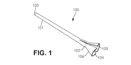
[0009] FIG. 1 shows a front perspective view of an introducer device, in
accordance with an
embodiment of the present invention.
2
CA 03106138 2020-11-06
WO 2019/229703 PCT/I132019/054502
[0010] FIG. 2 shows a side view of an introducer device, in accordance with an
embodiment of
the present invention.
[0011] FIG. 3 shows a cross sectional view of an introducer device, in
accordance with an
embodiment of the present invention.
[0012] FIG. 4A is a diagram demonstrating a use of the introducer device,
wherein a
nasopharyngoscope has been utilized to insert the introducer device into a
patient's body under
visualization, in accordance with an embodiment of the present invention.
[0013] FIG. 4B is a diagram demonstrating a use of the introducer device,
wherein the
introducer device is positioned within a patient's body, in accordance with an
embodiment of the
present invention.
[0014] FIG. 4C is a diagram demonstrating a use of the introducer device,
wherein a second
medical device, namely, a manometry probe, has been inserted into the
introducer device for
entry into a patient's body, in accordance with an embodiment of the present
invention.
[0015] FIG. 4D is a diagram demonstrating a use of the introducer device,
wherein the
introducer device is removed from within a patient's body while a second
medical device,
namely, a manometry probe, maintains its general position within the patient's
body, in
accordance with an embodiment of the present invention.
DETAILED SPECIFICATION
[0016] The present invention generally relates to an introducer device
comprised of an elongated
tubular body, engageable with the outer portion of endoscopes, catheters,
tubes, esophageal
probes and the like medical devices.
[0017] Endoscopes, catheters, and similar elongate tubular devices are
utilized during medical
procedures performed by healthcare professionals, such as doctors and
surgeons, to examine
patients' internal organs and vessels. Endoscopic procedures that utilize
natural body openings,
such as the mouth and nasal cavity, to visualize, examine, and/or operate on
inner organs include
esophagogastroduodenoscopy (EGDs), bronchoscopy, enteroscopy, laryngoscopy,
and
nasopharyngoscopy.
3
CA 03106138 2020-11-06
WO 2019/229703 PCT/I132019/054502
[0018] A variety of medical procedures utilizing natural body openings, such
as esophageal pH
tests, esophageal manometry and the implantation of trans-nasal feeding tubes
require the
insertion of elongate tubular devices into a patient's mouth or nasal cavity
and are performed
without the visualization aide of an endoscope. These and similar procedures
entail the insertion
of a catheter or the like elongate tubular device into a patient's mouth,
nasal cavity, or
esophagus. Once inserted through the mouth or nasal cavity in accordance with
the protocol for
the given procedure, the medical device utilized during such procedures allows
health care
professionals, including doctors, to examine, treat, and/or operate on organs
and lumens such as
the gastrointestinal (GI) tract, including the esophagus and stomach.
[0019] Traditionally, during procedures requiring the insertion of medical
devices into a
patient's oral or nasal cavities or esophagus, a physician will place a
catheter or similar medical
device inside a patient's nasal cavity or mouth and, without the ability to
visualize or know the
device's exact location, the physician will continuously insert the tubular
device until the device
reaches a barrier in the patient's body, for example the post-pharyngeal wall
in procedures where
the insertion is through the patient's nose. Because this process is performed
without the use of
an endoscope providing visualization, the device must be maneuvered around
until the physician
believes the device is located inside the patient's pharynx. Without knowing
the exact location of
the medical device along the patient's pharynx, the physician asks the patient
to initiate multiple
attempts to swallow the device, while the physician pushes the device forward.
Multiple swallow
attempts are initiated until the device is finally swallowed, leading to the
insertion of the device
into the patient's esophagus. Embarking on this procedure without an endoscope
or the ability to
visualize the location of the medical device makes the process of inserting
the catheter,
esophageal probe, tube or similar medical device through the mouth or nasal
cavity and into the
esophagus uncomfortable, and at times intolerable, to patients whose medical
care requires
adherence to examinations and procedures such as these. Moreover, the
attempted placement of
the medical device without functional visualization may be a dangerous
procedure for patients,
as the medical device may be mistakenly inserted into the patient's trachea
instead of the
patient's esophagus. Thus, an introducer device in accordance with embodiments
of the present
invention is needed to couple with medical devices such as endoscopes having
visualization
functionalities and other medical devices, such as catheters, manometry probes
and similar
4
CA 03106138 2020-11-06
WO 2019/229703 PCT/162019/054502
probes or elongate medical devices or portions of these and other medical
devices, to allow for
visualization prior to the insertion of medical devices, and enable the easy
and comfortable
insertion of such medical devices into the esophagus through the mouth or
nose.
[0020] It is an aspect of the present invention to provide an introducer
device, configured to
couple with both endoscope devices having visualization capabilities and other
medical devices,
including medical devices lacking visualization capabilities. For example, the
introducer device
may couple with endoscopes to enable physicians to insert the introducer
device and the
endoscope into the esophagus of a patient, while visualizing the insertion
process. Once the
introducer device and the endoscope are inserted into the esophagus to the
desired extent, the
physician is able to retract the endoscope from within the introducer device,
and insert a second
medical device, for example, a medical device lacking visualization
capabilities, into the
introducer device to examine, treat, or conduct tests on the desired organs
and lumens, such as
the esophagus, located within the anatomy of the patient.
[0021] Embodiments of the present invention are generally directed to an
introducer device
comprising an elongated tubular member, engageable with the outer portion of
both endoscopes
and other medical devices, allowing, in one respect, for visualization during
the insertion of an
endoscope or similar visualization device into the esophagus through the nose
or mouth, and in a
second respect, the retraction of the endoscope while the position of the
introducer device is
maintained, allowing for the introduction of a second medical device into the
desired body lumen
through the introducer device which has been placed in the desired body lumen
or organ with
visualization.
[0022] The various embodiments of the introducer are designed to make the
introduction of both
the introducer and other medical devices into the esophagus more comfortable
and tolerable for
patients. Furthermore, the novel construction of the introducer decreases the
number of
unsuccessful and painful swallow attempts previously required in order for
traditional
intracorporeal medical devices to be swallowed by the patient.
[0023] In accordance with several embodiments of the present invention, a
first body portion of
the introducer may be comprised of a substantially pliable and elastic
material and a second body
portion may be comprised of a substantially rigid material. In some
embodiments, the first and
CA 03106138 2020-11-06
WO 2019/229703 PCT/162019/054502
second body portions are formed of the same material, wherein the first body
portion is formed
from one layer of the material, while the second body portion is formed from
several layers of
the material, rendering the second body portion more rigid than the first body
portion. In several
embodiments of the present invention, the first body portion of the introducer
device and the
second body portion of the introducer device collectively form a single
continuous and
substantially hollow tube, thereby providing a first opening formed at the
first body portion of
the introducer device and a second opening formed at the second body portion
of the introducer
device. In a preferred embodiment, the first body portion of the introducer
device is configured
to be the portion of the introducer device that is first inserted into the
patient and therefore the
portion of the introducer device that will be the most internal within the
patient. Conversely, the
second body portion of the introducer device is generally configured to first
receive the medical
device and to be the portion of the introducer device that remains external to
the patient at all
times.
[0024] In an embodiment of the present invention, both the first and second
body portions of the
introducer device facilitate engagement with endoscopes and other medical
devices for
introduction of the medical devices into organs or body lumens during various
intracorporeal
procedures. In other embodiments, only one of either the first or second body
portions engages
with the endoscopes and other medical device for introduction of the endoscope
and the medical
device into organs or body lumens during various intracorporeal procedures.
[0025] According to some embodiments of the present invention, the first body
portion of the
introducer device is formed out of elastic and pliable material to facilitate
fluid engagement of
the introducer device with organs and lumens such as the esophagus, through
which a medical
device such as an endoscope, catheter, manometry probe, tube or other medical
device or a
portion of a medical device is intended to travel, thereby improving patient
comfort and
facilitating smooth and stable engagement of the devices during the insertion
process.
Furthermore, the elastic and pliable material forming the first body portion
of the introducer
device allows it to take advantage of the articulation functionality of an
endoscope to place the
device. In some embodiments, the articulating force imparted by the endoscope
can be used to
bend the tip of the first body portion of the introducer device in a desired
direction to further ease
the placement of the introducer device.
6
CA 03106138 2020-11-06
WO 2019/229703 PCT/I132019/054502
[0026] According to an embodiment of the present invention, the introducer
device may include
a separation element along opposing side walls of both the first and second
body portions,
making the introducer separable into two substantially semi-circular halves.
In accordance with
embodiments of the present invention, the separation element may be a slit. In
some
embodiments, the separation element may be a groove. In some embodiments, the
separation
element may be a perforated line. In some embodiments, the separation element
may be a
corrugated line. In some embodiments, the separation element may be a
substantially thin layer
of the same or similar material used to create the introducer device. In some
embodiments, only
the first body portion of the introducer device includes a separation element
along its opposing
sidewalls, making the first body portion separable into two substantially semi-
circular halves. In
some embodiments, only the second body portion of the introducer device
includes a separation
element along its opposing sidewalls, making the second body portion separable
into two
substantially semi-circular halves. In any embodiment, the separation element
aids in the
detachment of the introducer device from a medical device such that the
introducer device can be
removed from over the medical device while the medical device is maintained
within the patient,
without necessitating the removal of the medical device or causing its
displacement.
[0027] According to an embodiment of the present invention, the exterior
surface of the
introducer device is composed of a material having a lower coefficient of
friction than an
endoscope or other medical device, to reduce the amount of discomfort
associated with the
traditional procedure of inserting the medical devices into the patient. In
some embodiments, the
opening at the end of the first body portion is rounded or curved to mitigate
patient discomfort
when the introducer device is inserted into a body lumen.
[0028] According to an embodiment of the present invention, the second opening
of the
introducer device includes a flare end, having a conical profile to aide in
the insertion of the
introducer device into a patient's nasal cavity. In some embodiments the
conical profile includes
a notch or indent. In situations where the introducer device is used in
conjunction with medical
devices intended for introduction into the body through a patients' nasal
cavity, the flare or
conical profile prevents the introducer device from being inserted more than
necessary into the
nasal passageway. For example, a nasopharyngoscope, or similar endoscope
intended for
introduction through a patients' nasal cavity, and the introducer device are
advanced as far as
7
CA 03106138 2020-11-06
WO 2019/229703 PCT/I132019/054502
possible until the flare end of the introducer device is placed at the distal
end of the nasal
passageway. Once the introducer device, coupled to the endoscope, is advanced
as far as
permitted by the flare end, the endoscope is retracted from within the
introducer device so that a
second medical device may be inserted into the patient, through the opening in
the introducer
device.
[0029] According to embodiments of the present invention, the flare at the
second opening of the
introducer device may be attached to a holder configured to hold the
introducer device. In
another embodiment, the holder facilitates the insertion of the introducer
device into the desired
body lumen.
[0030] Turning to FIGs. 1-4, a medical device, in accordance with an
embodiment of the present
invention, is shown. As shown in FIG. 1-3, the introducer device 100 comprises
a first body
portion 101 and a second body portion 102. The first body portion 101 may be
greater in length
than the second body portion 102. As shown in FIG.1, the first body portion
101 and the second
body portion 102 collectively form a single continuous and substantially
hollow tube, provided
with a first opening 103 formed at an end of the first body portion 101 and a
second opening 104
formed at an end of the second body portion 102. In some embodiments, the
first body portion
101 is formed of a pliable and elastic material, while the second body portion
102 is formed of a
substantially rigid material. In some embodiments, the first and second body
portions 101 and
102 are formed of the same material, wherein the first body portion 101 is
formed from one layer
of the material, while the second body portion 102 is formed from several
layers of the material,
rendering the second body portion 102 more rigid than the first body portion
101. In some
embodiments, the second opening 104 at the second body portion 102 of the
introducer device
100 may include a flare end. In some embodiments, at least a portion of the
introducer device is
formed from a polyvinyl chloride (PVC) material. In some embodiments, at least
a portion of the
introducer device is formed from a polycarbonate (PC) material.
[0031] In accordance with several embodiments of the present invention, the
first body portion
101 is configured to be the portion of the introducer device 100 that is first
inserted into the
patient and therefore the portion of the introducer device 100 that will be
the most internal Within
the patient. Conversely, the second body portion 102 of the introducer device
100 is generally
8
CA 03106138 2020-11-06
WO 2019/229703 PCT/I132019/054502
configured to first receive a medical device and to be the portion of the
introducer device 100
that remains external to the patient at all times.
[0032] In an embodiment of the present invention, both the first body portion
101 and the second
body portion 102 of the introducer device 101 facilitate engagement with
medical devices such
as endoscopes, tubes, probes, catheters and the like for introduction of
medical devices such as
endoscopes in a first instance and other medical devices such as tubes, probes
and catheters in a
second instance to introduce these medical devices into organs or body lumens
during various
intracorporeal procedures. In other embodiments, only one of either the first
body portion 101 or
the second body portion 102 engages with the medical devices for introduction
of a chosen
medical device and the introducer device 100 into organs or body lumens during
various
intracorporeal procedures. In some embodiments, the introducer device 100 is
greater in length
than the probe or tube of an endoscope or other medical device. In some
embodiments, the
introducer device 100 is shorter in length than the probe or tube of an
endoscope or other
medical device.
[0033] According to an embodiment of the present invention, the introducer
device may include
a separation element 105 along opposing side walls of both the first body
portion 101 and the
second body portion 102, making the introducer device 100 separable into two
substantially
semi-circular halves. In accordance with embodiments of the present invention,
the separation
element 105 may be a slit. In some embodiments, the separation element 105 may
be a groove.
In some embodiments, the separation element 105 may be a perforated line. In
some
embodiments, the separation element 105 may be a corrugated line. In some
embodiments, the
separation element 105 may be a substantially thin layer of the same or
similar material used to
create the introducer device 100. In some embodiments, only the first body
portion 101 of the
introducer device 100 includes a separation element 105 along its opposing
sidewalls, making
the first body portion 101 separable into two substantially semi-circular
halves. In some
embodiments, only the second body portion 102 of the introducer device 100
includes a
separation element 105 along its opposing sidewalls, making the second body
portion 102
separable into two substantially semi-circular halves. In any embodiment, the
separation element
105 aids in the detachment of the introducer device 100 from a medical device
such that the
introducer device 100 can be removed from over the medical device while the
medical device is
9
CA 03106138 2020-11-06
WO 2019/229703 PCT/1B2019/054502
maintained within the patient, without necessitating the removal of the
medical device or causing
its displacement.
[0034] According to an embodiment of the present invention, the exterior
surface of the
introducer device 100 is comprised of a material having a lower coefficient of
friction than a
medical device such as an endoscope, to reduce the amount of discomfort
associated with the
traditional procedure of inserting the medical device into the patient.
[0035] According to an exemplary embodiment of the present invention, the
second opening 104
of the introducer device 100 may have a flared end 106. In some embodiments,
the flared end
106 has a conical profile to aide in the insertion of the introducer into a
patient's nasal cavity. In
some embodiments, the conical profile may have a notch or dent. In situations
where the
introducer device 100 is used in conjunction with medical devices intended for
introduction into
the body through a patients' nasal cavity, the flare or conical profile
prevents the introducer
device from being inserted more than necessary into the nasal passageway. For
example, a
nasopharyngoscope, or similar endoscope intended for introduction through a
patients' nasal
cavity and the introducer device 100 are advanced as far as possible until the
flare end of the
introducer device 100 is placed at the distal end of the nasal passageway.
Once the introducer
device 100, coupled to the endoscope, is advanced as far as permitted by the
flare end, the
endoscope is retracted from within the introducer device 100 so that a second
medical device
such as a tube, catheter or other probe may be inserted into the patient,
through the introducer
device 100.
[0036] In some embodiments, the flare at the second opening 104 of the
introducer device 101 is
attached to a holder, meant for holding the introducer device 101. In some
embodiments, the
holder facilitates the insertion of the introducer device into the desired
body lumen.
[0037] As demonstrated by FIG. 4A, the introducer device 100 may be utilized
by inserting an
endoscope into and through the introducer device 100. The introducer device
100 may be shorter
in length than the probe of the medical device such that the end of the probe
is not covered by the
device 100 when the devices are coupled to each other. The endoscope may be
turned on to
activate the visualization protocols of the device such as video imaging and
light production. The
endoscope coupled to the introducer device 100 may then be inserted into the
body. The
CA 03106138 2020-11-06
WO 2019/229703 PCT/I132019/054502
introducer device 100 may bend as necessary during the insertion process. For
example, if the
introducer device 100 is being inserted through a patient's nose, the
introducer device 100, along
with the endoscope will travel into the patient's nose and bend in order to
continue traveling
through the patient's throat and down the patient's esophagus. The introducer
device 100 may be
inserted into the body to the extent necessary or desired by the healthcare
professional. As
demonstrated by FIG. 4B, the endoscope may be disengaged from the introducer
device 100 and
removed, while the introducer device 100 maintains it general position in the
patient's body.
[0038] As demonstrated by FIG. 4C, after installation of the introducer device
100 with the aide
of an endoscope, a probe of a second medical device may be easily inserted
into the body
through the introducer device 100. As demonstrated by FIG. 4D, once the
medical device is
placed in the appropriate position, as determined by the health professional,
the introducer
device 100 may be removed from the body by sliding the introducer device 100
off the medical
device and out of the body, while the medical device maintains its general
position within the
body. Once the introducer device 100 is external to the body, the introducer
device 100 may be
disengaged and removed from around the medical device by splitting the
introducer device 100
into two substantially semicircular halves by slicing the introducer device
100 separation element
105 provided on the opposing side walls of the introducer device 100.
[0039] According to embodiments of the present invention, an introducer device
100 comprises
an elongated tubular body having a substantially circular wall with an
interior surface that
defines an interior chamber extending from a first opening 103 formed at an
end of a first body
portion 101 of the tubular body to a second opening 104 formed at an end of a
second body
portion 102 of the tubular body, wherein the interior chamber is adapted to
receive a medical
device and the first body portion is more pliable than the second portion. In
some embodiments,
the elongated tubular body may be configured to be at least partially
separable into two
substantially semi-circular halves.
[0040] The terminology used herein is for the purpose of describing particular
embodiments only
and is not intended to be limiting of the invention. As used herein, the term
"and/or" includes
any and all combinations of one or more of the associated listed items. It
will further be
understood that the terms "comprises" and/or "comprising," when used in this
specification,
11
CA 03106138 2020-11-06
WO 2019/229703 PCT/1B2019/054502
specify the presence of stated features, steps, operations, elements, and/or
components, but do
not preclude the presence or addition of one or more other features, steps,
operations, elements,
components, and/or groups thereof.
[0041] While multiple embodiments are disclosed, still other embodiments of
the present
invention will become apparent to those skilled in the art from this detailed
description. The
invention is capable of myriad modifications in various obvious aspects, all
without departing
from the spirit and scope of the present invention. Accordingly, the drawings
and descriptions
are to be regarded as illustrative in nature and not restrictive.
12