Note: Descriptions are shown in the official language in which they were submitted.
A QUADRICISTRONIC SYSTEM COMPRISING A HOMING RECEPTOR
OR A CYTOKINE, AND CHIMERIC ANTIGEN RECEPTOR FOR
STABLE GENETIC MODIFICATION OF CELLULAR
IMMUNOTHERAPIES
[0001] This application claims priority to our co-pending US provisional
applications with the
serial numbers 62/713,264, filed August 1, 2018; 62/713,278, filed August 1,
2018, 62/713,310
filed August 1, 2018; and 62/713,323 filed on August 1, 2018.
[0002] The content of the ASCII text file of the sequence listing named
104077 0007PCT Seq_listing rev004 ST25, which is 97 KB in size was created on
August 1,
2019 and electronically submitted via EFS-Web along with the present
application.
FIELD OF THE INVENTION
[0003] The field of the invention is engineered cells using the cytotoxic
activated Natural
Killer cell line (NK-92) as the basis to improve immunotherapies to cancer and
tumors.
BACKGROUND
[0004] The background description includes information that may be useful in
understanding
the present invention. It is not an admission that any of the information
provided herein is prior
art or relevant to the presently claimed invention, or that any publication
specifically or
implicitly referenced is prior art.
[0005] Where a definition or use of a term in a reference herein is
inconsistent or contrary to
the definition of that term provided herein, the definition
1
Date Recue/Date Received 2022-04-19
CA 03106324 2021-01-12
WO 2020/028656 PCT/US2019/044655
of that term provided herein applies and the definition of that term in the
reference does not
apply.
[0006] Cancer immunotherapies based on adoptively transferred tumor-specific
cytotoxic
lymphocytes hold promise for the treatment of patients with tumor
malignancies. Despite this
early success in certain cancers, the treatment of tumors remains a challenge,
mostly due to the
immunosuppressive nature of the tumor microenvironment. See Swans et al.
"Tumor
Microenvironment Complexity: Emerging Roles in Cancer Therapy," Cancer Res,
vol., 72, pages
2473-2480, 2012. In addition to modified T-cells, immunotherapies based on NK
cells are being
explored. Natural killer (NK) cells are cytotoxic lymphocytes that constitute
a major component
of the innate immune system. Natural killer (NK) cells, generally representing
about 10-15% of
circulating lymphocytes, bind and kill targeted cells, including virus-
infected cells and many
malignant cells, non-specifically with regard to antigen and without prior
immune sensitization.
Herberman et aL, Science 214:24 (1981). NK-928 is a cytolytic cancer cell line
which was
discovered in the blood of a subject suffering from a non-Hodgkin's lymphoma
and then
immortalized ex vivo. NK-92 cells are derived from NK cells, but lack the
major inhibitory
receptors that are displayed by normal NK cells, while retaining the majority
of the activating
receptors. NK-92 cells do not, however, attack normal cells nor do they
elicit an unacceptable
immune rejection response in humans.
[0007] A common driver of lymph node metastasis is the hypoxia-driven
upregulation of CCR7,
a chemokine receptor primarily found in naive T-cells and dendritic cells.
Upregulation of the
CCR7 receptor on blood NK cells has previously been demonstrated to improve
the homing of
NK cells to lymph nodes, allowing them to follow the same path to the lymph
node
compartments that are common pathways of metastatic spread, but has not yet
been
demonstrated in a clinically relevant cell line.
[0008] Therefore, there is still a need to imprive NK cells and NK cell based
therapies,
especially in the context of NK cell homing to and modulation of a tumor
microenvironment.
BRIEF SUMMARY
[0009] Provided herein are modified NK-92i cells comprising a nucleic acid
encoding multiple
function elements. The functional elements are typically proteins or
polypeptides that provide a
2
CA 03106324 2021-01-12
WO 2020/028656 PCT/US2019/044655
specific function that improves the effectiveness of the cells as a cell line
for immunotherapy. In
one aspect, the NK-92 cells comprise a nucleic acid construct encoding four
functional
elements. In some embodiments, the nucleic acid construct comprises sequences
encoding four
functional elements operably linked to a promoter (referred to as a
"Quadricistronic Construct").
[0010] In some embodiments, the first element encoded by the nucleic acid
construct is a
cytokine that provides selection for NK-92 cells that express the cytokine,
such as IL-2 or IL-
15. Thus, in some embodiments, the nucleic acid encodes a cytokine such as IL-
2 or IL-15. In
one embodiment, the IL-2 is expressed with a signal sequence that directs the
IL-2 to the
endoplasmic reticulum IL-2 ("erIL-2"). In another embodiment, the IL-15 is
expressed with a
signal sequence that directs the IL-15 to the endoplasmic reticulum IL-15
("erIL-15").
[0011] In some embodiments, the second element encoded by the nucleic acid
construct is an Fc
receptor. In some embodiments, the Fc receptor is an Fc-gamma receptor (FCyR).
In some
embodiments, the Fc-gamma receptor is FCyRIII-A (also called CD16), which is a
low affinity
Fc receptor that binds to IgG antibodies and activates ADCC. In some
embodiments, the CD16
receptor comprises a phenylalanine (F) to valine (V) substitution at amino
acid position 158
(F158V) of the mature form of the polypeptide (SEQ ID NO: 12) (corresponding
to position 176
of the full length form of the polypeptide comprising the signal sequence). In
one embodiment,
the Fc receptor comprises the nucleic acid sequence of SEQ ID NO:13 or the
amino acid
sequence of SEQ ID NO:12.
[0012] In some embodiments, the first and second elements are present in the
nucleic acid
construct. Thus, in some embodiments, the nucleic acid construct encodes an Fc
receptor (such
as CD16) and erIL-2.
[0013] In some embodiments, the third element encoded by the nucleic acid
construct is a
homing receptor. In some embodiments, the homing receptor is a cytokine
receptor, a G protein-
coupled receptor, a chemokine receptor, a cell adhesion molecule, a selectin,
or an integrin. In
some embodiments, the homing receptor is operably linked to a promoter that
allows
transcription of the nucleic acid. The modified NK-92 '7) cells are capable of
migrating toward a
source of the chemokine that is the ligand for the receptor. Unlike normal
blood-derived NK
cells, the modified NK-92 cells can be developed into cell lines that are
relevant for human
3
CA 03106324 2021-01-12
WO 2020/028656 PCT/US2019/044655
clinical trials, which provides a distinct advantage for immunotherapy.
Examples of homing
receptors include G protein-coupled receptors such as chemokine receptors,
including but not
limited to CCR1, CCR2, CCR3, CCR4, CCR5, CCR6, CCR7, CCR8, CCR9, CCR10, CXCR1,
CXCR2, CXCR3, CXCR4, CXCR5, CXCR6, CXCR7, CX3CR1, XCR1, CCXCKR, D6, DARC,
or the receptor for CXCL14; cytokine receptors; cell adhesion molecules such
as selectins,
including L-selectin (CD62L); integrins such as a4[37 integrin, LPAM-1, and
LFA-1. In some
embodiments, the homing receptor is a cell adhesion molecule such as LFA-1. In
some
embodiments, the homing receptor is a selectin, such as L-selectin (CD62L). In
some
embodiments, the homing receptor is an integrin such as a4137 integrin, I,PAM-
1, or VIA-4. In
some embodiments, the homing receptor is a C-C or C-X-C chemokine receptor.
Thus, in some
embodiments, third element encoded by the nucleic acid is a homing receptor
described herein.
[0014] In some embodiments, the third element encoded by the nucleic acid
construct is a
secreted cytokine, whereby the cytokine increases or improves the function of
the NK-92e4 cells
as immunotherapeutic agents. The cytokine may also modulate the tumor
microenvironment. In
some embodiments, the secreted cytokine that modulates the tumor
microenvironment is IL-12
or IFN-alpha. Thus, in some embodiments, the third element encoded by the
nucleic acid
construct is a cytokine such as IL-12 or IFN-alpha.
100151 Thus, in some embodiments, the third element encoded by the nucleic
acid construct is a
chemokine such as XCL1, CCL5, CCL21 or CCL16. In some embodiments, the third
element
encoded by the nucleic acid construct is a Toll-like Receptor (TLR) agonist.
[0016] In one aspect, the third element encoded by the nucleic acid construct
is IL-12.
[0017] TGF-13 expression within tumors is known to suppress the antitumor
activity of
leukocytes in the tumor microenvironment. Thus, in some embodiments, the third
element
encoded by the nucleic acid construct is a TGF-beta inhibitor, for example a
peptide that inhibits
.. TGF-13. In some embodiments, the third element encoded by the nucleic acid
construct is a TGF-
beta trap. In some embodiments, the TGF-beta trap comprises the extracellular
domain of a
TGFPRII molecule. In some embodiments, the TGF-beta trap comprises a single
chain dimer of
the extracellular domain of a TGFPRII molecule, and most preferably comprises
a single chain
dimer of the TGF-beta Receptor H ectodomain.
4
CA 03106324 2021-01-12
WO 2020/028656 PCT/US2019/044655
[0018] In some embodiments, the NK cells described herein are administered
with a TGF-f3
inhibitor to block TGF-f3 and help remove immunosuppression. In some
embodiments, the NK
cells desribed herein are administered with other immunotherapies to help
decrease or eliminate
a tumor. For example, TGF-f3 can be inhibited by intratumoral injection of
inhibitory peptides in
combination with intratumoral injections of poly(I:C) and an a-CD40 antibody.
In some
embodiments, the TGF-I3 inhibitor is combined with IL-2.
[0019] In some embodiments, the fourth element encoded by the nucleic acid
construct is an
antigen binding protein ("ABP"). In some embodiments, the antigen binding
protein specifically
binds a tumor associated antigen. In some embodiments, the ABP comprises a
fragment of an
antibody, such as an scFv. In some embodiments, the antigen binding protein
comprises or is
part of a chimeric antigen receptor (CAR). In some embodiments, the nucleic
acid encodes an
ABP or CAR that specifically binds CD19, CD20, NKG2D ligands, CS1, GD2, CD138,
EpCAM, HER-2, EBNA3C, GPA7, CD244, CA-125, MUC-1, ETA, MAGE, CEA, CD52,
CD30, MUC5AC, c-Met, EGFR, FAP, WT-1, PSMA, NY-ES01, CSPG-4, IGF1-R, Flt-3,
CD276, CD123, PD-Li, BCMA, CD33, B7-H4, or 41BB.
[0020] In another aspect, the modified NK-92 cells comprise a nucleic acid
encoding IL-12. In
another aspect, the modified NK-92 cells comprise a nucleic acid encoding a
TGF-beta trap. In
some embodiments, the TGF-beta trap comprises a single chain dimer of the
extracellular
domain of a TGFORII molecule.
[0021] In another aspect, the modified NK-92 cells comprise a nucleic acid
encoding a
cytokine that provides selection or allows survival of NK-92 cells that
express the cytokine. In
some embodiments, the nucleic acid encodes a cytokine such as IL-2 or IL-15.
In one
embodiment, the 1L-2 is expressed with a signal sequence that directs the IL-2
to the
endoplasmic reticulum IL-2 ("erIL-2"). In one embodiment, the IL-I5 is
expressed with a signal
sequence that directs the IL-15 to the endoplasmic reticulum ("erIL-15").
[0022] In some embodiments, the modified NK-920 cells comprise a nucleic acid
encoding an
Fc receptor. In some embodiments, the Fc receptor is an Fc-gamma receptor
(FC7R). In some
embodiments, the Fc-gamma receptor is FCyRIII-A (also called CD16), which is a
low affinity
Fc receptor that binds to IgG antibodies and activates ADCC. In some
embodiments, the CD16
5
CA 03106324 2021-01-12
WO 2020/028656 PCT/US2019/044655
receptor comprises a phenylalanine (F) to valine (V) substitution at amino
acid position 158
(F158V) of the mature form of the polypeptide (SEQ ID NO:12) (corresponding to
position 176
of the full length form of the polypeptide comprising the signal sequence). In
one embodiment,
the Fc receptor comprises the nucleic acid sequence of SEQ ID NO:13 or the
amino acid
sequence of SEQ ID NO:12.
100231 In some embodiments, the modified NK-92i cells comprise a nucleic acid
encoding an
antigen binding protein ("ABP"). In some embodiments, the antigen binding
protein specifically
binds a tumor associated antigen. In some embodiments, the ABP comprises a
fragment of an
antibody, such as an scFv. In some embodiments, the antigen binding protein
comprises or is
part of a chimeric antigen receptor (CAR). In some embodiments, the nucleic
acid encodes an
ABP or CAR that specifically binds CD19, CD20, NKG2D ligands, CS1, GD2, CD138,
EpCAM, HER-2, EBNA3C, GPA7, CD244, CA-125, MUC-1, ETA, MAGE, CEA, CD52,
CD30, MUC5AC, c-Met, EGFR, FAP, WT-1, PSMA, NY-ES01, CSPG-4, IGF1-R, Flt-3,
CD276, CD123, PD-L1, BCMA, CD33, B7-H4, or 41BB.
[0024] In another aspect, the modified NK-92 cells comprise a nucleic acid
encoding a
secreted cytokine that modulates the tumor microenvironment. In some
embodiments, the
cytokine that modulates the tumor microenvironment is a chemokine such as
XCL1, CCL5,
CCL21 or CCL16. In some embodiments, the modified NK-92 cells comprise a
nucleic acid
encoding a Toll-like Receptor (TLR) agonist. In some embodiments, the modified
NK-92 cells
comprise a nucleic acid encoding a IL-12 or IFN-alpha. In some embodiments,
the modified
NK-924 cells comprise a nucleic acid encoding a TGF-beta inhibitor, for
example a peptide that
inhibits TGF-I3. In some embodiments, the modified NK-92 cells comprise a
nucleic acid
encoding a TGF-beta trap. In some embodiments, the TGF-beta trap comprises the
extracellular
domain of a TGFI3RII molecule, or a single chan dimer of the extracellular
domain of a TGFI3RII
molecule.
[0025] In one aspect, the modified NK-92 cells comprise one or more, or a
plurality, of nucleic
acid molecules encoding a homing receptor, an ABP or CAR, an Fc receptor,
and/or a cytokine
that provides selection or allows survival of NK-92 cells that express the
cytokine. Thus, in
some embodiments, the modified NK-92 cells comprise nucleic acid molecules
encoding a
6
CA 03106324 2021-01-12
WO 2020/028656 PCT/US2019/044655
chemokine receptor, a CAR, CD16, and erIL-2. In some embodiments, the modified
NK-920
cells comprise nucleic acid molecules encoding CCR7 or CXCR2, a CAR, CD16, and
erIL-2. In
some embodiments, the modified NK-92 cells comprise nucleic acid molecules
encoding 1L-12
or a TGF-beta trap, a CAR, CD16, and erIL-2.
[0026] In some embodiments, the CAR comprises an intracellular signaling
domain from the Fc
epsilon receptor gamma (FceRI7). In one embodiment, the CAR is transiently
expressed by the
NK-92 cell. In one embodiment, the CAR is stably expressed by the NK-928
cell.
[0027] To date, FccRIy-containing CARs have not been utilized in NK-92 cells,
other NK cell
lines, or endogenous NT( cells because other signaling domains (e.g., CD3()
were determined to
be more efficient, especially when combined with additional signaling domains
(second and third
generation CARs). Described herein is the unexpected and surprising finding
that NK-92 cells
expressing a "first-generation" CAR comprising an intracellular domain from
FccRIy have equal
or higher cytotoxic activity against cancer cells expressing the antigen
recognized by the CAR
than NK-92 cells expressing CARs with a CD3C signaling domain alone or in
combination
with other signaling domains (i.e., second or third generation CARs). In one
embodiment, the
CD3C signaling domain contemplated herein may comprise a polypeptide sequence
having at
least 80%, 85%, 90%, 91%, 92%, 93%, 94%, 95%, 96%, 97%, 98% or 99% identity to
SEQ ID
NO: 40.
[0028] In one aspect, an NK-92 cell or cell line expressing a chimeric
antigen receptor (CAR)
on the surface of the NK-92 cell is described, wherein said CAR comprises a
cytoplasmic
domain of Fcc.RIT. In one embodiment, the cytoplasmic domain of FceRIy
comprises an amino
acid sequence having at least 95% sequence identity to SEQ ID NO: 31.
[0029] In some embodiments, the cytoplasmic domain of FceRty is encoded by a
nucleic acid
having at least 95% sequence identity to SEQ ID NO:32.
[0030] In some embodiments, the CAR comprises a hinge region from CD8. In some
embodiments, the CAR comprises a transmembrane domain from CD28,
[0031] In some embodiments, the NK-92 cell or cell line is genetically
modified with a nucleic
acid construct that comprises SEQ ID NO:31 (Featly intracellular cytoplasmic
domain), SEQ ID
7
CA 03106324 2021-01-12
WO 2020/028656 PCT/US2019/044655
NO:32 (FceRIy intracellular signaling domain minus transmembrance domain), SEQ
ID NO: 33
(CD8 hinge region), SEQ ID NO: 34 (CD8 hinge region DNA), SEQ ID NO:35 (CD28
transmembrane domain) and/or SEQ ID NO:36 (CD28 transmembrane domain, minus
ITAM or
intracellular sequence). Jr one embodiment, the CD8 hinge region, CD28
transmembrane, and
FceRIgamma signaling domain amino acid sequence comprises a polypeptide or a
polynucletodie sequence having at least 80%, 85%, 90%, 91%, 92%, 93%, 94%,
95%, 96%,
97%, 98% or 99% identity to SEQ ID NO: 37 or SEQ ID NO: 38. In some
embodiments, the
nucleic acid construct further comprises a promoter that promotes
transcription of the nucleic
acid sequences. In some emodiments, the promoter is an inducible promoter. In
some
embodiments, the nucleic acid construct is a multi-cistronic vector comprising
one or more
Internal Ribosome Entry Site (IRES) to allow for initiation of translation
from an internal region
of an mRNA transcribed from the nucleic acid sequences. In some embodiments,
the nucleic
acid construct comprises a sequence that encodes a 2A peptide, such as a T2A,
P2A, E2A, or
F2A peptide, in order to produce equimolar levels of polypeptides encoded by
the same mRNA.
In some embodiments, the nucleic acid construct further comprises a nucleic
acid sequence that
encodes an antigen binding protein (ABP). In some embodiments, the ABP is an
scFy or a
codon optimized scFv. In some embodiments, the ABP specifically binds an
antigen expressed
by a tumor cell. In some embodiments, the ABP is part of a chimeric antigen
receptor (CAR).
In some embodiments, the construct comprises a nuclei acid that encodes a
cytokine, that
provides selection or allows survival of NK-92 cells that express the
cytokine, such a IL-2. In
one embodiment, the cytokine is targeted to the endoplasmic reticulum. In one
embodiment, the
CAR scFy may comprise a polypeptide sequence having at least 80%, 85%, 90%,
91%, 92%,
93%, 94%, 95%, 96%, 97%, 98% or 99% identity to SEQ ID NO: 39.
[0032] In some embodiments, the construct comprises the vector shown in Fig.
10. In some
embodiments, the NK-92 cell or cell line is genetically modified to express
CD16 on the cell
surface. In one embodiment, the NK-92 cell or cell line is genetically
modified to express a
high affinity CD16 (F158V) on the cell surface.
[0033] In one embodiment, the ABP or CAR targets or specifically binds a tumor-
associated
antigen. In one embodiment, the tumor-associated antigen is selected from the
group consisting
of CD19, CD20, NKG2D ligands, CS I, GD2, CD138, EpCAM, HER-2, EBNA3C, GPA7,
8
CA 03106324 2021-01-12
WO 2020/028656 PCT/US2019/044655
CD244, CA-125, MUC-1, ETA, MAGE, CEA, CD52, CD30, MUC5AC, c-Met, EGFR, FAP,
WT-1, PSMA, NY-ES01, CSPG-4, IGF1-R, Flt-3, CD276, CD123, PD-L1, BCMA, CD33 B7-
H4, and 41BB. In one embodiment, the tumor-associated antigen is CD19. In
another
embodiment, the tumor-associated antigen is CD33.
[0034] In one aspect, the present disclosure relates to a NK-92 cell line
that is transformed by a
nucleic acid encoding a chimeric antigen receptor (CAR) with a cytoplasmic
domain of FccRIT,
wherein the CAR is expressed on the surface of the NK-92 cell. In one
embodiment, the
nucleic acid is RNA. In one embodiment, the nucleic acid is DNA.
100351 In some embodiments, the NK-920 cell is further modified to express at
least one
cytokine or variant thereof that provides selection or allows survival of NK-
920 cells that
express the cytokine. In one embodiment, the at least one cytokine is
transiently expressed by the
NK-92 cell. In one embodiment, the at least one cytokine is stably expressed
by the NK-92
[0036] In some embodiments, the modified NK-920 cells comprise an expression
vector
comprising one or more, or a plurality, of the nucleic acid molecules
described herein. In some
embodiments, the nucleic acid molecule is operably linked to a promoter that
is capable of
initiating transcription of the nucleic acid molecule. In some embodiments,
each nucleic acid
molecule of the plurality of nucleic acid molecules is operably linked to a
separate, distinct
and/or different promoter. In some embodiments, one or more of the nucleic
acid molecules are
operably linked to the same promoter. In one embodiment, the nucleic acid
molecules encoding
the homing receptor, the CAR, the Fc receptor and the cytokine are operably
linked to the same
promoter or a single promoter. In some embodiments the promoter is an
inducible promoter. In
one embodiment, the nucleic acid molecule encoding the cytokine is located
downstream or 3' of
the nucleic acid molecules encoding the homing receptor, the CAR, and the Fc
receptor (e.g.,
CD16 or high affinity CD16).
100371 In some embodiments, the NK-92 cells express the proteins encoded by
the nucleic acid
molecules described herein on the cell surface. For example, in some
embodiments, the
modified NK-92 cells express the homing receptor, the ABP or CAR, and the Fc
receptor (e.g.,
CD16 or high affinity CD16) on the cell surface.
9
[0037a] In one aspect, the invention provides a modified NK-92 cell comprising
a nucleic acid
encoding a homing receptor operably linked to a promoter and further
comprising a nucleic acid
encoding an antigen binding protein operably linked to a promoter, wherein the
wherein the
antigen binding protein comprises a chimeric antigen receptor (CAR), and
wherein the CAR
specifically binds CD19, or has an amino acid sequence with at least 90%, 91%,
92%, 93%,
94%, 95%, 96%, 97%, 98% or 99% identity to SEQ ID NO:25, and wherein the
homing receptor
is CCR7.
[0038] Also provided are compositions and kits comprising the modified NK-92
cells.
Provided are methods of making the modified cells and methods of treating
cancer using the
cells.
[0039] In another aspect, methods for treating cancer or reducing the size of
a tumor are
described. In some embodiments, the methods of treating cancer or reducing the
size of a tumor
comprise administering to a subject in need thereof a therapeutically
effective amount of the
modified NK-92 cells described herein, wherein administration treats the
cancer or reduces the
size of a tumor in the subject. In some embodiments, the methods comprise
administering to the
subject a therapeutically effective amount of modified NK-92 cells that
comprise a nucleic acid
encoding a homing receptor, an ABP or CAR that specifically binds to a target
antigen, an Fc
Receptor such as CD16 or CD16-158V, and/or a cytokine such as erIL-2 or erIL-
15. In some
embodiments, the methods comprise administering to the subject a
therapeutically effective
amount of modified NK-92 cells that comprise a nucleic acid encoding a
secreted cytokine, an
ABP or CAR that specifically binds to a target antigen, an Fc Receptor such as
CD16 or CD16-
158V, and/or a cytokine such as erIL-2 or erIL-15.
[0040] In some embodiments, the NK cells described herein are administered
with a TGF-13
inhibitor to block TGF-P and help remove immunosuppression. In some
embodiments, the NK
cells desribed herein are administered with other immunotherapies to help
decrease or eliminate
a tumor. For example, TGF-f3 can be inhibited by intratumoral injection of
inhibitory peptides in
combination with intratumoral injections of poly(I:C) and an a-CD40 antibody.
In some
embodiments, the TGF-13 inhibitor is combined with IL-2.
[0041] In another aspect, use of a composition described herein for treating a
disease is
provided. In some embodiments, a modified NK-92 cell described herein is
provided for use
Date recue/Date received 2023-03-10
as a medicament for treating a disease. In some embodiments, a modified NK-92
cell
described herein is provided for use in the treatment of a disease. In some
embodiments, the
modified NK-92 cells comprise a nucleic acid encoding a homing receptor, an
ABP or CAR
that specifically binds to a target antigen, an Fc Receptor such as CD16 or
CD16-158V, and/or a
cytokine such as erIL-2 or er1L-15. In some embodiments, the modified NK-9201
cells comprise
a nucleic acid encoding a secreted cytokine, an ABP or CAR that specifically
binds to a target
antigen, an Fc
10a
Date Recue/Date Received 2022-04-19
CA 03106324 2021-01-12
WO 2020/028656 PCT/US2019/044655
Receptor such as CD16 or CD16-158V, and/or a cytokine such as erIL-2 or erIL-
15. In some
embodiments, the disease is cancer.
[0042] The details of one or more embodiments are set forth in the
accompanying drawings and
the description below. Other features, objects, and advantages will be
apparent from the
description and drawings, and from the claims.
BRIEF DESCRIPTION OF THE DRAWINGS
[0043] Figure 1 is a schematic showing plasmid pNKAT-CCR7-LP3 containing the
CCR7
receptor for insertion at the AAVS1 locus in NK-92 cells.
[0044] Figure 2 is a schematic showing plasmid pCRENFAT-CCL21 containing a
NFAT-
responsive CCL21 gene.
[0045] Figure 3 are graphs showing expression of phenotypic markers associated
with NK-92
cells in wild type NK-92 cells and modified NK-92 cells expressing CCR7.
(Lane 1: aNK
(Wild Type); Lane 2: Modified NK-92 cells (MA3); Lane 3: Modified NK-92
cells (MB4);
Lane 4: Modified NK-92 cells (MB6); Lane 5: Modified NK-92 cells (ME6); Lane
6:
Modified NK-920' cells (MH3); A: Isotype (APC); B: CD54(ICAM-1); C: NKp30; D:
NKG2D
[0046] Figure 4 is a graph showing cytoxic activity of modified NK-92 cells
expressing CCR7
against K562 cells.
[0047] Figure 5 is a graph showing cytoxic activity of modified NK-920 cells
expressing CCR7
against HL-60 cells.
[0048] Figures 6A and 6B are graphs showing activation of an NFAT-Luciferase
reporter gene
in NK-92 cells demonstrated in the context of binding to K562 and SUP-B15
cells (when NK-92
cells were electroporated with mRNA for a CD19-CAR).
[0049] Figure 7 is a graph showing modified NK-92 cells expressing CCR7 (Mi-
aNK)
migrated towards the chemokines CCL19 and CCL21.
11
CA 03106324 2021-01-12
WO 2020/028656 PCT/US2019/044655
[0050] Fig. 8 shows a diagram representing an exemplary method for in vitro
testing of the
modified NIC-92014 cells described herein. Activated NK-92m cells (aNK) were
modified to
express a chemokine receptor (e.g., CCR7), and the target cells were modified
to express a
chemokine that binds to the receptor (e.g., CCL19 or CCL21). The modified NK-
92 cells were
tested in a Modified Boyden Chamber Transwell Assay as shown.
[0051] Fig. 9 shows a representative cytotoxicity assay using the modified NK-
92 ".:4. cells
described herein. The modified NK-92 cells from Fig. 8 were tested for
cytotoxicity against
K562 target cells that express and secrete one or both chemokine ligands. The
ML4 clone
showed the highest percentage of lysis of target cells, and the percentage was
increased when the
K562 target cells expressed both CCL19 and CCL21.
[0052] Fig. 10 is a schematic showing plasmid pNKAT-CCR7-CD19CAR-CD16-ERIL2,
referred to as a "Quadricistronic Vector," which can be used to stably
transfect a cell at a single
insertion position.
[0053] Fig. 11 is a schematic showing the linearized plasmid from Fig. 10.
[0054] Fig. 12 shows cell surface expression of CCR7, CD16, and CD19 CAR by NK-
92 cells.
"aNK" is the wild-type NK-92 cell line. "ML4" is the aNK cell line
transfected with a nucleic
acid construct encoding CCR7 operably linked to a promoter (i.e. Mi-aNK). "P2"
is the aNK
cell line transfected with a nucleic acid construct that encodes CCR7, CD16,
ER-1L2 and CD19
CAR (i.e. Mi- T-haNK).
[0055] Fig. 13. Homing of non-CR versus Mi-T-haNK cells to parental or CCL19-
expressing
tumors at indicated hours post NK cell administration. Data are Mean SEM.
The ¨ and + signs
indicate the expression status of CCR7 receptor (first sign) and CCL19 ligand
(second sign). *, P
<0.05 by one-way ANOVA followed by multiple comparison by Tukey's test. The
last panel
presents the time course curve.
[0056] Figure 14. A head-to-head comparison of non-CR and Mi-T-haNK cells
infiltration to
parental or CCL19-expressing tumors in single animals at 24 hours post dosing.
3 out of 4
animals receiving the Mi-T-haNK cells showed higher infiltration to the CCL
_19+ tumors, while
3 out of 4 animals receiving the non-CR CD19 t-haNK cells showed similar
levels of infiltration
12
CA 03106324 2021-01-12
WO 2020/028656 PCT/US2019/044655
to both K562 and K-19 tumors. The one "outlier" animal in each group is
indicated by a dashed
line,
[0057] Fig.15 illustrates the survival curve of IV Raji-19.5 tumor-bearing
animals. Survival
curves for Raji-19.5 IV tumor-bearing NSG mice treated with vehicle, CD19 t-
haNK cells, or
R7-19.1 cells. Statistical analysis was done by Log-rank (Mantel-Cox) test.
***, P = 0.0002;
****, P < 0.0001.
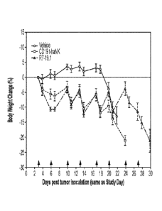
[0058] Fig. 16 illustrates the Body weight change in the IV Raji-19.5 tumor
model. Body weight
change curves (% change over Day 0 body weight) for IV Raji-19.5 tumor bearing
animals
treated with vehicle, CD19 t-haNK cells, or R7-19.1 cells. Data are Mean I
SEM. Red arrows
indicate dosing days. The weight measurements taken on dosing days were
performed prior to
dose administration. For all time points prior to Day 20 the curves for the NK
cell treated groups
reached statistically significant difference (P < 0.05) compared to the
vehicle control group by 2-
way ANOVA followed by multiple comparison by Tukey test.
[0059] Fig. 17 illustrates the sizes of SC Raji-19.5 tumors upon
randomization. Shown are
individual tumor sizes upon randomization. The black box encircled large
tumors that were >
200 mm3 in size, while the blue box encircled small tumors that were <200 mm3.
Group mean
SEM is also indicated.
[0060] Fig. 18 illustrates tumor growth for the large-tumor sub-population of
SC Raj i-19.5
tumor-bearing mice. (A) Group analysis. Data are Mean SEM. Statistical
analyses were done
using 2-way mixed-effects analysis followed by multiple comparison by Tukey
test. No
statistical significance was achieved. (B) Individual curves. Red arrows
indicate dosing days. Tx:
treatment.
[0061] Fig. 19 illustrates tumor growth for the small-tumor sub-population of
SC Raji-19.5
tumor-bearing mice. (A) Group analysis. Data are Mean SEM. Statistical
analyses were done
using 2-way mixed-effects analysis followed by multiple comparison by Tukey
test. No
statistical significance was detected between any 2 groups at any time point.
(B) Individual
curves. Red arrows indicate dosing days. Tx: treatment.
13
CA 03106324 2021-01-12
WO 2020/028656 PCT/US2019/044655
[0062] Fig. 20 illustrates body weight change in the SC Raji-19.5 tumor model.
Body weight
change curves (% change over Day 1 body weight) for SC Raji-19.5 tumor bearing
animals
treated with vehicle, CD19 t-haNK cells, or R7-19.1 cells. Data are Mean
SEM. Red arrows
indicate dosing days. The body weight measurements taken on dosing days were
performed prior
to dose administration. For all time points prior to Day 16, the curves for
the NK cell treated
groups reached statistically significant difference compared to the vehicle
control group by 2-
way mixed-effects analysis followed by multiple comparison by Tukey test.
[0063] Fig. 21 illustrates one embodiment of a quadri-cistronic TGFP-trap
armored PD-Li CAR
construct.
[0064] Fig. 22 illustrates expression analysis of PD-Li CAR and CD16 in PD-
L1(TG93-trap) t-
haNK Clones.
100651 Fig. 23 illustrates TGFb trap is secreted into the culture supernatant
of TGFPtrap/PD-L1
t-haNK clones.
[0066] Fig. 24 illustrates cytotoxicity of the quadri-cistronic TGFP-trap
contruct against K562
target cells.
[0067] Fig. 25 illustrates CAR killing of the quadri-cistronic TGFP-trap
contruct against SUP-
B15 target cells expressing PD-Ll.
[0068] Fig. 26 illustrates CAR killing of the quadri-cistronic TGFP-trap
contruct against MDA-
MB 231 target cells.
[0069] Fig. 27 illustrates ADCC of the quadri-cistronic TGFP-trap contruct
against SUP-B15
CD19-CD20+.
[0070] Fig. 28 illustrates TGFf3/SMAD Luciferase reporter HEK293 cells induced
by TGFP.
[0071] Fig. 29 illustrates secreted TGFP-trap sequestered TGFP and inhibited
luciferase
expression in HEK293T reporter assay.
100721 Fig. 30 illustrates 11-12 secretion from IL-12 virally transduced NK-92
cell lines.
14
CA 03106324 2021-01-12
WO 2020/028656 PCT/US2019/044655
[0073] Fig. 31 illustrates one embodiment of a quadri-cistronic IL-12/PD-L1 t-
haNK construct.
[0074] Fig. 32 illustrates cytotoxicity data for CCR7 CD19 t-haNK cells.
[0075] Fig. 33 illustrates IL-12 secretion from IL-12/PD-L1 thaNKTM cell line.
DEFINITIONS
[0076] Unless defined otherwise, all technical and scientific terms used
herein have the same
meaning as commonly understood by one of ordinary skill in the art in the
field of immunology
and immunotherapy.
[0077] In this specification and in the claims that follow, reference will be
made to a number of
terms that shall be defined to have the following meanings.
[0078] The terminology used herein is for the purpose of describing particular
embodiments only
and is not intended to be limiting of the invention. As used herein, the
singular fauns "a", "an"
and "the" are intended to include the plural forms as well, unless the context
clearly indicates
otherwise.
[0079] All numerical designations, e.g., pH, temperature, time, concentration,
amounts, and
molecular weight, including ranges, include variations normally encountered by
one of ordinary
skill in the art. Therefore, numerical values can include variations of (+) or
(-) increments of 0.1
or 1.0, where appropriate, depending on the relevant significant digit. It is
to be understood,
although not always explicitly stated, that all numerical designations may be
preceded by the
term "about." The term "about" as used herein may also mean that the value can
vary by +1%,
2%, 3%, 4%, 5%, 6%, 7%, 8%, 9%, or 10%.
[0080] It is also to be understood, although not always explicitly stated,
that the reagents
described herein are merely exemplary and that equivalents of such are known
in the art.
[0081] "Optional" or "optionally" means that the subsequently described event
or circumstance
can or cannot occur, and that the description includes instances where the
event or circumstance
occurs and instances where it does not.
CA 03106324 2021-01-12
WO 2020/028656 PCT/US2019/044655
[0082] The term "comprising" is intended to mean that the compositions and
methods include
the recited elements, but not excluding others. "Consisting essentially of,"
when used to define
compositions and methods, shall mean excluding other elements of any essential
significance to
the combination. For example, a composition consisting essentially of the
elements as defined
herein would not exclude other elements that do not materially affect the
basic and novel
characteristic(s) of the claimed invention. "Consisting of' shall mean
excluding more than trace
amount of other ingredients and substantial method steps recited. Embodiments
defined by each
of these transition terms are within the scope of this disclosure.
[0083] The term "homing receptor" refers to a receptor that activates a
cellular pathway that
results directly or indirectly in the cell migrating toward a target cell or
tissue. For example,
homing receptors expressed by leukocytes are used by leukocytes and
lymphocytes to enter
secondary lymphoid tissues via high endothelial venules. Homing receptors can
also be used by
cells to migrate toward the source of a chemical gradient, such as a chemokine
gradient.
Examples of homing receptors include G-protein coupled receptors such as
chemokine receptors,
including but not limited to CCR1, CCR2, CCR3, CCR4, CCR5, CCR6, CCR7, CCR8,
CCR9,
CCR10, CXCR1, CXCR2, CXCR3, CXCR4, CXCR5, CXCR6, CXCR7, CX3CR1, XCR1,
CCXCKR, D6, and DARC; cytokine receptors; cell adhesion molecules such as
selectins,
including L-selectin (CD62L), integrins such as DAV integrin, LPAM-1, and LFA-
1. Homing
receptors generally bind to cognate ligands on the target tisues or cell. In
some embodiments,
.. homing receptors bind to Addressins on the endothelium of venules, such as
mucosal vascular
addressin cell adhesion molecule 1 (MAdCAM-1).
[0084] As used herein, "immunotherapy" refers to the use of NK-92 cells,
modified or
unmodified, naturally occurring or modified NK cell or T-cell, whether alone
or in combination,
and which are capable of inducing cytotoxicity when contacting a target cell.
[0085] As used herein, "natural killer (NK) cells" are cells of the immune
system that kill target
cells in the absence of a specific antigenic stimulus, and without restriction
according to major
histocompatibility complex (MHC) class. NK cells are characterized by the
presence of CD56
and the absence of CD3 surface markers.
16
[0086] The term "endogenous NK cells" is used to refer to NK cells derived
from a donor (or
the patient), as distinguished from the NK-920 cell line. Endogenous NK cells
are generally
heterogeneous populations of cells within which NK cells have been enriched.
Endogenous NK
cells may be intended for autologous or allogeneic treatment of a patient.
[0087] The term "NK-92" refers to natural killer cells derived from the highly
potent unique
cell line described in Gong et al. (1994), rights to which are owned by
NantKwest (hereafter,
"NK-92 cells"). The immortal NK cell line was originally obtained from a
patient having non-
Hodgkin's lymphoma. Unless indicated otherwise, the term "NK-920" is intended
to refer to the
original NK-92 cell lines as well as NK-92 cell lines that have been
modified (e.g., by
introduction of exogenous genes). NK-92 cells and exemplary and non-limiting
modifications
thereof are described in U.S. Patent Nos. 7,618,817; 8,034,332; 8,313,943;
9,181,322; 9,150,636;
and published U.S. Application No. 10/008,955, and include wild type NK-92S,
NK-92S-
CD16, NK-920-CD16-y, NK-92 -CD16-C, NK-928-CD16(F176V), NK-920MI, and NK-
928CI. NK-92 cells are known to persons of ordinary skill in the art, to whom
such cells are
readily available from NantKwest, Inc.
[0088] The term "aNK" refers to an unmodified natural killer cells derived
from the highly
potent unique cell line described in Gong et al. (1994), rights to which are
owned by NantKwest
(hereafter, "aNK cells"). The term "haNK" refers to natural killer cells
derived from the highly
potent unique cell line described in Gong et al. (1994), rights to which are
owned by NantKwest,
modified to express CD16 on the cell surface (hereafter, "CD16+ NK-92 cells"
or "haNKL
cells"). In some embodiments, the CD16+ NK-92 cells comprise a high affinity
CD16 receptor
on the cell surface. The term "taNK" refers to natural killer cells derived
from the highly potent
unique cell line described in Gong et al. (1994), rights to which are owned by
NantKwest,
modified to express a chimeric antigen receptor (hereafter, "CAR-modified NK-
92 cells" or
"taNKS cells"). The term "t-haNK" refers to natural killer cells derived from
the highly potent
unique cell line described in Gong et al. (1994), rights to which are owned by
NantkWest,
modified to express CD 16 on the cell surface and to express a chimeric
antigen receptor
(hereafter, "CAR-modified CD16+ NK-92 cells" or "t-haNK cells"). In some
embodiments,
the t-haNK cells express a high affinity CD16 receptor on the cell surface.
17
Date Recue/Date Received 2022-04-19
CA 03106324 2021-01-12
WO 2020/028656 PCT/US2019/044655
[0089] The term "chemokine targeted t-haNK" and "Mi-T-haNK" refer to a t-haNK
cell that is
modified to express a chemokine receptor on the cell surface.
[0090] As used herein, the terms "cytotoxic" and "cytolytic," when used to
describe the activity
of effector cells such as NK-92 cells, are intended to be synonymous. In
general, cytotoxic
activity relates to killing of target cells by any of a variety of biological,
biochemical, or
biophysical mechanisms. Cytolysis refers more specifically to activity in
which the effector
lyses the plasma membrane of the target cell, thereby destroying its physical
integrity. This
results in the killing of the target cell. Without wishing to be bound by
theory, it is believed that
the cytotoxic effect of NK-920 cells is due to cytolysis.
.. [0091] The term "kill" with respect to a cell/cell population is directed
to include any type of
manipulation that will lead to the death of that cell/cell population.
[0092] The term "Fc receptor" refers to a protein found on the surface of
certain cells (e.g.,
natural killer cells) that contribute to the protective functions of the
immune cells by binding to
part of an antibody known as the Fc region. Binding of the Fc region of an
antibody to the Fc
receptor (FcR) of a cell stimulates phagocytic or cytotoxic activity of a cell
via antibody-
mediated phagocytosis or antibody-dependent cell-mediated cytotoxicity (ADCC).
FcRs are
classified based on the type of antibody they recognize. For example, Fc-gamma
receptors
(FCyR) bind to the IgG class of antibodies. FCyRIII-A (also called CD16) is a
low affinity Fc
receptor that binds to IgG antibodies and activates ADCC. FCyRIII-A are
typically found on NK
cells. NK-92 cells do not express FCyRIII-A. Fc-epsilon receptors (FceR) bind
to the Fc
region of IgE antibodies.
[0093] The term "chimeric antigen receptor" (CAR), as used herein, refers to
an extracellular
antigen-binding domain that is fused to an intracellular signaling domain.
CARs can be
expressed in T cells or NI( cells to increase cytotoxicity. In general, the
extracellular antigen-
binding domain is a scFy that is specific for an antigen found on a cell of
interest. A CAR-
expressing NK-92 cell is targeted to cells expressing certain antigens on the
cell surface, based
on the specificity of the scFy domain. The scFy domain can be engineered to
recognize any
antigen, including tumor-specific antigens. For example, CD19CAR recognizes
CD19, a cell
surface marker expressed by some cancers.
18
CA 03106324 2021-01-12
WO 2020/028656 PCT/US2019/044655
[0094] The term "tumor-specific antigen" as used herein refers to antigens
that are present on a
cancer or neoplastic cell but not detectable on a normal cell derived from the
same tissue or
lineage as the cancer cell. Tumor-specific antigens, as used herein, also
refers to tumor-
associated antigens, that is, antigens that are expressed at a higher level on
a cancer cell as
compared to a normal cell derived from the same tissue or lineage as the
cancer cell.
[0095] The terms "polynucleotide", "nucleic acid" and "oligonucleotide" are
used
interchangeably and refer to a polymeric form of nucleotides of any length,
either
deoxyribonucleotides or ribonucleotides or analogs thereof. Polynucleotides
can have any
three-dimensional structure and may perform any function, known or unknown.
The following
are non-limiting examples of polynucleotides: a gene or gene fragment (for
example, a probe,
primer, EST or SAGE tag), exons, introns, messenger RNA (mRNA), transfer RNA,
ribosomal
RNA, ribozymes, cDNA, recombinant polynucleotides, branched polynucleotides,
plasmids,
vectors, isolated DNA of any sequence, isolated RNA of any sequence, nucleic
acid probes and
primers. A polynucleotide can comprise modified nucleotides, such as
methylated nucleotides
and nucleotide analogs. If present, modifications to the nucleotide structure
can be imparted
before or after assembly of the polynucleotide. The sequence of nucleotides
can be interrupted
by non-nucleotide components. A polynucleotide can be further modified after
polymerization,
such as by conjugation with a labeling component. The term also refers to both
double- and
single-stranded molecules. Unless otherwise specified or required, any
embodiment of this
invention that is a polynucleotide encompasses both the double-stranded form
and each of two
complementary single-stranded forms known or predicted to make up the double-
stranded form.
[0096] A polynucleotide is composed of a specific sequence of four nucleotide
bases: adenine
(A); cytosine (C); guanine (G); thymine (T); and uracil (U) for thymine when
the polynucleotide
is RNA. Thus, the term "polynucleotide sequence" is the alphabetical
representation of a
polynucleotide molecule.
[0097] "Homology" or "identity" or "similarity" refers to sequence similarity
between two
peptides or between two nucleic acid molecules. Sequence similarity can be
determined by
comparing a position in each sequence which may be aligned for purposes of
comparison. When
a position in the compared sequence is occupied by the same base or amino
acid, then the
19
CA 03106324 2021-01-12
WO 2020/028656 PCT/US2019/044655
molecules are homologous at that position. The percent of sequence similarity
between
sequences is a function of the number of matching or homologous positions
shared by the
sequences over a given comparison window. A sequence can be at least 60%, 70%,
80%, 85%,
90%, 91%, 92%, 93%, 94%, 95%, 96%, 97%, 98% or 99% identical to a sequence
described
herein.
[0098] The terms identical or percent identity, in the context of two or more
nucleic acids or
polypeptide sequences, refer to two or more sequences or subsequences that are
the same or have
a specified percentage of amino acid residues or nucleotides that are the same
(i.e., at least about
60%, 65%, 70%, 75%, 80%, 85%, 90%, 91%, 92%, 93%, 94%, 95%, 96%, 97%, 98%,
99%, or
higher identity over a specified region, when compared and aligned for maximum
correspondence over a comparison window or designated region) as measured
using a BLAST or
BLAST 2.0 sequence comparison algorithms with default parameters described
below, or by
manual alignment and visual inspection (see, e.g., NCBI web site or the like).
Such sequences
are then said to be substantially identical. This definition also refers to,
or may be applied to, the
compliment of a test sequence. The definition also includes sequences that
have deletions and/or
additions, as well as those that have substitutions. As described below, the
preferred algorithms
can account for gaps and the like. In some embodiments, identity exists over a
region that is at
least about 25 amino acids or nucleotides in length, or over a region that is
50-100 amino acids
or nucleotides in length.
[0099] For sequence comparison, typically one sequence acts as a reference
sequence, to which
test sequences are compared. When using a sequence comparison algorithm, test
and reference
sequences are entered into a computer; subsequence coordinates are designated,
if necessary; and
sequence algorithm program parameters are designated. Preferably, default
program parameters
can be used, or alternative parameters can be designated. The sequence
comparison algorithm
then calculates the percent sequence identities for the test sequences
relative to the reference
sequence, based on the program parameters.
[00100] A comparison window, as used herein, includes reference to a segment
of any one of
the number of contiguous positions selected from the group consisting of from
20 to 600, usually
about 50 to about 200, more usually about 100 to about 150, in which a
sequence may be
CA 03106324 2021-01-12
WO 2020/028656 PCT/US2019/044655
compared to a reference sequence of the same number of contiguous positions
after the two
sequences are optimally aligned. Methods of alignment of sequences for
comparison are well-
known in the art. Optimal alignment of sequences for comparison can be
conducted, e.g., by the
local homology algorithm of Smith & Waterman, Adv. Appl. Math. 2:482 (1981);
by the
homology alignment algorithm of Needleman & Wunsch, J. Mol. Biol. 48:443
(1970); by the
search for similarity method of Pearson & Lipman, Proc. Nat'l. Acad. Sci. USA
85:2444 (1988);
by computerized implementations of these algorithms (GAP, BESTFIT, FASTA, and
TFASTA
in the Wisconsin Genetics Software Package, Genetics Computer Group, 575
Science Dr.,
Madison, WI); or by manual alignment and visual inspection (see, e.g., Current
Protocols in
Molecular Biology (Ausubel et al., eds. 1995 supplement)).
[00101] A preferred example of an algorithm that is suitable for determining
percent sequence
identity and sequence similarity are the BLAST and BLAST 2.0 algorithms, which
are described
in Altschul et al., Nuc. Acids Res. 25:3389-3402 (1977), and Altschul et al.,
J. Mol. Biol.
215.403-410 (1990), respectively. BLAST and BLAST 2.0 are used, with the
parameters
described herein, to determine percent sequence identity for nucleic acids or
proteins. Software
for performing BLAST analyses is publicly available through the National
Center for
Biotechnology Information, as known in the art. This algorithm involves first
identifying high
scoring sequence pairs (HSPs) by identifying short words of a selected length
(W) in the query
sequence, which either match or satisfy some positive-valued threshold score T
when aligned
with a word of the same length in a database sequence. T is referred to as the
neighborhood
word score threshold (Altschul et al., supra). These initial neighborhood word
hits act as seeds
for initiating searches to find longer HSPs containing them. The word hits are
extended in both
directions along each sequence for as far as the cumulative alignment score
can be increased.
Cumulative scores are calculated for nucleotide sequences using the parameters
M (reward score
for a pair of matching residues; always > 0) and N (penalty score for
mismatching residues;
always < 0). For amino acid sequences, a scoring matrix is used to calculate
the cumulative
score. Extension of the word hits in each direction are halted when: the
cumulative alignment
score falls off by the quantity X from its maximum achieved value; the
cumulative score goes to
zero or below, due to the accumulation of one or more negative-scoring residue
alignments; or
the end of either sequence is reached. The BLAST algorithm parameters W, T,
and X determine
the sensitivity and speed of the alignment. The Expectation value (E)
represents the number of
21
CA 03106324 2021-01-12
WO 2020/028656 PCT/US2019/044655
different alignments with scores equivalent to or better than what is expected
to occur in a
database search by chance. The BLASTN program (for nucleotide sequences) uses
as defaults a
wordlength (W) of 11, an expectation (E) of 10, M=5, N=-4 and a comparison of
both strands.
For amino acid sequences, the BLASTP program uses as defaults a wordlength of
3, expectation
(E) of 10, and the BLOSUM62 scoring matrix (see Henikoff & Henikoff, Proc.
Natl. Acad. Sci.
USA 89:10915 (1989)), alignments (B) of 50, expectation (E) of 10, M=5, N=-4.
[00102] The term transformation as used herein refers to a process by which an
exogenous or
heterologous nucleic acid molecule (e.g., a vector or recombinant nucleic acid
molecule) is
introduced into a recipient cell. The exogenous or heterologous nucleic acid
molecule may or
may not be integrated into (i.e., covalently linked to) chromosomal DNA making
up the genome
of the host cell. For example, the exogenous or heterologous polynucleotide
may be maintained
on an episomal element, such as a plasmid. Alternatively or additionally, the
exogenous or
heterologous polynucleotide may become integrated into a chromosome so that it
is inherited by
daughter cells through chromosomal replication. Methods for transformation
include, but are not
limited to, calcium phosphate precipitation; fusion of recipient cells with
bacterial protoplasts
containing the recombinant nucleic acid; treatment of the recipient cells with
liposomes
containing the recombinant nucleic acid; DEAE dextran; fusion using
polyethylene glycol
(PEG); electroporation; magnetoporation; biolistic delivery; retroviral
infection; lipofection; and
micro-injection of DNA directly into cells.
[00103] The term transformed, as used in reference to cells, refers to cells
that have undergone
transformation as described herein such that the cells carry exogenous or
heterologous genetic
material (e.g., a recombinant nucleic acid). The term transformed can also or
alternatively be
used to refer to cells, types of cells, tissues, organisms, etc. that contain
exogenous or
heterologous genetic material.
[00104] The term introduce, as used herein with reference to introduction of a
nucleic acid
into a cell or organism, is intended to have its broadest meaning and to
encompass introduction,
for example by transformation methods (e.g., calcium-chloride-mediated
transformation,
electroporation, particle bombardment), and also introduction by other methods
including
22
CA 03106324 2021-01-12
WO 2020/028656 PCT/US2019/044655
transduction, conjugation, and mating. Optionally, a construct is utilized to
introduce a nucleic
acid into a cell or organism.
1001051 The terms modified and recombinant when used with reference to a cell,
nucleic acid,
polypeptide, vector, or the like indicates that the cell, nucleic acid,
polypeptide, vector or the like
has been modified by or is the result of laboratory methods and is non-
naturally occurring. Thus,
for example, modified cells include cells produced by or modified by
laboratory methods, e.g.,
transformation methods for introducing nucleic acids into the cell. Modified
cells can include
nucleic acid sequences not found within the native (non-recombinant) form of
the cells or can
include nucleic acid sequences that have been altered, e.g., linked to a non-
native promoter.
1001061 As used herein, the term exogenous refers to a substance, such as a
nucleic acid (e.g.,
nucleic acids including regulatory sequences and/or genes) or polypeptide,
that is artificially
introduced into a cell or organism and/or does not naturally occur in the cell
in which it is
present. In other words, the substance, such as nucleic acid or polypeptide,
originates from
outside a cell or organism into which it is introduced. An exogenous nucleic
acid can have a
nucleotide sequence that is identical to that of a nucleic acid naturally
present in the cell. For
example, an NK-92 cell can be engineered to include a nucleic acid having a
NK-92
sequence, e.g., heparanase. Optionally, an endogenous NK-92 heparanase
sequence is
operably linked to a gene with which the regulatory sequence is not involved
under natural
conditions. Although the NK-92 heparanase sequence may naturally occur in the
host cell, the
introduced nucleic acid is exogenous according to the present disclosure. An
exogenous nucleic
acid can have a nucleotide sequence that is different from that of any nucleic
acid that is
naturally present in the cell. For example, the exogenous nucleic acid can be
a heterologous
nucleic acid, i.e., a nucleic acid from a different species or organism. Thus,
an exogenous
nucleic acid can have a nucleic acid sequence that is identical to that of a
nucleic acid that is
.. naturally found in a source organism but that is different from the cell
into which the exogenous
nucleic acid is introduced. As used herein, the term endogenous, refers to a
nucleic acid
sequence that is native to a cell. As used herein, the term heterologous
refers to a nucleic acid
sequence that is not native to a cell, i.e., is from a different organism than
the cell. The terms
exogenous and endogenous or heterologous are not mutually exclusive. Thus, a
nucleic acid
sequence can be exogenous and endogenous, meaning the nucleic acid sequence
can be
23
CA 03106324 2021-01-12
WO 2020/028656 PCT/US2019/044655
introduced into a cell but have a sequence that is the same as or similar to
the sequence of a
nucleic acid naturally present in the cell. Similarly, a nucleic acid sequence
can be exogenous
and heterologous meaning the nucleic acid sequence can be introduced into a
cell but have a
sequence that is not native to the cell, e.g., a sequence from a different
organism.
[00107] As described herein, a control or standard control refers to a sample,
measurement, or
value that serves as a reference, usually a known reference, for comparison to
a test sample,
measurement, or value. For example, a test cell, e.g., a cell transformed with
nucleic acid
sequences encoding genes for an Fc Receptor can be compared to a known normal
(wild-type)
cell (e.g., a standard control cell). A standard control can also represent an
average measurement
or value gathered from a population of cells (e.g., standard control cells)
that do not express the
Fc Receptor or that do not have or have minimal levels of Fc Receptor
activity. One of skill will
recognize that standard controls can be designed for assessment of any number
of parameters
(e.g., RNA levels, polypeptide levels, specific cell types, and the like).
[00108] The term "express" refers to the production of a gene product (e.g., a
protein). The
term "transient" when referring to expression means a polynucleotide is not
incorporated into the
genome of the cell. The term "stable" when referring to expression means a
polynucleotide is
incorporated into the genome of the cell, or a positive selection marker
(i.e., an exogenous gene
expressed by the cell that confers a benefit under certain growth conditions)
is utilized to
maintain expression of the transgene.
[00109] The term "cytokine" or "cytokines" refers to the general class of
biological molecules
which affect cells of the immune system. Exemplary cytokines include but are
not limited to
interferons and interleuldns (IL) in particular IL-2, IL-12, M-15, IL-18
and IL-21. In
preferred embodiments, the cytokine is IL-2.
[00110] The term "cytokine that modulates the tumor microenvironment" refers
to a molecule
that is expressed by NK-92 cells and functions to increase the anti-tumor
response. Certain
cytokines can inhibit the endogenous immune system's response to the tumor,
and therefore
decrease the effectiveness of immunotherapies in treating cancer. Therefore,
the term also
includes inhibitors of cytokines that promote tumor growth, such as peptide
inhibitors and/or
ligands or receptors that bind to cytokines that promote tumor growth, for
example ligand traps.
24
CA 03106324 2021-01-12
WO 2020/028656 PCT/US2019/044655
1001111 As used herein, the term "vector" refers to a non-chromosomal nucleic
acid
comprising an intact replicon such that the vector may be replicated when
placed within a
permissive cell, for example by a process of transformation. A vector may
replicate in one cell
type, such as bacteria, but have limited or no ability to replicate in another
cell, such as
mammalian cells. Vectors may be viral or non-viral. Exemplary non-viral
vectors for delivering
nucleic acid include naked DNA; DNA complexed with cationic lipids, alone or
in combination
with cationic polymers; anionic and cationic liposomes; DNA-protein complexes
and particles
comprising DNA condensed with cationic polymers such as heterogeneous
polylysine, defined-
length oligopeptides, and polyethylene imine, in some cases contained in
liposomes; and the use
of ternary complexes comprising a virus and polylysine-DNA. In one embodiment,
the vector is
a viral vector, e.g. adenovirus. Viral vectors are well known in the art.
[00112] As used herein, the term "targeted," when referring to protein
expression, is intended
to include, but is not limited to, directing proteins or polypeptides to
appropriate destinations in
the cell or outside of it. The targeting is typically achieved through signal
peptides or targeting
peptides, which are a stretch of amino acid residues in a polypeptide chain.
These signal
peptides can be located anywhere within a polypeptide sequence, but are often
located on the N-
terminus. Polypeptides can also be engineered to have a signal peptide on the
C-terminus.
Signal peptides can direct a polypeptide for extracellular section, location
to plasma membrane,
golgi, endosomes, endoplasmic reticulum, and other cellular compartments. For
example,
polypeptides with a particular amino acid sequence on their C-terminus (e.g.,
KDEL) are
retained in the ER lumen or transported back the ER lumen.
[00113] As used herein, the term "target," when referring to targeting of a
tumor, refers to the
ability of NK-92 cells to recognize and kill a tumor cell (i.e., target
cell). The term "targeted"
in this context refers, for example, to the ability of a CAR expressed by the
NK-92 cell to
recognize and bind to a cell surface antigen expressed by the tumor.
1001141 As used herein, the term "transfect" refers to the insertion of
nucleic acid into a cell.
Transfection may be performed using any means that allows the nucleic acid to
enter the cell.
DNA and/or mRNA may be transfected into a cell. Preferably, a transfected cell
expresses the
gene product (i.e., protein) encoded by the nucleic acid.
CA 03106324 2021-01-12
WO 2020/028656 PCT/US2019/044655
1001151 Titles or subtitles may be used in the specification for the
convenience of a reader,
which are not intended to influence the scope of the present invention.
Additionally, some terms
used in this specification are more specifically defined below.
DETAILED DESCRIPTION
[00116] Provided herein are engineered cells using the cytotoxic activated
Natural Killer cell
line (NK-92) as the basis to improve immunotherapies to cancer and tumors,
and/or to increase
homing (migration) towards a target of interest. In some embodiments, the NK-
92 cells are
engineered to express a homing receptor known to direct lymphocytes to lymph
nodes when
expressed. In some embodiments, the NK-920 cells are engineered to express a
secreted
cytokine that modulates the tumor microenvironment or an inhibitor that blocks
a cytokine that
modulates the tumor microenvironment.
1001171 The disclosure provides the benefit of using a quadracistronic
vector to insert multiple
genes driven by a single highly active promoter to produce stable
immunotherapeutic cell lines
for use in clinical immunotherapies. A quadracistronic vector uses one or more
approaches
including P2A peptides and 1RES elements to string together four genes under
control of a single
promoter, and tied to the expression of the final element ¨ in this case a
selective agent which
requires expression for cellular survival and/or expansion.
[00118] In the proof of concept embodiment, the four genes used to generate a
modified gene
.. expression profile in the therapeutic cell line are: CCR7, a CD19 chimeric
antigen receptor, the
high-affinity variant of CD16, and endoplasmic reticulum bound IL-2. The ER-
bound 1L-2
serves as a selection agent, in addition to it's role in stimulating the
cytotoxic capabilities of the
NK-924 based cell line into which it is integrated. The IL-2 producing gene is
placed last in the
quadracistronic vector, furthest from the promoter and thus the most likely
element to be lost
should the gene construct fragment (leading to negative selection and self-
excision from the
pool, since the cells require IL-2 for continued survival). If, however, all
elements successfully
integrate into the genome, the cells will be selected through the removal of
IL-2 from the media
as only those cells which have integrated their own source of IL-2 survive. As
the IL-2 element
is furthest from the promoter, this favors a complete integration of the
entire cassette of four
26
CA 03106324 2021-01-12
WO 2020/028656 PCT/US2019/044655
elements - which can be further verified through flow cytometry staining
analysis of the other
components (see Examples).
[00119] The constructs described herein provide the advantage of reducing the
development
time in generating new therapeutic cell lines, as well as reducing the stress
and adverse effects on
.. the cells from multiple rounds of genomic manipulation and subsequent
selection. In addition,
by putting the selective agent (in this case ER-IL-2) at the end of the
construct, it is expected that
it will be difficult for the cells to silence any given component of the
construct without resulting
in IL-2 starvation, due to the nature of RNA transcription and processing.
Thus stable cell lines
constructed in this manner should retain expression of all the components
which have been
included in the vector, so long as they continue to produce their own IL-2.
[00120] To demonstrate proof-of-concept, four specific goals are addressed by
the 4
components which are a part of this quadracistronic construct. The IL-2
functions as the
selective agent and is a known agonist of the cytotoxic effect of NK-92 cells
which makes
them effective cancer therapeutics. The CCR7 element (C-C Chemokine Receptor
type 7) is a
chemokine receptor responsible for making immune cells which express it
migrate in the
direction of chemokine gradients of ligands CCL19 and CCL21, commonly
expressed in the
lymph nodes. In one embodiment, the CCR7 element contemplated herein may
comprise a
polynucleotide sequence having at least 80%, 85%, 90%, 91%, 92%, 93%, 94%,
95%, 96%,
97%, 98% or 99% identity to SEQ ID NO: 1 (CCR7 sequence). In one embodiment,
the CCL21
contemplated herein may comprise a polynucleotide sequence having at least
80%, 85%, 90 4,
91%, 92%, 93%, 94%, 95 4), 96%, 97%, 98% or 99% identity to SEQ ID NO: 2
(CCL21
sequence). In one embodiment, the CCL19 contemplated herein may comprise a
polynucleotide
sequence having at least 80%, 85%, 90%, 91%, 92%, 93%, 94%, 95%, 96%, 97%, 98%
or 99%
identity to SEQ ID NO: 16 (CCL19 sequence).
[00121] The CD19 CAR (chimeric antigen receptor) is a construct used to
increase the
cytotoxicity of the cells when they encounter the CD19 cluster of
differentiation on the surface
of cells they encounter - this is a cluster of differentiation which is
commonly expressed on B-
cell s, both normal and malignant, and has shown efficacy in directed
immunotherapy trials
against B cell lymphomas. The high-affinity CD16 receptor allows the modified
NK-92 cells
27
CA 03106324 2021-01-12
WO 2020/028656 PCT/US2019/044655
to recognize and respond to cells which have been recognized by IgG
antibodies, such as those
used as monoclonal antibodies in cancer treatment regimens like Rituxumab and
Herceptin.
When an immune cell armed with the CD16 receptor encounters a cell coated in
one of these
antibodies, it triggers ADCC (antibody dependent cellular cytotoxicity), and
attempts to destroy
the cell. In practical terms, this allows for cells which have been so armed
to be used in
combination therapy regimens with monoclonal antibodies against cancer
neoantigens or the
like, While each of these components has a utility by itself, combining this
specific combination
will, it is proposed, create a potent therapy for B-cell lymphomas ¨ capable
of migrating to the
common sites of tumor outgrowth (lymph nodes), there recognizing the B-cell
antigen CD19 and
.. initiating CAR-mediated cytotoxicity, or acting with a monoclonal antibody
like Rituxumab to
avoid the potential for antigen escape. It will be understood that this proof
of concept example is
non-limiting, and that NK-92 cells can be modified using the methods
described herein to
express other homing receptors and/or CARs that target other antigens of
interest in order to
produce effective immunotherapeutic cell lines.
.. [00122] As described herein, modified NK-92 cells have been generated with
stable long-
term expression of the CCR7 lymph node homing receptor driven by the
Elongation Factor la
(EF1a) promoter after electroporation with a linearized gene construct
containing a CCR7
expression cassette along with a removable selection cassette comprising a
selectable marker.
After one week of Puromycin selection, followed by serial dilution cloning,
monoclonal cell
lines were established retaining a high level of CCR7 expression. These CCR7
overexpressing
NK cells have functional responses to lymph node associated chemokines CCL21
and CCL19 in
migration/invasion assays. In one embodiment, the EFla promoter contemplated
herein may
comprise a polynucleotide sequence having at least 80%, 85%, 90%, 91%, 92%,
93%, 94%,
95%, 96%, 97%, 98% or 99% identity to SEQ ID NO: 3 (EFla promoter sequence).
[00123] In some exemplary embodiments, the chemokines and homing receptors
contemplated herein may may comprise a polypeptide sequence or a
polynucleotide sequence
having at least 80%, 85%, 90%, 91%, 92%, 93%, 940/0, 95%, 96%, 97%, 98% or 99%
identity to
SEQ ID NO: 44 (CCR7 a.a. sequence), or SEQ ID NO: 45 (CCL19 a.a. sequence), or
SEQ ID
NO: 46 (CCL21 a.a. sequence), or SEQ ID NO: 47 (CXCR2 n.t, sequence), or SEQ
ID NO: 48
(CXCR2 a.a. sequence), or SEQ ID NO: 49 (CXCL14 n.t. sequence), or SEQ ID NO:
50
28
CA 03106324 2021-01-12
WO 2020/028656 PCT/US2019/044655
(CXCL14 a.a. sequence), or SEQ ID NO: 51 (CD62L n.t. sequence), or SEQ ID NO:
52 (CD62L
a.a. sequence), or SEQ ID NO: 53 (IL-8 nt. sequence), or SEQ ID NO: 54 (IL-8
a.a. sequence),
or SEQ ID NO: 55 (CXCL1 nt. sequence), or SEQ ID NO: 56 (CXCL1 a.a. sequence).
[00124] Target engagement of susceptible cell lines is shown to be recognized
in NK-92
cells by activation of the NFAT transcription factor and its nuclear
translocation. Target binding
involving the FceRIg or CD3zeta pathway (including ADCC or CAR mediated target
recognition) is sufficient to induce NFAT activation in NK-92 cells. This was
demonstrated by
inserting a reporter cassette containing 3 stop region flanking NFAT binding
domains and a
minimal promoter driving firefly luciferase. NFAT activation by the CD3zeta
pathway through
electroporation of CD19 CAR mRNA into this reporter cell line, followed by co-
culture with
SUP-B15 (CD19+, but resistant to non-specific cytotoxicity) resulted in
luciferase expression.
[00125] In one embodiment, the NFAT Response Element Sequence (Binding site
for
activated NFAT) contemplated herein may comprise a polynucleotide sequence
having at least
80%, 85%, 90%, 91%, 92%, 93%, 94%, 95%, 96%, 97%, 98% or 99% identity to SEQ
ID NO:
4.
[00126] In one embodiment, the minimal promoter (downstream of 3 NFAT Response
Elements) contemplated herein may comprise a polynucleotide sequence having at
least 80%,
85%, 90%, 91%, 92%, 93%, 94%, 95%, 96%, 97%, 98% or 99% identity to SEQ ID NO:
5.
[00127] In one embodiment, the Complete NFAT Response Cassette (Poly-A+Pause
site
followed by 3 NFAT RE, followed by Minimal Promoter) contemplated herein may
comprise a
polynucleotide sequence having at least 80%, 85%, 90%, 91%, 92%, 93%, 94%,
95%, 96%,
97%, 98% or 99% identity to SEQ ID NO: 6.
[00128] In one embodiment, the complete sequence of initial insertion (EF la
promoter, CCR7
gene with Poly-A, and LoxP flanked puromycin resistance gene driven by the
ubiquitin promoter
all encased in homology arms targeting the AAVS1 locus) contemplated herein
may comprise a
polynucleotide sequence having at least 80%, 85%, 90%, 91%, 92%, 93%, 94%,
95%, 96%,
97%, 98% or 99% identity to SEQ ID NO: 7.
29
CA 03106324 2021-01-12
WO 2020/028656 PCT/US2019/044655
[00129] In one embodiment, the complete sequence for second insertion
(contains an NFAT
response cassette driving CCL21+Poly-A, and a FRT-embedded blasticidin
resistance gene
driven by CMV) contemplated herein may comprise a polynucleotide sequence
having at least
80%, 85%, 90%, 91%, 92%, 93%, 94%, 95%, 96%, 97%, 98% or 99% identity to SEQ
ID NO:
8. The SEQ ID NO: 8 sequence may replace the LoxP flanked Puromycin resistance
cassette
from the first insertion, the FRT sequences surrounding the Blasticidin gene
in this sequence to
allow for later removal or replacement of that cassette using a similar Flp-
FRT recombination.
1001301 The NK-92 cell line is a human, IL-2-dependent NK cell line that was
established
from the peripheral blood mononuclear cells (PBMCs) of a 50-year-old male
diagnosed with
non-Hodgkin lymphoma (Gong, et al., Leukemia. 8:652-8 (1994)). NK-92 cells
are
characterized by the expression of CD56bright and CD2, in the absence of CD3,
CD8, and CD16.
A CD56briglit/CD16neg/low phenotype is typical for a minor subset of NK cells
in peripheral
blood, which have immunomodulatory functions as cytokine producers. Unlike
normal NK cells,
NK-92 lacks expression of most killer cell inhibitor receptors (KIRs) (Maki,
et al., J
.. Hematother Stem Cell Res. 10:369-83 (2001)). Only KIR2DL4, a KIR receptor
with activating
function and inhibitory potential that is expressed by all NK cells, was
detected on the surface of
NK-92. KIR2DL4 is considered to mediate inhibitory effects through binding to
the HLA allele
G (Suck, Cancer Immunol. Immunother. 65(4):485-92 (2015)). The predominant
pathway of
cytotoxic killing of NK-92 cells is through the perforiniesterase pathway; NK-
92 expresses
high levels of perforin and granzyme B (Maki, et al., J Hematother Stem Cell
Res. 10:369-83
(2001)).
[00131] NK-92 cells have a very broad cytotoxic range and are active against
cell lines
derived from hematologic malignancies and solid tumors (Klingemann, Blood,
87(11):4913-4
(1996); Swift, Haematologica. 97(7):1020-8 (2012); Yan, et al., Clin Cancer
Res. 4:2859-68
(1998)). Safety assessments in severe combined immunodeficiency (SCID) mice
showed no NK-
92 treatment-related effects, such as acute toxicity or long-term
carcinogenicity (Tam, et al., J
Hematother. 8:281-90 (1999), Yan, et al., Clin Cancer Res 4:2859-68(1998)),
Administration of
NK-92 cells to mice challenged with human leukemia cells or mouse models of
human
melanoma resulted in improved survival and suppression of tumor growth,
including complete
remissions in some mouse tumors (Tam, et al., J Hematother. 8:281-90 (1999),
Yan, et al., Clin
Cancer Res. 4:2859-68 (1998)). Phase I clinical trials have confirmed its
safety profile.
Characterization of the NK-92 cell line is disclosed in WO 1998/49268 and
U.S. Patent
Application Publication No. 2002-0068044.
1001321 Optionally, the modified NK-92 cells may also express the Fc receptor
CD16. As
used herein, the term "Fc receptor" refers to a protein found on the surface
of certain cells (e.g.,
natural killer cells) that contribute to the protective functions of the
immune cells by binding to
part of an antibody known as the Fc region. Binding of the Fc region of an
antibody to the Fc
receptor (FcR) of a cell stimulates phagocytic or cytotoxic activity of a cell
via antibody-
mediated phagocytosis or antibody-dependent cell-mediated cytotoxicity (ADCC).
FcRs are
classified by the type of antibody they recognize. For example, Fc-gamma
receptors (FC7R)
bind to the IgG class of antibodies. FC7RIII-A (also called CD16) is a low
affinity Fc receptor
that binds to IgG antibodies and activates ADCC. FC7RIII-A are typically found
on NK cells.
A representative amino acid sequence encoding CD16 is shown in SEQ ID NO:12. A
representative polynucleotide sequence encoding CD16 is shown in SEQ ID NO:13.
The
complete sequences of CD16 can be found in the SwissProt database as entry
P08637.
1001331 In some embodiments, the CD16 receptor comprises a phenylalanine (F)
to valine (V)
substitution at amino acid position 158 (F158V) in the IgG binding domain of
the mature CD16
receptor (corresponding to Val at position 176 of the full length protein),
which effects the
antibody-dependent cell cytotoxic (ADCC) function of NK cells. The CD16 158V
variant binds
with higher affinity to human IgG1 and IgG3 than the 158F variant.
1001341 Optionally, the modified NK-92 cells comprise a nucleic acid sequence
with 70%,
80%, 90%, or 95% identity to SEQ ID NO:13. Optionally, the modified NK-9201
cells comprise
a nucleic acid sequence with 90%, 91%, 92%, 93%, 94%, 95%, 96%, 97%, 98% or
99% identity
to SEQ ID NO:13. Optionally, the modified NK-92 cells comprise a polypeptide
with 70%,
80%, 90%, or 95% identity to SEQ ID NO:12 (having valine at position 176 of
the full length
polypeptide). Optionally, the modified NK-92 cells comprise a polypeptide
with 90%, 91%,
92%, 93%, 94%, 95%, 96%, 97%, 98% or 99% identity to SEQ ID NO:12.
31
Date Recue/Date Received 2022-04-19
CA 03106324 2021-01-12
WO 2020/028656 PCT/US2019/044655
1001351 The cytotoxicity of NK-92 cells is dependent on the presence of
cytokines (e.g.,
interleukin-2 (IL-2)). Thus, optionally, modified NK-9211 cells are further
modified to express
at least one cytokine. Optionally, the at least one cytokine is 1L-2, 1L-12,
IL-15, 1L-18, IL-21 or
a variant thereof. Optionally, the at least one cytokine is IL-2, IL-15 or a
combination thereof.
Optionally, the IL-2 and/or IL-15 is expressed with a signal sequence that
directs the cytokine to
the endoplasmic reticulum. Directing the IL-2 to the endoplasmic reticulum
permits expression
of 1L-2 at levels sufficient for autocrine activation and without releasing
substantial amounts of
1L-2 extracellularly. See Konstantinidis et al "Targeting 1L-2 to the
endoplasmic reticulum
confines autocrine growth stimulation to NK-92 cells" Exp Hematol. 2005
Feb;33(2):159-64.
A representative nucleic acid encoding IL-2 is shown in SEQ ID NO:14 and a
representative
polypeptide of IL-2 is shown in SEQ ID NO:15.
1001361 Optionally, the modified NK-92 cells comprise a nucleic acid sequence
encoding
IL-2, with 70%, 80%, 90%, or 95% identity to SEQ ID NO:14. Optionally, the
modified NK-
921., cells comprise a nucleic acid sequence with 90%, 91%, 92%, 93%, 94%,
95%, 96%, 97%,
98% or 99% identity to SEQ ID NO:14. Optionally, the modified NK-92 cells
comprise a IL-2
polypeptide with 70%, 80%, 90%, or 95% identity to SEQ ID NO:15. Optionally,
the modified
NK-92 cells comprise a IL-2 polypeptide with 90%, 91%, 92%, 93%, 94%, 95%,
96%, 97%,
98% or 99 A identity to SEQ ID NO:15. The provided modified NK-92 cells
advantageously
are capable of being maintained in the absence of IL-2 without secreting 1L-2
in an amount to
cause a clinical adverse effect.
1001371 In one aspect, the inventive subject matter comprises modified INK-92
cells that are
capable of modulating the tumor microenvironment. The modified NK-92 cell
preferably
comprises a quadracistronic vector comprising one or more nucleic acids
encoding i) IL-12 or a
TGF-beta trap, ii) an Antigen Binding Protein (ABP) or Chimeric Antigen
Recpetor (CAR) that
specifically binds to a target antigen, iii) an Fc Receptor such as CD16 or
CD16-158V, and/or iv)
a cytokine such as erIL-2 or erIL-15, wherein the nucleic acid sequence is
operably linked to a
promoter. In one embodiment, the quadracistronic vector contemplated herein is
illustrated in
Fig. 21, 30, and 31 respectively. The IL-12 as contemplated herein may
comprise a nucleic acid
sequence with with at least 80%, 85%, 90%, 91%, 92%, 93%, 94%, 95%, 96%, 97%,
98% or
.. 99% identity to SEQ ID NO: 57 (p35 n.t. sequence), or SEQ ID NO: 59 (p40
n.t. sequence). The
32
CA 03106324 2021-01-12
WO 2020/028656 PCT/US2019/044655
IL-12 contemplated herein may also comprise an amino acid sequence with at
least 80%, 85%,
90%, 91%, 92%, 93%, 94%, 95%, 96%, 97%, 98% or 99% identity to SEQ ID NO: 58
(p35 a.a.
sequence, isoform 1 precurser), or SEQ ID NO: 60 (p40 a.a. sequence,
precurser).
1001381 In one exemplary embodiment, the IL-12 single chain p40_p35 sequence
in IL-2/PD-
LI Quadricistronic vector may comprise a polypeptide sequence with with at
least 80%, 85%,
90%, 91%, 92%, 93%, 94%, 95%, 96%, 97%, 98% or 99% identity to SEQ ID NO: 61,
or may
comprise an polynucleotide sequence with at least 80%, 85%, 90%, 91%, 92%,
93%, 94%, 950/0,
96%, 97%, 98% or 99% identity to SEQ ID NO: 62.
1001391 The TGF-beta trap as contemplated herein may comprise a polynucleotide
sequence
with with at least 80%, 85%, 90%, 91%, 92%, 93%, 94%, 95%, 96%, 97%, 98% or
99% identity
to SEQ ID NO: 63 (TGFBRII extracellular domain), or SEQ ID NO: 65 (TGFb trap
sequence).
The TGF-beta trap contemplated herein may also comprise an amino acid sequence
with at least
80%, 85%, 90%, 91%, 92%, 93%, 94%, 95%, 96%, 97%, 98% or 99% identity to SEQ
ID NO:
64 (TGFBRII extracellular domain), or SEQ ID NO: 66 (TGFb trap sequence).
Other suitable
TGF-beta traps include those described in Mol. Canc. THer. 2012, Vol 11(7),
1477-1487.
[00140] Furthermore, the nucleic acid construct of the inventive subject
matter may also
comprises a sequence that encodes a 2A peptide, such as a T2A, P2A, E2A, or
F2A peptide, in
order to produce equimolar levels of polypeptides encoded by the same mRNA.
The E2A
peptide contemplated herein may comprise a polynucleotide sequence with at
least 80%, 85%,
90%, 91%, 92%, 93%, 94%, 95%, 96%, 97%, 98% or 99% identity to SEQ ID NO: 17.
The T2A
peptide as contemplated herein may comprise a polynucleotide sequence with at
least 80%, 85%,
90%, 91%, 92%, 93%, 94%, 95%, 96%, 97%, 98% or 99% identity to SEQ ID NO: 18.
[00141] In one exemplary, non-limiting example, the plasmid disclosed herein
may comprise
a polynucleotide sequence having at least 80%, 85%, 90%, 91%, 92%, 93%, 94%,
95%, 96%,
97%, 98% or 99% identity to SEQ ID NO: 19 (5' Homology arm of AAVS1), SEQ ID
NO:20
(EF la Promoter), SEQ ID NO: 21(T7 Promoter), SEQ ID NO: 22 (CCR7 cDNA), SEQ
ID NO:
23 (P2A element), SEQ ID NO: 24 (IgHC leader), SEQ ID NO: 25 (CD19 CAR minus
signal
peptide), SEQ ID NO: 26 (high affinity CD16), SEQ ID NO: 27 (TRES), SEQ ID NO:
28(SC40
33
Poly-A), SEQ ID NO: 29 (3' Homology arm of AAVS1), and/or SEQ ID NO: 30
(Homology
arm of AAVS1).
CHIMERIC ANTIGEN RECEPTORS
[00142] Optionally, the modified NK-92 cells are further engineered to
express a chimeric
antigen receptor (CAR) on the cell surface. Optionally, the CAR is specific
for a tumor- specific
antigen. Tumor-specific antigens are described, by way of non-limiting
example, in US
2013/0189268; WO 1999024566 Al; US 7098008; and WO 2000020460 Al. Tumor-
specific
antigens include, without limitation, NKG2D, CS1, GD2, CD138, EpCAM, EBNA3C,
GPA7,
CD244, CA-125, ETA, MAGE, CAGE, BAGE, HAGE, LAGE, PAGE, NY-SEO-1, GAGE,
.. CEA, CD52, CD30, MUC5AC, c-Met, EGFR, FAP, WT-1, PSMA, NY-ES01, AFP, CEA,
CTAG1B, CD19, CD33, B7-H4, CD20 and 41BB. CARs can be engineered as described,
for
example, in Patent Publication Nos. WO 2014039523; US 20140242701; US
20140274909; US
20130280285; and WO 2014099671. Optionally, the CAR is a CD19 CAR, a CD33 CAR
or
CSPG-4 CAR. In one exemplary, non-limiting example, the CD19CAR_CD3a may
comprise a
polynucleotide sequence having at least 80%, 85%, 90%, 91%, 92%, 93%, 94%,
95%, 96%,
97%, 98% or 99% identity to SEQ ID NO: 41.
HOMING RECEPTORS
[00143] Provided herein are modified NK-92 cells comprising a nucleic acid
encoding a
homing receptor. In some embodiments, the homing receptor is operably linked
to a promoter.
In some embodiments, the homing receptor is a G protein-coupled receptor. In
some
embodiments, the homing receptor is a chemokine receptor selected from CCR1,
CCR2, CCR3,
CCR4, CCR5, CCR6, CCR7, CCR8, CCR9, CCR10, CXCR1, CXCR2, CXCR3, CXCR4,
CXCR5, CXCR6, CXCR7, CX3CR1, XCR1, CCXCKR, D6, DARC, or the receptor for
CXCL14. In some embodiments, the nucleic acid encoding CCR7 has at least 90%,
91%, 92%,
93%, 94%, 95%, 96%, 97%, 98% or 99% identity to SEQ ID NO: 1. Optionally, the
homing
receptor is expressed on the cell surface of the modified NK-92 cells.
Optionally, the modified
NK-926 cells further comprise a CAR. Optionally, the CAR is CD19. Optionally,
the modified
NK-92 cells further comprise an Fc Receptor. Optionally, the Fc Receptor is
CD16.
34
Date Recue/Date Received 2022-04-19
CA 03106324 2021-01-12
WO 2020/028656 PCT/US2019/044655
Optionally, the modified NK-92 cells further comprise a cytokine, such as IL-
2. In some
embodiments, the IL-2 polypeptide may have a sequence with at least 80%, 85%,
90%, 91%,
92%, 93%, 94%, 95%, 96%, 97%, 98% or 99% identity to SEQ ID NO:42. In some
embodiments, the IL-2 polypeptide may have a sequence with at least 80%, 85%,
90%, 91%,
92%, 93%, 94%, 95%, 96%, 97%, 98% or 99% identity to SEQ ID NO:43.
EXPRESSION VECTORS
1001441 Provided herein are expression vectors comprising a nucleic acid
operably linked to a
promoter. The nucleic acids encoding the different elements of the vector can
each be operably
linked to the same or different promoters. Exemplary promoters include, but
are not limited to,
the CMV promoter, ubiquitin promoter, PGK promoter, and EF1 also promoter.
Optionally,
provided herein are expression vectors comprising a nucleic acid having SEQ ID
NO:1 or a
nucleic acid with 90%, 91%, 92%, 93%, 94%, 95%, 96%, 97%, 98% or 99% identity
to SEQ ID
NO: 1. Optionally, the nucleic acid is operably linked to a promoter.
Optionally, the promoter is
selected from the group consisting of SEQ ID NOs:3, 9, 10 or 11 or a promoter
having 90%,
91%, 92%, 93%, 94%, 95%, 96%, 97%, 98% or 99% identity to SEQ ID NOs:3, 9, 10
or 11.
Optionally, the nucleic acid is operably linked to a promoter. Optionally, the
promoter
comprises SEQ ID NO:4 and/or SEQ ID NO:5. Optionally, the promoter comprises
SEQ ID
NO:6 or a nucleic acid with 90%, 91%, 92%, 93%, 94%, 95%, 96%, 97%, 98% or 99%
identity
to SEQ ID NO:6.
1001451 In some embodiments, the provided expression vector comprises SEQ ID
NO:1 or a
nucleic acid with 90%, 91%, 92%, 93%, 94%, 95%, 96%, 97%, 98% or 99% identity
to SEQ ID
NO:1; SEQ ID NO:25 (CD19 CAR) or a nucleic acid with 90%, 91%, 92%, 93%, 94%,
95%,
96%, 97%, 98% or 99% identity to SEQ ID NO:13 (CD16 158V), or a nucleic acid
or
polypeptide sequence with 90%, 91%, 92%, 93%, 94%, 95%, 96%, 97%, 98% or 99%
identity to
SEQ ID NO:14 (erIL-2 nt. sequence); and/or SEQ ID NO:15 (erIL-2 a.a.
sequence), or a nucleic
acid with 90%, 91%, 92%, 93%, 94%, 95%, 96%, 97%, 98% or 99% identity to SEQ
ID NO:14.
In some embodiments, the provided expression vector comprises SEQ ID NO:47
(CXCR2) or a
nucleic acid with 90%, 91%, 92%, 93%, 94%, 95%, 96%, 97%, 98% or 99% identity
to SEQ ID
NO:47; SEQ ID NO:25 (CD19 CAR)or a nucleic acid with 90%, 91%, 92%, 93%, 94%,
95%,
CA 03106324 2021-01-12
WO 2020/028656 PCT/US2019/044655
96%, 97%, 98% or 99% identity to SEQ ID NO:12; and SEQ ID NO:13 (CD16 158V),
and/or a
nucleic acid with 90%, 91%, 92%, 93%, 94%, 95%, 96%, 97%, 98% or 99% identity
to SEQ ID
NO:14; and/or SEQ ID NO:15 (erIL-2), or a nucleic acid with 90%, 91%, 92%,
93%, 94%, 95%,
96%, 97%, 98% or 99% identity to SEQ ID NO:14. Suitable expression vectors are
known in
the art and can be used. In further aspects, the recombinant nucleic acid
comprises a segment
encoding er1L-15, and the nucleic acid encoding erIL-15 has at least 90%, 91%,
92%, 93%, 94%,
95%, 96%, 97%, 98% or 99% identity to SEQ ID NO:67. Optionally, the expression
vector is a
plasmid.
[00146] The expression analysis of PD-Li CAR and CD-16 in PD-L1 (TGFP-trap) t-
haNK
cells are illustrated in Fig, 22. Moreover as illustrated in Fig. 23, TGFP-
trap is secreted into the
culture supernatant of' TGFP-trap/PD-L1 t-haNK cells. Similarly, IL-12
secretion from IL-12
virally transduced NK-92 cell lines are shown in Fig. 30, right column.
METHODS OF MAKING MODIFIED NK-92 CELLS
[00147] Provided herein are methods of making modified NK-9211 cells
comprising the
nucleic acid molecules described herein. The methods include transforming NK-
92D cells with
an expression vector comprising a nucleic acid described herein operably
linked to a promoter.
[00148] As used herein, the terms promoter, promoter element, and regulatory
sequence refer
to a polynucleotide that regulates expression of a selected polynucleotide
sequence operably
linked to the promoter, and that effects expression of the selected
polynucleotide sequence in
cells. In some embodiments, a promoter element is or comprises untranslated
regions (UTR) in a
position 5' of coding sequences. 5' UTRs form part of the mRNA transcript and
so are an
integral part of protein expression in eukaryotic organisms. Following
transcription 5'UTRs can
regulate protein expression at both the transcription and translation levels.
Promoters controlling
transcription from vectors in mammalian host cells may be obtained from
various sources, for
example, the genomes of viruses such as polyoma, Simian Virus 40 (SV40),
adenovirus,
retroviruses, hepatitis B virus and cytomegalovirus (e.g., SEQ ID NO: 11), or
from heterologous
mammalian promoters, e.g. beta actin promoter, Eukaryotic translation
elongation factor 1 alpha
I (EF1a) promoter (e.g., SEQ ID NO:3), phosphoglycerate kinase (PGK) promoter
(e.g., SEQ ID
NO: 10) and ubiquitin promoter (e.g., SEQ ID NO:9). Provided herein are
promoters having
36
CA 03106324 2021-01-12
WO 2020/028656 PCT/US2019/044655
90%, 91%, 92%, 93%, 94%, 95%, 96%, 97%, 98% or 99% identity to SEQ ID NOs:3,
9, 10 or
11.
[00149] The phrase selectable marker, as used herein, refers either to a
nucleotide sequence,
e.g., a gene, that encodes a product (polypeptide) that allows for selection,
or to the gene product
(e.g., polypeptide) itself. The term selectable marker is used herein as it is
generally understood
in the art and refers to a marker whose presence within a cell or organism
confers a significant
growth or survival advantage or disadvantage on the cell or organism under
certain defined
culture conditions (selective conditions). The phrase selection agent, as used
herein refers to an
agent that introduces a selective pressure on a cell or populations of cells
either in favor of or
against the cell or population of cells that bear a selectable marker. For
exampleõ the selection
agent is an antibiotic and the selectable marker is an antibiotic resistance
gene. Examples of
suitable selectable markers for mammalian cells are dihydrofolate reductase
(DHFR), thymidine
kinase, neomycin, neomycin analog G418, hydromycin, and puromycin.
[00150] Nucleic acid, as used herein, refers to deoxyribonucleotides or
ribonucleotides and
polymers and complements thereof. The term includes deoxyribonucleotides or
ribonucleotides
in either single- or double-stranded form. The term encompasses nucleic acids
containing known
nucleotide analogs or modified backbone residues or linkages, which are
synthetic, naturally
occurring, and non-naturally occurring, which have similar binding properties
as the reference
nucleic acid, and which are metabolized in a manner similar to the reference
nucleotides.
Examples of such analogs include, without limitation, phosphorothioates,
phosphoramidates,
methyl phosphonates, chiral-methyl phosphonates, 2-0-methyl ribonucleotides,
peptide-nucleic
acids (PNAs). Unless otherwise indicated, conservatively modified variants of
nucleic acid
sequences (e.g., degenerate codon substitutions) and complementary sequences
can be used in
place of a particular nucleic acid sequence recited herein. Specifically,
degenerate codon
substitutions may be achieved by generating sequences in which the third
position of one or
more selected (or all) codons is substituted with mixed-base and/or
deoxyinosine residues
(Batzer et al., Nucleic Acid Res. 19'5081 (1991); Ohtsuka et al., J. Biol.
Chem. 260:2605-2608
(1985); Rossolini et al., Mol. Cell. Probes 8:91-98 (1994)). The term nucleic
acid is used
interchangeably with gene, cDNA, mRNA, oligonucleotide, and polynucleotide.
37
CA 03106324 2021-01-12
WO 2020/028656 PCT/US2019/044655
[00151] A nucleic acid is operably linked when it is placed into a
functional relationship with
another nucleic acid sequence. For example, DNA that encodes a presequence or
secretory leader
is operably linked to DNA that encodes a polypeptide if it is expressed as a
preprotein that
participates in the secretion of the polypeptide; a promoter or enhancer is
operably linked to a
coding sequence if it affects the transcription of the sequence; or a ribosome
binding site is
operably linked to a coding sequence if it is positioned so as to facilitate
translation, Generally,
operably linked means that the DNA sequences being linked are near each other,
and, in the case
of a secretory leader, contiguous and in reading phase. However, enhancers do
not have to be
contiguous. For example, a nucleic acid sequence that is operably linked to a
second nucleic
acid sequence is covalently linked, either directly or indirectly, to such
second sequence,
although any effective three-dimensional association is acceptable. A single
nucleic acid
sequence can be operably linked to multiple other sequences. For example, a
single promoter
can direct transcription of multiple RNA species. Linking can be accomplished
by ligation at
convenient restriction sites. If such sites do not exist, the synthetic
oligonucleotide adaptors or
linkers are used in accordance with conventional practice.
METHODS OF TREATMENT
[00152] Described herein are methods of treating cancer or a tumor in a
subject. As used
herein, the term "cancer" refers to all types of cancer, neoplasm, or
malignant tumors found in
mammals, including leukemia, carcinomas and sarcomas. Exemplary cancers
include cancer of
the brain, breast, cervix, colon, head & neck, liver, kidney, lung, non-small
cell lung, melanoma,
mesothelioma, ovary, sarcoma, stomach, uterus and Medulloblastoma. Additional
examples
include, Hodgkin's Disease, Non-Hodgkin's Lymphoma, multiple myeloma,
neuroblastoma,
ovarian cancer, rhabdomyosarcoma, primary thrombocytosis, primary
macroglobulinemia,
primary brain tumors, cancer, malignant pancreatic insulanoma, malignant
carcinoid, urinary
bladder cancer, premalignant skin lesions, testicular cancer, lymphomas,
thyroid cancer,
neuroblastoma, esophageal cancer, genitourinary tract cancer, malignant
hypercalcemia,
endometrial cancer, adrenal cortical cancer, neoplasms of the endocrine and
exocrine pancreas,
and prostate cancer.
38
CA 03106324 2021-01-12
WO 2020/028656 PCT/US2019/044655
1001531 As used herein, the terms "metastasis," "metastatic," and "metastatic
cancer" can be
used interchangeably and refer to the spread of a proliferative disease or
disorder, e.g., cancer,
from one organ or another non-adjacent organ or body part. Cancer occurs at an
originating site,
e.g., breast, which site is referred to as a primary tumor, e.g., primary
breast cancer. Some
.. cancer cells in the primary tumor or originating site acquire the ability
to penetrate and infiltrate
surrounding normal tissue in the local area and/or the ability to penetrate
the walls of the
lymphatic system or vascular system circulating through the system to other
sites and tissues in
the body. A second clinically detectable tumor formed from cancer cells of a
primary tumor is
referred to as a metastatic or secondary tumor. When cancer cells metastasize,
the metastatic
tumor and its cells are presumed to be similar to those of the original tumor.
Thus, if lung cancer
metastasizes to the breast, the secondary tumor at the site of the breast
consists of abnormal lung
cells and not abnormal breast cells. The secondary tumor in the breast is
referred to a metastatic
lung cancer. Thus, the phrase metastatic cancer refers to a disease in which a
subject has or had
a primary tumor and has one or more secondary tumors. The phrases non-
metastatic cancer or
subjects with cancer that is not metastatic refers to diseases in which
subjects have a primary
tumor but not one or more secondary tumors. For example, metastatic lung
cancer refers to a
disease in a subject with or with a history of a primary lung tumor and with
one or more
secondary tumors at a second location or multiple locations, e.g., in the
breast.
1001541 As used herein, "treating" or "treatment of' a condition, disease or
disorder or
symptoms associated with a condition, disease or disorder refers to an
approach for obtaining
beneficial or desired results, including clinical results. Beneficial or
desired clinical results can
include, but are not limited to, alleviation or amelioration of one or more
symptoms or
conditions, diminishment of extent of condition, disorder or disease,
stabilization of the state of
condition, disorder or disease, prevention of development of condition,
disorder or disease,
prevention of spread of condition, disorder or disease, delay or slowing of
condition, disorder or
disease progression, delay or slowing of condition, disorder or disease onset,
amelioration or
palliation of the condition, disorder or disease state, and remission, whether
partial or total.
"Treating" can also mean prolonging survival of a subject beyond that expected
in the absence of
treatment. "Treating" can also mean inhibiting the progression of the
condition, disorder or
disease, slowing the progression of the condition, disorder or disease
temporarily, although in
some instances, it involves halting the progression of the condition, disorder
or disease
39
CA 03106324 2021-01-12
WO 2020/028656 PCT/US2019/044655
permanently. As used herein the terms treatment, treat, or treating refers to
a method of reducing
the effects of one or more symptoms of a disease or condition characterized by
expression of the
protease or symptom of the disease or condition characterized by expression of
the protease.
Thus in the disclosed method, treatment can refer to a 10%, 20%, 30%, 40%,
50%, 60%, 70%,
80%, 90%, or 100% reduction in the severity of an established disease,
condition, or symptom of
the disease or condition. For example, a method for treating a disease is
considered to be a
treatment if there is a 10% reduction in one or more symptoms of the disease
in a subject as
compared to a control. Thus the reduction can be a 10%, 20%, 30%, 40%, 50%,
60%, 70%,
80%, 90%, 100%, or any percent reduction in between 10% and 100% as compared
to native or
control levels. It is understood that treatment does not necessarily refer to
a cure or complete
ablation of the disease, condition, or symptoms of the disease or condition.
Further, as used
herein, references to decreasing, reducing, or inhibiting include a change of
10%, 20%, 30%,
40%, 50%, 60%, 70%, 80%, 90% or greater as compared to a control level and
such terms can
include but do not necessarily include complete elimination.
[00155] The terms subject, patient, individual, etc. are not intended to be
limiting and can be
generally interchanged. That is, an individual described as a patient does not
necessarily have a
given disease, but may be merely seeking medical advice. As used throughout, a
subject can be a
vertebrate, more specifically a mammal (e.g., a human, horse, cat, dog, cow,
pig, sheep, goat,
mouse, rabbit, rat, and guinea pig), birds, reptiles, amphibians, fish, and
any other animal. The
term does not denote a particular age or sex. Thus, adult and newborn
subjects, whether male or
female, are intended to be covered. As used herein, patient, individual and
subject may be used
interchangeably and these terms are not intended to be limiting. That is, an
individual described
as a patient does not necessarily have a given disease, but may be merely
seeking medical
advice. The terms patient or subject include human and veterinary subjects.
[00156] "Administration" or "administering," as used herein, refers to
providing, contacting,
and/or delivering a compound or compounds by any appropriate route to achieve
the desired
effect. Administration may include, but is not limited to, oral, sublingual,
parenteral (e.g.,
intravenous, subcutaneous, intracutaneous, intramuscular, intraarticular,
intraarterial,
intrasynovial, intrastemal, intrathecal, intralesional or intracranial
injection), transdermal,
topical, buccal, rectal, vaginal, nasal, ophthalmic, via inhalation, and
implants. Optionally, the
CA 03106324 2021-01-12
WO 2020/028656 PCT/US2019/044655
NK-92 cells are administered parenterally. Optionally, the NK-92 cells are
administered
intravenously. Optionally, the NK-92v cells are administered peritumorally.
1001571 Thus, provided herein are methods of reducing cancer metastasis in a
subject
comprising administering to the subject a therapeutically effective amount of
modified NK-92
cells described herein, thereby reducing cancer metastasis in the subject.
Also provided are
methods of treating cancer in a subject, which include the steps of selecting
a subject having
cancer and administering to the subject a therapeutically effective amount of
modified NK-92
cells described herein, wherein administration treats the cancer in the
subject. Optionally, the
methods further include administering to the subject an additional therapeutic
agent.
1001581 In some embodiments, the methods further comprise administering to a
subject a
therapeutically effective amount of the modified NK-92 cells described
herein, wherein
administration treats the cancer or reduces the size of a tumor in the
subject. In some
embodiments, the methods comprise administering to the subject modified NK-92
cells that
comprise a nucleic acid encoding i) IL-12 or TGFb trap, ii) an ABP or CAR that
specifically
binds to a target antigen, iii) an Fc Receptor such as CD16 or CD16-158V,
and/or iv) a cytokine
such as edL-2 or erIL-15. In some embodiments, the methods comprise
administering to the
subject modified NK-92 cells that comprise a nucleic acid encoding i) a
homing receptor, ii) an
ABP or CAR that specifically binds to a target antigen, iii) an Fc Receptor
such as CD16 or
CD16-158V, and/or iv) a cytokine such as erIL-2 or erIL-15.
1001591 The cytotoxicity of TGFIEl-trap/PD-L1 against K562 target cells are
illustrated in Fig.
24. Futhermore, CAR killing against SUP-B15PD-11+ target cells are showin in
Fig. 25, while
CAR killing against MDA-MB 231 target cells are showin in Fig. 26. ADCC of
TGFP-trap/PD-
LI against SUP-B15C019-CD20+ is illustrated in Fig. 27. Fig. 28 illustrates
TGFP/SMAD
Luciferase reporter HEK293 cells are induced by TGFI3. Secreted TGFI3-trap
sequestered TGFI3
and inhibited luciferase expression in 1-IEK293T reporter assay as shown in
Fig. 29. Reporter
cells were treated with 1 ng/mL TGF131 for 19 hours. Among other options,
preferred CAR
molecules specifically bind PD-L1, and may have an amino acid sequence at
least 90%, 91%,
92%, 93%, 94%, 95%, 96%, 97%, 98% or 99% identitical to SEQ ID NO:69 (which
may be
41
CA 03106324 2021-01-12
WO 2020/028656 PCT/US2019/044655
encoded by a nucleic acid sequence at least 90%, 91%, 92%, 93%, 94%, 95%, 96%,
97%, 98%
or 99% identitical to SEQ ID NO:68.
[00160] The NK-92 cells may be administered to the subject by a variety of
routes. For
example, the NK-92 cells can be administered to the subject by infusion
(e.g., intravenous
infusion) over a period of time. Typically, for a single dose of NK-92 cells,
the period of time
is between 5 and 130 minutes. Optionally, the period of time is between 90 and
120 minutes.
Optionally, the period of time is between 15 to 30 minutes.
[00161] The NK-92 cells, and optionally other anti-cancer agents can be
administered once
to a patient with cancer can be administered multiple times, e.g., once every
1, 2, 3, 4, 5, 6, 7, 8,
9, 10, 11, 12, 13, 14, 15, 16, 17, 18, 19, 20, 21, 22 or 23 hours, or once
every 1, 2, 3, 4, 5, 6 or 7
days, or once every 1, 2, 3, 4, 5, 6, 7, 8, 9, 10 or more weeks during
therapy, or any ranges
between any two of the numbers, end points inclusive. Thus, for example, NK-92
cells can be
administered to the subject once daily for 1, 2, 3, 4, 5, 6, 7, 8, 9, 10, 11,
12, 13, 14, 15, 16, 17,
18, 19, 20 or more days. Optionally, the NK-920 cells are administered in a
cycle of once daily
for two days. The cycle is then followed by one or more hours, days, or weeks
of no treatment
with NK-92 cells. As used herein, the term "cycle" refers to a treatment that
is repeated on a
regular schedule with periods of rest (e.g., no treatment or treatment with
other agents) in
between. For example, treatment given for one week followed by two weeks of
rest is one
treatment cycle. Such cycles of treatment can be repeated one or more times.
Thus, the NK-92
cells can be administered in 1, 2, 3, 4, 5, 6, 7, 8, 9, 10, 11, 12, 13, 14,
15, 16, 17, 18, 19, 20 or
more cycles.
[00162] NK-92 cells can be administered to a subject by absolute numbers of
cells, e.g., said
subject can be administered from about 1000 cells/injection to up to about 10
billion
cells/injection, such as at about, at least about, or at most about, lx101 ,
ix lx108, lx 107,
5x10⁷, 1 x 10⁶, 5x10⁶ 1x10⁵, 5x10, 1 x 10⁴, 5 x10⁴, 1 x 10³, 5x10 (and so
forth) NK-92 cells per
injection, or any ranges between any two of the numbers, end points inclusive.
Optionally, from
lx108 to 1x101 cells are administered to the subject. Optionally, the cells
are administered one
or more times weekly for one or more weeks. Optionally, the cells are
administered once or
twice weekly for 1,2, 3,4, 5, 6,7, 8,9, 10 or more weeks.
42
CA 03106324 2021-01-12
WO 2020/028656
PCT/US2019/044655
1001631
Optionally, subject are administered from about 1000 cells/injection/m2 to up
to
about 10 billion cells/injection/m2, such as at about, at least about, or at
most about, lx 1010/m2,
)< 109/M2, 1x 10/m2, X 107/M2, 5 X 10⁷/M2, )< 106/M2, 5 x10 6/m.2, 1 x10⁵/m2,
5x10⁵/m2, 1 x 10⁴/m2,
5x 04/m2,
103/I112, 5x10³/m2 (and so forth) NK-92 cells per injection, or any ranges
between
any two of the numbers, end points inclusive. Optionally, from I x103 to 1
x10' , per m2
of the
NK-924 cells are administered to the subject. Optionally, 2x10⁹ per m2, of the
NK-92'i cells
are administered to the subject.
1001641 Optionally, NK-924' cells can be administered to such individual by
relative numbers
of cells, e.g., said individual can be administered about 1000 cells to up to
about 10 billion cells
per kilogram of the individual, such as at about, at least about, or at most
about, lx101 , lx109,
1x10⁸, 1x10, 5x10⁷, 1x10⁶, 5x10⁶, lx105, 5x10⁵, l x104, 5x10⁴, l x103, 5x10³
(and so forth)
NK-92 cells per kilogram of the individual, or any ranges between any two of
the numbers, end
points inclusive.
1001651 Optionally, the total dose may calculated by m2 of body surface area,
including about
ixi-11,
u lx 1010, 1 x10⁹, 1x10⁸, 1x10⁷, per m2, or any ranges between any two
of the numbers, end
points inclusive. Optionally, between about 1 billion and about 3 billion NK-
92 cells are
administered to a patient. Optionally, the amount of NK-92 cells injected per
dose may
calculated by m2 of body surface area, including 1x10¹⁰, 1x10⁹, 1x10⁸,
1x10⁷, 1x10⁶
,
1 x10⁵, 1 x 10⁴, 1 x10³, per m2.
1001661 Optionally, NK-92 cells are administered in a composition comprising
NK-92
cells and a medium, such as human serum or an equivalent thereof. Optionally,
the medium
comprises human serum albumin. Optionally, the medium comprises human plasma.
Optionally,
the medium comprises about 1% to about 15% human serum or human serum
equivalent.
Optionally, the medium comprises about 1% to about 10% human serum or human
serum
equivalent. Optionally, the medium comprises about 1% to about 5% human serum
or human
serum equivalent. Optionally, the medium comprises about 2.5% human serum or
human serum
equivalent. Optionally, the serum is human AB serum. Optionally, a serum
substitute that is
acceptable for use in human therapeutics is used instead of' human serum. Such
serum substitutes
may be known in the art. Optionally, NK-92 cells are administered in a
composition
43
CA 03106324 2021-01-12
WO 2020/028656 PCT/US2019/044655
comprising NK-92 cells and an isotonic liquid solution that supports cell
viability. Optionally,
NK-920 cells are administered in a composition that has been reconstituted
from a
cryopreserved sample.
[00167] According to the methods provided herein, the subject is administered
an effective
amount of one or more of the agents provided herein. The terms effective
amount and effective
dosage are used interchangeably. The term effective amount is defined as any
amount necessary
to produce a desired physiologic response (e.g., reduction of inflammation).
Effective amounts
and schedules for administering the agent may be determined empirically by one
skilled in the
art. The dosage ranges for administration are those large enough to produce
the desired effect in
.. which one or more symptoms of the disease or disorder are affected (e.g.,
reduced or delayed).
The dosage should not be so large as to cause substantial adverse side
effects, such as unwanted
cross-reactions, anaphylactic reactions, and the like. Generally, the dosage
will vary with the
age, condition, sex, type of disease, the extent of the disease or disorder,
route of administration,
or whether other drugs are included in the regimen, and can be determined by
one of skill in the
art. The dosage can be adjusted by the individual physician in the event of
any contraindications.
Dosages can vary and can be administered in one or more dose administrations
daily, for one or
several days. Guidance can be found in the literature for appropriate dosages
for given classes of
pharmaceutical products. For example, for the given parameter, an effective
amount will show
an increase or decrease of at least 5%, 10%, 15%, 20%, 25%, 40%, 50%, 60%,
75%, 80%, 90%,
or at least 100%. Efficacy can also be expressed as "-fold" increase or
decrease. For example, a
therapeutically effective amount can have at least a 1.2-fold, 1.5-fold, 2-
fold, 5-fold, or more
effect over a control. The exact dose and formulation will depend on the
purpose of the
treatment, and will be ascertainable by one skilled in the art using known
techniques (see, e.g.,
Lieberman, Pharmaceutical Dosage Forms (vols. 1-3, 1992); Lloyd, The Art,
Science and
Technology of Pharmaceutical Compounding (1999); Remington: The Science and
Practice of
Pharmacy, 22nd Edition, Gennaro, Editor (2012), and Pickar, Dosage
Calculations (1999)).
[00168] Pharmaceutically acceptable compositions can include a variety of
carriers and
excipients. A variety of aqueous carriers can be used, e.g., buffered saline
and the like. These
solutions are sterile and generally free of undesirable matter. Suitable
carriers and their
formulations are described in Remington: The Science and Practice of Pharmacy,
22nd Edition,
44
CA 03106324 2021-01-12
WO 2020/028656 PCT/US2019/044655
Loyd V. Allen et al., editors, Pharmaceutical Press (2012). By
pharmaceutically acceptable
carrier is meant a material that is not biologically or otherwise undesirable,
i.e., the material is
administered to a subject without causing undesirable biological effects or
interacting in a
deleterious manner with the other components of the pharmaceutical composition
in which it is
contained. If administered to a subject, the carrier is optionally selected to
minimize degradation
of the active ingredient and to minimize adverse side effects in the subject.
As used herein, the
term pharmaceutically acceptable is used synonymously with physiologically
acceptable and
pharmacologically acceptable. A pharmaceutical composition will generally
comprise agents for
buffering and preservation in storage and can include buffers and carriers for
appropriate
delivery, depending on the route of administration.
[00169] The compositions may contain acceptable auxiliary substances as
required to
approximate physiological conditions such as pH adjusting and buffering
agents, toxicity
adjusting agents and the like, for example, sodium acetate, sodium chloride,
potassium chloride,
calcium chloride, sodium lactate and the like. The concentration of cells in
these foimulations
and/or other agents can vary and will be selected primarily based on fluid
volumes, viscosities,
body weight and the like in accordance with the particular mode of
administration selected and
the subject's needs.
COMBINATION THERAPIES
[00170] Optionally, the NK-92 cells are administered to the subject in
conjunction with one
or more other treatments for the cancer being treated. Without being bound by
theory, it is
believed that co-treatment of a subject with NK-92 cells and another therapy
for the cancer will
allow the NK-924 cells and the alternative therapy to give the endogenous
immune system a
chance to clear the cancer that heretofore had overwhelmed such endogenous
action. Optionally,
two or more other treatments for the cancer being treated includes, for
example, an antibody, a
bi-specific engager, radiation, chemotherapeutic, stem cell transplantation,
or hormone therapy.
[00171] Optionally, an antibody is administered to the patient in conjunction
with the NK-
92 cells. Optionally, the NK-92 cells and an antibody are administered to
the subject
together, e.g., in the same formulation; separately, e.g., in separate
formulations, concurrently; or
can be administered separately, e.g., on different dosing schedules or at
different times of the
CA 03106324 2021-01-12
WO 2020/028656
PCT/US2019/044655
day. When administered separately, the antibody can be administered in any
suitable route, such
as intravenous or oral administration.
[00172]
Optionally, antibodies may be used to target cancerous cells or cells that
express
cancer-associated markers. A number of antibodies have been approved for the
treatment of
cancer, alone.
[00173] The provided methods may be further combined with other tumor
therapies such as
radiotherapy, surgery, hormone therapy and/or immunotherapy. Thus, the
provided methods can
further include administering one or more additional therapeutic agents to the
subject. Suitable
additional therapeutic agents include, but are not limited to, analgesics,
anesthetics, analeptics,
corticosteroids, anticholinergic agents, anticholinesterases, anticonvulsants,
antineoplastic
agents, allosteric inhibitors, anabolic steroids, antirheumatic agents,
psychotherapeutic agents,
neural blocking agents, anti-inflammatory agents, antihelmintics, antibiotics,
anticoagulants,
antifungals, antihistamines, antimuscarinic agents, antimycobacterial agents,
antiprotozoal
agents, antiviral agents, dopaminergics, hematological agents, immunological
agents,
muscarinics, protease inhibitors, vitamins, growth factors, and hormones. The
choice of agent
and dosage can be determined readily by one of skill in the art based on the
given disease being
treated. Optionally, the additional therapeutic agent is octreotide acetate,
interferon,
pembrolizumab, glucopyranosyl lipid A, carboplatin, etoposide, or any
combination thereof.
[00174] Optionally, the additional therapeutic agent is a chemotherapeutic
agent. A
chemotherapeutic treatment regimen can include administration to a subject of
one
chemotherapeutic agent or a combination of chemotherapeutic agents.
Chemotherapeutic agents
include, but are not limited to, alkylating agents, anthracyclines, taxanes,
epothilones, histone
deacetylase inhibitors, inhibitors of Topoisomerase I, inhibitors of
Topoisomerase II, kinase
inhibitors, monoclonal antibodies, nucleotide analogs and precursor analogs,
peptide antibiotics,
platinum-based compounds, retinoids, and vinca alkaloids and derivatives.
Optionally, the
chemotherapeutic agent is carboplatin.
[00175] Combinations of agents or compositions can be administered either
concomitantly
(e.g., as a mixture), separately but simultaneously (e.g., via separate
intravenous lines) or
sequentially (e.g., one agent is administered first followed by administration
of the second
46
CA 03106324 2021-01-12
WO 2020/028656 PCT/US2019/044655
agent). Thus, the term combination is used to refer to concomitant,
simultaneous, or sequential
administration of two or more agents or compositions. The course of treatment
is best
determined on an individual basis depending on the particular characteristics
of the subject and
the type of treatment selected. The treatment, such as those disclosed herein,
can be administered
to the subject on a daily, twice daily, bi-weekly, monthly, or any applicable
basis that is
therapeutically effective. The treatment can be administered alone or in
combination with any
other treatment disclosed herein or known in the art. The additional treatment
can be
administered simultaneously with the first treatment, at a different time, or
on an entirely
different therapeutic schedule (e.g., the first treatment can be daily, while
the additional
treatment is weekly).
KITS
[00176] Provided herein are kits comprising the modified NK-92 cells
described herein. In
some embodiments, the kit comprises modified NK-921. cells comprising one or
more nucleic
acid sequences encoding i) a homing receptor, ii) an ABP or CAR that
specifically binds to a
.. target antigen, iii) an Fc Receptor such as CD16 or CD16-158V, and/or iv) a
cytokine such as
erIL-2, operably linked to a promoter. Optionally, one or more proteins
encoded by the nucleic
acid sequences are expressed on the cell surface of the modified NK-92 cells.
In some
embodiments, kit comprises a modified NK-92 cell comprising a nucleic acid
encoding C-C
chemokine receptor type 7 (CCR7), CXCR2, or the receptor for CXCL14 operably
linked to a
promoter. Optionally, the nucleic acid encoding CCR7 has at least 90%, 91%,
92%, 93%, 94%,
95%, 96%, 97%, 98% or 99% identity to SEQ ID NO:l. Optionally, the homing
receptor is
expressed on the cell surface of the modified NK-92 cells. Optionally, the
promoter comprises
one or more NFAT binding elements and a minimal promoter. Optionally, the
promoter has at
least 90%, 91%, 92%, 93%, 94%, 95%, 96%, 97%, 98% or 99% identity to SEQ ID
NO:6.
Optionally, one or more proteins encoded by the nucleic acid sequences are
expressed on the cell
surface of the modified NK-92 cells.
[00177] Optionally, the modified NK-92 cells are provided in a composition
comprising a
pharmaceutically acceptable excipient. Optionally, the kit may contain
additional compounds
such as therapeutically active compounds or drugs that are to be administered
before, at the same
47
time or after administration of the modified NK-92 cells. Optionally,
instructions for use of
the kits will include directions to use the kit components in the treatment of
a cancer. The
instructions may further contain information regarding how to prepare (e.g.,
dilute or
reconstitute, in the case of freeze-dried protein) the antibody and the NK-92
cells (e.g., thawing
and/or culturing). The instructions may further include guidance regarding the
dosage and
frequency of administration.
[00178] Disclosed are materials, compositions, and components that can be used
for, can be
used in conjunction with, can be used in preparation for, or are products of
the disclosed methods
and compositions. These and other materials are disclosed herein, and it is
understood that when
combinations, subsets, interactions, groups, etc. of these materials are
disclosed while, specific
references to each various individual and collective combinations and
permutations of these
compounds may not be explicitly disclosed, each is specifically contemplated
and described
herein. For example, if a method is disclosed and discussed and a number of
modifications that
can be made to a number of molecules including the method are discussed, each
and every
combination and permutation of the method and the modifications that are
possible are
specifically contemplated unless specifically indicated to the contrary.
Likewise, any subset or
combination of these is also specifically contemplated and disclosed. This
concept applies to all
aspects of this disclosure including, but not limited to, steps in methods
using the disclosed
compositions. Thus, if there are a variety of additional steps that can be
performed, it is
understood that each of these additional steps can be performed with any
specific method steps
or combination of method steps of the disclosed methods, and that each such
combination or
subset of combinations is specifically contemplated and should be considered
disclosed.
[00179]
[00180] The examples below are intended to further illustrate certain aspects
of the methods and
compositions described herein, and are not intended to limit the scope of the
claims.
48
Date Recue/Date Received 2022-04-19
EXAMPLES
EXAMPLE 1. MODIFIED NK CELL LINE EXPRESSING CCR7, A CYTOKINE THAT
MODULATES THE TUMOR 1VHCROENVIRONMENT.
[00181] Modified NK-92 cells were made by electroporation with a linearized
pNKAT-
CCR7-LP3 plasmid (Figure 1) using a NEON transfection system (Thermo Fisher
Scientific,
Waltham, MA) on NK-92 cells. After 1 week of puromycin selection, the
resulting polyclonal
population was tested for CCR7 expression, and monoclonal cell lines were
derived by serial
dilution in growth media supplemented with 5% human serum and IL-2. The
modified NK-92
cells contained the EFla promoter, CCR7 Gene with Poly-Atail, and the LoxP
flanked
puromycin resistance gene driven by the ubiquitin promoter all encased in
homology arms
targeting the AAVS1 locus (SEQ ID NO:7).
[00182] To verify expression of CCR7 does not affect NK-92 cells, expression
of markers of
NK-92 cells was determined. The results are shown in Figure 3. All data in
Figure 3 was
generated using an lntellicytTM iQue screener plus. Cells were incubated at 4
C for 30 minutes
with either APC conjugated antibody against the described phenotypic marker,
or appropriate
isotype as negative control. Cells were then rinsed in PBS +1%BSA, pelleted,
and re-suspended
in 30uL of PBS +1%BSA. The readout was then gated as shown in the upper left
quadrant to
eliminate cellular debris from the readings, and the percentage of cells above
the fluorescence
thresholds shown in the upper right quadrant were then displayed as two
separate heatrnaps,
showing the percentage above the "positive" threshold, and the "very positive"
threshold in the
lower left and lower right quadrants respectively. Figure 3 shows that driving
expression of
CCR7 does not meaningfully affect the primary phenotypic markers associated
with our cell
lines. Specifically, CCR7 expression does not appear to affect CD54, NKp30 or
NKG2D
expression.
[00183] To determine cytotoxicity of the modified NK-92 cells, effector cells
(NK-92 cells
and modified NK-92 cell clones) were serially diluted in a 96-well V-bottom
plate, with 100k
effectors left in the highest concentration well, and with 7 further 2-fold
dilutions across the 8
rows of the plate. Stained target cells (K562 (Figure 4), HL-60 (Figure 5)
were then seeded at
10k/well in all wells containing effectors, along with control wells of just
targets to measure
background death. The plate was then briefly spun down and incubated at 37 C
and 5% CO2 for
49
Date Recue/Date Received 2022-04-19
4 hours. The plate was then spun down, the supernatant aspirated off, and the
cells re-suspended
in PBS containing propidium iodide to measure cell death. The cells were then
run through an
Intellicyt iQue screener plus, and the proportion of target cells
(differentiated from effectors by
their stain) which are also positive for PI staining was measured. The
percentage of dead cells
was then compared against the number of naturally dying cells in the control
wells, and a
percentage of cells that are specifically killed by the effectors was
calculated. The results are
shown in Figures 4 and 5. Figure 4 shows comparable cytotoxicity in CCR7
upregulated clones
as compared to parental cell line vs. K562 cells and Figure 5 shows comparable
cytotoxicity in
CCR7 upregulated clones as compared to parental cell line vs. HL-60 cells.
[00184] In vitro testing consisted of using Boyden chamber assays and a
Matrigel layer to block
migration. The modified cells expressing CCR7 showed migration towards CCL21
and CCL19
(an alternate CCR7 ligand) in these assays. Cells were placed in an upper
well, and separated
from a lower chamber by a thin layer of MatrigelTM (an ECM-like substrate)
coated on 8uM
pores. The cells (25k/well) were placed in the upper chamber, in reduced-serum
media
(supplemented with 1% Human Serum and 500U/mL IL-2), and the same reduced
serum
medium was used in the lower chamber, either by itself or containing a
chemokine of interest. In
this case, CCL21 was used at 15ng/mL, CCL19 was used at 15ng/mL, and SDF-la
was used at
20ng/mL. Each test was done in triplicate for either NK-92 cells or modified
NK-9211 cells
expressing CCR7. The plate was then placed in the incubator overnight for an
18 hour invasion
assay, after which the upper chambers were removed, and 150 L (of 7501AL total
volume) was
sampled from the lower well after thorough mixing and read on a MacsQuant FACS
analysis
machine. Live cells in the lower chamber were counted, and the number of cells
was then
compared against the wells containing no chemokine and an invasiveness index
number was
generated. These numbers were averaged and statistical relevance calculated
using a two-tailed
t-test. As the lower well was sampled without any detachment of cells from the
lower
membrane, those cells still attached to the lower portion of the ECM would not
be represented in
these numbers, likely resulting in the differences between CCL19 and CCL21.
The results are
shown in Figure 7. Specifically, Figure 7 shows statistically significant
increases in invasiveness
of modified NK-92 cells expressing CCR7 towards CCL19, a CCR7 chemokine. The
lack of a
statistically significant response to CCL21 is likely due to the nature of the
assay performed.
The assay measures both invasion and subsequent detachment from the ECM, a
behavior
Date Recue/Date Received 2022-04-19
consistent with CCL19 gradient migration. CCL21, while inducing migration,
does not induce
detachment from the matrix, requiring an additional step to demonstrate
statistically significant
invasive potential.
EXAMPLE 2. GENERATION OF NFAT RESPONSIVE CONSTRUCT FOR
CONTROLLED EXPRESSION OF CCL21.
[00185] To identify an NFAT responsive element, a cell line stably expressing
an NFAT-based
Luciferase expression cassette (NR2.2) was created by electroporating a
linearized construct into
NK-92 cells which contains a stop region, followed by 3 NFAT response
elements (SEQ ID
NO:4), and a minimal promoter (SEQ ID NO:5), which in the presence of
activated NFAT will
thus drive the production of Firefly Luciferase. A subset of these cells were
then also
electroporated with mRNA containing an anti-CD19 CAR (an antigen present on
Sup-B15 cells,
otherwise resistant to killing by NK-920 cells). These cells are represented
in the left graph as
ENR2.2. The cells were then plated in triplicate in the absence or presence of
target cells, and
incubated for periods ranging from 2.5 hours to 24 hours. At the end of the
incubation period,
the Step 1 reagent from a Promega DualGlo system was added to the wells to
activate luciferase
(by providing its substrate, luciferin). The result was then read on a
SpectraMaxTm i3x plate
reader, and presented as an average with standard deviation as calculated in
Microsoft Excel.
The results are shown in Figures 6A and 6B. NFAT activation was demonstrated
in the context
of target binding to K562 in a time-dependent manner and to Sup-B15 but only
when
electroporated with mRNA for a CD19-CAR.
EXAMPLE 3. MODIFIED NK CELL LINE EXPRESSING CCR7 AND CCL21.
[00186] The pCRENFAT-CCL21 plasmid is incorporated into the NK-920 cells
containing
CCR7 using the LoxP sites embedded in the pNKAT-CCR7-LP3 construct in a
recombinase
mediated cassette exchange. Following electroporation of the circular plasmid
(pCRENFAT-
CCL21), Cre Recombinase is transiently expressed mediating the exchange of the
new LoxP-
flanked cassette for the old selection cassette. Selection in Blasticidin is
used to favor the
51
Date Recue/Date Received 2022-04-19
CA 03106324 2021-01-12
WO 2020/028656 PCT/US2019/044655
incorporation of the new cassette, and monoclonal cell lines are sub-cloned
from the resulting
population in the same manner as previously described in Example 1 to obtain
modified NK-92
cells expressing CCR7 and CCL21.
[00187] To evaluate Modified NK-920 cells expressing CCR7 and CCL21, unstained
modified NK-92 cells are co-cultured in the lower well of a Boyden chamber
with cells known
to cause NFAT activation (K562 or other cell line) and stained modified NK-920
cells are
placed in the upper chamber. If migration is demonstrated to be induced by co-
culture with
sensitive cell lines, then this system is working.
EXAMPLE 4: IN VITRO CYTOTOXICITY ASSAYS USING MODIFIED NK CELL
LINE EXPRESSING CCR7
[00188] Fig. 9 shows that NK-92D cells modified to express CCR7 maintain
cytotoxicity to
target cells after migration in a modified Boyden chamber transwell assay as
described in
Example 1.
EXAMPLE 5: CELL SURFACE EXPRESSION OF CCR7, CD16, AND CD19 CAR IN
NK-92 CELLS TRANSFECTED WITH NUCLEIC ACID CONSTRUCTS
[00189] Fig. 12 shows that modified NK-92 cells transfected with nucleic acid
constructs
encoding CCR7, CD16, and CD19 CAR express high levels of the respective
proteins on the cell
surface.
EXAMPLE 6: BIODISTRIBUTION OF CCR7-EXPRESSING CHEMOICINE
RESPONSIVE NK CELLS IN NSG MICE THAT BEAR CCL19 POSITIVE
SUBCUTANEOUS K562 TUMORS
[00190] This study demonstrates that Chemokine Responsive aNK cells home to
chemokine-
expressing target tissue after intravenous administration. Because mouse and
human chemokines
do not cross react, the inventors developed a localized ligand-expressing
tumor model as a
surrogate model.
[00191] Experimental Methods (Table 1):
52
CA 03106324 2021-01-12
WO 2020/028656 PCT/US2019/044655
a. Animals:
i. Animal type: NSG mice (JAX), females, 7-8 weeks old
ii. Number of animals: 30 (28 animals [24 + 4 extras] who received NK cell
injections; 2 additional mice received no NK treatment and were used as
negative controls for flow cytometry)
b. Tumor model:
i. Cell line: K562, parental and CCL19-expressing subline, K-19
ii. Route of inoculation: subcutaneous; parental K562 on the left flank, K-
19
on the right flank
iii. Inoculum: 1E6 cells in 100 pi. serum five medium/Matrigel (v/v 1:1)
iv. Tumor burden upon treatment initiation: average 107 mm3for K-19;
average 135 mm3 for parental K562
v. Randomization: animals were randomized primarily based on the volume
of K-19 tumors.
c. Test articles:
i. NK cells:
1. CD19 t-haNK (non-CR) (NantKwest Torrey Pines)
2. Quandracistronic Mi-aNK R7-19.1 (NantKwest Woburn)
ii. Fluorescent labeling:
1. Both types of NK cells were labeled with CFSE according to the
manufacturer's manual immediately prior to in vivo administration.
2. For each time point, cultured CFSE labeled cells were harvested to
use as positive controls for flow cytometry.
iii. Method of administration: Intravenous
iv. Dosage:
1. 1E7 cells/mouse
v. Dosing frequency: single dose
d. Tumor collection:
i. Time points: 3, 24 and 48 hours ( 2 hours) post dosing
ii. N = 4 mice/group/time point
53
CA 03106324 2021-01-12
WO 2020/028656 PCT/US2019/044655
iii. Tumor processing: collected tumors were dissociated into single cell
suspensions according to an in-house protocol (attached), and subject to
flow cytometry enumeration of CF SE-positive NK cells.
e. Formulas and Statistical Analyses:
i. Tumor volume = Length x Width2/ 2 (Length and Width being the longest
and shortest diameters of the tumor, respectively)
ii. Statistical analyses were performed by 1-way ANOVA followed by
multiple comparison by Tukey test using GraphPad Prism version 7Ø P <
0.05 is considered statistically significant.
Table 1. Experimental Setup
Group N Treatment NI( NK Euthanasia
Cell dosing
Dose route
A 12+2 extras CD19 t-haNK 1E7 IV N=4 at 3, 24 and 48h
post
dosing
12+2 extras Quadracistronic R7- 1E7 IV N=4 at 3, 24 and 48h
post
19.1 dosing
[00192] Results:
a. Safety:
iii. Animals receiving both types of NK cells demonstrated mild to moderate
degrees of acute reactions immediately after cell infusion (G1-G2, mildly-
markedly depression, lethargic, slow or non-responsive).
iv. 2 (out of 14) animals in the R7-19.1 group were found dead within 24
hours post injection, while there was no mortality in the CD19 t-haNK
group.
v. After 24h, animals in the CD19 t-haNK group were able to recover,
whereas animals in the R7-19.1 group continued to appear mildly
depressed and less responsive to stimulations (G1). They became more
54
CA 03106324 2021-01-12
WO 2020/028656 PCT/US2019/044655
responsive at 48 hours (GO), but still appeared to have rough fur and fast
breathing.
b. NK cell homing:
vi. The numbers of tumor infiltrating NK cells at each time
point are
tabulated in Table 2 and graphed in Figure 13.
1. The four CCR7 receptor-CCL19 ligand combinations are:
a. CD19 t-haNK , K562 tumor: no receptor-no ligand [- -];
b. CD19 t-haNK , K-19 tumor: no receptor-yes ligand [- +];
c. R7-19.1 cells, 1(562 tumor: yes receptor-no ligand [+ -];
and
d. R7-19.1 cells, K-19 tumor: yes receptor-yes ligand [+ +]
vii. At 3 hours, homing of R7-19.1 cells to K-19 tumors [+ +] was
significantly greater than that of non-CR CD19 t-haNK cells to either
CCL19- or CCL19+ tumors ([- -] and [- +], respectively; P <0.05). Since
we were not able to recover many cells from 2 of the four K562 tumors in
the B Group, however, a direct comparison of R7-19.1 cell homing to
CCL19 negative vs. positive tumors in the same animals could not be
achieved.
viii. At 24 hours, there were no statistically significant differences in NK
cell
homing among any of the four CCR7 receptor-CCL19 ligand
combinations. However, when comparing the homing of each NK cell line
to CCL19 + and ¨ tumors within the same animal, 3 of the 4 animals that
received R7-19.1 cells showed improved infiltration to the CCL19+
tumor, while the majority of animals receiving CD19 t-haNK8s showed
similar levels of NK infiltration to both tumors regardless of their CCL19
expression (Figure 14).
ix. At 48 hours, the overall number of tumor infiltrating NK
cells decreased
in all combinations, and there was no difference among any groups.
CA 03106324 2021-01-12
WO 2020/028656 PCT/US2019/044655
Table 2. Homing of non-CR versus R7-19.1 cells to parental or CCL19-expressing
tumors
at indicated time points. Results are Mean SEM. N=4 unless indicated
otherwise.
NK type Tumor type Receptor- NK cells in the tumor
(permyriad)
ligand
3 hours 24 hours 48 hours
combination
CD19 t-haNK (non- 1(562 [- 0.35 0.03 0.58 0.18 0.46 + 0.24
CR) (parental)
K-19 [- 0.35 0.07 0.85 0.27 0.31 +
0.05
Quadracistronic R7- K562 [ -] 0.85 + 0.48 0.65 + 0.36 0.31
0.06
19.1 (parental)
K-19 [+ 0.92 + 0.12 1.01 + 0.11 0.32
+ 0.09
1001931 This example demonstrates that, at 3 hours post dosing, the co-
presence of CCR7
receptor and CCL19 ligand resulted in more efficient NK cell infiltration. At
24 hours post
dosing, in the non-CR aNK cell receiving animals, the NK cells exhibited
similar levels of tumor
homing regardless of CCL19 expression status in 3 out of 4 animals. In
contrast, in animals
receiving R7-19.1 cells, the NK cells were able to home more efficiently to
CCL19 positive
tumors compared to the parental non-ligand expressing control in 3 out of 4
animals. The
number of tumor infiltrating NK cells decreased at 48 hours, regardless of the
receptor or ligand
expression There is a visible trend of greater homing of R7-19.1 cells to
CCL19 tumors
especially during the early hours (<48 hours post dosing), suggesting higher
exposure and
potentially stronger cytotoxicity if the cells are used in a therapeutic
setting.
EXAMPLE 7: COMPARATIVE EFFICACY EVALUATION OF CCR7-EXPRESSING
R7-19.1 CELLS UST NSG MICE BEARING INTRAVENOUS CCL19-POSITIVE RAJI
TUMORS
1001941 CCR7 is a chemokine receptor that induces migration of cells towards
the gradient of
chemokines CCL19 and CCL21, typically expressed in lymph nodes and other
lymphoid organs,
56
CA 03106324 2021-01-12
WO 2020/028656 PCT/US2019/044655
which are the main sites of disease manifestation for B-cell lymphoma. We have
created
chemokine responsive NK-926 cells expressing a CD19-CAR, CCR7, CD16.158V, and
ERIL-2
(i.e. R7-19.1 cells), based on the t-haNK platform. These cells harbor cancer-
targeting chimeric
antigen receptor (CAR) against the CD19 cancer antigen, a CD16 variant, and ER-
IL-2 in
addition to CCR7 expression. Previous distribution studies have demonstrated
preferential
homing of R7-19.1 cells towards CCL19-expressing subcutaneous (SC) tumors as
compared to
the parental counterpart.
[00195] In the present study, the anti-tumor effect of repeated intravenous
(IV)
administrations of R7-19.1 cells was evaluated in an IV xenograft model of
Raji-19.5, which are
Raji human Burkitt's lymphoma cells engineered to express CCL19, in NSG mice.
The non-
CCR7-expressing CD19 t-haNK cells (NK-92 [anti-CD19-CAR, CD16.158V, ERIL-2])
were
used as the control NK cell line. A vehicle control group was also included.
[00196] While both NK cell lines demonstrated significant therapeutic efficacy
in prolonging
the survival of IV Raji-19.5 tumor-bearing animals when compared to the
vehicle control,
treatment with R7-19.1 cells conveyed significantly greater survival benefits
than CD19 t-haNK
cells. Although apparent treatment-related reactions were observed with both
NK cell lines, these
reactions are speculated to be mouse-specific issues connected with the
administration of a
relatively high dose of human derived cells.
[00197] Study rationale and objectives: In vivo distribution data showed
that IV administered
R7-19.1 cells exhibited increased homing towards CCL19-expressing SC tumors.
In the present
study, the anti-tumor effect of repeated IV administrations of R7-19.1 cells
was evaluated in an
IV xenograft model of Raji-19.5 (a CCL19-expressing subline of Raji) in NSG
mice. Note that
the original study protocol contained additional groups of animals (groups A-
C) that are not
being included in this report. They are not relevant to the efficacy
determination of R7-19.1 cells
in this tumor model (see Table 3 for the abbreviated experimental design).
Study Materials:
[00198] Test Article(s). The test articles were R7-19.1 cells and CD19 t-haNK
cells (non-
CCR7-expressing control NK cells; clone 6). R7-19.1 cells were cultured in
growth medium
57
supplemented with 5% heat inactivated human AB serum and 0.05% Pluronic F68.
With regards
to CD19 t-haNK cells (non-CCR7-expressing control NK cells; clone 6), CD19 t-
haNK cells
were cultured in growth medium supplemented with 5% heat inactivated human AB
serum and
0.05% PluronicTM F68. Serum-free growth medium was used as vehicle control.
[00199] Test System: Test Animals: NOD.Cg-Prkdecid H2renirv1l/SzJ (NSG) female
mice of age
¨ 11 weeks at study initiation (after quarantine and acclimation), and body
weight between 20
¨ 27 grams upon randomization was used. The number of animals used in the
study was 30, and
the supplier was The Jackson Laboratory (610 Main Street Bar Harbor, ME 04609
US). The
animals were identified with ear tag, cage number, and tail mark number.
10 Raji-19.5 Tumor Model (Cancer Cell Line)
[00200] Cell Culture Medium: Raji-19.5 cancer cells were grown in ATCC-
formulated RPMI-
1640 Medium Modified supplemented with 10% fetal bovine serum.
[00201] Cell Harvest: Raji-19.5 cells (passage 16) in exponential phase were
collected by
centrifugation following NantKwese s SOP Suspension Cancer Cell Collection for
In Vivo
Studies. The cells were then washed and re-suspended in serum-free medium at
the concentration
of 5 x 10⁵ cells/mL, and stored on ice prior to animal inoculation. Cells used
in the in vivo study
had a viability of 97%.
[00202] Inoculation: 30 mice were inoculated via the lateral tail vein with 1
x 105 cells in a 0.2
mL volume. This was defined as Day 0.
Experimental Procedures
[00203] Body Weight: Animals were weighed prior to enrollment (after
quarantine/acclimation), prior to randomization, on the day of each dosing
(but prior to dosing),
the day after each dosing, and prior to euthanasia. Animals demonstrating a
body weight loss of
> 20% when compared to the baseline (Day 0) body weight were euthanized
according to the
institutional IACUC policy, and subsequently necropsied at the Study
Director's discretion.
58
Date Recue/Date Received 2022-04-19
CA 03106324 2021-01-12
WO 2020/028656 PCT/US2019/044655
[00204] Clinical Observation: Animals were observed daily for
mortality/morbidity (GO to G4;
see Table 2 in Study Protocol in Appendix 1). Moribund and paralyzed animals
were euthanized,
and subsequently necropsied at the Study Director's discretion.
[00205] Randomization: On Day 3 (3 days post tumor cell inoculation), the 30
tumor-bearing
mice were pseudo-randomized into 3 study groups of 10 based on animal body
weight.
[00206] Test Article Administration: Twice weekly for 4 consecutive weeks (on
Days 3, 6, 10,
13, 17, 20, 24, and 27), R7-19.1 and the control CD19 t-haNK cells grown in
the exponential
phase were harvested by centrifugation, and formulated in serum-free growth
medium at the
concentration of 5 x 10⁷ cells/mL for IV administration at the dose of 1 x 10⁷
cells per mouse
with an injection volume of 200 L. All cell processing and formulation
procedures were
performed at Room Temperature. Cell viability was above 80% for all dose
preparations. As
shown in Table 3, while Group D received the vehicle control, Groups E and F
received CD19 t-
haNK and R7-19.1 cells, respectively.
[00207] Endpoint: Animals were euthanized when they became paralyzed,
moribund, or met
any other endpoint criteria defined by the institutional IACUC. The experiment
was ended on
Day 30, when the last surviving animals succumbed to disease. Euthanasia was
performed via
CO2 inhalation followed by cervical dislocation. Within the Study Director's
discretion, some
euthanized animals were necropsied to identify visible tumor nodules on
internal organs. The
complete record of mortality and death event as well as necropsy findings can
be found in
Appendix 5.
Table 3: Study Design (abbreviated)
NK cell treatment
Primary
Group N Dosing Dosing Endpoint
Cell type Dose
Volume Schedule Route Readout
BIW x 4
D 10 Vehicle 0.2 nil, IV
weeks
Paralyzed or moribund, or .
CD19 t- 1 x BIW x 4
E 10 0.2 mL IV other endpoints defined by ..
Survival
haNK 107 weeks ratc
the IACUC
x 4
F 10 R7-19.1 0.2 inL BIW x IV
107 weeks
BIW: twice weekly; IACUC: institutional animal care and use committee; IV:
intravenous.
59
CA 03106324 2021-01-12
WO 2020/028656
PCT/US2019/044655
Data analysis:
[00208] Body Weight Curves:Body weight curves were analyzed by 2-way ANOVA (or
mixed-effects analysis when there are missing values; see Amendment 2 in
Appendix 1),
followed by multiple comparison by Tukey test.
[00209] Survival Curves: Survival curves were analyzed by Log-rank (Mantel-
Cox) test.
[00210]
Statistical Analysis: All statistical analyses were performed using GraphPad
Prism
version 8. P < 0.05 is considered statistically significant.
Results
[00211] Efficacy: The main readout for efficacy was animal survival. A death
event was
counted when an animal was euthanized due to morbidity, paralysis, or body
weight loss of >
20%. No animals were found dead. As shown in Fig. 15 and Table 4, both
treatments with R7-
19.1 and with the control CD19 t-haNK cells were able to significantly prolong
the survival of
Raji-19.5 IV tumor bearing animals when compared to the vehicle control (P
<0.0001 for R7-
19.1; P = 0.0002 for CD19 t-haNK, by Log-rank test), with an increase of 6.5
and 2.5 days,
respectively, in Median Survival. This corresponds to an increase of 30% and
12%, respectively,
over the vehicle control. More importantly, the expression of CCR7 chemokine-
responsive
receptor in the R7-19.1 cells conveyed an additional 4 days of increase in
Median Survival (a
17% improvement) when compared to the CD19 t-haNIC cells (P < 0.0001).
Table 4: Summary of Median Survival of Raji-19.5 IV tumor-bearing NSG mice
treated with vehicle, CD19 t-haNK cells, or R7-19.1 cells.
Increase in Median
Increase in Median
Group (treatment) Median Survival (days)
Survival, over Vehicle Survival, over CD19 t-
(days ( %)) haNK
(days (/0))
D (Vehicle) 21.5 NA NA
E (CD19 t-haNKO) 24 2.5 (12%) NA
F (R7-19.1) 28 6.5 (30%) 4 (17%)
NA: Not applicable.
CA 03106324 2021-01-12
WO 2020/028656 PCT/US2019/044655
Safety
1002121 As shown in Fig. 16, NK cell treated animals consistently demonstrated
a 5-10%
body weight loss after each treatment administration. In most cases, animals
were able to recover
from the cell injections and exhibited recovering body weight, resulting in
the oscillation of the
body weight change curves between doses. The initial dose, however, seemed to
have caused the
most severe reactions, and was associated with the longest recovery time in
both clinical
symptoms as well as body weight changes. Such reactions are not uncommon in
animals
receiving IV NK infusions, and certainly not specific to the R7-19.1 cells.
The recoveries in body
weight suggest that the weight loss was temporary and reversible.
[00213] Of note, towards the end of the study, animals displayed a precipitous
decline in body
weight (Fig. 16), likely due to disease progression. Necropsy revealed tumor
nodules in the liver,
ovary, and occasionally spleen, in almost all of the animals examined. The one
exception was the
first mouse euthanized in the CD19 t-haNK cell group, which reached a body
weight loss of >
20% the day after the 6th dosing. No visible tumor nodules were identified
during necropsy for
this animal. Therefore, the exact cause for the body weight loss that
warranted euthanasia could
not be determined.
Conclusion
[00214] IV dosing of freshly prepared R7-19.1 cells at the dosing level of
1 x 10⁷ cells/dose,
twice a week for 4 weeks, showed remarkable and statistically significant anti-
tumor efficacy in
the IV Raji-19.5 xenograft model. The treatment provided 6.5 days, or 30%, of
an increase in
median survival over the vehicle control group, and 4 days, or 17%, of an
increase over the
CD19 t-haNK treatment group.
[00215] Although apparent treatment-related reactions were observed, these
reactions were
transient, and surviving animals did show signs of recovery. These reactions
are speculated to be
mouse-specific issues connected with the administration of a relatively high
dose of human
derived NK cells, and therefore, unlikely to translate to humans.
61
CA 03106324 2021-01-12
WO 2020/028656 PCT/US2019/044655
[00216] Overall, CCR7-expressing R7-19.1 cells displayed significant
therapeutic efficacy
compared to vehicle and their non-chemokine-responsive counterpart in this IV
model of Raji-
19.5.
EXAMPLE 8: COMPARATIVE EFFICACY EVALUATION OF CCR7-EXPRESSING
R7-19.1 CELLS IN NSG MICE BEARING SUBCUTANEOUS CCL19-POSITIVE RAJI
TUMORS
[00217] CCR7 is a chemokine receptor that induces migration of cells towards
the gradient of
chemokines CCL19 and CCL21, typically expressed in lymph nodes and other
lymphoid organs,
which are the main sites of disease manifestation for B-cell lymphoma. We have
created
.. chemokine responsive NK-92 cells that express aCD19-CAR, CCR7, CD16.158V,
and ERIL-2
(i.e. R7-19.1 cells), based on the t-haNK platform. These cells harbor cancer-
targeting chimeric
antigen receptor (CAR) against the CD19 cancer antigen, a CD16 variant, and ER-
IL-2 in
addition to CCR7 expression. Previous distribution studies have demonstrated
preferential
homing of R7-19.1 cells towards CCL19-expressing subcutaneous (SC) tumors as
compared to
the parental counterpart.
[00218] In the present study, the anti-tumor effect of repeated intravenous
(IV)
administrations of R7-19.1 cells was evaluated in a SC xenograft model of Raji-
19.5, which are
Raji human Burkitt's lymphoma cells engineered to express CCL19, in NSG mice.
The non-
CCR7-expressing CD19 t-haNK cells (NK-92 [anti-CD19-CAR, CD16.158V, ERIL-21)
were
used as the control NK cell line. A vehicle control group was also included.
[00219] In a sub-population of the tumor-bearing animals, both NK cell
treatments were able
to show appreciable effect in suppressing tumor growth, with the R7-19.1
treatment
demonstrating a stronger inhibition than the control CD19 t-haNK cells.
Although apparent
treatment-related reactions were observed with both NK cell lines, these
reactions are speculated
to be mouse-specific issues connected with the administration of a relatively
high dose of human
derived cells.
[00220] Study rationale and objectives: In vivo distribution data showed
that IV administered
R7-19.1 cells exhibited increased homing towards CCL19-expressing SC tumors.
In the present
62
CA 03106324 2021-01-12
WO 2020/028656 PCT/US2019/044655
study, the anti-tumor effect of repeated IV administrations of R7-19.1 cells
was evaluated in IV
and SC xenograft models of Raji-19.5 (a CCL19-expressing subline of Raji) in
NSG mice.
Study materials
[00221] Test Article(s): R7-19.1 cells (clone: and CD19 t-haNK cells (non-
CCR7-expressing
control NK cells; clone 6) were used as test articles while growth medium was
used as vehicle
control.
[00222] R7-19.1 cells were cultured in growth medium supplemented with 5% heat
inactivated human AB serum and 0.05% Pluronic F68.
[00223] CD19 t-haNK cells were cultured in growth medium supplemented with 5%
heat
.. inactivated human AB serum and 0.05% Pluronic F68.
[00224] Test System: 30 NOD.Cg-Prkdecidll2rel FvflIS zJ (NSG) female mice of
age 10 ¨ 11
weeks at study initiation (after quarantine and acclimation), and body weight
19 ¨ 28 grams on
the day of tumor implantation were used in the study. The supplier was The
Jackson Laboratory
(610 Main Street Bar Harbor, ME 04609 US). The animals were identidied using
ear tag; cage
number; and tail mark number.
Raji-19.5 Tumor Model (Cancer Cell Line)
[00225] Cell Culture Medium: Raji-19.5 cancer cells were cultured in ATCC-
formulated
RPMI-1640 Medium Modified supplemented with 10% fetal bovine serum.
[00226] Cell Harvest: Raji-19.5 cells (passage 16) in exponential phase
were collected by
.. centrifugation following NantKwest's SOP_Suspension Cancer Cell Collection
for In Vivo
Studies. The cells were then washed and re-suspended in serum free medium
before mixing with
equal part of Matrigel to reach a final concentration of 2.5 x 10⁶ cells/mL.
Cells were stored on
ice prior to animal inoculation. Cells used in the in vivo study had a
viability of 97%.
[00227] Inoculation: 30 animals were unilaterally inoculated on the right
flank with 2.5 x 10'
cancer cells in a 100 L volume. The skin was shaved prior to injection.
63
CA 03106324 2021-01-12
WO 2020/028656 PCT/US2019/044655
Experimental procedures
[00228] Tumor Volume Measurement: After SC tumor implantation, animals were
examined
at least twice a week for tumor establishment. When tumors became measurable,
tumor volumes
(TV) were measured with a digital hand held caliper twice weekly, and
calculated using this
formula: TV = Length x Width2/ 2 [Length being the greatest diameter and Width
being the
shortest diameter of the tumor]. Animals with a tumor volume of over 2000 mm3
or with ulcerating
tumors were euthanized according to the institutional IACUC policy, and
subsequently necropsied.
Tumor growth inhibition (TGI) was calculated as: TGI = (Tc - Tt) / AT x 100%,
where Tc and T1
is the average tumor volume for control and treatment groups, respectively, at
a specific time
point; and AT is the change in average tumor volume in the control group.
[00229] Body Weight: Animals were weighed prior to enrollment (after
quarantine/acclimation), prior to randomization, on the day of each dosing
(but prior to dosing),
the day after each dosing, and prior to euthanasia. Animals demonstrating a
body weight loss of
> 20% when compared to the baseline (Day 1) body weight were euthanized
according to the
institutional IACUC policy, and subsequently necropsied.
[00230] Clinical Observation: Animals were observed daily for
mortality/morbidity (GO to G4;
see Table 5). Moribund or paralyzed animals were euthanized, and subsequently
necropsied.
[00231] Randomization: When the average tumor volume reached 190 mm3, 30 mice
were
pseudo-randomized into 3 study groups with 10 mice per group, to achieve
similar tumor
volumes among the groups. This was defined as Day 1.0f note, due to the large
variability in
tumor sizes upon randomization, each group contained two sub-populations: 4-5
animals were
with large (> 200 mm3) tumors and 5-6 with small (<200 mm3) tumors (see Fig.
17).
[00232] Test Article Administration: Twice weekly for 4 consecutive weeks (on
Days 1, 4, 9,
12, and 15), R7-19.1 and the control CD19 t-haNK cells grown in the
exponential phase were
harvested by centrifugation, and formulated in serum-free growth medium at the
concentration of
5 x 10⁷ cells/mL for IV administration at the dose of 1 x 10⁷ cells per mouse
with an injection
volume of 200 L. All cell processing and formulation procedures were
performed at Room
Temperature. Cell viability was above 80% for all dose preparations. As shown
in Table 5, while
64
CA 03106324 2021-01-12
WO 2020/028656
PCT/US2019/044655
Group A received the vehicle control, Groups B and C received CD19 t-haNK and
R7-19.1 cells,
respectively.
[00233] Endpoint: Animals were euthanized when they reached any of the above-
mentioned
endpoints defined by the institutional IACUC. Euthanasia was performed via CO2
inhalation
followed by cervical dislocation. Based on Study Director's judgement, some
euthanized animals
were necropsied to identify visible tumor nodules on internal organs.
Table 5: Study Design (abbreviated)
NK cell treatment
Primary
Group N Dosing Dosing Endpoint
Cell type Dose Route Readout
Volume Schedule
BIW x 4
A 10 Vehicle 0.2 nth IV
weeks
Tumor burden or other
CDI9 t- 1 x BIW x 4
IV endpoints Tumor
g
nts defined by
inhibition
B 10 - 0.2 mL
haNK(% 10' weeks
(TGI)
IACUC1
lx B1W x 4
C 10 R7-19.1 - 0.2 niL IV
10' weeks
BIW: twice weekly; IACUC: institutional animal care and use committee; IV:
intravenous; TGI:
tumor growth inhibition.
1, tumor burden endpoint defined by the IACUC: tumor volume exceeding 2000
mm3; ulcerating
tumor; and tumor interfering with normal gait.
Data analysis
[00234] Tumor Volume Calculation: Tumor volume = Length x Width' / 2 (Length
and Width
being the longest and shortest diameters of the tumor, respectively)
[00235] Tumor Growth Inhibition (TGI) Calculation: TGI = (Tc-T) / AT x 100%,
where Tc
and Tt is the average tumor volume for control and treatment groups at a
specific time point,
respectively, and AT is the change in average tumor volume in the control
group.
[00236] Statistical Analysis of Tumor Growth and Body Weight Curves: Tumor
growth and
body weight curves were analyzed by 2-way ANOVA (or mixed-effects analysis
when there are
missing values; see Amendment 2 in Appendix 1), followed by multiple
comparison by Tukey
test. All statistical analyses were performed using GraphPad Prism version 8.
P <0.05 is
considered statistically significant.
CA 03106324 2021-01-12
WO 2020/028656 PCT/US2019/044655
Results
1002371 Efficacy: The primary readout in this study is tumor growth
inhibition. SC tumors
with different initial tumor volumes may have had differentially developed
vasculature and
CCL19 gradient, which may affect the test articles' distribution to the tumor.
Additionally, small
and large tumors may respond to NK cell treatments in different manners. For
these reasons, the
two sub-populations (i.e. large and small tumors) are analyzed separately.
1002381 As shown in Fig. 18, in the sub-population of animals bearing large
initial tumors,
treatment with both NK cell lines showed appreciable tumor growth inhibition
when compared
to the vehicle control, in which 3 out of 4 animals had to be euthanized on
Day 9 due to tumor
volume in excess of 2000 mm3 or presence of an ulcerated tumor. In contrast,
in the NK cell-
treated groups, the majority of animals survived until Day 12 or Day 15. More
importantly, on
Day 15, there was an ostensible enhancement in tumor growth inhibition in the
R7-19 .l group
when compared to its non-CCR7-expressing counterpart, achieving a TGI of 35%.
This
difference failed to reach statistical significance, possibly due to the small
cohort size (N of 2
and 4 for CD19 t-haNK and R7-19.1 groups, respectively).
1002391 For animals that bore small tumors upon treatment initiation, however,
neither of the
NK cell therapies were effective in suppressing tumor growth when compared to
the vehicle
control, or against each other (Fig. 19). This is plausibly attributed to
three factors: 1) the small
tumors may have had underdeveloped vasculature and therefore lower
exposure/accessibility to
the test articles, causing both NK cell therapies to fail. 2) Rather than
responding to the mere
presence of chemolcines, CCR7-mediated chemotaxis requires a chemokine
gradient. Such
gradient of CCL19 may have been inadequately established in small tumors,
leading to the lack
of differentiability between R7-19.1 cells versus the CD19 t-haNK control. And
3) there were a
few "outliers" in tumor growth in these already small cohorts, further
precluding statistical
analysis.
1002401 As shown in Fig. 20, NK cell treated animals typically demonstrated a
5-10% body
weight loss after each treatment administration. In most cases, animals were
able to recover from
the cell injections and exhibited recovering body weight, resulting in the
oscillation of the body
weight change curves between doses. The initial dose, however, seemed to have
caused the most
66
CA 03106324 2021-01-12
WO 2020/028656 PCT/US2019/044655
acute and severe reactions, and was associated with the longest recovery time
for both clinical
symptoms as well as body weight changes. Such reactions are not uncommon in
animals
receiving IV NK infusions, and certainly not specific to the R7-19.1 cells.
The recoveries in body
weight suggest that the weight loss was temporary and reversible.
Conclusion
[00241] IV dosing of freshly prepared chemokine-responsive R7-19.1 cells at
the dosing level
of 1 x 10⁷ cells/dose, twice a week for 4 weeks, led to an appreciable tumor
growth inhibition in
animals that bore large SC Raji-19.5 tumors, although such effect did not
reach statistical
significance, possibly due to the small cohort size. This treatment regimen
did not show
therapeutic efficacy in animals with small initiating tumor volume. This may
be attributed to the
underdevelopment of tumor vasculature and/or inadequately established
chemokine gradient in
small tumors. Apparent treatment-related reactions were observed. These
reactions, however,
were transient, and animals did show signs of recovery. These reactions are
speculated to be
mouse-specific issues connected with the administration of a relatively high
dose of human
derived NK cells, and therefore, unlikely to translate to humans.
Example 9: Producing the aTGF13/PD-L1 CAR modified NK-92 cells
A TGF13 trap composed of a single chain dimer of the extracellular domain of
TGFORII was
cloned into a quadricistronic plasmid vector that also contains PD-L1 CAR,
CD16 and erIL-2
transgenes (Figure 21). The quadri ci stroni c plasmids were el ectroporated
into the aNK cells to
.. create modified NK-92 cells. The modified NK-928 cells were selected by IL-
2-depleted media
because untransformed aNK cells, being IL-2 dependent, could not survive in 1L-
2 depleted
media. Figure 22 shows that aTGF13/PD-L1 CAR modified NK-92 cells co-express
high level
of PD-Li CAR and CD16.
Limiting dihnion cloning
An aliquot of a polyclonal aTG93/PD-L1 thaNKTM pool culture diluted to a
density of 3
cells/ml in growth medium without IL-2 supplementation. This cell suspension
was aliquoted in
96-well plates at a volume of 200111 per well, corresponding to 0.6 cells per
well on average. The
67
CA 03106324 2021-01-12
WO 2020/028656 PCT/US2019/044655
plates were incubated at 37oC for 10 days, then visually checked for cell
growth. Clones were
picked and transferred to larger vessels for expansion and characterization.
Bioanalytical Methods
Cell culture:
Polyclonal and clonal aTGFP/PD-L1 thaNKTM cells were culture in growth medium
supplemented with 5% heat inactivated human AB serum without IL-2.
aNK cells were cultured in growth medium supplemented with 5% heat inactivated
human AB
serum and 500 IU/m1 recombinant human IL-2.
haNK cells were cultured in growth medium supplemented with 5% heat
inactivated human AB
serum without IL-2.
K562 and MDA-MB-231 cells were cultured in RPMI-1640 supplemented with 10%
heat
inactivated fetal bovine serum and a cocktail of antibiotics/antimycotic. K562
cells were
passaged every 2-5 days, or whenever the culture medium appeared yellow.
SUP-B15'' and SUP-B15 CD 191(0/CO20,
cells were cultured in RPMI-1640 supplemented with
20% heat inactivated fetal bovine serum, 55 uM of beta-mercaptoethanol, and a
cocktail of
antibiotics/antimycotic. Cells were otherwise passaged as K562 cells above.
Antibody staining for flow cytometry analysis:
Cells were harvested by centrifugation, washed twice in FACS buffer (5% FBS in
1X D-PBS),
and resuspended in lml FACS buffer. For direct fluorophore-conjugated antibody
staining of
surface proteins, cells were incubated with an appropriate conjugated antibody
(or isotype
control) for 20 mins at 4 C in the dark, then washed twice with FACS buffer.
For detection of
CAR proteins, cells were incubated with Biotinylated Anti F(ab1)2Fragment
antibody, followed
by incubation with Streptavidin-APC antibody. Samples were analyzed on a
MACSQuant Flow
cytometer.
Cytotoxicity:
Suspension-growing cell lines were resuspended by up and down pipetting of the
cell cultures.
Cells viability was determined by automated counting (trypan blue exclusion
method). Target
68
CA 03106324 2021-01-12
WO 2020/028656 PCT/US2019/044655
cells were labelled with CFSE dye, and dilutions of target and effector cells
to the required cell
concentrations were made in RPMI-1640 supplemented with 10% heat-inactivated
FBS and
antibiotics/antimycotic. Effector and target cells were mixed at different
effector to target (E:T of
20:1, 10:1, 5:1, 2.5:1, 1.25:1, 0.62:1, 0.31:1, and 0.15:1) ratios in a 96-
well plate and co-
incubated for 4h in a 5% CO2 atmosphere 37oC incubator. PI was then added for
fluorescent
labelling of dead cells and the assay was analyzed on a MACSquant flow
cytometry device.
ADC C:
Suspension-growing cell lines were resuspended by up and down pipetting of the
cell cultures.
Cells viability was determined by automated counting (trypan blue exclusion
method). Target
cells were labelled with PKH67-GL dye, and dilutions of target and effector
cells to the required
cell concentrations were made in RPMI-1640 supplemented with 10% heat-
inactivated FBS and
antibiotics/antimycotic. Target cells were pre-incubated with monoclonal
antibodies
trastuzumab, rituximab, or no antibody for 30 min at R.T. Antibody-labelled
target cells (and no-
antibody controls) were then mixed with effector cells at different effector
to target ratios (E:T of
20:1, 10:1, 5:1, 2.5:1, 1,25:1, 0.62:1, 0.31:1, and 0.15:1) in a 96-well plate
and co-incubated for
4h in a 5% CO2 atmosphere 37 C incubator. PI was then added for fluorescent
labelling of dead
cells and the assay was analyzed on a MACSquant flow cytometry device.
Quantification of TGF13 tray secreted by aTGFf3/PD-L1t-haNK cells
Sample supernatants for analysis were prepared by a first centrifugation step
at 500 x g for 5 min
to remove cells, followed by a second centrifugation at 2000 x g for 5 min to
remove cell debris.
Sample supernatants were frozen at -80 C until analysis. Cell pellets from the
500 x g
centrifugation step were resuspended, triplicates were pooled and the cell
density was recorded.
The concentration of TGF13 trap in the sample supernatants was measured using
a human
TGF1:3RII ELISA detection kit, according to the manufacturer's instructions
and compared to a
.. provided standard. TGFI3 trap concentrations were normalized to cell
numbers and expressed as
pg/m1/106 cells. Figure 23 shows that all aTGFI3 PD-Li t-haNK clones secreted
large amounts of
TGF(3 trap (between ¨6 and ¨13 ng/m1/106 cells).
69
CA 03106324 2021-01-12
WO 2020/028656 PCT/US2019/044655
Cytotoxicity of aTGF13/PD-L1 thaNKTM cells on target cell lines
The cytotoxicity of aTGF13/PD-L1 thaNKTM cells were analyzed by incubating
with target cells
K562 cells, SUP-B15''+ cells, and MDA-MB-231 cells. FIG. 24 shows that
aTGF13/PD-L1 t-
haNKTM cells maintained comparable cytotoxicity to parental aNK cells in
killing K562 cells
(target cells).
FIG. 25 shows that aTGFO/PD-L1 t-haNKTm cells demonstrated enhanced specific
killing of the
aNKTm-resistant, PD-Li-positive SUP-B15 cell line- about 70% of cells were
killed by
aTGF13/PD-L1 thaNKTM cells relative to only about 10% of cells were killed by
aNK cells at an
effector to target ratio of 8.
FIG. 26 shows that aTGF3/PD-L1 thaNKTM cells demonstrated enhanced specific
killing of the
MDA-MB-231 cell line- about 90% of cells were killed by aTGF13/PD-L1 thaNKTM
cells
relative to only about 40% of cells were killed by aNK cells at an effector to
target ratio of 8.
FIG. 27 shows that the ADCC activity of the aTGF13/PD-L1 thaNKTM cells on SUP-
B15 CD 19KO/CD20+
cells (CD19-, CD20+, Her2-neu-, NK-resistant) in combination with anti-CD20
.. rituximab monoclonal antibody or with anti-Her2-neu trastuzumab monoclonal
antibody. In a 4h
cytotoxicity assay, aTGF13/PD-L1 t-haNK cells were able to efficiently target
and kill the
resistant SUP-B15 CD 1 9KO/CD20+ when combined with the anti-CD20 antibody
rituximab. Neither
haNK nor the aTGF13/PD-L1 thaNKTM clones were able to kill target SUP-B15 CD
19KO/CD 20--
cells when combined with the anti-Her2/neu control antibody trastuzumab.
Example 10: TGFI3 trap secreted by modified NK-92 cells inhibits TGF13
activity
1-lEK293 cells engineered with a TGFO-responsive element (SMAD-binding
promoter) directly
expression of a Luciferase reporte gene display a dose-dependent increase in
Luciferase activity
when treated with TGFI3 (Figure 28).
Figure 29 shows that TGF13 induction of luciferase activity in HEK293 reporter
cells can be
inhibited by co-incubation with culture supernatant of aTGF13/PD-L1 t-haNK
cells, whereas
culture supernatant from haNK control cells has limited effect on the
luciferase activity.
Example 11: Production of IL-12 by modified NK-92 cells
NK-92 cells were transduced with lentiviral contructs encoding the functional
IL-12 p70 dimer
as a single chain polypeptide, either in the p35-p40 orientation or the p40-
p35 orientation (Figure
30). Following Neomycin selection, transduced NK-92 cells were able to secrete
detectable level
of p70 IL-12. The addition of a 2A peptide at the C-terminal end of the IL-12
dimer did not
affect secretion of the protein.
Production of IL-12/PD-L1 CAR modified NK-92 cells
A single chain dimer of IL-12 (scIL-12 p70) was cloned into a quadricistronic
plasmid vector
that also contains PD-L1 CAR, CD16 and erIL-2 transgenes. The quadricistronic
plasmids
(Figure 31) were electroporated into the aNK cells to create modified NK-92
cells. The modified
NK-92 cells were selected by IL-2-depleted media because untransformed aNK
cells, being IL-
2 dependent, cannot survive in IL-2 depleted media. Figure 33 shows that IL-
12/PD-L1 CAR
modified NK-920 cells were able to secrete significant amount of the scIL-12
p70 cytokine.
[00242]
[00243] It is understood that the examples and embodiments described herein
are for illustrative
purposes only and that various modifications or changes in light thereof will
be suggested to
persons skilled in the art and are to be included within the spirit and
purview of this application
and scope of the appended claims.
[00244] It should be apparent to those skilled in the art that many more
modifications besides
.. those already described are possible without departing from the inventive
concepts herein. The
inventive subject matter, therefore, is not to be restricted except in the
scope of the appended
claims. Moreover, in interpreting both the specification and the claims, all
terms should be
interpreted in the broadest possible manner consistent with the context. In
particular, the terms
"comprises" and "comprising" should be interpreted as referring to elements,
components, or
71
Date Recue/Date Received 2022-04-19
CA 03106324 2021-01-12
WO 2020/028656 PCT/US2019/044655
steps in a non-exclusive manner, indicating that the referenced elements,
components, or steps
may be present, or utilized, or combined with other elements, components, or
steps that are not
expressly referenced. Where the specification claims refers to at least one of
something selected
from the group consisting of A, B, C .... and N, the text should be
interpreted as requiring only
one element from the group, not A plus N, or B plus N, etc.
72