Note: Descriptions are shown in the official language in which they were submitted.
CA 03106634 2021-01-15
WO 2020/023245
PCT/US2019/041985
- 1 -
HEPATO-BILIARY-PANCREATIC TISSUES AND METHODS OF MAKING SAME
CROSS-REFERENCE TO RELATED APPLICATIONS
[0001] This application claims priority to and benefit of 62/703,559, entitled
"Modeling hepato-biliary-pancreatic organogenesis from the foregut-midgut
boundary in
humans," filed July 26th, 2019, the contents of which are incorporated in
their entirety for all
purposes.
BACKGROUND
[0002] Organogenesis is a complex and inter-connected process, orchestrated by
multiple boundary tissue interactions 1-7. However, to date, it has been
unclear how
individual, neighboring components coordinate to establish an integral multi-
organ structure.
Thus, multi-organ integration in stem cell culture has been a critical unmet
challenge.
Specifically, obtaining structurally and functionally integrated organoids
having more than
one tissue type is an unmet need in the art. More particularly, the patterning
and balanced
organogenesis of the hepato-biliary-pancreatic (HBP) system has not been
successfully
modelled in tissue culture due to technical complexities, hindering detailed
mechanistic
studies16,17. The instant disclosure seeks to address one or more of the
aforementioned needs
in the art.
BRIEF SUMMARY
[0003] Disclosed herein are hepato-biliary-pancreatic organoid ("HBPO" or "HBP
organoid") compositions, and methods of making and using hepato-biliary-
pancreatic
organoid compositions. The disclosed compositions may have two or more
functions selected
from hepatic tissue function, biliary tissue function, exocrine pancreatic
function, and
endocrine pancreatic tissue function. Methods of treating individuals using
the hepato-biliary-
pancreatic organoid compositions is also disclosed.
BRIEF DESCRIPTION OF THE DRAWINGS
CA 03106634 2021-01-15
WO 2020/023245
PCT/US2019/041985
- 2 -
[0004] Those of skill in the art will understand that the drawings, described
below,
are for illustrative purposes only. The drawings are not intended to limit the
scope of the
present teachings in any way.
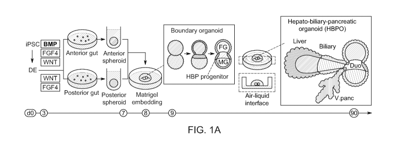
[0005] FIG 1A-1G. Boundary organoid generates multi-endoderm domains. 1A.
Schematic overview for establishing the hepato-biliary-pancreatic (HBP)
organoid ("HBPO")
from iPSC. Human PSCs were differentiated into anterior or posterior gut
cells. The cells
were dissociated into single cells and reaggregated to form anterior/posterior
gut spheroids,
and anterior-posterior boundary organoids were generated in Matrigel. Matrigel
embedding
initiated multi-organ specification and invagination from boundary organoids.
1B. Generation
of boundary organoid via fusion of 50X2+ anterior and CDX2+ posterior gut
spheroids.
SOX2 and CDX2 expressions were confirmed by wholemount immunostaining for SOX2
in
Red, CDX2 in Green, and DAPI in White, flowcytometry for percentage of each
population
showed as inlet numbers, and by qPCR. Data is mean s.d.; n =3 independent
experiments.
Unpaired, two-tailed Student's t-test. 1C. Tracing of human iPSC derived
anterior and
posterior organoids mix from Day 8 to Day 11. Upper row: Bright-field. Middle
row: whole
mount immunostaining for SOX2 in Red, PDX1 in Green, CDX2 in Blue and DAPI in
White.
Lower row: whole mount immunostaining for CDX2 in Blue, HHEX in Green, PDX1 in
Red
and DAPI in White. Arrow: PDX1 and HHEX positive region. 1D. Frequency of
detected
PDX1 positive cell in each area of boundary organoid immunofluorescent stained
for PDX1.
1E. Position of PDX1 positive cells in each boundary organoid. Y axis showed
percentages of
PDX1 positive cells in each area compared to DAPI stained total cell numbers.
f. Percentage
of HHEX and PDX1 positive cells for fused organoid in various combination,
anterior-
posterior (AP) (n = 4), anterior-anterior (AA) (n = 3) and posterior-posterior
(PP) (n = 3) at
Day 11. Y axis showed percentages of positive cells compared to DAPI stained
total cell
numbers. Data are mean s.d. *P< 0.05, **P < 0.01; one-way ANOVA.g.
Transcriptomic
characterization of boundary organoids with time course, from Day 8 (D8) to
Day 12 (D12).
Anterior (A), boundary (B), and posterior (P) domains were dissected,
separated, and applied
for RNA sequencing as indicated in left representative image (anterior gut
spheroid was
labeled by GFP and posterior gut spheroid was labeled by RFP). Anterior,
boundary, and
posterior domains showed the enrichment of the gene-sets of anterior foregut,
CA 03106634 2021-01-15
WO 2020/023245
PCT/US2019/041985
- 3 -
liver/biliary/pancreas primordium and mid/hindgut markers reported 31'32,
respectively. Scale
bars, 50 um (1B), 100 um (1C), 50 um (1G).
[0006] FIG 2A-2I. Self-emergence of hepato-biliary-pancreatic progenitors from
boundary organoid without inductive factors. 2A. Generating PROX1-tdTomato
reporter line
by CRISPR-Cas9 gene editing system. 2B. PROX1-tdTomato expression in
established
PROX1 reporter iPSCs from Day 9 to Day 11. 2C. PROX1-tdTomato positive area in
each
organoid. AP, AA and PP combinations were evaluated. The tdTomato expression
was
confirmed in the cell membrane of boundary organoids in AP combination. 2D.
Hepatic
invagination in mouse liver primordium explant (Proxl::GFP) and human iPSC-
derived
boundary organoids (PROX1::m-tdTomato). 2E. The immunostaining of PROX1 in
E8.75
mice embryo and boundary organoids at Day 13. 2F. Trans criptomic
characterization of
boundary organoids. Anterior, boundary, and posterior domains were dissected,
separated,
and applied for RNA sequencing. Heatmap shows downstream gene expression
related to
FGF, BMP, Hedgehog, NOTCH and RA signal pathway selected from GO term category
and
KEGG pathway category. Heatmap was separated into 8 detailed groups (Cl ¨ C8)
by
unbiased hierarchical clustering. C6 which showed unique expression pattern of
highly
expressed genes in B indicated an enrichment of RA down-stream gene-sets
compared to
others. 2G. Gene expression of HHEX, PDX1, SOX2, and CDX2 at Day 11 with three
days
culture of retinoic acid, BM5493, R-75251, or WIN18446. 2H. Gene expression
analysis for
RA signal pathway related genes in epithelial or mesenchymal cells from
original anterior or
posterior gut spheroids. Each gut spheroids were differentiated using RFP or
GFP labeled
iPSCs and dissociated into single cells after boundary formation.
Anterior/Posterior
separation was performed by RFP or GFP expression, whereas
epithelial/mesenchymal
separation was by EpCAM expression. 21. Default developmental potential of
transplanted
boundary organoid. Middle panels show H&E staining and immunohistochemistry
analysis,
whereas right panels show immunofluorescent analysis. Scale bars, 50 um (2B),
200 um
(2D), 100 um (2E), 100 um (2I).
[0007] FIG 3A-30. Modeling human hepato-biliary-pancreatic organogenesis. 3A.
Illustration of dissection of PROX1 positive region from HBP organoids and
image of
before/after dissection. 3B. Optimization of cultured system with 1) Floating,
2) Embedded
CA 03106634 2021-01-15
WO 2020/023245
PCT/US2019/041985
- 4 -
into Matrigel, 3) Embedded into Matrigel and cultured with Transwell from D13,
4).
Dissected, embedded into Matrigel, and cultured with Transwell from D13. Left
panel shows
the classification of invaginating or branching organoid. 3C. Morphogenesis of
boundary
organoids through 2 days from Day 13. 3D. Morphogenetical change of PROX1
dissected
tissue from boundary organoids through 30 days of air-liquid interface
cultured system. 3E.
Stereomicroscopic image of Day 37 organoids. 3F. Boundary organoid has
tdTomato
expressing hepatobiliary tissues branched out for putative pancreatic domains,
similar to
cultured mouse E10.5 derived hepato-biliary-pancreas. Left: cultured mouse
embryonic tissue
during 4 days, Right: PROX1-tdTomato reporter iPSCs at Day 90. 3G. Right:
Illustration of
invagination liver, bile duct and pancreas connected with intestine. Left: H&E
in D90
boundary tissue. 3H-3I. Immunostaining for combination of CK19, PDX1, PROX1,
SOX9
and NGN3, and alpha-SMA and SOX17 (3H) and AFP, EpCAM, and alpha-SMA (3I). 3J,
3K. Whole mount staining of PDX1, NKX6.1 and GATA4, and DBA, PDX1, and PROX1
(3K). 3L-3N. Immunostaining of NKX6.1 and HNF1B (3L), Amylase and GATA4 (3M)
and
Amylase and CCKAR (3N). 30. CCK treatment response in putative biliary
structure. 3P.
Hormone induced secretory function of exocrine pancreatic domain. Enzyme-
linked
immunosorbent assay of amylase in boundary tissue before and after 3 days-
CCK. Scale
bars, 100 um (a), 100 um (3B), 100 um (3C), 200 um (3D), 1 mm (3E), 200 um
(3F), 200
um (3G), 200 um (3H), 200 um (3I), 200 um (3J), 100 um (3K'), 200 um (3L), 100
um
(3M), 500 um (3N), 50 um (30).
[0008] FIG 4A-4J. Modeling HES1-mediated organ segregation error in HBP
organoids. 4A. Gene targeting strategy for HES1 knock out (KO) line by CRISPR-
Cas9
system. 4B. Confirmation of modified gene sequence of WT and HES1K0 (Del #11)
4C.
Photo of HES1-/- iPSC culture 4D. Gene expression of HES1 in HES1 KO iPSC-
derived
boundary organoid at Day 20. 4E. Confirmation of boundary organoid formation
from HES1-
/- iPS line by wholemount immunostaining of 50X2, CDX2, PDX1 and HHES. Hepato-
biliary-pancreatic precursor specification was preserved even in the presence
of HES1
mutation. 4F. PROX1-tdTomato expression in anterior and posterior boundary
spheroids
generated from HES1+/+ and HES1-/- at D11. 4G. RNAseq of pancreas associated
markers
at Day 22 of HES1+/+ and HES1-/- HBP organoids. 4H. Gene expression of GCG,
NEUROG3, INS, and NKX2-2 in HES1+/+, HES1-/- organoids, and human adult
pancreatic
CA 03106634 2021-01-15
WO 2020/023245
PCT/US2019/041985
- 5 -
tissue. 41. Macroscopic observation of boundary tissue of HES1+/+ and HES1-/-.
4J.
Wholemount immunostaining for DBA and PDX1 shows enhanced PDX1 expression and
diminished DBA stained area in HES1 KO organoids. Scale bars, 500 pin (4C),
200 pin (4E),
100 pin (4F), 500 pin (40, 500 pin (4J).
[0009] FIG 5. Anterior and posterior gut cell characterization. Flow cytometry
of
EpCAM in Day 7 anterior and posterior gut cells using TkDA human iPSCs and
72_3 human
iPSCs.
[0010] FIG 6A-6B Reproducibility of boundary organoid formation. 6A. The image
of Dll boundary organoids. Anterior and posterior gut spheroids were
differentiated from H1
ESCs or 1383D6 iPSCs, mixed and transferred into Matrigel. Scale bar is 200
pm. 6B.
Immunofluorescent staining of CDX2, epithelial marker ECAD, and HHEX in
boundary
spheroids derived from HlESC. 6C. Immunofluorescent staining of PDX1, CDX2,
FOXF1,
and HHEX in boundary spheroids derived from 72_3 iPS.
[0011] FIG 7A-7B Cell-cell contact dependent HBP gene induction. 7A. Anterior
and
posterior gut spheroids were mixed at D8, fused the following day at D9,
cultured and
collected at D12 for quantitative RT-PCR. The spheroids that not fused were
also collected at
D12 for comparison. 7B. PDX1 and HHEX gene expressions in the condition of
fused, not-
fused, posterior gut spheroid (day8), and iPS cells.
[0012] FIG 8 Comparison of different anterior and posterior gut combinations.
Immunofluorescent staining of CDX2, HHEX, and PDX1 in the combination of AP,
AA, and
PP spheroids at D12. Scale bar is 200 pm.
[0013] FIG 9A- 9C. HBP progenitors develop from posterior gut cells. 9A. Non
labeled iPS cells were differentiated into anterior gut spheroid while AAVS1-
GFP labeled
iPS cells were differentiated into posterior gut spheroid. Top column showed
bright field and
GFP fluorescent image during boundary organoid formation. Bottom column showed
whole
mount immunostaining for HHEX and PDX1 at Day 13. The HHEX expression was
overlapped with GFP expression. Scale bar is 200 pm. 9B. H2B-GFP labeled and
unlabeled
PROX1-tdTomato reporter iPSCs were differentiated into anterior and posterior
gut spheroid,
respectively. tdTomato expression was only detected in unlabeled original
posterior gut
CA 03106634 2021-01-15
WO 2020/023245
PCT/US2019/041985
- 6 -
spheroid. Scale bar is 200 pm. 9C. Using unlabeled iPSCs and PROX1-tdTomato
reporter
iPSCs, anterior and posterior gut spheroids were differentiated. Two
combinations, reporter
cell derived anterior and unlabeled cell posterior (left column), or unlabeled
cell derived
anterior and reporter cell derived posterior gut spheroid (right column) were
examined by
tdTomato expression. Top row: bright field image, bottom row: tdTomato
fluorescence
image. Scale bar is 200 pm
[0014] FIG 10. Abolishment of HHEX and PDX1 induction in posterior gut
specific
BM5493. BM5493 pretreated anterior or posterior gut spheroids were fused to
induce HBP
anlage formation. Compared to untreated control group, the group of BM5493
pretreated
posterior gut spheroid was inhibited HHEX and PDX1 expression at boundary,
suggesting
retinoic acid receptor function in posterior side was important to establish
HBP boundary
organoid. Scale bar: 200 pm
[0015] FIG 11A and 11B. PROX1 inhibition by BM5493 exposure with E9.0
PROX1::GFP reporter mouse embryo explant culture. Embryonic Day 9.0 Proxl-GFP
whole
embryo was cultured in the rotator-type bottle culture system for 24 hrs.
Retinoic acid
receptors antagonist BM5493 treated group was compared with control (adding
DMSO)
group. 11A. Bright field image and GFP fluorescent image for embryo after
culture. 11B. The
area of GFP expressing parts was quantified from GFP image in (a). Scale bar:
1 mm
[0016] FIG 12 Optimization of in vitro culture system. In FIG 3A-3C, various
culture
formats were compared to enhance morphological change, such as invagination
and
branching morphogenesis, of PROX1 positive HBP precursor region. At D7,
anterior and
posterior gut spheroids were mixed and connected after 24 hour-culture.
Connected spheroids
were transferred into Matrigel drop or low binding culture plate to compare
between non-
floating and floating conditions during HBP precursor emergence. The organoid
in Matrigel
embedded group was started to express tdTomato at D11. The tdTomato positive
region was
manually dissected under microscope according to the fluorescence expression
and
transferred into Matrigel drop again or Transwell to compare the effect from
various agonist
and antagonist in medium.
CA 03106634 2021-01-15
WO 2020/023245
PCT/US2019/041985
- 7 -
[0017] FIG 13. Comparison of organoid size, PROX1 positive area, branching and
invagination. Only AP combination increased the size of the organoids and
PROX1
expressing region. Moreover, AP combination showed the spheroids with
branching and
invagination while other two combination did not. Scale bar: 500 pm
[0018] FIG 14. Failure to branch and invaginate from posterior region of HBP
organoids. While HBP organoid formed Proxl expressing branching structure,
posterior
region of HBP that contain PDX1 expression did not form its structure. Scale
bar is 200 pm.
[0019] FIG 15A-15C. Expression of organ domain-specific markers in HBP
organoids. 15A. Immunofluorescent staining of AFP, Albumin, and HHEX at Day
30. AFP
and Albumin expressed in the same region but not HHEX. HHEX were hepatocyte
progenitor
marker which result in disappearance of the expression at the later stage.
15B.
Immunofluorescent staining of NKX6.1, NKX6.3 and PDX1. NKx6.3 were expressed
in the
area of pancreatic markers PDX1 and NKX6.1 expression. 15C. Immunofluorescent
staining
of EpCAM, PROX1, 50X9, and CLF. Scale bar: 100 pm
[0020] FIG 16. Pancreatic associated genes were upregulated in HES-/-
organoids.
RNAseq of pancreatic associated markers at Day 22 of HES1+/+ and HES1-/- HBP
organoids. This is related to FIG 4G.
[0021] FIG 17. Connected structure in long term cultured organoid. Whole mount
staining of DBA and 50X9 in HES1-/- and HES1+/+ organoids. DBA and 50X9
disappeared in HES1-/- organoids. Scale bar: 200 pm.
DETAILED DESCRIPTION
[0022] DEFINITIONS
[0023] Unless otherwise noted, terms are to be understood according to
conventional
usage by those of ordinary skill in the relevant art. In case of conflict, the
present document,
including definitions, will control. Preferred methods and materials are
described below,
although methods and materials similar or equivalent to those described herein
may be used
in practice or testing of the present invention. All publications, patent
applications, patents
and other references mentioned herein are incorporated by reference in their
entirety. The
CA 03106634 2021-01-15
WO 2020/023245
PCT/US2019/041985
- 8 -
materials, methods, and examples disclosed herein are illustrative only and
not intended to be
limiting.
[0024] As used herein and in the appended claims, the singular forms "a,"
"and," and
"the" include plural referents unless the context clearly dictates otherwise.
Thus, for example,
reference to "a method" includes a plurality of such methods and reference to
"a dose"
includes reference to one or more doses and equivalents thereof known to those
skilled in the
art, and so forth.
[0025] The term "about" or "approximately" means within an acceptable error
range
for the particular value as determined by one of ordinary skill in the art,
which will depend in
part on how the value is measured or determined, e.g., the limitations of the
measurement
system. For example, "about" may mean within 1 or more than 1 standard
deviation, per the
practice in the art. Alternatively, "about" may mean a range of up to 20%, or
up to 10%, or
up to 5%, or up to 1% of a given value. Alternatively, particularly with
respect to biological
systems or processes, the term may mean within an order of magnitude,
preferably within 5-
fold, and more preferably within 2-fold, of a value. Where particular values
are described in
the application and claims, unless otherwise stated the term "about" meaning
within an
acceptable error range for the particular value should be assumed.
[0026] As used herein, the term "definitive endoderm (DE) cell" means one of
the
three primary germ layers produced by the process of gastrulation.
[0027] As used herein the term "wnt signalling pathway" means the wnt/beta-
catenin
pathway and is a signal transduction pathway that is mediated by Wnt ligands
and frizzled
cell surface receptors that acts through the beta-catenin protein.
[0028] As used herein the term "activator" with respect to a pathway, such as
a "wnt
pathway" means a substance that activates the Wnt/beta-catenin pathway such
that Wnt/beta-
catenin targets are increased.
[0029] As used herein, the term "FGF signaling pathway activator" means a
substance that activates the FGF pathway such that FGF targets are increased.
CA 03106634 2021-01-15
WO 2020/023245
PCT/US2019/041985
- 9 -
[0030] As used herein, the term "BMP signaling pathway inhibitor" a substance
that
interferes with the BMP pathway and causes BMP targets to be decreased.
[0031] As used herein, the term "growth factor" means a substance capable of
stimulating cellular processes including but not limited to growth,
proliferation,
morphogenesis or differentiation.
[0032] As used herein, the term "stable expression" of a marker means
expression
that does not change upon modification of the growth environment.
[0033] As used herein, the term "totipotent stem cells" (also known as
omnipotent
stem cells) are stem cells that can differentiate into embryonic and extra-
embryonic cell
types. Such cells can construct a complete, viable, organism. These cells are
produced from
the fusion of an egg and sperm cell. Cells produced by the first few divisions
of the fertilized
egg are also totipotent.
[0034] As used herein, the term "pluripotent stem cells (PSCs)," also commonly
known as PS cells, encompasses any cells that can differentiate into nearly
all cells, i.e., cells
derived from any of the three germ layers (germinal epithelium), including
endoderm
(interior stomach lining, gastrointestinal tract, the lungs), mesoderm
(muscle, bone, blood,
urogenital), and ectoderm (epidermal tissues and nervous system). PSCs can be
the
descendants of totipotent cells, derived from embryos (including embryonic
germ cells) or
obtained through induction of a non-pluripotent cell, such as an adult somatic
cell, by forcing
the expression of certain genes.
[0035] As used herein, the term "induced pluripotent stem cells (iPSCs)," also
commonly abbreviated as iPS cells, refers to a type of pluripotent stem cells
artificially
derived from a normally non-pluripotent cell, such as an adult somatic cell,
by inducing a
"forced" expression of certain genes.
[0036] As used herein, the term "precursor cell" encompasses any cells that
can be
used in methods described herein, through which one or more precursor cells
acquire the
ability to renew itself or differentiate into one or more specialized cell
types. In some aspects,
a precursor cell is pluripotent or has the capacity to becoming pluripotent.
In some aspects,
the precursor cells are subjected to the treatment of external factors (e.g.,
growth factors) to
CA 03106634 2021-01-15
WO 2020/023245
PCT/US2019/041985
- 10 -
acquire pluripotency. In some aspects, a precursor cell can be a totipotent
stem cell; a
pluripotent stem cell (induced or non-induced); a multipotent stem cell; and a
unipotent stem
cell. In some aspects, a precursor cell can be from an embryo, an infant, a
child, or an adult.
In some aspects, a precursor cell can be a somatic cell subject to treatment
such that
pluripotency is conferred via genetic manipulation or protein/peptide
treatment.
[0037] In developmental biology, cellular differentiation is the process by
which a
less specialized cell becomes a more specialized cell type. As used herein,
the term "directed
differentiation" describes a process through which a less specialized cell
becomes a particular
specialized target cell type. The particularity of the specialized target cell
type can be
determined by any applicable methods that can be used to define or alter the
destiny of the
initial cell. Exemplary methods include but are not limited to genetic
manipulation, chemical
treatment, protein treatment, and nucleic acid treatment.
[0038] Disclosed herein are hepato-biliary-pancreatic organoid ("HBPO" or "HBP
organoid") compositions. The disclosed HBPO compositions may have, in some
aspects, two
or more functions selected from hepatic tissue function, biliary tissue
function, exocrine
pancreatic function, and endocrine pancreatic tissue function. In one aspect,
the HBPO
disclosed herein may be referred to interchangeably as a "multi-organ three-
dimensional
organoid" and may comprise an anterior region, a posterior region and a
boundary region. In
one aspect, the boundary region may expresse Pancreatic and Duodenal Homeobox
1 (PDX1)
and Hematopoietically-Expressed Homeobox Protein (HHEX). In one aspect, the
HBPO may
comprise bile duct tissue and pancreatic tissue. In one aspect, the bile duct
tissue and
pancreatic tissue may be connected in the HBPO. In one aspect, the HBPO may
comprise
liver cells, pancreas cells, bile duct cells, and intestinal cells.
[0039] In one aspect, the HBPO may comprise endothelial cells, mesenchymal
cells,
or both endothelial cells and mesenchymal cells. The HBPO may comprise, in
certain
aspects, endoderm and mesoderm.
[0040] The disclosed HBPOs may be characterized by a branched structure, as
shown, for example, in the corresponding figures herein.
CA 03106634 2021-01-15
WO 2020/023245
PCT/US2019/041985
- 11 -
[0041] In one aspect, the HBPOs disclosed herein may be characterized by
expression
of a functional exocrine marker. In one aspect, the marker may be amylase. In
one aspect, the
marker may be an endocrine marker, for example, insulin.
[0042] In certain aspects, the HBPOs disclosed herein may be characterized by
a
functional response to an exogenously administered agent. For example, in one
aspect, the
HBPO may secrete amylase in response to contact with cholecystokinin (CCK).
[0043] In certain aspects, the disclosed HBPOs may be characterized in that
the
HBPO may be substantially free of one or more of submucosal glands, transition
zones,
vasculature, immune cells, or submucosal layers. Such features distinguish the
three-
dimensional organoid structure from that of native tissue found endogenously
in a human, or
other mammal.
[0044] In one aspect, the HBPO is obtained by expansion of one or more
precursor
cells as defined above, for example, an iPSC obtained from an individual.
[0045] Also disclosed herein are methods of making a hepato-biliary-pancreatic
organoid ("HBPO" or "HBP organoid"). In this aspect, the method may comprise
contacting
a first definitive endoderm with a Wnt signaling pathway activator, an FGF
signaling
pathway activator, and a BMP signaling pathway inhibitor to form an anterior
gut spheroid;
contacting a second definitive endoderm with a Wnt signaling pathway activator
and an FGF
signaling pathway activator to form a posterior gut spheroid; contacting the
anterior gut
spheroid with the posterior gut spheroid until the anterior gut spheroid and
the posterior gut
spheroid are fused to form a boundary organoid having a foregut-midgut
boundary; and
culturing the boundary organoid having the foregut-midgut boundary to form the
HBPO;
wherein the HBPO comprises biliary tissue and pancreatic tissue. (See, for
example, FIG 12)
In one aspect, the disclosed methods may allow for the production of an HBPO
that may
comprise liver tissue, pancreatic tissue, bile duct tissue, and intestinal
tissue.
[0046] As used herein, the term hepato-biliary-pancreatic organoid ("HBPO" or
"HBP organoid") generally refers to the organoid at the stage in which multi-
organ domains
(hepatic, biliary and pancreatic organ domains) are segregated, which usually
occurs around
CA 03106634 2021-01-15
WO 2020/023245
PCT/US2019/041985
- 12 -
D30. With regard to the term "boundary organoid," this may include an organoid
after D7,
which includes organoid compositions that do not yet have tissue
specification.
[0047] In one aspect, the anterior gut spheroids may comprise cells expressing
SRY-
box 2 (S0X2). In one aspect, the anterior gut spheroid may be characterized by
SRY-box 2
(S0X2) expression. In one aspect, the anterior gut spheroids may be
substantially free of
Pancreatic and Duodenal Homeobox 1 (PDX1) expressing cells. In one aspect,the
posterior
gut spheroids comprise cells expressing Pancreatic and Duodenal Homeobox 1
(PDX1). In
one aspect, the posterior gut spheroids may express CDX2. In one aspect, the
posterior gut
spheroid may be characterized by Caudal Type Homeobox 2 (CDX2) expression. In
one
aspect, the posterior gut spheroids may comprise cells expressing Caudal type
homeobox 2
(CDX2). In one aspect, the HBPO may comprise cells expressing Pancreatic and
Duodenal
Homeobox 1 (PDX1), cells expressing Hematopoietically-expressed homeobox
protein
(HHEX), and cells expressing Prospero-Related Homeobox 1 (PROX1). In one
aspect, the
fused anterior gut spheroid and posterior gut spheroid may comprise a biliary-
pancreatic
primordium characterized by expression of SOX2, CDX2, HHEX, and PDX1, wherein
PDX1
expression is localized to the boundary region of the fused anterior gut
spheroid and the
posterior gut spheroid.
[0048] In one aspect, the HBPO may be embedded into a morphogenic factor,
preferably a basement membrane matrix, such as, for example, Matrigel. The
method may
further comprise excising PROX1 positive regions from the HBPO and culturing
the excised
HBPO to form invaginating epithelium and a branching structure.
[0049] In one aspect, the HBPO may be cultured with rotation, for example,
using the
methods disclosed herein.
[0050] In one aspect, the disclosed methods may be used to obtain an HBPO as
described herein, in particular, an HBPO comprising endoderm and mesoderm.
[0051] In one aspect, the definitive endoderm may be derived from a precursor
cell
selected from an embryonic stem cell, an embryonic germ cell, an induced
pluripotent stem
cell, a mesoderm cell, a definitive endoderm cell, a posterior endoderm cell,
a posterior
endoderm cell, and a hindgut cell, preferably a definitive endoderm derived
from a
CA 03106634 2021-01-15
WO 2020/023245
PCT/US2019/041985
- 13 -
pluripotent stem cell, more preferably a definitive endoderm derived from a
pluripotent stem
cell selected from an embryonic stem cell, an adult stem cell, or an induced
pluripotent stem
cell.
[0052] In one aspect, the definitive endoderm may be derived from contacting a
pluripotent stem cell with one or more molecules selected from Activin, the
BMP subgroups
of the TGF-beta superfamily of growth factors; Nodal, Activin A, Activin B,
BMP4, Wnt3a,
and combinations thereof.
[0053] In one aspect, the WNT signaling pathway activator may be selected from
one
or more molecules selected from the group consisting of Wntl, Wnt2, Wnt2b,
Wnt3, Wnt3a,
Wnt4, Wnt5a, Wnt5b, Wnt6, Wnt7a, Wnt7b, Wnt8a, Wnt8b, Wnt9a, Wnt9b, Wntl0a,
Wntl0b, Wntll, Wnt16, a GSKr3 inhibitor (e.g., CHIR99021, i.e. "CHIRON"), BIO,
LY2090314, SB-216763, lithium, porcupine inhibitors IWP, LGK974, C59, SFRP
inhibitor
WAY-316606, beta-catenin activator DCA.
[0054] In one aspect, the FGF signaling pathway activator may be selected from
one
or more molecules selected from the group consisting of FGF1, FGF2, FGF3,
FGF4, FGF10,
FGF11, FGF12, FGF13, FGF14, FGF15, FGF16, FGF17, FGF18, FGF19, FGF20, FGF21,
FGF22, FGF23, and combinations thereof, preferably FGF4 or FGF10, or a
combination
thereof.
[0055] In one aspect, the BMP signaling pathway inhibitor may be selected from
Noggin, Dorsomorphin, LDN189, DMH-1, and combinations thereof, preferably
wherein the
BMP signaling pathway inhibitor is Noggin.
[0056] In one aspect, the disclosed methods may be conducted in vitro. In
certain
aspect, at least one step of the method may be carried out on a solid support.
For example, the
solid support may be selected from collagen, basement membrane matrix
(Matrigel), or a
combination thereof.
[0057] In one aspect, a method for making an HBPO having an endothelium is
disclosed. In this aspect, the method may comprise the step of culturing an
HBPO according
to the methods disclosed herein, with endothelial cells derived from induced
pluripotent stem
cells. The endothelial cells may be formed into an endothelial cell (EC)
spheroid prior to
CA 03106634 2021-01-15
WO 2020/023245
PCT/US2019/041985
- 14 -
culturing with an HBPO. For example, for endothelial cell (EC)
differentiation, human iPSCs
may be dissociated using Accutase (Thermo Fisher Scientific Inc., Waltham, MA,
USA) and
plated on Laminin 511 E8 fragment (iMatrix-511Tm, provided by Nippi, Inc.) at
varying
optimal density (depending on the cell line) in StemFit (Ajinomoto Co., Inc.)
with Rho-
associated kinase inhibitor (Y-27632). From the next day, they may first be
differentiated into
mesoderm using the Priming Medium having B27 medium (1:1 mixture of DMEM:F12
(1:1)
with 1% Glutamax and 1% B27 (all Life Technologies) with Wnt activator and
bone
morphogenetic protein 4 (BMP4) for three days. The priming medium may then be
replaced
with EC induction medium, StemPro-34 SFM medium (Life Technologies)
supplemented
with VEGF and forskolin. The induction medium may be renewed daily. On day
seven of
differentiation, ECs are dissociated and subjected to FACS analysis. iPSC
derived ECs
should display typical endothelial morphology with junctional localization of
CD144 and
CD31. ECs may be re-plated on Fibronectin coated dishes at a density of 50,000
cells cm-2
in EC Expansion Medium consisting of StemPro-34 SFM supplemented with VEGF-A.
EC
Expansion Medium may be replaced every other day. The differentiated EC may be
dissociated to single cells and formed into spheroids using the spheroid
formation protocol of
the anterior gut spheroids and posterior gut spheroids as described herein.
The ECs may then
be mixed with the fused anterior gut spheroid and the posterior gut spheroid
on 96 well round
bottom ultra-low attachment plate in gut growth medium for 24 hours to form
fused EC
spheroid, the anterior gut spheroid and the posterior gut spheroid.
[0058] In one aspect, a method for making an HBPO having a mesenchyme is
disclosed. In this aspect, the method may comprise the step of culturing an
HBPO according
to a method as disclosed herein with mesenchymal cells derived from induced
pluripotent
stem cells. The mesenchyme cells may be formed into a mesenchyme cell (MC)
spheroid
prior to culturing with the HBPO. The disclosed multi-organ, three-dimensional
organoid and
mesenchymal cells or a mesenchymal spheroid may be cultured together to
improve the
growth and maturation of the multi-organ, three-dimensional organoid. For
example, for
mesenchyme (MSC) differentiation, human iPSCs may be differentiated to
mesoderm. The
mesoderm cells may then be exposed to 3 days of additional Activin A and
PDGFBB.
Differentiated MSC may then be dissociated to single cells and formed into
spheroids using
CA 03106634 2021-01-15
WO 2020/023245
PCT/US2019/041985
- 15 -
the spheroid formation protocol of the anterior gut spheroid and posterior gut
spheroid. The
MSC spheroid may then be mixed with the fused anterior gut spheroid and
posterior gut
spheroid on 96 well round bottom ultra-low attachment plate in gut growth
medium for 24
hours to form a fused MSC spheroid, anterior gut spheroid and posterior gut
spheroid.
[0059] In one aspect, a method of making a hepato-biliary-pancreatic organoid
("HBPO") is disclosed, wherein the method may comprise deriving an anterior
gut spheroid
from anterior gut cells and a deriving a posterior gut spheroid from posterior
gut cells to
make a boundary organoid; and culturing the boundary organoid in gut growth
medium to
make the HBPO. In one aspect, a method of making a hepato-biliary-pancreatic
organoid
("HBPO"), may comprise deriving an anterior gut spheroid and deriving a
posterior gut
spheroid from anterior gut cells and posterior gut cells to make the HBPO in
the absence of
inducing factors; and culturing the boundary organoids in a gut growth medium
to make
HBPO.
[0060] By "inducing factors," it is meant a factor used to direct
differentiation, for
example, retinoic acid (RA; Sigma, MO, USA), hepatocyte growth factor (HGF;
PeproTech,
NJ, USA), 0.1 pM Dexamethasone (Dex; Sigma) and 20 ng/mL Oncostatin M (OSM;
R&D
Systems) for hepatocyte differentiation. Many other inducing factors are known
for pancreas
and biliary also, and would be readily understood by one of skill in the art.
[0061] In one aspect, a of method of making a hepato-biliary-pancreatic
organoid
("HBPO") is disclosed, wherein the method may comprise culturing a definitive
endoderm
(DE) differentiated from a pluripotent stem cell (PSC) in gut growth medium to
make
anterior gut cells and posterior gut cells, making an anterior gut spheroid
from anterior gut
cells and a posterior gut spheroid from posterior gut cells to make a boundary
organoid in the
absence of inducing factors; and (iii) culturing boundary organoids in gut
growth medium to
make an HBPO. In one aspect, the gut growth medium may be a medium comprising
a
ROCK inhibitor. In one aspect, the ROCK inhibitor may be Y-27632. In one
aspect, the
growth medium is Advanced DMEM/F-12 (Dulbecco's Modified Eagle Medium/Ham's F-
12), a widely used basal medium that allows the culture of mammalian cells
with reduced
Fetal Bovine Serum (FBS) supplementation, available at
CA 03106634 2021-01-15
WO 2020/023245
PCT/US2019/041985
- 16 -
https://www.thermofisher.com/order/catalog/product/12634010, with addition of
Glutamine,
HEPES, N2 and B27. In one aspect, the media may include addition of the
following
cytokines to Advanced DMEM: Noggin (BMP inhibitor), CHIR (WNT activator), FGF4
for
anterior gut cells, CHIR and FGF4 (without Noggin) for posterior gut cells.
[0062] In one aspect, a machine for producing an HBPO is disclosed herein. The
machine may comprise a gut growth medium containing a Wnt signaling pathway
activator
and an FGF signaling pathway activator, in the presence or absence of a BMP
signaling
pathway inhibitor; and a gut growth medium containing a ROCK inhibitor.
[0063] In one aspect, use of a plurality of culture mediums for the production
of an
HBPO, is disclosed, wherein the plurality of culture mediums comprise a gut
growth medium
containing a Wnt signaling pathway activator and an FGF signaling pathway
activator,
optionally comprising a BMP signaling pathway inhibitor; and an intestinal
growth medium
containing a ROCK inhibitor.
[0064] In one aspect, the HBPO may be transplanted into a mammal, such as a
non-
human mammal, for a period of time to increase growth and maturation of the
HBPO. In one
aspect, the HBPO may be transplanted to rescue organ dysfunction, for example,
in an
individual in need thereof. In one example, In vitro generated HBPOs may be
collected and
transplanted into the subscapular site of kidney or mesentery of the small
intestine in non-
obese diabetic/severe combined immunodeficient (NOD/SCID) mice. An increase in
growth
of the organoid may be observed at 8 weeks after the transplant. The viability
and growth can
be monitored based on blood tests such as human-Alb, human-C-peptide levels.
[0065] In one aspect, a method of treating an individual, comprising
transplanting an
HBPO as described herein is disclosed. In this aspect, an organoid produced
according to any
of the methods used herein, may be transplanted into an individual in need
thereof, using
standard surgical procedures as known in the art. In one aspect, the
individual may have a
disease state selected from hepatic disease, such as liver failure and
congenital liver diseases,
pancreatic disease, such as diabetes, or biliary disease.
[0066] In one aspect, methods of identifying a treatment for one or more
disease
states selected from a biliary inflammatory disease and/or a pancreatic
inflammatory disease
CA 03106634 2021-01-15
WO 2020/023245
PCT/US2019/041985
- 17 -
such as pancreatitis is disclosed. In this aspect, the disclosed organoid
compositions may be
contacted with a potential therapeutic agent of interest with an organoid
composition as
disclosed herein. A measure of organ activity may then be detected in the
organoid. Based on
the output of this detection, it may be determined whether the potential
therapeutic agent of
interest improves the measure of the disease state, and may thereby be used to
identify
therapeutic agents likely to be useful in treating a disease state which
involves a malfunction
of any of the tissues embodied in the HBPO.
[0067] Pluripotent Stem Cells Derived from Embryonic Cells
[0068] In some aspects, one step may include obtaining stem cells that are
pluripotent
or that can be induced to become pluripotent. In some aspects, pluripotent
stem cells are
derived from embryonic stem cells, which are in turn derived from totipotent
cells of the
early mammalian embryo and are capable of unlimited, undifferentiated
proliferation in vitro.
Embryonic stem cells are pluripotent stem cells derived from the inner cell
mass of the
blastocyst, an early-stage embryo. Methods for deriving embryonic stem cells
from
blastocytes are well known in the art. Human embryonic stem cells H9 (H9-
hESCs) are used
in the exemplary embodiments described in the present application, but it
would be
understood by one of skill in the art that the methods and systems described
herein are
applicable to any stem cells.
[0069] Additional stem cells that can be used in embodiments in accordance
with the
present invention include but are not limited to those provided by or
described in the database
hosted by the National Stem Cell Bank (NSCB), Human Embryonic Stem Cell
Research
Center at the University of California, San Francisco (UCSF); WISC cell Bank
at the Wi Cell
Research Institute; the University of Wisconsin Stem Cell and Regenerative
Medicine Center
(UW-SCRMC); Novocell, Inc. (San Diego, Calif.); Cellartis AB (Goteborg,
Sweden); ES
Cell International Pte Ltd (Singapore); Technion at the Israel Institute of
Technology (Haifa,
Israel); and the Stem Cell Database hosted by Princeton University and the
University of
Pennsylvania. Exemplary embryonic stem cells that can be used in embodiments
in
accordance with the present invention include but are not limited to SA01
(SA001); 5A02
(5A002); ES01 (HES-1); E502 (HES-2); E503 (HES-3); E504 (HES-4); E505 (HES-5);
E506 (HES-6); BG01 (BGN-01); BG02 (BGN-02); BG03 (BGN-03); TE03 (13); TE04
(14);
CA 03106634 2021-01-15
WO 2020/023245
PCT/US2019/041985
- 18 -
TE06 (16); UCO1 (HSF1); UC06 (HSF6); WA01 (H1); WA07 (H7); WA09 (H9); WA13
(H13); WA14 (H14). More details on embryonic stem cells can be found in, for
example,
Thomson et al., 1998, "Embryonic Stem Cell Lines Derived from Human
Blastocysts,"
Science 282 (5391):1145-1147; Andrews et al., 2005, "Embryonic stem (ES) cells
and
embryonal carcinoma (EC) cells: opposite sides of the same coin," Biochem Soc
Trans
33:1526-1530; Martin 1980, "Teratocarcinomas and mammalian embryogenesis,".
Science
209 (4458):768-776; Evans and Kaufman, 1981, "Establishment in culture of
pluripotent
cells from mouse embryos," Nature 292(5819): 154-156; Klimanskaya et al.,
2005, "Human
embryonic stem cells derived without feeder cells," Lancet 365 (9471): 1636-
1641; each of
which is hereby incorporated herein in its entirety.
[0070] Induced Pluripotent Stem Cells (iPSCs)
[0071] In some aspects, iPSCs are derived by transfection of certain stem cell-
associated genes into non-pluripotent cells, such as adult fibroblasts.
Transfection may be
achieved through viral vectors, such as retroviruses. Transfected genes
include the master
transcriptional regulators Oct-3/4 (Pouf51) and 5ox2, although it is suggested
that other
genes enhance the efficiency of induction. After 3-4 weeks, small numbers of
transfected
cells begin to become morphologically and biochemically similar to pluripotent
stem cells,
and are typically isolated through morphological selection, doubling time, or
through a
reporter gene and antibiotic selection. As used herein, iPSCs include but are
not limited to
first generation iPSCs, second generation iPSCs in mice, and human induced
pluripotent stem
cells. In some aspects, a retroviral system is used to transform human
fibroblasts into
pluripotent stem cells using four pivotal genes: 0ct3/4, 5ox2, Klf4, and c-
Myc. In alternative
aspects, a lentiviral system is used to transform somatic cells with OCT4,
50X2, NANOG,
and LIN28. Genes whose expression are induced in iPSCs include but are not
limited to Oct-
3/4 (e.g., Pou5fl); certain members of the Sox gene family (e.g., Soxl, 5ox2,
5ox3, and
Sox15); certain members of the Klf family (e.g., Klfl, Klf2, Klf4, and Klf5),
certain members
of the Myc family (e.g., C-myc, L-myc, and N-myc), Nanog, and LIN28.
[0072] In some aspects, non-viral based technologies are employed to generate
iPSCs.
In some aspects, an adenovirus can be used to transport the requisite four
genes into the DNA
of skin and liver cells of mice, resulting in cells identical to embryonic
stem cells. Since the
CA 03106634 2021-01-15
WO 2020/023245
PCT/US2019/041985
- 19 -
adenovirus does not combine any of its own genes with the targeted host, the
danger of
creating tumors is eliminated. In some aspects, reprogramming can be
accomplished via
plasmid without any virus transfection system at all, although at very low
efficiencies. In
other aspects, direct delivery of proteins is used to generate iPSCs, thus
eliminating the need
for viruses or genetic modification. In some embodiment, generation of mouse
iPSCs is
possible using a similar methodology: a repeated treatment of the cells with
certain proteins
channeled into the cells via poly-arginine anchors was sufficient to induce
pluripotency. In
some aspects, the expression of pluripotency induction genes can also be
increased by
treating somatic cells with FGF2 under low oxygen conditions.
[0073] More details on embryonic stem cells can be found in, for example, Kaji
et al.,
2009, "Virus free induction of pluripotency and subsequent excision of
reprogramming
factors," Nature 458:771-775; Woltjen et al., 2009, "piggyBac transposition
reprograms
fibroblasts to induced pluripotent stem cells," Nature 458:766-770; Okita et
al., 2008,
"Generation of Mouse Induced Pluripotent Stem Cells Without Viral Vectors,"
Science
322(5903):949-953; Stadtfeld et al., 2008, "Induced Pluripotent Stem Cells
Generated
without Viral Integration," Science 322(5903):945-949; and Zhou et al., 2009,
"Generation of
Induced Pluripotent Stem Cells Using Recombinant Proteins," Cell Stem Cell
4(5):381-384;
each of which is hereby incorporated herein in its entirety.
[0074] In some aspects, exemplary iPS cell lines include but not limited to
iPS-DF19-
9; iPS-DF19-9; iPS-DF4-3; iPS-DF6-9; iPS(Foreskin); iPS(IMR90); and
iPS(IMR90).
[0075] More details on the functions of signaling pathways relating to DE
development can be found in, for example, Zorn and Wells, 2009, "Vertebrate
endoderm
development and organ formation," Annu Rev Cell Dev Biol 25:221-251; Dessimoz
et al.,
2006, "FGF signaling is necessary for establishing gut tube domains along the
anterior-
posterior axis in vivo," Mech Dev 123:42-55; McLin et al., 2007, "Repression
of Wnt/r3-
catenin signaling in the anterior endoderm is essential for liver and pancreas
development.
Development," 134:2207-2217; Wells and Melton, 2000, Development 127:1563-
1572; de
Santa Barbara et al., 2003, "Development and differentiation of the intestinal
epithelium,"
Cell Mol Life Sci 60(7): 1322-1332; each of which is hereby incorporated
herein in its
entirety.
CA 03106634 2021-01-15
WO 2020/023245
PCT/US2019/041985
- 20 -
[0076] Any methods for producing definitive endoderm from pluripotent cells
(e.g.,
iPSCs or ESCs) are applicable to the methods described herein. In some
aspects, pluripotent
cells are derived from a morula. In some aspects, pluripotent stem cells are
stem cells. Stem
cells used in these methods can include, but are not limited to, embryonic
stem cells.
Embryonic stem cells can be derived from the embryonic inner cell mass or from
the
embryonic gonadal ridges. Embryonic stem cells or germ cells can originate
from a variety of
animal species including, but not limited to, various mammalian species
including humans. In
some aspects, human embryonic stem cells are used to produce definitive
endoderm. In some
aspects, human embryonic germ cells are used to produce definitive endoderm.
In some
aspects, iPSCs are used to produce definitive endoderm.
[0077] In one aspect, the definitive endoderm may be derived from a precursor
cell
selected from an embryonic stem cell, an embryonic germ cell, an induced
pluripotent stem
cell, a mesoderm cell, a definitive endoderm cell, a posterior endoderm cell,
a posterior
endoderm cell, and a hindgut cell. In one aspect, the definitive endoderm may
be derived
from a pluripotent stem cell. In one aspect, the definitive endoderm may be
derived from a
pluripotent stem cell selected from an embryonic stem cell, an adult stem
cell, or an induced
pluripotent stem cell. In one aspect, the DE may be a DE monolayer, wherein
greater than
90% of the cells in the DE monolayer co-express FOXA2 and 50X17.
[0078] In one aspect, the definitive endoderm may be derived from contacting a
pluripotent stem cell with one or more molecules selected from Activin, the
BMP subgroups
of the TGF-beta superfamily of growth factors; Nodal, Activin A, Activin B,
BMP4, Wnt3a,
and combinations thereof.
[0079] In one aspect, the BMP signaling pathway inhibitor may be selected from
Noggin, Dorsomorphin, LDN193189, DMH-1, and combinations thereof. In one
aspect, the
BMP signaling pathway inhibitor is Noggin. The BMP inhibitor may be present at
a
concentration of between from about 50 to about 1500 ng/ml.
[0080] In one aspect, the WNT signaling pathway activator may be selected from
one
or more molecules selected from the group consisting of Wntl, Wnt2, Wnt2b,
Wnt3, Wnt3a,
Wnt4, Wnt5a, Wnt5b, Wnt6, Wnt7a, Wnt7b, Wnt8a, Wnt8b, Wnt9a, Wnt9b, Wntl0a,
CA 03106634 2021-01-15
WO 2020/023245
PCT/US2019/041985
-21 -
Wntl0b, Wntll, Wnt16, a GSKr3 inhibitor (e.g., CHIR99021, i.e. "CHIRON"), BIO,
LY2090314, SB-216763, lithium, SFRP inhibitor WAY-316606, beta-catenin
activator DCA.
The concentration of the Wnt pathway activator may be, for example, used at a
concentration
between about 50 to about 1500 ng/ml. There are many ways to activate the
Wnt/beta-catenin
pathway (see http://web.stanford.edu/group/nusselab/cgi-bin/wnt/). Suitable
Some existing
wnt signaling pathway activators include but are not limited to protein-based
activators,
which may include Wnt ligands including but not limited to Wntl, Wnt2, Wnt2b,
Wnt3,
Wnt3a, Wnt8, et al; modifiers of Wnt ligand activity including but not limited
to activated
Wnt frizzled receptors, (LRP) co-receptors, R-spondin proteins, Dkk proteins,
regulators of
Wnt ligand secretion and trafficking (Wntless, Porcupine), inhibiting beta-
catenin
degredation APC and GSK3beta inhibition, activated beta-catenin,
constitutively active
TCF/Lef proteins and chemical activators, which may include over 28 known
chemicals that
either activate or inhibit Wnt/beta-catenin signaling. Some activators include
but are not
limited to GSK3-beta inhibitors CHIR99021 (CHIRON), BIO, LY2090314, SB-216763,
lithium, SFRP inhibitor WAY-316606, beta-catenin activator DCA.
[0081] In one aspect, the FGF signaling pathway activator may be one or more
molecules selected from the group consisting of FGF1, FGF2, FGF3, FGF4, FGF10,
FGF11,
FGF12, FGF13, FGF14, FGF15, FGF16, FGF17, FGF18, FGF19, FGF20, FGF21, FGF22,
FGF23, and combinations thereof, preferably FGF4 or FGF10, or a combination
thereof. In
one aspect, the concentration of the FGF pathway activator may be used at a
concentration
between about 50 to about 1500 ng/ml, though it will be understood by one of
ordinary skill
in the art that various concentrations may be used. Proteins and chemicals
that stimulate the
FGF receptor and signaling components downstream of the receptors including
MAPK,
MEK, ERK proteins and chemicals that modulate their activity. FGF signaling
can be
activated by inhibiting inhibitors of FGF signaling pathways including but not
limited to
Sprouty protein family members.
EXAMPLES
[0082] The following non-limiting examples are provided to further illustrate
embodiments of the invention disclosed herein. It should be appreciated by
those of skill in
the art that the techniques disclosed in the examples that follow represent
approaches that
CA 03106634 2021-01-15
WO 2020/023245
PCT/US2019/041985
- 22 -
have been found to function well in the practice of the invention, and thus
may be considered
to constitute examples of modes for its practice. However, those of skill in
the art should, in
light of the present disclosure, appreciate that many changes may be made in
the specific
embodiments that are disclosed and still obtain a like or similar result
without departing from
the spirit and scope of the invention.
[0083] Organogenesis is a complex and inter-connected process, orchestrated by
multiple boundary tissue interactions1-7. However, it is currently unclear how
individual,
neighboring components coordinate to establish an integral multi-organ
structure. Disclosed
herein is the continuous patterning and dynamic morphogenesis of hepatic,
biliary and
pancreatic structures, invaginating from a three-dimensional culture of human
pluripotent
stem cell (PSC).
[0084] Applicant has found that the boundary interactions between anterior and
posterior gut spheroids differentiated from human PSCs enables autonomous
emergence of
hepato-biliary-pancreatic (HBP) organ domains specified at the foregut-midgut
boundary
organoids in the absence of extrinsic factor supply. Whereas transplant-
derived tissues were
dominated by midgut derivatives, long-term culture of micro dissected HBP
organoids
develop into a segregated hepato-pancreato-biliary anlage, followed by the
recapitulation of
early morphogenetic events including the invagination and branching of three
different and
inter-connected organ structures, reminiscent of tissues derived from mouse
explanted
foregut-midgut culture. Mis-segregation of multi-organ domains incurred by a
genetic
mutation in HES1 abolishes the biliary specification potential in culture, as
seen in vivo".
Applicant has demonstrated that the experimental multi-organ integrated model
can be
established by the juxta-positioning of foregut and midgut tissues, and which
may serve as a
tractable, manipulatable and easily accessible model for the study of
complicated endoderm
organogenesis in human.
[0085] The hepato-biliary-pancreatic (HBP) anlage, which is demarcated by HHEX
(Hematopoietically-expressed homeobox protein) and PDX1 (Pancreatic and
duodenal
homeobox 1) expression is first specified at the boundary between the foregut-
midgutl , and
forms an epithelial vesicle invaginating ventrally from the primitive gut'
''s. The disruption
of boundary-defining genes around this area such as BMPR1A (Bone morphogenetic
protein
CA 03106634 2021-01-15
WO 2020/023245
PCT/US2019/041985
- 23 -
receptor, type 1A)', HLX (H2.0-like homeobox)2, CDX2 (Caudal type homeobox
2)3, NKX3-2
(NK3 Homeobox 2)4, HHEX 5, PDX1 and SOX9 (SRY-box 9)6 significantly alters
balanced
organogenesis along the stomach-HBP-intestine in vivo' . Subsequent
diversification of HBP
lineages is likely mediated by adjacent mesenchymal BMP at their boundary by
indirectly
repressing SOX9 in the posterior liver bud cells14. Thus, contiguous, dynamic
organogenesis
occurs in a complex environment and is likely driven by successive neighboring
tissue
interactions5'6'15. However, the patterning and balanced organogenesis of the
HBP system has
not been successfully modelled in tissue culture due to technical
complexities, hindering
detailed mechanistic studies16'17.
[0086] Here, Applicant used a three-dimensional differentiation approach using
human pluripotent stem cells (PSCs) to specify gut spheroids with distinct
regional identities
comprised of both endoderm and mesoderm. Applicant demonstrated that antero-
posterior
interactions recapitulate the foregut (marked by SOX2, SRY-Box 2) and the
midgut (marked
by CDX2) boundary in vitro, modeling the inter-coordinated specification and
invagination of
the human hepato-biliary-pancreatic system.
[0087] To develop a foregut-midgut boundary model, Applicant first exposed
definitive endoderm cells, which were patterned as previously described 18'19,
to the
recombinant protein FGF4, and the small molecule CHIR99021 (that modulates the
WNT
pathway) in the presence of a BMP antagonist Noggin for anterior gut and in
the absence of
Noggin for posterior gut identity (FIG. 1A and FIG 5). At Day 7 (D7), anterior
or posterior
gut cells were individually aggregated to form spheroids with a preferential
expression of the
foregut and mid/hindgut transcription factors SOX2 and CDX2, respectively
(FIG. 1B).
Applicant then transferred a posterior gut spheroid adjacent to an anterior
gut spheroid. At
D9, the two spheroids were fused in 94.8% of the wells, and fused spheroids
were embedded
into a morphogenetic factor, namely Matrigel (FIG. 1A and B). Surprisingly,
without adding
any exogenous factors, the HBP primordium emerged at the interface of the
anterior and
posterior gut spheroids, as evidenced by whole mount staining of the spheroids
over D9, D10
and D11, using the markers 50X2, CDX2, the immature liver marker HHEX, and the
antrum, duodenum and pancreas progenitor marker PDX1 (FIG.1C). At D9, the
anterior and
posterior gut spheroids did not exhibit any expression of HHEX and PDX1. At
D10, HHEX
CA 03106634 2021-01-15
WO 2020/023245
PCT/US2019/041985
- 24 -
and PDX1 expressions were emerged, and HHEX was only detectable at the
boundary of the
anterior and posterior (bAP) organoids. Both PDX1 and HHEX positive cells were
distinctively increased in the boundary region at Dll (FIG.1C). The use of
three additional
induced pluripotent stem cells (iPSCs) and embryonic stem cells (ESCs) lines
confirmed the
reproducibility in developing HHEX and PDX1 expressing cells at the boundary
of bAP
organoids (FIG 6A-6C). HBP progenitor induction requires cell-to-cell contact
between the
anterior and posterior gut spheroids (FIG. 3A and FIG 3B). Of all the detected
PDX1 positive
cells at the boundary site (30 out of 30 stained organoids) (FIG. 1D), the
percentage of PDX1
positive cells per total cell number was 5% in the boundary region while 0 and
1% in the
anterior and the posterior region, respectively (FIG. 1E). PDX1 expressing
cells were
observed in A-P organoids and posterior-posterior (P-P) gut spheroid
combinations. In
contrast, HHEX positive cells were only detected in A-P organoids, but not in
anterior-
anterior (A-A) nor P-P combinations (FIG. 1F and FIG 8), indicating the
balanced induction
of the HBP progenitors involves A-P fusion.
[0088] To trace the source of HHEX and PDX1 expressing cells, the bAP
organoids
were established using non-labeled anterior and GFP-labeled posterior gut
spheroids.
Applicant found that both HHEX and PDX1 expression overlapped with GFP,
suggesting
that the HBP progenitors originate from the posterior gut (FIG. 9A). RNA-
sequencing of D8,
D9, Dll and D12 micro-dissected anterior, boundary and posterior regions
showed that the
boundary tissues at Dll and D12 progressively expressed the arrays of HBP
specification
markers, whereas anterior or posterior regions gained foregut or mid/hindgut
identity,
respectively (FIG. 1G). Of note, in agreement with A-P recombination
experiments, posterior
tissues possess a closer identity to boundary regions (FIG. 1G). Taken
together, the AP
boundary strategy orchestrates autonomous patterning of HHEX and PDX1 positive
HBP
progenitors in the absence of exogenous inductive factors.
[0089] Applicant further established a reporter human iPSC to track their fate
by
visualizing tdTomato under the common progenitor marker of the liver, bile
duct, and
pancreas, a prospero-related homeobox 1 (PROX1) using CRISPRICas9 genome
editing
(FIG. 2A). Similar to HHEX and PDX1, PROX1 expression initiated at the
boundary at D10
and increased afterwards (FIG. 2B). The PROX1 emergence was specific to the A-
P
CA 03106634 2021-01-15
WO 2020/023245
PCT/US2019/041985
- 25 -
organoids but not in A-A nor P-P combinations (FIG. 2C). A-P recombination
assays
indicated PROX1 positive cells also originated from the posterior gut cells
(FIG 9B and 9C).
Air-liquid interface culture induced additional growth of the PROX1 positive
area, as seen in
E8.75 Proxl ::EGFP mouse embryonic liver tissue (FIG. 2D). PROX1
immunostaining
confirmed that the morphologically invaginating tissue induced from bAP
organoids was
similar with the boundary region of mouse at E8.5-8.75 (FIG. 2E).
[0090] To delineate the HBP progenitor self-inductive mechanism, Applicant
evaluated the boundary specific expression profiles of known inductive
signaling pathways.
Amongst FGF, BMP, HH (hedgehog), NOTCH and RA (retinoic acid) signals, RNAseq
identified that the signal downstream of RA were activated prominently at the
boundary
region, but not in the anterior or posterior regions at Dll (FIG. 2F and
Tablet).
[0091] Table 1. Gene ontology analysis for RNA sequencing (related to FIG 2E)
After
Original restricting to
Term size dataset
Cl
BIOCARTA_TGFB_PATHWAY 19 12 63.2%
PID_RETINOIC_ACID_PATHWAY 30 17 56.7%
GO_NOTCH_RECEPTOR_PROCESSING 16 9 56.3%
GO_POSITIVE_REGULATION_OF_NOTCH_SIGNALI
NG_PATHWAY 33 18 54.5%
PID_FGF_PATHWAY 55 29 52.7%
GO_POSITIVE_REGULATION_OF_BMP_SIGNALING
PATHWAY 29 15 51.7%
GO_REGULATION_OF_NOTCH_SIGNALING_PATH
WAY 63 32 50.8%
GO_NOTCH_SIGNALING_PATHWAY 111 50 45.0%
HALLMARK_HEDGEHOG_SIGNALING 35 15 42.9%
PID_BMP_PATHWAY 42 18 42.9%
GO_REGULATION_OF_BMP_SIGNALING_PATHWA
74 27 36.5%
GO_NEGATIVE_REGULATION_OF_NOTCH_SIGNAL
ING_PATHWAY 25 9 36.0%
KEGG_HEDGEHOG_SIGNALING_PATHWAY 56 19 33.9%
GO_RESPONSE_TO_RETINOIC_ACID 103 33 32.0%
GO_NOTCH_BINDING 17 5 29.4%
CA 03106634 2021-01-15
WO 2020/023245
PCT/US2019/041985
- 26 -
GO REGULATION_OF_RETINOIC_ACID_RECEPTOR
_SIGNALING_PATHWAY 29 8 27.6%
GO_CELLULAR_RES PONS E_TO_RETINOIC_ACID 61 15 24.6%
GO_RETINOIC_ACID METABOLIC_PROCESS 21 5 23.8%
GO_NEGATIVE_REGULATION_OF_BMP_SIGNALIN
G_PATHWAY 42 10 23.8%
GO_RETINOIC_ACID_RECEPTOR_SIGNALING_PAT
HWAY 17 4 23.5%
GO_NEGATIVE_REGULATION_OF_RETINOIC_AC ID
RECEPTOR_SIGNALING_PATHWAY 23 3 13.0%
C2
GO_NEGATIVE_REGULATION_OF_NOTCH_SIGNAL
ING_PATHWAY 25 5 20.0%
GO_NEGATIVE_REGULATION_OF_BMP_SIGNALIN
G_PATHWAY 42 8 19.0%
BIOCARTA_TGFB_PATHWAY 19 3 15.8%
GO_RES PONS E_TO_RETINOIC_ACID 103 16 15.5%
GO_REGULATION_OF_BMP_SIGNALING_PATHWA
Y 74 11 14.9%
PID_FGF_PATHWAY 55 8 14.5%
GO_RETINOIC_ACID_METAB OLIC_PROC ES S 21 3 14.3%
GO_NOTCH_SIGNALING_PATHWAY 111 15 13.5%
GO_NOTCH_RECEPTOR_PROCES SING 16 2 12.5%
PID_BMP_PATHWAY 42 5 11.9%
GO_RETINOIC_ACID_RECEPTOR_SIGNALING_PAT
HWAY 17 2 11.8%
GO_CELLULAR_RES PONS E_TO_RETINOIC_ACID 61 7 11.5%
HALLMARK_HEDGEHOG_SIGNALING 35 4 11.4%
KEGG_HEDGEHOG_SIGNALING_PATHWAY 56 6 10.7%
GO_POSITIVE_REGULATION_OF_BMP_SIGNALING
PATHWAY 29 3 10.3%
GO_REGULATION_OF_NOTCH_SIGNALING_PATH
WAY 63 6 9.5%
PID_RETINOIC_ACID_PATHWAY 30 2 6.7%
GO_NOTCH_BINDING 17 1 5.9%
GO_REGULATION_OF_RETINOIC_ACID_RECEPTOR
SIGNALING_PATHWAY 29 1 3.4%
GO_POSITIVE_REGULATION_OF_NOTCH_SIGNALI
NG_PATHWAY 33 1 3.0%
GO_NEGATIVE_REGULATION_OF_RETINOIC_AC ID
_RECEPTOR_SIGNALING_PATHWAY 23 0.0%
C3
PID_BMP_PATHWAY 42 9 21.4%
CA 03106634 2021-01-15
WO 2020/023245
PCT/US2019/041985
-27 -
GO_NEGATIVE_REGULATION_OF_BMP_SIGNALIN
G_PATHWAY 42 9 21.4%
GO_POSITIVE_REGULATION_OF_BMP_SIGNALING
_PATHWAY 29 6 20.7%
GO_REGULATION_OF_BMP_SIGNALING_PATHWA
Y 74 15 20.3%
GO_NOTCH_RECEPTOR_PROCESSING 16 3 18.8%
GO_NOTCH_BINDING 17 3 17.6%
PID_RETINOIC_ACID_PATHWAY 30 5 16.7%
BIOCARTA_TGFB_PATHWAY 19 3 15.8%
GO_CELLULAR_RESPONSE_TO_RETINOIC_ACID 61 9 14.8%
GO_RETINOIC_ACID_METABOLIC_PROCESS 21 3 14.3%
KEGG_HEDGEHOG_SIGNALING_PATHWAY 56 8 14.3%
GO_RESPONSE_TO_RETINOIC_ACID 103 14 13.6%
GO_NEGATIVE_REGULATION_OF_RETINOIC_ACID
RECEPTOR_SIGNALING PATHWAY 23 3 13.0%
GO_POSITIVE_REGULATION_OF_NOTCH_SIGNALI
NG_PATHWAY 33 4 12.1%
GO_NOTCH_SIGNALING_PATHWAY 111 12 10.8%
GO_REGULATION_OF_RETINOIC_ACID_RECEPTOR
_SIGNALING_PATHWAY 29 3 10.3%
HALLMARK_HEDGEHOG SIGNALING 35 3 8.6%
GO_REGULATION_OF_NOTCH_SIGNALING_PATH
WAY 63 5 7.9%
PID_FGF_PATHWAY 55 4 7.3%
GO_RETINOIC_ACID_RECEPTOR_SIGNALING_PAT
HWAY 17 1 5.9%
GO_NEGATIVE_REGULATION_OF_NOTCH_SIGNAL
ING_PATHWAY 25 1 4.0%
C4
GO_NEGATIVE_REGULATION_OF_NOTCH_SIGNAL
ING_PATHWAY 25 3 12.0%
PID_RETINOIC_ACID_PATHWAY 30 2 6.7%
GO_NOTCH_SIGNALING_PATHWAY 111 7 6.3%
GO_NOTCH_BINDING 17 1 5.9%
GO_RETINOIC_ACID_RECEPTOR_SIGNALING_PAT
HWAY 17 1 5.9%
GO_REGULATION_OF_NOTCH_SIGNALING_PATH
WAY 63 3 4.8%
PID_FGF_PATHWAY 55 2 3.6%
KEGG_HEDGEHOG_SIGNALING_PATHWAY 56 2 3.6%
GO_CELLULAR_RESPONSE_TO_RETINOIC_ACID 61 2 3.3%
GO_POSITIVE_REGULATION_OF_NOTCH_SIGNALI
NG_PATHWAY 33 1 3.0%
CA 03106634 2021-01-15
WO 2020/023245
PCT/US2019/041985
- 28 -
GO_RESPONSE_TO_RETINOIC_ACID 103 3 2.9%
PID_BMP_PATHWAY 42 1 2.4%
GO_NEGATIVE_REGULATION_OF_BMP_SIGNALIN
G PATHWAY 42 1 2.4%
GO_REGULATION_OF_BMP_SIGNALING_PATHWA
Y 74 1 1.4%
GO_NOTCH_RECEPTOR_PROCESSING 16 0.0%
GO_RETINOIC_ACID_METABOLIC_PROCESS 21 0.0%
BIOCARTA_TGFB_PATHWAY 19 0.0%
HALLMARK_HEDGEHOG_SIGNALING 35 0.0%
GO_POSITIVE_REGULATION_OF_BMP_SIGNALING
PATHWAY 29 0.0%
GO_REGULATION_OF_RETINOIC_ACID_RECEPTOR
SIGNALING_PATHWAY 29 0.0%
GO_NEGATIVE_REGULATION_OF_RETINOIC_ACID
RECEPTOR_SIGNALING_PATHWAY 23 0.0%
C5
HALLMARK_HEDGEHOG_SIGNALING 35 6 17.1%
PID_BMP_PATHWAY 42 5 11.9%
GO_NOTCH_BINDING 17 2 11.8%
GO_RETINOIC_ACID_RECEPTOR_SIGNALING_PAT
HWAY 17 2 11.8%
GO_CELLULAR_RESPONSE_TO_RETINOIC_ACID 61 6 9.8%
GO_RETINOIC_ACID METABOLIC_PROCESS 21 2 9.5%
GO_NEGATIVE_REGULATION_OF_BMP_SIGNALIN
G_PATHWAY 42 4 9.5%
KEGG_HEDGEHOG_SIGNALING_PATHWAY 56 5 8.9%
GO_RESPONSE TO RETINOIC ACID 103 8 7.8%
GO_REGULATION_OF_BMP_SIGNALING_PATHWA
Y 74 5 6.8%
GO_NOTCH_SIGNALING_PATHWAY 111 7 6.3%
PID_FGF_PATHWAY 55 3 5.5%
BIOCARTA_TGFB_PATHWAY 19 1 5.3%
GO_NEGATIVE_REGULATION_OF_RETINOIC_ACID
RECEPTOR_SIGNALING_PATHWAY 23 1 4.3%
GO_NEGATIVE_REGULATION_OF_NOTCH_SIGNAL
ING_PATHWAY 25 1 4.0%
GO_POSITIVE_REGULATION_OF_BMP_SIGNALING
PATHWAY 29 1 3.4%
GO REGULATION_OF_RETINOIC_ACID_RECEPTOR
_SIGNALING_PATHWAY 29 1 3.4%
PID_RETINOIC_ACID_PATHWAY 30 1 3.3%
GO_REGULATION_OF_NOTCH_SIGNALING_PATH
WAY 63 2 3.2%
CA 03106634 2021-01-15
WO 2020/023245
PCT/US2019/041985
- 29 -
GO_NOTCH_RECEPTOR_PROCESSING 16 0.0%
GO_POSITIVE_REGULATION_OF_NOTCH_SIGNALI
NG_PATHWAY 33 0.0%
C6
GO_RETINOIC_ACID_METABOLIC PROCESS 21 4 19.0%
GO_RETINOIC_ACID_RECEPTOR_SIGNALING_PAT
HWAY 17 2 11.8%
GO_CELLULAR_RESPONSE_TO_RETINOIC_ACID 61 5 8.2%
KEGG_HEDGEHOG_SIGNALING_PATHWAY 56 4 7.1%
GO_RESPONSE_TO_RETINOIC_ACID 103 6 5.8%
HALLMARK_HEDGEHOG_SIGNALING 35 2 5.7%
GO_NEGATIVE_REGULATION_OF_RETINOIC_ACID
RECEPTOR_SIGNALING_PATHWAY 23 1 4.3%
GO_NEGATIVE_REGULATION_OF_NOTCH_SIGNAL
ING_PATHWAY 25 1 4.0%
GO_REGULATION_OF_RETINOIC_ACID_RECEPTOR
_SIGNALING_PATHWAY 29 1 3.4%
PID_RETINOIC_ACID_PATHWAY 30 1 3.3%
GO_NOTCH_SIGNALING_PATHWAY 111 3 2.7%
GO_REGULATION_OF_NOTCH_SIGNALING_PATH
WAY 63 1 1.6%
GO_NOTCH_RECEPTOR_PROCESSING 16 0.0%
GO_NOTCH_BINDING 17 0.0%
BIOCARTA_TGFB PATHWAY 19 0.0%
GO_POSITIVE_REGULATION_OF_NOTCH_SIGNALI
NG_PATHWAY 33 0.0%
PID_BMP_PATHWAY 42 0.0%
PID_FGF_PATHWAY 55 0.0%
GO_POSITIVE_REGULATION_OF_BMP_SIGNALING
_PATHWAY 29 0.0%
GO_NEGATIVE_REGULATION_OF_BMP_SIGNALIN
G_PATHWAY 42 0.0%
GO_REGULATION_OF_BMP_SIGNALING_PATHWA
Y 74 0.0%
C7
GO_CELLULAR_RESPONSE_TO_RETINOIC_ACID 61 5 8.2%
GO_NOTCH_RECEPTOR_PROCESSING 16 1 6.3%
GO_POSITIVE_REGULATION_OF_NOTCH_SIGNALI
NG_PATHWAY 33 2 6.1%
GO_NOTCH_BINDING 17 1 5.9%
GO_RESPONSE TO RETINOIC ACID 103 6 5.8%
GO_REGULATION_OF_BMP_SIGNALING_PATHWA
Y 74 4 5.4%
CA 03106634 2021-01-15
WO 2020/023245
PCT/US2019/041985
- 30 -
KEGG_HEDGEHOG_SIGNALING_PATHWAY 56 3 5.4%
GO_RETINOIC_ACID_METABOLIC_PROCESS 21 1 4.8%
GO_REGULATION_OF_NOTCH_SIGNALING_PATH
WAY 63 3 4.8%
GO_NEGATIVE_REGULATION_OF_BMP_SIGNALIN
G_PATHWAY 42 2 4.8%
GO_NEGATIVE_REGULATION_OF_NOTCH_SIGNAL
ING_PATHWAY 25 1 4.0%
GO_NOTCH_SIGNALING_PATHWAY 111 4 3.6%
GO_POSITIVE_REGULATION_OF_BMP_SIGNALING
PATHWAY 29 1 3.4%
PID_BMP_PATHWAY 42 1 2.4%
PID_FGF_PATHWAY 55 1 1.8%
GO_RETINOIC_ACID_RECEPTOR_SIGNALING_PAT
HWAY 17 0.0%
BIOCARTA_TGFB_PATHWAY 19 0.0%
HALLMARK_HEDGEHOG_SIGNALING 35 0.0%
PID_RETINOIC ACID_PATHWAY 30 0.0%
GO_REGULATION_OF_RETINOIC_ACID_RECEPTOR
_SIGNALING_PATHWAY 29 0.0%
GO_NEGATIVE REGULATION_OF_RETINOIC_ACID
RECEPTOR_SIGNALING_PATHWAY 23 0.0%
C8
GO_NOTCH_BINDING 17 3 17.6%
GO_CELLULAR_RESPONSE_TO_RETINOIC_ACID 61 10 16.4%
GO_POSITIVE_REGULATION_OF_NOTCH_SIGNALI
NG_PATHWAY 33 5 15.2%
KEGG_HEDGEHOG_SIGNALING_PATHWAY 56 8 14.3%
GO_RESPONSE_TO_RETINOIC_ACID 103 14 13.6%
GO_REGULATION_OF_NOTCH_SIGNALING_PATH
WAY 63 8 12.7%
GO NEGATIVE_REGULATION_OF_NOTCH_SIGNAL
ING_PATHWAY 25 3 12.0%
HALLMARK_HEDGEHOG_SIGNALING 35 4 11.4%
PID_FGF_PATHWAY 55 6 10.9%
PID_RETINOIC_ACID_PATHWAY 30 2 6.7%
GO_NOTCH_SIGNALING_PATHWAY 111 7 6.3%
GO_NOTCH_RECEPTOR_PROCESSING 16 1 6.3%
GO_RETINOIC_ACID_METABOLIC_PROCESS 21 1 4.8%
PID_BMP_PATHWAY 42 2 4.8%
GO_NEGATIVE_REGULATION_OF_BMP_SIGNALIN
G PATHWAY 42 2 4.8%
GO_NEGATIVE REGULATION_OF_RETINOIC_ACID
RECEPTOR_SIGNALING_PATHWAY 23 1 4.3%
CA 03106634 2021-01-15
WO 2020/023245
PCT/US2019/041985
-31 -
GO_POSITIVE_REGULATION_OF_BMP_SIGNALING
_PATHWAY 29 1 3.4%
GO_REGULATION_OF_RETINOIC_ACID_RECEPTOR
_SIGNALING_PATHWAY 29 1 3.4%
GO_REGULATION_OF_BMP_SIGNALING_PATHWA
74 2 2.7%
GO_RETINOIC_ACID_RECEPTOR_SIGNALING_PAT
HWAY 17 0.0%
BIOCARTA_TGFB_PATHWAY 19 0.0%
[0092] In support, Applicant exposed the bAP organoids at D9 to various RA
signaling agonists or antagonists. The RA receptor antagonist BMS493 strongly
suppressed
the gene expression of both HHEX and PDX1 (FIG. 2G). Animal studies suggested
that RA
signaling has an important role in the lineage specification into the hepato-
biliary-pancreatic
systems'''. Lateral plate mesoderm population acts as an activator for RA
signaling during
the specification in vivo22-24. To implicate the cellular source for RA in the
model system, RA
signaling related genes were assessed in the isolated epithelial and non-
epithelial cell
populations. FACS analysis showed that there were 90.3% Epithelial Cell
Adhesion
Molecule (EpCAM) positive epithelial cells in the anterior gut cells, whereas
94.8% EpCAM
positive in the posterior gut cells (FIG 5). Interestingly, the anterior non-
epithelial cells, but
not in other populations, highly expressed the RA synthesis gene Aldehyde
Dehydrogenase 1
Family Member A2 (ALDH1A2) similar to previous in vivo animal model studies
(FIG. 2H).
Complementing this, exposing only the posterior gut spheroid, and not the
anterior gut
spheroid to BMS493 prior to fusion suppressed the protein-level induction of
HHEX and
PDX1 (FIG. 10). An E9.0 PROX1::GFP reporter mouse embryo, cultured in a whole
embryo
culture system with BMS493, also displayed significant inhibition of PROX1
expressing cells
after 2 days (FIG. 11A and 11B). Taken together, HBP progenitor self-
specification derived
from posterior region in boundary organoid model system is regulated by RA
signals, and
may be supported by co-differentiating anterior non-epithelial, most likely
mesenchymal,
lineages.
[0093] It has been noted that stem cell-derived embryonic endodermal cells are
highly
plastic and usually generate intestinal tissues 18'25. To examine whether
there is an ability to
form HBP tissues from progenitors in vivo, transplantations of human PROX1
expressing
organoids into immunodeficient mice were performed. One month transplant
derived tissues
CA 03106634 2021-01-15
WO 2020/023245
PCT/US2019/041985
- 32 -
exhibited the small intestinal tissue markers Keratin 20 (CK20), CDX2, and
EpCAM, but
negligible expression of the other HBP markers (FIG. 21). In addition, the
duodenum maker
Receptor Accessory Protein 6 (REEP6/DP1L1) and SOX9 expression pattern
informed that
these human tissues were most similar to duodenum tissue (FIG. 21). These
results indicated
that despite the presence of HBP progenitors, ectopically transplanted
organoids tend to
develop intestinal tissues in vivo.
[0094] Because the HBP organoids generated predominantly duodenum tissue in
vivo,
Applicant next excised PROX1 positive regions from the D13 boundary organoids
and
cultured them in different formats to effectively model HBP organogenesis
(FIG. 3A and B).
Strikingly, among the various tested culture conditions (FIG. 12), excised
tissues were
cultured for two weeks in Matrigel drop on Transwell without specific growth
factors; most
of the PROX1 tissues developed into spatially organized invaginating
epithelium, and formed
a branching structure (FIG. 3b). In contrast, only a small number of PROX1
positive
epithelium cultured in floating conditions or non-dissected organoids were
invaginated (FIG.
3B). Time course imaging showed the dissected PROX1 positive tissue changed
from an
epithelial morphology into a more convoluted structure during 2 days of
culture (FIG. 3C).
Around D25, the PROX1 epithelium portion of the organoid started to
invaginate. The
PROX1 positive region subsequently started to grow outward in multiple
directions, forming
a branching structure via progressive invagination (FIG. 3D and D). The
branching structures
were not observed in A-A and P-P combination (FIG. 13). Furthermore, the
posterior gut
spheroids alone, which expressed PDX1 initially but dissected from the bAP
organoids, were
not capable of growing invaginating structures (FIG. 14).
[0095] To determine whether longer-term culture can produce more mature
tissue,
organoids were cultured until D90. The D90 organoids were morphologically
similar to
mouse E14.5 explanted organ culture (FIG. 3F). H&E staining showed that the
long-term
cultured organoids morphologically contain liver, pancreas, bile duct and
intestine tissues
(FIG. 3G). Furthermore, the immunofluorescent staining detected the expression
of the
pancreas marker PDX1 and NGN3, the liver marker PROX1, and the bile duct
markers CK19
and SOX9 in the organoids (FIG. 3H). Alpha-SMA expressing mesenchyme cells
wrapped
around bile duct SOX17+ cells similar to developing gallbladder tissue26 (FIG.
3h).
CA 03106634 2021-01-15
WO 2020/023245
PCT/US2019/041985
-33 -
Immunofluorescent and whole mount staining with liver markers AFP and albumin,
pancreatic markers PDX1, NKX6.1 and GATA4, and bile duct markers DBA and SOX9
confirmed that each lineage of tissues segregated from the same boundary
organoids after
more than 30 days of culture (FIG. 3I-L and FIG. 15A-15B). NKX6.1, HNF1B and
GATA4
were differentially expressed on the PDX1 positive region, and pancreatic
mesenchymal
marker NKX6.3 expression was observed alongside the PDX1 expressing cells, as
seen in in
vivo developing pancreas (FIG. 3I-L and FIG. 15A-15C). Remarkably, the bile
duct and
pancreas tissue are directly connected in branching organoids, as evidenced by
whole mount
co-staining of DBA, SOX9, and PDX1 and by the capacity to incorporate
fluorescein-labelled
bile acid (CLF) (FIG. 3K and 3K' and FIG. 15C). Moreover, at 90 days, a
pancreatic region
expressing the exocrine markers amylase and GATA4 was identified in the
organoids (FIG.
3M). Given the Cholecystokinin A Receptor (CCKAR) expression in the organoids
(FIG.
3N), organoids were exposed to CCK and the pancreatic secretory function was
analyzed
with an amylase Enzyme-linked immune sorbent assay (ELISA). The ductal
structures
constricted on the following day, and the CCK treated tissues increased
amylase secretion in
supernatants compared with untreated controls (FIG. 30 and 3P). These results
indicate that
the boundary organoid strategy not merely generates multiple organ (HBP)
tissues but also
establishes a functional connection of the pancreas, especially exocrine
lineage, and bile duct.
[0096] HES1 (Hes family bHLH transcription factor 1) is a transcription factor
that
regulates the posterior foregut lineage 15'27. In Hes] knock-out rodents,
conversion of the
biliary system to pancreatic tissue occurs due to failed pancreato-biliary
organ segregation".
To elucidate whether the HBP organoid recapitulates HES1 mediated
developmental process,
Applicant established HES1 KO on PROX1 reporter iPSCs by CRISPR/Cas9 system
(FIG.
4A-4C), and confirmed the absence of HES1 gene expression in HES1-1- iPSC
derived
organoids at D20 (FIG. 4D). HES1-1- organoids retained PROX1 reporter activity
(FIG. 4E)
and HHEX/PDX1 expression at the bAP at Dll (FIG. 4F). RNAseq of HES1-1- and
HES1+I+
organoids at Day 22 showed that the significant upregulation of reported
pancreatic
associated murine genes, including endocrine markers, as HES1 targets9'28, in
HES1-I-
organoids, compared with HES1+I+ organoids (FIG. 4G and FIG. 16). qRT-PCR
showed that
all the pancreatic gene expression levels of GCG, NEUROG3, INS, and NKX2-2
were
upregulated in HES1-/- organoids, compared with HES1+/+ organoids (GCG: 264
fold;
CA 03106634 2021-01-15
WO 2020/023245
PCT/US2019/041985
- 34 -
NEUROG3: 29.8 fold; INS: 212 fold), however the expression level of GCG, INS
were still
lower than human pancreases tissue, in both of HES1+/+ and HES1-/- organoids
(FIG. 4H).
Moreover, consistent with in vivo rodent studies, HES1-1- human organoids
produced less
DBA and SOX9 positive ductal tissue and more PDX1 positive pancreatic
structures
compared with HES1+I+ organoids (FIG. 41 and J and FIG. 17), highlighting the
phenotypic
relevance to animal model studies.
[0097] Multi-organ integration in stem cell culture is a critical unmet
challenge. The
instant disclosure demonstrates the generation of a human three-dimensional
antero-posterior
boundary system that leads to structurally and functionally integrated HBP
organoids
developed at foregut-midgut border.
[0098] METHODS
[0099] Maintenance of PSCs
[00100] Human PSC lines were maintained as described previously.
Undifferentiated hPSCs were maintained on feeder-free conditions in mTeSR1
medium
(StemCell technologies, Vancouver, Canada) or Stem Fit medium (Ajinomoto Co,
Japan) on
plates coated either with Matrigel Growth Factor Reduced (Corning Inc., New
York, NY,
USA) at 1/30 dilution or iMatrix-511 (Nippi, Japan) at 0.25 ug/cm2 in an
incubator with 5%
CO2/95% air at 37 C.
[00101] Differentiation of PSCs into anterior and posterior gut
spheroid
[00102] Differentiation of hPSCs into definitive endoderm was induced
using
previously described methods18'19 with modifications. In brief, colonies of
hiPSCs were
isolated in Accutase (Thermo Fisher Scientific Inc., Waltham, MA, USA) and
150,000
cells/mL were plated on Matrigel coated tissue culture plate (VWR Scientific
Products, West
Chester, PA). Medium was changed to RPMI 1640 medium (Life Technologies,
Carlsbad,
CA) containing 100 ng/mL Activin A (R&D Systems, Minneapolis, MN) and 50 ng/mL
bone
morphogenetic protein 4 (BMP4; R&D Systems) at Day 1, 100 ng/mL Activin A and
0.2 %
fetal calf serum (FCS; Thermo Fisher Scientific Inc.) at Day 2 and 100 ng/mL
Activin A and
2% FCS at Day 3. For Day 4-7, cells were cultured in gut growth medium
(Advanced
DMEM/F12 (Thermo Fisher Scientific Inc.) with 15 mM HEPES, 2 mM L-glutamine,
CA 03106634 2021-01-15
WO 2020/023245
PCT/US2019/041985
- 35 -
penicillin-streptomycin, B27 (Life Technologies) and N2 (Gibco, Rockville,
MD))
supplemented with 200 ng/mL noggin (NOG; R&D Systems), 500 ng/ml fibroblast
growth
factor 4 (FGF4; R&D Systems) and 2 uM CHIR99021 (Stemgent, Cambridge, MA, USA)
for
anterior gut cell induction and supplemented with 500 ng/ml FGF 4 and 3 uM
CHIR99021
for posterior gut cell induction. Cultures for cell differentiation were
maintained at 37 C in an
atmosphere of 5% CO2/95% air and the medium was replaced every day.
[00103] Anterior-posterior boundary spheroid formation
[00104] On Day 7, anterior or posterior gut cells were dissociated to
single
cells by incubation with TrypLE Express (Life Technologies) at 37 C. Cells
were centrifuged
at 1000 rpm for 3 minutes and, after removing supernatant, the pellet was re-
suspended in gut
growth medium containing 10 uM of Y-27632 dihydrochloride (Tocris Bioscience,
Bristol,
United Kingdom). The anterior or posterior gut cell suspensions were plated on
96 well round
bottom ultra-low attachment plate (Corning Inc) at density of 10,000
cells/well and incubated
at 37 C for 24 hours to form spheroid. On Day 8, generated single anterior gut
spheroid and
posterior gut spheroid were mixed on 96 well round bottom ultra-low attachment
plate in gut
growth medium for 24 hours to form fused boundary spheroids (A-P spheroids).
[00105] Hepato-biliary-pancreatic (HBP) organoid culture and
transplantation
[00106] On Day 9, A-P spheroids were embedded in Matrigel drop and
were
cultured in gut growth medium to generate multi-organ HBP organoids. For
longer-term
culture, HBP organoids were dissected and/or transferred to transwell for air-
liquid interface
culture at Day13. Cultures for spheroid were maintained at 37 C in an
atmosphere of 5%
CO2/95% air and the gut growth medium were replaced every 4 days. Single HBP
organoid
at Day 13 was transplanted into the subcapsule of the kidney in male immune
deficient NSG
(NOD.Cg-Prkelcsc (1112reml ISzJ) mice, aged 12 weeks old. All experiments were
performed
under the approval of the Institutional Animal Care and Use Committee of CCHMC
(protocols IACUC2018-0096).
[00107] H&E staining and immunohistochemistry
[00108] Spheroid and organoid were collected from Matrigel, fixed in
4%
paraformaldehyde (PFA) and embedded in paraffin. Sections were subjected to
H&E and
CA 03106634 2021-01-15
WO 2020/023245
PCT/US2019/041985
- 36 -
immunohistochemical staining. The primary antibodies were listed in Table 1.
Immunohistochemical staining was performed by using ultraView Universal DAB
Detection
Kit (Roche Diagnostics, Basel, Switzerland). The specimens were observed under
a
microscope.
[00109] For whole mount immunohistochemical staining, spheroid and
organoid were collected from Matrigel and removed remaining Matrigel by
treating with Cell
recovery solution at 4 C for 30 mm. The tissues were washed by PBS and were
fixed in 4%
PFA at 4 C for overnight. The fixed samples were treated by 4% PFA with 0.5 %
Triton
X100 at room temperature for 15 mm and permeabilized with 0.1% Tween 20
(Sigma) at
room temperature for 15 mm. The samples were treated with blocking solution
(1% BSA,
0.3% Triton X100) at room temperature for 1 hour and were incubated overnight
at 4 C with
the primary antibodies diluted in blocking solution. After washing,
fluorescent dye-
conjugated secondary antibodies were applied to the samples at room
temperature for 2
hours. The primary and secondary antibodies are listed in Table 1. After the
secondary
antibody reaction, the samples were washed three times. Nuclei were stained
with DAPI
mounting solution.
[00110] The stained section and whole mount samples were observed
under a
Nikon A 1Rsi inverted confocal microscope.
[00111] RNA isolation, RT¨qPCR
[00112] RNA was isolated using the RNeasy mini kit (Qiagen, Hilden,
Germany). Reverse transcription was carried out using the SuperScript IV First-
Strand
Synthesis System for RT-PCR (Invitrogen, CA, USA) according to manufacturer's
protocol.
qPCR was carried out using TaqMan gene expression master mix (Applied
Biosystems) on a
QuantStudio 3 Real-Time PCR System (Thermo). All primers and probe information
for each
target gene was obtained from the Universal ProbeLibrary Assay Design Center
(https://qper.probefinder.com/organism.jsp) and listed in Table 2.
[00113] RNA sequencing
CA 03106634 2021-01-15
WO 2020/023245
PCT/US2019/041985
-37 -
[00114] Sample preparation for RNA sequencing was performed using
SMART-seq v4 Ultra Low Input RNA Kit for Sequencing (Clontech Laboratories)
according
manufacture's user manual.
[00115] Briefly, First-strand cDNA synthesis was primed by the 3'
SMART-
seq CDS Primer II A and uses the SMART-Seq v4 Oligonucleotide for template
switching at
the 5' end of the transcript. PCR Primer II A amplified cDNA from the SMART
sequences
introduced by 3' SMART-Seq CDS Primer II A and the SMART-Seq v4
Oligonucleotide by
PCR. PCR-amplified cDNA was purified by immobilization on AMPure XP beads. The
beads were then washed with 80% ethanol and cDNA was eluted with Elution
Buffer.
Amplified cDNA was validated using the Agilent 2100 Bioanalyzer and Agilent's
High
Sensitivity DNA Kit (Agilent) according Kit User Manual. The full-length cDNA
output of
the SMART-Seq v4 Ultra Low Input RNA Kit for Sequencing was processed with the
Nextera XT DNA Library Preparation Kit (IIlumina).
[00116] The RNA profiles were compiled with Kallisto software and
expressed
as transcripts per million (TPM). The data sets filtered by the threshold
requiring greater than
0 in at least 1 sample were first subjected to gene functional classification
based on gene sets
related to FGF, BMP, Hedgehog, NOTCH and RA signaling pathway. The lists of
gene sets
were acquired from Molecular Signatures Database (ver 6.2). Unbiased cluster
analysis for
filtered data sets performed by using Cluster 3.0 software. Detail of gene
sets included in
each cluster were listed in Table 3.
[00117] Flow cytometry.
[00118] For flow cytometry, Anterior gut cells were differentiated
from GFP-
Labeled iPSCs and posterior gut cells were differentiated from mCherry-Labeled
iPSCs. At
day13, A-P spheroid were dissociated to single cells by the treatment of
TrypLE Express for
min at 37 C. After PBS wash, the single cells were incubated with BV421-
conjugated
EpCAM antibody (BioLegend) at room temperature for 30 mm. After PBS wash, cell
sorting
was performed by BD FACS AriaII (BD Biosciences). Analysis was performed by BD
FACS
DIVA software and FlowJo (FlowJo, LLC).
[00119] CRISPR Editing
CA 03106634 2021-01-15
WO 2020/023245
PCT/US2019/041985
- 38 -
[00120] The plasmid encoding Cas9-2A-GFP was acquired from addgene
(#44719, doi: 10.1016/j.stem.2013.03.006). Guide RNA targeting the N-terminus
of PROX1
or HES1 was synthesized by Integrated DNA Technologies, cloned into the pGL3-
U6-
sgRNA-PGK-puromycin vector (addgene #51133, doi: 10.1038/nmeth.2857) and
sequenced
using the RV3 universal primer. To construct the HDR template, homology arms
flanking the
PROX1 start codon were independently amplified from genomic DNA and then fused
to
tdTomato via overlap extension PCR using the high-fidelity taq polymerase
iProof (Bio-Rad).
The resulting PCR product was then cloned into the pCR-Blunt II-TOPO cloning
vector
(Invitrogen) and confirmed by Sanger sequencing.
[00121] Human iPSCs were transfected with 2pg of each plasmid using
the
Lipofectamine 3000 following the manufacturer's instructions. Twenty-four
hours after
transfection, cells were sorted by GFP expression to select for positively
transfected cells.
Clonal cells were expanded for 2 weeks and screened for inserted PROX1-
tdTomato or
depleted HES1 exonl sequence and karyotyped.
[00122] Amylase ELISA.
[00123] To measure Amylase secretion level of organoids, 200 pL of
culture
supernatant was collected from organoid embedded in Matrigel. The culture
supernatants
were collected at 48 hrs time points after the culture and stored at - 80 C
until use. The
supernatant was centrifuged at 1,500 rpm for 3 mm and to pellet debris, and
the resulting
supernatant was assayed with Human Amylase ELISA Kit according to the
manufacturer's
instructions.
[00124] Statistics and reproducibility.
[00125] Statistical analysis was performed using unpaired two- tailed
Student's
t-test, Dunn-Holland-Wolfe test, or Welch's t-test. Results were shown mean
s.d.; P values
<0.05 were considered statistically significant. N-value refers to
biologically independent
replicates, unless noted otherwise. For comparisons between unpaired 2 groups,
non-
parametric Brunner-Munzel test was performed when 2 samples were independent,
and the
variances of the samples were unequal. For comparisons between more than 2
samples, non-
parametric Kruskal-Wallis and post hoc Dunn-Holland-Wolf test were performed.
CA 03106634 2021-01-15
WO 2020/023245
PCT/US2019/041985
- 39 -
[00126] Mouse whole embryo culture
[00127] The culturing of the multi-organ three-dimensional organoid
may be
carried out using a whole embryo culture system (11(emoto Scientific
Technology, Tokyo,
Japan) with improved growth. The multi-organ three-dimensional organoid may be
transferred into a sterile glass roller bottle containing culture media and
the flask closed by
silicon plug. The number of transferred organoids depends on how long they
will be cultured.
The culture bottles may be attached to a rotator drum and rotated at 20 rpm
and 37 C in the
dark while being continuously supplied with a gas mixture (mainly 20% 02, 5%
CO2 and
75% N2, but the 02 concentration can be modified with balanced with N2). The
organoids
can be cultured for a few weeks (with the longest trial being 10 weeks). The
culture media is
changed every 5-7 days and the viability and growth can be monitored based on
the organoid
length and media components such as Alb, Amy, C-peptide levels.
[00128] The Rotator-type Bottle Culture System (Ikemoto Scientific
Technology Co., Ltd) was used for whole embryo culture. E9.0 Proxl-GFP mouse
embryo
was dissected and transfer to culture bottle with Advanced DMEM/F12
supplemented B27
and N2 supplements. The temperature inside the whole embryo culture system was
kept at
37.0 C.
[00129] Table 2. Antibodies used in this study
Target Company Catalog
SOX2 Seven Hills WRAB-1236
CDX2 Biogenex MU392A-UC
PDX1 Abcam ab47308
HHEX R&D MAB83771
E-Cadherin R&D AF648
E-Cadherin BD Pharmingen 610182
RFP Rockland RL600-401-3795
REEP6 Proteintech 12088-1-AP
REEP6 Novus NBP2-37919
50X9 R&D AF3075
Insulin eBioscience 14-9769
Amylase Sigma-Aldrich HPA045394
GATA4 Santa cruz sc-25310
CA 03106634 2021-01-15
WO 2020/023245
PCT/US2019/041985
- 40 -
GATA4 Abcam ab134057
Cytokeratin 19 eBioscience 14-9898
Neurogenin 3 R&D MAB3444
PROX1 Abcam ab38692
PROX1 Millipore AB5475
Alpha smooth muscle Actin Abcam ab5694
SOX17 R&D AF1924
SOX17 R&D MAB1924
Alpha-Fetoprotein eBioscience 14-9499
EpCAM R&D AF960
NKX6.1 Novus NBP1-49672SS
Albumin Sigma-Aldrich A6684
CD31 eBioscience 14-0318
CD34 Abcam ab198395
HNF1B Santa cruz sc-7411
CCKAR Novus AF2680
NKX6-3 Sigma-Aldrich HPA042790
[00130] Table 3. Primers for qPCR used in this study
Gene Probe 5' primer 3' primer
gggggaatggaccttgtatag gcaaagctcctaccgtacca
SOX2 65 (SEQ ID NO: 1) (SEQ ID NO: 2)
atcaccatccggaggaaag tgcggttctgaaaccagatt
CDX2 34 (SEQ ID NO: 3) (SEQ ID NO: 4)
aagctcacgcgtggaaag gccgtgagatgtacttgttgaa
PDX1 78 (SEQ ID NO: 5) (SEQ ID NO: 6)
cggacggtgaacgactaca agaaggggctccagagtagag
HHEX 61 (SEQ ID NO: 7) (SEQ ID NO: 8)
Ccacagtgattccaacgtc (SEQ tcctgaacagggccaaag
ALDH1A2 63 ID NO: 9) (SEQ ID NO: 10)
Cgagcactcgtgggagag (SEQ ccaaagaggagttcggttga
CYP26A1 28 ID NO: 11) (SEQ ID NO: 12)
Gccatctgcctcatctgc (SEQ tccgcacgtagaccntagc
RARA 67 ID NO: 13) (SEQ ID NO: 14)
Ccgaaaagctcaccagga (SEQ cgatggtcagcactggaat
RARB 53 ID NO: 15) (SEQ ID NO: 16)
Cagccctacatgttcccaag (SEQ ggcctggaatctccatcttc
RARG 24 ID NO: 17) (SEQ ID NO: 18)
tgccagctgatataatggagaa ctccataataggctttgatgacttt
HES1 20 (SEQ ID NO: 19) (SEQ ID NO: 20)
aggcttcttctacacacccaag Cacaatgccacgcttctg (SEQ
INS 27 (SEQ ID NO: 21) ID NO: 22)
CA 03106634 2021-01-15
WO 2020/023245
PCT/US2019/041985
-41 -
gtacaaggcagctggcaac Tgggaagctgagaatgatctg
GCG 82 (SEQ ID NO: 23) (SEQ ID NO: 24)
cgagggccttcagtactcc ggggacttggagcttgagt(SEQ
NKX2-2 71 (SEQ ID NO: 25) ID NO: 26)
cagcatcatcaaggcaatttat ggagctcgaaggtgaagga(SEQ
CPA] 37 (SEQ ID NO: 27) ID NO: 28)
caccttcaactaccgtgtcg gatcccgaaggccatagttac(SEQ
CEL 73 (SEQ ID NO: 29) ID NO: 30)
gaagtagtgatggagaatgtaacagc gctttctcaaataattccccaaa
CFTR 52 (SEQ ID NO: 31) (SEQ ID NO: 32)
aggctgctataacaaccgtga catcctcaggcacagtgaag
CAPN6 4 (SEQ ID NO: 33) (SEQ ID NO: 34)
gtgaggttgctcatcggttt gagcaaaggcaatcaacacc
ALB 7 (SEQ ID NO: 35) (SEQ ID NO: 36)
caaacttggccgtggaaa agtccatgtgtacgggttcc
CYP3A7 2 (SEQ ID NO: 37) (SEQ ID NO: 38)
gagatccatggtgttcaagga gtgccgagggacaatgtagt
HNF4A 68 (SEQ ID NO: 39) (SEQ ID NO: 39)
tgtactgcagagataagtttagctgac tccttgtaagtggcttcttgaac
AFP 61 (SEQ ID NO: 41) (SEQ ID NO: 42)
gagaaggtcaagagcccaga ccncttgatcaggggtgtc
AP0A2 68 (SEQ ID NO: 43) (SEQ ID NO: 44)
[00131] References
[00132] 1 Smith, D. M., Nielsen, C., Tabin, C. J. & Roberts, D. J.
Roles
of BMP signaling and Nkx2.5 in patterning at the chick midgut-foregut
boundary.
Development 127, 3671-3681(2000).
[00133] 2 Hentsch, B. et al. Hlx homeo box gene is essential for
an
inductive tissue interaction that drives expansion of embryonic liver and gut.
Genes Dev 10,
70-79 (1996).
[00134] 3 San Roman, A. K. & Shivdasani, R. A. Boundaries,
junctions
and transitions in the gastrointestinal tract. Exp Cell Res 317, 2711-2718,
doi:10.1016/j.yexcr.2011.07.011 (2011).
[00135] 4 Nielsen, C., Murtaugh, L. C., Chyung, J. C., Lassar, A.
&
Roberts, D. J. Gizzard formation and the role of Bapxl. Dev Biol 231, 164-174,
doi:10.1006/dbio.2000.0151 (2001).
CA 03106634 2021-01-15
WO 2020/023245
PCT/US2019/041985
- 42 -
[00136] 5 Bort, R., Martinez-Barbera, J. P., Beddington, R. S. &
Zaret, K.
S. Hex homeobox gene-dependent tissue positioning is required for
organogenesis of the
ventral pancreas. Development 131, 797-806, doi:10.1242/dev.00965 (2004).
[00137] 6 Shih, H. P. et al. A Gene Regulatory Network
Cooperatively
Controlled by Pdxl and Sox9 Governs Lineage Allocation of Foregut Progenitor
Cells. Cell
Rep 13, 326-336, doi:10.1016/j.celrep.2015.08.082 (2015).
[00138] 7 Tepass, U. & Hartenstein, V. Epithelium formation in the
Drosophila midgut depends on the interaction of endoderm and mesoderm.
Development
120, 579-590 (1994).
[00139] 8 Fukuda, A. et al. Ectopic pancreas formation in Hesl -
knockout
mice reveals plasticity of endodermal progenitors of the gut, bile duct, and
pancreas. J Clin
Invest 116, 1484-1493, doi:10.1172/JC127704 (2006).
[00140] 9 Sumazaki, R. et al. Conversion of biliary system to
pancreatic
tissue in Hesl-deficient mice. Nat Genet 36, 83-87, doi:10.1038/ng1273 (2004).
[00141] 10 Udager, A., Prakash, A. & Gumucio, D. L. Dividing the
tubular
gut: generation of organ boundaries at the pylorus. Prog Mol Biol Transl Sci
96, 35-62,
doi:10.1016/B978-0-12-381280-3.00002-6 (2010).
[00142] 11 Zhang, Z., Rankin, S. A. & Zorn, A. M. Syndecan4
coordinates
Wnt/JNK and BMP signaling to regulate foregut progenitor development. Dev Biol
416, 187-
199, doi:10.1016/j.ydbio.2016.05.025 (2016).
[00143] 12 McLin, V. A., Rankin, S. A. & Zorn, A. M. Repression of
Wnt/beta-catenin signaling in the anterior endoderm is essential for liver and
pancreas
development. Development 134, 2207-2217, doi:10.1242/dev.001230 (2007).
[00144] 13 Nissim, S. et al. Iterative use of nuclear receptor
Nr5a2
regulates multiple stages of liver and pancreas development. Dev Biol 418, 108-
123,
doi:10.1016/j.ydbio.2016.07.019 (2016).
CA 03106634 2021-01-15
WO 2020/023245
PCT/US2019/041985
-43 -
[00145] 14 Palaria, A., Angelo, J. R., Guertin, T. M., Mager, J. &
Tremblay, K. D. Patterning of the hepato-pancreatobiliary boundary by BMP
reveals
heterogeneity within the murine liver bud. Hepatology 68, 274-288,
doi:10.1002/hep.29769
(2018).
[00146] 15 Spence, J. R. et al. Sox17 regulates organ lineage
segregation
of ventral foregut progenitor cells. Dev Cell 17, 62-74,
doi:10.1016/j.devce1.2009.05.012
(2009).
[00147] 16 Amen, J. et al. FGF2 specifies hESC-derived definitive
endoderm into foregut/midgut cell lineages in a concentration-dependent
manner. Stem Cells
28, 45-56, doi:10.1002/stem.249 (2010).
[00148] 17 Zhang, R. R. et al. Human iPSC-Derived Posterior Gut
Progenitors Are Expandable and Capable of Forming Gut and Liver Organoids.
Stem Cell
Reports 10, 780-793, doi:10.1016/j.stemcr.2018.01.006 (2018).
[00149] 18 Spence, J. R. et al. Directed differentiation of human
pluripotent stem cells into intestinal tissue in vitro. Nature 470, 105-109,
doi:10.1038/nature09691 (2011).
[00150] 19 McCracken, K. W. et al. Modelling human development and
disease in pluripotent stem-cell-derived gastric organoids. Nature 516, 400-
404,
doi:10.1038/nature13863 (2014).
[00151] 20 Wang, Z., Done, P., Cardoso, W. V. & Niederreither, K.
Retinoic acid regulates morphogenesis and patterning of posterior foregut
derivatives. Dev
Biol 297, 433-445, doi:10.1016/j.ydbio.2006.05.019 (2006).
[00152] 21 Rankin, S. A. et al. Timing is everything: Reiterative
Wnt,
BMP and RA signaling regulate developmental competence during endoderm
organogenesis.
Dev Biol 434, 121-132, doi:10.1016/j.ydbio.2017.11.018 (2018).
[00153] 22 Cunningham, T. J. & Duester, G. Mechanisms of retinoic
acid
signalling and its roles in organ and limb development. Nat Rev Mol Cell Biol
16, 110-123,
doi:10.1038/nrm3932 (2015).
CA 03106634 2021-01-15
WO 2020/023245
PCT/US2019/041985
- 44 -
[00154] 23 Rankin, S. A. et al. A Retinoic Acid-Hedgehog Cascade
Coordinates Mesoderm-Inducing Signals and Endoderm Competence during Lung
Specification. Cell Rep 16, 66-78, doi:10.1016/j.celrep.2016.05.060 (2016).
[00155] 24 Bayha, E., Jorgensen, M. C., Serup, P. & Grapin-Botton,
A.
Retinoic acid signaling organizes endodermal organ specification along the
entire antero-
posterior axis. PLoS One 4, e5845, doi:10.1371/journal.pone.0005845 (2009).
[00156] 25 Watson, C. L. et al. An in vivo model of human small
intestine
using pluripotent stem cells. Nat Med 20, 1310-1314, doi:10.1038/nm.3737
(2014).
[00157] 26 Higashiyama, H. et al. Embryonic cholecystitis and
defective
gallbladder contraction in the. Development 144, 1906-1917,
doi:10.1242/dev.147512
(2017).
[00158] 27 Thamm, K. & Seaver, E. C. Notch signaling during larval
and
juvenile development in the polychaete annelid Capitella sp. I. Dev Biol 320,
304-318,
doi:10.1016/j.ydbio.2008.04.015 (2008).
[00159] 28 JOrgensen, M. C. et al. Neurog3-dependent pancreas
dysgenesis
causes ectopic pancreas in. Development 145, doi:10.1242/dev.163568 (2018).
[00160] 29 Camp, J. G. et al. Multilineage communication regulates
human
liver bud development from pluripotency. Nature 546, 533-538,
doi:10.1038/nature22796
(2017).
[00161] 30 Takebe, T. et al. Vascularized and functional human
liver from
an iPSC-derived organ bud transplant. Nature 499, 481-484,
doi:10.1038/nature12271 (2013).
[00162] 31 Davenport, C., Diekmann, U., Budde, I., Detering, N. &
Naujok, 0. Anterior-Posterior Patterning of Definitive Endoderm Generated from
Human
Embryonic Stem Cells Depends on the Differential Signaling of Retinoic Acid,
Wnt-, and
BMP-Signaling. Stem Cells 34, 2635-2647, doi:10.1002/stem.2428 (2016).
CA 03106634 2021-01-15
WO 2020/023245
PCT/US2019/041985
- 45 -
[00163] 32 Green, M. D. et al. Generation of anterior foregut
endoderm
from human embryonic and induced pluripotent stem cells. Nat Biotechnol 29,
267-272,
doi:10.1038/nbt.1788 (2011).
[00164] All percentages and ratios are calculated by weight unless
otherwise
indicated.
[00165] All percentages and ratios are calculated based on the total
composition unless otherwise indicated.
[00166] It should be understood that every maximum numerical
limitation
given throughout this specification includes every lower numerical limitation,
as if such
lower numerical limitations were expressly written herein. Every minimum
numerical
limitation given throughout this specification will include every higher
numerical limitation,
as if such higher numerical limitations were expressly written herein. Every
numerical range
given throughout this specification will include every narrower numerical
range that falls
within such broader numerical range, as if such narrower numerical ranges were
all expressly
written herein.
[00167] The dimensions and values disclosed herein are not to be
understood as
being strictly limited to the exact numerical values recited. Instead, unless
otherwise
specified, each such dimension is intended to mean both the recited value and
a functionally
equivalent range surrounding that value. For example, a dimension disclosed as
"20 mm" is
intended to mean "about 20 mm."
[00168] Every document cited herein, including any cross referenced or
related
patent or application, is hereby incorporated herein by reference in its
entirety unless
expressly excluded or otherwise limited. The citation of any document is not
an admission
that it is prior art with respect to any invention disclosed or claimed herein
or that it alone, or
in any combination with any other reference or references, teaches, suggests
or discloses any
such invention. Further, to the extent that any meaning or definition of a
term in this
document conflicts with any meaning or definition of the same term in a
document
incorporated by reference, the meaning or definition assigned to that term in
this document
shall govern.
CA 03106634 2021-01-15
WO 2020/023245
PCT/US2019/041985
- 46 -
[00169] While particular embodiments of the present invention have
been
illustrated and described, it would be obvious to those skilled in the art
that various other
changes and modifications may be made without departing from the spirit and
scope of the
invention. It is therefore intended to cover in the appended claims all such
changes and
modifications that are within the scope of this invention.