Note: Descriptions are shown in the official language in which they were submitted.
CA 03106692 2021-01-18
WO 2019/014748
PCT/CA2018/000145
1
METHOD TO ABATE ACUTE AIRWAY HYPERSENSITIVITY
AND ASTHMA ATTACKS
BACKGROUND
Asthma is one of the most common lung diseases, affecting 241 million people
worldwide and is the cause of 380,00 deaths per yearl. In the United States,
on the
order of 8% of the population has asthma and in 2014 over 3600 deaths were
attributed
to asthma2.
Asthma is a chronic lung disease with inflammation and narrowing of the
airways.
Asthma is characterized by airflow limitation caused by inflammation, excess
mucous
secretion, remodelling changes, such as goblet cell metaplasia and increased
smooth
muscle mass and acute conducting airway constriction in response to stimuli,
usually
allergens (i.e., bronchoconstriction)3.
Asthma cannot be cured, but can in most cases be managed by at least avoiding
triggers and use of inhaled short-acting 02-agonists, corticosteroids and/or
ipratroprium
bromide, but inhaled drugs have only limited value in situations of severe
bronchoconstriction and mucus plugging, when access to their site of action is
blocked.
Asthma attacks (acute asthma or acute exacerbation of asthma) are however not
uncommon and may require emergency room treatment with administration of
inhaled
or systemic medication and possibly oxygen. Asthma attacks may result from
poorly
controlled asthma. Poor control may result from inadequate treatment, failure
to adhere
to treatment, or the presence of other diseases or disorders (e.g., obesity).
A subset of
asthma patients have refractory or difficult to control asthma3, which does
not respond
to standard treatment noted above. Asthma attacks may develop gradually over a
few
days or may be sudden-onset due exposure to an allergen (trigger). Acute
severe
asthma is an acute exacerbation of asthma which does not respond to
bronchodilators
or steroids and as such is potentially life threatening. In acute severe
asthma, a patient
can exhibit severe breathlessness that makes it impossible to speak. The
severity of
such asthma attacks can be assessed, for example, by assessment of elevation
of
respiration and/or heart rate, decreased peak expiratory flow and/or reduced
oxygen
saturation. Additional levels of severity of asthma attacks include life-
threatening
asthma and near-fatal asthma4. Acute bronchoconstriction remains the leading
cause
CA 03106692 2021-01-18
WO 2019/014748
PCT/CA2018/000145
2
of hospitalization and asthmatic sudden death, dictating the need for new
medications.5,6,7 Understanding of how allergens trigger asthma is advancing
rapidly7,
but how immune responses cause bronchoconstriction remains an important
knowledge gap.
In view of the high and increasing prevalence of asthma in the population and
the significant potential for emergency intervention, particularly for acute
severe
asthma, there is a significant need in the art for additional and improved
therapies for
prevention and treatment of this disease and acute asthma attacks.
Asthma is typically characterized as inflammatory-mediated and
corticosteroid/13-
agonist responsive8,9,10. However, asthmatic sufferers can display a diverse
range of
etiologies associated with Th2 specific asthma, IgE prevalent asthma and
corticosteroid
unresponsive asthma 11,12,13,14. Persistent asthma is associated with higher
risk of
cardiovascular disease15. The diverse phenotypes of asthma increase complexity
of
care leading to inadequate therapy which, in some cases, result in death7.
Thus, there
is a need in the art for therapies for the prevention and treatment of asthma,
particularly
acute asthma attacks and severe acute asthma attacks, that are effective for
the various
phenotypes of asthma.
This disclosure focuses on neuronal-regulated mechanisms of airway calibre16,
which are common to all asthma phenotypes, for prevention and treatment
options for
asthma.
All lungs demonstrate acute airway bronchoconstriction in response to
irritants
such as capsaicin, but only asthmatic lungs respond acutely to allergens
and/or
bradyk1n1n17,18,19,20,21,22,23,24. The effects of allergens/bradykinin on
asthmatic lungs is
not simply the release of local inflammatory mediators activating strictly
local
reflexes16,17-24, 25 , but likely involves a circuit that includes the
brainstem because
vagotomy annuls allergen-induced bronchoconstriction in animal models25, 26 .
This
circuit may be the lung vagal afferent to parasympathetic efferent reflex
pathway that
mediates the effects of irritants in naïve lungs16,25,26 or, as carotid body
activation elicits
bronchoconstriction, the afferent arm of this reflex may originate at the
carotid
body27,28,29,30,31,32,33,34,35,36,37.
The carotid bodies consist of glomus cells, glia-like sustentacular cells and
sensory afferents fibres38,39. Carotoid bodies are sensory organs that detect
changes
in arterial blood oxygen which are located near the bifurcation of the carotid
artery in
CA 03106692 2021-01-18
WO 2019/014748
PCT/CA2018/000145
3
the throat. The carotid bodies are reported to respond to hypoxia,
hypercapnia,
temperature, endocannabinoids and cytokines38,40,41,42. Chemo-afferents from
the
carotid body project via the carotid sinus nerve (CSN) and glossopharyngeal
nerve to
synapses in the brainstem28. Via this pathway, the carotid bodies are reported
to
activate cardiorespiratory circuits including parasympathetic efferents that
innervate
the lung and cause airway (e.g. bronchial) constriction43. This pathway is
reported to
be activated in asthma and to contribute to bronchial hyper-responsiveness.
Bronchial hyper-responsiveness is reported to be normalized in response to
carotid
body denervation44, 46.
A role for the carotid body in asthma is further suggested by surgical therapy
of
carotid body resection for asthmatics. Early (and now largely abandoned)
attempts to
use unilateral carotid body resection in over 5000 humans as a treatment for
asthma
are not supported by clinical trials46,47 and to date, there is no conclusive
evidence for
the therapeutic effects of the more risky, but possibly more efficacious
bilateral resection
in humans48,49. Further hindering the acceptance for a role of the carotid
body in
asthma, no feasible mechanism has emerged linking enhanced activity to the
asthmatic
lung50,51,52.
A growing realization of the importance of the carotid bodies in sleep apnea
and
cardiovascular diseases has led to a resurgence of interest in their
properties63,64,66.
Continuous positive airway pressure (CPAP) which raises measured oxygen in
blood
(Pa02)66,67 and reduces carotid body activity is reported to diminish airway
reactivity by
30%58,59,60,61. These reports suggest a role for carotid body-regulated
parasympathetic
outflow in exacerbating asthmatic attacks62.
It has recently been reported that the exquisite heat sensitivity of the
carotid body
is mediated in large part by transient receptor potential cation channel
vanilloid 1
receptors (TRPV1) in axons of chemosensory afferents (cell bodies in the
petrosal
ganglia, post-synaptic to oxygen-sensing glomus cells"). However, the
physiological
significance of this observation was unknown.
TRPV1 (Transient receptor potential cation channel subfamily V member 1) is a
multimodal sensor capable of responding to heat, pH, anandamide and
inflammatory
mediators including IL-163. TRPV1 is also known as the capsaicin receptor or
the
vanilloid receptor. TRPV1 is reported to be genetically associated with
childhood
asthma64,86. Activation of TRPV1 is reported to play a role in airway
hypersensitivity
CA 03106692 2021-01-18
WO 2019/014748
PCT/CA2018/000145
4
which is associated with patients having airway inflammatory diseases, such as
asthma66. Ablation of neurons that are found in the vagal ganglia and
characterized by
the expression of TRPV1 ion channel are reported to abolish hyperreactive
bronchoconstrictions even in the presence of a full lung inflammatory
response. The
expression of TRPV1 is reported to be increased by inflammatory stimuli67. The
expression of TRPV1 is reported to be increased in asthma patients68 and in
animal
models of asthma69. TRPV1 is reported to have a role in allergic asthma as a
regulator of the activation and inflammatory properties of Cat+ cells70.
Lysophosphatidic acid (LPA) is an endogenous and highly potent agonist of
TRPV1 receptor71,72. LPA is upregulated with oxidative stress and has recently
been
reported to be a major cause of asthmatic inflammation. LPA is produced by
autotaxin (ATX)-mediated cleavage of lysophosphatidylcholine (LPC). Asthmatic
lungs are reported to have an increased expression of ATX73 and LPC74,75 and
upon
allergic provocation, LPA is reported to be readily produced and excreted into
the
blood.33 In asthma, LPA blood concentrations are reported to range from <1-5pM
and to increase to 7-10pM following asthmatic attacks76,77.
LPA receptor antagonists have been proposed to be useful for the treatment of
a disease or condition that would benefit from inhibition of the activity of
at least one
LPA receptor. See: US patent 8,975,235, which is incorporated by reference
herein in
its entirety for disclosure therein of LPA receptor antagonists and uses
thereof. LPA1
receptor antagonists have been proposed to be useful for the treatment of a
variety of
diseases including among others heart failure, cardiomyopathy, myocardial
infarction,
myocardial remodeling, vascular remodeling, hypertension, atherosclerosis,
peripheral
arterial occlusive disease (PAOD), restenosis, thrombosis, vascular
permeability
disorders, inflammation or inflammatory diseases such as rheumatoid arthritis,
osteoarthritis, pulmonary diseases (such as chronic obstructive pulmonary
disease,
asthma or acute respiratory distress syndrome), immunological diseases,
allergic
diseases, tumor growth, metastasis, metabolic diseases, fibrotic diseases,
collagenosis, scleroderma, progressive systemic sclerosis and nephrogenic
fibrosing
dermopathy, psoriasis, pain such as neuropathic pain, diabetic pain or
inflammatory
pain, pruritus, retinal ischemia/reperfusion damage, macular degeneration,
psychiatric
disorders, neurodegenerative diseases, cerebral nerve disorders, peripheral
nerve
disorders, endocrinic disorders such as hyperthyroidism, scarring disorders or
wound
CA 03106692 2021-01-18
WO 2019/014748
PCT/CA2018/000145
healing disorders. See, for example U.S. patents 9,328,071; 9,018,383;
8,802,720;
8,618,304; 8,445, 530; and 8,362,073, each of which is incorporated by
reference
herein in its entirety for disclosures therein of LPA1 receptor antagonists
and potential
uses thereof.
TRPV1 agonists and antagonists (modulators) have been reported to possibly
be therapeutically useful in the treatment or prophylaxis of various disease
states,
disorders, and conditions associated with TRPV1 activity, including pain,
itch, and
various inflammatory disorders, inner ear disorders, fever and other disorders
or
symptoms affected by thermoregulation, tracheobronchial and diaphragmatic
dysfunction (including asthma and allergy-related immune responses, cough,
bronchospasm, chronic obstructive pulmonary disease, chronic bronchitis,
emphysema, and hiccups, gastrointestinal and urinary tract disorders; anxiety
eye-
related disorders; baldness, and diabetes, among others. See for example, U.S.
patents 8,289,397; 8,637,527; 8,673,895; 9,422,293; and 9,440,978, each of
which
are incorporated by reference herein in its entirety for disclosures therein
of TRPV1
antagonists and potential uses thereof.
The present disclosure provides compounds, compositions, the use of
compounds and compositions for treating asthma, methods of treatment for
asthma,
and methods of making medicaments for treating asthma. Treatments relate
particularly to those for acute asthma attacks and acute severe asthma attacks
to
meet the needs in the art.
SUMMARY
This disclosure generally relates to methods, compounds and pharmaceutical
compositions that inhibit or prevent carotid body activation by inflammatory
mediators
released from the lung during asthma attacks. Inflammatory mediators include
among
others, cytokines, lipid signalling molecules and/or neuropeptides. Compounds
useful
in such methods include antagonists to receptors on glomus cells, glia-like
sustentacular cells and/or petrosal afferent that are activated by
inflammatory mediators
and/or reagents that inhibit the carotid body non-specifically by activating
carotid body
Gi receptors, blocking inward currents or enhancing outward currents.
More particularly the disclosure relates to the use of one or more antagonist
of
activation of carotid body receptors that are activated/sensitized by
lysophosphatidic
acid (LPA) and/or by bradykinin released by the lung. More specifically, the
disclosure
CA 03106692 2021-01-18
WO 2019/014748
PCT/CA2018/000145
6
relates to administration of one or more of such antagonists to carotid body
receptors
that are activated/sensitized by lysophosphatidic acid (LPA) and/or bradykinin
released
by the lung during an acute asthma attack. Specific antagonists are those of
the
transient receptor potential cation channel subfamily V member 1 (TRPV1, also
known
as the capsaicin receptor and/or the vanilloid receptor 1) and a Low density
lipoprotein
receptor (LPAr, particularly LPAr1-LPAr4). A number of small molecule
antagonists of
these receptors is known in the art. See, for example, the IUPHAR/BPSA Guide
to
Pharmacology78 , 79. Particular examples of effective TRPV1 antagonists
include
AMG9810. Particular examples of effective LPAr antagonist include BRP-LPA and
Ki16425.
These types of antagonists, singularly or in combination, are useful in the
treatment of asthma, and particularly in the treatment of acute asthma (asthma
attacks)
and severe acute asthma. These types of antagonists are particularly useful in
those
circumstances where a rescue inhaler or nebulizer do not ameliorate acute or
severe
acute asthma symptoms. These types of antagonists are also useful for the
prevention
of an asthma attack, when symptoms or indications would lead one of
appropriate
medical skill to predict the onset of an attack. Administration may be by any
appropriate
route. In an embodiment, administration may be by inhalation. In an
embodiment,
administration may be any route of administration other than inhalation. More
specifically, a TRPV1 antagonist, a LPAr antagonist or a combination thereof
may be
effectively administered by a route other than inhalation, when airway access
is severely
impeded. In a specific embodiment, administration of the TRVP1 antagonist,
LPAr
antagonist or a combination thereof is by injection.
In specific embodiments, administration of a combination of a TRPV1
antagonist and an LPAr antagonist prevents asthmatic bronchoconstriction. In
specific embodiments, administration of a combination of a TRPV1 antagonist
and an
LPAr antagonist prevents allergen-induced asthmatic bronchoconstriction. In
specific
embodiments, prophylactic administration of a combination of a T RPV1
antagonist
and an LPAr antagonist prevents asthmatic bronchoconstriction. In specific
embodiments, prophylactic administration of a combination of a TRPV1
antagonist
and an LPAr antagonist prevents allergen-induced asthmatic
bronchoconstriction. In
specific embodiments, administration of a combination of a TRPV1 antagonist
and an
LPAr antagonist administered after allergen exposure ameliorates the
associated
CA 03106692 2021-01-18
WO 2019/014748
PCT/CA2018/000145
7
respiratory distress. In specific embodiments, administration of a combination
of a
TRPV1 antagonist and an LPAr antagonist administered after allergen exposure
can
half the associated respiratory distress. In specific embodiments, the TRPV1
antagonist is AMG9810 ((2E)-N-(2,3-Dihydro-1,4-benzodioxin-6-y1)-344-(1,1-
dimethylethyl)pheny1]-2-propenamide). In specific embodiments, the LPAr
antagonist
is BrP-LPA. In specific embodiments, the LPAr antagonist is Ki16425 (3-(444-
([1-(2-
chlorophenypethoxy]carbonyl amino)-3-methyl-5-isoxazolyl] benzylsulfanyl)
propanoic
acid or a pharmaceutically acceptable salt thereof. In specific embodiments,
the
pharmaceutical compositions herein comprises or the methods herein employ a
pharmaceutically acceptable salt of a TRPV1 antagonist. In specific
embodiments,
the pharmaceutical compositions herein comprises or the methods herein employ
a
pharmaceutically acceptable salt of a LPAr antagonist.
Compounds of the disclosure useful for the methods herein include one or more
TRPV1 antagonist, one or more LPAr antagonists or a combinations of one or
more
TRPV1 antagonists and one or more LPAr antagonists. More specifically, useful
compounds include one or more LPAr1 selective antagonists, one or more LPAr2
selective antagonists, one or more LPAr3 selective antagonist, one or more
LPAr4
selective antagonists or one or more LPAr6 antagonists. A given LPAr
antagonist may
be selective for inhibition of a given LPAr or may be an antagonist of two or
more or all
of LPAr1-LPAr4 and LPAr6. Yet more specifically, useful compounds include one
or
more LPAr1 selective antagonists, one or more LPAr3 selective antagonist, one
or more
LPAr4 selective antagonists or one or more LPAr6 antagonists. In
specific
embodiments, the LPAr antagonist is other than a selective LPAr2 antagonist.
The
disclosure relates to such antagonist compounds for use in treating asthma,
treating
acute asthma, treating acute severe asthma, treating refractory asthma, or
preventing
the onset of acute asthma attacks.
The disclosure provides pharmaceutically acceptable compositions comprising
one or more TRPV1 antagonist, one or more LPAr antagonist or a combination of
one
or more TRPV1 antagonist and one or more LPAr antagonist. In specific
embodiments,
these pharmaceutically acceptable compositions comprise in addition to the
antagonist
active ingredient(s), one or more pharmaceutically acceptable excipients or
carriers.
The disclosure relates to such pharmaceutically acceptable compositions for
use in
treating asthma, treating acute asthma, treating acute severe asthma, treating
refractory
CA 03106692 2021-01-18
WO 2019/014748
PCT/CA2018/000145
8
asthma, or preventing the onset of acute asthma attacks. In specific
embodiments, the
pharmaceutically acceptable composition is a composition suitable for
inhalation. In
specific embodiments, the pharmaceutically acceptable composition is a
composition
suitable for injection.
The disclosure provides methods for making medicaments for treating asthma,
treating acute asthma, treating acute severe asthma, treating refractory
asthma, or
preventing the onset of acute asthma attacks wherein one or more TRPV1
antagonist,
one or more LPAr antagonist or a combination of one or more LPAr antagonist
and one
or more TRPV1 antagonist are formulated for appropriate administration for
treatment
of the listed disease or disorder.
Medicaments include those for inhalation.
Medicaments include those for intravenous administration.
In specific embodiments, one or more TRPV1 antagonist and one or more LPAr
antagonist are administered at about the same time to a patient in need
thereof. In an
embodiment, the one or more TRPV1 antagonist and the one or more LPAr
antagonist
are formulated separately and administered separately at about the same time.
In an
embodiment, the one or more TRPV1 antagonist and the one or more LPAr
antagonist
are formulated together and administered together at the same time. Combined
administration includes separate administration of one or more TRPV1
antagonist and
one or more LPAr antagonist which are separately formulated, where separate
administered is administration of the two types of antagonist by the same or
different
administrative route, but where administration of the two types of antagonist
is timed
such that the two types of antagonists exert their biological affect(s) in
vivo at about the
same time. In a specific embodiment, combined administration includes
administration
by the same or different administration routes at about the same time.
Combined
administration also includes administration of a formulation containing one or
more
TRPV1 antagonist and one or more LPAr antagonist. In specific embodiments,
pharmaceutically acceptable compositions are formulated for injection.
Pharmaceutically acceptable formulations include solutions or suspensions of
the active
ingredients in a solvent appropriate for administration to a patient. Solvents
include
pharmaceutically acceptable aqueous solvents.
In a specific embodiment, combined LPAr and TRPV1 antagonist administration
provides acute relief to allergen-induced asthma patients who are refractory
to 8-agonist
therapy, corticosteroids or leukotriene inhibitors. In a specific embodiment,
combined
CA 03106692 2021-01-18
WO 2019/014748
PCT/CA2018/000145
9
LPAr and TRPV1 antagonist treatment provides prophylactic treatment to
patients at
risk of acute asthma (asthma attacks) and severe acute asthma. The
prophylactic affect
of this combination treatment can last for, for example, up to 3 or more days.
Other aspects and embodiments of the disclosure will be apparent to one of
ordinary skill in the art in view of the drawings, detailed description, and
examples.
BRIEF DESCRIPTION OF THE DRAWINGS
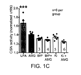
Figures 1A-1C: 1A) Effects of TRPV1 and LPAr antagonists on CSN response
to LPA. No blockade (1), TRPV1 blockade (AMG9810 10pM, 2) or LPAr blockade
(BrP-LPA 1.5pM, 3); Ki16425 5pM, 4). 1B) Summary data. Responses at 2.5pM LPA:
F3,23 (group) =5.031, p=0.01, **. Post-hoc testing-LPA is different from BrP-
LPA
(p=0.026) and Ki16425 (p=0.018). Responses at 5pM: F3, 23 (group) =24.547,
p<0.001,
***. Post-hoc testing- LPA is different from BrP-LPA, Ki16425 and AMG9810
(p<0.001); AMG is different from Brp-LPA (p=0.018) and Ki16425 (p=0.044);
Responses at 10pM: F3, 23 (group) =14.231, p<0.001, '. Post-hoc testing-LPA is
different from BrP-LPA (p<0.001); Ki16425 (p<0.001); AMG9810 (p=0.022); AMG is
different from BrP-LPA (p=0.022). 1C) Summary data of 5pM LPA (1), with
individual
TRPV1 blockade (AMG 9810, 10pM; 2), individual LPAr blockade (BrP-LPA, 1.5pM;
3); Ki16425, 5pM; 4), or combined LPAr and TRPV1 blockade (AMG + BrP red; AMG
+ Ki16425 5), F5, 35 (group) =26.164, p<0.001. Post-hoc testing: AMG + LPA is
significantly different from Ki16425 + AMG + LPA, p=0.005; LPA is
significantly
different from all other groups, p<0.001, '.
Figures 2A-2B: LPA-mediated carotid sinus nerve excitation. 2A) The
application of BrP-LPA (1.5 pM) or 2B) Ki16425 (5 pM) diminishes the response
to
LPA (5 pM); the remaining response is almost abolished by subsequent
application of
AMG9810 (10 pM); Dual block portion is denoted in each of Figure 2A and 2B.
Figures 3A-B: Plasma from ovalbumin-sensitized rats increases carotid body
activity in LPA receptor dependent manner. 3A) Carotid sinus nerve activity
from a
naïve en bloc carotid body preparation in response to plasma from naïve (1)
and
OVA-sensitized (2) rats. Application of dual LPAr and TRPV1 blockade (AMG9810,
10pM + BrP-LPA, 1.5pM) is indicated by the arrow and subsequent trace, 3. 3B)
Summary data of the effect of naïve (1) and OVA (2) plasma as well as
subsequent
CA 03106692 2021-01-18
WO 2019/014748
PCT/CA2018/000145
dual blockade (3). F2, 17 (group)=40.193, p<0.001, ***. Post hoc testing: OVA
vs
naïve p<0.001; OVA vs blockade p<0.001.
Figures 4A-4B:Bradykinin-induced bronchoconstriction in ovalbumin
sensitized rats is dependent on the carotid body and LPA signalling. 4A)
Illustration of OVA-sensitization protocol (see Examples, OVA Cohort 2) to
test lung-
carotid body-lung pathway. 4B) OVA-sensitized and naïve rats were exposed to
nebulized saline (baseline) and three consecutive nebulizations of 0.4mg
bradykinin at
1 (solid), 10 (hatched) and 20 (crossed) min while measuring RL and EL (data
not
shown). Bradykinin had group specific effects: See Examples OVA Cohort 2;
F14,143
(time x group) =4.035, p<0.001. Post hoc testing; bradykinin caused a marked
increase in RL in OVA-sensitized (1; p<0.01, **) but not naïve rats (8; p>0.3)
rats;
carotid body (CB) denervated (2), vagi (VaG) denervated (3), TRPV1 blockade
(AMG9810, 5), LPAr blockade (BrP-LPA, 6), TRPV1 blockade (Ki16425, 7), and
dual
TRPV1 and LPAr blockade (AMG9810 + BrP-LPA, 4), abolished the effects of
bradykinin compared to OVA (p<0.01, **; p<0.001,
Figures 5A-F. Dual LPAr and TRPV1 blockade abates acute asthmatic
respiratory distress in conscious rats. 5A) Illustration of OVA-sensitization
protocol
to demonstrate respiratory distress in asthmatic model (see Examples OVA
Cohort 6).
5B) Inspiratory:expiratory time decreased (Ti:Te; F35,431(time x group)=8.577,
p<0.001,
post hoc test: p<0.05, *) and 5C) expiratory time increased (Te; F35,431 (time
x
group)=3.948, p<0.001, post hoc test: p<0.05, * difference between groups at
indicated time points) in response to acute OVA provocation following OVA
sensitization, confirming these parameters as indices of acute asthmatic
respiratory
distress in conscious animals. 50) 21-day sensitization and testing protocol
to test the
effects of dual LPAr and TRPV1 blockade on respiratory distress (see Examples
OVA
Cohort 7). 5E) Decrease in Ti:Te and 5F) increase in Te caused by allergen
provocation are rescued by dual blockade (squares). Ti:Te: F70, 1293 (group x
time)
=3.169, p<0.001; Te: F35,385 (time)=10.590, p<0.001, F2,35(group) =7.393,
p=0.004).
Post-hoc testing-dual block is significantly different from OVA-sensitized
saline
injected (circles)* and vehicle injected (triangles)" groups, at indicated
time points,
p<0.05. 5G) The peak Ti:Te responses recorded 120 min after OVA exposure on
day
21 in animals never having received dual blockade (1, from 5B), having dual
blockade
on day 21 and recorded on day 21 (Acute treatment 3, from 5E), or having dual
CA 03106692 2021-01-18
WO 2019/014748
PCT/CA2018/000145
11
blockade on day 18 and recorded on day 21 (Long term treatment, 3, from 5E
F2,15
(group)=45.805, p<0.001). 5H) The peak Te (120min) response recorded on day 21
following OVA exposure; groups 1, 2 and 3 as per 5G. Dual antagonist injection
on
days 18 or 21 reduced respiratory indices of acute bronchoconstriction; and
remarkably, dual antagonist injection on day 18 also had beneficial effects
three days
later, on day 21, without a subsequent dual antagonist injection
(F2,15(group)=25.906,
p<0.001). Significant difference between indicated groups determined by post
hoc
testing p<0.001,
DETAILED DESCRIPTION
This disclosure is based at least in part on the demonstration by the
inventors
hereof that a systemic increase in lysophosphatidic acid (LPA) released by the
lung
during asthmatic provocation induces pronounced vagal-mediated
bronchoconstriction
through stimulation of the carotid bodies (main peripheral autonomic
chemoreceptors).
More specifically, carotid body activation during airborne allergic
provocation is
demonstrated to be caused by systemic release of LPA from the lung. This
carotid
body activation is sufficient to cause acute bronchoconstriction. This
disclosure
confirms the systemic upregulation of lysophosphatidic acid in response to
allergen
provocation and the importance of carotid bodies to asthmatic attacks. It is
demonstrated that LPA activates the carotid body, via transient receptor
potential 1
(TRPV1) and LPA-specific receptors, which in turn activates vagal efferents.
Moreover, the carotid body-mediated pathway is found to form a significant
component of acute asthmatic bronchoconstriction. This mechanism has medical
importance. Blocking both TRPV1 and LPA-specific receptors suppresses
asthmatic
airway bronchoconstriction in response to immune challenge with ovalbumin.
This
disclosure provides new evidence linking inflammatory mediators to a novel
reflex
pathway inducing bronchoconstriction. In an embodiment, the disclosure
provides at
least a new form of emergency therapy to mitigate asthmatic attacks.
The inventors have also demonstrated that lysophosphatidic acid stimulates the
carotid body (CB): linking inflammation and carotid sinus nerve activity.
Systemic levels
of the inflammatory mediator lysophosphatidic acid (LPA) increase with
inflammatory
lung disease73, LPA is a TRPV1 agonist71 and TRPV1 is expressed in CB41. The
inventors evaluated the effects of LPA on carotid sinus nerve and phrenic
nerve
discharge in novel en bloc carotid body perfused and in situ decerebrate,
vagotomized,
CA 03106692 2021-01-18
WO 2019/014748
PCT/CA2018/000145
12
rodent dual-perfused preparations, respectively (see Examples). In carotid
sinus nerve
intact and denervated in situ preparations, carotid bodies and the brainstem
were
perfused separately. The CB were perfused with normoxia/normocapnia and the
brainstem was perfused with hypocapnia to induce apnea and thus increase
phrenic
burst frequency sensitivity to carotid body activation. LPA (5pM) was injected
into the
carotid body perfusate. To determine whether non-TRPV1 LPA receptors might
also be
involved, the effects of LPA were tested in the presence of the TRPV1
antagonist
AMG9810 using the en bloc preparation.
LPA delivered directly to the CB circulation in in situ preparations
significantly
(p<0.05) increased phrenic nerve burst rate (from 1.2 0.8 burst-min-1 to 20.8
2.9
burst-m1n-1) and amplitude (from 1 0.5 to 2.5 0.5 normalized units). In CB
denervated
preparations, LPA had no effect. Blocking TRPV1 receptors reduced but did not
eliminate the effects of LPA on carotid sinus nerve activity in the en bloc
preparation.
RT-PCR revealed expression of LPAr 1, 3, and 4 in the carotid body, LPAr 3 in
the petrosal ganglia and LPArr 1, and 3 in the superior cervical ganglia. The
data
indicated that the CB are sensitive to LPA via TRPV1 and LPAr receptors and
involved
in the neural components of inflammatory pulmonary diseases.
TRPV1 receptor is a member of a family of TRP receptors and is broadly
expressed in various tissues which contact the environment (e.g., skin, gut,
airways).
TRPV1 is reported to be activated by various agents including capsaicin among
which
chemical irritants, inflammatory mediators and tissue damaging stimuli can be
identified. TRPV1 is also reported to be activated by high temperature (>43
C), acidic
pH (<5.3), intracellular redox states and electrostatic charges . Modulators
(particularly antagonists) of TRPV1, and particularly those modulators and
antagonists
that are selective for TRPV1 over other TRP receptor family members, have, for
example, been investigated as therapeutic targets for treatment of pain and
inflammation. TRPV1 antagonists are reported to be effective in models of
inflammatory, osteoarthritis and neuropathic pain. Kort and Kym provide a
review of
TRPV1 antagonists as clinical targetssl. This reference provides description
of a
number of TRPV1 antagonists with in some cases descriptions of the results of
clinical
trials. The authors note that the clinical application of first generation
TRPV1 clinical
leads has been made problematic by the undesired side effect of increased core
body
temperature associated with administration of TRPV1 antagonists. The authors,
CA 03106692 2021-01-18
WO 2019/014748
PCT/CA2018/000145
13
however, also report that more recent identification of TRPV1 antagonists that
do not
exhibit the undesired core body increase and as such are called temperature-
neutral.
Examples of such temperature neutral TRPV1 antagonists are provided. The
authors
note TRPV1 antagonists which differentially block TPRV1 activation by
different
stimuli, e.g., block capsaicin activation but not heat or proton activation.
TRPV1
antagonists which exhibit differentiated blockage (stronger) of capsaicin-
induced
calcium efflux, compared to blockage (weaker) of acid-induced TRPV1 are
associated
with temperature-neutrality.
For certain clinical applications as described herein which involve short-term
administration (hours, or days) of a TRPV1 antagonist to treat symptoms of
asthma
attacks, it is presently believed that undesired effects on core body
temperature are
not a significant detriment to use of the antagonist. However, in certain
embodiments
herein, the TRPV1 antagonists employed for treatment of asthma, acute asthma
and
severe acute asthma are temperature-neutral TRPV1 antagonists as described in
Kart
and Kym81 and references cited therein, which exhibit little or no effect on
core body
temperature and are thus temperature-neutral. This reference is incorporated
by
reference herein in its entirety for names and structures of useful TRPV1
antagonists
for applications as described herein. An exemplary temperature neutral TRPV1
antagonist is AS192837982. Another exemplary temperature neutral
TRPV1antagonist
is A116544283.
Wong and Gavva84 is another review of therapeutic potential of TRPV1
agonists and antagonists which appears to focus on analgesic applications.
This
reference is incorporated by reference herein in its entirety for descriptions
of TRPV1
antagonists which may be useful in applications herein.
LPA is a phospholipid signalling molecule that is reported to activate at
least
five known G protein-coupled receptors (GPCRs), designated LPA1-LPA588. A
sixth
receptor designated LPA6 has been described88. LPA is associated with a
variety of
developmental, physiological, and pathophysiological effects. LPAr have been
identified as targets for the treatment of important diseases including
neuropsychiatric
disorders, neuropathic pain, infertility, cardiovascular disease,
inflammation, fibrosis,
and cancer. LPA1 shares significant amino acid sequences identity with LPA2
and
LPA3. LPA4, LPA5 and LPA6 are more diverse in sequence to the other receptors
in
the family and to each other. Most known LPA antagonists inhibit LPA1, LPA2
and
CA 03106692 2021-01-18
WO 2019/014748
PCT/CA2018/000145
14
LPA3. LPA is reported to be a potent mediator of immune response. A number of
LPA antagonists are reported in Yung et al.86 where in Table 2, therein, a
summary of
LPA receptor modulators, receptor subtype target and activity and disease
relevance
are listed. This reference is incorporated by reference herein in its entirety
for
descriptions of LPAr antagonists and their selectivity and activity.
This disclosure relates to compounds, compositions and methods for treating
asthma, particularly acute asthma and severe acute asthma and the prevention
of
acute and acute severe asthma attacks. The methods employ one or more TRPV1
antagonist or one or more LPAr antagonist and preferably employ a combination
of
one or more TRPV1 antagonist and one or more LPAr antagonists. Preferred LPAr
antagonists are those that are antagonists of LPAr1, LPAr2, LPAr3, LPAr4
and/or
LPAr6. Further preferred LPAr antagonists are those that are antagonists of
LPAr1,
LPAr2, LPAr3 and/or LPAr6. Further preferred LPAr antagonists are those that
are
antagonists of at least LPAr1. Further preferred LPAr antagonists are those
that are
antagonists of at least LPAr3. Further preferred LPAr antagonists are those
that are
antagonists of at least LPAr4. Further preferred LPAr antagonists are those
that are
antagonists of at least LPAr6. Further preferred LPAr antagonists are those
that are
antagonists of at least LPAr2. Further preferred LPAr antagonists are those
that are
antagonists of at least LPAr1, LPAr3, and LPAr4. Further preferred LPAr
antagonists
are those that are antagonists of LPAr other than selective antagonists of
LPAr2.
Selective LPAr antagonists are useful in the methods herein. In specific
embodiments, preferred selective LPAr antagonists are those that are selective
antagonists of LPAr1. In specific embodiments, preferred selective LPAr
antagonists
are those that are selective antagonists of LPAr2. In specific embodiments,
preferred
selective LPAr antagonists are those that are selective antagonists of LPAr3.
In
specific embodiments, preferred selective LPAr antagonists are those that are
selective antagonists of LPAr4. In specific embodiments, preferred selective
LPAr
antagonists are those that are selective antagonists of LPAr6. In specific
embodiments, selective LPAr antagonists are those that are selective
antagonists of
LPAr1, LPAr3 and LPAr4. In specific embodiments, selective LPAr antagonists
are
those that are selective antagonists of LPAr1, LPAr2, LPAr3 and LPAr4. In
specific
embodiments, selective LPAr antagonists are those that are selective
antagonists of
LPAr5 and/or LPAr6.
CA 03106692 2021-01-18
WO 2019/014748
PCT/CA2018/000145
TRPV1 antagonists are known in the art and are available from commercial
sources or can be prepared from known readily available starting materials and
reagents using known methods or routine adaptations of known methods. LPAr
antagonists are known in the art and are available from commercial sources or
can be
prepared from known readily available starting materials and reagents using
known
methods or routine adaptations of known methods. This disclosure provides the
names of a variety of useful antagonists. The chemical structures of named
antagonists are known in the art and in many cases provided in references
cited
herein. Methods are also known in the art for identifying new TRPV1
antagonists and
new LPAr antagonists. It will be appreciated that such newly identified
antagonists
can be employed in the methods described herein.
The terms "TRPV1 antagonist" and "LPAr antagonist" are used generally as
these terms are used in the art. Preferred for use in methods and compositions
herein
are those antagonists that are pharmaceutically acceptable. Antagonists
include
pharmaceutically acceptable salts of TRPV1 antagonists and LPAr antagonists.
Also
preferred for use in methods and compositions herein are antagonists that are
small
molecules of molecular weight less than 900 daltons and more specifically of
500
daltons or less. Small molecule antagonists are generally organic compounds
which
may be isolated from natural sources or which are synthetic non-naturally-
occurring
organic molecules.
Exemplary useful TRPV1 antagonists are provided in Tables 1 and 2.
Exemplary useful LPAr antagonists are provided in Tables 3 and 4. Any one or
more
of the antagonists identified in the Tables 1-4 can be used in the
compositions and
methods herein.
Selective LPAr antagonists are known in the art. For example, compound 15 of
the reference Beck et al.87 is reported to be an LAPR2 selective antagonist.
Compound 12 of reference Fells et al. 88 is reported to be an LAPR3 selective
antagonist. Selective LPAr antagonists can be used in the compositions and
methods
herein.
Additional exemplary TRPV1 antagonists are reported in US patents:
9,440,978; 9,422,293; 9,029,378; 8,901,155; 8,815,930; 8,748,610; 8,765,815;
8,691,855; 8,637,527; 8,557,872; 8,383,839; 8,350,083; 8,343,971; 8,338,603;
8,232,309; 8,211,927; 8,030,504; 7,960,584; 7,919,624; 7,910,751; 7,858,621;
CA 03106692 2021-01-18
WO 2019/014748
PCT/CA2018/000145
16
7,767,705; 7,632,519; and 7,482,469. Each of these patents is incorporated by
reference herein in its entirety for descriptions of TRPV1 antagonists and
methods of
preparing such antagonists.
Additional exemplary LPAr antagonists are reported in US patents: 9,624,182;
9,556,133; 9,527,850; 9,346,762; 9,272,940; 9,090,573; 9,067,938; 9,018,383;
8,859,775; 8,785,442; 8,778,983; 8,686,177; 8,664,220; 8,592,402; 8,541,587;
8,455,499; 8,440,707; 8,362,073; 8,283,339; 8,124,645; 7,947,665; 7,820,703;
and
7,217,704. Each of these patents in incorporated by reference herein in its
entirety for
descriptions of LPAr antagonists and methods of preparing such antagonists.
One or
more of these patents has description with respect to selective LPAr
antagonists.
TABLE 1: Exemplary Trpv1 Antagonists#
2-APB (2-Anninoethoxydiphenyl borate)
5'-IRTX
6-iodo-nordihydrocapsaicin
AA-5-HT
A1165442
A425619
A778317
AMG517
AMG628
AMG8562
AMG9810
AMG21629
AS1928370
BCTC
Capsazepine
JNJ17203212
JYL1421
L-R4W2
CA 03106692 2021-01-18
WO 2019/014748
PCT/CA2018/000145
17
NADA
a-spinasterol
SB366791
SB452533
SB705498
# Chemical names/structures of most of the TPRV1 antagonist in this list can
be found in
reference 78. The structures of several of the compounds are found in specific
references
included in Table 2.
TABLE 2: Preferred TRPV1 antagonists:
A 784168: N-5-lsoquinolinyl-W-[[(4-(trifluoromethyl)phenyl]methyl]urea (A
425619)
3,6-Dihydro-3'-(trifluoromethyl)-N44-
[(trifluoromethyl)sulfonyl]pheny1H1(2H),2'-
bipyridine]-4-carboxamide:
Bianchi et al (2007) [3H]-A-778317 [1-((R-5-tert-butyl-indan-1-yI)-3-
isoquinolin-5-yl-urea]: a novel, stereoselective, high-affinity antagonist is
a
useful radioligand for the human transient receptor potential vanilloid-1
(TRPV1) receptor. J.Pharmacol.Exp.Ther. 323 285. PM1D: 17660385.
Cui et al (2006) TRPV1 receptors in the CNS play a key role in broad-
spectrum analgesia of TRPV1 antagonists. J.Neurosci. 269385. PMID:
16971522.
AMG21629: 3-Amino-5-[[2-[(2-methoxyethyl)amino]-6-[4-(trifluoromethyl)pheny1]-
4-
pyrimidinyl]oxy]-2(1H)-quinoxalinone:
Tamayo et al (2008) Design and synthesis of peripherally restricted transient
receptor potential vanilloid 1 (TRPV1) antagonists. J.Med.Chem. 51 2744.
PMID: 18386885.
Gavva et al (2007) The vanilloid receptor TRPV1 is tonically activated in vivo
and involved in body temperature regulation. J.Neurosci. 27 3366. PMID:
17392452.
CA 03106692 2021-01-18
WO 2019/014748
PCT/CA2018/000145
18
AMG517: N-E4-[[644-(Trifluoromethyl)phenyl]-4-pyrimidinyl]oxy]-2-
benzothiazolyliacetamide:
Doherty et al (2007) Novel vanilloid receptor-1 antagonists: 2. Structure-
activity relationships of 4-oxopyrimidines leading to the selection of a
clinical
candidate. J.Med.Chem. 50 3515. PMID: 17585750.
Gavva et al (2007) Repeated administration of vanilloid receptor TRPV1
antagonists attenuates hyperthermia elicited by TRPV1 blockade.
J.Pharmacol.Exp.Ther. 323 128. PMID: 17652633.
Wang et al (2007) Novel vanilloid receptor-1 antagonists: 3. The
identification
of a second-generation clinical candidate with improved physicochemical and
pharmacokinetic properties. J.Med.Chem. 503528. PMID: 17585751.
AMG8562: (R,E)-N-(2-hydroxy-2,3-dihydro-1H-inden-4-y1)-3-(2-(piperidin-1-y1)-4-
(trifluoromethyl)pheny1)-acrylamide
Lehto SG et al. (2008) J Pharmacol Exp Ther 326:218-229
AMG9810: (2E)-N-(2,3-Dihydro-1,4-benzodioxin-6-y1)-344-(1,1-
dimethylethyl)pheny1]-2-propenamide:
Doherty et al (2005) Discovery of potent, orally available vanilloid receptor-
1
antagonists. Structure-activity relationship of N-aryl cinnamides.
J.Med.Chem. 48 71. PMID: 15634002.
Gavva et al (2005) AMG 9810 RE)-3-(4-t-Butylpheny1)-N-(2,3-
dihydrobenzo[b][1,4]dioxin-611)acrylamide], a novel vanilloid receptor 1
(TRPV1) antagonist with antihyperalgesic properties. J.Pharmacol.Exp.Ther.
313474. PMID: 15615864.
AA-5-HT: N-[2-(5-Hydroxy-1H-indo1-3-yDethyl]-5,8,11,14-eicosatetraenamide
(Alternative name: Arachidonyl serotonin):
Maione et al (2007) Analgesic actions of N-arachidonoyl-serotonin, a fatty
acid amide hydrolase inhibitor with antagonistic activity at vanilloid TRPV1
receptors. Br.J.Pharmacol. 150 766. PMID: 17279090.
CA 03106692 2021-01-18
WO 2019/014748
PCT/CA2018/000145
19
Di Marzo et al (2004) The endocannabinoid system and its therapeutic
exploitation. Nat.Rev.Drug Discov. 3771. PMID: 15340387.
Bisogno et al (1998) Arachidonoylserotonin and other novel inhibitors of fatty
acid amide hydrolase. Biochem.Biophys.Res.Comms. 248 515.
AS1928370: (R)-N-(1-methy1-2-oxo-1,2,3,4-tetrahydro-7-quinoly1)-2-[(2-
methylpyrrolidin-1-y1)methyl]biphenyl-4-carboxamide
Watabiki T, Kiso T, Tsukamoto M, Aoki T, Matsuoka N. Biol Pharm Bull.
2011;34(7):1105-8.
Watabiki T, Kiso T, Kuramochi T, Yonezawa K, Tsuji N, Kohara A, Kakimoto
S, Aoki T, Matsuoka N. J Pharmacol Exp Ther. 2011 Mar;336(3):743-50. doi:
10.1124/jpet.110.175570.
A1165442: (R)-1-(7-chloro-2,2-bis(fluoromethyl)chroman-4-y1)-3-(3-
methylisoquinolin-5-yl)urea (A-1165442)
Regina M. Reilly et al. (2012) J. Pharmacology and Experimental
Therapeutics. 342 (2) 416-428.
BCTC: 4-(3-Chloro-2-pyridiny1)-N44-(1,1-dimethylethyl)pheny1]-1-
piperazinecarboxamide:
Valenzano, K.J.,Grant, E.R.,Wu, G., et al. N-(4-tertiarybutylpheny1)-4-(3-
chloropyridin-2-yl)tetrahydropyrazine-1 (2H)-carbox-amide (BCTC), a novel
orally effective vanilloid receptor 1 antagonist with analgesic properties: I.
In
vitro characterization and pharmacokinetic properties. Journal of
Pharmacology and Experimental Therapeutics 306(1), 377-386 (2003).
Gavva, N.R.,Tamir, R.,Klionsky, L., et al. Proton activation does not alter
antagonist interaction with the capsaicin-binding pocket of TRPV1. Molecular
Pharmacology 68(6), 1524-1533 (2005).
5'-IRTX: 6,7-Deepoxy-6,7-didehydro-5-deoxy-21-depheny1-21-(phenylmethyl)-
daphnetoxin,20-(4-hydroxy-5-iodo-3-methoxybenzeneacetate) (Alternative
Names:5'-lodoresiniferatoxin, lodoresiniferatoxin):
CA 03106692 2021-01-18
WO 2019/014748
PCT/CA2018/000145
Marinelli et al (2002) Capsaicin activation of glutamatergic synaptic
transmission in the rat locus coeruleus in vitro. J.Physiol. 543 531. PMID:
12205187.
Seabrook et at (2001) Properties of iodo-resiniferatoxin - a high affinity VR1
vanilloid receptor antagonist. Soc.Neurosci.Abstr. 925.9.
Wahl et at (2001) lodo-resiniferatoxin, a new potent vanilloid receptor
antagonist. Mol.Pharmacol. 599. PMID: 11125018.
JNJ 172032124: [3-(Trifluoromethyl)-2-pyridiny1]-N45-(trifluoromethyl)-2-
pyridinyl]-1-
piperazinecarboxamide:
Bhattacharya et at (2007) Pharmacology and antitussive efficacy of 4-(3-
trifluoromethyl-pyridin-2-y1)-piperazine-1-carboxylic acid (5-trifluoromethyl-
pyridin-2-y1)-amide (JNJ17203212), a transient receptor potential vanilloid 1
antagonist in guinea pigs. J.Pharmacol.Exp.Ther. 323 665. PMID: 17690251.
Ghilardi et at (2005) Selective blockade of the capsaicin receptor TRPV1
attenuates bone cancer pain. J.Neurosci. 253126. PMID: 15788769.
Swanson et at (2005) Identification and biological evaluation of 4-(3-
trifluoromethylpyridin-2-yl)piperazine-1-carboxylic acid (5-
trifluoromethylpyridin-2-yl)amide, a high affinity TRPV1 (VR1) vanilloid
receptor antagonist. J.Med.Chem. 48 1857. PMID: 15771431.
L-R4W2(Arginine-rich hexapeptide):
Himmel et at (2002) The arginine-rich hexapeptide R4W2 is a stereoselective
antagonist at the vanilloid receptor 1: a Ca2+ imaging study in adult rat
dorsal
root ganglion neurons. J.Pharmacol.Exp.Ther. 301 981. PMID: 12023528.
Planells-Cases et at (2000) Arginine-rich peptides are blockers of VR-1
channels with analgesic activity. FEBS Lett. 481 131. PMID: 10996311.
SB366791: 4'-Chloro-3-methoxycinnamanilide:
Gavva et al (2005) Proton activation does not alter antagonist interaction
with
the capsaicin-binding pocket of TRPV1. Mol.Pharmacol. 68 1524. PMID:
16135784.
CA 03106692 2021-01-18
WO 2019/014748
PCT/CA2018/000145
21
Gunthorpe et al (2004) Identification and characterisation of SB-366791, a
potent and selective vanilloid receptor (VR1/TRPV1) antagonist.
Neuropharmacology 46 133. PMID: 14654105.
Fowler et al (2003) Inhibition of C6 glioma cell proliferation by anandamide,
1-arachidonylglycerol, and by a water soluble phosphate ester of
anandamide: variability in response and involvement of arachidonic acid.
Biochem.Pharmacol. 66 757. PMID: 12948856.
SB452533: N-(2-BromophenyI)-N'-[2-[ethyl(3-methylphenyl)amino]ethyl]-urea:
Bianchi et al (2007) [31-1]A-778317 [1-((R)-5-tert-Butyl-indan-l-yI)-3-
isoquinolin-5-ylurea]: a novel, stereoselective, high-affinity antagonist is a
useful radioligand for the human transient receptor potential vanilloid-1
(TRPV1) receptor. J.Pharmacol.Exp.Ther. 323 285. PMID: 17660385.
Weil et al (2005) Conservation of functional and pharmacological properties
in the distantly related temperature sensors TRPV1 and TRPM8.
Mol.Pharmacol. 68518. PMID: 15911692.
Rami et al (2004) Discovery of small molecule antagonists of TRPV1.
Bioorg.Med.Chemlett. 14 3631. PMID: 15203132.
a-Spinasterol:(3/3,5a,22E)-Stigmasta-7.22-dien-3-ol
Trevisan et al (2012) Identification of the plant steroid a-spinasterol as a
novel transient receptor potential vanilloid 1 antagonist with antinociceptive
properties. J.Pharmacol.Exp.Ther. 343 258. PMID: 22837009.
CA 03106692 2021-01-18
WO 2019/014748
PCT/CA2018/000145
22
TABLE 3: Exemplary LPAr Antagonists#
AM095
Am966
BMS986020
Ki16425- 3-(444-([1-(2-chlorophenypethoxy]carbonyl
amino)-3-methyl-5-isoxazolyl] benzylsulfanyl)
propanoic acid
ONO-3080573
ONO-7300243
ONO-9780307
ONO-9910539
VPC 12249
VPC 32179
VPV 32183
BrP-LPA- R3S)-1-bromo-4-hexadecanoyloxy-3-
hydroxybutyllphosphonic acid
Syn BrP-LPA
Anti BrR-LPA
Dioctanoylglycerolpyrophosphate
Farnesyl monophosphate
Farnesyl di phosphate
Dodecyl thiophosphate
1-bromo-(3S)-hydroxy-4-(palmitoyl) butyl phosphate
#Chemical names/structures of most of the LPAr antagonist in this table can be
found in
reference 89. Reference 89 also provides lists of possibly selective LPAr
antagonists.
TABLE 4: Additional LPAr Antagonists
CA 03106692 2021-01-18
WO 2019/014748
PCT/CA2018/000145
23
H2L 5765834: 2,3-Dihydro-2-[3-(4-nitrophenoxy)pheny1]-1,3-dioxo-1H-isoindole-5-
carboxylic
acid:
Fells et al (2010) 2D binary QSAR modeling of LPA3 receptor antagonism.
J.Mol.Graph
Model. 28 828. PMID: 20356772.
Tigyi (2010) Aiming drug discovery at lysophosphatidic acid targets.
Br.J.Pharmacol.
161 241. PMID: 20735414.
Williams et at (2009) Unique ligand selectivity of the GPR92/LPA5
lysophosphatidate
receptor indicates role in human platelet activation. J.Biol.Chem. 284 17304.
PMID:
19366702.
H2L5186303: (Z,Z)-4,4'41,3-Phenylenebis(oxy-4,1-phenyleneimino)]bis[4-oxo-2-
butenoic acid:
Fells et al (2009) Structure-based drug design identifies novel LPA3
antagonists.
Bioorg.Med.Chenn. 17 7457. PMID: 19800804.
Fells et at (2008) Identification of non-lipid LPA3 antagonists by virtual
screening.
Bioorg.Med.Chem. 16 6207. PMID: 18467108.
Ro 6842262: 1-[4'-[4-Methy1-5-[[[(1R)-1-phenylethoxy]carbonylamino]-1H-1,2,3-
triazol-1-yl][1,1'-
bipheny1]-4-yl]cyclopropanecarboxylic acid:
Qian et al (2012) Discovery of highly selective and orally active
lysophosphatidic acid
receptor-1 antagonists with potent activity on human lung fibroblasts.
J.Med.Chem. 55
7920. PMID: 22894757.
Spiroxatrine: 8-[(2,3-Dihydro-1,4-benzodioxin-2-yOmethyl]-1-pheny1-1,3,8-
triazaspiro[4,5]decan-4-one:
Bylund et al (1992) Pharmacological characteristics of a2-adrenergic
receptors:
comparison of pharmacologically defined subtypes with subtypes identified by
molecular
cloning. Mol.Pharmacol. 42 1. PMID: 1353247.
Schoeffter and Hoyer (1988) Centrally acting hypotensive agents with affinity
for 5-HT1A
binding sites inhibit forskolin-stimulated adenylate cyclase activity in calf
hippocampus.
Br.J.Pharmacol. 95 975. PMID: 3207999.
Nelson and Taylor (1986) Spiroxatrine: a selective serotonin 1A receptor
antagonist.
Eur.J.Pharmacol. 124 207. PMID: 3720840.
TC LPA5 4: 5-(3-Chloro-4-cyclohexylphenyI)-1-(3-methoxypheny1)-1H-pyrazole-3-
carboxylic
acid
CA 03106692 2021-01-18
WO 2019/014748
PCT/CA2018/000145
24
Kozian et al (2012) Selective non-lipid modulator of LPA5 activity in human
platelets.
Biorg.Med.Chem.Lett. 22 5239. PMID: 22801643.
In specific embodiments, the disclosure provides methods for treatment of
acute asthmatic attack or for prevention of an acute attack that is predicted.
In specific embodiments, treatment involves administration of one or more
antagonist targeting the family of carotid body receptors activated by LPA
(e.g.
AMG9810 and/or BrP-LPA), and optionally one or more antagonist targeting
carotid
body receptors activated by LPA and/or other substances released by the lung
during
an asthmatic attack (e.g. bradykinin) and/or optionally one or more reagent
that
inhibits LPA stimulation of carotid body through LPA-independent mechanisms
(e.g.,
somatostatin or other natural substances that inhibit the carotid body;
blockers of
endogenous channels or receptors causing inward currents; activators of
endogenous
channels or receptors causing outward current; activation of artificial
channels or
receptors causing outward currents).
In specific embodiments, the route of administration may be by inhalation,
injection (syringe, autoinjector), adsorption through skin/mucus membrane,
suppository, patch or ingestion. In specific embodiments, the route of
administration is
other than by inhalation.
Compounds of the disclosure may contain chemical groups (acidic or basic
groups) that can be in the form of salts. Pharmaceutically acceptable salts of
antagonists of this disclosure can be employed in the compositions and methods
herein. Exemplary acid addition salts include acetates (such as those formed
with
acetic acid or trihaloacetic acid, for example, trifluoroacetic acid),
adipates, alginates,
ascorbates, aspartates, benzoates, benzenesulfonates, bisulfates, borates,
butyrates,
citrates, camphorates, camphorsulfonates, cyclopentanepropionates,
digluconates,
dodecylsulfates, ethanesulfonates, fumarates, glucoheptanoates,
glycerophosphates,
hemisulfates, heptanoates, hexanoates, hydrochlorides (formed with
hydrochloric
acid), hydrobromides (formed with hydrogen bromide), hydroiodides, 2-
hydroxyethanesulfonates, lactates, maleates (formed with maleic acid),
methanesulfonates (formed with methanesulfonic acid), 2-naphthalenesulfonates,
nicotinates, nitrates, oxalates, pectinates, persulfates, 3-phenylpropionates,
phosphates, picrates, pivalates, propionates, salicylates, succinates,
sulfates (such as
CA 03106692 2021-01-18
WO 2019/014748
PCT/CA2018/000145
those formed with sulfuric acid), sulfonates (such as those mentioned herein),
tartrates, thiocyanates, toluenesulfonates such as tosylates, undecanoates,
and the
like.
Exemplary basic salts include ammonium salts, alkali metal salts such as
sodium, lithium, and potassium salts, alkaline earth metal salts such as
calcium and
magnesium salts, salts with organic bases (e.g., organic amines) such as
benzathines, dicyclohexylamines, hydrabamines [formed with N,N-bis(dehydro-
abietypethylenediamine], N-methyl-D-glucamines, N-methyl-D-glucamides, t-butyl
amines, salts with amino acids such as arginine, lysine and the like and salts
with
amino sugars, such as meglumine. Basic nitrogen-containing groups may be
quaternized with agents such as lower alkyl halides (e.g., methyl, ethyl,
propyl, and
butyl chlorides, bromides and iodides), dialkyl sulfates (e.g., dimethyl,
diethyl, dibutyl,
and diamyl sulfates), long chain halides (e.g., decyl, lauryl, myristyl and
stearyl
chlorides, bromides and iodides), aralkyl halides (e.g., benzyl and phenethyl
bromides), and others. Salt of the disclosure include "pharmaceutically
acceptable
salts" which refers to those salts which retain the biological effectiveness
and
properties of the free bases or free acids, and which are not biologically or
otherwise
undesirable and acceptable for use in pharmaceutical compositions.
Pharmaceutically
acceptable salts comprise pharmaceutically-acceptable anions and/or cations.
Compounds of the present disclosure, and salts thereof, may exist in their
tautomeric form, in which hydrogen atoms are transposed to other parts of the
molecules and the chemical bonds between the atoms of the molecules are
consequently rearranged. It should be understood that all tautomeric forms,
insofar as
they may exist, are included within the disclosure. Additionally, disclosed
compounds
may have trans and cis isomers and may contain one or more chiral centers,
therefore
exist in enantiomeric and diastereomeric forms. The disclosure includes all
such
isomers, as well as mixtures of cis and trans isomers, mixtures of
diastereomers and
racemic mixtures of enantiomers (optical isomers). When no specific mention is
made
of the configuration (cis, trans or R or S) of a compound (or of an asymmetric
carbon),
then any one of the isomers or a mixture of more than one isomer is intended.
The
processes for preparation can use racemates, enantiomers, or diastereomers as
starting materials. When enantiomeric or diastereomeric products are prepared,
they
can be separated by conventional methods, for example, by chromatographic or
CA 03106692 2021-01-18
WO 2019/014748
PCT/CA2018/000145
26
fractional crystallization. The inventive compounds may be in the free or
hydrate form.
The term enantiomerically pure refers to a sample containing molecules of a
given
structure whose molecules have the same chirality sense (i.e., are the same
optical
isomer) within the limits of detection. The term substantially
enantiomerically pure
refers to a sample containing molecules of a given structure, wherein equal to
or less
than 1% of the molecules of the sample have a different chirality sense.
Compounds
of the invention include those which are enatiomerically pure and those that
are
substantially enatiomerically pure.
The disclosure provides pharmaceutical compositions for use in the treatment
methods herein. Pharmaceutical compositions comprise one or more of the active
antagonists as described optionally in combination with a pharmaceutically
acceptable
carrier. Pharmaceutically acceptable carriers are those carriers that are
compatible
with the other ingredients in the formulation and are biologically acceptable.
Carriers
can be solid or liquid. Solid carriers can include one or more substances that
can also
act as flavoring agents, lubricants, solubilizers, suspending agents, fillers,
glidants,
compression aids, binders, tablet-disintegrating agents, or encapsulating
materials.
Liquid carriers can be used in preparing solutions, suspensions, emulsions,
syrups
and elixirs. The active ingredient can be dissolved or suspended in a
pharmaceutically
acceptable liquid carrier such as water (of appropriate purity, e.g., pyrogen-
free,
sterile, etc.), an organic solvent, a mixture of both, or a pharmaceutically
acceptable
oil or fat. The liquid carrier can contain other suitable pharmaceutical
additives such
as, for example, solubilizers, emulsifiers, buffers, preservatives,
sweeteners, flavoring
agents, suspending agents, thickening agents, colors, viscosity regulators,
stabilizers
or osmo-regulators. Compositions for oral administration can be in either
liquid or solid
form.
Suitable solid carriers include, for example, alumina, calcium phosphate, ion
exchange material, aluminum or magnesium stearate, talc, sugars, lactose,
dextrin,
starch, gelatin, cellulose, methyl cellulose, sodium carboxymethyl cellulose,
polyvinylpyrrolidine, low melting waxes and ion exchange resins.
Suitable examples of liquid carriers for oral and parenteral administration
include water of appropriate purity, aqueous solutions (particularly
containing
additives, e.g. cellulose derivatives, sodium carboxymethyl cellulose
solution),
alcohols (including monohydric alcohols and polyhydric alcohols e.g. glycols)
and their
CA 03106692 2021-01-18
WO 2019/014748
PCT/CA2018/000145
27
derivatives, and oils. For parenteral administration, the carrier can also be
an oily
ester such as ethyl oleate and isopropyl myristate. Sterile liquid carriers
are used in
sterile liquid form compositions for parenteral administration. Compositions
for
administration can be solutions or suspensions. The liquid carrier for
pressurized
compositions can be halogenated hydrocarbon or other pharmaceutically
acceptable
propellant. Liquid pharmaceutical compositions that are sterile solutions or
suspensions can be administered parenterally by, for example, intramuscular,
intraperitoneal or subcutaneous injection. Sterile solutions can also be
administered
intravenously. Compositions for oral administration can be in either liquid or
solid form.
Liquid or solid pharmaceutical compositions can contain one or more of
buffering agents (e.g., phosphates and/or hydrogen phosphate salts), salts
(e.g.,
NaCI, zinc salts), electrolytes, osmotic agents (e.g., glycerol), protamine
sulfate,
polymeric materials (such as pyrrolidone, cellulose and cellulose derivatives,
polyethylene glycol, polyacrylates, and polyethylene glycol). Liquid
solutions,
suspensions, gels, creams and the like may include surface active ingredients
(including among others dispersing agents, wetting agents, suspending agents,
etc.)
Solid pharmaceutical compositions may contain among others, colloidal silica,
magnesium trisilicate, glycerides, fatty acids and salts thereof. Carriers for
oral
dosage forms (e.g., tablets and capsules) include among others lactose and
corn
starch.
Methods of this disclosure comprise the step of administering a
"therapeutically
effective amount" of the present therapeutic formulations containing one or
more of
the present active compounds, to treat, reduce or regulate a disease state in
a patient.
A given therapeutic formulation may also prevent the onset of a disease or
disorder,
slow the development of the disease or disorder or ameliorate one or more
symptoms
of the disease or disorder. As is understood in the art, the therapeutically
effective
amount of a given compound or formulation will depend at least in part upon,
the
mode of administration (e.g., intravenous, oral, topical administration), any
carrier or
vehicle employed, and the specific individual to whom the formulation is to be
administered (age, weight, condition, sex, etc.). The dosage requirements need
to
achieve the "therapeutically effective amount" vary with the particular
formulations
employed, the route of administration, and clinical objectives. Based on the
results
CA 03106692 2021-01-18
WO 2019/014748
PCT/CA2018/000145
28
obtained in standard pharmacological test procedures, projected daily dosages
of
active compound can be determined as is understood in the art.
The methods of treatment and prophylaxis herein are useful in treating
animals,
particularly mammals, and more particularly in treating humans. The methods,
compositions, medicaments and kits herein can be applied for human or
veterinary
applications. Human applications are generally to any human susceptible to or
suffering from asthma and particularly those humans at risk of acute asthma
attacks.
Veterinary applications are generally to any animal susceptible to or
suffering from
asthma. In particular, the methods, compositions, medicaments and kits herein
can
be used to treat bovine or equine asthma.
Exemplary daily dosage levels of individual active ingredients or combinations
thereof in a combined formulation range from 0.001 to 100 mg/kg body weight or
more
specifically from 1 to 10 mg/kg body weight.
Any suitable form of administration can be employed in the methods herein.
Administration includes any form of administration that is known in the art
and is
intended to encompass administration in any appropriate dosage form and
further is
intended to encompass administration of a compound, alone or in a
pharmaceutically
acceptable carrier. Pharmaceutical carriers are selected as is known in the
art based
on the chosen route of administration and standard pharmaceutical practice.
The
compounds of this disclosure can, for example, be administered in oral dosage
forms
including tablets, capsules, pills, powders, granules, elixirs, tinctures,
suspensions,
syrups and emulsions. Oral dosage forms may include sustained release or timed
release formulations. The compounds of this disclosure may also be
administered
topically, intravenously, intraperitoneally, subcutaneously, or
intramuscularly, all using
dosage forms well known to those of ordinary skill in the pharmaceutical arts.
Compounds of this disclosure can also be administered in intranasal form by
topical
use of suitable intranasal vehicles. For intranasal or intrabronchial
inhalation or
insulation, the compounds of this disclosure may be formulated into an aqueous
or
partially aqueous solution, which can then be utilized in the form of an
aerosol. In
specific embodiments, a form of administration that is other than inhalation
is
employed.
The compounds of this disclosure may be administered rectally or vaginally in
the form of a conventional suppository. The compounds of this disclosure may
also be
CA 03106692 2021-01-18
WO 2019/014748
PCT/CA2018/000145
29
administered transdermally through the use of a transdermal patch containing
the
active compound and a carrier that is inert to the active compound, is non-
toxic to the
skin, and allows delivery of the agent for systemic absorption into the blood
stream via
the skin.
Pharmaceutical compositions and medicaments of this disclosure comprise one
or more compounds of the disclosure of formula I (or other formulas herein) in
optional
combination with a pharmaceutically acceptable carrier, excipient, or diluent.
Such
compositions and medicaments are prepared in accordance with acceptable
pharmaceutical procedures, such as, for example, those described in Remingtons
Pharmaceutical Sciences, 17th edition, ed. Alfonoso R. Gennaro, Mack
Publishing
Company, Easton, Pa. (1985), which is incorporated herein by reference in its
entirety.
The disclosure also encompasses method for making a medicament employing one
or
more compounds of this disclosure which exhibit a therapeutic effect.
Compounds useful in the methods of this disclosure include pharmaceutically-
acceptable salts of the compounds of formulas herein. Compounds useful in the
methods of this disclosure include pharmaceutically-acceptable prodrugs of the
compounds of formulas herein. Salts include any salts derived from the acids
of the
formulas herein which are acceptable for use in human or veterinary
applications.
In a preferred embodiment, pharmaceutical compositions herein comprise a
combination of a TRPV1 antagonist and an LPAr antagonist. In specific
embodiments, the pharmaceutical composition comprises a TRPV1 antagonist of
Table 2 and an LPAr antagonist of Table 4. In specific embodiments, the molar
ratio
of TRPV1 antagonist to LPAr antagonist in the composition ranges from 50:1 to
1:50.
In specific embodiments, the molar ratio of TRPV1 antagonist to LPAr
antagonist in
the composition ranges from 20:1 to 1:20. In specific embodiments, the molar
ratio of
TRPV1 antagonist to LPAr antagonist in the composition ranges from 10:1 to
1:10. In
specific embodiments, the molar ratio of TRPV1 antagonist to LPAr antagonist
in the
composition ranges from 10:1 to 1:1. In specific embodiments, the molar ratio
of
TRPV1 antagonist to LPAr antagonist in the composition ranges from 10:1 to
2:1. In
specific embodiments, the molar ratio of TRPV1 antagonist to LPAr antagonist
in the
composition ranges from 5:1 to 2:1.
In specific embodiments, the disclosure provides methods of making
medicaments wherein the medicaments are pharmaceutical compositions comprising
CA 03106692 2021-01-18
WO 2019/014748
PCT/CA2018/000145
a combination of a TRPV1 antagonist and an LPAr antagonist. In specific
embodiments, the medicament comprises a TRPV1 antagonist of Table 2 and an
LPAr antagonist of Table 4. In specific embodiments, the molar ratio of TRPV1
antagonist to LPAr antagonist in the medicament ranges from 50:1 to 1:50. In
specific
embodiments, the molar ratio of TRPV1 antagonist to LPAr antagonist in the
medicament ranges from 20:1 to 1:20. In specific embodiments, the molar ratio
of
TRPV1 antagonist to LPAr antagonist in the medicament ranges from 10:1 to
1:10. In
specific embodiments, the molar ratio of TRPV1 antagonist to LPAr antagonist
in the
medicament ranges from 10:1 to 1:1. In specific embodiments, the molar ratio
of
TRPV1 antagonist to LPAr antagonist in the medicament ranges from 10:1 to 2:1.
In
specific embodiments, the molar ratio of TRPV1 antagonist to LPAr antagonist
in the
medicament ranges from 5:1 to 2:1. In an embodiment, the medicament comprises
a
TRPV1 antagonist separately formulated from a LPAr antagonist. In an
embodiment,
the medicament comprises a TRPV1 antagonist formulated together with a LPAr
antagonist. In an embodiment, the medicament comprises a TRPV1 antagonist
formulated together with a LPAr antagonist for administration by injection. In
an
embodiment, the medicament comprises a TRPV1 antagonist formulated together
with a LPAr antagonist for administration by inhalation.
In an embodiment, the medicament is in the form of a kit comprising a TRPV1
antagonist and an LPAr antagonist which are separately formulated. In an
embodiment, the medicament is in the form of a kit comprising a TRPV1
antagonist
and an LPAr antagonist which are separately formulated for administration at
the
same time. In an embodiment, the medicament is in the form of a kit comprising
a
TRPV1 antagonist and an LPAr antagonist which are separately formulated for
administration by injection at the same time. The term at the same time refers
to
administration by any suitable means within 24 hours. Administration at the
same
time preferably refers to administration any suitable means within 12 hours.
Administration at the same time more preferably refers to administration any
suitable
means within 6 hours. Administration at the same time yet more preferably
refers to
administration any suitable means within 2 hours. Administration at the same
time
most preferably refers to administration by any suitable means within 1 hour.
Administration at the same time can refer to administration by any suitable
means
within 30 minutes. In an embodiment, the medicament is in the form of a kit
CA 03106692 2021-01-18
WO 2019/014748
PCT/CA2018/000145
31
comprising a TRPV1 antagonist and an LPAr antagonist which are separately
formulated for administration by injection. In an embodiment, the medicament
is in
the form of a kit comprising a TRPV1 antagonist and an LPAr antagonist which
are
separately formulated for administration by separate injection at the same
time. In
an embodiment, the medicament is in the form of a kit comprising a TRPV1
antagonist
and an LPAr antagonist which are separately formulated for administration by
inhalation and/or injection. In an embodiment, the medicament is in the form
of a kit
comprising a TRPV1 antagonist and an LPAr antagonist which are separately
formulated for administration by inhalation. Certain kits herein comprise two
or more
pharmaceutically active ingredients which are separately packaged for use
together.
Kits further optionally comprise one or more devices for administration of the
one or
more active ingredients. The one or more active ingredients are optionally
packaged
within the one or more devices. Exemplary devices include one or more syringe
or
one or more inhaler device.
In a preferred embodiment, the methods herein comprise administration of a
combination of a TRPV1 antagonist and an LPAr antagonist. In a preferred
embodiment, the methods herein comprise administration of a combination of a
TRPV1 antagonist and an LPAr antagonist at the same time. In a preferred
embodiment, the methods herein comprise administration of a combination of a
TRPV1 antagonist and an LPAr antagonist which are formulated together for
administration at the same time. In an embodiment, formulation is for
administration
by injection. Injection includes, among others, intravenous injection,
intramuscular
injection and intraperitienal injection. In an embodiment, administration is
by
inhalation. In specific embodiments of methods herein, the TRPV1 antagonist
and the
LPAr antagonist are formulated separately for administration at the same time.
In specific embodiments, a pharmaceutical composition is provided which
comprises one or more TRPV1 antagonist, one or more LPAr antagonist or a
combination of one or more TRPV1 antagonist and one or more LPAr antagonist
and
optionally a pharmaceutically acceptable carrier for use in the treatment of
asthma or
more specifically in the treatment of acute asthma, or an asthma attack. In an
embodiment, such pharmaceutical compositions comprise a combination of one or
more TRPV1 antagonist and one or more LPAr antagonist. In an embodiment, such
pharmaceutical compositions comprise a combination of one TRPV1 antagonist and
CA 03106692 2021-01-18
WO 2019/014748
PCT/CA2018/000145
32
one LPAr antagonist. In an embodiment, in such pharmaceutical compositions the
molar ratio of TRPV1 antagonist to LPAr antagonist in the composition ranges
from
50:1 to 1:50. In an embodiment, in such pharmaceutical compositions the molar
ratio
of TRPV1 antagonist to LPAr antagonist in the composition ranges from 10:1 to
1:10.
In an embodiment, such pharmaceutical compositions comprise a pharmaceutically
acceptable carrier. In an embodiment, such pharmaceutical compositions
comprise a
pharmaceutically acceptable carrier suitable for administration by injection.
In an
embodiment, such pharmaceutical compositions comprise a pharmaceutically
acceptable carrier suitable for administration by inhalation.
In specific embodiments, a pharmaceutical composition is provided which
comprises one or more TRPV1 antagonist, one or more LPAr antagonist or a
combination of one or more TRPV1 antagonist and one or more LPAr antagonist
and
optionally a pharmaceutically acceptable carrier for use in the prevention or
treatment
of an asthma attack. In an embodiment, such pharmaceutical compositions
comprise
a combination of one or more TRPV1 antagonist and one or more LPAr antagonist.
In
an embodiment, such pharmaceutical compositions comprise a combination of one
TRPV1 antagonist and one LPAr antagonist. In an embodiment, in such
pharmaceutical compositions the molar ratio of TRPV1 antagonist to LPAr
antagonist
in the composition ranges from 50:1 to 1:50. In an embodiment, in such
pharmaceutical compositions the molar ratio of TRPV1 antagonist to LPAr
antagonist
in the composition ranges from 10:1 to 1:10. In an embodiment, such
pharmaceutical
compositions comprise a pharmaceutically acceptable carrier. In an embodiment,
such pharmaceutical compositions comprise a pharmaceutically acceptable
carrier
suitable for administration by injection. In an embodiment, such
pharmaceutical
compositions comprise a pharmaceutically acceptable carrier suitable for
administration by inhalation.
In a specific embodiment, a kit for treating asthma is provided which
comprises
one or more TRPV1 antagonist and one or more LPAr antagonist separately
packaged for use together. In an embodiment of such a kit, the one or more
TRPV1
antagonist and one or more LPAr antagonist are separately formulated for
administration at the same time. In a specific embodiment, a kit for prevent
an asthma
attack or treating an asthma attack is provided which comprises one or more
TRPV1
antagonist and one or more LPAr antagonist separately packaged for use
together. In
CA 03106692 2021-01-18
WO 2019/014748
PCT/CA2018/000145
33
an embodiment of such a kit, the one or more TRPV1 antagonist and one or more
LPAr antagonist are separately formulated for administration at the same time.
In embodiments, the pharmaceutical compositions, medicaments and kits
herein optionally comprise additional active ingredients that are useful in
addition for
treating asthma or asthma attacks or are useful for treating allergic
reactions.
Optional additional active ingredients include, among others, one or more of
the
following: short-term 13-agonists, long-term 13-agonists, methylxanthines,
anticholinergic agents, antihistamines, corticosteroids, leukotriene
antagonists,
decongestants, or non-steroidal anti-inflammatory agents.
Methods for making a medicament for the treatment of asthma or the
prevention of or treatment of an asthma attack, which comprises combining one
or
more TPRPV1 antagonists and/or one or more LPAr antagonists with a
pharmaceutically acceptable carrier, is provided. In an embodiment of such
methods,
one or more TPRPV1 antagonists and one or more LPAr antagonists are combined
with a pharmaceutically acceptable carrier. In an embodiment of such method,
the
medicament comprises one or more TPRPV1 antagonists and one or more LPAr
which are separately packaged in a kit for use together. In an embodiment of
such
method, the medicament comprises one or more TPRPV1 antagonists and one or
more LPAr which are separately formulated for use together.
The disclosure provides use of a TRPV1 antagonist, an LPAr antagonist or
both for the treatment of asthma or the prevention of asthma attacks. In a
specific
embodiment, use of the TRPV1 antagonist, the LPAr antagonist or both is for
treatment of an asthma attack. In a specific embodiment, the disclosure
provides use
of a combination of a TRPV1 antagonist and an LPAr antagonist for the
treatment of
asthma. In a specific embodiment, the disclosure provides use of a combination
of a
TRPV1 antagonist and an LPAr antagonist for the prevention or treatment of an
asthma attack. In such uses, the TRPV1 antagonist, the LPAr antagonist or both
are
formulated for administration by injection. In such uses, the TRPV1
antagonist, the
LPAr antagonist or both are formulated for administration by inhalation.
All references throughout this application, for example patent documents
including issued or granted patents or equivalents; patent application
publications; and
non-patent literature documents or other source material; are hereby
incorporated by
reference herein in their entireties, as though individually incorporated by
reference.
CA 03106692 2021-01-18
WO 2019/014748
PCT/CA2018/000145
34
All patents and publications mentioned in the specification are indicative of
the
levels of skill of those skilled in the art to which the invention pertains.
References
cited herein are incorporated by reference herein in their entirety to
indicate the state
of the art, in some cases as of their filing date, and it is intended that
this information
can be employed herein, if needed, to exclude (for example, to disclaim)
specific
embodiments that are in the prior art. For example, when a compound is
claimed, it
should be understood that compounds known in the prior art, including certain
compounds disclosed in the references disclosed herein (particularly in
referenced
patent documents), are not intended to be included in the claim.
When a group is disclosed herein, it is understood that all individual members
of the group and all subgroups, including any isomers and enantiomers of the
group
members, and classes of compounds that can be formed using the substituents
are
disclosed separately. When a Markush group or other grouping is used herein,
all
individual members of the group and all combinations and subcombinations
possible
of the group are intended to be individually included in the disclosure.
Every formulation or combination of components described or exemplified can
be used to practice the invention, unless otherwise stated. Specific names of
compounds are intended to be exemplary, as it is known that one of ordinary
skill in
the art can name the same compounds differently. When a compound is described
herein such that a particular isomer or enantiomer of the compound is not
specified,
for example, in a formula or in a chemical name, that description is intended
to include
each isomers and enantiomer of the compound described individual or in any
combination.
One of ordinary skill in the art will appreciate that methods, device
elements,
starting materials, and synthetic methods other than those specifically
exemplified can
be employed in the practice of the invention without resort to undue
experimentation.
All art-known functional equivalents, of any such methods, device elements,
starting
materials, and synthetic methods are intended to be included in this
invention.
Whenever a range is given in the specification, for example, a temperature
range, a
time range, or a composition range, all intermediate ranges and subranges, as
well as
all individual values included in the ranges given are intended to be included
in the
disclosure.
CA 03106692 2021-01-18
WO 2019/014748
PCT/CA2018/000145
As used herein, "comprising" is synonymous with "including," "containing," or
"characterized by," and is inclusive or open-ended and does not exclude
additional,
unrecited elements or method steps. As used herein, "consisting of" excludes
any
element, step, or ingredient not specified in the claim element. As used
herein,
"consisting essentially of" does not exclude materials or steps that do not
materially
affect the basic and novel characteristics of the claim. Any recitation herein
of the term
"comprising", particularly in a description of components of a composition or
in a
description of elements of a device, is understood to encompass those
compositions
and methods consisting essentially of and consisting of the recited components
or
elements. The invention illustratively described herein suitably may be
practiced in the
absence of any element or elements, limitation or limitations which is not
specifically
disclosed herein.
Without wishing to be bound by any particular theory, there can be discussion
herein of beliefs or understandings of underlying principles relating to the
invention. It
is recognized that regardless of the ultimate correctness of any mechanistic
explanation or hypothesis, an embodiment of the invention can nonetheless be
operative and useful.
The terms and expressions which have been employed are used as terms of
description and not of limitation, and there is no intention in the use of
such terms and
expressions of excluding any equivalents of the features shown and described
or
portions thereof, but it is recognized that various modifications are possible
within the
scope of the invention claimed. Thus, it should be understood that although
the
present invention has been specifically disclosed by preferred embodiments and
optional features, modification and variation of the concepts herein disclosed
may be
resorted to by those skilled in the art, and that such modifications and
variations are
considered to be within the scope of this invention.
THE EXAMPLES
A novel lung ¨ carotid body ¨ lung reflex pathway has been demonstrated in
acute allergen/bradykinin-evoked bronchoconstriction and respiratory
disturbances in
an animal model of asthma. The examples demonstrate that blocking LPA
signaling
ameliorates bradykinin-invoked bronchoconstriction in anesthetized asthmatic
animals. Further, in conscious animals, where the severity of respiratory
disturbances
CA 03106692 2021-01-18
WO 2019/014748
PCT/CA2018/000145
36
are fully exhibited without the mitigating effects of anesthesia, the
treatment remains
effective. The results presented in these examples provide a mechanistic
explanation
for the importance of the carotid body in acute asthmatic symptoms and
demonstrates
therapeutic drug targets for emergency intervention.
U. S. provisional application 62/534,638, filed July 19, 2017 is incorporated
by
reference herein in its entirety. More specifically, figures and examples
therein are
incorporated by reference herein.
EXAMPLE 1: LPA activates TRPV1 and LPA-specific receptors in the carotid body
LPA activates TRPV1 receptors71,72. TRPV1 receptors are expressed in the
terminals of petrosal neurons that innervate the carotid body (i.e., the axon
terminals
of chemosensory afferent"). LPA also binds to 6 LPA-specific G-protein coupled
receptors (GPCR), LPAr 1 through 6. RT-PCR was used to test for the presence
of
these receptors in the carotid body. cDNA for LPAr 1, 3, 4, and 6 were found
in the
carotid body. cDNA for LPAr 3 and 6 were found in petrosal ganglia. cDNA for
LPAr
1, 3, 4 and 6 were found in the superior cervical ganglia. No evidence was
found for
expression of LPAr 2 in these carotid body associated tissues (data not
shown).
The functional effects of LPA (18:1 unless otherwise stated) on glomus cells
were assessed using Fura 2 calcium imaging. LPA (5pM) increased intracellular
calcium release producing calcium spikes of similar order of magnitude to that
produced by 20mM potassium (data not shown). LPA species present in blood
(LPA(16:0), LPA(18:1) and LPA(18:2)) were shown to have functional effects on
the
carotid body output using an en bloc perfused carotid body preparation (data
not
sh0wn41,89). The LPA species tested exhibited a dose dependent effect on
carotid
sinus nerve activity (data not shown). To determine which receptors mediate
this, the
effects of LPAr blockade (BrP-LPA, 1.5pM or Ki16425, 5pM), TRPV1 blockade
(AMG9810, 10pM) and dual blockade (FIGs. 1A-C, FIGs. 2A-B) were assessed.
AMG9810 reduced 5pM LPA-mediated carotid sinus nerve excitation by 41 6%; BrP-
LPA or Ki16425 reduced 5pM LPA-mediated excitation by 70 3% or 61 9%,
respectively; and dual blockade with AMG9810 and either BrP-LPA or Ki16425
reduced excitation by 77 5% or 89 3%, respectively. These data demonstrate LPA
stimulation of the carotid body involves both TRPV1 and LPA-specific GPCR's.
CA 03106692 2021-01-18
WO 2019/014748
PCT/CA2018/000145
37
EXAMPLE 2: LPA activation of the carotid body causes vagal efferent activity
To determine if LPA activation of the carotid body stimulates chemoafferents
(not baro-afferents) and, is capable of inducing parasympathetic (vagal)
activity with
the capacity to cause bronchoconstriction, an in situ dual-perfused
preparation as
described below was used. This preparation allows artificial perfusion of the
carotid
bodies and brainstem independently, while recording from phrenic and vagal
efferents90. Once baseline conditions were established (brainstem perfused
with
40Torr PCO2, balanced with 02; carotid bodies perfused with 35Torr PCO2 and
100Torr P02, balanced with N2), carotid body viability was tested using a 10
min bout
of hypoxic perfusate (50Torr P02 and 40Torr PCO2 balanced with N2). Following
recovery, the brainstem perfusate was switched to hypocapnia (PCO2 20Torr,
balance 02). Central hypocapnia induces a state of complete apnea (cessation
of
phrenic bursts >1 min) and optimizes the sensitivity of the preparation to
carotid body
stimuli91. Next, a bolus of 5pM LPA was delivered to the carotid body
circulation. With
carotid bodies intact, hypoxia and LPA induced increases in phrenic
(indicating LPA
recruits carotid body chemoafferents) and vagal nerve efferent discharge (data
not
shown). With the carotid sinus nerve resected, the response to hypoxia and LPA
was
abolished (data not shown).
EXAMPLE 3: LPA activation of the carotid body causes acute bronchoconstriction
To determine if LPA activation of the carotid body is capable of causing acute
bronchoconstriction in vivo, lung function measurement were performed in naïve
animals using an anesthetized artificially ventilated preparation with the
Flexivent
respirator system. Saline, 5pM LPA and 6pM NaCN (an independent test of
carotid
body sensory viability38), were delivered consecutively to each animal (0.5 ml
bolus
injected over 1 min via vena cava catheter; 10 min between challenges). LPA
and
NaCN, but not saline, induced increased airway resistance in preparations with
intact
CSN (data not shown). These challenges had no effect in preparations with
bilateral
carotid sinus nerve resection, demonstrating the necessity of the carotid body
in LPA-
induced bronchoconstriction.
EXAMPLE 4: Acute asthmatic bronchoconstriction is associated with an increase
in
arterial LPA
CA 03106692 2021-01-18
WO 2019/014748
PCT/CA2018/000145
38
If asthma-associated acute bronchoconstriction is caused by activation of the
carotid bodies by LPA released from an allergen-challenged asthmatic lung, (a)
the
arterial concentration of LPA should increase significantly during acute
inflammation
and (b) the plasma from asthmatic rats should stimulate an isolated carotid
body in a
TRPV1 and LPA receptor dependent fashion. To assess this, an ovalbumin-
sensitized
(OVA) Brown Norway rat model of asthma92, which exhibits many of the salient
features of human asthma, was used. This model exhibits an increased
Inflammatory
Index Score (i.e., elevated eosinophil cell counts in airways and
bronchoalveolar
lavage fluid, increased airway smooth muscle thickening and epithelial goblet
cell
metaplasia, increased baseline airway resistance and increased gene expression
of
chemoattractant in lung tissue. While methacholine is often used to trigger
robust
acute bronchoconstriction in this model, bradykinin was used in these
experiments
because parasympathetic efferents are cholinergic, and thus exogenous
methacholine
is likely to mask parasympathetic involvement. Bradykinin delivered on day 28
(0.4mg;
nebulized) caused a marked increase in airway resistance in OVA but not naïve
rats
within 20 min. Arterial plasma samples were analyzed with ELISA prior to and 5
min
following bradykinin stimulation showing that bradykinin had no effect on
plasma
concentration of LPA in naïve rats, but significantly increased LPA in the OVA
group.
The coefficient of variation for all duplicate samples was 2.4 0.3%.
To test if the LPA in plasma from asthmatic rats is sufficient to stimulate
the
carotid body, plasma was harvested from naïve and asthmatic animals 3hrs after
allergen challenge and the effects of harvested plasma on carotid sinus nerve
activity
in the isolated en bloc carotid body preparation was measured. Naïve and
asthma
plasma caused 16 2% and 39 3% increases in carotid sinus nerve activity,
respectively. Dual blockade of TRPV1 and LPA receptors with AMG9810 and Brp-
LPA
reduced the response to asthma plasma by 79 2% (FIG. 3A-36).
EXAMPLE 5: Neuronal pathway involving LPA signaling is required for acute
asthmatic bronchoconstriction
In order to examine the involvement of LPA-induced carotid body activity on
airway resistance in response to bradykinin nebulization, alfaxan-anesthetized
animals and the Flexivent system were used. These experiments were performed
with a number of acute interventions affecting the proposed pathway, including
CA 03106692 2021-01-18
WO 2019/014748
PCT/CA2018/000145
39
vagotomy, carotid body denervation, and TRPV1 and/or LPA receptor blockade.
Prior
to bradykinin, these interventions had no effect on airway resistance (Holm-
Sidak post
hoc test comparing asthma control vs any manipulation group: p>0.06) and as
expected, bradykinin had no effect on non-asthmatic lungs (p>0.3). However,
the
acute bronchoconstriction induced by bradykinin in asthmatic lungs was
diminished by
at least 60% with all interventions (FIG. 4B). The loss of bradykinin-induced
bronchoconstriction also occurred in chronic carotid body denervated, but not
sham
rats (data not shown).
To ensure maintenance of vagal-vagal reflexes after carotid body denervation
and thereby rule out the possibility of indirect effects of carotid body
denervation, it
was tested whether carotid body denervation abolished bronchoconstriction
induced
by aerosolized capsaicin (a potent activator of C-fibre-mediated vagal-vagal
reflexes).
Capsaicin-induced bronchoconstriction was abolished by vagotomy, but not
affected
by carotid body denervation. Similar results were obtained in naïve and
asthmatic
animals. In addition, to ensure that increased plasma LPA was not provoking
increased airway resistance via a vagal-vagal reflex, asthmatic rats were
exposed to
nebulized LPA. LPA had no immediate effect on airway resistance. An increase
in
airway resistance occurred after 30 min, but this was abolished by carotid
body
denervation. Together, these data indicate that the increase in lung
resistance with
nebulized LPA is dependent on the carotid body.
EXAMPLE 6: Blocking LPA signaling reduces acute bronchoconstriction following
allergen challenge
CA 03106692 2021-01-18
WO 2019/014748
PCT/CA2018/000145
In order to determine if blocking LPA signalling can be used to limit the
severity
of acute allergen-induced respiratory distress in conscious animals, 12 OVA
rats were
exposed to an aerosol containing ovalbumin (150mg, nebulized) to trigger acute
asthmatic bronchoconstriction and 20 min later, injected with a cocktail of
TRPV1 and
LPAr (ip 10pM/kg AMG9810 and 3mg/kg BrP-LPA, in 0.5m1, Fig 6d) antagonists.
After
the injection, animals were placed in a whole-body plethysmograph to monitor
breathing. Following ovalbumin, expiratory time (Te) increased and inspiratory
time:
expiratory time (Ti:Te) ratio decreased, indicative of expiratory difficulty
associated
with acute asthmatic bronchoconstriction (Figure 6a-f93). However, the
increase in Te
and decrease in Ti:Te after 100 min was reduced in dual-block treated animals
(Ti:Te:
-28.3 3.4%; Te: -72.9 2.3%) compared to vehicle-injected or sham groups
(Ti:Te: -49.2 4.8%; Te: -59.1 3.1% and, Ti:Te: -55.0 4.9%; Te: -62.7 2.9%;
Ti:Te:
F2,35=10.064, p<0.001; Te: F2,35=6.495, p=0.004, Figure 6e, f), indicating
that this
treatment can be used to reduce the severity of acute allergen-induced
respiratory
disturbances. Furthermore, treatment given 3 days prior to a final ovalbumin
challenge
was also mitigating, suggesting a long-term beneficial effect of this
treatment (Figure
6g, h).
EXAMPLE 7: Essential role for the carotid body in asthma
The carotid bodies have been implicated in asthma94,95,96 but a role for the
carotid bodies is unsupported by clinical trials46 and bilateral carotid body
resection in
humans is not advised because it abolishes the hypoxic ventilatory response.
Nonetheless, activating the carotid bodies causes bronchoconstriction in
normal
1ungs28-30, 32-35 and in one study in an animal model of asthma27, carotid
body
denervation is reported to reduce the severity of nebulized methacholine
induced
hyper-responsiveness. While recent focus on the carotid bodies has been
related to
their role in exciting sympathetic activation, expected to cause
bronchodilation via
beta-receptor activation, as described herein the dual-perfused in situ
preparation91
was used to show that carotid body stimulation also causes an increase in
vagal
(presumably parasympathetic and acetyl-cholinergic) activity likely capable of
causing
bronchoconstriction. Nebulized methacholine, used as the provocation in most
animal
studies of asthma, is likely to short circuit this parasympathetic pathway,
possibly
explaining why its importance has been underestimated in asthma. In the
studies
CA 03106692 2021-01-18
WO 2019/014748
PCT/CA2018/000145
41
described herein, acute asthmatic symptoms were induced with allergen or
bradykinin;
both of which only cause pronounced bronchoconstriction in asthmatic lungs.
Bradykinin produced a ¨3-fold increase in lung resistance in asthmatic (but
not
naïve) rats that was ameliorated by carotid body denervation and/or vagotomy.
It was
also shown that bradykinin and allergen increase plasma LPA in asthmatic (but
not
naïve) rats and that this plasma is capable of stimulating an isolated carotid
body via
an LPA receptor-dependent mechanism. These data demonstrate the existence of a
carotid body-mediated vagal reflex capable of increasing vagal efferent
activity
causing bronchoconstriction in response to LPA. As the carotid bodies are a
main
driver of ventilation, a role in mediating bronchoconstriction appears
counterintuitive,
however this mechanism may be responsible for maintaining rigidity of airways
during
increased ventilation and cough, and/or minimizing dead-space
ventilation97,98.
LPA is a trigger of carotid body activity in acute asthma
The data herein demonstrate a role for a carotid body-vagal reflex in an
animal
model of asthma. Arterial hypoxemia caused by poor lung function during an
asthmatic attack was suspected as a trigger of the carotid body in asthma
9910010152-54.
Indeed, acute bronchoconstriction caused by bradykinin resulted in hypoxemia
in the
OVA model. However, in asthmatic humans, the severity of respiratory distress
does
not always correlate with arterial hypoxemia10210355,56, and others have
demonstrated
no bronchoconstriction with reduced inspired 026 1 4 67, suggesting additional
and/or
different carotid body triggering mechanisms.
Notwithstanding a possible role for hypoxia, these data demonstrate that the
carotid body is activated by pM concentrations of LPA. LPA is a central player
in
allergen induced asthmatic lung inflammation10510658,59. During allergen
challenge,
several species of LPA are released by lung epithelial cells into the
surrounding
ti5sue73,1076 and regulate prostaglandin levels, expression of Th2 cytokine
receptors
and IL13 signal transduction10861. LPA may also increase the sensitivity of
airway
smooth muscle independently10962 and has been shown to augment acetylcholine
mediated airway smooth muscle contractility11063. However, LPA is also
released
systemically, in arterial plasma73, 106-108. In the OVA model, LPA in arterial
plasma
(measured by ELISA) increases from ¨4pM to 8pM following allergen/bradykinin.
In
the en bloc preparation, pM concentrations of exogenous LPA caused increased
carotid sinus nerve activity; and in the dual perfused in situ and naïve in
vivo
CA 03106692 2021-01-18
WO 2019/014748
PCT/CA2018/000145
42
preparations, LPA caused increased vagal activity and bronchoconstriction,
respectively, both of which were abolished by carotid body denervation.
Carotid sinus
nerve activity was heightened in the en bloc carotid body preparation when
plasma
collected from asthmatic rats following OVA challenge was delivered into the
preparation. This effect was abrogated by TRPV1 and LPA receptor blockade and
not
demonstrated with plasma drawn from naïve rats. Furthermore, blocking LPA
signaling in the OVA model reduced bronchoconstriction (in anesthetized
preparations) and ventilatory effects associated with respiratory difficulty
(in conscious
animals) following allergen provocation. Together, these data demonstrate that
LPA
released systemically by an allergen/bradykinin-provoked lung is likely a
trigger of
carotid body activity.
Approximately 70% of the en bloc carotid body's response to LPA was blocked
by BrP-LPA (an LPAr 1-4 antagonist) and Ki16425 (a LPAr 1,3 and weak LPAr2
antagonist). The carotid bodies contain LPAr 1, 3, 4, and 6 and the petrosal
ganglia,
containing cell bodies of carotid body chemosensory afferents, contain LPAr 3
and 6.
Recent studies suggest LPA also activates TRPV1 both directly by binding to
the C-
terminus of TRPV171 and indirectly via LPAr activation and PKCE
phosphorylation72.
As TRPV1 is expressed in chemosensory afferents41, and the TRPV1 antagonist
AMG9810 blocks the LPA response of the en bloc preparation that remains after
BrP-
LPA or Ki16425 blockade, TRPV1 activation must also be part of the trigger
mechanism. TRPV1 is activated by several other inflammatory mediators that
have
demonstrated effects on the carotid body, including IL-6111,112
prostaglandins, and
TNFa113. These inflammatory mediators are also released by the lung during an
acute
asthmatic attack73,108, but it is presently unknown if they act as additional
triggers for
carotid body-mediated bronchoconstriction.
EXAMPLE 8: LPA signaling at the carotid body provides new targets for
pharmacological intervention
Although using an in vivo anesthetized ventilated preparation allows the
ability
to measure lung mechanics directly, monitoring asthmatic responses over
extended
periods with this method is limited due to the accumulating risk of lung
injury. In
experiments described herein, dual blockade following allergen challenge was
delivered in the ovalbumin rat model in order to assess the efficacy of
combined LPAr
CA 03106692 2021-01-18
WO 2019/014748
PCT/CA2018/000145
43
+ TRPV1 inhibition as a method of therapy. Behavioral indices of airway
resistance
(Ti:Te) were reduced ¨ 60% by combined blockade indicating that the bulk of
neurogenic bronchoconstriction in the ovalbumin rat model of asthma is
dependent on
the LPAr + TRPV1 pathway. The protocol used a randomized within-subject design
allowing the effects of dual treatment, saline and vehicle to be tested and
compared in
the same rat. Remarkably, it was found that the subset of animals receiving
OVA +
dual blockade on day 18 and receiving OVA + saline on day 21, had lower
indices of
airway resistance on day 21 than animals receiving OVA+ saline on both days.
The
combined blockade of LPAr + TRPV1 as an asthmatic treatment is therefore of
particular interest because it reduces airway resistance even when
administered
following allergic induction and had beneficial effects lasting several days.
It is also
noteworthy that much of the efficacy of the dual blockade in relieving
asthmatic
symptoms was preserved when ovalbumin was used in conscious ovalbumin-
sensitized animals.
EXAMPLE 9: Materials and Methods
Animals
Male Brown Norway (BN/Crl, p28-35, 80-150g) and Sprague Dawley rats (p21-
28, 50-80g) rats were purchased from Charles River (QC). Where appropriate,
the use
of rat strain is described in the experimental procedures below. Experimental
procedures were approved by the University of Calgary Animal Care and Use
Committee. Animals were housed in pairs in a 12h light/dark cycle with water
and
chow freely available.
Chemicals and reagents
Oleoyl-Lysophosphatidic acid (18:1 LPA), BrP-LPA and AMG9810 were
purchased from Cayman Chemical (Cayman, Ann Arbor, MI). D-(+)-sn-1-0-linoleoyl-
glycery1-3-phosphate (18:2 Linoleoyl LPA) was purchased from Echelon
Biosciences
(Salt Lake City, UT). 1-palmitoy1-2-hydroxy-sn-glycero-3-phosphate (16:0
Lysophosphatidic acid) was purchased from Avanti Polar Lipids (Alabaster, AL).
FURA-2AM was purchased from Invitrogen (Carlsbad, CA). Ovalbumin, pertussis
toxin, aluminum hydroxide, MgSO4, NaH2PO4, KCI, NaHCO3, NaCI, glucose,
sucrose,
CaCl2, tween 80, dimethyl sulfoxide, pancuronium bromide, sodium cyanide,
trypsin,
CA 03106692 2021-01-18
WO 2019/014748
PCT/CA2018/000145
44
Ham's F12, Ki16425 and ethanol were purchased from Sigma-Aldrich (Sigma-
Aldrich,
Oakville ON).
Statistics and analysis
All statistical analysis was performed in SigmaPlot Vs 13.0 (Systat Software
San Jose, CA). Tests used are provided at the end of each specified experiment
section. Normally-distributed data were analyzed using parametric statistics
and
presented as mean sem; the Holm- Sidak Post hoc test was used for pairwise
multi-
comparisons of normal data, unless otherwise stated. All other data were
analyzed
using non-parametric statistics and presented as median range.
RT-PCR
Carotid bodies, petrosal and superior cervical ganglia were collected from n=6
Sprague Dawley rats and tissue were stored in RNALater (Sigma-Aldrich, St.
Catherines, QC) at 4 C until analysis. Total RNA extracted from isolated
carotid
bodies and superior cervical and petrosal ganglia was purified with the RNeasy
Mini
kit (Qiagen, Germantown, MD) per manufacturer's instructions. Total RNA (200
ng)
from each sample was converted to single-stranded cDNA using the Quantiect RI-
PCR (Qiagen, Germantown, MD) with random primers 10pM. PCR amplification was
carried with a 20p1 reaction volume containing 1p1 of a cDNA, 7pIddH20,
primers at
1pL each, and 10pL PCR enzyme. PCR was performed under the following
conditions: 95 C for 3 min followed by 45 cycles of denaturation (95 C for 30
s),
annealing (60 C for 30 s), and elongation (72 C for 1 min), followed by 3 min
at 72 C
before refrigeration (4 C). Suitable primer sequences (sense and antisense)
for
LPAr1- LPAr6 and hypoxanthine phosphoribosyltransferase were employed. The
amount of cDNA for each tissue was confirmed with a NanoDrop spectrophotometer
(Thermo Scientific, Burlington Ontario). Changing the lane of the ladder
identified
which tissue was run on which gel. The PCR products were analyzed by
electrophoresis using a 1% agarose gel and visualized under UV light with
BioRad
Image Lab 3.0 Software (Missisauga ON).
Carotid body type! cell isolation and calcium imaging
Sprague-Dawley rats (80-150g, P21-28) were anaesthetised with isoflurane (3-
5% in 02) and carotid bodies were harvested and digested in 0.4 mg m1-I
collagenase
typel (Worthington Biochemical Corporation, Lakewood, NJ) and 0.2 mg m1-I
trypsin
type! (Sigma-Aldrich, St. Catherines, QC) in DPBS enzyme solution with low
CA 03106692 2021-01-18
WO 2019/014748
PCT/CA2018/000145
CaCl2 (86 pM) and MgCl2 (350 pM), for 20 min at 37 C followed by dissociation
with
forceps and incubated for an additional 7 min. The tissue was centrifuged (115
g) for
3 min, supernatant removed and the pellet re-suspended in Ham's F12 (Sigma-
Aldrich, St. Catherines, QC) supplemented with 10% heat inactivated fetal
bovine
serum (Biowest, San Marcos, TX). Cells were released by trituration with fire
polished
silanised Pasteur pipettes (Sigma-Aldrich, St. Catherines, QC). Type I cells
were
plated on 15 mm round poly-d-lysine-coated (0.1 mg m1-1) glass coverslips
(Warner
Instruments, Hamden, CT) and incubated at 37 C in 5% CO2, 10% 02 -2 hours
before
use; cells were used for experiments within 8 hours of isolation.
Type I cells were loaded with 5 pM FURA-2AM (Invitrogen, Carlsbad CA) in
serum free Ham's F12 nutrient media for 30 min at room temperature in
humidified 5%
CO2, 10% 02, before being transferred to FURA-2AM-free media in the same
conditions for 20 min. Coverslips were placed in an RC-25F (Warner
Instruments,
Hamden, CT) 500 pl recording chamber at 34-36 C. Image acquisition was
controlled
by Metafluor software (Molecular Devices, Sunnyvale CA) and cells were
visualised
using a Nikon TE2000-U inverted microscope with a CFI super fluor x40 oil
immersion
objective. The FURA-2 loaded cells were excited by 50 ms exposures to
340/380 nm light using a Lambda 10-3 filter wheel every 5s and emitted light
was
recorded at 510 nm using a Coolsnap HQ2 CCD camera (Photometrics, Tucson AZ).
Cells were continuously perfused with a standard HEPES buffered salt solution
containing: 140mM NaCI, 4.5mM KCI, 2.5mM CaCl2, 1mM MgCl2, 11mM glucose,
10mM HEPES, adjusted to pH 7.57 with NaOH at room temperature to yield a pH of
7.4 at 37 C. Solution containing LPA (5pM, 300 seconds) or high potassium
(20mM,
70 seconds) was switched from independent reservoirs and superfused over the
cover slips. The change in fluorescence ratio (F340/F380) from baseline to
peak
(AF340/F380) was measured for each challenge. Significance was tested using
one-way
ANOVA of peak emission against baseline values.
Broncho-alveolar lavage fluid
Bronchoalveolar lavage fluid was collected from n=7 OVA and n=7 Naïve rats
following challenge with bradykinin (below). With the upper trachea
cannulated, lungs
were lavaged (10 ml/lavage) with saline (0.9%). Cells in bronchoalveolar were
sedinnented by centrifugation (20 min at 4500g, 4 C) and resuspended in
phosphate
buffered saline. 100m1 of bronchoalveolar lavage fluid was centrifuged
(Shandon
CA 03106692 2021-01-18
WO 2019/014748
PCT/CA2018/000145
46
Cytospin 4 cytocentrifuge, Thermo Scientific, Waltham, MA, 6min at 4500g) and
cells
collected on non-coated glass slides, fixed in 95% ethanol and stained with
hematoxylin and eosin. Total leukocytes were determined by hemacytometer
counting. Differentiation of 200 cells was completed according to standard
morphologic criteria114,116. Samples were compared with Student's unpaired two-
sided
t-test.
Lung histology, immunohistochemistry, and gene expression
Following exposure to bradykinin (below) both lungs were inflated with
formalin
(10-15m1, -30mmHg) in n=7 OVA and n=7 Naïve rats, rapidly excised then fixed
in
formalin. The left lung was hemisected at the level of the bronchus according
to
previously described methods116, embedded in paraffin and subsequently 4pm
thick
sections cut and de-paraffinized. To determine goblet cell metaplasia,
sections were
stained with periodic acid and schiffs reagent and counter-stained with
hematoxylin.
The numbers of goblet cells were expressed as cells per circumference of
bronchial
epithelium. lmmunohistochemistry was perfirned for smooth muscle actin, using
DAB
as chromogen. Smooth muscle hyperplasia was quantified as the intensity of
stain per
area of section using Image J (NIH, Bethesda ML). Total inflammation score was
evaluated by a blinded observer (MMK) using a semi-quantitative scoring system
to
evaluate the fraction of the airway that was occupied by inflammatory cell
infiltrates: 4
= robust inflammation (more than 50% of airway circumference surrounded by
inflammatory cell infiltrates); 3 = moderate inflammation (25-50% of airway
circumference surrounded by inflammatory cell infiltrates); 2 = mild
inflammation (10-
25% of airway circumference surrounded by inflammatory cell infiltrates); 1 =
minimal
inflammation (<10% of airway circumference surrounded by inflammatory cell
infiltrates) and a score of 0 = no inflammatory cell infiltrates; for lung
parenchyma the
same system was used but in reference to the percent of alveoli involved by
inflammation.
Gene expression of IL4 and eotaxin (CCL11) were probed with SYBR green
qPCR in relation to 13-actin. Total RNA extracted from left lungs was purified
with the
RNeasy Mini kit (Qiagen, Germantown, MD) per manufacturer's instructions. The
amount of cDNA for each tissue was confirmed with a NanoDrop spectrophotometer
(Thermo Scientific, Burlington Ontario). Total RNA (1000 ng) from each sample
was
converted to single-stranded cDNA using the Quantiect RT-PCR kit (Qiagen,
CA 03106692 2021-01-18
WO 2019/014748
PCT/CA2018/000145
47
Germantown, MD) with 10pM of random primers. PCR amplification was carried
with a
20p1 reaction volume containing 1p1 of a cDNA, 7p1 ddH20, primers at 1pL each,
and
10pL SYBR green (QuantiNova Qiagen, Germantown, MD). PCR was performed
under the following conditions: 95 C for 3 min followed by 45 cycles of
denaturation
(95 C for 30 s), annealing (60 C for 30 s), and elongation (72 C for 1 min),
followed by
3 min at 72 C before refrigeration (4 C) with melt curves produced for all
samples;
samples for each rat and gene were run in triplicate with the Eppendorf
Mastercycler
(Eppendorf, Mississauga ON). Suitable primer sequences for IL4, CCL11, and 13-
actin
were used. Data are expressed as cycles threshold and 26.cuwas calculated by
standard methods117 Data were compared with Student's unpaired two-sided t-
test or
Mann Whitney Rank Sum Test (inflammation score).
LPA concentration
Arterial blood samples (0.7m1) were drawn prior to and following bradykinin
challenge in anesthetized experiments. Blood samples were spun for 20min at
4000g
in heparinized (5 iu) tubes at room temperature, plasma was drawn and snap
frozen
and stored at -80 C until analysis (no sample experienced freeze thaw cycles).
Plasma samples were tested for LPA concentration by the K-2800S ELISA plate
(LPath Inc, San Diego, CA; Echelon Biosciences, Salt Lake City, UT) in strict
accordance with manufacturers specifications. Samples were run in duplicate as
per
manufacturers recommendations and coefficient of variation calculated. Samples
were
analyzed by two-way repeated measures ANOVA.
In order to verify the standard curve under experimentally appropriate
conditions 1) the curve was compared with a plasma sample spiked with known
LPA
18:1 concentrations; 2) Plasma preparation with EDTA and Heparin were compared
and; 3) Venous vs arterial samples were compared. Data were analyzed with
Pearson
correlation (standard curves) and Students' independent West.
EXAMPLE 10: En bloc perfused carotid body preparation
Sprague-Dawley rats were anesthetized with isoflurane and then decapitated,
the carotid bifurcation, including the carotid body, carotid sinus nerve, and
superior
cervical ganglion, was quickly removed and transferred to a beaker (100m1)
containing
carbogen (95% 02, 5% CO2) equilibrated physiological saline (1mM MgSO4, 1.25mM
NaH2PO4, 4mM KCI, 24mM NaHCO3, 115mM NaCI, 10mM glucose, 12mM sucrose,
CA 03106692 2021-01-18
WO 2019/014748
PCT/CA2018/000145
48
and 2mM CaCl2). After -20 min, the carotid bifurcation was transferred to a
recording
chamber with a built-in water-fed heating circuit (AR, custom made) and the
common
carotid artery was immediately cannulated for luminal perfusion with
physiological
saline (as above) with a peristaltic pump set at 15 ml/min to maintain a
constant
pressure of 100 mmHg. The perfusate was equilibrated with computer-controlled
gas
mixtures of 100Torr P02 and 35Torr PCO2 balanced with N2 and recirculated
throughout the experiments (yielding pH -7.4) and heated to 37 0.5 C. The
carotid
sinus region was bisected and the carotid sinus nerve was de-sheathed.
Chemosensory discharge was recorded extracellularly from the whole de-sheathed
carotid sinus nerve, hooked to a platinum electrode and lifted into a thin
film of paraffin
oil. A reference electrode was placed close to the carotid artery bifurcation.
Nerve
activity was monitored using a differential AC amplifier (model 1700, AM
Systems),
secondary amplifier (model AM502, Tektronix, Beaverton, OR), filtered (300-Hz
low
cut-off, 5-kHz high cut-off), displayed on an oscilloscope, rectified,
integrated (200-ms
time constant), and stored on a computer using an analog-to-digital data
acquisition
system (Digidata 1322A, Axon Instruments; Axoscope 9.0). Preparations were
exposed to a brief hypoxic challenge (60Torr P02) to determine viability.
Preparations
that failed to show a clear increase in activity during this challenge were
discarded.
After this challenge, preparations were left undisturbed for 30-45 min to
stabilize
before the experimental protocol was begun. 1) LPA was infused at three
separate
concentrations (2.5, 5, 10pM) n=6 each for 18:1, 18:2 and, 16:0 species, each.
2)
TRPV1 blockade (AMG9810 dissolved in dH20 10pM) was infused, 5 min later LPA
(18:1) was infused (2.5, 5, 10pM, n=6). 3) LPAr blockade (BrP-LPA dissolved in
DMSO 1.5pM) was infused, 5 min later, LPA (18:1) was infused (2.5, 5, 10pM,
n=6).
4) LPAr blockade (Ki16425 dissolved in DMSO 5pM) was infused, 5 min later, LPA
(18:1) was infused (2.5, 5, 10pM, n=6). 5) LPAr blockade (BrP-LPA dissolved in
DMSO 1.5pM) was infused, 20 min later, LPA (18:1, 5pM) was infused, and
subsequently TRPV1 blockade (AMG9810 dissolved in dH20 10pM) was infused
(n=6). 6) LPAr blockade (Ki16425 dissolved in DMSO 5pM) was infused, 20 min
later,
LPA (18:1, 5pM) was infused, and subsequently TRPV1 blockade (AMG9810
dissolved in dH20 10pM) was infused (n=6). 7) 1m1 of plasma from naïve Brown
Norway rats after saline nebulization (as per model, below) was circulated
through the
preparation (-100m1) for 10min. 8) 1mL of plasma drawn from ovalbumin-
sensitized
CA 03106692 2021-01-18
WO 2019/014748
PCT/CA2018/000145
49
Brown Norway rats was drawn after ovalbumin challenge (below) and circulated
through the preparation (-100m1), 5min later BrP-LPA (1.51JM) + AMG9810 (10pM)
were co-infused. Neural traces were analyzed offline using custom software
(written in
VEE by R. J. A. W.). One minute of carotid sinus nerve activity during each
condition
was rectified, summed and expressed as integrated neural discharge. The neural
responses for different conditions were normalized to the baseline (normoxic)
condition. LPA species differences were analyzed with two-way repeated
measures
ANOVA (dose x species), all other data were analyzed with one-way ANOVA for
each
dose or between conditions.
EXAMPLE 11: Dual perfused preparation
Sprague-Dawley, rats were deeply anesthetized with isoflurane via inhalation.
Then, rats were cooled in 54 C physiological saline while maintaining
isoflurane
anesthesia. Once respiratory movement began to subside, the rat was
decerebrated
at midcollicular level, transected above the renal arteries and skinned. All
tissue
rostral to the decerebration and all remaining cortex dorsal to the colliculi
were
removed. Transection and decerebration were performed in 54 C physiological
saline
containing 115mM NaCI, 24mM NaHCO3, 4mM KCI, 2mM CaCl2, 1mM MgSO4,
1.25mM NaH2PO4, 10mM glucose, and 12mM sucrose, equilibrated with 95% 02, 5%
CO2. Once dissection was complete, the preparation was placed in a supine
position
in a specially designed plexiglass chamber and secured with ear bars. The
descending aorta was cannulated with a double-lumen catheter. One lumen of the
catheter was connected to a peristaltic pump (Gilson Minipuls 3) and used to
perfuse
the descending aorta in a retrograde direction with perfusate at room
temperature
(20 C) and equilibrated with 40 Torr PCO2 in 02 (central perfusion). The other
lumen
was attached to a pressure transducer and used to monitor perfusion pressure.
After
the cannulation procedure, the speed of the peristaltic pump was increased to
elevate
perfusion pressure to 90 mmHg over the first few minutes with use of a custom-
built
computer-controlled feedback system. The common carotid arteries were tied off
above the clavicles and cannulated caudal to the carotid bifurcation. A
separate
peristaltic pump with two channels was used to perfuse the common carotid
arteries at
18.5 ml/min in order to maintain a perfusion pressure of -90mmHg. Up to this
point in
the dissection, central and peripheral perfusions were from the same
tonometer.
CA 03106692 2021-01-18
WO 2019/014748
PCT/CA2018/000145
Independent perfusion of central (descending aorta) and peripheral (carotid
arteries)
circuits was then initiated by pulling fresh media from two different
reservoirs of a
custom-built tonometer. This custom-built system was designed to accommodate a
common return while preventing mixing of perfusate once equilibration was
achieved
in the two reservoirs. Between the reservoir and the preparation, central and
peripheral perfusate passed through a heat exchanger, bubble trapper, and 25pm
filter. After the initiation of independent perfusion, the central perfusate
was
equilibrated with 40Torr PCO2 in 02, and the peripheral perfusate was
equilibrated
with 35Torr PCO2 and 100Torr P02 in N2. Of note, the decerebration transects
the
circle of Willis therefore preventing any arterial mixing of perfusates. Once
stabilized
and distinct breath-like movement was detected, the phrenic nerve was
dissected free
and attached to a suction electrode. The vagus nerves were dissected free and
transected at the level of the clavicle, well below the nodose and vagal
ganglia, and
the right proximal vagus (descending from brainstem) was attached to a suction
electrode. Neurograms were amplified (10000x Phrenic, 20000x Vagus;
Differential
AC Amplifier Model 1700, A-M Systems Inc., Carlsborg, WA, USA), filtered (low
cut-
off, 300 Hz, high cut-off, 5 kHz), rectified and integrated (200-ms time
constant, CWE
moving averager, Ardmore, PA) and computer archived (Digidata 1322A and
Axoscope 9.0, Axon Instruments/Molecular Devices, Union City, CA, USA) at a
sampling rate of 5 kHz, and analysed off-line with custom software written in
VEE
(R.J.A.W.). Once carotid body activity was assessed with a hypoxic bout
(50Torr 02,
40Torr CO2) normoxic conditions were re-established; any preparation which
failed to
demonstrate a hypoxic response was discarded. Upon recovery from hypoxia, the
brainstem was made hypocapnic, in order to render the preparation apneic. Once
apnea was achieved, LPA (18:1, 5pM) was delivered into the line supplying the
carotid
body. Ten minutes were given to record the presence or absence of a response,
upon
which normoxia was re-established to demonstrate the viability of the
preparation.
This experiment was completed in n=6 carotid body intact and n=6 carotid body
denervated preparations. Variables were normalized to the initial normoxic
condition.
The difference between LPA and hypocapnic conditions was calculated for
phrenic
(nVT- neural tidal volume (amplitude, normalized units), fR- frequency
(bursts.min-1),
nVE (neural minute ventilation, fRxnVT- normalized units) and vagal total
activity
(normalized units) and used for statistical analysis. Differences between
intact and
CA 03106692 2021-01-18
WO 2019/014748
PCT/CA2018/000145
51
denervated preparations were compared with Mann Whitney Rank Test (fR,nVT and
nVE) or Student's unpaired two-sided t-test (Vagus).
EXAMPLE 12: In vivo demonstration of LPA mediated bronchoconstriction
To investigate the carotid body-mediated bronchoconstricting pathway in
response to LPA, naïve Brown Norway (160-200g) rats were anesthetized with
isoflurane (5%, balance 02) and instrumented for surgery. The femoral artery
and vein
were cannulated for the measurement of arterial pressure, the infusion of
intravenous
anesthetic, alfaxan (-15mg/kg/min) delivered by syringe pump (Kent Scientific,
Torrington CT) and the jugular vein was cannulated for the delivery of drugs
and
saline. The trachea was cannulated and the rat was subsequently paralyzed with
pancuronium bromide (1mg/kg, ia, dissolved in 0.9% saline) and the animal
attached
to the Flexivent respirator system (SCIREQ, Montreal QC) for ventilation and
measurement of airway resistance. Upon stabilization to the ventilator and
intervention, single oscillator maneuvers (Snapshot 90) were repeated 5 times
during
saline (control condition), injection of 5pM LPA bolus into the jugular vein,
and finally
an injection of 6pM bolus sodium cyanide (iv, an independent test of carotid
body
function") at least 10 min were given between challenges. This experiment was
repeated in n=6 rats with intact carotid sinus nerves and n=6 bilateral
carotid sinus
nerve denervated rats.
In order to demonstrate that C-fibre mediated vagal-vagal reflexes remain
intact
in carotid body denervated rats, naïve Brown Norway carotid body denervated
rats
with vagus intact (n=6) or denervated (n=6) were exposed to aerosolized
capsaicin
(100 breaths, 50pM) and lung resistance was measured at 2 and 10 min73. The
average of the 5 single oscillator maneuvers was taken to calculate total lung
resistance (RI) for each time point118,119 expressed as normalized units
(normalized to
the baseline saline condition). Data were analyzed using Student's unpaired
two-sided
t-test for each condition for LPA injection and NaCN. Capsaicin data were
analyzed
with two-way repeated measures and one-way repeated measures ANOVA.
EXAMPLE 13: Asthmatic model
Brown Norway rats (80-120g) were sensitized to ovalbumin (1mg) with
pertussis toxin (0.5ng) and aluminum hydroxide as adjuvant (0.15g) dissolved
in
CA 03106692 2021-01-18
WO 2019/014748
PCT/CA2018/000145
52
saline (1m1 ip; OVA sensitized), or saline (1m1 ip; Naïve) for three
consecutive days
(Days 1, 2, and 3) and challenged with aerosolized 5% ovalbumin (OVA,
dissolved in
0.9% saline) or saline (Naïve) on days 15, 18 and 21 for 10 min. All
anaesthetized
experiments conducted on OVA and Naïve rats were conducted -7 days (day 28)
following the last aerosol exposure. Differences in timeline are indicated for
specific
experiments below.
EXAMPLE 14: Lung mechanics and respiratory distress in OVA sensitized rats
Seven cohorts of OVA sensitized Brown Norway rats were used for certain
experiments (see above).
OVA Cohort 1 (n=26): To determine the effect of bradykinin and OVA-
sensitization on
airway resistance (RL) OVA-sensitization (day 28) and naïve rats were
anesthetized
with isoflurane (5%, balance 02) and instrumented for surgery. The femoral
artery and
vein were cannulated for the measurement of arterial pressure, the infusion of
intravenous anesthetic, alfaxan (15mg/kg/min delivered by syringe pump; Kent
Scientific, Torrington CT) and the jugular vein was cannulated for the
delivery of saline
(and drugs in several of the cohorts described below). The trachea was
cannulated
and the rat was subsequently paralyzed with pancuronium bromide (1mg/kg, ía,
dissolved in 0.9% saline) and the animal attached to the Flexivent respirator
system
(SCIREQ, Montreal QC) for ventilation and measurement of airway mechanics.
Bradykinin (0.4mg) was nebulized for 30 breaths at 1, 10 and 20 min following
an
initial (saline) baseline challenge. Single oscillator maneuvers (Snapshot 90)
were
repeated 5 times during baseline challenge and each bradykinin inhalation and
the
average of the 5 maneuvers was taken to calculate total lung resistance (RL)
and/or
elastance (EL) for each time point74,75. Responses to bradykinin was
normalized to the
initial saline baseline challenge. Data were analyzed with two-way repeated
measures
ANOVA (time x group).
OVA Cohort 2 (n=48): To investigate the carotid body-vagal-lung
bronchoconstricting
pathway, naïve and OVA-sensitized rats were instrumented and attached to the
Flexivent as in Cohort 1. OVA-sensitized rats were randomly separated into 7
different
groups: no intervention (OVA), vagotomy (VaG), carotid body denervation (CB),
LPAr
blockade (BrP-LPA; 3mg/kg iv, dissolved in dimethyl sulfoxide), LPAr blockade
(Ki16425; 5mg/kg iv, dissolved in dimethyl sulfoxide), TRPV1 Blockade
(AMG9810;
CA 03106692 2021-01-18
WO 2019/014748
PCT/CA2018/000145
53
10pM/kg iv, 10x solution dissolved in 0.6m1tween 80 and 0.4m1100% ethanol and
diluted in ddH20), LPAr+TRPV1 blockade (iv). Once surgical procedure and
intervention (antagonist injection or nerve dissection) were complete, rats
were
allowed to stabilize for 30 min while being ventilated. Airway mechanics were
measured in response to bradykinin nebulizations (as in Cohort 1). Heart rate
was
attained from the blood pressure waveform, Sa02 was monitored from a pulse
oximeter (Kent Scientific, Torrington CT) and blood gases were analyzed from
0.1m1
arterial samples drawn before and after bradykinin with a blood gas analyzer
(Element
POC, Heska Barrie, ON). Data were analyzed using two-way repeated measures
ANOVA (group x time) and certain results are shown in FIGs. 4A and 4B.
OVA Cohort 3 (n=11): To ensure effects of carotid body denervation were
persistent,
the effects of bradykinin 4-5 days after carotid body denervation were tested
in OVA
sensitized animals. Bilateral carotid body denervation was performed under
ketamine/xylazine (100mg/kg IM) anesthetic. In sham counterparts, carotid
bodies
were identified but left intact. Both groups of animals received buprenorphine
(0.05mg/kg SQ) post-operatively. Airway mechanics were measured in response to
bradykinin nebulizations (as in Cohort 1). Data were analyzed with two-way
repeated
measures ANOVA (group x time).
OVA Cohort 4 (n=12): To test if the bradykinin-mediated asthmatic
bronchoconstriction involves a distinct mechanism to the C-fibre mediated
vagal-vagal
reflex, all animals were bilaterally carotid body denervated to eliminate the
lung-
carotid body-lung reflex. OVA sensitized rats were instrumented to the
Flexivent (see
Cohort 1) and carotid body denervated. 6 rats were also vagotomised to
evaluate the
contribution of the C-fibre mediated vagal-vagal pathway. Upon stabilization,
the C-
fibre mediated vagal-vagal pathway was activated with aerosolized capsaicin
(100
breaths, 50pM) and airway mechanics measured at 2 and 10 min120. Data were
analyzed with two-way repeated measures ANOVA (group x time).
Cohort 5 (n=12): To test if LPA has local bronchoconstricting effects, rats
were
instrumented to the Flexivent (see Cohort 1) and either bilaterally carotid
body
denervated or sham exposed. Upon stabilization, LPA (5pM) was nebulized for
100
breaths and RL measured at 1 and 30 min post-inhalation110 as above. Data were
analyzed with two-way repeated measures ANOVA (group x time).
CA 03106692 2021-01-18
WO 2019/014748
PCT/CA2018/000145
54
OVA Cohort 6 (n=6): To characterize breathing in the asthmatic model, rats
were
studied in a plethysmograph. In this cohort, prior to sensitization, rats were
exposed to
OVA aerosol for 10 min and then placed in a plethysmograph (Buxco, DSI systems
Minneapolis, MN) to obtain baseline measurements. On day 21, following their
final
aerosol exposure, plethysmography was repeated to determine effects of OVA
sensitization. Increases in expiratory time (Te) and decreases in inspiratory
time:
expiratory time ratio (Ti:Te) were evaluated as indices of respiratory
difficulty/airway
constriction. Data were averaged in 5-min bins and compared using two-way
repeated
measures ANOVA (time x group) and are presented in Fig 6a-c.
OVA Cohort 7 (n=12): To demonstrate therapeutic effectiveness of the
antagonist
intervention following allergen provocation, a subset of n=12 OVA rats
underwent
separate plethysmograph experiments. Rats were OVA-sensitized as described
above, but on day 15, following OVA exposure, rats were treated with saline or
vehicle; on day 18 and 21, aerosolized ovalbumin was delivered for 10 min,
followed
min later by saline, vehicle, or LPA receptor blocking cocktail delivered ip.
The LPA
receptor blocking cocktail contained 3mg/kg BrP-LPA to block LPA-specific
receptors
(dissolved in dimethyl sulfoxide) and 10pM/kg AMG9810 to block TRPV1 (10x
solution
dissolved in 0.6m1tween 80 and 0.4m1 100% ethanol and diluted in ddH20). Rats
were
then placed in a plethysmograph (Buxco, DSI systems Minneapolis, MN; total
time
after beginning of aerosolization = 20 min) and Ti:Te and Te were measured as
above
for 3 hours in order to attain the early and late-onset of asthmatic
responses121. Five-
minute averages of each variable were calculated. Data was analyzed using a
two-
way repeated measures ANOVA (time x group) and are presented in Fig 6d-h.
CA 03106692 2021-01-18
WO 2019/014748
PCT/CA2018/000145
References:
1 Global Burden of Disease Study 2013 Collaborators. Global, regional, and
national
incidence, prevalence, and years lived with disability for 301 acute and
chronic
diseases and injuries in 188 countries, 1990-2013: a systematic analysis for
the
Global Burden of Disease Study 2013. Lancet Lond. Eng;. 386,743-800 (2015).
2 Centers for Disease Control and Prevention; National Health Statistics, see
the web
site www.cdc.gov/nchs/fastats/asthma.htm.
3 Wener R.R. L. & Bel E. H.(2103) Severe refractory asthma: an update, Eur.
Respiratory Review, 22:227-235.
4 SIGN 141 British Guideline on the Management of Asthma (October 2014)
available
from the web site www.brit-thoracic.org.uk/document-library/clinical-
information/asthma/btssign-asthma-guideline-2014/.
5 2017 GINA Report: Global Strategy for Asthma Management and Prevention.
Global
Initiative for Asthma ¨ GINA.
6 McFadden, E. R. & Warren, E. L. Observations on asthma mortality. Ann.
Intern.
Med. 127,142-147 (1997).
7 Wenzel, S. E. Asthma phenotypes: the evolution from clinical to molecular
approaches. Nat Med 18,716-25 (2012).
8 Flood-Page, P. et al. A study to evaluate safety and efficacy of mepolizumab
in
patients with moderate persistent asthma. Am. J. Respir. Crit. Care Med. 176,
1062-
1071 (2007).
9 Borish, L. C. et al. Interleukin-4 receptor in moderate atopic asthma. A
phase I/II
randomized, placebo-controlled trial. Am. J. Respir. Crit. Care Med. 160, 1816-
1823
(1999).
10 Leckie, M. J. et al. Effects of an interleukin-5 blocking monoclonal
antibody on
eosinophils, airway hyper-responsiveness, and the late asthmatic response.
Lancet
Lond. Engl. 356, 2144-2148 (2000).
11 Jayaram, L. et al. Determining asthma treatment by monitoring sputum cell
counts:
effect on exacerbations. Eur. Respir. J. 27, 483-494 (2006).
12 Moore, W. C. et al. Identification of asthma phenotypes using cluster
analysis in the
Severe Asthma Research Program. Am. J. Respir. Crit. Care Med. 181, 315-323
(2010).
CA 03106692 2021-01-18
WO 2019/014748
PCT/CA2018/000145
56
13 Miranda, C., Busacker, A., Balzar, S., Trudeau, J. & Wenzel, S. E.
Distinguishing
severe asthma phenotypes: role of age at onset and eosinophilic inflammation.
J.
Allergy Clin. lmmunol. 113, 101-108 (2004).
14 Haldar, P. et al. Cluster analysis and clinical asthma phenotypes. Am. J.
Respir.
Crit. Care Med. 178, 218-224 (2008).
16 Tattersall, M. C. et al. Asthma Predicts Cardiovascular Disease
EventsSignificance.
Arterioscler. Thromb. Vasc. Biol. 35, 1520-1525 (2015).
16 Barnes, P. J. Neural Control of Human Airways in Health and Disease. Am.
Rev.
Respir. Dis. 134, 1289-1314 (1986).
17 Sato, E., Koyama, S., Nomura, H., Kubo, K. & Sekiguchi, M. Bradykinin
stimulates
alveolar macrophages to release neutrophil, monocyte, and eosinophil
chemotactic
activity. J. Immunol. Baltim. Md 1950 157, 3122-3129 (1996).
18 Eric, J., Gabra, B. H. & Sirois, P. Implication of the bradykinin receptors
in antigen-
induced pulmonary inflammation in mice. Br. J. Pharmacol. 138, 1589-1597
(2003).
19 Ehrenfeld, P. et al. Activation of kinin B1 receptors induces chemotaxis of
human
neutrophils. J. Leukoc. Biol. 80, 117-124 (2006).
29 Broadley, K. J., Blair, A. E., Kidd, E. J., Bugert, J. J. & Ford, W. R.
Bradykinin-
Induced Lung Inflammation and Bronchoconstriction: Role in Parainfluenze-3
Virus-
Induced Inflammation and Airway Hyperreactivity. J. Pharmacol. Exp. Ther. 335,
681-
692 (2010).
21 Reynolds, C. J., Togias, A. & Proud, D. Airway neural responses to kinins:
tachyphylaxis and role of receptor subtypes. Am. J. Respir. Crit. Care Med.
159, 431-
438 (1999).
22 Ellis, K. M., Cannet, C., Mazzoni, L. & Fozard, J. R. Airway
hyperresponsiveness to
bradykinin induced by allergen challenge in actively sensitised Brown Norway
rats.
Naunyn. Schmiedebergs Arch. Pharmacol. 369, 166-178 (2004).
23 Hannon, J. P., Tigani, B., Williams, I., Mazzoni, L. & Fozard, J. R.
Mechanism of
airway hyperresponsiveness to adenosine induced by allergen challenge in
actively
sensitized Brown Norway rats. Br. J. Pharmacol. 132, 1509-1523 (2001).
24 Christiansen, S. C., Proud, D. & Cochrane, C. G. Detection of tissue
kallikrein in the
bronchoalveolar lavage fluid of asthmatic subjects. J. Clin. Invest. 79, 188-
197 (1987).
25 Undem, B. J. & Carr, M. J. The role of nerves in asthma. Curr. Allergy
Asthma Rep.
2, 159-165 (2002).
CA 03106692 2021-01-18
WO 2019/014748
PCT/CA2018/000145
57
26 Trankner, D., Hahne, N., Sugino, K., Hoon, M. A. & Zuker, C. Population of
sensory
neurons essential for asthmatic hyperreactivity of inflamed airways. Proc.
Natl. Acad.
Sci. 111,11515-11520 (2014).
27 Denjean, A., Canet, E., Praud, J. P., Gaultier, C. & Bureau, M. Hypoxia-
induced
bronchial responsiveness in awake sheep: role of carotid chemoreceptors.
Respir
Physiol 83, 201-10 (1991).
28 Finley, J. C. & Katz, D. M. The central organization of carotid body
afferent
projections to the brainstem of the rat. Brain Res. 572, 108-116 (1992).
29 Nadel, J. A. & Widdicombe, J. G. Effect of changes in blood gas tensions
and
carotid sinus pressure on tracheal volume and total lung resistance to
airflow. J.
Physiol. 163, 13-33 (1962).
39 Shen, M.-Y., Luo, Y.-L., Yang, C.-H., Ruan, T. & Lai, C. J.
Hypersensitivity of lung
vagal C fibers induced by acute intermittent hypoxia in rats: role of reactive
oxygen
species and TRPA1. Am. J. Physiol.-Regul. Integr. Comp. Physiol. 303, R1175¨
R1185 (2012).
31 Vidruk, E. H. Hypoxia potentiates, oxygen attenuates deflation-induced
reflex
tracheal constriction. J Appl Physiol 1985 59, 941-6 (1985).
32 Widdicombe, J. G. Action potentials in parasympathetic and sympathetic
efferent
fibres to the trachea and lungs of dogs and cats. J. Physiol. 186, 56-88
(1966).
33 lscoe, S. & Fisher, J. Bronchomotor Responses to Hypoxia and Hypercapnia in
Decerebrate Cats. J. App!. Physiol. 78, 117-123 (1995).
34 Haxhiu, M. A. et al. Brain stem excitatory and inhibitory signaling
pathways
regulating bronchoconstrictive responses. J. App!. Physiol. 98, 1961-1982
(2005).
35 Mazzone, S. B. & Canning, B. J. Central nervous system control of the
airways:
pharmacological implications. Curr. Opin. Pharmacol. 2, 220-228 (2002).
38 Ahmed, T. & Marchette, B. Hypoxia enhances nonspecific bronchial
reactivity. Am
Rev Respir Dis 132, 839-44 (1985).
37 Xu, F., Zhuang, J., Zhou, T. & Lee, L. Y. Ovalbumin sensitization alters
the
ventilatory responses to chemical challenges in guinea pigs. J App! Physiol
1985 99,
1782-8 (2005).
38 Kumar, P. & Prabhakar, N. R. Peripheral chemoreceptors: function and
plasticity of
the carotid body. Compr Physiol 2, 141-219 (2012).
CA 03106692 2021-01-18
WO 2019/014748
PCT/CA2018/000145
58
39 Nurse, C. A. Neurotransmission and neuromodulation in the chemosensory
carotid
body. Auton Neurosci 120, 1-9 (2005).
40 Messenger, S. A., Moreau, J. M. & Ciriello, J. Effect of chronic
intermittent hypoxia
on leptin and leptin receptor protein expression in the carotid body. Brain
Res. 1513,
51-60 (2013).
41 Roy, A. et al. Anandamide modulates carotid sinus nerve afferent activity
via
TRPV1 receptors increasing responses to heat. J. App!. Physiol. Bethesda Md
1985
112, 212-224 (2012).
42 Roy, A. & Wilson, R. J. A. Encyclopedia of Neuroscience 571-576 (2009).
43 011erenshaw, S. L., Jarvis, D., Sullivan, C. E. & Woolcock, A. J. Substance
P
immunoreactive nerves in airways from asthmatics and nonasthmatics. Eur Respir
J
4, 673-82 (1991).
44 Denjean, A., Canet, E., Praud, J. P., Gaultier, C. & Bureau, M. Hypoxia-
induced
bronchial responsiveness in awake sheep: role of carotid chemoreceptors.
Respir
Physiol 83, 201-10 (1991).
45 Vidruk, E. H. Hypoxia potentiates, oxygen attenuates deflation-induced
reflex
tracheal constriction. J Appl Physiol 1985 59, 941-6 (1985).
46 Anderson, J. A. et al. Carotid body resection: Approved by the executive
committee
of the American academy of allergy and immunology. J. Allergy Clin. lmmunol.
78,
273-275 (1986).
47 Nakayama, K. Surgical removal of the carotid body for bronchial asthma.
Dis. Chest
40, 595-604 (1961).
48 Winter, B. Bilateral carotid body resection for asthma and emphysema. A new
surgical approach without hypoventilation or baroreceptor dysfunction. Int
Surg 57,
458-66 (1972).
49 Winter, B. & Whipp, B. J. Immediate effects of bilateral carotid body
resection on
total respiratory resistance and compliance in humans. Adv. Exp. Med. Biol.
551, 15-
21(2004).
50 Poppius, H. & Stenius, B. Changes in arterial oxygen saturation in patients
with
hyperreactive airways during a histamine inhalation test. Scand J Respir Dis
58, 1-4
(1977).
CA 03106692 2021-01-18
WO 2019/014748
PCT/CA2018/000145
59
51 Smith, T. F. & Hudgel, D. W. Arterial oxygen desaturation during sleep in
children
with asthma and its relation to airway obstruction and ventilatory drive.
Pediatrics 66,
746-51 (1980).
52 Wagner, P. D. Gas exchange in chronic pulmonary disease. Clin Physiol 5
Suppl 3,
9-17 (1985).
53 Del Rio, R., Moya, E. A., Parga, M. J., Madrid, C. & lturriaga, R. Carotid
body
inflammation and cardiorespiratory alterations in intermittent hypoxia. Eur.
Respir. J.
39, 1492-1500 (2012).
54 Ackland, G. L., Kazymov, V., Marina, N., Singer, M. & Gourine, A. V.
Peripheral
neural detection of danger-associated and pathogen-associated molecular
patterns.
Grit. Care Med. 41, e85-92 (2013).
55 Schultz, H. D., Marcus, N. J. & Del Rio, R. Mechanisms of carotid body
chemoreflex
dysfunction during heart failure. Exp. Physiol. 100, 124-129 (2015).
56 CoveIli, H. D., Weled, B. J. & Beekman, J. F. Efficacy of continuous
positive airway
pressure administered by face mask. Chest 81, 147-150 (1982).
57 Smith, R. A., Kirby, R. R., Gooding, J. M. & Civetta, J. M. Continuous
positive
airway pressure (CPAP) by face mask. Grit. Care Med. 8, 483-485 (1980).
58 Busk, M. et al. Use of continuous positive airway pressure reduces airway
reactivity
in adults with asthma. Eur. Respir. J. 41, 317-322 (2013).
59 D'Amato, M. et al. Nocturnal continuous positive airway pressure in severe
non-
apneic asthma. A pilot study. Clin. Respir. J. 8,417-424 (2014).
60 Esquinas, A. M. & Ozyilmaz, E. Mechanical bronchodilatation for asthmatic
airway:
role of CPAP therapy. Taberkijloz Ve Toraks 61, 263-264 (2013).
61 Lin, H.-C. et al. Effect of nasal continuous positive airway pressure on
methacholine-induced bronchoconstriction. Respir. Med. 89, 121-128 (1995).
62 Roberts, J. A., Raeburn, D., Rodger, I. W. & Thomson, N. C. Comparison of
in vivo
airway responsiveness and in vitro smooth muscle sensitivity to methacholine
in man.
Thorax 39, 837-843 (1984).
63 Tsuji, F. & Aono, H. Role of Transient Receptor Potential Vanilloid 1 in
Inflammation
and Autoimmune Diseases. Pharmaceuticals 5,837-852 (2012).
64 Cantero-Recasens G, Gonzalez JR, Fandos C, Duran-Tauleria E, Smit L a M,
Kauffmann F, Anto JM, Valverde Ma. Loss of function of transient receptor
potential
CA 03106692 2021-01-18
WO 2019/014748
PCT/CA2018/000145
vanilloid 1 (TRPV1) genetic variant is associated with lower risk of active
childhood
asthma. J Biol Chem. 2010;285:27532-5.
Ren Y-F, Li H, Xing X-H, Guan H-S, Zhang B-A, Chen C-L, Zhang J-H. Preliminary
study on pathogenesis of bronchial asthma in children. Pediatr Res.
2015;77:506-10.
66 L.Y. Lee & Q. Gu (2009) "Role of TRPVI in inflammation-induced airway
hypersensitivity," Curr Opin Pharmacol., 9(3):243-249.
67 Sadofsky LR, Ramachandran R, Crow C, Cowen M, Compton SJ, Morice AH.
Inflammatory stimuli up-regulate transient receptor potential vanilloid-1
expression in
human bronchial fibroblasts. Exp Lung Res. 2012;38:75-81.
68 McGarvey LP, Butler CA, Stokesberry S, Polley L, McQuaid S, Abdullah H,
Ashraf
S, McGahon MK, Curtis TM, Anon J, Choy D, Warke TJ, Bradding P, Ennis M,
Zholos
A, Costello RW, Heaney LG. Increased expression of bronchial epithelial
transient
receptor potential vanilloid 1 channels in patients with severe asthma. J
Allergy Clin
lmmunol. 2014;133:704-12.
69 Watanabe N, Hone S, Spina D, Michael GJ, Page CP, Priestley JV.
Immunohistochemical localization of transient receptor potential vanilloid
subtype 1 in
the trachea of ovalbumin-sensitized Guinea pigs. Int Arch Allergy lmmunol.
2008;146:28-32.
79 K. Baker, K. Raemdonck, B. Dekkak, R.J. Snelgrove, J. Ford, F. Shala. M. G.
Belvisi, M.A. Birrell (2016) Role of the ion channel, transient receptor
potential cation
channel subfamily V member 1 (TRPV1), in allergic asthma. Respiratory
Research17:67
71 Nieto-Posadas, A. et al. Lysophosphatidic acid directly activates TRPV1
through a
C-terminal binding site. Nat. Chem. Biol. 8, 78-85 (2012).
72 Pan, H.-L., Zhang, Y.-Q. & Zhao, Z.-Q. Involvement of lysophosphatidic acid
in
bone cancer pain by potentiation of TRPV1 via PKCE pathway in dorsal root
ganglion
neurons. Mol. Pain 6, 85 (2010).
73 Park, G. Y. et al. Autotaxin production of lysophosphatidic acid mediates
allergic
asthmatic inflammation. Am J Respir Crit Care Med 188, 928-40 (2013).
74 Yoder, M. etal. Bioactive lysophosphatidylcholine 16:0 and 18:0 are
elevated in
lungs of asthmatic subjects. Allergy Asthma lmmunol. Res. 6, 61-65 (2014).
CA 03106692 2021-01-18
WO 2019/014748
PCT/CA2018/000145
61
75 Zhuge, Y. et al. Stimulated bronchial epithelial cells release bioactive
lysophosphatidylcholine 16:0, 18:0, and 18:1. Allergy Asthma Immunol. Res. 6,
66-74
(2014).
78 Eichholtz, T., Jalink, K., Fahrenfort, I. & Moolenaar, W. H. The bioactive
phospholipid lysophosphatidic acid is released from activated platelets.
Biochem. J.
291, 677-680 (1993).
77 Tigyi, G. Lipids: LPA activates TRPV1--and it hurts. Nat. Chem. Biol. 8, 22-
23
(2012).
78 David Julius, David E. Clapham. Transient Receptor Potential channels:
TRPV1.
Last modified on 24/04/2017. Accessed on 19/07/2017. IUPHAR/BPS Guide to
PHARMACOLOGY; obtained from the web site: guidetopharmacology.org
/GRAC/ObjectDisplayForward?objectld=507.
79 Jerold Chun, Timothy Hla, Wouter Moolenaar, Sarah Spiegel, Yun C. Yung,
Chido
Mpamhanga. Lysophospholipid (LPA) receptors. Accessed on 19/07/2017.
IUPHAR/BPS Guide to PHARMACOLOGY, obtained from web site
guidetopharmacology.org/GRAC/FamilyDisplayForward?familyld=36.
89 B. Veronesi & M. Oortgiesen (2006) The TRPV1 Receptor: Target of Toxicants
and
Therapeutics, Toxicological Sciences 89(1):1-13.
81 M.E. Kort & P. R. Kym (2012) TRPV1 Antagonists: Clinical Setbacks and
Prospects
for Future Development Prog Med Chem. 51:57-70.
82 Watabiki, T., Kiso, T., Kuramochi, T., Yonezawa, K., Tsuji, N., Kohara, A.,
Kakimoto, S., Aoki, T. and Matsuoka, N.J. (2011) J. Pharmacol. Exp. Ther. 336,
743-
750.
83 Reilly, R.M. et al. (2012) Pharmacology of Modality-Specific Transient
Receptor
Potential Vanilloid-1 Antagonists That Do Not Alter Body Temperature. J.
Pharmacology and Experimental Therapeutics August 2012, 342 (2) 416-428.
84 G.Y. Wong & N. R. Gavva (2008) Therapeutic potential of vanilloid receptor
TRPV1
agonists and antagonists as analgesics: Recent advances and setbacks, Brain
Research Reviews 60:267-277
85 Choi JW, Herr DR, Noguchi K, Yung YC, Lee CW, Mutoh T, Lin ME, Teo ST, Park
KE, Mosley AN, Chun J.(2010) LPA receptors: subtypes and biological actions,
Annu.
Rev. Pharmacol. Toxicol, 50:157-186.
CA 03106692 2021-01-18
WO 2019/014748
PCT/CA2018/000145
62
86 Y. C. Yung, Nicole C. Stoddard, and Jerold Chun (2014) LPA receptor
signaling:
pharmacology, physiology, and pathophysiology, J. Lipid Research, 55:1192-12-
14.
87 Beck HP, Kohn T, Rubenstein S, Hedberg C, Schwandner R, Hasslinger K, Dai
K,
Li C, Liang L, Wesche H, Frank B, An S, Wickramasinghe D, Jaen J, Medina J,
Hungate R, Shen W, Discovery of potent LPA2 (EDG4) antagonists as potential
anticancer agents. Bioorg Med Chem Lett. 2008 Feb 1;18(3):1037-41.
88 Fells JI, Tsukahara R, Liu J, Tigyi G, Parrill AL (2009) Structure-based
drug design
identifies novel LPA3 antagonists. Bioorg Med Chem. Nov 1,17(21):7457-64.
89 Cummings, K. J. & Wilson, R. J. A. Time-dependent modulation of carotid
body
afferent activity during and after intermittent hypoxia. Am. J. PhysioL Regul.
Integr.
Comp. Physiol. 288, R1571-1580 (2005).
90 Day, T. A. & Wilson, R. J. A. Specific carotid body chemostimulation is
sufficient to
elicit phrenic poststimulus frequency decline in a novel in situ dual-perfused
rat
preparation. Am. J. Physiol. Regul. Integr. Comp. Physiol. 289, R532¨R544
(2005).
91 Fiamma, M.-N., O'Connor, E. T., Roy, A., Zuna, I. & Wilson, R. J. A. The
essential
role of peripheral respiratory chemoreceptor inputs in maintaining breathing
revealed
when CO2 stimulation of central chemoreceptors is diminished. J. Physiol. 591,
1507-
1521 (2013).
92 Bice, D. E., Seagrave, J. & Green, F. H. Animal models of asthma: potential
usefulness for studying health effects of inhaled particles. Inhal. Toxicol.
12, 829-862
(2000).
93 Pellegrino, R., Wilson, 0., Jenouri, G. & Rodarte, J. R. Lung mechanics
during
induced bronchoconstriction. J. App!. Physiol. 81, 964-975 (1996).
94 Winter, B. Bilateral Carotid-Body Resection in Children with Severe
Bronchial-
Asthma - a Retrospective Study. Vasc. Surg. 15, 399-408 (1981).
95 Whipp, B. J. Carotid bodies and breathing in humans. Thorax 49, 1081-1084
(1994).
96 Bencini, C. & Pulera, N. The carotid bodies in bronchial asthma.
Histopathology 18,
195-200 (1991).
97 Mitzner, W. Airway Smooth Muscle. Am. J. Respir. Crit. Care Med. 169, 787-
790
(2004).
98 Widdicombe, J. Chemoreceptor Control of the Airways. Respir. PhysioL 87,
373-
381 (1992).
CA 03106692 2021-01-18
WO 2019/014748
PCT/CA2018/000145
63
99 Ferrante, E., Quatela, M. M., PisteIli, R. & Fuso, L. Hypoxemia in young
asthmatics
with mild airway obstruction by methacholine challenge. Mil Med 164, 820-3
(1999).
100 Stewart, I. C., Parker, A., Catterall, J. R., Douglas, N. J. & Flenley, D.
C. Effect of
bronchial challenge on breathing patterns and arterial oxygenation in stable
asthma.
Chest 95, 65-70 (1989).
101 D'Brot, J. & Ahmed, T. Hypoxia-induced enhancement of nonspecific
bronchial
reactivity: role of leukotrienes. J. App!. Physiol. Bethesda Md 1985 65, 194-
199
(1988).
102 Perrin, K. et al. Randomised controlled trial of high concentration versus
titrated
oxygen therapy in severe exacerbations of asthma. Thorax 66, 937-941 (2011).
103 McFadden, E. R. & Lyons, H. A. Arterial-blood gas tension in asthma. N.
Engl. J.
Med. 278, 1027-1032 (1968).
104 Saito, H. et al. Effect of mild hypoxia on airway responsiveness to
methacholine in
subjects with airway hyperresponsiveness. Chest 116, 1653-8 (1999).
105 Georas, S. N. Allergic to autotaxin. A new role for lysophospholipase d
and
lysophosphatidic Acid in asthma? Am J Respir Crit Care Med 188, 889-91 (2013).
106 Knowlden, S. & Georas, S. N. The autotaxin-LPA axis emerges as a novel
regulator of lymphocyte homing and inflammation. J Immunol 192, 851-7 (2014).
107 Duff, R. F. et al. Semi-Quantitative Analysis of Plasma Lysophosphatidic
Acid
Levels in Patients With and Without Asthma. Am. J. Respir. Crit. Care Med.
181,
(2010).
108 Zhao, Y. & Natarajan, V. Lysophosphatidic acid (LPA) and its receptors:
role in
airway inflammation and remodeling. Biochim Biophys Acta 1831, 86-92 (2013).
109 Toews, M. L., Ustinova, E. E. & Schultz, H. D. Lysophosphatidic acid
enhances
contractility of isolated airway smooth muscle. J. App!. Physiol. Bethesda Md
1985 83,
1216-1222 (1997).
110 Hashimoto, T., Nakano, Y., Ohata, H. & Momose, K. Lysophosphatidic acid
enhances airway response to acetylcholine in guinea pigs. Life Sci 70, 199-205
(2001).
111 Fan, J. et al. Interleukin-6 increases intracellular Ca2+ concentration
and induces
c8techolamine secretion in rat carotid body glomus cells. J. Neurosci. Res.
87, 2757-
2762 (2009).
CA 03106692 2021-01-18
WO 2019/014748
PCT/CA2018/000145
64
112 Liu, X., He, L., Stensaas, L., Dinger, B. & Fidone, S. Adaptation to
chronic hypoxia
involves immune cell invasion and increased expression of inflammatory
cytokines in
rat carotid body. Am. J. Physiol. - Lung Cell. Mo/. Physiol. 296, L158¨L166
(2009).
113 Del Rio, R., Moya, E. A. & lturriaga, R. Contribution of inflammation on
carotid
body chemosensory potentiation induced by intermittent hypoxia. Adv. Exp. Med.
Biol.
758, 199-205 (2012).
114 Schuster, M., Tschernig, T., Krug, N. & Pabst, R. Lymphocytes migrate from
the
blood into the bronchoalveolar lavage and lung parenchyma in the asthma model
of
the brown Norway rat. Am. J. Respir. Crit. Care Med. 161, 558-566 (2000).
115 Schneider, T., van Velzen, D., Moqbel, R. & lssekutz, A. C. Kinetics and
quantitation of eosinophil and neutrophil recruitment to allergic lung
inflammation in a
brown Norway rat model. Am. J. Respir. Cell MoL Biol. 17, 702-712 (1997).
116 Ellis, R., Leigh, R., Southam, D., O'Byrne, P. M. & Inman, M. D.
Morphometric
analysis of mouse airways after chronic allergen challenge. Lab. lnvestig. J.
Tech.
Methods PathoL 83, 1285-1291 (2003).
117 Livak, K. J. & Schmittgen, T. D. Analysis of relative gene expression data
using
real-time quantitative PCR and the 2(-Delta Delta C(T)) Method. Methods San
Diego
Calif 25, 402-408 (2001).
118 Davis, G. M., Coates, A. L., Dalle, D. & Bureau, M. A. Measurement of
pulmonary
mechanics in the newborn lamb: a comparison of three techniques. J. App!.
Physiol.
Bethesda Md 1985 64, 972-981 (1988).
119 Hirota, J. A., Ellis, R. & Inman, M. D. Regional differences in the
pattern of airway
remodeling following chronic allergen exposure in mice. Respir. Res. 7, 120
(2006).
120 Jiao, H. et al. Therapeutic effects of naringin in a guinea pig model of
ovalbumin-
induced cough-variant asthma. Pulm. PharmacoL Ther. 33, 59-65 (2015).
121 Raemdonck, K. et al. A role for sensory nerves in the late asthmatic
response.
Thorax 67, 19-25 (2012).